Aquatic Mammals 2012, 38(4), 402-411, DOI 10.1578/AM.38.4.2012.402 Bacterial Community Structure in the Hindgut of Wild and Captive Dugongs (Dugong dugon) Karen A Eigeland, 1 Janet M. Lanyon, 1 Darren J. Trott, 2, 3 Diane Ouwerkerk, 4 Wendy Blanshard, 5 Gabriel J. Milinovich, 2 Lisa-Maree Gulino, 4 Emilio Martinez, 4, 6 Samuel Merson, 1, 4 and Athol V. Klieve 2, 4, 6 1 School of Biological Sciences, The University of Queensland, St. Lucia, Queensland 4072, Australia E-mail: [email protected] 2 School of Veterinary Science, The University of Queensland, Gatton, Queensland 4343, Australia 3 School of Animal and Veterinary Sciences, The University of Adelaide, Roseworthy, South Australia 5371, Australia 4 Agri-Science Queensland, Department of Employment Economic Development and Innovation, EcoSciences Precinct, Dutton Park 4102, Queensland, Australia 5 Sea World, Main Beach Drive, Surfers Paradise, Queensland 4217, Australia 6 School of Agriculture and Food Sciences, The University of Queensland, Gatton, Queensland 4343, Australia Abstract Dugongs (Dugong dugon) are marine mammals that obtain nutrients through hindgut fermentation of seagrass, however, the microbes responsible have not been identified. This study used dena- turing gradient gel electrophoresis (DGGE) and 454-pyrosequencing to profile hindgut bacterial communities in wild dugongs. Faecal samples obtained from 32 wild dugongs representing four size/maturity classes, and two captive dugongs fed on cos lettuce were screened using DGGE. Partial 16S rRNA gene profiles of hindgut bacte- ria from wild dugong calves and juveniles were grouped together and were different to those in subadults and adults. Marked differences between hindgut bacterial communities of wild and captive dugongs were also observed, except for a single captive whose profile resembled wild adults fol- lowing an unsuccessful reintroduction to the wild. Pyrosequencing of hindgut communities in two wild dugongs confirmed the stability of bacterial populations, and Firmicutes (average 75.6% of Operational Taxonomic Units [OTUs]) and Bacteroidetes (19.9% of OTUs) dominated. Dominant genera were Roseburia, Clostridium, and Bacteroides. Hindgut microbial composi- tion and diversity in wild dugongs is affected by ontogeny and probably diet. In captive dugongs, the absence of the dominant bacterial DNA bands identified in wild dugongs is probably dependent upon prevailing diet and other captive conditions such as the use of antibiotics. This study repre- sents a first step in the characterisation of a novel microbial ecosystem—the marine hindgut of Sirenia. Key Words: Dugong, Dugong dugon, bacteria, hindgut, DGGE, pyrosequencing, Sirenia, captives, Firmicutes, Bacteroidetes, Moreton Bay Introduction The dugong (Dugong dugon) is a large (up to 600 kg), fully marine mammal belonging to the order Sirenia (Marsh et al., 1978). Dugongs and the three species of related manatees are unique in that they are the only herbivorous marine mammals (Murray et al., 1977). Inhabiting the warm, shallow waters of the Indo-Pacific region, dugongs feed almost exclusively on benthic sea- grasses (Johnstone & Hudson, 1981; Lanyon & Sanson, 2006b). Several studies have reported that dugongs prefer to forage on low fibre Halophila and Halodule genera compared to more fibrous Zostera and Cymodocea (e.g., Preen, 1992; Lanyon & Marsh, 1995; Yamamuro & Chirapart, 2005), a preference attributed to differences in the dugong’s ability to digest these seagrass species (Lanyon & Sanson, 2006a, 2006b) Dugongs digest seagrasses through hindgut fermentation in their greatly enlarged colon (greater than 30 m length) (Murray et al., 1977; Lanyon & Marsh, 1995) and are unusual in that they lack the well-developed, enamelled dentition typical of mammalian hindgut fermenters (Lanyon & Sanson, 2006a, 2006b). Instead, they possess soft mouthparts that are unable to properly masticate high fibre seagrasses but can effectively comminute low fibre species (Lanyon & Sanson, 2006a, 2006b). Dugongs are also atypical hindgut fermenters as they retain forage for extended periods (6 to 7 d) within their long

Welcome message from author
This document is posted to help you gain knowledge. Please leave a comment to let me know what you think about it! Share it to your friends and learn new things together.
Transcript
Aquatic Mammals 2012, 38(4), 402-411, DOI 10.1578/AM.38.4.2012.402
Bacterial Community Structure in the Hindgut of Wild and Captive Dugongs (Dugong dugon)
Karen A Eigeland,1 Janet M. Lanyon,1 Darren J. Trott,2, 3 Diane Ouwerkerk,4 Wendy Blanshard,5 Gabriel J. Milinovich,2 Lisa-Maree Gulino,4 Emilio Martinez,4, 6 Samuel Merson,1, 4 and Athol V. Klieve2, 4, 6
1School of Biological Sciences, The University of Queensland, St. Lucia, Queensland 4072, Australia E-mail: [email protected]
2School of Veterinary Science, The University of Queensland, Gatton, Queensland 4343, Australia 3School of Animal and Veterinary Sciences, The University of Adelaide, Roseworthy, South Australia 5371, Australia
4Agri-Science Queensland, Department of Employment Economic Development and Innovation, EcoSciences Precinct, Dutton Park 4102, Queensland, Australia
5Sea World, Main Beach Drive, Surfers Paradise, Queensland 4217, Australia 6School of Agriculture and Food Sciences, The University of Queensland, Gatton, Queensland 4343, Australia
Abstract
Dugongs (Dugong dugon) are marine mammals that obtain nutrients through hindgut fermentation of seagrass, however, the microbes responsible have not been identified. This study used dena-turing gradient gel electrophoresis (DGGE) and 454-pyrosequencing to profile hindgut bacterial communities in wild dugongs. Faecal samples obtained from 32 wild dugongs representing four size/maturity classes, and two captive dugongs fed on cos lettuce were screened using DGGE. Partial 16S rRNA gene profiles of hindgut bacte-ria from wild dugong calves and juveniles were grouped together and were different to those in subadults and adults. Marked differences between hindgut bacterial communities of wild and captive du gongs were also observed, except for a single captive whose profile resembled wild adults fol-lowing an unsuccessful reintroduction to the wild. Pyrosequencing of hindgut communities in two wild dugongs confirmed the stability of bacterial populations, and Firmicutes (average 75.6% of Operational Taxonomic Units [OTUs]) and Bacteroidetes (19.9% of OTUs) dominated. Dominant genera were Roseburia, Clostridium, and Bacteroides. Hindgut microbial composi-tion and diversity in wild dugongs is affected by ontogeny and probably diet. In captive dugongs, the absence of the dominant bacterial DNA bands identified in wild dugongs is probably dependent upon prevailing diet and other captive conditions such as the use of antibiotics. This study repre-sents a first step in the characterisation of a novel microbial ecosystem—the marine hindgut of Sirenia.
Key Words: Dugong, Dugong dugon, bacteria, hindgut, DGGE, pyrosequencing, Sirenia, captives, Firmicutes, Bacteroidetes, Moreton Bay
Introduction
The dugong (Dugong dugon) is a large (up to 600 kg), fully marine mammal belonging to the order Sirenia (Marsh et al., 1978). Dugongs and the three species of related manatees are unique in that they are the only herbivorous marine mammals (Murray et al., 1977). Inhabiting the warm, shallow waters of the Indo-Pacific region, dugongs feed almost exclusively on benthic sea-grasses (Johnstone & Hudson, 1981; Lanyon & Sanson, 2006b). Several studies have reported that dugongs prefer to forage on low fibre Halophila and Halodule genera compared to more fibrous Zostera and Cymodocea (e.g., Preen, 1992; Lanyon & Marsh, 1995; Yamamuro & Chirapart, 2005), a preference attributed to differences in the dugong’s ability to digest these seagrass species (Lanyon & Sanson, 2006a, 2006b)
Dugongs digest seagrasses through hindgut fermentation in their greatly enlarged colon (greater than 30 m length) (Murray et al., 1977; Lanyon & Marsh, 1995) and are unusual in that they lack the well-developed, enamelled dentition typical of mammalian hindgut fermenters (Lanyon & Sanson, 2006a, 2006b). Instead, they possess soft mouthparts that are unable to properly masticate high fibre seagrasses but can effectively comminute low fibre species (Lanyon & Sanson, 2006a, 2006b). Dugongs are also atypical hindgut fermenters as they retain forage for extended periods (6 to 7 d) within their long
403 Dugong Hindgut Bacterial Community
hindgut (Lanyon & Marsh, 1995). During the dugong’s slow gut passage, Murray et al. (1977) reported elevated fibre digestibility in the order of 80 to 90% compared to 70% in terrestrial hindgut fermenters (Flint & Bayer, 2008). Differences in the digestibility of seagrasses have been reported, with Halophila and Halodule genera almost completely (> 85%) digested and the more fibrous Zostera and Cymodocea species approximately 60% digested (Murray et al., 1977). These anatomical and physiological adaptations of the dugong therefore appear to correlate with dietary preferences for low fibre seagrasses (Lanyon, 1991; Preen, 1992).
The continued preference for seagrass species by dugongs may have led to the evolutionary selection of unique hindgut microbial species capable of effective seagrass fibre digestion. However, no studies to date have examined the gastrointestinal microbial community of dugongs (Reynolds & Rommel, 1996), with the exception of a 16S rRNA gene library produced from the faeces of a single captive dugong (Tsukinowa et al., 2008). As an essential first step in the characterisation of a potentially novel hindgut microbial ecosystem, the present study utilised denaturing gradient gel electrophoresis (DGGE) to investigate the composition of hindgut microbiomes of 32 wild dugongs representing various age classes (i.e., calf, juvenile, subadult, and adult) captured in Moreton Bay, Queensland, and two captive dugongs held at Sea World in Queensland. A preliminary snapshot of the phyla and genera of bacteria present was obtained from 454-pyrosequencing the hindgut contents from two wild dugongs. We hypothesised that captive dugongs maintained on an artificial diet would have microbial populations distinct to those of wild animals.
Materials and Methods
Samples from Wild DugongsA mark-recapture program to examine the ecol-ogy and population dynamics of dugongs in Moreton Bay, southeast Queensland, has been conducted since 2000 (Lanyon et al., 2002, 2006). In this program, individual dugongs have been captured and tagged, and faecal samples, uncon-taminated by seawater, have been obtained by insertion of a soft latex tube into the distal rectum of each animal (Lanyon et al., 2002, 2006). Faecal samples (2 to 10 g) were stored at -20° C until further analysis. Over the course of this program, faecal samples have been collected from over 200 individual dugongs, although not all samples were suitable for DNA extraction. To obtain a repre-sentative sample of wild dugongs of both sexes and varying size classes, faecal samples from four dugongs of each size class and of both sexes were
randomly selected (N = 32). Size classes were based on total body length measurements as fol-lows: non-neonatal calf in second or later season accompanied by cow, ! 200 cm; juvenile, 201 to 240 cm; subadult, 241 to 260 cm; and adult, > 260 cm (Lanyon et al., 2010; Burgess et al. 2012a, 2012b).
Samples from Captive DugongsFresh faecal samples from two captive dugongs raised at Sea World, Queensland (recently relo-cated to Sydney Aquarium, New South Wales) were collected opportunistically throughout their periods of captivity and archived at -20° C. Both captive dugongs were orphaned and entered cap-tivity as young calves: the male was approxi-mately 1 wk old upon capture in November 1998, and the female was approximately 2 wks old upon capture in January 2005. Both dugongs were bot-tle-fed on low-lactose milk for 1.6 y. Following weaning, leafy vegetables (principally cos [or romaine] lettuce and some cabbage) and sea-grasses (Halophila and Zostera species) at lesser quantities were offered; however, their preference was cos lettuce. Faeces collected from wild dug-ongs in Moreton Bay were administered orally to both calves in an attempt to establish a healthy gastrointestinal microbial community. The cap-tive female was inoculated once post-weaning, and the male both before and after weaning, and twice (December 2002 and September 2005) after recapture from the wild as an attempt to alleviate perceived digestive problems.
All faecal samples from captive dugongs were collected by scooping faeces from the holding pool soon after defecation or through sampling faeces directly from beneath the anus during out-of-water health procedures. Twelve archived faecal samples were selected to represent key stages in the dugongs’ development in captivity. Approximately 3 g of each archived faecal sample was stored at -20° C until microbial analysis. To avoid contamination of the faecal surface by microbes in the pool water, samples for analysis were taken from the centre of each faecal core. Analysis of samples was performed blinded, and the identity and clinical history of the dugong from which the samples came were revealed only after completion of microbial analysis.
DNA ExtractionTotal microbial DNA was extracted from each faecal sample using the bead-beating process described by Yu & Forster (2005). DNA was then extracted using a commercial kit (QIAGEN QIAamp DNA Mini Kit 250; QIAGEN Inc., Valencia, CA, USA) following the manufacturer’s instructions.
404 Eigeland et al.
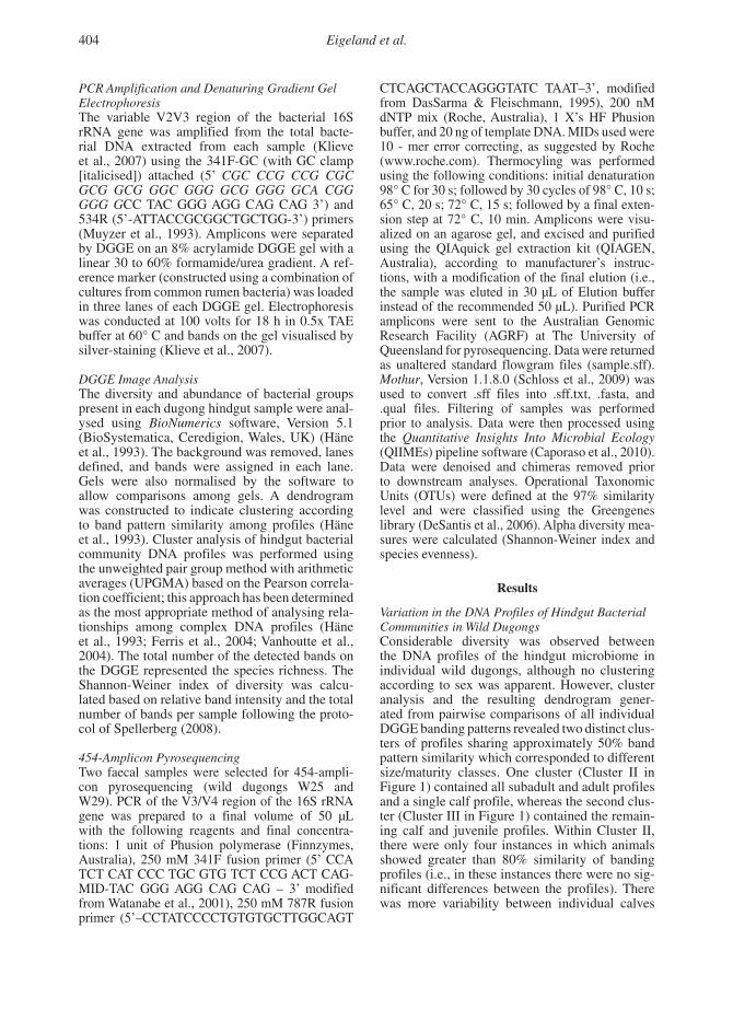
PCR Amplification and Denaturing Gradient Gel ElectrophoresisThe variable V2V3 region of the bacterial 16S rRNA gene was amplified from the total bacte-rial DNA extracted from each sample (Klieve et al., 2007) using the 341F-GC (with GC clamp [italicised]) attached (5’ CGC CCG CCG CGC GCG GCG GGC GGG GCG GGG GCA CGG GGG GCC TAC GGG AGG CAG CAG 3’) and 534R (5’-ATTACCGCGGCTGCTGG-3’) primers (Muyzer et al., 1993). Amplicons were separated by DGGE on an 8% acrylamide DGGE gel with a linear 30 to 60% formamide/urea gradient. A ref-erence marker (constructed using a combination of cultures from common rumen bacteria) was loaded in three lanes of each DGGE gel. Electrophoresis was conducted at 100 volts for 18 h in 0.5x TAE buffer at 60° C and bands on the gel visualised by silver-staining (Klieve et al., 2007).
DGGE Image AnalysisThe diversity and abundance of bacterial groups present in each dugong hindgut sample were anal-ysed using BioNumerics software, Version 5.1 (BioSystematica, Ceredigion, Wales, UK) (Häne et al., 1993). The background was removed, lanes defined, and bands were assigned in each lane. Gels were also normalised by the software to allow comparisons among gels. A dendrogram was constructed to indicate clustering according to band pattern similarity among profiles (Häne et al., 1993). Cluster analysis of hindgut bacterial community DNA profiles was performed using the unweighted pair group method with arithmetic averages (UPGMA) based on the Pearson correla-tion coefficient; this approach has been determined as the most appropriate method of analysing rela-tionships among complex DNA profiles (Häne et al., 1993; Ferris et al., 2004; Vanhoutte et al., 2004). The total number of the detected bands on the DGGE represented the species richness. The Shannon-Weiner index of diversity was calcu-lated based on relative band intensity and the total number of bands per sample following the proto-col of Spellerberg (2008).
454-Amplicon PyrosequencingTwo faecal samples were selected for 454-ampli-con pyrosequencing (wild dugongs W25 and W29). PCR of the V3/V4 region of the 16S rRNA gene was prepared to a final volume of 50 µL with the following reagents and final concentra-tions: 1 unit of Phusion polymerase (Finnzymes, Australia), 250 mM 341F fusion primer (5’ CCA TCT CAT CCC TGC GTG TCT CCG ACT CAG-MID-TAC GGG AGG CAG CAG – 3’ modified from Watanabe et al., 2001), 250 mM 787R fusion primer (5’–CCTATCCCCTGTGTGCTTGGCAGT
CTCAGCTACCAGGGTATC TAAT–3’, modified from DasSarma & Fleischmann, 1995), 200 nM dNTP mix (Roche, Australia), 1 X’s HF Phusion buffer, and 20 ng of template DNA. MIDs used were 10 - mer error correcting, as suggested by Roche (www.roche.com). Thermocyling was performed using the following conditions: initial denaturation 98° C for 30 s; followed by 30 cycles of 98° C, 10 s; 65° C, 20 s; 72° C, 15 s; followed by a final exten-sion step at 72° C, 10 min. Amplicons were visu-alized on an agarose gel, and excised and purified using the QIAquick gel extraction kit (QIAGEN, Australia), according to manufacturer’s instruc-tions, with a modification of the final elution (i.e., the sample was eluted in 30 µL of Elution buffer instead of the recommended 50 µL). Purified PCR amplicons were sent to the Australian Genomic Research Facility (AGRF) at The University of Queensland for pyrosequencing. Data were returned as unaltered standard flowgram files (sample.sff). Mothur, Version 1.1.8.0 (Schloss et al., 2009) was used to convert .sff files into .sff.txt, .fasta, and .qual files. Filtering of samples was performed prior to analysis. Data were then processed using the Quantitative Insights Into Microbial Ecology (QIIMEs) pipeline software (Caporaso et al., 2010). Data were denoised and chimeras removed prior to downstream analyses. Operational Taxonomic Units (OTUs) were defined at the 97% similarity level and were classified using the Greengenes library (DeSantis et al., 2006). Alpha diversity mea-sures were calculated (Shannon-Weiner index and species evenness).
Results
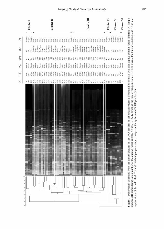
Variation in the DNA Profiles of Hindgut Bacterial Communities in Wild DugongsConsiderable diversity was observed between the DNA profiles of the hindgut microbiome in individual wild dugongs, although no clustering according to sex was apparent. However, cluster analysis and the resulting dendrogram gener-ated from pairwise comparisons of all individual DGGE banding patterns revealed two distinct clus-ters of profiles sharing approximately 50% band pattern similarity which corresponded to different size/maturity classes. One cluster (Cluster II in Figure 1) contained all subadult and adult profiles and a single calf profile, whereas the second clus-ter (Cluster III in Figure 1) contained the remain-ing calf and juvenile profiles. Within Cluster II, there were only four instances in which animals showed greater than 80% similarity of banding profiles (i.e., in these instances there were no sig-nificant differences between the profiles). There was more variability between individual calves
Dugong Hindgut Bacterial Community 405
I I I I V or
I I II V V
r
r r r rr
te te te te te te
s s s s s sam
ple
wild
s
u u u u ul ul l l l (F)
C
l
C C C C C (A)
and
)F sa
mpl
es:
(
aeca
l sa
mpl
ing,
f
dugo
ng of
)E(
time
) e the
D v
(
capt
i at
)C
clas
s
( and
size
)B
wild
(E
)
( all
) (cm
),
A from
(
com
mun
ities
sa
mpl
ing
of
time
iles
(%).
bact
eria
l th
e at
hi
ndgu
t le
ngth
th
e bo
dy
of
(D)
iles
x,
prof se
(C)
A
DN
the
sam
ple,
of
ae
cal
anal
ysis
f
dugo
ng
clus
ter
the
the of
fr
om
num
ber
gene
rate
d ic
atio
n The
sca
le a
t the
top
repr
esen
ts p
erce
ntag
e si
mila
rity
bet
wee
n D
GG
E p
rof
vidu
al.
Den
drog
ram
na
me/
iden
tif(B
) 1.
e
num
ber, e st
atus
of t
he in
di
Fig
ur vca
pti
406 Eigeland et al.
and juveniles in Cluster III, with no two profiles being identical.
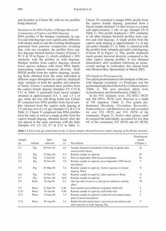
Variation in the DNA Profiles of Hindgut Bacterial Communities of Captive and Wild DugongsDNA profiles of the hindgut community in cap-tive and wild dugongs were significantly different, with cluster analysis and the resulting dendrogram generated from pairwise comparisons revealing that, with one exception, the profiles from cap-tive dugongs formed distinct clusters (Clusters I, IV, V & VI in Figure 1), which exhibited < 30% similarity with the profiles of wild dugongs. Hindgut profiles from captive dugongs showed lower species richness with fewer DNA bands, representing reduced bacterial diversity. Each DGGE profile from the captive dugongs, includ-ing those obtained from the same individual at different stages throughout its captivity, displayed large variation in both the number and intensity of bands. Cluster I contained three profiles from the captive female dugong (Samples C9, C10 & C11 in Table 1) generated from faecal samples obtained at approximately 0.5, 1, and 1.5 y of age, while she was still being bottle-fed. Cluster IV contained two DNA profiles from faecal sam-ples obtained from the captive male dugong at 5.5 and just over 6 y of age (Samples C2 & C3 in Table 1). Cluster V comprised four DNA profiles from the male as well as a single profile from the captive female dugong, obtained shortly after she was placed in the same enclosure with the male (Samples C4, C5, C6, C7 & C12 in Table 1).
Cluster VI contained a unique DNA profile from the captive female dugong, generated from a faecal sample obtained 1 d after rescue as a young calf approximately 2 wks of age (Sample C8 in Table 1). This profile displayed < 20% similarity to all other hindgut bacterial profiles from cap-tive and wild dugongs. A single profile from the captive male dugong at approximately 4 y of age (juvenile) (Sample C1 in Table 1) clustered with the profiles from subadult and adult wild dugongs (Cluster II in Figure 1). This profile displayed much greater bacterial diversity compared to the other captive dugong profiles. It was obtained immediately after recapture following an unsuc-cessful attempt to reintroduce this animal back into Moreton Bay, Queensland, in March 2002.
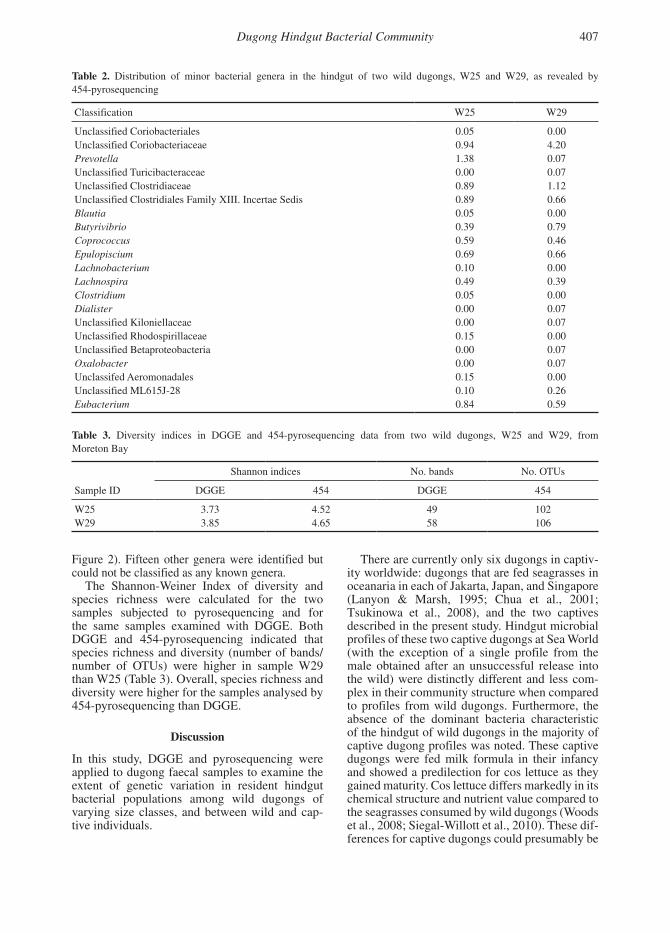
454-Amplicon PyrosequencingTwo phyla predominated in the hindguts of the two wild dugongs examined: (1) Firmicutes was the predominant phylum followed by (2) Bacteroidetes (Table 2). The next prevalent phyla were Actinobacteria and Proteobacteria (Table 2).
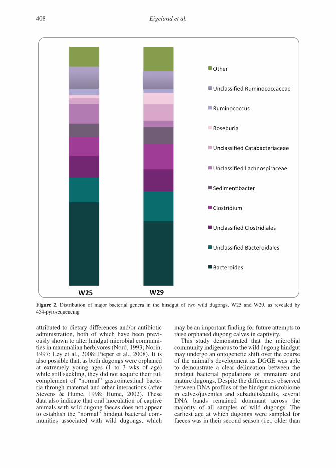
At the 97% similarity level, 102 OTUs (W25) and 106 OTUs (W29) were observed at a depth of 750 sequences (Table 3). Five genera pre-dominated (Roseburia, Clostridium, Bacteroides, Sedimentibacter, and Ruminococcus) and accounted for over 52% (W25) and 51% (W29) of the community (Figure 2). Twelve other genera could be assigned but individually, accounted for less than 6% of the community (5% [W25] and 4% [W29];
Table 1. Clinical and age-related data for the 12 faecal samples obtained from two captive dugongs at Sea World, Australia
Sample no.
Animal
Date
collected
Age (y)
Description
Dendrogram cluster
in Figure 1
C1 Pig 09-Nov-02 4.0 Sample obtained immediately following recapture after unsuccessful release
II
C2 Pig 27-May-04 5.5 Routine sample in captivity IVC3 Pig 12-Feb-05 6.2 Prior to September 2005 faecal inoculation IVC4 Pig 30-Nov-05 7.0 Routine sample in captivity; post-September 2005 faecal
inoculationV
C5 Pig 03-May-06 7.4 Routine sample in captivity; before exposure to female dugong, Wuru
V
C6 Pig 15-Nov-07 9.0 Routine sample in captivity; after exposure to Wuru VC7 Pig 26-Nov-08 10.0 Routine sample in captivity VC8 Wuru 26-Jan-05 0.1 One day after rescue as a calf (given antibiotics shortly
after)VI
C9 Wuru 22-June-05 0.40 Four months post-antibiotic treatment; bottle-fed IC10 Wuru 03-Jan-06 0.90 Routine sample in captivity (still bottle-fed) IC11 Wuru 13-June-06 1.4 Routine sample in captivity; bottle-fed and eating lettuce;
prior to faecal inoculationI
C12 Wuru 29-Aug-06 1.6 Bottle-fed and eating lettuce; post-faecal inoculation and after exposure to male dugong, Pig
V
Dugong Hindgut Bacterial Community 407
Figure 2). Fifteen other genera were identified but could not be classified as any known genera.
The Shannon-Weiner Index of diversity and species richness were calculated for the two samples subjected to pyrosequencing and for the same samples examined with DGGE. Both DGGE and 454-pyrosequencing indicated that species richness and diversity (number of bands/number of OTUs) were higher in sample W29 than W25 (Table 3). Overall, species richness and diversity were higher for the samples analysed by 454-pyrosequencing than DGGE.
Discussion
In this study, DGGE and pyrosequencing were applied to dugong faecal samples to examine the extent of genetic variation in resident hindgut bacterial populations among wild dugongs of varying size classes, and between wild and cap-tive individuals.
There are currently only six dugongs in captiv-ity worldwide: dugongs that are fed seagrasses in oceanaria in each of Jakarta, Japan, and Singapore (Lanyon & Marsh, 1995; Chua et al., 2001; Tsukinowa et al., 2008), and the two captives described in the present study. Hindgut microbial profiles of these two captive dugongs at Sea World (with the exception of a single profile from the male obtained after an unsuccessful release into the wild) were distinctly different and less com-plex in their community structure when compared to profiles from wild dugongs. Furthermore, the absence of the dominant bacteria characteristic of the hindgut of wild dugongs in the majority of captive dugong profiles was noted. These captive dugongs were fed milk formula in their infancy and showed a predilection for cos lettuce as they gained maturity. Cos lettuce differs markedly in its chemical structure and nutrient value compared to the seagrasses consumed by wild dugongs (Woods et al., 2008; Siegal-Willott et al., 2010). These dif-ferences for captive dugongs could presumably be
Table 2. Distribution of minor bacterial genera in the hindgut of two wild dugongs, W25 and W29, as revealed by 454-pyrosequencing
Classification W25 W29
Unclassified Coriobacteriales 0.05 0.00Unclassified Coriobacteriaceae 0.94 4.20Prevotella 1.38 0.07Unclassified Turicibacteraceae 0.00 0.07Unclassified Clostridiaceae 0.89 1.12Unclassified Clostridiales Family XIII. Incertae Sedis 0.89 0.66Blautia 0.05 0.00Butyrivibrio 0.39 0.79Coprococcus 0.59 0.46Epulopiscium 0.69 0.66Lachnobacterium 0.10 0.00Lachnospira 0.49 0.39Clostridium 0.05 0.00Dialister 0.00 0.07Unclassified Kiloniellaceae 0.00 0.07Unclassified Rhodospirillaceae 0.15 0.00Unclassified Betaproteobacteria 0.00 0.07Oxalobacter 0.00 0.07Unclassifed Aeromonadales 0.15 0.00Unclassified ML615J-28 0.10 0.26Eubacterium 0.84 0.59
Table 3. Diversity indices in DGGE and 454-pyrosequencing data from two wild dugongs, W25 and W29, from Moreton Bay
Shannon indices No. bands No. OTUs
Sample ID DGGE 454 DGGE 454
W25 3.73 4.52 49 102W29 3.85 4.65 58 106
408 Eigeland et al.
attributed to dietary differences and/or antibiotic administration, both of which have been previ-ously shown to alter hindgut microbial communi-ties in mammalian herbivores (Nord, 1993; Norin, 1997; Ley et al., 2008; Pieper et al., 2008). It is also possible that, as both dugongs were orphaned at extremely young ages (1 to 3 wks of age) while still suckling, they did not acquire their full complement of “normal” gastrointestinal bacte-ria through maternal and other interactions (after Stevens & Hume, 1998; Hume, 2002). These data also indicate that oral inoculation of captive animals with wild dugong faeces does not appear to establish the “normal” hindgut bacterial com-munities associated with wild dugongs, which
may be an important finding for future attempts to raise orphaned dugong calves in captivity.
This study demonstrated that the microbial community indigenous to the wild dugong hindgut may undergo an ontogenetic shift over the course of the animal’s development as DGGE was able to demonstrate a clear delineation between the hindgut bacterial populations of immature and mature dugongs. Despite the differences observed between DNA profiles of the hindgut microbiome in calves/juveniles and subadults/adults, several DNA bands remained dominant across the majority of all samples of wild dugongs. The earliest age at which dugongs were sampled for faeces was in their second season (i.e., older than
26
1
2
3
4
Figure 2. Distribution of major bacterial genera in the hindgut of two wild dugongs, W25 and 5
W29, as revealed by 454-pyrosequencing. 6
Figure 2. Distribution of major bacterial genera in the hindgut of two wild dugongs, W25 and W29, as revealed by 454-pyrosequencing
Dugong Hindgut Bacterial Community 409
1 y of age). Dugong calves, while still suckling, are presumably consuming increasing quantities of seagrass and may be beginning to acquire the dominant hindgut microbes required for cellulose degradation and volatile fatty acid production (Woods et al., 2008). Additionally, they may be acquiring these dominant microbes by ingesting bacteria that are associated with the surfaces of their seagrass forage (Delong et al., 1993). Wild dugong calves may also have acquired these dominant microbes from maternal interactions (i.e., suckling) and/or consumption of egesta as is common with other herbivorous mammals that are classified as hindgut fermenters (Stevens & Hume, 1998; Hume, 2002).
The single faecal sample obtained from the captive male dugong, that generated a hindgut bacterial community profile similar to those of wild subadult and adult profiles, was obtained immediately following recapture after an unsuccessful release into Moreton Bay, Queensland, in March 2002. Following the loss of a satellite-tracking device, the juvenile male dugong was eventually found in November 2002 in a poor state of health, with numerous open wounds, and was recaptured. This suggests that during the 8-mo reintroduction into Moreton Bay, the captive male dugong was exposed to natural environmental conditions, natural forage, and possible interactions with wild dugongs, all of which may have contributed to his hindgut bacterial community DNA profile resembling that of a wild seagrass-eating subadult/adult dugong. Faecal samples obtained after his subsequent captive period showed a significant reduction in bacterial diversity and a loss of the common bands shared with profiles of wild dugongs. Marked alteration or loss of bacterial diversity in the first post-capture sample followed by the stability of subsequent hindgut microbiome profiles (two profiles in Cluster IV and four profiles in Cluster V) could be attributed to the sudden dietary shift from seagrass to cos lettuce, the reintroduction to an artificial water environment, and/or the antibiotic administration required during convalescence.
A single hindgut microbiome profile from the captive female dugong (generated from the faecal sample obtained at 2 wks of age, 1 d after rescue in January 2005) was unique. At this age, the captive female dugong would have been completely dependent on her mother’s milk (Marsh et al., 1984; Woods et al., 2008). Consequently, this profile may be representative of the bacterial community diversity in the hindgut of a neonatal wild dugong calf. Three of the profiles from the captive female dugong that formed a distinct, separate cluster were obtained during bottle-feeding and a slow introduction to a lettuce diet. The remaining profile from the captive female dugong that clustered with the four profiles from the male was obtained
following placement in the same enclosure as the male during weaning to a diet of cos lettuce.
Given that the hindgut bacterial populations in wild dugongs appear to be relatively stable between individuals, a 454-pyrosequencing approach was used to obtain a preliminary indication of the phyla and genera of bacteria present in the hindgut of two wild dugongs. As expected, both dugongs had similar bacterial communities, and these were dominated by the phyla Firmicutes and Bacteroidetes. This is in keeping with other terrestrial herbivorous microbial ecosystems such as the cow rumen (Brulc et al., 2009). The five genera that dominated the community (Roseburia, Clostridium, Bacteroides, Sedimentibacter, and Ruminococcus accounted for over 51% of the community) are also commonly associated with terrestrial herbivores (Brulc et al., 2009; Hess et al., 2011). These bacteria may therefore have an important role in seagrass fibre degradation in the dugong hindgut. Further, 16S rRNA gene sequence analysis of representatives of these major genera and phylogenetic comparison with known sequences of related microorganisms isolated from terrestrial herbivores may help to answer important questions about dugong evolution and adaptation to the marine environment.
In conclusion, this study has provided preliminary information on the population structure of hindgut bacterial communities in wild dugongs. Several hypotheses have been previously formulated for the origins of established hindgut microbial communities in mammals, the most prominent being maternal interactions (i.e., mother to offspring) or the influence of diet (Gordon & Phillips, 1993; Flint & Bayer, 2008; Ley et al., 2008; Fraser et al., 2009). However, no studies have previously reported the marked differences between wild and captive members of the same species as observed in this study.
Acknowledgments
We would like to thank The University of Queensland Dugong Research Team for their assis-tance in the capture and sampling of wild dugongs. In particular, we thank Helen Sneath, Liz Burgess, Merrick Ekins, Erin Neal, Ben Schemel, Rob Slade, Paul Sprecher, and Trevor Long. Faecal samples from wild dugongs were collected under The University of Queensland Animal Ethics #ZOO/ENT/344/04/NSF/CRL, Moreton Bay Marine Parks Permit #QS2004/CVL228, and Scientific Purposes Permit #WISP01660304. Faecal sam-ples from captive dugongs were collected under The University of Queensland Animal Ethics #ZOO/ENT/328/05/NSF/CRL. This project was funded by a grant from the Sea World Research and Rescue Foundation Australia, with additional
410 Eigeland et al.
funding for fieldwork from Unimin Australia (for-merly Consolidated Rutile Ltd) and the Winifred Violet Scott Foundation. Order of authorship fol-lows the “sequence-determines-credit” approach.
Literature Cited
Brulc, J. M., Antonopoulos, D. A., Berg Miller, M. E., Wilson, M. K., Yannarell, A. C., Dinsdale, E. A., . . . White, B. A. (2009). Gene-centric metagenomics of the fiber-adherent bovine rumen microbiome reveals forage specific glycoside hydrolases. Proceedings of National Academy of Science, 106, 1948-1953. http://dx.doi.org/10.1073/pnas.0806191105
Burgess, E. A., Lanyon, J. M., & Keeley, T. (2012a). Testosterone and tusks: Maturation and seasonal reproduc-tive patterns of live, free-ranging male dugongs (Dugong dugon) in a subtropical population. Reproduction, 143, 1-16. http://dx.doi.org/10.1530/REP-11-0434
Burgess, E. A., Lanyon, J. M., Brown, J. L., Blyde, D., & Keeley, T. (2012b). Diagnosing pregnancy in free-rang-ing dugongs using fecal progesterone metabolite concen-trations and body morphometrics: A population applica-tion. General and Comparative Endocrinology, 177, 82-92. http://dx.doi.org/10.1016/j.ygcen.2012.02.008
Caporaso, G., Kuczynski, J., Stombaugh, J., Bittinger, K., Bushman, F. D., Costello, E. K., . . . Knight, R. (2010). QIIME allows analysis of high-throughput community sequencing data. Nature Methods, 7, 335-336. http://dx.doi.org/10.1038/nmeth.f.303
Chua, F. H. C., Mackay, B., Whitfield, M., & Ooi, E. (2001). Rehabilitation and display of a stranded dugong (Dugong dugon) calf in Singapore. Bulletin of the Institute of Oceanography (Monaco), 20, 403-406.
DasSarma, S., & Fleischmann, E. F. (1995). Archaea: A laboratory manual: Halophiles. New York: Cold Spring Harbour Laboratory Press.
Delong, E. F., Franks, D. G., & Alldredge, A. L. (1993). Phylogenetic diversity of aggregate-attached vs. free-living marine bacterial assemblages. Limnology and Oceanography, 38, 924-934.
DeSantis, T. Z., Hugenholtz, P., Larsen, N., Rojas, M., Brodie, E. L., Keller, K., . . . Andersen, G. L. (2006). Greengenes, a chimera-checked 16S rRNA gene database and workbench compatible with ARB. Applied and Environmental Microbiology, 72, 5069-5072. http://dx.doi.org/10.1128/AEM.03006-05
Ferris, M. M., Yan, X. M., Shou, Y. L., Lemanski, C. L., Jett, J. H., Yoshida, T. M., & Marrone, B. L. (2004). Performance assessment of DNA fragment sizing by high-sensitivity flow cytometry and pulsed-field gel electrophoresis. Journal of Clinical Microbiology, 42, 1965-1976. http://dx.doi.org/10.1128/JCM.42.5.1965-1976.2004
Flint, H. J., & Bayer, E. A. (2008). Plant cell wall breakdown by anaerobic microorganisms from the mammalian digestive tract. Incredible Anaerobes: From Physiology to Genomics to Fuels, 1125, 280-288.
Fraser, M. D., Theobald, V. J., Davies, D. R., & Moorby, J. M. (2009). Impact of diet selected by cattle and sheep grazing heathland communities on nutrient supply and faecal micro-flora activity. Agriculture, Ecosystems and Environment, 129, 367-377. http://dx.doi.org/10.1016/j.agee.2008.10.011
Gordon, G. L. R., & Phillips, M. W. (1993). Removal of anaerobic fungi from the rumen of sheep by chemical treatment and the effect on feed consumption and in vivo fiber digestion. Letters in Applied Microbiology, 17, 220-223. http://dx.doi.org/10.1111/j.1472-765X.1993.tb01451.x
Häne, B. G., Jaeger, K., & Drexler, H. G. (1993). The Pearson product-moment correlation coefficient is better suited for identification of DNA fingerprint profiles than band matching algorithms. Electrophoresis, 14, 967-972. http://dx.doi.org/10.1002/elps.11501401154
Hess, M., Sczyrba, A., Egan, R., Kim, T. W., Chokhawala, H., Schroth, G., . . . Rubin, E. M. (2011). Metagenomic discovery of biomass-degrading genes and genomes from cow rumen. Science, 331, 463-467. http://dx.doi.org/10.1126/science.1200387
Hume, I. D. (2002). Digestive strategies of mammals. Acta Zoologica Sinica, 48, 1-19.
Johnstone, I. M., & Hudson, B. E. T. (1981). The dugong diet: Mouth sample analysis. Bulletin of Marine Science, 31, 681-690.
Klieve, A. V., O’Leary, M. N., McMillen, L., & Ouwerkerk, D. (2007). Ruminococcus bromii: Identification and isolation as a dominant community member in the rumen of cattle fed a barley diet. Journal of Applied Microbiology, 103, 2065-2073. http://dx.doi.org/10.1111/j.1365-2672.2007.03492.x
Lanyon, J. M. (1991). The nutritional ecology of the dugong (Dugong dugon) in tropical North Queensland. (Unpublished doctoral dissertation). Monash University, Melbourne. 337 pp.
Lanyon, J. M., & Marsh, H. (1995). Digesta passage times in the dugong. Australian Journal of Zoology, 43, 119-127. http://dx.doi.org/10.1071/ZO9950119
Lanyon, J. M., & Sanson, G. D. (2006a). Degenerate dentition of the dugong (Dugong dugon), or why a grazer does not need teeth: Morphology, occlusion and wear of mouthparts. Journal of Zoology, 268, 133-152. http://dx.doi.org/10.1111/j.1469-7998.2005.00004.x
Lanyon, J. M., & Sanson, G. D. (2006b). Mechanical disruption of seagrass in the digestive tract of the dugong. Journal of Zoology, 270, 277-289.
Lanyon, J. M., Sneath, H. L., Kirkwood, J. M., & Slade, R. W. (2002). Establishing a mark-recapture program for dugongs in Moreton Bay, south-east Queensland. Australian Mammalogy, 24, 51-56. http://dx.doi.org/10.1071/AM02051
Lanyon, J. M., Sneath, H. L., Long, T., & Bonde, R. K. (2010). Physiological response of wild dugongs (Dugong dugon) to out-of-water sampling for health assessment. Aquatic Mammals, 36(1), 46-58. http://dx. doi.org/10.1578/AM.36.1.2010.46
Dugong Hindgut Bacterial Community 411
Lanyon, J. M., Slade, R. W., Sneath, H. L., Broderick, D., Kirkwood, J. M., Limpus, D., . . . Jessop, T. (2006). A method for capturing dugongs (Dugong dugon) in open water. Aquatic Mammals, 32(2), 196-201. http://dx.doi.org/10.1578/AM.32.2.2006.196
Ley, R. E., Hamady, M., Lozupone, C., Turnbaugh, P. J., Ramey, R. R., Bircher, J. S., . . . Gordon, J. I. (2008). Evolution of mammals and their gut microbes. Science, 320, 1647-1651. http://dx.doi.org/10.1126/science.1155725
Marsh, H., Heinsohn, G. E., & Marsh, L. M. (1984). Breeding cycle, life history and population dynamics of the dugong, Dugong dugon (Sirenia, Dugongidae). Australian Journal of Zoology, 2, 767-788. http://dx.doi.org/10.1071/ZO9840767
Marsh, H., Spain, A. V., & Heinsohn, G. E. (1978). Physiology of the dugong. Comparative Biochemistry and Physiology, 61, 159-168. http://dx.doi.org/10.1016/ 0300-9629(78)90089-0
Murray, R. M., Marsh, H., Heinsohn, G. E., & Spain, A. V. (1977). Role of the midgut cecum and large intestine in digestion of seagrasses by the dugong (Mammalia: Sirenia). Comparative Biochemistry and Physiology, 56, 7-10. http://dx.doi.org/10.1016/0300-9629(77)90432-7
Muyzer, G., Dewaal, E. C., & Uitterlinden, A. G. (1993). Profiling of complex microbial populations by denaturing gradient gel electrophoresis analysis of polymerase chain reaction-amplified genes coding for 16S ribosomal RNA. Applied Environmental Microbiology, 59, 695-700.
Nord, C. E. (1993). The effect of antimicrobial agents on the ecology of the human intestinal microflora. Veterinary Microbiology, 35, 193-197. http://dx.doi.org/10.1016/0378-1135(93)90144-V
Norin, K. E. (1997). Influence of antibiotics on some intestinal microflora associated characteristics. Anaerobe, 3, 145-148. http://dx.doi.org/10.1006/anae.1997.0091
Pieper, R., Jha, R., Rossnagel, B., Van Kessel, A. G., Souffrant, W. B., & Leterme, P. (2008). Effect of barley and oat cultivars with different carbohydrate compositions on the intestinal bacterial communities in weaned piglets. FEMS Microbiology Ecology, 66, 556-566. http://dx.doi.org/10.1111/j.1574-6941.2008.00605.x
Preen, A. R. (1992). Interactions between dugongs and seagrasses in a sub-tropical environment (Unpublished doctoral dissertation). James Cook University of North Queensland, Townsville. 386 pp.
Reynolds, J. E., & Rommel, S. A. (1996). Structure and function of the gastrointestinal tract of the Florida manatee, Trichechus manatus latirostris. Anatomical Record, 245, 539-558. http://dx.doi.org/10.1002/(SICI)1097-0185(199 607)245:3<539::AID-AR11>3.0.CO;2-Q
Schloss, P. D., Westcott, S. L., Ryabin, T., Hall, J. R., Hartmann, M., Hollister, E. B., . . . Weber, C. F. (2009). Introducing mothur: Open source, platform-independent, community-supported software for describing and com-paring microbial communities. Applied Environmental Microbiology, 75, 7537-7541. http://dx.doi.org/10.1128/AEM.01541-09
Siegal-Willott, J. L., Harr, K., Hayek, L-A. C., Scott, K. C., Gerlach, T., Sirois, P., . . . Hill, R. C. (2010). Proximate nutrient analyses of four species of submerged aquatic vegetation consumed by Florida manatee (Trichechus manatus latirostris) compared to romaine lettuce (Lactuca sativa var. longifolia). Journal of Zoo and Wildlife Medicine, 41, 594-602. http://dx.doi.org/10.1638/2009-0118.1
Spellerberg, I. F. (2008). Shannon-Weiner index. In S. E. Jorgensen & B. Fath (Eds.), Encyclopedia of ecology (pp. 3249-3252). Oxford, UK: Academic Press. http://dx.doi.org/10.1016/B978-008045405-4.00132-4
Stevens, C. E., & Hume, I. D. (1998). Contributions of microbes in vertebrate gastrointestinal tract to production and conservation of nutrients. Physiological Review, 78, 393-427.
Tsukinowa, E., Karita, S., Asano, S., Wakai, Y., Oka, Y., Furuta, M., & Goto, M. (2008). Faecal microbiota of a dugong (Dugong dugong) in captivity at Toba Aquarium. Journal of General and Applied Microbiology, 54, 25-38. http://dx.doi.org/10.2323/jgam.54.25
Vanhoutte, T., Huys, G., De Brandt, E., & Swings, J. (2004). Temporal stability analysis of the microbiota in human faeces by denaturing gradient gel electrophoresis using universal and group-specific 16S rRNA gene primers. FEMS Microbiology Ecology, 48, 437-446. http://dx.doi.org/10.1016/j.femsec.2004.03.001
Watanabe, K., Kodama, Y., & Harayama, S. (2001). Design and evaluation of PCR primers to amplify bacterial 16S ribosomal fragments used for community fingerprinting. Journal of Microbiological Methods, 44, 253-262.
Woods, R., Ladds, P., & Blyde, D. (2008). Dugongs. In L. Vogelnest & R. Woods (Eds.), Medicine of Australian mammals (pp. 615-627). Melbourne: CSIRO Publishing.
Yamamuro, M., & Chirapart, A. (2005). Quality of the seagrass Halophila ovalis on a Thai intertidal flat as food for the dugong. Journal of Oceanography, 61, 183-186. http://dx.doi.org/10.1007/s10872-005-0030-6
Yu, Z., & Forster, R. J. (2005). Nucleic acid extraction, oligonucleotide probes and PCR methods. In H. P. S. Makkar & C. S. McSweeney (Eds.), Methods in gut microbial ecology for ruminants (pp. 81-104). Dordrecht, The Netherlands: Springer.
Related Documents