Hindgut Innate Immunity and Regulation of Fecal Microbiota through Melanization in Insects * □ S Received for publication, February 19, 2012 Published, JBC Papers in Press, February 28, 2012, DOI 10.1074/jbc.M112.354548 Qimiao Shao ‡ , Bing Yang ‡1 , Qiuyun Xu ‡1 , Xuquan Li ‡ , Zhiqiang Lu § , Chengshu Wang ‡ , Yongping Huang ‡ , Kenneth Söderhäll ¶2 , and Erjun Ling ‡3 From the ‡ Key Laboratory of Insect Developmental and Evolutionary Biology, Institute of Plant Physiology and Ecology, Shanghai Institutes for Biological Sciences, Chinese Academy of Sciences, Shanghai 200032, People’s Republic of China, the § College of Plant Protection, Northwest A&F University, 22 Xinong Road, Yangling, Shanxi 712100, People’s Republic of China, and the ¶ Department of Comparative Physiology, Uppsala University, Norbyvägen 18 A, 752 18 Uppsala, Sweden Background: Many insects eat green leaves but excrete black feces using an unknown mechanism. Results: Hindgut cells produce prophenoloxidase and secrete it into the hindgut content. Conclusion: Prophenoloxidase induces the hindgut content and feces melanization by which bacteria flora are reduced. Significance: This is a first-time disclosure of the enigma of insect black feces and its biological significance. Many insects eat the green leaves of plants but excrete black feces in an as yet unknown mechanism. Insects cannot avoid ingesting pathogens with food that will be specifically detected by the midgut immune system. However, just as in mammals, many pathogens can still escape the insect midgut immune system and arrive in the hindgut, where they are excreted out with the feces. Here we show that the melanization of hind- gut content induced by prophenoloxidase, a key enzyme that induces the production of melanin around invaders and at wound sites, is the last line of immune defense to clear bacteria before feces excretion. We used the silkworm Bombyx mori as a model and found that prophenoloxidase produced by hindgut cells is secreted into the hindgut contents. Several experiments were done to clearly demonstrate that the blackening of the insect feces was due to activated phenoloxidase, which served to regulate the number of bacteria in the hindgut. Our analysis of the silkworm hindgut prophenoloxidase discloses the natural secret of why the phytophagous insect feces is black and pro- vides insight into hindgut innate immunity, which is still rather unclear in mammals. There are over 10 11 microbe cells per gram content in the mammalian large intestine because it lacks Peyer’s patches (1). Among those bacteria, fecal pathogens cause health problems throughout the world, particularly through water pollution (2). As in mammals, many pathogens can still escape insect midgut immune system and pass to the hindgut, which is an organ similar to the mammalian large intestine (3, 4). Because of sim- ilarities with mammals, the insect midgut immune system upon aging, stress, or infection has been extensively studied (4, 5). However, as yet, it is unclear whether there is an innate immu- nity system in mammalian large intestine and the insect hind- gut that destroys pathogens before feces excretion. Insects make up the largest group of animals on earth, and almost 45% of insects are herbivorous (6). Most herbivorous insects live on green leaves, and typical insects such as the silk- worm eat green mulberry leaves although they excrete black feces. As yet, the mechanism of how feces are blackened is still unknown. The insect gut is a digestion system that is divided into foregut, midgut, and hindgut. Insect hindgut is generally thought to re-absorb certain salts and amino acids from the content (before excretion as feces) to maintain the osmotic pressure in the hemolymph (7). Many symbiotic bacteria including un-killed pathogens cannot avoid being excreted with feces, and those microbes may further pollute insect feed and its habitat. Little is known whether insect hindgut has an immune system to deal with microbes in feces before excretion. Prophenoloxidase (PPO) 4 is a very important innate immu- nity protein that is produced by hemocytes in invertebrates (8 –12). Activated phenoloxidase (PO) induces the production of melanin (melanization) around invading microorganisms to first seal them off from circulation and then kill them. Melanization is also induced around wounds to prevent addi- tional infections. Activated PO produces melanin in the epider- mis and cuticle and is therefore also responsible for insect body color and patterns (13, 14). Two PPO genes occur in the silk- worm (8, 15). Because the feces of phytophagous insects is black, we hypothesized that PPO may be responsible for melanization of feces. However, like PPO, laccase and peroxi- dase also oxidize some phenols (10), and thus, these enzymes may also induce melanization of feces. In this study, we used biochemical assays and in situ hybrid- ization to show that cells in the hindgut of Bombyx mori pro- * This work was supported by the National Natural Science Foundation of China (30970408), National Basic Research Program of China (2012CB114605), Chinese Academy of Sciences (KSCX2-EW-J-12 and 2009OHTP05), and the Swedish Science Research Council (319-2010-6250). □ S This article contains supplemental Tables S1–S3 and Figs. S1–S7. 1 Both authors contributed equally to this work. 2 To whom correspondence may be addressed. Tel.: 46-18-4712818; Fax: 46-18-4716425; E-mail: [email protected]. 3 To whom correspondence may be addressed. Tel.: 86-21-54924155; Fax: 86-21-54924155; E-mail: [email protected]. 4 The abbreviations used are. PPO, prophenoloxidase; PO, phenoloxidase; FG, foregut; MG, midgut; HG, hindgut; PY, pylorus; PTU, phenylthiourea; TMB, 3,3,5,5;-tetramethylbenzidine. THE JOURNAL OF BIOLOGICAL CHEMISTRY VOL. 287, NO. 17, pp. 14270 –14279, April 20, 2012 © 2012 by The American Society for Biochemistry and Molecular Biology, Inc. Published in the U.S.A. 14270 JOURNAL OF BIOLOGICAL CHEMISTRY VOLUME 287 • NUMBER 17 • APRIL 20, 2012 by guest on February 15, 2018 http://www.jbc.org/ Downloaded from

Welcome message from author
This document is posted to help you gain knowledge. Please leave a comment to let me know what you think about it! Share it to your friends and learn new things together.
Transcript

Hindgut Innate Immunity and Regulation of Fecal Microbiotathrough Melanization in Insects*□S
Received for publication, February 19, 2012 Published, JBC Papers in Press, February 28, 2012, DOI 10.1074/jbc.M112.354548
Qimiao Shao‡, Bing Yang‡1, Qiuyun Xu‡1, Xuquan Li‡, Zhiqiang Lu§, Chengshu Wang‡, Yongping Huang‡,Kenneth Söderhäll¶2, and Erjun Ling‡3
From the ‡Key Laboratory of Insect Developmental and Evolutionary Biology, Institute of Plant Physiology and Ecology, ShanghaiInstitutes for Biological Sciences, Chinese Academy of Sciences, Shanghai 200032, People’s Republic of China, the §College of PlantProtection, Northwest A&F University, 22 Xinong Road, Yangling, Shanxi 712100, People’s Republic of China, and the ¶Departmentof Comparative Physiology, Uppsala University, Norbyvägen 18 A, 752 18 Uppsala, Sweden
Background:Many insects eat green leaves but excrete black feces using an unknown mechanism.Results: Hindgut cells produce prophenoloxidase and secrete it into the hindgut content.Conclusion: Prophenoloxidase induces the hindgut content and feces melanization by which bacteria flora are reduced.Significance: This is a first-time disclosure of the enigma of insect black feces and its biological significance.
Many insects eat the green leaves of plants but excrete blackfeces in an as yet unknown mechanism. Insects cannot avoidingesting pathogens with food that will be specifically detectedby the midgut immune system. However, just as in mammals,many pathogens can still escape the insect midgut immunesystem and arrive in the hindgut, where they are excreted outwith the feces. Here we show that the melanization of hind-gut content induced by prophenoloxidase, a key enzyme thatinduces the production of melanin around invaders and atwound sites, is the last line of immune defense to clear bacteriabefore feces excretion.We used the silkworm Bombyx mori as amodel and found that prophenoloxidase produced by hindgutcells is secreted into the hindgut contents. Several experimentswere done to clearly demonstrate that the blackening of theinsect feces was due to activated phenoloxidase, which served toregulate the number of bacteria in the hindgut. Our analysis ofthe silkworm hindgut prophenoloxidase discloses the naturalsecret of why the phytophagous insect feces is black and pro-vides insight into hindgut innate immunity, which is still ratherunclear in mammals.
There are over 1011 microbe cells per gram content in themammalian large intestine because it lacks Peyer’s patches (1).Among those bacteria, fecal pathogens cause health problemsthroughout the world, particularly through water pollution (2).As in mammals, many pathogens can still escape insect midgutimmune system and pass to the hindgut, which is an organsimilar to the mammalian large intestine (3, 4). Because of sim-
ilaritieswithmammals, the insectmidgut immune systemuponaging, stress, or infection has been extensively studied (4, 5).However, as yet, it is unclear whether there is an innate immu-nity system in mammalian large intestine and the insect hind-gut that destroys pathogens before feces excretion.Insects make up the largest group of animals on earth, and
almost 45% of insects are herbivorous (6). Most herbivorousinsects live on green leaves, and typical insects such as the silk-worm eat green mulberry leaves although they excrete blackfeces. As yet, the mechanism of how feces are blackened is stillunknown.The insect gut is a digestion system that is divided into
foregut, midgut, and hindgut. Insect hindgut is generallythought to re-absorb certain salts and amino acids from thecontent (before excretion as feces) to maintain the osmoticpressure in the hemolymph (7). Many symbiotic bacteriaincluding un-killed pathogens cannot avoid being excretedwith feces, and those microbes may further pollute insect feedand its habitat. Little is known whether insect hindgut has animmune system to deal withmicrobes in feces before excretion.Prophenoloxidase (PPO)4 is a very important innate immu-
nity protein that is produced by hemocytes in invertebrates(8–12). Activated phenoloxidase (PO) induces the productionof melanin (melanization) around invading microorganisms tofirst seal them off from circulation and then kill them.Melanization is also induced around wounds to prevent addi-tional infections. Activated POproducesmelanin in the epider-mis and cuticle and is therefore also responsible for insect bodycolor and patterns (13, 14). Two PPO genes occur in the silk-worm (8, 15). Because the feces of phytophagous insects isblack, we hypothesized that PPO may be responsible formelanization of feces. However, like PPO, laccase and peroxi-dase also oxidize some phenols (10), and thus, these enzymesmay also induce melanization of feces.In this study, we used biochemical assays and in situ hybrid-
ization to show that cells in the hindgut of Bombyx mori pro-
* This work was supported by the National Natural Science Foundation ofChina (30970408), National Basic Research Program of China(2012CB114605), Chinese Academy of Sciences (KSCX2-EW-J-12and 2009OHTP05), and the Swedish Science Research Council(319-2010-6250).
□S This article contains supplemental Tables S1–S3 and Figs. S1–S7.1 Both authors contributed equally to this work.2 To whom correspondence may be addressed. Tel.: 46-18-4712818; Fax:
46-18-4716425; E-mail: [email protected] To whom correspondence may be addressed. Tel.: 86-21-54924155; Fax:
86-21-54924155; E-mail: [email protected].
4 The abbreviations used are. PPO, prophenoloxidase; PO, phenoloxidase; FG,foregut; MG, midgut; HG, hindgut; PY, pylorus; PTU, phenylthiourea; TMB,3,3�,5,5;-tetramethylbenzidine.
THE JOURNAL OF BIOLOGICAL CHEMISTRY VOL. 287, NO. 17, pp. 14270 –14279, April 20, 2012© 2012 by The American Society for Biochemistry and Molecular Biology, Inc. Published in the U.S.A.
14270 JOURNAL OF BIOLOGICAL CHEMISTRY VOLUME 287 • NUMBER 17 • APRIL 20, 2012
by guest on February 15, 2018http://w
ww
.jbc.org/D
ownloaded from
duced PPO. PPO was also found in the hindgut contents.Melanization of the silkworm larvae feceswas blocked, andbac-terial number increased in hindgutwhen phenylthiourea (PTU)was introduced through feeding. Therefore, activated PO andPO-induced melanization of hindgut content helped reducebacterial numbers, suggesting that PO is an important regulatorof the bacterial flora in hindgut and feces.
EXPERIMENTAL PROCEDURES
Insect Feeding and Dissection—B. mori larvae (Nistari) werereared on mulberry leaves at 25 °C under a 12-h photoperiod.Larvae on day 3 of the fourth larval stage (IV-3), the fourthmolting stage (IV-M), day 3 of the fifth larval stage (V-3), or atthe wandering-stage (W) were used for experiments. To obtainsamples for Western blot, immunostaining, or native gel assay,silkworms and other larval species were dissected in autoclaved0.85% NaCl solution after bleeding. The dissected tissues werewashed in fresh 0.85% NaCl solution three times to removehemolymph. The gut was washed in 0.85% NaCl three times,and the corresponding gut parts were dried and then cut opento transfer contents to a new tube. Larvae were bled, and thehemolymph was transferred to a new tube after centrifuging at10,000 � g for 5 min. The supernatant was the plasma.Dianemobius nigrofasciatus, Tribolium castaneum,Drosophilamelanogaster (w1118),Coptotermes formosanus,Ostrinia furna-clis, Heliothis armigero, and Culex pipiens quinquefasciatuswere fed as usual.PPO, Laccase, and Peroxidase Enzymatic Assays—PPO, lac-
case, and peroxidase are a group of enzymes that may oxidizedopamine to produce melanin and metabolites (10). The activ-ities of the three enzymes were compared in hindgut contentusing 3 �g of laccase (38429; Sigma), 1.67 ng of peroxidase(P719; Invitrogen), and 1.25 �g of purified recombinant Dro-sophila prophenoloxidase 1 (DmrPPO1) (16). A 200 �l aliquotof 2 mM ABTS (Sigma) for laccase and TMB solution for per-oxidase was used (1:50; Invitrogen). 10 mM dopamine (Sigma)was used for DmrPPO1. DmrPPO1 needed to be activated with30% ethanol before use (16). The inhibitors were incubatedwith each enzyme for 5 min before adding the substrate. 3 mM
NaN3 (final concentration) was used for inhibition of laccaseand peroxidase, and saturated PTU was used for inhibition ofDmrPPO1. Gut content equal to feces wet weight was sus-pended in 200 �l of Tris buffer (10 mM, pH 7.4) and vortexedseveral times. The suspension (20 �l) was mixed with 200 �l ofeach substrate plus the corresponding enzyme inhibitor and/orTris buffer to make the total volume the same. Because theinhibitors were used to detect whether they could inhibit dif-ferent enzymes, each substratewith inhibitor addedwas treatedas a blank. They were added in Tris buffer containing inhibitoralone, at the same volume as the blank, to determine HG1 andHG2 content. The solutions were incubated at room tempera-ture for 8 min. The mixture was centrifuged at 10,000 � g for 1min, and the absorbance of the supernatant was read at 10 minusing the EXPERT 96 microplate reader (Biochrom, Holliston,MA). Absorbance was read at 490, 450, and 405 nm to detectPPO, laccase, and peroxidase activities, respectively. One unitof each enzyme activity was defined as �A�/min � 0.001 (�,wavelength).
Gut Staining, Gut Content, and Feces Collection—The dis-sected gut of the silkworms was incubated in TMB, ABTS, ordopamine containing 30% ethanol or not for staining after foodcontents were removed. Dopamine is a PPO substrate, and eth-anol can be used to activate PPO (8). NaN3 or PTUwas added tomonitor the change inmelanization when dopamine (with eth-anol) was used for staining.Different parts of the gutwere opened, and the contents were
transferred to pre-weighed tubes to measure the wet weight ofthe contents.Gut contentswere suspended in either 10mMTrisbuffer (pH7.4) (for POactivity assay or PPOdegradation obser-vation) or 500 mM NaCl and 5 mM EDTA (for Western blotassay). The tubes were vortexed several times, centrifuged at10,000 � g at 4 °C for 5 min, and the supernatant was trans-ferred to a new tube. The supernatant was incubated at roomtemperature for different times to determine whether PPOwasdegradable.To check whether there is PPO in feces, 20 feces samples
excreted for 0 or 60 min (V-3 black feces and wandering-stagegreen feces, respectively) were suspended in 10 mM Tris buffer(pH 7.4) containing 500 mM NaCl and 5 mM EDTA and con-centrated to�40�l by ultrafiltration. 15�l of the concentratedsolution were loaded for Western blot assay.Fluorescent Bead Injection and Tracing—The injected fluo-
rescent beads phagocytosed by insect hemocyteswere observedunder a fluorescent microscope, to monitor hemocyte move-ment (17). In the same way, fluorescent beads (1.0 mm 4, Red(580/605); Molecular Probes) were injected into the silkwormlarvae (on V-3) for at least 6 h (17). The phagocytosed beadsinside hemocytes were first observed under a fluorescentmicroscope. The dissected hindguts were fixed, sectioned, anddeparaffinized as described (17) to examine under the fluores-cent microscope whether there were phagocytosed fluorescentbeads brought by circulating hemocytes.Immune Challenge and Lysozyme Detection—V-3 silkworm
larvae were injected with 5 � 106 formalin-killed Escherichiacoli cells suspended in sterilized 0.85% NaCl solution or 0.85%NaCl alone for immune challenge for 12 h. Then the silkwormlarvae were bled to obtain plasma. HG1 and HG2 and its con-tents were also sampled. These samples were used for detectinglysozyme by Western blot.Bacterial Culture—Gut content (equal to the weight of feces)
and feces fromV-3 andwandering-stage larvaewere suspendedin 1 ml of LB medium, and the suspended solutions with gutcontent fragments (25 �l plus 75 �l of fresh LB medium) werespread on LB plates cultured at 37 °C overnight. Colonies ofbacteria were counted and calculated.Phenylthiourea (PTU) Feeding—PTU is a strong PO inhibitor
(18). It was dissolved in autoclaved water, and saturated PTUwas filtered to remove bacteria. The PTU solution was spreadon mulberry leaves and dried before feeding to the silkwormlarvae. The change in the color of feces was observed continu-ously. HG1 and HG2 contents from PTU-fed larvae wereremoved forWestern blot as described above. After feeding thePTU, the excreted green feces were collected immediately forbacterial culture, as described above.Tissue Culture and Native Gel Analysis—Midgut and hind-
gut fromV-3 larvae were cultured in Gracemedium containing
Hindgut Innate Immunity
APRIL 20, 2012 • VOLUME 287 • NUMBER 17 JOURNAL OF BIOLOGICAL CHEMISTRY 14271
by guest on February 15, 2018http://w
ww
.jbc.org/D
ownloaded from
10% fetal bovine serum.The silk glandwas cultured as a control.The culture medium was sampled at scheduled times for thenative gel assay as described previously (19).SDS-PAGEandWestern Blot Analysis—Different parts of the
silkworm larvae gut were dissected. Tissues were homogenizedand sonicated in 10 mM Tris-HCl (pH 7.4), and centrifuged at10,000� g at 4 °C for 5min. The supernatant was collected, andtotal protein concentration was determined with bovine serumalbumin as the standard. Approximately 10 �g of protein wasloaded per lane, and SDS-PAGE and Western blot assay wereperformed. Antibody against the silkworm PPO (a gift fromDr.T. Asano; 1:5,000) (20), lysozyme (a gift from Dr. K. Suzuki;1:5,000) (21), or Manduca sexta laccase (a gift from Dr. M.Kanost; 1:2,000) (22) was used as the first antibody, and theAP-conjugated goat anti-rabbit IgG (1:5,000) was used as thesecond antibody. EasySee Western Marker (DM201; Trans-Gen) was used as the protein marker.LC-MS/MS—The EttanTM MDLC system (GE Healthcare,
Piscataway, NJ) was used for desalting and separating trypticpeptide mixtures. In this system, samples were desalted onreverse phase (RP) trap columns (Zorbax 300 SB C18, AgilentTechnologies, Santa Clara, CA), and then separated on an RPcolumn (150�m i.d., 100mm length, ColumnTechnology Inc.,Fremont, CA). Mobile phase A (0.1% formic acid in high per-formance liquid chromatography grade water) and mobilephase B (0.1% formic acid in acetonitrile) were selected. Thetryptic peptide mixture (20 �g) was loaded onto the columns,and separation was performed at a flow rate of 2 �l/min using alinear gradient of 4–50% B for 120 min. A FinniganTM LTQTM
linear ion trap MS (Thermo Electron Corp. Rockford, IL)equipped with an electrospray interface was connected to theLC setup to detect the eluted peptides.Data-dependentMS/MSspectra were obtained simultaneously. Each scan cycle con-sisted of one full MS scan in profile mode followed by fiveMS/MS scans in centroid mode with the following DynamicExclusionTM settings: repeat count 2, repeat duration 30 s,exclusion duration 90 s. Each sample was analyzed in triplicate.The silkworm protein sequence database used in this analysiswas downloaded from the NCBI website using the keywordBombyx mori.In Situ Hybridization—B. mori PPO1 and PPO2mRNAwere
detected in hindgut by in situ hybridization following describedmethods (23, 24). The antisense and sense RNA probe waslabeled with digoxigenin by in vitro transcription with T7 RNApolymerase in a reaction containing digoxigenin-UTP (DIGRNALabelingKit; Boehringer, Ingelheim amRhein, Germany).The primers for cloning PPO1 and PPO2 and those for synthe-sizing antisense and sense RNAprobes are listed in supplemen-tal Table S1.Immunohistochemistry—Different parts of the silkworm gut
were dissected as described above and fixed overnight at 4 °C inBouin’s fluid (23). Samples were sectioned and deparaffinizedas described (17). To detect PPO, a polyclonal antibody againstthe silkworm PPO (1:1,000) was used as the first antibody (20),and rhodamine-conjugated goat anti-rabbit IgG (1:200) wasused as the second antibody. To detect melanin in the hindguttissue, a monoclonal antibody 6D2 (IgM) (1:200) against mela-nin derived from Coccidioides posadasii was used as the first
antibody (a gift from Dr. Garry T. Cole) (25), and FITC-conju-gated goat anti-mouse IgM (1:1000) was the second antibody.All other procedureswere followed as described (26), andDAPIwas used to counterstain nuclei. All pictures were taken under afluorescent microscope (Olympus BX51) under differentialinterference contrast using the appropriate filter.
RESULTS
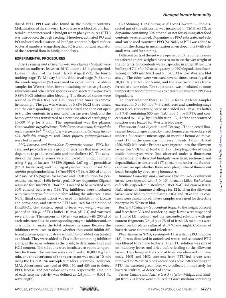
Black Feces Excreted by the Feeding Stage Silkworm: What Isthe Secret Behind It?—Silkworm larvae that are fed on greenmulberry leaves excreted black feces in which the leaf frag-ments were also black (Fig. 1,A,B-a, andB-b). However, duringthe wandering-stage, the last feces andmulberry leaf fragmentswere green (Fig. 1, B-c and B-d). After dissecting a silkwormlarva on day 3 of the fifth larval stage (V-3), we found that thecontents in different parts of the midgut (MG) were all green(Fig. 1C, bottom). The hindgut (HG) is divided into two parts,and in the first part of the hindgut (HG1), the content wasalways green. When the content passed to the second part ofthe hindgut (HG2), it became black (Fig. 1C). Feces excreted bylarvae before the wandering-stage are always black.PPO is a key enzyme in the silkworm that induces melaniza-
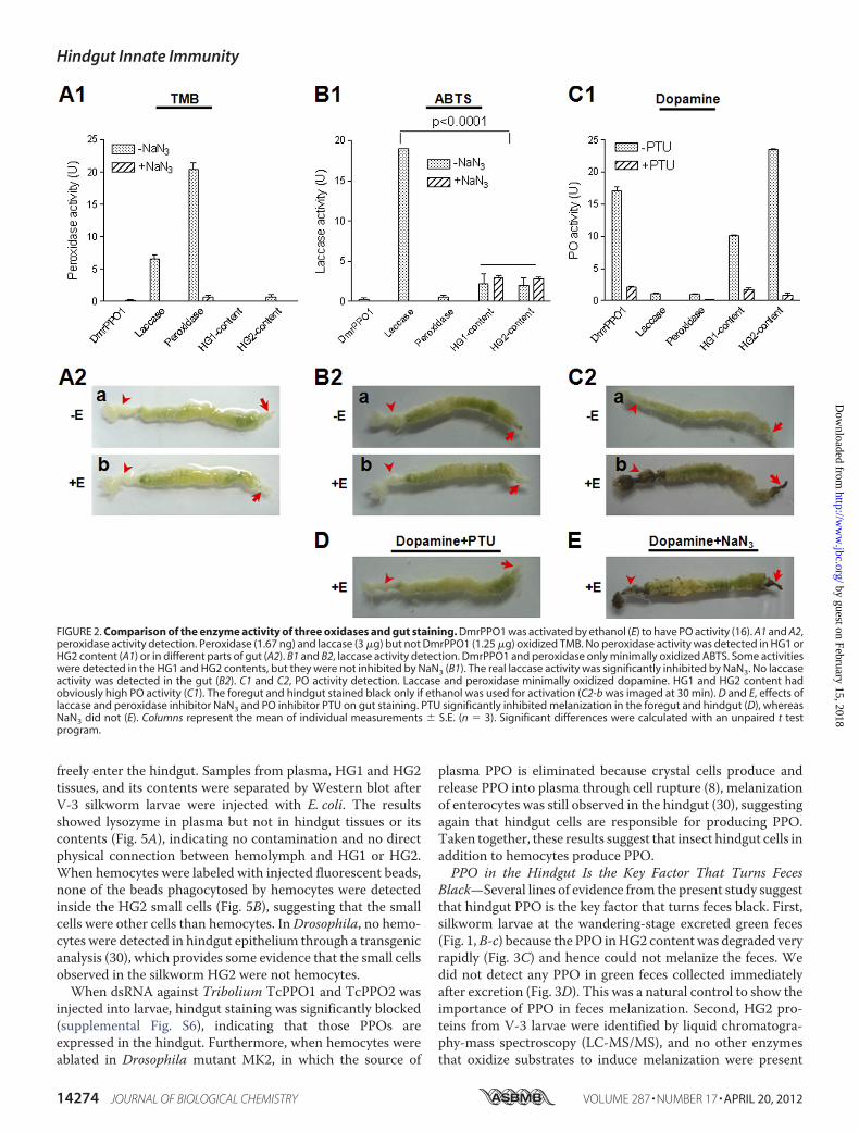
tion around wounds and invading foreign particles (8). Similarto PPO, laccase and peroxidase also oxidize some phenols (10);thus, it was necessary to investigate which of these redoxenzymes is responsible for the melanization of feces. Whendiluted 3,3�,5,5;-tetraMethylBenzidine (TMB) solution (1:50),which is a peroxidase substrate, was incubated with a commer-cial preparation of peroxidase (1.67 ng), it resulted in very highenzyme activity (Fig. 2A1). Laccase (3 �g) also oxidized TMB,but the activity was significantly lower than 1.67 ng of peroxi-dase. Purified recombinant Drosophila PPO1 (DmrPPO1, 1.25�g) did not oxidize TMB after the PPO had been activated byethanol (16). No peroxidase activity was present in HG1 andHG2 of the hindgut (Fig. 2A1). When ABTS solution (a laccasesubstrate) was added to DmrPPO1 or peroxidase, neither ofthem could oxidize this substrate (Fig. 2B1). Significant lowlaccase-like enzyme activity was observed inHG1 andHG2, butwhenNaN3 was added, they had almost the same activity as thegroup that did not receiveNaN3, suggesting that ABTS oxidiza-tion was not due to laccase, as NaN3 is a potent inhibitor oflaccase activity (27). Further, a Western blot assay usingM. sexta laccase antibody did not detect any positive band inthe hindgut content (supplemental Fig. S1).When dopamine, avery good PPO substrate, was added to different samples, Dmr-PPO1 oxidized the substrate very efficiently (Fig. 2C1). How-ever, laccase and peroxidase minimally oxidized dopamine.Larval guts were stained using the above substrates with or
without addition of ethanol. When TMB (Fig. 2A2) and ABTS(Fig. 2B2) were used, neither peroxidase nor laccase activitywere detected in gut tissues. No staining occurred when dop-amine was used if ethanol was absent (Fig. 2, C2-a). However,the foregut and hindgut were stained black if ethanol was added(Fig. 2, C2-b). When phenylthiourea (PTU) (strong PO activityinhibitor) but not NaN3 (laccase and peroxidase inhibitor) wasadded, melanization of the foregut and hindgut was clearlyinhibited (Fig. 2, D and E). Taken together, these observationsdemonstrate that hindgut content melanization is a result of
Hindgut Innate Immunity
14272 JOURNAL OF BIOLOGICAL CHEMISTRY VOLUME 287 • NUMBER 17 • APRIL 20, 2012
by guest on February 15, 2018http://w
ww
.jbc.org/D
ownloaded from

PPO activity, and accordingly that PPOmight be present in thehindgut.Typical insect larvae of different Orders including Diptera
(Drosophilamelanogaster andCulex pipiens quinquefasciatus),Isoptera (Coptotermes formosanus), Orthoptera (Dianemobiusnigrofasciatus), Coleoptera (Tribolium castaneum), and Lepi-doptera (Ostrinia furnaclis and Heliothis armigero) were dis-sected and stained as shown in Fig. 2,C2-b. Both the foregut andhindgut of all different species were stained black (supplemen-tal Fig. S2), suggesting that PPO is present in the guts (foregutand hindgut) of other insect species as well.PPO in the SilkwormHindgut—The larval hindgut (HG1 and
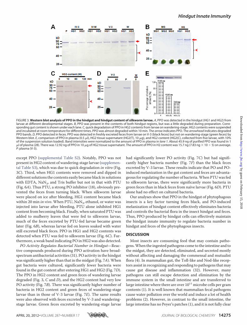
HG2) was dissected out from larvae of different stages and thenused for Western blot assay to detect PPO. The PPO proteinwas detected in hindgut tissues, and the band was at the corre-sponding position as plasma PPO (Fig. 3A). In wandering-stagelarvae, many bands smaller than PPO appeared (�65 kDa).When sections of the hindgut were cultured in vitro, PPO wasreleased into the culture medium according to native gel assay(supplemental Fig. S3). No PPOwas foundwhen sections of themidgut and silk gland were cultured.Midgut and hindgut contents (of V-3 larvae) were suspended
in Tris buffer for a Western blot assay. We found a PPOsignal in the hindgut contents, and many smaller bands weredetected in HG2 (Fig. 3B). PPO from V-3 larvae was found tobe degraded very slowly (supplemental Fig. S4), whereas that inHG2 content from wandering-stage larvae degraded very rap-idly (Fig. 3C). No PPO was detected in the midgut content.Feces excreted by V-3 larvae were collected immediately (0min) and 60 min later. TheWestern blot results showed a PPOband when feces were collected at 0 min but not at 60 min afterexcretion (Fig. 3D), suggesting that activated PO had alreadybeen bound to other proteins to form a large complex that can-not be easily extracted from feces and, thus, was not detected.
Green feces excreted by wandering-larvae were immediatelycollected, but no PPO band was detected (Fig. 3D). PPO in theHG2 part of the hindgut during the wandering-stage may alsobe degraded quickly in vivo. PPO in plasma, HG2 tissue lysate,and HG2 content (on V-3) were compared by Western blot(Fig. 3E). Plasma contained 45.9 ng/�l PPO after purification(28). The amount of PPO in HG2 content (equal to a feces) wasabout 15.7 ng on average.PPO Is Produced by Hindgut Cells but Not Contamination
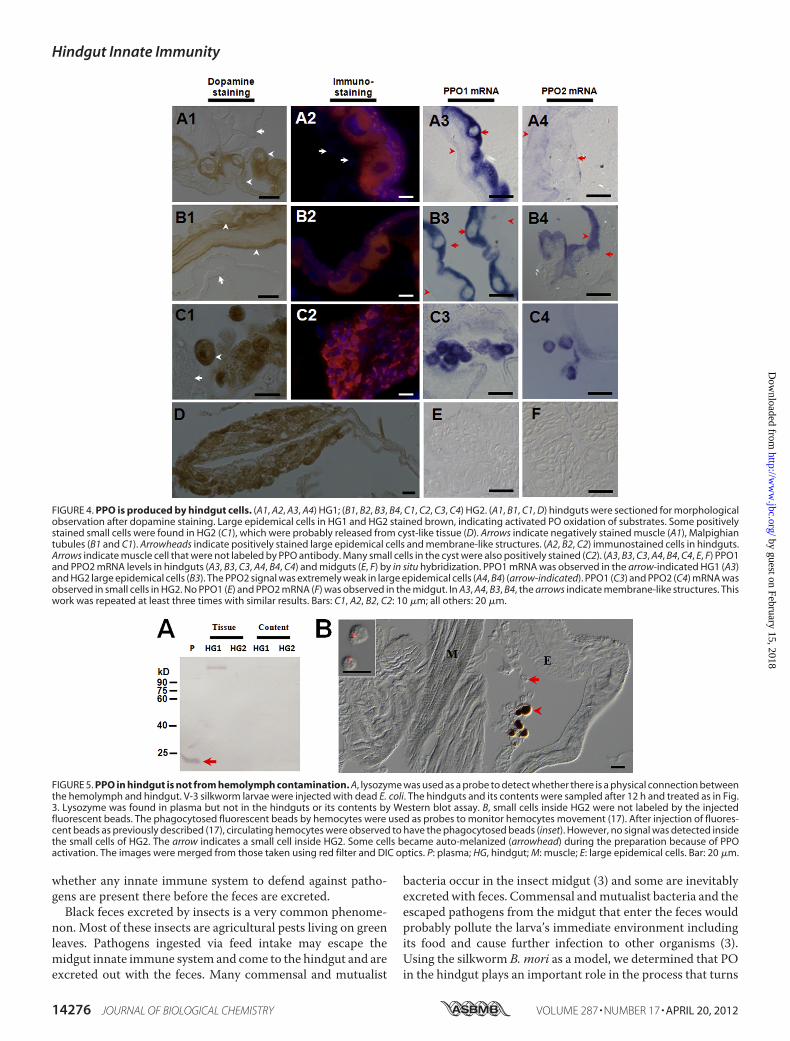
fromHemolymph—Hindguts of the silkworm (Fig. 2,C2-b) andother insects (supplemental Fig. S2) were stained black by amixture of dopamine and ethanol, indicating that PPO is pro-duced in those tissues. Large epidemical cells in the HG1 andHG2 were positively stained with dopamine after ethanol acti-vation (Fig. 4,A1 andB1).Many small cells (Fig. 4,C1) and somecyst-like tissues inHG2 (Fig. 4D) were also positively stained bydopamine. The small cells in HG2 (Fig. 4,C1) might be releasedfrom the cyst-like tissue. However, tissues such as muscle (Fig.4, A1) and Malpighian tubules (Fig. 4, B1 and C1) were notpositively stained in HG1 and HG2. The immunostainingresults also indicated that PPO was localized to some large andsmall cells in the hindgut but not in muscle cells (Fig. 4, A2, B2,and C2). In situ assays showed strong PPO1 mRNA signals inlarge cells of HG1 and HG2 (Fig. 4, A3 and B3). However, thesignal for PPO2 was extremely weak in large epidemical cells(Fig. 4, A4 and B4). PPO1 and PPO2 mRNA were clearly iden-tified in small cells in HG2 (Fig. 4, C3 and C4) but were notdetected in the midgut as a control (Fig. 4, E and F). No signalwas observed when sense RNA probe was used (data notshown).Lysozyme is a 14-kDa plasma protein that can be induced by
prior immunization (29). We used this protein as a probe tomonitor whether the dissection could cause plasma proteincontamination and whether the hemolymph materials can
FIGURE 1. Black feces excreted by silkworm larvae. A, V-3 silkworm larvae, black feces, and mulberry leaves. B, morphology of black feces excreted byfeeding-stage larvae (V-3) and that of green feces excreted by wandering-stage (W) larvae. The mulberry leaf fragments were black (b) or green (d) in thecorresponding black (a) or green (c) feces. C, morphology of the gut dissected from a V-3 silkworm larva, and its gut content from the corresponding location.The midgut is equally divided into three parts (MG1, MG2, and MG3). The hindgut is divided into two parts due to its different morphology (HG1 and HG2). TheHG2 content was black. FG, foregut; MG, midgut; HG, hindgut; PY, pylorus.
Hindgut Innate Immunity
APRIL 20, 2012 • VOLUME 287 • NUMBER 17 JOURNAL OF BIOLOGICAL CHEMISTRY 14273
by guest on February 15, 2018http://w
ww
.jbc.org/D
ownloaded from
freely enter the hindgut. Samples from plasma, HG1 and HG2tissues, and its contents were separated by Western blot afterV-3 silkworm larvae were injected with E. coli. The resultsshowed lysozyme in plasma but not in hindgut tissues or itscontents (Fig. 5A), indicating no contamination and no directphysical connection between hemolymph and HG1 or HG2.When hemocytes were labeled with injected fluorescent beads,none of the beads phagocytosed by hemocytes were detectedinside the HG2 small cells (Fig. 5B), suggesting that the smallcells were other cells than hemocytes. InDrosophila, no hemo-cytes were detected in hindgut epithelium through a transgenicanalysis (30), which provides some evidence that the small cellsobserved in the silkworm HG2 were not hemocytes.When dsRNA against Tribolium TcPPO1 and TcPPO2 was
injected into larvae, hindgut staining was significantly blocked(supplemental Fig. S6), indicating that those PPOs areexpressed in the hindgut. Furthermore, when hemocytes wereablated in Drosophila mutant MK2, in which the source of
plasma PPO is eliminated because crystal cells produce andrelease PPO into plasma through cell rupture (8), melanizationof enterocytes was still observed in the hindgut (30), suggestingagain that hindgut cells are responsible for producing PPO.Taken together, these results suggest that insect hindgut cells inaddition to hemocytes produce PPO.PPO in the Hindgut Is the Key Factor That Turns Feces
Black—Several lines of evidence from the present study suggestthat hindgut PPO is the key factor that turns feces black. First,silkworm larvae at the wandering-stage excreted green feces(Fig. 1,B-c) because the PPO inHG2 content was degraded veryrapidly (Fig. 3C) and hence could not melanize the feces. Wedid not detect any PPO in green feces collected immediatelyafter excretion (Fig. 3D). This was a natural control to show theimportance of PPO in feces melanization. Second, HG2 pro-teins from V-3 larvae were identified by liquid chromatogra-phy-mass spectroscopy (LC-MS/MS), and no other enzymesthat oxidize substrates to induce melanization were present
FIGURE 2. Comparison of the enzyme activity of three oxidases and gut staining. DmrPPO1 was activated by ethanol (E) to have PO activity (16). A1 and A2,peroxidase activity detection. Peroxidase (1.67 ng) and laccase (3 �g) but not DmrPPO1 (1.25 �g) oxidized TMB. No peroxidase activity was detected in HG1 orHG2 content (A1) or in different parts of gut (A2). B1 and B2, laccase activity detection. DmrPPO1 and peroxidase only minimally oxidized ABTS. Some activitieswere detected in the HG1 and HG2 contents, but they were not inhibited by NaN3 (B1). The real laccase activity was significantly inhibited by NaN3. No laccaseactivity was detected in the gut (B2). C1 and C2, PO activity detection. Laccase and peroxidase minimally oxidized dopamine. HG1 and HG2 content hadobviously high PO activity (C1). The foregut and hindgut stained black only if ethanol was used for activation (C2-b was imaged at 30 min). D and E, effects oflaccase and peroxidase inhibitor NaN3 and PO inhibitor PTU on gut staining. PTU significantly inhibited melanization in the foregut and hindgut (D), whereasNaN3 did not (E). Columns represent the mean of individual measurements � S.E. (n � 3). Significant differences were calculated with an unpaired t testprogram.
Hindgut Innate Immunity
14274 JOURNAL OF BIOLOGICAL CHEMISTRY VOLUME 287 • NUMBER 17 • APRIL 20, 2012
by guest on February 15, 2018http://w
ww
.jbc.org/D
ownloaded from
except PPO (supplemental Table S2). Notably, PPO was notpresent inHG2 content of wandering-stage larvae (supplemen-tal Table S3), which was due to quick degradation in vitro (Fig.3C). Third, when HG1 contents were removed and dipped indifferent solutions the contents easily becameblack in solutionswith EDTA, NaN3, and Tris buffer but not in that with PTU(Fig. 6A). Thus PTU, a strong PO inhibitor (18), obviously pre-vented the feces from turning black. When silkworm larvaewere placed on ice after bleeding, HG1 content became blackwithin 20 min in vivo. When PTU, NaN3, ethanol, or water wasinjected into larvae after bleeding, PTU alone inhibited HG1content frombecoming black. Finally, when saturated PTUwasadded to mulberry leaves that were fed to silkworm larvae,much of the feces excreted by PTU-fed larvae became greenlater (Fig. 6B), whereas larvae fed on leaves soaked with waterstill excreted black feces. PPO in HG1 and HG2 contents wasdetected when PTU was fed to silkworm larvae (Fig. 6C). Fur-thermore, a weak band indicating PO inHG2was also detected.PO Activity Regulates Bacterial Number in Hindgut—Reac-
tive compounds produced during PPO activation have broad-spectrum antibacterial activities (31). PO activity in the hindgutwas significantly higher than that in themidgut (Fig. 7A).Whengut bacteria were cultured, significantly fewer bacteria werefound in the gut content after entering HG1 andHG2 (Fig. 7D).The PPO in HG2 content and green feces of wandering larvaedegraded (Fig. 3, C and D), and the HG2 content had very lowPO activity (Fig. 7B). There was significantly higher number ofbacteria in HG2 content and green feces of wandering-stagelarvae than in those of V-3 larvae (Fig. 7E). The same resultswere also observed with feces excreted by V-3 and wandering-stage larvae. Green feces excreted by wandering-stage larvae
had significantly lower PO activity (Fig. 7C) but had signifi-cantly higher bacteria number (Fig. 7F) than the black fecesexcreted by V-3 larvae. These results indicate that PO and PO-induced melanization in the gut content and feces are advanta-geous for regulating the number of bacteria.WhenPTUwas fedto silkworm larvae, there were significantly more bacteria ingreen feces than in black feces from naïve larvae (Fig. 6D). PTUalone had no effect on cultured bacteria.Our analyses show that PPO in the hindgut of phytophagous
insects is a key factor turning feces black, and PO-inducedmelanization of hindgut content effectively eliminates bacteriaand controls the bacterial flora in the insect hindgut and feces.Thus, PPO produced by hindgut cells can effectively maintainthe hindgut innate immunity to regulate bacteria number inhindgut and feces of the phytophagous insects.
DISCUSSION
Most insects are consuming feed that may contain patho-gens.When the ingested pathogens come to the intestine and tothe midgut, they are detected, eliminated, and secreted mostlywithout affecting and damaging the commensal and mutualistflora (4). In mammalian gut, the Toll-like and Nod-like recep-tors assist in recognizing and responding to pathogens thatmaycause gut disease and inflammation (32). However, manypathogens can still escape detection and elimination by theimmune system in the small intestine and are transferred tolarge intestine where there are over 1011microbe cells per gramcontents (1). It is well known that mammalian fecal pathogenscause water pollution over the world and induce a lot of healthproblems (2). However, in contrast to the small intestine, thelarge intestine has no Peyer’s patches (1), and it is not fully clear
FIGURE 3. Western blot analysis of PPO in the hindgut and hindgut content of silkworm larvae. A, PPO was detected in the hindgut (HG1 and HG2) fromlarvae at different developmental stages. B, PPO was present in the contents of both hindgut regions, but was a little degraded during preparation. Corre-sponding gut content is shown under each lane. C, quick degradation of PPO in HG2 contents from larvae on wandering-stage. HG2 contents were suspendedand incubated at room temperature for different times. PPO was almost degraded within 10 min. The arrow indicates PPO. The arrowhead indicates degradedPPO bands. D, PPO detected in feces. PPO was detected in freshly excreted feces from larvae on V-3 (black feces) but not on wandering-stage (green feces) byWestern blot. E, comparison of PPO in plasma (0.5 �l), HG2 tissue supernatant (HG2(T), 10 �g), and HG2 content (HG2(C), collected from five larvae, with 10%of the suspension solution loaded). Band intensities were normalized to the amount of PPO in plasma in lane 1. About 45.9 ng of purified PPO was found in 1�l of plasma (28). There was 12.92 ng of PPO in 10 �g of HG2 tissue supernatant. The amount of PPO in H2 content was 15.7 ng (7.83 ng � 10 � 5) on average.P: plasma (V-3).
Hindgut Innate Immunity
APRIL 20, 2012 • VOLUME 287 • NUMBER 17 JOURNAL OF BIOLOGICAL CHEMISTRY 14275
by guest on February 15, 2018http://w
ww
.jbc.org/D
ownloaded from
whether any innate immune system to defend against patho-gens are present there before the feces are excreted.Black feces excreted by insects is a very common phenome-
non. Most of these insects are agricultural pests living on greenleaves. Pathogens ingested via feed intake may escape themidgut innate immune system and come to the hindgut and areexcreted out with the feces. Many commensal and mutualist
bacteria occur in the insect midgut (3) and some are inevitablyexcretedwith feces. Commensal andmutualist bacteria and theescaped pathogens from the midgut that enter the feces wouldprobably pollute the larva’s immediate environment includingits food and cause further infection to other organisms (3).Using the silkworm B. mori as a model, we determined that POin the hindgut plays an important role in the process that turns
FIGURE 4. PPO is produced by hindgut cells. (A1, A2, A3, A4) HG1; (B1, B2, B3, B4, C1, C2, C3, C4) HG2. (A1, B1, C1, D) hindguts were sectioned for morphologicalobservation after dopamine staining. Large epidemical cells in HG1 and HG2 stained brown, indicating activated PO oxidation of substrates. Some positivelystained small cells were found in HG2 (C1), which were probably released from cyst-like tissue (D). Arrows indicate negatively stained muscle (A1), Malpighiantubules (B1 and C1). Arrowheads indicate positively stained large epidemical cells and membrane-like structures. (A2, B2, C2) immunostained cells in hindguts.Arrows indicate muscle cell that were not labeled by PPO antibody. Many small cells in the cyst were also positively stained (C2). (A3, B3, C3, A4, B4, C4, E, F) PPO1and PPO2 mRNA levels in hindguts (A3, B3, C3, A4, B4, C4) and midguts (E, F) by in situ hybridization. PPO1 mRNA was observed in the arrow-indicated HG1 (A3)and HG2 large epidemical cells (B3). The PPO2 signal was extremely weak in large epidemical cells (A4, B4) (arrow-indicated). PPO1 (C3) and PPO2 (C4) mRNA wasobserved in small cells in HG2. No PPO1 (E) and PPO2 mRNA (F) was observed in the midgut. In A3, A4, B3, B4, the arrows indicate membrane-like structures. Thiswork was repeated at least three times with similar results. Bars: C1, A2, B2, C2: 10 �m; all others: 20 �m.
FIGURE 5. PPO in hindgut is not from hemolymph contamination. A, lysozyme was used as a probe to detect whether there is a physical connection betweenthe hemolymph and hindgut. V-3 silkworm larvae were injected with dead E. coli. The hindguts and its contents were sampled after 12 h and treated as in Fig.3. Lysozyme was found in plasma but not in the hindguts or its contents by Western blot assay. B, small cells inside HG2 were not labeled by the injectedfluorescent beads. The phagocytosed fluorescent beads by hemocytes were used as probes to monitor hemocytes movement (17). After injection of fluores-cent beads as previously described (17), circulating hemocytes were observed to have the phagocytosed beads (inset). However, no signal was detected insidethe small cells of HG2. The arrow indicates a small cell inside HG2. Some cells became auto-melanized (arrowhead) during the preparation because of PPOactivation. The images were merged from those taken using red filter and DIC optics. P: plasma; HG, hindgut; M: muscle; E: large epidemical cells. Bar: 20 �m.
Hindgut Innate Immunity
14276 JOURNAL OF BIOLOGICAL CHEMISTRY VOLUME 287 • NUMBER 17 • APRIL 20, 2012
by guest on February 15, 2018http://w
ww
.jbc.org/D
ownloaded from

feces black, through which fecal pathogens and possible com-mensal and mutualist bacteria are eliminated to prevent suchcontamination.Insect PPO is considered to be mainly produced by hemo-
cytes (8). However, we found that PPO can be transcribed andexpressed in some hindgut cells that were shown by using insitu hybridization and immunostaining analysis. PPO was alsodetected in the content of the hindgut, which suggests that PPO
produced by hindgut cells is secreted, although the exactmech-anism for this secretion is unknown. We also found that tran-scription of important enzymes in the plasma PPO activationpathway (e.g.PPAEandBAEE) (8) occurred in silkworm foregutand hindgut tissues (supplemental Fig. S7) but the correspond-ing proteins were not found in the gut content suspension (sup-plemental Tables S2 and S3), which suggests that the proteinlevel was too low to be detected. In the hindguts of Drosophila
FIGURE 6. Melanization of silkworm feces is inhibited by the PO inhibitor PTU. A, PTU inhibited melanization of HG1 content. HG1 content was removedand dipped into different solutions. Then the contents were placed on new parafilm, and the extra solution was absorbed. HG1 content became black within20 min after being dipped into EDTA, NaN3, and Tris solution. PTU inhibited HG1 content melanization. B, PTU solution was spread on mulberry leaves fed tothe silkworm larvae. One day later, feces excreted by PTU-fed silkworm larvae were green. C, PTU did not inhibit PPO production or secretion. The arrow andarrowhead indicate PPO and PO, respectively, detected in green feces. D, feeding PTU increased the number of bacterial colonies in green feces. The excretedgreen feces was suspended for bacterial culture after PTU feeding. Each dot corresponds to the colonies of bacteria suspended from gut content or feces fromone silkworm larva. The average for each group is indicated by a horizontal black bar (n � 7). Significant differences were calculated with an unpaired t testprogram.
FIGURE 7. PO activity regulating bacteria number in hindgut and feces. A–C, PO activities were compared among different parts of gut contents from V-3larvae (A) as well as among HG2 contents (B) and fresh feces (C) from V-3 and wandering-stage larvae, respectively. PO activities in the gut contents of V-3 larvaeare: HG2HG1MG3 (A). PO activities in the hindgut contents (B) and fresh feces (C) of V-3 larvae were higher than in those of wandering-stage larvae,respectively. Columns represent the mean of individual animal measurements � S.E. (n � 5). D–F, bacteria number in the gut contents and fresh feces. Bacteriacolonies in the gut contents and fresh feces from V-3 and wandering-stage larvae as shown in A–C, were counted after being cultured. The bacterial numberwas significantly lower when PO activity was high in gut contents (D, E) and fresh feces (F). Each dot corresponds to the colonies of bacteria suspended from gutcontent or feces from one silkworm larva. The average for each group is indicated by a horizontal black bar (n � 7). Significant differences were calculated usingan unpaired t test program.
Hindgut Innate Immunity
APRIL 20, 2012 • VOLUME 287 • NUMBER 17 JOURNAL OF BIOLOGICAL CHEMISTRY 14277
by guest on February 15, 2018http://w
ww
.jbc.org/D
ownloaded from

mutants with developmental defects, some cells were found tobe melanized upon infection or stress (30, 33), which also pro-vides some support to the existence of PPO in Drosophilahindgut.PPO is a very important immune protein in many inverte-
brate animal and when it is knocked-down by RNAi in crayfishor shrimp (34), the number of bacteria in hemolymph increasesresulting in mortality. Reactive compounds produced by acti-vated PO during melanization can kill many types of bacteria(31). During dissection and treatment, some cells in hindgutbecame auto-melanized (Fig. 5B and supplemental Fig. S5, Aand C). When monoclonal antibody against melanin was usedfor immunostaining, the signal for melanin was detected inthose epidemical cells (supplemental Fig. S5). A decrease in thenumber of bacteria in hindgut contents and feces was corre-latedwith the POactivity anddarkening.Alongwith the changein color may also be dehydration, which could partially beattributed to the incorporation of polymerized hydrophobicpolyphenols into the feces. In addition to any toxic properties ofoxidized products of phenols by PO, the dehydration mightcontribute to decreasing the bacterial load. This is probably thereason why there is not any hindgut specific pathogen forstudying the relationship between insect fitness and hindgutPO activity. Therefore, PPO in the hindgut is a very importantcomplement to other intestinal innate immune processes.Many herbivorous mammalians such as sheep, cattle, and
elephants also excrete black feces. Mammalian animals alsohave tyrosinase, which belongs to the same group of proteins asinsect PPO (8). We do not know whether the mechanism is thesame or if it is biologically significant. However, the hindgutinnate immunity provided by the silkworm PPO for clearingbacteria in the hindgut indicates that innate immunity mightoccur in the mammalian large intestine.
Acknowledgment—We thank Lynn M. Riddiford for helpfulcomments.
REFERENCES1. Walter, J., and Ley, R. (2011) The human gut microbiome: ecology and
recent evolutionary changes. Annu. Rev. Microbiol. 65, 411–4292. Field, K.G., and Samadpour,M. (2007) Fecal source tracking, the indicator
paradigm, and managing water quality.Water Res. 41, 3517–35383. Dillon, R. J., and Dillon, V. M. (2004) The gut bacteria of insects: non-
pathogenic interactions. Annu. Rev. Entomol. 49, 71–924. Apidianakis, Y., and Rahme, L. G. (2011) Drosophila melanogaster as a
model for human intestinal infection and pathology. Dis. Model. Mech. 4,21–30
5. Hakim, R. S., Baldwin, K., and Smagghe, G. (2010) Regulation of midgutgrowth, development, and metamorphosis. Annu. Rev. Entomol. 55,593–608
6. Schoonhoven, L. M., van Loon, J. J. A., and Dicke, M. (2005), Insect-PlantBiology, Chapman & Hall, London
7. Lehane, M. J., and Billingsley, P. B. (1996) Biology of the Insect Midgut,Chapman & Hall, London
8. Ashida, M., and Brey, P. (1998) Recent Advances on the Research of theInsect Prophenoloxidase Cascade, Chapman & Hall, London
9. Cerenius, L., Kawabata, S., Lee, B. L., Nonaka,M., and Söderhäll, K. (2010)Proteolytic cascades and their involvement in invertebrate immunity.Trends Biochem. Sci. 35, 575–583
10. Kanost, M. R., and Gorman, M. J. (2008) Phenoloxidases in Insect Immu-
nity, Academic Press & Elsevier, San Diego11. Kanost, M. R., Jiang, H., and Yu, X. Q. (2004) Innate immune responses of
a lepidopteran insect,Manduca sexta. Immunol. Rev. 198, 97–10512. Strand, M. R. (2008) The insect cellular immune response. Insect Science
15, 1–1413. Wittkopp, P. J., and Beldade, P. (2009) Development and evolution of
insect pigmentation: genetic mechanisms and the potential consequencesof pleiotropy. Semin. Cell Dev. Biol. 20, 65–71
14. Hiruma, K., and Riddiford, L. M. (2009) The molecular mechanisms ofcuticular melanization: the ecdysone cascade leading to dopa decarboxy-lase expression inManduca sexta. Insect Biochem. Mol. Biol. 39, 245–253
15. Xia, Q., Zhou, Z., Lu, C., Cheng, D., Dai, F., Li, B., Zhao, P., Zha, X., Cheng,T., Chai, C., Pan, G., Xu, J., Liu, C., Lin, Y., Qian, J., Hou, Y., Wu, Z., Li, G.,Pan,M., Li, C., Shen, Y., Lan, X., Yuan, L., Li, T., Xu, H., Yang, G.,Wan, Y.,Zhu, Y., Yu, M., Shen, W., Wu, D., Xiang, Z., group, G. a., Yu, J., Wang, J.,Li, R., Shi, J., Li, H., Li, G., Su, J., Wang, X., Li, G., Zhang, Z., Wu, Q., Li, J.,Zhang, Q., Wei, N., Xu, J., Sun, H., Dong, L., Liu, D., Zhao, S., Zhao, X.,Meng, Q., Lan, F., Huang, X., Li, Y., Fang, L., Li, C., Li, D., Sun, Y., Zhang,Z., Yang, Z., Huang, Y., Xi, Y., Qi, Q., He, D., Huang, H., Zhang, X., Wang,Z., Li, W., Cao, Y., Yu, Y., Yu, H., Li, J., Ye, J., Chen, H., Zhou, Y., Liu, B.,Wang, J., Ye, J., Ji, H., Li, S., Ni, P., Zhang, J., Zhang, Y., Zheng, H., Mao, B.,Wang, W., Ye, C., Li, S., Wang, J., Wong, G. K.-S., and Yang, H. (2004) Adraft sequence for the genome of the domesticated silkworm (Bombyxmori). Science 306, 1937–1940
16. Li, X., Ma, M., Liu, F., Chen, Y., Lu, A., Ling, Q. Z., Li, J., Beerntsen, B. T.,Yu, X. Q., Liu, C., and Ling, E. (2012) Properties of Drosophila melano-gaster prophenoloxidases expressed in Escherichia coli. Dev. Comp. Im-munol. 36, 648–656
17. Ling, E., Shirai, K., Kanekatsu, R., Kiguchi, K., Kobayashi, Y., Funayama, T.,and Watanabe, H. (2006) Contribution of circulating hemocytes to theregeneration of heavy ion beams 12C5 irradiated hematopoietic organs inthe silkworm, Bombyx mori, through the way of phagocytosis of injuredcells after invasion. Dev. Comp. Immunol. 30, 531–543
18. Ryazanova, A. D., Alekseev, A. A., and Slepneva, I. A. (2012) The phenyl-thiourea is a competitive inhibitor of the enzymatic oxidation of DOPA byphenoloxidase. J. Enzym. Inhib. Med. Chem. 27, 78–83
19. Wang, Z., Lu, A., Li, X., Shao, Q., Beerntsen, B. T., Liu, C., Ma, Y., Huang,Y., Zhu, H., and Ling, E. (2011) A systematic study on hemocyte identifi-cation and plasma prophenoloxidase fromCulex pipiens quinquefasciatusat different developmental stages. Exp. Parasitol. 127, 135–141
20. Asano, T., and Takebuchi, K. (2009) Identification of the gene encodingpro-phenoloxidase A(3) in the fruitfly, Drosophila melanogaster. InsectMol. Biol. 18, 223–232
21. Tan, A., Tanaka, H., Sato, N., Yaguchi, M., Nagata, M., and Suzuki, K.(2003) Identification of novel tissue-specific proteins in the suboesopha-geal body of the silkworm, Bombyx mori. J. Insect Biotechnol. Sericol. 72,41–50
22. Dittmer, N. T., Gorman, M. J., and Kanost, M. R. (2009) Characterizationof endogenous and recombinant forms of laccase-2, amulticopper oxidasefrom the tobacco hornworm, Manduca sexta. Insect Biochem. Mol. Biol.39, 596–606
23. Fouda, M.M., Hiragaki, S., Tufail, M., Shao, Q.M., and Takeda, M. (2010)Precursor structure, distribution, and possible functions of pigment-dis-persing hormone (PDH) in the terrestrial isopod Armadillidium vulgare(Latreille). J. Insect Physiol. 56, 1728–1737
24. Jiang, H.,Wang, Y.,Ma, C., and Kanost,M. R. (1997) Subunit compositionof pro-phenol oxidase fromManduca sexta: molecular cloning of subunitProPO-P1. Insect Biochem. Mol .Biol. 27, 835–850
25. Nosanchuk, J. D., Yu, J. J., Hung, C. Y., Casadevall, A., and Cole, G. T.(2007) Coccidioides posadasii produces melanin in vitro and during infec-tion. Fungal Genet. Biol. 44, 517–520
26. Ling, E., Ao, J., andYu, X.Q. (2008)Nuclear translocation of immulectin-3stimulates hemocyte proliferation.Mol Immunol. 45, 2598–2606
27. Dittmer, N. T., and Kanost, M. R. (2010) Insect multicopper oxidases:diversity, properties, and physiological roles. Insect Biochem.Mol. Biol. 40,179–188
28. Ashida, M. (1971) Purification and characterization of pre-phenoloxidasefrom hemolymph of the silkworm Bombyx mori. Arch. Biochem. Biophys.
Hindgut Innate Immunity
14278 JOURNAL OF BIOLOGICAL CHEMISTRY VOLUME 287 • NUMBER 17 • APRIL 20, 2012
by guest on February 15, 2018http://w
ww
.jbc.org/D
ownloaded from

144, 749–76229. Gillespie and, J. P., Kanost, M. R., and Trenczek, T. (1997) Biological me-
diators of insect immunity. Annu. Rev. Entomol. 42, 611–64330. Seisenbacher, G., Hafen, E., and Stocker, H. (2011) MK2-dependent p38b
signaling protects Drosophila hindgut enterocytes against JNK-inducedapoptosis under chronic stress. PLoS Genet. 7, e1002168
31. Zhao, P., Li, J., Wang, Y., and Jiang, H. (2007) Broad-spectrum antimicro-bial activity of the reactive compounds generated in vitro by Manducasexta phenoloxidase. Insect Mol. Biol. 37, 952–959
32. Carvalho, F. A., Aitken, J. D., Vijay-Kumar, M., and Gewirtz, A. T. (2012)Annu. Rev. Physiol. 74, 177–198
33. Chen, J., Xie, C., Tian, L., Hong, L.,Wu,X., andHan, J. (2010) Participationof the p38 pathway inDrosophila host defense against pathogenic bacteriaand fungi. Proc. Natl. Acad. Sci. U.S.A. 107, 20774–20779
34. Liu, H., Jiravanichpaisal, P., Cerenius, L., Lee, B. L., Söderhäll, I., and Sö-derhäll, K. (2007) Phenoloxidase is an important component of the de-fense against Aeromonas hydrophila Infection in a crustacean, Pacifasta-cus leniusculus. J. Biol. Chem. 282, 33593–33598
Hindgut Innate Immunity
APRIL 20, 2012 • VOLUME 287 • NUMBER 17 JOURNAL OF BIOLOGICAL CHEMISTRY 14279
by guest on February 15, 2018http://w
ww
.jbc.org/D
ownloaded from

Yongping Huang, Kenneth Söderhäll and Erjun LingQimiao Shao, Bing Yang, Qiuyun Xu, Xuquan Li, Zhiqiang Lu, Chengshu Wang,
Melanization in InsectsHindgut Innate Immunity and Regulation of Fecal Microbiota through
doi: 10.1074/jbc.M112.354548 originally published online February 28, 20122012, 287:14270-14279.J. Biol. Chem.
10.1074/jbc.M112.354548Access the most updated version of this article at doi:
Alerts:
When a correction for this article is posted•
When this article is cited•
to choose from all of JBC's e-mail alertsClick here
Supplemental material:
http://www.jbc.org/content/suppl/2012/02/28/M112.354548.DC1
http://www.jbc.org/content/287/17/14270.full.html#ref-list-1
This article cites 30 references, 4 of which can be accessed free at
by guest on February 15, 2018http://w
ww
.jbc.org/D
ownloaded from
Related Documents