Proteomics 2014, 14, 441–451 441 DOI 10.1002/pmic.201300311 REVIEW Applications of mass spectrometry for quantitative protein analysis in formalin-fixed paraffin-embedded tissues Carine Steiner 1,2,3 , Axel Ducret 3 , Jean-Christophe Tille 4 , Marlene Thomas 5 , Thomas A. McKee 4 , Laura Rubbia-Brandt 4 , Alexander Scherl 1,2 , Pierre Lescuyer 1,2 and Paul Cutler 3 1 Division of Laboratory Medicine, Geneva University Hospital, Geneva, Switzerland 2 Human Protein Sciences Department, University of Geneva, Geneva, Switzerland 3 Translational Technologies and Bioinformatics, Pharma Research and Early Development, F. Hoffmann-La Roche AG, Basel, Switzerland 4 Division of Clinical Pathology, Geneva University Hospital, Geneva, Switzerland 5 Oncology Division, Roche Pharma Research & Early Development, Penzberg, Germany Proteomic analysis of tissues has advanced in recent years as instruments and methodologies have evolved. The ability to retrieve peptides from formalin-fixed paraffin-embedded tissues followed by shotgun or targeted proteomic analysis is offering new opportunities in biomedical research. In particular, access to large collections of clinically annotated samples should en- able the detailed analysis of pathologically relevant tissues in a manner previously considered unfeasible. In this paper, we review the current status of proteomic analysis of formalin-fixed paraffin-embedded tissues with a particular focus on targeted approaches and the potential for this technique to be used in clinical research and clinical diagnosis. We also discuss the limitations and perspectives of the technique, particularly with regard to application in clinical diagnosis and drug discovery. Keywords: Biomarkers / Biomedicine / Formalin-fixed paraffin-embedded tissue / Oncology / Shotgun proteomics / SRM Received: July 24, 2013 Revised: November 4, 2013 Accepted: November 11, 2013 1 Introduction A better understanding of complex diseases goes hand in hand with a constant need for the discovery of novel tar- gets and biomarkers that facilitate disease diagnosis, clas- sification, and treatment. While targets and biomarkers are both relevant in the clinic, the literature mainly focuses on Correspondence: Dr. Carine Steiner, Translational Technologies and Bioinformatics, Pharma Research and Early Development, F. Hoffmann-La Roche Ltd, CH-4070 Basel, Switzerland E-mail: [email protected] Fax: +41-61-688-19-29 Abbreviations: FF, fresh frozen; FFPE, formalin-fixed paraffin- embedded; IHC, immunohistochemistry; LLOD, lower LOD; LLOQ, lower LOQ biomarker discovery in clinical research rather than on tar- get discovery. In 2001, the term “biomarker” was defined by the American National Institute of Health as “a characteristic that is objectively measured and evaluated as an indicator of normal biological processes, pathogenic processes, or phar- macologic responses to a therapeutic intervention” [1]. One of the preferred biological sources for the quantification of biomarkers is blood as it is easily obtained in a relatively non- invasive manner. However, in certain areas such as oncol- ogy, diagnostic, and/or prognostic biomarkers are measured directly in biopsies or surgically resected tumoral tissues to support diagnosis and treatment. Tissue analysis allows di- rect access to the proteins of interest, at tissue concentra- tions, without the dilution effect implicit in the analysis of plasma. In the field of clinical pathology, immunohistochemistry (IHC) represents a useful tool for tumor diagnosis and C 2013 The Authors. PROTEOMICS published by Wiley-VCH Verlag GmbH & Co. KGaA, Weinheim www.proteomics-journal.com This is an open access article under the terms of the Creative Commons Attribution-NonCommercial-NoDerivs License, which permits use and distribution in any medium, provided the original work is properly cited, the use is non-commercial and no modifications or adaptations are made.

Welcome message from author
This document is posted to help you gain knowledge. Please leave a comment to let me know what you think about it! Share it to your friends and learn new things together.
Transcript

Proteomics 2014, 14, 441–451 441DOI 10.1002/pmic.201300311
REVIEW
Applications of mass spectrometry for quantitative
protein analysis in formalin-fixed paraffin-embedded
tissues
Carine Steiner1,2,3, Axel Ducret3, Jean-Christophe Tille4, Marlene Thomas5,Thomas A. McKee4, Laura Rubbia-Brandt4, Alexander Scherl1,2, Pierre Lescuyer1,2
and Paul Cutler3
1 Division of Laboratory Medicine, Geneva University Hospital, Geneva, Switzerland2 Human Protein Sciences Department, University of Geneva, Geneva, Switzerland3 Translational Technologies and Bioinformatics, Pharma Research and Early Development, F. Hoffmann-La RocheAG, Basel, Switzerland
4 Division of Clinical Pathology, Geneva University Hospital, Geneva, Switzerland5 Oncology Division, Roche Pharma Research & Early Development, Penzberg, Germany
Proteomic analysis of tissues has advanced in recent years as instruments and methodologieshave evolved. The ability to retrieve peptides from formalin-fixed paraffin-embedded tissuesfollowed by shotgun or targeted proteomic analysis is offering new opportunities in biomedicalresearch. In particular, access to large collections of clinically annotated samples should en-able the detailed analysis of pathologically relevant tissues in a manner previously consideredunfeasible. In this paper, we review the current status of proteomic analysis of formalin-fixedparaffin-embedded tissues with a particular focus on targeted approaches and the potentialfor this technique to be used in clinical research and clinical diagnosis. We also discuss thelimitations and perspectives of the technique, particularly with regard to application in clinicaldiagnosis and drug discovery.
Keywords:
Biomarkers / Biomedicine / Formalin-fixed paraffin-embedded tissue / Oncology /Shotgun proteomics / SRM
Received: July 24, 2013Revised: November 4, 2013
Accepted: November 11, 2013
1 Introduction
A better understanding of complex diseases goes hand inhand with a constant need for the discovery of novel tar-gets and biomarkers that facilitate disease diagnosis, clas-sification, and treatment. While targets and biomarkers areboth relevant in the clinic, the literature mainly focuses on
Correspondence: Dr. Carine Steiner, Translational Technologiesand Bioinformatics, Pharma Research and Early Development, F.Hoffmann-La Roche Ltd, CH-4070 Basel, SwitzerlandE-mail: [email protected]: +41-61-688-19-29
Abbreviations: FF, fresh frozen; FFPE, formalin-fixed paraffin-embedded; IHC, immunohistochemistry; LLOD, lower LOD; LLOQ,lower LOQ
biomarker discovery in clinical research rather than on tar-get discovery. In 2001, the term “biomarker” was defined bythe American National Institute of Health as “a characteristicthat is objectively measured and evaluated as an indicator ofnormal biological processes, pathogenic processes, or phar-macologic responses to a therapeutic intervention” [1]. Oneof the preferred biological sources for the quantification ofbiomarkers is blood as it is easily obtained in a relatively non-invasive manner. However, in certain areas such as oncol-ogy, diagnostic, and/or prognostic biomarkers are measureddirectly in biopsies or surgically resected tumoral tissues tosupport diagnosis and treatment. Tissue analysis allows di-rect access to the proteins of interest, at tissue concentra-tions, without the dilution effect implicit in the analysis ofplasma.
In the field of clinical pathology, immunohistochemistry(IHC) represents a useful tool for tumor diagnosis and
C© 2013 The Authors. PROTEOMICS published by Wiley-VCH Verlag GmbH & Co. KGaA, Weinheim www.proteomics-journal.com
This is an open access article under the terms of the Creative Commons Attribution-NonCommercial-NoDerivs License, which permits use anddistribution in any medium, provided the original work is properly cited, the use is non-commercial and no modifications or adaptations are made.
442 C. Steiner et al. Proteomics 2014, 14, 441–451
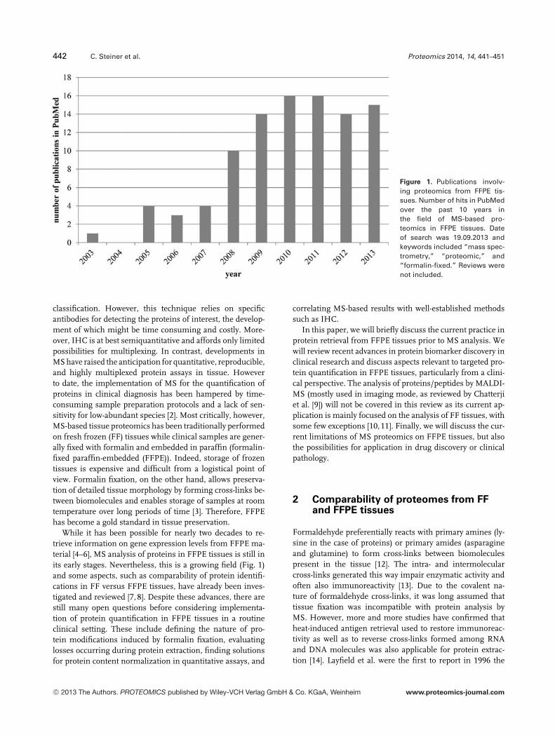
Figure 1. Publications involv-ing proteomics from FFPE tis-sues. Number of hits in PubMedover the past 10 years inthe field of MS-based pro-teomics in FFPE tissues. Dateof search was 19.09.2013 andkeywords included “mass spec-trometry,” “proteomic,” and“formalin-fixed.” Reviews werenot included.
classification. However, this technique relies on specificantibodies for detecting the proteins of interest, the develop-ment of which might be time consuming and costly. More-over, IHC is at best semiquantitative and affords only limitedpossibilities for multiplexing. In contrast, developments inMS have raised the anticipation for quantitative, reproducible,and highly multiplexed protein assays in tissue. Howeverto date, the implementation of MS for the quantification ofproteins in clinical diagnosis has been hampered by time-consuming sample preparation protocols and a lack of sen-sitivity for low-abundant species [2]. Most critically, however,MS-based tissue proteomics has been traditionally performedon fresh frozen (FF) tissues while clinical samples are gener-ally fixed with formalin and embedded in paraffin (formalin-fixed paraffin-embedded (FFPE)). Indeed, storage of frozentissues is expensive and difficult from a logistical point ofview. Formalin fixation, on the other hand, allows preserva-tion of detailed tissue morphology by forming cross-links be-tween biomolecules and enables storage of samples at roomtemperature over long periods of time [3]. Therefore, FFPEhas become a gold standard in tissue preservation.
While it has been possible for nearly two decades to re-trieve information on gene expression levels from FFPE ma-terial [4–6], MS analysis of proteins in FFPE tissues is still inits early stages. Nevertheless, this is a growing field (Fig. 1)and some aspects, such as comparability of protein identifi-cations in FF versus FFPE tissues, have already been inves-tigated and reviewed [7, 8]. Despite these advances, there arestill many open questions before considering implementa-tion of protein quantification in FFPE tissues in a routineclinical setting. These include defining the nature of pro-tein modifications induced by formalin fixation, evaluatinglosses occurring during protein extraction, finding solutionsfor protein content normalization in quantitative assays, and
correlating MS-based results with well-established methodssuch as IHC.
In this paper, we will briefly discuss the current practice inprotein retrieval from FFPE tissues prior to MS analysis. Wewill review recent advances in protein biomarker discovery inclinical research and discuss aspects relevant to targeted pro-tein quantification in FFPE tissues, particularly from a clini-cal perspective. The analysis of proteins/peptides by MALDI-MS (mostly used in imaging mode, as reviewed by Chatterjiet al. [9]) will not be covered in this review as its current ap-plication is mainly focused on the analysis of FF tissues, withsome few exceptions [10,11]. Finally, we will discuss the cur-rent limitations of MS proteomics on FFPE tissues, but alsothe possibilities for application in drug discovery or clinicalpathology.
2 Comparability of proteomes from FFand FFPE tissues
Formaldehyde preferentially reacts with primary amines (ly-sine in the case of proteins) or primary amides (asparagineand glutamine) to form cross-links between biomoleculespresent in the tissue [12]. The intra- and intermolecularcross-links generated this way impair enzymatic activity andoften also immunoreactivity [13]. Due to the covalent na-ture of formaldehyde cross-links, it was long assumed thattissue fixation was incompatible with protein analysis byMS. However, more and more studies have confirmed thatheat-induced antigen retrieval used to restore immunoreac-tivity as well as to reverse cross-links formed among RNAand DNA molecules was also applicable for protein extrac-tion [14]. Layfield et al. were the first to report in 1996 the
C© 2013 The Authors. PROTEOMICS published by Wiley-VCH Verlag GmbH & Co. KGaA, Weinheim www.proteomics-journal.com
Proteomics 2014, 14, 441–451 443
amino acid sequencing of a polypeptide corresponding to animmunoglobulin light chain extracted from formalin-fixedtissue [15]. Ikeda et al. then reported extraction of proteinsfrom FFPE tissues followed by Western blot analysis. Theirmost effective extraction protocol included a 20 min heatingstep at 100�C followed by a 2 h incubation at 60�C in a RIPAbuffer containing 2% w/v SDS, pH 7.6 [16]. Vasilescu et al.reported MS analysis of proteins extracted from cell lines,which had been formalin-fixed in order to cross-link proteincomplexes. Proteins were extracted at 95�C for 20 min in abuffer containing 2% w/v SDS, pH 6.8, after which they weresubjected to SDS-PAGE and digested prior to LC-MS analy-sis [17]. Later publications have reported similar protocols forprotein or peptide extraction from FFPE samples prior to shot-gun proteomic analysis on various MS platforms, includingRPLC-MS/MS, SELDI-TOF, and MALDI-TOF/TOF [18–21].It is now well accepted that the key components to extractproteins from FFPE tissues involve heat, a detergent (typi-cally SDS) and an alkaline buffer. The mechanism of antigenretrieval [22], as well as extraction conditions, have been ex-tensively reviewed elsewhere [8, 23–26].
In addition to extraction conditions, other preanalytical pa-rameters are susceptible to influence protein extraction fromFFPE tissues. These include ischemic time (time betweensample collection and formalin fixation), fixation time (timethe sample was left in formalin solution), and storage du-ration (time from tissue fixation until protein extraction andanalysis) [8,25–28]. It has been demonstrated that a prolongedischemic time (>60 min) negatively impacts the measure-ment of HER2 by IHC in FFPE breast tissue [29, 30]. More-over, duration of formalin fixation affects the extent to whichproteins can be recovered. While longer fixation times ensurebetter preservation of morphological features (in particularfor large-sized samples), overall shorter fixation times (con-sensus being 24 h) almost invariably result in better proteinyields. In contrast, tissue dehydration, paraffin-embedding,and storage time of the resulting tissue FFPE blocks appearto play a less significant role. A recent study suggested thatstorage time of up to 10 years did not significantly impactprotein profiling [31].
Several studies have been performed with the aim ofdemonstrating equivalence between proteomes retrievedfrom FFPE and FF tissues using shotgun proteomics. Guzelet al. compared equal areas of microdissected paired FFPEand FF placental parenchyme tissue sections from womenwith pregnancies complicated by early onset preeclampsiaand normotensive control women [32]. An average overlapin protein identities of 60% was observed with no significantdifference in the overall number of proteins identified. In an-other study, Guo et al. reported a protein identification overlapof 83% between FF and FFPE from microdissected glioblas-tomas [33]. Crockett et al. investigated cell lines derived froma human transformed follicular lymphoma (SUDHL-4) usinga complementary Glu-C and trypsin enzymatic digesting stepto improve the overall protein identification rate, followed bynanoRPLC-MS/MS [21]. A total of 263 proteins, represent-
ing 52% of the total number of proteins identified from FFcells, were found to overlap between both types of samples.More importantly, the GO cellular location and molecularfunction of the proteins identified from a 3-year-old SUDHL-4 FFPE cell block and a fresh cell lysate were highly similar.In addition, analysis of the FFPE samples provided identi-fication of low-abundance proteins including transcriptionfactors. Tanca et al. used a canine mammary tumor model tocompare the proteomic information generated from pairedFFPE and FF specimens using gel-based protein fraction-ation followed by LC-MS/MS and spectral counting quan-tification [34]. A high level of consistency was seen for allbiological and cell localization categories. More significantly,both data sets highlighted comparable protein pathways, sug-gesting consistent biological information was obtained fromboth sample types. Interestingly, Tanca et al. observed thathigh molecular weight proteins were more abundant in FFtissues, possibly because large, intact proteins were moredifficult to extract from FFPE material, while basic proteinswere overrepresented in FFPE tissues. A similar observationwas made in another study where colorectal cancer tumorsof three different cellularity levels (low, middle, and high)were analyzed by direct LC-MS/MS (Ducret et al. unpub.data). Mirrored FF and FFPE tissue showed intratissue repro-ducibility in terms of both protein number and abundance,with an overlap in protein identification of 55% between FFPEand FF tissue. However, the FFPE tissue showed a bias forsmall structural proteins (actin, calponin, etc.), DNA/RNA-associated proteins (histones, ribosomal proteins), and heatshock proteins, while large, structural multisubunits proteins(myosin, collagen, etc.) and blood proteins (serum albumin,hemoglobin, etc.) were more represented in FF samples.
All of these studies suggest that, while some differencesexist with FF samples, the analysis of FFPE tissues pro-vides reliable biological information. It is noteworthy that,while most recently developed extraction protocols appearto achieve nearly equivalent protein yields for FFPE and FFsamples, formalin reversal may not be completely achievable.Most notably, publications based on whole protein fractiona-tion, such as 2DE, consistently report lower yields and iden-tification power than peptide-based fractionation methods,possibly because digestion might release analyzable peptidesfrom even partially blocked proteins [35]. Moreover, a lowerrate of lysine C-terminal peptides was observed in FFPE com-pared to FF tissue extracts [28]. There also seems to be dif-ferences in extraction recoveries for individual proteins. Thisfact was elegantly demonstrated by formalin-fixating differ-ent solutions of cytoplasmic proteins and HeLa cells as FFPEtissue surrogates [13]. After testing a range of conditions,the optimal extraction pH for lysozyme surrogates was de-termined to be pH 4, whereas it was pH 6 for carbonic an-hydrase. When surrogates containing a mixture of proteinswere analyzed, carbonic anhydrase was proportionally under-represented in the extract. This indicates that multiple extrac-tion conditions might be necessary for comprehensive pro-tein recovery. Consequently, as stated above, some classes of
C© 2013 The Authors. PROTEOMICS published by Wiley-VCH Verlag GmbH & Co. KGaA, Weinheim www.proteomics-journal.com
444 C. Steiner et al. Proteomics 2014, 14, 441–451
proteins (e.g. nuclear, cytoskeletal or membrane proteins) willbe variably extracted from FFPE tissues. However, althoughone might expect the cellular localization (e.g. membraneproteins) to majorly impact the extraction efficiency, it seemsthat the physicochemical properties of the proteins play aneven more important role in this regard [13, 21].
The observations discussed above also apply to thecharacterization and quantification of PTMs such asphosphorylation and N-glycosylation. While some studiesclaimed quantitative recovery from FFPE tissue [36,37], otherinvestigations [38–40] underlined the need for establishingguidelines and standardized extraction procedures to keepthe already naturally occurring biological variability of PTMsin tissue to a minimum. In particular, Gundisch et al.reported that the degree of sensitivity of proteins andphosphoproteins to delayed cold ischemia varied betweendifferent patients and tissue types, with some proteinsbeing up- or downregulated in an unspecific and unpre-dictable fashion while some others, such as glyceraldehyde3-phosphate dehydrogenase, remained stable across all ex-periments [39]. However, the quantitative analysis of PTMsfrom FFPE tissues is still in its infancy and much remainsto be done in order for it to become routinely applicable.
3 Protein biomarker discovery in FFPEtissues by MS
The discovery of new biomarkers in FFPE tissues using MShas been typically performed using an untargeted shotgunapproach wherein samples are digested using a proteolyticenzyme to generate peptides prior to LC-MS analysis. One ofthe main advantages of an untargeted approach is the abilityto analyze samples in an unbiased (i.e. hypothesis-free) ap-proach with respect to the characterization and the relativequantification of the proteins within the dynamic range ofthe mass spectrometer. However, the stochastic nature of theidentification process (as the mass spectrometer is set up tofragment as many peaks as possible) and the finite scanningspeed of the instrument limit both the number of proteinsthat can be confidently identified and the reproducibility ofthe measurement. One strategy to improve the odd for char-acterizing a biomarker of interest in FFPE tissues has been tomicrodissect specific cells of interest. Additionally, peptide-based (or more rarely, protein-based, due to the difficulty toreproducibly extract intact proteins from FFPE tissues) frac-tionation methods, such as IEF or bidimensional liquid frac-tionation, have been shown to significantly increase the num-ber of proteins identified in a shotgun approach, at the cost ofcomplex sample processing schemes and significantly longermeasurement times. As for untargeted proteomic studies per-formed on FF tissues, studies on FFPE tissues usually leadto the identification of several hundred to several thousandproteins without and with prior fractionation, respectively.
The discovery of differentially regulated proteins in shot-gun proteomic experiments has been typically relying on
the co-detection of internal standards for normalization pur-poses (both technical and biological). However, the difficultyin generating appropriate reference proteome standards inthe FFPE paradigm has constrained the use of the otherwisewidespread stable isotope-based protein quantification meth-ods, such as SILAC [41]. Recently, a few publications reportedthe relative quantification of proteins based on differentialchemical labeling of peptides, such as iTRAQ [42, 43]. Also,binary comparisons can make use of H2
18O-based tryptic di-gestion to increase analytical precision [18]. Nevertheless, alarge majority of discovery proteomics studies in FFPE tissuehave used label-free quantification methods, such as spectralcounting, to derive differential protein abundance by com-paring peptide counts [44]. As a consequence of the substan-tially reduced accuracy compared to intensity-based methods,hardly any experimental design will reach significance exceptfor the very extreme large changes.
In spite of those limitations, a large number of biomarkerdiscovery studies using an untargeted approach have beenconducted in the last 5 years, the outcomes of which havebeen recently reviewed [8, 22, 24, 25]. Not surprisingly, moststudies focus on oncology, most likely due to the large num-ber of annotated samples in tissue repositories. Examplesof nononcology studies include the analysis of renal tissuesfrom diabetic patients [42], to find markers of nephropathy,and the investigation of open oral human papilloma viruslesions to differentiate patients that may be co-infected withthe human immunodeficiency virus [45]. Overall, those stud-ies share two general characteristics: a large variety of an-alytical methods and a usually very low number of biolog-ical replicates [8, 24], resulting in most shotgun proteomicexperiments to remain underpowered. The use of modernhigh-resolution mass spectrometers has considerably im-proved our ability to confidently identify thousands of pro-teins in complex mixtures [46]. However, the multiplicity ofexperimental designs, combined with the numerous experi-mental variables to be taken into account, renders data inter-pretation extremely difficult. Finally, it is only very recentlythat generic biostatistical models have been proposed to takeadvantage of the rich but complex MS data that are generatedin such experiments [47,48]. It is, therefore, encouraging thatin nearly all cases where there was a subsequent verificationstep either by IHC, SRM, or other targeted methods, candi-date protein markers could be positively associated with theinvestigated disease. This indicates that the results obtainedfrom untargeted proteomic analysis of FFPE tissues are con-sistent with well-established targeted techniques.
In summary, shotgun proteomics experiments will remainan important tool in clinical research to provide lists of proteincandidates for biomarker discovery but the need for extensivesample preparation will naturally prevent the use of such astrategy for routine clinical applications. In the future, weexpect the release of novel types of mass spectrometers tofurther increase the range of proteins amenable to quantifi-cation, the data of which should be better taken into accountby biostatistical models. With respect to SRM, untargeted
C© 2013 The Authors. PROTEOMICS published by Wiley-VCH Verlag GmbH & Co. KGaA, Weinheim www.proteomics-journal.com

Proteomics 2014, 14, 441–451 445
proteomics experiments are useful tools to generate a list ofsuitable peptides for development of SRM assays.
4 Protein quantification in FFPE tissuesusing targeted MS approaches
While targeted methods such as SRM require prior knowl-edge of the analytes (such as m/z of the precursor and frag-ment ions), it has the advantage of being highly specific,reproducible, and provides a larger dynamic range than shot-gun proteomics, while retaining a degree capacity to analyzemultiple proteins. SRM is most often performed on triple-stage quadrupole instruments, with the first and the thirdquadrupoles acting as m/z filters and resulting in high speci-ficity, whereas the second quadrupole serves as a collisioncell [49]. The monitoring of several transitions for a givenpeptide, as well as monitoring several peptides per protein,further increases the specificity of this method.
The application of MS for the analysis of FFPE tissues islikely to gain importance in the future and it may becomea helpful tool in clinical research and diagnosis, providingthat a number of conditions are fulfilled. Compared to IHC,targeted MS analysis allows multiplexing of analytes and ismore quantitative. However, in order to be implemented inpharmaceutical research or the clinic, it will need to un-dergo extensive analytical validation and to demonstrate anincreased benefit compared to IHC. In opposition to proteins,the quantification by MS of small molecule drugs and theirmetabolites has been performed routinely for many years inclinical laboratories and stringent criteria apply to these as-says. Guidelines for this purpose have been released by theAmerican Food and Drug Administration (http://www.fda.gov/downloads/Drugs/Guidances/ucm070107.pdf). If pro-teins are to be quantified by MS for pharmaceutical researchand clinical diagnosis purposes, the developed assays willneed to show robustness and reliability comparable to that ofexisting assays used for small molecules and metabolites [2].
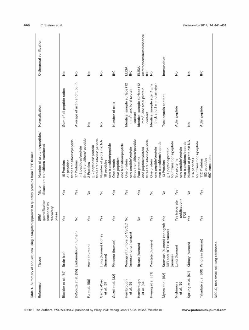
Table 1 summarizes current applications of targeted MSmethods in FFPE tissues. The size of the different studies interms of number of samples was very variable, ranging from3 [50] to 55 samples [51].
Even though the procedure for analytical validation of pro-teomic studies still needs to be established and standardized,several of the studies in the literature contain an analyticalvalidation step, with variations in the approaches and extentof validation. Analytical precision is often assessed and re-ported CVs are typically below 20–30% [32, 52–57]. Indeedprecision is a criterion for selecting individual peptides andtransitions. Considering the chemical modifications inducedin proteins by formalin fixation, which are not completely un-derstood [25], the reported CVs were surprisingly low. Vari-ability across different slices of the same tumor was repre-sented by CVs below 25%. While this is higher than someclinical standards, it shows promise for clinical use [57]. Lin-earity has been assessed with R2 ≥ 0.97 over dynamic ranges
spanning up to —three to four orders of magnitude [32, 56].The definition of the lower LOD (LLOD) and lower LOQ(LLOQ) has not yet been standardized. LLOD and LLOQwere defined by a threshold of the S/N (typically S/N > 3for LLOD and S/N > 10 for LLOQ) or as the lowest mea-sured concentration with a CV or a relative error below 20–25% or a combination of both [50, 53, 54]. These criteria areapproaching the threshold set by the Food and Drug Ad-ministration for small molecules, where the CV at LLOQshould not exceed 20% (http://www.fda.gov/downloads/Drugs/Guidances/ucm070107.pdf). The reported on columnlevels for LLODs and LLOQs for studies in FFPE tissues werein the picogram [50] or attomol [53, 54] range.
The effect of background matrix on linearity and repro-ducibility was compared in three different sections of thesame tissue [32]. While linearity and reproducibility in thethree different sections were comparable, differences in theslope of the three regression curves suggest matrix-dependentsignal enhancement/suppression due to the presence in thesample of species competing for ionization. Such effectsshould always be accounted for when performing an assayin a complex and variable background matrix [58], but theyare often missing in the literature.
A few studies did not report any analytical validation oftheir assay and the conclusions derived from these studiesshould therefore be taken with caution [37, 51, 59, 60].
The addition of heavy isotope-labeled internal standards iscritical to the quantification and precision and is usually usedto account for variations in ionization efficiency. While moststudies used 13C- and 15N-labeled standards [50, 53, 54, 59],one study used a deuterium-labeled standard [51], while an-other used synthetic peptides with a glycine insertion in thesequence [32]. This last option might work well provided thatthe retention time of the analyte and standard are close toeach other and that the standard has a unique sequence inorder to avoid signal contamination. The authors showed viadirect infusion that the ionization efficiency was similar forthe analyte and its internal standard. However, it would addi-tionally be useful to check for ionization suppression effectsin the elution window of both peaks. Two further studies useda stable isotope-labeled �-actin peptide spiked at a constantconcentration of 20 fmol/�L for quantification [52, 57]. Thisoption is not recommended as there is a relatively high riskof fluctuations in signal intensities throughout the chromato-graphic run, and these fluctuations will not be compensatedfor with an internal standard monitored at a single retentiontime throughout the analysis. Three studies have reported nouse of internal standards for quantification [37, 56, 60].
The quantification step is rarely described in a detailedmanner, and particularly if quantification was performed us-ing a single reference point or a calibration curve (normalor reversed) [61]. For example, Hwang et al. [51] reporteduse of a single reference point quantification, although thismethod was shown to be highly inaccurate in the lower andupper range of the quantification domain [61]. Hembroughet al. performed a normal calibration curve in a nonhuman
C© 2013 The Authors. PROTEOMICS published by Wiley-VCH Verlag GmbH & Co. KGaA, Weinheim www.proteomics-journal.com
446 C. Steiner et al. Proteomics 2014, 14, 441–451
Ta
ble
1.
Su
mm
ary
of
app
licat
ion
su
sin
ga
targ
eted
app
roac
hto
qu
anti
fyp
rote
ins
fro
mFF
PE
tiss
ues
Ref
eren
ceT
issu
eS
RM
qu
anti
fica
tio
np
rece
ded
by
dis
cove
ryp
has
e
Mic
ro-
dis
sect
ion
Nu
mb
ero
fp
rote
ins/
pep
tid
es/
tran
siti
on
sm
on
ito
red
No
rmal
izat
ion
Ort
ho
go
nal
veri
fica
tio
n
Bla
ckle
ret
al.[
59]
Bra
in(r
at)
Yes
Yes
15Pr
ote
ins
21p
epti
des
thre
etr
ansi
tio
ns/
pep
tid
e
Su
mo
fal
lpep
tid
era
tio
sN
o
DeS
ou
zaet
al.[
55]
En
do
met
riu
m(h
um
an)
No
Yes
17Pr
ote
ins
≥2
pep
tid
es/p
rote
inth
ree
tran
siti
on
s/p
epti
de
Ave
rag
eo
fac
tin
and
tub
ulin
No
Fuet
al.[
50]
Ao
rta
(hu
man
)Ye
sN
o2
Pro
tein
s≥
2p
epti
des
/pro
tein
thre
etr
ansi
tio
ns/
pep
tid
e
No
No
Gam
ez-P
ozo
etal
.[37
]Lu
ng
(hu
man
)ki
dn
ey(h
um
an)
Yes
No
Nu
mb
ero
fp
rote
ins:
NA
18p
epti
des
on
etr
ansi
tio
n/p
epti
de
No
No
Gu
zele
tal
.[32
]P
lace
nta
(hu
man
)Ye
sYe
sO
ne
pro
tein
two
pep
tid
eso
ne
tran
siti
on
/pep
tid
e
Nu
mb
ero
fce
llsIH
C
Hem
bro
ug
het
al.[
53]
Xen
og
raft
tum
ors
of
NS
CLC
(mo
use
)lu
ng
(hu
man
)N
oYe
sO
ne
pro
tein
on
ep
epti
de/
pro
tein
thre
etr
ansi
tio
ns/
pep
tid
e
Iden
tica
lsam
ple
surf
ace
(12
mm
2)
and
tota
lpro
tein
con
ten
t
ELI
SA
IHC
Hem
bro
ug
het
al.[
54]
Bre
ast
(hu
man
)N
oYe
sFo
ur
pro
tein
so
ne
pep
tid
e/p
rote
inth
ree
tran
siti
on
s/p
epti
de
Iden
tica
lsam
ple
surf
ace
(12
mm
2)
and
tota
lpro
tein
con
ten
t
ELI
SA
/el
ectr
och
emilu
min
esce
nce
IHC
Hw
ang
etal
.[51
]Pr
ost
ate
(hu
man
)Ye
sN
oO
ne
pro
tein
on
ep
epti
de/
pro
tein
two
tran
siti
on
s/p
epti
de
Iden
tica
lsam
ple
size
(4�
mth
ick
and
2m
md
iam
eter
)N
o
Mye
rset
al.[
52]
Sto
mac
h(h
um
an)
xen
og
raft
DiF
ian
dH
CT
116
tum
ors
(mo
use
)
Yes
No
13Pr
ote
ins
≥1
pep
tid
e/p
rote
info
ur
tran
siti
on
s/p
epti
de
Tota
lpro
tein
con
ten
tIm
mu
no
blo
t
Nis
him
ura
etal
.[56
]Lu
ng
(hu
man
)Ye
s(s
epar
ate
pu
blic
atio
n)
[72]
Yes
Six
pro
tein
so
ne
pep
tid
e/p
rote
intw
otr
ansi
tio
ns/
pep
tid
e
Act
inp
epti
de
No
Sp
run
get
al.[
57]
Kid
ney
(hu
man
)Ye
sN
oN
um
ber
of
pro
tein
s:N
A11
4p
epti
des
fou
rtr
ansi
tio
ns/
pep
tid
e
No
No
Taka
dat
eet
al.[
60]
Pan
crea
s(h
um
an)
Yes
Yes
112
Pro
tein
s18
3p
epti
des
697
tran
siti
on
s
Act
inp
epti
de
IHC
NS
CLC
,no
n-s
mal
lcel
llu
ng
carc
ino
ma.
C© 2013 The Authors. PROTEOMICS published by Wiley-VCH Verlag GmbH & Co. KGaA, Weinheim www.proteomics-journal.com
Proteomics 2014, 14, 441–451 447
complex matrix (peptide mixture of Pyrococcus furiosus), asthey rightly state that there is no existing standard tissue ma-trix [53]. They also compared the linear response in the non-human and an expectedly more complex human matrix andshowed that the response was similar in both cases, exceptfor one protein where the slope and intercept were differentin both backgrounds [54].
Typically, three peptides per protein and three transitionswith the highest intensities per peptide are monitored in anSRM assay as this increases specificity [49, 62]. There is alsovalue in monitoring peptides distributed throughout the se-quence (constant and variable domains) to monitor the dif-ferent variants of a protein [62]. However the number ofpeptides/transitions monitored varies in the literature withbetween one to two peptides per protein and one to two tran-sitions per peptide [32, 51, 56] up to two to three peptides perprotein and three to four transitions per peptide [50, 52, 55].It is also noteworthy that all studies cited developed SRM as-says for unmodified peptides, thus disregarding any potentialmodifications induced by formalin fixation.
While monitoring several peptides/transitions representsan advantage for specificity, it raises the question of how todeal with the multiple data points generated in order to ob-tain a single value representing the expression level of a givenprotein. In the majority of publications, there is no detaileddescription of how the SRM data were further processed. Itis, for example, unclear how two peptides belonging to thesame protein but with inconsistent results should be handled.Guzel et al. mentioned expressing the result as the average ofboth peptides measured [32], while Sprung et al. used the sumof the four transitions for a given peptide [57]. An option forhandling this type of data is to use linear mixed-effects models[63, 64]. However, to our knowledge, none of the SRM meth-ods on FFPE tissues cited here have used this strategy yet.
Normalization is particularly critical when working withtissue since the sampled amount is less quantifiable com-pared to the volume in the case of biological fluids for exam-ple. Several options have been considered for normalization.The simplest is to sample defined tissue volumes (e.g. biop-sies of identical size) [51]. Others have used cell number [32],total protein content [52], sum of all peptide ratios for eachanalysis [59], or specific housekeeping proteins, such as actinor an average of actin and tubulin [55, 56, 60]. Hembroughet al. sampled identical surface areas and additionally nor-malized the result obtained for total protein content [53, 54].Three studies did not report any normalization for tissuecontent [37, 50, 57]. A flaw with most of these approaches isthat tissue heterogeneity is not accounted for. The number ofcells can be obtained when performing microdissection andthis measure can therefore account to some extent for tissueheterogeneity. However, the cell number is approximate andadding another normalization parameter, such as total pro-tein content or a housekeeping protein might lead to moreaccurate results. The option of using the sum of all peptideratios is interesting, however, it contains the risk of showinga bias toward the proteins of interest included in the assay,
which will naturally be more elevated in certain cases (e.g. indiseased tissues versus healthy tissues), although the amountof sampled tissue might originally be identical. The most ap-propriate normalization factor still needs to be determinedand its performance should be assessed as any imprecisionor inaccuracy will be reported on the final measurement.
Alternatively, because sampling whole tissue sections doesnot account for heterogeneity of cellular protein expression,several studies included a microdissection step prior to pro-tein extraction [32, 53–56, 59, 60]. Microdissection of specificcells, however, is a time-consuming process that must beperformed by a trained pathologist and the small amountof tissue sampled may lead to sensitivity issues. Neverthe-less, several SRM studies on FFPE tissues reported the useof microdissection, which simultaneously decreases samplecomplexity and increases the dynamic range of the analysis.
Several of the SRM assays developed on FFPE tissues pub-lished to date represent confirmation of findings observedduring a preliminary discovery phase and orthogonal verifi-cation using other approaches is therefore limited in thesecases [37, 50, 56, 59]. Myers et al., however, obtained im-munoblot data consistent with SRM data [52] and Guzel et al.showed a good correlation between IHC and SRM data in apreeclampsia model, albeit with a limited number of sam-ples [32]. However, in both cases, the degree of correlationwas not defined. The largest cohort for orthogonal verificationwas presented by Takadate et al. in which IHC was used toconfirm the association of four candidate prognostic markersand outcomes in 87 cases of pancreatic ductal adenocarci-noma [60]. The association of SRM data with IHC could beverified for four proteins, but further orthogonal verificationusing Kaplan–Meier curves led to ambiguous results for atleast one of these proteins. Hembrough et al. correlated IHCwith SRM data for EGFR in ten human non-small cell lungcarcinoma xenografts and indicated a correlative trend [53].In a more recent study, they determined HER2 levels by IHCin two cohorts of 10 and 19 breast cancer samples, but a cor-relation between IHC and SRM data was observed only inthe first cohort, suggesting to the authors that many patientswith a 3+ IHC staining for HER2 probably do not expresselevated levels of this protein and are therefore unlikely tobenefit from trastuzumab [54]. This example illustrates thepotential of this type of method for the generation of newhypotheses.
In summary, targeted proteomics on FFPE tissues is slowlymoving towards more routine applications, either in phar-maceutical research or in the clinical field. However, muchwork still needs to be achieved in order to obtain a consen-sus on several critical issues: sample preparation protocols,quantification, parameters for analytical validation, data pro-cessing, normalization, etc. More emphasis is still needed onthe comparison with reference methods for protein or geneexpression measurement in FFPE tissues (IHC, fluorescencein situ hybridization, etc.), as current reports of MS assays arestill ambiguous and there is a lack of quantitative measuresassessing how well these methods perform.
C© 2013 The Authors. PROTEOMICS published by Wiley-VCH Verlag GmbH & Co. KGaA, Weinheim www.proteomics-journal.com

448 C. Steiner et al. Proteomics 2014, 14, 441–451
5 Perspectives for clinical applications
The analysis of proteins and peptides from FFPE tissuesis constantly improving in terms of extraction, detection,and quantification. In particular, the characterization of pro-teomes from FFPE tissues and their comparison with pro-teomes from FF tissues are evolving rapidly and have thepotential to significantly complement data obtained usingexpression arrays. Even though a large amount of data is al-ready available through gene expression assays, it remainsof importance to obtain protein expression data, due to thelimited correlation between protein levels and gene expres-sion [65–67]. Compared to IHC, which is traditionally usedin clinical practice to classify tumors according to biomarkerexpression levels, MS-based protein analysis has the poten-tial to provide a more quantitative assessment of tissue pro-tein expression levels with a dynamic range of quantificationof up to five orders of magnitude using SRM [49]. Further-more, MS enables multiplexing assay formats with the mea-surement of several analytes in parallel. A key feature of anMS-based method is the analytical specificity enabling theanalysis of isoforms, such as those resulting from somaticmutations [68]. Such discrimination is more difficult withIHC as antibodies selectively recognizing specific forms ofan epitope rarely are available.
From a clinical and pharmaceutical perspective, analysisof the proteome from retrospective FFPE tissues presentsseveral advantages. Such tissues exist in repositories witha high degree of clinical annotation. Notwithstanding theethical and consent issues, such samples have great utilityin clinical and research settings. The ability to profile largeand well-annotated cohorts would facilitate understandingof pathophysiological processes and may enable the discov-ery of novel biomarkers or therapeutic targets. In particular,accurate protein quantification will be important to unravelderegulated cellular signaling events by providing a repre-sentative insight of cellular effectors. This includes PTMssuch as phosphorylation, a process whose deregulation isoften involved in cancerous cells [37]. There is an increas-ing recognition that personalized healthcare is of importancein cancer therapy. Functional classification of tumors, dis-ease staging, biomarker definition, patient stratification, andchoice of treatment may all be enhanced by the wealth ofdata generated from FFPE tissues. From a drug discoveryperspective, the ability to analyze a target in its tissue envi-ronment and to understand its underlying regulation is ab-solutely essential to test the efficacy and selectivity of novelpharmaceutical agents. However, due to the invasive natureof tissue sampling, human samples are usually restricted toplasma or urine, more rarely cerebrospinal fluid is used. On-cology is a notable exception as tumor resection is often apart of the medical treatment. In this particular domain, theability to analyze proteins from FFPE tissues combined withtissue collections available have the potential to further al-low a deeper understanding of the mechanisms underlyingcancer.
While MS analysis of FFPE tissue has advanced in re-cent years and is already showing some impact in medicalresearch, it is important to also acknowledge current limita-tions, which need to be addressed before this technique canbe considered as an option in clinical diagnosis. For example,it is now clear that retrieving peptides or even whole pro-teins from FFPE tissues is possible and to a significant extentcomparable with proteomes from FF tissues. But questionsremain to be answered regarding the optimal way to processthese types of samples in a standardized manner. A key lim-itation of both shotgun and targeted proteomics in tissues isthe lack of spatial resolution due to the sample preparationprocess. This aspect can be circumvented by dissecting outan area of interest by laser capture microdissection. How-ever, despite recent advances in automation, laser capturemicrodissection requires significant pathology expertise, re-sources, and time, which is not easily compatible even withmiddle-scale studies (e.g. 50 samples) or a clinical diagnosticapplication.
Due to the many issues related to MS-based quantificationof proteins from FFPE tissues, this technique is still a longway from being adopted in clinical settings. This is in partdue to the complexity of the sample preparation procedure,which is relatively long and requires a number of manualsteps including many variables, such as antigen retrieval ortrypsin digestion. This is in contrast with IHC, where stain-ing is performed by robotic systems with the concomitantspeed and reproducibility. Another important aspect is tissueheterogeneity and the requirement for normalization to allowcomparison between samples. Tumor samples typically con-tain a high degree of cellular heterogeneity, represented byvarious amounts of tumoral cells and stroma, with possiblyhypoxic or necrotic areas. Accordingly, normalization can bebased, among others, on the surface of dissection area, on anaveraged protein or peptide amount (measured using a colori-metric assay), or on the inclusion of housekeeping proteinsin the quantification assay. However, there is at this time noconsensus within the research community on normalizationor an optimal method for expressing protein expression levelsin tissue.
Another factor specific to shotgun and SRM proteomics isthe use of peptide quantification as a surrogate for proteinexpression level, with the former not necessarily agreeingin absolute terms with the latter. This factor is true wherebottom-up proteomics is performed, but of particular signif-icance in the FFPE setting. Studies with tissue surrogateshave shown that >90% of proteins are recovered when usingthe appropriate extraction protocol, with the extraction ratevarying between proteins, probably due to different physico-chemical properties [13]. Therefore, the exact proportion ofproteins effectively extracted during the antigen retrieval pro-cess remains unknown and is probably different for individ-ual proteins. In plasma (and other biofluids), the spiking ofknown amounts of stable isotope-labeled proteins representsan elegant strategy to account for losses during the diges-tion and extraction procedure. This approach is not suitable
C© 2013 The Authors. PROTEOMICS published by Wiley-VCH Verlag GmbH & Co. KGaA, Weinheim www.proteomics-journal.com

Proteomics 2014, 14, 441–451 449
for tissue as it would require a heavy isotope-labeled proteinto be present in the sample in a physiological manner be-fore the sample is fixed with formalin, which is not feasible.However, a relative quantification procedure using a stableisotope-labeled cell culture of the appropriate lineage for nor-malization might be applicable for tissue if the proteins ofinterest are physiologically present at appropriate amountsin the standard before formalin fixation [69]. The additionof heavy isotope-labeled peptides in the sample prior to MSanalysis therefore remains the best option, although a com-promised solution, as the peptides may decay at variable ratesdepending on the time point of their introduction in the ex-traction procedure, which in turn may affect the accuracy ofmeasurements [70]. Moreover, preanalytical factors includingsteps from the surgical removal of the tissue to the MS analy-sis need to be controlled for, since any preanalytical variationwill be reflected in the final result.
Finally, the acceptance of protein quantification by SRMfrom FFPE tissue in the clinical community will depend onthe availability of standardized procedures and quality con-trols that need to be defined and on the interlaboratory stan-dardization, which must be established. The chemical modi-fications induced by formalin fixation [7,25,71] and the effectson protein retrieval of parameters such as fixation time andstorage time have already been reviewed [8, 25–27] but mustbe well understood and better managed before quantificationof proteins from FFPE tissues can be performed routinely.Correlations of MS with IHC will indicate whether this newtechnique is suitable for wider clinical use. In addition, massspectrometers remain a large capital investment for an indi-vidual laboratory, although the reagents costs associated withIHC (antibodies) may ultimately compensate for this. Whilethey are increasingly being used in a clinical setting, com-bining the expert knowledge of the pathologist to examinethe tissue morphology and of the analytical chemist to set upanalytical methods requires co-ordination and commitment.
The authors thank Dr. Michelle Butterfield, Dr. Miro Venturi,and Prof. Denis Hochstrasser for their helpful insight during thepreparation of the review. Dr. Carine Steiner was funded via aRoche postdoctoral fellowship.
The authors have declared no conflict of interest.
6 References
[1] Biomarkers Definitions Working Group, Biomarkers andsurrogate endpoints: preferred definitions and conceptualframework. Clin. Pharmacol. Ther. 2001, 69, 89–95.
[2] Hoofnagle, A. N., Wener, M. H., The fundamental flaws ofimmunoassays and potential solutions using tandem massspectrometry. J. Immunol. Methods 2009, 347, 3–11.
[3] Yamashita, S., Heat-induced antigen retrieval: mechanismsand application to histochemistry. Prog. Histochem. Cy-tochem. 2007, 41, 141–200.
[4] Cronin, M., Pho, M., Dutta, D., Stephans, J. C. et al., Mea-surement of gene expression in archival paraffin-embeddedtissues: development and performance of a 92-gene re-verse transcriptase-polymerase chain reaction assay. Am.J. Pathol. 2004, 164, 35–42.
[5] Specht, K., Richter, T., Muller, U., Walch, A. et al., Quanti-tative gene expression analysis in microdissected archivalformalin-fixed and paraffin-embedded tumor tissue. Am. J.Pathol. 2001, 158, 419–429.
[6] Godfrey, T. E., Kim, S. H., Chavira, M., Ruff, D. W. et al.,Quantitative mRNA expression analysis from formalin-fixed,paraffin-embedded tissues using 5’ nuclease quantitative re-verse transcription-polymerase chain reaction. J. Mol. Di-agn. 2000, 2, 84–91.
[7] Tanca, A., Pagnozzi, D., Addis, M. F., Setting proteins free:progresses and achievements in proteomics of formalin-fixed, paraffin-embedded tissues. Proteomics Clin. Appl.2012, 6, 7–21.
[8] Giusti, L., Lucacchini, A., Proteomic studies of formalin-fixedparaffin-embedded tissues. Expert Rev. Proteomics 2013, 10,165–177.
[9] Chatterji, B., Pich, A., MALDI imaging mass spectrometry andanalysis of endogenous peptides. Expert Rev. Proteomics2013, 10, 381–388.
[10] Morita, Y., Ikegami, K., Goto-Inoue, N., Hayasaka, T.et al., Imaging mass spectrometry of gastric carcinoma informalin-fixed paraffin-embedded tissue microarray. CancerSci. 2010, 101, 267–273.
[11] Morgan, T. M., Seeley, E. H., Fadare, O., Caprioli, R. M., Clark,P. E., Imaging the clear cell renal cell carcinoma proteome.J. Urol. 2013, 189, 1097–1103.
[12] Fox, C. H., Johnson, F. B., Whiting, J., Roller, P. P.,Formaldehyde fixation. J. Histochem. Cytochem. 1985, 33,845–853.
[13] Fowler, C. B., Cunningham, R. E., O’Leary, T. J., Mason,J. T., ‘Tissue surrogates’ as a model for archival formalin-fixed paraffin-embedded tissues. Lab. Invest. 2007, 87,836–846.
[14] Shi, S. R., Cote, R. J., Wu, L., Liu, C. et al., DNA extractionfrom archival formalin-fixed, paraffin-embedded tissue sec-tions based on the antigen retrieval principle: heating un-der the influence of pH. J. Histochem. Cytochem. 2002, 50,1005–1011.
[15] Layfield, R., Bailey, K., Lowe, J., Allibone, R. et al., Extractionand protein sequencing of immunoglobulin light chain fromformalin-fixed cerebrovascular amyloid deposits. J. Pathol.1996, 180, 455–459.
[16] Ikeda, K., Monden, T., Kanoh, T., Tsujie, M. et al., Extractionand analysis of diagnostically useful proteins from formalin-fixed, paraffin-embedded tissue sections. J. Histochem.Cytochem. 1998, 46, 397–403.
[17] Vasilescu, J., Guo, X., Kast, J., Identification of protein-protein interactions using in vivo cross-linking and massspectrometry. Proteomics 2004, 4, 3845–3854.
[18] Hood, B. L., Darfler, M. M., Guiel, T. G., Furusato, B. et al.,Proteomic analysis of formalin-fixed prostate cancer tissue.Mol. Cell. Proteomics 2005, 4, 1741–1753.
C© 2013 The Authors. PROTEOMICS published by Wiley-VCH Verlag GmbH & Co. KGaA, Weinheim www.proteomics-journal.com
450 C. Steiner et al. Proteomics 2014, 14, 441–451
[19] Palmer-Toy, D. E., Krastins, B., Sarracino, D. A., Nadol, J.B., Jr., Merchant, S. N., Efficient method for the proteomicanalysis of fixed and embedded tissues. J. Proteome Res.2005, 4, 2404–2411.
[20] Prieto, D. A., Hood, B. L., Darfler, M. M., Guiel, T. G. et al.,Liquid Tissue: proteomic profiling of formalin-fixed tissues.Biotechniques 2005, 38, 32–35.
[21] Crockett, D. K., Lin, Z., Vaughn, C. P., Lim, M. S., Elenitoba-Johnson, K. S., Identification of proteins from formalin-fixedparaffin-embedded cells by LC-MS/MS. Lab. Invest. 2005, 85,1405–1415.
[22] Shi, S. R., Shi, Y., Taylor, C. R., Antigen retrieval immuno-histochemistry: review and future prospects in research anddiagnosis over two decades. J. Histochem. Cytochem. 2011,59, 13–32.
[23] Berg, D., Hipp, S., Malinowsky, K., Bollner, C., Becker, K. F.,Molecular profiling of signalling pathways in formalin-fixedand paraffin-embedded cancer tissues. Eur. J. Cancer 2010,46, 47–55.
[24] Maes, E., Broeckx, V., Mertens, I., Sagaert, X. et al., Analysisof the formalin-fixed paraffin-embedded tissue proteome:pitfalls, challenges, and future prospectives. Amino Acids2013, 45, 205–218.
[25] Magdeldin, S., Yamamoto, T., Toward deciphering pro-teomes of formalin-fixed paraffin-embedded (FFPE) tissues.Proteomics 2012, 12, 1045–1058.
[26] Shi, S. R., Taylor, C. R., Fowler, C. B., Mason, J. T., Com-plete solubilization of formalin-fixed, paraffin-embedded tis-sue may improve proteomic studies. Proteomics Clin. Appl.2013, 7, 264–272.
[27] Thompson, S. M., Craven, R. A., Nirmalan, N. J., Harnden, P.et al., Impact of pre-analytical factors on the proteomic anal-ysis of formalin-fixed paraffin-embedded tissue. ProteomicsClin. Appl. 2013, 7, 241–251.
[28] Sprung, R. W., Jr., Brock, J. W., Tanksley, J. P., Li, M. et al.,Equivalence of protein inventories obtained from formalin-fixed paraffin-embedded and frozen tissue in multidimen-sional liquid chromatography-tandem mass spectrometryshotgun proteomic analysis. Mol. Cell. Proteomics 2009, 8,1988–1998.
[29] Hicks, D. G., Schiffhauer, L., Standardized assessment ofthe HER2 status in breast cancer by immunohistochemistry.LabMedicine 2011, 42, 459–467.
[30] Hicks, D. G., Boyce, B. F., The challenge and importanceof standardizing pre-analytical variables in surgical pathol-ogy specimens for clinical care and translational research.Biotech. Histochem. 2012, 87, 14–17.
[31] Craven, R. A., Cairns, D. A., Zougman, A., Harnden, P. et al.,Proteomic analysis of formalin-fixed paraffin-embedded re-nal tissue samples by label-free MS: assessment of overalltechnical variability and the impact of block age. ProteomicsClin. Appl. 2013, 7, 273–282.
[32] Guzel, C., Ursem, N. T., Dekker, L. J., Derkx, P. et al., Multiplereaction monitoring assay for pre-eclampsia related calcy-clin peptides in formalin fixed paraffin embedded placenta.J. Proteome Res. 2011, 10, 3274–3282.
[33] Guo, T., Wang, W., Rudnick, P. A., Song, T. et al., Proteome
analysis of microdissected formalin-fixed and paraffin-embedded tissue specimens. J. Histochem. Cytochem. 2007,55, 763–772.
[34] Tanca, A., Pagnozzi, D., Burrai, G. P., Polinas, M. et al., Com-parability of differential proteomics data generated frompaired archival fresh-frozen and formalin-fixed samples byGeLC-MS/MS and spectral counting. J. Proteomics 2012, 77,561–576.
[35] Tanca, A., Pisanu, S., Biosa, G., Pagnozzi, D. et al., Applica-tion of 2D-DIGE to formalin-fixed diseased tissue samplesfrom hospital repositories: results from four case studies.Proteomics Clin. Appl. 2013, 7, 252–263.
[36] Ostasiewicz, P., Zielinska, D. F., Mann, M., Wisniewski,J. R., Proteome, phosphoproteome, and N-glycoproteomeare quantitatively preserved in formalin-fixed paraffin-embedded tissue and analyzable by high-resolution massspectrometry. J. Proteome Res. 2010, 9, 3688–3700.
[37] Gamez-Pozo, A., Sanchez-Navarro, I., Calvo, E., Diaz, E. et al.,Protein phosphorylation analysis in archival clinical cancersamples by shotgun and targeted proteomics approaches.Mol. Biosyst. 2011, 7, 2368–2374.
[38] Espina, V., Edmiston, K. H., Heiby, M., Pierobon, M. et al.,A portrait of tissue phosphoprotein stability in the clinicaltissue procurement process. Mol. Cell. Proteomics 2008, 7,1998–2018.
[39] Gundisch, S., Hauck, S., Sarioglu, H., Schott, C. et al., Vari-ability of protein and phosphoprotein levels in clinical tissuespecimens during the preanalytical phase. J. Proteome Res.2012, 11, 5748–5762.
[40] Gundisch, S., Grundner-Culemann, K., Wolff, C., Schott, C.et al., Delayed times to tissue fixation result in unpredictableglobal phosphoproteome changes. J. Proteome Res. 2013,12, 4424–4434.
[41] Ong, S. E., Blagoev, B., Kratchmarova, I., Kristensen, D. B.et al., Stable isotope labeling by amino acids in cell culture,SILAC, as a simple and accurate approach to expression pro-teomics. Mol. Cell. Proteomics 2002, 1, 376–386.
[42] Nakatani, S., Wei, M., Ishimura, E., Kakehashi, A. et al.,Proteome analysis of laser microdissected glomeruli fromformalin-fixed paraffin-embedded kidneys of autopsies ofdiabetic patients: nephronectin is associated with the devel-opment of diabetic glomerulosclerosis. Nephrol. Dial. Trans-plant. 2012, 27, 1889–1897.
[43] Jain, M. R., Li, Q., Liu, T., Rinaggio, J. et al., Proteomic iden-tification of immunoproteasome accumulation in formalin-fixed rodent spinal cords with experimental autoimmuneencephalomyelitis. J. Proteome Res. 2012, 11, 1791–1803.
[44] Nahnsen, S., Bielow, C., Reinert, K., Kohlbacher, O., Tools forlabel-free peptide quantification. Mol. Cell. Proteomics 2013,12, 549–556.
[45] Jain, M. R., Liu, T., Hu, J., Darfler, M. et al., Quantitativeproteomic analysis of formalin fixed paraffin embedded oralHPV lesions from HIV patients. Open Proteomics J. 2008, 1,40–45.
[46] Choi, H., Nesvizhskii, A. I., False discovery rates and relatedstatistical concepts in mass spectrometry-based proteomics.J. Proteome Res. 2008, 7, 47–50.
C© 2013 The Authors. PROTEOMICS published by Wiley-VCH Verlag GmbH & Co. KGaA, Weinheim www.proteomics-journal.com
Proteomics 2014, 14, 441–451 451
[47] Sandin, M., Krogh, M., Hansson, K., Levander, F., Genericworkflow for quality assessment of quantitative label-freeLC-MS analysis. Proteomics 2011, 11, 1114–1124.
[48] Clough, T., Thaminy, S., Ragg, S., Aebersold, R., Vitek, O.,Statistical protein quantification and significance analysis inlabel-free LC-MS experiments with complex designs. BMCBioinform. 2012, 13(Suppl 16), 1–17.
[49] Lange, V., Picotti, P., Domon, B., Aebersold, R., Selected reac-tion monitoring for quantitative proteomics: a tutorial. Mol.Syst. Biol. 2008, 4, 1–14.
[50] Fu, Z., Yan, K., Rosenberg, A., Jin, Z. et al., Improved proteinextraction and protein identification from archival formalin-fixed paraffin-embedded human aortas. Proteomics Clin.Appl. 2013, 7, 217–224.
[51] Hwang, S. I., Thumar, J., Lundgren, D. H., Rezaul, K. et al., Di-rect cancer tissue proteomics: a method to identify candidatecancer biomarkers from formalin-fixed paraffin-embeddedarchival tissues. Oncogene 2007, 26, 65–76.
[52] Myers, M. V., Manning, H. C., Coffey, R. J., Liebler, D. C.,Protein expression signatures for inhibition of epidermalgrowth factor receptor-mediated signaling. Mol. Cell. Pro-teomics 2012, 11, M111 015222.
[53] Hembrough, T., Thyparambil, S., Liao, W. L., Darfler, M. M.et al., Selected reaction monitoring (SRM) analysis of epider-mal growth factor receptor (EGFR) in formalin fixed tumortissue. Clin. Proteomics 2012, 9, 1–10.
[54] Hembrough, T., Thyparambil, S., Liao, W. L., Darfler, M.M. et al., Application of selected reaction monitoring formultiplex quantification of clinically validated biomarkersin formalin-fixed, paraffin-embedded tumor tissue. J. Mol.Diagn. 2013, 15, 454–465.
[55] DeSouza, L. V., Krakovska, O., Darfler, M. M., Krizman, D.B. et al., mTRAQ-based quantification of potential endome-trial carcinoma biomarkers from archived formalin-fixedparaffin-embedded tissues. Proteomics 2010, 10, 3108–3116.
[56] Nishimura, T., Nomura, M., Tojo, H., Hamasaki, H. et al., Pro-teomic analysis of laser-microdissected paraffin-embeddedtissues: (2) MRM assay for stage-related proteins upon non-metastatic lung adenocarcinoma. J. Proteomics 2010, 73,1100–1110.
[57] Sprung, R. W., Martinez, M. A., Carpenter, K. L., Ham, A. J.et al., Precision of multiple reaction monitoring mass spec-trometry analysis of formalin-fixed, paraffin-embedded tis-sue. J. Proteome Res. 2012, 11, 3498–3505.
[58] Matuszewski, B. K., Constanzer, M. L., Chavez-Eng, C. M.,Strategies for the assessment of matrix effect in quantitativebioanalytical methods based on HPLC-MS/MS. Anal. Chem.2003, 75, 3019–3030.
[59] Blackler, A. R., Morgan, N. Y., Gao, B., Olano, L. R. et al.,Proteomic analysis of nuclei dissected from fixed rat brain
tissue using expression microdissection. Anal. Chem. 2013,85, 7139–7145.
[60] Takadate, T., Onogawa, T., Fukuda, T., Motoi, F. et al., Novelprognostic protein markers of resectable pancreatic canceridentified by coupled shotgun and targeted proteomics us-ing formalin-fixed paraffin-embedded tissues. Int. J. Cancer2013, 132, 1368–1382.
[61] Campbell, J., Rezai, T., Prakash, A., Krastins, B. et al., Eval-uation of absolute peptide quantitation strategies using se-lected reaction monitoring. Proteomics 2011, 11, 1148–1152.
[62] Gallien, S., Duriez, E., Domon, B., Selected reaction moni-toring applied to proteomics. J. Mass Spectrom. 2011, 46,298–312.
[63] Chang, C. Y., Picotti, P., Huttenhain, R., Heinzelmann-Schwarz, V. et al., Protein significance analysis in selectedreaction monitoring (SRM) measurements. Mol. Cell. Pro-teomics 2012, 11, M111 014662.
[64] Lamerz, J., Friedlein, A., Soder, N., Cutler, P., Dobeli, H., Deter-mination of free desmosine in human plasma and its appli-cation in two experimental medicine studies. Anal. Biochem.2013, 436, 127–136.
[65] Rogers, S., Girolami, M., Kolch, W., Waters, K. M. et al., In-vestigating the correspondence between transcriptomic andproteomic expression profiles using coupled cluster models.Bioinformatics 2008, 24, 2894–2900.
[66] Dhingra, V., Gupta, M., Andacht, T., Fu, Z. F., New frontiers inproteomics research: a perspective. Int. J. Pharm. 2005, 299,1–18.
[67] Gygi, S. P., Rochon, Y., Franza, B. R., Aebersold, R., Correla-tion between protein and mRNA abundance in yeast. Mol.Cell. Biol. 1999, 19, 1720–1730.
[68] Smith, L. M., Kelleher, N. L., Proteoform: a single term de-scribing protein complexity. Nat. Methods 2013, 10, 186–187.
[69] Geiger, T., Wisniewski, J. R., Cox, J., Zanivan, S. et al., Useof stable isotope labeling by amino acids in cell culture asa spike-in standard in quantitative proteomics. Nat. Protoc.2011, 6, 147–157.
[70] Shuford, C. M., Sederoff, R. R., Chiang, V. L., Muddiman,D. C., Peptide production and decay rates affect the quanti-tative accuracy of protein cleavage isotope dilution massspectrometry (PC-IDMS). Mol. Cell. Proteomics 2012, 11,814–823.
[71] Klockenbusch, C., O’Hara, J. E., Kast, J., Advancing formalde-hyde cross-linking towards quantitative proteomic applica-tions. Anal. Bioanal. Chem. 2012, 404, 1057–1067.
[72] Kawamura, T., Nomura, M., Tojo, H., Fujii, K. et al., Pro-teomic analysis of laser-microdissected paraffin-embeddedtissues: (1) stage-related protein candidates upon non-metastatic lung adenocarcinoma. J. Proteomics 2010, 73,1089–1099.
C© 2013 The Authors. PROTEOMICS published by Wiley-VCH Verlag GmbH & Co. KGaA, Weinheim www.proteomics-journal.com
Related Documents