REVIEW Open Access Apoptotic markers in protozoan parasites Antonio Jiménez-Ruiz 1* , Juan Fernando Alzate 2 , Ewan Thomas MacLeod 3 , Carsten Günter Kurt Lüder 4 , Nicolas Fasel 5 , Hilary Hurd 6 Abstract The execution of the apoptotic death program in metazoans is characterized by a sequence of morphological and biochemical changes that include cell shrinkage, presentation of phosphatidylserine at the cell surface, mitochon- drial alterations, chromatin condensation, nuclear fragmentation, membrane blebbing and the formation of apop- totic bodies. Methodologies for measuring apoptosis are based on these markers. Except for membrane blebbing and formation of apoptotic bodies, all other events have been observed in most protozoan parasites undergoing cell death. However, while techniques exist to detect these markers, they are often optimised for metazoan cells and therefore may not pick up subtle differences between the events occurring in unicellular organisms and multi- cellular organisms. In this review we discuss the markers most frequently used to analyze cell death in protozoan parasites, paying special attention to changes in cell morphology, mitochondrial activity, chromatin structure and plasma membrane structure/permeability. Regarding classical regulators/executors of apoptosis, we have reviewed the present knowl- edge of caspase-like and nuclease activities. Review Recently there has been a move to clarify the classifica- tion of cell death [1] and to set up guidelines for cell death assays in eukaryotes [2]. While these events have been studied in metazoans for more than two decades, the presence of apoptosis-like processes in protozoans was not widely accepted when the first papers on events similar to metazoan apoptosis were published 15 years ago [3,4]. Therefore, considering the diversity of para- sites and conditions for their study, there is an urgent need to define the markers, both cellular and molecular, which are the most accurate to assign as apoptosis-like events that occur during the death process of protozoan parasites. Defining the events that occur during death are of paramount importance to this field of study, since so far there is no definitive guide as to what happens in each parasitic protozoan when it undergoes this process, nor is it clear whether similar cell death phenotypes are exhibited by all taxa of unicellular organisms. Moreover, some of these assays are likely to be positive in cells undergoing different types of cell death. This fact stresses the relevance of using several markers simulta- neously to confirm an apoptotic phenotype. According to the Nomenclature Committee on Cell Death (NCCD) [1], the term apoptosis describes a speci- fic morphological aspect of cell death. Cellular altera- tions during this process include rounding-up of the cell, reduction of cellular volume (pyknosis), chromatin condensation, nuclear fragmentation, plasma membrane blebbing and, if in the right context, engulfment by resi- dent phagocytes. Little or no ultrastructural modifica- tions of cytoplasmic organelles are observed during apoptosis. On the contrary, necrotic cell death is mor- phologically characterized by a gain in cell volume (oncosis), swelling of organelles, plasma membrane rup- ture and subsequent loss of intracellular contents. In the absence of common biochemical markers, early plasma membrane permeabilization is considered the main hall- mark of necrosis. Otherwise, necrotic cell death is still largely identified in negative terms by the absence of apoptotic or autophagic markers. Following these guidelines, several changes seem to be useful to define whether cells die following an apoptotic or a necrotic death. From a morphological point of view, a pyknotic cell should be considered to be in the process of apoptotic cell death. Regarding biochemical markers, detection of DNA fragmentation or caspase * Correspondence: [email protected] 1 Departamento de Bioquímica y Biología Molecular, Universidad de Alcalá, 28871 Alcalá de Henares, Madrid, Spain Full list of author information is available at the end of the article Jiménez-Ruiz et al. Parasites & Vectors 2010, 3:104 http://www.parasitesandvectors.com/content/3/1/104 © 2010 Jiménez-Ruiz et al; licensee BioMed Central Ltd. This is an Open Access article distributed under the terms of the Creative Commons Attribution License (http://creativecommons.org/licenses/by/2.0), which permits unrestricted use, distribution, and reproduction in any medium, provided the original work is properly cited.

Welcome message from author
This document is posted to help you gain knowledge. Please leave a comment to let me know what you think about it! Share it to your friends and learn new things together.
Transcript
REVIEW Open Access
Apoptotic markers in protozoan parasitesAntonio Jiménez-Ruiz1*, Juan Fernando Alzate2, Ewan Thomas MacLeod3, Carsten Günter Kurt Lüder4,Nicolas Fasel5, Hilary Hurd6
Abstract
The execution of the apoptotic death program in metazoans is characterized by a sequence of morphological andbiochemical changes that include cell shrinkage, presentation of phosphatidylserine at the cell surface, mitochon-drial alterations, chromatin condensation, nuclear fragmentation, membrane blebbing and the formation of apop-totic bodies. Methodologies for measuring apoptosis are based on these markers. Except for membrane blebbingand formation of apoptotic bodies, all other events have been observed in most protozoan parasites undergoingcell death. However, while techniques exist to detect these markers, they are often optimised for metazoan cellsand therefore may not pick up subtle differences between the events occurring in unicellular organisms and multi-cellular organisms.In this review we discuss the markers most frequently used to analyze cell death in protozoan parasites, payingspecial attention to changes in cell morphology, mitochondrial activity, chromatin structure and plasma membranestructure/permeability. Regarding classical regulators/executors of apoptosis, we have reviewed the present knowl-edge of caspase-like and nuclease activities.
ReviewRecently there has been a move to clarify the classifica-tion of cell death [1] and to set up guidelines for celldeath assays in eukaryotes [2]. While these events havebeen studied in metazoans for more than two decades,the presence of apoptosis-like processes in protozoanswas not widely accepted when the first papers on eventssimilar to metazoan apoptosis were published 15 yearsago [3,4]. Therefore, considering the diversity of para-sites and conditions for their study, there is an urgentneed to define the markers, both cellular and molecular,which are the most accurate to assign as apoptosis-likeevents that occur during the death process of protozoanparasites. Defining the events that occur during deathare of paramount importance to this field of study, sinceso far there is no definitive guide as to what happens ineach parasitic protozoan when it undergoes this process,nor is it clear whether similar cell death phenotypes areexhibited by all taxa of unicellular organisms. Moreover,some of these assays are likely to be positive in cellsundergoing different types of cell death. This fact
stresses the relevance of using several markers simulta-neously to confirm an apoptotic phenotype.According to the Nomenclature Committee on Cell
Death (NCCD) [1], the term apoptosis describes a speci-fic morphological aspect of cell death. Cellular altera-tions during this process include rounding-up of thecell, reduction of cellular volume (pyknosis), chromatincondensation, nuclear fragmentation, plasma membraneblebbing and, if in the right context, engulfment by resi-dent phagocytes. Little or no ultrastructural modifica-tions of cytoplasmic organelles are observed duringapoptosis. On the contrary, necrotic cell death is mor-phologically characterized by a gain in cell volume(oncosis), swelling of organelles, plasma membrane rup-ture and subsequent loss of intracellular contents. In theabsence of common biochemical markers, early plasmamembrane permeabilization is considered the main hall-mark of necrosis. Otherwise, necrotic cell death is stilllargely identified in negative terms by the absence ofapoptotic or autophagic markers.Following these guidelines, several changes seem to be
useful to define whether cells die following an apoptoticor a necrotic death. From a morphological point ofview, a pyknotic cell should be considered to be in theprocess of apoptotic cell death. Regarding biochemicalmarkers, detection of DNA fragmentation or caspase
* Correspondence: [email protected] de Bioquímica y Biología Molecular, Universidad de Alcalá,28871 Alcalá de Henares, Madrid, SpainFull list of author information is available at the end of the article
Jiménez-Ruiz et al. Parasites & Vectors 2010, 3:104http://www.parasitesandvectors.com/content/3/1/104
© 2010 Jiménez-Ruiz et al; licensee BioMed Central Ltd. This is an Open Access article distributed under the terms of the CreativeCommons Attribution License (http://creativecommons.org/licenses/by/2.0), which permits unrestricted use, distribution, andreproduction in any medium, provided the original work is properly cited.
activation while maintaining plasma membrane integrityclearly identify apoptotic cells. Mitochondrial membranepotential (ΔΨm) dissipation in the context of a non-ruptured plasma membrane and opening of the mito-chondrial membrane pore are also biochemical featuresof apoptotic cells that are not present in necrotic cells[1]. The use of ΔΨm-sensitive probes or the identifica-tion of the subcellular localization of mitochondrialmarkers such as cytochrome c or endonuclease G arealso very useful tools to identify an apoptotic phenotype.Based on this consensus, the purpose of this review is tocritically analyze the use of these markers in parasiticprotozoa and give some recommendations about howthey should be used. Throughout the text we will usethe term “apoptotic phenotype” to define what isobserved using the markers under discussion, althoughthis term may not be universally accepted as a descrip-tor for cell death in protists. While markers and/ormolecular correlates of apoptosis have indeed beenobserved in the protozoan parasites analyzed so far [5],the biochemical pathways that precede their appearancehave not yet been clarified.Although the first descriptions of apoptotic pheno-
types in protozoa were made in Trypanosoma cruzi [3]and Trypanosoma brucei [4], Leishmania has beenused as the main model organism to study them anddefine possible cell death markers. Studies have beenmade of the different life stages of the parasites and awide arsenal of drugs or stress conditions have beenused to induce death. Lectins such as ConA [4,6] wereamongst the first compounds shown to induce theexpression of apoptotic markers in T. brucei. Since theturn of the century several other compounds and con-ditions (reviewed in Duszenko et al., [7]) includingprostaglandins and high density culture have also beenreported to stimulate the appearance of apoptotic phe-notypes. Cell death can also be induced by RNA inter-ference of essential genes [8]. This tool, not possible inLeishmania [9], could be quite instrumental to induceor block cell death in defined conditions. Of the fewstudies of the manifestation of apoptotic markers inthe malaria parasites, the majority have been madewith the intra-erythrocytic asexual stages of Plasmo-dium falciparum with observations made in vitro.Observations of drug-induced cell death have not beenconsistent from parasite strain to strain and conclu-sions concerning definitions of cell death type are con-troversial. Markers of apoptosis-like cell death havealso been observed in the motile zygote, the ookinete,which develops in the midgut lumen of the mosquitoboth in vivo and in vitro. Several cellular processesthat resemble apoptosis in metazoans have also beendescribed in tachyzoites of the apicomplexan parasiteT. gondii [10].
Therefore, because of the extensive work of manygroups on Leishmania, the most relevant data concern-ing this parasite will be presented for each category ofmarker and, where possible, data from Trypanosoma,Plasmodium and Toxoplasma will also be reviewed.Table 1 summarizes the most relevant assays developedin these species. However, we should keep in mind that,in the reported studies, some parameters could be dif-ferent: density of the parasite culture, life cycle stage orsimply possible difference between species or strains.
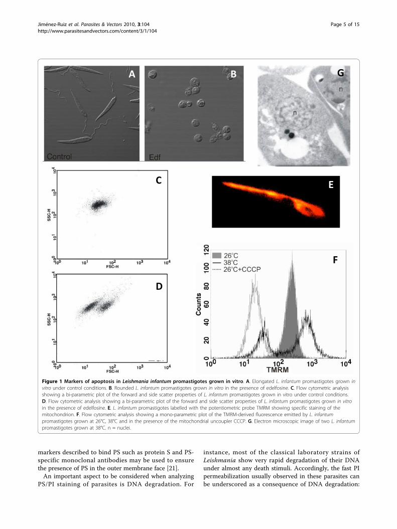
Morphological changesApoptosis in higher eukaryotes involves a series of bio-chemical events leading to characteristic changes in cellmorphology and subsequent death. During the execu-tion phase of apoptosis, the proteolytic activity of cas-pases disrupts the cytoskeleton, which is no longer ableto maintain cell shape and, as a consequence of thehomogeneous osmotic pressure, all cells becomerounded. Many protozoan parasites are clearly identifiedby their characteristic shapes that, similarly to metazo-ans, are lost during cell death. Changes in cell shape canbe clearly observed under the microscope (compare theelongated shape of untreated cultured Leishmania pro-mastigotes with that of edelfosine-treated ones, Figure1A and 1B).Apoptotic death in metazoans is also characterized by
cell shrinkage [11], which can be analysed either bymicroscopic observation or by flow cytometry based onthe changes in the scattering properties of the cellswhen the laser beam is directed to them. Forward scat-ter values (FSC) correlate with cell volume and can beused to determine changes in it. Bi-parametric plotsshowing the forward (FSC) and side scatter (SSC) prop-erties of the cell population are very useful to detectchanges in cell volume and have been successfullyemployed to detect cell shrinkage associated with deathin protozoan parasites such as Leishmania (Figure 1Cand 1D) [12]. Some of the earliest reports of apoptoticphenotypes in Trypanosoma concentrated on morpholo-gical changes. For instance Ameisen et al. [3] and Wel-burn et al. [4] described cytoplasmic vacuolisation andmargination, extensive membrane blebbling and conden-sation of nuclear chromatin in T. cruzi and T. bruceirespectively.In unfavourable culture conditions or following drug
treatment, condensed, so called ‘crisis forms’, of theintra-erythrocytic stage of P. falciparum (3D7 strain)appear. Although these forms have no precise definitionthey have been hypothesised to be undergoing apoptosis[13] and their formation can be blocked by the caspase-inhibitor Z-VAD-FMK [14]. In contrast, no drug-induced cell shrinkage was reported in the CSC-1 strain,though ultrastructural analysis additionally revealed food
Jiménez-Ruiz et al. Parasites & Vectors 2010, 3:104http://www.parasitesandvectors.com/content/3/1/104
Page 2 of 15
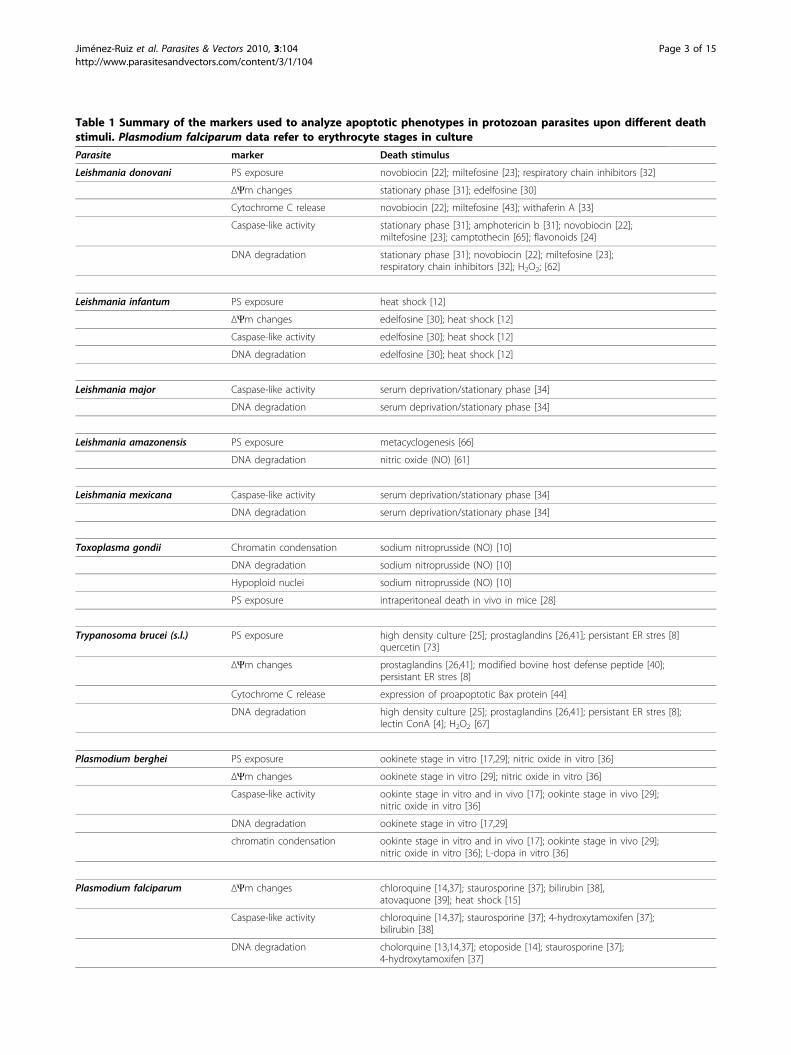
Table 1 Summary of the markers used to analyze apoptotic phenotypes in protozoan parasites upon different deathstimuli. Plasmodium falciparum data refer to erythrocyte stages in culture
Parasite marker Death stimulus
Leishmania donovani PS exposure novobiocin [22]; miltefosine [23]; respiratory chain inhibitors [32]
ΔΨm changes stationary phase [31]; edelfosine [30]
Cytochrome C release novobiocin [22]; miltefosine [43]; withaferin A [33]
Caspase-like activity stationary phase [31]; amphotericin b [31]; novobiocin [22];miltefosine [23]; camptothecin [65]; flavonoids [24]
DNA degradation stationary phase [31]; novobiocin [22]; miltefosine [23];respiratory chain inhibitors [32]; H2O2; [62]
Leishmania infantum PS exposure heat shock [12]
ΔΨm changes edelfosine [30]; heat shock [12]
Caspase-like activity edelfosine [30]; heat shock [12]
DNA degradation edelfosine [30]; heat shock [12]
Leishmania major Caspase-like activity serum deprivation/stationary phase [34]
DNA degradation serum deprivation/stationary phase [34]
Leishmania amazonensis PS exposure metacyclogenesis [66]
DNA degradation nitric oxide (NO) [61]
Leishmania mexicana Caspase-like activity serum deprivation/stationary phase [34]
DNA degradation serum deprivation/stationary phase [34]
Toxoplasma gondii Chromatin condensation sodium nitroprusside (NO) [10]
DNA degradation sodium nitroprusside (NO) [10]
Hypoploid nuclei sodium nitroprusside (NO) [10]
PS exposure intraperitoneal death in vivo in mice [28]
Trypanosoma brucei (s.l.) PS exposure high density culture [25]; prostaglandins [26,41]; persistant ER stres [8]quercetin [73]
ΔΨm changes prostaglandins [26,41]; modified bovine host defense peptide [40];persistant ER stres [8]
Cytochrome C release expression of proapoptotic Bax protein [44]
DNA degradation high density culture [25]; prostaglandins [26,41]; persistant ER stres [8];lectin ConA [4]; H2O2 [67]
Plasmodium berghei PS exposure ookinete stage in vitro [17,29]; nitric oxide in vitro [36]
ΔΨm changes ookinete stage in vitro [29]; nitric oxide in vitro [36]
Caspase-like activity ookinte stage in vitro and in vivo [17]; ookinte stage in vivo [29];nitric oxide in vitro [36]
DNA degradation ookinete stage in vitro [17,29]
chromatin condensation ookinte stage in vitro and in vivo [17]; ookinte stage in vivo [29];nitric oxide in vitro [36]; L-dopa in vitro [36]
Plasmodium falciparum ΔΨm changes chloroquine [14,37]; staurosporine [37]; bilirubin [38],atovaquone [39]; heat shock [15]
Caspase-like activity chloroquine [14,37]; staurosporine [37]; 4-hydroxytamoxifen [37];bilirubin [38]
DNA degradation cholorquine [13,14,37]; etoposide [14]; staurosporine [37];4-hydroxytamoxifen [37]
Jiménez-Ruiz et al. Parasites & Vectors 2010, 3:104http://www.parasitesandvectors.com/content/3/1/104
Page 3 of 15
vacuole swelling and lysis [15]. Even though shrinkagein cells with non-compromised plasma membranes isone of the main morphological markers of apoptosis [1],necrotic cells also diminish their volume once theplasma membrane breaks, so these morphologicalchanges could also be indicators of necrosis and moreefforts need to be made to distinguish between morpho-logical changes between different forms of cell death inPlasmodium. Vacuole formation in the chloroquine-resistant Brazilian PSS1 strain was regarded as evidenceof autophagy, although no other autophagic markerswere detected [16]. No obvious cell shrinkage or otherchange in cell shape has been detected in dying ooki-netes of the rodent malaria Plasmodium berghei, sug-gesting no major disruption of the cytoskeleton occurswhen other markers of apoptosis are exhibited [17]. Dis-tinct morphological changes that resemble apoptosis inmetazoans have been also described in tachyzoites ofthe apicomplexan parasite T. gondii following treatmentwith the nitric oxide (NO) donor sodium nitroprusside(SNP) [10]. Rounding-up of the normally elongatedtachyzoites and cell shrinkage were most prominent asdetermined by transmission electron microscopy [10].Since it coincided with the occurrence of chromatincondensation (see also below) these alterationsresembled apoptotic cell death in metazoans. Unfortu-nately, cell sizes of T. gondii treated or not with SNPhave not been analysed by flow cytometry as describedabove. It thus remains unclear to what extent the para-sites shrink following exposure to NO.
Plasma membrane alterationsTwo main alterations in the plasma membrane havebeen described during cell death processes: phosphatidylserine (PS) externalization and permeabilization to pro-pidium iodide (PI). Under normal physiologic condi-tions, PS is predominantly located in the inner leaflet orcytosol-facing part of the plasma membrane. Uponinitiation of apoptosis, PS loses its asymmetric distribu-tion in the phospholipid bilayer and is translocated tothe extracellular membrane leaflet where it identifiescells as targets for phagocytosis. PS presentation in theouter membrane face is easily analyzed using labelledAnnexin V. This protein binds to PS as part of its biolo-gical activity [18]. Accordingly, non-permeabilized cellsare incubated with labelled (often FITC-labelled)Annexin V, which only stains the cells when PS hasalready been translocated to the outer leaflet.An early event of apoptosis in metazoans is PS presen-
tation in the outer face of the plasma membrane [19]. Atthis stage, the plasma membrane is still able to excludeviability dyes such as PI, so single staining with AnnexinV serves as an early marker of apoptosis. In late stageapoptosis the membrane may lose its integrity allowingAnnexin V to access the interior of the cell and stain PSstill located in the inner membrane leaflet. The absenceof PI staining signals membrane integrity and ensuresthat Annexin V is only binding the cells through the PSlocated in the external membrane surface [20]. However,it must be pointed out that Annexin V can also bindanionic phospholipids other than PS [21]. Additional
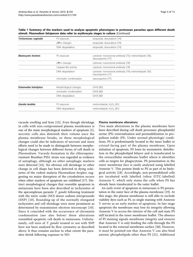
Table 1 Summary of the markers used to analyze apoptotic phenotypes in protozoan parasites upon different deathstimuli. Plasmodium falciparum data refer to erythrocyte stages in culture (Continued)
Trichomonas vaginalis PS exposure etoposide, doxorubicin [74]
ΔΨm changes etoposide, doxorubicin [74]
DNA degradation etoposide, doxorubicin [74]
Blastocystis hominis PS exposure cytotoxic monoclonal antibody [75]; metronidazole [76];staurosporine [77]
ΔΨm changes cytotoxic monoclonal antibody [78]
Caspase-like activity cytotoxic monoclonal antibody [78]
DNA degradation cytotoxic monoclonal antibody [79]; metronidazole [76];staurosporine [77]
chromatin condensation staurosporine [77]
Entamoeba histolytica Morphological changes G418 [80]
chromatin condensation G418 [80]
DNA degradation G418 [80]
Giardia lamblia PS exposure metronidazole, H2O2; [81]
DNA degradation metronidazole, H2O2; [81]
Jiménez-Ruiz et al. Parasites & Vectors 2010, 3:104http://www.parasitesandvectors.com/content/3/1/104
Page 4 of 15
markers described to bind PS such as protein S and PS-specific monoclonal antibodies may be used to ensurethe presence of PS in the outer membrane face [21].An important aspect to be considered when analyzing
PS/PI staining of parasites is DNA degradation. For
instance, most of the classical laboratory strains ofLeishmania show very rapid degradation of their DNAunder almost any death stimuli. Accordingly, the fast PIpermeabilization usually observed in these parasites canbe underscored as a consequence of DNA degradation:
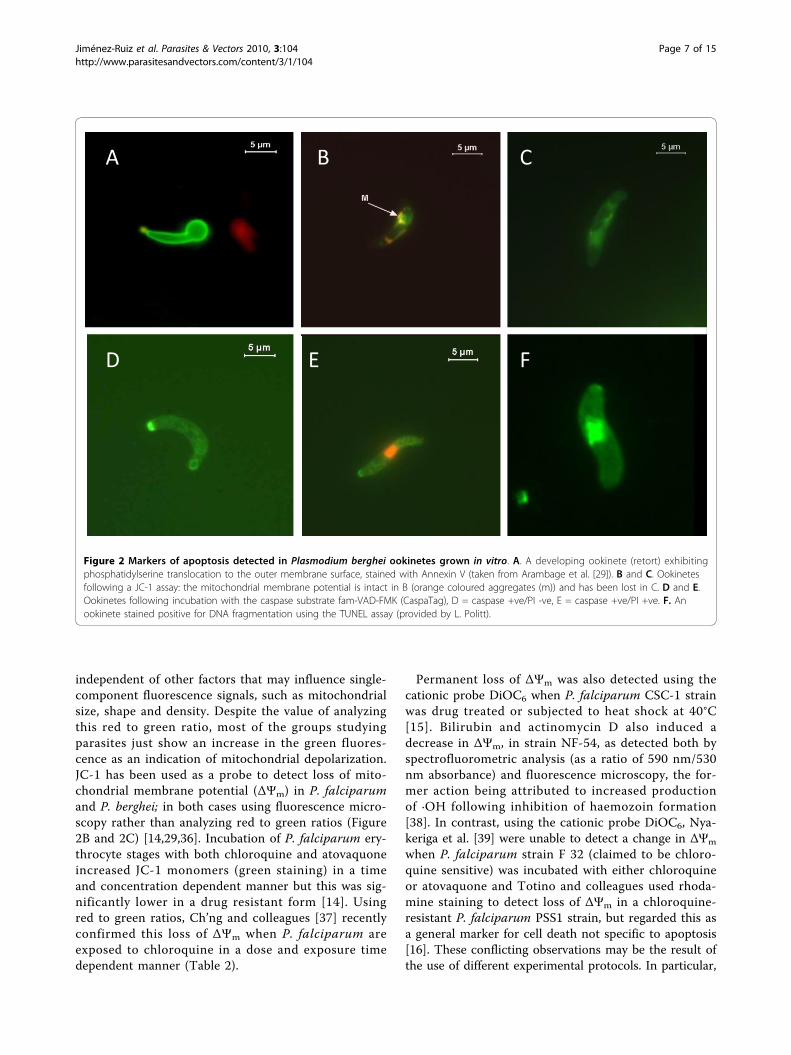
Figure 1 Markers of apoptosis in Leishmania infantum promastigotes grown in vitro. A. Elongated L. infantum promastigotes grown invitro under control conditions. B. Rounded L. infantum promastigotes grown in vitro in the presence of edelfosine. C. Flow cytometric analysisshowing a bi-parametric plot of the forward and side scatter properties of L. infantum promastigotes grown in vitro under control conditions.D. Flow cytometric analysis showing a bi-parametric plot of the forward and side scatter properties of L. infantum promastigotes grown in vitroin the presence of edelfosine. E. L. infantum promastigotes labelled with the potentiometric probe TMRM showing specific staining of themitochondrion. F. Flow cytometric analysis showing a mono-parametric plot of the TMRM-derived fluorescence emitted by L. infantumpromastigotes grown at 26°C, 38°C and in the presence of the mitochondrial uncoupler CCCP. G. Electron microscopic image of two L. infantumpromastigotes grown at 38°C. n = nuclei.
Jiménez-Ruiz et al. Parasites & Vectors 2010, 3:104http://www.parasitesandvectors.com/content/3/1/104
Page 5 of 15
cells may become PI negative shortly after cell death asa consequence of a reduction in the amount of DNAinside the cells (Jiménez-Ruiz; unpublished results). Forthis reason, PS externalization analyses in parasitesshould be designed to include the observation of sam-ples at different times after death induction in order tofollow the movement of the population from PS nega-tive/PI negative (living cells) to PS positive/PI negative(canonical apoptotic marker) and lastly to PS positive/PIpositive (late apoptotic marker). Further incubation willrender most of the population PS positive/PI negativeagain as a consequence of DNA degradation which caneasily mislead observers to consider that these cellsexpress a canonical apoptotic phenotype. Probablybecause of the difficulty to obtain canonical PS positive/PI negative populations experienced with several para-site species, in many published manuscripts the authorsonly present the results obtained after Annexin V label-ling either by fluorescent microscopy or by flowcytometry.PS presentation has been reported in Leishmania
under diverse cell death inducers such as treatment withnovobiocin [22], miltefosine [23], luteolin [24] or heatshock [12]. PS exposure has also been observed inT. brucei following exposure to a variety of stimuliincluding high density culture [25], prostaglandins ofthe J series [26] and persistent endoplasmic reticulum(ER) stress [8]. At high density culture, Tsuda et al.showed that after 24 hours the majority of cells wereAnnexin V positive and PI negative [25]. Incubation ofbloodstream forms of T. brucei with prostaglandinsfrom the J series for between two and six hours causedthe appearance of PS on the outer membrane; the pro-cess was inhibited by cyclohexmide suggesting thatactive protein synthesis is required [26]. Goldschmidt etal. [8] induced PS translocation to the outer membraneof procyclic forms by persistent ER stress with dithio-threitol. This was achieved two days after the silencingof ER translocation machinery by RNAi, followed by 12hours exposure to dithiothreitol. In each case trypano-somes became permeable to PI after a further 12 hours.As discussed above in relation to the timeframe for PSflipping and DNA degradation, the exposure of trypano-somes to dithiothreitol over time showed that theyfirstly expressed PS on their surface before becomingpermeable to PI. It is interesting to highlight that thepresence of PS-positive Leishmania promastigotes hasbeen shown to inhibit the host macrophage inflamma-tory response, which allows efficient in vitro and in vivoinfections by the PS-negative parasites [27].Externalization of PS to the outer leaflet of the cell
membrane appears to also occur in Toxoplasma [28].Importantly, a significant proportion of PS-positive cellswere detected among parasites that had been freshly
isolated from the peritoneal cavity of infected mice sug-gesting that it may occur during infection in vivo [28].Since the PS-positive parasites from the peritoneum ofmice were predominantly PI-negative they clearly metan important characteristic of the apoptotic phenotype.However, it has to be mentioned that the transitionfrom Annexin V-positive/PI-negative to Annexin V-positive/PI-positive after isolation from infected micewas not further analysed. In addition, the occurrence ofother apoptotic markers in Toxoplasma parasites dis-playing PS on their surface also awaits clarification.The various membranes surrounding the intra-
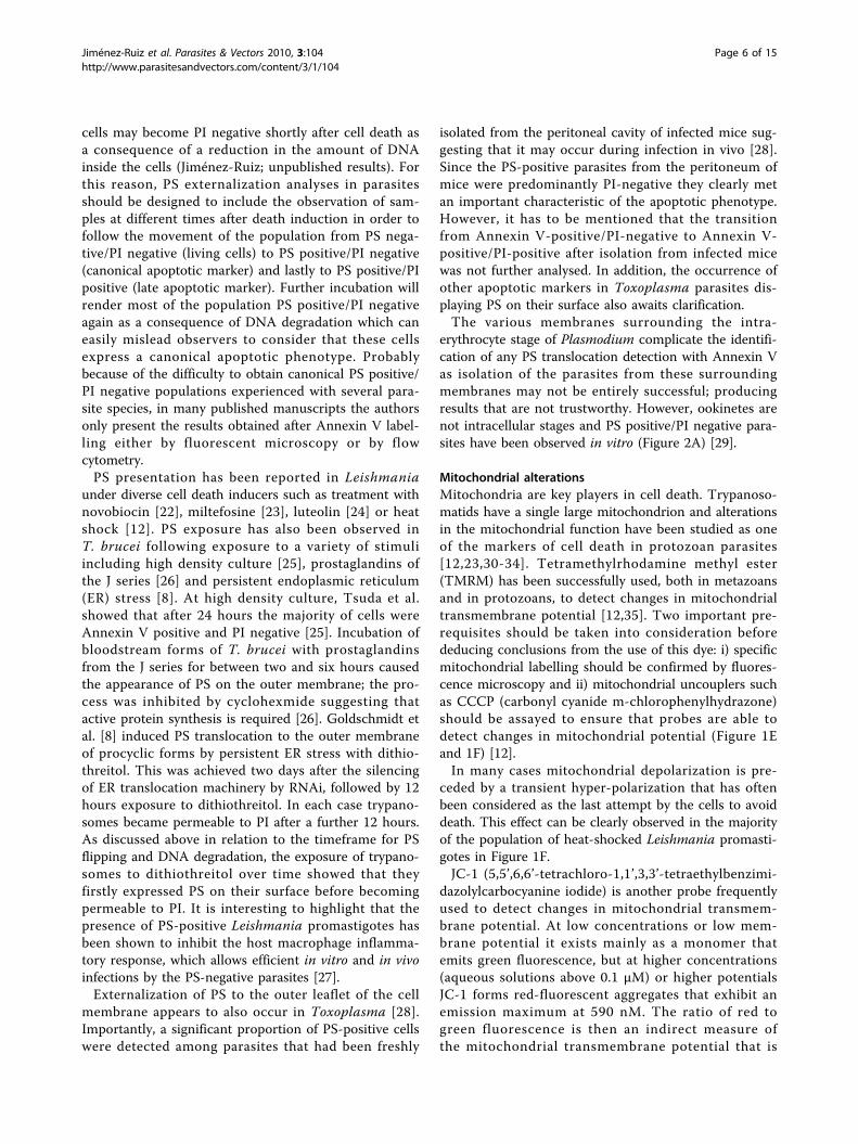
erythrocyte stage of Plasmodium complicate the identifi-cation of any PS translocation detection with Annexin Vas isolation of the parasites from these surroundingmembranes may not be entirely successful; producingresults that are not trustworthy. However, ookinetes arenot intracellular stages and PS positive/PI negative para-sites have been observed in vitro (Figure 2A) [29].
Mitochondrial alterationsMitochondria are key players in cell death. Trypanoso-matids have a single large mitochondrion and alterationsin the mitochondrial function have been studied as oneof the markers of cell death in protozoan parasites[12,23,30-34]. Tetramethylrhodamine methyl ester(TMRM) has been successfully used, both in metazoansand in protozoans, to detect changes in mitochondrialtransmembrane potential [12,35]. Two important pre-requisites should be taken into consideration beforededucing conclusions from the use of this dye: i) specificmitochondrial labelling should be confirmed by fluores-cence microscopy and ii) mitochondrial uncouplers suchas CCCP (carbonyl cyanide m-chlorophenylhydrazone)should be assayed to ensure that probes are able todetect changes in mitochondrial potential (Figure 1Eand 1F) [12].In many cases mitochondrial depolarization is pre-
ceded by a transient hyper-polarization that has oftenbeen considered as the last attempt by the cells to avoiddeath. This effect can be clearly observed in the majorityof the population of heat-shocked Leishmania promasti-gotes in Figure 1F.JC-1 (5,5’,6,6’-tetrachloro-1,1’,3,3’-tetraethylbenzimi-
dazolylcarbocyanine iodide) is another probe frequentlyused to detect changes in mitochondrial transmem-brane potential. At low concentrations or low mem-brane potential it exists mainly as a monomer thatemits green fluorescence, but at higher concentrations(aqueous solutions above 0.1 μM) or higher potentialsJC-1 forms red-fluorescent aggregates that exhibit anemission maximum at 590 nM. The ratio of red togreen fluorescence is then an indirect measure ofthe mitochondrial transmembrane potential that is
Jiménez-Ruiz et al. Parasites & Vectors 2010, 3:104http://www.parasitesandvectors.com/content/3/1/104
Page 6 of 15
independent of other factors that may influence single-component fluorescence signals, such as mitochondrialsize, shape and density. Despite the value of analyzingthis red to green ratio, most of the groups studyingparasites just show an increase in the green fluores-cence as an indication of mitochondrial depolarization.JC-1 has been used as a probe to detect loss of mito-chondrial membrane potential (ΔΨm) in P. falciparumand P. berghei; in both cases using fluorescence micro-scopy rather than analyzing red to green ratios (Figure2B and 2C) [14,29,36]. Incubation of P. falciparum ery-throcyte stages with both chloroquine and atovaquoneincreased JC-1 monomers (green staining) in a timeand concentration dependent manner but this was sig-nificantly lower in a drug resistant form [14]. Usingred to green ratios, Ch’ng and colleagues [37] recentlyconfirmed this loss of ΔΨm when P. falciparum areexposed to chloroquine in a dose and exposure timedependent manner (Table 2).
Permanent loss of ΔΨm was also detected using thecationic probe DiOC6 when P. falciparum CSC-1 strainwas drug treated or subjected to heat shock at 40°C[15]. Bilirubin and actinomycin D also induced adecrease in ΔΨm, in strain NF-54, as detected both byspectrofluorometric analysis (as a ratio of 590 nm/530nm absorbance) and fluorescence microscopy, the for-mer action being attributed to increased productionof ·OH following inhibition of haemozoin formation[38]. In contrast, using the cationic probe DiOC6, Nya-keriga et al. [39] were unable to detect a change in ΔΨm
when P. falciparum strain F 32 (claimed to be chloro-quine sensitive) was incubated with either chloroquineor atovaquone and Totino and colleagues used rhoda-mine staining to detect loss of ΔΨm in a chloroquine-resistant P. falciparum PSS1 strain, but regarded this asa general marker for cell death not specific to apoptosis[16]. These conflicting observations may be the result ofthe use of different experimental protocols. In particular,
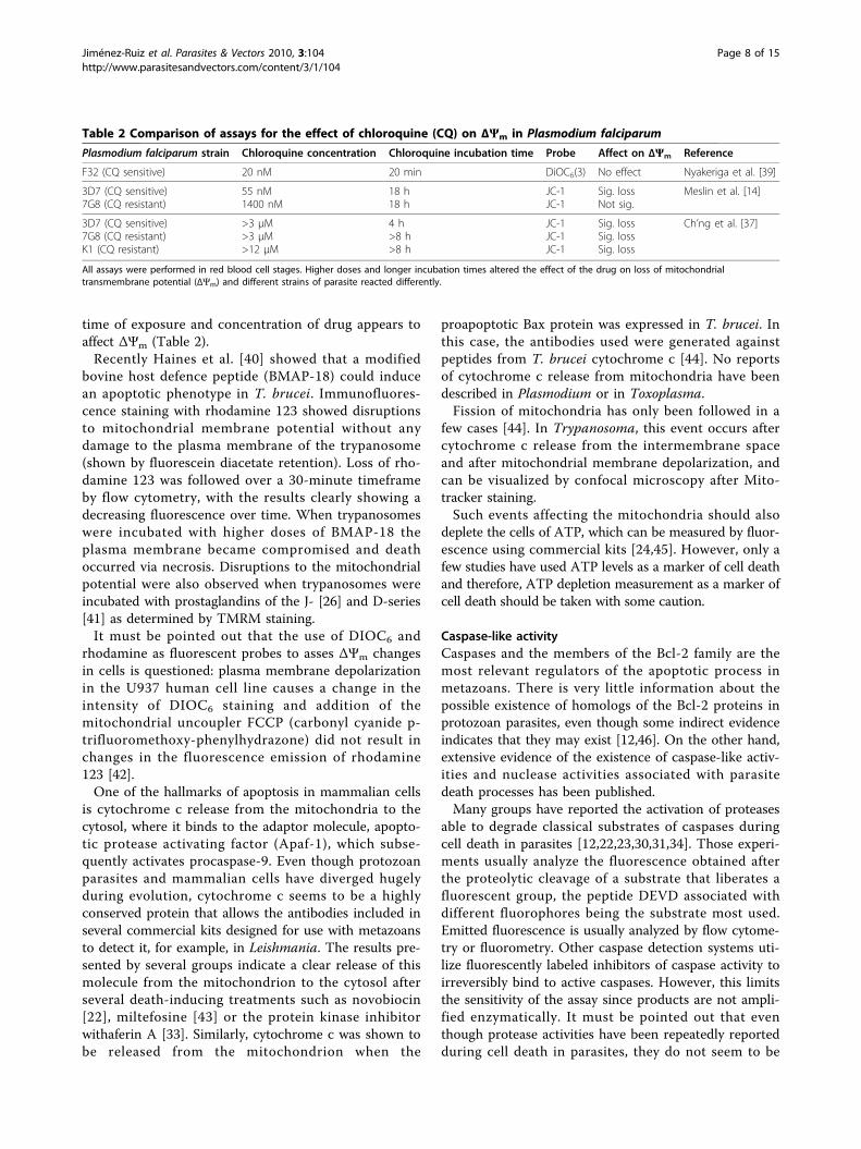
Figure 2 Markers of apoptosis detected in Plasmodium berghei ookinetes grown in vitro. A. A developing ookinete (retort) exhibitingphosphatidylserine translocation to the outer membrane surface, stained with Annexin V (taken from Arambage et al. [29]). B and C. Ookinetesfollowing a JC-1 assay: the mitochondrial membrane potential is intact in B (orange coloured aggregates (m)) and has been lost in C. D and E.Ookinetes following incubation with the caspase substrate fam-VAD-FMK (CaspaTag), D = caspase +ve/PI -ve, E = caspase +ve/PI +ve. F. Anookinete stained positive for DNA fragmentation using the TUNEL assay (provided by L. Politt).
Jiménez-Ruiz et al. Parasites & Vectors 2010, 3:104http://www.parasitesandvectors.com/content/3/1/104
Page 7 of 15
time of exposure and concentration of drug appears toaffect ΔΨm (Table 2).Recently Haines et al. [40] showed that a modified
bovine host defence peptide (BMAP-18) could inducean apoptotic phenotype in T. brucei. Immunofluores-cence staining with rhodamine 123 showed disruptionsto mitochondrial membrane potential without anydamage to the plasma membrane of the trypanosome(shown by fluorescein diacetate retention). Loss of rho-damine 123 was followed over a 30-minute timeframeby flow cytometry, with the results clearly showing adecreasing fluorescence over time. When trypanosomeswere incubated with higher doses of BMAP-18 theplasma membrane became compromised and deathoccurred via necrosis. Disruptions to the mitochondrialpotential were also observed when trypanosomes wereincubated with prostaglandins of the J- [26] and D-series[41] as determined by TMRM staining.It must be pointed out that the use of DIOC6 and
rhodamine as fluorescent probes to asses ΔΨm changesin cells is questioned: plasma membrane depolarizationin the U937 human cell line causes a change in theintensity of DIOC6 staining and addition of themitochondrial uncoupler FCCP (carbonyl cyanide p-trifluoromethoxy-phenylhydrazone) did not result inchanges in the fluorescence emission of rhodamine123 [42].One of the hallmarks of apoptosis in mammalian cells
is cytochrome c release from the mitochondria to thecytosol, where it binds to the adaptor molecule, apopto-tic protease activating factor (Apaf-1), which subse-quently activates procaspase-9. Even though protozoanparasites and mammalian cells have diverged hugelyduring evolution, cytochrome c seems to be a highlyconserved protein that allows the antibodies included inseveral commercial kits designed for use with metazoansto detect it, for example, in Leishmania. The results pre-sented by several groups indicate a clear release of thismolecule from the mitochondrion to the cytosol afterseveral death-inducing treatments such as novobiocin[22], miltefosine [43] or the protein kinase inhibitorwithaferin A [33]. Similarly, cytochrome c was shown tobe released from the mitochondrion when the
proapoptotic Bax protein was expressed in T. brucei. Inthis case, the antibodies used were generated againstpeptides from T. brucei cytochrome c [44]. No reportsof cytochrome c release from mitochondria have beendescribed in Plasmodium or in Toxoplasma.Fission of mitochondria has only been followed in a
few cases [44]. In Trypanosoma, this event occurs aftercytochrome c release from the intermembrane spaceand after mitochondrial membrane depolarization, andcan be visualized by confocal microscopy after Mito-tracker staining.Such events affecting the mitochondria should also
deplete the cells of ATP, which can be measured by fluor-escence using commercial kits [24,45]. However, only afew studies have used ATP levels as a marker of cell deathand therefore, ATP depletion measurement as a marker ofcell death should be taken with some caution.
Caspase-like activityCaspases and the members of the Bcl-2 family are themost relevant regulators of the apoptotic process inmetazoans. There is very little information about thepossible existence of homologs of the Bcl-2 proteins inprotozoan parasites, even though some indirect evidenceindicates that they may exist [12,46]. On the other hand,extensive evidence of the existence of caspase-like activ-ities and nuclease activities associated with parasitedeath processes has been published.Many groups have reported the activation of proteases
able to degrade classical substrates of caspases duringcell death in parasites [12,22,23,30,31,34]. Those experi-ments usually analyze the fluorescence obtained afterthe proteolytic cleavage of a substrate that liberates afluorescent group, the peptide DEVD associated withdifferent fluorophores being the substrate most used.Emitted fluorescence is usually analyzed by flow cytome-try or fluorometry. Other caspase detection systems uti-lize fluorescently labeled inhibitors of caspase activity toirreversibly bind to active caspases. However, this limitsthe sensitivity of the assay since products are not ampli-fied enzymatically. It must be pointed out that eventhough protease activities have been repeatedly reportedduring cell death in parasites, they do not seem to be
Table 2 Comparison of assays for the effect of chloroquine (CQ) on ΔΨm in Plasmodium falciparum
Plasmodium falciparum strain Chloroquine concentration Chloroquine incubation time Probe Affect on ΔΨm Reference
F32 (CQ sensitive) 20 nM 20 min DiOC6(3) No effect Nyakeriga et al. [39]
3D7 (CQ sensitive)7G8 (CQ resistant)
55 nM1400 nM
18 h18 h
JC-1JC-1
Sig. lossNot sig.
Meslin et al. [14]
3D7 (CQ sensitive)7G8 (CQ resistant)K1 (CQ resistant)
>3 μM>3 μM>12 μM
4 h>8 h>8 h
JC-1JC-1JC-1
Sig. lossSig. lossSig. loss
Ch’ng et al. [37]
All assays were performed in red blood cell stages. Higher doses and longer incubation times altered the effect of the drug on loss of mitochondrialtransmembrane potential (ΔΨm) and different strains of parasite reacted differently.
Jiménez-Ruiz et al. Parasites & Vectors 2010, 3:104http://www.parasitesandvectors.com/content/3/1/104
Page 8 of 15

due to real caspases, as no authentic caspase has beenidentified in protozoans. In Leishmania, this activity canbe inhibited by E-64, suggesting that these proteasescould be similar to cathepsin [34]. Several substrate ana-logues (DEVD-FMK) are currently used as inhibitorsand their ability to decrease the fluorescent signal isconsidered by some groups as strong evidence of thepresence of caspase-like molecules in some parasites.Those results should be analyzed with caution. They donot allow us to conclude that caspase-like activity is pre-sent in protozoa until the enzyme has been molecularlycharacterized. However, it must be pointed out that sev-eral groups have been able to prevent the appearance ofclassical apoptotic markers such as reduction in thetransmembrane mitochondrial potential, DNA degrada-tion or PARP cleavage by incubating the cells with theseinhibitors [47]. Thus far, we can only conclude that aprotease recognizing DEVD or inhibited by DEVD-FMKmay be involved in cell death. It has been reported thata number of commonly used small peptide caspase inhi-bitors efficiently inhibit other cysteine proteases thancaspases [48]. The pan-caspase inhibitor VAD-FMK, hasbeen shown to bind and inhibit cathepsin B [48,49].This inhibition is able to interfere with cell death path-ways in mammalian cells [50], which suggests that databased on the use of these inhibitors should be takenwith caution because other proteases different to cas-pases may be participating in these processes. Recentresults demonstrate the relevance of cathepsin activityin Leishmania cell death [51]. Moreover, these data putsome emphasis on a lysosomal cell death pathway basedon lysosomal membrane permeabilisation and release oflysosomal enzymes.Several metacaspases have been described in Trypano-
soma and Leishmania but their substrate specificity dif-fers to caspases as it seems to be directed to substrateswith an arginine or lysine residue in P1 position [52-54].Metacaspase genes have also been found in the genomeof Plasmodium [55]. Even though some authors considerthat plant and fungal metacaspases lack caspase activityand that they are not responsible for the caspase-likeactivities detected during their cell death [56], there isevidence that indicates that metacaspases are involvedin regulating some death processes in Leishmania[53,57]. The use of fluorogenic substrates to detectmetacaspase activity associated to cell death is not wide-spread yet. Thus far, there is no experimental evidencethat a fluorogenic substrate such as VRPR-AMC used ina metacaspase assay in Arabidopsis would be adaptedfor a proteolytic cell death in protozoa.A protease able to bind the substrate FAM-VAD-FMK
is present in P. berghei ookinetes (Figure 2D and 2E)but it should be noted that when assays were performedat 37°C (as per manufacturer’s instructions) over 25% of
these high-temperature-sensitive mosquito stages haddisrupted membranes that were permeable to PI com-pared to approximately 12% if the assays were per-formed at 19°C. Incubation with the general caspaseinhibitors Z-VAD-FMK and Boc-ASP-FMK and Z-DEVD-FMK, an inhibitor more specific to the caspase-3subfamily, almost eliminated chromatin condensation inthe parasite population. In contrast, incubation with Z-YVAD-CMK, a caspase-1 inhibitor, had no effect onchromatin condensation [17] nor did the cysteine pro-tease clan CA inhibitors E64d or K11177, suggesting theparasite protease belongs to clan CD (Arambage andHurd unpublished data). Further evidence that acysteine protease is involved in ookinete cells deathcame from in vivo studies where feeding Z-VAD-FMKto mosquitoes during an infective blood meal signifi-cantly increased the intensity of infection [17]. Howeverinterpretation of in vivo experiments is complicated bythe probability that inhibitors fed with the blood mealalso inhibited the apoptosis occurring in midgut epithe-lial cells that have been traversed by ookinetes, withunknown effects on the induction of mosquito immuneresponses.Two cysteine proteases have been annotated as meta-
caspase-like in the P. falciparum genome database. Incu-bation with Z-VAD-FMK resulted in a 75% decrease inthe crisis forms of the parasite that has been proposedto be a form of apoptotic body [14]. Orthologues ofPfMC1 were identified in several other species of Plas-modium and genetically modified P. berghei were pro-duced in which the PbMC1 coding sequence had beenremoved (PbMC1-KO) [58]. Assays to compare markersfor apoptosis in wild type and knock out parasites didnot detect any significant differences in binding to thefluorochrome-labelled caspase inhibitor FAM-VAD-FMK. However, activation of caspase-like molecules intheir wild type was extremely low (9.0% at 24 h) andthey were unable to find any evidence of nuclear con-densation or DNA fragmentation, and very few ooki-netes were PS positive and PI negative. This is inmarked contrast to the findings of Al-Olayan and collea-gues [17,59] using the same P. berghei ANKA clone.Arambage and co-workers were, however, unable todetect any differences in the expression of apoptoticmarkers when this PbMCA1-KO was compared withthe wild type, nor when comparisons between aPbMC2-KO and the wild type were made (Hurd,unpublished). Thus there is no sound evidence that themalaria metacaspase is associated with cell death,although there is some support for the view thatcysteine proteases are related to the expression of otherapoptotic markers.A six-fold increase in the catalytic activity of a cas-
pase-3-like protein was detected in bilirubin-treated
Jiménez-Ruiz et al. Parasites & Vectors 2010, 3:104http://www.parasitesandvectors.com/content/3/1/104
Page 9 of 15
erythrocyte stages of P. falciparum using the substrateDEVD-pNA and this activity was inhibited by Ac-DEVD-CHO, a potent inhibitor of caspase-3 [38]. Theauthors also detected a significant up-regulation of theexpression of a putative apoptosis-like gene, PfARP,upon bilirubin treatment and both caspase-3-like activityand PfARP expression were prevented by antioxidantsand ROS scavengers. The use of substrates and inhibi-tors to demonstrate a functional role for caspase-likemolecules in malaria cell death and their use to pull outand identify these molecules warrants more study.
Changes in the nucleusSeveral changes to the nucleus have been described dur-ing the effector phase of apoptosis in metazoan cells.Together with DNA degradation, chromatin condensa-tion and changes in nuclear morphology are often con-sidered the best indicators of an apoptotic process.Those changes can be detected easily in higher eukar-yotes by fluorescence microscopy using any of the sev-eral dyes that stain DNA in the nucleus. However, thesmall size of most of the protozoan parasites, and conse-quently of their nuclei, is an important drawback thathas strongly restricted the use of this technique amongparasitologists. Despite this difficulty, electron micro-scopy has been successfully used by some researchers todescribe nuclear condensation, as can be seen in one ofthe two Leishmania promastigotes shown in Figure 1G(parasite in the right side of the picture). In Trypano-soma brucei, Welburn et al. [4] showed migration ofchromatin to the periphery of the nuclear membranewhile Figarella et al. showed that chromatin becameequally distributed after incubation with prostaglandinsfrom the J series [26].Nuclear chromatin condensation and fragmentation
has been detected in P. berghei ookinetes, as determinedby fluorescence and electron microscopy [17,29,59]. Thepresence of nitric oxide donors in the culture mediumincreased the proportion of parasites exhibiting thismarker [36]. Nuclear condensation was also observed tooccur in bilirubin-treated P. falciparum intra-erythrocytestages [38].Beside other morphological changes (see above), con-
densed chromatin, particularly beneath the nuclearenvelope, and nuclear pyknosis was also detected in T.gondii tachyzoites treated with SNP [10]. Importantly,these changes were accompanied by a time- and dose-dependent increase of parasites with hypoploid nuclei asdetermined by flow cytometry [10]. The latter quantita-tive analyses revealed that considerable amounts of SNP(actual concentrations of nitrate and nitrite as stableend products of NO turnover have not been measured)were required to induce high levels of hypoploid para-sites, therefore the physiological relevance of this finding
for the infection in vivo remains unclear. It neverthelessindicates that in Toxoplasma, several characteristic fea-tures, including nuclear changes, indicative of an apop-totic phenotype can occur.Poly (ADP-ribose) polymerase (PARP) degradation is
another characteristic marker of apoptosis frequentlyused in metazoans. PARP is a family of abundantnuclear proteins some of which are involved in theDNA base excision repair system, where they arepotently activated following DNA strand nicks andbreaks. In metazoans, the specific cleavage of PARP-1by caspase-3/7 within the nuclear localization signal(211DEVD↓G215) generates two fragments of 24 and 89kDa and this phenomenon has been used extensively asa biochemical marker of apoptosis. Some groups havedescribed the use of monoclonal antibodies againstPARP to detect PARP cleavage associated to cell deathin Leishmania [22,60]. Unfortunately, whether the anti-bodies are raised against the mammalian or the parasiteprotein is not described in their manuscripts. Further-more, in the genome database, there is no clear evidenceof presence of protozoan PARP-1 encoding gene. There-fore, cleavage of PARP-1 as a marker of cell death inprotozoan parasites still needs further investigationbefore being accepted as a marker.DNA degradation is probably the most frequent mar-
ker of apoptotic death used in metazoans. In protozoanssuch as Leishmania it has been reported to occur afterserum deprivation [34], heat shock [12], treatment withnitric oxide [61] or hydrogen peroxide [62] and with dif-ferent drugs including pentostam [31], antimonials [63],novobiocin [22], miltefosine [23], edelfosine [46] orrespiratory chain inhibitors [32].In the beginning, analysis of DNA degradation in
agarose gels became the easiest way to approach thisprocess of cell death. During apoptosis, activatednucleases migrate to the nucleus to degrade DNA pre-ferentially in the most accessible sequences: thoselocated in the linker region of the nucleosomes, whichgenerates the typical apoptotic DNA ladder. Whereasthis methodology is widely used in research groups ana-lyzing apoptosis in metazoans, several difficulties occurwhen these protocols were transferred to protozoans. Infact, some groups with vast experience in analysingapoptosis in human cells have been unable to adapttheir protocols to detect a DNA ladder in parasites suchas Leishmania (Mollinedo F. personal communication).Classical protocols are based on a simple direct purifica-tion of DNA from the cells by a phenol/chloroformextraction followed by ethanol precipitation or by incu-bation of the lysates with high NaCl concentration todisrupt nucleosomes and remove histones from DNA. Adetailed analysis of the manuscripts that show clearDNA ladders in Trypanosomatids reveals that none of
Jiménez-Ruiz et al. Parasites & Vectors 2010, 3:104http://www.parasitesandvectors.com/content/3/1/104
Page 10 of 15
these “classical” protocols is used. Instead, most of themuse a different method that includes an incubation ofthe samples with proteinase K. Alternatively, other pro-tocols based on one specific commercial kit incubatechromatin for one hour at room temperature (eventhough the protocol recommends only 10 minutes) [64].It must be stressed that these protocols only renderclear oligonucleosomal ladders in samples obtained fromdying parasites and not from healthy controls, whicheffectively indicates that one or several nucleasesbecome active during the cell death process. On theother hand, DNA purified from the parasites withoutextracellular chromatin incubation shows death-induceddegradation but without a clear pattern of oligonucleo-somal laddering, which could indicate that the laddersare generated during the incubation of the extractedchromatin and not inside the parasites. In fact, Sen andco-workers reported that in vitro incubation of isolatednuclei from untreated Leishmania parasites with Ca2+
and Mg2+ at 37°C caused oligonucleosomal DNA degra-dation, a process that they named autodigestion [65].Similarly, Zangger and co-workers found that no frag-mentation could be detected in nuclei extracted fromstationary phase parasites without any incubation pro-cess. Fragmentation was, however, visible following 30minutes incubation and no increase in degradation wasobserved upon addition of Ca2+ or Mn2+, while Mg2+
seemed to increase the effect slightly [34]. In this latterstudy, DNA laddering was observed after isolation ofthe nuclei using a cavitation chamber. This experimentalapproach was instrumental to obtain a clear laddering.In conclusion, the two independent results obtained bySen et al. and by Zangger et al. [34,65] reinforce thenotion that a short period of incubation of the chroma-tin may be needed to obtain a clear laddering, whichseems to explain the different results obtained by differ-ent groups depending on the protocols used.In the last few years, the use of flow cytometry to
determine the DNA content of the cells and detect thefraction of the population with DNA content lower thanthat characteristic of G1 cell cycle phase has been widelyextended. Very simple protocols based on a mild per-meabilization of the cells with ethanol and a short incu-bation of the permeabilized cells with RNAse inPBS with PI can be used [30]. Alternatively, non-permeabilized living cells can be directly stained withHoechst 33342 and analyzed in a flow cytometer withan UV laser beam [37]. Experts in flow cytometryrecommend analysing the fluorescence derived frompropidium intercalation in the DNA in a linear scaleand not in a logarithmic one.Currently, DNA fragmentation revealed by the pre-
sence of a multitude of DNA strand breaks is consid-ered to be the gold standard for identification of
apoptotic cells. Several variants of the methodology thatis based on fluorochrome-labeling of 3’-OH termini ofDNA strand breaks in situ with the use of exogenousterminal deoxynucleotidyl transferase (TdT), commonlydefined as the TUNEL assay, have been developed.Labelled cells can be analyzed by fluorescence micro-scopy or by flow cytometry, which can render a clearquantification of the percentage of labelled cells and theintensities of the fluorescence. The TUNEL approachwas efficient in determining the percentage of deadLeishmania parasites in culture upon heat shock treat-ment [12], in their host cell [34] and ultimately, couldbe used to estimate the role of dead parasites enhancingthe virulence of an inoculum [21,66]. Fragmentation ofDNA was also detected in African trypanosomes inresponse to reactive oxygen species [67], incubationwith PGJ2 [26] or the lectin ConA [6].Fragmentation of genomic DNA from Toxoplasma
into oligomers of nucleosomes became very evidentafter treatment of extracellular tachyzoites with SNP[10]. As already mentioned for the induction of hypo-ploid parasites (see above), treatment with high amountsof SNP for extended periods (20 hours) was required todetect a significant level of DNA laddering. In their pro-tocol, the authors employed proteinase K and RNase Atreatments suggesting that also in Toxoplasma alterna-tive methods than those described for mammalian cellsmight be necessary to unequivocally detect DNA frag-mentation. In addition to gel electrophoresis of genomicDNA oligonucleosomes, the TUNEL assay was also suc-cessfully employed to detect DNA strand breaks in Tox-oplasma (Lüder, unpublished). In combination with flowcytometry it can be easily used to quantify the occur-rence of DNA strand breaks in relatively large numberof samples.Following chloroquine treatment, Picot and colleagues
detected the formation of DNA ladders in a populationof the drug sensitive P. falciparum, 3D7, but not in thechloroquine resistant P. falciparum Lili strain. Conven-tional detection methodology was unsuccessful due, assuggested, to low sensitivity with small amounts ofDNA. Their approach instead was 3’-OH end-labellingof extracted DNA with labelled nucleotides. Electro-phoresis of samples was followed by Southern blottingand autoradiography [13]. DNA ladder formation wasnot detected following exposure of P. falciparum F32strain to antimalarial parasite drugs when electrophor-esis of DNA was followed by visualisation using ethi-dium bromide staining [39] or in the CSC-1 strain whenSYBR Green 1 dye was used, where preliminary detec-tion of DNA ladders was attributed to the presence ofapoptotic white blood cells in the parasite culture [15].These observations suggest the strategy adopted byPicot and co-workers may be required to detect DNA
Jiménez-Ruiz et al. Parasites & Vectors 2010, 3:104http://www.parasitesandvectors.com/content/3/1/104
Page 11 of 15
ladders in dead cells from the inter-erythrocytic stagesof P. falciparum. However, in both of those latter stu-dies drug treatments did not result in visualisation ofother typical markers of apoptosis; a phenomenon thatmay be a strain effect related to drug sensitivity. Nyaker-iga et al. [39] also failed to detect degradation of DNAinto the larger sized structures associated with chromo-some buttoning as a prelude to chromatin condensationusing field inversion electrophoresis.TUNEL was used to detect the increase in ookinetes
containing nuclei with DNA fragmentation over time inculture (Figure 2F), even without the addition of stres-sors to the medium [29]. Following chloroquine (CQ)treatment the TUNEL assay was also used to detect amuch higher proportion of DNA breaks in the CQ-sensitive 3D7 clone of P. falciparum than in the CQ-resistant 7G8 clone (over 50% compared with under10%) [14]. Heat shock treatment (41°C) also inducedapproximately 60% of P. falciparum 3D7 strain to beTUNEL positive [68]. However, no TUNEL positive cellswere observed in F32 strain of P. falciparum followingexposure to chloroquine, atovaquone or etoposide [39]and only a few were seen in the chloroquine resistantPSS1 strain when exposed to chloroquine, staurosporineor the nitric oxide donor S-nitroso-N-penicillamide(SNAP), even though loss of ΔΨm had occurred [39].DNA degradation in mammals is carried out by two
nucleases, CAD and EndoG; CAD being the most activewhile EndoG seems to be especially relevant in caspase-independent apoptosis. Recently, three different groupshave characterized nucleases similar to mammalianEndoG in different Leishmania species and also in T.brucei [69-71]. Two of them have also been able toshow migration of these Leishmania EndoGs from themitochondrion to the nucleus during the cell death pro-cess, either by using specific antibodies or by fusion ofthe protein to the green fluorescent protein (GFP)[69,71]. These antibodies or the chimaeric fusions couldalso be used as apoptotic markers in Leishmania.
ConclusionsDespite the initial controversy, nowadays it is widelyassumed that protozoan parasites are able to displaysome of the most characteristic apoptotic markers dur-ing cell death. The physiological role of the expressionof apoptotic phenotypes in unicellular parasites has beenquestioned for many years but recent results demon-strate that this process is relevant, at least in some situa-tions, for efficient progression of the parasitepopulations. For example, in vitro and in vivo infectionsof Leishmania promastigotes depend on the presence ofPS positive parasites in the inoculums [21,66]. Similarly,Figarella et al. proposed that the stumpy forms ofT. brucei produce prostaglandin D2, which induces cell
death primarily of stumpy form cells [41]. This con-trolled process of cell death allows an efficient regula-tion of the size of the T. brucei population. Therelevance of controlling cell size population in malariaparasites is discussed in another paper within this the-matic series [72].Typical markers of mammalian apoptosis have been
widely used to characterize cell death in protozoans and,as shown in this review, many of the conventional assaysfor metazoans can be directly adapted for this purpose.However, several concerns have been raised for some ofthe most relevant markers. Regarding PS exposure,reviewers should be strict about the need to use doublePS/PI staining and, preferably, at several time points.Similarly, probes used to analyze mitochondrial trans-membrane potential in protozoans should initially bevalidated by the use of mitochondrial uncouplers. Whenstudying protease activity, results derived from the useof caspase fluorogenic substrates or inhibitors should beanalyzed with caution until the caspase-like activitiesobserved in protozoa have been molecularly character-ized. Moreover, the fact that metacaspases have alreadybeen demonstrated to be implicated in cell death insome protozoans should encourage the development ofmetacaspases-specific substrates as putative markers ofthis process. Regarding the protocols for DNA ladderingin trypanosomatids, it must be stressed that ladders areonly clearly observed when the nuclei are incubated inspecific conditions after their purification. Finally, it maybe important to consider that several protocols seem toperform better in protozoans when done at lower tem-peratures than those recommended for metazoans by kitmanufacturers.
List Of AbbreviationsΔΨm: Mitochondrial trans-membrane potential; NO: Nitric oxide; NCCD:Nomenclature Committee on Cell Death; FSC: Forward scatter; SSC: Sidescatter; SNP: Sodium nitroprusside; PS: Phosphatidylserine; PI: PropidiumIodide; FITC: Fluorescein isothiocyanate; ER: Endoplasmic reticulum; TMRM:Tetramethylrhodamine methyl ester; CCCP: Carbonyl cyanide m-chlorophenylhydrazone; FCCP: Carbonyl cyanide p-trifluoromethoxy-phenylhydrazone); DEVD-FMK: Asp(OMe)-Glu(OMe)-Val-Asp(OMe)-Fluoromethylketone; PARP: Poly (ADP-ribose) polymerase; JC-1: 5,5’,6,6’-tetrachloro-1,1’,3,3’-tetraethylbenzimidazolylcarbocyanine iodide; TdT:Terminal deoxynucleotidyl transferase; CQ: Chloroquine; TUNEL: TerminalTransferase dUTP Nick End Labeling; SNAP: S-nitroso-N-penicillamide; GFP:Green fluorescent protein; DiOC6: 3,3’-dihexyloxacarbocyanine iodide.
AcknowledgementsThe authors are members of COST action BM0802 ‘Life and death ofprotozoan parasites’ and appreciate support from this action. AJacknowledges financial support from the Ministerio de Educación y Ciencia,Spain (grant SAF 2006-12713-CO2-O2)
Author details1Departamento de Bioquímica y Biología Molecular, Universidad de Alcalá,28871 Alcalá de Henares, Madrid, Spain. 2Grupo de Parasitología,Departamento de Microbiología y Parasitología, Facultad de Medicina,Universidad de Antioquia. Carrera 51D # 62-29, Medellín, Colombia. 3Division
Jiménez-Ruiz et al. Parasites & Vectors 2010, 3:104http://www.parasitesandvectors.com/content/3/1/104
Page 12 of 15
of Pathway Medicine, School of Biomedical Sciences, College of Medicineand Veterinary Medicine, University of Edinburgh, 1Summerhall Square,Edinburgh EH9 1QH, UK. 4Institute for Medical Microbiology, Georg-August-University, Kreuzbergring 57, 37075 Göttingen, Germany. 5Department ofBiochemistry, Univerisity of Lausanne, 1066 Epalinges, Switzerland. 6Institutefor Science and Technology in Medicine, Centre for Applied Entomologyand Parasitology, School of Life Sciences, Keele University, Staffordshire, ST55BG, UK.
Authors’ contributionsAJ drafted and coordinated the contributions of the different authors to themanuscript and specifically contributed with the information related to theLeishmania parasite. JFA collected data, helped to draft the informationrelated to Leishmania and critically reviewed the manuscript. EM collecteddata, helped to draft the information related to Trypanosoma and criticallyreviewed the manuscript. CL collected data, helped to draft the informationrelated to Toxoplasma and critically reviewed the manuscript. NF collecteddata, helped to draft the information related to Leishmania and criticallyreviewed the manuscript. HH collected data, helped to draft the informationrelated to Plasmodium and critically reviewed the manuscript.
Competing interestsThe authors declare that they have no competing interests.
Received: 2 June 2010 Accepted: 9 November 2010Published: 9 November 2010
References1. Kroemer G, Galluzzi L, Vandenabeele P, Abrams J, Alnemri ES, Baehrecke EH,
Blagosklonny MV, El-Deiry WS, Golstein P, Green DR, Hengartner M,Knight RA, Kumar S, Lipton SA, Malorni W, Nuñez G, Peter ME, Tschopp J,Yuan J, Piacentini M, Zhivotovsky B, Melino G, Nomenclature Committee onCell Death 2009: Classification of cell death: recommendations of theNomenclature Committee on Cell Death 2009. Cell Death Differ 2009,16:3-11.
2. Galluzzi L, Aaronson SA, Abrams J, Alnemri ES, Andrews DW, Baehrecke EH,Bazan NG, Blagosklonny MV, Blomgren K, Borner C, et al: Guidelines for theuse and interpretation of assays for monitoring cell death in highereukaryotes. Cell Death Differ 2009, 16:1093-1107.
3. Ameisen JC, Idziorek T, Billaut-Mulot O, Loyens M, Tissier JP, Potentier A,Ouaissi A: Apoptosis in a unicellular eukaryote (Trypanosoma cruzi):implications for the evolutionary origin and role of programmed celldeath in the control of cell proliferation, differentiation and survival. CellDeath Differ 1995, 2:285-300.
4. Welburn SC, Dale C, Ellis D, Beecroft R, Pearson TW: Apoptosis in procyclicTrypanosoma brucei rhodesiense in vitro. Cell Death Differ 1996, 3:229-236.
5. van Zandbergen G, Luder CG, Heussler V, Duszenko M: Programmed celldeath in unicellular parasites: a prerequisite for sustained infection?Trends Parasitol 26:477-483.
6. Pearson TW, Beecroft RP, Welburn SC, Ruepp S, Roditi I, Hwa KY,Englund PT, Wells CW, Murphy NB: The major cell surface glycoproteinprocyclin is a receptor for induction of a novel form of cell death inAfrican trypanosomes in vitro. Mol Biochem Parasitol 2000, 111:333-349.
7. Duszenko M, Figarella K, Macleod ET, Welburn SC: Death of atrypanosome: a selfish altruism. Trends Parasitol 2006, 22:536-542.
8. Goldshmidt H, Matas D, Kabi A, Carmi S, Hope R, Michaeli S: Persistent ERstress induces the spliced leader RNA silencing pathway (SLS), leadingto programmed cell death in Trypanosoma brucei. PLoS Pathog 2010, 6:e1000731.
9. Robinson KA, Beverley SM: Improvements in transfection efficiency andtests of RNA interference (RNAi) approaches in the protozoan parasiteLeishmania. Mol Biochem Parasitol 2003, 128:217-228.
10. Peng BW, Lin J, Lin JY, Jiang MS, Zhang T: Exogenous nitric oxide inducesapoptosis in Toxoplasma gondii tachyzoites via a calcium signaltransduction pathway. Parasitology 2003, 126:541-550.
11. Lang F, Foller M, Lang K, Lang P, Ritter M, Vereninov A, Szabo I, Huber SM,Gulbins E: Cell volume regulatory ion channels in cell proliferation andcell death. Methods Enzymol 2007, 428:209-225.
12. Alzate JF, Alvarez-Barrientos A, Gonzalez VM, Jimenez-Ruiz A: Heat-inducedprogrammed cell death in Leishmania infantum is reverted by Bcl-X(L)expression. Apoptosis 2006, 11:161-171.
13. Picot S, Burnod J, Bracchi V, Chumpitazi BF, Ambroise-Thomas P: Apoptosisrelated to chloroquine sensitivity of the human malaria parasitePlasmodium falciparum. Trans R Soc Trop Med Hyg 1997, 91:590-591.
14. Meslin B, Barnadas C, Boni V, Latour C, De Monbrison F, Kaiser K, Picot S:Features of apoptosis in Plasmodium falciparum erythrocytic stagethrough a putative role of PfMCA1 metacaspase-like protein. J Infect Dis2007, 195:1852-1859.
15. Porter H, Gamette MJ, Cortes-Hernandez DG, Jensen JB: Asexual bloodstages of Plasmodium falciparum exhibit signs of secondary necrosis, butnot classical apoptosis after exposure to febrile temperature (40 C). JParasitol 2008, 94:473-480.
16. Totino PR, Daniel-Ribeiro CT, Corte-Real S, de Fatima Ferreira-da-Cruz M:Plasmodium falciparum: erythrocytic stages die by autophagic-like celldeath under drug pressure. Exp Parasitol 2008, 118:478-486.
17. Al-Olayan EM, Williams GT, Hurd H: Apoptosis in the malaria protozoan,Plasmodium berghei: a possible mechanism for limiting intensity ofinfection in the mosquito. Int J Parasitol 2002, 32:1133-1143.
18. van Heerde WL, Poort S, van ‘t Veer C, Reutelingsperger CP, de Groot PG:Binding of recombinant annexin V to endothelial cells: effect of annexinV binding on endothelial-cell-mediated thrombin formation. Biochem J1994, 302(Pt 1):305-312.
19. Martin SJ, Reutelingsperger CP, McGahon AJ, Rader JA, van Schie RC,LaFace DM, Green DR: Early redistribution of plasma membranephosphatidylserine is a general feature of apoptosis regardless of theinitiating stimulus: inhibition by overexpression of Bcl-2 and Abl. J ExpMed 1995, 182:1545-1556.
20. Vermes I, Haanen C, Steffens-Nakken H, Reutelingsperger C: A novel assayfor apoptosis. Flow cytometric detection of phosphatidylserineexpression on early apoptotic cells using fluorescein labelled Annexin V.J Immunol Methods 1995, 184:39-51.
21. van Zandbergen G, Bollinger A, Wenzel A, Kamhawi S, Voll R, Klinger M,Muller A, Holscher C, Herrmann M, Sacks D, Solbach W, Laskay T:Leishmania disease development depends on the presence of apoptoticpromastigotes in the virulent inoculum. Proc Natl Acad Sci USA 2006,103:13837-42.
22. Singh G, Jayanarayan KG, Dey CS: Novobiocin induces apoptosis-like celldeath in topoisomerase II over-expressing arsenite resistant Leishmaniadonovani. Mol Biochem Parasitol 2005, 141:57-69.
23. Paris C, Loiseau PM, Bories C, Breard J: Miltefosine induces apoptosis-likedeath in Leishmania donovani promastigotes. Antimicrob AgentsChemother 2004, 48:852-859.
24. Sen N, Das BB, Ganguly A, Banerjee B, Sen T, Majumder HK: Leishmaniadonovani: intracellular ATP level regulates apoptosis-like death inluteolin induced dyskinetoplastid cells. Exp Parasitol 2006, 114:204-214.
25. Tsuda A, Witola WH, Ohashi K, Onuma M: Expression of alternativeoxidase inhibits programmed cell death-like phenomenon inbloodstream form of Trypanosoma brucei rhodesiense. Parasitol Int 2005,54:243-251.
26. Figarella K, Uzcategui NL, Beck A, Schoenfeld C, Kubata BK, Lang F,Duszenko M: Prostaglandin-induced programmed cell death inTrypanosoma brucei involves oxidative stress. Cell Death Differ 2006,13:1802-1814.
27. Wanderley JL, Moreira ME, Benjamin A, Bonomo AC, Barcinski MA: Mimicryof apoptotic cells by exposing phosphatidylserine participates in theestablishment of amastigotes of Leishmania (L) amazonensis inmammalian hosts. J Immunol 2006, 176:1834-1839.
28. Seabra SH, de Souza W, Damatta RA: Toxoplasma gondii exposesphosphatidylserine inducing a TGF-beta1 autocrine effect orchestratingmacrophage evasion. Biochem Biophys Res Commun 2004, 324:744-752.
29. Arambage SC, Grant KM, Pardo I, Ranford-Cartwright L, Hurd H: Malariaookinetes exhibit multiple markers for apoptosis-like programmed celldeath in vitro. Parasit Vectors 2009, 2:32.
30. Alzate JF, Arias AA, Moreno-Mateos D, Alvarez-Barrientos A, Jimenez-Ruiz A:Mitochondrial superoxide mediates heat-induced apoptotic-like death inLeishmania infantum. Mol Biochem Parasitol 2007, 152:192-202.
31. Lee N, Bertholet S, Debrabant A, Muller J, Duncan R, Nakhasi HL:Programmed cell death in the unicellular protozoan parasite Leishmania.Cell Death Differ 2002, 9:53-64.
32. Mehta A, Shaha C: Apoptotic death in Leishmania donovanipromastigotes in response to respiratory chain inhibition: complex II
Jiménez-Ruiz et al. Parasites & Vectors 2010, 3:104http://www.parasitesandvectors.com/content/3/1/104
Page 13 of 15
inhibition results in increased pentamidine cytotoxicity. J Biol Chem 2004,279:11798-11813.
33. Sen N, Banerjee B, Das BB, Ganguly A, Sen T, Pramanik S, Mukhopadhyay S,Majumder HK: Apoptosis is induced in leishmanial cells by a novelprotein kinase inhibitor withaferin A and is facilitated by apoptotictopoisomerase I-DNA complex. Cell Death Differ 2007, 14:358-367.
34. Zangger H, Mottram JC, Fasel N: Cell death in Leishmania induced bystress and differentiation: programmed cell death or necrosis? Cell DeathDiffer 2002, 9:1126-1139.
35. Loew LM, Tuft RA, Carrington W, Fay FS: Imaging in five dimensions: time-dependent membrane potentials in individual mitochondria. Biophys J1993, 65:2396-2407.
36. Ali M, Al-Olayan EM, Lewis S, Matthews H, Hurd H: Naturally occurringtriggers that induce apoptosis-like programmed cell death inPlasmodium berghei ookinetes. PLoS One 5(9):pii, e12634.
37. Ch’ng J-H, Kotturi SR, Chong AG-L, Lear MJ, Tan KS-W: A programmed celldeath pathway in the malaria parasite Plasmodium falciparum hasgeneral features of mammalian apoptosis but is mediated by clan CAcysteine proteases. Cell Death and Disease 2010, 1:e26.
38. Kumar S, Guha M, Choubey V, Maity P, Srivastava K, Puri SK,Bandyopadhyay U: Bilirubin inhibits Plasmodium falciparum growththrough the generation of reactive oxygen species. Free Radic Biol Med2008, 44:602-613.
39. Nyakeriga AM, Perlmann H, Hagstedt M, Berzins K, Troye-Blomberg M,Zhivotovsky B, Perlmann P, Grandien A: Drug-induced death of theasexual blood stages of Plasmodium falciparum occurs without typicalsigns of apoptosis. Microbes Infect 2006, 8:1560-1568.
40. Haines LR, Thomas JM, Jackson AM, Eyford BA, Razavi M, Watson CN,Gowen B, Hancock RE, Pearson TW: Killing of Trypanosomatid Parasites bya Modified Bovine Host Defense Peptide, BMAP-18. PLoS Negl Trop Dis2009, 3:e373.
41. Figarella K, Rawer M, Uzcategui NL, Kubata BK, Lauber K, Madeo F,Wesselborg S, Duszenko M: Prostaglandin D2 induces programmed celldeath in Trypanosoma brucei bloodstream form. Cell Death Differ 2005,12:335-346.
42. Salvioli S, Ardizzoni A, Franceschi C, Cossarizza A: JC-1, but not DiOC6(3) orrhodamine 123, is a reliable fluorescent probe to assess delta psichanges in intact cells: implications for studies on mitochondrialfunctionality during apoptosis. FEBS Lett 1997, 411:77-82.
43. Verma NK, Singh G, Dey CS: Miltefosine induces apoptosis in arsenite-resistant Leishmania donovani promastigotes through mitochondrialdysfunction. Exp Parasitol 2007, 116:1-13.
44. Esseiva AC, Chanez AL, Bochud-Allemann N, Martinou JC, Hemphill A,Schneider A: Temporal dissection of Bax-induced events leading tofission of the single mitochondrion in Trypanosoma brucei. EMBO Rep2004, 5:268-273.
45. Kulkarni MM, McMaster WR, Kamysz W, McGwire BS: Antimicrobial peptide-induced apoptotic death of Leishmania results from calcium-dependent,caspase-independent mitochondrial toxicity. J Biol Chem 2009,284:15496-504.
46. Alzate JF, Arias A, Mollinedo F, Rico E, de la Iglesia-Vicente J, Jimenez-Ruiz A: Edelfosine induces an apoptotic process in Leishmania infantumthat is regulated by the ectopic expression of Bcl-XL and Hrk. AntimicrobAgents Chemother 2008, 52:3779-3792.
47. Roy A, Ganguly A, Bosedasgupta S, Das BB, Pal C, Jaisankar P, Majumder HK:Mitochondria dependent ROS-mediated programmed cell death (PCD)induced by 3,3’-Diindolylmethane (DIM) through Inhibition of FoF1-ATPsynthase in unicellular protozoan parasite Leishmania donovani. MolPharmacol 2008, 74:1292-307.
48. Rozman-Pungercar J, Kopitar-Jerala N, Bogyo M, Turk D, Vasiljeva O, Stefe I,Vandenabeele P, Bromme D, Puizdar V, Fonovic M, Trstenjak-Prebanda M,Dolenc I, Turk V, Turk B: Inhibition of papain-like cysteine proteases andlegumain by caspase-specific inhibitors: when reaction mechanism ismore important than specificity. Cell Death Differ 2003, 10:881-888.
49. Schotte P, Declercq W, Van Huffel S, Vandenabeele P, Beyaert R: Non-specific effects of methyl ketone peptide inhibitors of caspases. FEBS Lett1999, 442:117-121.
50. Boya P, Kroemer G: Lysosomal membrane permeabilization in cell death.Oncogene 2008, 27:6434-6451.
51. El-Fadili AK, Zangger H, Desponds C, Gonzalez IJ, Zalila H, Schaff C, Ives A,Masina S, Mottram JC, Fasel N: Cathepsin B-like and cell death in the
unicellular human pathogen Leishmania. Cell Death and Disease 2010, 1:e71.
52. Berg M, Van der Veken P, Joossens J, Muthusamy V, Breugelmans M,Moss CX, Rudolf J, Cos P, Coombs GH, Maes L, Haemers A, Mottram JC,Augustyns K: Design and evaluation of Trypanosoma brucei metacaspaseinhibitors. Bioorg Med Chem Lett 2010, 20:2001-2006.
53. Gonzalez IJ, Desponds C, Schaff C, Mottram JC, Fasel N: Leishmania majormetacaspase can replace yeast metacaspase in programmed cell deathand has arginine-specific cysteine peptidase activity. Int J Parasitol 2007,37:161-172.
54. Moss CX, Westrop GD, Juliano L, Coombs GH, Mottram JC: Metacaspase 2of Trypanosoma brucei is a calcium-dependent cysteine peptidase activewithout processing. FEBS Lett 2007, 581:5635-5639.
55. Wu Y, Wang X, Liu X, Wang Y: Data-mining approaches reveal hiddenfamilies of proteases in the genome of malaria parasite. Genome Res2003, 13:601-616.
56. Vercammen D, Declercq W, Vandenabeele P, Van Breusegem F: Aremetacaspases caspases? J Cell Biol 2007, 179:375-380.
57. Lee N, Gannavaram S, Selvapandiyan A, Debrabant A: Characterization ofmetacaspases with trypsin-like activity and their putative role inprogrammed cell death in the protozoan parasite Leishmania. EukaryotCell 2007, 6:1745-1757.
58. Le Chat L, Sinden RE, Dessens JT: The role of metacaspase 1 inPlasmodium berghei development and apoptosis. Mol Biochem Parasitol2007, 153:41-47.
59. Al-Olayan E, Williams G, Hurd H: Erratum to “Apoptosis in the malariaprotozoan, Plasmodium berghei: a possible mechanism for limitingintensity of infection in the mosquito”. International Journal forParasitology 2003, 33:105.
60. Jayanarayan KG, Dey CS: Altered expression, polymerisation and cellulardistribution of alpha-/beta-tubulins and apoptosis-like cell death inarsenite resistant Leishmania donovani promastigotes. Int J Parasitol 2004,34:915-925.
61. Holzmuller P, Sereno D, Cavaleyra M, Mangot I, Daulouede S, Vincendeau P,Lemesre JL: Nitric oxide-mediated proteasome-dependentoligonucleosomal DNA fragmentation in Leishmania amazonensisamastigotes. Infect Immun 2002, 70:3727-3735.
62. Das M, Mukherjee SB, Shaha C: Hydrogen peroxide induces apoptosis-likedeath in Leishmania donovani promastigotes. J Cell Sci 2001,114:2461-2469.
63. Sereno D, Holzmuller P, Mangot I, Cuny G, Ouaissi A, Lemesre JL:Antimonial-mediated DNA fragmentation in Leishmania infantumamastigotes. Antimicrob Agents Chemother 2001, 45:2064-2069.
64. Das R, Roy A, Dutta N, Majumder HK: Reactive oxygen species andimbalance of calcium homeostasis contributes to curcumin inducedprogrammed cell death in Leishmania donovani. Apoptosis 2008,13:867-882.
65. Sen N, Das BB, Ganguly A, Mukherjee T, Bandyopadhyay S, Majumder HK:Camptothecin-induced imbalance in intracellular cation homeostasisregulates programmed cell death in unicellular hemoflagellateLeishmania donovani. J Biol Chem 2004, 279:52366-52375.
66. Wanderley JL, Pinto da Silva LH, Deolindo P, Soong L, Borges VM,Prates DB, de Souza AP, Barral A, de Freitas Balanco JM, do Nascimento MT,Saraiva EM, Barcinski MA: Cooperation between Apoptotic and ViableMetacyclics Enhances the Pathogenesis of Leishmaniasis. PLoS ONE 2009,4:e5733.
67. Ridgley EL, Xiong ZH, Ruben L: Reactive oxygen species activate a Ca2+-dependent cell death pathway in the unicellular organismTrypanosoma brucei brucei. Biochem J 1999, 340(Pt 1):33-40.
68. Oakley MS, Kumar S, Anantharaman V, Zheng H, Mahajan B, Haynes JD,Moch JK, Fairhurst R, McCutchan TF, Aravind L: Molecular factors andbiochemical pathways induced by febrile temperature inintraerythrocytic Plasmodium falciparum parasites. Infect Immun 2007,75:2012-2025.
69. Bosedasgupta S, Das BB, Sengupta S, Ganguly A, Roy A, Dey S, Tripathi G,Dinda B, Majumder HK: The caspase-independent algorithm ofprogrammed cell death in Leishmania induced by baicalein: the role ofLdEndoG, LdFEN-1 and LdTatD as a DNA ‘degradesome’. Cell Death Differ2008, 15:1629-40.
Jiménez-Ruiz et al. Parasites & Vectors 2010, 3:104http://www.parasitesandvectors.com/content/3/1/104
Page 14 of 15
70. Gannavaram S, Vedvyas C, Debrabant A: Conservation of the pro-apoptotic nuclease activity of endonuclease G in unicellulartrypanosomatid parasites. J Cell Sci 2008, 121:99-109.
71. Rico E, Alzate JF, Arias AA, Moreno D, Clos J, Gago F, Moreno I,Dominguez M, Jimenez-Ruiz A: Leishmania infantum expresses amitochondrial nuclease homologous to EndoG that migrates to thenucleus in response to an apoptotic stimulus. Mol Biochem Parasitol 2009,163:28-38.
72. Pollit LC, Colegrave N, Khan SM, Sajid M, Reece SE: Investigating theevolution of apoptosis in malaria parasites: the importance of ecology.Parasit Vectors 2010, 3:105.
73. Mamani-Matsuda M, Rambert J, Malvy D, Lejoly-Boisseau H, Daulouede S,Thiolat D, Coves S, Courtois P, Vincendeau P, Mossalayi MD: Quercetininduces apoptosis of Trypanosoma brucei gambiense and decreases theproinflammatory response of human macrophages. Antimicrob AgentsChemother 2004, 48:924-929.
74. Chose O, Noel C, Gerbod D, Brenner C, Viscogliosi E, Roseto A: A form ofcell death with some features resembling apoptosis in theamitochondrial unicellular organism Trichomonas vaginalis. Exp Cell Res2002, 276:32-39.
75. Nasirudeen AM, Tan KS, Singh M, Yap EH: Programmed cell death in ahuman intestinal parasite, Blastocystis hominis. Parasitology 2001,123:235-246.
76. Nasirudeen AM, Hian YE, Singh M, Tan KS: Metronidazole inducesprogrammed cell death in the protozoan parasite Blastocystis hominis.Microbiology 2004, 150:33-43.
77. Yin J, Howe J, Tan KS: Staurosporine-induced programmed cell death inBlastocystis occurs independently of caspases and cathepsins and isaugmented by calpain inhibition. Microbiology 156:1284-1293.
78. Nasirudeen AM, Tan KS: Programmed cell death in Blastocystis hominisoccurs independently of caspase and mitochondrial pathways. Biochimie2005, 87:489-497.
79. Nasirudeen AM, Tan KS: Caspase-3-like protease influences but is notessential for DNA fragmentation in Blastocystis undergoing apoptosis.Eur J Cell Biol 2004, 83:477-482.
80. Villalba JD, Gomez C, Medel O, Sanchez V, Carrero JC, Shibayama M,Ishiwara DG: Programmed cell death in Entamoeba histolytica induced bythe aminoglycoside G418. Microbiology 2007, 153:3852-3863.
81. Ghosh E, Ghosh A, Ghosh AN, Nozaki T, Ganguly S: Oxidative stress-induced cell cycle blockage and a protease-independent programmedcell death in microaerophilic Giardia lamblia. Drug Des Devel Ther 2009,3:103-110.
doi:10.1186/1756-3305-3-104Cite this article as: Jiménez-Ruiz et al.: Apoptotic markers in protozoanparasites. Parasites & Vectors 2010 3:104.
Submit your next manuscript to BioMed Centraland take full advantage of:
• Convenient online submission
• Thorough peer review
• No space constraints or color figure charges
• Immediate publication on acceptance
• Inclusion in PubMed, CAS, Scopus and Google Scholar
• Research which is freely available for redistribution
Submit your manuscript at www.biomedcentral.com/submit
Jiménez-Ruiz et al. Parasites & Vectors 2010, 3:104http://www.parasitesandvectors.com/content/3/1/104
Page 15 of 15
Related Documents