1 Intestinal protozoan parasites in Northern India – investigations on transmission routes Philosophiae Doctor (PhD) Thesis Kjersti Selstad Utaaker Department of Food Safety and Infection Biology Faculty of Veterinary Medicine Norwegian University of Life Sciences Adamstuen (2017) Thesis number 2018:10 ISSN 1894-6402 ISBN 978-82-575-1750-2

Welcome message from author
This document is posted to help you gain knowledge. Please leave a comment to let me know what you think about it! Share it to your friends and learn new things together.
Transcript
1
Intestinal protozoan parasites in Northern India –
investigations on transmission routes
Philosophiae Doctor (PhD) Thesis
Kjersti Selstad Utaaker
Department of Food Safety and Infection Biology
Faculty of Veterinary Medicine
Norwegian University of Life Sciences
Adamstuen (2017)
Thesis number 2018:10
ISSN 1894-6402
ISBN 978-82-575-1750-2

2

3
To Jenny, Vilmer, Viljar and Ivo.
“India can do it. People of India can do it.”
– PM Modi on Swachh Bharat Abhiyan

4
5
Contents
Acknowledgements ................................................................................................................................. 7
Abbreviations ........................................................................................................................................ 10
List of research papers .......................................................................................................................... 12
List of additional papers ........................................................................................................................ 14
Summary ............................................................................................................................................... 15
Sammendrag (Norwegian summary) .................................................................................................... 18
सारााश (Hindi summary) ......................................................................................................................... 21
1. Introduction ................................................................................................................................... 24
1.1 Background .............................................................................................................................. 24
1.2 Giardia and Cryptosporidium in developing countries ............................................................ 25
1.3 General presentation of Cryptosporidium and Giardia .......................................................... 30
1.4 Giardia and Cryptosporidium as waterborne pathogens ........................................................ 46
1.5 Fresh produce as vehicles of infection for Giardia and Cryptosporidium ............................... 51
1.6 Domestic animals as potential sources of environmental contamination with Giardia and
Cryptosporidium in the Indian context .......................................................................................... 58
1.7 Knowledge gaps ....................................................................................................................... 70
2. Aims of study ................................................................................................................................. 72
2.1 Main objectives ....................................................................................................................... 75
3. Summary of papers ....................................................................................................................... 77
4. Materials and Methods ................................................................................................................. 83
4.1 Diagnostic tools for environmental samples ........................................................................... 83
4.2 Diagnostic tools for detection of Cryptosporidium oocysts and Giardia cysts in faeces......... 88
4.3 Viability assessment methods ................................................................................................. 89
4.4 Material and methods used in this study ................................................................................ 93
4.5 Completion of sample processing at the Parasitology Lab at NMBU .................................... 103
4.6. Molecular methods .............................................................................................................. 108
6
4.7 Statistics ................................................................................................................................. 113
5. Results and general discussion .................................................................................................... 115
5.1 Experimental studies ............................................................................................................. 115
5.2 Survey studies ........................................................................................................................ 117
5.3 Limitations and challenges experienced in the study ........................................................... 126
6. Concluding remarks and future perspectives.............................................................................. 129
7. References ................................................................................................................................... 134
8. Compilation of papers…………………………………………………………………………………………..…………………161
7
Acknowledgements
This present doctorate was carried out at the Department of Food Safety and Infection
Biology at the Norwegian University of Life Sciences, Campus Adamstuen, and the
Department of Medical Parasitology, at the Postgraduate Institute of Medical Education and
Research, Chandigarh, India during the period of 2013 – 2017. It was financed by the
Norwegian Research Council, project number 227965.
Lucy J. Robertson, There sure were some marvellous moments. Thank you for all the
support, from nicking blankets on the plane to India to proof-reading and making these
studies come through. You have been a fantastic supervisor, role model, and mentor
through the years of this thesis, and this work would never have seen the light of day
without your enthusiasm and guidance. You are a person and professional I strive to be.
Eystein Skjerve, for always being ready to help me with the statistics, and for your good
advice and encouragement to turn every problem on the way into a challenge.
Valeria Letkova, who supported me all the way through the writing of my in-depth study in
parasitology during my Veterinary degree, and for her enthusiasm and encouragement
which made me follow my dream to go into research.
Ingrid and Arne Utaaker, my dear parents to whom I owe all my achievements. This thesis
would never see light of day if it wasn`t for your continuous support. I deeply love and
appreciate you.
Camilla, Hedvig, Sveinung and Oscar-Torjus, my beloved siblings who I was lucky to grow up
in the middle of. I would never be able to do my PhD without your continuous support, love
and the friendship we have. Special thanks to Sveinung, who was always ready to help me
with tables, statistics and outlay in some of the articles.
Per Gunnar Karlsen, for being my mentor, colleague and support during my veterinary
practice days both before and during my PhD, for showing a great interest in my research
work and always inspiring me.
8
Bror Jonathan Myhren and Nina Myhr, for cheering me on from beginning till end of this
work, sharing my concerns when results were absent, as well as celebrated the small
victories along the way. You really have showed what true friendship is.
Kristoffer Relling Tysnes, The support you have given me through my PhD years have been
more than I can ever begin to elaborate. you are a great friend, researcher, mentor and
person. Thanks for all the talks (and runs!) we`ve had over the years, and I look forward to
our continuing scientific journeys ahead!
NMBU labmates; Jemere Bekele Harito, Birgitte Kasin Hønsvall, John James Debenham,
Teresa Hagen, Anna Barzcak, Hanne Landuyt, Sophie Kosters, Ane Holbø and Gabriel Gati,
your enthusiasm, drive and skills have been a great inspiration to me. Thanks for always
being ready to help and for the good advice, laughs and inspirations along the way.
Dr. Rajinder Singh Bajwa, in my opinion, Indias finest Veterinarian. thanks for helping with
the collection of samples, your good advice and willingness to cooperate. Veterinarians rock!
Sandhya Khunger, Kirti Megha, Suman Chaudhary, and Harpreet Singh. We have danced,
we have worked and we have laughed and cried through some amazing times together in
India, and there is yet more to come.
Paramvir Singh and Kavita, Thanks for the whisky, flying foxes and some crazy scooter-rides!
Himanshu Joshi and Anil Kumar. We had some good times collecting the samples, thanks for
all the help in both collection and logistics.
PGIMER staff and labmates, you welcomed me as one of your own. Thanks for all the good
times we have shared together!
My hostel mates in Sector 11, for making the hostel stays so much more enjoyable. Thanks
for invading my room when I thought I wanted to be alone. I believe and hope all your plans
and dreams for this life comes true.
Aman Khurana, your gym was my safe haven during my stays in Chandigarh! You are a great
trainer and an even better person.

9
Mera bhedh, I`ll always hate you the least.
10
Abbreviations
18S rDNA: 18S ribosomal DNA
AR: Attributable Risk. the portion of disease incidence in the exposed that is due to the exposure. cowp: Cryptosporidium oocyst wall protein.
CPG: Number of Giardia cysts per gramme feces.
DAPI: 4´, 6-diamino-2-phenylindole.
DNA: Deoxyribonucleic acid.
DAPI: 4′6 diamidino-2-phenylindole, a non-specific fluorescent stain that binds to double-
stranded DNA.
ELISA: Enzyme-linked immunosorbent assay.
gdh: Glutamate dehydrogenase.
GP60: 60kDa glycoprotein.
IFAT: Indirect fluorescent antibody test.
Immuno-magnetic separation (IMS): the separation of oocysts and cyst by para-magnetic
beads covered with specific antibodies.
ISO: International Organization for Standardization, a worldwide federation of national
standards bodies.
Monoclonal Antibodies (mAbs): monospecific antibodies that are made by identical
immune cells that are all clones of a unique parent cell
mRNA: Messenger RNA
OPG: Number of Cryptosporidium oocysts per gramme feces
Polymerase Chain Reaction (PCR): Method used to amplify, and therefore enables detection
and sequencing of specific strands of nucleic acids (DNA or RNA)
rRNA: the RNA component of the ribosome
11
SSU rRNA: Small sub-unit ribosomal RNA
Tpi: Triosephosphate isomerase
12
List of research papers
Paper I
A reduced cost-approach for analyzing fresh produce for contamination with
Cryptosporidium oocysts and/or Giardia cysts.
Authors: Kjersti Selstad Utaaker, Qirong Huang and Lucy J. Robertson.
Published: Food Research International (2015) 77 326-332.
Paper II
Keeping it cool: Survival of Giardia cysts and Cryptosporidium oocysts on lettuce leaves
Authors: Kjersti Selstad Utaaker, Eystein Skjerve and Lucy J Robertson.
Published: International Journal of Food Microbiology (2017) 255 51-57.
Paper III
Checking the detail in retail: Occurrence of Cryptosporidium and Giardia on vegetables
sold across different counters in Chandigarh, India.
Authors: Kjersti Selstad Utaaker, Himanshu Joshi, Anil Kumar, Suman Chaudhary and Lucy J.
Robertson
Published: International Journal of Food Microbiology (2017) 263 1-8.
13
Paper IV
Goats in the city: prevalence of Giardia and Cryptosporidium in extensively reared goats in
northern India
Authors: Kjersti Selstad Utaaker, Nina Myhr, Rajinder S. Bajwa, Himanshu Joshi, Anil Kumar
and Lucy J. Robertson.
Submitted: Acta Veterinaria Scandinavica.
Paper V
Is drinking water making waves in Chandigarh? Occurrence of Cryptosporidium and Giardia
in potable water sources.
Authors: Kjersti Selstad Utaaker, Himanshu Joshi, Anil Kumar, Lucy J. Robertson.
Submitted: Journal of Water and Health
Paper VI
Prevalence and zoonotic potential of intestinal protozoans in bovines in Northern India
Kjersti Selstad Utaaker, Suman Chaudhary, Rajinder S. Bajwa, Lucy J. Robertson.
Submitted: Veterinary Parasitology – Regional Studies and Reports.
Paper VII
Not just a walk in the park: prevalence and seasonal variation of parasites in faeces shed in
recreational parks in Chandigarh, India.
Authors: Kjersti Selstad Utaaker, Kristoffer Relling Tysnes, Marie Myklatun Krosness, Lucy J.
Robertson.
Manuscript.
14
List of additional papers
Paper I
Climate change and foodborne transmission of parasites: a consideration of possible
interactions and impacts for selected parasites.
Authors: Kjersti Selstad Utaaker and Lucy J. Robertson.
Published: Food Research International (2015) 68 16-23.
Paper II
Keeping parasitology under the One Health umbrella.
Authors: Lucy J. Robertson, Kjersti Selstad Utaaker, Kapil Goyal, Rakesh Sehgal
Published: Trends in Parasitology (2014) 30.8 369-372.
15
Summary
Cryptosporidium and Giardia are protozoan parasites that have been confirmed as major
causes of diarrhoea, particularly in children. They represent a significant, but often
neglected, threat to public health, and particularly so in developing countries. They are able
to cause widespread human and animal disease, and both protozoa contain species that are
able to infect a wide range of host species, and are well-suited to cross the human ↔
animal boundaries. The robust transmission stages of both parasites, along with their high
excretion rates and low infective dose, means that they can be transmitted through
contamination of drinking water and fresh produce, as well as directly.
Despite these facts, there are fewer reports on occurrences and outbreaks of
cryptosporidiosis and giardiasis in developing countries, where there is no surveillance of
contamination of the water supply, the fresh produce chain is not properly monitored, and
animals roam with less restriction than in developed countries making the human ↔ animal
boundaries fade. The reasons for this are many, and probably include diagnostic difficulties,
lack of reporting, and an absence of investigation; it is unlikely to reflect that these
infections occur more frequently in developed countries.
This thesis consists of an experimental part and a survey part. The experimental part has a
focus on affordable health, where expensive standard methods were modified and made
accessible as cheaper options for analysis of fresh produce and drinking water for
contamination with Cryptosporidium and Giardia. Also, the survival of infective stages of
Giardia and Cryptosporidium on experimentally contaminated fresh produce was assessed;
Giardia cysts were less capable of survival when stored at room temperature than
refrigerated, whereas Cryptosporidium oocysts survived well both when refrigerated and at
room temperature. This may partly explain the few documented foodborne outbreaks of
giardiasis.
I also present five survey studies that investigate the epidemiology, as well as occurrence
and prevalence, of these protozoans in Chandigarh, a city in Northern India. Chandigarh has
a structured outlay and a relatively well-developed infrastructure, although the city is facing
problems that can be found in many situations in the developing world, such as
16
overpopulation, slum areas, poor water quality and access, sanitation difficulties from
handling of sewage to the level of toilet availability, roaming stray animals, and enormous
cultural and socio-economic divisions among both human and animals.
Traditional markets and street vendors, as well as modern supermarkets, sell fresh produce
in all areas of Chandigarh, and it seems that traditional retail outlets have the lowest
occurrence of produce contaminated with parasites in comparison to the modern ones. This
may represent a reflection of a developing country mimicking the developed in terms of
trade, but the infrastructure perhaps not yet being ready to handle these changes.
Like many Indian cities, water shortage is a common problem, even in Chandigarh.
Contaminated water seems to affect the population living in higher density areas, although
low levels of parasites were found in most positive samples. Notably, one sample from the
slum area, where the residents commonly receive their drinking water in transported tanks,
contained a high number of Giardia cysts.
In backyard goats, Giardia was a common parasite, and the isolates found were the same as
those commonly found in humans, and differing from those usually identified in goats living
in the developed world. This result suggests that a “human ↔ goat”, rather than the
western “goat ↔ goat” transmission cycle may occur more frequently in this situation,
underlining the lack of basic sanitary facilities in these human and goat populations, which
reduces the barrier for infection between species.
However, the situation was completely different in dogs roaming the recreational parks in
Chandigarh, where the majority of Giardia isolates were canid-specific. Dogs are not
traditionally approached as pets in Indian culture, and stray dogs, especially, are avoided.
This may be due to a fear of being bitten and the likelihood of rabies. Thus, even though
human and dogs roam the same parks in Chandigarh, they do not share the same intestinal
protozoans.
Cattle in India are both worshipped and neglected, and some bovines roam the streets
alongside their human counterparts scavenging for food. Interestingly, these holy creatures
did not harbour many Giardia isolates with zoonotic potential, but Cryptosporidium subtypes
previously found in humans in Chandigarh was also found in calves.

17
Taken together, these studies provide information on possible transmission pathways of
Cryptosporidium and Giardia. It seems that cultural and socioeconomic levels also play a part
on transmission routes, and that although waterborne and foodborne outbreaks of
cryptosporidiosis and giardiasis are rarely reported or published from developing countries,
the potential is certainly there, and outbreaks may be grossly underestimated and
underreported.
18
Sammendrag (Norwegian summary)
Giardia og Cryptosporidium er parasittiske protozoer som har etablert seg som en av
hovedårsakene til diarè hos mennesker, og da spesielt hos barn i utviklingsland. Der
representerer de en signifikant, men ofte neglisjert, trussel for folkehelsen. De kan også
forårsake utbredt sykdom hos dyr, og er velegnet til å krysse smittebarrierer mellom arter.
På grunn av deres robuste overføringsstadier, høye ekskresjonsrate og lave infeksjonsdose,
er disse parasittene svært effektive smittespredere og de kan overføres via kontaminerte
drikkevannskilder og ferske råvarer, i tillegg til direkte smitte.
Til tross for av at dette har vært lenge kjent, så er det færre rapporter om forekomster og
utbrudd av kryptosporidiose og giardiose i utviklingsland, hvor hverken vannforsyning eller
ferskvarekjeden overvåkes i samme grad som i utviklede land. I tillegg kan ofte dyr streife
med mindre begrensninger enn i utviklede land, noe som resulterer i at smittebarrierene
mellom mennesker og dyr blir mindre robuste. Årsakene til dette er mange og sammensatte,
og sannsynligvis inkluderer de mangel på ressurser og utstyr til å utføre diagnostikk,
manglende rapportering og mangel på overvåkning.
I denne doktorgraden presenterer jeg en eksperimentell del og en deskriptiv del. Den
eksperimentelle delen fokuserer på utvikling av rimeligere diagnostiske metoder, der
kostbare standardiserte metoder ble modifisert og gjort tilgjengelige som billigere
alternativer for analyse av ferske råvarer og drikkevann for påvisning av kontaminering med
Cryptosporidium og Giardia. Overlevelsen av infektive stadier av Giardia og Cryptosporidium
på eksperimentelt kontaminerte ferske råvarer ble også evaluert; Giardia-cyster hadde
lavere viabilitet når de ble lagret ved romtemperatur enn kjølt, mens Cryptosporidium-
oocystene overlevde både når de var kjølt og ved romtemperatur. Dette kan delvis forklare
de få dokumenterte matbårne utbruddene av giardiose.
Denne tesen består i tillegg av fem deskriptive studier som undersøker epidemiologi,
forekomst og utbredelse av disse protozoene i Chandigarh, en by i Nord-India. Chandigarh
har en strukturert arkitektur og en relativt velutviklet infrastruktur, selv om byen står overfor
problemer som er vanlige i utviklingsland; som overbefolkning, slumområder, dårlig
vannkvalitet og tilgang på drikkevann, sanitetsproblemer ved håndtering av avløpsvann og
19
tilgjengelighet på toaletter, eierløse dyr og enorme kulturelle og sosioøkonomiske
forskjeller.
Tradisjonelle markeder, gateselgere og moderne supermarkeder selger ferske råvarer i alle
områder av Chandigarh, og basert på mine studier ser det ut til at tradisjonelle utsalgssteder
har den laveste forekomsten av råvarer som er kontaminert med parasitter i forhold til de
moderne. Dette kan representere et større bilde av et utviklingsland som etterligner
handelsformen i utviklede land, men med en infrastruktur som kanskje ikke er klar til å
håndtere disse endringene enda.
Som i mange indiske byer er vannmangel og vannkvalitet et vanlig problem i Chandigarh.
Kontaminert vann ser ut til å ramme befolkningen som bor i områder med høyere tetthet og
lavere sosioøkonomisk status, selv om lave nivåer av parasitter ble funnet i de fleste positive
prøver. Det er verdt å merke seg at en prøve fra slumområdene, der beboerne vanligvis
mottar drikkevann i transporterte tankbiler, inneholdt et stort antall Giardia-cyster.
Blant de undersøkte prøvene fra bakgårdsgeitene var Giardia en vanlig parasitt, og isolatene
som ble funnet var de samme som vi vanligvis finnes hos mennesker, og avviker fra de som
vanligvis blir identifisert i geiter som lever i den utviklede verden. Dette resultatet antyder at
Giardia-smitte mellom menneske og geit er vanligere i utviklingsland, og understreker
utfordringene knyttet til mangel på grunnleggende sanitære fasiliteter i denne delen av
Chandigarh, som er med på å redusere barrieren for infeksjon mellom arter.
Imidlertid var situasjonen helt annerledes hos hunder som oppholder seg i offentlige parker i
Chandigarh. Der ble det funnet at de fleste Giardia-isolatene var spesifikke for hund. Hunder
blir ikke tradisjonelt holdt som kjæledyr i indisk kultur, og i særdeleshet unngås eierløse
hunder. Dette kan skyldes en frykt for å bli bitt og potensiell smitte med rabies fra slike
hunder. På tross av at mennesker og hunder oppholder seg i de samme parkene i
Chandigarh, ser det ikke ut til at de deler ikke de samme genotyper av intestinale protozoer.
Storfe i India blir både tilbedt og forsømt, og noen storfe beveger seg rundt i gatene side om
side med mennesker som også leter etter mat. Interessant nok har disse hellige skapningene
ikke mange Giardia-isolater med zoonotisk potensial, mens subtyper av Cryptosporidium,
som er potensielt smittsomme for mennesker, ble også funnet i kalver.
20
Samlet sett gir disse studiene informasjon om mulige smitteveier for Cryptosporidium og
Giardia. Det ser ut til at kulturelle og sosioøkonomiske nivåer også har noe å si for
smitteveier, og selv om vann- og matbårne utbrudd av kryptosporidiose og giardiase sjelden
blir rapportert eller publisert fra utviklingsland, så er det et potensiale for slik smitte der, og
utbrudd fra disse landene kan være grovt undervurdert og underrapportert.
21
सारााश (Hindi summary)
जिआरडिया और करिपटोसपोरररडयम परोटोिोअन परिीवी खासकर बचचो म दसत का परमख कारण ह य
महतवपणि ह, लकरकन इनह अकसर निरादाि करकया िाता ह, य ववशष रप स ववकासशील दशो म
सावििननक सवासय क ललए खतरा ह। व बड तोर पर मानव और पश बीमारी का कारण बन सकत ह,
और दोनो परोटोिोआ म ऐसी परिानतयाा शालमल ह िो मिबान परिानतयो की एक ववसतत शाखला को
सािलमत करन म सकषम ह, और मानव-पश सीमाओा को पार करन क ललए अचछी तरह स अनकल ह।
दोनो परिीवी क मिबत टाासलमशन चरण, उनक उचच उतसििन दर और कम सािामक पीन क पानी
और तािा उपि क सादषण क माधयम स, साथ ही सीध सीध साचररत हो सकत ह।
इन तयो क बाविद, ववकासशील दशो म करिपटोसपोरररडयोलसस और जिआरडियालसस की घटनाओा
और परकोपो पर कम ररपोट ह, िहाा पानी की आपनति की ननगरानी नहीा ह, तािा उपि शाखला ठीक स
ननगरानी नहीा की िाती ह, और पश ववकासशील दशो की तलना म कम परनतबाध क साथ घमत ह,
जिसस मानव-पश की सीमाएा फीकी होती ह, इसक ललए कई कारण ह, इसम शायद ननदान साबाधी
कठठनाइयो, ररपोठटिग की कमी और िााच की अनपजसथनत शालमल ह; इसस यह साभावना ह करक य
सािमण ववकलसत दशो म अधधक बार होत ह।
इस थीलसस म एक परायोधगक भाग और एक सवकषण भाग शालमल ह। परयोगातमक भाग को सवासय पर
धयान क ठित करकया गया ह, िहाा महाग तरीको को ससता कर क िापटोसपोरररडयम और जिआरडिया स
सादवषत ताि उपि और पीन क पानी क ववशलषण क ललए योगय बनाया गया। इसक अलावा,
परयोगातमक रप स दवषत तािा उपि पर जिआरडिया और करिपटोसपोरररडयम क सािामक चरणो का
अजसततव मलयााकन करकया गया, जिआरडिया कोलशकाएा परशीनतत तापमान स कमर क तापमान पर
सागरहीत होन म कम सकषम थीा, िबकरक करिपटोसपोरररडयम ऑओलससस दोनो परशीनतत और कमर क
22
तापमान पर बची हई थी , यह आालशक रप स धगरड ियालसस क कछ परलखखत खादयिननत परकोपो की
वयाखया कर सकता ह।
मन उततरी भारत क एक शहर चाडीगढ म इन परोटोिोनस क महामारी ववजञान, साथ ही घटना और परसार
की िााच करन वाल पााच सवकषण अधययनो को भी परसतत करकया ह चाडीगढ म एक सारधचत पररवश और
एक अपकषाकत अचछी तरह स ववकलसत बननयादी ढााचा ह, हालााकरक शहर को भी अनक समसयाओा का
सामना करना पड रहा ह िो ववकासशील दशो म कई जसथनतयो म करना पड सकता ह, िसकरक अधधक
िनसाखया, झगगी कषतरो, खराब पानी की गणवतता और पहाच, सीवि को साफ करना , सवचछता रखन म
कठठनाइयाा, शौचालय की उपलबधता क सतर पर, आवारा पशओा को घमत हए, और मानव और पशओा
दोनो क बीच ववशाल साासकनतक और सामाजिक-आधथिक ववभािन पाया गया ह
पारापररक बािार और सडक वविताओा क साथ ही साथ आधननक सपरमाकट, चाडीगढ क सभी कषतरो म
तािा उपि बचत ह, और य दखा गया करक पारापररक ररटल आउटलस म आधननक सपरमाकरकि ट की
तलना म परिीवी क साथ परदवषत सबस कम उतपादन होता ह। यह एक ववकासशील दश का परनतबबाब ह
िो वयापार क सादभि म ववकलसत होन की नकल करता ह, लकरकन बननयादी ढााच शायद इन पररवतिनो को
साभाल करन क ललए तयार नहीा ह।
कई भारतीय शहरो की तरह, चाडीगढ म भी, पानी की कमी एक आम समसया ह। दवषत पानी उचच
घनतव वाल कषतरो म रहन वाल आबादी को परभाववत करता ह, हालााकरक सबस सकारातमक नमन म
परिीवी की साखया कम पाई गयी ववशष रप स, झगगी कषतर स एक पानी क नमन म सबस ियादा
जियरड िया लससट परापत हई, िहाा ननवालसयो को आमतौर पर पररवहन क टको म अपन पयिल परापत
होता ह
घरो म रखी बकररयो म जिआरडिया आम पाया गया, इसम पाए गय आइसोलट मनषयो म पाए िान
वाल आइसोलट क समान थ, पररात य ववकलसत दशो म रहन वाल बकररयो क आइसोलस स अलग थ
इस पररणाम स पता चलता ह करक इन मनषयो और बकरी आबादी म बननयादी सवचछता सववधाओा की
23
कमी क कारण इस जसथनत म पजशचमी "बकरी बकरी" साचरण चि की बिाय एक "मानव बकरी", चि
चलता ह
हालााकरक, चाडीगढ म मनोरािक पाको म घमन वाल कततो म जसथनत परी तरह स अलग थी, िहाा
जिआरडिया अलग-अलग इलाको म रडबब-ववलशषट होत थ। कततो को पारापररक रप स भारतीय सासकनत
म पालत िानवर क रप म नहीा दखा िाता ह, और ववशष रप स आवारा कततो स बचा िाता ह। इसका
कारण कत दवारा काट िाना और रबीि की साभावना का डर हो सकता ह। इस परकार, भल ही मानव
और कततो चाडीगढ म एक ही पाकि म घमत ह, व एक ही तरह क आातो क परोटोिोऑन को साझा नहीा
करत ह।
भारत म गाय की पिा की िाती ह और नजरअादाि भी करकया िाता ह, और कछ पश अपन भोिन की
तलाश म मानवो क साथ सडको पर घमती ह, ठदलचसप बात यह ह करक इन पशयो म पाए गय
जिआरडिया की जोटोठटक कषमता नहीा थी, लकरकन चाडीगढ म मनषयो म पाए गय करिपटोसपोरररडयम क
उपपरकार बछडो म भी पाए गए।
एक साथ यह कह सकत ह करक इस अधययनो म िापटोसपोरररडयम और जिआरडिया क साभाववत
टाासलमशन पथ क बार म िानकारी दी गई ह। ऐसा लगता ह करक साासकनतक और सामाजिक आधथिक
सतर टाासलमशन मागो पर भी एक भलमका ननभात ह, और यदयवप ववकासशील दशो म
करिपटोसपोरररडयोलसस और धगयारडायलसस क िलिननत और भोिनिनय परकोप शायद ही कभी
ररपोठटिग या परकालशत होत ह या यह कह सकत ह य परकोप बहद कम अनमाननत और अातननिठहत ह।
24
1. Introduction
1.1 Background
Human and animal populations in developing countries are under a constant threat from
diseases. This may be due to a combination of poor infrastructure, poor knowledge, poor
health facilities, and poor management, which may all boil down to poverty itself. In
addition, many pathogens seem to thrive in the climate of these parts of the world.
Transmission is exacerbated by the lack of infrastructure, including basic sanitation and
water treatments, along with lack of diagnosis and treatments. Cryptosporidium and Giardia
are only two of a plethora of pathogens causing disease in these settings, but, in contrast to
other well-known disease-causing microorganisms, they have not been the focus of the
attention they deserve as severe debilitating agents. Their biology makes them suitable for
both water- and food-borne transmission, and many outbreaks thereof have been described
in the developing countries, along with follow-up studies on their long-term health effects
on those affected. However, in developing countries such outbreaks are very seldom studied
and published, and do not garner the same attention or follow-up. Some species and
genotypes of these parasites are also zoonotic, which gives them the ability to spread across
species, and this potential has been widely studied and described in industrialised countries,
where the animal-human interface is more separated, especially in the context of farm
animals, and proper management and hygiene is part of the daily routine. Relatively little of
this potential and different ways of transmission have been described from developing parts
of the world.
The diseases that are the subject to most attention tend to be those with acute symptoms
and high morbidity and mortality, and are usually also those that create dramatic headlines
for the media in the developed parts of the world.
Diarrhoea might not be the most eye – catching topic for the general public, but for many
children in the developing areas of the world it is a common headline every day. In fact,
diarrhoea accounts for 4% of all deaths worldwide, and mostly affect children in developing
countries (WHO, 2000)
25
Furthermore, the standard methods for identifying protozoans in drinking water and
vegetables that are applied in developed countries, are currently too expensive for use in
settings where resources are already highly stretched, and the application of such methods
to routine laboratories are of questionable value with respect to source tracking, as
incidences and outbreaks occurs against a backdrop of high prevalence. Interfaces between
humans and animals are not so demarcated in such settings, and families may even share
household with their livestock under poor sanitary conditions. The risk of infection with
intestinal parasitic protozoans can be avoided by the implementation of proper hygiene,
appropriate livestock management, availability of foods that are safe to consume, and clean
water. These are facilities the population in developed countries may take for granted, and
even consider a human right, but for many inhabitants of this globe it is not safe to be
thirsty.
1.2 Giardia and Cryptosporidium in developing countries
Cryptosporidium as the pathogen of surprise
A multicentre study examined the underlying causes of childhood diarrhoea in different
developing regions spread across the globe, and some of the results were astonishing. There
were only five major causative agents, and amongst those there was a surprise for most
doctors, epidemiologists, and parasitologists: the protozoan Cryptosporidium was ranked as
being of second highest importance for causing moderate to severe diarrhoea in toddlers.
Cryptosporidiosis has previously been mostly known to cause generally self-limiting
diarrhoea, sometimes including nausea, vomiting and fever, which usually resolves within a
week in normally healthy people, but also may last for a month or more. It has largely been
considered a problem for the immunosuppressed population, due to the absence of
effective treatment. The diarrhoeal disease was found to have lasting health repercussions
after the acute phase of infection, manifested as increased mortality risk and significant
growth delay. Although the study sites were spread over developing regions across the
globe, these findings were largely consistent. The study concluded that changing the way
diarrhoeal disease is cared for, by longer-term monitoring and rehabilitation, could improve
health and survival, and emphasised that developing new tools for targeting the top
26
pathogens, especially Cryptosporidium, for which few measures of treatment currently exist,
is essential. It was also the only pathogen with an association with elevated mortality
(Kotloff et al. 2013). This was further established by a longitudinal study, giving a high
Attributed Factor (The proportion of cases or deaths from a disease which could be avoided
if exposure was eliminated) to Cryptosporidium spp. in the first year of life, and an
association between Cryptosporidium and more severe diarrhoea.
The study by Kotloff et al., (2013) focused on acute and moderate to severe diarrhoea, but
non-severe diarrhoea episodes are also important to the public health due to their high
prevalence and association with stunted growth and development, and even elevated
mortality in developing regions. A study documenting the broad range of pathogens (up to
25 pathogens in second year of life) associated with any severity in low- and middle income
countries suggested that causes of community diarrhoea are diverse, and although single
targeted pathogen interventions may have an important role in the reduction of the burden
of severe diarrhoeal disease, it may not have a substantial impact on the total diarrhoeal
incidence in a community (Platts-Mills et al. 2015). Giardia was the fourth most frequently
detected pathogen on an overall basis, and the results regarding moderate to severe
diarrhoea were consistent with the findings of Kotloff et al.(2013).
These two important studies concluded that:
- There are specific pathogens causing a high burden of moderate to severe diarrhoea
in children in developing countries.
- There is a plethora of pathogens, with a questionable attributable factor, circulating
at all times in developing communities, and which contribute to occasional disease
and general failure to thrive amongst children.
27
Not so severe right then and there, but there`s more to Giardia than just to adhere
Giardia is also a common aetiological agent of diarrhoea. As many as 280 million cases occur
per year, and severe symptoms may be persistent and sometimes even life-threatening for
the immunocompromised and aged population, as well as infants (Lane & Lloyd 2002).
Nonetheless, although perhaps not contributing substantially to severe disease, Giardia
infections in early life may be associated with stunted growth and development (Donowitz et
al. 2016). A meta–analysis concluded that Giardia generally does not cause acute diarrhoea
in children from developing regions, but is associated with persistent diarrhoea (Muhsen &
Levine 2012), and has been connected to long-term sequelae such as irritable bowel
syndrome (IBS), (Robertson et al. 2006), pruritis and urticaria (Prieto-Lastra et al. 2006),
uveitis, (Gelfer et al. 1984), food allergies, (Di Prisco et al. 1998; Hanevik et al. 2009) and
synovitis (Letts et al. 1998). All these sequelae have been studied in developed countries
with follow-up of patients. As Giardia is one of the most widespread pathogens in
developing countries, the number of people suffering from long-term effects could
represent a large part of the work-force, children attending school, and those already
immunosuppressed - all trying to survive on already scarce resources far from, be it
economically or physically or both, the nearest health care facility.
These infections are normally perceived as having a short duration and few complications,
although Giardia and Cryptosporidium infection in infants and children are in fact associated
with poor cognitive functions and failure to thrive (Berkman et al. 2002). This fact
emphasizes the need for these parasites to step into the limelight of important pathogens.
Sources of infection
Although both Giardia and Cryptosporidium are infectious immediately after excretion, and
thus direct faecal-oral transmission is probably the most common route of transmission,
several occurrences of waterborne outbreaks of cryptosporidiosis and giardiasis have been
reported and published, and the vast majority of these outbreaks have been described from
developed countries.
The WHO estimated in 2004 that 88% of diarrhoeal deaths are due to unsafe water supply
and poor hygiene and sanitation, more than 99% of these deaths occur in developing
countries (WHO 2004), and, furthermore, that about 84% of these occur in children (WHO
2009). Safe drinking water remains inaccessible for about 1.1 billion people in the world,
28
and, at any given time, about half the population in the developing world is suffering from
diseases associated with water supply and sanitation (Gadgil 1998). Since 1990, 2.6 billion
people have gained access to improved drinking water sources, still 663 million people are
without. As both Cryptosporidium and Giardia are common waterborne diseases, and when
considering that 1.8 billion people use a source of drinking water which is faecally
contaminated (UNDP 2017), contaminated drinking water may represent a significant source
of these infections.
The WHO also gives guidelines on the extent to which drinking water initiatives can reduce
these infections by means of improving sanitation and points of disinfection, and also by
improving the water supply itself (WHO 2004). Although these are worthy initiatives, it
seems that the sources of contamination are somehow overlooked, and the focus is on
treating already contaminated water, and how to treat humans who are already infected.
Although many studies have documented the prevalence and occurrence of different
parasitic infections, few of them included efforts to identify the sources of infection and how
the patients acquired the disease. One of the aims of this thesis is to search for clues
regarding these sources and give some suggestions on how they may be eliminated, trying to
shift the focus of these diseases to prevention before treatment, by tracking the possible
sources of infection and providing more affordable methods to detect these sources.
Contamination from livestock has been incriminated as the source of waterborne outbreaks
of cryptosporidiosis and giardiasis on various occasions. Indeed, close contact with farm
animals is known to increase the risk of acquiring infection with Cryptosporidium, and
outbreaks of cryptosporidiosis among veterinary students are widely reported. However,
among the more recent outbreaks of waterborne cryptosporidiosis and giardiasis, molecular
analyses tend to indicate that contamination of the supply by human sewage is often the
more likely culprit.
Vegetables and other fresh produce have also been noted as potential vehicles of infection
for Cryptosporidium and Giardia, although to a lesser extent than the human-to-human and
waterborne route. An expert elicitation found that the proportion of DALYs contributed by
the foodborne route for Cryptosporidium was 4 on a global scale with a dispersion from 6 in
the South-East Asian region and 0.2 for Europe. The corresponding numbers for Giardia was
0.3 with a dispersion from 0.4 in the Western pacific region and 0.03 in Europe (Kirk et al.
29
2015). In relation to other routes of contamination, the proportion of illnesses caused by
Cryptosporidium through different exposure routes was 0.37 for water and 0.10 for food in
South East Asia, and the same proportions were 0.38 and 0.10 for Europe. For Giardia, the
proportions for waterborne giardiasis was 0.35, and foodborne giardiasis was 0.13 for South-
East Asia, while in Europe the equivalent figures were estimated to be 0.32 and 0.11,
respectively (Hald et al. 2016).
Recent initiatives are being made to ensure clean and safe water where it is most needed,
for example through the Sustainable Development Goals formed in 2016, and one of their
missions is specifically named “Clean water and sanitation”, with an ambitious goal of
ensuring universal access to safe and affordable drinking water for all.
With these findings in mind, this projects aim was to find sources and occurrences of
Cryptosporidium and Giardia in potable water sources, Vegetables commonly consumed
raw, and common livestock, stray animals and pets kept in Chandigarh, a city in northern
India.
30
1.3 General presentation of Cryptosporidium and Giardia
1.3.1 Cryptosporidium taxonomy, species and life cyle
Cryptosporidium (Subphylum Apicomplexa) is a genus of protozoan parasites infecting the
microvilli of epithelial cells in the digestive, and sometimes the respiratory tract, of humans
and animals. Cryptosporidium has a wide host range, which includes at least 155 mammalian
species (Fayer 2004), as well as reptiles, birds, amphibians and fish.
Currently, 27 species of Cryptosporidium and over 40 genotypes are recognized (Ng et al,
2011). The majority of human infections are caused by Cryptosporidium hominis and C.
parvum. It has been proposed that the name Cryptosporidium parvum should be changed to
Cryptosporidium pestis (Slapeta, 2006), but this new nomenclature has not been widely
accepted due to lack of taxonomic description (Xiao et al, 2012). In addition to C. hominis
and C. parvum, C. meleagridis, C. felis, C. canis, C. suis, C. muris, C. andersoni, C. ubiquitum,
C. viatorum, C. cuniculus and the Cryptosporidium horse, skunk and chipmunk I genotypes
have also been detected in stools of immunocompetent and immunocompromised humans
(Fayer, 2010; Xiao, 2010; Elwin et al., 2012; Kvac et al., 2013). C. parvum is the major
zoonotic species causing cryptosporidiosis in livestock, and this species, in particular, makes
a substantial contribution to environmental contamination due to high excretion rates
(Smith et al, 1995). Various different molecular tools have been used in the differentiation of
Cryptosporidium species/genotypes and of subtypes among some species such as C. parvum
and C. hominis.
31
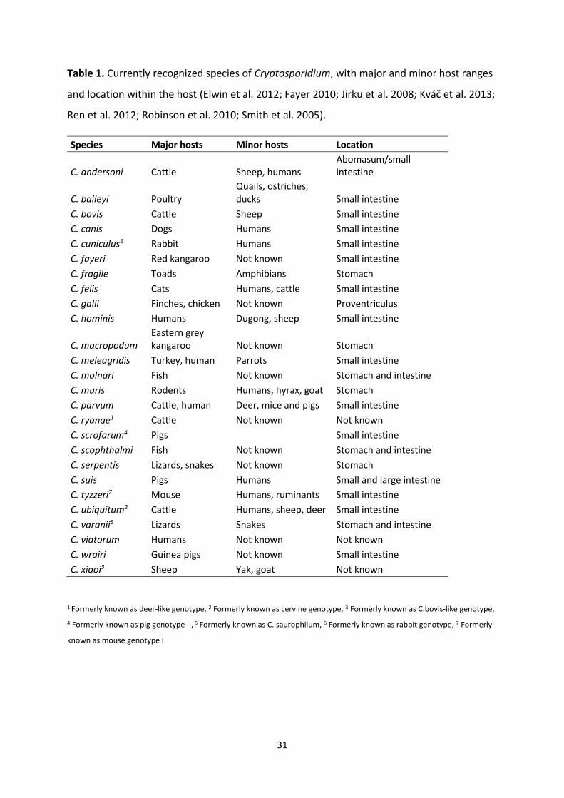
Table 1. Currently recognized species of Cryptosporidium, with major and minor host ranges
and location within the host (Elwin et al. 2012; Fayer 2010; Jirku et al. 2008; Kváč et al. 2013;
Ren et al. 2012; Robinson et al. 2010; Smith et al. 2005).
Species Major hosts Minor hosts Location
C. andersoni Cattle Sheep, humans Abomasum/small intestine
C. baileyi Poultry Quails, ostriches, ducks Small intestine
C. bovis Cattle Sheep Small intestine
C. canis Dogs Humans Small intestine
C. cuniculus6 Rabbit Humans Small intestine
C. fayeri Red kangaroo Not known Small intestine
C. fragile Toads Amphibians Stomach
C. felis Cats Humans, cattle Small intestine
C. galli Finches, chicken Not known Proventriculus
C. hominis Humans Dugong, sheep Small intestine
C. macropodum Eastern grey kangaroo Not known Stomach
C. meleagridis Turkey, human Parrots Small intestine
C. molnari Fish Not known Stomach and intestine
C. muris Rodents Humans, hyrax, goat Stomach
C. parvum Cattle, human Deer, mice and pigs Small intestine
C. ryanae1 Cattle Not known Not known
C. scrofarum4 Pigs Small intestine
C. scophthalmi Fish Not known Stomach and intestine
C. serpentis Lizards, snakes Not known Stomach
C. suis Pigs Humans Small and large intestine
C. tyzzeri7 Mouse Humans, ruminants Small intestine
C. ubiquitum2 Cattle Humans, sheep, deer Small intestine
C. varanii5 Lizards Snakes Stomach and intestine
C. viatorum Humans Not known Not known
C. wrairi Guinea pigs Not known Small intestine
C. xiaoi3 Sheep Yak, goat Not known
1 Formerly known as deer-like genotype, 2 Formerly known as cervine genotype, 3 Formerly known as C.bovis-like genotype,
4 Formerly known as pig genotype II, 5 Formerly known as C. saurophilum, 6 Formerly known as rabbit genotype, 7 Formerly
known as mouse genotype I
32
Cryptosporidium genotypes and subtypes, and their zoonotic potential
Molecular tools have been extensively used to characterize the transmission of human
cryptosporidiosis. Five Cryptosporidium spp are responsible for most infections, namely C.
hominis, C. parvum, C. meleagridis, C. canis and C. felis. In developing countries, C. hominis is
the causative agent for about 70% of infections, while C. parvum accounts for 10-20%. Some
differences have been found in endemic areas in proportion of infection attributable to
species, for example has C. meleagridis been found as the main causative agent in some
areas. Subtyping results suggests that there is high genetic heterogeneity in C. hominis in
developing countries, and geographical segregation of both C. hominis and C. parvum
subtypes (Xiao 2009). In Europe, both C. hominis and C. parvum are most common and
responsible for most human infections (Bajer et al. 2008; Chalmers et al. 2009; Leoni et al.
2006; Llorente et al. 2007; Nichols et al. 2006; Savin et al. 2008; Wielinga et al. 2008;
Wolska-Kusnierz et al. 2007; Zintl et al. 2009; Šoba et al. 2006), while in the middle east, C.
parvum is the dominant species infecting humans (Al-Brikan et al. 2008; Meamar et al. 2007;
Pirestani et al. 2008; Sulaiman et al. 2005; Tamer et al. 2007). Thus, there is a vast diversity
of cryptosporidiosis transmission, highlighting the need for more extensive studies of
cryptosporidiosis epidemiology in diverse areas, including several socioeconomic strata and
environmental conditions.
The combination of subtyping and conventional epidemiological tools can improve the
assessment of the disease burden attributable to zoonotic transmission. A large number of
studies have been conducted to subtype C. parvum in farm animals, with the focus on calves
as infection is largely associated with younger animals. Most subtyping studies have used
gp60 sequence analysis, and have been done in developed countries. The results have
shown that calves are commonly infected with subtypes in the IIa family, with the subtype
IIaA15G2R1 being especially common (Xiao 2010). Although have several subtypes been
found to be more regionally distributed, and IId subtypes have been found to be especially
common in lambs and goat kids in Spain, though the IIa subtypes are more common in calves
in the same area (Quilez et al. 2008a; Quílez et al. 2008b).
Many of the common bovine IIa family subtypes in North America, Europe and Australia are
also dominant C. parvum subtypes in humans in these areas(Alves et al. 2006; Feltus et al.
2006; Jex et al. 2007; Jex et al. 2008; Ng et al. 2008; O’Brien et al. 2008; Soba & Logar 2008;
33
Waldron et al. 2009; Zintl et al. 2009). IId is another major zoonotic genotype family
reported in Europe, Asia and Africa (Amer et al. 2013; Imre et al. 2013; Insulander et al.
2013; Iqbal et al. 2012; Wang et al. 2011), but this family has never been found in humans in
the United States and Canada, where they also seem to be absent in calves (Xiao 2010).
These findings suggest that there are differences in the role of zoonotic transmission of C.
parvum among geographic areas, and even the zoonotic implications of some subfamilies
have been questioned. Studies from Portugal and Slovenia showed that the genetic diversity
of C.parvum was much higher in humans than in calves, and subfamily IIc was not even
found in animals (Alves et al. 2006; Soba & Logar 2008)
Results of multi-locus genotyping studies have further supported the occurrence of
anthroponotic C. parvum (Xiao 2010). Thus, a significant fraction of C. parvum infections may
not have originated from a ruminant reservoir.
Cryptosporidium parvum transmission in developing countries appears largely
anthroponotic, as the most common subtype family is IIc, and has even been found to be the
only prevailing subtype in countries such as Lima, Peru and Jamaica, and studies from India,
Uganda, Malawi and Kenya have found some unusual C. parvum subtype families such as IIb
and IIe in humans, which have never been found in animals anywhere (Akiyoshi et al. 2006;
Cama et al. 2003; Cama et al. 2008; Gatei et al. 2008; Muthusamy et al. 2006; Savioli et al.
2006).
These anthroponotic speculations from developing countries still needs support of results
from animal studies, as gp60 subtyping has been done on only a few C. parvum isolates from
these areas.
34
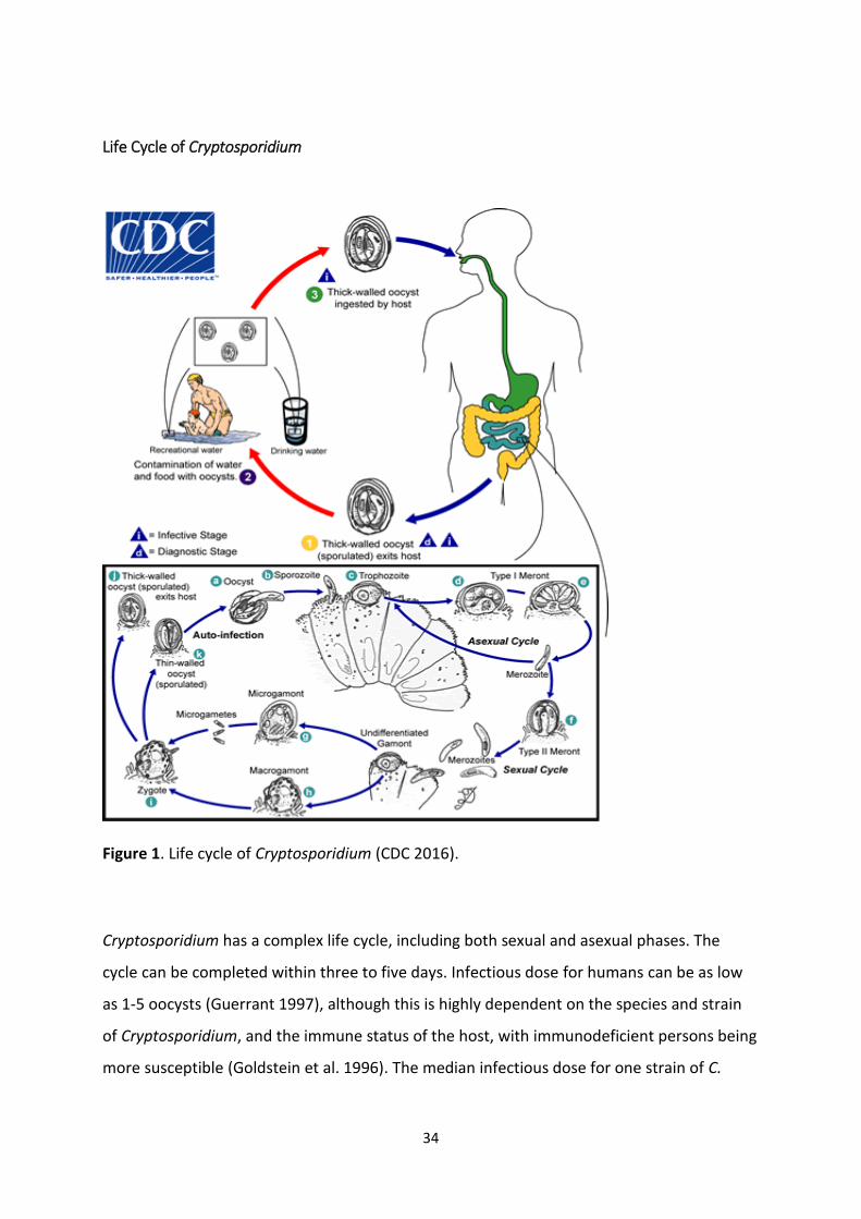
Life Cycle of Cryptosporidium
Figure 1. Life cycle of Cryptosporidium (CDC 2016).
Cryptosporidium has a complex life cycle, including both sexual and asexual phases. The
cycle can be completed within three to five days. Infectious dose for humans can be as low
as 1-5 oocysts (Guerrant 1997), although this is highly dependent on the species and strain
of Cryptosporidium, and the immune status of the host, with immunodeficient persons being
more susceptible (Goldstein et al. 1996). The median infectious dose for one strain of C.
35
parvum was demonstrated to be 132 oocysts in healthy adult volunteers (DuPont et al.
1995).
After the oocysts reach the small intestine, they excyst and four motile sporozoites leave the
oocyst (Hijjawi et al. 2002; Smith et al. 2005), and subsequently infect the epithelial cells. In
the cells, the sporozoites reside in a parasitophorous vacuole between the cell membrane
and cell cytoplasm. Inside this epicellular location, the sporozoites form trophozoites that
undergo asexual development, with two successive generations of merogony, resulting in
the formation of meronts. Merozoites develop into sexual developmental stages known as
the micro- and macrogametes. The microgametes are released from the host cells and
penetrate cells harbouring macrogametes, and their fusion results in the formation of a
zygote, which develops into an oocyst with a resistant oocyst wall. The oocysts are
approximately spherical and measures between 4-5 µm in diameter. Most (80%) of the
oocysts have a thick wall and are excreted with faeces, and represent the environmentally
resistant stage of the parasite, and are immediately infectious. The remaining 20% are thin-
walled oocysts, and are believed to cause autoinfection through recycling of sporozoites
from ruptured thin-walled oocysts (Hijjawi et al. 2001).
Diagnosis and detection of cryptosporidiosis
Although first described in 1907, Cryptosporidium was not considered as a pathogen in
livestock until 50 years later, when it was recognized to cause morbidity and mortality in
young turkeys in 1955, and it was first described as a disease-causing agent in humans 20
years thereafter, in 1976, when the first two cases were described histologically (Meisel et
al. 1976; Nime et al. 1976). This delay in recognition of Cryptosporidium as a pathogen, may
partly be due to the lack of effective methodologies at that time which were able to detect
parasites in clinical samples. The AIDS pandemic in the 1980s, in which Cryptosporidium was
found to be a concomitant, often fatal, pathogen, along with several large waterborne
outbreaks, brought the realisation of the public health significance of this parasite.
36
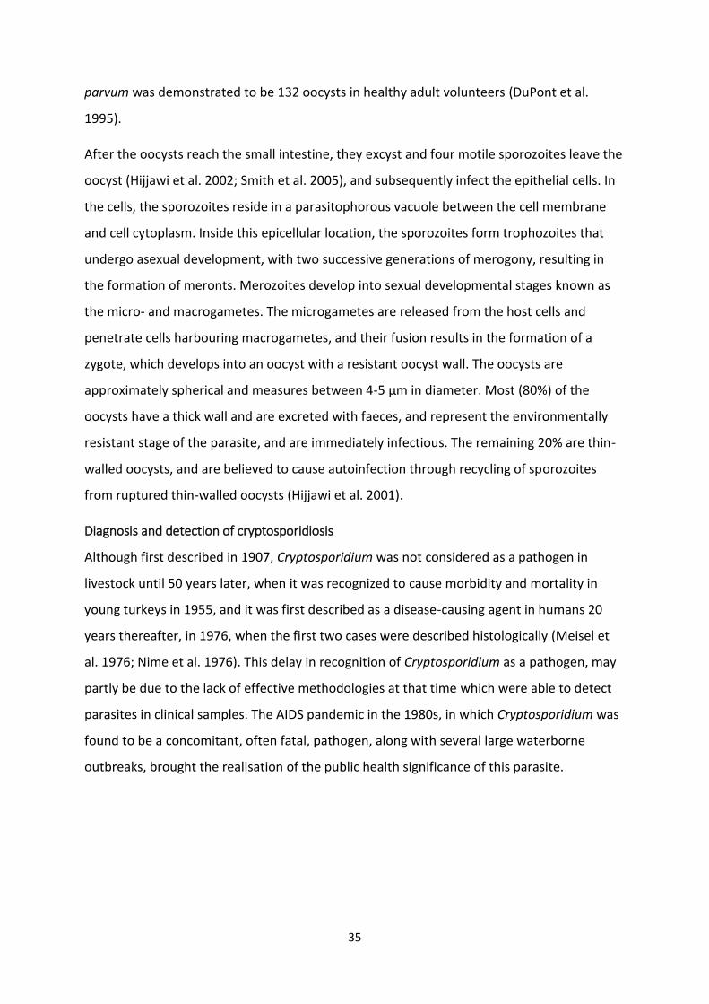
Oocyst morphology
Figure 2. Intact and ruptured Cryptosporidium
oocyst. (Picture acknowledgement, Birgitte Kasin
Hønsvall)
Sporulated oocysts are smooth, colourless, spherical or ovoid and contains four elongated
sporozoites, which are characterized by their comma-shape, and a residual body. These
contents can be difficult to distinguish by light microscopy. Their morphometry can be
helpful in distinguishing oocysts from other microscopic artefacts, but is not sufficient to
distinguish species. In diagnostics, it is common to use staining to identify oocysts in faecal
and environmental samples.
1.3.2 Giardia taxonomy, species and life cycle
The genus Giardia belongs to the kingdom Excavata, clade Fornicata and order
Diplomonadida. Giardia comprises 6 species, distinguished on the basis of light- and electron
microscopy of the trophozoite (Adam 2001), of which five are isolates from birds,
amphibians, mice and voles, and the sixth species, Giardia duodenalis (syn. G. lamblia, syn.
G. intestinalis) is a complex containing strains isolated from a large range of mammalian
hosts grouped into a single species by (Filice 1952).
Giardia genotypes and their zoonotic potential
Genetic analysis has so far revealed eight distinct assemblages within the species complex G.
duodenalis, named with letters from A to H.
Assemblages A and B cause infection in humans, as well as being reported from a range of
other mammals, whereas the remaining assemblages are more restricted in their host range;
Assemblages C and D are found in canids, E in livestock or ungulates, F in cats, G in rodents,
and H in pinnipeds. The genetic distance between assemblages of Giardia duodenalis is of
the same level as the other Giardia species, and new individual species names have been
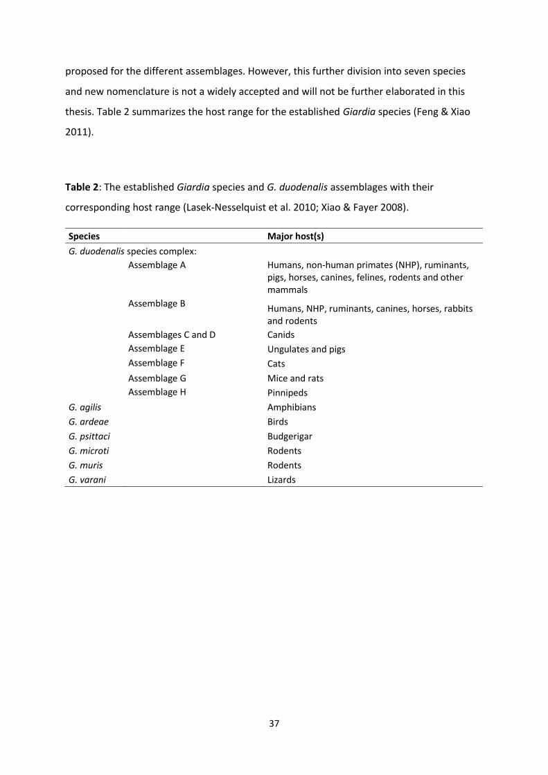
37
proposed for the different assemblages. However, this further division into seven species
and new nomenclature is not a widely accepted and will not be further elaborated in this
thesis. Table 2 summarizes the host range for the established Giardia species (Feng & Xiao
2011).
Table 2: The established Giardia species and G. duodenalis assemblages with their
corresponding host range (Lasek-Nesselquist et al. 2010; Xiao & Fayer 2008).
Species Major host(s)
G. duodenalis species complex:
Assemblage A Humans, non-human primates (NHP), ruminants, pigs, horses, canines, felines, rodents and other mammals
Assemblage B Humans, NHP, ruminants, canines, horses, rabbits and rodents
Assemblages C and D Canids
Assemblage E Ungulates and pigs
Assemblage F Cats
Assemblage G Mice and rats
Assemblage H Pinnipeds
G. agilis Amphibians
G. ardeae Birds
G. psittaci Budgerigar
G. microti Rodents
G. muris Rodents
G. varani Lizards
38
Life cycle of Giardia
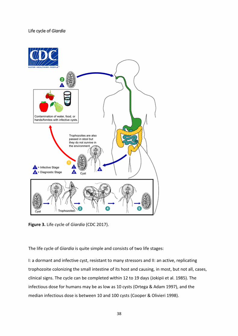
Figure 3. Life cycle of Giardia (CDC 2017).
The life cycle of Giardia is quite simple and consists of two life stages:
I: a dormant and infective cyst, resistant to many stressors and II: an active, replicating
trophozoite colonizing the small intestine of its host and causing, in most, but not all, cases,
clinical signs. The cycle can be completed within 12 to 19 days (Jokipii et al. 1985). The
infectious dose for humans may be as low as 10 cysts (Ortega & Adam 1997), and the
median infectious dose is between 10 and 100 cysts (Cooper & Olivieri 1998).
39
After ingestion, two motile, flagellated trophozoites emerge from the cyst when reaching
the small intestine, with excystation triggered by the intestinal environment. The
trophozoites attach themselves to the epithelial cells by their adhesive discs and colonise the
small intestine. The trophozoites replicate through repeated binary fission, and, unlike
Cryptosporidium sporozoites, Giardia trophozoites are not invasive, although the suction
force from the adhesive discs may damage the microvilli of the small intestine. Exposure to
biliary salts leads to the encystation of trophozoites in the jejunal part of the small intestine,
forming elliptical, 8-12 µm long and 7-10 µm wide cysts, which are excreted with the faeces,
and are immediately infectious.
Diagnosis and detection of giardiasis
The first description of Giardia was by van Leeuwenhoek in 1677, although clinical interest in
this protozoan species began only 40 years ago with the isolation of Giardia from
mammalian, avian, and amphibian hosts, and it was only in the late 1970s that Giardia was
recognized to cause disease (Kreier 1978). The parasite was added to the World Health
Organizations list of parasitic pathogens as late as in 1981 (WHO 1981).
Cyst and trophozoite morphology
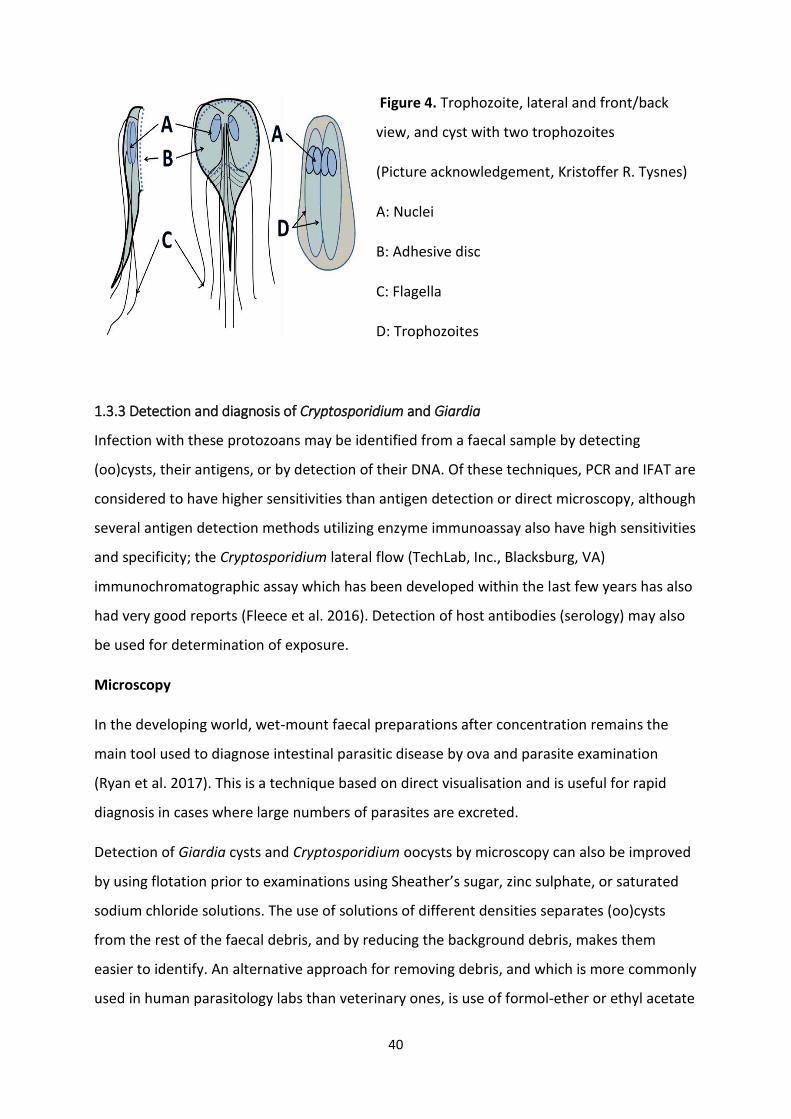
The trophozoite form of Giardia has a characteristic tear-shape, with a bi-radial symmetry.
Its ventral disc, used for attachment to epithelial cells, is composed of a single layer of
microtubules. Each trophozoite has four pairs of flagella that are situated anteriorly,
posterior-laterally, caudally and ventrally, and are used for motility within the host intestine.
The cytoskeleton makes up the unique structure of these flagella and ventral disc, as well as
the median body.
The cyst is the infective life stage of Giardia. They are already in an infective state when
excreted with the host feces. The cysts measure between 5 to 10µm, have an ellipsoid form
and carry between two to four nuclei, depending on whether they contain one or two
trophozoites.
40
Figure 4. Trophozoite, lateral and front/back
view, and cyst with two trophozoites
(Picture acknowledgement, Kristoffer R. Tysnes)
A: Nuclei
B: Adhesive disc
C: Flagella
D: Trophozoites
1.3.3 Detection and diagnosis of Cryptosporidium and Giardia
Infection with these protozoans may be identified from a faecal sample by detecting
(oo)cysts, their antigens, or by detection of their DNA. Of these techniques, PCR and IFAT are
considered to have higher sensitivities than antigen detection or direct microscopy, although
several antigen detection methods utilizing enzyme immunoassay also have high sensitivities
and specificity; the Cryptosporidium lateral flow (TechLab, Inc., Blacksburg, VA)
immunochromatographic assay which has been developed within the last few years has also
had very good reports (Fleece et al. 2016). Detection of host antibodies (serology) may also
be used for determination of exposure.
Microscopy
In the developing world, wet-mount faecal preparations after concentration remains the
main tool used to diagnose intestinal parasitic disease by ova and parasite examination
(Ryan et al. 2017). This is a technique based on direct visualisation and is useful for rapid
diagnosis in cases where large numbers of parasites are excreted.
Detection of Giardia cysts and Cryptosporidium oocysts by microscopy can also be improved
by using flotation prior to examinations using Sheather’s sugar, zinc sulphate, or saturated
sodium chloride solutions. The use of solutions of different densities separates (oo)cysts
from the rest of the faecal debris, and by reducing the background debris, makes them
easier to identify. An alternative approach for removing debris, and which is more commonly
used in human parasitology labs than veterinary ones, is use of formol-ether or ethyl acetate
41
sedimentation. This removes or decreases the fat in the samples, thereby aiding
identification. As cryptosporidiosis, and, more commonly giardiasis, are associated with
steatorrhoea, this may be a useful adjunct for clinical specimens. However, losses of oocysts
and cysts in the faecal plug may be expected.
Stains may be added to the sample to aid in identification, as such stains are selected to
highlight particular features of the parasites, thereby making them more easily distinguished
against the background debris. A number of different stains are available to assist in the
detection of Cryptosporidium oocysts in samples. Giemsa staining was the first to be used by
Edward Tyzzer himself, who first described the parasite, and is now commonly used to
differentiate nuclear and cytoplasmic morphology of parasites. Other stains include
Romanowsky, modified Ziehl-Neelsen, auramine phenol, carbol fuchsin, potassium
permanganate and safranin methylene blue. Most of the direct staining methods are cheap
and easy to perform, although some such as auramine phenol require a fluorescence
microscope for screening, but none of them have been reported to have sensitivities below
103-104 oocysts per gram faeces (Peeters & Villacorta 1995).
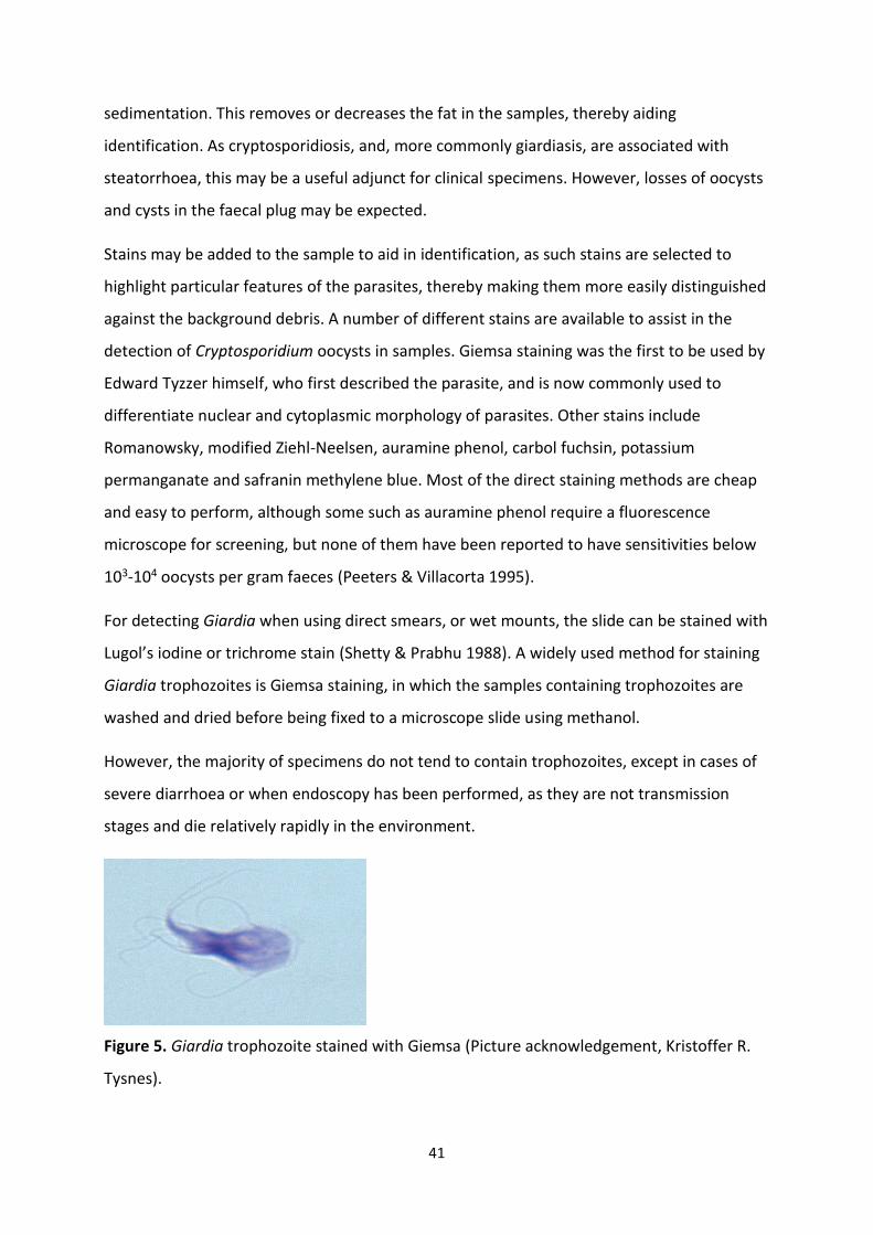
For detecting Giardia when using direct smears, or wet mounts, the slide can be stained with
Lugol’s iodine or trichrome stain (Shetty & Prabhu 1988). A widely used method for staining
Giardia trophozoites is Giemsa staining, in which the samples containing trophozoites are
washed and dried before being fixed to a microscope slide using methanol.
However, the majority of specimens do not tend to contain trophozoites, except in cases of
severe diarrhoea or when endoscopy has been performed, as they are not transmission
stages and die relatively rapidly in the environment.
Figure 5. Giardia trophozoite stained with Giemsa (Picture acknowledgement, Kristoffer R.
Tysnes).
42
There are also commercially available antibodies with fluorescent tags that bind to the
parasite (oo)cyst walls, making them “glow” when viewed under a fluorescent microscope
equipped with the appropriate filter blocks for the fluorophore being used. The most
commonly used fluorophore is fluorescein isothiocyanate (FITC), which
has excitation and emission spectrum peak wavelengths of approximately 495 nm/519 nm,
giving it a green colour, but other fluorochromes can be used.
As this staining technique is dependent on both antibody binding and fluorescent detection,
it is called an immunofluorescent antibody test (IFAT), and is currently considered to be the
gold standard in the detection of Cryptosporidium oocysts and Giardia cysts in faeces or
environmental samples, being the detection method of choice for standard methods, such as
ISO 15553 (ISO, 2006) and ISO 18744 (ISO, 2016).
Some limitations in using some of the microscopy techniques are that they are labour
intensive, time-consuming, can lack sensitivity and specificity, and require a high level of skill
for optimal interpretation, and there is a lack of skilled technicians in both developing and
developed countries (McHardy et al. 2014). In addition, IFAT not only is an expensive
fluorescence microscope equipped with appropriate filter blocks essential, but the reagents
necessary require to be kept in cold storage, thus diminishing their use in countries where
availability of refrigeration is not always optimal.
Antigen detection techniques
Enzyme-linked immunosorbent assays (ELISA) can also also be used to detect
Cryptosporidium and Giardia, and there are several commercial tests available for antigen
detection. Although these tests are mainly developed for parasite species that are
pathogenic for humans, and thus have unknown applicability for other species within these
genera.
DNA-based techniques for detection of Cryptosporidium and Giardia
By using molecular diagnostics, it is possible to detect the presence of small amounts of
parasite DNA in a sample. The code of the specific DNA can be sequenced, and information
on the specific genetic structure of the isolate, which then may allow determination of the
phylogeny of the isolate, and possible transmission pathways and epidemiology. Depending
on the genes targeted and whether the intention is to obtain more information rather than
43
just detection, other aspects, such as virulence, may also be explored. These techniques are
further elaborated in section 4.3.3.
1.3.4 Treatment of cryptosporidiosis and giardiasis
Treatment of cryptosporidiosis in humans
Considerable effort and resources have been directed towards trying to find an effective
cure for cryptosporidiosis. Over 100 compounds have been evaluated for the treatment of
cryptosporidiosis in humans, mice, and cattle, but none of them have been able to control or
eliminate clinical signs or infection in all hosts (Gargala 2008), and a large number of
antimicrobial drugs have been tested in both animals and humans infected with
Cryptosporidium, with no clear evidence of effect (Mead 2002).
A meta-study assessing treatment of cryptosporidiosis in immunocompromised individuals
with nitazoxanide and paramomycin found no effect on the patient groups in focus, and
although the study indicated that immunocompetent patients achieved parasitological
clearance after treatment with nitazoxanide, the authors concluded that there is no
evidence to support the role of chemotherapeutic agents in managing cryptosporidiosis in
immunocompromised individuals (Abubakar et al. 2007). From 2004, nitazoxanide was
licensed by US FDA for treatment of cryptosporidiosis in all immunocompetent patients over
1 year of age (from 2002 it was only licenced for children aged 1-11 years), but it is currently
not approved for treatment of cryptosporidiosis in immunodeficient persons, which are the
patient group most at risk of severe infection in the United States, because placebo
treatment was shown to be equally effective at treating Cryptosporidium-associated
diarrhoea in these patients.
However, for HIV-infected patients suffering from cryptosporidiosis, intense antiretroviral
therapy can lead to complete resolution of clinical symptoms and oocyst secretion(Grube et
al. 1997; Miao et al. 2000), although there are no established treatment regime against
Cryptosporidium infections in this patient group yet. It seems that the main strategy remains
as supportive management, including rehydration therapy, electrolyte replacement, and
antimotility agents until better treatment options emerge. With the completion of the C.
parvum genome sequence, new understandings of the biochemistry of mechanisms of
44
resistance in this parasite have been revealed (Striepen et al. 2004; Umejiego et al. 2004)
and more effective drugs against cryptosporidiosis may be available in the future, that are
hopefully more widely applicable to all patient groups and affordable for the general public.
Treatment of giardiasis in humans
Metronidazole and albendazole are the drugs most commonly used, either alone or in
combination. In one study no difference was found in efficacy of metronidazole versus
albendazole, although side-effects as headache, anorexia and abdominal pain occurred more
frequently in the metronidazole-treatment group (Karabay et al. 2004). These findings have
been supported in later studies, showing that albendazole was equally effective as
metronidazole, but with fewer side effects (Solaymani-Mohammadi & Singer 2011).
Single dose treatment using nitroimidazole-based drugs with long half-lives, i.e. tinidazole,
secnidazole and ornidazole have also proven to be effective (Escobedo et al. 2014).
Animal studies have shown that metronidazole has genotoxic, carcinogenic and teratogenic
potential (Palermo et al. 2004; Tiboni et al. 2008). Reports on possible malformations due to
metronidazole treatment are rare (Cantu & Garcia-Cruz 1982), but pregnant women are not
recommended to take metronidazole during the first trimester. In such cases, parmomycin
has proven to be a safe and effective alternative (Kreutner et al. 1981).
Treatment of giardiasis and cryptosporidiosis in ruminants
There is currently no licensed drug to treat giardiasis or cryptosporidiosis in ruminants,
although the need to treat has been questionable. Treatment alone is not sufficient for
controlling Giardia infection in ruminants because re-infection occurs rapidly and, given the
high level of environmental contamination, daily administration of drugs would be needed.
Halofuginone is reported to markedly reduce Cryptosporidium oocyst output in
experimentally infected lambs and naturally and experimentally infected calves; therapy was
also reported to prevent diarrhea, and this could be an important consideration as
treatment could reduce the extent of environmental contamination and thus onward
transmission of infection. Paromomycin sulfate has proven successful in preventing natural
disease in a controlled clinical field trial in goat kids, but is not licenced for treatment of this
infection per today.
45
Affected ruminants should be supported with fluids and electrolytes, both orally and
parenterally, as necessary until recovery occurs. Cows’ whole milk should be given in small
quantities several times daily (to the full level of requirement) to optimize digestion and to
minimize weight loss. Several days of intensive care and feeding may be required before
recovery is apparent. Parenteral nutrition may be considered for valuable calves (O'Handley
& Olson 2006a).
Treatment of giardiasis and cryptosporidiosis in dogs
The options for treating dogs against giardiasis are scarce. Two of the most commonly used
drug groups are benzimidazoles and nitroimidazoles. The first line treatment is fenbendazole
(Scorza & Tangtrongsup 2010), but in cases where giardiasis persists, a combination of
fenbendazole and metronidazole can be used. A combination of
pyrantel/praziquantel/febantel has shown variable effectiveness in clearing Giardia
infections (Barr et al. 1998; Payne et al. 2002). Albendazole may induce bone marrow
suppression in dogs and cats, and is not recommended for use in these species (Stokol et al.
1997). Quinacrine and tinidazole could be alternative options, but are advised to use with
caution due to scarce data on their adverse effects (Scorza & Tangtrongsup 2010).
Before treating dogs many factors of each individual case should be considered: symptoms,
contact with other dogs, contact with immunosuppressed people and whether the dog is
performing stressful and demanding work, e.g., sled dogs or police dogs (Tysnes et al. 2014).
As no current drug is documented to eliminate Cryptosporidium infection in canines, the
main goal is to stop diarrhoea by supportive care, such as highly digestible diets used for
small bowel diarrhoea which contains fibre and probiotics, and oral rehydration solutions
containing glutamine to replace lost absorptive cell surface. Severe dehydration should be
treated with parenteral fluid replacement, and in some cases antibiotics to control
secondary bacterial infections may be necessary. There are scant publications regarding
cryptosporidiosis therapy in cats and dogs, so treatment regimes should be adjusted
according to the need of each patient (Scorza & Tangtrongsup 2010).
46
1.4 Giardia and Cryptosporidium as waterborne pathogens
Cryptosporidium was discovered to infect humans as late as in 1976 (Nime et al. 1976), and
waterborne transmission was confirmed for the first time in 1984 (WHO 2004). Giardia was
recognized to cause disease in the 1970s (Kreier 1978; Walzer et al. 1971), and waterborne
outbreaks of giardiasis has been reported for the last 40 years.
Cryptosporidium concentrations have been reported to be as high as 14,000 oocysts per litre
in raw sewage, and 5 800 per litre in surface water, and for Giardia the equivalent numbers
are 88000 cysts per litre in raw sewage and 240 cysts per litre in surface water (WHO 2004).
This magnitude of (oo)cysts in different surface waters, together with the low infectious
dose and robust nature, provides the opportunity for large outbreaks to occur when water
sources used for drinking water, irrigation, bathing, or other human activities, are
contaminated.
The total number of reported waterborne outbreaks has increased dramatically over the last
few years, from a total of 325 from the start of the previous century till 2004 (Karanis &
Kourenti 2004), to 199 between 2004 and 2011 (Baldursson & Karanis 2011), to 381
outbreaks reported from 2011 to 2016 (Efstratiou et al. 2017). Whether this reflects a real
increase in outbreaks, or just greater awareness and knowledge is unknown, but little of
such increased knowledge or awareness seems to drip off onto developing countries, as
outbreaks from these countries have not been recorded in the last review of worldwide
outbreaks, although the authors of the review do note that the true magnitude of
waterborne protozoan illness in the regions most affected are still neglected and poorly
described, and states a deficiency in the knowledge of frequency and extent of undiagnosed
outbreaks worldwide. Establishment of surveillance systems in developing countries is a first
step in combating parasitic protozoans and thus improving the health of the population
(Efstratiou et al. 2017).
The cost of waterborne outbreaks has been estimated to be high in developing countries.
There are direct costs, which include the expenses for prevention or health, e.g. the
resources used for medical treatment, and there are direct non-medical costs, e.g. the
transportation costs to visit the physician. In addition, there are also indirect costs, e.g.,
earning losses due to sick leave from work or reduced productivity at work, or resources
47
spent to provide care for an ill individual, and, in some cases, costs of premature mortality
(EPA 2007).
The impact of cryptosporidiosis and giardiasis outbreaks is relatively high due to the large
numbers of people that may be involved, as well as the associated socioeconomic
implications. For instance, the cost of illness associated with the 1993 outbreak in
Milwaukee, USA, where about 400 000 people were infected, has been estimated at US $
96.2 million (WHO 2004). The outbreak of giardiasis in Bergen, Norway in 2004, where 2500
– 6000 persons were infected had an estimated cost of US $ 5.6 million, and the
cryptosporidiosis outbreak in Galway, Ireland in 2007 with 5000 persons infected had a price
tag of US $ 5.3 million (Lindberg et al. 2011). These costs average out to about US $ 750 per
patient, and does not include additional costs like lost workforce due to sick leave, overtime
costs for doctors and technicians, and follow-up of patients suffering from long-term effects
of infections. It is worth considering that US $ 750 is most of the monthly wage of a medical
doctor in India, and is an amount per patient that is just not feasible to spend on a single
patient in developing countries, where occurrences and outbreaks of waterborne
protozoans seems to go unnoticed. Nonetheless, these costs and burdens to society are still
real for those exposed, regardless of whether it gets attention in media, publications or
research projects. There is a need for developing reliable and affordable diagnostic tools for
detection and characterisation, as source tracking and prevention are key to preventing or,
at least, restricting the spread and outbreaks of waterborne parasites.
48
1.4.1 Waterborne Cryptosporidium and Giardia outbreaks and zoonotic implications
Waterborne Cryptosporidium outbreaks and outbreak investigations
Some reviews have suggested that calves are the only major reservoir of C. parvum
infections in humans (Xiao & Feng 2008), and zoonotic sources have been suspected in many
water-borne outbreaks. The greatest documented waterborne outbreak in Milwaukee, USA
never had its source properly identified, but cattle were initially the prime suspects. Reports
have argued that the source may in fact have been human waste drawn from a nearby lake
(Zhou et al. 2003). In contrast, the Cryptosporidium outbreak in Östersund, Sweden in 2010
was rapidly detected and molecular analysis confirmed the contamination to be of human
origin, namely C. hominis (Widerström et al. 2014). The tools for tracking waterborne
outbreaks have evolved greatly since the Milwaukee outbreak in 1993, and give great
advantages in source tracking. Subgenotyping allows us to investigate the sources of
contamination more precisely. A study from Thailand found that in three canals receiving
municipal, agricultural and industrial wastewater, there was a significant load of C. hominis,
pointing to a human source of contamination (Diallo et al. 2008). Three drinking-water
associated outbreaks occurring over a one-year period in the same area of Northern Ireland
was found to be unrelated by utilizing sub-genotyping tools. One of the three outbreaks was
associated with a bovine type of C. parvum, while the other two were caused by C. parvum
human genotypes (Glaberman et al. 2002).
The lack of utilizing these tools at the right time may also cause consequences for society
and economy, when assumptions of the contaminating source is made. A small waterborne
outbreak involving 24 people was reported in 1998 where sheep were suspected of being
the source, but molecular studies never confirmed this. An outbreak in England in 1999,
where over 200 persons were affected, was strongly associated with sheep grazing around
the reservoir, though this was never fully confirmed by molecular analysis (Cotruvo et al.
2004; Qamruddin et al. 2002), and this was also the case in an outbreak of waterborne
cryptosporidiosis in Scotland in 2000 (Cotruvo et al., 2004) where Cryptosporidium in the
faeces of the implicated sheep were not investigated by molecular methods until 3 months
after the outbreak, at which time they proved to have Cryptosporidium of a different
genotype to that of the outbreak (Chalmers et al. 2002). The 1999 outbreak caused several
49
preventive measures, such as upgrading water treatment plants and removal of grazing
sheep to other areas. These implementations led to frustration among sheep farmers (BBC
2000), and particularly so as there was no clear associated disease, and the decision seemed
to lack scientific grounding. This led to increased expenses and may as well have caused
unjustified stigma around sheep as potential transmitters of disease.
Waterborne Giardia outbreaks and outbreak investigations
Aquatic wildlife has been implicated in waterborne outbreaks of Giardia. Studies have shown
that beavers and muskrats shedding Giardia cysts harbour Assemblage B, though there is
little evidence supporting these animals are sources of contamination. It has been suggested
that these animals are more likely to become infected from water contaminated with faecal
material of human or even domestic animal origin, thus acting as amplifiers of the isolate
rather than the original source (Fayer et al. 2006; Sulaiman et al. 2003; Thompson 2004).
Dogs have also been suggested as being the source of contamination of the water during the
waterborne giardiasis outbreak in Bergen, Norway, in 2004, which resulted in thousands of
individuals being infected (Landvik 2015), However, the overwhelming evidence was to the
contrary, and the genotype associated with this outbreak (Assemblage B) is very uncommon
among dogs in Norway (Robertson et al. 2015b). This further underlines the importance of
molecular characterization of pathogens in geographical areas so that foci of infection are
recognizable and measurements of prevention may be less costly, but more targeted and
specific.
Source tracking seems to be a conundrum for many water-borne outbreaks. Most of the 325
outbreaks compiled by Karanis et al (2007) did not use molecular methods to verify the
genotype implied, leaving the zoonotic implications of the outbreaks unanswered.

50
1.4.2 Water supply in India and Chandigarh
Although India is one of the countries receiving most precipitation globally, it is in the midst
of a serious water problem. Droughts and water shortages are common, but, at the same
time, during the monsoon season, there is often too much water and the infrastructure to
deal with this is lacking. Whilst the widely reported current flooding due to Hurricane Harvey
in Texas and Louisiana USA have resulted in at least 47 deaths, and about 43,000 people
requiring emergency housing in shelters, the almost simultaneous flooding due to the
monsoons in floods across a swathe of South Asia, including India, have resulted in more
than 1,200 deaths, of which at least 500 were in the northern Indian state of Bihar, and with
millions of people estimated to have been displaced and at least 700,000 homes destroyed.
Even in non-emergency situations in most cities in India, the operation, maintenance and
quality monitoring of drinking water supply systems are inadequate. Water supply pipes
leading to individual households in India often have leaking joints, leading to the potential
for contamination of the water. In rural areas, even rudimentary facilities of water quality
monitoring and surveillance are not available. Similar to most cities in India, the public water
supply in Chandigarh is managed by a municipal body, and when the city was built in the
1950s, it was planned that every sector should have access to water for the public by having
one or more public water sources, from which the public can either drink directly or collect
water to take home, but the quality system of monitoring and surveillance for drinking water
quality is by far from optimal (Goel et al. 2015).
51
1.5 Fresh produce as vehicles of infection for Giardia and Cryptosporidium
Fresh produce may become contaminated with protozoa and, if eaten raw, may act as a
vehicle of infection. Food contamination may occur during production, processing, or
preparation.
The biology of Cryptosporidium and Giardia makes them suitable for transmission via fresh
produce; they have a low infectious dose, a robust transmission stage, are small sized, and
some genotypes have a zoonotic potential, giving the opportunity for contamination to
occur from both animal and human sources (Robertson & Lim 2011; Robertson 2013).
Cryptosporidium and Giardia can contaminate food as a surface contaminant. Contamination
of foods with small numbers of infectious (oo)cysts in produce that receives minimal
washing or treatment prior to ingestion, poses a threat to public health.
As fewer people are likely to be affected in a foodborne outbreak than in a waterborne
outbreak, and may be more scattered geographically, a lack of prompt diagnosis may
hamper epidemiological investigation. It is worth noting that the last major waterborne
outbreak of giardiasis in Europe, in which over 1500 people were infected, took several
weeks to be recognised as a waterborne outbreak (Robertson et al. 2006), and, for example,
in two documented foodborne outbreaks of cryptosporidiosis, the specific contaminated
produce was never detected (McKerr et al. 2015; Pönka et al. 2009).
Contamination of food with protozoa has been considered to occur either directly from
food-handlers, perhaps infected themselves or in close contact with an infected person or
animal, or from contact with a contaminated environment. Such environmental
contamination can be from soil, particularly soil amended with faeces or manure, or from
water such as irrigation water or wash water along the food chain (Cook and Lim, 2012;
Robertson & Chalmers 2013).
As with waterborne cryptosporidiosis and giardiasis, the potential for foodborne
transmission is considered rather similar for both parasites. Indeed, one recent estimate
regarding the burden of foodborne disease (Hald et al., 2016), concluded that
Cryptosporidium and Giardia were quite similar regarding source attribution. Kirk et al.,
(2015) used expert elicitation to estimate that whereas around 15 % of giardiasis cases were
52
foodborne (uncertainty intervals (UI of 0.08-0.27), a slightly lower proportion (13 %) of
cryptosporidiosis cases were foodborne (UI of 0.07-0.24).
Cryptosporidium oocysts and Giardia cysts are quite robust, and studies have suggested they
can survive numerous treatment processes (Duhain et al. 2012), and remain infective for
weeks on stored produce (Macarisin et al. 2010b). Thus, their biology makes them well
suited for transmission by fresh produce, as in addition to the infective stages (cysts and
oocysts) being robust, the infectious dose is low and infected individuals shed vast amounts
of immediately infectious (oo)cysts. Furthermore, the zoonotic potential from both parasites
means that contamination of fresh produce may be from infections in humans or in animals.
In addition, washing procedures may fail to remove Cryptosporidium oocysts as they seem to
adhere to surfaces and may infiltrate the stomatal openings of leafy vegetables (Macarisin et
al. 2010a; Macarisin et al. 2010b).
In the few documented outbreaks of foodborne transmission of giardiasis, the food handler
has been the suspected source of contamination in five of nine cases (Cook & Y.A.L. 2012).
However, in the work done as part of this thesis, Giardia was found in relatively low
numbers on the produce, which may suggest that contamination is more probable from
water or another vehicle rather than direct contact with an infected person.
In contrast, among the relatively large numbers of confirmed outbreaks of foodborne
cryptosporidiosis, direct associations with food handlers are less common (Robertson &
Fayer 2012). However, in some of the work conducted as part of this thesis, I found that
some produce was contaminated with high numbers of oocysts, suggesting direct contact
with a highly contaminated or infected source, possibly the salesperson or customer.
Foodborne outbreaks transmitted by fresh produce are also difficult to investigate, as by the
time an outbreak is identified, the implicated food is usually not available, being either
discarded or consumed. Also, as the incubation period for Cryptosporidium and Giardia may
be days, even weeks, so outbreak investigations generally rely on clinical samples and
interviews, and environmental inspections.
Many of the foodborne outbreaks for which fresh produce has been identified as the
probable vehicle of infection have occurred in Nordic countries, and thus it could be
speculated that the climate in these areas may prolong survival of the parasite. However, in
53
some of the cases the produce implied was imported, and for others the contamination
occurred at the serving location.
Indeed, considering the various factors, the association of outbreaks with Nordic countries
may be due to the more developed surveillance and outbreak investigations in these
regions, as it is not due to higher consumption of fresh produce in these countries
(Robertson & Chalmers 2013). In developing countries, where foodborne infections or
outbreaks may occur against a background of high infection rates, it is likely that it is difficult
to identify when outbreaks occur, and almost impossible to determine the likely vehicles of
infection. However, obtaining information on the extent of contamination of different food
products with these parasites may provide some pointers regarding the potential for
different transmission routes and vehicles for transmission.
Although molecular methods have been used for detection of Cryptosporidium oocysts and
Giardia cysts on fresh produce (Caradonna et al. 2017; Dixon et al. 2013; Rzeżutka et al.
2010), the standard ISO Method relies on isolating (oo)cysts from the sample, and has roots
in the standard methods for analysing water for these parasites (ISO 2006; U.S.-EPA 2012),
being based on concentration and isolation from debris by immunomagnetic separation
(IMS) and detection by immunofluorescent microscopy (Robertson & Chalmers 2013).
These methods for analysing fresh produce for contamination with these protozoa are quite
expensive and, as with the standard methods for detection of protozoans in drinking water,
may be cumbersome and their value for use in areas where resources are already
constrained may be considered questionable.
Another aspect when fresh produce is considered as a potential vehicle for transmission of
parasites or other pathogens, is the farm-to-fork chain in the particular situation. In
developed countries, where we have availability of all kinds of fresh produce, regardless of
season, the chain may be complicated, but it is usually well documented. For example, when
contamination of imported strawberries with Giardia cysts was identified in Norway in 2016
(Mattilsynet 2017), it was rapidly determined that the strawberries had been imported via a
Dutch firm from a particular grower in Spain; this enable investigations and other measures
to be implemented at the correct place. In contrast, in developing countries, supermarkets
and cold chains are still not common and properly implemented, and produce is commonly
54
not labelled with place of harvest. My studies were based in India, the second largest
producer of fruits and vegetables in the world (Sachdeva et al. 2013) and around 70% of the
Indian population depend directly or indirectly upon agriculture; the section below gives a
brief overview of the vegetable trade in this situation and how this may potentially
exacerbate the sale of contaminated fresh produce, and mean that investigations on the
sources of any contamination can be complicated.
1.5.1 The fresh produce trade in India; an industry in change bringing potential pitfalls and
possibilities
The distribution and sale of most vegetables in India is via the Traditional Retail Model,
which includes many participants. The first links in the chain are the farmers; farms in India
are most usually small, with relatively small yield volumes of crop. Farming in India is a low
prestige work (Robertson et al. 2015a) and farmers are often economically insecure and
struggling financially. Indeed, it is worth noting that farmer suicides account for 11.2% of all
suicides in India (N.C.R.B 2014), but it should also be noted that farmer suicide rates are
elevated in other countries, including industrialized ones. The farmers sell their produce to
agents, who distribute them to commercial customers, vendors, and wholesalers that sell
further to retailers. Wholesalers usually deal with a particular range of fresh produce, and
the retailers sell the produce directly to customers. In this complicated chain, none of
participants will take responsibility for cleaning the vegetables, and transport of vegetables
is not monitored. Thus, fresh produce can be transported in soiled containers or in lorries
that have, for example, previously been used for transporting animals. The mandis (Indian
traditional market places) in Chandigarh are arranged by an alternative system, the Apni
Mandi, which was introduced to shorten the chain of intermediaries and increase the
income of the farmer and allows only registered farmers to participate and thus sell their
produce directly to consumers. This scheme has proven to be financially beneficial for small
farmers, although they have the extra burden of organizing the transport and logistics
themselves. Problems that have arisen in this system include shortage of storage space, the
long duration of market hours, and inadequate facilities such as lighting, drinking water,
sanitation, and parking (Ghuman 2000). The mandis are crowded, and many farmers or
retailers display their produce on mats on the ground or on slightly elevated stalls. In order
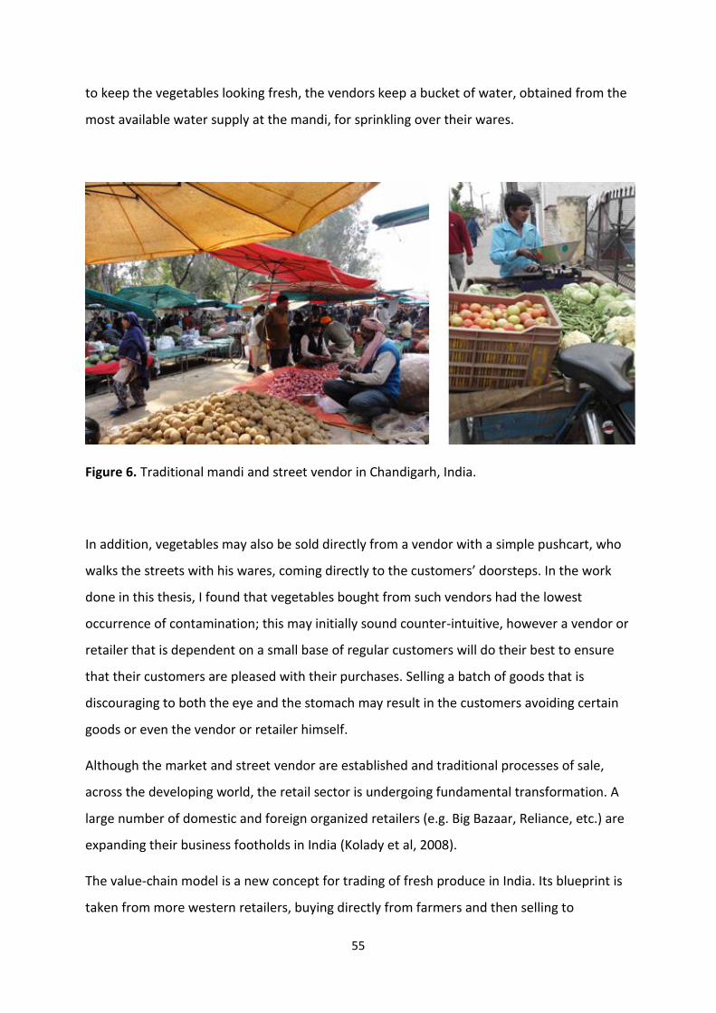
55
to keep the vegetables looking fresh, the vendors keep a bucket of water, obtained from the
most available water supply at the mandi, for sprinkling over their wares.
Figure 6. Traditional mandi and street vendor in Chandigarh, India.
In addition, vegetables may also be sold directly from a vendor with a simple pushcart, who
walks the streets with his wares, coming directly to the customers’ doorsteps. In the work
done in this thesis, I found that vegetables bought from such vendors had the lowest
occurrence of contamination; this may initially sound counter-intuitive, however a vendor or
retailer that is dependent on a small base of regular customers will do their best to ensure
that their customers are pleased with their purchases. Selling a batch of goods that is
discouraging to both the eye and the stomach may result in the customers avoiding certain
goods or even the vendor or retailer himself.
Although the market and street vendor are established and traditional processes of sale,
across the developing world, the retail sector is undergoing fundamental transformation. A
large number of domestic and foreign organized retailers (e.g. Big Bazaar, Reliance, etc.) are
expanding their business footholds in India (Kolady et al, 2008).
The value-chain model is a new concept for trading of fresh produce in India. Its blueprint is
taken from more western retailers, buying directly from farmers and then selling to
56
customers, avoiding intermediaries completely. The outlay of this system is that fresh
produce is cleaned at the arrival hub, and every hub has a warehouse and space available for
temperature-controlled storage. However, implementing this production chain in the Indian
situation could pose some difficulties, not only due to space and infrastructure limitations
may affect both the room for storage and the reliability of the temperature control, but also
because Indians often prefer to buy their groceries at their own particular mandi rather than
from a shop (Economist 2014). Although there are fewer hands touching the produce in the
farm-to-shop chain, this model introduces a further source of contamination as customers in
supermarkets in the value-chain model are allowed to make their own choices, rather than
pointing out produce that is handed to them by the seller. Customers having the opportunity
to pick up and put down items numerous times, means that the fingerprints of
contamination can become even more numerous and difficult to trace.
Although the traditional retail model probably remains the prevailing one for sale and
purchase of fresh produce in today’s India, the western style marketing is gaining a foothold
in many Indian cities. Furthermore, the growth of double-income families, who are more
likely to prefer convenience food, may result in a significant increase in the size of domestic
food markets (Pingali & Khwaja 2004), and probably especially so in relatively developed
cities like Chandigarh, where families with reasonably high income tend to settle.
Although it might be assumed that in a country where the dominant religion promotes
vegetarianism, the inhabitants would consume more fresh produce than the global average,
this is not the case for India, where results of surveys have indicated a consistently low
consumption of fresh produce, and 74% of the adult population consumes less than the
minimum recommended servings (Hall et al. 2009). However, although it is not common or
traditional to consume raw vegetables, as western habits and food outlets are influencing
the Indian palate, lettuce and other raw foodstuffs are being consumed more frequently
(Vashishtha 2014). If the safety and microbiological quality of fresh produce does not keep
pace with the consumption trends, then it can be expected that this may become an
increasingly common route for ingesting pathogens, including parasites. A study from Ghana,
where fresh salad is not a part of the traditional diet, found that increased consumption of
fresh salads through urban fast food inspired from western dishes increased the chance of
acquiring parasitic infections as the produce was contaminated due to poor sanitary
57
conditions during farming and irrigation (Amoah et al. 2007).
Given these rather comprehensive changes for a large market in an enormous country, food
safety standards cannot be ignored. A new model for retail and new production chains
should also be accompanied by new regulations and routines. Although the government of
India has, through the National Horticulture Mission, taken up many initiatives that include
handling systems, cold storage, long distance mobile units, pack houses and accreditation of
quality testing labs, the implementation of these initiatives is slow and unhurried, and poorly
monitored. Gaps may be issues like poor roads, transportation, inadequate electricity,
climate variability, inflation, corruption, and political and governance issues (Sachdeva et al.
2013). If food safety is to be recognized as adequate for the consumer, then these issues
must be addressed before the corporate models, with mass supplies and large
supermarkets, take over the traditional roles of the traditional vendor or small shopkeeper,
who has an eye on almost every item in stock.
58
1.6 Domestic animals as potential sources of environmental contamination with
Giardia and Cryptosporidium in the Indian context
India is home to every sixth person in the world, around 20 million dogs, 200 million cows
and buffaloes and 150 million goats (Paul et al. 2014; Singh et al. 2013). Most Indians are
Hindus, and even though the Hindu religion speaks of loving and caring for all animals,
sometimes the same religion comes in the way of practical animal welfare and disease
management. All animal species are generally thought of as being equally worth in
Hinduism, but in practice a difference seems to be made. This has a root in cultural,
religious, medical and financial factors. The common western view is that there is a
difference in between life and quality of life, which might not be as obvious to a Hindu, as
pain and misery might be considered as a way to cleanse bad karma. Taking a life means
karmic effects of the next life for a Hindu, and issues as letting animals roam freely without
food or care, or withholding euthanasia with the justification “we`ll leave it to God” is the
righteous way of practice in this culture, who believes that “we are not our bodies, they are
merely clothing for our soul”(Prabhupāda 1986), a concept which is hard to fathom for the
western world.
1.6.1 Environmental contamination with Giardia and Cryptosporidium in developed and
developing countries
It is clear that in developed countries, the human burden with intestinal parasites is much
lower than in developing countries; for example, whereas nearly 2% of adults and 6-8% of
children have Giardia infections in developed countries, in the developing world one third of
the population have had giardiasis (Flanagan 1992). The relatively high prevalence of human
infections with Giardia and Cryptosporidium in developing countries has been attributed to
factors as multiple exposure routes due to living and sanitation conditions, cycling through
domestic animals, contamination of aquatic ecosystems by the high environmental burden
and the robust nature of (oo)cysts against common water disinfectants (Slifko et al. 2000),
and, in a vicious circle, the greater number of human infections, coupled with inadequate
sanitation and hygiene, result in greater environmental contamination, including water
59
sources and food, resulting in more infections. Numerous studies have investigated relative
prevalences of these parasites in people in different settings, but the role that animals play
in environmental contamination has been less explored. In the context of infections with
Cryptosporidium and Giardia in domestic animals, and their contribution to environmental
contamination, it is important that not only the cold data of prevalence and infection
intensity are explored, but that also the context of animal husbandry within the situation
that may also contribute to enhanced exposure routes to the human population.
In the following sections, I describe Cryptosporidium and Giardia in three important animal
groups, goats, cattle and buffalo, and dogs, with particular emphasis on the Indian context
and how these animals interact with, and are perceived by, the Indian population. I finish
this section with some considerations of animals in Hinduism, as this can also be of
relevance regarding animals in parasite transmission cycles.
1.6.2 Cryptosporidium and Giardia in goats – and goat husbandry in India
In developing countries across the globe, goats make a vital contribution towards cutting the
cost of living for many hard-pressed families, and goat populations are increasing. Asia and
Africa have experienced the main portion of the total increase in goat numbers, accounting
for 89% of the increase between 1979 and 1991 (Mackenzie 2011). and the largest goat
population in the world is found in India where it constitutes around 154 million animals
(Paul et al. 2014), or around one goat for every 8 people.
The main advantages of goats as livestock are that they do not need expensive housing and
feed, and they have impressive abilities in converting limited resources into meat and milk.
These factors have boosted goat farming in rural areas and among small-scale farmers in
India, as well as their high capacity for adapting to extreme climatic conditions; they are
particularly important in arid and semi-arid regions where rainfall may be scant and hence
income from crops is uncertain and low (IndiaAgroNet 2017). They have a small body size
and docile nature, are prolific breeders, and they thrive better in dry areas, and they may be
2.5 times more economical than sheep under semi-arid conditions. Both female and male
animals are of equal value (Sirohi & Chauhan 2011). Furthermore, in India, where beef is
rarely consumed, goat meat makes up 35% of the total meat market, as goats are not
60
associated with any religious taboos among non-vegetarian Hindus, Sikhs, and Muslims in
India.
In India, goats are usually not penned in a shed, but rather tethered in the home premises
and sometimes, depending on the area, taken for grazing. They are generally reared under
semi-scavenging systems. Nearly all goats graze at liberty during daylight hours, and may
also be fed kitchen waste, e.g. rice and vegetables, but feed concentrates are usually not
utilized. The mortality rate among goat kids (up to 3 months) in India can be as high as 25%,
and this is usually attributed to low birth weight and poor milk yield of the ewe (Sirohi &
Chauhan 2011).
This means that goat keeping in India is not managed intensively and the goats are viewed as
a subsidiary income; the goats are allowed to roam relatively freely and do not receive the
routine health care that may be expected in animal husbandry from a western viewpoint.
In western countries, most studies have shown that sheep and goats do not harbour major
proportions of zoonotic genotypes of Giardia and Cryptosporidium, though the management
of small ruminants differs greatly between developed and developing countries.
Reported prevalences for both Giardia and Cryptosporidium in goats around the globe tend
to vary considerably. Studies from Belgium (Geurden et al. 2008b), Netherlands (Van der
Giessen et al. 2006), Spain (Castro-Hermida et al. 2007; Ruiz et al. 2008) Iran (Jafari et al.
2014) and Uganda (Johnston et al. 2010)have revealed prevalences of Giardia in goats
ranging between from 12.3% to 42.2%, with the lowest prevalence found in Uganda.
For Cryptosporidium, the prevalences in different studies from Belgium, Brazil, Sri Lanka,
France and Spain were in ranges varying from 4.8% to 33.6% (Bomfim et al. 2005; Delafosse
et al. 2006; Geurden et al. 2008b; Noordeen et al. 2000)
These data might not only reflect the prevalence of infection, but could also be due to the
sensitivity of the diagnostic tests used, the age of the goat, and whether only a single or
consecutive sample(s) were examined, given the intermittent shedding of Giardia cysts, and
the acute nature of cryptosporidiosis.
The role of small ruminants in transmission of zoonotic intestinal protozoans in developing
countries is not yet defined, and there is a lack of studies defining their part as disease
61
transmittors or recievers. Given the vast difference of livestock keeping between developed
and developing countries, studies made from intensive farms where contact between
humans and animals are restricted as well as sanitation and hygiene measures are
mandatory by law cannot be compared to the backyard livestock, often housed together
with their owners.
In western countries, most studies have shown that sheep and goats do not harbour major
proportions of zoonotic genotypes of Giardia and Cryptosporidium. However, as described
above, the management of small ruminants differs greatly between developed and
developing countries, and it is possible that in goat husbandry systems as described for
India, where the goats are not grazing in designated areas and are living in close contact with
their owner, a different situation may occur.
In general, the role of small ruminants in transmission of zoonotic intestinal protozoans in
developing countries is poorly defined, and studies investigating their role in zoonotic cycles
as disease transmitters or receivers are lacking. Given the vast difference in livestock keeping
between developed and developing countries, studies from intensive farms in developed
countries where contact between humans and animals is restricted and sanitation and
hygiene measures are mandatory are not comparable to the backyard livestock situation
seen in countries such as India, where the goats are often housed together with their
owners. Thus, the role of goats in the Indian situation, and, in particular investigation of the
genotypes or Assemblages of Cryptosporidium and Giardia found in goats in and around
Chandigarh, seemed very relevant in the context of this study.
1.6.3 Cryptosporidium and Giardia in bovids – and cattle and buffaloes in the Indian context
India today is home to about 200 million cattle, representing one-quarter of the cattle
population in the world (Jacobson & Grinker 1999). India is also the largest milk producer in
the world (Sudarshan et al. 2007), and most rural families in India own at least one dairy cow
(Agoramoorthy & Hsu 2012). However, India has long been unique in possessing so many
cattle but without making a maximal profit from cattle slaughter (Harris et al. 1966)
Most cattle in India belong to the species Bos indicus, commonly known as zebu cattle. This
species, which is extremely hardy and resistant to disease (Jacobson & Grinker 1999), is
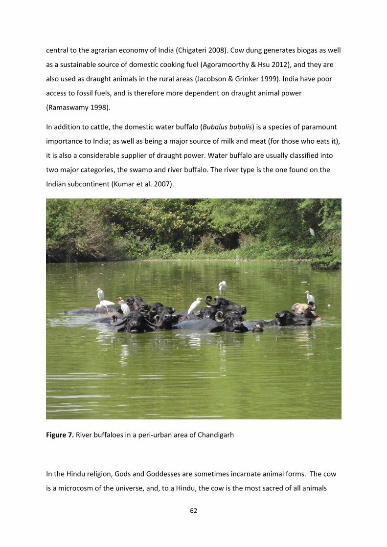
62
central to the agrarian economy of India (Chigateri 2008). Cow dung generates biogas as well
as a sustainable source of domestic cooking fuel (Agoramoorthy & Hsu 2012), and they are
also used as draught animals in the rural areas (Jacobson & Grinker 1999). India have poor
access to fossil fuels, and is therefore more dependent on draught animal power
(Ramaswamy 1998).
In addition to cattle, the domestic water buffalo (Bubalus bubalis) is a species of paramount
importance to India; as well as being a major source of milk and meat (for those who eats it),
it is also a considerable supplier of draught power. Water buffalo are usually classified into
two major categories, the swamp and river buffalo. The river type is the one found on the
Indian subcontinent (Kumar et al. 2007).
Figure 7. River buffaloes in a peri-urban area of Chandigarh
In the Hindu religion, Gods and Goddesses are sometimes incarnate animal forms. The cow
is a microcosm of the universe, and, to a Hindu, the cow is the most sacred of all animals
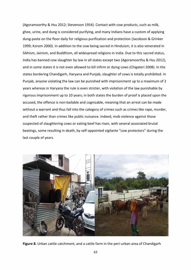
63
(Agoramoorthy & Hsu 2012; Stevenson 1954). Contact with cow products, such as milk,
ghee, urine, and dung is considered purifying, and many Indians have a custom of applying
dung-paste on the floor daily for religious purification and protection (Jacobson & Grinker
1999; Korom 2000). In addition to the cow being sacred in Hinduism, it is also venerated in
Sikhism, Jainism, and Buddhism, all widespread religions in India. Due to this sacred status,
India has banned cow slaughter by law in all states except two (Agoramoorthy & Hsu 2012),
and in some states it is not even allowed to kill infirm or dying cows (Chigateri 2008). In the
states bordering Chandigarh, Haryana and Punjab, slaughter of cows is totally prohibited. In
Punjab, anyone violating the law can be punished with imprisonment up to a maximum of 2
years whereas in Haryana the rule is even stricter, with violation of the law punishable by
rigorous imprisonment up to 10 years; in both states the burden of proof is placed upon the
accused, the offence is non-bailable and cognizable, meaning that an arrest can be made
without a warrant and thus fall into the category of crimes such as crimes like rape, murder,
and theft rather than crimes like public nuisance. Indeed, mob violence against those
suspected of slaughtering cows or eating beef has risen, with several associated brutal
beatings, some resulting in death, by self-appointed vigilante “cow protectors” during the
last couple of years.
Figure 8. Urban cattle catchment, and a cattle farm in the peri-urban area of Chandigarh
64
Although worshipped and holy, cattle seldom die of old age in India, as they are starved or
neglected until they die a “natural” death. Stray cattle are usually older cattle that have
reached the end of their productive years and are left out onto the streets. Cow sanctuaries,
or Gaushalas, provide shelter for hundreds of these cattle, but do not have sufficient
capacity to cope with the whole stray cattle population (Singh et al. 2013). The Indian
Veterinary Council has estimated that there is only food to sustain 60% of the Indian cattle
population (Kang 2003), and the rest are left to starve or roam the streets in search for food,
where garbage makes up the bulk of the diet. In New Delhi alone, there is an estimated
40 000 cows roaming the streets, spreading garbage and constituting traffic hazards
(Agoramoorthy & Hsu 2012) .The problem with stray cattle and traffic accidents is also
becoming an issue in Chandigarh, where the stray cattle population has risen in recent years,
together with traffic incidents (Victor 2013). The increase in stray cattle population is likely
due to the rise in urban dairy farming in India, as the growing middle classes in the cities are
increasing the demand for dairy products.
This management of husbandry on a countrywide basis is quite unique for India. People in
industrialized nations may find it difficult to fathom the extent to which the cow, or cattle in
general, is being worshipped and neglected at the same time, in a country that could benefit
immensely from effective husbandry. It has been argued that Hindu cattle practices are
irrational since religious and political prohibitions on killing and eating cows means wasting
animal products, while at the same time sustaining an unproductive cattle population
(Jacobson & Grinker 1999).
In addition, this close relationship between cattle and cattle products fades the interface of
human-animal interaction. In western countries, applying cow dung to the floor of your
house would be considered quite eccentric, while in India this is a respected tradition.
Although there is no easy solution regarding segregation of cattle and people, education
regarding risk factors and proper hygiene could lessen the pathogen interface between
humans and animals. With regards to the work described in this thesis, it was obviously of
interest to investigate the role that cattle play in the transmission cycles of Cryptosporidium
and Giardia.
Infections of cattle with Cryptosporidium species and G. duodenalis assemblages of zoonotic
potential have been frequently reported, indicating that cattle are a reservoir for human
65
cryptosporidiosis and giardiasis (Hunter & Thompson 2005; Ryan & Cacciò 2013). Cattle are
commonly infected with C. parvum, C. andersoni and C. ryanae (Xiao 2010), of which C.
parvum is of particular importance as a zoonotic species. Contact with infected calves have
been identified as a main reason for a number of small cryptosporidiosis outbreaks in
veterinary students, research technicians, and children on farm visits, validating the zoonotic
potential and transmission of cryptosporidiosis from cattle (Gait et al. 2008; Kiang et al.
2006; Preiser et al. 2003). In addition, case-control studies have reported that contact with
cattle is a significant risk factor for sporadic cryptosporidiosis in humans (Hunter et al. 2004;
Roy et al. 2004).
In dairy cattle, C. parvum is mostly found in pre-weaned calves and C. andersoni in yearlings
and adult cattle (Santín et al. 2008; Trout et al. 2006) Prevalences have been found to vary
from 1% (Kváč et al. 2006) to 59% (Olson et al. 1997) in calf holdings, and up to 100% on
farm level (Santın et al. 2004). The highest prevalence has been observed in calves up to 5
weeks of age (Quilez et al. 1996).
In cattle, the livestock specific Giardia Assemblage E is considered the most prevalent,
though up to 59% zoonotic assemblage A isolates and mixed infection with both E and A
have been reported (Geurden et al. 2008a; Sprong, H. et al. 2009). This would suggest that
calves may be considered as a potential reservoir for human infection, although within
assemblage A sub-assemblage AI is predominantly found in livestock and companion
animals, while assemblage AII is more prevalent in humans (Feng & Xiao 2011). Prevalences
have been found up to 73% (Olson et al. 1997), and on farm level it can be as high as 100%
(Geurden et al. 2010; Geurden et al. 2012; Olson et al. 1997). Nevertheless, the public health
risk from bovine giardiasis is generally considered to be minimal, as it has been estimated
that about 80% of Giardia infections in dairy cattle and 98% in beef cattle are due to
assemblage E (O'Handley & Olson 2006b). Studies from developed countries has shown that
less than 20% of cattle in a herd may harbour the most common zoonotic genotype,
Assemblage A (Santín et al. 2009; Sprong, Hein et al. 2009; Trout et al. 2004; Trout et al.
2005), Longitudinal studies have indicated that infections of zoonotic genotypes of Giardia in
cattle may be transient, as mixed infections of Assemblages A and E have been reported
frequently in cattle, and it has been suggested that immunologically mature cattle are able
to resist infection with assemblage A, while the host-adapted Assemblage E is capable or
66
establishing infection (Uehlinger et al. 2011). Yet another longitudinal study found a 43%
occurrence of Assemblage A concurrently with 57% Assemblage E in adult cattle (Uehlinger
et al. 2006).
However, whether these data from studies predominantly from North America and Europe
are indicative of what might be expected in India, the country of the holy cow, is less certain.
There are some studies published regarding Cryptosporidium prevalence of bovines in India.
(Bhat et al. 2012) found an overall prevalence of about 30% in buffalo calves in Punjab, with
the highest incidence in calves under one month, and the highest rate of infection during the
monsoon season. A study from six different states in Southern India found prevalences
ranging from 86.7% to 17.7% between the states. A wide variety of Cryptosporidium species
were identified, including C. andersoni, C. ryanae, C. parvum and C. bovis, with C. ryanae the
major species found, and the zoonotic C. parvum only accounted for 4% and only found in
one of the six states examined (Venu et al. 2012). A study from six targeted states ranging all
over India geographically, from South to North, revealed Cryptosporidium prevalences from
5.4% to 30.8%, with the lowest prevalence found in the southern parts, and the highest in
the northern parts of the country. In contrast with the previous study, the only species
identified in this study was C. parvum, and the highest occurrence was found in calves during
the monsoon season (Paul et al. 2008). Maurya et al. (2013) also examined the prevalence of
Cryptosporidium in bovine calves in 3 different climatic regions in India and found a higher
prevalence (35.4%) in the northern sub-temperate parts, and the lowest prevalence in sub-
tropical plains (13%), with a significantly higher prevalence of infection during the monsoon
season. Again, the only species identified in this study was C. parvum.
Molecular tools using markers in the 60 kDa glycoprotein (gp60) gene have improved the
ability of identifying and differentiating zoonotic Cryptosporidium at the genotype and
subgenotypic levels. These are useful for investigating the distribution of C. parvum variants
in cattle and human populations in different geographical regions (Robertson 2014b).
Although the majority of publications revealed a strong foothold of C. parvum in the cattle
and buffalo population of the country, none of the Cryptosporidium studies mentioned
above utilized this tool, or other subtyping approaches to further assess the zoonotic
potential of Cryptosporidium shed from bovines in India. Thus, of particular interest for the
investigations in this study was to investigate the sub-types, as well as the species, of
67
Cryptosporidium occurring in the bovid populations in and around Chandigarh, in order to
provide some data on the likelihood of spill-over occurring into the human population, as
some data on Cryptosporidium subtypes in people in Chandigarh have been published
(Sharma et al. 2013).
Furthermore, few studies regarding the zoonotic potential of cattle in India have been
published, though a study from West Bengal found a 12.2% prevalence of Giardia on a dairy
cattle farm, where also 27.5% of the farm workers were infected; however, although
approximately 12% of the Giardia isolates found in humans were of Assemblage A, under 3%
of Giardia in the cattle were also of Assemblage A (Khan et al. 2010). This seems to be in
concordance with the broad findings from studies in developed countries regarding the
distribution of genotypes, and for the present study the intention was to investigate
whether, in the Indian context, where relevant transmission factors, such as housing
conditions and management, differ wildly from those in Europe and North America, may
provide similar or different findings. Thus, the question being addressed partly included
whether having an apparently much greater opportunity for transmission of Giardia
between humans and cattle, resulted in any genetic barrier being defeated.
1.6.4 Cryptosporidium and Giardia in dogs – and man’s best friend in the Indian context
The population of dogs worldwide may be as high as half a billion (Hsu et al. 2003), and free
roaming dog populations have emerged as both animal welfare and public health problems
in developing countries, where they also face welfare issues such as malnutrition, starvation,
disease, and abuse. Additional social problems include bites, road accidents, fighting, noise,
faecal contamination, uncontrolled breeding and spread of garbage. Approaches to the
management of free-roaming dog populations have changed over the past twenty years,
from capture-and-kill to mass vaccination, spaying, habitat control, and responsible dog
ownership, though cultural differences in views of dog ownership and the role of dogs in
society influence the prevalence of dogs, the condition of stray dogs and dog – control
policies In India, there has been a rise in the canine population, both due to the shift to an
animal birth control programme instead of euthanasia in response to animal welfare
activists, and also due to the increase in popularity of keeping dogs as pets in middle-class
68
urban families. The success of animal birth programmes depends on sterilizing 70% of stray
dogs in a given area, and, due to the limited funds and resources allocated to this
programme, achieving this target seems an uphill, if not impossible, task (Menezes 2008;
Totton et al. 2010; Totton et al. 2011). Dog faeces in open spaces and public areas, left by
stray dogs or irresponsible dog owners, could represent an important source of zoonotic
pathogens, and among the parasites this may include not only Cryptosporidium and Giardia,
as discussed for cattle and goats, but also various helminths, including Toxocara canis,
taeniid worms (including Echinococcus spp.), and hookworms, such as Ancylostoma spp.
Although finding the same pathogen species in both human and animal populations, does
not necessarily indicate that zoonotic transmission has taken place, molecular studies may
provide good indications on pathogens` emergence. It is still more useful to explore the
dynamics of transmission between humans and animals living in the same household or
endemic focus. Some studies have investigated the transmission dynamics of Giardia
between dogs and humans living in close proximity, and a few of them found no significant
relation of genotypes shared with dogs and humans (Cooper et al. 2010; Traub et al. 2004),
whereas other studies have found a strong correlation between humans, dogs, and Giardia
genotypes (Inpankaew et al. 2007; Traub et al. 2009). In addition, a study from Assam in
India found a dominance of zoonotic assemblages in both humans and dogs living on the
same tea estate. This was further backed up by epidemiological data showing a significant
association between Giardia infection in humans and the presence of a Giardia positive dog
in the same household. However, it was also noticed that 30% of these dogs also had eggs of
Ascaris lumbrocoides, a human-specific roundworm, in high intensities in their faeces,
indicating coprophagy of human faeces and thus mechanical carriage, which could also have
been the explanation for the Giardia cysts of the same genotypes to those found in the
people (Traub et al. 2003; Traub et al. 2004).
It seems there are no general answers to the zoonotic potential of Giardia infection in dogs
to humans, and each endemic focus should be considered uniquely with a focus on
management, sanitation and human-animal interfaces.
Unlike with cattle, that the Indian population worship and respect, there is a wide religious
and cultural distance to the dog in Indian culture, sprinkled with a legitimate fear of roaming
dogs, as they are well-known in this country as transmitters of rabies virus through their
69
bites. In India, a person is bitten by an animal every two seconds, and about 15 million
people are bitten every year, mostly by dogs (Menezes 2008; Sudarshan 2005; Sudarshan et
al. 2007), so it is not hard to understand why stray dogs are regarded as dangerous and
unwanted by the general public. Indeed, if the cow is the most respected animal in the
Hindu religion, the stray dog might be the least respected animal. While many parts of the
cow are considered pure and purifying, it is quite the opposite for dogs (Prabhupāda 1986).
Nevertheless, it has been suggested that there are 20 million stray dogs in India (Traub et al.
2005), and the Indian pet dog population amounts to 28 million animals (Sudarshan et al.
2007), thus despite their lowly status there is clearly a large canid population, and as clearing
up dog faeces is not considered a normal reaction in Indian society, there remains the
potential for dogs in India to contribute to environmental contamination with potentially
zoonotic parasite transmission stages (Traub et al. 2005).
In order to investigate the role of dogs in the parasite transmission cycles in Chandigarh,
some of this work investigated the parasites of dog faeces sampled from Chandigarh’s green
spaces.
70
1.7 Knowledge gaps
Although many studies have investigated the prevalence of intestinal parasites in human
populations in India, and also the impact (morbidity and mortality) that diseases such as
giardiasis and cryptosporidiosis have on the paediatric population, we have very little
knowledge about the most common transmission routes, risk factors for infection, and the
role that animal infections may have in acting as reservoirs of infection.
Furthermore, although waterborne outbreaks of giardiasis and cryptosporidiosis have been
reported from developing countries, we have scant information about such outbreaks in
developing countries, despite logic indicating that in countries where the infrastructure is
poorer should be more vulnerable to those contamination events that are likely to lead to
such outbreaks.
This has led to a number of questions:
- What is the extent of contamination of potable water with intestinal parasites in
India and what are the risk factors for contamination?
- If contamination occurs, are the species concerned of infection risk to people
consuming the water?
- What is the extent of contamination of fresh produce with intestinal parasites in
India, which types of fresh produce are most at risk, and are there any obvious risk
factors for contamination?
- If contamination occurs, are the species concerned of infection risk to people
consuming the fresh produce?
- Which domestic as well as stray animals living in close contact with the human
population act as reservoirs of infection with zoonotic intestinal parasites, what are
the risk factors for infection in these animals, and is the likelihood of contamination
of water or fresh produce with such parasites greater from infected animals or
infected humans?
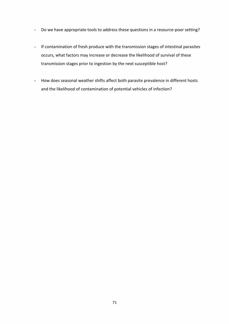
71
- Do we have appropriate tools to address these questions in a resource-poor setting?
- If contamination of fresh produce with the transmission stages of intestinal parasites
occurs, what factors may increase or decrease the likelihood of survival of these
transmission stages prior to ingestion by the next susceptible host?
- How does seasonal weather shifts affect both parasite prevalence in different hosts
and the likelihood of contamination of potential vehicles of infection?
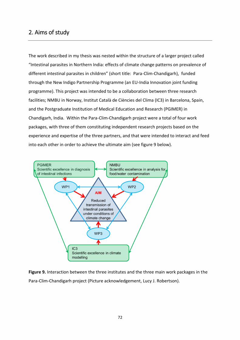
72
2. Aims of study
The work described in my thesis was nested within the structure of a larger project called
“Intestinal parasites in Northern India: effects of climate change patterns on prevalence of
different intestinal parasites in children” (short title: Para-Clim-Chandigarh), funded
through the New Indigo Partnership Programme (an EU-India Innovation joint funding
programme). This project was intended to be a collaboration between three research
facilities; NMBU in Norway, Institut Català de Ciències del Clima (IC3) in Barcelona, Spain,
and the Postgraduate Institution of Medical Education and Research (PGIMER) in
Chandigarh, India. Within the Para-Clim-Chandigarh project were a total of four work
packages, with three of them constituting independent research projects based on the
experience and expertise of the three partners, and that were intended to interact and feed
into each other in order to achieve the ultimate aim (see figure 9 below).
Figure 9. Interaction between the three institutes and the three main work packages in the
Para-Clim-Chandigarh project (Picture acknowledgement, Lucy J. Robertson).
73
The Para-Clim-Chandigarh project thus consisted of short-term and long-term objectives.
The short-term objective was to investigate foodborne, waterborne, and zoonotic
transmission of intestinal protozoan parasites in Chandigarh, and the impacts on their
transmission of large-scale and regional climate patterns and their interaction with the local
environment in the light of projected climate change for the region.
The long-term objective was to develop workable initiatives that could reduce transmission
of these pathogens and provide early – warning systems.
As described, in order to achieve these objectives, the project plan was divided into different
work packages, WP1, WP2, WP3 and WP4, and each of the partners had particular specialist
expertise relating to the assigned work package, giving the project a synergistic approach.
Whereas the first three work packages were research based, and the intention was the
combined results would be the basis for an initiative to reduce field prevalence or provide a
basis for an early warning system.
These basic elements of each of the work packages were (see Figure 10):
1) Assessing the prevalence of intestinal parasites in children in different regions of
Chandigarh, with particular focus on Cryptosporidium and Giardia, including
epidemiological, demographic, and clinical considerations. In addition, data from
records going back decades will be used to assess temporal changes in prevalence.
2) Investigating the potential for waterborne, foodborne and zoonotic transmission of
these parasites due to contamination and the impact of different weather patterns
on a seasonal level on the extent of contamination.
3) Assessing the effect of regional climate and the local environment on the prevalence
of these parasites in children, with modelling based on over 30 years of data.
4) Dissemination and use of the results through not only academic outputs (PhD theses,
conference communications, scientific articles), but also stakeholder workshops and
other local initiatives.
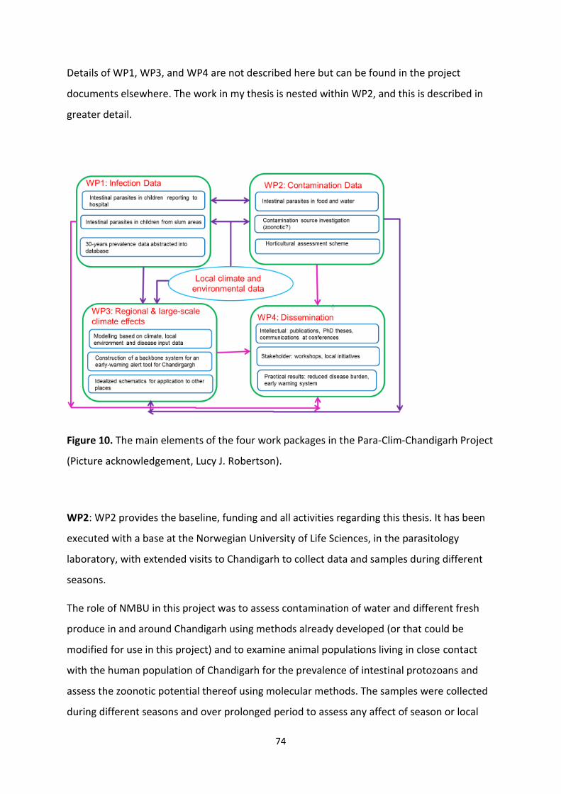
74
Details of WP1, WP3, and WP4 are not described here but can be found in the project
documents elsewhere. The work in my thesis is nested within WP2, and this is described in
greater detail.
Figure 10. The main elements of the four work packages in the Para-Clim-Chandigarh Project
(Picture acknowledgement, Lucy J. Robertson).
WP2: WP2 provides the baseline, funding and all activities regarding this thesis. It has been
executed with a base at the Norwegian University of Life Sciences, in the parasitology
laboratory, with extended visits to Chandigarh to collect data and samples during different
seasons.
The role of NMBU in this project was to assess contamination of water and different fresh
produce in and around Chandigarh using methods already developed (or that could be
modified for use in this project) and to examine animal populations living in close contact
with the human population of Chandigarh for the prevalence of intestinal protozoans and
assess the zoonotic potential thereof using molecular methods. The samples were collected
during different seasons and over prolonged period to assess any affect of season or local
75
weather conditions on prevalence. Other factors, such as within city differences, were also
to be assessed.
It was intended that the data obtained from the food, water, and animal surveillance would
be considered initially in conjunction with the data obtained in WP1, and subsequently in the
light of the data in WP3. Due to economic restrictions with the partner (IC3) responsible for
WP3, this was run at a reduced rate and the data are still under evaluation. Furthermore, the
partner responsible for WP1 were unable to fulfil their contribution to the project.
The objectives for this thesis were thus based on the plan for WP2, and can be considered
under the following main objectives:
2.1 Main objectives
1. Develop and/or modify methods to determine the occurrence, prevalence, and
concentration of food- and waterborne parasites (with focus on Cryptosporidium oocysts
and Giardia cysts) which can be applied to laboratories or projects on low budgets.
2. Identify the extent to which fresh produce for sale in Chandigarh and drinking water
supplied in Chandigarh is contaminated with Cryptosporidium and Giardia, the species and
genotypes of these parasites, and investigate any effect of seasonal or other factors on
contamination of fresh produce and drinking water in Chandigarh with these parasites.
3. Identify sources of zoonotic intestinal parasites in Chandigarh, with particular
emphasis on Cryptosporidium and Giardia, and their possible seasonal variations. Identify
any risk factors particularly associated with zoonotic parasites identified in animal hosts.
In order to break down these rather large objectives into more manageable tasks the
following sub-goals were specified and are addressed in the work described in this thesis:
76
2.1.1 Sub-goals
1. Modify standard methods for analysis of fresh produce and water for contamination
with protozoan parasites with the aim of reducing the cost, such that more samples can be
analysed within budget, but without losing method efficiency. Ensure the validity of the
methods developed through use of multi-lab testing.
2. Use the methods developed on samples collected from potable water sources and
vegetable markets in Chandigarh, and thus estimate the occurrence and species/genotypes
Cryptosporidium and Giardia as contaminants in food and water that may infect humans, as
well as any associations with environmental factors such as location and season/weather.
3. Investigate whether contamination of fresh produce by Cryptosporidium oocysts or
Giardia cysts necessarily means that the produce has infectious potential. By using
laboratory-based spiking studies of fresh produce, assessing the survival of these parasites
over time kept under typical household storage conditions.
4. Identify the domestic animal species that have the potential to act as reservoirs of
infection for intestinal parasites (particularly Cryptosporidium and Giardia), for people, and
investigate prevalences and species/genotypes:
a) Goats are a livestock on the rise in developing countries, and provide economic
relief for families on restricted budgets. This form of animal husbandry is commonly based
on free-ranging goats, often living close to, and sometimes in the same house as their
keepers. I wanted to assess whether these families and goats also shared intestinal
protozoan parasites as well as households.
b) In India, bovines are roaming freely and are kept and worshipped close to humans,
and Chandigarh is no exception. I wanted to assess whether these animals harbour zoonotic
intestinal protozoans and the possible seasonal variation in the shedding of them.
c) In Chandigarh, and India in general, stray dogs are a population on the rise.
Previous studies have indicated that dogs, whether strays or pets, in resource constrained
settings may act as carriers of zoonotic intestinal protozoans and helminths. I wanted to
assess whether this was the case for the dogs in Chandigarh, and whether there was any
seasonal variation in the shedding of these parasites.
77
3. Summary of papers
The papers published or prepared in association with this thesis are presented here in two
groups, research papers directly addressing the objectives of the project, and review or
opinion papers that help to place this project in context.
Research papers
Paper I
A reduced cost-approach for analyzing fresh produce for contamination with
Cryptosporidium oocysts and/or Giardia cysts.
Summary of paper
In this research article, a modified method for analysing fresh produce for contamination
with Cryptosporidium oocysts and Giardia cysts is optimised, and validated via an
international ring-trial involving 11 participating laboratories. The work for developing this
method, which is based on a recently published international standards (ISO 18744:2016)
was begun as part of a previous project, and was refined, tested, and validated as part of my
work. Based upon elution and immunomagnetic separation for isolation, and fluorescence
microscopy for detection, the important modification involves the use of fewer beads and
different buffers to reduce the cost of each analysis by 80% whilst retaining recovery
efficiency. This method may be useful for research projects or survey work in which large
number of samples need to be analysed, or laboratories investigating contamination events
in circumstances where constrained resources do not permit use of the standard ISO
method.
78
Paper II
Keeping it cool: Survival of Giardia cysts and Cryptosporidium oocysts on lettuce leaves
Summary of paper
Why are there more reported foodborne outbreaks of cryptosporidiosis than giardiasis, as
they are both well suited for transmission through fresh produce? The experiments
described in this research article aimed to partly answer this question by assessing the
survival of Cryptosporidium oocysts and Giardia cysts stored under different household
conditions. Giardia cysts were found to be not as robust as Cryptosporidium oocysts when
kept at ambient temperatures and died more rapidly, but both parasites survived for long
periods when refrigerated. This suggests that although fresh produce may be an appropriate
transmission vehicle for Cryptosporidium, it may be less ideal as a vehicle of transmission of
infection for Giardia. Thus, unless kept in cool, moist conditions, Giardia cysts on fresh
produce are unlikely to survive. This provides some explanation as to why most foodborne
outbreaks of giardiasis have been associated with food-handler contamination at the point
immediately prior to consumption, whereas many foodborne outbreaks of cryptosporidiosis
are associated with contamination relatively distant from the site of consumption, even in
another country.
Paper III
Checking the detail in retail: Occurrence of Cryptosporidium and Giardia on vegetables
sold across different counters in Chandigarh, India.
Summary of paper
In the research described in this article, vegetables traditionally eaten raw were sampled
from different retailers and socioeconomic areas in Chandigarh, India, and analysed for
presence of Cryptosporidium oocysts and Giardia cysts on their surfaces. The results showed
an overall prevalence of 5% for Giardia contamination and 6% for Cryptosporidium
contamination, with no obvious association with sampling season or type of produce. These
results are in line with those obtained from other similar surveys. Some of the parasites
detected were analysed using molecular techniques and the species and genotypes of the
79
contaminating parasites were ascertained. One unexpected finding was that traditional
retailers, the vendors and open markets, were less likely to be associated with contaminated
fresh produce than the up-and-coming supermarkets. Furthermore, the intensity of
Cryptosporidium contamination, in terms of oocysts per gramme of produce, was
significantly higher in the supermarkets. This may reflect the mode of sale in supermarkets,
where customers are able to handle the produce and make their own selection. In
traditional sales, the vendors themselves handle the produce and pass it to their customer.
Paper IV
Is drinking water making waves in Chandigarh? Occurrence of Cryptosporidium and Giardia
in potable water sources.
Summary of paper
An established method of analysis of water for contamination with Cryptosporidium and
Giardia was modified in a similar way to that described in Paper I for fresh produce, to give a
method with the same recovery efficiency but at a lower cost. This method was used to
analyse 71 potable water samples collected from and around Chandigarh over a period of 18
months; over 20% of the water analysed was found to be contaminated with either
Cryptosporidium or Giardia. Although seasonal variation in contamination was not found,
this could reflect the unusual climatic conditions during the period of sampling. One
significant finding was that water samples from the more wealthy areas of the city were less
likely to be contaminated with protozoan parasites.
80
Paper V
Goats in the city: prevalence of Giardia and Cryptosporidium in extensively reared goats in
northern India
Summary of paper
Goats are a very popular homestead livestock in India, where they are often kept to
supplement an already scarce income. They are often left to graze freely in the surrounding
environment and are frequently housed under the same roof as their owners, where hygiene
and sanitation standards are often low. This situation is very different from livestock keeping
in industrialised countries, and means that the barrier between human and animals with
regards to transmission of zoonotic diseases is relatively low. In this article, goats from
extensive farms and backyards were sampled, to assess the prevalence and zoonotic
potential of Cryptosporidium and Giardia. A prevalence of 34.3% for Giardia and 0.5% for
Cryptosporidium was found; coupled with the high prevalence of Giardia was the finding that
the majority of Giardia genotypes were potentially zoonotic. This contrasts with the findings
of previous studies on Giardia in goats, in which most Giardia isolates are of the non-
zoonotic Assemblage E. These results suggest that Giardia may be transmitted regularly
between goats and people in their environments, potentially to the detriment of both.
Paper VI
Prevalence and zoonotic potential of intestinal protozoans in bovines in Northern India
Summary of paper
Cattle have a dual position in India, being both worshipped and used in the food industry as
providers of dairy products. This paradoxical existence means that they are both protected,
but often left to fend for themselves as there are no clear owners of the livestock, and any
veterinary or other interventions may be against religious practice and often difficult to
apply. As these animals may roam the streets in search for food, often alongside their
human counterparts, the potential of zoonotic transmission of parasites may be high. In this
paper, the prevalence and zoonotic potential of Cryptosporidium and Giardia was assessed
81
in the bovine population in and around Chandigarh. The overall prevalence was 8.2% for
Giardia, and 2.4% for Cryptosporidium, with calves having the highest occurrence. Non-
zoonotic assemblages were predominantly found in the case of Giardia, and for
Cryptosporidium, zoonotic subgroups previously described from infected humans in the
same area were identified as well as non-zoonotic genotypes.
Paper VII
Not just a walk in the park: prevalence and seasonal variation of parasites in faeces shed in
recreational parks in Chandigarh, India.
Summary of paper
The stray dog population is on the rise in India, and packs of animals spend their lives in and
roam the same parks as humans. In this research article, the zoonotic potential of parasites
in dog faeces left in recreational parks was assessed, as well as their seasonal variation and
distribution among social layers of the city. The results showed that dogs and humans in
India are not only culturally separated, as the Giardia assemblages revealed from faecal
samples were mostly canid-specific and the helminth prevalence was low.
82
Review and Opinion papers
Paper VIII
Climate change and foodborne transmission of parasites: a consideration of possible
interactions and impacts for selected parasites.
Summary of paper
In this review article, different foodborne parasites and their biology is reviewed in the light
of climate change and the implications that climate change on the foodborne transmission
of such parasites is discussed
Paper IX
Keeping parasitology under the One Health umbrella.
Summary of paper
In this opinion article, the importance of parasites as threats to public health are discussed.
Although viruses causing acute disease (e.g. Ebola, MERS) may take the attention of the
media and research, parasites are just as widespread and may cause debilitating disease.
Written just before the major Ebola epidemic, this article takes MERS and Chagas Disease as
representatives of important viral and parasitic pathogens and compares them. Although
parasitic disease often has a less dramatic, more insidious, effect upon its hosts, there is a
clear need for parasitic infections to step into the limelight of attention of researchers and
interdisciplinary health personnel such as doctors and veterinarians.
83
4. Materials and Methods
4.1 Diagnostic tools for environmental samples
4.1.1. Tools for detection of Cryptosporidium oocysts and Giardia cysts in potable Water
Both Giardia and Cryptosporidium are able to survive well in the aquatic environment, and
Cryptosporidium oocysts are resistant to chlorine at the concentrations used for treatment
of drinking water, thus the absence of indicator bacteria does not indicate the absence of
Cryptosporidium oocysts or Giardia cysts. The international standard methods are applicable
for examination of potable and recreational water, which saw the light of day in the early
1980`s in USA, and have since then been further improved, leading to the most commonly
used Standard Methods today which are US EPA Method 1623.1 (2012)and ISO method
15553 (2006).
The procedure consists of three consecutive steps; Concentration (most often by filtration),
separation (isolation by immunomagnetic separation – IMS) and detection (by fluorescent
microscopy using specific fluorescent labels). All these steps are expensive, and there is
currently only one company supplying an IMS kit which is suitable for isolation of both
Giardia cysts and Cryptosporidium oocysts. The fluorescent labels are not expensive, but a
fluorescent microscope is, and such analyses should be performed by trained operators. A
laboratory performing such analyses should also be established and respected in this field,
with quality control data and regular participation in ring-testing. All these documentations
and training results in further costs and falls on the provider of the water sample. If a sample
is to be found positive, the cost of molecular analysis will be added to the bill (Robertson
2014a).
Concentration
This step is crucial, and its efficiency will largely affect the further steps, as well as the
recovery rate (% of initial number of (oo)cysts in the water that can be recovered). The
filtration techniques currently applied are filtration, flocculation and continuous flow
centrifugation.
84
Filtration
This technique concentrates (oo)cysts on filters. Cartridge, membrane or ultrafiltration can
be applied depending on the type of filter. Difference in the recovery rate between filters
have been observed (DiGiorgio et al. 2002; Helmi et al. 2011; Lee et al. 2004; Wohlsen et al.
2004), but also the matrix of the filtrate may hamper the isolation step. It has been shown
that factors as nature of solids, turbidity and pH of the sample may affect the filtration step
(DiGiorgio et al. 2002; Feng et al. 2003).
Cartridge filtration
This procedure allows for 10 – 1000 litres of water to pass at a flow rate of 1-5 L per minute
through filters with a pore size of 1µm. The trapped material is then eluted and
concentrated by centrifugation. This type of filtration is listed in the USEPA method 1623 for
the concentration of Cryptosporidium oocysts and Giardia cysts in water.
Membrane filtration
In membrane filtration, large diameter membrane filters of nominal pore size ranging
between 1 and 3 µm are applied (Ongerth & Stibbs 1987; Shepherd & Wyn-Jones 1996;
Wohlsen et al. 2004). Flatbed membranes are generally prone to earlier clogging than
cartridge filters when processing turbid samples and therefore suit better for water with low
turbidity and treated water (SCA, 2010). (Oo)cyst loss has been reported and scraping of the
filter has been suspected to damage recovered (oo)cysts (Stanfield et al. 2000; Wohlsen et
al. 2004).
Ultrafiltration
Ultrafiltration has been shown to be more efficient and robust at recovering
Cryptosporidium oocysts and Giardia cysts from different water matrices. In addition, it is
less expensive than cartridge filters. (Francy et al. 2013; Hill et al. 2005; Hill et al. 2007; Hill et
al. 2009; Rhodes et al. 2011; Rhodes et al. 2012).This method is nonetheless currently not
standardized as an ISO – method.
85
Flocculation
Flocculation concentrates (oo)cysts with calcium carbonate (Vesey et al. 1993). This
technique is not recommended as it has a poor recovery rate compared to filtration (Feng et
al. 2011), and it may affect the viability of the (oo)cysts (Campbell et al. 1995).
Continuous flow centrifugation
Portable continuous flow centrifugation (PCFC) has been approved by USEPA method 1623
for concentration of Cryptosporidium oocysts and Giardia cysts in different types of water
samples. The technique is cost-effective and can be used for the simultaneous concentration
of pathogens over a wide size range, including bacteria, and is suitable for monitoring large
volumes of potable water for contamination with protozoa (Zuckerman & Tzipori 2006).
Immunomagnetic separation
Immunomagnetic separation (IMS) involves the attachment of (oo)cysts with para-magnetic
beads coated with specific antibodies, the beads are separated from the unwanted
particulate matter using a magnet and then the (oo)cysts are dissociated from the beads
using acid, and neutralized using alkali before immunostaining. Some (oo)cysts might still
remain attached to the beads after acid treatment (McCuin & Clancy 2005). IMS is the only
purification technique prescribed by USEPA. Water quality may also affect the IMS
efficiency; high turbidity may reduce the recovery efficiency(Campbell & Smith 1997; Hsu &
Huang 2007; Stanfield et al. 2000). Dissolved iron in water may also reduce the recovery
efficiency down to 0% (Yakub & Stadterman-Knauer 2000).
The complex of beads and (oo)cysts is dissociated by the addition of acid. This is another
crucial step, where (oo)cysts may remain attached to the beads after acid treatment
(McCuin & Clancy 2005). Some modifications have been attempted to eliminate this loss and
improve the recovery rate. By adjusting the pH to neutrality, the oocysts recovery rate was
increased by 26% (Kuhn et al. 2002), and some studies have suggested to increase both the
bead and sample ratio as well as incubation time (Carey et al. 2006; Hsu & Huang 2007; Hsu
& Huang 2001; Pezzana et al. 2000). This should be balanced by the cost of the reagents, and
recent studies have revealed that the same recovery rate can be acquired by using a less
amount of beads (Utaaker et al. 2015) as long as the matrix is within standards of turbidity. If
86
the sample is of high turbidity this can be overcome by increasing the sample ratio by
dividing the eluate (ISO 2006).
However, these methods do not give parasite identification to genotype level and as
environmental samples often contain debris and inhibitors hampering PCR, it makes source
tracking and further identification of the possible source cumbersome.
4.1.2 Tools for detection of Cryptosporidium oocysts and Giardia cysts in vegetables
Methods for analysing fresh produce for Cryptosporidium and Giardia have been under
development since the 1990s, when outbreaks associated with these parasites were
identified, and have had a great importance in western countries. Their impact on public
health in water-borne outbreaks have rushed scientists to the drawing table, and the
availability of reagents developed for testing water made the task more comprehensible.
Different methods have been described in the literature, with varying recovery efficiencies
and reproducibility, though all includes the four main procedures:
I) Elution.
II) Concentration of eluate.
III) Isolation/separation of (oo)cysts from sample debris.
IV) Detection.
Bier et al (1991) were the first to publish a method for finding Cryptosporidium oocysts on
fresh produce. This method was based on sonification in detergents and concentration by
centrifugation. However, this method had a recovery efficiency of only 1% from seeded
cabbage and lettuce leaves.
Robertson and Gjerde (2000) modified a protocol for water samples, and the method based
on elution, sonification, concentration, IMS separation and IFAT detection, gave a recovery
efficiency of 40% for Cryptosporidium oocysts and around 70% for Giardia cysts on a variety
of fresh produce(lettuces, strawberries, cabbage and carrots). Bean sprouts were also
tested, but the recovery efficiency for this particular produce was significantly lower. This
study elevated the recovery efficiency as well as identified sample details of importance for
recovery efficiency.
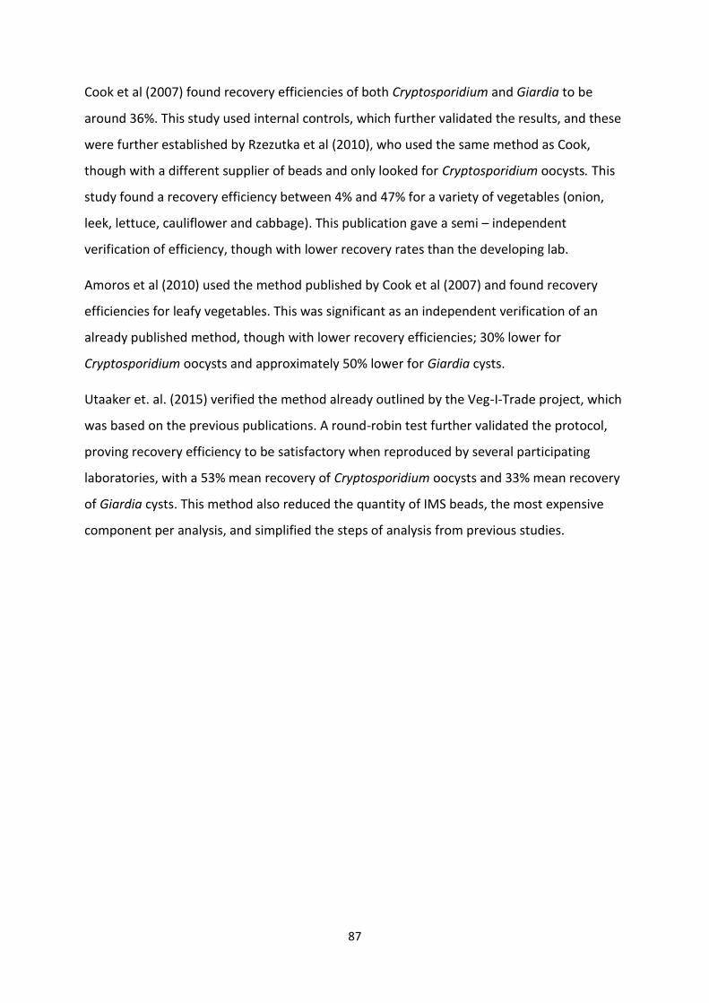
87
Cook et al (2007) found recovery efficiencies of both Cryptosporidium and Giardia to be
around 36%. This study used internal controls, which further validated the results, and these
were further established by Rzezutka et al (2010), who used the same method as Cook,
though with a different supplier of beads and only looked for Cryptosporidium oocysts. This
study found a recovery efficiency between 4% and 47% for a variety of vegetables (onion,
leek, lettuce, cauliflower and cabbage). This publication gave a semi – independent
verification of efficiency, though with lower recovery rates than the developing lab.
Amoros et al (2010) used the method published by Cook et al (2007) and found recovery
efficiencies for leafy vegetables. This was significant as an independent verification of an
already published method, though with lower recovery efficiencies; 30% lower for
Cryptosporidium oocysts and approximately 50% lower for Giardia cysts.
Utaaker et. al. (2015) verified the method already outlined by the Veg-I-Trade project, which
was based on the previous publications. A round-robin test further validated the protocol,
proving recovery efficiency to be satisfactory when reproduced by several participating
laboratories, with a 53% mean recovery of Cryptosporidium oocysts and 33% mean recovery
of Giardia cysts. This method also reduced the quantity of IMS beads, the most expensive
component per analysis, and simplified the steps of analysis from previous studies.
88
4.2 Diagnostic tools for detection of Cryptosporidium oocysts and Giardia cysts in
faeces
Many methods are available for diagnosing and detecting giardiasis and cryptosporidiosis, as
mentioned in the general presentation. These include direct visualisation of cysts,
trophozoites and oocysts in faeces, and indirect methods that identify Giardia and
Cryptosporidium antigens in faecal samples. at specialized laboratories samples may be
tested with immunofluorescence assay tests (IFAT) and/or molecular methods such as PCR.
4.3.1 Immunofluorescent antibody testing (IFAT)
Immunofluorescence antibody testing uses antibodies that bind specifically to an antigen, in
this case the same or similar surface wall glycoproteins on Giardia and Cryptosporidium
(oo)cysts as used for IMS. These antibodies have a pre-bound fluorescence molecule,
allowing them to be observed under the right wavelength of fluorescence. For this project, a
commercial test kit was used (Aqua-glo, Waterborne Inc., New Orleans).
4.3.2 DAPI staining (4´6-diamino-2-phenylindole)
DAPI visualizes nuclear DNA in both living and fixed cells, and can be used to determine the
number of nuclei and assess gross cell morphology, in combination with IFAT, these stains
are useful tools for determination of presence of Giardia cysts and Cryptosporidium oocysts
in a sample and their morphology.
Figure 11. Cryptosporidium oocysts visualised using IFAT
89
4.3 Viability assessment methods
Determination of viability of Cryptosporidium oocysts and Giardia cysts is a field of high
importance as it takes only a few infective (oo)cysts to establish infection. Dead (oo)cysts
have no public health significance, as they are not able to complete their life cycle. The
current standard methods for detection does not include viability testing or genotyping,
which is crucial knowledge in terms of establishing the potential for transmission or
outbreaks. Robertson and Gjerde (2007) concluded that the gold standard for determining
infectivity are bioassays with neonatal mice. However, this is an invasive method which
requires resources and sacrifice of animals, and there are additional methods including
simple dye inclusion and exclusion which should be further tested and approved for
standard use in a laboratory setting, making a simple, cheap and practical approach for
assessing infectivity. In view of public health, a (oo)cyst that is incapable of initiating
infection has as little public health significance as a dead and disintegrated (oo)cyst. A viable
(oo)cyst, defined as “capable of reproducing under appropriate conditions” (Robertson et al.
1998) can be infective or non-infective, and a non-viable (oo)cyst is always non-infective.
Different techniques have been developed for assessing the viability and/or infective
potential of Cryptosporidium oocysts and Giardia cysts:
Infectivity studies:
The neonatal mouse model has been used for assessing infectivity in disinfection studies,
and gnotobiotic pigs and human volunteers have been used for assessment of infective dose
and drug efficacy. This method is expensive, time-consuming and labour intensive, and strict
ethical standards must be met. Also, not all host species are suitable (e.g. C. hominis is not
infective to mice) and the extrapolation of results from an animal model to a human
situation must be evaluated carefully. In addition, infectivity studies are not suited for
assessing small numbers of (oo)cysts, and are therefore unsuitable for environmental
samples. Nonetheless, such studies are the only method providing definitive evidence of
infectivity of (oo)cysts that have been exposed to different environmental or chemical
pressures, and thus remains the golden standard (Robertson & Gjerde 2007).
90
Cell Culture
Cell culture models have been proposed as an alternative for the detection of infectious
Cryptosporidium oocysts. The method implies basically the in vitro excystation with
sporozoites enabled to invade cultured cell monolayers. Invasion can be assessed and
quantified by a variety of methods, such as PCR techniques, immunofluorescence and in situ
hybridisation. This is a technically demanding technique, and high variability has been
demonstrated, including unexpected stimulatory responses. (Rochelle et al. 2002; Rochelle
et al. 2005). Nonetheless, results under certain conditions have shown to be equivalent to
those from infectivity studies while being cheaper, faster and without ethical drawbacks, and
it can be applied to assess the infectivity of low numbers of oocysts (Di Giovanni &
LeChevallier 2005). However, the cell culture method alone cannot be used to enumerate
the number of oocysts present in environmental samples as it is not clear if the presence of
one infectious oocyst will infect one or more cells in the cell culture. Although cell culture
can detect infectious C. parvum and C. hominis, it is not certain which other Cryptosporidium
species can infect cell monolayers. Also, cell cultures cannot be used to assess the infectivity
of Giardia (Burnet 2012).
Detection of mRNA
mRNA detected from heat shock protein synthesis in viable (oo)cysts (Stinear et al. 1996).
The heat shock proteins are known to be synthesized in stressed organisms. Thus, when
(oo)cysts are exposed to a thermal shock, the induction of a heat shock response provides an
index of viability (Abbaszadegan et al. 1997). Decay of mRNA transcripts for β-tubulin
(Widmer et al. 1999), amyloglucosidase (Jenkins et al. 2000) and other markers have been
used similarly. In addition, as real-time PCR allows the continuous monitoring of amplicon
formation throughout the reaction, quantitative aspects could also be studied. This method
has also been compared to vital dye staining and found comparable in measuring viability
(Bertrand et al. 2009), but comparisons with gold-standard infectivity have only given
marginal correlation for some assays (Jenkins et al. 2002).
Fluorescent in situ hybridization
This method is based on the theory of rRNA breakdown following cell death. By targeting a
specific sequence of rRNA in Cryptosporidium oocysts and Giardia cysts, FISH probes are
91
designed only to label potentially infective or recently inactivated (oo)cysts (Dorsch & Veal
2001; Vesey et al. 1998). In situ hybridization seems to correlate well with in vitro
excystation, which though overestimates infective potential, and it seems not to correlate
well with cell cultures or infectivity studies(Jenkins et al. 2002). In addition, some studies
have indicated prolonged rRNA stability after oocyst death (Fontaine & Guillot 2003), and
research during the Sydney water enquiry demonstrated high FISH signals from oocysts
killed by radiation (McClellan 1998).
In vitro excystation
Various protocols have been described in which exposure to given conditions will activate
excystation in potentially infective (oo)cysts. It has also been used in combination with real-
time PCR to quantitatively estimate (oo)cyst viability (Bertrand et al. 2009). However, in vitro
excystation requires relatively high numbers of (oo)cysts (Robertson & Gjerde 2007; Sauch
1991) and it is considered to be a subjective method due to the presence of partially
emerged trophozoites and sporozoites (Labatiuk et al. 1991). It may be useful for supporting
results obtained from other assays.
Vital dye inclusion/exclusion
Vital dye techniques developed by (Campbell et al. 1992; Schupp & Erlandsen 1987) are the
ones most frequently used, and includes inclusion or exclusion of (oo)cysts stained with DAPI
(4`, 6-diamidino-2-phenylindole) and PI (Propidium iodide), and differential interference
contrast microscopy (DIC) for assessing viability of individual (oo)cysts. DAPI stains the
nucleus sky blue, while PI can only pass through damaged cell membranes and intercalates
with the nucleic acids of injured and dead cells to form a bright red fluorescent complex. An
intact cyst wall may be insufficient to induce an infection in the host (Bertrand et al. 2009),
and this technique has been found to frequently overestimate infective potential of
(oo)cysts. On the other hand, this is a cheap and readily implemented method, and can be
applied a small number of, or even single (oo)cysts, and can divide the parasites into
different categories based on staining and content of the (oo)cyst when combined with
sophisticated optics. Due to the simplicity and availability of vital dye techniques, this
method was used to assess viability of Cryptosporidium oocysts and Giardia cysts on lettuce
in this thesis.
92
4.3.3. Molecular tools for detection of Cryptosporidium and Giardia
Molecular tools for genotyping and subtyping gives a deeper insight into host specificity of
Cryptosporidium species and subspecies as well as Giardia Assemblages and subassemblages
and their transmission. Identification of species infecting humans and animals and the
proportion of infection attributable to each species in various geographic, epidemiologic and
socioeconomic settings is a valuable tool in unravelling foci of infection, such as infection or
contamination sources and characterization of transmission dynamics in communities.
In the polymerase chain reaction (PCR), primers complementary to Giardia and
Cryptosporidium gene sequences are used together with DNA polymerases that will amplify
small amounts of Giardia or Cryptosporidium DNA present in the sample, to enable
detection. Both generic and genotype specific primers and protocols have been published.
Conventional PCR often ends with running the samples on an agarose gel by electrophoresis,
and visualizing bands of DNA with different types of staining, e.g. cyanine dyes. The size of
the product is determined with a ladder that is loaded simultaneously with the samples.
Different methods have been developed for real time identification and quantification of
DNA, and by adding fluorescently labelled nucleotide probes that are complementary to the
amplified sequence it is also possible to distinguish between different genotypes (Elwin et al.
2014). Genotyping is more commonly performed in relation to research, as it may provide
more certain answers regarding epidemiology, focus of infection and source tracking.
93
4.4 Material and methods used in this study
The Materials and Methods described in this section are divided into two sections. The first
part is concerned with the Materials and Methods used in the lab-based experimental part
of the study, in which all the work was conducted in the Parasitology Laboratory at NMBU.
This includes the work described in Papers I and II.
The second part is concerned with the Materials and Methods concerned with the surveys
conducted in India, and describes the sampling sites and sample collection methods, the lab
work conducted in the Parasitology Laboratory at PGIMER, and the lab work associated with
the survey work conducted in the Parasitology Laboratory at NMBU (referring sometimes to
the Materials and Methods described in Section 1 – Experimental Studies).
Statistical approaches used are described at the end of this section and apply to both
experimental work (Section 1) and survey work (Section 2).
4.4.1 Section 1 – Experimental Work
Source of Cryptosporidium oocysts and Giardia cysts used in experimental studies
1) Positive control Cryptosporidium oocysts and Giardia cysts in buffered mixed
aldehyde solution for use with Waterborne Kits produced by Waterborne Inc., New Orleans,
LA, USA was used for the initial spiking of vegetable and water samples when these
protocols were incorporated into the thesis work.
2) Pre-counted Cryptosporidium oocysts and Giardia cysts from EasySeed™ produced by
Biomérieux Industry was used for the interlaboratory trials when establishing the reduced-
cost approach, as well as establishing the recovery rates for the water sample analysis.
3) For the viability studies, Cryptosporidium parvum oocysts isolated from faeces of
naturally infected calves by salt flotation by Hønsvall and Robertson (2017) and live Giardia
cysts belonging to Assemblage B from the H3 isolate (gerbil source) suspended in PBS
ordered from Waterborne Inc.
Enumeration of Cryptosporidium and Giardia
For the live Giardia cysts and Cryptosporidium oocysts used in the viability studies, the
94
(oo)cysts were diluted into individual stock solutions. 10 µL aliquots from the stock was
pipetted on a multispot microscope slide (C.A. Hendley (Essex) LTD), stained with a
monoclonal antibody (mAb; Aqua-glo, Waterborne Inc, New Orleans, USA) before
enumeration and further dilution.
Spiking studies
In the experimental studies where recovery of Cryptosporidium oocysts and Giardia cysts
were evaluated, the (oo)cysts from EasySeed™ were already counted by the manufacturer.
When inter-laboratory trials were initiated, in order to save costs, the spikes sent were
equivalent to 50 % EasySeed™ spikes that had been prepared in-house. This was done by
shaking individual EasySeed™ spike vials vigorously and then dividing into two aliquots, each
containing 50 % of the original spike.
Thus, each participant was sent a seeding sample containing approximately a 50 %
EasySeed™ size spike and an empty EasySeed tube that had been cleaned by soaking in 15 %
sodium hypochlorite overnight, then washed in hot soapy water three times, and then
rinsed, and contained only water (same volume as for the spike) – that is a negative control.
None of the participants were aware that only one sample contained parasites.
When the method for water samples was assessed, only in-house laboratory trials was done,
and EasySeed™ (oo)cysts were used. This was assessed by performing the method according
to the ISO-standard for water analysis, but replacing the reagents as described for the
reduced-cost approach.
For the viability assessments, the matrix to be evaluated, in this case lettuce leaves, were
sprinkled with approximately 50 000 Giardia cysts and 50 000 Cryptosporidium oocysts. The
(oo)cysts, diluted in distilled water from stock solution to a volume of 100 µl, were spread on
the lettuce leaves in aliquots of 20 µl using a pipette.
95
Assessment of viability
Viability was assessed based on morphology and inclusion and exclusion of the vital dyes
4’,6-diamino-2-phenylindole (DAPI) and propidium iodide (PI), as described by Campbell et
al., (1992). In order to identify the parasites eluted from the lettuce, a monoclonal antibody
(mAb; Aqua-glo, Waterborne Inc. New Orleans, USA) was added to the suspension in the
final 15 minutes of the staining procedure. The stained sediment was examined in
suspension (with the cover slip on the microscope sealed with nail varnish to avoid drying)
by fluorescence microscopy using a Leica DMCB microscope equipped with a UV filter block
(350-nm excitation, 450-nm emission) for DAPI and a green filter block (500-nm excitation,
630-nm emission) for PI. Nomarski (differential interference contrast) optics on the same
microscope was used to examine morphology of individual cysts and oocysts.
The cysts and oocysts were evaluated and categorised according to exclusion or inclusion of
the different stains and their morphology. Empty or shrunken (ghost) cysts and oocysts were
identified under Nomarski optics, containing no nuclei or shrunken residues thereof. They
were also non-refractile, apart from the residual body when present.
PI+ cysts and oocysts fluoresce bright red under the green filter block; this fluorescence
varies from distinct points corresponding to the sporozoite / trophozoite nuclei, to a more
diffuse fluorescence within the cyst or oocyst. Cysts and oocysts were categorised as DAPI+,
PI- if they did not include PI (as described above), but the nuclei of the sporozoites/
trophozoites fluoresced a distinctive sky blue under the UV filter block. Cysts and oocysts
that were neither PI+, nor “ghosts”, and which showed either rim fluorescence or absence
thereof under UV filter block were considered DAPI-, PI-. The interpretation of these various
appearances is described in Table 3. For the purposes of this study, DAPI+, PI- parasites and
DAPI-, PI- parasites were summed together as viable or potentially viable.
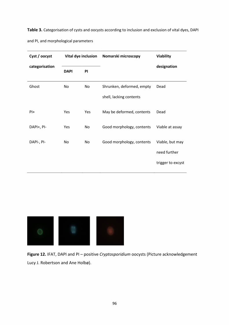
96
Table 3. Categorisation of cysts and oocysts according to inclusion and exclusion of vital dyes, DAPI
and PI, and morphological parameters
Cyst / oocyst
categorisation
Vital dye inclusion Nomarski microscopy Viability
designation DAPI PI
Ghost No No Shrunken, deformed, empty
shell, lacking contents
Dead
PI+ Yes Yes May be deformed, contents Dead
DAPI+, PI- Yes No Good morphology, contents Viable at assay
DAPI-, PI- No No Good morphology, contents Viable, but may
need further
trigger to excyst
Figure 12. IFAT, DAPI and PI – positive Cryptosporidium oocysts (Picture acknowledgement
Lucy J. Robertson and Ane Holbø).
97
4.4.2 Section 2 – Survey Work
Study area
Chandigarh is a city located northwest in India. It is the joint capital of its neighbouring
states, Punjab and Haryana, and is a relatively new dwelling, as its construction started in
the early 1950s. It is situated at the foot of the Shivalik hills, and is known as the “city
beautiful”, as it is one of the greenest cities of India, with many parks and recreational areas,
and the services provided by Chandigarh Administration and the Municipal corporation are
considered as a role model for other urban cities. Chandigarh is also one of the fastest
growing cities in India, with a current population of over a million inhabitants (JNNURM
2006).
The city was designed by a French architect, LeCorbusier, in the 1950s, and reflects the
modernism movement that arose in Europe around that time. The city’s outline is
considered as head, lungs, heart and limbs, but it seems that these organs of bricks and
humans are currently undergoing an uncontrollable hypertrophy. The original plan was to
divide the city into phases, and these phases are further divided into sectors. Sectors 1 to 30
were designed for a population of 150 000 inhabitants, and this area is referred to as phase
I. Sectors 31 to 47, phase II, and were supposed to house 350 000, meaning the population
density in this area would be 4 times higher than phase I. Now, with the population increase,
phase III is on the rise, and this is also an area with a planned higher density than those of
the previous two. The rise in population has given the “city beautiful” quite a challenge in
terms of the rehabilitation of slum colonies, presence of urban villages with unregulated
growth in the planned sectors, the presence of unauthorised settlements, limited land for
future growth and development, as well as how to address the problems of waste
management, water supply, and general infrastructure in a city with an accelerated
population growth. These are a few of the many trials for a city with a population of the very
rich to the very poor, and which seems to be growing into a fractal city pattern of economic
and social differences (Chandigarh Administration 2016).
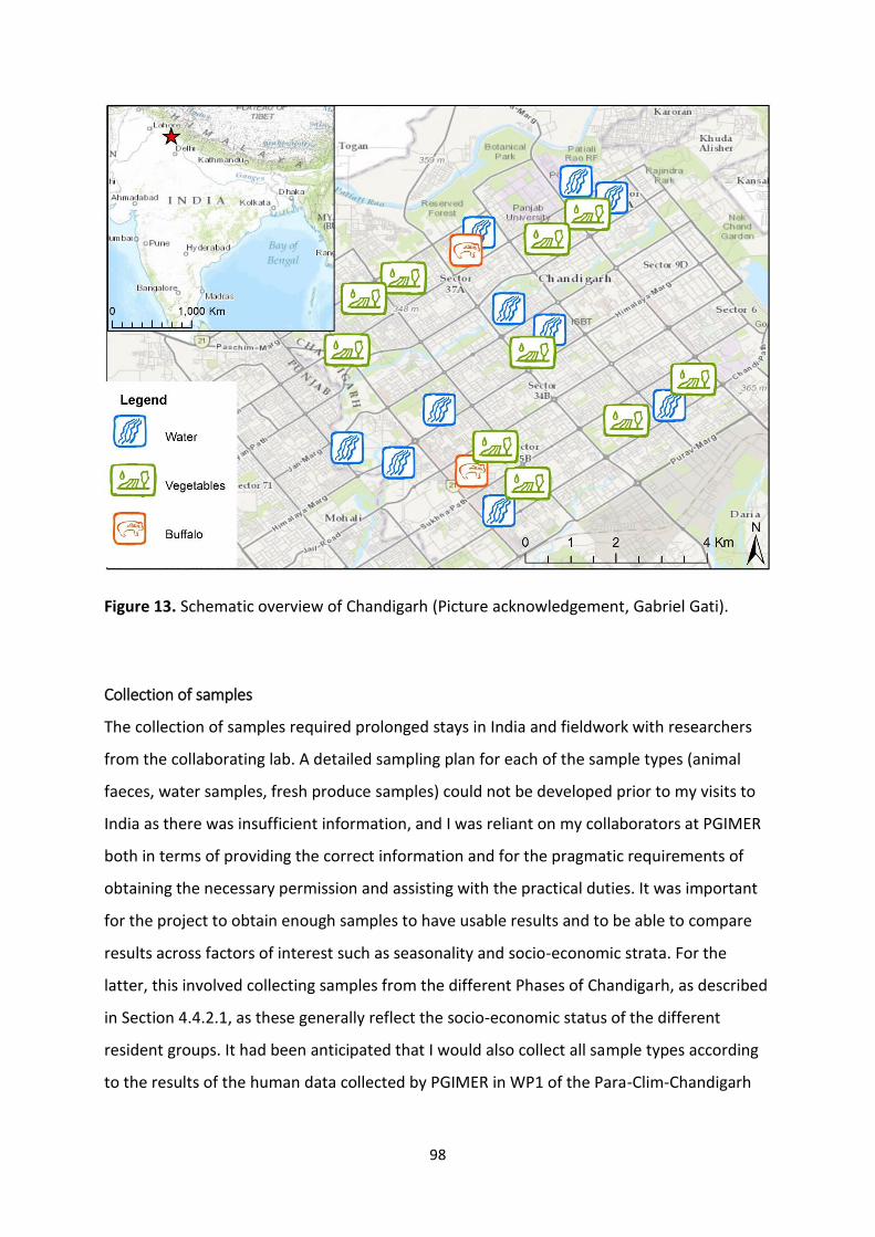
98
Figure 13. Schematic overview of Chandigarh (Picture acknowledgement, Gabriel Gati).
Collection of samples
The collection of samples required prolonged stays in India and fieldwork with researchers
from the collaborating lab. A detailed sampling plan for each of the sample types (animal
faeces, water samples, fresh produce samples) could not be developed prior to my visits to
India as there was insufficient information, and I was reliant on my collaborators at PGIMER
both in terms of providing the correct information and for the pragmatic requirements of
obtaining the necessary permission and assisting with the practical duties. It was important
for the project to obtain enough samples to have usable results and to be able to compare
results across factors of interest such as seasonality and socio-economic strata. For the
latter, this involved collecting samples from the different Phases of Chandigarh, as described
in Section 4.4.2.1, as these generally reflect the socio-economic status of the different
resident groups. It had been anticipated that I would also collect all sample types according
to the results of the human data collected by PGIMER in WP1 of the Para-Clim-Chandigarh
99
project; however, due to the delay in PGIMER conducting their work, I had to collect my
samples without taking their data collection and results into consideration.
As PGIMER does not routinely analyse drinking water or fresh produce for parasitic protozoa,
samples were partly processed and analysed at PGIMER in India, then shipped to Norway for
completion of the analyses at the Parasitology Lab at NMBU. This meant that samples had to
be preserved to ensure that they could be successfully and safely shipped between India and
Norway.
Animal faeces, collection and storage of samples prior to shipment
As contact with animal faeces in India is traditionally a job for the so-called “untouchables”,
the Dalits, which are the lowest caste in India and who have traditionally been stigmatized
and isolated, collection of faecal samples proved to be initially difficult to approach.
Although the caste system has officially been abolished, in practicality it still exists in some
areas, and the social stigma of touching animal faeces continues.
Faecal samples from goats, cattle, buffaloes, and dogs were collected from farms,
households, parks and wasteland. The samples were collected non-invasively either from the
animal or directly from the ground as fresh, morphologically consistent stools in some study
areas. For samples taken from animals, only one sample per animal was obtained due to
practicality and logistics. For samples collected from the ground, it was not always possible
to determine whether the samples were from different animals or repeated faecal samples
from the same animal or group of animals.
Samples were put directly into containers and transported to the laboratory at PGIMER
within maximum two hours. Here they were immersed in 2.5 % potassium dichromate, a
cheap preservative which allows future molecular diagnostics, for preservation and before
shipment and processing. Various storage media and approaches have been associated with
lack of PCR amplification. Potassium dichromate was chosen as preservative for faecal
samples in this study, as they seem to keep both Giardia cysts and Cryptosporidium oocysts
intact and preserves DNA well during storage (Wilke & Robertson 2009).
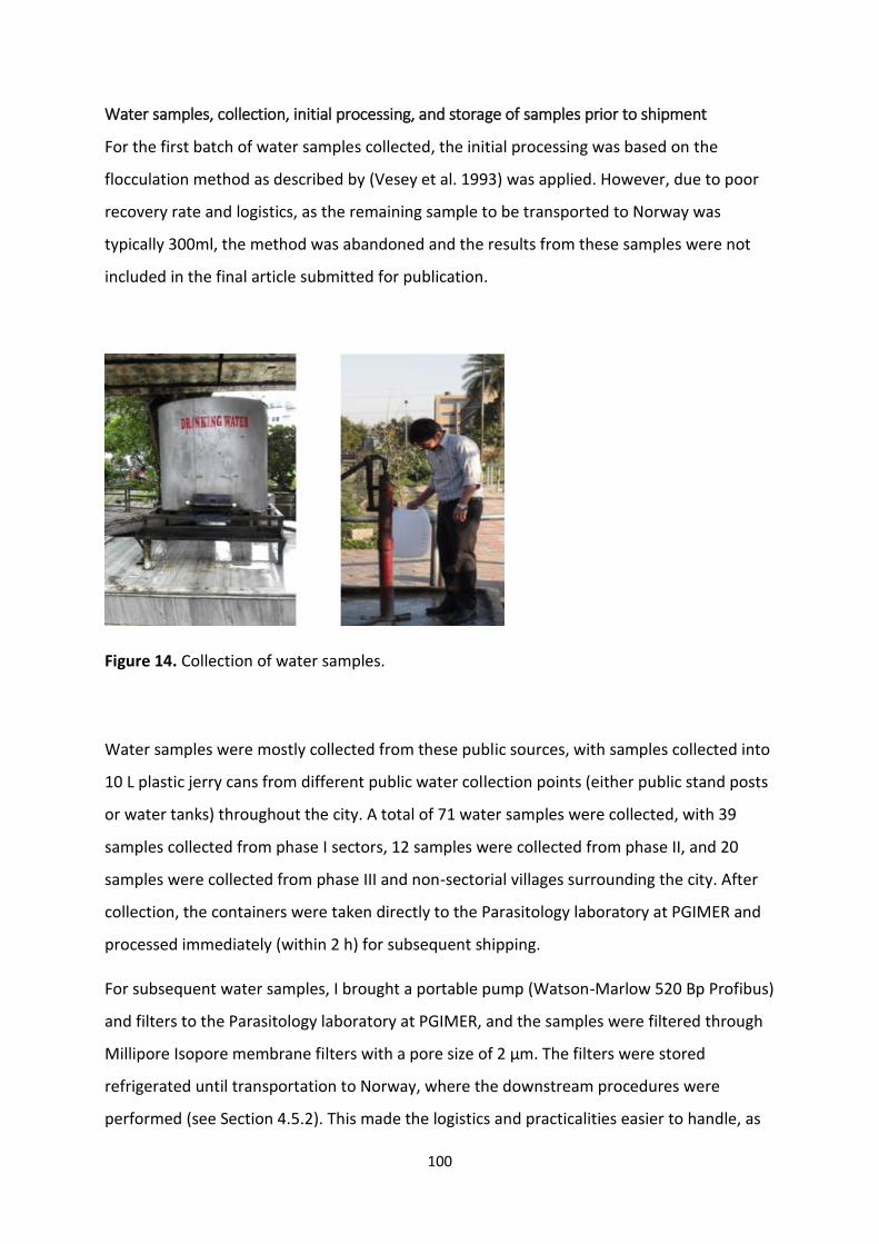
100
Water samples, collection, initial processing, and storage of samples prior to shipment
For the first batch of water samples collected, the initial processing was based on the
flocculation method as described by (Vesey et al. 1993) was applied. However, due to poor
recovery rate and logistics, as the remaining sample to be transported to Norway was
typically 300ml, the method was abandoned and the results from these samples were not
included in the final article submitted for publication.
Figure 14. Collection of water samples.
Water samples were mostly collected from these public sources, with samples collected into
10 L plastic jerry cans from different public water collection points (either public stand posts
or water tanks) throughout the city. A total of 71 water samples were collected, with 39
samples collected from phase I sectors, 12 samples were collected from phase II, and 20
samples were collected from phase III and non-sectorial villages surrounding the city. After
collection, the containers were taken directly to the Parasitology laboratory at PGIMER and
processed immediately (within 2 h) for subsequent shipping.
For subsequent water samples, I brought a portable pump (Watson-Marlow 520 Bp Profibus)
and filters to the Parasitology laboratory at PGIMER, and the samples were filtered through
Millipore Isopore membrane filters with a pore size of 2 µm. The filters were stored
refrigerated until transportation to Norway, where the downstream procedures were
performed (see Section 4.5.2). This made the logistics and practicalities easier to handle, as
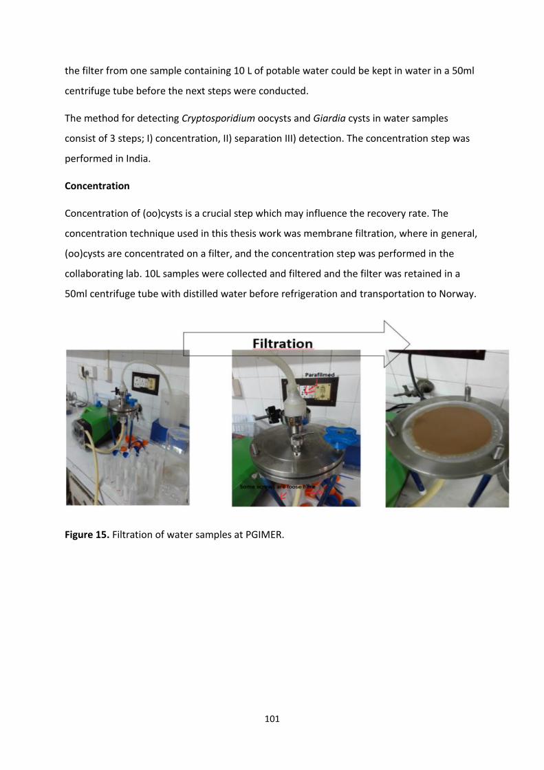
101
the filter from one sample containing 10 L of potable water could be kept in water in a 50ml
centrifuge tube before the next steps were conducted.
The method for detecting Cryptosporidium oocysts and Giardia cysts in water samples
consist of 3 steps; I) concentration, II) separation III) detection. The concentration step was
performed in India.
Concentration
Concentration of (oo)cysts is a crucial step which may influence the recovery rate. The
concentration technique used in this thesis work was membrane filtration, where in general,
(oo)cysts are concentrated on a filter, and the concentration step was performed in the
collaborating lab. 10L samples were collected and filtered and the filter was retained in a
50ml centrifuge tube with distilled water before refrigeration and transportation to Norway.
Figure 15. Filtration of water samples at PGIMER.
102
Vegetable samples, collection, initial processing, and storage of samples prior to shipment
Vegetables are sold at different markets in Chandigarh, which are arranged at different
weekdays for individual sectors. Street vendors usually have a push-cart or a cart connected
to a bike, which enables them to roam different areas of the city in search of customers, and
supermarkets are unevenly dispersed throughout the city. Samples for this study was bought
directly from these retailers, put in a plastic bag and transported to the laboratory for
further processing.
Vegetable samples
The principle of analysing vegetable samples is quite similar to that of water samples, and
can be divided into four stages; I) Elution, II) concentration, III) Isolation and IV) detection.
Only step I) will be discussed here in detail, as step II), III) and IV) are equivalent to the water
protocol.
Elution
Elution is the removal of parasites from the surface of the vegetable into a liquid medium
that can be more readily manipulated. Vegetables were put into filtered stomacher bags and
eluted with 200 ml 1M Glycine buffer, a buffer readily available and easy to make on-site.
The stomacher bag was then manipulated for 4 minutes by hand, as no stomacher was
available in the lab, and the eluate transferred into 5 centrifuge tubes of 50 ml each, as the
stomacher bag was rinsed with 50ml distilled water to improve parasite recovery. The tubes
were centrifuged at 1550 rfg for 10 minutes, and the supernatant was removed using a
sterile 10ml plastic syringe. The remaining pellets were vortexed and combined per sample
and stored at 4°C before transportation to Norway.
103
4.5 Completion of sample processing at the Parasitology Lab at NMBU
Samples arriving at NMBU were kept refrigerated until processing. All samples were
processed as soon as possible after arrival.
4.5.1 Detection of Cryptosporidium oocysts and Giardia cysts in faecal samples
The samples preserved in potassium dichromate were rinsed with phosphate buffer before
further processing. Canine, bovine and goat faecal samples were processed by McMasters
for analysis for helminths. 3 grams of faeces was homogenized with distilled water (57 ml
per 3 grams) with a blender. After sieving through 125 µm porosity steel mesh, allowing the
parasite eggs and (oo)cysts to move through while removing larger particulates, the
suspension was poured into plastic tubes and centrifuged at 3000 rpm for 3 minutes after
which the supernatant was decanted. The tube was added flotation solutions, either NaCl or
NaCl/ZnCl salts, which due to their density gradient allows parasite eggs and (oo)cysts to
float to the top while debris sinks to the bottom, creating a plane to focus on. The solution
was pipetted into a McMaster counting chamber, which allows for quantitative evaluation of
the number of eggs / oocysts per gramme of faeces.
Using a plastic loop, 20µl of sample material from the concentrated pellet was placed in a
microscope slide, dried and fixed with methanol. After the methanol dried off, 10-20 µl of
FITC conjugated monoclonal antibodies (Waterborne Inc., USA) were applied and the slides
incubated in a humidifier at 37°C for 30 minutes. Slides were investigated under a
fluorescent microscope (Leica MLD) with filters for viewing FITC.
To increase the yield of Cryptosporidium oocysts and Giardia cysts, pelleted goat feces was
run by an IMS protocol, which significantly improved the recovery of parasites compared to
the traditional smear (Utaaker 2017, unpublished results). In addition, retrieving (oo)cysts
from IMS has proven to give more positive results when DNA is extracted and PCR
performed (Coklin et al. 2011).
104
4.5.2 Methods for isolation of Cryptosporidium and Giardia from water and vegetable samples.
The methods used for analysing water and vegetable samples are derived from the ISO-
standards, with modifications to reduce the costs. The modifications were made in terms of
reducing some of the reagents and replacing other with cheaper options. Both methods
have the basic concepts of filtration, either through micropores in the case of water, or
filtered stomacher bags filled with elution buffer for vegetables, then concentration for
creation of a pellet with concentrates of debris from the samples, and immunomagnetic
separation using magnetic beads to separate parasites from debris, following the final step
of examination using fluorescent dye and DAPI staining.
Concentration of water samples in Norway
In Norway, the filter was washed with membrane buffer as described in method
15553:2006, and the eluate was centrifuged at 1550 rfg for 10 minutes to create a pellet
from water debris.
Preparation of concentrated vegetable samples before separation
The centrifuge tubes containing the concentrated eluate was added SDS before vortexing
and centrifugation at 1550 rfg for 10 minutes. The supernatant was removed using an
aspirator and the pellet was transferred to an L10 tube together with buffers and magnetic
beads. The procedure is further described in section 4.1.1.
Separation of parasites from water and vegetable samples
The separation procedure was performed using immunomagnetic separation (IMS), which
involves the attachment of (oo)cysts with magnetic beads coated with parasite-specific
monoclonal antibodies. The continuous mixing of beads in the water sample causes
separation of (oo)cysts present in the samples from debris.
The pellet was resuspended into a L10 tube and filled with buffers and magnetic beads, and
the tube was rotated for approximately one hour in order to create a cyst/oocyst complex
with the beads. The beads were washed into an Eppendorf tube, and the complexes were
dissociated using 0.1M HCl and vigorous vortexing. After dissociation, the remaining solution
was pipetted onto a hydrophobic welled slide where NaOH had been added to neutralize the
pH, and the slide was left to dry overnight.
105
Microscopy and detection of Cryptosporidium oocysts and Giardia cysts from water and
vegetable samples
Dried samples were fixed with methanol and stained with FITC-conjugated monoclonal
antibodies (mAbs) against Cryptosporidium oocyst walls and Giardia cyst walls (Aqua-gloTM,
WaterborneTM Inc, USA) and nuclei were stained with the fluorogenic DNA intercalator 4´, 6
diamidino-2-phenylindole (DAPI) according to Smith et al., (2002). Samples were mounted
with M101 No-Fade Mounting Medium, then each slide was covered by a glass coverslip and
viewed promptly by fluorescent microscopy.
Microscopy was performed on a Leica DCMB microscope (x 20, x 40 and x100 objectives),
equipped with Nomarski differential interference contrast (DIC) optics. A blue filter block
(480 nm – excitation, 520 nm – emission) was used for the detection of cysts and oocysts
labelled with FITC-conjugated mAbs, and a UV filter block (350-nm excitation, 450-nm
emission) was used for investigating DAPI-staining.
Each well was scanned systematically in an up-and-down or side-to-side manner at 20X, and
Cryptosporidium oocysts and Giardia cysts were enumerated. When brilliant apple-green
fluorescing ovoid or spherical objects within the appropriate size range for Cryptosporidium
and Giardia were observed, magnification was increased to 40X, and the UV filter block was
used for visualization of DAPI staining. Each (oo)cyst was recorded as DAPI-negative or DAPI-
positive according to the presence of internal light blue staining.
Nomarski (DIC) objectives were used to examine morphological characteristics of the
(oo)cysts.
A sample was considered positive if the (oo)cyst(s) exhibited typical fluorescence, with
correct shape and size, and being DAPI-positive. If internal contents were lacking, but the
morphometry was correct and the structure had a typical fluorescence, the (oo)cysts were
described as “putative”, as they lacked sufficient characteristics for definitive identification.
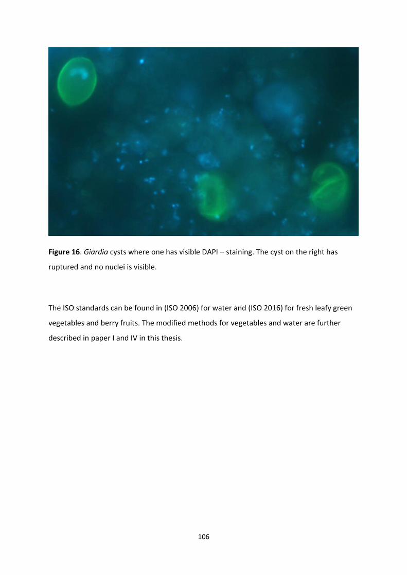
106
Figure 16. Giardia cysts where one has visible DAPI – staining. The cyst on the right has
ruptured and no nuclei is visible.
The ISO standards can be found in (ISO 2006) for water and (ISO 2016) for fresh leafy green
vegetables and berry fruits. The modified methods for vegetables and water are further
described in paper I and IV in this thesis.
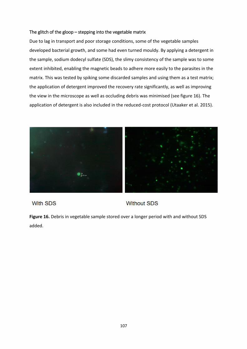
107
The glitch of the gloop – stepping into the vegetable matrix
Due to lag in transport and poor storage conditions, some of the vegetable samples
developed bacterial growth, and some had even turned mouldy. By applying a detergent in
the sample, sodium dodecyl sulfate (SDS), the slimy consistency of the sample was to some
extent inhibited, enabling the magnetic beads to adhere more easily to the parasites in the
matrix. This was tested by spiking some discarded samples and using them as a test matrix;
the application of detergent improved the recovery rate significantly, as well as improving
the view in the microscope as well as occluding debris was minimised (see figure 16). The
application of detergent is also included in the reduced-cost protocol (Utaaker et al. 2015).
Figure 16. Debris in vegetable sample stored over a longer period with and without SDS
added.
108
4.6. Molecular methods
4.6.1 DNA isolation of faecal samples
Disruption of the oocysts and cysts is performed to make DNA more available for extraction.
Techniques that can be used may include freeze-thawing, boiling, bead-beating and lysis
(Checkley et al, 2015).
For Cryptosporidium oocysts and Giardia cysts, DNA was isolated using a QIAmp mini kit
(Qiagen GmbH). This was performed either directly on the faecal pellet, on purified (oo)cysts
after IMS. The protocols followed the manufacturers` instructions with slight modifications;
cysts / oocysts were first mixed with 150 µl of TE buffer (100mM Tris and 100 mM EDTA) and
incubated at 90°C for Giardia cysts and 100°C for Cryptosporidium oocysts for 1 hour before
an overnight proteinase K lysis step at 56°C and spin column purification. DNA was finally
eluted in 30 µl of PCR-grade water and stored at 4°C.
4.6.2 DNA isolation from water and vegetable samples
Following microscopy, Cryptosporidium oocysts and Giardia cysts were retrieved from
positive slides and DNA was prepared according to Robertson et al. (2009). Briefly, the
coverslip from each slide was carefully removed and retained, whilst 25 µl aliquots of AL lysis
buffer (Qiagen GmbH, Germany) were added to the slide wells, which were then scraped
using a sterile scalpel blade. The buffer and scrapings were pipetted into a microcentrifuge
tube. This process was repeated four times, and then the coverslip was replaced onto the
slide that was then re-screened. For each slide no cyst or oocysts could be detected after the
procedure.
The contents of each microcentrifuge tube containing slide scrapings were re-suspended in
Tris-EDTA buffer and held at 100°C for Cryptosporidium oocysts and 90°C for Giardia cysts
for one hour, before the DNA was isolated using QIamp DNA mini kit (Qiagen GmbH), using
an overnight step at 56°C.
109
4.6.3 Conventional polymerase chain reaction (PCR)
PCR may allow detection of very small quantities of parasite DNA within a sample. As it
generates many copies of the target DNA, the result can then be sequenced, and this genetic
code used to characterise an isolate and determine its taxonomy and phylogenetic
relationship to other isolates (Lymbery and Thompson, 2012).
The PCR reaction occurs in a welled chamber where two primer sets, which bind specifically
the target DNA; RNAse – free water, bovine serum albumin (BSA), which stabilises the
enzymes and reactions, DNA polymerase, which is the enzyme creating the copies of DNA,
and a mixture of DNA nucleotides. PCR products are visualised after separation across a 2%
agarose gel using electrophoresis, and can be compared with a DNA ladder to determine
fragment lengths.
For Cryptosporidium, use of the first generation genotyping tools has decreased as the
techniques have become more specific to target species, though one of these targets, the
Cryptosporidium oocyst wall protein (COWP) is still used as a confirmative genotyping
method due to the robustness of the technique. The small subunit ribosomal ribonucleic acid
(SSU rRNA) gene is a widely used target in PCR, and can be used to distinguish between
species, which can then be subtyped at the 60 kDA glycoprotein (gp60) gene in the case of C.
parvum and C. hominis (Checkley et al. 2015; Shirley et al. 2012).
For Giardia, four commonly targeted genes for molecular characterisation of an isolate were
utilised in these studies; Small subunit ribosomal ribonucleic acid (SSU rRNA), beta-giardin
(bg), glutamate dehydrogenase (gdh) and triosephosphate isomerase (tpi). These genes vary
in the substitution rate per nucleotide, thus their ability to distinguish isolates at different
levels of genetic detail vary from SSU rRNA being useful to distinguish species, to the more
heterogenous tpi gene where isolates at sub-assemblage levels can be typed (Feng & Xiao
2011).
Positive control for molecular methods were obtained by isolating DNA from
Cryptosporidium parvum oocyst isolated by Hønsvall and Robertson (2017) and Holbø
(2017), and Giardia cysts belonging to Assemblage B from the H3 isolate (gerbil source)
suspended in PBS ordered from Waterborne Inc.
PCR protocols used in this study are given in Table 4.
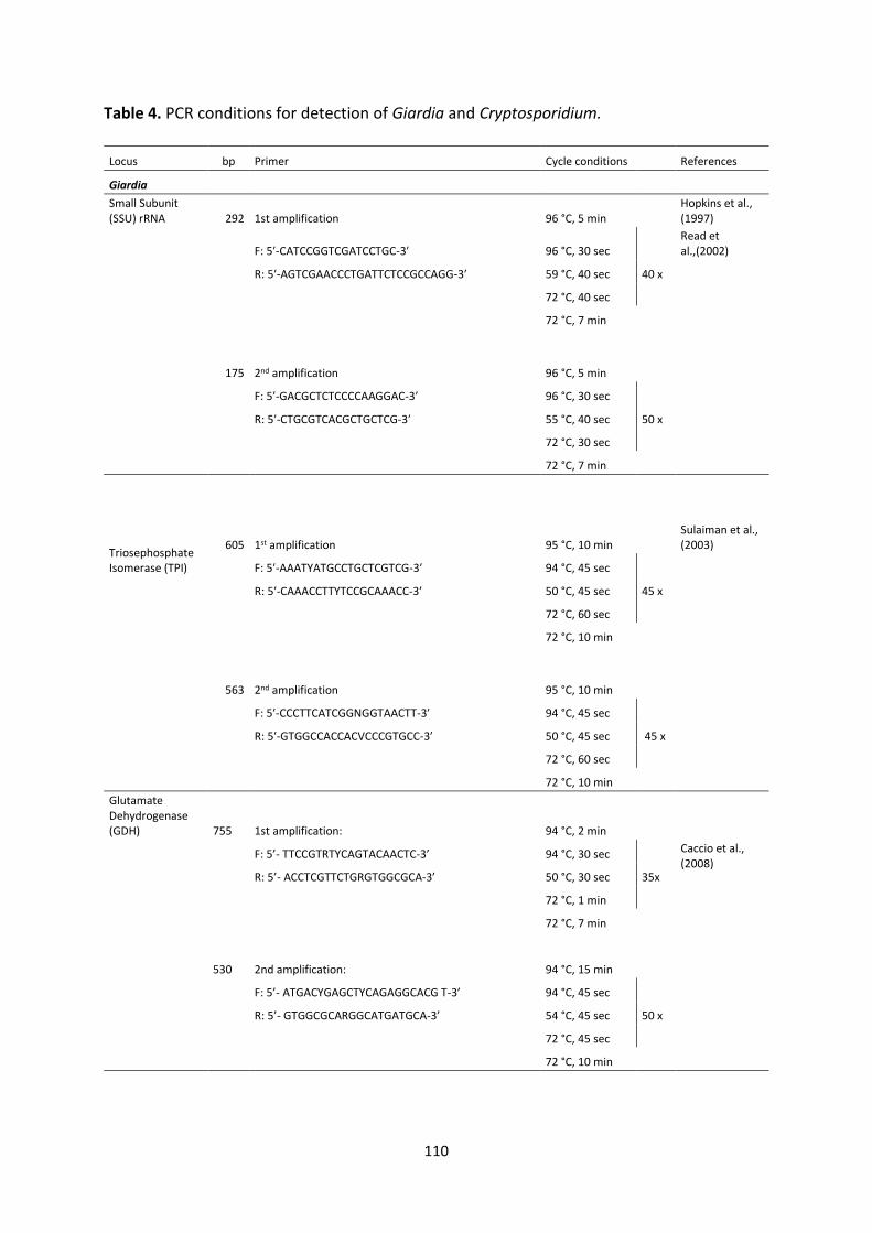
110
Table 4. PCR conditions for detection of Giardia and Cryptosporidium.
Locus bp Primer Cycle conditions References
Giardia
Small Subunit (SSU) rRNA 292 1st amplification 96 °C, 5 min
Hopkins et al., (1997)
F: 5′-CATCCGGTCGATCCTGC-3′ 96 °C, 30 sec Read et al.,(2002)
R: 5′-AGTCGAACCCTGATTCTCCGCCAGG-3′ 59 °C, 40 sec 40 x
72 °C, 40 sec
72 °C, 7 min
175 2nd amplification 96 °C, 5 min
F: 5′-GACGCTCTCCCCAAGGAC-3′ 96 °C, 30 sec
R: 5′-CTGCGTCACGCTGCTCG-3′ 55 °C, 40 sec 50 x
72 °C, 30 sec
72 °C, 7 min
Triosephosphate Isomerase (TPI)
605 1st amplification 95 °C, 10 min Sulaiman et al., (2003)
F: 5′-AAATYATGCCTGCTCGTCG-3′ 94 °C, 45 sec
R: 5′-CAAACCTTYTCCGCAAACC-3′ 50 °C, 45 sec 45 x
72 °C, 60 sec
72 °C, 10 min
563 2nd amplification 95 °C, 10 min
F: 5′-CCCTTCATCGGNGGTAACTT-3′ 94 °C, 45 sec
R: 5′-GTGGCCACCACVCCCGTGCC-3′ 50 °C, 45 sec 45 x
72 °C, 60 sec
72 °C, 10 min
Glutamate Dehydrogenase (GDH) 755 1st amplification: 94 °C, 2 min
F: 5’- TTCCGTRTYCAGTACAACTC-3’ 94 °C, 30 sec Caccio et al., (2008)
R: 5’- ACCTCGTTCTGRGTGGCGCA-3’ 50 °C, 30 sec 35x
72 °C, 1 min
72 °C, 7 min
530 2nd amplification: 94 °C, 15 min
F: 5’- ATGACYGAGCTYCAGAGGCACG T-3’ 94 °C, 45 sec
R: 5’- GTGGCGCARGGCATGATGCA-3’ 54 °C, 45 sec 50 x
72 °C, 45 sec
72 °C, 10 min
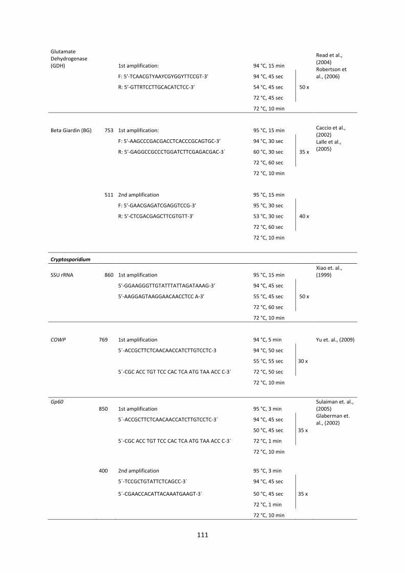
111
Glutamate Dehydrogenase (GDH) 1st amplification: 94 °C, 15 min
Read et al., (2004) Robertson et al., (2006) F: 5′-TCAACGTYAAYCGYGGYTTCCGT-3′ 94 °C, 45 sec
R: 5′-GTTRTCCTTGCACATCTCC-3′ 54 °C, 45 sec 50 x
72 °C, 45 sec
72 °C, 10 min
Beta Giardin (BG) 753 1st amplification: 95 °C, 15 min Caccio et al., (2002) Lalle et al., (2005)
F: 5′-AAGCCCGACGACCTCACCCGCAGTGC-3′ 94 °C, 30 sec
R: 5′-GAGGCCGCCCTGGATCTTCGAGACGAC-3´ 60 °C, 30 sec 35 x
72 °C, 60 sec
72 °C, 10 min
511 2nd amplification 95 °C, 15 min
F: 5′-GAACGAGATCGAGGTCCG-3′ 95 °C, 30 sec
R: 5′-CTCGACGAGCTTCGTGTT-3′ 53 °C, 30 sec 40 x
72 °C, 60 sec
72 °C, 10 min
Cryptosporidium
SSU rRNA 860 1st amplification 95 °C, 15 min Xiao et. al., (1999)
5′-GGAAGGGTTGTATTTATTAGATAAAG-3′ 94 °C, 45 sec
5′-AAGGAGTAAGGAACAACCTCC A-3′ 55 °C, 45 sec 50 x
72 °C, 60 sec
72 °C, 10 min
COWP 769 1st amplification 94 °C, 5 min Yu et. al., (2009)
5´-ACCGCTTCTCAACAACCATCTTGTCCTC-3 94 °C, 50 sec
5´-CGC ACC TGT TCC CAC TCA ATG TAA ACC C-3`
55 °C, 55 sec 30 x
72 °C, 50 sec
72 °C, 10 min
Gp60 850 1st amplification 95 °C, 3 min
Sulaiman et. al., (2005) Glaberman et. al., (2002)
5´-ACCGCTTCTCAACAACCATCTTGTCCTC-3` 94 °C, 45 sec
5´-CGC ACC TGT TCC CAC TCA ATG TAA ACC C-3`
50 °C, 45 sec 35 x
72 °C, 1 min
72 °C, 10 min
400 2nd amplification 95 °C, 3 min
5´-TCCGCTGTATTCTCAGCC-3` 94 °C, 45 sec
5´-CGAACCACATTACAAATGAAGT-3` 50 °C, 45 sec 35 x
72 °C, 1 min
72 °C, 10 min
112
4.6.4 Sanger sequencing, sequence alignment and analysis
Purified PCR products were sent to external laboratories where conventional Sanger DNA
sequencing was performed. This is a process involving a similar system to the PCR described
above, though some nucleotides have fluorescent labels and result in cessation of the
amplification process. The solution is then passed through a reading chamber, with the order
dependent on the size of the amplified sequence, and the fluorescent labels are counted by
a machine. In this way, a chromatogram of the resulting fluorescent labels is created,
representing the sequential nucleotides of the original PCR products, which can be read in
the form of a DNA sequence.
The sequences obtained were manually checked for consistency using the program
Genious®, and the resulting sequence compared against other reported sequences in the
GenBank database. For gp60 positive samples, the 5` end of the sequence was manually
checked for tandem repeats of the serine-coding trinucleotides TCA, TCG and TCT, and
checked for any repetitive sequences for determination of subtype family in addition to
comparing other reported sequences in the GenBank database. When the genotype or
subtype was established, the sequence was sent to NCBI with and given an accession
number (See tables 5,6,7,8 and 9).
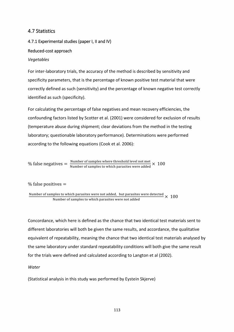
113
4.7 Statistics
4.7.1 Experimental studies (paper I, II and IV)
Reduced-cost approach
Vegetables
For inter-laboratory trials, the accuracy of the method is described by sensitivity and
specificity parameters, that is the percentage of known positive test material that were
correctly defined as such (sensitivity) and the percentage of known negative test correctly
identified as such (specificity).
For calculating the percentage of false negatives and mean recovery efficiencies, the
confounding factors listed by Scotter et al. (2001) were considered for exclusion of results
(temperature abuse during shipment; clear deviations from the method in the testing
laboratory; questionable laboratory performance). Determinations were performed
according to the following equations (Cook et al. 2006):
% false negatives = Number of samples where threshold level not met
Number of samples to which parasites were added× 100
% false positives =
Number of samples to which parasites were not added, but parasites were detected
Number of samples to which parasites were not added× 100
Concordance, which here is defined as the chance that two identical test materials sent to
different laboratories will both be given the same results, and accordance, the qualitative
equivalent of repeatability, meaning the chance that two identical test materials analysed by
the same laboratory under standard repeatability conditions will both give the same result
for the trials were defined and calculated according to Langton et al (2002).
Water
(Statistical analysis in this study was performed by Eystein Skjerve)
114
For comparing the results for the modified water analysis protocol, recovery efficiencies of
the spiked Cryptosporidium and Giardia by the modified method were compared with those
obtained by the ISO 15556 method by performing linear regression.
Survival study
(Statistical analysis in this study was performed by Eystein Skjerve)
Relative viability at each time point were obtained by normalizing the data to the initial
viability by using the equation: Percentage viability = (Nt/N0) x 100
Where Nt is the number of viable parasites at time t (of 100 parasites), and N0 is the number
of viable parasites at time 0 (of 100 parasites).
Survival was analysed using a linear regression model, with method as a categorical variable
and time (log10 hours) as a continuous predictor was utilised, and a follow up logistic
regression comparing the viability data of the parasites on the lettuce from initial
contamination point until final sampling point. Standard graphical methods were used to
assess mode fits and residual patterns using the statistical software Stata /SE/14 for
Windows, StataCorp, College Station, TX.
Comparing methods of Giardia cyst isolation
Two methods were analysed using 40 randomly selected samples and compared using
Fisher’s exact test, based on categorical data in a two-by-two contingency table.
4.7.2 Survey studies
A database of results was created in excel and parametric and non-parametric (ANOVA and
Mann-Whitney U-tests) were used to compare mean and median values. Contingency table
analysis (chi-square and Freeman-halton) was used to test for associations between positive
results, prevalences in different areas, difference in positive finding between seasons and
socioeconomic layers and other factors. Statistical significance was considered for p values <
0.05.
For comparison of Giardia prevalence according to location (urban/peri-urban), Students T-
test was used.
115
5. Results and general discussion
5.1 Experimental studies
Reduced cost modified version for analysing fresh produce and water for Cryptosporidium
and Giardia
The initial studies, that focused on modifying the technique for analysis of fresh produce for
occurrence of Cryptosporidium oocysts and Giardia cysts, resulted in a method of 80%
reduced cost per analysis, but with comparable mean recovery efficiencies (53% for
Cryptosporidium oocysts and 33% for Giardia cysts) to the standard method. These
modifications certainly cut the costs of this project, and may be of use to other research
projects or laboratories on constrained budgets as the supplementary reagents can easily be
obtained. The inter-laboratory trial provides independent confirmation of the method’s
applicability, and is an important step in such processes of identifying methods that work in
different laboratories, as new approaches that produce improved recovery efficiencies may
not always transfer well to other labs. One downside of the independent laboratory testing
of the method was only possible for labs in Europe. Although we did try to include labs
further away (Canada, Malaysia), we found that unstable storage during shipping of the
samples made the effort useless. This does emphasise that correct storage of reagents is
important, and this may also be problematic in some situations, for example when duration
of shipping of reagents is prolonged.
Based on the results obtained, this modified approach could be a useful tool for surveillance
projects in both developed and developing countries, where efforts should be directed
towards monitoring the production chain, and where detection and genotyping could be an
important step for finding glitches and evaluating preventive measures in the production
chain.
In addition, by using the same approach and reagents, we were able to analyse water
samples using this protocol; again, this also reduced the cost of this study while apparently
not affecting recovery efficiency, and this approach could also hopefully benefit other
research projects.
116
On the more costly end, by utilizing immunomagnetic separation beads for faecal samples
from goats, a significantly higher recovery of Giardia cysts was achieved. This separation
technique may be used in projects where Giardia prevalence is assessed, but where it is only
possible to obtain one sample per animal. As Giardia cyst shedding is intermittent, a single
negative sample does not immediately state that the individual is not shedding cysts, and
this separation technique may give a more true reflection of the actual prevalence. However,
as a diagnostic tool from clinical samples it is of low cost-benefit, as it is only likely to identify
samples that would otherwise be missed where cyst excretion is very low (and thus less
likely to be of clinical relevance). In addition, tests that identify antigens (rather than cysts),
such as immunochromatographic procedures, may be another approach to identifying
animals infected by Giardia. On the positive side, use of IMS may enable an efficient,
although costly, approach for isolating cysts or oocysts for downstream molecular analyses
with fewer inhibitors being present.
Survival of Giardia cysts and Cryptosporidium oocysts on fresh produce in the household
The results of the study described in Article II confirmed that although Giardia cysts seem to
survive when kept in cool, moist conditions, exposure to ambient temperatures results in
relatively rapid die off. In contrast, the relatively more robust Cryptosporidium oocysts did
not seem to be affected by storage temperature. Based on these results, it would seem likely
that unless fresh produce is consumed rapidly after contamination events, or is kept
refrigerated until consumption, Giardia cysts will not remain viable and thus be unable to
cause infection when ingested. As fresh produce in India is mostly sold by traditional
retailers, who sell their fruits and vegetables outdoors at ambient temperature, and as only
30% of Indian households own a refrigerator (Mahambare 2017), the likelihood of
foodborne giardiasis may be generally low, when contamination has occurred early in the
production chain, and is of greatest relevance when contamination occurs directly prior to
consumption, possibly in the kitchen, which are again linked to the importance of basic
hygiene knowledge. However, due to the robustness of the oocysts and the lack of control in
the production chain, the likelihood and impact of foodborne cryptosporidiosis may be
considerable.
117
5.2 Survey studies
5.2.1 Environmental surveys
Occurrence of Cryptosporidium and Giardia on vegetables from different retailers in
Chandigarh
It has been stated that foodborne transmission of these protozoan parasites is an under-
recognized but important emerging issue, especially in developed countries, due to the
increasing globalization of food trade, international travel, increased number of
immunocompromised and other susceptible individuals as well as changes in consumer
habits, and that fresh produce imported from developing countries are of great concern
(Dixon 2016). In my opinion, the elimination of this threat should start at the source, as the
threat is just as valid, if not more so, for the populations in developing countries as the
developed. The possible sources of contamination have been listed as poor personal hygiene
of workers during production, harvesting, packaging or transport, or by indirect
contamination of produce at the farm level through faecally contaminated water in
irrigation, mixing of pesticides, or washing of produce, hands or equipment, as well direct
contamination by the salesperson, food handler and consumer. Control measures, such as
properly treated water, health monitoring of workers, improved on-farm sanitation, and
restricted access of livestock and other animals to crops and surface water have been
proposed. However, these measures are difficult to implement overnight in a country where
animals traditionally roam freely. Basic hygienic measures seems to be the key in preventing
contamination of fresh produce, a food item not traditionally eaten raw, though with the
impact of western habits, culinary choices are now more diverse in urban areas of India, and
a contaminated salad just as infective in these regions as the one flown into developed
countries.
From the survey conducted in my study, a total occurrence of contamination of 11% was
found (Table 5), with a low median (oo)cyst count in positive samples. Noteably, outliers in
Cryptosporidium oocyst counts ranging from 350 and over 1000 per sample was found on
fresh produce bought at a supermarket and from a vendor in a slum area. From these
results, it seems that the traditional retailers keep their stock relatively free from
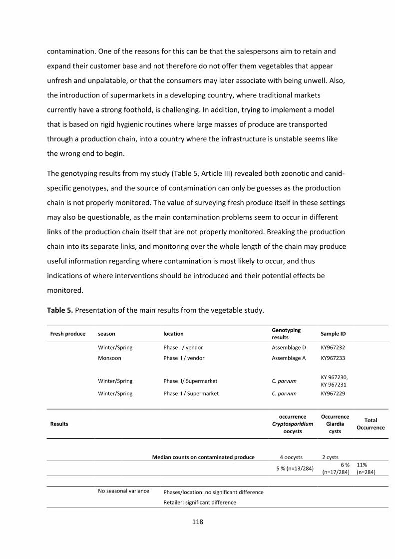
118
contamination. One of the reasons for this can be that the salespersons aim to retain and
expand their customer base and not therefore do not offer them vegetables that appear
unfresh and unpalatable, or that the consumers may later associate with being unwell. Also,
the introduction of supermarkets in a developing country, where traditional markets
currently have a strong foothold, is challenging. In addition, trying to implement a model
that is based on rigid hygienic routines where large masses of produce are transported
through a production chain, into a country where the infrastructure is unstable seems like
the wrong end to begin.
The genotyping results from my study (Table 5, Article III) revealed both zoonotic and canid-
specific genotypes, and the source of contamination can only be guesses as the production
chain is not properly monitored. The value of surveying fresh produce itself in these settings
may also be questionable, as the main contamination problems seem to occur in different
links of the production chain itself that are not properly monitored. Breaking the production
chain into its separate links, and monitoring over the whole length of the chain may produce
useful information regarding where contamination is most likely to occur, and thus
indications of where interventions should be introduced and their potential effects be
monitored.
Table 5. Presentation of the main results from the vegetable study.
Fresh produce season location Genotyping results
Sample ID
Winter/Spring Phase I / vendor Assemblage D KY967232 Monsoon Phase II / vendor Assemblage A KY967233
Winter/Spring Phase II/ Supermarket C. parvum KY 967230, KY 967231
Winter/Spring Phase II / Supermarket C. parvum KY967229
Results occurrence
Cryptosporidium oocysts
Occurrence Giardia
cysts
Total Occurrence
Median counts on contaminated produce 4 oocysts 2 cysts
5 % (n=13/284)
6 % (n=17/284)
11% (n=284)
No seasonal variance Phases/location: no significant difference
Retailer: significant difference
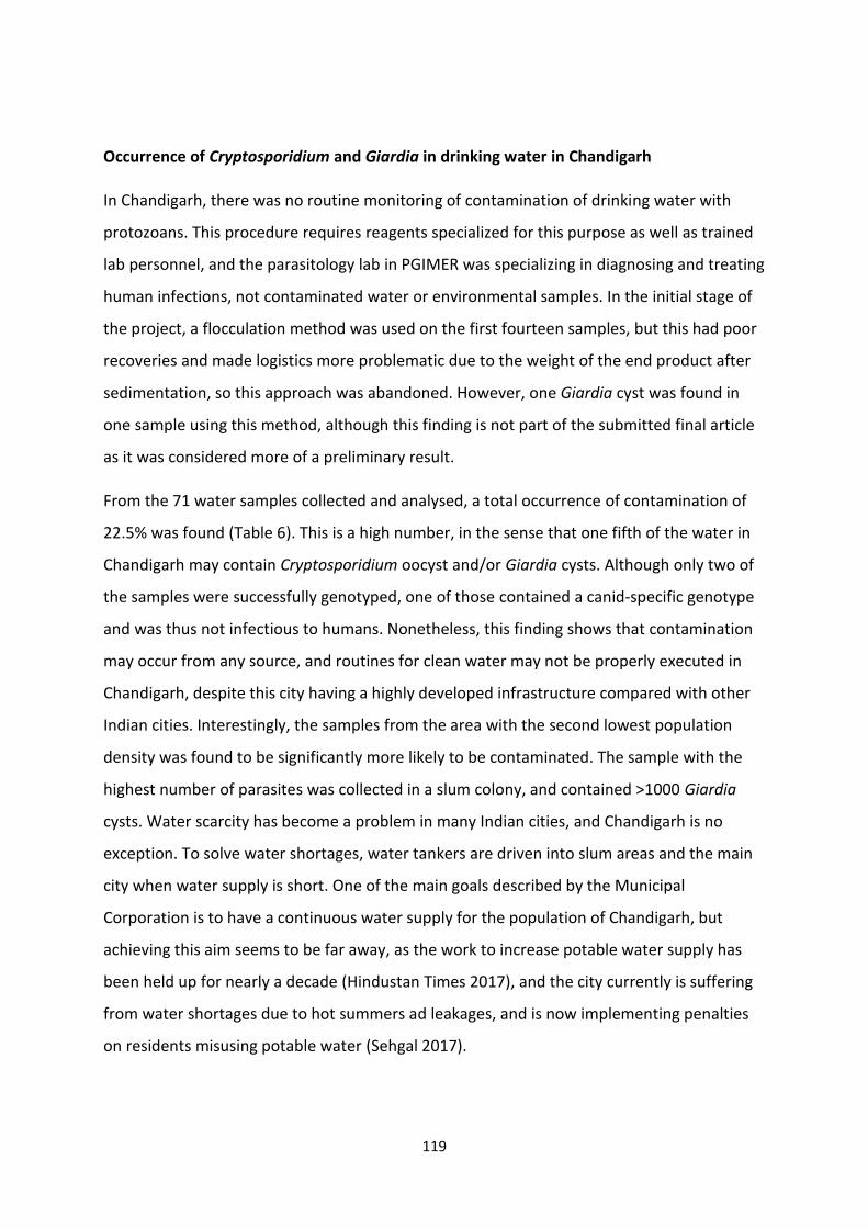
119
Occurrence of Cryptosporidium and Giardia in drinking water in Chandigarh
In Chandigarh, there was no routine monitoring of contamination of drinking water with
protozoans. This procedure requires reagents specialized for this purpose as well as trained
lab personnel, and the parasitology lab in PGIMER was specializing in diagnosing and treating
human infections, not contaminated water or environmental samples. In the initial stage of
the project, a flocculation method was used on the first fourteen samples, but this had poor
recoveries and made logistics more problematic due to the weight of the end product after
sedimentation, so this approach was abandoned. However, one Giardia cyst was found in
one sample using this method, although this finding is not part of the submitted final article
as it was considered more of a preliminary result.
From the 71 water samples collected and analysed, a total occurrence of contamination of
22.5% was found (Table 6). This is a high number, in the sense that one fifth of the water in
Chandigarh may contain Cryptosporidium oocyst and/or Giardia cysts. Although only two of
the samples were successfully genotyped, one of those contained a canid-specific genotype
and was thus not infectious to humans. Nonetheless, this finding shows that contamination
may occur from any source, and routines for clean water may not be properly executed in
Chandigarh, despite this city having a highly developed infrastructure compared with other
Indian cities. Interestingly, the samples from the area with the second lowest population
density was found to be significantly more likely to be contaminated. The sample with the
highest number of parasites was collected in a slum colony, and contained >1000 Giardia
cysts. Water scarcity has become a problem in many Indian cities, and Chandigarh is no
exception. To solve water shortages, water tankers are driven into slum areas and the main
city when water supply is short. One of the main goals described by the Municipal
Corporation is to have a continuous water supply for the population of Chandigarh, but
achieving this aim seems to be far away, as the work to increase potable water supply has
been held up for nearly a decade (Hindustan Times 2017), and the city currently is suffering
from water shortages due to hot summers ad leakages, and is now implementing penalties
on residents misusing potable water (Sehgal 2017).
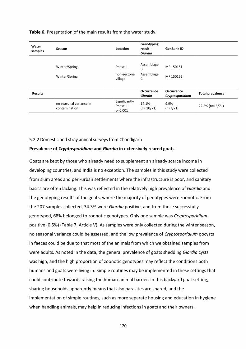
120
Table 6. Presentation of the main results from the water study.
Water samples
Season Location Genotyping result - Giardia
GenBank ID
Winter/Spring Phase II Assemblage B
MF 150151
Winter/Spring non-sectorial village
Assemblage C
MF 150152
Results
Occurrence Giardia
Occurrence Cryptosporidium
Total prevalence
no seasonal variance in contamination
Significantly Phase II p=0,001
14.1% (n= 10/71)
9.9% (n=7/71)
22.5% (n=16/71)
5.2.2 Domestic and stray animal surveys from Chandigarh
Prevalence of Cryptosporidium and Giardia in extensively reared goats
Goats are kept by those who already need to supplement an already scarce income in
developing countries, and India is no exception. The samples in this study were collected
from slum areas and peri-urban settlements where the infrastructure is poor, and sanitary
basics are often lacking. This was reflected in the relatively high prevalence of Giardia and
the genotyping results of the goats, where the majority of genotypes were zoonotic. From
the 207 samples collected, 34.3% were Giardia positive, and from those successfully
genotyped, 68% belonged to zoonotic genotypes. Only one sample was Cryptosporidium
positive (0.5%) (Table 7, Article V). As samples were only collected during the winter season,
no seasonal variance could be assessed, and the low prevalence of Cryptosporidium oocysts
in faeces could be due to that most of the animals from which we obtained samples from
were adults. As noted in the data, the general prevalence of goats shedding Giardia cysts
was high, and the high proportion of zoonotic genotypes may reflect the conditions both
humans and goats were living in. Simple routines may be implemented in these settings that
could contribute towards raising the human-animal barrier. In this backyard goat setting,
sharing households apparently means that also parasites are shared, and the
implementation of simple routines, such as more separate housing and education in hygiene
when handling animals, may help in reducing infections in goats and their owners.
121
Table 7. Presentation of the main results from the backyard goat study.
Goat samples
location Genotyping results Giardia
Sample ID Genotyping result Cryptosporidium
Sample ID
Urban Assemblage B MF069062
Urban Assemblage E MF069058
Urban Assemblage A MF069057
Urban Assemblage B MF069047
Urban Assemblage A MF069052
Urban Assemblage A MF069051
Urban Assemblage E
Assemblage C MF084938, MF069071
Urban Assemblage E MF084935, MF069070
Urban Assemblage A MF069056
Urban Assemblage E MF069059
Urban Assemblage D MF069055
Peri-urban Assemblage A MF069054
Peri-urban Assemblage E MF084936, MF095054, MF106203, MF069072
Peri-urban Assemblage E MF084934, MF095052
Peri-urban Assemblage B MF 095053, MF069053
Peri-urban Assemblage B MF069066
Peri-urban Assemblage B MF069064
Peri-urban Assemblage B MF069060
Peri-urban Assemblage E MF084937
Peri-urban Assemblage B MF069050
Peri-urban Assemblage A MF069068
Peri-urban Assemblage A MF069067
Peri-urban Assemblage E MF069065
Peri-urban Assemblage B MF069063
Peri-urban Assemblage B MF069061
Peri-urban Assemblage A MF069069
Peri-urban Assemblage A MF069049
Peri-urban Assemblage A MF069048
Peri-urban
C. ubiquitum MF124820
Results
Location not significant
Summation of results
Giardia Cryptosporidium
Total prevalence
Prevalence: 34.3% 0.5%
34.7% (n= 207)
Cyst shedding: Median cyst count: 275
Distribution of genotypes: Ass. A: 36%, Ass B: 32%
Ass. E, C and D: 32%
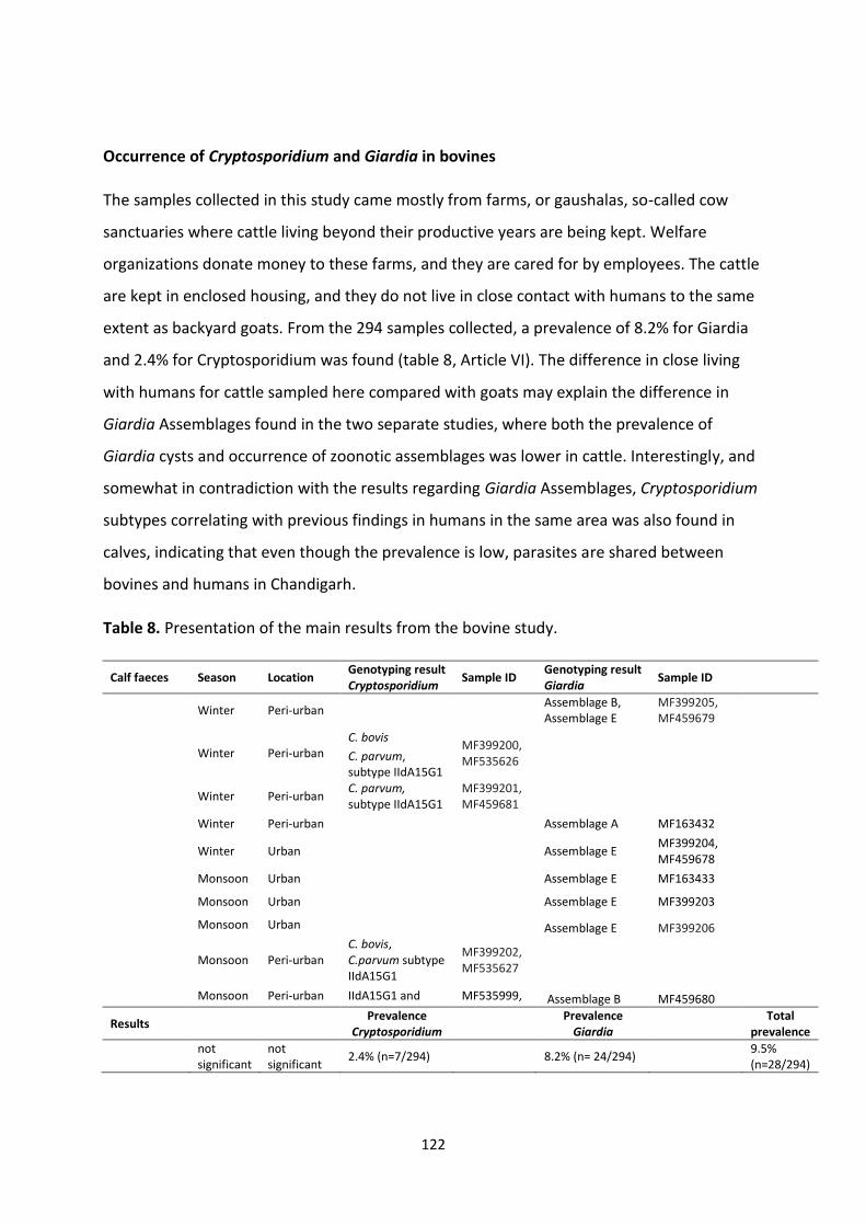
122
Occurrence of Cryptosporidium and Giardia in bovines
The samples collected in this study came mostly from farms, or gaushalas, so-called cow
sanctuaries where cattle living beyond their productive years are being kept. Welfare
organizations donate money to these farms, and they are cared for by employees. The cattle
are kept in enclosed housing, and they do not live in close contact with humans to the same
extent as backyard goats. From the 294 samples collected, a prevalence of 8.2% for Giardia
and 2.4% for Cryptosporidium was found (table 8, Article VI). The difference in close living
with humans for cattle sampled here compared with goats may explain the difference in
Giardia Assemblages found in the two separate studies, where both the prevalence of
Giardia cysts and occurrence of zoonotic assemblages was lower in cattle. Interestingly, and
somewhat in contradiction with the results regarding Giardia Assemblages, Cryptosporidium
subtypes correlating with previous findings in humans in the same area was also found in
calves, indicating that even though the prevalence is low, parasites are shared between
bovines and humans in Chandigarh.
Table 8. Presentation of the main results from the bovine study.
Calf faeces Season Location Genotyping result Cryptosporidium
Sample ID Genotyping result Giardia
Sample ID
Winter Peri-urban
Assemblage B, Assemblage E
MF399205, MF459679
Winter Peri-urban C. bovis
MF399200, MF535626
C. parvum, subtype IIdA15G1
Winter Peri-urban C. parvum, subtype IIdA15G1
MF399201, MF459681
Winter Peri-urban
Assemblage A MF163432
Winter Urban
Assemblage E MF399204, MF459678
Monsoon Urban
Assemblage E MF163433
Monsoon Urban
Assemblage E MF399203
Monsoon Urban Assemblage E MF399206
Monsoon Peri-urban C. bovis, C.parvum subtype IIdA15G1
MF399202, MF535627
Monsoon Peri-urban IIdA15G1 and MF535999, Assemblage B MF459680
Results
Prevalence
Cryptosporidium Prevalence
Giardia
Total prevalence
not significant
not significant
2.4% (n=7/294) 8.2% (n= 24/294)
9.5% (n=28/294)
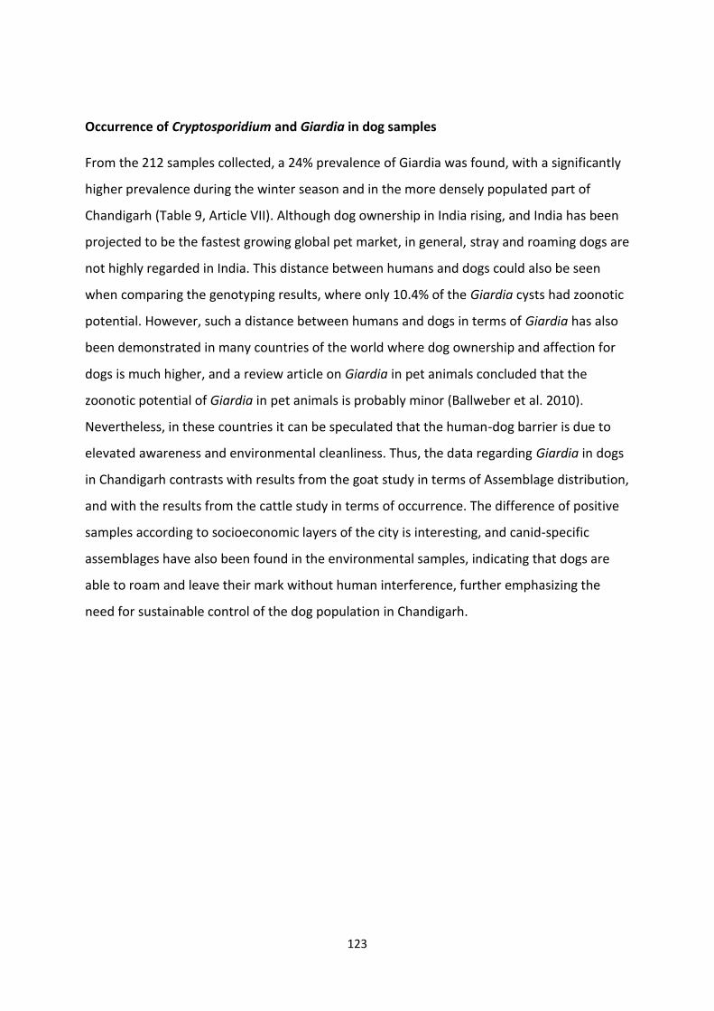
123
Occurrence of Cryptosporidium and Giardia in dog samples
From the 212 samples collected, a 24% prevalence of Giardia was found, with a significantly
higher prevalence during the winter season and in the more densely populated part of
Chandigarh (Table 9, Article VII). Although dog ownership in India rising, and India has been
projected to be the fastest growing global pet market, in general, stray and roaming dogs are
not highly regarded in India. This distance between humans and dogs could also be seen
when comparing the genotyping results, where only 10.4% of the Giardia cysts had zoonotic
potential. However, such a distance between humans and dogs in terms of Giardia has also
been demonstrated in many countries of the world where dog ownership and affection for
dogs is much higher, and a review article on Giardia in pet animals concluded that the
zoonotic potential of Giardia in pet animals is probably minor (Ballweber et al. 2010).
Nevertheless, in these countries it can be speculated that the human-dog barrier is due to
elevated awareness and environmental cleanliness. Thus, the data regarding Giardia in dogs
in Chandigarh contrasts with results from the goat study in terms of Assemblage distribution,
and with the results from the cattle study in terms of occurrence. The difference of positive
samples according to socioeconomic layers of the city is interesting, and canid-specific
assemblages have also been found in the environmental samples, indicating that dogs are
able to roam and leave their mark without human interference, further emphasizing the
need for sustainable control of the dog population in Chandigarh.
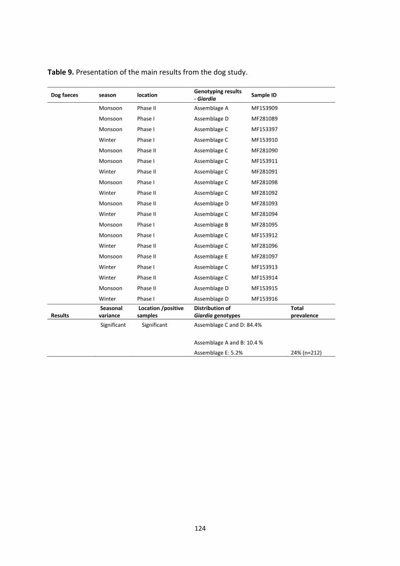
124
Table 9. Presentation of the main results from the dog study.
Dog faeces season location Genotyping results - Giardia
Sample ID
Monsoon Phase II Assemblage A MF153909
Monsoon Phase I Assemblage D MF281089
Monsoon Phase I Assemblage C MF153397
Winter Phase I Assemblage C MF153910
Monsoon Phase II Assemblage C MF281090
Monsoon Phase I Assemblage C MF153911
Winter Phase II Assemblage C MF281091
Monsoon Phase I Assemblage C MF281098
Winter Phase II Assemblage C MF281092
Monsoon Phase II Assemblage D MF281093
Winter Phase II Assemblage C MF281094
Monsoon Phase I Assemblage B MF281095
Monsoon Phase I Assemblage C MF153912
Winter Phase II Assemblage C MF281096
Monsoon Phase II Assemblage E MF281097
Winter Phase I Assemblage C MF153913
Winter Phase II Assemblage C MF153914
Monsoon Phase II Assemblage D MF153915
Winter Phase I Assemblage D MF153916
Results Seasonal variance
Location /positive samples
Distribution of Giardia genotypes
Total prevalence
Significant Significant Assemblage C and D: 84.4%
Assemblage A and B: 10.4 %
Assemblage E: 5.2% 24% (n=212)
125
Combined conclusions from all the survey-based studies
As these various survey studies have revealed, implementation of some actions towards
general runoff, hygiene, sanitation and simple education could reduce the burden of parasite
contamination, and thus of infection in both humans and animals. It seems that animals may
have a role in the spread of both Cryptosporidium and Giardia and in maintaining the
lifecycle in Chandigarh, although to different extents with different animals, and analysis of
the genotypes revealed that humans may be the group who can be considered to bear
responsibility for the cycle. In addition, and not considered here, wild animals in close
contact with humans may also play a role, as the worshipped and public fed maquace
monkeys in Chandigarh showed a high prevalence of zoonotic Giardia genotypes (Debenham
et al. 2017).
However, from the holy cow to the family goat, zoonotic potential was found in domestic
animals in close contact with humans, with goats being of particular importance. However,
no significant zoonotic potential was found in the dog study, and as stray dogs are usually
avoided by humans, this was not a surprising result.
126
5.3 Limitations and challenges experienced in the study
The application of molecular methods in samples traveling across the globe
Polymerase chain reaction (PCR) is one of the most extensively tested and widely used
techniques for investigating the origins of waterborne protozoan parasites. PCR is not only
often used to identify pathogens in a complex environment, but by targeting specific gene
sequences, PCR assays can be used to distinguish between different sub-groups
(assemblages or subtypes) of the same species or identify different species within a genus.
Although PCR is considered to be highly sensitive and accurate, and for pathogenic bacteria
is in the process of replacing traditional methods, for parasites it has limitations. Although
the potential for false positives should not be ignored, and may result from laboratory
contamination, false negatives are a more common problem. These may occur due to low
recovery of DNA during the extraction process, especially in the case of environmental
samples with a low number of (oo)cysts and a high degree of organic debris in the sample. In
addition, inhibitors like polysaccharides, polyphenols, pectin, xylan and chlorophyll may be
extracted along with the (oo)cysts and thus hamper the PCR reaction in the case of
environmental samples (Wei et al, 2008). Faecal samples are also particularly difficult, as
they are complex and highly variable matrices that contain an enormous quantity of DNA
from bacteria and cells other than the target DNA (Wilke and Robertson, 2009). In addition,
in the absence of a normal cellular processes, endogenous endonuclease activity, bacterial
degradation and spontaneous depurination results in relatively rapid breakage of DNA
strands, and DNA degradation may continue due to oxidative action and accumulation of
molecular cross-linkages (Deagle et al, 2006).
Many of the samples collected in this study, both faecal samples and water/vegetable
samples, contained low numbers of (oo)cysts, and as environmental samples are very
diverse, they may also have contained a number of PCR inhibitors, such as debris, fulmic and
humic acid, metal ions and polyphenols which may be extracted along with the parasites
during DNA isolation, and hamper DNA amplification during the PCR reaction (Abbaszadegan
et al. 1993; Ijzerman et al. 1997). Although faecal samples from a symptomatic individual
often have a higher concentration of (oo)cysts and thus DNA, faeces also contain inhibitors
such as complex polysaccharides, bile salts, lipids and urate (Schrader et al. 2012). In my
127
study, the faecal samples were not necessarily obtained from animals with clinical
symptoms, and my study was to investigate potential reservoirs of human infection, thus
they also include non-symptomatic low excretors. In general, the samples collected in this
study were from a plethora of sources and settings, and many inhibitors may have been
extracted along with the DNA.
Due to practical and logistical issues between the collaborating laboratories, the period
between collection and examination of faecal, drinking water and vegetable samples was in
some cases prolonged. Storage of Giardia cysts has proven to cause alterations in membrane
morphology, intense vacuolization as well as damage of its wall (Santos et al. 2015), and a
decline in sporozoite ratio during storage has also been found in Cryptosporidium oocysts
(Dawson et al. 2004). Parasites may have degenerated during storage and the formation of
other microorganisms during the storage and transport period may have had a deteriorating
effect on the DNA in terms of both degeneration and formation of inhibitors. Especially the
environmental samples proved hard to genotype.
Opportunities and Challenges
There are numerous challenges when working in developing communities where resources
are not readily available, and the cultural differences are sometimes hard to comprehend for
a foreigner, so the sparsely understood and underdocumented relations of socioeconomic,
cultural, religious, educational and bureaucratic nuances cannot be investigated with
confidence in this work. However, these variables were influential in shaping the view of
society. As a female veterinarian collaborating with native farmers and collecting faecal
samples, challenges in obtaining samples sometimes went beyond communication and
caused misunderstandings, though almost always with the outcome of collaboration and
mutual understanding in the end. Traditionally the domestic animals are viewed as a part of
the family and culture, they are greatly cared for, and the myriad of cultural differences
were hard to rectify during the relatively short duration of field work.
Even though there were difficulties in logistics and transport of over a thousand samples
with DNA (and inhibitors) it was possible to apply refined molecular tools to unravel zoonotic
relationships. Minimal quantities of sample are required for molecular screening and
128
characterization of parasites, which makes air transportation between collaborating
laboratories possible, and the chemical properties of potassium dichromate in solution make
them a safe transport vehicle for biological material. Moreover, the DNA for all parasite
species in whole faeces preserved in these solutions can remain stable even at temperatures
up to 40°C for prolonged periods before analysis, which proved to be a useful trait.
The hurdle of applying molecular-based tools does not lie in having a specialized laboratory
on-site, or time delays in sample processing, but in the considerable cost of equipment,
reagents and personnel associated with processing the samples. By dividing the labour to
two workplaces; collection and preparation for transport at PGIMER with further processing
at NMBU, where the laboratory already had the necessary equipment and resources, both
collection and analysis were possible to perform.
The ability to selectively and sensitively detect and genetically characterize parasitic stages
directly from faeces and the environment has served as a major advantage for studying the
epidemiology of parasites within populations and geographical areas. This is especially
valuable information for a country where these relationships have not been explored to a
great extent yet, and where these diseases have a generally higher prevalence. Molecular
tools in combination with classical parasitological and epidemiological methods to detect,
diagnose and genetically characterize parasites are imperative in studies aiming to assess
sources and foci of infection.
129
6. Concluding remarks and future perspectives
Although we know that Cryptosporidium and Giardia can be foodborne and waterborne, and
both have zoonotic potential, the bulk of the studies that address this issue and attempt to
determine the importance of these transmission routes and sources of infection have been
conducted in developed countries. Although developed countries have had many more
reported outbreaks of foodborne and waterborne cryptosporidiosis and giardiasis, and the
zoonotic potential has been quite extensively investigated, such studies from less developed
countries are lacking. The epidemiology of these infections is likely to differ considerably
between such different settings, and, as the infrastructure is considerably less robust in
developing countries, I would assume that the potential for transmission via environmental
contamination would be greater. Thus, there is a notable gap in our understanding of the
transmission routes and infection sources for these parasites in countries such as India, and
the work described in this thesis goes someway to addressing these questions in a city in
northern India.
The experimental, lab-based studies enable the development and validation of analytical
procedures based on standard methods, but at considerably reduced costs, thereby enabling
me to generate reliable data in a challenging environment. The survival studies went some
way to providing data indicating one reason why foodborne giardiasis may be less of a
concern than foodborne cryptosporidiosis, particularly in a setting where fresh produce is
not often refrigerated.
From the results in the survey studies, there seems to be a correlation between how close
humans interact with the animals around them, and the occurrence of zoonotic protozoans
in animal faeces. This can only be amended by human intervention on transmission routes,
and to be able to do that, awareness is the first step towards prevention.
The survey study from this project indicated that clean water seems to be a privilege for the
population in Chandigarh living in the socioeconomic higher layers of the city. No seasonal
variance was found in the water samples, and this may be due to a backdrop of constant
contamination and poor surveillance, or the fact that the seasons during the sampling period
was somewhat not as predicted.
130
Based on the results generated during the work described in this thesis, it is clear that if
fresh produce in India is to be both produced and consumed as happens in western
countries, then the production chain needs to be evaluated and improved. In the Apni Mandi
system, the farmer may be more accessible, but the hazards in a system where the farmer is
responsible for all steps in the trade should be closely supervised, and hazards in the
production chain should be pointed out to those engaged in this type of trade. There is a
clear need for implementation of such systems as Good Agricultural Practice (GAP), Good
Manufacturing Practice, (GMP) and Good Handling Practice (GHP) (FDA 1998). In the
supermarket model, the origin of the wares may be less simple to track, and thus relevant
factors in how the produce have been grown, harvested, or processed. This makes results of
analysis of fresh produce less valuable if the sources and critical control points are to be
evaluated and improved. In addition, my study showed that drinking water in Chandigarh
has relatively high occurrences of protozoan parasites, and more effective surveillance
studies with emphasis on source tracking through genotyping is a useful tool in
epidemiological investigations. Although the results of my study may act as an indicator, the
results cannot be extrapolated to all cities in India. However, the potential for contamination
in such settings is clear, and thus the need for interventions to make clean, safe water
granted, not a privilege.
During the course of the work described in this thesis, several obstacles were met on the
way, and to proceed to obtain meaningful, robust results was not always straightforward.
Equipment for performing analyses was lacking in the collaborative lab due to the
differences in routine analyses performed at the respective laboratories, and had to be
transported across the globe, although the method modification made the analytical costs
more bearable for WP2. One of the incentives of this thesis that the methods developed
here may be used to benefit other projects also aiming to investigate bulk samples at a
lower cost, and where the budget may be restrained, the reduction in cost may expand the
sample size to give more representative results.
A further challenge was in attempting to ensure that the data generated by my work, which
represented just one Work Package (WP2) of a larger project, could be of value in the
contexts of the work from other WPs. In particular, the results from WP1, for which data on
intestinal parasites in children living in the same areas as sampled in my study, would have
131
added value to the information that I obtained, as findings regarding genotype, geographical
area, sampling season, and factors such as animal ownership or fresh produce consumption,
were to be collected and could have been compared with my data. Unfortunately, the
partner responsible for WP1 were unable to fulfil their contribution within the timescale of
the project. However, sampling has been performed, questionnaires have been completed
and analysis and genotyping of these are in progress.
In 2014, India’s Prime Minister Modi launched the “Clean India Mission” (Swachh Bharat
Abhyian), with the objective of India being a “clean country” by 2019. This mission includes
the development of awareness about sanitation, health education and promotion of
sustainable sanitation facilities, as well as increasing sanitation coverage and eliminating
open defecation by 2019 through the construction of community and public toilets. A
staggering 597 million Indians do not have access to toilets, and due to cultural practices,
open defecation is preferred in some communities. Between April 2014 and January 2015,
3.2 million toilets have been built, and the Swachh Bharat campaign has made hygiene
education a major part of its mission, including regular surveys on toilet use (Jacob, 2016).
Another mission is to eradicate the practice of manual scavenging. The now abandoned
caste culture left perceptions that has been suggested to be one of a number of reasons for
unclean India, as it externalised the responsibility for maintaining cleanliness to be the duty
of a particular caste. Manual scavenging is illegal, but carries on regardless (Jacob, 2016).
Some people derive a sense of superiority in littering their environment while others have to
clean it, and this perception may be exacerbated in a community where it was expected to
be cleaned by the lower-caste scavenger; this is a perception that persists despite the spread
of education, globalisation, and urbanisation. Unless the responsibility for cleanliness taken
is to the level of each individual, no number of campaigns are going to succeed (Teltumbde
2014), neither in India or any country.
To achieve sustainable changes in an enormous country, it must be realised that cleanliness
is more than an adjunct of socioeconomic status, but also a cultural and habitual concern.
Countries poorer than India, such as Bangladesh and Sri Lanka, are almost open defecation
free (Jacobs, 2016). It is up to each and every individual of any nation, regardless of social
status and income, to address these issues, then simple things concerning sanitation and
hygiene have the potential to make a huge impact on both population health and the
132
perception of previously lower castes. Although employment of manual scavengers was
prohibited in 1993, over 180 000 households in India are still engaged in manual scavenging
(Indian 15th National Census Survey, 2011), indicating that the abolished labour system still
prevails.
Another initiative associated with “Swachh Bharat Abhiyan” includes the call for the
population to devote 100 hours a year towards the cause of cleaning. The participation and
collaboration of all social layers of the population in such initiatives may hopefully give an
insight not only to the Indian nation, but all of us, that we are, in fact, one people, all living
on the same one globe, all affected by the same One Health.
With studies described in this thesis considered against this background, the following points
provide a few concrete suggestions on future research that could build on the results that I
obtained and could provide a small contribution towards the “Swachh Bharat Abhiyan”
campaign.
• When analysis of the samples obtained in WG1 has been completed, it would be
useful to examine the results obtained with those that I obtained from water,
vegetables, and different domestic animals. This may indicate the transmission
routes and sources of most relevance to the children participating in WG1 as well as
indicating which variables are of importance for the possible transmission pathways
and any potential foci of infection.
• Clearly fresh produce on sale in Chandigarh is contaminated – but apart from
suggesting that customer handling in supermarkets may be one route of
contamination, my results are unable to determine the most important routes of
contamination. This could be in the field, but other sources that I observed include
the buckets of water that are regularly splashed over fresh produce to keep them
looking fresh; analysis of these buckets could provide interesting information. Other
close investigations of the fresh produce chain, such as harvest, transport and
storage may provide other insights and thus offer the opportunity for implementing
preventive measures.
133
• Some of the drinking water available in Chandigarh is contaminated, but, again, how
this contamination arises cannot be determined from the information in my studies.
Investigation of the barriers for contamination in place for the water supplies could
indicate potential weaknesses and indicate where the situation could be improved,
for example by catchment control, water treatment initiatives, or efforts to reduce
post-treatment contamination that may be associated with leakages and pressure
drops within the distribution network.
• Much of the animal sampling was done, by necessity, on an ad hoc basis. A more
systematic sampling strategy focussed on areas and age groups of particular interest
could provide more useful information. My results indicate that backyard goats may
be particularly important regarding zoonotic/anthropozoonotic transmission of
Giardia, and closer investigation of this potential transmission cycle could provide
useful insights. In addition to addressing the human-goat-human transmission web, it
would also be of value to learn the clinical significance of Giardia for the goats
themselves; although overt diarrhoea was not observed, more subtle effects, such as
reduced weight gain and lower milk production, may mean that Giardia in goats not
only has the potential to affect the owners by being a reservoir of infection, but also
reduce the economic gains that the owners hope to derive from goat husbandry.
• Although the main focus of my research was on the (potentially) zoonotic intestinal
protozoa, Cryptosporidium and Giardia, other zoonotic parasites are also worthy of
further exploration. A 17-year time-series study on the occurrence of cysticercosis
conducted during this study (Robertson et al. 2017), indicated that this remains an
important disease in Chandigarh, particularly in women, despite the very low level of
meat consumption in this society. This is due to the enormous potential for
environmental contamination from a single individual with taeniosis, and the fact
that pigs tend to be kept in the poorest areas of the city where open defecation is a
fact of life. Exploration of the prevalence of porcine cysticercosis, the extent of
contamination of the environment with Taenia eggs, and factors associated with
transmission could provide valuable insights that could perform a basis for
addressing this issue.
134
7. References
Abbaszadegan, M., Huber, M., Gerba, C. P. & Pepper, I. (1993). Detection of enteroviruses in groundwater with the polymerase chain reaction. Appl Environ Microbiol, 59 (5): 1318-1324.
Abbaszadegan, M., Huber, M. S., Gerba, C. P. & Pepper, I. L. (1997). Detection of viable
Giardia cysts by amplification of heat shock-induced mRNA. Appl Environ Microbiol, 63 (1): 324-328.
Abubakar, I., Aliyu, S., Arumugam, C., Usman, N. & Hunter, P. (2007). Treatment of
cryptosporidiosis in immunocompromised individuals: systematic review and meta‐analysis. Br J Clin Pharmacol, 63 (4): 387-393.
Adam, R. D. (2001). Biology of Giardia lamblia. Clin Microbiol Rev, 14 (3): 447-475. Agoramoorthy, G. & Hsu, M. J. (2012). The significance of cows in Indian society between
sacredness and economy. Anthropol Noteb, 18 (3): 5-12. Akiyoshi, D. E., Tumwine, J. K., Bakeera-Kitaka, S. & Tzipori, S. (2006). Subtype analysis of
Cryptosporidium isolates from children in Uganda. J Parasitol, 92 (5): 1097-1100. Al-Brikan, F. A., Salem, H. S., Beeching, N. & Hilal, N. (2008). Multilocus genetic analysis of
Cryptosporidium isolates from Saudi Arabia. J Egyptian Soc Parasitol, 38 (2): 645-58. Alves, M., Xiao, L., Antunes, F. & Matos, O. (2006). Distribution of Cryptosporidium subtypes
in humans and domestic and wild ruminants in Portugal. Parasitology research, 99 (3): 287-292.
Amer, S., Zidan, S., Feng, Y., Adamu, H., Li, N. & Xiao, L. (2013). Identity and public health
potential of Cryptosporidium spp. in water buffalo calves in Egypt. Vet Parasitol, 191 (1): 123-127.
Amoah, P., Drechsel, P., Henseler, M. & Abaidoo, R. (2007). Irrigated urban vegetable
production in Ghana: microbiological contamination in farms and markets and associated consumer risk groups. J Water Health, 5 (3): 455-466.
Amorós, I., Alonso, J. L. & Cuesta, G. (2010). Cryptosporidium oocysts and Giardia cysts on
salad products irrigated with contaminated water. J Food Prot, 73 (6): 1138-1140. Bajer, A., Bednarska, M., Cacciò, S. M., Wolska-Kuśnierz, B., Heropolitanska-Pliszka, E.,
Bernatowska, E., Wielopolska, M., Paziewska, A., Welc-Falęciak, R. & Siński, E. (2008). Genotyping of Cryptosporidium isolates from human clinical cases in Poland. Parasitology research, 103 (1): 37-42.

135
Baldursson, S. & Karanis, P. (2011). Waterborne transmission of protozoan parasites: review of worldwide outbreaks–an update 2004–2010. Water Res, 45 (20): 6603-6614.
Ballweber, L. R., Xiao, L., Bowman, D. D., Kahn, G. & Cama, V. A. (2010). Giardiasis in dogs
and cats: update on epidemiology and public health significance. Trends Parasitol, 26 (4): 180-189.
Barr, S., Bowman, D., Frongillo, M. & Joseph, S. (1998). Efficacy of a drug combination of
praziquantel, pyrantel pamoate, and febantel against giardiasis in dogs. Am J Vet Res, 59 (9): 1134-1136.
BBC. (2000). Sheep battle over bug at reservoir. BBC news. Available at:
http://news.bbc.co.uk/2/hi/uk_news/northern_ireland/910594.stm (accessed: 17.09.2017).
Berkman, D. S., Lescano, A. G., Gilman, R. H., Lopez, S. L. & Black, M. M. (2002). Effects of
stunting, diarrhoeal disease, and parasitic infection during infancy on cognition in late childhood: a follow-up study. Lancet, 359 (9306): 564-571.
Bertrand, I., Maux, M., Helmi, K., Hoffmann, L., Schwartzbrod, J. & Cauchie, H.-M. (2009).
Quantification of Giardia transcripts during in vitro excystation: Interest for the estimation of cyst viability. Water Res, 43 (10): 2728-2738.
Bhat, S., Juyal, P. & Singla, L. (2012). Prevalence of cryptosporidiosis in neonatal buffalo
calves in Ludhiana district of Punjab, India. Asian J Anim Vet Adv, 7 (6): 512-520. Bier, J. W. (1991). Isolation of parasites on fruits and vegetables. Southeast Asian J Trop Med
Public Health, 22: 144-145. Bomfim, T. C. B., Huber, F., Gomes, R. S. & Alves, L. L. (2005). Natural infection by Giardia sp.
and Cryptosporidium sp. in dairy goats, associated with possible risk factors of the studied properties. Vet Parasitol, 134 (1): 9-13.
Burnet, J.-B. (2012). Dynamique spatio-temporelle de Cryptosporidium et de Giardia dans un
réservoir d'eau potable et son bassin versant: cas des lacs de barrage de la Haute-Sûre (Luxembourg).
Cacciò, S. M., De Giacomo, M. & Pozio, E. (2002). Sequence analysis of the β-giardin gene
and development of a polymerase chain reaction–restriction fragment length polymorphism assay to genotype Giardia duodenalis cysts from human faecal samples. Int J Parasitol, 32 (8): 1023-1030.
Cacciò, S. M., Beck, R., Lalle, M., Marinculic, A. & Pozio, E. (2008). Multilocus genotyping of
Giardia duodenalis reveals striking differences between assemblages A and B. Int J Parasitol, 38 (13): 1523-1531.
136
Cama, V. A., Bern, C., Sulaiman, I. M., Gilman, R. H., Ticona, E., Vivar, A., Kawai, V., Vargas, D., Zhou, L. & Xiao, L. (2003). Cryptosporidium Species and Genotypes in HIV‐Positive Patients in Lima, Peru. J Eukaryot Microbiol, 50 (s1): 531-533.
Cama, V. A., Bern, C., Roberts, J., Cabrera, L., Sterling, C. R., Ortega, Y., Gilman, R. H. & Xiao,
L. (2008). Cryptosporidium species and subtypes and clinical manifestations in children, Peru. Emerg Infect Dis, 14 (10): 1567.
Campbell, A., Robertson, L. & Smith, H. (1992). Viability of Cryptosporidium parvum oocysts:
correlation of in vitro excystation with inclusion or exclusion of fluorogenic vital dyes. Appl Environ Microbiol, 58 (11): 3488-3493.
Campbell, A. & Smith, H. (1997). Immunomagnetic separation of Cryptosporidium oocysts
from water samples: round robin comparison of techniques. Water Sci Technol, 35 (11-12): 397-401.
Campbell, I., Tennant, W., Seal, D., Hay, J., McLuckie, I. & Patterson, W. (1995). UK national
study of Legionella contamination of hospital transplant unit water supplies: relevance of free-living amoebae. Special Publication - Royal Society of Chemistry, 168: 30-30.
Cantu, J. & Garcia-Cruz, D. (1982). Midline facial defect as a teratogenic effect of
metronidazole. Birth Defects: original article series. Caradonna, T., Marangi, M., Del Chierico, F., Ferrari, N., Reddel, S., Bracaglia, G., Normanno,
G., Putignani, L. & Giangaspero, A. (2017). Detection and prevalence of protozoan parasites in ready-to-eat packaged salads on sale in Italy. Food Microbiol.
Carey, C. M., Lee, H. & Trevors, J. T. (2006). Comparison of most probable number-PCR and
most probable number-foci detection method for quantifying infectious Cryptosporidium parvum oocysts in environmental samples. J Microbiol Methods, 67 (2): 363-372.
Castro-Hermida, J. A., Almeida, A., González-Warleta, M., da Costa, J. M. C., Rumbo-Lorenzo,
C. & Mezo, M. (2007). Occurrence of Cryptosporidium parvum and Giardia duodenalis in healthy adult domestic ruminants. Parasitology research, 101 (5): 1443-1448.
CDC. (2016). Cryptosporidiosis: U.S. Department of Health & Human Services. Available
at: https://www.cdc.gov/dpdx/cryptosporidiosis/index.html (accessed: 17.09.2017). CDC. (2017). Giardia - Pathogen & Environment: U.S. Department of Health & Human
Services. Available at: https://www.cdc.gov/parasites/giardia/pathogen.html (accessed: 17.09.2017).
Chalmers, R., Elwin, K., Reilly, W., Irvine, H., Thomas, A. & Hunter, P. (2002). Cryptosporidium
in farmed animals: the detection of a novel isolate in sheep. Int J Parasitol, 32 (1): 21-26.
137
Chalmers, R., Elwin, K., Thomas, A., Guy, E. & Mason, B. (2009). Long-term Cryptosporidium
typing reveals the aetiology and species-specific epidemiology of human cryptosporidiosis in England and Wales, 2000 to 2003. Euro surveillance: bulletin Europeen sur les maladies transmissibles; European communicable disease bulletin, 14 (2): 785-794.
Chandigarh Administration. (2016). Chandigarh Master Plan 2031. Available at:
http://chandigarh.gov.in/cmp_2031.htm (accessed on 07.05.2017). Checkley, W., White, A., Jagannath, D., Arrowood, M., Chalmers, R. & Chen, X. (2015).
Cryptosporidiosis: global burden, novel diagnostics, therapeutics and vaccine targets. Lancet Infect Dis, 15: 85-94.
Chigateri, S. (2008). ‘Glory to the Cow’: Cultural Difference and Social Justice in the Food
Hierarchy in India 1. South Asia: Journal of South Asian Studies, 31 (1): 10-35. Coklin, T., Farber, J. M., Parrington, L. J., Kingombe, C. I. B., Ross, W. H. & Dixon, B. R. (2011).
Immunomagnetic Separation Significantly Improves the Sensitivity of Polymerase Chain Reaction in Detecting Giardia duodenalis and Cryptosporidium spp. in Dairy Cattle. J Vet Diagn Invest, 23 (2): 260-267.
Cook, N., Paton, C., Wilkinson, N., Nichols, R., Barker, K. & Smith, H. (2006). Towards
standard methods for the detection of Cryptosporidium parvum on lettuce and raspberries. Part 1: development and optimization of methods. Int J Food Microbiol, 109 (3): 215-221.
Cook, N., Nichols, R. A. B., Wilkinson, N., Paton, C. A., Barker, K. & Smith, H. V. (2007).
Development of a method for detection of Giardia duodenalis cysts on lettuce and for simultaneous analysis of salad products for the presence of Giardia cysts and Cryptosporidium oocysts. Appl Environ Microbiol, 73 (22): 7388-7391.
Cook, N. & Y.A.L., L. (2012). Giardia duodenalis Part1: Contamination of fresh produce. In
Robertson L.J., S. H. V. (ed.) vol. 1 Foodborne protozoan parasites, pp. 107-132. New York: Nova Science Publishers. Inc.
Cooper, M. A., Sterling, C. R., Gilman, R. H., Cama, V., Ortega, Y. & Adam, R. D. (2010).
Molecular analysis of household transmission of Giardia lamblia in a region of high endemicity in Peru. J Infect Dis, 202 (11): 1713-1721.
Cooper, R. & Olivieri, A. (1998). Infectious disease concerns in wastewater reuse.
Wastewater Reclamation and Reuse, Technomic Publishing Co., Lancaster, PA, pp 489–520.
Cotruvo, J. A., Dufour, A., Rees, G., Bartram, J., Carr, R., Cliver, D. O., Craun, G. F., Fayer, R. &
Gannon, V. P. (2004). Waterborne zoonoses: IWA Publishing.
138
Dawson, D. J., Samuel, C. M., Scrannage, V. & Atherton, C. J. (2004). Survival of Cryptosporidium species in environments relevant to foods and beverages. J Appl Microbiol, 96 (6): 1222-1229.
Deagle, B. E., Eveson, J. P. & Jarman, S. N. (2006). Quantification of damage in DNA
recovered from highly degraded samples–a case study on DNA in faeces. Front. Zool., 3 (1): 11.
Debenham, J. J., Tysnes, K., Khunger, S. & Robertson, L. J. (2017). Occurrence of Giardia,
Cryptosporidium, and Entamoeba in wild rhesus macaques (Macaca mulatta) living in urban and semi-rural North-West India. Int J Parasitol Parasites Wildl, 6 (1): 29-34.
Delafosse, A., Castro-Hermida, J. A., Baudry, C., Ares-Mazás, E. & Chartier, C. (2006). Herd-
level risk factors for Cryptosporidium infection in dairy-goat kids in western France. Prev Vet Med, 77 (1): 109-121.
Di Giovanni, G. D. & LeChevallier, M. W. (2005). Quantitative-PCR assessment of
Cryptosporidium parvum cell culture infection. Appl Environ Microbiol, 71 (3): 1495-1500.
Di Prisco, M. C., Hagel, I., Lynch, N. R., Jimenez, J. C., Rojas, R., Gil, M. & Mata, E. (1998).
Association between giardiasis and allergy. Ann Allergy Asthma Immunol, 81 (3): 261-265.
Diallo, M. B., Anceno, A. J., Tawatsupa, B., Houpt, E. R., Wangsuphachart, V. & Shipin, O. V.
(2008). Infection risk assessment of diarrhea-related pathogens in a tropical canal network. Sci Total Environ, 407 (1): 223-232.
DiGiorgio, C. L., Gonzalez, D. A. & Huitt, C. C. (2002). Cryptosporidium and Giardia recoveries
in natural waters by using environmental protection agency method 1623. Appl Environ Microbiol, 68 (12): 5952-5955.
Dixon, B., Parrington, L., Cook, A., Pollari, F. & Farber, J. (2013). Detection of Cyclospora,
Cryptosporidium, and Giardia in ready-to-eat packaged leafy greens in Ontario, Canada. J Food Prot, 76 (2): 307-313.
Dixon, B. R. (2016). Parasitic illnesses associated with the consumption of fresh produce—an
emerging issue in developed countries. Curr Opin Food Sci, 8: 104-109. Donowitz, J. R., Alam, M., Kabir, M., Ma, J. Z., Nazib, F., Platts-Mills, J. A., Bartelt, L. A.,
Haque, R. & Petri, J. W. A. (2016). A prospective longitudinal cohort to investigate the effects of early life giardiasis on growth and all cause diarrhea. Clin Infect Dis, 63 (6): 792-797.
Dorsch, M. R. & Veal, D. A. (2001). Oligonucleotide probes for specific detection of Giardia
lamblia cysts by fluorescent in situ hybridization. J Appl Microbiol, 90 (5): 836-842.
139
Duhain, G., Minnaar, A. & Buys, E. M. (2012). Effect of chlorine, blanching, freezing, and microwave heating on Cryptosporidium parvum viability inoculated on green peppers. J Food Prot, 75 (5): 936-941.
DuPont, H. L., Chappell, C. L., Sterling, C. R., Okhuysen, P. C., Rose, J. B. & Jakubowski, W.
(1995). The infectivity of Cryptosporidium parvum in healthy volunteers. N Engl J Med, 332 (13): 855-859.
Economist, T. (2014). A long way from the supermarket. The Economist. Available at:
http://www.economist.com/news/business/21625799-modern-food-retailing-has-struggled-win-customers-indias-old-fashioned-merchants-long (Accessed: 25.04.2017)
Efstratiou, A., Ongerth, J. E. & Karanis, P. (2017). Waterborne transmission of protozoan
parasites: Review of worldwide outbreaks - An update 2011–2016. Water Res. Elwin, K., Hadfield, S. J., Robinson, G., Crouch, N. D. & Chalmers, R. M. (2012).
Cryptosporidium viatorum n. sp. (Apicomplexa: Cryptosporidiidae) among travellers returning to Great Britain from the Indian subcontinent, 2007–2011. Int J Parasitol, 42 (7): 675-682.
Elwin, K., Fairclough, H., Hadfield, S. & Chalmers, R. (2014). Giardia duodenalis typing from
stools: a comparison of three approaches to extracting DNA, and validation of a probe-based real-time PCR typing assay. J Med Microbiol, 63 (1): 38-44.
EPA, U. S. (2007). U.S. EPA. Estimating the Burden of Disease Associated with Outbreaks
Reported to the U.S. Waterborne Disease Outbreak Surveillance System: Identifying Limitations and Improvements (Final Report). U.S. Environmental Protection Agency, Washington, DC, EPA/600/R-06/069, 2007.
Escobedo, A. A., Hanevik, K., Almirall, P., Cimerman, S. & Alfonso, M. (2014). Management of
chronic Giardia infection. Expert review of anti-infective therapy, 12 (9): 1143-1157. Fayer, R., Santín, M., Trout, J. M., DeStefano, S., Koenen, K. & Kaur, T. (2006). Prevalence of
Microsporidia, Cryptosporidium spp., and Giardia spp. in beavers (Castor canadensis) in Massachusetts. J Zoo Wildl Med, 37 (4): 492-497.
Fayer, R. (2010). Taxonomy and species delimitation in Cryptosporidium. Exp Parasitol, 124
(1): 90-97. FDA. (1998). Guidance for industry; Guide to minimize microbial food safety hazards for fresh
fruits and vegetables. Available at: https://web.archive.org/web/20071012061427/http:/www.cfsan.fda.gov/~dms/prodguid.html (accessed: 18.09.2017).
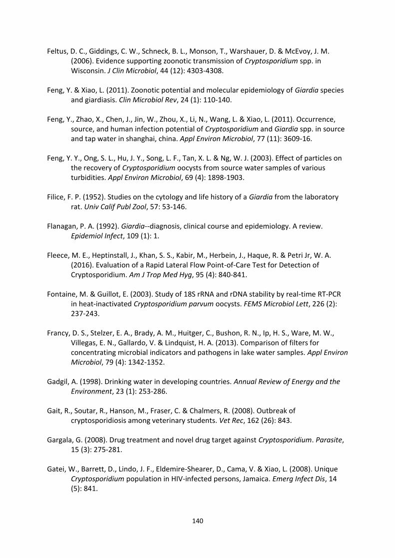
140
Feltus, D. C., Giddings, C. W., Schneck, B. L., Monson, T., Warshauer, D. & McEvoy, J. M. (2006). Evidence supporting zoonotic transmission of Cryptosporidium spp. in Wisconsin. J Clin Microbiol, 44 (12): 4303-4308.
Feng, Y. & Xiao, L. (2011). Zoonotic potential and molecular epidemiology of Giardia species
and giardiasis. Clin Microbiol Rev, 24 (1): 110-140. Feng, Y., Zhao, X., Chen, J., Jin, W., Zhou, X., Li, N., Wang, L. & Xiao, L. (2011). Occurrence,
source, and human infection potential of Cryptosporidium and Giardia spp. in source and tap water in shanghai, china. Appl Environ Microbiol, 77 (11): 3609-16.
Feng, Y. Y., Ong, S. L., Hu, J. Y., Song, L. F., Tan, X. L. & Ng, W. J. (2003). Effect of particles on
the recovery of Cryptosporidium oocysts from source water samples of various turbidities. Appl Environ Microbiol, 69 (4): 1898-1903.
Filice, F. P. (1952). Studies on the cytology and life history of a Giardia from the laboratory
rat. Univ Calif Publ Zool, 57: 53-146. Flanagan, P. A. (1992). Giardia--diagnosis, clinical course and epidemiology. A review.
Epidemiol Infect, 109 (1): 1. Fleece, M. E., Heptinstall, J., Khan, S. S., Kabir, M., Herbein, J., Haque, R. & Petri Jr, W. A.
(2016). Evaluation of a Rapid Lateral Flow Point-of-Care Test for Detection of Cryptosporidium. Am J Trop Med Hyg, 95 (4): 840-841.
Fontaine, M. & Guillot, E. (2003). Study of 18S rRNA and rDNA stability by real-time RT-PCR
in heat-inactivated Cryptosporidium parvum oocysts. FEMS Microbiol Lett, 226 (2): 237-243.
Francy, D. S., Stelzer, E. A., Brady, A. M., Huitger, C., Bushon, R. N., Ip, H. S., Ware, M. W.,
Villegas, E. N., Gallardo, V. & Lindquist, H. A. (2013). Comparison of filters for concentrating microbial indicators and pathogens in lake water samples. Appl Environ Microbiol, 79 (4): 1342-1352.
Gadgil, A. (1998). Drinking water in developing countries. Annual Review of Energy and the
Environment, 23 (1): 253-286. Gait, R., Soutar, R., Hanson, M., Fraser, C. & Chalmers, R. (2008). Outbreak of
cryptosporidiosis among veterinary students. Vet Rec, 162 (26): 843. Gargala, G. (2008). Drug treatment and novel drug target against Cryptosporidium. Parasite,
15 (3): 275-281. Gatei, W., Barrett, D., Lindo, J. F., Eldemire-Shearer, D., Cama, V. & Xiao, L. (2008). Unique
Cryptosporidium population in HIV-infected persons, Jamaica. Emerg Infect Dis, 14 (5): 841.
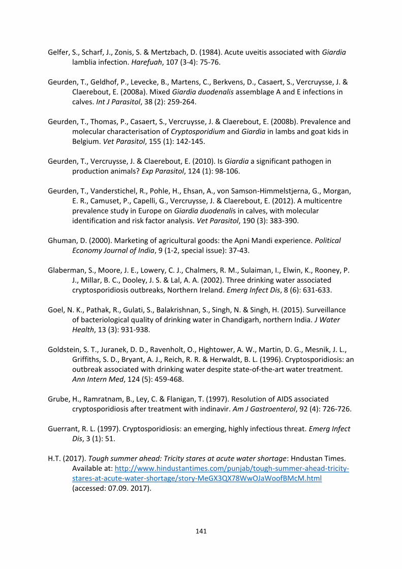
141
Gelfer, S., Scharf, J., Zonis, S. & Mertzbach, D. (1984). Acute uveitis associated with Giardia lamblia infection. Harefuah, 107 (3-4): 75-76.
Geurden, T., Geldhof, P., Levecke, B., Martens, C., Berkvens, D., Casaert, S., Vercruysse, J. &
Claerebout, E. (2008a). Mixed Giardia duodenalis assemblage A and E infections in calves. Int J Parasitol, 38 (2): 259-264.
Geurden, T., Thomas, P., Casaert, S., Vercruysse, J. & Claerebout, E. (2008b). Prevalence and
molecular characterisation of Cryptosporidium and Giardia in lambs and goat kids in Belgium. Vet Parasitol, 155 (1): 142-145.
Geurden, T., Vercruysse, J. & Claerebout, E. (2010). Is Giardia a significant pathogen in
production animals? Exp Parasitol, 124 (1): 98-106. Geurden, T., Vanderstichel, R., Pohle, H., Ehsan, A., von Samson-Himmelstjerna, G., Morgan,
E. R., Camuset, P., Capelli, G., Vercruysse, J. & Claerebout, E. (2012). A multicentre prevalence study in Europe on Giardia duodenalis in calves, with molecular identification and risk factor analysis. Vet Parasitol, 190 (3): 383-390.
Ghuman, D. (2000). Marketing of agricultural goods: the Apni Mandi experience. Political
Economy Journal of India, 9 (1-2, special issue): 37-43. Glaberman, S., Moore, J. E., Lowery, C. J., Chalmers, R. M., Sulaiman, I., Elwin, K., Rooney, P.
J., Millar, B. C., Dooley, J. S. & Lal, A. A. (2002). Three drinking water associated cryptosporidiosis outbreaks, Northern Ireland. Emerg Infect Dis, 8 (6): 631-633.
Goel, N. K., Pathak, R., Gulati, S., Balakrishnan, S., Singh, N. & Singh, H. (2015). Surveillance
of bacteriological quality of drinking water in Chandigarh, northern India. J Water Health, 13 (3): 931-938.
Goldstein, S. T., Juranek, D. D., Ravenholt, O., Hightower, A. W., Martin, D. G., Mesnik, J. L.,
Griffiths, S. D., Bryant, A. J., Reich, R. R. & Herwaldt, B. L. (1996). Cryptosporidiosis: an outbreak associated with drinking water despite state-of-the-art water treatment. Ann Intern Med, 124 (5): 459-468.
Grube, H., Ramratnam, B., Ley, C. & Flanigan, T. (1997). Resolution of AIDS associated
cryptosporidiosis after treatment with indinavir. Am J Gastroenterol, 92 (4): 726-726. Guerrant, R. L. (1997). Cryptosporidiosis: an emerging, highly infectious threat. Emerg Infect
Dis, 3 (1): 51. H.T. (2017). Tough summer ahead: Tricity stares at acute water shortage: Hndustan Times.
Available at: http://www.hindustantimes.com/punjab/tough-summer-ahead-tricity-stares-at-acute-water-shortage/story-MeGX3QX78WwOJaWoofBMcM.html (accessed: 07.09. 2017).
142
Hald, T., Aspinall, W., Devleesschauwer, B., Cooke, R., Corrigan, T., Havelaar, A. H., Gibb, H. J., Torgerson, P. R., Kirk, M. D. & Angulo, F. J. (2016). World Health Organization estimates of the relative contributions of food to the burden of disease due to selected foodborne hazards: a structured expert elicitation. PloS one, 11 (1): e0145839.
Hall, J. N., Moore, S., Harper, S. B. & Lynch, J. W. (2009). Global variability in fruit and
vegetable consumption. Am J Prev Med, 36 (5): 402-409. Hanevik, K., Dizdar, V., Langeland, N. & Hausken, T. (2009). Development of functional
gastrointestinal disorders after Giardia lamblia infection. BMC gastroenterology, 9 (1): 27.
Harris, M., Bose, N. K., Klass, M., Mencher, J. P., Oberg, K., Opler, M. K., Suttles, W. & Vayda,
A. P. (1966). The Cultural Ecology of India's Sacred Cattle [and Comments and Replies]. Curr Anthropol, 7 (1): 51-66.
Helmi, K., Skraber, S., Burnet, J.-B., Leblanc, L., Hoffmann, L. & Cauchie, H.-M. (2011). Two-
year monitoring of Cryptosporidium parvum and Giardia lamblia occurrence in a recreational and drinking water reservoir using standard microscopic and molecular biology techniques. Environ Monit Assess, 179 (1): 163-175.
Hijjawi, N., Meloni, B., Morgan, U. & Thompson, R. (2001). Complete development and long-
term maintenance of Cryptosporidium parvum human and cattle genotypes in cell culture. Int J Parasitol, 31 (10): 1048-1055.
Hijjawi, N., Meloni, B., Ryan, U., Olson, M. & Thompson, R. (2002). Successful in vitro
cultivation of Cryptosporidium andersoni: evidence for the existence of novel extracellular stages in the life cycle and implications for the classification of Cryptosporidium. Int J Parasitol, 32 (14): 1719-1726.
Hill, V. R., Polaczyk, A. L., Hahn, D., Narayanan, J., Cromeans, T. L., Roberts, J. M. &
Amburgey, J. E. (2005). Development of a rapid method for simultaneous recovery of diverse microbes in drinking water by ultrafiltration with sodium polyphosphate and surfactants. Appl Environ Microbiol, 71 (11): 6878-6884.
Hill, V. R., Kahler, A. M., Jothikumar, N., Johnson, T. B., Hahn, D. & Cromeans, T. L. (2007).
Multistate evaluation of an ultrafiltration-based procedure for simultaneous recovery of enteric microbes in 100-liter tap water samples. Appl Environ Microbiol, 73 (13): 4218-4225.
Hill, V. R., Polaczyk, A. L., Kahler, A. M., Cromeans, T. L., Hahn, D. & Amburgey, J. E. (2009).
Comparison of hollow-fiber ultrafiltration to the USEPA VIRADEL technique and USEPA method 1623. J Environment Qual, 38 (2): 822-825.
143
Holbø, A. E. (2017). Viability of Cryptosporidium parvum oocysts in experimentally contaminated parsley and strawberries when subjected to different freezing regimes: Norwegian University of Life Sciences, Ås.
Hopkins, R. M., Meloni, B. P., Groth, D. M., Wetherall, J. D., Reynoldson, J. A. & Thompson, R.
C. A. (1997). Ribosomal RNA sequencing reveals differences between the genotypes of Giardia isolates recovered from humans and dogs living in the same locality. J Parasitol: 44-51.
Hsu, B.-M. & Huang, C. (2007). IMS method performance analyses for Giardia in water under
differing conditions. Environ Monit Assess, 131 (1): 129-134. Hsu, B. M. & Huang, C. (2001). Performances of the immunomagnetic separation method for
Cryptosporidium in water under various operation conditions. Biotechnol Prog, 17 (6): 1114-1118.
Hsu, Y., Liu Severinghaus, L. & Serpell, J. A. (2003). Dog keeping in Taiwan: its contribution to
the problem of free-roaming dogs. J Appl Anim Welf Sci, 6 (1): 1-23. Hunter, P. R., Hughes, S., Woodhouse, S., Syed, Q., Verlander, N. Q., Chalmers, R. M.,
Morgan, K., Nichols, G., Beeching, N. & Osborn, K. (2004). Sporadic cryptosporidiosis case-control study with genotyping. Emerg Infect Dis, 10 (7): 1241.
Hunter, P. R. & Thompson, R. A. (2005). The zoonotic transmission of Giardia and
Cryptosporidium. Int J Parasitol, 35 (11): 1181-1190. Hønsvall, B. K. & Robertson, L. J. (2017). Real-time nucleic acid sequence-based amplification
(NASBA) assay targeting MIC1 for detection of Cryptosporidium parvum and Cryptosporidium hominis oocysts. Exp Parasitol, 172: 61-67.
Ijzerman, M. M., Dahling, D. R. & Fout, G. S. (1997). A method to remove environmental
inhibitors prior to the detection of waterborne enteric viruses by reverse transcription-polymerase chain reaction. J Virol Methods, 63 (1): 145-153.
Indian 15th National Census Survey, 2012. (Available
at: http://www.census2011.co.in/census/state/chandigarh.html Accessed on: 21.06.2017)
Imre, K., Luca, C., Costache, M., Sala, C., Morar, A., Morariu, S., Ilie, M. S., Imre, M. &
Dărăbuş, G. (2013). Zoonotic Cryptosporidium parvum in Romanian newborn lambs (Ovis aries). Vet Parasitol, 191 (1): 119-122.
IndiaAgroNet. (2017). Goat Farming in India [Web page]. Available at:
http://www.indiaagronet.com/indiaagronet/animal_husbandry/animal_husbandry.htm (Accessed on 15.09.2017).

144
Inpankaew, T., Traub, R., Thompson, R. & Sukthana, Y. (2007). Canine parasitic zoonoses in Bangkok temples.
Insulander, M., Silverlås, C., Lebbad, M., Karlsson, L., Mattsson, J. G. & Svenungsson, B.
(2013). Molecular epidemiology and clinical manifestations of human cryptosporidiosis in Sweden. Epidemiol Infect, 141 (05): 1009-1020.
Iqbal, A., Lim, Y. A. L., Surin, J. & Sim, B. L. H. (2012). High diversity of Cryptosporidium
subgenotypes identified in Malaysian HIV/AIDS individuals targeting gp60 gene. PloS one, 7 (2): e31139.
ISO 18744:2016. Microbiology of the food chain - Detection and enumeration of
Cryptosporidium and Giardia in fresh leafy green vegetables and berry fruits. London: British Standards Institution
ISO 15553:2006. Water quality - Isolation and identification of Cryptosporidium oocysts and
Giardia cysts from water. London: British Standards Institution. Jacob, M. J. K. (2016). Filthy to Healthy Nations–India as a Case Study. Proceedings of the
12th International Conference on Energy, Environment, Ecosystems and Sustainable Development (EEESD '16), Venice, Italy.: WSEAS Press. 278-284 pp.
Jacobson, D. & Grinker, L. (1999). A Reverence for Cows: For millenia, cattle have supplied
South Asians with milk, animal power, and dung for fetilizer and fuel. Tractors, petrochemicals, and nuclear power seem unlikely to dethrone these animals from their special place in Indian society. Natural History, 108 (5): 58-63.
Jafari, H., Jalali, M. H. R., Shapouri, M. S. A. & Hajikolaii, M. R. H. (2014). Determination of
Giardia duodenalis genotypes in sheep and goat from Iran. J Parasit Dis, 38 (1): 81-84. Jenkins, M., Trout, J., Higgins, J., Dorsch, M., Veal, D. & Fayer, R. (2002). Comparison of tests
for viable and infectious Cryptosporidium parvum oocysts. Parasitology research, 89 (1): 1-5.
Jenkins, M. C., Trout, J., Abrahamsen, M. S., Lancto, C. A., Higgins, J. & Fayer, R. (2000).
Estimating viability of Cryptosporidium parvum oocysts using reverse transcriptase-polymerase chain reaction (RT-PCR) directed at mRNA encoding amyloglucosidase. J Microbiol Methods, 43 (2): 97-106.
Jex, A. R., Whipp, M., Campbell, B. E., Cacciò, S. M., Stevens, M., Hogg, G. & Gasser, R. B.
(2007). A practical and cost‐effective mutation scanning‐based approach for investigating genetic variation in Cryptosporidium. Electrophoresis, 28 (21): 3875-3883.
Jex, A. R., Pangasa, A., Campbell, B. E., Whipp, M., Hogg, G., Sinclair, M. I., Stevens, M. &
Gasser, R. B. (2008). Classification of Cryptosporidium species from patients with
145
sporadic cryptosporidiosis by use of sequence-based multilocus analysis following mutation scanning. J Clin Microbiol, 46 (7): 2252-2262.
Jirku, M., Valigurová, A., Koudela, B., Krizek, J., Modrý, D. & Slapeta, J. (2008). New species of
Cryptosporidium Tyzzer, 1907 (Apicomplexa) from amphibian host: morphology, biology and phylogeny. Folia parasitologica, 55 (2): 81.
JNNURM. (2006). City Development Plan Chandigarh. Administration, F. S. C.: Chandigarh
Administration. Johnston, A. R., Gillespie, T. R., Rwego, I. B., McLachlan, T. L. T., Kent, A. D. & Goldberg, T. L.
(2010). Molecular epidemiology of cross-species Giardia duodenalis transmission in western Uganda. PLoS Negl Trop Dis, 4 (5): e683.
Jokipii, A. M., Hemilä, M. & Jokipii, L. (1985). Prospective study of acquisition of
Cryptosporidium, Giardia lamblia, and gastrointestinal illness. Lancet, 326 (8453): 487-489.
Kang, B. (2003). The milky way. Outlook India. Available at:
http://www.outlookindia.com/magazine/story/the-milky-way/219289 (accessed: 13.05.2017).
Karabay, O., Tamer, A., Gunduz, H., Kayas, D., Arinc, H. & Celebi, H. (2004). Albendazole
versus metronidazole treatment of adult giardiasis: An open randomized clinical study. World J Gastroenterol, 10 (8): 1215-1217.
Karanis, P. & Kourenti, C. (2004). Waterborne transmission of protozoan parasites: a review
of world-wide outbreaks. Fourth International Giardia Conference and First Combined Giardia - Cryptosporidium Meeting. 20-24 pp.
Karanis, P., Kourenti, C. & Smith, H. (2007). Waterborne transmission of protozoan parasites:
a worldwide review of outbreaks and lessons learnt. J Water Health, 5 (1): 1-38. Khan, S. M., Debnath, C., Pramanik, A. K., Xiao, L., Nozaki, T. & Ganguly, S. (2010). Molecular
characterization and assessment of zoonotic transmission of Cryptosporidium from dairy cattle in West Bengal, India. Vet Parasitol, 171 (1): 41-47.
Kiang, K., Scheftel, J., Leano, F., Taylor, C., Belle-Isle, P., Cebelinski, E., Danila, R. & Smith, K.
(2006). Recurrent outbreaks of cryptosporidiosis associated with calves among students at an educational farm programme, Minnesota, 2003. Epidemiol Infect, 134 (04): 878-886.
Kirk, M. D., Pires, S. M., Black, R. E., Caipo, M., Crump, J. A., Devleesschauwer, B., Döpfer, D.,
Fazil, A., Fischer-Walker, C. L. & Hald, T. (2015). World Health Organization estimates of the global and regional disease burden of 22 foodborne bacterial, protozoal, and viral diseases, 2010: a data synthesis. PLoS Med, 12 (12): e1001921.
146
Korom, F. J. (2000). Holy cow! The apotheosis of Zebu, or why the cow is sacred in Hinduism. Asian Folkl Stud: 181-203.
Kotloff, K. L., Nataro, J. P., Blackwelder, W. C., Nasrin, D., Farag, T. H., Panchalingam, S., Wu,
Y., Sow, S. O., Sur, D., Breiman, R. F., et al. (2013). Burden and aetiology of diarrhoeal disease in infants and young children in developing countries (the Global Enteric Multicenter Study, GEMS): a prospective, case-control study. Lancet, 382 (9888): 209-22.
Kreier, J. (1978). Flagellates of the human intestine and of intestines of other species.
Parasitic protozoa, 2. Kreutner, A. K., Del Bene, V. E. & Amstey, M. S. (1981). Giardiasis in pregnancy. Ame J Obstet
Gynecol, 140 (8): 895-901. Kuhn, R. C., Rock, C. M. & Oshima, K. H. (2002). Effects of pH and magnetic material on
immunomagnetic separation of Cryptosporidium oocysts from concentrated water samples. Appl Environ Microbiol, 68 (4): 2066-2070.
Kumar, S., Nagarajan, M., Sandhu, J., Kumar, N., Behl, V. & Nishanth, G. (2007).
Mitochondrial DNA analyses of Indian water buffalo support a distinct genetic origin of river and swamp buffalo. Anim Genet, 38 (3): 227-232.
Kváč, M., Kouba, M. & Vítovec, J. (2006). Age-related and housing-dependence of
Cryptosporidium infection of calves from dairy and beef herds in South Bohemia, Czech Republic. Vet Parasitol, 137 (3): 202-209.
Kváč, M., Kestřánová, M., Pinková, M., Květoňová, D., Kalinová, J., Wagnerová, P., Kotková,
M., Vítovec, J., Ditrich, O. & McEvoy, J. (2013). Cryptosporidium scrofarum n. sp.(Apicomplexa: Cryptosporidiidae) in domestic pigs (Sus scrofa). Vet Parasitol, 191 (3): 218-227.
Labatiuk, C., Schaefer, F., Finch, G. & Belosevic, M. (1991). Comparison of animal infectivity,
excystation, and fluorogenic dye as measures of Giardia muris cyst inactivation by ozone. Appl Environ Microbiol, 57 (11): 3187-3192.
Lalle, M., Pozio, E., Capelli, G., Bruschi, F., Crotti, D. & Cacciò, S. M. (2005). Genetic
heterogeneity at the β-giardin locus among human and animal isolates of Giardia duodenalis and identification of potentially zoonotic subgenotypes. Int J Parasitol, 35 (2): 207-213.
Landvik, T. (2015). Giardia outbreak in Bergen 2004–what was the source of infection?
Tidsskrift for den Norske lægeforening: tidsskrift for praktisk medicin, ny række, 135 (16): 1435.
Lane, S. & Lloyd, D. (2002). Current trends in research into the waterborne parasite Giardia.
Crit Rev Microbiol, 28 (2): 123-147.
147
Langton, S., Chevennement, R., Nagelkerke, N. & Lombard, B. (2002). Analysing collaborative
trials for qualitative microbiological methods: accordance and concordance. Int J Food Microbiol, 79 (3): 175-181.
Lasek-Nesselquist, E., Welch, D. M. & Sogin, M. L. (2010). The identification of a new Giardia
duodenalis assemblage in marine vertebrates and a preliminary analysis of G. duodenalis population biology in marine systems. Int J Parasitol, 40 (9): 1063-1074.
Lee, Y., Gomez, L., McAuliffe, I. & Tsang, V. (2004). Evaluation of Cryptosporidium parvum
oocyst recovery efficiencies from various filtration cartridges by electrochemiluminescence assays. Lett Appl Microbiol, 39 (2): 156-162.
Leoni, F., Amar, C., Nichols, G., Pedraza-Diaz, S. & McLauchlin, J. (2006). Genetic analysis of
Cryptosporidium from 2414 humans with diarrhoea in England between 1985 and 2000. J Med Microbiol, 55 (6): 703-707.
Letts, M., Davidson, D. & Lalonde, F. (1998). Synovitis secondary to giardiasis in children.
American journal of orthopedics (Belle Mead, NJ), 27 (6): 451-454. Lindberg, A., Lusua, J. & Nevhage, B. (2011). Cryptosporidium i Östersund vintern 2010/2011:
konsekvenser och kostnader av ett stort vattenburet sjukdomsutbrott: Totalförsvarets forskningsinstitut (FOI).
Llorente, M. T., Clavel, A., Goñi, M. P., Varea, M., Seral, C., Becerril, R., Suarez, L. & Gómez-
Lus, R. (2007). Genetic characterization of Cryptosporidium species from humans in Spain. Parasitol Int, 56 (3): 201-205.
Macarisin, D., Bauchan, G. & Fayer, R. (2010a). Spinacia oleracea L. leaf stomata harboring
Cryptosporidium parvum oocysts: a potential threat to food safety. Appl Environ Microbiol, 76 (2): 555-559.
Macarisin, D., Santín, M., Bauchan, G. & Fayer, R. (2010b). Infectivity of Cryptosporidium
parvum oocysts after storage of experimentally contaminated apples. J Food Prot, 73 (10): 1824-1829.
Mackenzie, D. (2011). Goat husbandry: Faber & Faber. Mahambare, V. (2017). The hidden truth behind India’s low refrigerator ownership: The
Economic Times, India. Available at: https://blogs.economictimes.indiatimes.com/et-commentary/the-hidden-truth-behind-indias-low-refrigerator-ownership/ (accessed: 12.09.2017).
Mattilsynet. (2017). Smittestoffer i importerte bær, 2015-2016. In Norwegian Food Safety
Authority, N. V. I., Norwegian University of Life Sciences (ed.). www.mattilsynet.no. Available at: https://www.mattilsynet.no/mat_og_vann/import_av_mat/kommersiell_import_av_
148
mat/rapport__smittestoffer_i_baer_20152016.26160/binary/Rapport%20-%20Smittestoffer%20i%20bær%202015-2016 (accessed: 31.08.2017).
Maurya, P. S., Rakesh, R. L., Pradeep, B., Kumar, S., Kundu, K., Garg, R., Ram, H., Kumar, A. &
Banerjee, P. S. (2013). Prevalence and risk factors associated with Cryptosporidium spp. infection in young domestic livestock in India. Trop Anim Health Prod, 45 (4): 941-946.
McClellan, P. (1998). Cryptosporidium and Giardia: a picture of uncertainty. Sydney Water
Inquiry, Fifth Report, Final Report, 2. McCuin, R. M. & Clancy, J. L. (2005). Methods for the recovery, isolation and detection of
Cryptosporidium oocysts in wastewaters. J Microbiol Methods, 63 (1): 73-88. McHardy, I. H., Wu, M., Shimizu-Cohen, R., Couturier, M. R. & Humphries, R. M. (2014).
Detection of intestinal protozoa in the clinical laboratory. J Clin Microbiol, 52 (3): 712-720.
McKerr, C., Adak, G. K., Nichols, G., Gorton, R., Chalmers, R. M., Kafatos, G., Cosford, P.,
Charlett, A., Reacher, M. & Pollock, K. G. (2015). An outbreak of Cryptosporidium parvum across England & Scotland associated with consumption of fresh pre-cut salad leaves, May 2012. PloS one, 10 (5): e0125955.
Mead, J. R. (2002). Cryptosporidiosis and the challenges of chemotherapy. Drug Resist
Updat, 5 (1): 47-57. Meamar, A. R., Guyot, K., Certad, G., Dei-Cas, E., Mohraz, M., Mohebali, M., Mohammad, K.,
Mehbod, A. A., Rezaie, S. & Rezaian, M. (2007). Molecular characterization of Cryptosporidium isolates from humans and animals in Iran. Appl Environ Microbiol, 73 (3): 1033-1035.
Meisel, J., Perera, D., Meligro, C. & Rubin, C. (1976). Overwhelming watery diarrhea
associated with a Cryptosporidium in an immunosuppressed patient. Gastroenterol, 70 (6): 1156-1160.
Menezes, R. (2008). Rabies in India. Can Med Assoc J, 178 (5): 564-566. Miao, Y. M., Awad, F. M., Franzen, C., Ellis, D. S., Müller, A., Counihan, H. M., Hayes, P. J. &
Gazzard, B. G. (2000). Eradication of cryptosporidia and microsporidia following successful antiretroviral therapy. JAIDS J Acquir Immune Defic Syndr, 25 (2): 124-129.
Microbiology of the food chain — Detection and enumeration of Cryptosporidium and Giardia in fresh leafy green vegetables and berry fruits. In 9, C. I. T. S. (ed.).
Muhsen, K. & Levine, M. M. (2012). A systematic review and meta-analysis of the association
between Giardia lamblia and endemic pediatric diarrhea in developing countries. Clin Infect Dis, 55 (suppl 4): S271-S293.
149
Muthusamy, D., Rao, S. S., Ramani, S., Monica, B., Banerjee, I., Abraham, O. C., Mathai, D. C.,
Primrose, B., Muliyil, J. & Wanke, C. A. (2006). Multilocus genotyping of Cryptosporidium sp. isolates from human immunodeficiency virus-infected individuals in South India. J Clin Microbiol, 44 (2): 632-634.
N.C.R.B. (2014). Accidental deaths and suicides in India. 1 ed. http://ncrb.gov.in: National
Crime Records Bureau, Ministry of Home Affairs, Government of India. Available at: http://ncrb.nic.in/StatPublications/ADSI/ADSI2014/adsi-2014%20full%20report.pdf (accessed: 31.08.2017).
Ng, J., Eastwood, K., Durrheim, D., Massey, P., Walker, B., Armson, A. & Ryan, U. (2008).
Evidence supporting zoonotic transmission of Cryptosporidium in rural New South Wales. Exp Parasitol, 119 (1): 192-195.
Nichols, G., Chalmers, R., Lake, I., Sopwith, W., Regan, M., Hunter, P., Grenfell, P., Harrison,
F. & Lane, C. (2006). Cryptosporidiosis: a report on the surveillance and epidemiology of Cryptosporidium infection in England and Wales. Drinking Water Directorate Contract Number DWI, 70 (2): 201.
Nime, F. A., Burek, J. D., Page, D. L., Holscher, M. A. & Yardley, J. H. (1976). Acute
enterocolitis in a human being infected with the protozoan Cryptosporidium. Gastroenterol, 70 (4): 592-598.
Noordeen, F., Rajapakse, R., Faizal, A. C. M., Horadagoda, N. U. & Arulkanthan, A. (2000).
Prevalence of Cryptosporidium infection in goats in selected locations in three agroclimatic zones of Sri Lanka. Vet Parasitol, 93 (2): 95-101.
O'Handley, R. M. & Olson, M. E. (2006a). Giardiasis and cryptosporidiosis in ruminants. Vet
Clin Food Anim Pract, 22 (3): 623-643. O'Handley, R. M. & Olson, M. E. (2006b). Giardiasis and cryptosporidiosis in ruminants. Vet
Clin North Am Food Anim Pract, 22 (3): 623-643. Olson, M. E., Guselle, N. J., O'Handley, R. M., Swift, M. L., McAllister, T. A., Jelinski, M. D. &
Morck, D. W. (1997). Giardia and Cryptosporidium in dairy calves in British Columbia. The Can Vet J, 38 (11): 703.
Ongerth, J. E. & Stibbs, H. H. (1987). Identification of Cryptosporidium oocysts in river water.
Appl Environ Microbiol, 53 (4): 672-676. Ortega, Y. R. & Adam, R. D. (1997). Giardia: overview and update. Clin Infect Dis, 25 (3): 545-
549. O’Brien, E., McInnes, L. & Ryan, U. (2008). Cryptosporidium GP60 genotypes from humans
and domesticated animals in Australia, North America and Europe. Exp Parasitol, 118 (1): 118-121.
150
Palermo, A. M., Reynoso, A., López Nigro, M., Carballo, M. & Mudry, M. (2004). Teratogenic
evaluation of metronidazole and ornidazole using Drosophila melanogaster as an experimental model. Birth Defects Research part A: Clinical and Molecular Teratology, 70 (4): 157-162.
Paul, S., Chandra, D., Ray, D., Tewari, A., Rao, J., Banerjee, P., Baidya, S. & Raina, O. (2008).
Prevalence and molecular characterization of bovine Cryptosporidium isolates in India. Vet Parasitol, 153 (1): 143-146.
Paul, S., Sharma, D. K., Boral, R., Mishra, A. K., Nayakwadi, S., Banerjee, P. S. & Pawaiya, R. S.
(2014). Cryptosporidiosis in goats: a review. Adv. Anim. Vet. Sci, 2 (3S): 49-54. Payne, P. A., Ridley, R. K., Dryden, M. W., Bathgate, C., Milliken, G. A. & Stewart, P. W.
(2002). Efficacy of a combination febantel-praziquantelpyrantel product, with or without vaccination with a commercial Giardia vaccine, for treatment of dogs with naturally occurring giardiasis. Journal of the American Veterinary Medical Association, 220 (3): 330-333.
Peeters, J. & Villacorta, I. (1995). Cryptosporidium. Biotechnology: Guidelines on techniques
in coccidiosis research, J. Eckert, R. Braun, MW Shirley, and P. Coudert (eds.). ECSC-EC-EAEC, Luxembourg, Luxembourg: 202-240.
Pezzana, A., Vilagines, P., Bordet, F., Coquard, D., Sarrette, B. & Vilagines, R. (2000).
Optimization of the Envirochek capsule method and immunomagnetic separation procedure for the detection of low levels of Cryptosporidium in large drinking water samples. Water Sci Technol, 41 (7): 111-117.
Pingali, P. & Khwaja, Y. (2004). Globalisation of Indian diets and the transformation of food
supply systems. Indian Journal of Agricultural Marketing, 18 (1): 2004. Pirestani, M., Sadraei, J., Zavvar, M. & Vaeznia, H. (2008). Molecular characterization of
Cryptosporidium isolates from human and bovine using 18s rRNA gene in Shahriar county of Tehran, Iran. Parasitology research, 103 (2): 467.
Platts-Mills, J. A., Babji, S., Bodhidatta, L., Gratz, J., Haque, R., Havt, A., McCormick, B. J.,
McGrath, M., Olortegui, M. P. & Samie, A. (2015). Pathogen-specific burdens of community diarrhoea in developing countries: a multisite birth cohort study (MAL-ED). Lancet Glob Health, 3 (9): e564-e575.
Prabhupāda, A. B. S. (1986). Bhagavad-gītā as it is: With the Original Sanskrit Text, Roman
Transliteration, English Equivalents, Translation, and Elaborate Purports: The Bhaktivedanta Book Trust.
Preiser, G., Preiser, L. & Madeo, L. (2003). An outbreak of cryptosporidiosis among
veterinary science students who work with calves. Journal of American College Health, 51 (5): 213-215.
151
Prieto-Lastra, L., Pérez-Pimiento, A., Gonzalez-Sanchez, L. & Iglesias-Cadarso, A. (2006).
Chronic urticaria and angioedema in Giardia lamblia infection. Medicina clinica, 126 (9): 358-359.
Pönka, A., Kotilainen, H., Rimhanen-Finne, R., Hokkanen, P., Hänninen, M., Kaarna, A., Meri,
T. & Kuusi, M. (2009). A foodborne outbreak due to Cryptosporidium parvum in Helsinki, November 2008. Euro surveillance: bulletin Européen sur les maladies transmissibles= European communicable disease bulletin, 14 (28): 29-35.
Qamruddin, A., Keaney, M., McCann, R. & Chadwick, P. (2002). Increased stool sampling
during a waterborne outbreak of cryptosporidiosis does not increase the detection of other faecal pathogens. Journal of clinical pathology, 55 (4): 271-274.
Quilez, J., Sanchez-Acedo, C., Del Cacho, E., Clavel, A. & Causape, A. C. (1996). Prevalence of
Cryptosporidium and Giardia infections in cattle in Aragon (northeastern Spain). Vet Parasitol, 66 (3-4): 139-146.
Quilez, J. b., Torres, E., Chalmers, R. M., Robinson, G., Del Cacho, E. & Sanchez-Acedo, C.
(2008a). Cryptosporidium species and subtype analysis from dairy calves in Spain. Parasitology, 135 (14): 1613-1620.
Quílez, J., Torres, E., Chalmers, R. M., Hadfield, S. J., del Cacho, E. & Sánchez-Acedo, C.
(2008b). Genotype and subtype characterization of Cryptosporidium in lambs and goat kids in Spain Appl Environ Microbiol.
Ramaswamy, N. (1998). Draught animal welfare. Applied Animal Behaviour Science, 59 (1):
73-84. Read, C., Walters, J., Robertson, I. & Thompson, R. (2002). Correlation between genotype of
Giardia duodenalis and diarrhoea. Int J Parasitol, 32 (2): 229. Read, C. M., Monis, P. T. & Thompson, R. C. A. (2004). Discrimination of all genotypes of
Giardia duodenalis at the glutamate dehydrogenase locus using PCR-RFLP. Infection, Genetics and Evolution, 4 (2): 125-130.
Ren, X., Zhao, J., Zhang, L., Ning, C., Jian, F., Wang, R., Lv, C., Wang, Q., Arrowood, M. J. &
Xiao, L. (2012). Cryptosporidium tyzzeri n. sp.(Apicomplexa: Cryptosporidiidae) in domestic mice (Mus musculus). Exp Parasitol, 130 (3): 274-281.
Rhodes, E. R., Hamilton, D. W., See, M. J. & Wymer, L. (2011). Evaluation of hollow-fiber
ultrafiltration primary concentration of pathogens and secondary concentration of viruses from water. Journal of virological methods, 176 (1): 38-45.
Rhodes, E. R., Villegas, L. F., Shaw, N. J., Miller, C. & Villegas, E. N. (2012). A modified EPA
Method 1623 that uses tangential flow hollow-fiber ultrafiltration and heat
152
dissociation steps to detect waterborne Cryptosporidium and Giardia spp. Journal of visualized experiments: JoVE (65).
Robertson, L., Campbell, A. & Smith, H. (1998). Viability of Cryptosporidium parvum oocysts:
assessment by the dye permeability assay. Appl Environ Microbiol, 64 (9): 3544-3545. Robertson, L., Hermansen, L., Gjerde, B., Strand, E., Alvsvåg, J. & Langeland, N. (2006).
Application of genotyping during an extensive outbreak of waterborne giardiasis in Bergen, Norway, during autumn and winter 2004 Appl Environ Microbiol, 72 (3): 2212-2217.
Robertson, L., Gjerde, B., Hansen, E. F. & Stachurska-Hagen, T. (2009). A water
contamination incident in Oslo, Norway during October 2007; a basis for discussion of boil-water notices and the potential for post-treatment contamination of drinking water supplies. J Water Health, 7 (1): 55-66.
Robertson, L. (2014a). Analysis of water for protozoan parasites is expensive – why not use
indicator organisms? Tidsskriftet VANN: Tidsskriftet VANN. pp. 77 - 81. Robertson, L. (2014b). Introduction to Cryptosporidium: the parasite and the disease. In
Cryptosporidium as a Foodborne Pathogen, pp. 1-10: Springer. Robertson, L., Sehgal, R. & Goyal, K. (2015a). An Indian multicriteria-based risk ranking of
foodborne parasites. Food Res Int, 77: 315-319. Robertson, L., Tysnes, K. R., Hanevik, K., Langeland, N., Mørch, K., Hausken, T. & Nygård, K.
(2015b). Dogs as the source of Giardia in Bergen in 2004–barking up the wrong tree? Tidsskrift for den Norske laegeforening: tidsskrift for praktisk medicin, ny raekke, 135 (19): 1718-1720.
Robertson, L. J. & Gjerde, B. (2000). Isolation and enumeration of Giardia cysts,
Cryptosporidium oocysts, and Ascaris eggs from fruits and vegetables. J Food Prot, 63 (6): 775-778.
Robertson, L. J. & Gjerde, B. K. (2007). Cryptosporidium oocysts: challenging adversaries?
Trends Parasitol, 23 (8): 344-347. Robertson, L. J. & Lim, Y. A. L. (2011). Waterborne and environmentally-borne giardiasis. In
Giardia, pp. 29-69: Springer. Robertson, L. J. & Fayer, R. (2012). Cryptosporidium. In Robertson, L. J., Smith, H.V. (ed.)
Foodborne Protozoan Parasites, pp. 32-64. New York: Nova Science Publishers. Robertson, L. J. (2013). Protozoan Parasites: A Plethora of Potentially Foodborne Pathogens.
Food Associated Pathogens: 169.
153
Robertson, L. J. & Chalmers, R. M. (2013). Foodborne cryptosporidiosis: is there really more in Nordic countries? Trends Parasitol, 29 (1): 3-9.
Robertson, L. J., Joshi, H., Utaaker, K. S., Kumar, A., Chaudhary, S., Goyal, K. & Sehgal, R.
(2017). Changes in the seroprevalence of cysticercosis in suspected patients in Chandigarh, India between 1998 and 2014: analysis of 17 years of data. Epidemiology & Infection, 145 (6): 1159-1167.
Robinson, G., Wright, S., Elwin, K., Hadfield, S. J., Katzer, F., Bartley, P. M., Hunter, P. R.,
Nath, M., Innes, E. A. & Chalmers, R. M. (2010). Re-description of Cryptosporidium cuniculus (Apicomplexa: Cryptosporidiidae): Morphology, biology and phylogeny. Int J Parasitol, 40 (13): 1539-1548.
Rochelle, P. A., Marshall, M. M., Mead, J. R., Johnson, A. M., Korich, D. G., Rosen, J. S. & De
Leon, R. (2002). Comparison of in vitro cell culture and a mouse assay for measuring infectivity of Cryptosporidium parvum. Appl Environ Microbiol, 68 (8): 3809-3817.
Rochelle, P. A., Marshall, M. M., Mead, J. & Rosen, J. (2005). Comparing Cell Culture and
Mouse Assays for Measuring Infectivity of Cryptosporidium: IWA Publishing. Roy, S. L., DeLong, S. M., Stenzel, S. A., Shiferaw, B., Roberts, J. M., Khalakdina, A., Marcus,
R., Segler, S. D., Shah, D. D. & Thomas, S. (2004). Risk factors for sporadic cryptosporidiosis among immunocompetent persons in the United States from 1999 to 2001. J Clin Microbiol, 42 (7): 2944-2951.
Ruiz, A., Foronda, P., González, J. F., Guedes, A., Abreu-Acosta, N., Molina, J. M. & Valladares,
B. (2008). Occurrence and genotype characterization of Giardia duodenalis in goat kids from the Canary Islands, Spain. Vet Parasitol, 154 (1): 137-141.
Ryan, U. & Cacciò, S. M. (2013). Zoonotic potential of Giardia. Int J Parasitol, 43 (12): 943-
956. Ryan, U., Paparini, A. & Oskam, C. (2017). New technologies for detection of enteric
parasites. Trends Parasitol. Rzeżutka, A., Nichols, R. A. B., Connelly, L., Kaupke, A., Kozyra, I., Cook, N., Birrell, S. & Smith,
H. V. (2010). Cryptosporidium oocysts on fresh produce from areas of high livestock production in Poland. Int J Food Microbiol, 139 (1): 96-101.
Sachdeva, S., Sachdev, T. R. & Sachdeva, R. (2013). Increasing fruit and vegetable
consumption: challenges and opportunities. Indian journal of community medicine: official publication of Indian Association of Preventive & Social Medicine, 38 (4): 192.
Santın, M., Trout, J. M., Xiao, L., Zhou, L., Greiner, E. & Fayer, R. (2004). Prevalence and age-
related variation of Cryptosporidium species and genotypes in dairy calves. Vet Parasitol, 122 (2): 103-117.
154
Santos, S. R. d., Branco, N., Franco, R. M. B., Paterniani, J. E. S., Katsumata, M., Barlow, P. W. & Gallep, C. (2015). Fluorescence decay of dyed protozoa: differences between stressed and non‐stressed cysts. Luminescence, 30 (7): 1139-1147.
Santín, M., Trout, J. M. & Fayer, R. (2008). A longitudinal study of cryptosporidiosis in dairy
cattle from birth to 2 years of age. Vet Parasitol, 155 (1): 15-23. Santín, M., Trout, J. M. & Fayer, R. (2009). A longitudinal study of Giardia duodenalis
genotypes in dairy cows from birth to 2 years of age. Vet Parasitol, 162 (1): 40-45. Sauch, e. a. (1991). Propidium iodide as an indicator of Giardia cyst viability. Appl Environ
Microbiol, 57 (11): 3243-3247. Savin, C., Sarfati, C., Menotti, J., Jaouhari, J., Wurtzer, S., Garin, Y. & Derouin, F. (2008).
Assessment of cryptodiag for diagnosis of cryptosporidiosis and genotyping Cryptosporidium species. J Clin Microbiol, 46 (8): 2590-2594.
Savioli, L., Smith, H. & Thompson, A. (2006). Giardia and Cryptosporidium join the ‘neglected
diseases initiative’. Trends Parasitol, 22 (5): 203-208. Schrader, C., Schielke, A., Ellerbroek, L. & Johne, R. (2012). PCR inhibitors–occurrence,
properties and removal. J Appl Microbiol, 113 (5): 1014-1026. Schupp, D. G. & Erlandsen, S. L. (1987). A new method to determine Giardia cyst viability:
correlation of fluorescein diacetate and propidium iodide staining with animal infectivity. Appl Environ Microbiol, 53 (4): 704-707.
Scorza, V. & Tangtrongsup, S. (2010). Update on the diagnosis and management of
Cryptosporidium spp infections in dogs and cats. Topics in companion animal medicine, 25 (3): 163.
Scotter, S., Langton, S., Lombard, B., Lahellec, C., Schulten, S., Nagelkerke, N., In't Veld, P. &
Rollier, P. (2001). Validation of ISO method 11290: Part 2. Enumeration of Listeria monocytogenes in foods. Int J Food Microbiol, 70 (1): 121-129.
Sehgal, M. (2017). Rs 2,000 fine for wasting potable water in Chandigarh and adjoining
areas: India Today. Available at: http://indiatoday.intoday.in/story/water-shortage-hits-chandigarh-panchkula-mohali/1/925703.html (accessed: 07.09.2017).
Sharma, P., Sharma, A., Sehgal, R., Malla, N. & Khurana, S. (2013). Genetic diversity of
Cryptosporidium isolates from patients in North India. International Journal of Infectious Diseases, 17 (8): e601-e605.
Shepherd, K. & Wyn-Jones, A. (1996). An evaluation of methods for the simultaneous
detection of Cryptosporidium oocysts and Giardia cysts from water. Appl Environ Microbiol, 62 (4): 1317-1322.
155
Shetty, N. & Prabhu, T. (1988). Evaluation of faecal preservation and staining methods in the diagnosis of acute amoebiasis and giardiasis. Journal of clinical pathology, 41 (6): 694-699.
Shirley, D.-A. T., Moonah, S. N. & Kotloff, K. L. (2012). Burden of disease from
cryptosporidiosis. Current opinion in infectious diseases, 25 (5): 555. Singh, B. B., Ghatak, S., Banga, H. S., Gill, J. P. S. & Singh, B. (2013). Veterinary urban hygiene:
a challenge for India. Rev Sci Tech, 32: 645-56. Sirohi, S. & Chauhan, A. (2011). Current scenario of livestock development and potential
interventions for livelihood improvement: Case of Jharkhand, India. Slifko, T. R., Smith, H. V. & Rose, J. B. (2000). Emerging parasite zoonoses associated with
water and food. Int J Parasitol, 30 (12): 1379-1393. Smith, H. V., Campbell, B. M., Paton, C. A. & Nichols, R. A. B. (2002). Significance of enhanced
morphological detection of Cryptosporidium sp. oocysts in water concentrates determined by using 4′, 6′-diamidino-2-phenylindole and immunofluorescence microscopy. Appl Environ Microbiol, 68 (10): 5198-5201.
Smith, H. V., Nichols, R. A. & Grimason, A. M. (2005). Cryptosporidium excystation and
invasion: getting to the guts of the matter. Trends Parasitol, 21 (3): 133-142. Soba, B. & Logar, J. (2008). Genetic classification of Cryptosporidium isolates from humans
and calves in Slovenia. Parasitology, 135 (11): 1263-1270. Solaymani-Mohammadi, S. & Singer, S. M. (2011). Host immunity and pathogen strain
contribute to intestinal disaccharidase impairment following gut infection. The Journal of Immunology, 187 (7): 3769-3775.
Sprong, H., Cacciò, S. M. & van der Giessen, J. W. (2009). Identification of zoonotic genotypes
of Giardia duodenalis. PLoS Negl Trop Dis, 3 (12): e558. Sprong, H., Cacciò, S. M. & van der Giessen, J. W. (2009). ZOOPNET network and partners
(2009) Identification of zoonotic genotypes of Giardia duodenalis. PLoS Negl Trop Dis, 3 (12): e558.
Stanfield, G., Carrington, E., Albinet, F., Compagnon, B., Dumoutier, N., Hambsch, B.,
Lorthioy, A., Medema, G., Pezoldt, H. & De Roubin, M. (2000). An optimised and standardised test to determine the presence of the protozoa Cryptosporidium and Giardia in water. Water Sci Technol, 41 (7): 103-110.
Stevenson, H. N. C. (1954). Status evaluation in the Hindu caste system. The Journal of the
Royal Anthropological Institute of Great Britain and Ireland, 84 (1/2): 45-65.
156
Stinear, T., Matusan, A., Hines, K. & Sandery, M. (1996). Detection of a single viable Cryptosporidium parvum oocyst in environmental water concentrates by reverse transcription-PCR. Appl Environ Microbiol, 62 (9): 3385-3390.
Stokol, T., Randolph, J., Nachbar, S., Rodi, C. & Barr, S. (1997). Development of bone marrow
toxicosis after albendazole administration in a dog and cat. Journal of the American Veterinary Medical Association, 210 (12): 1753-1756.
Striepen, B., Pruijssers, A. J., Huang, J., Li, C., Gubbels, M.-J., Umejiego, N. N., Hedstrom, L. &
Kissinger, J. C. (2004). Gene transfer in the evolution of parasite nucleotide biosynthesis. Proceedings of the National Academy of Sciences of the United States of America, 101 (9): 3154-3159.
Sudarshan, M. (2005). assessing burden of rabies in India: WHO sponsored national
multicentric rabies survey, 2003. Indian Journal of Community Medicine, 30 (3): 100. Sudarshan, M., Madhusudana, S., Mahendra, B., Rao, N., Narayana, D. A., Rahman, S. A.,
Meslin, F.-X., Lobo, D. & Ravikumar, K. (2007). Assessing the burden of human rabies in India: results of a national multi-center epidemiological survey. International Journal of Infectious Diseases, 11 (1): 29-35.
Sulaiman, I. M., Fayer, R., Bern, C., Gilman, R. H., Trout, J. M., Schantz, P. M., Das, P., Lal, A.
A. & Xiao, L. (2003). Triosephosphate isomerase gene characterization and potential zoonotic transmission of Giardia duodenalis. Emerg Infect Dis, 9 (11): 1444-1452.
Sulaiman, I. M., Hira, P. R., Zhou, L., Al-Ali, F. M., Al-Shelahi, F. A., Shweiki, H. M., Iqbal, J.,
Khalid, N. & Xiao, L. (2005). Unique endemicity of cryptosporidiosis in children in Kuwait. J Clin Microbiol, 43 (6): 2805-2809.
Tamer, G. S., Turk, M., Dagci, H., Pektas, B., Guy, E. C., Guruz, A. Y. & Uner, A. (2007). The
prevalence of cryptosporidiosis in Turkish children, and genotyping of isolates by nested polymerase chain reaction-restriction fragment length polymorphism. Saudi medical journal, 28 (8): 1243-1246.
Teltumbde, A. (2014). No Swachh Bharat without Annihilation of Caste. Economic and
Political weekly, XLIX (45): 11-12. Thompson, R. A. (2004). The zoonotic significance and molecular epidemiology of Giardia
and giardiasis. Vet Parasitol, 126 (1): 15-35. Tiboni, G. M., Marotta, F. & Castigliego, A. P. (2008). Teratogenic effects in mouse fetuses
subjected to the concurrent in utero exposure to miconazole and metronidazole. Reproductive toxicology, 26 (3): 254-261.
Totton, S. C., Wandeler, A. I., Zinsstag, J., Bauch, C. T., Ribble, C. S., Rosatte, R. C. & McEwen,
S. A. (2010). Stray dog population demographics in Jodhpur, India following a population control/rabies vaccination program. Prev Vet Med, 97 (1): 51-57.
157
Totton, S. C., Wandeler, A. I., Ribble, C. S., Rosatte, R. C. & McEwen, S. A. (2011). Stray dog
population health in Jodhpur, India in the wake of an animal birth control (ABC) program. Prev Vet Med, 98 (2): 215-220.
Traub, R., Robertson, I., Irwin, P., Mencke, N., Monis, P. & Thompson, R. (2003). Humans,
dogs and parasitic zoonoses–unravelling the relationships in a remote endemic community in northeast India using molecular tools. Parasitology Research, 90: S156-S157.
Traub, R., Monis, P., Robertson, I., Irwin, P., Mencke, N. & Thompson, R. (2004).
Epidemiological and molecular evidence supports the zoonotic transmission of Giardia among humans and dogs living in the same community. Parasitology, 128 (03): 253-262.
Traub, R., Inpankaew, T., Reid, S. A., Sutthikornchai, C., Sukthana, Y., Robertson, I. D. &
Thompson, R. A. (2009). Transmission cycles of Giardia duodenalis in dogs and humans in Temple communities in Bangkok—a critical evaluation of its prevalence using three diagnostic tests in the field in the absence of a gold standard. Acta tropica, 111 (2): 125-132.
Traub, R. J., Robertson, I. D., Irwin, P. J., Mencke, N. & Thompson, R. A. (2005). Canine
gastrointestinal parasitic zoonoses in India. Trends Parasitol, 21 (1): 42-48. Trout, J. M., Santín, M., Greiner, E. & Fayer, R. (2004). Prevalence of Giardia duodenalis
genotypes in pre-weaned dairy calves. Vet Parasitol, 124 (3): 179-186. Trout, J. M., Santín, M., Greiner, E. & Fayer, R. (2005). Prevalence and genotypes of Giardia
duodenalis in post-weaned dairy calves. Vet Parasitol, 130 (3): 177-183. Trout, J. M., Santín, M., Greiner, E. C. & Fayer, R. (2006). Prevalence and genotypes of
Giardia duodenalis in 1–2 year old dairy cattle. Vet Parasitol, 140 (3): 217-222. Tysnes, K. R., Skancke, E. & Robertson, L. J. (2014). Subclinical Giardia in dogs: a veterinary
conundrum relevant to human infection. Trends Parasitol, 30 (11): 520-527. U.S.-EPA. (2012: EPA 816-R-12-001). Method 1623.1: Cryptosporidium and Giardia in Water
by Filtration/IMS/FA. Uehlinger, F. D., Barkema, H. W., Dixon, B. R., Coklin, T. & O’Handley, R. M. (2006). Giardia
duodenalis and Cryptosporidium spp. in a veterinary college bovine teaching herd. Vet Parasitol, 142 (3): 231-237.
Uehlinger, F. D., Greenwood, S. J., O’Handley, R., McClure, J. T., Coklin, T., Dixon, B. R., de
Boer, M., Zwiers, H. & Barkema, H. W. (2011). Prevalence and genotypes of Giardia duodenalis in dairy and beef cattle in farms around Charlottetown, Prince Edward Island, Canada. Can Vet J, 52 (9): 967.
158
Umejiego, N. N., Li, C., Riera, T., Hedstrom, L. & Striepen, B. (2004). Cryptosporidium parvum
IMP Dehydrogenase Identification of functional, structural and dynamic properties that can be exploited for drug design. J Biol Chem, 279 (39): 40320-40327.
UNDP. (2017). United nations development programme, Sustainable development goals,
Goal 6: Clean water and sanitation, Goal 6 targets. Available at: http://www.undp.org/content/undp/en/home/sustainable-development-goals/goal-6-clean-water-and-sanitation/targets/.
Utaaker, K. S., Huang, Q. & Robertson, L. J. (2015). A reduced-cost approach for analyzing
fresh produce for contamination with Cryptosporidium oocysts and/or Giardia cysts. Food Res Int, 77: 326-332.
Van der Giessen, J. W. B., De Vries, A., Roos, M., Wielinga, P., Kortbeek, L. M. & Mank, T. G.
(2006). Genotyping of Giardia in Dutch patients and animals: a phylogenetic analysis of human and animal isolates. Int J Parasitol, 36 (7): 849-858.
Vashishtha, A. (2014). Indians favour nutrition over 'stodge' as fruit and veg consumption
beats wheat and grain for the first time. Mail Online India. Available at: http://www.dailymail.co.uk/indiahome/indianews/article-2609757/Indians-favour-nutrition-fruit-veg-consumption-beats-wheat-grain-time.html#ixzz4qsZoBGPD (accessed on: 28.05.2017)
Venu, R., Latha, B., Basith, S. A., Raj, G. D., Sreekumar, C. & Raman, M. (2012). Molecular
prevalence of Cryptosporidium spp. in dairy calves in Southern states of India. Vet Parasitol, 188 (1): 19-24.
Vesey, G., Slade, J., Byrne, M., Shepherd, K. & Fricker, C. (1993). A new method for the
concentration of Cryptosporidium oocysts from water. J Appl Bacteriol, 75 (1): 82-86. Vesey, G., Ashbolt, N., Fricker, E., Deere, D., Williams, K., Veal, D. & Dorsch, M. (1998). The
use of a ribosomal RNA targeted oligonucleotide probe for fluorescent labelling of viable Cryptosporidium parvum oocysts. J Appl Microbiol, 85 (3): 429-440.
Victor, H. (2013). Hindustan Times. Available at:
http://www.hindustantimes.com/chandigarh/municipal-corporation-fails-to-contain-stray-cattle-dog-menace/story-m4ifEkWzdSFf5UGkQgEEfO.html (accessed: 13.05.2017).
Waldron, L. S., Ferrari, B. C. & Power, M. L. (2009). Glycoprotein 60 diversity in C. hominis
and C. parvum causing human cryptosporidiosis in NSW, Australia. Exp Parasitol, 122 (2): 124-127.
Walzer, P. D., Wolfe, M. S. & Schultz, M. G. (1971). Giardiasis in travelers. J Infect Dis, 124 (2):
235-7.
159
Wang, R., Wang, H., Sun, Y., Zhang, L., Jian, F., Qi, M., Ning, C. & Xiao, L. (2011). Characteristics of Cryptosporidium transmission in preweaned dairy cattle in Henan, China. J Clin Microbiol, 49 (3): 1077-1082.
Wei, T., Lu, G. & Clover, G. (2008). Novel approaches to mitigate primer interaction and
eliminate inhibitors in multiplex PCR, demonstrated using an assay for detection of three strawberry viruses. J. Virol. Methods, 151 (1): 132-139.
WHO. (1981). Intestinal protozoan and helminthic infections. WHO Tech Rep Ser, 58: 666-
671. WHO. (2004). Guidelines for drinking-water quality, vol. 1: World Health Organization. WHO. (2009). Global health risks: mortality and burden of disease attributable to selected
major risks: World Health Organization. Widerström, M., Schönning, C., Lilja, M., Lebbad, M., Ljung, T., Allestam, G., Ferm, M.,
Björkholm, B., Hansen, A. & Hiltula, J. I. (2014). Large outbreak of Cryptosporidium hominis infection transmitted through the public water supply, Sweden. Emerg Infect Dis, 20 (4): 581-589.
Widmer, G., Orbacz, E. A. & Tzipori, S. (1999). β-Tubulin mRNA as a marker of
Cryptosporidium parvum oocyst viability. Appl Environ Microbiol, 65 (4): 1584-1588. Wielinga, P. R., de Vries, A., van der Goot, T. H., Mank, T., Mars, M. H., Kortbeek, L. M. & van
der Giessen, J. W. (2008). Molecular epidemiology of Cryptosporidium in humans and cattle in The Netherlands. Int J Parasitol, 38 (7): 809-817.
Wilke, H. & Robertson, L. J. (2009). Preservation of Giardia cysts in stool samples for
subsequent PCR analysis. J Microbiol Methods, 78 (3): 292-296. Wohlsen, T., Bates, J., Gray, B. & Katouli, M. (2004). Evaluation of five membrane filtration
methods for recovery of Cryptosporidium and Giardia isolates from water samples. Appl Environ Microbiol, 70 (4): 2318-2322.
Wolska-Kusnierz, B., Bajer, A., Caccio, S., Heropolitanska-Pliszka, E., Bernatowska, E., Socha,
P., van Dongen, J., Bednarska, M., Paziewska, A. & Sinski, E. (2007). Cryptosporidium infection in patients with primary immunodeficiencies. J Pediatr Gastroenterol Nutr, 45 (4): 458-464.
Xiao, L., Escalante, L., Yang, C., Sulaiman, I., Escalante, A. A., Montali, R. J., Fayer, R. & Lal, A.
A. (1999). Phylogenetic analysis of Cryptosporidium parasites based on the small-subunit rRNA gene locus. Appl Environ Microbiol, 65 (4): 1578-1583.
Xiao, L. & Fayer, R. (2008). Molecular characterisation of species and genotypes of
Cryptosporidium and Giardia and assessment of zoonotic transmission. Int J Parasitol, 38 (11): 1239-1255.
160
Xiao, L. & Feng, Y. (2008). Zoonotic cryptosporidiosis. FEMS Immunol Med Microbiol, 52 (3):
309-323. Xiao, L. (2009). Molecular epidemiology of human cryptosporidiosis in developing countries.
In Ortega-Pierres G., C. S., Fayer R., Mank R.T., Smith H.V., Thompson R.C.A. (ed.) vol. 1 Giardia and Cryptosporidium: from molecules to disease., pp. 51-63. Wallingford, UK: CAB International.
Xiao, L. (2010). Molecular epidemiology of cryptosporidiosis: an update. Exp Parasitol, 124
(1): 80-89. Yakub, G. P. & Stadterman-Knauer, K. L. (2000). Evaluation of immunomagnetic separation
for recovery of Cryptosporidium parvum and Giardia duodenalis from high-iron matrices. Appl Environ Microbiol, 66 (8): 3628-3631.
Yu, J.-R., Lee, S.-U. & Park, W.-Y. (2009). Comparative sensitivity of PCR primer sets for
detection of Cryptosporidium parvum. Korean J Parasitol 47 (3): 293-297. Zhou, L., Singh, A., Jiang, J. & Xiao, L. (2003). Molecular surveillance of Cryptosporidium spp.
in raw wastewater in Milwaukee: implications for understanding outbreak occurrence and transmission dynamics. J Clin Microbiol, 41 (11): 5254-5257.
Zintl, A., Proctor, A. F., Read, C., Dewaal, T., Shanaghy, N., Fanning, S. & Mulcahy, G. (2009).
The prevalence of Cryptosporidium species and subtypes in human faecal samples in Ireland. Epidemiol Infect, 137 (02): 270-277.
Zuckerman, U. & Tzipori, S. (2006). Portable continuous flow centrifugation and method
1623 for monitoring of waterborne protozoa from large volumes of various water matrices. J Appl Microbiol, 100 (6): 1220-1227.
Šoba, B., Petrovec, M., Mioč, V. & Logar, J. (2006). Molecular characterisation of
Cryptosporidium isolates from humans in Slovenia. Clin Microbiol Infect, 12 (9): 918-921.

161
8. Compilation of papers

162

I

A reduced-cost approach for analyzing fresh produce for contaminationwith Cryptosporidium oocysts and/or Giardia cysts
Kjersti Selstad Utaaker ⁎, Qirong Huang 1, Lucy J. RobertsonParasitology Lab, Department for Food Safety and Infection Biology, Norwegian University of Life Sciences, Adamstuen Campus, PO Box 8146 Dep., 0033 Oslo, Norway
a b s t r a c ta r t i c l e i n f o
Article history:Received 15 March 2015Received in revised form 27 April 2015Accepted 2 May 2015Available online 9 May 2015
Keywords:AnalysisCryptosporidiumCystGiardiaLettuceOocyst
Fresh produce is a recognized vehicle for transmission of various protozoan parasites, including Toxoplasmagondii, Cyclospora cayetanensis, Giardia duodenalis, and Cryptosporidium spp. For Giardia and Cryptosporidium, aStandard ISOMethod for analyzing fresh produce is being developed, based on the standardmethods for analyz-ing water. Although it is undoubtedly of value to have a Standard Method available, if the Method is very ex-pensive or difficult to perform this may hamper routine surveys, particularly in settings where resources arerestrained, although arguably such settingsmay produce the results ofmost importance. Herewe present amod-ified method for analyzing green leafy vegetables such as lettuce or spinach for Cryptosporidium oocysts andGiardia cysts.Themodifiedmethod is considerably cheaper than the StandardMethod; by using a smaller volume ofmagneticbeads in the immunomagnetic separation (IMS) step and buffers that are complementary to those provided inthe IMS kit, the cost per analysis is reduced significantly.In-house seeding trials resulted in acceptable levels of recovery. Themodifiedmethod has also been trialed in 10different microbiology analysis labs with experience of detecting protozoa, and results have been shown to besatisfactory; recovery rates ranged from 4% to 88% with a mean of 53% for Cryptosporidium and 33% for Giardia.Generally poor results were associated with problems in shipping reagents. This modified method is not pro-posed as an alternative to the Standard Method, but as a complementary approach providing a cheaper optionfor projects on limited budgets or for laboratories performing analyses in situations or countries where applica-tion of the ISO Standard Method is too expensive.
© 2015 Published by Elsevier Ltd.
1. Introduction
Fresh produce is a recognized vehicle for transmission of various pro-tozoan parasites, including Toxoplasma gondii, Cyclospora cayetanensis,Giardia duodenalis, and Cryptosporidium spp.
Although such protozoan parasites do not multiply in foodstuffs,they can survive in or on moist foods for months. Such transmissionroutes can generally only be identified in an outbreak situation inwhich several people become clinically ill due to consumption of con-taminated products. Outbreaks of protozoan foodborne infections arerelatively rare compared with bacterial or viral pathogens, though theepidemiology of this route of infection is evolving and outbreaks or sin-gle cases of infectionmay becomemore numerous in the future (Pollock& Nichols, 2012).
Outbreaks of foodborne protozoan infection in recent years havehighlighted the need for development of a method for investigatingfresh produce for contamination with the transmission stages.
StandardMethods for analysis of water samples for Cryptosporidiumoocysts and Giardia cysts have been available for several years (e.g. ISO15553, 2006; US EPA 1623, 2005). Initial work in developing a StandardMethod for analyzing fresh produce for contamination has largely fo-cused upon these two parasites, and the methods have been basedbroadly on the water method, with elution from the surfaces of thefresh produce as the initial steps (Cook et al., 2006a, 2006b, 2007;Robertson & Gjerde, 2000, 2001). In brief, these methods depend onan elution step, followed by concentration based on centrifugation andimmunomagnetic separation (IMS), and detection by immunofluores-cent antibody testing (IFAT). The method described by Cook et al.(2006a), has been tested in a round-robin interlaboratory trial (Cooket al., 2006b). This method has been used as the basis for the develop-ment of an ISO Standard Method (ISO 18744; Microbiology of the foodchain — Detection and enumeration of Cryptosporidium and Giardia infresh leafy green vegetables and berry fruits), which is in the final stagesof adjustment and approval before release.
Although it is undoubtedly useful to have an ISO Standard Method,choice of a specific method always raises some concerns. In this case,the concerns include that the recovery efficiencies achieved by Cooket al. (2006a, 2007) were not duplicated in independent or semi-
Food Research International 77 (2015) 326–332
⁎ Corresponding author.E-mail address: [email protected] (L.J. Robertson).
1 Present address: Qirong Huang, Furulundtoppen 2c, 0282 Oslo, Norway.
http://dx.doi.org/10.1016/j.foodres.2015.05.0100963-9969/© 2015 Published by Elsevier Ltd.
Contents lists available at ScienceDirect
Food Research International
j ourna l homepage: www.e lsev ie r .com/ locate / foodres
independent studies (e.g. Amoros, Alonso, & Cuesta, 2010; Rzezutkaet al., 2010); even in the validation studies in which ‘expert’ labs partici-pated, although recovery efficiencies forCryptosporidium from raspberrieswere similar to those of the developing lab, the recovery efficienciesof Cryptosporidium from lettuce were significantly lower (Cook et al.,2006b) and also misidentification of oocysts was a problem, despite theparticipating labs being described as ‘expert’. Furthermore, the StandardISOMethod 18744 is for both Cryptosporidium and Giardia, but the publi-cations on which the Standard Method are largely based (Cook et al.,2006a, 2006b), only address Cryptosporidium parvum.
A further concern is the cost of the method, which may result in theuse of the Standard Method being prohibitively expensive, especially incountries where it may provide the most valuable results. Here we de-scribe experiments to develop a reduced cost version of this methodand its validation through independent inter-laboratory trials.
Thus, the intention of this article is not to provide a replacement forISO Method 18744, which we believe should always be used in situa-tions such as outbreak investigation, but to provide a complementarymethod that is cheaper and still robust, as demonstrated by testing ina blinded ring-trial of different laboratories. It may be more suitablefor use in situationswhen amore reduced price is important, for examplein research projects with limited budgets and for laboratories performingroutine analyses in countries where application of the ISO method is tooexpensive.
2. Materials and methods
2.1. Method development
2.1.1. Fresh produceAlthough a range of fresh produce can be contaminated with proto-
zoan parasites, types that are eaten raw are more likely to act as trans-mission vehicles resulting in infection. These produce include berryfruits (which has been particularly associated with outbreaks ofcyclosporiasis) and salad vegetables. The latter has been associatedwith outbreaks of cryptosporidiosis (Robertson & Chalmers, 2013).Thus, for the purposes of method development, all fresh produce usedwere green leafy vegetables (iceberg lettuce), purchased locally at ageneral store and used immediately. For each spiking experiment,30 g samples of lettuce leaves were used.
2.1.2. ParasitesInitial studies were based on Cryptosporidium oocysts and Giardia
cysts isolated from animal fecal samples submitted to the diagnosticlaboratory, purified by sodium chloride flotation, and held refrigerated.Dilutionsweremade using Kova Glasstic slides. For each sample spiked,three control spikes (directly to a Spot-on slide)were used to determinethe size of spike used.
Following the initial studies, testing of the method internally usedcommercially-obtained spike samples (AccuSpike™-IR; WaterborneInc., New Orleans, USA and EasySeed™, TCS Biosciences Ltd, BotolphClaydon, UK) in which known numbers of Cryptosporidium oocystsand Giardia cysts (around 100) have been sorted by flow cytometryinto a precise volume of buffer.
2.1.3. Seeding of samplesFor sample seeding, the lettuce leaves were pre-weighed into a ho-
mogenizer bag with a filter (Seward BA6041/STR filter bag) before thespike was added. The samples were then left to dry for at least 5 h,more often overnight, at ambient temperature.
For initial experiments, parasites were seeded directly into a glycinebuffer and also into glycine buffer eluate obtained from washing non-seeded lettuce samples, such that other contaminants from the lettucesamples were in the buffer solution used for seeding experiments.
2.1.4. Elution of parasites from lettuce samples and concentration of eluateby centrifugation
This method is the same as that described in Draft ISO Method18744. In brief, 200 ml of 1 M glycine was added to the stomacher bagcontaining the spiked lettuce leaves, and mixed well by hand from out-side the bag (external manipulation) to ensure the leaves are covered.The bag containing the leaves and solution was then stomached for1 min in a paddle beater (stomacher). The eluate was collected into50 ml centrifuge tubes and then the bag and produce were rinsed thor-oughly with 2 × 20ml distilled water, whichwas poured into the 50mltubes. The produce and bagwere discarded, and the eluate concentratedby centrifugation at 1550 rcf for 10 min. The supernatant was removedfrom each tube by aspiration, and the pellets resuspended, combined,and re-centrifuged until a single 50 ml tube with a 5 ml concentrateremained. This was resuspended in a weak detergent solution contain-ing SDS (sodium dodecyl sulfate), Tween 80, and antifoam A.
2.1.5. Investigation on reduction in quantity of IMS reagentsIn terms of reagents, IMS is the most expensive step of the protocol.
There is only one supplier of magnetic beads for both Cryptosporidiumand Giardia and buffers (Dynabeads® GC-Combo, Life Technologies,Thermo Fisher Scientific Inc.) and therefore the option of using alterna-tive beads is excluded. However, as the IMS beads are used in excess inthe Standard Method (100 μl of each bead type), it was hypothesizedthat the method may be equally successful with fewer beads.
The protocol provided by the beadmanufacturer is explicitly direct-ed towards the analysis of water sample concentrates, and therefore thedifferent factors in concentrates from fresh produce eluates may lendthemselves tomanipulating themethod so that smaller quantities of re-agents are used. Indeed, the publication by Cook et al. (2006a) statesthat another IMS kit (now no longer commercially available) outper-forms the one used, “particularly when higher particulate densities orelevated divalent cation concentrates are encountered”. Additionally,the work by Robertson and Gjerde (2001) also suggests that the IMSstep could be adjusted and improved for fruit and vegetable analyses,stating “an improved IMS technique thus has the potential to increaserecovery efficiency of the parasites”. Thus, although both these publica-tions (Cook et al., 2006a; Robertson & Gjerde, 2001) discuss the poten-tial for altering or tweaking the IMS step for analysis of fresh produce,their focus is on improving recovery efficiency, while the focus of the re-search described here was on using less reagents (and thereby decreas-ing the cost per analysis), while maintaining an acceptable recoveryefficiency.
2.1.5.1. Reduction of volume of beads used in analysis. IMS was attemptedon spiked concentrates using 100 μl beads (standard volume), 50 μlbeads, 20 μl beads, and 10 μl beads, but with the same buffer volumesas used for each experiment as for when 100 μl of beads are used (i.e.1 ml SL-Buffer A and 1 ml SL-Buffer B for the initial capture). Wheninitial results had identified a potential reduction in bead volume thatcontinued to provide acceptable recovery efficiencies, then this was in-vestigated more closely in three different replicate experiments usingcommercially obtained spiked samples, with seeding into either glycinebuffer (twice) or glycine buffer eluate (once) with five replicates perseeding experiment.
2.1.5.2. Buffering of samples for IMS analysis.As bead reductionwas foundto provide acceptable recovery efficiencies, decreasing the bufferscommensurately was investigated as the buffers cannot be obtainedseparately from the supplier. These experiments were conducted in du-plicatewith seeding of parasites into glycine buffer eluate, withfive rep-licates per seeding experiment.
Results suggested that the buffers are an essential part of the IMSprocedure, and reducing the buffer volumes has a negative effect onrecovery efficiency. Thus, the IMS buffering must be adjusted in orderto maintain recovery efficiency but, at the same time, reduce costs.
327K.S. Utaaker et al. / Food Research International 77 (2015) 326–332
PBS-Tween-20 buffers of different concentrations (ten times concentra-tions of 0.02%, 0.05%, 0.1%, 0.2%, 0.5%) were investigated to supplementSL-buffer A, while a range of commercially available buffers (SurModics)were investigated for supplementing SL-buffer B.
2.2. Final protocol for internal testing and independent inter-laboratorytrials
Based on the results from the testing of reduced volumes of beadsand buffers, a final protocol was described. Following elution and con-centration of the eluate by centrifugation as previously described, theeluate is transferred to a glass L10 tube, alongwith rinsate from the cen-trifuge tube, so that the final volume in the L10 tube is 10 ml. Purifica-tion/isolation by IMS can, then, follow the standard protocol describedfor water by the manufacturer. However, 0.05% PBS-Tween 20 is usedinstead of 10× SL-A buffer of the IMS kit (or 200 μl of 10 × SL-A bufferis used and 800 μl of 0.05% PBS-Tween 20) and a commercially availablebuffer (SurModics StabilZyme AP buffer, abbreviated to SM-SZ here) isused to replace 800 μl of buffer SL-B. Thus, instead of using 1000 μlSL-B buffer as in Draft ISO Method 18744, 200 μl of buffer SL-B is usedalong with 800 μl of SM-SZ.
Followingmixing between the eluate, beads, and buffers for 1 h, thebeads are collected using a magnet, and the eluate, buffers, and anydebris were discarded. The beads are then transferred to a smaller tube(1.5 ml centrifuge tube) in a small volume, the beads collected again,and finally the beads dissociated from the parasites by vigorous shakingin 50 μl 1 M hydrochloric acid.
Detection by IFAT using DAPI and Normarski optics are as describedin the Draft ISO Method 18744 or in the standard methods for analysisof water concentrates (US EPA 1623, 2005; ISO 15553, 2006).
2.2.1. In-house testing of protocolThe protocol developed was tested in the developing lab by another
analyst not involved in the initial developmental work to ensure accept-able in-house recovery efficiencies before initiating an independentround-robin trial of this method. Four replicates and one control ofspiked samples of Romaine lettuce were used, with spiking withcommercially-obtained spike samples (EasySeed™).
2.2.2. Independent multi-laboratory testing of final protocolFor a more thorough and systematic evaluation of the method, a
round-robin test was organized with 10 participating “expert labs”, 8fromwithin Europe, 1 in Malaysia and 1 in Canada. Each lab that agreedto participate was sent a detailed protocol several weeks before the dis-tribution of samples, including a list of the necessary equipment (seeSupplementary material 1). The labs were asked their preferred timefor receiving the samples for analysis, then each lab was sent twotubes (labeled A and B) that they were informed were parasites fortest spiking, two single-welled spot-on slides, and 5 labeled tubes con-taining: a) PBS-Tween 20 (labeled Q4), b) buffer SL-B (labeled as such),c) SurModics StabilZyme AP buffer (labeled SM-SZ), d) Dynabeads anti-Cryptosporidium (labeled as such), e) Dynabeads anti-Giardia (labeled assuch). Some labs had requested other reagents or disposables requiredin the method including anti-foam A and stomacher bags. These wereprovided in the package as requested.
The two spike tubes (A and B) looked identical. In order to save costsof running this trial, the spikes sent were equivalent to 50% EasySeed™spikes that had been prepared in-house. This was done by shaking indi-vidual EasySeed™ spike vials vigorously and then dividing into two ali-quots, each containing 50% of the original spike.
Thus, each participant was sent a seeding sample containing ap-proximately a 50% EasySeed™ size spike and an empty EasySeed tubethat had been cleaned by soaking in 15% sodiumhypochlorite overnight,then washed in hot soapy water three times, and then rinsed, andcontained onlywater (same volumeas for the spike)— that is a negative
control. None of the participants were aware that only one samplecontained parasites.
A trip controlwas sent out on two distributions (analyzed in the send-ing lab), and recovery efficiencies of 54% and 66% for Cryptosporidium re-corded and 44% and 56% for Giardia.
Participants were requested to buy their own leafy green vegetablefor spiking, and to record details on the form provided. Should parasitesbe detected in samples that were not spiked with parasites, these num-berswere taken into account in calculating the recovery efficiency in thespiked samples.
2.2.3. StatisticsDescriptive statistics were used in the development of the mod-
ified method. For comparison of the results obtained in testing themodified method, the approach described by Langton, Chevennement,Nagelkerke, and Lombard (2002) was used.
For the spiked samples, the accuracy of the method is described bythe sensitivity and specificity parameters, that is the percentage ofknown positive testmaterial that were correctly defined as such (sensi-tivity) and the percentage of known negative test correctly identified assuch (specificity). However, as the vegetable used for spiking onto wasnot provided, but actually purchased by the individual analytical labora-tories, there was a risk for the negatives actually being positive due tolow-level contamination. Although themean spike size for each parasitewas found to be 43 for Cryptosporidium and 42 for Giardia based on rep-licate counts, the theoretical amounts (50 of each parasite)were used inthe calculations in order to be conservative in estimating recoveryefficiencies.
In calculating the percentage of false negatives and mean recoveryefficiencies, the confounding factors listed by Scotter et al. (2001)were considered for exclusion of results (temperature abuse duringshipment; clear deviations from the method in the testing laboratory;questionable laboratory performance). Determinationswere performedaccording to the following equations (Cook et al., 2006b):
% falsenegatives ¼ Numberof sampleswherethresholdlevelnotmetNumberof samplestowhichparasiteswereadded 100
% falsepositives
¼ Numberof samplestowhichparasiteswerenotadded; butparasitesweredetectedNumberof samplestowhichparasiteswerenotadded
100:
3. Results
3.1. Reduction in volume of beads used for IMS step
For initial studies with seeding onto lettuce, an acceptable recoveryefficiency was considered to be 30%. Recovery efficiencies from spikinginto glycine will be higher as the elution and concentration steps areexcluded. The lowest volume of beads found to give an acceptablerecovery rate was 20 μl (Tables 1 and 2).
Table 1Initial recovery efficiencies from lettuce samples seededwith Cryptosporidium oocysts andGiardia cysts using different volumes of IMS beads, but non-adjusted volumes of buffers(same volume of buffers used as for 100 μl beads).
Mean % recovery efficiency (n)
Cryptosporidium Giardia
100 μl beads 67 (n = 5) 58 (n = 5)50 μl beads 49 (n = 5) 61 (n = 5)20 μl beads 30 (n = 5) 45 (n = 5)10 μl beads 18 (n = 5) 26 (n = 5)
328 K.S. Utaaker et al. / Food Research International 77 (2015) 326–332
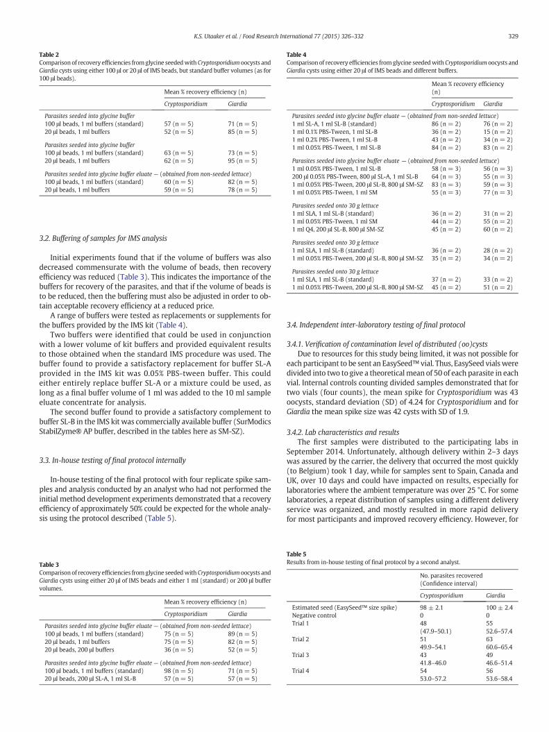
3.2. Buffering of samples for IMS analysis
Initial experiments found that if the volume of buffers was alsodecreased commensurate with the volume of beads, then recoveryefficiency was reduced (Table 3). This indicates the importance of thebuffers for recovery of the parasites, and that if the volume of beads isto be reduced, then the buffering must also be adjusted in order to ob-tain acceptable recovery efficiency at a reduced price.
A range of buffers were tested as replacements or supplements forthe buffers provided by the IMS kit (Table 4).
Two buffers were identified that could be used in conjunctionwith a lower volume of kit buffers and provided equivalent resultsto those obtained when the standard IMS procedure was used. Thebuffer found to provide a satisfactory replacement for buffer SL-Aprovided in the IMS kit was 0.05% PBS-tween buffer. This couldeither entirely replace buffer SL-A or a mixture could be used, aslong as a final buffer volume of 1 ml was added to the 10 ml sampleeluate concentrate for analysis.
The second buffer found to provide a satisfactory complement tobuffer SL-B in the IMS kit was commercially available buffer (SurModicsStabilZyme® AP buffer, described in the tables here as SM-SZ).
3.3. In-house testing of final protocol internally
In-house testing of the final protocol with four replicate spike sam-ples and analysis conducted by an analyst who had not performed theinitial method development experiments demonstrated that a recoveryefficiency of approximately 50% could be expected for the whole analy-sis using the protocol described (Table 5).
3.4. Independent inter-laboratory testing of final protocol
3.4.1. Verification of contamination level of distributed (oo)cystsDue to resources for this study being limited, it was not possible for
each participant to be sent an EasySeed™ vial. Thus, EasySeed vialsweredivided into two to give a theoreticalmean of 50 of each parasite in eachvial. Internal controls counting divided samples demonstrated that fortwo vials (four counts), the mean spike for Cryptosporidium was 43oocysts, standard deviation (SD) of 4.24 for Cryptosporidium and forGiardia the mean spike size was 42 cysts with SD of 1.9.
3.4.2. Lab characteristics and resultsThe first samples were distributed to the participating labs in
September 2014. Unfortunately, although delivery within 2–3 dayswas assured by the carrier, the delivery that occurred the most quickly(to Belgium) took 1 day, while for samples sent to Spain, Canada andUK, over 10 days and could have impacted on results, especially forlaboratories where the ambient temperature was over 25 °C. For somelaboratories, a repeat distribution of samples using a different deliveryservice was organized, and mostly resulted in more rapid deliveryfor most participants and improved recovery efficiency. However, for
Table 2Comparison of recovery efficiencies fromglycine seededwith Cryptosporidium oocysts andGiardia cysts using either 100 μl or 20 μl of IMS beads, but standard buffer volumes (as for100 μl beads).
Mean % recovery efficiency (n)
Cryptosporidium Giardia
Parasites seeded into glycine buffer100 μl beads, 1 ml buffers (standard) 57 (n = 5) 71 (n = 5)20 μl beads, 1 ml buffers 52 (n = 5) 85 (n = 5)
Parasites seeded into glycine buffer100 μl beads, 1 ml buffers (standard) 63 (n = 5) 73 (n = 5)20 μl beads, 1 ml buffers 62 (n = 5) 95 (n = 5)
Parasites seeded into glycine buffer eluate — (obtained from non-seeded lettuce)100 μl beads, 1 ml buffers (standard) 60 (n = 5) 82 (n = 5)20 μl beads, 1 ml buffers 59 (n = 5) 78 (n = 5)
Table 3Comparison of recovery efficiencies fromglycine seededwith Cryptosporidium oocysts andGiardia cysts using either 20 μl of IMS beads and either 1 ml (standard) or 200 μl buffervolumes.
Mean % recovery efficiency (n)
Cryptosporidium Giardia
Parasites seeded into glycine buffer eluate — (obtained from non-seeded lettuce)100 μl beads, 1 ml buffers (standard) 75 (n = 5) 89 (n = 5)20 μl beads, 1 ml buffers 75 (n = 5) 82 (n = 5)20 μl beads, 200 μl buffers 36 (n = 5) 52 (n = 5)
Parasites seeded into glycine buffer eluate — (obtained from non-seeded lettuce)100 μl beads, 1 ml buffers (standard) 98 (n = 5) 71 (n = 5)20 μl beads, 200 μl SL-A, 1 ml SL-B 57 (n = 5) 57 (n = 5)
Table 4Comparison of recovery efficiencies from glycine seededwith Cryptosporidium oocysts andGiardia cysts using either 20 μl of IMS beads and different buffers.
Mean % recovery efficiency(n)
Cryptosporidium Giardia
Parasites seeded into glycine buffer eluate — (obtained from non-seeded lettuce)1 ml SL-A, 1 ml SL-B (standard) 86 (n = 2) 76 (n = 2)1 ml 0.1% PBS-Tween, 1 ml SL-B 36 (n = 2) 15 (n = 2)1 ml 0.2% PBS-Tween, 1 ml SL-B 43 (n = 2) 34 (n = 2)1 ml 0.05% PBS-Tween, 1 ml SL-B 84 (n = 2) 83 (n = 2)
Parasites seeded into glycine buffer eluate — (obtained from non-seeded lettuce)1 ml 0.05% PBS-Tween, 1 ml SL-B 58 (n = 3) 56 (n = 3)200 μl 0.05% PBS-Tween, 800 μl SL-A, 1 ml SL-B 64 (n = 3) 55 (n = 3)1 ml 0.05% PBS-Tween, 200 μl SL-B, 800 μl SM-SZ 83 (n = 3) 59 (n = 3)1 ml 0.05% PBS-Tween, 1 ml SM 55 (n = 3) 77 (n = 3)
Parasites seeded onto 30 g lettuce1 ml SLA, 1 ml SL-B (standard) 36 (n = 2) 31 (n = 2)1 ml 0.05% PBS-Tween, 1 ml SM 44 (n = 2) 55 (n = 2)1 ml Q4, 200 μl SL-B, 800 μl SM-SZ 45 (n = 2) 60 (n = 2)
Parasites seeded onto 30 g lettuce1 ml SLA, 1 ml SL-B (standard) 36 (n = 2) 28 (n = 2)1 ml 0.05% PBS-Tween, 200 μl SL-B, 800 μl SM-SZ 35 (n = 2) 34 (n = 2)
Parasites seeded onto 30 g lettuce1 ml SLA, 1 ml SL-B (standard) 37 (n = 2) 33 (n = 2)1 ml 0.05% PBS-Tween, 200 μl SL-B, 800 μl SM-SZ 45 (n = 2) 51 (n = 2)
Table 5Results from in-house testing of final protocol by a second analyst.
No. parasites recovered(Confidence interval)
Cryptosporidium Giardia
Estimated seed (EasySeed™ size spike) 98 ± 2.1 100 ± 2.4Negative control 0 0Trial 1 48
(47.9–50.1)5552.6–57.4
Trial 2 5149.9–54.1
6360.6–65.4
Trial 3 4341.8–46.0
4946.6–51.4
Trial 4 5453.0–57.2
5653.6–58.4
329K.S. Utaaker et al. / Food Research International 77 (2015) 326–332
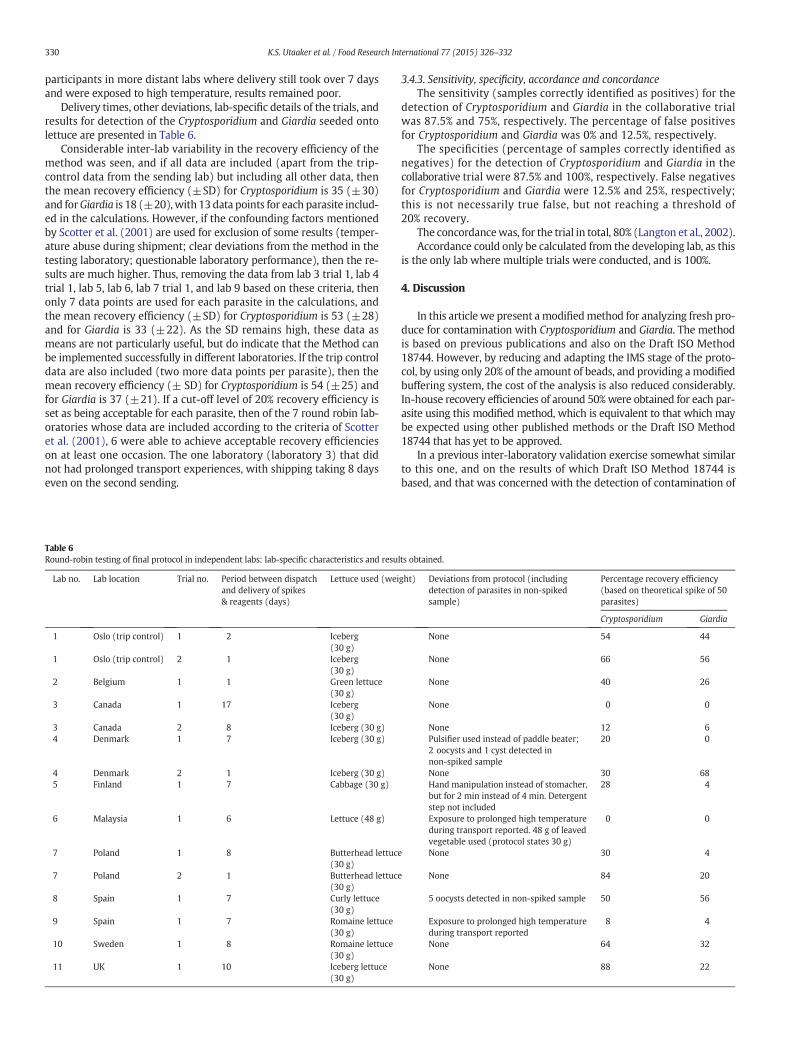
participants in more distant labs where delivery still took over 7 daysand were exposed to high temperature, results remained poor.
Delivery times, other deviations, lab-specific details of the trials, andresults for detection of the Cryptosporidium and Giardia seeded ontolettuce are presented in Table 6.
Considerable inter-lab variability in the recovery efficiency of themethod was seen, and if all data are included (apart from the trip-control data from the sending lab) but including all other data, thenthe mean recovery efficiency (±SD) for Cryptosporidium is 35 (±30)and forGiardia is 18 (±20),with 13data points for each parasite includ-ed in the calculations. However, if the confounding factors mentionedby Scotter et al. (2001) are used for exclusion of some results (temper-ature abuse during shipment; clear deviations from the method in thetesting laboratory; questionable laboratory performance), then the re-sults are much higher. Thus, removing the data from lab 3 trial 1, lab 4trial 1, lab 5, lab 6, lab 7 trial 1, and lab 9 based on these criteria, thenonly 7 data points are used for each parasite in the calculations, andthe mean recovery efficiency (±SD) for Cryptosporidium is 53 (±28)and for Giardia is 33 (±22). As the SD remains high, these data asmeans are not particularly useful, but do indicate that the Method canbe implemented successfully in different laboratories. If the trip controldata are also included (two more data points per parasite), then themean recovery efficiency (± SD) for Cryptosporidium is 54 (±25) andfor Giardia is 37 (±21). If a cut-off level of 20% recovery efficiency isset as being acceptable for each parasite, then of the 7 round robin lab-oratories whose data are included according to the criteria of Scotteret al. (2001), 6 were able to achieve acceptable recovery efficiencieson at least one occasion. The one laboratory (laboratory 3) that didnot had prolonged transport experiences, with shipping taking 8 dayseven on the second sending.
3.4.3. Sensitivity, specificity, accordance and concordanceThe sensitivity (samples correctly identified as positives) for the
detection of Cryptosporidium and Giardia in the collaborative trialwas 87.5% and 75%, respectively. The percentage of false positivesfor Cryptosporidium and Giardia was 0% and 12.5%, respectively.
The specificities (percentage of samples correctly identified asnegatives) for the detection of Cryptosporidium and Giardia in thecollaborative trial were 87.5% and 100%, respectively. False negativesfor Cryptosporidium and Giardia were 12.5% and 25%, respectively;this is not necessarily true false, but not reaching a threshold of20% recovery.
The concordancewas, for the trial in total, 80% (Langton et al., 2002).Accordance could only be calculated from the developing lab, as this
is the only lab where multiple trials were conducted, and is 100%.
4. Discussion
In this article we present amodifiedmethod for analyzing fresh pro-duce for contamination with Cryptosporidium and Giardia. The methodis based on previous publications and also on the Draft ISO Method18744. However, by reducing and adapting the IMS stage of the proto-col, by using only 20% of the amount of beads, and providing a modifiedbuffering system, the cost of the analysis is also reduced considerably.In-house recovery efficiencies of around 50%were obtained for each par-asite using this modified method, which is equivalent to that which maybe expected using other published methods or the Draft ISO Method18744 that has yet to be approved.
In a previous inter-laboratory validation exercise somewhat similarto this one, and on the results of which Draft ISO Method 18744 isbased, and that was concerned with the detection of contamination of
Table 6Round-robin testing of final protocol in independent labs: lab-specific characteristics and results obtained.
Lab no. Lab location Trial no. Period between dispatchand delivery of spikes& reagents (days)
Lettuce used (weight) Deviations from protocol (includingdetection of parasites in non-spikedsample)
Percentage recovery efficiency(based on theoretical spike of 50parasites)
Cryptosporidium Giardia
1 Oslo (trip control) 1 2 Iceberg(30 g)
None 54 44
1 Oslo (trip control) 2 1 Iceberg(30 g)
None 66 56
2 Belgium 1 1 Green lettuce(30 g)
None 40 26
3 Canada 1 17 Iceberg(30 g)
None 0 0
3 Canada 2 8 Iceberg (30 g) None 12 64 Denmark 1 7 Iceberg (30 g) Pulsifier used instead of paddle beater;
2 oocysts and 1 cyst detected innon-spiked sample
20 0
4 Denmark 2 1 Iceberg (30 g) None 30 685 Finland 1 7 Cabbage (30 g) Hand manipulation instead of stomacher,
but for 2 min instead of 4 min. Detergentstep not included
28 4
6 Malaysia 1 6 Lettuce (48 g) Exposure to prolonged high temperatureduring transport reported. 48 g of leavedvegetable used (protocol states 30 g)
0 0
7 Poland 1 8 Butterhead lettuce(30 g)
None 30 4
7 Poland 2 1 Butterhead lettuce(30 g)
None 84 20
8 Spain 1 7 Curly lettuce(30 g)
5 oocysts detected in non-spiked sample 50 56
9 Spain 1 7 Romaine lettuce(30 g)
Exposure to prolonged high temperatureduring transport reported
8 4
10 Sweden 1 8 Romaine lettuce(30 g)
None 64 32
11 UK 1 10 Iceberg lettuce(30 g)
None 88 22
330 K.S. Utaaker et al. / Food Research International 77 (2015) 326–332
fresh produce with only Cryptosporidium oocysts (Cook et al., 2006b),the already spiked samples were distributed to the expert labs (8 labs,all in UK). Three levels of spike were used and also negative controls.In our exercise, only 1 positive spike was used and 1 negative control,and the participants provided their own leafy produce for spiking.However, despite the possibility of some participants using a naturallycontaminated product for spiking, only two labs detected parasites inthe negative control sample. Given the low level of parasites tested inthis case it seems possible that this was natural contamination ratherthan cross-contamination in the laboratory.
In the exercise by Cook et al. (2006b), five out of eight labs detectedCryptosporidium oocysts on samples that were known to be negative,and thus these results compare favorably.
In our study, problems with delivery of the samples (with deliverytaking up to 17 days, and at temperatures of around 28 °C, with thecold block in the package entirely defrosted by delivery) are consideredto have resulted in probable inactivation of the Dynabeads (or the activ-ity of the antibody on the Dynabeads), and hence very low or negativesamples from positive spikes. It seems unlikely that other reagents inthe package would have been so adversely affected, although someparasites may have become deformed. Sending repeat samples with adifferent delivery service resulted in more rapid delivery for most par-ticipants and improved recovery efficiency. However, for participantsin more distant labs where delivery still took over 7 days and were ex-posed to high temperature, recovery efficiency remained very low. Inthe exercise by Cook et al. (2006b), in which delivery presumably oc-curred within a matter of days, one lab failed to detect oocysts at aspike level of between 50 and 100 oocysts. This demonstrates that var-iation does occur in such exercises, and also that shipment is critical.This is a well-recognized weak spot for inter-laboratory trials.
The fact that some shipments took longer time than anticipated, andthat some labs thus received a thawed cooling blockwith temperate re-agents justifies the exclusion of some of the results from this trial. Otherlabs who also received their reagents over the proposed time managedto obtain acceptable recoveries, though these labswere situated inmoretemperate regions and it is plausible to assume that the temperatureexposed to the parcel during the prolonged transport inactivated thebeads. In addition, some labs requested a new set of reagents afterexperiencing poor recoveries in the initial trial, and significantly im-proved their results with reagents shipped more swiftly.
The exclusion of results was based on criteria set by Scotter et al.(2001):
1. Test material had received a significant temperature abuse duringshipment.
2. Testing laboratory had clearly deviated from the specified standardoperating procedure.
3. Performance of laboratory was questionable as indicated by largenumbers of false-positive or false-negative results more than wouldbe expected by chance.
Though these criteria were originally set for the detection of bacteriain food, they can be applied to our study also as the same variables are ofrelevance (temperature, method deviation and detection of false-positives).
Although the results of the inter-laboratory trial did not provide ashigh or stable recovery efficiencies as were found in the developinglab, this is probably to be expected given that the developing lab hadused considerable time on the method and therefore was more experi-enced. Furthermore, shipping problems (delays and exposure to elevat-ed temperatures) apparently affected the recovery efficiencies. It isworth noting that although all the laboratories that participated in thetrial are considered to have expertise in the analysis of samples forthese parasites, method-specific training was not provided. Thissuggests that the modified method is sufficiently simple and robustthat it can be readily implemented into a competent laboratorywithout difficulty.
5. Conclusions
Based on the results of these experiments, we believe that the mod-ified method developed and tested here provides a useful complemen-tary approach to the Draft ISO Method 18744, particularly useful forresearch projects with limited funding or for use in situationswhere re-sources are stretched. The next step for validating thismethodwill be toreturn parallel analyses on naturally contaminated fresh produce.
Acknowledgments
Initial work on the development of this modified method wasperformed as part of the Veg-i-Trade research project, funded byEU through the Framework 7 program (Contract Number: 244994). Fur-ther development and testing of theMethodhas been funded through thePara-Clim-Chandigarh project, partly funded by the Norwegian ResearchCouncil via the New Indigo Partnership Programme (Contract number:227965).
Cost of IMS reagents was subsidized by Life Technologies AS and theEasySeed™was kindly provided by TCS Biosciences Ltd.We are gratefulto both companies for their support.
We are very grateful to all the labs and personnel who verywillinglyparticipated in the inter-laboratory testing of the modified methodincluding:
Stéphane De Craeye (Belgian Scientific Institute for Public Health,Brussels, Belgium).
Brent Dixon, Asma Iqbal and Ryan Boone (Bureau of MicrobialHazards, Food Directorate, Health Canada, Canada).
Heidi Huus Petersen (National Veterinary Institute, Technical Uni-versity of Denmark, Denmark).
Tiina Thure (MetropoliLab, Finland).Yvonne Lim, Redzuan Naziri and Reena Richard (Department of Par-
asitology, Faculty of Medicine, University of Malaya, Malaysia).Agnieszka Kaupke and Artur Rzezutka (Department of Food and En-
vironmental Virology, National Veterinary Research Institute, Poland).Hipólito Gómez-Couso, Aurora Reboredo-Fernández and Elvira
Ares-Mazás (Department of Microbiology and Parasitology, Universityof Santiago de Compostela, Spain).
Inmaculada Amorós and Jose L. Alonso (Institute of Water Engineer-ing and Environment, Technical University of Valencia, Spain).
Karin Jacobsson (National Food Agency, Sweden).Guy Robinson (Cryptosporidium Reference Unit, Public Health
Wales, UK).
Appendix A. Supplementary data
Supplementary data to this article can be found online at http://dx.doi.org/10.1016/j.foodres.2015.05.010.
References
Amoros, I., Alonso, J. L., & Cuesta, G. (2010). Cryptosporidium oocysts and Giardia cysts onsalad products irrigated with contaminated water. Journal of Food Protection, 73,1138–1140.
Anonymous (2005). United States Environmental Protection Agency (USEPA).Method 1623:Cryptosporidium and Giardia in water by filtration/IMS/FA. EPA-815-R-05-002. Wash-ington, DC: USA EPA. Available at: http://www.epa.gov/microbes/documents/1623de05.pdf.
Anonymous (2006). Water quality: isolation and identification of Cryptosporidium oocystsand Giardia cysts fromwater. ISO 15553. Geneva, Switzerland: International Organiza-tion for Standardization.
Cook, N., Nichols, R. A., Wilkinson, N., Paton, C. A., Barker, K., & Smith, H. V. (2007). Develop-ment of a method for detection of Giardia duodenalis cysts on lettuce and for simulta-neous analysis of salad products for the presence of Giardia cysts and Cryptosporidiumoocysts. Applied and Environmental Microbiology, 73, 7388–7391.
Cook, N., Paton, C. A., Wilkinson, N., Nichols, R. A., Barker, K., & Smith, H. V. (2006a). To-wards standard methods for the detection of Cryptosporidium parvum on lettuceand raspberries. Part 1: Development and optimization of methods. InternationalJournal of Food Microbiology, 109, 215–221.
Cook, N., Paton, C. A., Wilkinson, N., Nichols, R. A., Barker, K., & Smith, H. V. (2006b). To-wards standard methods for the detection of Cryptosporidium parvum on lettuce
331K.S. Utaaker et al. / Food Research International 77 (2015) 326–332
and raspberries. Part 2: Validation. International Journal of Food Microbiology, 109,222–228.
ISO (International Organizaton for Standardization) (2006). Water quality — Isolation andidentification of Cryptosporidium oocysts and Giardia cysts from water. Available at:http://www.iso.org/iso/iso_catalogue/catalogue_t/catalogue_detail.htm?csnumber=39804, Last revised, 2010.
Langton, S. D., Chevennement, R., Nagelkerke, N., & Lombard, B. (2002). Analysing collab-orative trials for qualitative microbiological methods: Accordance and concordance.International Journal of Food Microbiology, 79, 175–181.
Pollock, K. G., & Nichols, G. (2012). Foodborne protozoa and outbreak investigations.In L. J. Robertson, & H. V. Smith (Eds.), Foodborne Parasitic Protozoa. NovaPublishers9781614700081.
Robertson, L. J., & Chalmers, R. M. (2013). Foodborne cryptosporidiosis: Is there reallymore in Nordic countries? Trends in Parasitology, 29, 3–9.
Robertson, L. J., & Gjerde, B. (2000). Isolation and enumeration of Giardia cysts, Cryptospo-ridium oocysts and Ascaris eggs from fruits and vegetables. Journal of Food Protection,63, 775–778.
Robertson, L. J., & Gjerde, B. (2001). Factors affecting recovery efficiency in isolation ofCryptosporidium oocysts and Giardia cysts from vegetables for standard method de-velopment. Journal of Food Protection, 64, 1799–1805.
Rzezutka, A., Nichols, R. A., Connelly, L., Kaupke, A., Kozyra, I., Cook, N., et al. (2010). Cryp-tosporidium oocysts on fresh produce from areas of high livestock production inPoland. International Journal of Food Microbiology, 139, 96–101.
Scotter, S. L., Langton, S., Lombard, B., Lahellec, C., Schulten, S., Nagelkerke, N., et al.(2001). Validation of ISO method 11290: Part 2. Enumeration of Listeriamonocytogenes in foods. International Journal of Food Microbiology, 70, 121–129.
332 K.S. Utaaker et al. / Food Research International 77 (2015) 326–332

II

Contents lists available at ScienceDirect
International Journal of Food Microbiology
journal homepage: www.elsevier.com/locate/ijfoodmicro
Keeping it cool: Survival of Giardia cysts and Cryptosporidium oocysts onlettuce leaves
Kjersti Selstad Utaaker⁎, Eystein Skjerve, Lucy J. RobertsonDepartment for Food Safety and Infection Biology, Norwegian University of Life Sciences, Adamstuen Campus, PO Box 8146 Dep., 0033 Oslo, Norway
A R T I C L E I N F O
Keywords:Cryptosporidium oocystGiardia cystSurvivalFoodborne infectionViabilityRefrigeration
A B S T R A C T
Fresh produce has been recognized as a vehicle for transmission of protozoan parasites for many years, and thereare numerous publications regarding their occurrence on such foodstuffs, indicating their potential importanceas foodborne parasites. Nevertheless, few studies have been published regarding the effectiveness of thistransmission route, and whether contamination is likely to result in transmission. The purpose of this study wasto assess the viability of Cryptosporidium oocysts and Giardia cysts, two protozoa associated with both waterborneand foodborne transmission, by spiking fresh produce (lettuce leaves) with viable transmission stages anddetermining changes in viability. These investigations were performed under different conditions and over timespans that may be used in a regular household; a fridge at 4 °C, under ambient temperatures exposed to naturalcycles of light during night and day, and inside a cupboard to ensure no light exposure, for a duration of up totwo weeks, or as long as the produce remained visually palatable. The major finding from this study is thatwhereas both Cryptosporidium oocysts and Giardia cysts survive well when kept moist and refrigerated, survivalof Giardia cysts was abrogated on lettuce at room temperature. Indeed, almost 50% die-off of Giardia cysts wasrecorded within the first 24 h.
Cryptosporidium oocysts had a stable viability throughout the experiment under all the conditionsinvestigated, indicating that fresh produce is a suitable transmission vehicle for Cryptosporidium, even ifcontamination occurs on-farm and the parasites are exposed to non-favourable storage conditions, as may becommon in developing countries.
Giardia cysts were not as robust as Cryptosporidium oocysts, and would be probably unlikely to survive underambient storage conditions on-farm, during sale, or at home. However, if kept refrigerated, then somecontaminating Giardia cysts may remain viable and therefore may pose a threat to the consumer.
Thus, as the cold chain for transport and storage of fresh produce improves, it is important that similarimprovements are implemented to reduce the contamination of fresh produce with parasite transmission stages.
1. Introduction
The protozoan parasites, Cryptosporidium spp. and Giardia duodenalisare among the most frequently found intestinal protozoan parasites inhumans worldwide. Both parasites can cause diarrhoeal disease, and aglobal study revealed that Cryptosporidium spp. and G. duodenalis aretwo of the most common aetiological agents in paediatric diarrhoea indeveloping countries, and are associated with mortality as well asmorbidity (Kotloff et al., 2013). Also, 8–19% of diarrhoeal diseases canbe attributed to Cryptosporidium in developing countries (Gatei et al.,2006), and 10% of the population in developing countries excretesoocysts. In developed countries, this proportion is estimated to be 1–3%(Lozano et al., 2012). For G. duodenalis, an estimated 280 million casesoccur annually (Lane and Lloyd, 2002). Both parasites have long been
recognized as being potentially waterborne pathogens, and manyoutbreaks of waterborne cryptosporidiosis and waterborne giardiasishave been described. Out of 199 reported outbreaks of human diseasedue to waterborne transmission between 2004 and 2010, Cryptospor-idium was the aetiological agent for around 60%, and Giardia was theaetiological agent for around 35% (Baldursson and Karanis, 2011).
Food, particularly fresh produce eaten raw, has also been recog-nized as a potential transmission vehicle for these parasites. Althoughwashing fresh produce may reduce the risk of contaminated food beingingested, numerous outbreaks demonstrate that washing is not alwayseffective. Contamination of food has been considered to occur eitherdirectly from food-handlers, perhaps infected themselves or in closecontact with an infected person or animal, or from contact with acontaminated environment. Such environmental contamination can be
http://dx.doi.org/10.1016/j.ijfoodmicro.2017.05.009Received 9 February 2017; Received in revised form 3 May 2017; Accepted 13 May 2017
⁎ Corresponding author.E-mail address: [email protected] (K.S. Utaaker).
International Journal of Food Microbiology 255 (2017) 51–57
Available online 15 May 20170168-1605/ © 2017 Elsevier B.V. All rights reserved.
MARK
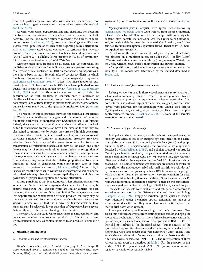
from soil, particularly soil amended with faeces or manure, or fromwater such as irrigation water or wash water along the food chain (Cookand Lim, 2012).
As with waterborne cryptosporidiosis and giardiasis, the potentialfor foodborne transmission is considered rather similar for bothparasites. Indeed, one recent estimate regarding the burden of food-borne disease (Hald et al., 2016), concluded that Cryptosporidium andGiardia were quite similar to each other regarding source attribution.Kirk et al. (2015) used expert elicitation to estimate that whereasaround 15% of giardiasis cases were foodborne (uncertainty intervals(UI) of 0.08–0.27), a slightly lower proportion (13%) of cryptospor-idiosis cases were foodborne (UI of 0.07–0.24).
Although these data are based on all cases, not just outbreaks, thepublished outbreak data tend to indicate a different pattern. Accordingto a review article published in 2013 on foodborne cryptosporidiosis,there have been at least 18 outbreaks of cryptosporidiosis in whichfoodborne transmission has been epidemiologically implicated(Robertson and Chalmers, 2013). At least two more foodborne out-breaks (one in Finland and one in UK) have been published subse-quently and are not included in that review (Åberg et al., 2015; McKerret al., 2012), and 8 of these outbreaks were directly linked toconsumption of fresh produce. In contrast, only 9 outbreaks ofgiardiasis with foodborne transmission proven or implicated have beendocumented, and of these it may be questionable whether some of theseoutbreaks were really due to the apparently implicated food (Cook andLim, 2012).
The reason for this discrepancy between the perceived importanceof Giardia as a foodborne pathogen and the number of reportedfoodborne outbreaks, as compared with Cryptosporidium, is of interest.Indeed, the same reasons that Cryptosporidium and Giardia are bothsuited to waterborne transmission have been cited as to why they arealso suited to transmission by foods: they are shed in high concentra-tions from infected hosts, the infectious dose is low, and they are robust,surviving a number of different environmental pressures. However,whether these factors are of the same importance for foodbornetransmission as waterborne transmission may be less clear, and otherfactors may be of relevance in either transmission or recognition oftransmission. For example, the more zoonotic nature of some species ofCryptosporidium, such as C. parvum, that enables direct transmissionfrom animals, may mean that the relative proportion of foodborneinfections is lower in comparison with other routes, such as fromanimals or via contaminated water (Hald et al., 2016). Alternatively, itis possible that the more acute symptoms of cryptosporidiosis comparedwith giardiasis may give rise to more rapid diagnosis, and thus thepossibility of proper investigation and source attribution.
A third possibility is that food is, indeed, a less efficient transmissionvehicle for Giardia than for Cryptosporidium, and, therefore, despiteexperts considering that food and water are similar vehicles for bothparasites, this is not the case. It is possible that Giardia cysts may haveless opportunity for contamination of fresh produce, that they may bemore easily removed from contaminated produce by food preparationwashing procedures, or that the survival of Giardia cysts on foodmatrices may be relatively lower than that of Cryptosporidium oocysts.Data on these possibilities are lacking in the literature.
The objective of this study was to investigate the last possibility, anddetermine whether the relative survival of Giardia cysts andCryptosporidium oocysts as contaminants of fresh produce is similar ornot.
2. Materials and methods
2.1. Giardia cysts and Cryptosporidium oocysts
Giardia duodenalis cysts, H3 isolate belonging to Assemblage B,were obtained from a commercial supplier (Waterborne Inc., NewOrleans, USA) and their initial viability was determined shortly after
arrival and prior to contamination by the method described in Section2.3.
Cryptosporidium parvum oocysts, with species identification byHønsvall and Robertson (2017) were isolated from faeces of naturallyinfected calves by salt flotation. For one sample with very high fatcontent, ethyl acetate sedimentation was used prior to salt flotation,and, as considerable fat quantities remained after flotation, was furtherpurified by immunomagnetic separation (IMS) (Dynabeads® GC-Com-bo, Applied Biosystems™).
To determine the concentration of (oo)cysts, 10 μl of diluted stockwas pipetted on a multispot microscope slide (C.A. Hendley (Essex)LTD), stained with a monoclonal antibody (mAb; Aqua-glo, WaterborneInc., New Orleans, USA) before enumeration and further dilution.
After purification, and immediately prior to the experiments, theviability of the oocysts was determined by the method described inSection 2.3.
2.2. Food matrix used for survival experiments
Iceberg lettuce was used in these experiments as a representative ofa food matrix commonly eaten raw. The lettuce was purchased from agreengrocer and prior to the experiments, 3 leaves were taken fromboth internal and external layers of the lettuce, weighed, and the intactleaves were analysed for contamination with Giardia cysts and/orCryptosporidium oocysts using a previously published and indepen-dently validated protocol (Utaaker et al., 2015). None of the sampleswere found to be contaminated.
2.3. Assessment of parasite viability
Both prior to the experiments, and throughout the experiments, theviability was assessed based on morphology and inclusion and exclu-sion of the vital dyes 4′,6-diamino-2-phenylindole (DAPI) and propi-dium iodide (PI). For Cryptosporidium, the protocol for staining was asdescribed by Campbell et al. (1992), and a similar protocol was used forGiardia cysts. In order to identify the parasites eluted from the lettuce, amonoclonal antibody (mAb; Aqua-glo, Waterborne Inc., New Orleans,USA) was added to the suspension in the final 15 min of the stainingprocedure. The stained sediment was examined in suspension (with thecover slip on the microscope sealed with nail varnish to avoid drying)by fluorescence microscopy using a Leica DMCB microscope equippedwith a UV filter block (350-nm excitation, 450-nm emission) for DAPIand a green filter block (500-nm excitation, 630-nm emission) for PI.Nomarski (differential interference contrast) optics on the same micro-scope was used to examine morphology of individual cysts and oocysts.
The cysts and oocysts were evaluated and categorised according toexclusion or inclusion of the different stains and their morphology(Campbell et al., 1992). Empty or shrunken (ghost) cysts and oocystswere identified under Nomarski optics, containing no nuclei orshrunken residues thereof. They were also non-refractile, apart fromthe residual body when present.
PI+ cysts and oocysts fluoresce bright red under the green filterblock; this fluorescence varies from distinct points corresponding to thesporozoite/trophozoite nuclei, to a more diffuse fluorescence within thecyst or oocyst. Cysts and oocysts were categorised as DAPI+, PI− ifthey did not include PI (as described above), but the nuclei of thesporozoites/trophozoites fluoresced a distinctive sky blue under the UVfilter block. Cysts and oocysts that were neither PI+, nor “ghosts”, andwhich showed either rim fluorescence or absence thereof under UVfilter block were considered DAPI−, PI−. The interpretation of thesevarious appearances are described in Table 1. For the purposes of thisstudy, DAPI+, PI− parasites and DAPI−, PI− parasites were summedtogether as viable or potentially viable.
K.S. Utaaker et al. International Journal of Food Microbiology 255 (2017) 51–57
52
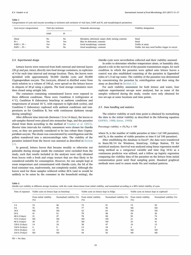
2.4. Experimental design
Lettuce leaves were removed from both external and internal layers(30 g) and put, intact, directly into food storage containers, in replicatesof 4 for each time interval and storage location. Then, the leaves weresprinkled with approximately 50,000 Giardia cysts and 50,000Cryptosporidium oocysts. The (oo)cysts, diluted in distilled water fromstock solution to a volume of 100 μl, were spread on the lettuce leavesin aliquots of 20 μl using a pipette. The food storage containers werethen closed using airtight lids.
The containers containing contaminated leaves were exposed tothree different conditions. These were: Condition A (refrigerator at4 °C), Condition B (laboratory benchtop at ambient conditions andtemperatures of around 18 °C, with exposure to light-dark cycles), andCondition C (laboratory cupboard with ambient conditions and tem-peratures as for Condition B, but with continuous darkness exceptduring sampling).
After different time intervals (between 1 h to 14 days), the leaves orsub-samples thereof were placed into stomacher bags, and the parasiteseluted from them according to the method of Utaaker et al. (2015).Shorter time intervals for viability assessment were chosen for Giardiacysts, as they are generally considered to be less robust than Cryptos-poridium oocysts. The eluate was concentrated by centrifugation and thepellets transferred into a microcentrifuge tube. The viability of theparasites isolated from the leaves was assessed as described in Section2.3.
In general, lettuce leaves that became mouldy or otherwise notpalatable during storage inside the container were excluded from thestudy, such that results included in the analyses were only obtainedfrom leaves with a fresh and crispy texture that are thus likely to beconsidered suitable for consumption. However, for one sample kept atroom temperature and contaminated with Giardia cysts, the lid of thefood container was, inadvertently, not completely sealed. Although theleaves used for these samples withered within 20 h (and so would beunlikely to be eaten by the consumer in the household setting), the
Giardia cysts were nevertheless collected and their viability assessed.In order to determine whether temperature alone, or humidity also,
played a role in the survival of the parasite transmission stages, for eachcondition in which the parasites were spiked onto lettuce leaves acontrol was also established consisting of the parasites in Eppendorftubes of 1.5 ml tap water. The viability of the parasites was determinedby concentrating the parasites by centrifugation and then using theassay as described in Section 2.3.
For each viability assessment for both lettuce and water, fourreplicate experimental set-ups were analysed, but as some of thesamples withered during the study, results were only obtained fortwo set-ups at some locations and time points.
2.5. Data handling and statistics
The relative viability at each time point is obtained by normalisingthe data to the initial viability as described in the following equation(AWWA, 1988; Sattar, 1999):
Percentage viability = (N N ) × 100t 0
where Nt is the number of viable parasites at time t (of 100 parasites),and N0 is the number of viable parasites at time 0 (of 100 parasites).
After establishing the database in Excel®, the data were transferredto Stata/SE/14 for Windows, StataCorp, College Station, TX forstatistical analyses. Survival was analysed using linear regression modelusing method as a categorical variable and time (log 10 h) as acontinuous predictor was utilised, and a follow up logistic regressioncomparing the viability data of the parasites on the lettuce from initialcontamination point until final sampling point. Standard graphicalmethods were used to assess mode fits and residual patterns.
Table 1Categorisation of cysts and oocysts according to inclusion and exclusion of vital dyes, DAPI and PI, and morphological parameters.
Cyst/oocyst categorisation Vital dye inclusion Nomarski microscopy Viability designation
DAPI PI
Ghost No No Shrunken, deformed, empty shell, lacking contents DeadPI+ Yes Yes May be deformed, contents DeadDAPI+, PI− Yes No Good morphology, contents Viable at assayDAPI−, PI− No No Good morphology, contents Viable, but may need further trigger to excyst
Table 2Giardia cyst viability at different storage locations, with the crude observations from initial viability, and normalised according to a 80% initial viability of cysts.
Time of exposure Viable cysts on lettuce kept on benchtop Viable cysts on lettuce kept in fridge Viable cysts on lettuce kept in cupboard
From initial viability(%)
Normalised viability (%) From initial viability(%)
Normalised viability (%) From initial viability(%)
Normalised viability (%)
1 h 72 90 73 91 74 934 h 76 95 77 96 ND ND18 h ND ND 52 65 ND ND24 h 41 51 78 98 24 3048 h (2 days) 21 27 78 98 59 7472 h (3 days) ND ND ND ND 31 3996 h (4 days) 30 37 ND ND 33 41120 h (5 days) 14 18 38 47 10 13144 h (6 days) 4 5 14 18 4 5192 h (8 days) 13 16 53 66 ND ND216 h (9 days) ND ND 44 55 ND ND
ND = No data.
K.S. Utaaker et al. International Journal of Food Microbiology 255 (2017) 51–57
53
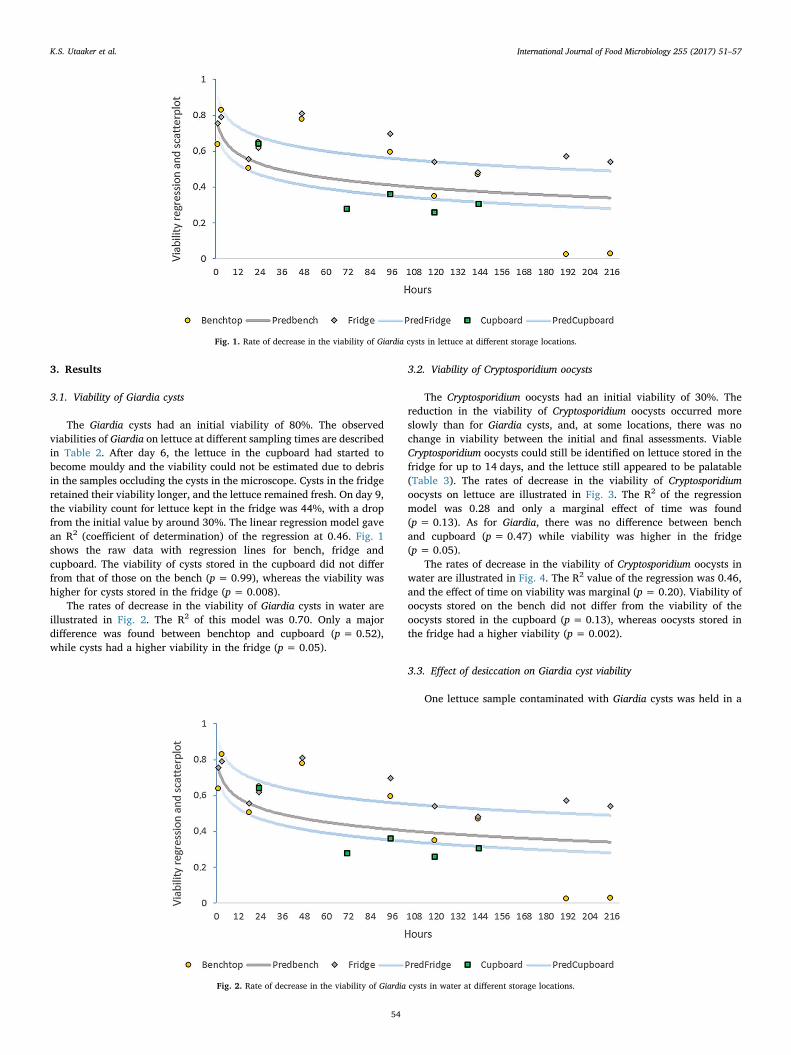
3. Results
3.1. Viability of Giardia cysts
The Giardia cysts had an initial viability of 80%. The observedviabilities of Giardia on lettuce at different sampling times are describedin Table 2. After day 6, the lettuce in the cupboard had started tobecome mouldy and the viability could not be estimated due to debrisin the samples occluding the cysts in the microscope. Cysts in the fridgeretained their viability longer, and the lettuce remained fresh. On day 9,the viability count for lettuce kept in the fridge was 44%, with a dropfrom the initial value by around 30%. The linear regression model gavean R2 (coefficient of determination) of the regression at 0.46. Fig. 1shows the raw data with regression lines for bench, fridge andcupboard. The viability of cysts stored in the cupboard did not differfrom that of those on the bench (p= 0.99), whereas the viability washigher for cysts stored in the fridge (p= 0.008).
The rates of decrease in the viability of Giardia cysts in water areillustrated in Fig. 2. The R2 of this model was 0.70. Only a majordifference was found between benchtop and cupboard (p = 0.52),while cysts had a higher viability in the fridge (p= 0.05).
3.2. Viability of Cryptosporidium oocysts
The Cryptosporidium oocysts had an initial viability of 30%. Thereduction in the viability of Cryptosporidium oocysts occurred moreslowly than for Giardia cysts, and, at some locations, there was nochange in viability between the initial and final assessments. ViableCryptosporidium oocysts could still be identified on lettuce stored in thefridge for up to 14 days, and the lettuce still appeared to be palatable(Table 3). The rates of decrease in the viability of Cryptosporidiumoocysts on lettuce are illustrated in Fig. 3. The R2 of the regressionmodel was 0.28 and only a marginal effect of time was found(p = 0.13). As for Giardia, there was no difference between benchand cupboard (p = 0.47) while viability was higher in the fridge(p = 0.05).
The rates of decrease in the viability of Cryptosporidium oocysts inwater are illustrated in Fig. 4. The R2 value of the regression was 0.46,and the effect of time on viability was marginal (p = 0.20). Viability ofoocysts stored on the bench did not differ from the viability of theoocysts stored in the cupboard (p = 0.13), whereas oocysts stored inthe fridge had a higher viability (p= 0.002).
3.3. Effect of desiccation on Giardia cyst viability
One lettuce sample contaminated with Giardia cysts was held in a
Fig. 1. Rate of decrease in the viability of Giardia cysts in lettuce at different storage locations.
Fig. 2. Rate of decrease in the viability of Giardia cysts in water at different storage locations.
K.S. Utaaker et al. International Journal of Food Microbiology 255 (2017) 51–57
54
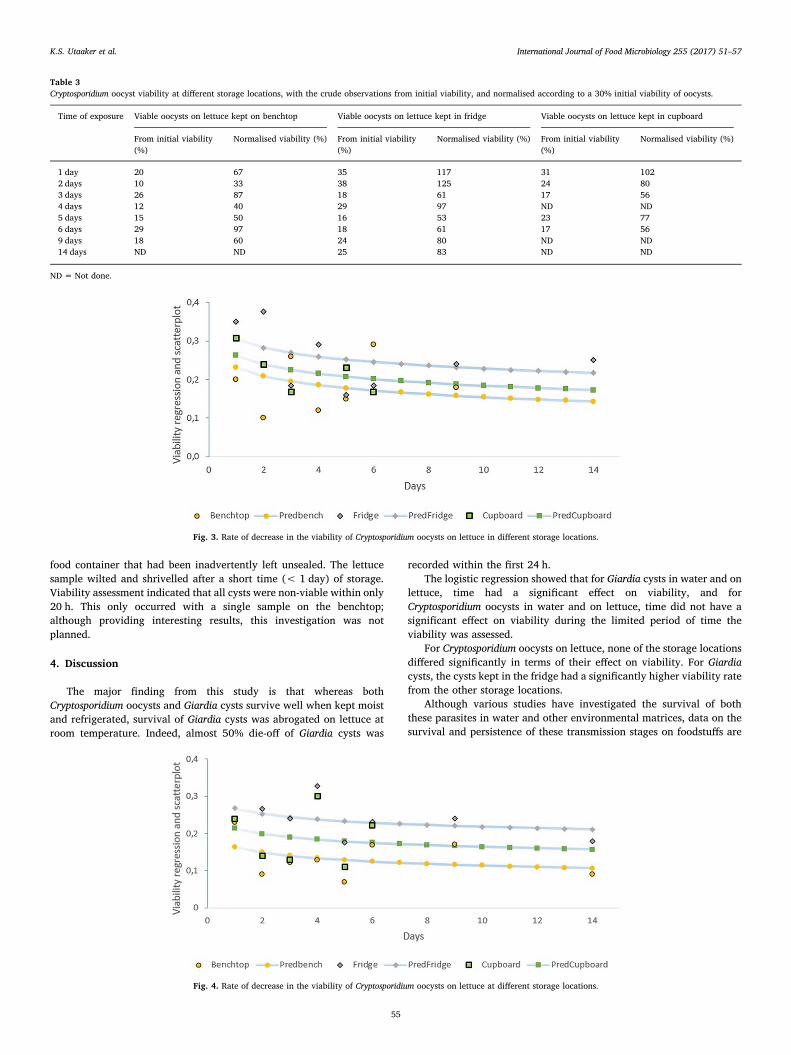
food container that had been inadvertently left unsealed. The lettucesample wilted and shrivelled after a short time (< 1 day) of storage.Viability assessment indicated that all cysts were non-viable within only20 h. This only occurred with a single sample on the benchtop;although providing interesting results, this investigation was notplanned.
4. Discussion
The major finding from this study is that whereas bothCryptosporidium oocysts and Giardia cysts survive well when kept moistand refrigerated, survival of Giardia cysts was abrogated on lettuce atroom temperature. Indeed, almost 50% die-off of Giardia cysts was
recorded within the first 24 h.The logistic regression showed that for Giardia cysts in water and on
lettuce, time had a significant effect on viability, and forCryptosporidium oocysts in water and on lettuce, time did not have asignificant effect on viability during the limited period of time theviability was assessed.
For Cryptosporidium oocysts on lettuce, none of the storage locationsdiffered significantly in terms of their effect on viability. For Giardiacysts, the cysts kept in the fridge had a significantly higher viability ratefrom the other storage locations.
Although various studies have investigated the survival of boththese parasites in water and other environmental matrices, data on thesurvival and persistence of these transmission stages on foodstuffs are
Table 3Cryptosporidium oocyst viability at different storage locations, with the crude observations from initial viability, and normalised according to a 30% initial viability of oocysts.
Time of exposure Viable oocysts on lettuce kept on benchtop Viable oocysts on lettuce kept in fridge Viable oocysts on lettuce kept in cupboard
From initial viability(%)
Normalised viability (%) From initial viability(%)
Normalised viability (%) From initial viability(%)
Normalised viability (%)
1 day 20 67 35 117 31 1022 days 10 33 38 125 24 803 days 26 87 18 61 17 564 days 12 40 29 97 ND ND5 days 15 50 16 53 23 776 days 29 97 18 61 17 569 days 18 60 24 80 ND ND14 days ND ND 25 83 ND ND
ND = Not done.
Fig. 3. Rate of decrease in the viability of Cryptosporidium oocysts on lettuce in different storage locations.
Fig. 4. Rate of decrease in the viability of Cryptosporidium oocysts on lettuce at different storage locations.
K.S. Utaaker et al. International Journal of Food Microbiology 255 (2017) 51–57
55
currently scarce and often unclear. As foodborne transmission has beenfrequently postulated to be important for both parasites (Smith et al.,2007), despite the discrepancy in recorded outbreaks, obtaining robustdata on this is important and also identifying which factors mayprolong survival and thus be more likely to result ingestion of viabletransmission stages.
Table 4 lists some of the published survival experiments for bothGiardia cysts and Cryptosporidium oocysts, and these illustrate that, ingeneral, cooler temperatures, but not below freezing, promote survivalof these transmission stages.
Similar results were obtained in the studies described here, and itwas apparent that, in general, Cryptosporidium oocysts are more robustthan Giardia cysts, particularly at room temperature.
Thus, to extrapolate our findings into a foodborne transmissionscenario, especially with respect to fresh produce, it seems thattemperature and humidity are critical factors regarding survival.Although fresh foods, such as salad, may act as a transmission vehiclefor Giardia when contaminated by a food-handler shortly beforeconsumption (as has been reported for at least two of the ninedocumented outbreaks of foodborne giardiasis), it seems that on-farmGiardia contamination or during the farm-to-fridge chain, in which theparasite is likely to be exposed to ambient temperatures for someperiods, will be less likely to result in the potential for infection.
However, for Cryptosporidium oocysts, it appears that contaminationof fresh produce, even very early in the farm-to-fork production chain,may result in viable parasites still being on the produce at consumption,even with a long-production chain; this is probably reflected in some ofthe outbreaks of foodborne cryptosporidiosis reported such as theoutbreak in Finland involving 72 cases and associated with lettuceimported from the Netherlands (Pönka et al., 2009) and the UK-wideoutbreak involving 300 cases (McKerr et al., 2012). For both theseoutbreaks, the widespread number of cases clearly indicate thatfoodhandlers at the serving or consumption places are not likely sourcesof the contamination.
Increased demand for fresh produce and current food trendsadvising consumers to increase their intake of vegetables and fruits,in combination with global sourcing and improved transport chains,may increase the possibility of fresh produce contaminated with
parasite transmission stages being distributed more widely. In addition,and for Cryptosporidium in particular, it seems probable that oocyststhat contaminate fresh produce, even at harvesting or before then, mayremain in an infectious state on the foodstuff until it is consumed.Although those conditions that are optimal for transport of freshproduce (for example, for lettuce a temperature of 0 °C and a relativehumidity of 98–100%; (Saltveit, 2014)) also tend to be ideal, oressential, for survival of Giardia cysts, most fresh produce is nottransported under ideal conditions (Vigneault et al., 2009), althoughcold chain logistics are improving rapidly (Rodrigue and Notteboom,2013). Whereas problems in the cold chain may be detrimental to thesurvival of Giardia cysts, Cryptosporidium oocysts are probably likely tobe alive for as long as the fresh produce remains in a condition thatmakes it acceptable for sale.
In some countries transport, storage, and sale of fresh produce is lesssophisticated. For example, in India, fresh produce is mostly sold eitherdirectly from the producer through open-air markets or street vendors,or, more usually, initially through mandi (trading hubs for agriculturalproduce), and, even at the mandi, the cold storage is usually insufficient(Ahmad and Siddiqui, 2015). In such conditions it would seem, again,that although Cryptosporidium oocysts contaminating the produce at thefarm level may remain viable until consumption, this is less likely forGiardia cysts despite the farm-to-fork chain being shorter. This isparticularly important for such regions where these parasitic infectionsare more prevalent, have a greater impact on the population, and whereidentification of the most important transmission routes is important forimplementation of appropriate control measures.
In conclusion, although both Giardia cysts and Cryptosporidiumoocysts survive well on fresh produce under cool, moist conditions,such are also ideal for transport and storage of fresh produce, if producecontamination occurs at the farm level (during production or harvest-ing) then Cryptosporidium oocysts are much more likely than Giardiacysts to survive until they reach the consumer. Thus, if we wish toensure that our fresh produce does not become more likely to becontaminated with infectious parasitic transmission stages, it behovesus to implement improvements regarding removal or inactivation ofparasite transmission stages, or, preferably, decreasing contaminationat the farm level. Implementation of such measures should be con-
Table 4Summary of publications on survival of Giardia cysts and Cryptosporidium oocysts in different environmental matrices.
Giardia viability assessments
Conditions Viability assessment method Viability after different exposure periods Reference
Soil at 20 °C Dye exclusion and bioassay 180 days/3% viability Olson et al. (1999)Soil at 4–5 °C Dye exclusion and bioassay 180 days/almost no decline Olson et al. (1999)Soil during Norwegian winters (freeze–thaw cycles) Dye exclusion 10–12 days/10% viability
75 days/> 1% viabilityRobertson and Gjerde (2006)
Lake water at 6–7 °C Dye exclusion and bioassay 7 days/91% viability28 days/1% viability
Cole et al. (1989)
Lake water at 17–20 °C Dye exclusion and bioassay 7 days/12% viability Cole et al. (1989)Tap water at 20–28 °C Dye exclusion and bioassay 7 days/2% viability
14 days/no cysts found viableCole et al. (1989)
Cryptosporidium viability assessments
Conditions Viability assessment method Viability Reference
River water samples Tissue culture assay Infectivity decreased as temperature rises (from 4to 23 °C)
Pokorny et al. (2002)
Cattle faeces Bioassay Survival time higher in cooler (above freezing)conditions
Li et al. (2010)
Distilled water at 15 °C Bioassay and cell culture Oocyst remained infective for 7 months. Jenkins et al. (2002)Environmental stresses (freezing, seawater,
desiccation)Dye exclusion Freezing reduced viability greatly. Desiccation was
detrimental.Robertson et al. (1992)
On field crops at 20–30 °C Not specified Oocyst survival for< 3 days. WHO – Wastewater use in agriculture(2006)
K.S. Utaaker et al. International Journal of Food Microbiology 255 (2017) 51–57
56
ducted in parallel with improvements in the fresh produce cold chain.
Acknowledgements
This work has been funded through the Para-Clim-Chandigarhproject, partly funded by the Norwegian Research Council via theNew Indigo Partnership Programme (Contract number: 227965).
The authors are grateful to Birgitte Kasin Hønsvall for providingpurified Cryptosporidium parvum oocysts.
References
Åberg, R., Sjöman, M., Hemminki, K., Pirnes, A., Räsänen, S., Kalanti, A., Pohjanvirta, T.,Caccio, S.M., Pihlajasaari, A., Toikkanen, S., Huusko, S., 2015. Cryptosporidiumparvum caused a large outbreak linked to frisée salad in Finland, 2012. ZoonosesPublic Health 62 (8), 618–624.
Ahmad, M.S., Siddiqui, M.W., 2015. Postharvest Quality Assurance of Fruits: PracticalApproaches for Developing Countries. Springerhttp://dx.doi.org/10.1007/978-3-319-21197-8_2.
AWWA, 1988. Roundtable: Cryptosporidium. J. Am. Water Works Assoc. 80, 14–28.Baldursson, S., Karanis, P., 2011. Waterborne transmission of protozoan parasites: review
of worldwide outbreaks–an update 2004–2010. Water Res. 45 (20), 6603–6614.Campbell, A.T., Robertson, L.J., Smith, H.V., 1992. Viability of Cryptosporidium parvum
oocysts: correlation of in vitro excystation with inclusion or exclusion of fluorogenicvital dyes. Appl. Environ. Microbiol. 58 (11), 3488–3493.
Cole, L., Schupp, D.G., Erlandsen, S.L., 1989. Viability of Giardia cysts suspended in lake,river, and tap water. Appl. Environ. Microbiol. 55 (5), 1223–1229.
Cook, N., Lim, Y.A.L., 2012. Giardia duodenalis: part 1: contamination of fresh produce. In:Robertson, L.J., Smith, H. (Eds.), Foodborne Protozoan Parasites. Nova Publishers,Hauppauge, pp. 105–107.
Gatei, W., Wamae, C.N., Mbae, C., Waruru, A., Mulinge, E., Waithera, T., Hart, C.A., 2006.Cryptosporidiosis: prevalence, genotype analysis, and symptoms associated withinfections in children in Kenya. Am.J.Trop. Med. Hyg. 75 (1), 78–82.
Hald, T., Aspinall, W., Devleesschauwer, B., Cooke, R., Corrigan, T., Havelaar, A.H., Gibb,H.J., Torgerson, P.R., Kirk, M.D., Angulo, F.J., Lake, R.J., Speybroeck, N., Hoffman,S., 2016. World Health Organization estimates of the relative contributions of food tothe burden of disease due to selected foodborne hazards: a structured expertelicitation. PLoS One 19 (11(1)), e0145839.
Hønsvall, B.K., Robertson, L.J., 2017. Real-time nucleic acid sequence-basedamplification (NASBA) assay targeting MIC1 for detection of Cryptosporidium parvumand Cryptosporidium hominis oocysts. Exp. Parasitol. 172, 61–67.
Jenkins, M., Trout, J., Higgins, J., Dorsch, M., Veal, D., Fayer, R., 2002. Comparison oftests for viable and infectious Cryptosporidium parvum oocysts. Parasitol. Res. 89(1), 1–5.
Kirk, M.D., Pires, S.M., Black, R.E., Caipo, M., Crump, J.A., Devleesschauwer, B., Döpfer,D., Fazil, F., Fischer-Walker, C.L., Hald, T., Hall, A.J., Keddy, K.H., Lake, R.J., Lanata,C.F., Torgerson, P.R., Havelaar, A.H., Angulo, F.J., 2015. World Health OrganizationEstimates of the global and regional disease burden of 22 foodborne bacterial,protozoal, and viral diseases: a data synthesis. PLoS Med. 12 (12), e1001921. http://dx.doi.org/10.1371/journal.pmed.1001921. (2015 Dec 3).
Kotloff, K.L., Nataro, J.P., Blackwelder, W.C., Nasrin, D., Farag, T.H., Panchalingam, S.,Wu, Y., Sow, S.O., Sur, D., Breiman, R.F., Faruque, A.S.G., Zaidi, A.K.M., Saha, D.,Alonso, P.L., Tamboura, B., Sanogo, D., Onwuchekwa, U., Manna, B., Ramamurthy,T., Kanungo, S., Ochieng, J.B., Omore, R., Oundo, J.O., Hossain, A., Das, S.K., Ahmed,S., Qureshi, S., Quadri, F., Adegbola, R.A., Antonio, M., Hossain, J.M., Akinsola, A.,Mandomando, I., Nhampossa, T., Acácio, S., Biswas, K., O'Reilly, C., Mintz, E.D.,Berkeley, L.Y., Muhsen, K., Sommerfelt, H., Robins-Browne, R.M., Levine, M.M.,2013. Burden and aetiology of diarrhoeal disease in infants and young children indeveloping countries (the Global Enteric Multicenter Study, GEMS): a prospective,case-control study. Lancet 382 (9888), 209–222.
Lane, S., Lloyd, D., 2002. Current trends in research into the waterborne parasite Giardia.Crit. Rev. Microbiol. 28 (2), 123–147.
Li, X., Atwill, E.R., Dunbar, L.A., Tate, K.W., 2010. Effect of daily temperature fluctuationduring the cool season on the infectivity of Cryptosporidium parvum. Appl. Environ.Microbiol. 76 (4), 989–993.
Lozano, R., Naghavi, M., Foreman, K., Lim, S., Shibuya, K., Aboyans, V., Abraham, J.,Adair, T., Aggarwal, R., Ahn, S.Y., Alvarado, M., Anderson, H.R., Anderson, L.M.,Andrews, K.G., Atkinson, C., Baddour, L.M., Barker-Collo, S., Bartels, D.H., Bell, M.L.,
Benjamin, E.J., Bennett, D., Bhalla, K., Bikbov, B., Bin Abdulhak, A., Birbeck, G.,Blyth, F., Bolliger, I., Boufous, S., Bucello, C., Burch, M., Burney, P., Carapetis, J.,Chen, H., Chou, D., Chugh, S.S., Coffeng, L.E., Colan, S.D., Colquhoun, S., Colson,K.E., Condon, J., Connor, M.D., Cooper, L.T., Corriere, M., Cortinovis, M., de Vaccaro,K.C., Couser, W., Cowie, B.C., Criqui, M.H., Cross, M., Dabhadkar, K.C., Dahodwala,N., De Leo, D., Degenhardt, L., Delossantos, A., Denenberg, J., Des Jarlais, D.C.,Dharmaratne, S.D., Dorsey, E.R., Driscoll, T., Duber, H., Ebel, B., Erwin, P.J.,Espindola, P., Ezzati, M., Feigin, V., Flaxman, A.D., Forouzanfar, M.H., Fowkes, F.G.,Franklin, R., Fransen, M., Freeman, M.K., Gabriel, S.E., Gakidou, E., Gaspari, F.,Gillum, R.F., Gonzalez-Medina, D., Halasa, Y.A., Haring, D., Harrison, J.E.,Havmoeller, R., Hay, R.J., Hoen, B., Hotez, P.J., Hoy, D., Jacobsen, K.H., James, S.L.,Jasrasaria, R., Jayaraman, S., Johns, N., Karthikeyan, G., Kassebaum, N., Keren, A.,Khoo, J.P., Knowlton, L.M., Kobusingye, O., Koranteng, A., Krishnamurthi, R.,Lipnick, M., Lipshultz, S.E., Ohno, S.L., Mabweijano, J., MacIntyre, M.F., Mallinger,L., March, L., Marks, G.B., Marks, R., Matsumori, A., Matzopoulos, R., Mayosi, B.M.,McAnulty, J.H., McDermott, M.M., McGrath, J., Mensah, G.A., Merriman, T.R.,Michaud, C., Miller, M., Miller, T.R., Mock, C., Mocumbi, A.O., Mokdad, A.A., Moran,A., Mulholland, K., Nair, M.N., Naldi, L., Narayan, K.M., Nasseri, K., Norman, P.,O'Donnell, M., Omer, S.B., Ortblad, K., Osborne, R., Ozgediz, D., Pahari, B., Pandian,J.D., Rivero, A.P., Padilla, R.P., Perez-Ruiz, F., Perico, N., Phillips, D., Pierce, K., Pope3rd, C.A., Porrini, E., Pourmalek, F., Raju, M., Ranganathan, D., Rehm, J.T., Rein,D.B., Remuzzi, G., Rivara, F.P., Roberts, T., De Leon, F.R., Rosenfeld, L.C., Rushton,L., Sacco, R.L., Salomon, J.A., Sampson, U., Sanman, E., Schwebel, D.C., Segui-Gomez, M., Shepard, D.S., Singh, D., Singleton, J., Sliwa, K., Smith, E., Steer, A.,Taylor, J.A., Thomas, B., Tleyjeh, I.M., Towbin, J.A., Truelsen, T., Undurraga, E.A.,Venketasubramanian, N., Vijayakumar, L., Vos, T., Wagner, G.R., Wang, M., Wang,W., Watt, K., Weinstock, M.A., Weintraub, R., Wilkinson, J.D., Woolf, A.D., Wulf, S.,Yeh, P.H., Yip, P., Zabetian, A., Zheng, Z.J., Lopez, A.D., Murray, C.J., AlMazroa,M.A., Memish, Z.A., 2012. Global and regional mortality from 235 causes of death for20 age groups in 1990 and 2010: a systematic analysis for the Global Burden ofDisease Study 2010. Lancet 380, 2095–2128.
McKerr, C., Adak, G.K., Nichols, G., Gorton, R., Chalmers, R.M., Kafatos, G., Cosford, P.,Charlett, A., Reacher, M., Pollock, K.G., Alexander, C.L., Morton, S., 2012. Anoutbreak of Cryptosporidium parvum across England & Scotland associated withconsumption of fresh pre-cut salad leaves, May 2012. PloS One 10 (5), e0125955.
Olson, M.E., Goh, J., Phillips, M., Guselle, N., McAllister, T.A., 1999. Giardia cyst andCryptosporidium oocyst survival in water, soil, and cattle feces. J. Environ. Qual. 28(6), 1991–1996.
Pokorny, N.J., Weir, S.C., Carreno, Trevors, J.T., Lee, H., 2002. Influence of temperatureon Cryptosporidium parvum oocyst infectivity in river water samples as detected bytissue culture assay. J. Parasitol. 88 (3), 641–643.
Pönka, A., Kotilainen, H., Rimhanen-Finne, R., Hokkanen, P., Hänninen, M.L., Kaarna, A.,Meri, T., Kuusi, M., 2009. A foodborne outbreak due to Cryptosporidium parvum inHelsinki, November 2008. Eur. Secur. 14 (28), 29–35.
Robertson, L.J., Chalmers, R.M., 2013. Foodborne cryptosporidiosis: is there really morein Nordic countries? Trends Parasitol. 29 (1), 3–9.
Robertson, L.J., Gjerde, B.K., 2006. Fate of Cryptosporidium oocysts and Giardia cysts inthe Norwegian aquatic environment over winter. Microb. Ecol. 52 (4), 597–602.
Robertson, L.J., Campbell, A.T., Smith, H.V., 1992. Survival of Cryptosporidium parvumoocysts under various environmental pressures. Appl. Environ. Microbiol. 58 (11),3494–3500.
Rodrigue, J.-P., Notteboom, T., 2013. The cold chain and its logistics. In: Rodrigue, J.-P.(Ed.), The Geography of Transport Systems, third ed. Routledge, New York (416 pp).
Saltveit, M.E., 2014. Lettuce. In: Gross, K.C. (Ed.), Agriculture Handbook Number 66. TheCommercial Storage of Fruits, Vegetables and Florist and Nursery Stocks, Availableonline: http://www.ba.ars.usda.gov/hb66/contents.html.
Sattar, S.A., 1999. Giardia cyst and Cryptosporidium oocyst survival in watersheds andfactors affecting inactivation. J. Am. Water Works Assoc Available online: https://books.google.no/books?isbn=0898679753.
Smith, H.V., Caccio, S.M., Cook, N., Nichols, R.A.B., Tait, A., 2007. Cryptosporidium andGiardia as foodborne zoonoses. Vet. Parasitol. 149 (1), 29–40.
Utaaker, K.S., Huang, Q., Robertson, L.J., 2015. A reduced-cost approach for analysingfresh produce for contamination with Cryptosporidium oocysts and/or Giardia cysts.Food Res. Int. 77, 326–332.
Vigneault, C., Thompson, J., Wu, S., Hui, K.C., LeBlanc, D.I., 2009. Transportation of freshhorticultural produce. In: Benkeblia, N. (Ed.), Postharvest Technologies forHorticultural Crops. 2. pp. 1–24. Available at: http://ucce.ucdavis.edu/files/datastore/234-1291.pdf.
World Health Organization, 2006. Guidelines for the safe use of wastewater, excreta andgreywater: excreta and greywater use in agriculture. In: World Health Organization.Vol. 4.
K.S. Utaaker et al. International Journal of Food Microbiology 255 (2017) 51–57
57

III

Contents lists available at ScienceDirect
International Journal of Food Microbiology
journal homepage: www.elsevier.com/locate/ijfoodmicro
Checking the detail in retail: Occurrence of Cryptosporidium and Giardia onvegetables sold across different counters in Chandigarh, India
Kjersti Selstad Utaakera,⁎, Anil Kumarb, Himanshu Joshib, Suman Chaudharyb,c,Lucy J. Robertsona
a Parasitology Lab, Department for Food Safety and Infection Biology, Faculty of Veterinary Medicine, Norwegian University of Life Sciences, Adamstuen Campus, PO Box8146 Dep., 0033 Oslo, Norwayb Department of Medical Parasitology, Postgraduate Institute of Medical Education and Research, Chandigarh 16002, Indiac Nanomedicine-Laboratory of Immunology and Molecular Biomedical Research (NLIMBR), School of Medicine (SoM), Centre for Molecular and Medical Research (C-MMR), Strategic Research Centre, Faculty of Health, Deakin University, Waurn Ponds, Victoria 3216, Australia
A R T I C L E I N F O
Keywords:CryptosporidiumGiardiaFresh produceContaminationFood safety
A B S T R A C T
Fresh produce has been recognized as a vehicle of infection for protozoan parasites, particularly Cryptosporidium,and, to a lesser extent, Giardia. For both parasites, outbreaks associated with fresh produce have been docu-mented. Although documented outbreaks tend to be from industrialized countries, contamination of freshproduce with these parasites is a global issue. In developing countries, infections with these parasites are oftenendemic in the community, and basic infrastructure and hygiene measures may be inadequate, thus the like-lihood of contamination of fresh produce with these parasites may be higher. Realization of the importance ofthis transmission route comes against a backdrop of raw salads and more Western culinary habits gaining afoothold, and fresh produce being encouraged as part of the diet due to their associated health benefits.However, if consumption of uncooked fresh produce is going to increase its market sector in India, it is importantthat it is safe. In this study, various types of fresh produce obtained from three types of vendors in Chandigarh, amajor city in Northern India, were analyzed for contamination with Cryptosporidium oocysts and Giardia cystsusing a method that has been previously validated in inter-laboratory spiking experiments. A total of 284samples of different fresh produce items were analyzed, obtained from the different retailers situated in differentsocietal layers of the city. The overall prevalence of contamination of fresh produce with these parasites was justunder 11%, with 6% of the vegetables contaminated with Cryptosporidium oocysts, and 5% with Giardia cysts.Contaminated vegetables included turnip, cabbage, carrot, chili, coriander, cucumber, radishes, and tomatoes.Molecular analyses identified contamination with Cryptosporidium parvum and Giardia duodenalis of AssemblageA and Assemblage D, indicating that contamination from animals may be of relevance. Although the prevalenceof contamination is similar to those reported in previous studies, the levels of contamination on some items offresh produce were relatively high. Although the different socioeconomic areas of Chandigarh from which thesamples were obtained was not associated with likelihood of contamination, fresh produce from supermarketshad heavier contamination with Cryptosporidium oocysts than fresh produce purchased through other salesoutlets. The results are discussed in relation to the fresh produce chain and sales models in Chandigarh, both interms of where contamination may occur and the potential importance of fresh produce as a transmission ve-hicle.
1. Introduction
Cryptosporidium spp. and Giardia duodenalis are among the mostfrequently occurring intestinal protozoan parasites in humans and an-imals worldwide (Fayer, 2004; Thompson and Monis, 2004; Thompson,2004). Both parasites can cause diarrheal disease. Global studies haverevealed that G. duodenalis and Cryptosporidium spp. are two of the most
common etiological agents in pediatric diarrhea in developing coun-tries, and are associated with mortality as well as morbidity (Kotloffet al., 2013; Platts-Mills et al., 2015).
The biology of Cryptosporidium and Giardia makes them suitable fortransmission via fresh produce; they have a low infectious dose, a ro-bust transmission stage, are small sized, and some genotypes have azoonotic potential, giving the opportunity for contamination to occur
http://dx.doi.org/10.1016/j.ijfoodmicro.2017.09.020Received 24 June 2017; Received in revised form 17 September 2017; Accepted 26 September 2017
⁎ Corresponding author.E-mail address: [email protected] (K.S. Utaaker).
International Journal of Food Microbiology 263 (2017) 1–8
Available online 01 October 20170168-1605/ © 2017 Elsevier B.V. All rights reserved.
MARK
from both animal and human sources (Robertson and Lim, 2011;Robertson and Fayer, 2012). Infected individuals also have a high ex-cretion rate, ranging from> 5 × 10³ to 9.2 × 10⁵ oocysts per gramfeces for Cryptosporidium (Goodgame et al., 1993) and 580,000 cysts pergram feces may be shed over a period of days or longer in the case ofGiardia infection (Danciger and Lopez, 1975).
As fewer people are affected in a foodborne outbreak than in awaterborne outbreak, and may be more scattered geographically (see,for example, the UK outbreak described by McKerr et al. (2015)), a lackof prompt diagnosis may hamper epidemiological investigation. It isworth noting that the last major waterborne outbreak of giardiasis inEurope, in which over 1500 people were infected, took several weeks tobe recognized as a waterborne outbreak (Robertson et al., 2006), and inthe UK, the specific produce causing the outbreak was never detected.
Cryptosporidium and Giardia can contaminate food as a surfacecontaminant. Contamination with small numbers of infectious (oo)cystsin produce that receives minimal washing or treatment prior to inges-tion, poses a threat to public health. Food products can be contaminateddirectly by cysts and oocysts in feces from humans and animals or viathe environment, such as soil and water, and thus serve as vehicles fortransmission, at any step in the farm-to-fork chain. For fresh produce,contamination may persist until infection as the production chain re-quires cool temperatures and moist conditions to maximize foodfreshness, and that also enhance survival of Giardia cysts;Cryptosporidium oocysts seem to be more tolerant to temperaturechanges on fresh produce (Utaaker et al., 2017). For foods such as, forexample, bakery produce, storage conditions (dry, at room tempera-ture) are likely to be deleterious to parasite survival. Although there hasbeen discussion around Cryptosporidium oocysts surviving for longer inconditions of cool temperatures and high humidity, it has neverthelessbeen concluded that the likelihood of foodborne outbreaks occurring isno greater in cooler environments than anywhere else in the world(Robertson and Chalmers, 2013). Indeed, the foodborne transmissionroute is probably particularly relevant in places where infection is morelikely underdiagnosed and underreported, and especially so in the de-veloping countries where infrastructure and resources for investigationand reporting are limited. However, in such settings, where variousintestinal infections are endemic, outbreaks caused by contamination offood or water may be more difficult to identify against a background ofhigh infection.
Methods for detecting contamination of foodstuff by protozoans hadbeen relatively poorly developed until recently, until the publication ofISO Method 18744 (ISO, 2016). However, this method is both ex-pensive and time-consuming, and essential reagents must be storedrefrigerated. To implement such methods for routine analysis in la-boratories that are already poor in resources may be prohibitively ex-pensive and impractical. Furthermore, considering the vast amount offresh produce from different traders in the chain of retail events in adeveloping country's retail model, using lab analyses may provide scantinformation regarding tracking the sources of contamination. None-theless, these methods enable surveys to be conducted and an assess-ment of contamination levels to be made, and such data are essential forassessment of risk and determining the extent of significance of suchcontamination.
In this study, the aim was to analyze fresh produce sold at differentretailers in Chandigarh, India for the occurrence of Cryptosporidiumoocysts and Giardia cysts, and to use molecular typing of parasites frompositive samples as a further indication of the possible sources of con-tamination.
2. Materials and methods
2.1. Sampling site
Over a two-year period between February 2014 and February 2016,284 vegetables were purchased at local mandi, street vendors, and
supermarkets in Chandigarh, Northern India.Chandigarh is a union territory of its two neighboring states,
Haryana and Punjab, although not considered a part of either state.According to the State Agricultural Marketing Board of the UnionTerritory Chandigarh, Chandigarh has no major crop itself, and mostfresh produce available in Chandigarh comes from these neighboringstates. The Union Territory Chandigarh has only a limited area avail-able for agriculture, and this land is being gradually diminishing withthe expansion of Chandigarh city. In addition, farmers who keep a largenumber of dairy cattle utilize these areas to grow fodder for livestock(Chandigarh Administration, 2016a).
Chandigarh has only one principal Market Yard, and there are noofficial seasonal or other kinds of market yards, or any unregulatedmarkets (C.S.A.B, 1961) Thus, most of the samples collected in thisstudy came via the principal Market Yard, from where they are dis-tributed to different trading hubs and retailers.
Chandigarh is a city undergoing rapid growth and development. Thecity is organized according to “phases”, which can be a proxy for socio-economic status, as reflected by density of inhabitants: Phase I (highersocio-economic status), Phase II (moderate socio-economic status),Phase III and non-sectorial villages (lower socio-economic status). Inaddition, Chandigarh is divided into sectors, based on the grid conceptof the roads, and different sectors are also considered more or less af-fluent than others, according to the Chandigarh Master Plan – 2031(Chandigarh Administration, 2016b).
2.2. Source of samples of fresh produce
The sampling strategy aimed at analyzing vegetables representingeach sector of the city, including both rural and urban areas, the“phases” of the city, and to enable comparison of the three main salestypes used for fresh produce in India: street vendors, mandi (see de-scription below), and supermarkets. Nevertheless, due to access to ve-getables and markets, the number of samples from each sector are non-uniform, as are the distribution of vegetable types and salespoints (seeTable 1).
Among the total of 284 samples, 137 (48%) were obtained fromindividual street vendors, with either a stationary or mobile salespoint.The vendor handles the produce until it is purchased by the customer.In addition, 125 (44%) samples were obtained at mandi, which arelocal trading hubs for agricultural produce, and are arranged on aregular weekday basis in different sectors in Chandigarh. At the mandi,the local producer or salesperson brings his products and displays themfor the customer. As with street vendors, only the vendor handles thefoodstuffs until purchased by the customer.
The remaining 22 (8%) samples were obtained from modern su-permarkets, where the vegetables are displayed for the customer tochoose their preferred items, touching and handling the produce as theymake their selection before paying at the counter.
The samples were collected from different areas (phases), or sectors,of Chandigarh, thus also representing vegetables from different socio-economic layers, as well as salespoints. Of the 284 samples, 119 werecollected from Phase I sectors, 112 were collected from Phase II sectors,and 51 were collected from sectors in Phase III and non-sectorial vil-lages.
The samples most probably all came from the same principal MarketYard, but were sold in different areas of the city, and thus under dif-ferent conditions.
2.3. Fresh produce selected for analysis and their use in India
The vegetables to be analyzed were chosen according to the seasonof availability, with emphasis on those commonly consumed raw. Theseincluded coriander leaves, lettuce, radish, tomatoes, cucumber, fenu-greek leaves, cabbage, chili, mint leaves, carrot, and turnip.
K.S. Utaaker et al. International Journal of Food Microbiology 263 (2017) 1–8
2
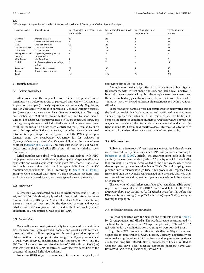
3. Sample analysis
3.1. Sample preparation
After collection, the vegetables were either refrigerated (for amaximum 48 h before analysis) or processed immediately (within 4 h).A portion of sample (for leafy vegetables, approximately 30 g leaves,and for vegetables with smooth surfaces 1–2 pieces weighing approx.30 g) were put into stomacher bags (Seward BA6041/STR filter bag)and washed with 200 ml of glycine buffer for 4 min by hand manip-ulation. The eluate was transferred into 5 × 50 ml centrifuge tubes, andthe bag was again washed with distilled water and the wash water usedto fill up the tubes. The tubes were centrifuged for 10 min at 1550 rfgand, after aspiration of the supernatant, the pellets were concentratedinto one tube per sample and refrigerated until the IMS step was per-formed, using the Dynabeads® GC-combo kit for isolation ofCryptosporidium oocysts and Giardia cysts, following the reduced costprotocol (Utaaker et al., 2015). The final suspension of 50 μl was pi-petted onto a single-well slide (Novakemi ab) and air-dried at roomtemperature.
Dried samples were fixed with methanol and stained with FITC-conjugated monoclonal antibodies (mAbs) against Cryptosporidium oo-cyst walls and Giardia cyst walls (Aqua-glo™, Waterborne™ Inc., USA)and nuclei were stained with the fluorogenic DNA intercalator 4′,6diamidino-2-phenylindole (DAPI) according to Smith et al. (2002).Samples were mounted with M101 No-Fade Mounting Medium, theneach slide was covered by a glass coverslip and viewed promptly.
3.2. Microscopy
Microscopy was performed on a Leica DCMB microscope (× 20, ×40, and ×100 objectives), equipped with Nomarski differential inter-ference contrast (DIC) optics. A blue filter block (480 nm – excitation,520 nm – emission) was used for the detection of cysts and oocystslabelled with FITC-conjugated mAbs, and a UV filter block (350 nmexcitation, 450 nm emission) was used for DAPI.
3.3. Enumeration
Each well was scanned systematically in an up-and-down or side-to-side manner, and Cryptosporidium oocysts and Giardia cysts were en-umerated. When brilliant apple-green fluorescing ovoid or sphericalobjects within the appropriate size range for Cryptosporidium andGiardia were observed, magnification was increased to 40×, and theUV filter block was used for visualization of DAPI staining. Each (oo)cyst was recorded as DAPI-negative or DAPI-positive according to thepresence of internal light blue staining.
Nomarski (DIC) objectives were used to examine morphological
characteristics of the (oo)cysts.A sample was considered positive if the (oo)cyst(s) exhibited typical
fluorescence, with correct shape and size, and being DAPI-positive. Ifinternal contents were lacking, but the morphometry was correct andthe structure had a typical fluorescence, the (oo)cysts were described as“putative”, as they lacked sufficient characteristics for definitive iden-tification.
These “putative” samples were not considered for genotyping due tothe lack of nuclei, but both putative and confirmed parasites weresummed together for inclusion in the results as positive findings. Insome of the samples containing numerous Cryptosporidium oocysts, theoocysts were occluded due to debris when examined under the UV-light, making DAPI-staining difficult to assess. However, due to the highnumbers of parasites, these were also included for genotyping.
3.4. DNA extraction
Following microscopy, Cryptosporidium oocysts and Giardia cystswere retrieved from positive slides and DNA was prepared according toRobertson et al. (2009). Briefly, the coverslip from each slide wascarefully removed and retained, whilst 25 μl aliquots of AL lysis buffer(Qiagen GmbH, Germany) were added to the slide wells, which werethen scraped using a sterile scalpel blade. The buffer and scrapings werepipetted into a microcentrifuge tube. This process was repeated fourtimes, and then the coverslip was replaced onto the slide that was thenre-screened. For each slide, neither cysts nor oocysts could be detectedafter scraping.
The contents of each microcentrifuge tube containing slide scrap-ings were re-suspended in Tris-EDTA buffer and held at 100 °C forCryptosporidium oocysts and 90 °C for Giardia cysts for 1 h, before theDNA was isolated using QIamp DNA mini kit (Qiagen GmbH), using anovernight step at 56 °C.
3.5. Molecular methods and sequencing
PCR was conducted with the primers and protocols listed in Table 2for Cryptosporidium and Giardia. The products were separated and vi-sualized by electrophoresis on 2% agarose gels using SYBRsafe® DNAgel stain under UV radiation. Positive samples were purified using.
High Pure PCR product purification kit (Roche Diagnostics), andsequenced on both strands at GATC Biotech, Germany. Sequences wereexamined using Geneious 10.1.2 software and sequence comparisonsconducted using NCBI BLAST. New sequences have been submitted toGenBank and have been allocated accession numbers KY967229,KY967230, KY967231, KY967232, KY967233.
Table 1Different types of vegetables and number of samples collected from different types of salespoints in Chandigarh.
Common name Scientific name No. of samples from mandi (wholesale markets)
No. of samples from streetvendors
No. of samples fromsupermarkets
Total number ofsamples
Cabbage Brassica oleracea 22 21 4 47Carrot Daucus carota subsp. sativus 8 14 3 25Chili Capsicum annuum 18 21 3 42Coriander leaves Coriandrum sativum 10 15 3 28Cucumber Cucumis sativus 26 22 4 52Fenugreek leaves Trigonella foenum-graecum 5 3 – 8Lettuce Lactuca sativa 1 1 – 2Mint leaves Mentha spicata 4 2 1 7Radish Raphanus raphanistrum subsp.
sativus7 7 – 14
Tomatoes Solanum lycopersicum 22 30 4 56Turnip Brassica rapa var. rapa 2 1 – 3Total 125 137 22 284
K.S. Utaaker et al. International Journal of Food Microbiology 263 (2017) 1–8
3
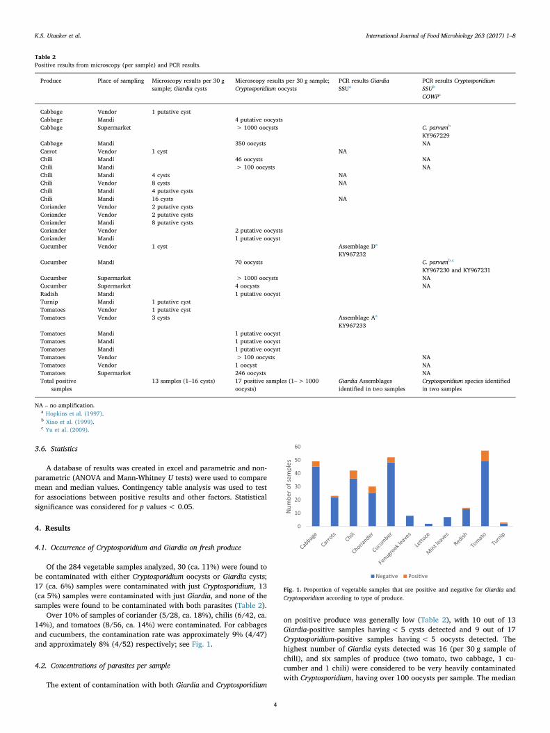
3.6. Statistics
A database of results was created in excel and parametric and non-parametric (ANOVA and Mann-Whitney U tests) were used to comparemean and median values. Contingency table analysis was used to testfor associations between positive results and other factors. Statisticalsignificance was considered for p values< 0.05.
4. Results
4.1. Occurrence of Cryptosporidium and Giardia on fresh produce
Of the 284 vegetable samples analyzed, 30 (ca. 11%) were found tobe contaminated with either Cryptosporidium oocysts or Giardia cysts;17 (ca. 6%) samples were contaminated with just Cryptosporidium, 13(ca 5%) samples were contaminated with just Giardia, and none of thesamples were found to be contaminated with both parasites (Table 2).
Over 10% of samples of coriander (5/28, ca. 18%), chilis (6/42, ca.14%), and tomatoes (8/56, ca. 14%) were contaminated. For cabbagesand cucumbers, the contamination rate was approximately 9% (4/47)and approximately 8% (4/52) respectively; see Fig. 1.
4.2. Concentrations of parasites per sample
The extent of contamination with both Giardia and Cryptosporidium
on positive produce was generally low (Table 2), with 10 out of 13Giardia-positive samples having< 5 cysts detected and 9 out of 17Cryptosporidium-positive samples having< 5 oocysts detected. Thehighest number of Giardia cysts detected was 16 (per 30 g sample ofchili), and six samples of produce (two tomato, two cabbage, 1 cu-cumber and 1 chili) were considered to be very heavily contaminatedwith Cryptosporidium, having over 100 oocysts per sample. The median
Table 2Positive results from microscopy (per sample) and PCR results.
Produce Place of sampling Microscopy results per 30 gsample; Giardia cysts
Microscopy results per 30 g sample;Cryptosporidium oocysts
PCR results GiardiaSSUa
PCR results CryptosporidiumSSUb
COWPc
Cabbage Vendor 1 putative cystCabbage Mandi 4 putative oocystsCabbage Supermarket > 1000 oocysts C. parvumb
KY967229Cabbage Mandi 350 oocysts NACarrot Vendor 1 cyst NAChili Mandi 46 oocysts NAChili Mandi > 100 oocysts NAChili Mandi 4 cysts NAChili Vendor 8 cysts NAChili Mandi 4 putative cystsChili Mandi 16 cysts NACoriander Vendor 2 putative cystsCoriander Vendor 2 putative cystsCoriander Mandi 8 putative cystsCoriander Vendor 2 putative oocystsCoriander Mandi 1 putative oocystCucumber Vendor 1 cyst Assemblage Da
KY967232Cucumber Mandi 70 oocysts C. parvumb,c
KY967230 and KY967231Cucumber Supermarket > 1000 oocysts NACucumber Supermarket 4 oocysts NARadish Mandi 1 putative oocystTurnip Mandi 1 putative cystTomatoes Vendor 1 putative cystTomatoes Vendor 3 cysts Assemblage Aa
KY967233Tomatoes Mandi 1 putative oocystTomatoes Mandi 1 putative oocystTomatoes Mandi 1 putative oocystTomatoes Vendor > 100 oocysts NATomatoes Vendor 1 oocyst NATomatoes Supermarket 246 oocysts NATotal positive
samples13 samples (1–16 cysts) 17 positive samples (1–>1000
oocysts)Giardia Assemblagesidentified in two samples
Cryptosporidium species identifiedin two samples
NA – no amplification.a Hopkins et al. (1997).b Xiao et al. (1999).c Yu et al. (2009).
Fig. 1. Proportion of vegetable samples that are positive and negative for Giardia andCryptosporidium according to type of produce.
K.S. Utaaker et al. International Journal of Food Microbiology 263 (2017) 1–8
4
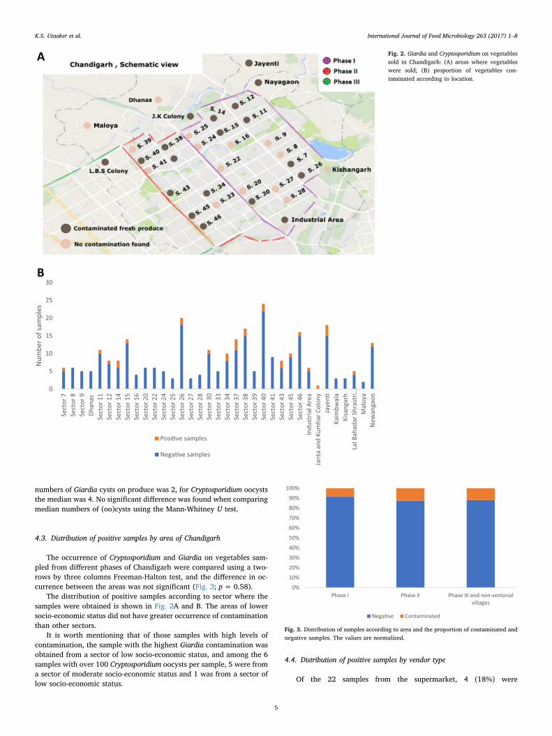
numbers of Giardia cysts on produce was 2, for Cryptosporidium oocyststhe median was 4. No significant difference was found when comparingmedian numbers of (oo)cysts using the Mann-Whitney U test.
4.3. Distribution of positive samples by area of Chandigarh
The occurrence of Cryptosporidium and Giardia on vegetables sam-pled from different phases of Chandigarh were compared using a two-rows by three columns Freeman-Halton test, and the difference in oc-currence between the areas was not significant (Fig. 3; p= 0.58).
The distribution of positive samples according to sector where thesamples were obtained is shown in Fig. 2A and B. The areas of lowersocio-economic status did not have greater occurrence of contaminationthan other sectors.
It is worth mentioning that of those samples with high levels ofcontamination, the sample with the highest Giardia contamination wasobtained from a sector of low socio-economic status, and among the 6samples with over 100 Cryptosporidium oocysts per sample, 5 were froma sector of moderate socio-economic status and 1 was from a sector oflow socio-economic status.
4.4. Distribution of positive samples by vendor type
Of the 22 samples from the supermarket, 4 (18%) were
Fig. 2. Giardia and Cryptosporidium on vegetablessold in Chandigarh: (A) areas where vegetableswere sold; (B) proportion of vegetables con-taminated according to location.
Fig. 3. Distribution of samples according to area and the proportion of contaminated andnegative samples. The values are normalized.
K.S. Utaaker et al. International Journal of Food Microbiology 263 (2017) 1–8
5
contaminated. Of the 125 samples from the mandi, 15 (ca. 12%) werecontaminated, and of the 137 samples from the street vendors, 11 (ca.8%) were contaminated. Thus, supermarket bought produce was no lesslikely to be contaminated than produce from mandi or street vendors.
With respect to the more highly contaminated samples (highernumbers of oocysts or cysts), the sample with the highest number ofGiardia cysts was obtained from a mandi, whereas of the produce morehighly contaminated with Cryptosporidium oocysts, three were obtainedfrom a supermarket (with two of these having over 1000 oocysts persample), two were from a mandi, and one was from a street vendor.
The median number of Cryptosporidium oocysts per positive samplefrom supermarkets was 623, from mandi was 2.5, and from streetvendors was 2. The median values from Giardia cyst andCryptosporidium oocyst contamination from vendors and mandi werecompared by Mann-Whitney U test, and the difference was not sig-nificant.
None of the samples from the supermarkets were contaminated withGiardia cysts. Thus, the extent of contamination with Cryptosporidiumoocysts of positive produce from supermarkets was significantly higherthan Cryptosporidium-positive produce from traditional retailers as de-termined by Mann-Whitney U test.
4.5. Molecular analyses from positive samples
4.5.1. Genotyping of GiardiaThe DNA isolated from 6 Giardia positive samples was run through
PCR reactions using several primer sets targeting different genes, in-cluding triosephosphate isomerase (TPI), glutamate dehydrogenase(GDH) and small-subunit RNA (SSU), however DNA amplification waslargely unsuccessful. Results from which usable sequences could beobtained derived only from 2 samples and only at the SSU gene(Table 2). Comparison using BLAST indicated that the closest hits (both96%) were Assemblage A (closest hit GenBank Accession numberLN811460.1) and D (JQ245138.1).
4.5.2. Species identification of CryptosporidiumThe DNA isolated from 10 Cryptosporidium-positive samples pro-
duced amplification and usable sequences from 2 samples using boththe SSU and COWP primers; both samples were found to be C. parvum.
5. Discussion
One of the most important findings from this study is that a rela-tively high proportion of fresh produce on the market in Chandigarh(just under 11%) were contaminated by protozoan parasites, with 6%contaminated with Cryptosporidium oocysts and 5% with Giardia cysts.
Given that Chandigarh is generally regarded as one of the cleanestcities in India (Chandani, 2016), this seems a high proportion. How-ever, studies from some other low- or middle-income countries haveindicated similar or higher levels of contamination (e.g., Ebrahimzadehet al. (2013), Maikai et al. (2013), Said (2012)). In more developedcountries, the proportion of fresh produce contaminated with theseparasites tends to be lower (e.g., Rzeżutka et al. (2010), Robertson andGjerde (2001), Dixon et al. (2013)).
An important difference, however, between the results obtainedfrom our study in Chandigarh and the data from other studies is thatsome of our samples had very high levels of contamination. Studiesfrom developing countries often do not often report the extent of con-tamination, but when quantitative data are provided, the maximumcontamination per sample is generally a few (oo)cysts (e.g., Fallahet al., 2012; Al-Shawa and Mwafy, 2007). Whereas the majority of oursamples had low levels of contamination, several samples had con-siderably higher levels of contamination for Cryptosporidium.
Many studies from developing countries investigating contamina-tion of fresh produce with protozoan parasites have not used estab-lished methods and do not provide recovery efficiency data. This makes
interpretation of the results difficult, and it is likely that both the extentof contamination and contamination levels may be under-estimated.Our study utilized an established protocol that has been validated indifferent laboratories (Utaaker et al., 2015); the recovery efficiency isaround 30–50%.
The stage along the farm-to-fork continuum at which the freshproduce in our study became contaminated is impossible to determinefrom our data. However, this information is important for identifyingwhere preventive measures should be implemented. Various publica-tions have shown that irrigation water may be a source of contamina-tion of crops (Chaidez et al., 2005; Thurston-Enriquez et al., 2002)(Amorós et al., 2010), although Cryptosporidium and Giardia were notfound in water samples from an irrigation canal in Thailand (Chuahet al., 2016). Irrigation in the Punjab and Haryana states is largely fromground water being pumped directly onto growing crops from tubewells (Pandey, 2016). Ground water is usually relatively protected fromcontamination and may be considered less likely to be the source ofCryptosporidium oocysts and Giardia cysts. However, canal irrigation(e.g. the Upper Bari Doab canal in Punjab) is also an important irri-gation source in this region. With canal irrigation, floodwater is carriedto the agricultural areas, and high levels of contamination may occur,particularly if there is the potential for the canals to be contaminatedwith sewage. Based on our results, investigation of irrigation water inthese areas is recommended.
Water, as a potential source of contamination, can come into contactwith fresh produce not only during cultivation, but also duringwashing. It is common practice in Asia for street vendors and mandisalespeople to keep a bucket of water alongside their wares and tomoisten their displays so that they appear fresh and appealing. A studyfrom Vietnam examined water from 200 such buckets and found Giardiacysts in 17 buckets (median concentration of 20 cysts per ml), andCryptosporidium oocysts in 9 buckets, with a median value of 10 oocystsper ml (Tram and Dalsgaard, 2014). Although such buckets of waterwere not analyzed in this study, they were noted as potential sources ofcontamination.
People in the chain from field to fork who handle the produce mightalso be sources of contamination. These include people working in thefields harvesting the crops, people packing the crops for transport to thecity, and the various links in the sales chain within the city, as well asthe last link in the chain, the salesperson themselves, or other customershandling the produce. Our results from supermarkets suggest that insituations where various customers may handle the goods, but not ne-cessarily purchase them, contamination may occur. Indeed, the morepeople who handle the produce the greater the potential risk of con-tamination. Whereas salespeople have a vested interest in not sellingtheir customers contaminated goods, this does not apply to other cus-tomers who handle goods.
Molecular analyses providing information on species and genotypemay provide further clues regarding sources of contamination, parti-cularly whether the parasites are likely to be from humans or animals.The information from our molecular analyses does not rule out eitherhuman or animal sources of contamination with Cryptosporidium. Astudy of Cryptosporidium species in human infections in Chandigarhindicated that C. hominis was the most prevalent species (75%) followedby C. parvum, with a prevalence of 25% (Sharma et al., 2013). In-formation on Cryptosporidium species in animals in around Chandigarhis scant, but our own studies have not indicated widespread C. paruvminfections among animals, although subgenotypes of C. parvum detectedin livestock in this area (unpublished data) are the same as those re-ported in human infections by Sharma et al. (2013). The source ofcontamination of fresh produce could thus be from either dirty hands orinfected livestock or other animals.
Similarly, for Giardia, either humans or animals could be the sourceof contamination, although the finding of Assemblage D as a con-taminant of cucumber indicates contamination from canine feces ismost likely, and probably does not pose a risk to human health. The
K.S. Utaaker et al. International Journal of Food Microbiology 263 (2017) 1–8
6
sample containing these cysts was obtained from a vendor sellingproduce from an open pushcart in the street, giving ample opportunityfor contamination from dog feces (Mahajan, 2014). Assemblage A,which was found contaminating tomatoes, is infectious to both humansand a range of other animals, and thus it is not possible to narrow downthe likely source of contamination.
The widespread occurrence of these parasites on vegetables inChandigarh along with the high levels of contamination and somesamples being shown to be contaminated with species or genotypesknown to be infectious to humans, indicate that there is the potentialfor infection transmission by consumption of contaminated fresh pro-duce, although whether the contaminating parasites were infective atpoint of purchase was not investigated, and the question remains re-garding the threat from this contamination to public health.
An expert elicitation study (Hald et al., 2016), estimated that theproportion of cases of cryptosporidiosis and giardiasis caused by thefoodborne route in Southeast Asia was 0.10 and 0.13, whereasequivalent data from Western Europe were 0.10 for Cryptosporidium and0.11 for Giardia. Unlike with other foodborne pathogens, this studyindicated that the importance of the foodborne route of infection wasquite similar across regions, but, in general, was rather low, with waterand human-to-human contact being of greater importance (Hald et al.,2016). Interestingly, based on this expert elicitation, foodborne trans-mission seemed to be considered slightly more important for Giardiathan Cryptosporidium consistently across regions. However, this may notreflect that there are fewer foodborne cases of cryptosporidiosis thangiardiasis, but that infections from animals are possibly of greater im-portance with Cryptosporidium than Giardia. Although foodborne out-breaks of these infections have been reported much more frequentlyfrom wealthier countries than poorer countries, this does not mean suchfoodborne outbreaks do not occur in less wealthy countries. However,with high levels of endemicity, it may be difficult to determine when anoutbreak associated with a specific vehicle of infection is occurring, andfollowing up such an outbreak requires enormous resources andprioritization of effort, both of which are unlikely to be available indeveloping countries.
The results of our study thus support the potential for foodbornetransmission, but should not be read to indicate that this transmissionmode is more important than any other. However, given the levels ofcontamination that we found, it seems probable to us that foodbornetransmission may be more likely to occur in this setting than inEuropean countries or other wealthy environments.
India is a hierarchical society, both within and between families,and also other social groups. Particular social groups tend to clustertogether in terms of where they live in a city, and our intention was toinvestigate whether particular strata of the society were more or lesslikely to be exposed to these parasites through fresh produce thanothers. In Chandigarh, such investigations are relatively easy, as thecity was originally planned for a differential pattern of density.
Due to cheaper housing, Phase III areas and non-sectorial villagersare characterized by high population pressure. They also have un-sanitary conditions, flood problems, poor garbage disposal, disposal oflivestock dung into open drains, and discharge of untreated sewage(Chandigarh Administration, 2016b).
Although it was expected that fresh produce from Phase III areasand non-sectorial villages would have higher rates of contaminationthan from other areas, this was not the case. This could be because thecity as a whole has a relatively high population density, with ap-proximately 9300 persons per km2, according to the census organiza-tion of India (Indian 15th National Census Survey, 2012). This meansthat the demarcations between the phases of the city are not very clear,as the sectors spread into each other. Furthermore, as most of the freshproduce supply passes through a single principal Market Yard inChandigarh (Chandigarh Administration, 2016c), and from there isdistributed to all the different sectors, the origin of the fresh produce isalike, regardless of hierarchical position at point of sale.
One difference that our study did bring out, however, was thatcontamination of fresh produce with Cryptosporidium was at higher le-vels (significantly greater numbers of oocysts) in supermarkets than instreet vendors or mandi. Interestingly, modern food retailing has ap-parently not been highly successful in India, with most Indian shoppers,regardless of disposable income bracket, preferring to buy fresh pro-duce from a street trader or mandi than a supermarket (Economist,2014). Price and convenience are often cited reasons, but it has alsobeen suggested that the benefits of supermarket shopping is in choicerange rather than quality, and our data would seem to support this.Why produce in supermarkets should have higher levels of Cryptos-poridium oocysts, than fresh produce at other salespoints is not clear,but may reflect greater handling potential via more people (customers).
India is the second largest producer of fruits and vegetables in theworld, and this production is considered a labor-intensive and high-riskactivity (Sachdeva et al., 2013). The fresh produce business in India hasnumerous infrastructure problems, including insufficient cold storage,unreliable transport, poor compliance with safety standards, in-sufficient quality control, lack of research and development, challengesin labeling etc., according to the Ministry of Food Processing Industries,G.o.I (2006), New Delhi. This situation hampers both prevention andtraceability of contamination, and indicates that the best measure toavoid infection by consumption of contaminated produce is to maintaingood hygienic practices in the preparation of fresh produce by properrinsing and washing.
Although our study produced useful data acquired by standard andrecognized methods, there were challenges. Inhibitors such as poly-saccharides, polyphenols, pectin, xylan, and chlorophyll from the plantmaterial in our samples may have hampered the PCR reactions (Weiet al., 2008). Furthermore, due to practical and logistical issues be-tween the collaborating laboratories, the period between elution andexamination of slides was often prolonged. Parasites may have de-generated during storage and the formation of other microorganismsduring the storage and transport period may have had a deterioratingeffect on the DNA in terms of both degeneration and formation of in-hibitors.
In conclusion, our study found that contamination withCryptosporidium oocysts and Giardia cysts of fresh produce on themarket in Chandigarh was relatively frequent. In some cases, highnumbers of oocysts and cysts were detected. Molecular studies suggestthat some of this contamination probably originates from animals, butspecies and genotypes infectious to humans were also indicated.
Of particular note is that supermarkets, which are generally con-sidered more modern, and thus (intuitively) safer, were no less likely tosell contaminated produce and produce was highly contaminated. Thismay reflect the greater handling in supermarkets, where the customersare able to touch and handle the produce themselves.
Acknowledgements
This study was funded through the Para-Clim-Chandigarh project,partly funded by the Norwegian Research Council via the New IndigoPartnership program (Contract number: 227965).
The authors are grateful to Dr. Rakesh Seghal and Dr. Kapil Goyalfor facilitating use of the PGIMER facilities.
References
Al-Shawa, R.M., Mwafy, S.N., 2007. The enteroparasitic contamination of commercialvegetables in Gaza Governorates. JIDC 1, 62–66.
Amorós, I., Alonso, J.L., Cuesta, G., 2010. Cryptosporidium oocysts and Giardia cysts onsalad products irrigated with contaminated water. J. Food Prot. 73, 1138–1140.
C.S.A.B, 1961. Punjab Agricultural Produce Market Act. (Available at: http://www.cosamb.org/wp-content/uploads/2013/07/chandigarh.pdf. Accessed on: 21.06.2017).
Chaidez, C., Soto, M., Gortares, P., Mena, K., 2005. Occurrence of Cryptosporidium andGiardia in irrigation water and its impact on the fresh produce industry. Int. J.Environ. Health Res. 15, 339–345.
K.S. Utaaker et al. International Journal of Food Microbiology 263 (2017) 1–8
7
Chandani, S.K., 2016. Clean cities of India: comparative study of Kanpur and Mysuru inspecial reference to Swach Bharat Abhiyan. SSRN 2, 2456–2645 ISSN Online.
Chandigarh Administration., 2016a. Agriculture. (Available at: http://chandigarh.gov.in/dept_agri.htm Accessed on: 21.06.2017).
Chandigarh Administration., 2016b. Chandigarh Master Plan 2031. (Available at: http://chandigarh.gov.in/cmp_2031.htm Accessed on: 21.06.2017).
Chandigarh Administration, (2016c). Introduction: Market Committee, UT Chandigarh.(Available at: http://chdmktcommittee.gov.in/intro.htm Accessed on: 17.09.2017).
Chuah, C.J., Mukhaidin, N., Choy, S.H., Smith, G.J.D., Mendenhall, I.H., Lim, Y.A.L.,Ziegler, A.D., 2016. Prevalence of Cryptosporidium and Giardia in the water resourcesof the Kuang River catchment, Northern Thailand. Sci. Total Environ. 562, 701–713.
Danciger, M., Lopez, M., 1975. Numbers of Giardia in the feces of infected children. Am. J.Trop. Med. Hyg. 24, 237–242.
Dixon, B., Parrington, L., Cook, A., Pollari, F., Farber, J., 2013. Detection of Cyclospora,Cryptosporidium, and Giardia in ready-to-eat packaged leafy greens in Ontario,Canada. J. Food Prot. 76, 307–313.
Ebrahimzadeh, A., Jamshidi, A., Mohammadi, S., 2013. The parasitic contamination ofraw vegetables consumed in Zahedan, Iran. J. Health Scope 1, 205–209.
Economist, 2014. (Available at: http://www.economist.com/news/business/21625799-modern-food-retailing-has-struggled-win-customers-indias-old-fashioned-merchants-long Accessed on: 21.06.2017).
Fallah, A.A., Pirali-Kheirabadi, K., Shirvani, F., Saei-Dehkordi, S.S., 2012. Prevalence ofparasitic contamination in vegetables used for raw consumption in Shahrekord, Iran:influence of season and washing procedure. Food Control 25, 617–620.
Fayer, R., 2004. Cryptosporidium: a water-borne zoonotic parasite. Vet. Parasitol. 126,37–56.
Goodgame, R.W., Genta, R.M., White, A.C., Chappell, C.L., 1993. Intensity of infection inAIDS-associated cryptosporidiosis. J Infect Dis 167, 704–709.
Hald, T., Aspinall, W., Devleesschauwer, B., Cooke, R., Corrigan, T., Havelaar, A.H., Gibb,H.J., Torgerson, P.R., Kirk, M.D., Angulo, F.J., 2016. World Health Organizationestimates of the relative contributions of food to the burden of disease due to selectedfoodborne hazards: a structured expert elicitation. PLoS One 11, e0145839.
Hopkins, R.M., Meloni, B.P., Groth, D.M., Wetherall, J.D., Reynoldson, J.A., Thompson,R.C., 1997. Ribosomal RNA sequencing reveals differences between the genotypes ofGiardia isolates recovered from humans and dogs living in the same locality. J.Parasitol. 83, 44e51.
Indian 15th National Census Survey, 2012. (Available at: http://www.census2011.co.in/census/state/chandigarh.html Accessed on: 21.06.2017).
ISO, 2016. ISO 18744:2016. In: Microbiology of the Food Chain — Detection andEnumeration of Cryptosporidium and Giardia in Fresh Leafy Green Vegetables andBerry Fruits, 1 ed. .
Kotloff, K.L., Nataro, J.P., Blackwelder, W.C., Nasrin, D., Farag, T.H., Panchalingam, S.,Wu, Y., Sow, S.O., Sur, D., Breiman, R.F., Faruque, A.S., Zaidi, A.K., Saha, D., Alonso,P.L., Tamboura, B., Sanogo, D., Onwuchekwa, U., Manna, B., Ramamurthy, T.,Kanungo, S., Ochieng, J.B., Omore, R., Oundo, J.O., Hossain, A., Das, S.K., Ahmed, S.,Qureshi, S., Quadri, F., Adegbola, R.A., Antonio, M., Hossain, M.J., Akinsola, A.,Mandomando, I., Nhampossa, T., Acácio, S., Biswas, K., O'Reilly, C.E., Mintz, E.D.,Berkeley, L.Y., Muhsen, K., Sommerfelt, H., Robins-Browne, R.M., Levine, M.M.,2013. Burden and aetiology of diarrhoeal disease in infants and young children indeveloping countries (the Global Enteric Multicenter Study, GEMS): a prospective,case-control study. Lancet 382, 209–222.
Mahajan, N., 2014. 7,928 stray dog roaming Chandigarh's streets, (The Pioneer. Availableat: http://www.dailypioneer.com/state-editions/chandigarh/7928-stray-dogs-roaming-chandigarhs-streets.html Accessed on: 21.06.2017).
Maikai, B.V., Baba-Onoja, E.B.T., Elisha, I.A., 2013. Contamination of raw vegetableswith Cryptosporidium oocysts in markets within Zaria metropolis, Kaduna State,Nigeria. Food Control 31, 45–48.
McKerr, C., Adak, G.K., Nichols, G., Gorton, R., Chalmers, R.M., Kafatos, G., Cosford, P.,Charlett, A., Reacher, M., Pollock, K.G., 2015. An outbreak of Cryptosporidium parvumacross England & Scotland associated with consumption of fresh pre-cut salad leaves,May 2012. PLoS One 10, e0125955.
Ministry of Food Processing Industries, G.o.I, 2006. Eleventh Five Year Plan. Report of theWorking Group on Food Processing Sector. (Available at: http://
planningcommission.nic.in/aboutus/committee/wrkgrp11/wg11_foodpro.pdf.Accessed on: 21.06.2017).
Pandey, R., 2016. Groundwater irrigation in Punjab: some issues and a way forward,economic transformation of a developing economy. Springer, pp. 97–117.
Platts-Mills, J.A., Babji, S., Bodhidatta, L., Gratz, J., Haque, R., Havt, A., McCormick, B.J.,McGrath, M., Olortegui, M.P., Samie, A., 2015. Pathogen-specific burdens of com-munity diarrhoea in developing countries: a multisite birth cohort study (MAL-ED).Lancet Glob. Health 3, e564–e575.
Robertson, L.J., Chalmers, R.M., 2013. Foodborne cryptosporidiosis: is there really morein Nordic countries? Trends Parasitol. 29, 3–9.
Robertson, L.J., Fayer, R., 2012. Cryptosporidium. In: Robertson, L.J., Smith, H.V. (Eds.),Foodborne Protozoan Parasites. Nova Science Publishers, New York, pp. 32–64.
Robertson, L.J., Gjerde, B., 2001. Occurrence of parasites on fruits and vegetables inNorway. J. Food Prot. 64, 1793–1798.
Robertson, L.J., Lim, Y.L.A., 2011. Waterborne and environmentally-borne giardiasis. In:Lujan, H., Svärd, S. (Eds.), Giardia - A Model Organism. Springer, Wien, New York,pp. 29–70.
Robertson, L., Hermansen, L., Gjerde, B., Strand, E., Alvsvåg, J., Langeland, N., 2006.Application of genotyping during an extensive outbreak of waterborne giardiasis inBergen, Norway, during autumn and winter 2004. Appl. Environ. Microbiol. 72,2212–2217.
Robertson, L., Gjerde, B., Hansen, E.F., Stachurska-Hagen, T., 2009. A water con-tamination incident in Oslo, Norway during October 2007; a basis for discussion ofboil-water notices and the potential for post-treatment contamination of drinkingwater supplies. J. Water Health 7, 55–66.
Rzeżutka, A., Nichols, R.A.B., Connelly, L., Kaupke, A., Kozyra, I., Cook, N., Birrell, S.,Smith, H.V., 2010. Cryptosporidium oocysts on fresh produce from areas of high li-vestock production in Poland. Int. J. Food Microbiol. 139, 96–101.
Sachdeva, S., Sachdev, T.R., Sachdeva, R., 2013. Increasing fruit and vegetable con-sumption: challenges and opportunities. Indian J. Community Med. 38, 192–197.
Said, D.E.S., 2012. Detection of parasites in commonly consumed raw vegetables.Alexandria J. Med. 48, 345–352.
Sharma, P., Sharma, A., Sehgal, R., Malla, N., Khurana, S., 2013. Genetic diversity ofCryptosporidium isolates from patients in North India. Int. J. Infect. Dis. 17,e601–e605.
Smith, H.V., Campbell, B.M., Paton, C.A., Nichols, R.A.B., 2002. Significance of enhancedmorphological detection of Cryptosporidium sp. oocysts in water concentrates de-termined by using 4, 6′-diamidino-2-phenylindole and immunofluorescence micro-scopy. Appl. Environ. Microbiol. 68, 5198–5201.
Thompson, R.C.A., 2004. The zoonotic significance and molecular epidemiology ofGiardia and giardiasis. Vet. Parasitol. 126, 15–35.
Thompson, R., Monis, P., 2004. Variation in Giardia: implications for taxonomy andepidemiology. Adv. Parasitol. 58, 69–137.
Thurston-Enriquez, J.A., Watt, P., Dowd, S.E., Enriquez, R., Pepper, I.L., Gerba, C.P.,2002. Detection of protozoan parasites and microsporidia in irrigation waters usedfor crop production. J. Food Prot. 65, 378–382.
Tram, N.T., Dalsgaard, A., 2014. Water used to moisten vegetables is a source ofEscherichia coli and protozoan parasite contamination at markets in Hanoi, Vietnam.J. Water Health 12, 896–900.
Utaaker, K.S., Huang, Q., Robertson, L.J., 2015. A reduced-cost approach for analyzingfresh produce for contamination with Cryptosporidium oocysts and/or Giardia cysts.Food Res. Int. 77, 326–332.
Utaaker, K.S., Skjerve, E., Robertson, L.J., 2017. Keeping it cool: survival of Giardia cystsand Cryptosporidium oocysts on lettuce leaves. Int. J. Food Microbiol. 255, 51–57.
Wei, T., Lu, G., Clover, G., 2008. Novel approaches to mitigate primer interaction andeliminate inhibitors in multiplex PCR, demonstrated using an assay for detection ofthree strawberry viruses. J. Virol. Methods 151, 132–139.
Xiao, L., Escalante, L., Yang, C., Sulaiman, I., Escalante, A.A., Montali, R.J., Fayer, R., Lal,A.A., 1999. Phylogenetic analysis of Cryptosporidium parasites based on the small-subunit rRNA gene locus. Appl. Environ. Microbiol. 65, 1578–1583.
Yu, J.-R., Lee, S.-U., Park, W.-Y., 2009. Comparative sensitivity of PCR primer sets fordetection of Cryptosporidium parvum. Korean J. Parasitol. 47, 293–297.
K.S. Utaaker et al. International Journal of Food Microbiology 263 (2017) 1–8
8

IV

Journal of Water and Health
Is drinking water making waves in Chandigarh? Occurrence of Cryptosporidium andGiardia in potable water sources.
--Manuscript Draft--
Manuscript Number: JWH-D-17-00190
Full Title: Is drinking water making waves in Chandigarh? Occurrence of Cryptosporidium andGiardia in potable water sources.
Article Type: Research Paper
Corresponding Author: Kjersti Selstad Utaaker, DVMNoragricOslo, Oslo NORWAY
Corresponding Author SecondaryInformation:
Corresponding Author's Institution: Noragric
Corresponding Author's SecondaryInstitution:
First Author: Kjersti Selstad Utaaker, DVM
First Author Secondary Information:
Order of Authors: Kjersti Selstad Utaaker, DVM
Himanshu Joshi, Msc
Anil Kumar, Msc
Suman Chaudhary, Msc
Lucy Jane Robertson, Professor
Order of Authors Secondary Information:
Abstract: Contamination with Cryptosporidium and Giardia of potable water in a city in NorthernIndia was assessed. A protocol modified from the standard ISO protocol was testedand showed to produce comparable recovery efficiencies at a substantial costreduction. This protocol was used for analysing 71 ten-litre samples of potable waterfrom different areas of Chandigarh, where sampling locations were divided into groupsaccording to socio-economic status and population density, which also partiallyequates with level of infrastructure. Samples were collected during (n=29) and outsidethe monsoon season (n=42). Of all samples analysed, 16 (22.5%) were positive forCryptosporidium and/or Giardia. Numbers of parasites per sample was generally low,although one sample contained large numbers of Giardia cysts. Molecular analysestended to be unsuccessful, although Giardia cysts of Assemblage B and C wereidentified. No association with season was detected, but an association with location ofwater supply was identified. Samples from areas with lowest infrastructure were notassociated with higher levels of contamination, but samples from the middle level weresignificantly more likely to be contaminated. Results indicate that even in a modern citylike Chandigarh, contamination of potable water with protozoan parasites remains asignificant risk.
Powered by Editorial Manager® and ProduXion Manager® from Aries Systems Corporation
1
Is drinking water making waves in Chandigarh? Occurrence of Cryptosporidium and Giardia in 1
potable water sources. 2
Kjersti Selstad Utaaker1, Himanshu Joshi2, Anil Kumar2, Suman Chaudhary2,3, Lucy J. Robertson1. 3
Parasitology Lab, Department for Food Safety and Infection Biology, Faculty of Veterinary Medicine, Norwegian University 4
of Life Sciences, Adamstuen Campus, PO Box 8146 Dep. 0033 Oslo, Norway1 5
Department of Medical Parasitology, Postgraduate Institute of Medical Education and Research, Chandigarh, 16002 India2 6
Nanomedicine-Laboratory of Immunology and Molecular Biomedical Research, (NLIMBR), School of Medicine (SoM), Centre 7
for Molecular and Medical Research (C-MMR) Strategic research Centre, Faculty of Health, Deakin University, Waurn Ponds, 8
Victoria 3216, Australia3 9
*Corresponding author: [email protected] 10
11
12
Manuscript Click here to download Manuscript Drinking water inChandigarh JWH.docx
1 2 3 4 5 6 7 8 9 10 11 12 13 14 15 16 17 18 19 20 21 22 23 24 25 26 27 28 29 30 31 32 33 34 35 36 37 38 39 40 41 42 43 44 45 46 47 48 49 50 51 52 53 54 55 56 57 58 59 60 61 62 63 64 65
2
Abstract 13
Contamination with Cryptosporidium and Giardia of potable water in a city in Northern India was 14
assessed. A protocol modified from the standard ISO protocol was tested and showed to produce 15
comparable recovery efficiencies at a substantial cost reduction. This protocol was used for analysing 16
71 ten-litre samples of potable water from different areas of Chandigarh, where sampling locations 17
were divided into groups according to socio-economic status and population density, which also 18
partially equates with level of infrastructure. Samples were collected during (n=29) and outside the 19
monsoon season (n=42). Of all samples analysed, 16 (22.5%) were positive for Cryptosporidium 20
and/or Giardia. Numbers of parasites per sample was generally low, although one sample contained 21
large numbers of Giardia cysts. Molecular analyses tended to be unsuccessful, although Giardia cysts 22
of Assemblage B and C were identified. No association with season was detected, but an association 23
with location of water supply was identified. Samples from areas with lowest infrastructure were not 24
associated with higher levels of contamination, but samples from the middle level were significantly 25
more likely to be contaminated. Results indicate that even in a modern city like Chandigarh, 26
contamination of potable water with protozoan parasites remains a significant risk. 27
Keywords: Cryptosporidium oocysts, detection, Drinking water, Giardia cysts, public health. 28
1 2 3 4 5 6 7 8 9 10 11 12 13 14 15 16 17 18 19 20 21 22 23 24 25 26 27 28 29 30 31 32 33 34 35 36 37 38 39 40 41 42 43 44 45 46 47 48 49 50 51 52 53 54 55 56 57 58 59 60 61 62 63 64 65
3
29
Introduction 30
The World Health Organization (WHO) estimates that about 1.1 billion people globally drink unsafe 31
water (Kindhauser and WHO 2003), and the vast majority of diarrhoeal diseases in the world (88 %) is 32
attributable to unsafe water, poor sanitation, and general lack of hygiene. Waterborne parasitic 33
protozoan diseases result in 842.000 deaths per year and 1.7 billion cases of diarrhoea (WHO 2002) 34
(Checkley et al. 2015), and the morbidity and mortality caused by waterborne diarrhoeal disease 35
make them one of the planet`s biggest environmental health threats to many populations (Gadgil 36
1998). 37
Cryptosporidium and Giardia are two of the most common aetiological agents of childhood diarrhoea 38
in developing countries, causing morbidity as well as mortality (Kotloff et al. 2013, Platts-Mills et al. 39
2015), and an expert elicitation found that waterborne transmission accounts for, proportionally, 40
0.37 Cryptosporidium infections and 0.35 Giardia infections in South-East Asia, making the 41
waterborne route one of the most important exposure pathways (Hald et al. 2016). Between 2011 42
and 2016, out of 381 waterborne outbreaks of cryptosporidiosis and giardiasis that have been 43
documented, all of them are reported from developed parts of the world, where there have been 44
significant advances in setting up surveillance systems and reporting (Baldursson and Karanis 2011). 45
The most frequent aetiological agent was Cryptosporidium spp., reported in 63% of the outbreaks, 46
while Giardia was implicated in 37%, making it clear that these parasites contribute substantially to 47
waterborne diarrhoeal disease. 48
A questionnaire study from Bolivia reported that only 30% of the respondents associated dirty water 49
with diarrhoea, and that diarrhoea was perceived as a normal childhood occurrence (Quick et al. 50
1997). This perception would indeed substantially contribute to underreporting of outbreaks and 51
disease in developing countries, where these parasites are endemic, the disease burden caused by 52
these pathogens is more common, and waterborne infections are seldom reported. This is not 53
1 2 3 4 5 6 7 8 9 10 11 12 13 14 15 16 17 18 19 20 21 22 23 24 25 26 27 28 29 30 31 32 33 34 35 36 37 38 39 40 41 42 43 44 45 46 47 48 49 50 51 52 53 54 55 56 57 58 59 60 61 62 63 64 65
4
because they do not occur, but because there is virtually no organized system for identification, 54
description, and analysis of these events (Efstratiou et al. 2017), and the proportion that are 55
specifically waterborne is seldom identified. Indeed, those regions that are probably most affected 56
by Cryptosporidium and Giardia do not have the resources to identify or provide any reports of 57
waterborne outbreak events, and nor do they have the resources available to implement routines 58
regarding surveillance and monitoring that can be the basis of risk assessment and introducing 59
interventions where needed. 60
The lack of data around these fundamental questions was the basis for this study in which we 61
investigated the occurrence of Cryptosporidium oocysts and Giardia cysts in potable water samples in 62
different seasons in and around Chandigarh, a city in Northern India. 63
64
Materials and Methods 65
Validation and use of a reduced-cost protocol for analysis 66
Methods of analysis of water for contamination with Cryptosporidium and/or Giardia cysts have been 67
developed, validated, and adopted throughout many countries in the developed world and the ISO 68
15553 standard is probably the most commonly used approach in Europe (ISO 2006). This is a costly 69
method, and one of the most expensive steps in the protocol incorporates the use of 70
immunomagnetic separation (IMS). The IMS procedure is also an essential step in other standard 71
methods of water analysis for these parasites, such as US EPA Method 1623. 72
As we have previously been successful modifying another ISO Method, 18774 (ISO 2016) that also 73
incorporates IMS in order to reduce the cost (Utaaker et al. 2015), a similar approach was used here 74
to enable us to have the resources to analyse more samples. 75
In order to validate the modified method for use with potable water, ten 10-L samples of tap water 76
1 2 3 4 5 6 7 8 9 10 11 12 13 14 15 16 17 18 19 20 21 22 23 24 25 26 27 28 29 30 31 32 33 34 35 36 37 38 39 40 41 42 43 44 45 46 47 48 49 50 51 52 53 54 55 56 57 58 59 60 61 62 63 64 65
5
were spiked with EasySeedTM (BTF Pty Ltd, Australia) that contains 100 Cryptosporidium oocysts and 77
100 Giardia cysts, sorted in suspension. Five samples were analysed according to the ISO 15553:2006 78
protocol (for which our laboratory is accredited by the National Accreditation authority), and the 79
other five samples were analysed by the modified method, in which, rather than using the IMS 80
reagents as in the ISO Method, the IMS was performed according to the method published by 81
Utaaker et al, (2015). In brief, following concentration of the sample into an 5-7 ml volume following 82
filtration and centrifugation, only 20 µl volume of each type of beads was used, rather than 100 µl, 83
and the buffers were modified such that rather than using 1 ml of each of the buffers provided with 84
the IMS kit (Dynabeads®: Cryptosporidium/Giardia Combo Kit, Idexx Laboratories), a smaller volume 85
was used augmented with buffers as described in Utaaker et al, (2015). 86
Field Sampling 87
From August 2014 to February 2016, 71 potable water samples of 10 L each were collected in and 88
around Chandigarh, Northern India. 89
Chandigarh is divided into sectors, distributed into three phases that are based on population 90
density; 39 samples were collected from phase I sectors, which has the lowest population density, 12 91
samples were collected from phase II, which has a higher density than phase I, and 20 samples were 92
collected from phase III and non-sectorial villages surrounding the city. 93
The samples were collected into 10 L plastic containers directly from a public drinking water source, 94
that was either supplied by a tubewell or from a water tank transported into the area. After 95
collection, the containers were taken directly to the laboratory and processed immediately. 96
Analysis of water samples 97
The samples were analysed for the presence of Giardia cysts and Cryptosporidium oocysts using the 98
modified protocol of the ISO 15553 (2006) standard, which had been tested prior to use in the same 99
1 2 3 4 5 6 7 8 9 10 11 12 13 14 15 16 17 18 19 20 21 22 23 24 25 26 27 28 29 30 31 32 33 34 35 36 37 38 39 40 41 42 43 44 45 46 47 48 49 50 51 52 53 54 55 56 57 58 59 60 61 62 63 64 65
6
laboratory and provided recovery rates comparable to the ISO method (see sections 2.1 and 3.1). The 100
reagents were used as according to Utaaker et al, (2015). 101
The water samples were first filtered within 2 hours after collection at the parasitology laboratory at 102
the Postgraduate Institute of Medical Education and Research (PGIMER), Chandigarh through 103
Millipore Isopore membrane filters with a pore size of 2 µm using a Watson-Marlow 520 Bp Profibus 104
pump. Following filtration, the filters were placed in 50 mL centrifuge tubes that were then filled 105
with sample water and stored at 4°C. These sample tubes containing filters were transported from 106
PGIMER to the parasitology lab in Norway for the final stages of the analysis (IMS, 107
immunofluorescent antibody testing (IFAT), and any molecular analyses). 108
In Norway, the filters were washed as according to 15553 (ISO, 2006), and the eluate was poured 109
into 50 mL centrifuge tubes. The tubes were centrifuged at 1550 rfg for 10 minutes and, following 110
aspiration of the supernatant, the remaining pellets were concentrated into one tube before the IMS 111
step was performed, using the Dynabeads® kit for isolation of Cryptosporidium oocysts and Giardia 112
cysts, but using the reagents as described in Section 2.1 (Utaaker et al, 2015). Following dissociation 113
of the beads by vortexing under acidic conditions, the final suspension of 50 µL was pipetted onto a 114
single-well slide (Novakemi ab, Sweden) and air-dried at room temperature. 115
Dried samples were fixed with methanol and stained with FITC-conjugated monoclonal antibodies 116
(mAbs) against Cryptosporidium oocyst walls and Giardia cyst walls (Aqua-gloTM, WaterborneTM Inc, 117
USA) and nuclei were stained with the fluorogenic DNA intercalator 4´, 6 diamidino-2-phenylindole 118
(DAPI) according to Smith et al., (Smith et al. 2002). Samples were mounted with M101 No-Fade 119
Mounting Medium, then each slide was covered by a glass coverslip and viewed promptly by 120
fluorescent microscopy. 121
122
1 2 3 4 5 6 7 8 9 10 11 12 13 14 15 16 17 18 19 20 21 22 23 24 25 26 27 28 29 30 31 32 33 34 35 36 37 38 39 40 41 42 43 44 45 46 47 48 49 50 51 52 53 54 55 56 57 58 59 60 61 62 63 64 65
7
Microscopy and Enumeration 123
Microscopy was performed on a Leica DCMB microscope (x 20, x 40 and x100 objectives), equipped 124
with Nomarski differential interference contrast (DIC) optics. A blue filter block (480 nm – excitation, 125
520 nm – emission) was used for the detection of cysts and oocysts labelled with FITC-conjugated 126
mAbs, and a UV filter block (350-nm excitation, 450-nm emission) was used for investigating DAPI-127
staining. 128
Each well was scanned systematically in an up-and-down or side-to-side manner at 20X, and 129
Cryptosporidium oocysts and Giardia cysts were enumerated. When brilliant apple-green fluorescing 130
ovoid or spherical objects within the appropriate size range for Cryptosporidium and Giardia were 131
observed, magnification was increased to 40X, and the UV filter block was used for visualization of 132
DAPI staining. Each (oo)cyst was recorded as DAPI-negative or DAPI-positive according to the 133
presence of internal light blue staining. 134
Nomarski (DIC) objectives were used to examine morphological characteristics of the (oo)cysts. 135
A sample was considered positive if the (oo)cyst(s) exhibited typical fluorescence, with correct shape 136
and size, and being DAPI-positive. If internal contents were lacking, but the morphometry was 137
correct and the structure had a typical fluorescence, the (oo)cysts were described as “putative”, as 138
they lacked sufficient characteristics for definitive identification. 139
Both putative and confirmed parasites were summed together for inclusion in the results as positive 140
findings. In some of the samples the (oo)cysts were also quite occluded due to debris when examined 141
under the UV-light, making DAPI- staining difficult to assess. 142
Molecular analyses 143
Extraction of DNA from oocysts 144
Following microscopy, Cryptosporidium oocysts and Giardia cysts were retrieved from positive slides 145
and DNA was prepared according to Robertson et al., (2009). Briefly, the coverslip from each slide 146
1 2 3 4 5 6 7 8 9 10 11 12 13 14 15 16 17 18 19 20 21 22 23 24 25 26 27 28 29 30 31 32 33 34 35 36 37 38 39 40 41 42 43 44 45 46 47 48 49 50 51 52 53 54 55 56 57 58 59 60 61 62 63 64 65
8
was carefully removed and retained, whilst 25 µl aliquots of AL lysis buffer (Qiagen GmbH, Germany) 147
were added to the slide wells, which were then scraped using a sterile scalpel blade. The buffer and 148
scrapings were pipetted into a microcentrifuge tube. This process was repeated four times, and then 149
the coverslip was replaced onto the slide that was then re-screened. For each slide no cyst or oocysts 150
could be detected after the procedure. 151
The contents of each microcentrifuge tube containing slide scrapings were re-suspended in Tris-EDTA 152
buffer and held at 100°C for Cryptosporidium oocysts and 90°C for Giardia cysts for one hour, before 153
the DNA was isolated using QIamp DNA mini kit (Qiagen GmbH), using an overnight step at 56°C. 154
PCR 155
The samples were run through several PCR reactions using different primer sets listed in 156
supplementary table 1, and two sequences from PCR targeting the SSU gene of Giardia were 157
obtained. The PCR products were purified using ExoSAP-IT® (Affymetrix USB) and sent to GATC 158
Biotech for sequencing. The sequences were analysed using Geneious 10.2.1©, compared by NCBI 159
Blast and submitted to GenBank. 160
Statistics 161
The recovery efficiencies of the spiked Cryptosporidium and Giardia by the modified method were 162
compared with those obtained by the ISO 15556 method by linear regression. 163
The results of sample analysis were collated in an excel database. A chi-square test was used to 164
compare contamination according to season of collection of the samples and a Freeman-Halton test 165
was used to compare contamination according to the location of the site of sample collection. 166
Results 167
Recovery efficiency using modified method 168
1 2 3 4 5 6 7 8 9 10 11 12 13 14 15 16 17 18 19 20 21 22 23 24 25 26 27 28 29 30 31 32 33 34 35 36 37 38 39 40 41 42 43 44 45 46 47 48 49 50 51 52 53 54 55 56 57 58 59 60 61 62 63 64 65
9
The recovery of Giardia cysts was significantly higher using the reduced cost approach (reduced cost 169
approach; mean = 77.4%, ISO 15556 approach; mean recovery efficiency = 61.6%; p = 0.017). 170
Cryptosporidium recovery efficiency was not significantly different when the methods were 171
compared (p = 0.320). 172
These results (Figure 1) were the basis for using the reduced cost approach for analysing the samples. 173
Occurrence of Cryptosporidium oocysts and Giardia cysts in potable water samples from different 174
locations in Chandigarh. 175
Of the 71 samples analysed, 16 (22.5%) were positive for either Cryptosporidium oocysts or Giardia 176
cysts. Seven samples (9.9%) were contaminated with Cryptosporidium oocysts, and ten samples 177
(14.1%) were contaminated with Giardia cysts. One sample contained both parasites. 178
An overview of the results with location of sampling site, season of sample collection, number of 179
parasites per 10 L sample, and the results from molecular analyses are summarised in table 1. 180
Comparison of results according to season indicated that there was no association of positive results 181
with the monsoon season or any other season (p= 0.995); Figure 2 182
However, location of sample collection area had a significant effect on the probability of a sample 183
being contaminated (PA and PB = > 0.001), with samples more likely to be positive if obtained from 184
locations in Phase II of Chandigarh (Figures 3 and 4). Although Phase III samples were no more likely 185
to be positive than samples from Phase I, in a slum area in Phase III one highly positive sample was 186
found containing large numbers of Giardia cysts. Genotyping of Giardia cysts from this location 187
indicated that Assemblage B Giardia cysts were present. The two sequences from Giardia-positive 188
samples have been issued Accession numbers MF 150151 and MF 150152. 189
Amplification of the target genes from Cryptosporidium-positive slides was unsuccessful in all cases. 190
1 2 3 4 5 6 7 8 9 10 11 12 13 14 15 16 17 18 19 20 21 22 23 24 25 26 27 28 29 30 31 32 33 34 35 36 37 38 39 40 41 42 43 44 45 46 47 48 49 50 51 52 53 54 55 56 57 58 59 60 61 62 63 64 65
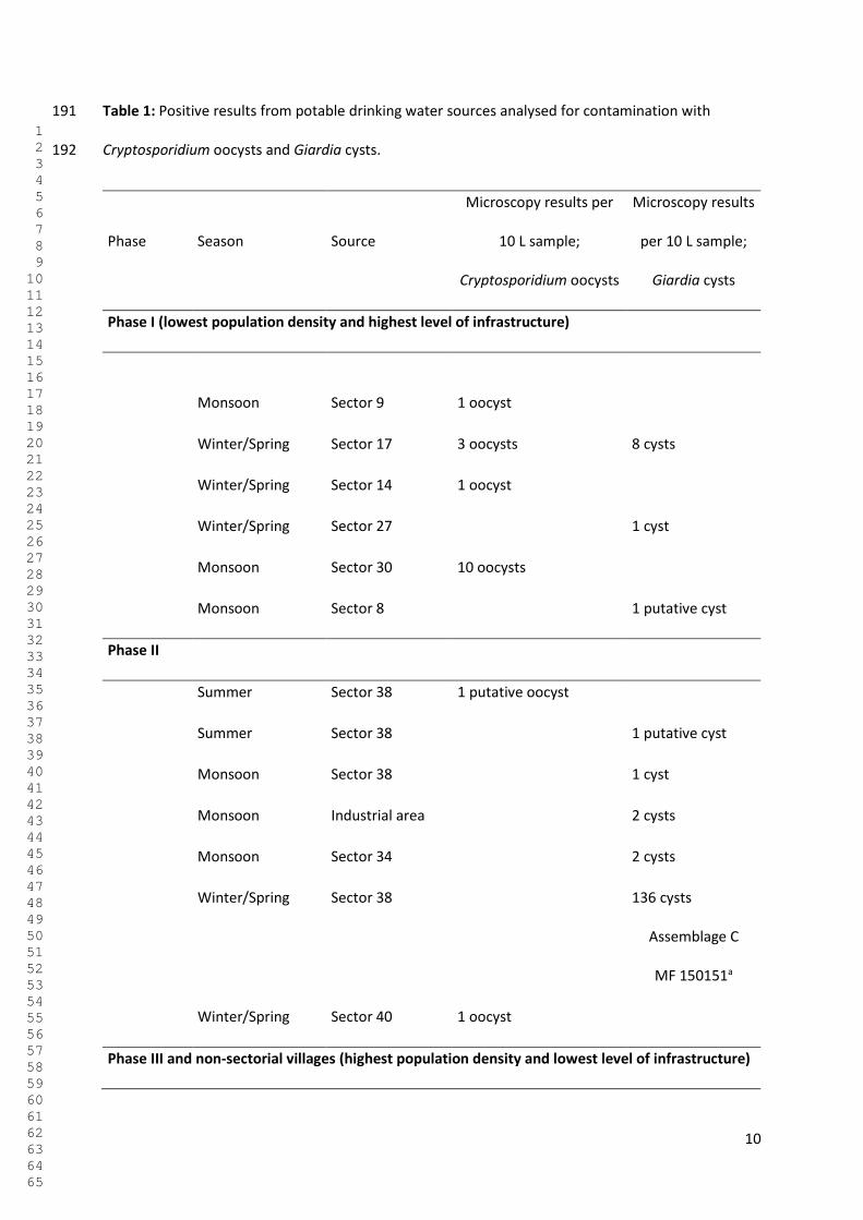
10
Table 1: Positive results from potable drinking water sources analysed for contamination with 191
Cryptosporidium oocysts and Giardia cysts. 192
Phase Season Source
Microscopy results per
10 L sample;
Cryptosporidium oocysts
Microscopy results
per 10 L sample;
Giardia cysts
Phase I (lowest population density and highest level of infrastructure)
Monsoon
Sector 9 1 oocyst
Winter/Spring Sector 17 3 oocysts 8 cysts
Winter/Spring Sector 14 1 oocyst
Winter/Spring Sector 27 1 cyst
Monsoon Sector 30 10 oocysts
Monsoon Sector 8 1 putative cyst
Phase II
Summer Sector 38 1 putative oocyst
Summer Sector 38 1 putative cyst
Monsoon Sector 38 1 cyst
Monsoon Industrial area 2 cysts
Monsoon Sector 34 2 cysts
Winter/Spring Sector 38 136 cysts
Assemblage C
MF 150151a
Winter/Spring Sector 40 1 oocyst
Phase III and non-sectorial villages (highest population density and lowest level of infrastructure)
1 2 3 4 5 6 7 8 9 10 11 12 13 14 15 16 17 18 19 20 21 22 23 24 25 26 27 28 29 30 31 32 33 34 35 36 37 38 39 40 41 42 43 44 45 46 47 48 49 50 51 52 53 54 55 56 57 58 59 60 61 62 63 64 65
11
Monsoon Kishangarh, 1 putative oocyst
Winter/Spring
Janta & Kumhar
Colony
>1000 cysts
Winter/Spring Janta & Kumhar
Colony
3 cysts
Assemblage B
MF 150152a
Total 16 7 10
a: GenBank Accession number 193
194
Discussion 195
The main result of this study is that the potable water samples analysed from in and around 196
Chandigarh were rather likely to be contaminated with Cryptosporidium oocysts and/or Giardia cysts. 197
Although other studies with a similar design have found varying occurrences, in these reports the 198
positive samples are often from raw and untreated water sources; e.g. in Norway a survey reported a 199
prevalence of positive samples 16.5% for Cryptosporidium and 11.5% of Giardia (Robertson and 200
Gjerde 2001), but the samples were from untreated surface water. Similarly, a study from Ankara, 201
Turkey, investigated municipal water supply, wells, river water and untreated dams. No 202
contamination was found in the municipal water supply, but the wells, dam and river samples gave 203
an overall occurrence of 5.8% of samples being Giardia positive (Bakir et al. 2003). In countries that 204
are perhaps more comparable with India, a study encompassing analysis of water samples from 205
Malaysia, Thailand, Philippines, and Vietnam reported only a single potable water sample (from the 206
Philippines) containing both Cryptosporidium and Giardia, whereas neither parasite was detected in 207
potable water samples from the other three countries (Kumar et al. 2016). Nevertheless, earlier 208
studies from North America have reported both Cryptosporidium and Giardia contamination in 209
1 2 3 4 5 6 7 8 9 10 11 12 13 14 15 16 17 18 19 20 21 22 23 24 25 26 27 28 29 30 31 32 33 34 35 36 37 38 39 40 41 42 43 44 45 46 47 48 49 50 51 52 53 54 55 56 57 58 59 60 61 62 63 64 65
12
potable drinking water; a survey from US revealed a 17% prevalence of Cryptosporidium, and no 210
Giardia (Rose et al. 1991), and a survey from Canada reported a prevalence of 18.2% Giardia cysts 211
and 3.5% Cryptosporidium oocysts (Wallis et al. 1996). Although our findings from Chandigarh’s 212
drinking water may give some cause for concern, particularly in relation to results from potable 213
water in other Asian countries, they are not abnormally high compared with some earlier reports 214
from some other places in the world. It should be noted that Chandigarh is a modern city, and has 215
been ranked as one of the most advanced and cleanest cities in India (Chandani 2016). Thus, our 216
findings from here should not be taken as representative for the whole country. 217
One obstacle to surveying water supplies for these parasites in developing countries is the expense 218
of the technique. In this study, the ISO-standard and a modified alternative were compared prior to 219
sample collection, and the modified version was found to perform with compatible recovery rates at 220
a lower cost and was thus used in the survey. This method could be further tested and validated in 221
multi-laboratory ring trials, and could offer a cheaper alternative for laboratories or research projects 222
with constrained budgets. 223
Climate is considered to be likely to impact the occurrence of protozoan parasites as contaminants in 224
drinking water, particularly with respect to extreme weather events. A meta-analysis indicated that 225
the likelihood of contamination of fresh surface water with Cryptosporidium oocysts and Giardia 226
cysts was significantly increased during extreme weather events (Young et al. 2015); thus, it may be 227
expected that during the monsoon season contamination would be increased. In Chandigarh, there 228
are three distinct seasons: from April to mid-July is summer with hot, dry weather, leading to 229
occasional heat waves, and a mean average rainfall of about 30mm/month; the water demand for 230
domestic supply and irrigation during these months is very high. Monsoon season, from mid-July to 231
mid-September, is when Chandigarh receives moderate to very heavy rainfall, with an average of 222 232
mm/month, with a peak in precipitation in July and August, with 288 mm and 304mm, respectively. 233
The winter, when a small part of the rainfall also occurs, averages on 33 mm/month. Spring season is 234
1 2 3 4 5 6 7 8 9 10 11 12 13 14 15 16 17 18 19 20 21 22 23 24 25 26 27 28 29 30 31 32 33 34 35 36 37 38 39 40 41 42 43 44 45 46 47 48 49 50 51 52 53 54 55 56 57 58 59 60 61 62 63 64 65
13
from mid-September to March, with mild temperatures and less precipitation (O.P. Singh 2012) 235
(Indian meteorological department). Chandigarh receives an annual average rainfall of approximately 236
1000 mm, most of which is confined to the monsoon season (JNNURM 2006). 237
In this study, the seasons had no apparent effect on contamination, and samples taken in the 238
monsoon season, in particular, were no more likely to be positive than samples taken during other 239
seasons. However, this result may reflect the unstable precipitation during the period of sampling as 240
during this period, the seasons and precipitation differed to that which is normal for Chandigarh. 241
During the monsoon season in 2014, the Chandigarh district only received about 45% of its normal 242
rainfall, and 2014 was ranked as “dry to extremely dry” (IMD, 2015). Furthermore, 2015 was the third 243
warmest year in India recorded since 1901, and although the year was considered within the normal 244
range in terms of overall precipitation, the rain came in unexpected seasons. During the summer, 245
Chandigarh received three times more than the normal rainfall, but during the usual monsoon 246
season, there was a 30% deficiency in expected precipitation, and the winter season was below 247
normal in terms of rainfall (IMD, 2016). 248
Nevertheless, from our data no specific season appeared to be more associated with contamination 249
than others, and the risk of contamination is known to be associated with many variables. It is 250
possible that season and precipitation are not major drivers for contamination in this area or that 251
insufficient samples were analysed to reveal a seasonal pattern, particularly in the light of the 252
unusual and unexpected weather patterns during sampling. 253
Although season was not associated with contamination of drinking water, the location where the 254
water was collected from did affect the likelihood of the sample being contaminated. Chandigarh city 255
is an area planned for a specific number of inhabitants, and, additionally, was originally planned for a 256
differential pattern of density. Like most cities in India, Chandigarh is overpopulated, with a current 257
population density of approximately 9300 persons per km2 (census organization of India). Citizens of 258
Chandigarh are unevenly distributed throughout the city, in so-called phases numbered from I to III, 259
1 2 3 4 5 6 7 8 9 10 11 12 13 14 15 16 17 18 19 20 21 22 23 24 25 26 27 28 29 30 31 32 33 34 35 36 37 38 39 40 41 42 43 44 45 46 47 48 49 50 51 52 53 54 55 56 57 58 59 60 61 62 63 64 65
14
with increasing densities from phase I, situated in the northern part of the city, to phase III, which is 260
at the south end. The city is also divided into sectors, in a grid-pattern based on the roads, and 261
beyond those, there are so-called non-sectorial villages and slum colonies. These villages and slum 262
colonies lack much of the infrastructure and sanitary facilities of the planned sectors. Due to cheaper 263
housing, the Phase III areas and non-sectorial villagers are characterized by high population pressure, 264
with consequences such as unsanitary conditions, flood problems, poor garbage disposal, disposal of 265
livestock dung into open drains, and discharge of untreated sewage (Chandigarh Master Plan – 266
2031). 267
Water table elevation studies have revealed that the flow of ground water is from the north to the 268
south within the city (O.P. Singh 2012), which, in turn, means that the groundwater flows from phase 269
I through phase II and III. Thus, not only does Phase III have the highest population density, but also 270
the groundwater has run through the other two sectors before reaching this stratum. This 271
stratification within the city also results in the social layers being more exposed and accessible for 272
investigation, as particular social groups tend to cluster together in terms of where they live in a city, 273
and this is probably particularly demarcated in India, which is a hierarchical society. Although our 274
results indicate significant differences in likelihood of contamination of the potable water according 275
to Phase, our expectation that Phase III areas and non-sectorial villagers would have higher rates of 276
contamination of drinking water compared with other areas was not the case. Despite the factors of 277
poor infrastructure and lack of basic hygiene, the Phase III areas and villages did not have a 278
significantly higher contamination rate than those in Phase I. Nevertheless, it should be noted that 279
one sample with a very high level of contamination was from a slum colony. 280
The similarity in contamination between Phase I and Phase III may reflect the fact that the city as a 281
whole has a relatively high population density, such that the demarcations between the phases of 282
the city are not very clear, and the water sources are distributed on a relatively small area. However, 283
1 2 3 4 5 6 7 8 9 10 11 12 13 14 15 16 17 18 19 20 21 22 23 24 25 26 27 28 29 30 31 32 33 34 35 36 37 38 39 40 41 42 43 44 45 46 47 48 49 50 51 52 53 54 55 56 57 58 59 60 61 62 63 64 65
15
this does not explain why sampling sites in Phase II were significantly more likely to show 284
contamination. 285
Previously, the water supply of Chandigarh was based on tubewell sources alone. As the population 286
has increased, so has the demand for water supply, and the underground source alone is currently 287
not sufficient to meet the city’s requirements. This gap is met by tapping surface water from the 288
Bhakra Main Line. The city has also been divided into 7 zones for the purpose of distribution, and it is 289
interesting that the local water works supply water to distinct zones of density in the city as well. The 290
three first zones supply to sectors 1-30, which have lower population densities. Zones four and five 291
supply water to phase II, a more densely populated area than the previous 30, and the last two zones 292
supply water to Manimajra, an area situated just outside Phase I. The non-sectorial villages and 293
Phase III are not supplied with water from waterworks, according to the City Development Plan, and 294
thus these non-sectorial villages do not have access to piped drinking water supply, sewer systems, 295
or storm water drainage (Administration 2016). Thus, drinking water supplies may be driven in to 296
non-sectorial villages and Phase III, and thus the residents here may actually be provided with a 297
cleaner water supply than those people living in the, arguably wealthier, Phase II areas. 298
However, as the highest contamination was found in a water tank in a Phase III slum area, it is likely 299
that individual tanks have the potential to be hot-spots of contamination and sources of infection. 300
They are more prone to on-the-spot contamination, and as they are stagnant, enclosed sources of 301
drinking water, and removal of contamination due to flow-through may be difficult. However, in this 302
study, only 8 tanks were sampled, of which 2 samples were positive and thus further studies are 303
warranted. 304
It was hoped that molecular methods would supply further information on the source of 305
contamination, and also on the likelihood of infection to people. Although most of the PCR studies 306
did not result in DNA amplification, the two successfully genotyped samples were of G. duodenalis, 307
Assemblage B and Assemblage C. Whereas Assemblage B is usually associated with human infection, 308
1 2 3 4 5 6 7 8 9 10 11 12 13 14 15 16 17 18 19 20 21 22 23 24 25 26 27 28 29 30 31 32 33 34 35 36 37 38 39 40 41 42 43 44 45 46 47 48 49 50 51 52 53 54 55 56 57 58 59 60 61 62 63 64 65
16
Assemblage C is commonly associated with canids and is not associated with human infection. This 309
Assemblage C Giardia was identified in a contaminated public water source in a Sikh temple. The 310
public frequently visits these temples, and it is common to wash and drink from the taps. This means 311
the source could have been someone handling an infected dog, or could have come directly from one 312
of the many dogs in Chandigarh, the population of which is currently increasing (Victor 2013). 313
314
Conclusions 315
The major factors that lower the significance and impact of diarrhoeal diseases on public health are 316
good sanitation, plentiful availability of good quality water, adequate disposal of human and animal 317
excrement, and public education in hygienic practices (Gadgil, 1998). The high morbidity caused by 318
Giardia and Cryptosporidium in developing countries has been attributed to factors including the 319
multiple exposure routes and cycling through domestic animals, high environmental burden, and 320
resistance of the (oo)cysts against common water disinfectants (Slifko et al. 2000). 321
The rapid population growth in Chandigarh, which places further stress on an already scarce water 322
supply, has led to solutions that may not be sustainable or appropriate on a long-term basis. The 323
highest proportion of contaminated water was found in more densely populated areas, and the 324
sample containing the highest number of parasites was taken from a water tank in a slum area. It 325
seems that the safe potable water with respect to contamination with protozoans is currently largely 326
restricted to the higher socioeconomic layers in Chandigarh. 327
It is noteworthy that in situations with poor quality drinking water, the benefits derived from 328
improving only the sanitation tend to be larger than from improving only the quality of drinking 329
water (Esrey 1996). However, combined efforts to improve both knowledge and standards of 330
drinking water by establishing surveillance systems as a first step, and implementing adequate 331
reporting systems and appropriate epidemiological surveillance, would be expected to give positive 332
results in combating parasitic protozoa and improving the health of the population. 333
1 2 3 4 5 6 7 8 9 10 11 12 13 14 15 16 17 18 19 20 21 22 23 24 25 26 27 28 29 30 31 32 33 34 35 36 37 38 39 40 41 42 43 44 45 46 47 48 49 50 51 52 53 54 55 56 57 58 59 60 61 62 63 64 65
17
Although the results of our study do not implicate the potable water supply as a source of intestinal 334
protozoan infections for the citizens of Chandigarh, or provide evidence that it is a major 335
transmission vehicle, they do indicate that it can be a potential source of infection, particularly in less 336
wealthy areas of the city. Further research that identifies how and where contamination occurs 337
would be of value, such that appropriate barriers can be implemented. 338
339
Funding information 340
This work has been funded through the Para-Clim-Chandigarh project, partly funded by the 341
Norwegian Research Council via the New Indigo Partnership Programme (Contract number: 227965). 342
Acknowledgements 343
The authors would like to thank Dr. Rakesh Seghal and Dr. Kapil Goyal for facilitating use of the 344
PGIMER facilities in Chandigarh. 345
Competing interests 346
The authors state that they have no competing interests. 347
348
349
References 350
Administration, C. (2016) Chandigarh Master Plan 2031. Available at: 351
http://chandigarh.gov.in/cmp_2031.htm Last accessed: 19.08.2017 352
353
Bakir, B., Tanyuksel, M., Saylam, F., Tanriverdi, S., Araz, R.E., Hacim, A.K. and Hasde, M. 2003 354
Investigation of waterborne parasites in drinking water sources of Ankara, Turkey. J. Microbiol. 41(2), 355
148-151. 356
1 2 3 4 5 6 7 8 9 10 11 12 13 14 15 16 17 18 19 20 21 22 23 24 25 26 27 28 29 30 31 32 33 34 35 36 37 38 39 40 41 42 43 44 45 46 47 48 49 50 51 52 53 54 55 56 57 58 59 60 61 62 63 64 65
18
357
Baldursson, S. & Karanis, P. 2011 Waterborne transmission of protozoan parasites: review of 358
worldwide outbreaks–an update 2004–2010. Water Res. 45(20), 6603-6614. 359
360
Chandani, S.K. 2016 Clean cities of India: Comparative study of Kanpur and Mysuru in special 361
reference to Swach Bharat Abhiyan. Ac. Soc. Res., ISSN Online: 2456-2645 2(4). 362
363
Checkley, W., White, A., Jagannath, D., Arrowood, M., Chalmers, R. & Chen, X. 2015 364
Cryptosporidiosis: global burden, novel diagnostics, therapeutics and vaccine targets. Lancet Infec. 365
Dis. 15, 85-94. 366
367
Efstratiou, A., Ongerth, J.E. & Karanis, P. 2017 Waterborne transmission of protozoan parasites: 368
Review of worldwide outbreaks-An update 2011–2016. Water Res. 369
370
Esrey, S.A. 1996 Water, waste, and well-being: a multicountry study. Am. J. Epidemiol. 143, 608-623. 371
372
Gadgil, A. 1998 Drinking water in developing countries. Annual Review of Energy and the 373
Environment 23, 253-286. 374
375
Hald, T., Aspinall, W., Devleesschauwer, B., Cooke, R., Corrigan, T., Havelaar, A.H., Gibb, H.J., 376
Torgerson, P.R., Kirk, M.D. & Angulo, F.J. 2016 World Health Organization estimates of the relative 377
contributions of food to the burden of disease due to selected foodborne hazards: a structured 378
expert elicitation. PloS one 11(1), e0145839. 379
380
IMD 2015. Annual Climate Summary - 2014. India Meteorological Department, Government of India, 381
Minstry of Earth Sciences, Earth System Science Organization, India Meteorological Department. 382
1 2 3 4 5 6 7 8 9 10 11 12 13 14 15 16 17 18 19 20 21 22 23 24 25 26 27 28 29 30 31 32 33 34 35 36 37 38 39 40 41 42 43 44 45 46 47 48 49 50 51 52 53 54 55 56 57 58 59 60 61 62 63 64 65
19
Available at: 383
http://www.imdpune.gov.in/Clim_Pred_LRF_New/Reports/Annual_Climate_Summary/annual_sum384
mary_2014.pdf Accessed on 19.08.2017. 385
386
IMD 2016. Annual Climate Summary 2015. India Meteorological Department, Government of India, 387
Minstry of Earth Sciences, Earth System Science Organization, India Meteorological Department. 388
Available at: 389
http://imdpune.gov.in/Clim_RCC_LRF/Annual_Climate_Summary/annual_summary_2015.pdf 390
Accessed on 19.08.2017. 391
392
ISO. 2016 ISO 18744:2016. Microbiology of the food chain — Detection and enumeration of 393
Cryptosporidium and Giardia in fresh leafy green vegetables and berry fruits. 9, C.I.T.S. (ed). 394
395
ISO. 2006 15553:2006 Water quality—isolation and identification of Cryptosporidium oocysts and 396
Giardia cysts from water. Geneva, Switzerland: International Organization for Standardization. 397
398
JNNURM 2006 City Development Plan Chandigarh. Administration, F.S.C. (ed), Chandigarh 399
Administration. Available at: http://admin.indiaenvironmentportal.org.in/reports-documents/city-400
development-plan-chandigarh Last accessed: 19.08.2017. 401
402
Kindhauser, M.K. & WHO 2003 Communicable diseases 2002: global defence against the infectious 403
disease threat. 404
405
Kotloff, K.L., Nataro, J.P., Blackwelder, W.C., Nasrin, D., Farag, T.H., Panchalingam, S., Wu, Y., Sow, 406
S.O., Sur, D., Breiman, R.F., Faruque, A.S., Zaidi, A.K., Saha, D., Alonso, P.L., Tamboura, B., Sanogo, D., 407
1 2 3 4 5 6 7 8 9 10 11 12 13 14 15 16 17 18 19 20 21 22 23 24 25 26 27 28 29 30 31 32 33 34 35 36 37 38 39 40 41 42 43 44 45 46 47 48 49 50 51 52 53 54 55 56 57 58 59 60 61 62 63 64 65
20
Onwuchekwa, U., Manna, B., Ramamurthy, T., Kanungo, S., Ochieng, J.B., Omore, R., Oundo, J.O., 408
Hossain, A., Das, S.K., Ahmed, S., Qureshi, S., Quadri, F., Adegbola, R.A., Antonio, M., Hossain, M.J., 409
Akinsola, A., Mandomando, I., Nhampossa, T., Acácio, S., Biswas, K., O'Reilly, C.E., Mintz, E.D., 410
Berkeley, L.Y., Muhsen, K., Sommerfelt, H., Robins-Browne, R.M. & Levine, M.M. 2013 Burden and 411
aetiology of diarrhoeal disease in infants and young children in developing countries (the Global 412
Enteric Multicenter Study, GEMS): a prospective, case-control study. Lancet. 382(9888), 209-222. 413
414
Kumar, T., Majid, M.A.A., Onichandran, S., Jaturas, N., Andiappan, H., Salibay, C.C., Tabo, H., Tabo, N., 415
Dungca, J.Z. & Tangpong, J. 2016 Presence of Cryptosporidium parvum and Giardia lamblia in water 416
samples from Southeast Asia: towards an integrated water detection system. Infect. Dis. Poverty 5(1), 417
3. 418
419
Platts-Mills, J.A., Babji, S., Bodhidatta, L., Gratz, J., Haque, R., Havt, A., McCormick, B.J., McGrath, M., 420
Olortegui, M.P. & Samie, A. 2015 Pathogen-specific burdens of community diarrhoea in developing 421
countries: a multisite birth cohort study (MAL-ED). Lancet Glob. health 3(9), e564-e575. 422
423
Quick, R.E., Mintz, E., Sobel, J., Mead, P., Reiff, F. & Tauxe, R. 1997 A new strategy for waterborne 424
disease prevention, pp. 340-342, Water, Engineering and Development Centre. 425
426
Robertson, L., Gjerde, B., Hansen, E.F. & Stachurska-Hagen, T. 2009 A water contamination incident 427
in Oslo, Norway during October 2007; a basis for discussion of boil-water notices and the potential 428
for post-treatment contamination of drinking water supplies. J. Water Health 7(1), 55-66. 429
430
Robertson, L.J. & Gjerde, B. 2001 Occurrence of Cryptosporidium oocysts and Giardia cysts in raw 431
waters in Norway. Scand. J. Public Health 29(3), 200-207. 432
433
1 2 3 4 5 6 7 8 9 10 11 12 13 14 15 16 17 18 19 20 21 22 23 24 25 26 27 28 29 30 31 32 33 34 35 36 37 38 39 40 41 42 43 44 45 46 47 48 49 50 51 52 53 54 55 56 57 58 59 60 61 62 63 64 65
21
Rose, J.B., Gerba, C.P. & Jakubowski, W. 1991 Survey of potable water supplies for Cryptosporidium 434
and Giardia. Environ. Sci. Technol. 25(8), 1393-1400. 435
436
Singh, O.P., Paul, Jain, R. (2012) Climate of Chandigarh. India Meteorological Department, 437
Government of India, Ministry of Earth Sciences. 438
439
Slifko, T.R., Smith, H.V. & Rose, J.B. 2000 Emerging parasite zoonoses associated with water and food. 440
Int. J. Parasitol. 30(12), 1379-1393. 441
442
Smith, H.V., Campbell, B.M., Paton, C.A. & Nichols, R.A.B. 2002 Significance of enhanced 443
morphological detection of Cryptosporidium sp. oocysts in water concentrates determined by using 444
4′, 6′-diamidino-2-phenylindole and immunofluorescence microscopy. Appl. Environ. Microbiol. 445
68(10), 5198-5201. 446
447
Utaaker, K.S., Huang, Q. & Robertson, L.J. 2015 A reduced-cost approach for analyzing fresh produce 448
for contamination with Cryptosporidium oocysts and/or Giardia cysts. Food Res. Int. 77, 326-332. 449
450
Victor, H. (2013), Hindustan Times. Municipal corporation fails to contain stray cattle, dog menace. 451
Available at: http://www.hindustantimes.com/chandigarh/municipal-corporation-fails-to-contain-452
stray-cattle-dog-menace/story-m4ifEkWzdSFf5UGkQgEEfO.html Last accessed: 19.08.2017. 453
454
Wallis, P.M., Erlandsen, S.L., Isaac-Renton, J.L., Olson, M.E., Robertson, W.J. and Van Keulen, H. 455
(1996) Prevalence of Giardia cysts and Cryptosporidium oocysts and characterization of Giardia spp. 456
isolated from drinking water in Canada. Appl. Environ. Microbiol. 62(8), 2789-2797. 457
458
WHO (2002) The world health report 2002: reducing risks, promoting healthy life, WHO. 459
1 2 3 4 5 6 7 8 9 10 11 12 13 14 15 16 17 18 19 20 21 22 23 24 25 26 27 28 29 30 31 32 33 34 35 36 37 38 39 40 41 42 43 44 45 46 47 48 49 50 51 52 53 54 55 56 57 58 59 60 61 62 63 64 65
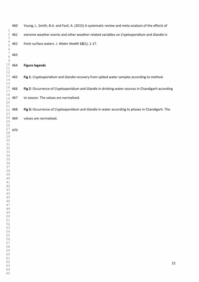
22
Young, I., Smith, B.A. and Fazil, A. (2015) A systematic review and meta-analysis of the effects of 460
extreme weather events and other weather-related variables on Cryptosporidium and Giardia in 461
fresh surface waters. J. Water Health 13(1), 1-17. 462
463
Figure legends 464
Fig 1: Cryptosporidium and Giardia recovery from spiked water samples according to method. 465
Fig 2: Occurrence of Cryptosporidium and Giardia in drinking water sources in Chandigarh according 466
to season. The values are normalised. 467
Fig 3: Occurrence of Cryptosporidium and Giardia in water according to phases in Chandigarh. The 468
values are normalised. 469
470
1 2 3 4 5 6 7 8 9 10 11 12 13 14 15 16 17 18 19 20 21 22 23 24 25 26 27 28 29 30 31 32 33 34 35 36 37 38 39 40 41 42 43 44 45 46 47 48 49 50 51 52 53 54 55 56 57 58 59 60 61 62 63 64 65
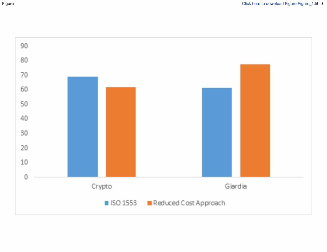
Figure Click here to download Figure Figure_1.tif
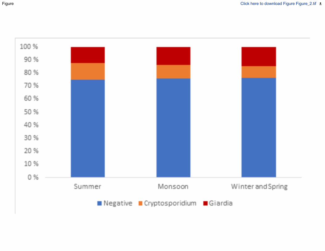
Figure Click here to download Figure Figure_2.tif
Figure Click here to download Figure Figure_3.tif
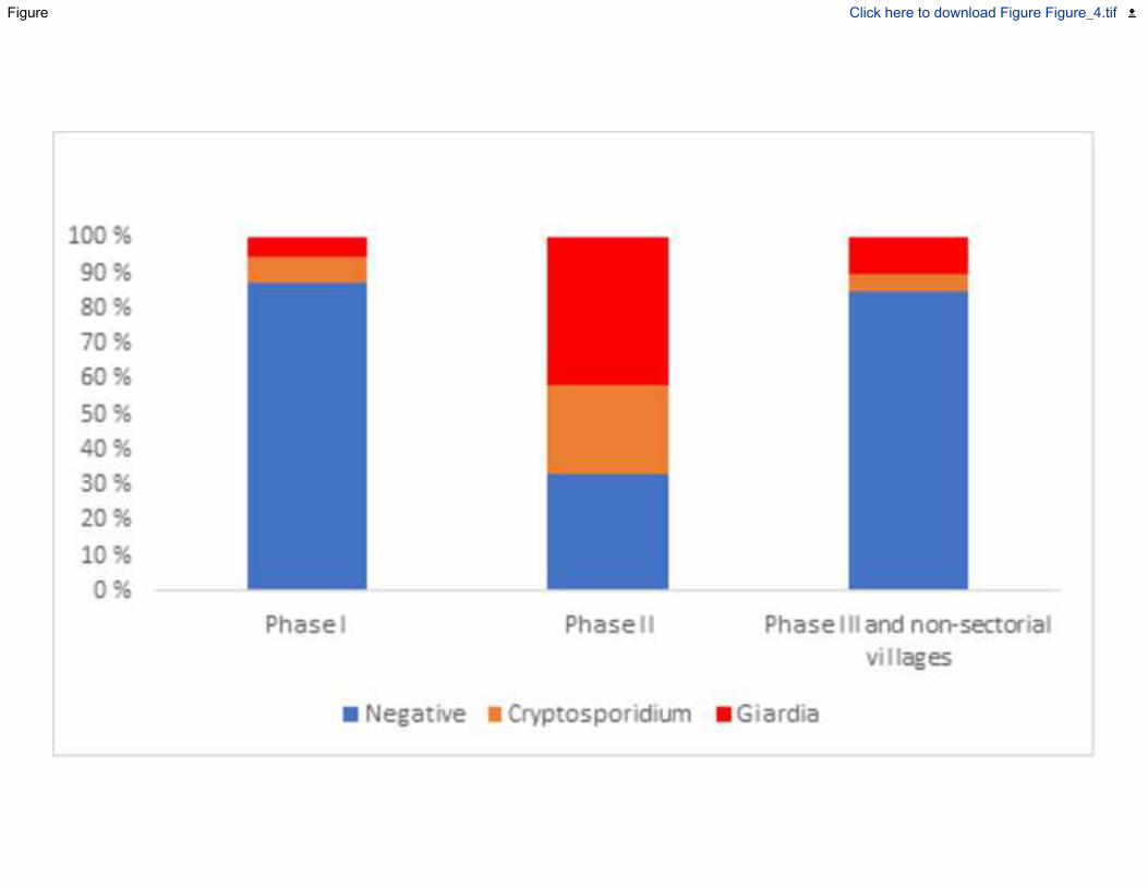
Figure Click here to download Figure Figure_4.tif
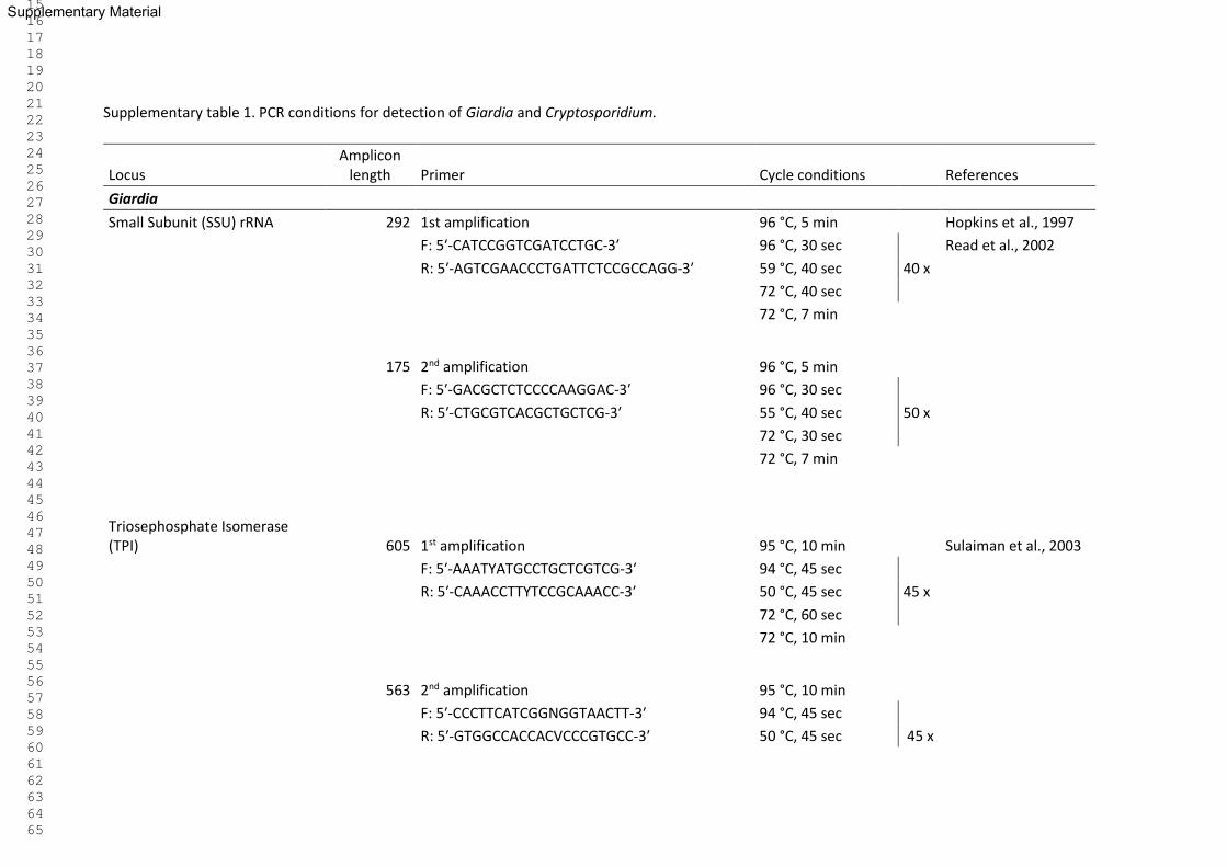
Supplementary table 1. PCR conditions for detection of Giardia and Cryptosporidium.
Locus Amplicon
length Primer Cycle conditions References
Giardia
Small Subunit (SSU) rRNA 292 1st amplification 96 °C, 5 min Hopkins et al., 1997
F: 5′-CATCCGGTCGATCCTGC-3′ 96 °C, 30 sec Read et al., 2002
R: 5′-AGTCGAACCCTGATTCTCCGCCAGG-3′ 59 °C, 40 sec 40 x
72 °C, 40 sec
72 °C, 7 min
175 2nd amplification 96 °C, 5 min
F: 5′-GACGCTCTCCCCAAGGAC-3′ 96 °C, 30 sec
R: 5′-CTGCGTCACGCTGCTCG-3′ 55 °C, 40 sec 50 x
72 °C, 30 sec
72 °C, 7 min
Triosephosphate Isomerase (TPI) 605 1st amplification 95 °C, 10 min Sulaiman et al., 2003
F: 5′-AAATYATGCCTGCTCGTCG-3′ 94 °C, 45 sec
R: 5′-CAAACCTTYTCCGCAAACC-3′ 50 °C, 45 sec 45 x
72 °C, 60 sec
72 °C, 10 min
563 2nd amplification 95 °C, 10 min
F: 5′-CCCTTCATCGGNGGTAACTT-3′ 94 °C, 45 sec
R: 5′-GTGGCCACCACVCCCGTGCC-3′ 50 °C, 45 sec 45 x
Supplementary Material
1 2 3 4 5 6 7 8 9 10 11 12 13 14 15 16 17 18 19 20 21 22 23 24 25 26 27 28 29 30 31 32 33 34 35 36 37 38 39 40 41 42 43 44 45 46 47 48 49 50 51 52 53 54 55 56 57 58 59 60 61 62 63 64 65
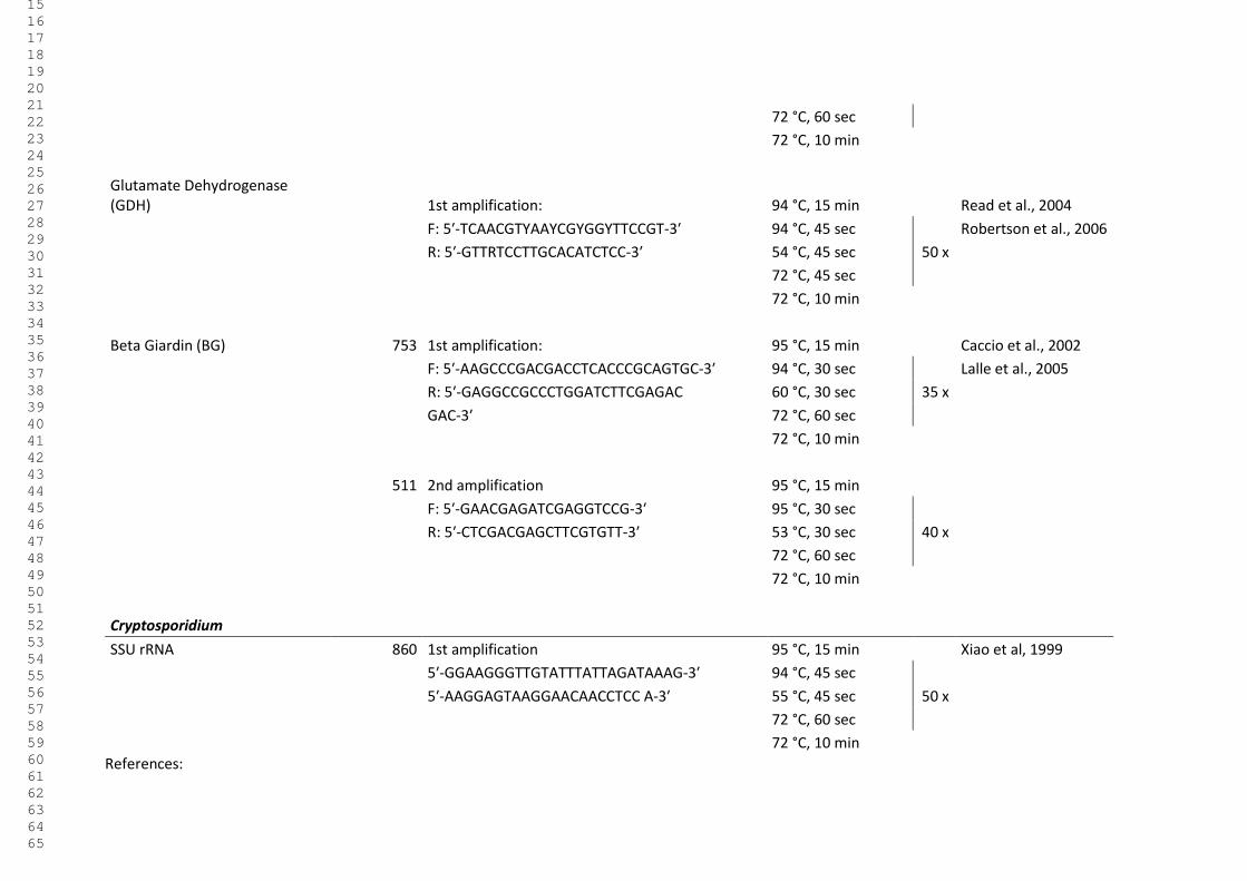
72 °C, 60 sec
72 °C, 10 min
Glutamate Dehydrogenase (GDH) 1st amplification: 94 °C, 15 min Read et al., 2004
F: 5′-TCAACGTYAAYCGYGGYTTCCGT-3′ 94 °C, 45 sec Robertson et al., 2006
R: 5′-GTTRTCCTTGCACATCTCC-3′ 54 °C, 45 sec 50 x
72 °C, 45 sec
72 °C, 10 min
Beta Giardin (BG) 753 1st amplification: 95 °C, 15 min Caccio et al., 2002
F: 5′-AAGCCCGACGACCTCACCCGCAGTGC-3′ 94 °C, 30 sec Lalle et al., 2005
R: 5′-GAGGCCGCCCTGGATCTTCGAGAC 60 °C, 30 sec 35 x
GAC-3′ 72 °C, 60 sec
72 °C, 10 min
511 2nd amplification 95 °C, 15 min
F: 5′-GAACGAGATCGAGGTCCG-3′ 95 °C, 30 sec
R: 5′-CTCGACGAGCTTCGTGTT-3′ 53 °C, 30 sec 40 x
72 °C, 60 sec
72 °C, 10 min
Cryptosporidium
SSU rRNA 860 1st amplification 95 °C, 15 min Xiao et al, 1999
5′-GGAAGGGTTGTATTTATTAGATAAAG-3′ 94 °C, 45 sec
5′-AAGGAGTAAGGAACAACCTCC A-3′ 55 °C, 45 sec 50 x
72 °C, 60 sec
72 °C, 10 min
References:
1 2 3 4 5 6 7 8 9 10 11 12 13 14 15 16 17 18 19 20 21 22 23 24 25 26 27 28 29 30 31 32 33 34 35 36 37 38 39 40 41 42 43 44 45 46 47 48 49 50 51 52 53 54 55 56 57 58 59 60 61 62 63 64 65
Hopkins, R. M., Meloni, B. P., Groth, D. M., Wetherall, J. D., Reynoldson, J. A. & Thompson, R. A. 1997 Ribosomal RNA sequencing reveals differences
between the genotypes of Giardia isolates recovered from humans and dogs living in the same locality. J. parasitol., 44-51.
Lalle, M., Pozio, E., Capelli, G., Bruschi, F., Crotti, D. & Cacciò, S. M. 2005. Genetic heterogeneity at the β-giardin locus among human and animal isolates of
Giardiaduodenalis and identification of potentially zoonotic subgenotypes. Int. J. parasitol. 35(2), 207-213.
Read, C., Walters, J., Robertson, I. D. & Thompson, R. C. A. 2002. Correlation between genotype of Giardiaduodenalis and diarrhoea. Int. J. Parasitol. 32(2),
229-231.
Read, C. M., Monis, P. T. & Thompson, R. A. 2004 Discrimination of all genotypes of Giardia duodenalis at the glutamate dehydrogenase locus using PCR-
RFLP. Infect. Genet. Evol. 4(2), 125-130.
Robertson, L. J., Hermansen, L., Gjerde, B. K., Strand, E., Alvsvåg, J. O. & Langeland, N. 2006 Application of genotyping during an extensive outbreak of
waterborne giardiasis in Bergen, Norway, during autumn and winter 2004. Appl. Environ. Microbiol. 72(3), 2212-2217.
Sulaiman, I. M., Fayer, R., Bern, C., Gilman, R. H., Trout, J. M., Schantz, P. M., Das, P., Lal, A.A. & Xiao, L. 2003. Triosephosphate isomerase gene
characterization and potential zoonotic transmission of Giardia duodenalis. Emerg. Infect. Dis. 9(11), 1444-1452.
Xiao, Lihua, Escalante, L., Yang, C., Sulaiman, I., Escalante A. A., Montali, R. J., Fayer, R. & Lal, A. A. 1999 Phylogenetic analysis of Cryptosporidium
parasites based on the small-subunit rRNA gene locus. Appl. Environ. Microbiol. 65.4, 1578-1583.
1 2 3 4 5 6 7 8 9 10 11 12 13 14 15 16 17 18 19 20 21 22 23 24 25 26 27 28 29 30 31 32 33 34 35 36 37 38 39 40 41 42 43 44 45 46 47 48 49 50 51 52 53 54 55 56 57 58 59 60 61 62 63 64 65

V

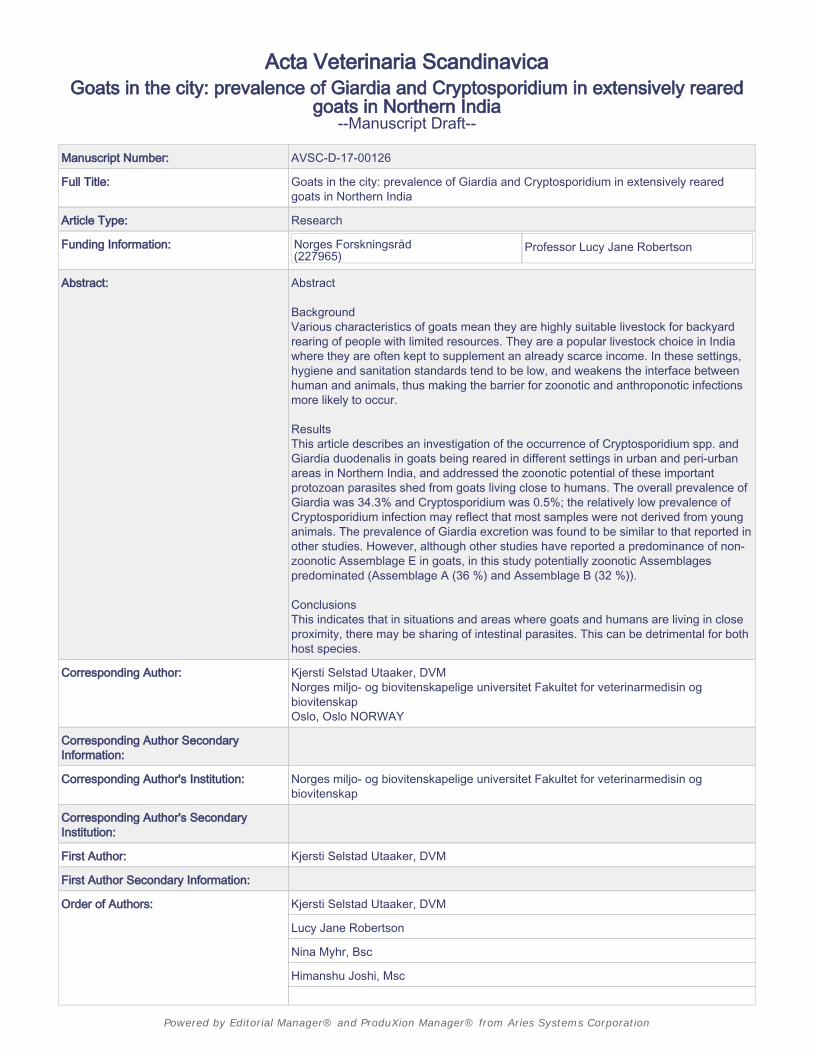
Acta Veterinaria Scandinavica
Goats in the city: prevalence of Giardia and Cryptosporidium in extensively rearedgoats in Northern India
--Manuscript Draft--
Manuscript Number: AVSC-D-17-00126
Full Title: Goats in the city: prevalence of Giardia and Cryptosporidium in extensively rearedgoats in Northern India
Article Type: Research
Funding Information: Norges Forskningsråd(227965)
Professor Lucy Jane Robertson
Abstract: Abstract
BackgroundVarious characteristics of goats mean they are highly suitable livestock for backyardrearing of people with limited resources. They are a popular livestock choice in Indiawhere they are often kept to supplement an already scarce income. In these settings,hygiene and sanitation standards tend to be low, and weakens the interface betweenhuman and animals, thus making the barrier for zoonotic and anthroponotic infectionsmore likely to occur.
ResultsThis article describes an investigation of the occurrence of Cryptosporidium spp. andGiardia duodenalis in goats being reared in different settings in urban and peri-urbanareas in Northern India, and addressed the zoonotic potential of these importantprotozoan parasites shed from goats living close to humans. The overall prevalence ofGiardia was 34.3% and Cryptosporidium was 0.5%; the relatively low prevalence ofCryptosporidium infection may reflect that most samples were not derived from younganimals. The prevalence of Giardia excretion was found to be similar to that reported inother studies. However, although other studies have reported a predominance of non-zoonotic Assemblage E in goats, in this study potentially zoonotic Assemblagespredominated (Assemblage A (36 %) and Assemblage B (32 %)).
ConclusionsThis indicates that in situations and areas where goats and humans are living in closeproximity, there may be sharing of intestinal parasites. This can be detrimental for bothhost species.
Corresponding Author: Kjersti Selstad Utaaker, DVMNorges miljo- og biovitenskapelige universitet Fakultet for veterinarmedisin ogbiovitenskapOslo, Oslo NORWAY
Corresponding Author SecondaryInformation:
Corresponding Author's Institution: Norges miljo- og biovitenskapelige universitet Fakultet for veterinarmedisin ogbiovitenskap
Corresponding Author's SecondaryInstitution:
First Author: Kjersti Selstad Utaaker, DVM
First Author Secondary Information:
Order of Authors: Kjersti Selstad Utaaker, DVM
Lucy Jane Robertson
Nina Myhr, Bsc
Himanshu Joshi, Msc
Powered by Editorial Manager® and ProduXion Manager® from Aries Systems Corporation
Anil Kumar, Msc
Rajinder Singh Bajwa, DVM
Order of Authors Secondary Information:
Opposed Reviewers:
Powered by Editorial Manager® and ProduXion Manager® from Aries Systems Corporation
1
Goats in the city: prevalence of Giardia and Cryptosporidium in extensively reared goats in 1
northern India 2
Kjersti Selstad Utaaker1, Nina Myhr1, Rajinder S. Bajwa2, Himanshu Joshi3, Anil Kumar3, Lucy J. 3
Robertson1 4
Parasitology Lab, Department for Food Safety and Infection Biology, Faculty of Veterinary Medicine, 5
Norwegian University of Life Sciences, Adamstuen Campus, PO Box 8146 Dep. 0033 Oslo, Norway1 6
Veterinary Hospital for Large Animals, Sector 38, Chandigarh, 160036, India2 7
Department of Medical Parasitology, Postgraduate Institute of Medical Education and Research, 8
Chandigarh, 16002 India3 9
Corresponding author: [email protected] 10
Nina Myhr: [email protected] 11
Rajinder S. Bajwa: [email protected] 12
Himanshu Joshi: [email protected] 13
Anil Kumar: [email protected] 14
Lucy J. Robertson: [email protected] 15
16
17
Keywords: Backyard livestock, Cryptosporidium, developing countries, Giardia, goat, One Health, 18
zoonosis. 19
20
21
Manuscript Click here to download ManuscriptGoats_in_Northern_India.docx
Click here to view linked References
1 2 3 4 5 6 7 8 9 10 11 12 13 14 15 16 17 18 19 20 21 22 23 24 25 26 27 28 29 30 31 32 33 34 35 36 37 38 39 40 41 42 43 44 45 46 47 48 49 50 51 52 53 54 55 56 57 58 59 60 61 62 63 64 65
2
22
23
24
25
26
27
28
29
30
31
32
Abstract 33
Background 34
Various characteristics of goats mean they are highly suitable livestock for backyard rearing of people 35
with limited resources. They are a popular livestock choice in India where they are often kept to 36
supplement an already scarce income. In these settings, hygiene and sanitation standards tend to be 37
low, and weakens the interface between human and animals, thus making the barrier for zoonotic 38
and anthroponotic infections more likely to occur. 39
Results 40
This article describes an investigation of the occurrence of Cryptosporidium spp. and Giardia 41
duodenalis in goats being reared in different settings in urban and peri-urban areas in northern India, 42
1 2 3 4 5 6 7 8 9 10 11 12 13 14 15 16 17 18 19 20 21 22 23 24 25 26 27 28 29 30 31 32 33 34 35 36 37 38 39 40 41 42 43 44 45 46 47 48 49 50 51 52 53 54 55 56 57 58 59 60 61 62 63 64 65
3
and addressed the zoonotic potential of these important protozoan parasites shed from goats living 43
close to humans. The overall prevalence of Giardia was 34.3% and Cryptosporidium was 0.5%; the 44
relatively low prevalence of Cryptosporidium infection may reflect that most samples were not 45
derived from young animals. The prevalence of Giardia excretion was found to be similar to that 46
reported in other studies. However, although other studies have reported a predominance of non-47
zoonotic Assemblage E in goats, in this study potentially zoonotic Assemblages predominated 48
(Assemblage A (36 %) and Assemblage B (32 %)). 49
Conclusions 50
This indicates that in situations and areas where goats and humans are living in close proximity, there 51
may be sharing of intestinal parasites. This can be detrimental for both host species. 52
53
1 2 3 4 5 6 7 8 9 10 11 12 13 14 15 16 17 18 19 20 21 22 23 24 25 26 27 28 29 30 31 32 33 34 35 36 37 38 39 40 41 42 43 44 45 46 47 48 49 50 51 52 53 54 55 56 57 58 59 60 61 62 63 64 65
4
Background 54
Cryptosporidium spp and Giardia duodenalis are among the most common enteric parasites of 55
domestic animals, humans and wildlife [1], and some species and assemblages are recognized to 56
have considerable zoonotic potential. They are also two of the most common aetiological agents of 57
paediatric diarrhoea in developing countries, where poverty and density are high, and hygiene 58
standards is low, and these protozoans are associated with mortality as well as morbidity [2, 3]. 59
Given the high prevalence of giardiasis and cryptosporidiosis in people living in underdeveloped 60
communities, these diseases were included in the WHO “neglected disease initiative” in 2004 [4]. 61
Several epidemiological studies from India have shown that human giardiasis is prevalent throughout 62
the country, and community-based studies from northern India revealed prevalence rates from 5.5% 63
to 70%, with highest rates in low socioeconomic groups in Chandigarh [5]. Giardiasis has a significant 64
public health impact due to the high prevalence and disease burden of infection, and its association 65
with causing major outbreaks. The effects of Giardia infection on growth and cognitive functions of 66
infected children, particularly in developing countries, where people are exposed to other insults to 67
their health, is of particular importance [6]. Giardia is also a common infection in livestock and 68
companion animals, and sometimes, but not always, associated with disease [7-9]. The zoonotic 69
potential of Giardia is controversial, with some Assemblages apparently host-specific, while others 70
are more promiscuous in host proclivity [10]. In northern India, a study found the prevailing 71
Assemblages among humans to be Assemblage B, which is associated with anthroponotic 72
transmission, followed by assemblage A, which has been found in a wide range of mammals [11]. 73
Cryptosporidiosis is a diarrhoeal disease found in humans and animals worldwide [12], and can be 74
caused by several species and genotypes of Cryptosporidium [13]. In humans, C. hominis and C. 75
parvum are the aetiological agents responsible for most infections [14], whereas C. hominis only 76
infects humans and C. parvum primarily infects humans and ruminants. Strong links between contact 77
with infected livestock and humans have been demonstrated in Cryptosporidium infections [15, 16]. 78
1 2 3 4 5 6 7 8 9 10 11 12 13 14 15 16 17 18 19 20 21 22 23 24 25 26 27 28 29 30 31 32 33 34 35 36 37 38 39 40 41 42 43 44 45 46 47 48 49 50 51 52 53 54 55 56 57 58 59 60 61 62 63 64 65
5
In India, Cryptosporidium have been reported with prevalence ranging from 3.8% in patients in 79
northern India, with the majority of infections attributed to C. hominis [17], to 39.7% in the rural 80
population [18]. 81
In areas with inadequate infrastructure of water supply and sanitation, and where animals are 82
roaming free and often in close contact with humans, the importance of Giardia and Cryptosporidium 83
as One Health parasites remains to be fully defined. 84
The potential for transmission of zoonotic agents between humans and animals is amplified when 85
the interface between them is close, and the management of animals and hygienic setting is poor. A 86
huge part of the populations of developing countries live under such conditions, and often backyard 87
livestock are relied upon to provide extra income or food. Goats are highly suitable livestock for 88
backyard rearing of people with limited resources as their grazing preferences enable them to feed 89
on weeds, shrubs and other plants that other domestic animals refuse, their small size means they 90
require less space than larger animals and they are easier to work with than large ruminants and are 91
cheaper to buy and maintain [19]. Also, unlike sheep, goats have a high capacity for adapting to 92
extreme climatic conditions, and are therefore are particularly valuable in arid and semi-arid regions 93
to which sheep are unable to adapt so readily. Although on a global scale, sheep are much more 94
common than goats, in India the goat population is more than double the sheep population, being 95
133 million goats and 63 million sheep in 2014 [20]. 96
There are very few studies available that report on prevalence of infections with the potential to be 97
transmitted between goats and their owners or consumers in areas where they are most likely to 98
exert the greatest impact on each other. Indeed, in a review from 2009 on Cryptosporidium and 99
Giardia in sheep and goats [21], the vast majority of data were reported from sheep. We identified 100
one report of Cryptosporidium and Giardia among goats in India [22] with 35 % prevalence of 101
Cryptosporidium and 20 % prevalence of Giardia found among goats in Odisha in Eastern India. 102
1 2 3 4 5 6 7 8 9 10 11 12 13 14 15 16 17 18 19 20 21 22 23 24 25 26 27 28 29 30 31 32 33 34 35 36 37 38 39 40 41 42 43 44 45 46 47 48 49 50 51 52 53 54 55 56 57 58 59 60 61 62 63 64 65
6
However, molecular analyses were not conducted and therefore the zoonotic potential was not 103
ascertained. 104
The study described in this article aimed at determining the prevalence and zoonotic potential of 105
Cryptosporidium and Giardia in small-scale goat farms and backyard livestock goats in urban and 106
peri-urban areas in northern India. 107
108
Material and methods 109
Sampling 110
A total of 207 faecal samples from goats in Chandigarh, Punjab and Haryana were collected with 111
consent from the owners. The samples were collected from both urban “village” areas in Chandigarh, 112
and peri-urban villages in the neighbouring states of Punjab and Haryana. These goats were mainly 113
kept for meat production. 114
Singha Devi, Jayenti and Kurali are small towns and villages located in the S.A.S Nagar district of the 115
state Punjab. The district has a population of close to a million, with an almost even dispersion in 116
rural and urban areas. The population density is 909/ Km2 [23] The goat population of the S.A.S Nagar 117
district has been estimated to be 6330 [24]. 118
Saketri is a village in the Panchkula district of the Haryana state. The district has a population of 119
about half a million, with a density of 625/ Km2, and the population is almost evenly distributed in 120
rural and urban areas [23]. The goat population of Panchkula has been estimated to be 8199 [25]. 121
The city Chandigarh has a population of about one million, with almost all of its inhabitants living in 122
urban areas. The population density of the city is 9258/Km2. Kansal and Maloya are so-called non-123
sectorial villages associated with the city, but with poorer infrastructure and lower socioeconomic 124
levels compared with the city itself [26]. Sector 38 West is the location of the slum colony Rajiv. Slum 125
settlements have grown in the past decades in Chandigarh, especially in the periphery of the city, 126
1 2 3 4 5 6 7 8 9 10 11 12 13 14 15 16 17 18 19 20 21 22 23 24 25 26 27 28 29 30 31 32 33 34 35 36 37 38 39 40 41 42 43 44 45 46 47 48 49 50 51 52 53 54 55 56 57 58 59 60 61 62 63 64 65
7
where poor families tend to settle due to cheap housing. It was in these areas goats were kept as 127
well. The goat population of Chandigarh has been estimated to be 805. (Department of Animal 128
husbandry and Fisheries, Chandigarh Administration, [27] 129
The samples (each approximately 5-10 g) were collected either rectally or non-invasively promptly 130
after defecation, and were immediately mixed with 2.5% potassium dichromate and stored at 4°C 131
before transportation to the Parasitology Department, Norwegian University of Life Sciences (NMBU) 132
for analysis. The ages of the goats were not recorded, but the majority were adults. 133
Figure 1: Areas where samples were collected. 134
135
Analysis of goat faeces for Cryptosporidium oocysts and Giardia cysts 136
The samples were analysed by immunomagnetic separation (IMS) and direct faecal smears with 137
immunofluorescent antibody staining for the presence of Giardia cysts and/ or Cryptosporidium 138
oocysts. 139
IMS analysis 140
Faecal samples were washed with phosphate buffered saline, and then passed through a faecal 141
parasite concentrator with a pore diameter 425 μm (Midi Parasep, Apacor, Berkshire, England) and 142
centrifuged to create a pellet. Giardia cysts and Cryptosporidium oocysts were isolated using an in-143
house immunomagnetic separation method (IMS) using Dynabeads®(GC-Combo, Life Technologies, 144
Carlsbad, CA) [28, 29]; 10 μL anti-Giardia beads, 10 μL anti-Cryptosporidium beads, 80 μL Sur-Modics 145
StabilZyme® , 20 μL SL Buffer B and 100µL Buffer Q4 were used to generate 55 μL of purified sample 146
from approximately 200 mg of the faecal pellet. 5 μL of the resulting purified sample was dried and 147
fixed with methanol to multispot welled slides for detection of Giardia cysts 148
and Cryptosporidium oocysts using a Cryptosporidium/Giardia direct immunofluorescent antibody 149
test (IFAT; Aqua-Glo, Waterborne Inc., New Orleans), in accordance with manufacturer's instructions. 150
1 2 3 4 5 6 7 8 9 10 11 12 13 14 15 16 17 18 19 20 21 22 23 24 25 26 27 28 29 30 31 32 33 34 35 36 37 38 39 40 41 42 43 44 45 46 47 48 49 50 51 52 53 54 55 56 57 58 59 60 61 62 63 64 65
8
Prior to being screened, samples were also stained with 4′6 diamidino-2-phenylindole (DAPI), a non-151
specific fluorescent stain that binds to double-stranded DNA. 152
Faecal smears 153
Between 5-20 µL of homogenized and sieved faecal material was placed on a microscope slide using 154
plastic bacteriological loops that take approx. 10µL amount of sample. The samples were left to dry 155
and then fixed with methanol before staining with 15 µL of monoclonal antibody and incubation as 156
described for IMS. DAPI staining was not used in this preparation due to the amount of other DNA – 157
containing debris in faecal smears. 158
After fixing and staining, preparations from both faecal smears and IMS were screened under a 159
fluorescent microscope with the following filter settings: FITC: emission- 490, excitation – 525 and 160
DAPI: emission- 350, excitation – 470. 161
The samples were graded after counting the number of cysts/oocysts per field of view at x20 162
magnification: 163
Table 1: Grading of cyst counts visualized using immunofluorescent microscopy 164
Cyst count Grading
1-9 +
10 – 50 ++
>51 +++
>100 ++++
165
Molecular methods 166
DNA extraction 167
Each microcentrifuge tube containing Cryptosporidium oocysts and Giardia cysts were re-suspended 168
1 2 3 4 5 6 7 8 9 10 11 12 13 14 15 16 17 18 19 20 21 22 23 24 25 26 27 28 29 30 31 32 33 34 35 36 37 38 39 40 41 42 43 44 45 46 47 48 49 50 51 52 53 54 55 56 57 58 59 60 61 62 63 64 65
9
in Tris-EDTA buffer and held at 100°C for Cryptosporidium oocysts and 90°C for Giardia cysts for one 169
hour, before the DNA was isolated using QIamp DNA mini kit (Qiagen GmbH), using an overnight step 170
at 56°C. 171
PCR, electrophoresis, purification of PCR product, and sequencing 172
Samples that were DAPI – positive were selected for genotyping and PCR, regardless of number of 173
(oo)cysts. 174
Four genes were used for genotyping investigations of the Giardia positive samples by conventional 175
PCR; the β-giardin gene, the glutamate dehydrogenase (gdh) gene, the triosphosphate isomerase (tpi) 176
gene, and the small subunit ribosomal RNA (SSU RNA). For the sample with Cryptosporidium oocysts, 177
primers targeting sections of the genes SSU, COWP, and Actin were used, also by conventional PCR. 178
The primers and reaction cycles are further described in the supplementary table. 179
For all genes, the following PCR mixture was used: 10 pmol of each primer, 0.4 μl of bovine serum 180
albumin (20 mg/mL), 5.8 μl of water, 25 μl of HotStartTaqmaster (QIAGEN® GmbH, Germany), and 2 181
μl of template. For each set of reactions, a negative control (water) and a positive control (DNA 182
from G. duodenalis H3 isolate belonging to assemblage B, Waterborne Inc., New Orleans, USA, and 183
Cryptosporidium parvum oocysts, with species identification by Hønsvall and Robertson [30]) were 184
included. 185
PCR products were electrophoresed on 1% agarose gels and stained with SYBRsafe® DNA gel stain 186
under UV radiation. Positive samples were purified using ROCHE® high pure PCR product purification 187
kit, and purified products were sent along with appropriate primers for sequencing on both strands 188
at GATC biotech, Germany. Sequences were examined using Geneious 10.1.2 software and sequence 189
comparisons conducted using NCBI BLAST. 190
Sequences were submitted to GenBank and their Accession numbers are provided in the results. 191
Statistics 192
1 2 3 4 5 6 7 8 9 10 11 12 13 14 15 16 17 18 19 20 21 22 23 24 25 26 27 28 29 30 31 32 33 34 35 36 37 38 39 40 41 42 43 44 45 46 47 48 49 50 51 52 53 54 55 56 57 58 59 60 61 62 63 64 65
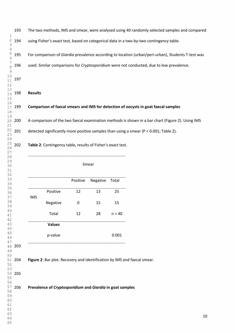
10
The two methods, IMS and smear, were analysed using 40 randomly selected samples and compared 193
using Fisher’s exact test, based on categorical data in a two-by-two contingency table. 194
For comparison of Giardia prevalence according to location (urban/peri-urban), Students T-test was 195
used. Similar comparisons for Cryptosporidium were not conducted, due to low prevalence. 196
197
Results 198
Comparison of faecal smears and IMS for detection of oocysts in goat faecal samples 199
A comparison of the two faecal examination methods is shown in a bar chart (Figure 2). Using IMS 200
detected significantly more positive samples than using a smear (P < 0.001; Table 2). 201
Table 2: Contingency table, results of Fisher’s exact test. 202
Smear
Positive Negative Total
IMS
Positive 12 13 25
Negative 0 15 15
Total 12 28 n = 40
Values
p-value 0.001
203
Figure 2: Bar plot. Recovery and identification by IMS and faecal smear. 204
205
Prevalence of Cryptosporidium and Giardia in goat samples 206
1 2 3 4 5 6 7 8 9 10 11 12 13 14 15 16 17 18 19 20 21 22 23 24 25 26 27 28 29 30 31 32 33 34 35 36 37 38 39 40 41 42 43 44 45 46 47 48 49 50 51 52 53 54 55 56 57 58 59 60 61 62 63 64 65
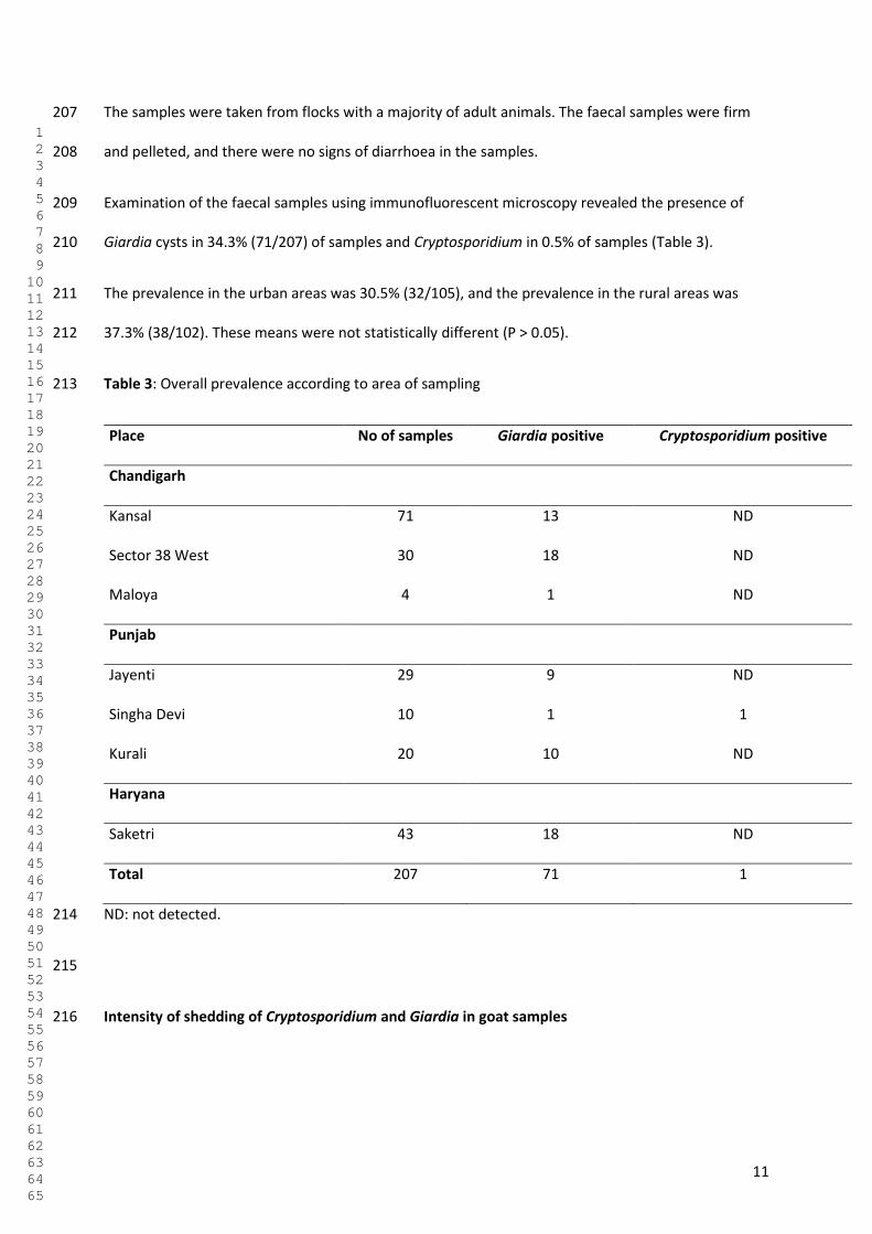
11
The samples were taken from flocks with a majority of adult animals. The faecal samples were firm 207
and pelleted, and there were no signs of diarrhoea in the samples. 208
Examination of the faecal samples using immunofluorescent microscopy revealed the presence of 209
Giardia cysts in 34.3% (71/207) of samples and Cryptosporidium in 0.5% of samples (Table 3). 210
The prevalence in the urban areas was 30.5% (32/105), and the prevalence in the rural areas was 211
37.3% (38/102). These means were not statistically different (P > 0.05). 212
Table 3: Overall prevalence according to area of sampling 213
Place No of samples Giardia positive Cryptosporidium positive
Chandigarh
Kansal 71 13 ND
Sector 38 West 30 18 ND
Maloya 4 1 ND
Punjab
Jayenti 29 9 ND
Singha Devi 10 1 1
Kurali 20 10 ND
Haryana
Saketri 43 18 ND
Total 207 71 1
ND: not detected. 214
215
Intensity of shedding of Cryptosporidium and Giardia in goat samples 216
1 2 3 4 5 6 7 8 9 10 11 12 13 14 15 16 17 18 19 20 21 22 23 24 25 26 27 28 29 30 31 32 33 34 35 36 37 38 39 40 41 42 43 44 45 46 47 48 49 50 51 52 53 54 55 56 57 58 59 60 61 62 63 64 65
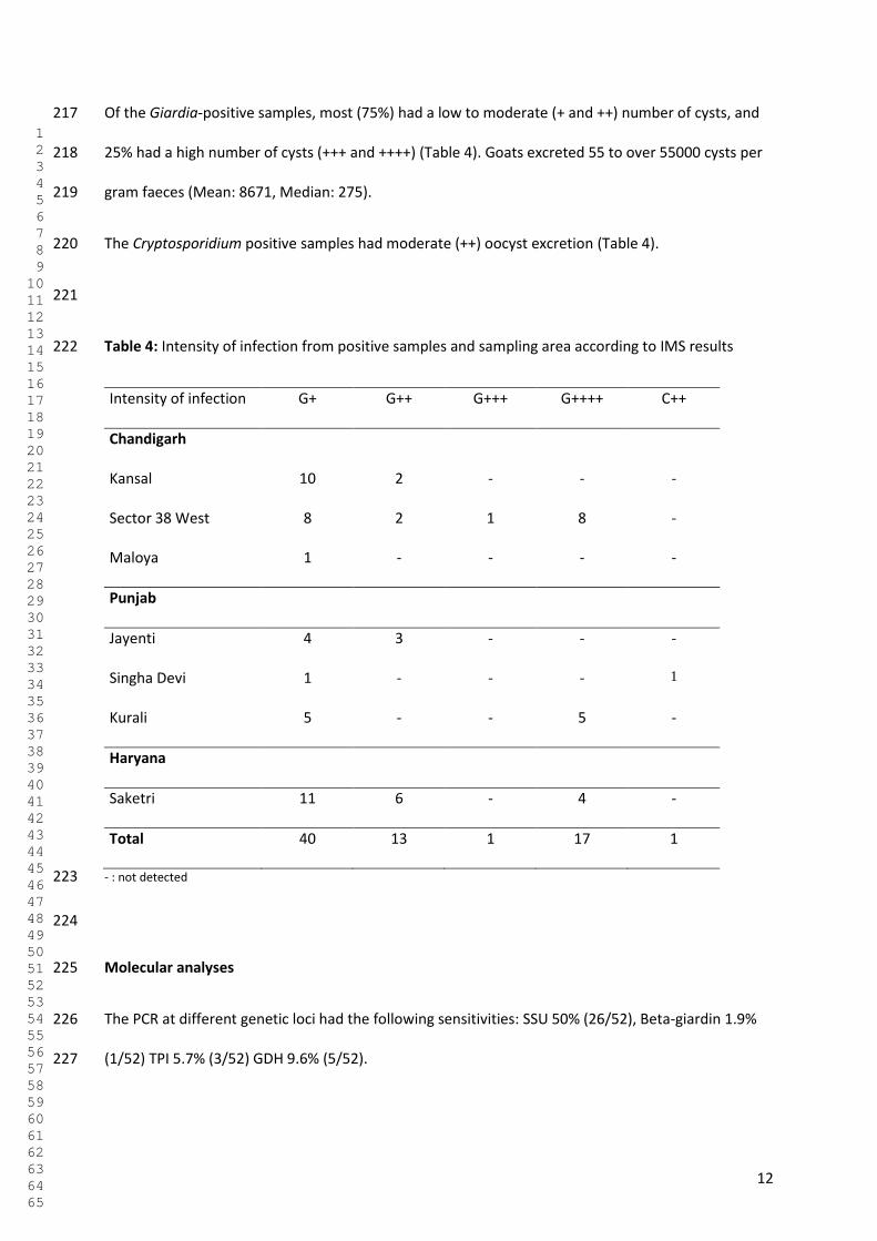
12
Of the Giardia-positive samples, most (75%) had a low to moderate (+ and ++) number of cysts, and 217
25% had a high number of cysts (+++ and ++++) (Table 4). Goats excreted 55 to over 55000 cysts per 218
gram faeces (Mean: 8671, Median: 275). 219
The Cryptosporidium positive samples had moderate (++) oocyst excretion (Table 4). 220
221
Table 4: Intensity of infection from positive samples and sampling area according to IMS results 222
Intensity of infection G+ G++ G+++ G++++ C++
Chandigarh
Kansal 10 2 - - -
Sector 38 West 8 2 1 8 -
Maloya 1 - - - -
Punjab
Jayenti 4 3 - - -
Singha Devi 1 - - - 1
Kurali 5 - - 5 -
Haryana
Saketri 11 6 - 4 -
Total 40 13 1 17 1
- : not detected 223
224
Molecular analyses 225
The PCR at different genetic loci had the following sensitivities: SSU 50% (26/52), Beta-giardin 1.9% 226
(1/52) TPI 5.7% (3/52) GDH 9.6% (5/52). 227
1 2 3 4 5 6 7 8 9 10 11 12 13 14 15 16 17 18 19 20 21 22 23 24 25 26 27 28 29 30 31 32 33 34 35 36 37 38 39 40 41 42 43 44 45 46 47 48 49 50 51 52 53 54 55 56 57 58 59 60 61 62 63 64 65
13
PCR and sequencing on the single Cryptosporidium-positive sample revealed C. ubiquitum (GenBank 228
Accession number: MF124820) 229
230
An overview of the Giardia genotyping results is provided in Table 5. Based on all the results 231
combined from the different PCR, the majority (68 %) of Giardia Assemblages identified were 232
potentially zoonotic (A or B), with 10 out of 28 (36 %) genotyped samples Assemblage A, 9 (32 %) 233
Assemblage B, 8 (29 %) Assemblage E, 1 and one (4 %) Assemblage D. One of the samples was 234
sequenced to be Assemblage E at the GDH gene, and Assemblage C at the SSU gene. 235
236
Table 5: 237
Discussion 238
The main finding of this cross-sectional survey is that although Cryptosporidium prevalence was 239
relatively low in the goats included, the prevalence of Giardia excretion was relatively high, and with 240
a preponderance of potentially zoonotic Assemblages, indicating that goats may be both a source for 241
human infection, and also may themselves be infected by Giardia excreted from humans. 242
Studies from Belgium[31], Netherlands [32], Spain [33, 34] Iran [35] and Uganda [36] have revealed 243
prevalences of Giardia in goats ranging between from 12.3% to 42.2%, with the lowest prevalence 244
found in Uganda. 245
For Cryptosporidium, the prevalences in different studies from Belgium, Brazil, Sri Lanka, France and 246
Spain were in ranges varying from 4.8% to 33.6% [31, 34, 37-39]. 247
Reported prevalences for both Giardia and Cryptosporidium in goats around the globe tend to vary 248
considerably. This might not only reflect the prevalence of infection, but could also be due to the 249
sensitivity of the diagnostic test used, the age of the goat, and whether only a single or consecutive 250
1 2 3 4 5 6 7 8 9 10 11 12 13 14 15 16 17 18 19 20 21 22 23 24 25 26 27 28 29 30 31 32 33 34 35 36 37 38 39 40 41 42 43 44 45 46 47 48 49 50 51 52 53 54 55 56 57 58 59 60 61 62 63 64 65
14
sample(s) was taken, given the intermittent shedding of Giardia cysts, and the acute nature of 251
cryptosporidiosis. The low prevalence of Cryptosporidium in our study probably reflects that most of 252
the samples were derived from adult animals. 253
When determining whether animal giardiasis and cryptosporidiosis is of relevance in a public health 254
context, it is imperative to identify the species and genotypes involved. The SSU rRNA marker, which 255
had the highest sensitivity in this study, is commonly used for assemblage differentiation of Giardia 256
duodenalis assemblages, though the amplicon is small and might be insufficient for confident 257
identification of the assemblage due to low levels of phylogenetic resolution, perhaps related to its 258
multi-copy nature [40]. However, although only a few samples provided genotyping results at more 259
than one gene, in these cases the results were consistent between the different loci. In addition, 260
infections with mixed assemblages are quite common in ruminants, especially those involving 261
assemblage A and E [40]. To improve the knowledge of endemicity of Giardia assemblages in specific 262
study areas, especially where zoonotic or anthroponotic infections are implied, primers specific for 263
assemblages should be further developed, as it could be imperative in source tracking and 264
identification[40]. 265
Although Assemblage E is generally considered to infect only hoofed animals, infections with 266
Assemblage E have been recently reported from humans in Egypt [41] and Brazil [42]. Finding Giardia 267
from Assemblage B in goats is rather unusual compared to other studies; a review from 2009 reports 268
that Assemblage E is most frequently reported – with potentially zoonotic infection, particularly with 269
Assemblage B, occurring relatively rarely [21]. The difference between our study and many of the 270
other studies is the close contact in our study between the goats being sampled and the human 271
environment. The proximity of humans and goats in our study area, along with the supporting 272
evidence for molecular results, might suggest zoonotic /anthropozoonotic spread of the parasite in 273
such situations. 274
1 2 3 4 5 6 7 8 9 10 11 12 13 14 15 16 17 18 19 20 21 22 23 24 25 26 27 28 29 30 31 32 33 34 35 36 37 38 39 40 41 42 43 44 45 46 47 48 49 50 51 52 53 54 55 56 57 58 59 60 61 62 63 64 65
15
One sample was sequenced as assemblage D, which is a canid-specific genotype. As this sample had 275
low numbers of cysts it seems likely that this represents carriage from the goat ingesting cysts from 276
dog faeces and then excreting them, rather than infection. This may also be the case where 277
Assemblage E was found at one gene, and Assemblage C at another. Whether this may apply to other 278
samples cannot be determined. 279
Goats are not picky eaters, however, their grazing habits are generally more similar to that of deer as 280
they are browsers, preferring woody shrubs and weeds rather than grass. This usually implies that 281
they are less likely to ingest parasites, but in an urban or peri-urban setting where shrubs are scant, 282
they will be forced to search for nutrients closer to the ground, thus being more likely to ingest 283
Giardia cyst or Cryptosporidium oocysts excreted by a human or another animal, and either getting 284
infected themselves or carry the parasite until it is excreted again. 285
One of the samples contained Cryptosporidium ubiquitum that perhaps infects the greatest number 286
of host species of all Cryptosporidium spp. This species has been found in a wide range of domestic 287
and wild animals as well as humans [43], and thus represents a considerable zoonotic and 288
anthroponotic potential, especially in the setting of a shared household between goats and humans 289
in lower socioeconomic areas. In addition, with Cryptosporidium, there is a vast amount of 290
epidemiological data demonstrating strong links between contact with infected livestock and human 291
infections [1]. 292
As most of these goats were living close to humans and often sharing the same household, it is not 293
unlikely that they might share some intestinal parasites as well. 294
There was no significant difference between the samples collected in urban and peri-urban areas, 295
and the genotyping results showed an even spread of the Giardia genotypes in the given areas. Goat 296
husbandry in India is usually a subsidiary activity to supplement income. It is generally based on a 297
free-ranging management, allowing them to graze in open fields and wastelands, as goats have the 298
economic advantages of not requiring expensive housing and feed. This makes them especially 299
1 2 3 4 5 6 7 8 9 10 11 12 13 14 15 16 17 18 19 20 21 22 23 24 25 26 27 28 29 30 31 32 33 34 35 36 37 38 39 40 41 42 43 44 45 46 47 48 49 50 51 52 53 54 55 56 57 58 59 60 61 62 63 64 65
16
attractive to human populations with limited resources and thus also limitations from clean water, 300
proper sewage, dung and garbage disposals making the hygiene of the husbandry prone to 301
contamination of parasites from both humans to animals and vice versa. 302
As only one faecal sample was collected per animal, the prevalence data may underestimate the 303
actual number of infected animals due to the intermittent shedding of cysts. Also, G. duodenalis 304
infections usually last longer than Cryptosporidium infections, often becoming chronic, whereas 305
cryptosporidiosis may be acute, but usually with spontaneous recovery over a relatively short time 306
period. Thus, Giardia cysts are more likely to be identified in single samples [34], which may partly 307
explain the low prevalence of Cryptosporidium oocysts in this study. 308
Although using IMS for analysis of faecal samples is more time-consuming and expensive our results 309
indicate that it provided more positive results; this is presumably due to the larger quantity of faecal 310
matter than can be analysed and also, perhaps, due to less debris in the sample. In cleaner samples it 311
is also easier to determine the suitability of the sample for molecular analyses based on DAPI-312
staining due to there being lower background fluorescence. 313
This method could be a useful tool for other field studies where it is only possible to obtain one 314
sample per animal, not the three consecutive ones which are recommended due to the intermittent 315
shedding of cysts to obtain a more certain answer of the true prevalence. 316
Infected livestock have long been suggested as sources for contaminating food and water in 317
outbreaks, but molecular analyses has often incriminated human effluent as the source[1]. 318
Nonetheless, as an adult goat produces between 1-3 kg of faeces on a daily basis, it is clear that the 319
potential for environmental contamination is considerable [21], especially when the animals are kept 320
on a free-range basis in a community where the overall density is high. The common characteristics 321
of Giardia and Cryptosporidium, having a low infectious dose, (oo)cysts being infective immediately 322
after excretion, and their robustness enabling them to survive for months in the environment [44], 323
1 2 3 4 5 6 7 8 9 10 11 12 13 14 15 16 17 18 19 20 21 22 23 24 25 26 27 28 29 30 31 32 33 34 35 36 37 38 39 40 41 42 43 44 45 46 47 48 49 50 51 52 53 54 55 56 57 58 59 60 61 62 63 64 65
17
are epidemiological traits well-suited for causing infectious foci in places with high population 324
densities and extensive animal husbandry. 325
In addition, a serious constraint to economical and intensive goat production is the mortality of kids 326
as a result of diarrhoea up to the age of three months [45], and among the pathogens causing the 327
diarrhoea Cryptosporidium is principally involved [38, 46]. Giardia infection in ruminants is, on the 328
other hand, often asymptomatic, but may also be associated with the occurrence of diarrhoea and ill-329
thrift [1], which may lead to economic losses as well as reduced welfare of the flock. 330
Within the global distribution of goat populations, there are very few available publications that 331
report on these infections in places where they are most likely to exert the greatest impact on the 332
human population [21]and vice versa. 333
Conclusion 334
As keeping goats in developing countries is usually a trade for the poorest in society, the awareness 335
of One Health for one household through proper hygienic routines and animal management could be 336
of benefit for both human and animal health, as well as improving both the economy and husbandry 337
of the goatkeepers and their herds. 338
339
Declarations 340
Ethics approval 341
As the sample collection was non-invasive and did not involve handling any animals, ethical 342
approval was not sought. 343
Authors Contribution 344
KSU planned the study, collected the samples and was in charge of handling, analysis and 345
interpretation of the results, and was the main writer of this article. 346
1 2 3 4 5 6 7 8 9 10 11 12 13 14 15 16 17 18 19 20 21 22 23 24 25 26 27 28 29 30 31 32 33 34 35 36 37 38 39 40 41 42 43 44 45 46 47 48 49 50 51 52 53 54 55 56 57 58 59 60 61 62 63 64 65
18
NM analysed a substantial amount of the samples, was in charge of comparing the results of 347
IMS/fecal smear analysis, and was a major contributor in writing the manuscript. 348
RSB facilitated the organizing of the sampling process, and contributed in writing the 349
manuscript. 350
HJ and AK made collection of the samples possible, as well as contributing in writing the 351
manuscript. 352
LJR was the main driver of putting this article together, and contributed substantially to the 353
outlay of the manuscript, statistical analysis and methods applied in this study. 354
All authors read and approved the final manuscript 355
Consent for publication 356
Not applicable 357
Availability of data 358
The sequences found in this study are available at the U.S National Library of Medicine, and 359
accession numbers can be found in the database NCBI BLAST available at 360
https://blast.ncbi.nlm.nih.gov/Blast.cgi 361
Funding 362
This work has been funded through the Para-Clim-Chandigarh project, partly funded by the 363
Norwegian Research Council via the New Indigo Partnership Programme (Contract number: 227965). 364
365
Acknowledgements 366
1 2 3 4 5 6 7 8 9 10 11 12 13 14 15 16 17 18 19 20 21 22 23 24 25 26 27 28 29 30 31 32 33 34 35 36 37 38 39 40 41 42 43 44 45 46 47 48 49 50 51 52 53 54 55 56 57 58 59 60 61 62 63 64 65
19
The authors would like to thank Dr. Rakesh Seghal and Dr. Kapil Goyal for facilitating use of the 367
PGIMER facilities in Chandigarh, and Bror Jonathan Myhren, NMBU, for the help of processing and 368
analysis of the samples. 369
Conflict of interest 370
The authors state that they have no competing interests. 371
372
References: 373
374
1. Thompson RCA, Palmer CS, O’Handley R: The public health and clinical significance of Giardia 375
and Cryptosporidium in domestic animals. Vet J 2008; 177:18-25. 376
377
2. Kotloff KL, Nataro JP, Blackwelder WC, Nasrin D, Farag TH, Panchalingam S, Wu Y, Sow SO, 378
Sur D, Breiman RF, et al: Burden and aetiology of diarrhoeal disease in infants and young 379
children in developing countries (the Global Enteric Multicenter Study, GEMS): a prospective, 380
case-control study. Lancet 2013; 382:209-222. 381
382
3. Platts-Mills JA, Babji S, Bodhidatta L, Gratz J, Haque R, Havt A, McCormick BJ, McGrath M, 383
Olortegui MP, Samie A: Pathogen-specific burdens of community diarrhoea in developing 384
countries: a multisite birth cohort study (MAL-ED). Lancet Glob health 2015; 3:e564-e575. 385
386
4. Savioli L, Smith H, Thompson A: Giardia and Cryptosporidium join the ‘neglected diseases 387
initiative’. Trends Parasitol 2006; 22:203-208. 388
389
5. Laishram S, Kang G, Ajjampur SSR: Giardiasis: a review on assemblage distribution and 390
epidemiology in India. Indian J Gastroenterol 2012; 31:3-12. 391
1 2 3 4 5 6 7 8 9 10 11 12 13 14 15 16 17 18 19 20 21 22 23 24 25 26 27 28 29 30 31 32 33 34 35 36 37 38 39 40 41 42 43 44 45 46 47 48 49 50 51 52 53 54 55 56 57 58 59 60 61 62 63 64 65
20
392
6. Berkman DS, Lescano AG, Gilman RH, Lopez SL, Black MM: Effects of stunting, diarrhoeal 393
disease, and parasitic infection during infancy on cognition in late childhood: a follow-up 394
study. Lancet 2002; 359:564-571. 395
396
7. Farthing MJG: The molecular pathogenesis of giardiasis. J Pediatr Gastroenterol Nutr 1997; 397
24:79-88. 398
399
8. Buret AG: Mechanisms of epithelial dysfunction in giardiasis. Gut 2007; 56:316-317. 400
401
9. Geurden T, Claerebout E, Dursin L, Deflandre A, Bernay F, Kaltsatos V, Vercruysse J: The 402
efficacy of an oral treatment with paromomycin against an experimental infection with 403
Giardia in calves. Vet Parasitol 2006; 135:241-247. 404
405
10. Ryan U, Cacciò SM: Zoonotic potential of Giardia. Int J Parasitol 2013; 43:943-956. 406
407
11. Ghoshal U, Shukla R, Pant P, Ghoshal UC: Frequency, diagnostic performance of coproantigen 408
detection and genotyping of the Giardia among patients referred to a multi-level teaching 409
hospital in northern India. Pathog Glob Health 2016; 110:316-320. 410
411
12. Speer CA, Dubey JP: General biology of Cryptosporidium. Cryptosporidium and 412
cryptosporidiosis, R Fayer (ed) CRC Press, Boca Raton, Florida 1997;1-41. 413
414
13. Xiao L, Feng Y: Zoonotic cryptosporidiosis. FEMS Immunol Med Microbiol 2008; 52:309-323. 415
416
1 2 3 4 5 6 7 8 9 10 11 12 13 14 15 16 17 18 19 20 21 22 23 24 25 26 27 28 29 30 31 32 33 34 35 36 37 38 39 40 41 42 43 44 45 46 47 48 49 50 51 52 53 54 55 56 57 58 59 60 61 62 63 64 65
21
14. Xiao L, Fayer R, Ryan U, Upton SJ: Cryptosporidium taxonomy: recent advances and 417
implications for public health. Clin Microbiol Rev 2004;17:72-97. 418
419
15. Fayer R, Morgan U, Upton SJ: Epidemiology of Cryptosporidium: transmission, detection and 420
identification. Int J Parasitol 2000; 30:1305-1322. 421
422
16. Stantic-Pavlinic M, Xiao L, Glaberman S, Lal AA, Oražen T, Rataj-Verglez A, Logar J, Berce I: 423
Cryptosporidiosis associated with animal contacts. Wien Klin Wochenschr 2003; 115:125-127. 424
425
17. Sharma P, Sharma A, Sehgal R, Malla N, Khurana S: Genetic diversity of Cryptosporidium 426
isolates from patients in North India. Int J Infecti Dis 2013; 17:e601-e605. 427
428
18. Kang G, Mathew MS, Prasanna Rajan D, Daniel JD, Mathan MM, Mathan VI, Muliyil JP: 429
Prevalence of intestinal parasites in rural Southern Indians. Trop Med Int Health 1998; 3:70-430
75. 431
432
19. Pollott G, Wilson RT: Sheep and goats for diverse products and profits. FAO Diversification 433
booklet 2009. 434
435
20. Food and Agriculture Organization of the United Nations. Faostat. 436
[http://www.fao.org/faostat/en/#data/QA] Accessed on 29 July 2017 437
438
21. Robertson LJ: Giardia and Cryptosporidium infections in sheep and goats: a review of the 439
potential for transmission to humans via environmental contamination. Epidemiol Infect 440
2009; 137:913-921. 441
442
1 2 3 4 5 6 7 8 9 10 11 12 13 14 15 16 17 18 19 20 21 22 23 24 25 26 27 28 29 30 31 32 33 34 35 36 37 38 39 40 41 42 43 44 45 46 47 48 49 50 51 52 53 54 55 56 57 58 59 60 61 62 63 64 65
22
22. Daniels ME, Shrivastava A, Smith WA, Sahu P, Odagiri M, Misra PR, Panigrahi P, Suar M, 443
Clasen T, Jenkins MW: Cryptosporidium and Giardia in humans, domestic animals, and village 444
water sources in rural India. Am Trop Med Hyg 2015; 93:596-600. 445
446
23. Population census of India, 2011 Government of India 447
[http://www.census2011.co.in/census/state/chandigarh.html] Accessed on 30 July 2017. 448
449
24. Brief Industrial Profile of District S.A.S NAGAR, Micro, Small and Medium Enterprises 450
development institute, Govt of India, Ministry of MSME, www.msmedildh.gov.in 451
[http://dcmsme.gov.in/dips/SAS%20Nagar.pdf] Accessed on 30 July 2017. 452
453
25. Chapter 14, Haryana, Livestock Population. Department of Animal Husbandry & Dairying, 454
State Government of Haryana, India. 455
[http://pashudhanharyana.gov.in/html/livestockcensus.htm] Accessed on 30 July 2017. 456
457
26. Chandigarh Master Plan 2031, Demography. [http://chandigarh.gov.in/cmp_2031.htm] 458
Accessed on 25 August 2017. 459
460
27. Livestock Population. Department of Animal Husbandry and Fisheries, Chandigarh 461
Administration [http://chdanimalhusbandry.gov.in/livestock.aspx] Accessed on 29 July 2017. 462
463
28. Utaaker KS, Huang Q, Robertson LJ: A reduced-cost approach for analyzing fresh produce for 464
contamination with Cryptosporidium oocysts and/or Giardia cysts. Food Res Int 2015; 77:326-465
332. 466
467
1 2 3 4 5 6 7 8 9 10 11 12 13 14 15 16 17 18 19 20 21 22 23 24 25 26 27 28 29 30 31 32 33 34 35 36 37 38 39 40 41 42 43 44 45 46 47 48 49 50 51 52 53 54 55 56 57 58 59 60 61 62 63 64 65
23
29. Robertson L, Hermansen L, Gjerde B, Strand E, Alvsvåg J, Langeland N: Application of 468
genotyping during an extensive outbreak of waterborne giardiasis in Bergen, Norway, during 469
autumn and winter 2004. Appl Environ Microb 2006; 72:2212-2217. 470
471
30. Hønsvall BK, Robertson LJ: Real-time nucleic acid sequence-based amplification (NASBA) 472
assay targeting MIC1 for detection of Cryptosporidium parvum and Cryptosporidium hominis 473
oocysts. Exp Parasitol 2017; 172:61-67. 474
475
31. Geurden T, Thomas P, Casaert S, Vercruysse J, Claerebout E: Prevalence and molecular 476
characterisation of Cryptosporidium and Giardia in lambs and goat kids in Belgium. Vet 477
Parasitol 2008; 155:142-145. 478
479
32. Van der Giessen JWB, De Vries A, Roos M, Wielinga P, Kortbeek LM, Mank TG: Genotyping of 480
Giardia in Dutch patients and animals: a phylogenetic analysis of human and animal isolates. 481
Int J Parasitol 2006; 36:849-858. 482
483
33. Ruiz A, Foronda P, González JF, Guedes A, Abreu-Acosta N, Molina JM, Valladares B: 484
Occurrence and genotype characterization of Giardia duodenalis in goat kids from the Canary 485
Islands, Spain. Vet Parasitol 2008; 154:137-141. 486
487
34. Castro-Hermida JA, Almeida A, González-Warleta M, da Costa JMC, Rumbo-Lorenzo C, Mezo 488
M: Occurrence of Cryptosporidium parvum and Giardia duodenalis in healthy adult domestic 489
ruminants. Parasitol Res 2007; 101:1443-1448. 490
491
35. Jafari H, Jalali MHR, Shapouri MSA, Hajikolaii MRH: Determination of Giardia duodenalis 492
genotypes in sheep and goat from Iran. J Parasit Dis 2014; 38:81-84. 493
1 2 3 4 5 6 7 8 9 10 11 12 13 14 15 16 17 18 19 20 21 22 23 24 25 26 27 28 29 30 31 32 33 34 35 36 37 38 39 40 41 42 43 44 45 46 47 48 49 50 51 52 53 54 55 56 57 58 59 60 61 62 63 64 65
24
494
36. Johnston AR, Gillespie TR, Rwego IB, McLachlan TLT, Kent AD, Goldberg TL: Molecular 495
epidemiology of cross-species Giardia duodenalis transmission in western Uganda. PLoS Negl 496
Trop Dis 2010; 4:e683. 497
498
37. Bomfim TCB, Huber F, Gomes RS, Alves LL: Natural infection by Giardia sp. and 499
Cryptosporidium sp. in dairy goats, associated with possible risk factors of the studied 500
properties. Vet Parasitol 2005; 134:9-13. 501
502
38. Noordeen F, Rajapakse R, Faizal ACM, Horadagoda NU, Arulkanthan A: Prevalence of 503
Cryptosporidium infection in goats in selected locations in three agroclimatic zones of Sri 504
Lanka. Vet Parasitol 2000; 93:95-101. 505
506
39. Delafosse A, Castro-Hermida JA, Baudry C, Ares-Mazás E, Chartier C: Herd-level risk factors 507
for Cryptosporidium infection in dairy-goat kids in western France. Prev Vet Med 2006; 508
77:109-121. 509
510
40. Feng Y, Xiao L: Zoonotic potential and molecular epidemiology of Giardia species and 511
giardiasis. Clin Microbiol Rev 2011; 24:110-140. 512
513
41. Abdel-Moein KA, Saeed H: The zoonotic potential of Giardia intestinalis assemblage E in rural 514
settings. Parasitol Res 2016; 115:3197-3202. 515
516
42. Fantinatti M, Bello AR, Fernandes O, Da-Cruz AM: Identification of Giardia lamblia 517
assemblage E in humans points to a new anthropozoonotic cycle. J Infect Dis 2016; 214:1256-518
1259. 519
1 2 3 4 5 6 7 8 9 10 11 12 13 14 15 16 17 18 19 20 21 22 23 24 25 26 27 28 29 30 31 32 33 34 35 36 37 38 39 40 41 42 43 44 45 46 47 48 49 50 51 52 53 54 55 56 57 58 59 60 61 62 63 64 65
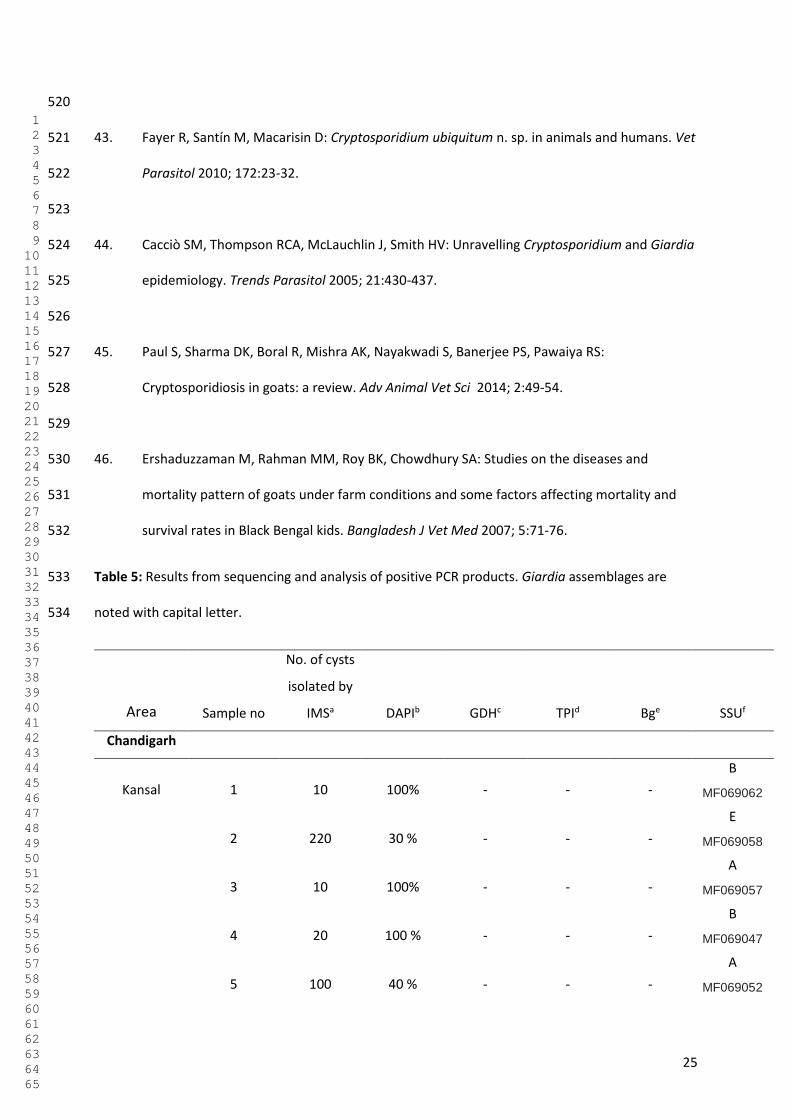
25
520
43. Fayer R, Santín M, Macarisin D: Cryptosporidium ubiquitum n. sp. in animals and humans. Vet 521
Parasitol 2010; 172:23-32. 522
523
44. Cacciò SM, Thompson RCA, McLauchlin J, Smith HV: Unravelling Cryptosporidium and Giardia 524
epidemiology. Trends Parasitol 2005; 21:430-437. 525
526
45. Paul S, Sharma DK, Boral R, Mishra AK, Nayakwadi S, Banerjee PS, Pawaiya RS: 527
Cryptosporidiosis in goats: a review. Adv Animal Vet Sci 2014; 2:49-54. 528
529
46. Ershaduzzaman M, Rahman MM, Roy BK, Chowdhury SA: Studies on the diseases and 530
mortality pattern of goats under farm conditions and some factors affecting mortality and 531
survival rates in Black Bengal kids. Bangladesh J Vet Med 2007; 5:71-76. 532
Table 5: Results from sequencing and analysis of positive PCR products. Giardia assemblages are 533
noted with capital letter. 534
Area Sample no
No. of cysts
isolated by
IMSa DAPIb GDHc TPId Bge SSUf
Chandigarh
Kansal 1 10 100% - - -
B
MF069062
2 220 30 % - - -
E
MF069058
3 10 100% - - -
A
MF069057
4 20 100 % - - -
B
MF069047
5 100 40 % - - -
A
MF069052
1 2 3 4 5 6 7 8 9 10 11 12 13 14 15 16 17 18 19 20 21 22 23 24 25 26 27 28 29 30 31 32 33 34 35 36 37 38 39 40 41 42 43 44 45 46 47 48 49 50 51 52 53 54 55 56 57 58 59 60 61 62 63 64 65
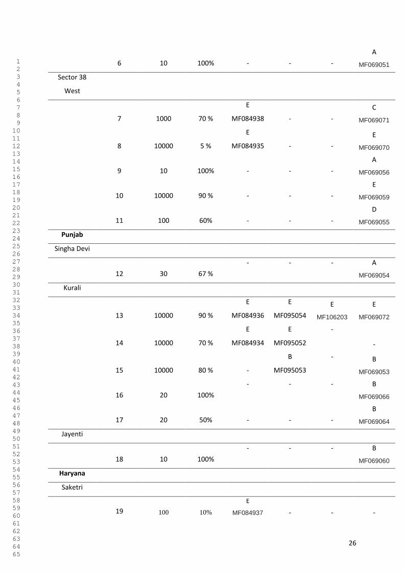
26
6 10 100% - - -
A
MF069051
Sector 38
West
7 1000 70 %
E
MF084938 - -
C
MF069071
8 10000 5 %
E
MF084935 - -
E
MF069070
9 10 100% - - -
A
MF069056
10 10000 90 % - - -
E
MF069059
11 100 60% - - -
D
MF069055
Punjab
Singha Devi
12 30 67 %
- - - A
MF069054
Kurali
13 10000 90 %
E
MF084936
E
MF095054
E
MF106203
E
MF069072
14 10000 70 %
E
MF084934
E
MF095052
-
-
15 10000 80 % -
B
MF095053
- B
MF069053
16 20 100%
- - - B
MF069066
17 20 50% - - -
B
MF069064
Jayenti
18 10 100%
- - - B
MF069060
Haryana
Saketri
19 100 10%
E
MF084937 - - -
1 2 3 4 5 6 7 8 9 10 11 12 13 14 15 16 17 18 19 20 21 22 23 24 25 26 27 28 29 30 31 32 33 34 35 36 37 38 39 40 41 42 43 44 45 46 47 48 49 50 51 52 53 54 55 56 57 58 59 60 61 62 63 64 65
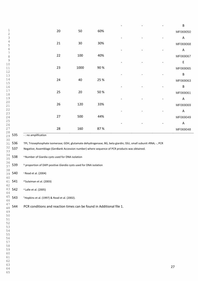
27
20 50 60%
- - - B
MF069050
21 30 30%
- - - A
MF069068
22 100 40%
- - - A
MF069067
23 1000 90 %
- - - E
MF069065
24 40 25 %
- - - B
MF069063
25 20 50 %
- - - B
MF069061
26 120 33%
- - - A
MF069069
27 500 44%
- - - A
MF069049
28 160 87 %
- - - A
MF069048
- : no amplification 535
TPI, Triosephosphate isomerase; GDH, glutamate dehydrogenase; BG, beta giardin; SSU, small subunit rRNA; -, PCR 536
Negative; Assemblage (GenBank Accession number) where sequence of PCR products was obtained. 537
a Number of Giardia cysts used for DNA isolation 538
b proportion of DAPI positive Giardia cysts used for DNA isolation 539
c Read et al. (2004) 540
d Sulaiman et al. (2003) 541
e Lalle et al. (2005) 542
f Hopkins et al. (1997) & Read et al. (2002). 543
PCR conditions and reaction times can be found in Additional file 1. 544
1 2 3 4 5 6 7 8 9 10 11 12 13 14 15 16 17 18 19 20 21 22 23 24 25 26 27 28 29 30 31 32 33 34 35 36 37 38 39 40 41 42 43 44 45 46 47 48 49 50 51 52 53 54 55 56 57 58 59 60 61 62 63 64 65
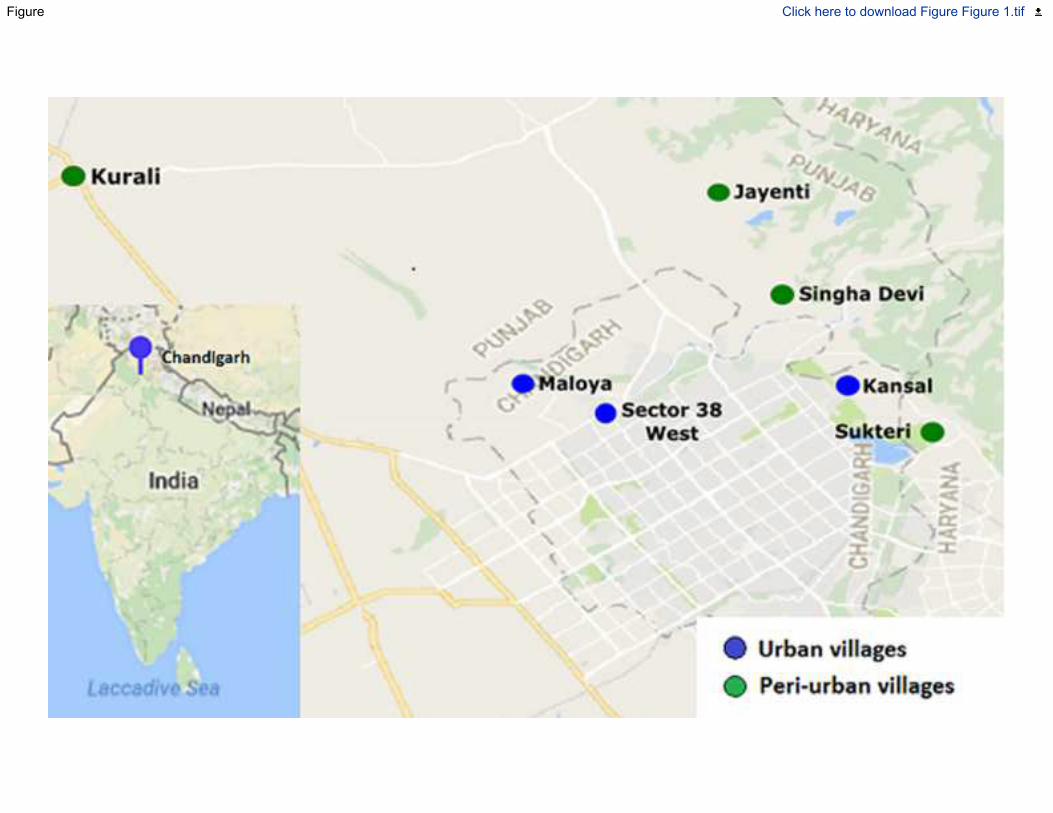
Figure Click here to download Figure Figure 1.tif
Figure Click here to download Figure Fig. 2.tif
Supplementary Material
Click here to access/downloadSupplementary Material
Additional file.docx
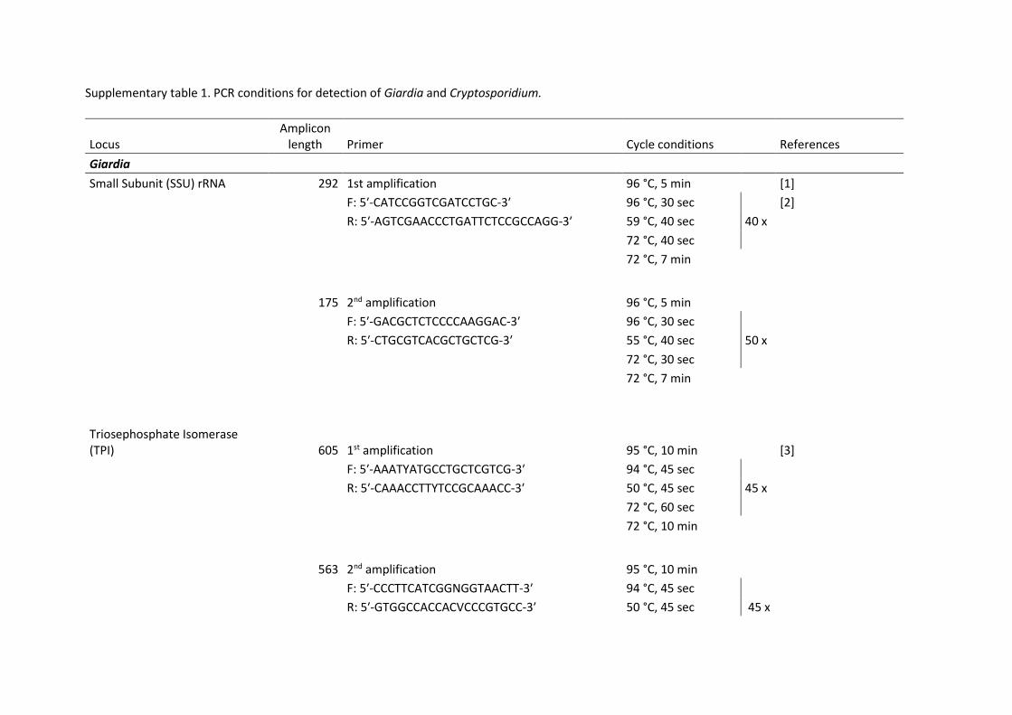
Supplementary table 1. PCR conditions for detection of Giardia and Cryptosporidium.
Locus Amplicon
length Primer Cycle conditions References
Giardia
Small Subunit (SSU) rRNA 292 1st amplification 96 °C, 5 min [1]
F: 5′-CATCCGGTCGATCCTGC-3′ 96 °C, 30 sec [2]
R: 5′-AGTCGAACCCTGATTCTCCGCCAGG-3′ 59 °C, 40 sec 40 x
72 °C, 40 sec
72 °C, 7 min
175 2nd amplification 96 °C, 5 min
F: 5′-GACGCTCTCCCCAAGGAC-3′ 96 °C, 30 sec
R: 5′-CTGCGTCACGCTGCTCG-3′ 55 °C, 40 sec 50 x
72 °C, 30 sec
72 °C, 7 min
Triosephosphate Isomerase (TPI) 605 1st amplification 95 °C, 10 min [3]
F: 5′-AAATYATGCCTGCTCGTCG-3′ 94 °C, 45 sec
R: 5′-CAAACCTTYTCCGCAAACC-3′ 50 °C, 45 sec 45 x
72 °C, 60 sec
72 °C, 10 min
563 2nd amplification 95 °C, 10 min
F: 5′-CCCTTCATCGGNGGTAACTT-3′ 94 °C, 45 sec
R: 5′-GTGGCCACCACVCCCGTGCC-3′ 50 °C, 45 sec 45 x
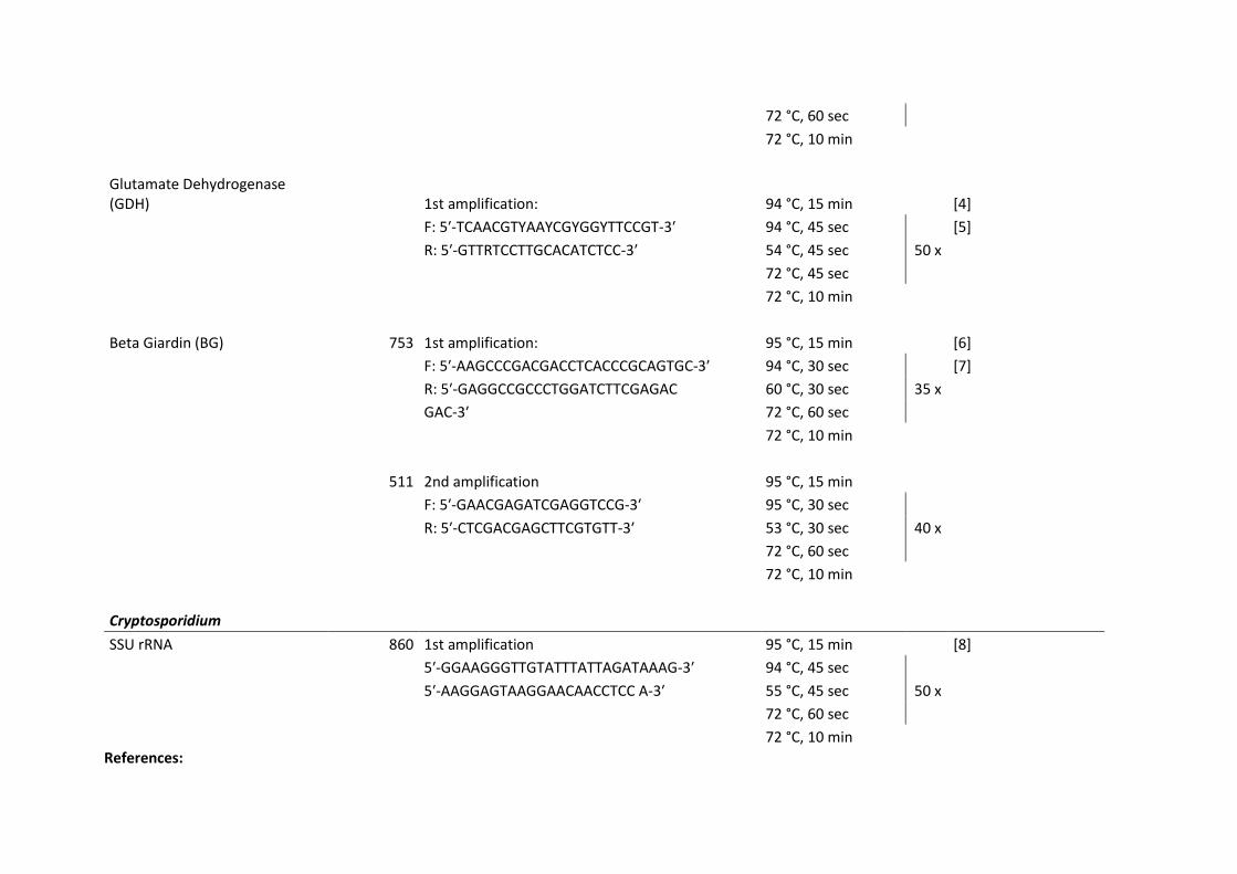
72 °C, 60 sec
72 °C, 10 min
Glutamate Dehydrogenase (GDH) 1st amplification: 94 °C, 15 min [4]
F: 5′-TCAACGTYAAYCGYGGYTTCCGT-3′ 94 °C, 45 sec [5]
R: 5′-GTTRTCCTTGCACATCTCC-3′ 54 °C, 45 sec 50 x
72 °C, 45 sec
72 °C, 10 min
Beta Giardin (BG) 753 1st amplification: 95 °C, 15 min [6]
F: 5′-AAGCCCGACGACCTCACCCGCAGTGC-3′ 94 °C, 30 sec [7]
R: 5′-GAGGCCGCCCTGGATCTTCGAGAC 60 °C, 30 sec 35 x
GAC-3′ 72 °C, 60 sec
72 °C, 10 min
511 2nd amplification 95 °C, 15 min
F: 5′-GAACGAGATCGAGGTCCG-3′ 95 °C, 30 sec
R: 5′-CTCGACGAGCTTCGTGTT-3′ 53 °C, 30 sec 40 x
72 °C, 60 sec
72 °C, 10 min
Cryptosporidium
SSU rRNA 860 1st amplification 95 °C, 15 min [8]
5′-GGAAGGGTTGTATTTATTAGATAAAG-3′ 94 °C, 45 sec
5′-AAGGAGTAAGGAACAACCTCC A-3′ 55 °C, 45 sec 50 x
72 °C, 60 sec
72 °C, 10 min
References:
1. Hopkins RM, Meloni BP, Groth DM, Wetherall JD, Reynoldson JA, Thompson RCA: Ribosomal RNA sequencing reveals differences between the
genotypes of Giardia isolates recovered from humans and dogs living in the same locality. The Journal of Parasitology 1997:44-51.
2. Read C, Walters J, Robertson I, Thompson R: Correlation between genotype of Giardia duodenalis and diarrhoea. International Journal for
Parasitology 2002, 32:229.
3. Sulaiman IM, Fayer R, Bern C, Gilman RH, Trout JM, Schantz PM, Das P, Lal AA, Xiao L: Triosephosphate isomerase gene characterization and
potential zoonotic transmission of Giardia duodenalis. Emerging Infectious Diseases 2003, 9:1444-1452.
4. Read CM, Monis PT, Thompson RCA: Discrimination of all genotypes of Giardia duodenalis at the glutamate dehydrogenase locus using PCR-RFLP.
Infection, Genetics and Evolution 2004, 4:125-130.
5. Robertson L, Hermansen L, Gjerde B, Strand E, Alvsvåg J, Langeland N: Application of genotyping during an extensive outbreak of waterborne
giardiasis in Bergen, Norway, during autumn and winter 2004. Applied and Environmental Microbiology 2006, 72:2212-2217.
6. Cacciò SM, Beck R, Lalle M, Marinculic A, Pozio E: Multilocus genotyping of Giardia duodenalis reveals striking differences between assemblages A
and B. International Journal for Parasitology 2008, 38:1523-1531.
7. Lalle M, Pozio E, Capelli G, Bruschi F, Crotti D, Cacciò SM: Genetic heterogeneity at the β-giardin locus among human and animal isolates of Giardia
duodenalis and identification of potentially zoonotic subgenotypes. International Journal for Parasitology 2005, 35:207-213.
8. Xiao L, Escalante L, Yang C, Sulaiman I, Escalante AA, Montali RJ, Fayer R, Lal AA: Phylogenetic analysis of Cryptosporidium parasites based on the
small-subunit rRNA gene locus. Applied and Environmental Microbiology 1999, 65:1578-1583.

VI

Manuscript Details
Manuscript number VPRSR_2017_201
Title Prevalence and zoonotic potential of intestinal protozoans in bovines in NorthernIndia
Article type Full Length Article
Abstract
Bovines, and especially cattle, have a dual position of appreciation in India, being both important in the food industryas providers of dairy products, and, culturally, being considered as holy creatures that it is forbidden by law to harm,kill, or eat. This status means that cattle have a paradoxical existence in India; as they are worshipped and protected,they are able to roam freely amongst humans, but they are also often left to fend for themselves. The vast numbers ofroaming cattle without clear owners are difficult to look after in terms of veterinary healthcare and appropriateinterventions when necessary, and have no regular supply of food. This article describes an investigation of theoccurrence of Cryptosporidium spp. and Giardia duodenalis in bovines either roaming the streets or being kept inanimal holdings in and around Chandigarh, a city in Northern India, and addresses the zoonotic potential of theseprotozoan parasites shed from bovines living in close contact with humans. Animals of all ages were sampled, and themajority of the positive samples were found from calves. The overall prevalence of Giardia was 8.2% andCryptosporidium was 2.4%. Non-zoonotic assemblages were predominantly found in the case of the Giardia – positivesamples, and in the case of Cryptosporidium, as well as non-zoonotic genotypes, zoonotic subgroups previouslydescribed from infected human infections in this area, were identified, indicating that there may be sharing of intestinalparasites in these settings, where cattle live in close connection to humans.
Keywords Bovines; Cryptosporidium; Giardia; zoonosis; India
Taxonomy Cattle, Cryptosporidium, Giardia, Public Health, India, Zoonoses
Corresponding Author Kjersti Selstad Utaaker
Order of Authors Kjersti Selstad Utaaker, Suman Chaudhary, Rajinder S. Bajwa, Lucy Robertson
Suggested reviewers Rebecca Traub, Balbir Bagicha Singh
Submission Files Included in this PDF
File Name [File Type]
Cover Letter.docx [Cover Letter]
Highlights.docx [Highlights]
Intestinal protozoans in bovines Northern India.pdf [Manuscript File]
Conflict of interest.pdf [Conflict of Interest]
Ethical statement.pdf [Ethical Statement]
To view all the submission files, including those not included in the PDF, click on the manuscript title on your EVISEHomepage, then click 'Download zip file'.

NMBUOsloNORWAY
11th September 2017
Dear Editor
Submission of Manuscript: Prevalence and zoonotic potential of intestinal
protozoans in bovines in Northern India
Please find attached a manuscript that we would like to have considered for publication in Veterinary Parasitology – Regional Studies and Reports.
In brief, the research described in the manuscript investigates the prevalence and zoonotic potential of Cryptosporidium oocysts and Giardia cysts in feces from bovines sampled over a longer period from different areas in and around Chandigarh, a city in Northern India. The results indicate that there is a relatively low occurrence of protozoans in bovines in Chandigarh, with some isolates having zoonotic potential.
All authors have agreed to the submission of this version of the manuscript.
Best regards
Kjersti Selstad Utaaker

In India, cattle are both worshipped and neglected
These bovines live in close contact with humans under poor hygienic settings
The close interface may break down barriers of diseases transmitted between them
This article investigates the occurrence of zoonotic protozoans in Indian bovines
1
Prevalence and zoonotic potential of intestinal protozoans in bovines in Northern India 1
Kjersti Selstad Utaaker1, Suman Chaudhary2,3, Rajinder S. Bajwa4, Lucy J. Robertson1. 2
Parasitology Lab, Department for Food Safety and Infection Biology, Faculty of Veterinary Medicine, 3
Norwegian University of Life Sciences, Adamstuen Campus, PO Box 8146 Dep. 0033 Oslo, Norway1 4
Department of Medical Parasitology, Postgraduate Institute of Medical Education and Research, 5
Chandigarh, 16002 India2 6
Nanomedicine-Laboratory of Immunology and Molecular Biomedical Research, (NLIMBR), School of 7
Medicine (SoM), Centre for Molecular and Medical Research (C-MMR) Strategic research Centre, 8
Faculty of Health, Deakin University, Waurn Ponds, Victoria 3216, Australia3 9
Government Veterinary Hospital, Sector 38, Chandigarh, India4 10
Corresponding author: [email protected] 11
12
2
Abstract: 13
Bovines, and especially cattle, have a dual position of appreciation in India, being both important in 14
the food industry as providers of dairy products, and, culturally, being considered as holy creatures 15
that it is forbidden by law to harm, kill, or eat. This status means that cattle have a paradoxical 16
existence in India; as they are worshipped and protected, they are able to roam freely amongst 17
humans, but they are also often left to fend for themselves. The vast numbers of roaming cattle 18
without clear owners are difficult to look after in terms of veterinary healthcare and appropriate 19
interventions when necessary, and have no regular supply of food. 20
This article describes an investigation of the occurrence of Cryptosporidium spp. and Giardia 21
duodenalis in bovines either roaming the streets or being kept in animal holdings in and around 22
Chandigarh, a city in Northern India, and addresses the zoonotic potential of these protozoan 23
parasites shed from bovines living in close contact with humans. Animals of all ages were sampled, 24
and the majority of the positive samples were found from calves. The overall prevalence of Giardia 25
was 8.2% and Cryptosporidium was 2.4%. Non-zoonotic assemblages were predominantly found in 26
the case of the Giardia – positive samples, and in the case of Cryptosporidium, as well as non-27
zoonotic genotypes, zoonotic subgroups previously described from infected human infections in this 28
area, were identified, indicating that there may be sharing of intestinal parasites in these settings, 29
where cattle live in close connection to humans. 30
31
Key words: Bovines, Cryptosporidium, Giardia, zoonosis, India 32
33
3
1. Introduction 34
Cryptosporidium and Giardia are two of the most common infectious agents of infectious enteritis in 35
humans and animals worldwide. From a public health perspective, it is imperative to understand the 36
sources and routes of transmission in different geographical regions. Cryptosporidium and Giardia 37
infections are known to cause production losses in bovines, and are considered potential sources of 38
human infection as well, with pre-weaned calves and lambs recognised as important reservoirs of the 39
zoonotic C. parvum in some countries (Santin-Duran and Trout, 2008). 40
The extent and relative importance of zoonotic transmission of these parasites in different parts of 41
the world, especially in the developing countries, are still poorly understood (Abeywardena et al., 42
2015). Although for Giardia, it is generally considered that most human infections are from direct or 43
indirect human-to-human transmission, rather than zoonotic (Monis and Thompson, 2003), for 44
Cryptosporidium a different picture occurs. 45
In human populations, it is generally considered that Cryptosporidium parvum tends to dominate in 46
Europe, New Zealand, and the Middle East, indicating a potential for zoonotic transmission, whereas 47
C. hominis infections are responsible for more human infections than C. parvum in USA, China, Japan, 48
and the majority of developing countries (Cacciò and Putignani, 2014). 49
Indeed, studies in India tend to support this distribution; three studies from Northern India indicated 50
that over 70 % of Cryptosporidium infections were caused by C. hominis (Yadav et al, 2017)(Gatei et 51
al., 2007; Sharma et al., 2013), which is in concordance with the general finding of Xiao and Fayer 52
(2008), who reported that, in the studies where molecular methods have been used, C. hominis has, 53
in general, been associated with a higher number of human infections than C. parvum. 54
However, despite this distribution, the role of cattle in transmission or propagation of these parasites 55
in India is of particular interest, due to the particular interactions between bovids and humans here. 56
India is home to a quarter of the world’s cattle population. In Hindu religious scriptures, the cow is 57
referred to as the “all-producing and all-containing universe”(Korom, 2000), and the cow is the most 58
4
sacred of all animals according to the Hindu religion. Northern India practices a total ban on cow 59
slaughter, and the Indian Veterinary Council has estimated that there is only sufficient food to 60
sustain 60% of the Indian cattle population, and the rest are left to starve or roam the streets 61
searching for food, which, in most cases, ends up being a diet of garbage (Agoramoorthy and Hsu, 62
2012; Kang, 2003). In New Delhi, there is an estimated number of 40 000 stray cattle, and in 63
Chandigarh this number has risen from 1400 to 2000 during the last five years (Victor, 2013). This 64
creates an obvious difference in animal management from industrialized countries, where cattle are 65
kept on enclosed farms and restrictions between animal and human contact are mandatory by law 66
due to zoonosis and hygiene considerations. The situation in India also differs from that in other 67
developing countries, where cow ownership and cattle as a staple means of livelihood means that 68
cattle are pastured as far as possible to ensure that their productivity as dairy or beef animals is 69
maximised, and unrestricted wandering, particularly in urban settings, is relatively uncommon. The 70
special role of cows in Indian culture means that the interface between human and animals is 71
minimal, and the sources of infection, for both human and animals, are likely to be strolling along, or 72
sometimes scavenging for food, in the same streets. 73
In this study, faecal samples from cattle and water buffaloes in urban and peri-urban areas of 74
Northern India were collected, and the prevalence and zoonotic potential of Cryptosporidium and 75
Giardia were assessed. 76
2. Material and Methods 77
2.1. Sampling 78
From March 2014 until February 2016, 294 samples were collected from different animal holdings in 79
Chandigarh, Northern India. Of these, seasonally, 109 samples were collected during the winter, and 80
185 during the monsoon season. With respect to where the various bovines lived, 153 samples were 81
collected from animal holdings within the city (urban), and 141 from animal holdings and settlements 82
surrounding the city (peri-urban). Only one sample was collected per animal per sampling occasion. 83
5
The ages of the animals ranged from calves under three months to adults. Presence of diarrhoea in 84
samples and approximate age of animal was recorded at sampling. The samples (each approximately 85
5-10 g) were collected either rectally or non-invasively promptly after defecation, and were 86
immediately mixed with 2.5% potassium dichromate and stored at 4°C before transportation to the 87
Parasitology Department, Norwegian University of Life Sciences (NMBU) for analysis. 88
89
6
Figure 1: Map of sampling sites 90
91
2.2. Analysis for occurrence 92
From each sample collected, 3 grams of faeces were homogenized with 57 mL water and passed 93
through a faecal parasite concentrator with a pore diameter 425 μm (Midi Parasep, Apacor, 94
Berkshire, England). The suspension was transferred to 10 mL centrifuge tubes and centrifuged at 95
1550 rfg) for 3 minutes to create a pellet. The supernatant was discarded, and between 5-20 µl of 96
homogenized and sieved faecal material was placed on a microscope slide using plastic 97
bacteriological loops that take approx. 10µl amount of sample. The samples were left to dry and then 98
fixed with methanol before staining with 15 µl of monoclonal antibody for Cryptosporidium oocysts 99
and Giardia cysts (AquaGlo etc), and then incubated at 37°C in a humid chamber for 45 minutes. The 100
staining solution was then rinsed off with distilled water, and a coverslip placed over the sample 101
before immediate microscopic examination. 102
Prepared samples were screened under a fluorescence microscope with the following filter settings: 103
FITC: emission- 490, excitation – 525. 104
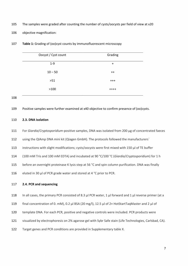
7
The samples were graded after counting the number of cysts/oocysts per field of view at x20 105
objective magnification: 106
Table 1: Grading of (oo)cyst counts by immunofluorescent microscopy 107
Oocyst / Cyst count Grading
1-9 +
10 – 50 ++
>51 +++
>100 ++++
108
Positive samples were further examined at x40 objective to confirm presence of (oo)cysts. 109
2.3. DNA isolation 110
For Giardia/Cryptosporidium-positive samples, DNA was isolated from 200 µg of concentrated faeces 111
using the QIAmp DNA mini kit (Qiagen GmbH). The protocols followed the manufacturers` 112
instructions with slight modifications; cysts/oocysts were first mixed with 150 μl of TE buffer 113
(100 mM Tris and 100 mM EDTA) and incubated at 90 °C/100 °C (Giardia/Cryptosporidium) for 1 h 114
before an overnight proteinase K lysis step at 56 °C and spin column purification. DNA was finally 115
eluted in 30 μl of PCR grade water and stored at 4 °C prior to PCR. 116
2.4. PCR and sequencing 117
In all cases, the primary PCR consisted of 8.3 μl PCR water, 1 μl forward and 1 μl reverse primer (at a 118
final concentration of 0. mM), 0.2 μl BSA (20 mg/l), 12.5 μl of 2× HotStartTaqMaster and 2 μl of 119
template DNA. For each PCR, positive and negative controls were included. PCR products were 120
visualized by electrophoresis on 2% agarose gel with Sybr Safe stain (Life Technologies, Carlsbad, CA). 121
Target genes and PCR conditions are provided in Supplementary table X. 122
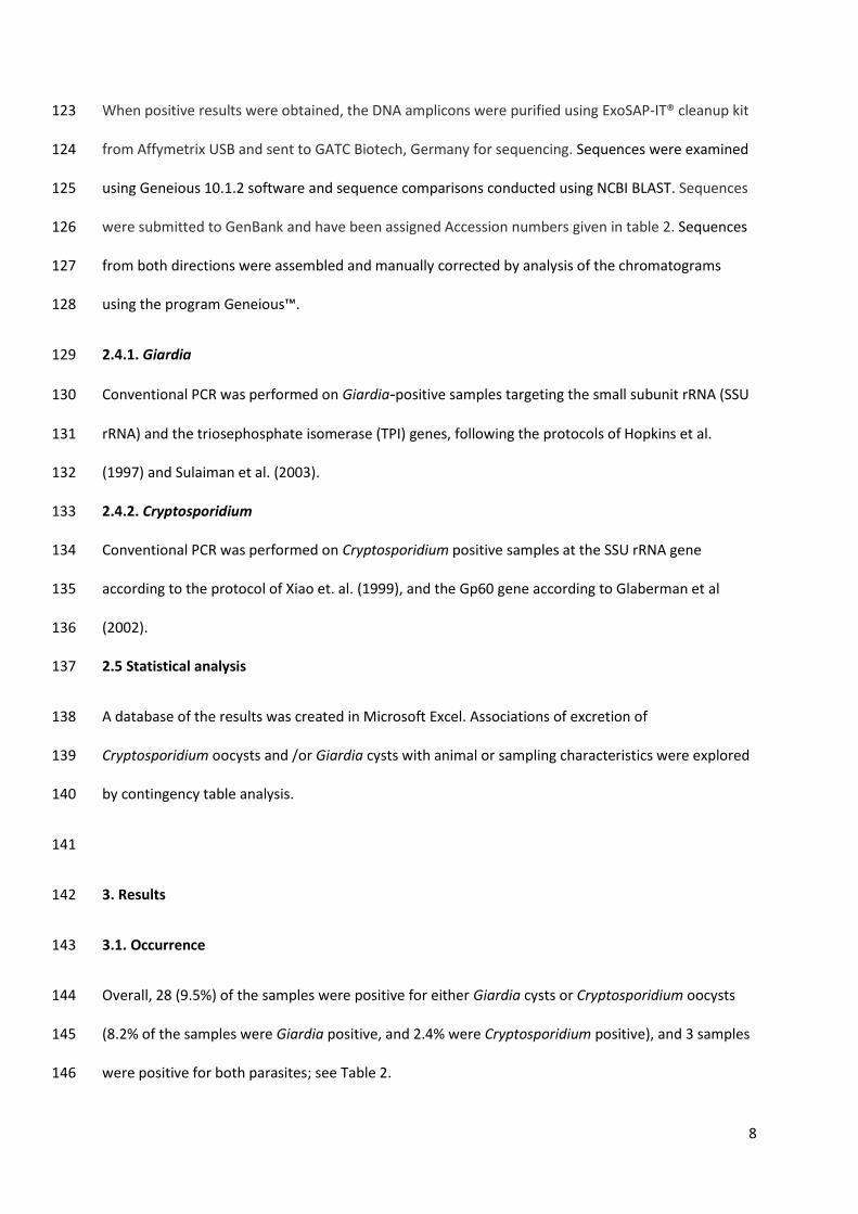
8
When positive results were obtained, the DNA amplicons were purified using ExoSAP-IT® cleanup kit 123
from Affymetrix USB and sent to GATC Biotech, Germany for sequencing. Sequences were examined 124
using Geneious 10.1.2 software and sequence comparisons conducted using NCBI BLAST. Sequences 125
were submitted to GenBank and have been assigned Accession numbers given in table 2. Sequences 126
from both directions were assembled and manually corrected by analysis of the chromatograms 127
using the program Geneious™. 128
2.4.1. Giardia 129
Conventional PCR was performed on Giardia-positive samples targeting the small subunit rRNA (SSU 130
rRNA) and the triosephosphate isomerase (TPI) genes, following the protocols of Hopkins et al. 131
(1997) and Sulaiman et al. (2003). 132
2.4.2. Cryptosporidium 133
Conventional PCR was performed on Cryptosporidium positive samples at the SSU rRNA gene 134
according to the protocol of Xiao et. al. (1999), and the Gp60 gene according to Glaberman et al 135
(2002). 136
2.5 Statistical analysis 137
A database of the results was created in Microsoft Excel. Associations of excretion of 138
Cryptosporidium oocysts and /or Giardia cysts with animal or sampling characteristics were explored 139
by contingency table analysis. 140
141
3. Results 142
3.1. Occurrence 143
Overall, 28 (9.5%) of the samples were positive for either Giardia cysts or Cryptosporidium oocysts 144
(8.2% of the samples were Giardia positive, and 2.4% were Cryptosporidium positive), and 3 samples 145
were positive for both parasites; see Table 2. 146
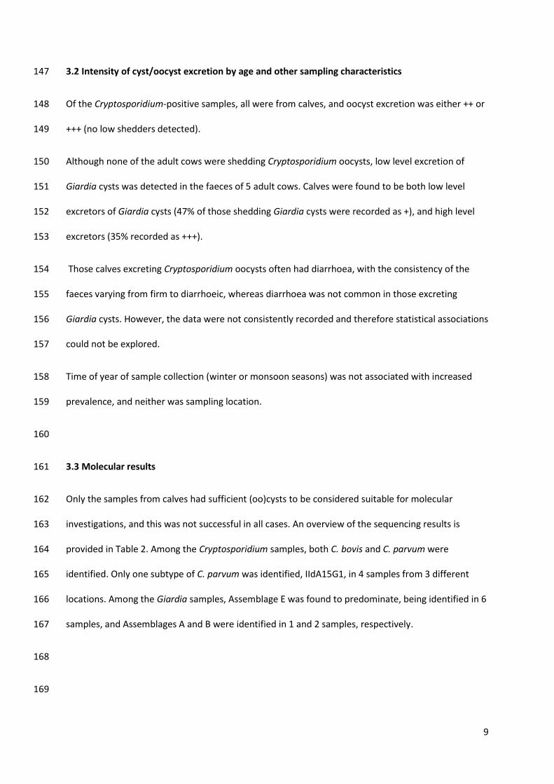
9
3.2 Intensity of cyst/oocyst excretion by age and other sampling characteristics 147
Of the Cryptosporidium-positive samples, all were from calves, and oocyst excretion was either ++ or 148
+++ (no low shedders detected). 149
Although none of the adult cows were shedding Cryptosporidium oocysts, low level excretion of 150
Giardia cysts was detected in the faeces of 5 adult cows. Calves were found to be both low level 151
excretors of Giardia cysts (47% of those shedding Giardia cysts were recorded as +), and high level 152
excretors (35% recorded as +++). 153
Those calves excreting Cryptosporidium oocysts often had diarrhoea, with the consistency of the 154
faeces varying from firm to diarrhoeic, whereas diarrhoea was not common in those excreting 155
Giardia cysts. However, the data were not consistently recorded and therefore statistical associations 156
could not be explored. 157
Time of year of sample collection (winter or monsoon seasons) was not associated with increased 158
prevalence, and neither was sampling location. 159
160
3.3 Molecular results 161
Only the samples from calves had sufficient (oo)cysts to be considered suitable for molecular 162
investigations, and this was not successful in all cases. An overview of the sequencing results is 163
provided in Table 2. Among the Cryptosporidium samples, both C. bovis and C. parvum were 164
identified. Only one subtype of C. parvum was identified, IIdA15G1, in 4 samples from 3 different 165
locations. Among the Giardia samples, Assemblage E was found to predominate, being identified in 6 166
samples, and Assemblages A and B were identified in 1 and 2 samples, respectively. 167
168
169
10
170
Table 2: Positive microscopy and sequencing results 171
172
Season Area
Result from
microscopya Animal
Cryptosporidium Giardia
SSUb Gp60c SSUd TPIe
Winter Urban G+ Cow - - - -
Winter Urban G+ Cow - - - -
Winter Urban G+ Cow - - - -
Winter Urban C ++ Calf * * - -
Winter Peri-urban G+++ Calf - - * *
Winter Peri-urban G+ Calf - - Ass B
MF399205
Ass E
MF459679
Winter Peri-urban G+ Calf - - * *
Winter Peri-urban C+++ Calf C. parvum
MF399201
C. parvum
IIdA15G1
MF459681
- -
Winter Peri-urban G+ Calf - - * *
Winter Peri-urban G+ Calf - - Ass A
MF163432
*
Winter Peri-urban C+++ Calf C. bovis
MF399200
C. parvum
IIdA15G1
MF535626
- -
Winter Urban G+++ Calf
Ass E
MF399204
Ass E
MF459678
11
Winter Urban G+++ Calf - - * *
Winter Urban G+ Calf - - - -
Winter Urban G+ Calf - - Ass E
MF163433
*
Winter Urban G+ Calf - - * *
Monsoon Peri-urban G+++ Calf - - * *
Monsoon Peri-urban G+ Calf - - * *
Monsoon Peri-urban G+ Cow - - - -
Monsoon Peri-urban G+ Cow - - - -
Monsoon Urban G+++ , C++ Calf * * Ass E
MF399203
*
Monsoon Urban C+++ Calf * * - -
Monsoon Urban G+++ Calf - - Ass E
MF399206
*
Monsoon Urban G+++ Calf - - * *
Monsoon Urban G+ Calf - - * *
Monsoon Peri-urban G++ Calf - - * *
Monsoon Peri-urban G++, C++ Calf C. bovis
MF399202
C. parvum
IIdA15G1
MF535627
* *
Monsoon Peri-urban G++, C++ Calf - C. parvum
IIdA15G1
MF535999
* Ass B
MF459680
173
- : No amplification performed 174
12
*: Amplification was performed, but was not successful. 175
a: C = Cryptosporidium, G = Giardia; the occurrence of cysts or oocysts was scored semi-176
quantitatively from + to +++ (see Table 1) 177
b: Species identification at SSU gene (Xiao et al, 1999) 178
c: Subtype identification at Gp60 gene (Glaberman et al, 2002). 179
d: Assemblage identification at SSU gene (Hopkins et al, 1997) 180
e: Assemblage identification at TPI gene (Sulaiman et al, 2003) 181
182
4. Discussion 183
The main finding from this study was that around 10 % of bovids wandering the streets of Chandigarh 184
or gathered in holdings for bovids, excrete Cryptosporidium oocysts and/or Giardia cysts. These 185
parasites were particularly prevalent in calves, and this age group also excreted higher numbers 186
(both Giardia and Cryptosporidium). 187
There was no correlation between season on shedding of (oo)cysts. Although some studies have 188
indicated seasonality of infection with Cryptosporidium and Giardia in cattle (e.g., Szonyi et al, 189
(2010); Huetink et al, (2001)are often more likely to show excretion, a study from Bangladesh found 190
no effect of season (rainy or non-rainy) on excretion of either parasite in cattle (Ehsan et al., 2015). 191
192
Chandigarh is divided into phases and sectors that differ from each other in terms of population 193
pressure and sanitary infrastructure. For simplicity, in this manuscript we have divided the sampling 194
from animals as urban (in the city, that tends to have lower human population density and better 195
infrastructure, but would not be considered appropriate for cattle in most western countries) and 196
peri-urban (generally on the edge of the city and with high population pressure, unsanitary 197
13
conditions, flood problems, poor garbage disposal, disposal of livestock dung into open drains, and 198
discharge of untreated sewage). Although we had anticipated that samples from bovids inhabiting 199
periurban locations would be more likely to be excreting Cryptosporidum and Giardia, our data did 200
not show this, with samples from the wealthier areas just as likely to be infected. 201
Globally, cattle are commonly infected with C. parvum, C. andersoni, C. bovis and C. ryanae, so our 202
findings are not unusual. C. parvum is most commonly found in pre – weaned calves. C. bovis and C. 203
ryanae in weaned calves, and C. andersoni in yearlings and adult cattle, and it seems mostly pre-204
weaned calves are the major sources of C. parvum. Of the 14 C. parvum subtype families, IIa and IId 205
are the two major zoonotic subtype families in humans and animals(Wang et al., 2014). Of particular 206
interest from our study is that a single C. parvum zoonotic subtype (IIdA15G1) predominated in those 207
samples that were successfully sequenced, despite the positive samples being collected from 208
different locations. The subtype IIdA15G1 has also been found in cattle, sheep and goats in China, 209
Iran, Malaysia and Spain (Muhid et al., 2011; Nazemalhosseini-Mojarad et al., 2011; Quilez et al., 210
2008; Quílez et al., 2008), and in humans in from Netherlands, Australia, India, Iran and Malaysia 211
(Ajjampur et al., 2010; Iqbal et al., 2012; Nazemalhosseini-Mojarad et al., 2011; Ng et al., 2010; 212
Wielinga et al., 2008), and the subtype family IId is considered a major zoonotic Europe, Africa, 213
Australia and Asia (Cui et al., 2014). Furthermore, this particular sub-type has previously been 214
reported from human patients with cryptosporidiosis delivering samples to a hospital in the same 215
area as where these samples were collected (Sharma, Sharma, Sehgal, Malla, & Khurana, 2013). This 216
indicates a high likelihood that cattle and humans in this area transmit C. parvum between each 217
other. Given the proximity of bovids and humans to each other in Indian culture, with cattle allowed 218
to wander at will and often participating in festivals, this seems not surprising. At the same time, 219
despite being worshipped, the lack of ownership means that cattle on the streets often do not have 220
access to a stable food supply, and are obliged to graze on rubbish in the ditches, which may be 221
contaminated with human faeces. Cow sanctuaries (Gaushalas) which take care of stray animals have 222
been established in many urban areas in India, and, to some extent alleviate, they do alleviate the 223
14
number of roaming cattle. However, their capacity is not sufficient for the rising numbers of stray 224
cattle, who often are unproductive, older cows that have been turned to the streets by their owner 225
(Singh et al., 2013). Although Cryptosporidium has previously been reported to be highly prevalent in 226
bovines in India, and especially among calves during the monsoon season (Hingole et al., 2017; 227
Maurya et al., 2013; Paul et al., 2008), these studies did not subtype positive samples, so the 228
transmission potential between humans and bovids could be based, at best, at the Cryptosporidium 229
species level. 230
Although the prevalence of Giardia in bovids was higher than that of Cryptosporidium, the majority 231
of samples were Assemblage E, suggesting that zoonotic transmission of this parasite may be less 232
likely. However, Assemblage A and Assemblage B were also detected in some samples, indicating 233
some potential for zoonotic/anthropozoonotic transmission between bovids and people in this 234
setting. Many studies on Giardia infections in cattle have revealed that the dominating Assemblage 235
among bovines is the species-specific Assemblage E; although generally considered not to be 236
zoonotic, some recent studies have suggested that this Assemblage may have zoonotic potential 237
(Abdel-Moein and Saeed, 2016). Assemblage A is increasingly being detected, indicating that this 238
zoonotic Assemblage may be more widespread among this livestock than assumed. A multicentre 239
trial in Germany, UK, France and Italy found an overall prevalence of G. duodenalis of 45.4%, and 43% 240
of those belonged to Assemblage A (Geurden et al., 2012). 241
In contrast, few studies are available regarding the zoonotic potential of Giardia in Indian bovines. A 242
study from West Bengal revealed a 12.2% prevalence of Giardia, with the majority of Assemblages 243
belonging to the species-specific Assemblage E, and 2.7% belonging to the zoonotic Assemblage A 244
(Khan et al., 2010). 245
Assemblage B is not a common finding in farm animals, and is usually associated with human 246
infection. In two of the calves sampled in this study, both Cryptosporidium oocysts and Giardia cysts 247
were found, and genotyping revealed that one calf was infected with C. parvum subtype IIdA15G1 248
15
and G. duodenalis Assemblage B, and this may indicate that both infections were derived originally 249
from a human source. 250
In contrast to the results from bovids described here, investigation on the occurrence of Giardia and 251
Cryptosporidium in backyard goats in Chandigarh have indicated that the majority of Giardia were 252
potentially zoonotic (Assemblages A and B), but Cryptosporidium infection were not identified 253
(Utaaker et al, unpublished results). These data suggest that in terms of zoonotic transmission of 254
Cryptosporidium and Giardia in Chandigarh with respect to livestock, goats may be of greatest 255
importance for Giardia, but bovids are probably of greatest importance for Cryptosporidium. 256
Veterinary urban hygiene in India has been pinpointed as an area of concern (Singh et al., 2013), and 257
our data here support this with respect to Cryptosporidium in cattle. Given the importance of 258
cryptosporidiosis in human health in India (e.g., Kotloff et al, 2013) and also that bovine health can 259
be seriously affected by this infection, this intestinal parasite should be of importance in One Health 260
studies in this region. However, as has been previously noted, in order to support effective One 261
Health research in India along with integrated zoonotic disease control, it is necessary that 262
substantial changes in behaviour, attitudes, and institutional policies are implemented (McKenzie et 263
al., 2016). 264
5. Conclusion 265
This study further emphasizes the status of Cryptosporidium parvum as a zoonotic pathogen, even in 266
India, where human cryptosporidiosis usually tends to be due to C. hominis infections. The 267
predominant C. parvum subgenotype that we identified in bovids has also been reported in people in 268
the same region. Although Giardia from bovids seemed to be less likely to be associated with 269
zoonotic transmission, the concomitant finding of Assemblage B in one calf indicates that 270
anthroponotic / zoonotic transmission between humans and bovids should not be excluded. We 271
speculate that the special cultural status of cattle in India may be partially responsible for increased 272
sharing of pathogens between cattle and humans, particularly in the urban environment. 273
16
Conflict of interest statement 274
No financial or personal relationship between the authors and other people or organizations have 275
inappropriately influenced this work. 276
Ethics statement 277
It is submitted that proper consideration has been given to any ethics issue raised. 278
Acknowledgments 279
This study was funded through the Para-Clim-Chandigarh project, partly funded by the Norwegian 280
Research Council via the New Indigo Partnership program (Contract number: 227965) 281
The authors are grateful to Dr. Rakesh Seghal and Dr. Kapil Goyal for facilitating use of the PGIMER 282
facilities, Himanshu Joshi and Anil Kumar for assistance in collection of the samples, and Omar Ismail 283
for the help of processing and analysis of the samples. 284
References: 285
286
Abdel-Moein, K.A., Saeed, H., 2016. The zoonotic potential of Giardia intestinalis assemblage E in 287
rural settings. Parasitol. Res. 115, 3197-3202. 288
289
Abeywardena, H., Jex, A.R., Gasser, R.B., 2015. Chapter Six-A Perspective on Cryptosporidium and 290
Giardia, with an Emphasis on Bovines and Recent Epidemiological Findings. Adv. Parasitol. 291
88, 243-301. 292
293
Agoramoorthy, G., Hsu, M.J., 2012. The significance of cows in Indian society between sacredness 294
and economy. Anthropol. Noteb. 18, 5-12. 295
296
17
Ajjampur, S.S.R., Liakath, F.B., Kannan, A., Rajendran, P., Sarkar, R., Moses, P.D., Simon, A., Agarwal, 297
I., Mathew, A., O'Connor, R., 2010. Multisite study of cryptosporidiosis in children with 298
diarrhea in India. J. Clin. Microbiol. 48, 2075-2081. 299
300
Cacciò, S.M., Putignani, L., 2014. Epidemiology of human cryptosporidiosis, In: Cryptosporidium: 301
parasite and disease. Springer, pp. 43-79. 302
303
Cui, Z., Wang, R., Huang, J., Wang, H., Zhao, J., Luo, N., Li, J., Zhang, Z., Zhang, L., 2014. 304
Cryptosporidiosis caused by Cryptosporidium parvum subtype IIdA15G1 at a dairy farm in 305
Northwestern China. Parasit. Vectors 7, 529. 306
307
Ehsan, A.M., Geurden, T., Casaert, S., Parvin, S.M., Islam, T.M., Ahmed, U.M., Levecke, B., Vercruysse, 308
J., Claerebout, E., 2015. Assessment of zoonotic transmission of Giardia and Cryptosporidium 309
between cattle and humans in rural villages in Bangladesh. PLoS One 10, e0118239. 310
311
Gatei, W., Das, P., Dutta, P., Sen, A., Cama, V., Lal, A.A., Xiao, L., 2007. Multilocus sequence typing 312
and genetic structure of Cryptosporidium hominis from children in Kolkata, India. Infect. 313
Genet. Evol. 7, 197-205. 314
315
Geurden, T., Vanderstichel, R., Pohle, H., Ehsan, A., von Samson-Himmelstjerna, G., Morgan, E.R., 316
Camuset, P., Capelli, G., Vercruysse, J., Claerebout, E., 2012. A multicentre prevalence study 317
in Europe on Giardia duodenalis in calves, with molecular identification and risk factor 318
analysis. Vet. Parasitol. 190, 383-390. 319
320
18
Glaberman, S., Moore, J.E., Lowery, C.J., Chalmers, R.M., Sulaiman, I., Elwin, K., Rooney, P.J., Millar, 321
B.C., Dooley, J.S., Lal, A.A., 2002. Three drinking water associated cryptosporidiosis 322
outbreaks, Northern Ireland. Emerg. Infect. Dis. 8, 631-633. 323
324
Hingole, A.C., Gudewar, J.G., Pednekar, R.P., Gatne, M.L., 2017. Prevalence and molecular 325
characterization of Cryptosporidium species in cattle and buffalo calves in Mumbai region of 326
India. J. Parasit. Dis. 41, 131-136. 327
328
Hopkins, R.M., Meloni, B.P., Groth, D.M., Wetherall, J.D., Reynoldson, J.A., Thompson, R.C.A., 1997. 329
Ribosomal RNA sequencing reveals differences between the genotypes of Giardia isolates 330
recovered from humans and dogs living in the same locality. J. Parasitol., 44-51. 331
332
Huetink, R.E.C., Van der Giessen, J.W.B., Noordhuizen, J., Ploeger, H.W., 2001. Epidemiology of 333
Cryptosporidium spp. and Giardia duodenalis on a dairy farm. Vet. Parasitol. 102, 53-67. 334
335
Iqbal, A., Lim, Y.A.L., Surin, J., Sim, B.L.H., 2012. High diversity of Cryptosporidium subgenotypes 336
identified in Malaysian HIV/AIDS individuals targeting gp60 gene. PloS one 7, e31139. 337
338
Kang, B. 2003. The milky way. Outlook India. Retrieved from: 339
http://www.outlookindia.com/magazine/story/the-milky-way/219289 Accessed on 340
04.09.2017 341
342
Khan, S.M., Debnath, C., Pramanik, A.K., Xiao, L., Nozaki, T., Ganguly, S., 2010. Molecular 343
characterization and assessment of zoonotic transmission of Cryptosporidium from dairy 344
cattle in West Bengal, India. Vet. Parasitol. 171, 41-47. 345
346
19
Korom, F.J., 2000. Holy cow! The apotheosis of Zebu, or why the cow is sacred in Hinduism. Asian 347
Folkl. Stud., 181-203. 348
349
Maurya, P.S., Rakesh, R.L., Pradeep, B., Kumar, S., Kundu, K., Garg, R., Ram, H., Kumar, A., Banerjee, 350
P.S., 2013. Prevalence and risk factors associated with Cryptosporidium spp. infection in 351
young domestic livestock in India. Trop. Anim. Health Prod. 45, 941-946. 352
353
McKenzie, J.S., Dahal, R., Kakkar, M., Debnath, N., Rahman, M., Dorjee, S., Naeem, K., Wijayathilaka, 354
T., Sharma, B.K., Maidanwal, N., 2016. One Health research and training and government 355
support for One Health in South Asia. Infect. Ecol. epidemiol. 6, 33842. 356
357
Monis, P.T., Thompson, R.C.A., 2003. Cryptosporidium and Giardia-zoonoses: fact or fiction? Infect. 358
Genet. Evol. 3, 233-244. 359
Muhid, A., Robertson, I., Ng, J., Ryan, U., 2011. Prevalence of and management factors contributing 360
to Cryptosporidium sp. infection in pre-weaned and post-weaned calves in Johor, Malaysia. 361
Exp. Parasitol. 127, 534-538. 362
363
Nazemalhosseini-Mojarad, E., Haghighi, A., Taghipour, N., Keshavarz, A., Mohebi, S.R., Zali, M.R., 364
Xiao, L., 2011. Subtype analysis of Cryptosporidium parvum and Cryptosporidium hominis 365
isolates from humans and cattle in Iran. Vet. Parasitol. 179, 250-252. 366
367
Ng, J., MacKenzie, B., Ryan, U., 2010. Longitudinal multi-locus molecular characterisation of sporadic 368
Australian human clinical cases of cryptosporidiosis from 2005 to 2008. Exp. Parasitol. 125, 369
348-356. 370
371
20
Paul, S., Chandra, D., Ray, D., Tewari, A., Rao, J., Banerjee, P., Baidya, S., Raina, O., 2008. Prevalence 372
and molecular characterization of bovine Cryptosporidium isolates in India. Vet. Parasitol. 373
153, 143-146. 374
375
Quilez, J., Torres, E., Chalmers, R., Robinson, G., Del Cacho, E., Sanchez-Acedo, C., 2008. 376
Cryptosporidium species and subtype analysis from dairy calves in Spain. Parasitology 135, 377
1613-1620. 378
379
Quílez, J., Torres, E., Chalmers, R.M., Hadfield, S.J., del Cacho, E., Sánchez-Acedo, C., 2008. 380
Cryptosporidium genotypes and subtypes in lambs and goat kids in Spain. Appl. Environ. 381
Microbiol. 74, 6026-6031. 382
383
Santin-Duran, M., Trout, J. 2008. Cryptosporidiosis of livestock (Cryptospiridium and 384
Cryptosporidiosis. 2nd edition. Boca Raton, FL. CRC Press), pp. 451-483. 385
386
Sharma, P., Sharma, A., Sehgal, R., Malla, N., Khurana, S., 2013. Genetic diversity of Cryptosporidium 387
isolates from patients in North India. Int. J. Infect. Dis. 17, e601-e605. 388
389
Singh, B.B., Ghatak, S., Banga, H.S., Gill, J.P.S., Singh, B., 2013. Veterinary urban hygiene: a challenge 390
for India. Rev. Sci. Tech. 32, 645-656. 391
392
Sulaiman, I.M., Fayer, R., Bern, C., Gilman, R.H., Trout, J.M., Schantz, P.M., Das, P., Lal, A.A., Xiao, L., 393
2003. Triosephosphate isomerase gene characterization and potential zoonotic transmission 394
of Giardia duodenalis. Emerg. infect. Dis. 9, 1444-1452. 395
396
21
Szonyi, B., Bordonaro, R., Wade, S.E., Mohammed, H.O., 2010. Seasonal variation in the prevalence 397
and molecular epidemiology of Cryptosporidium infection in dairy cattle in the New York City 398
Watershed. Parasitol. Res. 107, 317-325. 399
400
Victor, H. 2013 (Hindustan Times). Retrieved from 401
http://www.hindustantimes.com/chandigarh/municipal-corporation-fails-to-contain-stray-402
cattle-dog-menace/story-m4ifEkWzdSFf5UGkQgEEfO.html Accessed on 04.09.2017. 403
404
Wang, R., Zhang, L., Axén, C., Bjorkman, C., Jian, F., Amer, S., Liu, A., Feng, Y., Li, G., Lv, C., 2014. 405
Cryptosporidium parvum IId family: clonal population and dispersal from Western Asia to 406
other geographical regions. Sci. rep. 4, 4208. 407
408
Wielinga, P.R., de Vries, A., van der Goot, T.H., Mank, T., Mars, M.H., Kortbeek, L.M., van der Giessen, 409
J.W., 2008. Molecular epidemiology of Cryptosporidium in humans and cattle in The 410
Netherlands. Int. J. Parasitol. 38, 809-817. 411
412
Xiao, L., Escalante, L., Yang, C., Sulaiman, I., Escalante, A.A., Montali, R.J., Fayer, R., Lal, A.A., 1999. 413
Phylogenetic analysis of Cryptosporidium parasites based on the small-subunit rRNA gene 414
locus. Appl. Environ. Microbiol. 65, 1578-1583. 415
416
Xiao, L., Fayer, R., 2008. Molecular characterisation of species and genotypes of Cryptosporidium and 417
Giardia and assessment of zoonotic transmission. Int. J. Parasitol. 38, 1239-1255. 418
Yadav, P., Mirdha, B.R., Makharia, G.K., Chaudhry, R., 2017. Multilocus sequence typing of 419
Cryptosporidium hominis from northern India. Indian J. Med. Res. 145, 102. 420
421

VII

1
Not just a walk in the park: occurrence of intestinal parasites in dogs roaming recreational parks in
Chandigarh, Northern India.
Kjersti Selstad Utaaker1, Kristoffer Relling Tysnes1, Marie Myklatun Krosness1,2, Lucy J. Robertson1.
1Parasitology Lab, Department for Food Safety and Infection Biology, Faculty of Veterinary Medicine,
Norwegian University of Life Sciences, Adamstuen Campus, PO Box 8146 Dep. 0033 Oslo, Norway
2Production Animal Clinical Sciences, Faculty of Veterinary Medicine, Norwegian University of Life
Sciences, Adamstuen Campus, PO Box 8146 Dep. 0033 Oslo, Norway
Abstract
The population of stray dogs in India is on the rise. Chandigarh is a city in the North-west of India
with numerous parks and recreational areas where stray dogs roam free. This allows for extensive
human-dog interaction, which may pose a threat to human health.
The aim of this study is to determine the occurrence of gastrointestinal parasites of public health
importance, and their seasonal variance, in canine faecal samples obtained from recreational parks in
Chandigarh.
A total of 212 samples were collected from 10 parks in winter (January 2015; N=107) and during the
monsoon season (September 2015; N=105), to assess the prevalence of intestinal zoonotic parasites
and any seasonal variation in prevalence. The samples were analyzed for helminth eggs with
McMaster counting chambers. Immunofluorescent antibody testing was used to analyze the samples
for Cryptosporidium oocysts and Giardia cysts. The samples positive for Giardia were further
genotyped by conventional PCR to determine their assemblage and zoonotic potential.
Among the 212 samples, strongyle-type eggs were found in 34 (16%), Toxocara eggs were found in 6
(2.8%), taeniid eggs in 1 (0.5%), Cryptosporidium oocysts in 4 (1.9%) and Giardia cysts in 51 (24%).
The majority of the Giardia positive samples were canid specific assemblages. Trichuris eggs were not
detected. The prevalence of Giardia cysts in fecal samples was significantly higher in winter,
compared to the monsoon season, while the prevalence of helminth eggs did not alter with the time
of sampling. There was no significant difference in the intensity of infection between the winter
season and the monsoon season.
The study concludes that feces from dogs contaminating parks in Chandigarh do not usually contain
parasite transmission stages that pose a significant risk to human health. However, the study empha-
sizes the importance of avoiding contamination with dog feces in public parks.

2
Key words: stray dogs, public health, zoonotic parasites, India
3
1. Introduction
Both stray dogs and intestinal parasites are found worldwide, and are known to be more prevalent in
developing countries (Haque, 2007; Jackman and Rowan, 2007). Stray dogs have always been a part
of the landscape of the developing world, but the exploding dog population has made it a global
public health priority in recent times. There are about 700 million dogs worldwide (Hughes and
Macdonald, 2013), and about 75 % of this population are defined as stray dogs, free to roam and
reproduce (Massei and Miller, 2013). The population of stray dogs in India has been estimated to be
around 25 million, and the number appears to be rising regardless of sterilization campaigns (WHO,
2016). Given the lack of veterinary control in the stray dog populations (e.g., vaccination and worm
control) and their close contact with humans, these neglected animals may constitute an important
reservoir of zoonotic pathogens (Traub et al., 2005).
Dogs may be hosts for various potentially zoonotic intestinal parasites, including G. duodenalis,
Toxocara canis, Toxascaris leonina, Ancylostoma caninum and Echinococcus spp. Of the eight G.
duodenalis genotypes (A-H) described to date, four have been found to infect dogs and other canids:
Assemblage A and B, which have zoonotic potential, and C and D, which seem to be more specific for
members of the canidae family. Although most studies with molecular data on G. duodenalis
prevalence in dogs show that the canine specific genotypes are dominant (Beck et al 2012; Qi et al.
2016), there are exceptions that indicate that potentially zoonotic assemblages can predominate
(Dado et al. 2012; García-Cervantes et al. 2016).
Among the zoonotic helminths infecting dogs, cestodes of Echinococcus spp. are of particular
concern, due to the severity of infection in humans as aberrant intermediate hosts. Although the
symptoms are usually less severe, important sequelae may occur when humans act as accidental
hosts for the nematode, T. canis. A previous study from four sites in India found a low prevalence of
both parasites, where they were absent in most locations, but with a 2.3 % prevalence of
Echinococcus eggs were found in faeces from dogs from an arid mountainous region, and 3.2 %
Toxocara eggs in dogs living in a humid temperate region (Traub et al., 2014).
An underreported and perhaps neglected zoonotic parasite in India is the hookworm Ancylostoma
caninum, causing cuteanous larva migrans and eosinophilic enteritis in the aberrant human host.
Although it is seldom reported from India, it may be endemic in some socioeconomically
disadvantaged communities, where people cannot afford footwear and reside in areas heavily
contaminated by faeces of stray dogs (Heukelbach et al., 2002) . Previous studies have shown a high
prevalence of A. caninum in stray dogs in India, with prevalences ranging from 72 % to 99 % (Joshi
4
and Sabne, 1977; Malaki, 1966; Maplestone and Bhaduri, 1940; Sahai, 1969; Sahasrabudhe et al.,
1969)
Currently, there are few reports with information about the prevalence of zoonotic intestinal
parasites in stray dogs found in Northern-India, and given the rapid demographic changes in this
region there is need for updated information about these important pathogens. This study therefore
aimed to determine the occurrence of gastrointestinal parasites of public health importance, and
their seasonal variation, in canine faecal samples obtained from recreational parks in Chandigarh, a
city in Northern India.
2. Material and Methods
2.1 Sampling sites
Chandigarh is a city located north-west in India, and is a relatively new city, as its construction started
in the early 1950s. It is known as the “city beautiful”, as it is one of the greenest cities of India, with
many parks and recreational areas. Chandigarh is also one of the fastest growing cities in India, and
have a current population of over a million inhabitants (JNNURM, 2006). The city is divided into
phases, and each phase has a different population density with phase I being the less populated with
an increasing density throughout phase II and III; these phases thus also represent different socio-
economic levels of the population, with the more crowded areas tending to have the poorest
residents and the infrastructure being less well-developed.
Chandigarh has numerous public parks in every sector of the city, with an estimated total number of
big and small parks close to 1900, and 35 % of the district Chandigarh defined as “green space”
(Chaudhry et al., 2011), receiving many visitors on a daily basis. Currently, the city is facing problems
with maintenance of these parks, and they often remain untended and littered (India, 2016).
Stray dogs often reside in these recreational parks, where they scavenge for food, sleep, reproduce,
and defecate, and these parks are therefore important hot-spots for human-dog interactions.
Samples were collected from 10 parks (see Figure 1) during the winter (January 2015; N=107) and the
same parks were sampled again during the monsoon season (September 2015; N=105) to assess the
prevalence of intestinal zoonotic parasites in the samples and any seasonal variation.
5
Figure 1: Map of sampling sites.
2.2 Sample collection
Faeces were collected from the ground of the parks where free roaming dogs had been observed.
Samples were placed in plastic containers and transported directly to the laboratory where they
were re-suspended in 5 % potassium dichromate and kept at 4°C before analysis.
2.3 Sample preparation
Before analysis, potassium dichromate was removed by washing and centrifugation steps and a sub-
sample of 3 grams was rinsed with phosphate buffered saline (PBS). The pellet was then
homogenized with 57 mL water and sieved using metal sieve with a pore diameter of 425 μm. The
suspension was divded in two 10 ml tubes with screw cap and centrifuged at 1550 rfg for 3 minutes.
The supernatant was discarded and the pelleted samples were stored at 4oC until further processing.
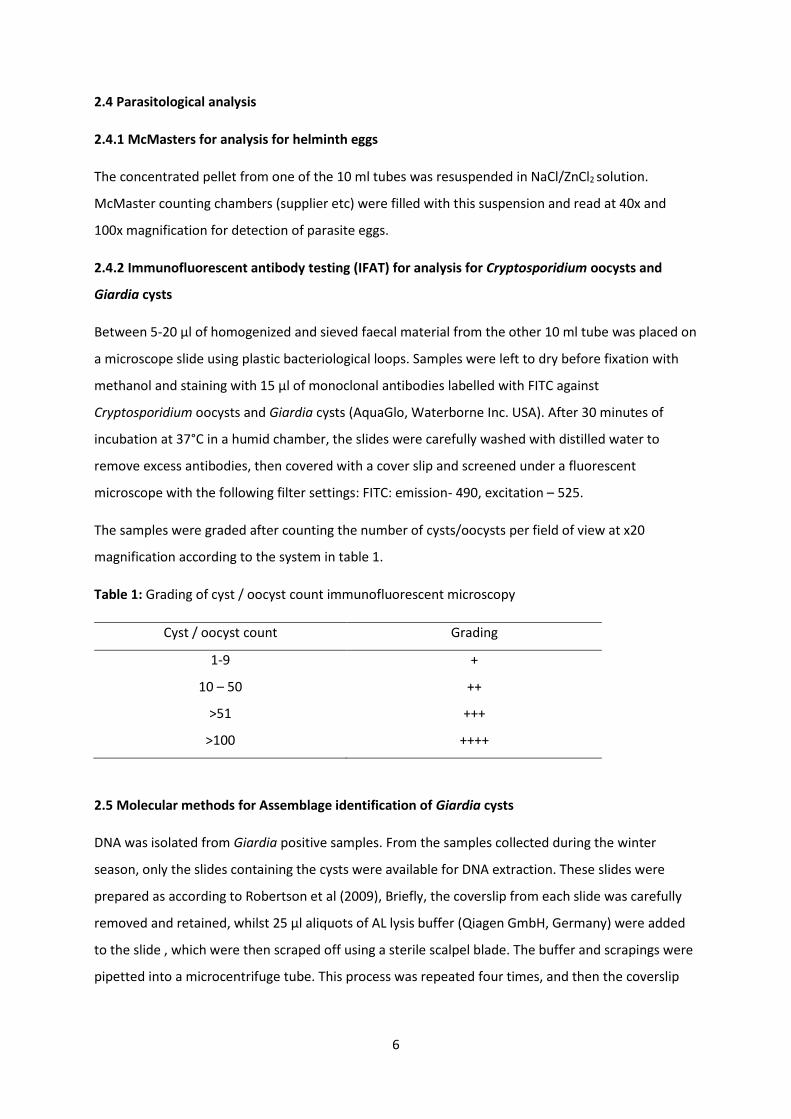
6
2.4 Parasitological analysis
2.4.1 McMasters for analysis for helminth eggs
The concentrated pellet from one of the 10 ml tubes was resuspended in NaCl/ZnCl2 solution.
McMaster counting chambers (supplier etc) were filled with this suspension and read at 40x and
100x magnification for detection of parasite eggs.
2.4.2 Immunofluorescent antibody testing (IFAT) for analysis for Cryptosporidium oocysts and
Giardia cysts
Between 5-20 µl of homogenized and sieved faecal material from the other 10 ml tube was placed on
a microscope slide using plastic bacteriological loops. Samples were left to dry before fixation with
methanol and staining with 15 µl of monoclonal antibodies labelled with FITC against
Cryptosporidium oocysts and Giardia cysts (AquaGlo, Waterborne Inc. USA). After 30 minutes of
incubation at 37°C in a humid chamber, the slides were carefully washed with distilled water to
remove excess antibodies, then covered with a cover slip and screened under a fluorescent
microscope with the following filter settings: FITC: emission- 490, excitation – 525.
The samples were graded after counting the number of cysts/oocysts per field of view at x20
magnification according to the system in table 1.
Table 1: Grading of cyst / oocyst count immunofluorescent microscopy
Cyst / oocyst count Grading
1-9 +
10 – 50 ++
>51 +++
>100 ++++
2.5 Molecular methods for Assemblage identification of Giardia cysts
DNA was isolated from Giardia positive samples. From the samples collected during the winter
season, only the slides containing the cysts were available for DNA extraction. These slides were
prepared as according to Robertson et al (2009), Briefly, the coverslip from each slide was carefully
removed and retained, whilst 25 µl aliquots of AL lysis buffer (Qiagen GmbH, Germany) were added
to the slide , which were then scraped off using a sterile scalpel blade. The buffer and scrapings were
pipetted into a microcentrifuge tube. This process was repeated four times, and then the coverslip
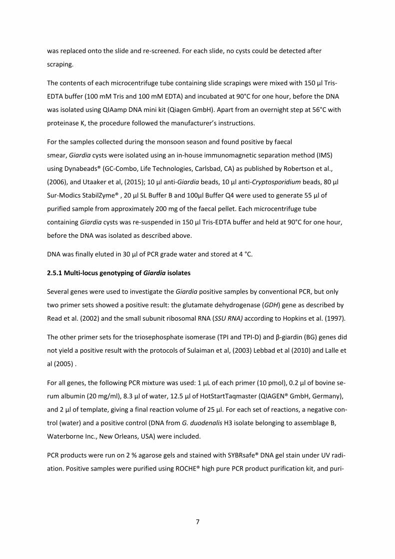
7
was replaced onto the slide and re-screened. For each slide, no cysts could be detected after
scraping.
The contents of each microcentrifuge tube containing slide scrapings were mixed with 150 µl Tris-
EDTA buffer (100 mM Tris and 100 mM EDTA) and incubated at 90°C for one hour, before the DNA
was isolated using QIAamp DNA mini kit (Qiagen GmbH). Apart from an overnight step at 56°C with
proteinase K, the procedure followed the manufacturer’s instructions.
For the samples collected during the monsoon season and found positive by faecal
smear, Giardia cysts were isolated using an in-house immunomagnetic separation method (IMS)
using Dynabeads® (GC-Combo, Life Technologies, Carlsbad, CA) as published by Robertson et al.,
(2006), and Utaaker et al, (2015); 10 μl anti-Giardia beads, 10 μl anti-Cryptosporidium beads, 80 μl
Sur-Modics StabilZyme® , 20 μl SL Buffer B and 100µl Buffer Q4 were used to generate 55 μl of
purified sample from approximately 200 mg of the faecal pellet. Each microcentrifuge tube
containing Giardia cysts was re-suspended in 150 µl Tris-EDTA buffer and held at 90°C for one hour,
before the DNA was isolated as described above.
DNA was finally eluted in 30 μl of PCR grade water and stored at 4 °C.
2.5.1 Multi-locus genotyping of Giardia isolates
Several genes were used to investigate the Giardia positive samples by conventional PCR, but only
two primer sets showed a positive result: the glutamate dehydrogenase (GDH) gene as described by
Read et al. (2002) and the small subunit ribosomal RNA (SSU RNA) according to Hopkins et al. (1997).
The other primer sets for the triosephosphate isomerase (TPI and TPI-D) and β-giardin (BG) genes did
not yield a positive result with the protocols of Sulaiman et al, (2003) Lebbad et al (2010) and Lalle et
al (2005) .
For all genes, the following PCR mixture was used: 1 µL of each primer (10 pmol), 0.2 μl of bovine se-
rum albumin (20 mg/ml), 8.3 μl of water, 12.5 μl of HotStartTaqmaster (QIAGEN® GmbH, Germany),
and 2 μl of template, giving a final reaction volume of 25 µl. For each set of reactions, a negative con-
trol (water) and a positive control (DNA from G. duodenalis H3 isolate belonging to assemblage B,
Waterborne Inc., New Orleans, USA) were included.
PCR products were run on 2 % agarose gels and stained with SYBRsafe® DNA gel stain under UV radi-
ation. Positive samples were purified using ROCHE® high pure PCR product purification kit, and puri-
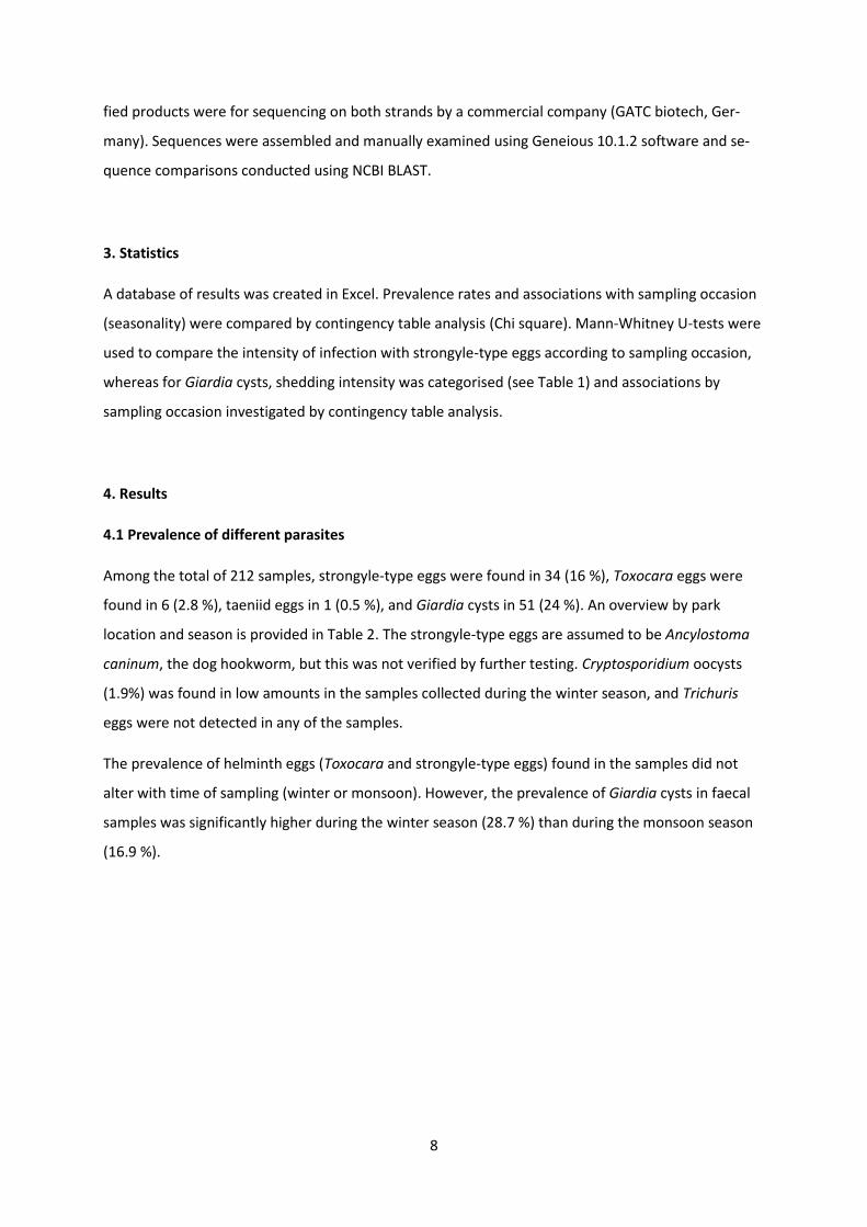
8
fied products were for sequencing on both strands by a commercial company (GATC biotech, Ger-
many). Sequences were assembled and manually examined using Geneious 10.1.2 software and se-
quence comparisons conducted using NCBI BLAST.
3. Statistics
A database of results was created in Excel. Prevalence rates and associations with sampling occasion
(seasonality) were compared by contingency table analysis (Chi square). Mann-Whitney U-tests were
used to compare the intensity of infection with strongyle-type eggs according to sampling occasion,
whereas for Giardia cysts, shedding intensity was categorised (see Table 1) and associations by
sampling occasion investigated by contingency table analysis.
4. Results
4.1 Prevalence of different parasites
Among the total of 212 samples, strongyle-type eggs were found in 34 (16 %), Toxocara eggs were
found in 6 (2.8 %), taeniid eggs in 1 (0.5 %), and Giardia cysts in 51 (24 %). An overview by park
location and season is provided in Table 2. The strongyle-type eggs are assumed to be Ancylostoma
caninum, the dog hookworm, but this was not verified by further testing. Cryptosporidium oocysts
(1.9%) was found in low amounts in the samples collected during the winter season, and Trichuris
eggs were not detected in any of the samples.
The prevalence of helminth eggs (Toxocara and strongyle-type eggs) found in the samples did not
alter with time of sampling (winter or monsoon). However, the prevalence of Giardia cysts in faecal
samples was significantly higher during the winter season (28.7 %) than during the monsoon season
(16.9 %).
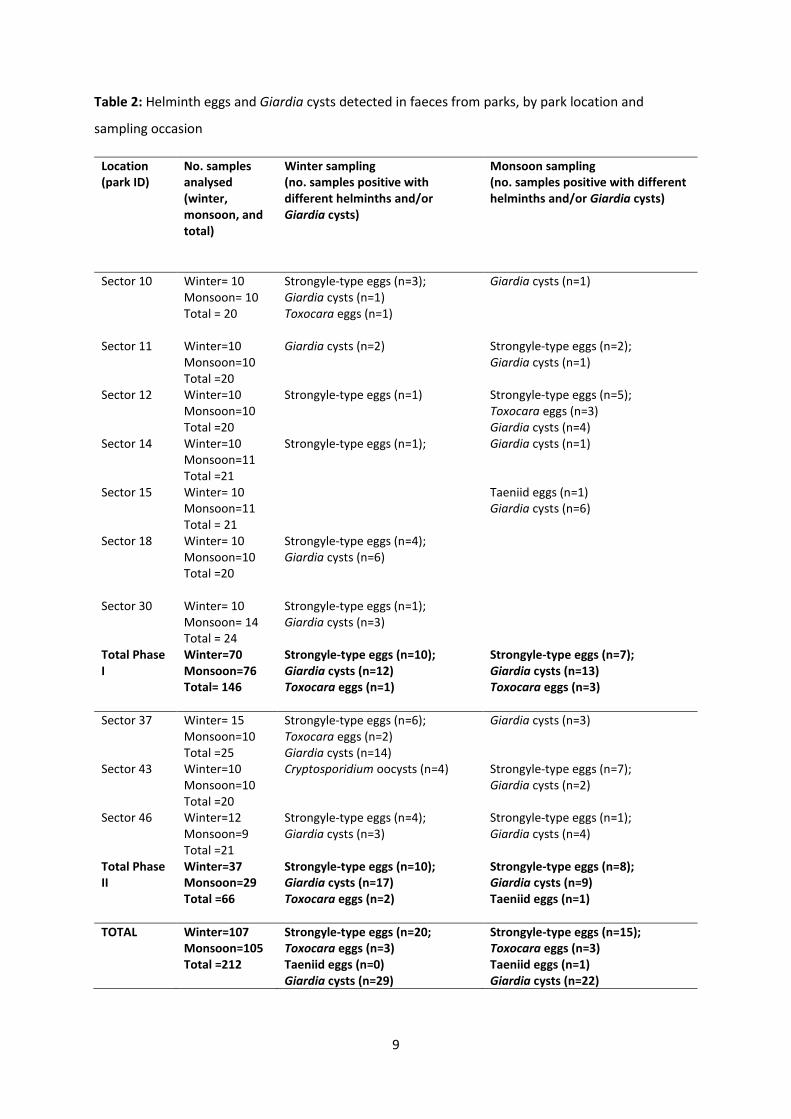
9
Table 2: Helminth eggs and Giardia cysts detected in faeces from parks, by park location and
sampling occasion
Location (park ID)
No. samples analysed (winter, monsoon, and total)
Winter sampling (no. samples positive with different helminths and/or Giardia cysts)
Monsoon sampling (no. samples positive with different helminths and/or Giardia cysts)
Sector 10 Winter= 10 Monsoon= 10 Total = 20
Strongyle-type eggs (n=3); Giardia cysts (n=1) Toxocara eggs (n=1)
Giardia cysts (n=1)
Sector 11 Winter=10 Monsoon=10 Total =20
Giardia cysts (n=2) Strongyle-type eggs (n=2); Giardia cysts (n=1)
Sector 12 Winter=10 Monsoon=10 Total =20
Strongyle-type eggs (n=1) Strongyle-type eggs (n=5); Toxocara eggs (n=3) Giardia cysts (n=4)
Sector 14 Winter=10 Monsoon=11 Total =21
Strongyle-type eggs (n=1); Giardia cysts (n=1)
Sector 15 Winter= 10 Monsoon=11 Total = 21
Taeniid eggs (n=1) Giardia cysts (n=6)
Sector 18 Winter= 10 Monsoon=10 Total =20
Strongyle-type eggs (n=4); Giardia cysts (n=6)
Sector 30 Winter= 10
Monsoon= 14 Total = 24
Strongyle-type eggs (n=1); Giardia cysts (n=3)
Total Phase I
Winter=70 Monsoon=76 Total= 146
Strongyle-type eggs (n=10); Giardia cysts (n=12) Toxocara eggs (n=1)
Strongyle-type eggs (n=7); Giardia cysts (n=13) Toxocara eggs (n=3)
Sector 37 Winter= 15 Monsoon=10 Total =25
Strongyle-type eggs (n=6); Toxocara eggs (n=2) Giardia cysts (n=14)
Giardia cysts (n=3)
Sector 43 Winter=10 Monsoon=10 Total =20
Cryptosporidium oocysts (n=4) Strongyle-type eggs (n=7); Giardia cysts (n=2)
Sector 46 Winter=12 Monsoon=9 Total =21
Strongyle-type eggs (n=4); Giardia cysts (n=3)
Strongyle-type eggs (n=1); Giardia cysts (n=4)
Total Phase II
Winter=37 Monsoon=29 Total =66
Strongyle-type eggs (n=10); Giardia cysts (n=17) Toxocara eggs (n=2)
Strongyle-type eggs (n=8); Giardia cysts (n=9) Taeniid eggs (n=1)
TOTAL Winter=107 Monsoon=105 Total =212
Strongyle-type eggs (n=20; Toxocara eggs (n=3) Taeniid eggs (n=0) Giardia cysts (n=29)
Strongyle-type eggs (n=15); Toxocara eggs (n=3) Taeniid eggs (n=1) Giardia cysts (n=22)
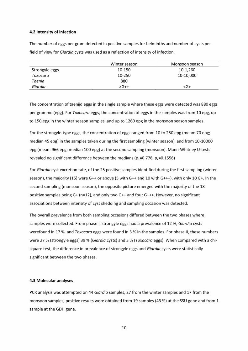
10
4.2 Intensity of infection
The number of eggs per gram detected in positive samples for helminths and number of cysts per
field of view for Giardia cysts was used as a reflection of intensity of infection.
Winter season Monsoon season
Strongyle eggs 10-150 10-1,260 Toxocara 10-250 10-10,000 Taenia 880 Giardia >G++ <G+
The concentration of taeniid eggs in the single sample where these eggs were detected was 880 eggs
per gramme (epg). For Toxocara eggs, the concentration of eggs in the samples was from 10 epg, up
to 150 epg in the winter season samples, and up to 1260 epg in the monsoon season samples.
For the strongyle-type eggs, the concentration of eggs ranged from 10 to 250 epg (mean: 70 epg;
median 45 epg) in the samples taken during the first sampling (winter season), and from 10-10000
epg (mean: 966 epg; median 100 epg) at the second sampling (monsoon). Mann-Whitney U-tests
revealed no significant difference between the medians (p1=0.778, p2=0.1556)
For Giardia cyst excretion rate, of the 25 positive samples identified during the first sampling (winter
season), the majority (15) were G++ or above (5 with G++ and 10 with G+++), with only 10 G+. In the
second sampling (monsoon season), the opposite picture emerged with the majority of the 18
positive samples being G+ (n=12), and only two G++ and four G+++. However, no significant
associations between intensity of cyst shedding and sampling occasion was detected.
The overall prevalence from both sampling occasions differed between the two phases where
samples were collected. From phase I, strongyle eggs had a prevalence of 12 %, Giardia cysts
werefound in 17 %, and Toxocara eggs were found in 3 % in the samples. For phase II, these numbers
were 27 % (strongyle eggs) 39 % (Giardia cysts) and 3 % (Toxocara eggs). When compared with a chi-
square test, the difference in prevalence of strongyle eggs and Giardia cysts were statistically
significant between the two phases.
4.3 Molecular analyses
PCR analysis was attempted on 44 Giardia samples, 27 from the winter samples and 17 from the
monsoon samples; positive results were obtained from 19 samples (43 %) at the SSU gene and from 1
sample at the GDH gene.
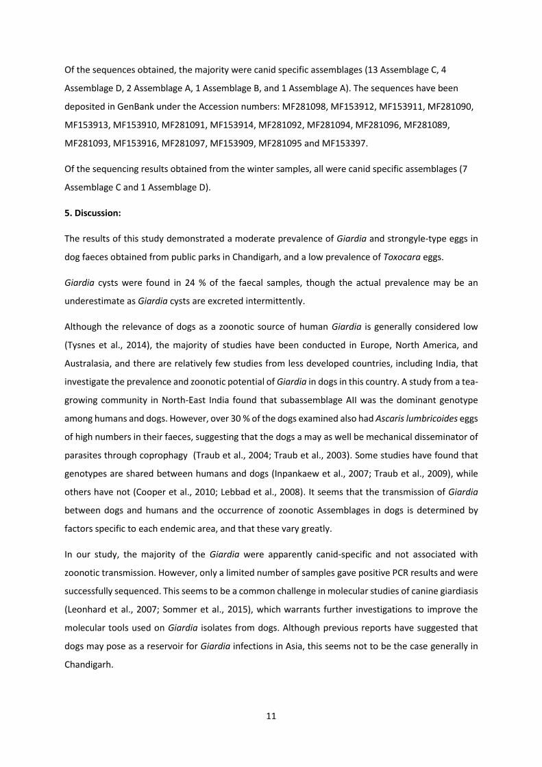
11
Of the sequences obtained, the majority were canid specific assemblages (13 Assemblage C, 4
Assemblage D, 2 Assemblage A, 1 Assemblage B, and 1 Assemblage A). The sequences have been
deposited in GenBank under the Accession numbers: MF281098, MF153912, MF153911, MF281090,
MF153913, MF153910, MF281091, MF153914, MF281092, MF281094, MF281096, MF281089,
MF281093, MF153916, MF281097, MF153909, MF281095 and MF153397.
Of the sequencing results obtained from the winter samples, all were canid specific assemblages (7
Assemblage C and 1 Assemblage D).
5. Discussion:
The results of this study demonstrated a moderate prevalence of Giardia and strongyle-type eggs in
dog faeces obtained from public parks in Chandigarh, and a low prevalence of Toxocara eggs.
Giardia cysts were found in 24 % of the faecal samples, though the actual prevalence may be an
underestimate as Giardia cysts are excreted intermittently.
Although the relevance of dogs as a zoonotic source of human Giardia is generally considered low
(Tysnes et al., 2014), the majority of studies have been conducted in Europe, North America, and
Australasia, and there are relatively few studies from less developed countries, including India, that
investigate the prevalence and zoonotic potential of Giardia in dogs in this country. A study from a tea-
growing community in North-East India found that subassemblage AII was the dominant genotype
among humans and dogs. However, over 30 % of the dogs examined also had Ascaris lumbricoides eggs
of high numbers in their faeces, suggesting that the dogs a may as well be mechanical disseminator of
parasites through coprophagy (Traub et al., 2004; Traub et al., 2003). Some studies have found that
genotypes are shared between humans and dogs (Inpankaew et al., 2007; Traub et al., 2009), while
others have not (Cooper et al., 2010; Lebbad et al., 2008). It seems that the transmission of Giardia
between dogs and humans and the occurrence of zoonotic Assemblages in dogs is determined by
factors specific to each endemic area, and that these vary greatly.
In our study, the majority of the Giardia were apparently canid-specific and not associated with
zoonotic transmission. However, only a limited number of samples gave positive PCR results and were
successfully sequenced. This seems to be a common challenge in molecular studies of canine giardiasis
(Leonhard et al., 2007; Sommer et al., 2015), which warrants further investigations to improve the
molecular tools used on Giardia isolates from dogs. Although previous reports have suggested that
dogs may pose as a reservoir for Giardia infections in Asia, this seems not to be the case generally in
Chandigarh.
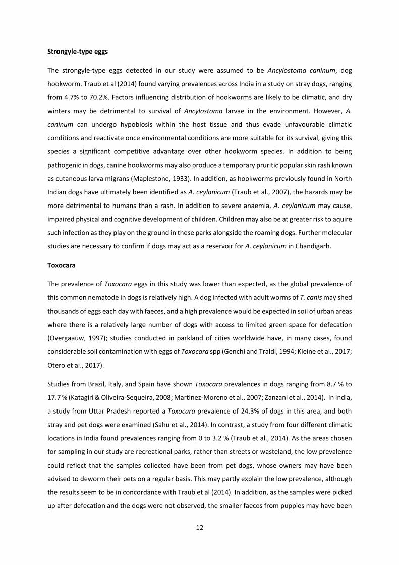
12
Strongyle-type eggs
The strongyle-type eggs detected in our study were assumed to be Ancylostoma caninum, dog
hookworm. Traub et al (2014) found varying prevalences across India in a study on stray dogs, ranging
from 4.7% to 70.2%. Factors influencing distribution of hookworms are likely to be climatic, and dry
winters may be detrimental to survival of Ancylostoma larvae in the environment. However, A.
caninum can undergo hypobiosis within the host tissue and thus evade unfavourable climatic
conditions and reactivate once environmental conditions are more suitable for its survival, giving this
species a significant competitive advantage over other hookworm species. In addition to being
pathogenic in dogs, canine hookworms may also produce a temporary pruritic popular skin rash known
as cutaneous larva migrans (Maplestone, 1933). In addition, as hookworms previously found in North
Indian dogs have ultimately been identified as A. ceylanicum (Traub et al., 2007), the hazards may be
more detrimental to humans than a rash. In addition to severe anaemia, A. ceylanicum may cause,
impaired physical and cognitive development of children. Children may also be at greater risk to aquire
such infection as they play on the ground in these parks alongside the roaming dogs. Further molecular
studies are necessary to confirm if dogs may act as a reservoir for A. ceylanicum in Chandigarh.
Toxocara
The prevalence of Toxocara eggs in this study was lower than expected, as the global prevalence of
this common nematode in dogs is relatively high. A dog infected with adult worms of T. canis may shed
thousands of eggs each day with faeces, and a high prevalence would be expected in soil of urban areas
where there is a relatively large number of dogs with access to limited green space for defecation
(Overgaauw, 1997); studies conducted in parkland of cities worldwide have, in many cases, found
considerable soil contamination with eggs of Toxocara spp (Genchi and Traldi, 1994; Kleine et al., 2017;
Otero et al., 2017).
Studies from Brazil, Italy, and Spain have shown Toxocara prevalences in dogs ranging from 8.7 % to
17.7 % (Katagiri & Oliveira‐Sequeira, 2008; Martinez-Moreno et al., 2007; Zanzani et al., 2014). In India,
a study from Uttar Pradesh reported a Toxocara prevalence of 24.3% of dogs in this area, and both
stray and pet dogs were examined (Sahu et al., 2014). In contrast, a study from four different climatic
locations in India found prevalences ranging from 0 to 3.2 % (Traub et al., 2014). As the areas chosen
for sampling in our study are recreational parks, rather than streets or wasteland, the low prevalence
could reflect that the samples collected have been from pet dogs, whose owners may have been
advised to deworm their pets on a regular basis. This may partly explain the low prevalence, although
the results seem to be in concordance with Traub et al (2014). In addition, as the samples were picked
up after defecation and the dogs were not observed, the smaller faeces from puppies may have been
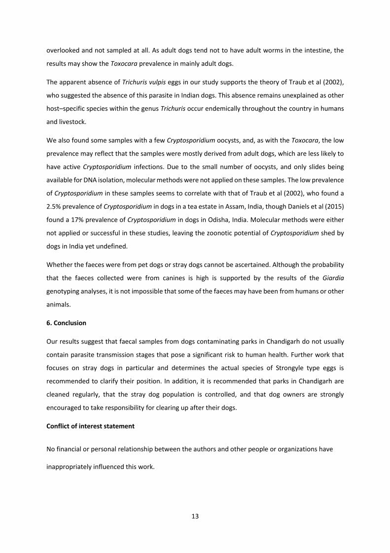
13
overlooked and not sampled at all. As adult dogs tend not to have adult worms in the intestine, the
results may show the Toxocara prevalence in mainly adult dogs.
The apparent absence of Trichuris vulpis eggs in our study supports the theory of Traub et al (2002),
who suggested the absence of this parasite in Indian dogs. This absence remains unexplained as other
host–specific species within the genus Trichuris occur endemically throughout the country in humans
and livestock.
We also found some samples with a few Cryptosporidium oocysts, and, as with the Toxocara, the low
prevalence may reflect that the samples were mostly derived from adult dogs, which are less likely to
have active Cryptosporidium infections. Due to the small number of oocysts, and only slides being
available for DNA isolation, molecular methods were not applied on these samples. The low prevalence
of Cryptosporidium in these samples seems to correlate with that of Traub et al (2002), who found a
2.5% prevalence of Cryptosporidium in dogs in a tea estate in Assam, India, though Daniels et al (2015)
found a 17% prevalence of Cryptosporidium in dogs in Odisha, India. Molecular methods were either
not applied or successful in these studies, leaving the zoonotic potential of Cryptosporidium shed by
dogs in India yet undefined.
Whether the faeces were from pet dogs or stray dogs cannot be ascertained. Although the probability
that the faeces collected were from canines is high is supported by the results of the Giardia
genotyping analyses, it is not impossible that some of the faeces may have been from humans or other
animals.
6. Conclusion
Our results suggest that faecal samples from dogs contaminating parks in Chandigarh do not usually
contain parasite transmission stages that pose a significant risk to human health. Further work that
focuses on stray dogs in particular and determines the actual species of Strongyle type eggs is
recommended to clarify their position. In addition, it is recommended that parks in Chandigarh are
cleaned regularly, that the stray dog population is controlled, and that dog owners are strongly
encouraged to take responsibility for clearing up after their dogs.
Conflict of interest statement
No financial or personal relationship between the authors and other people or organizations have
inappropriately influenced this work.
14
Ethics statement
It is submitted that proper consideration has been given to any ethics issue raised.
Acknowledgments
This study was funded through the Para-Clim-Chandigarh project, partly funded by the Norwegian
Research Council via the New Indigo Partnership program (Contract number: 227965)
The authors are grateful to Dr. Rakesh Seghal and Dr. Kapil Goyal for facilitating use of the PGIMER
facilities, Himanshu Joshi and Anil Kumar for assistance in collection of the samples, and Silje Nordås
for her contributions in the initial phase of this study.
References:
Cacciò, S.M., Beck, R., Lalle, M., Marinculic, A., Pozio, E., 2008. Multilocus genotyping of Giardia duodenalis reveals striking differences between assemblages A and B. Int. J. Parasitol. 38, 1523-1531.
Chaudhry, P., Bagra, K., Singh, B., 2011. Urban greenery status of some Indian cities: A short
communication. International Journal of Environmental Science and Development 2, 98. Cooper, M.A., Sterling, C.R., Gilman, R.H., Cama, V., Ortega, Y., Adam, R.D., 2010. Molecular analysis
of household transmission of Giardia lamblia in a region of high endemicity in Peru. J. Infect. Dis. 202, 1713-1721.
Daniels, M.E., Shrivastava, A., Smith, W.A., Sahu, P., Odagiri, M., Misra, P.R., Panigrahi, P., Suar, M.,
Clasen, T., Jenkins, M.W., 2015. Cryptosporidium and Giardia in humans, domestic animals, and village water sources in rural India. The American journal of tropical medicine and hygiene 93, 596-600.
Genchi, C., Traldi, G., 1994. Infezioni zoonosiche da Toxocara canis: stato dell’arte. Microbiol. Med 9,
457-461. Haque, R., 2007. Human intestinal parasites. J. Health. Popul. Nutr., 387-391. Heukelbach, J., Mencke, N., Feldmeier, H., 2002. Cutaneous larva migrans and tungiasis: the challenge
to control zoonotic ectoparasitoses associated with poverty. Trop. Med. Int. Health 7, 907-910. Hughes, J., Macdonald, D.W., 2013. A review of the interactions between free-roaming domestic dogs
and wildlife. Biol. Cons. 157, 341-351.
15
Times of India, 2016. Sector parks in Chandigarh in a shambles. Available at:
http://timesofindia.indiatimes.com/city/chandigarh/Sector-parks-in-Chandigarh-in-a-shambles/articleshow/52998606.cms (Accessed on: 17.06.2017)
Inpankaew, T., Traub, R., Thompson, R., Sukthana, Y., 2007. Canine parasitic zoonoses in Bangkok
temples. Jackman, J., & Rowan, A. (2007). Free-roaming dogs in developing countries: The benefits of capture,
neuter, and return programs. In D.J. Salem & A.N. Rowan (Eds.), The state of the animals 2007 (pp. 55-78). Washington, DC: Humane Society Press.
JNNURM 2006. City Development Plan Chandigarh, Administration, F.S.C., ed. (Chandigarh
Administration). Joshi, B., Sabne, S., 1977. Incidence of Toxocara canis infection in stray dogs in Miraj area. Indian J.
Pathol. Microbiol. 20, 239. Katagiri, S., & Oliveira‐Sequeira, T. (2008). Prevalence of dog intestinal parasites and risk perception of
zoonotic infection by dog owners in Sao Paulo State, Brazil. Zoonoses and public health, 55(8‐10), 406-413.
Kleine, A., Springer, A., Strube, C., 2017. Seasonal variation in the prevalence of Toxocara eggs on
children's playgrounds in the city of Hanover, Germany. Parasit Vectors 10, 248. Lalle, M., Pozio, E., Capelli, G., Bruschi, F., Crotti, D., Cacciò, S.M., 2005. Genetic heterogeneity at the
β-giardin locus among human and animal isolates of Giardia duodenalis and identification of potentially zoonotic subgenotypes. International Journal for Parasitology 35, 207-213.
Lebbad, M., Ankarklev, J., Tellez, A., Leiva, B., Andersson, J.O., Svärd, S., 2008. Dominance of Giardia
Assemblage B in Leon, Nicaragua. Acta tropica 106, 44-53. Lebbad, M., Mattsson, J.G., Christensson, B., Ljungström, B., Backhans, A., Andersson, J.O., Svärd, S.G.,
2010. From mouse to moose: multilocus genotyping of Giardia isolates from various animal species. Veterinary parasitology 168, 231-239.
Leonhard, S., Pfister, K., Beelitz, P., Wielinga, C., Thompson, R.C., 2007. The molecular characterisation
of Giardia from dogs in southern Germany. Veterinary parasitology 150, 33-38. Malaki, A., 1966. A survey of gastrointestinal parasites of dogs in Bangalore with notes on some
interesting observations. Indian Veterinary Journal 43, 409-412. Maplestone, P., Bhaduri, N., 1940. The Helminth Parasites of Dogs in Calcutta and their Bearing on
Human Parasitology. Indian Journal of Medical Research 28. Martinez-Moreno, F., Hernández, S., López-Cobos, E., Becerra, C., Acosta, I., & Martínez-Moreno, A.
(2007). Estimation of canine intestinal parasites in Cordoba (Spain) and their risk to public health. Veterinary parasitology, 143(1), 7-13.
Massei, G., Miller, L.A., 2013. Nonsurgical fertility control for managing free-roaming dog populations:
a review of products and criteria for field applications. Theriogenology 80, 829-838.
16
Otero, D., Alho, A.M., Nijsse, R., Roelfsema, J., Overgaauw, P., Madeira de Carvalho, L., 2017. Environmental contamination with Toxocara spp. eggs in public parks and playground sandpits of Greater Lisbon, Portugal. Journal of infection and public health.
Overgaauw, P.A., 1997. Aspects of Toxocara epidemiology: human toxocarosis. Crit Rev Microbiol 23,
215-231. Robertson, L., Gjerde, B., Hansen, E.F., Stachurska-Hagen, T., 2009. A water contamination incident in
Oslo, Norway during October 2007; a basis for discussion of boil-water notices and the potential for post-treatment contamination of drinking water supplies. Journal of water and health 7, 55-66.
Sahai, B., 1969. A survey of the helminth parasites of stray dogs in and around Bareilly, Uttar Pradesh.
Indian Veterinary Journal 46. Sahasrabudhe, V., Dubey, J., Srivastav, H., 1969. Helminth parasites of dogs in Madhya Pradesh and
their public health significance. Indian Journal of Medical Research 57, 56-59. Sahu, S., Samanta, S., Sudhakar, N., Raina, O., Gupta, S., Maurya, P., Pawde, A., Kumar, A., 2014.
Prevalence of canine toxocariasis in Bareilly, Uttar Pradesh, India. Journal of parasitic diseases 38, 111-115.
Sommer, M.F., Beck, R., Ionita, M., Stefanovska, J., Vasic, A., Zdravkovic, N., Hamel, D., Rehbein, S.,
Knaus, M., Mitrea, I.L., Shukullari, E., Kirkova, Z., Rapti, D., Capari, B., Silaghi, C., 2015. Multilocus sequence typing of canine Giardia duodenalis from South Eastern European countries. Parasitol Res 114, 2165-2174.
Sulaiman, I.M., Fayer, R., Bern, C., Gilman, R.H., Trout, J.M., Schantz, P.M., Das, P., Lal, A.A., Xiao, L.,
2003. Triosephosphate isomerase gene characterization and potential zoonotic transmission of Giardia duodenalis. Emerging infectious diseases 9, 1444-1452.
Traub, R., Inpankaew, T., Reid, S.A., Sutthikornchai, C., Sukthana, Y., Robertson, I.D., Thompson, R.A.,
2009. Transmission cycles of Giardia duodenalis in dogs and humans in Temple communities in Bangkok—a critical evaluation of its prevalence using three diagnostic tests in the field in the absence of a gold standard. Acta tropica 111, 125-132.
Traub, R., Monis, P., Robertson, I., Irwin, P., Mencke, N., Thompson, R., 2004. Epidemiological and
molecular evidence supports the zoonotic transmission of Giardia among humans and dogs living in the same community. Parasitology 128, 253-262.
Traub, R., Robertson, I., Irwin, P., Mencke, N., Monis, P., Thompson, R., 2003. Humans, dogs and
parasitic zoonoses–unravelling the relationships in a remote endemic community in northeast India using molecular tools. Parasitology Research 90, S156-S157.
Traub, R. J., Robertson, I. D., Irwin, P., Mencke, N., Thompson, R. A., 2002. The role of dogs in
transmission of gastrointestinal parasites in a remote tea-growing community in northeastern India. Am. J. Trop. Med. Hyg., 67(5), 539-545.
Traub, R.J., Hobbs, R.P., Adams, P.J., Behnke, J.M., Harris, P.D., Thompson, R.C.A., 2007. A case of
mistaken identity–reappraisal of the species of canid and felid hookworms (Ancylostoma) present in Australia and India. Parasitology 134, 113-119.
17
Traub, R.J., Pednekar, R.P., Cuttell, L., Porter, R.B., Rani, P.A.A.M., Gatne, M.L., 2014. The prevalence and distribution of gastrointestinal parasites of stray and refuge dogs in four locations in India. Veterinary parasitology 205, 233-238.
Traub, R.J., Robertson, I.D., Irwin, P.J., Mencke, N., Thompson, R.A., 2005. Canine gastrointestinal
parasitic zoonoses in India. Trends in parasitology 21, 42-48. Traub, R. J., Pednekar, R. P., Cuttell, L., Porter, R. B., Rani, P. A. A. M., & Gatne, M. L. (2014). The
prevalence and distribution of gastrointestinal parasites of stray and refuge dogs in four locations in India. Veterinary parasitology, 205(1), 233-238.
Tysnes, K.R., Skancke, E., Robertson, L.J., 2014. Subclinical Giardia in dogs: a veterinary conundrum
relevant to human infection. Trends in parasitology 30, 520-527. WHO 2016. In Eliminating rabies in India through awareness, treatment and vaccination, p. 2017.
Zanzani, S.A., Di Cerbo, A.R., Gazzonis, A.L., Genchi, M., Rinaldi, L., Musella, V., Cringoli, G., Manfredi, M.T., 2014. Canine fecal contamination in a metropolitan area (Milan, north-Western Italy): prevalence of intestinal parasites and evaluation of health risks. The Scientific World Journal 2014.
Additional articles
VIII: Utaaker, K. S. & Robertson, L. J. (2015). Climate change and foodborne transmission of parasites:
A consideration of possible interactions and impacts for selected parasites. Food Res. Int., 68:
16-23.
IX: Robertson, L. J., Utaaker, K. S., Goyal, K. & Sehgal, R. (2014). Keeping Parasitology under the One
Health umbrella. Trends Parasitol., 30 (8): 369-372.

Related Documents