Pharmaceutics 2021, 13, 1995. https://doi.org/10.3390/pharmaceutics13121995 www.mdpi.com/journal/pharmaceutics Review Antimicrobial Photodynamic Therapy: Latest Developments with a Focus on Combinatory Strategies Raphaëlle Youf 1 , Max Müller 2 , Ali Balasini 3 , Franck Thétiot 4, *, Mareike Müller 2 , Alizé Hascoët 1 , Ulrich Jonas 3 , Holger Schönherr 2, *, Gilles Lemercier 5, *, Tristan Montier 1,6 and Tony Le Gall 1, * 1 National Institute for Health and Medical Research (INSERM), Université de Bretagne Occidentale, EFS, UMR 1078, GGB, 29200 Brest, France; [email protected] (R.Y.); [email protected] (A.H.); [email protected] (T.M.) 2 Physical Chemistry I & Research Center of Micro- and Nanochemistry and (Bio)Technology of Micro and Nanochemistry and Engineering (Cμ), Department of Chemistry and Biology, University of Siegen, Adolf-Reichwein-Straße 2, 57076 Siegen, Germany; [email protected] (M.M.); [email protected] (M.M.) 3 Macromolecular Chemistry, Department of Chemistry and Biology, University of Siegen, Adolf-Reichwein-Straße 2, 57076 Siegen, Germany; [email protected] (A.B.); [email protected] (U.J.) 4 Unité Mixte de Recherche (UMR), Centre National de la Recherche Scientifique (CNRS) 6521, Université de Brest (UBO), CS 93837, 29238 Brest, France 5 Coordination Chemistry Team, Unité Mixte de Recherche (UMR), Centre National de la Recherche Scientifique (CNRS) 7312, Institut de Chimie Moléculaire de Reims (ICMR), Université de Reims Champagne-Ardenne, BP 1039, 51687 Reims CEDEX 2, France 6 CHRU de Brest, Service de Génétique Médicale et de Biologie de la Reproduction, Centre de Référence des Maladies Rares Maladies Neuromusculaires, 29200 Brest, France * Correspondence: [email protected] (F.T.); [email protected] (H.S.); [email protected] (G.L.); [email protected] (T.L.G.) Abstract: Antimicrobial photodynamic therapy (aPDT) has become a fundamental tool in modern therapeutics, notably due to the expanding versatility of photosensitizers (PSs) and the numerous possibilities to combine aPDT with other antimicrobial treatments to combat localized infections. After revisiting the basic principles of aPDT, this review first highlights the current state of the art of curative or preventive aPDT applications with relevant clinical trials. In addition, the most recent developments in photochemistry and photophysics as well as advanced carrier systems in the con- text of aPDT are provided, with a focus on the latest generations of efficient and versatile PSs and the progress towards hybrid-multicomponent systems. In particular, deeper insight into combina- tory aPDT approaches is afforded, involving non-radiative or other light-based modalities. Selected aPDT perspectives are outlined, pointing out new strategies to target and treat microorganisms. Finally, the review works out the evolution of the conceptually simple PDT methodology towards a much more sophisticated, integrated, and innovative technology as an important element of po- tent antimicrobial strategies. Keywords: antimicrobials; ROS; combinatory strategies; photodynamic therapy; multidrug resistance; nanoparticles; photosensitizers 1. Introduction Antimicrobial resistance (AMR) occurring in bacteria, viruses, fungi, and parasites is a global health and development threat, declared by the WHO as one of the top 10 global public health threats facing humanity. The misuse and overuse of antimicrobials make almost all disease-causing microbes resistant to drugs commonly used to treat them [1]. Multidrug resistance (MDR) to critical classes of antibiotics has gradually increased in nosocomial pathogens, including Enterococcus faecium, Staphylococcus aureus, Klebsiella Citation: Youf, R.; Müller, M.; Balasini, A.; Thétiot, F.; Müller, M.; Hascoët, A.; Jonas, U.; Schönherr, H.; Lemercier, G.; Montier, T.; et al. Antimicrobial Photodynamic Therapy: Latest Developments with a Focus on Combinatory Strategies. Pharmaceutics 2021, 13, 1995. https://doi.org/10.3390/ pharmaceutics13121995 Academic Editors: Udo Bakowsky, Matthias Wojcik, Eduard Preis and Gerhard Litscher Received: 7 October 2021 Accepted: 17 November 2021 Published: 24 November 2021 Publisher’s Note: MDPI stays neu- tral with regard to jurisdictional claims in published maps and insti- tutional affiliations. Copyright: © 2021 by the authors. Li- censee MDPI, Basel, Switzerland. This article is an open access article distributed under the terms and con- ditions of the Creative Commons At- tribution (CC BY) license (http://crea- tivecommons.org/licenses/by/4.0/).

Welcome message from author
This document is posted to help you gain knowledge. Please leave a comment to let me know what you think about it! Share it to your friends and learn new things together.
Transcript

Pharmaceutics 2021, 13, 1995. https://doi.org/10.3390/pharmaceutics13121995 www.mdpi.com/journal/pharmaceutics
Review
Antimicrobial Photodynamic Therapy: Latest Developments
with a Focus on Combinatory Strategies
Raphaëlle Youf 1, Max Müller 2, Ali Balasini 3, Franck Thétiot 4,*, Mareike Müller 2, Alizé Hascoët 1, Ulrich Jonas 3,
Holger Schönherr 2,*, Gilles Lemercier 5,*, Tristan Montier 1,6 and Tony Le Gall 1,*
1 National Institute for Health and Medical Research (INSERM), Université de Bretagne Occidentale, EFS,
UMR 1078, GGB, 29200 Brest, France; [email protected] (R.Y.); [email protected] (A.H.);
[email protected] (T.M.) 2 Physical Chemistry I & Research Center of Micro- and Nanochemistry and (Bio)Technology of Micro and
Nanochemistry and Engineering (Cμ), Department of Chemistry and Biology, University of Siegen,
Adolf-Reichwein-Straße 2, 57076 Siegen, Germany; [email protected] (M.M.);
[email protected] (M.M.) 3 Macromolecular Chemistry, Department of Chemistry and Biology, University of Siegen,
Adolf-Reichwein-Straße 2, 57076 Siegen, Germany; [email protected] (A.B.);
[email protected] (U.J.) 4 Unité Mixte de Recherche (UMR), Centre National de la Recherche Scientifique (CNRS) 6521, Université de
Brest (UBO), CS 93837, 29238 Brest, France 5 Coordination Chemistry Team, Unité Mixte de Recherche (UMR), Centre National de la Recherche
Scientifique (CNRS) 7312, Institut de Chimie Moléculaire de Reims (ICMR), Université de Reims
Champagne-Ardenne, BP 1039, 51687 Reims CEDEX 2, France 6 CHRU de Brest, Service de Génétique Médicale et de Biologie de la Reproduction, Centre de Référence des
Maladies Rares Maladies Neuromusculaires, 29200 Brest, France
* Correspondence: [email protected] (F.T.); [email protected] (H.S.);
[email protected] (G.L.); [email protected] (T.L.G.)
Abstract: Antimicrobial photodynamic therapy (aPDT) has become a fundamental tool in modern
therapeutics, notably due to the expanding versatility of photosensitizers (PSs) and the numerous
possibilities to combine aPDT with other antimicrobial treatments to combat localized infections.
After revisiting the basic principles of aPDT, this review first highlights the current state of the art
of curative or preventive aPDT applications with relevant clinical trials. In addition, the most recent
developments in photochemistry and photophysics as well as advanced carrier systems in the con-
text of aPDT are provided, with a focus on the latest generations of efficient and versatile PSs and
the progress towards hybrid-multicomponent systems. In particular, deeper insight into combina-
tory aPDT approaches is afforded, involving non-radiative or other light-based modalities. Selected
aPDT perspectives are outlined, pointing out new strategies to target and treat microorganisms.
Finally, the review works out the evolution of the conceptually simple PDT methodology towards
a much more sophisticated, integrated, and innovative technology as an important element of po-
tent antimicrobial strategies.
Keywords: antimicrobials; ROS; combinatory strategies; photodynamic therapy; multidrug
resistance; nanoparticles; photosensitizers
1. Introduction
Antimicrobial resistance (AMR) occurring in bacteria, viruses, fungi, and parasites is
a global health and development threat, declared by the WHO as one of the top 10 global
public health threats facing humanity. The misuse and overuse of antimicrobials make
almost all disease-causing microbes resistant to drugs commonly used to treat them [1].
Multidrug resistance (MDR) to critical classes of antibiotics has gradually increased in
nosocomial pathogens, including Enterococcus faecium, Staphylococcus aureus, Klebsiella
Citation: Youf, R.; Müller, M.;
Balasini, A.; Thétiot, F.; Müller, M.;
Hascoët, A.; Jonas, U.; Schönherr, H.;
Lemercier, G.; Montier, T.; et al.
Antimicrobial Photodynamic
Therapy: Latest Developments with
a Focus on Combinatory Strategies.
Pharmaceutics 2021, 13, 1995.
https://doi.org/10.3390/
pharmaceutics13121995
Academic Editors: Udo Bakowsky,
Matthias Wojcik, Eduard Preis
and Gerhard Litscher
Received: 7 October 2021
Accepted: 17 November 2021
Published: 24 November 2021
Publisher’s Note: MDPI stays neu-
tral with regard to jurisdictional
claims in published maps and insti-
tutional affiliations.
Copyright: © 2021 by the authors. Li-
censee MDPI, Basel, Switzerland.
This article is an open access article
distributed under the terms and con-
ditions of the Creative Commons At-
tribution (CC BY) license (http://crea-
tivecommons.org/licenses/by/4.0/).
Pharmaceutics 2021, 13, 1995 2 of 59
pneumoniae, Acinetobacter baumannii, Pseudomonas aeruginosa, and Enterobacter spp. (which
are gathered in the so-called ESKAPE group) [2]. Currently, in Europe, AMR is estimated
to be responsible for 33,000 deaths every year and the annual economic toll, covering extra
healthcare costs and productivity losses, amounts to at least EUR 1.5 billion [3,4]. Unless
adequately tackled, 10 million people a year will die from drug-resistant infections by
2050, according to the predictions of the government-commissioned O’Neill report [5].
With the decline in the discovery of new antimicrobials since 1970s, the mainstream
approach for the development of new drugs to combat emerging and re-emerging re-
sistant pathogens has focused on the modification of existing compounds. However, one
key recommendation encourages stimulation of early stage drug discovery [6]. Among
emerging antimicrobial therapeutic alternatives, light-based approaches show particular
promise [7]. Traditionally, phototherapy was already a common practice in ancient
Greece, Egypt, and India to treat skin diseases [8]. At the beginning of the 20th century,
Oscar Raab first described the phototoxicity of the dye acridine red against Paramecium
caudatum, and Tappenier and Jesionek reported the photodynamic effects of eosin suitable
for treating diverse cutaneous diseases. Since then, PDT was established as the admin-
istration of a non-toxic photosensitizer (PS) followed by exposure to light irradiation at an
appropriate wavelength focused on an area to treat [7]. While anti-cancer PDT is a clinical
reality for 25 years [9], PDT as an antimicrobial treatment was demonstrated for the first
time against drug-resistant infections in the healthcare sector in the early 1990s, leading
to a “photo-antimicrobial renaissance era”[7]. Major MDR bacteria have been found sus-
ceptible to antimicrobial PDT (aPDT), independently of their drug-resistance profiles
[10,11]. To date, resistance to aPDT is rarely reported, indicating that the possibility for
microbes to adapt and escape this treatment can occur but is still contained. More effective
aPDT systems are continuously developed, notably via combinatory approaches using
multiple chemical systems and/or modalities. At the current stage of development, aPDT
cannot address systemic infections but it holds great promise for treating localized infec-
tions and to fight AMR.
While excellent earlier authoritative reviews provide a detailed description of aPDT
[12–15], the present review focuses on most recent developments in the field for the last 5
years, with a focus on aPDT combinatory strategies. It should be noticed that aPDT is
sometimes referred to as photodynamic antimicrobial chemotherapy, light-based antimi-
crobial therapy, photo-controlled antimicrobial therapy, or antimicrobial photo-inactiva-
tion. In this review, all these synonyms were considered to present (i) the current state of
aPDT applications in preclinical and clinical settings, (ii) a state of the art with recent de-
velopments in photosystems, (iii) the implementation of multicomponent nanotechnolo-
gies and recent molecular engineering, and (iv) the exploration of combinatory aPDT ap-
proaches towards possible future therapeutic innovations.
2. PDT: General Presentation and Features
2.1. Photochemical Pathways and Reactive Oxygen Species Production
Generally speaking, a given PS has the potential to produce reactive oxygen species
(ROS) under specific conditions (Figure 1A). Typically, it possesses a stable electronic con-
figuration called ground state level. Following irradiation and absorption of a photon, the
PS is converted from a low (fundamental) energy level (1PS) to a “Frank Condon” short-
lived, very reactive, excited singlet state 1PS* [16–18]. Subsequently, the PS can lose energy
by emitting fluorescence or heat via internal conversion (IC), thereby returning to its ini-
tial ground state level; alternatively, it can be converted by a so-called inter-system cross-
ing (ISC) to a longer-living excited triplet state 3PS*. From this state, two types of chemical
reaction pathways can occur, known as Type I electron transfer and Type II energy trans-
fer, which can take place simultaneously [19]. In the Type I reaction, the 3PS* captures an
electron (e−) from a reducing molecule (R) in its vicinity, which induces an electron trans-
fer producing the superoxide anion radical (O2●−) and, after the subsequent reduction,

Pharmaceutics 2021, 13, 1995 3 of 59
leads to the generation of more cytotoxic ROS including hydrogen peroxide (H2O2) and
hydroxyl radical (HO●). In the Type II reaction, a direct energy transfer occurs from the 3PS* to the ground state molecular oxygen (3O2) that is then converted to singlet oxygen
(1O2). The ROS thus produced encompass O2●−, H2O2, HO●, and 1O2, the last two being the
most reactive and most cytotoxic species but also those with the shortest diffusion dis-
tance. One PS molecule can generate thousands of molecules of 1O2, depending notably
on its 1O2 quantum yield, the surrounding environment, and the respective occurrence of
Type I and Type II mechanisms [12,17,20].
Figure 1. (A) Modified Jablonski diagram describing the photochemical and photophysical mecha-
nisms leading to ROS production during PDT. (B) Overview of aPDT already applied to the critical
category of pathogens, as defined by the NIAID (https://www.niaid.nih.gov/research/emerging-in-
fectious-diseases-pathogens, accessed date: 1 September 2021). For each category, the chart specifies
the number and the proportion (percent of pathogens already assayed in at least one aPDT study
either before or after 2015).
In addition to the above-mentioned Type I and Type II mechanisms, Hamblin et al.
recently proposed introduction of a Type III photochemical pathway, following which the
radical anion PS•− and/or inorganic radicals formed in absence of oxygen could also lead
to photoinactivation [21]. These authors indeed identified several circumstances in which
oxygen-independent photoinactivation of bacteria using specific PSs can be obtained.
2.2. Biological Effects of aPDT: Potential Targets and Related Mechanisms
The main first targets of aPDT are external microbial structures, i.e., the cell wall, cell
membrane, or virus capsid and envelope [22,23]. Photodynamic inactivation (PDI) can be
Frequency (%)
B
Before 2015
After 2015
Not found
Already assayed with PDT:
0 4020 60 80 100
NA
ID c
riti
cal
cate
go
ry o
f p
ath
og
ens
Bacteria (n=30)
Viruses (n=46)
Fungi (n=2)
Mosquito-borne viruses (n=11)
Prion (n=1)
Toxins (n=3)
Protozoa (n=10)
1PS
1PS*
1O2
3O2
Ab
sorp
tio
n
Light
S0
S1
S2
Ground singlet state
Excited singlet states
ISC
O2 + e-
O2 -
H2O2
HO
Flu
ore
scen
ce
IC
e-
e-
IC
Excited triplet state
Type I
ROS
A
Microbial targets
3PS*e-
PS -
T1
R R +
Type II
En
erg
y

Pharmaceutics 2021, 13, 1995 4 of 59
obtained against microorganisms growing as planktonic cells and/or in biofilms [13]. In
biofilm matrices, the diffusion of PSs can be delayed or PSs can be sequestered, in spite of
photodamage induced on various components such as polysaccharides and extracellular
DNA [24,25]. The diffusion potential of ROS depends on (i) the maximal time-limited dif-
fusion length, especially for 1O2 that possesses a shorter half-life compared with other
ROS, (ii) the photostability in a given environmental medium, and (iii) the chemical prop-
erties of PSs (e.g., molecular size, charge, lipophilicity, stability), which influence the in-
teractions of the latter with target microorganisms [26]. Photoinactivation of Gram(+) bac-
teria can be obtained with a given PS, irrespective of its charge, whereas that of Gram(−)
bacteria generally requires a cationic PS, or a combination of a neutral PS with membrane-
disrupting agents [27].
Internalization of PSs in prokaryotic or eukaryotic cells can also occur, thus causing
various intracellular oxidative damage (such as in organelles in eukaryotic cells, e.g., nu-
cleus and mitochondria in fungal cells) [28]. To protect their intracellular components,
microbial cells can induce the production of antioxidant defenses such as protective en-
zymes (such as superoxide dismutase (SOD), catalase and glutathion (GSH)-peroxidase)
or pigments (such as carotenoids acting as nonphotochemical 1O2 quenchers). Neverthe-
less, these mechanisms can be insufficient to thwart aPDT-induced oxidative stress be-
cause intracellular components (including antioxidant defenses) can be also irreversibly
photodamaged by ROS [29]. The latter can act on the DNA level through two mechanisms,
i.e., alteration or modulation. Breaks in single-strand and double-strand DNA, and the
disappearance of the super-coiled form of plasmid DNA have been reported in both
Gram(+) and Gram(−) species. Indeed, PSs can interact with nucleic acids via electrostatic
interactions and induce reduction of guanine residues causing DNA cleavage [30]. Again,
microorganisms can induce the overproduction of proteins involved in the repair of pho-
todamaged DNA; however, some bacteria, such as Helicobacter pylori, possess only a few
of such protective repair mechanisms [31]. Upon PDT treatment, ROS and 1O2 can modify
bacterial gene expression profiles by modulating (i) the quorum sensing pathway, there-
fore inhibiting biofilm formation as shown in in vitro models, and/or (ii) the anti-virulence
activities by reducing the gene expression of virulence factors in diverse clinical patho-
gens [32–35].
Given the multi-targeted nature of aPDT, the possibility for microorganisms to de-
velop resistance is supposed to be very limited [36]. However, they may be able to respond
to aPDT in different ways. For instance, light response adaptation can occur in some mi-
croorganisms, such as E. coli upon exposure to blue light inducing the production of a
biofilm polysaccharide colonic acid [37]. Moreover, some PSs are substrates of efflux sys-
tems that may be overproduced; specific inhibitors of the latter can be used to restore
phototoxicity [38–40]. After sublethal aPDT, the biofilm-forming ability of bacteria can
increase, making them less susceptible to the same treatment [41]. Each strategy should
thus be carefully examined with regard to the ability of target microorganisms to adapt
and escape treatments. The latter may be noticeably reduced when using at the same time
multiple antimicrobial molecular partners and modalities (read below).
2.3. Important Parameters and Requirements for an “Ideal” aPDT
According to Cieplik et al. [12], an “ideal” aPDT system should meet the following
set of general requirements: (i) PS physicochemical properties: efficient PSs for aPDT pos-
sess most frequently a high hydrophilicity index and at least one cationic charge promot-
ing interactions with pathogens, especially Gram(−) bacteria; (ii) PS photosensitivity: fol-
lowing irradiation, a good PS produces a high rate of cytotoxic oxygen species (Figure
1A), depending notably on its 1O2 quantum yield, its stability, and the environmental me-
dia; (iii) light source parameters: efficient irradiation of PSs must take into account a co-
herent light exposure ensuring a good transmittance efficiency with no side effects; (iv)
safety: the PS has to be specific to target microorganism(s), inducing insignificant or only
a few side effects for the host, including no or few immunity responses; and (v) ease for

Pharmaceutics 2021, 13, 1995 5 of 59
implementation in clinical practice: aPDT has to be relatively easy to use (due to the rapid,
non-aggressive, and non-invasive light application), cost-effective, and accessible. It is ob-
vious that the detailed specification of requirements can vary depending on the target
applications. The improvement of PSs is an ongoing challenge that implies moving to-
wards more rational chemical engineering and biological investigations [11,42], as dis-
cussed in this review.
3. Positioning of aPDT in Current Human Healthcare Treatments
Over the past years, the number of studies dealing with aPDT has dramatically in-
creased, emphasizing the potential of this therapeutic approach to treat a broad spectrum
of microorganisms including bacteria, fungi, viruses, and parasites (Figure 1B). In this
part, recent in vitro screening studies, preclinical (using animal models) and clinical in-
vestigations are briefly reviewed. Details about the PSs involved and their structures are
provided in Section 4 “State of the art with recent photo(nano)system developments”.
3.1. Curative Preclinical aPDT
3.1.1. Treatment of Bacterial Infections
PDT is a promising alternative approach to antibiotherapy for photoinactivating a
broad spectrum of bacterial pathogens, either Gram(+) or Gram(−), responsible for diverse
infections in humans. The antibacterial versatility of PDT can be highlighted in different
ways, notably by considering lists of critically important human pathogens. First, in recent
years, more attention has been paid to the potential of aPDT to fight against bacteria in-
volved in hard-to-treat infections, especially those forming the ESKAPE group [2,43]. Sec-
ond, other critical pathogen lists can be considered, such as the NIAID emerging infec-
tious diseases/pathogens category that includes biodefense research and additional
emerging infectious diseases/pathogens. To our knowledge, the susceptibility of more
than 50% of the bacteria in the NIAID critical pathogen list has been considered in at least
one aPDT study. In other words, bacteria causing the worst endemic infections including
anthrax, botulism, melioidosis, cholerae, plague, and tuberculosis have already been con-
sidered. On the opposite, vector-borne diseases transmitted by human parasites, such as
Borrelia mayonii and Bartonella henselae have not been addressed in that regard yet. Multi-
ple experimental settings have been considered to demonstrate the potential of aPDT to
photoinactivate pathogenic bacteria, growing as planktonic forms, but also in biofilm ma-
trices, and using diverse animal models [44,45]. Among human pathogens, bacteria im-
plicated in oral infections, especially cariogenic, periodontic, and endodontic injuries,
have probably been the most intensely investigated [46]. Although less considered, other
indications have also been evaluated with aPDT, including osteomyelitis, meningitis,
pneumonia, lung abscess, and emphysema [47,48].
3.1.2. Treatment of Fungal Infections
Fungal infections caused by invasive candidiasis are widely recognized as a major
cause of morbidity and mortality in the health care environment [49]. In addition to the
opportunistic features of some fungi, resistance to first-line antifungals (such as echi-
nocandins and fluconazole) is spreading, compromising the efficiency of conventional an-
tifungal therapies. Despite the fact that yeasts are naturally more resistant to PDT than
bacteria, noticeable in vitro and in vivo effects against fungal infections have been re-
ported, including germ load reduction, biofilm inhibition and clearance, and eradication
of persistent colonization [50–53]. Furthermore, antifungal PDT has demonstrated its po-
tential as an adjunctive (potentially synergistic) treatment procedure to the conventional
fungicide Nystatin [54]. Chen et al. identified and summarized other fungal infections that
may be treated with aPDT, including onychomycosis, tinea cruris, pityriasis versicolor, chro-
moblastomycosis, and the cutaneous sporothricosis [55]. Recently, other fungal infections,

Pharmaceutics 2021, 13, 1995 6 of 59
such as fungi associated to mucormycosis (recognized as emerging critical pathogens in the
NIAID) were successfully treated with PDT [56].
3.1.3. Treatment of Viral Infections
Although vaccines have drastically reduced the spreading of some of the most viru-
lent viruses around the world, antiviral research and development remain a healthcare
priority notably due to emerging viral infectious diseases [57]. PDT holds promises to help
treat the latter, as well as other viruses implicated in complications of some cancers. The
oldest, but also the most current, application of antiviral PDT concerns the decontamina-
tion of blood products potentially containing hepatitis B/C or West Nile virus [23,58]. The
PDI of viral infections was explored in many studies considering other various viruses
including arbovirus, SV40, poliovirus, encephalitis virus, phages, and HSV [59,60]. In ad-
dition, emerging viruses such as Zika, Ebola, or Tickborne hemorrhagic fever viruses have
been considered [23], as well as viruses responsible for epidemic/pandemic crises such as
influenza virus, SARS-CoV-2 virus, and its mutants/variants [61,62].
3.1.4. Treatment of Parasite Infections
Drug resistance is also rapidly spreading in parasites. For example, resistance to ar-
temisinin (which is used to treat plasmodium infections causing malaria) increases dras-
tically, even when combined with other drugs (WHO, https://www.who.int/news-
room/fact-sheets/detail/antimicrobial-resistance, accessed date: 1 September 2021). An-
tiparasital effect of PDT was demonstrated toward critical parasites in public health such
as tropical pathogens including Leishmania, African trypanosoma, and Plasmodium
[63,64]. Another way to limit the propagation of the vectors is to rely on photoinductible
biolarvicides. Such an approach was investigated in order to control Aedes, Anopheles,
and Culex, which are tropical disease-carrying species [65,66]. This was also investigated
in Lyme disease using safranin-PDT for reducing the reproduction of ticks [67].
3.1.5. Treatment of Polymicrobial Infections
Quite recently, interest has grown to explore the potential of aPDT against polymi-
crobial infections involving multispecies pathogens. A study demonstrated that the sus-
ceptibility to PDI of S. aureus and C. albicans growing in mixed biofilms is lower compared
with single-species biofilms, which may be due to the difference in the chemical compo-
sition and viscosity of the composed matrix [24]. Nevertheless, aPDT applications are of
interest regarding hard-to-treat infections due to polymicrobial biofilms colonization,
such as chronic rhinosinusitis. They could effectively be eradicated by aPDT in a maxillary
sinus cavity model [68]. Moreover, Biel et al. showed that aPDT can eradicate polymicro-
bial biofilms in the endotracheal tubes, which are factors leading to ventilator-associated
pneumonia [69]. More recently, aPDT was shown capable of significantly improving
wound healing in mice with polymicrobial infections [70]. However, such an approach
remains a challenge due to the respective affinity of PSs to each species being usually re-
duced in polymicrobial systems.
3.2. Current Clinical aPDT Practices
Many clinical trials have been done for evaluating aPDT approaches in the treatment
of bacterial/fungal oral infections. This is facilitated by the development of easy to use
light sources in dentistry. On the opposite, it can be compromised by the development of
persistent (multispecies) biofilms. Skin infections such as Acnes vulgaris, caused by Propi-
onibacterium acnes, was one of the first microbial infections to reach the stage of aPDT clin-
ical trial. A few clinical trials demonstrated that onychomycosis, such as tinea cruris, tinea
pedis, and interdigital mycoses, could be treated with aPDT. Results demonstrated that
aPDT is effective and well-tolerated, but infections can recur frequently [71,72]. Among
cutaneous infections, non-healing chronic wounds in patients with chronic leg and/or foot
Pharmaceutics 2021, 13, 1995 7 of 59
ulcers were efficiently treated with aPDT, inducing a significant reduction in microbial
load (even immediately after the treatment), a better wound healing, and no safety issues
[73,74]. Osteomyelitis in patients with chronic ulcers can be treated with aPDT to prevent
gangrene and amputations in the extremities of diabetic patients [75]. Clinical studies for
treating H. pylori in gastric ulcers can be conducted using phototherapy without any PS
administration; H. pylori naturally accumulates intracellular PSs (porphyrins) and there-
fore could be inactivated by phototherapy thanks to an ingenious blue/violet light deliv-
ery system [76]. A list of recently closed aPDT clinical trials is provided in Table 1; many
other trials (not shown here) are still ongoing.

Pharmaceutics 2021, 13, 1995 8 of 59
Table 1. List of some recently completed or terminated clinical trials that evaluated aPDT to treat diverse infectious diseases.
Medical Conditions Target Micro-Organism(s) Photosensitizer Trial Phase Number and Year
Acne Propionibacterium acnes
Butenyl ALA N.A. NCT02313467, 2014
Lemuteporfin Phase 1/2 NCT01490736, 2011
5-ALA Phase 2 NCT01689935, 2012
Methyl aminolevulinate Phase 2 NCT00673933, 2013
Dental caries
Streptococcus mutans, Streptococcus sobrinus, Lactobacillus casei, Fusobacterium nucleatum,
and Atopobium rimae
TBO Phase 1 NCT02479958, 2015
MB Phase 1 NCT02479958, 2015
Aggregatibacter actinomycetemcomitans, Tannerella forsythia and Porphyromonas gingivalis N.C. N.A. NCT03309748, 2017
Denture-related stomatitis Candida albicans MB Phase 4 NCT02642900, 2015
Orthodontic N.D. Curcumin Phase 1 NCT02337192, 2015
Peri-implantitis N.D. N.D. Phase 3 NCT02848482, 2016
Periodontic Aggregatibacter Actinomycetemcomitans, Porphyromonas gingivalis, Prevotella intermedia,
Tannerella forsythia and Treponema denticola
MB N.A. NCT03750162, 2018
ICG Phase 2 NCT02043340, 2014
Methyl aminolevulinate Phase 2 NCT00933543, 2013
MB N.A. NCT03262077, 2017
MB Phase 2 NCT03074136, 2017
Phenothiazine hydrochloride Phase 4 NCT03498404, 2018
TB Phase 4 NCT03412331, 2018
Distal subungual onychomycosis Fungi infecting nails 5-ALA Phase 2 NCT02355899, 2015
Endodontic E. faecalis and C. albicans MB Phase 2 NCT02824601, 2016
HPV infection Human Papillomavirus (HPV) 5-ALA Phase 2 NCT02631863, 2015
Leg ulcers Streptococci, anaerobes, coliform, S. aureus, P. aeruginosa, yeast, and diphtheroids PPA904 Phase 2 NCT00825760, 2009
Studies collected from ClinicalTrials.gov (https://clinicaltrials.gov/ct2/results?cond=photodynamic+therapy, Accessed Date: 1 March 2021). ALA, alanine; MB, meth-
ylene blue; N.A., not applicable; N.C., not communicated; N.D., not determined; PPA904, 3,7-bis(di-n-butylamino)phenothiazin-5-ium bromide; and TB (or TBO),
toluidine blue.

Pharmaceutics 2021, 13, 1995 9 of 59
3.3. Toward Preventive/Prophylactic Treatments
Beside curative antimicrobial treatments, PDT may be also used to decontaminate
medical equipment and tools in hospitals, for preventive/prophylactic aims [27]. For ex-
ample, photoantimicrobial textiles were reported to efficiently photoinactivate bacteria
and viruses, suggesting that self-sterilizing medical gowns could be developed [77]. PDT
can also decontaminate medical tools similarly to conventional chemical agents, as
demonstrated in a recent comparative study [78]. Its application for the decontamination
of routine informatics tools, office equipment, or packing materials demonstrates sterili-
zation potential that could be useful to avoid hospital-acquired infections and to protect
healthcare workers. Furthermore, photodisinfection of water and photoinactivation of
food-borne pathogens can bring substantial benefits to people’s daily lives [65].
4. State of the Art with Recent Photo(nano)System Developments
4.1. Single PSs
4.1.1. Organic PSs and Their Derivatives
Organic PSs used in aPDT have been well described in some recent reviews [79–81]
(Figure 2A). Briefly, since the first use of eosin in 1904, various PSs were investigated,
especially in the phenothiazinium group, which includes methylene blue (MB) and tolui-
dine blue O (TBO). Thanks to an absorption spectrum in the red region of light, these PSs
can be effective in tissues while being less toxic than other PSs. Their aPDT properties are
mostly due to a high ROS production following Type I mechanism (Figure 1A). Structural
derivatives have been also reported, including new MB and dimethyl-MB [42]. Another
group gathers compounds featuring a macrocyclic structure composed of pyrroles, such
as porphyrins and its precursor 5-aminolevulinic acid (5-ALA), phthalocyanines, and
chlorins. Macrocyclic compounds are generally hydrophilic, positively charged, and they
exhibit a good singlet oxygen quantum yield. Modifications of their chemical structure
were intensively studied, especially for porphyrins and phthalocyanines [82–87]. The hal-
ogenated xanthenes gather PSs with a structure similar to that of fluorescein. Among
them, eosin Y, erythrosine, and rose bengal (RB) were the most studied [88–90]. These
compounds are anionic, which can limit their interaction with bacterial cells and their
aPDT effect in spite of good singlet oxygen quantum yields. Natural compounds, includ-
ing coumarins, furanocoumarins, benzofurans, anthraquinones, and flavin derivatives are
often found in plants and other organisms. They are characterized by an absorption spec-
trum in white light or UVA. The most used are curcumin, riboflavin, and hypericin [7,80].
Nanostructures such as fullerenes are interesting PSs because of their ability to modulate
Type I and Type II reactions (Figure 1A), depending on the near environment and the
light source applied [91,92]. In this family, some quantum dots (QDs) can act as photoan-
timicrobials [80]. Other synthetic fluorescent dyes such as organoboron compounds (e.g.,
boron–dipyrromethene (BODIPY)), and cyanine dyes (e.g., indocyanine green (ICG)) are
known for their high photostability, high extinction coefficients, and high fluorescence
quantum yields [93].
Over the past five years, some new organic PSs were reported. Among them, opti-
mized natural PSs such as anthraquinones and diacethylcurcumin can be listed. Others
include derivatives of synthetic dyes such as monobrominated neutral red or azure B [79–
81] (Figure 2A).

Pharmaceutics 2021, 13, 1995 10 of 59
Figure 2. Representative compounds in various classes of PSs used in aPDT. (A) Examples of some
organic PSs and their derivatives. (B) Examples of metallic-based PSs. (C) Different types of poly-
mer-based PS carriers, which can be functionalized with ligands for specific target delivery
(Adapted from [94], published by MDPI, 2020).
4.1.2. Coordination and Organometallic Complexes-Based PSs
Distinctly from metal nanoparticles (NPs; see Section 4.2.1 “Metal-based systems”),
metal complexes, either coordination or organometallic complexes, are of increasing in-
terest as PSs in PDT [95] (Figure 2B). They generally consist of a central metal core com-
bined with ligands, involving coordinate covalent bonds (in coordination compounds) or
at least one metal–carbon bond (in organometallic compounds). Compared with organic
compounds, metal complexes have been notably much less considered and still remain
largely underexploited regarding their potential use as new antibiotics [96]. Frei et al. re-
cently reported on the antimicrobial activity of 906 metal-containing compounds. They
B
PS Ligand
Polymeric NP
Micelle Dendrimer Polymersome Polyplex Polymer drugconjugate
C
Methylene Blue(MB)
m-tetra(hydroxyphenyl)chlorin (m-THPC)
5-aminolevulinic acid (5-ALA)
Phtalocyanines
Rose Bengal(RB)
Carboxypterine
Boron-dipyrromethene(BODIPY)
Anthraquinone
Ru(dqpCO2Me)2(ptpy)2+
Ru(phen)2(phen-Fluorene)]2+
A
Fullerene C60
Curcumin
Indocyanine green(ICG)
Chlorin e6 (Ce6)
Tookad
[Ir(phen)2(R-phen)]3+ TLD-1433

Pharmaceutics 2021, 13, 1995 11 of 59
showed that, considering antimicrobial activity against critical antibiotic-resistant patho-
gens, metal-bearing compounds displayed hit rates about 10 times higher than purely or-
ganic molecules [97].
Metal complexes display a panel of specific properties that make them promising PSs
candidates. The variety of metal ions and ligands can be assembled in scaffolds featuring
very diverse geometries [97,98]. Whereas most organic PSs are linear or planar molecules,
metal complexes can exhibit much more complex—three-dimensional—geometries,
which can improve interaction and molecular recognition with cellular targets, enlarge
the spectrum of activity, and impact on biological fate [98–100]. Furthermore, the modu-
lation of the design of metal complexes allows to fine tune their hydrophilic–lipophilic
balance, solubility, photophysical properties, and eventual “dark toxicity” (i.e., the tox-
icity in the absence of specific irradiation). Metal complexes can display many excited-
state electronic configurations associated with the central metal, the ligands, or involving
both the metal and the ligand(s) in charge-transfer states (metal-to-ligand charge transfer
or ligand-to-metal charge transfer). Although it is not always considered, the investigation
of excited states of metal complexes (Figure 1A) is however of prime importance. Triplet
states can be more easily accessed due to the enhanced spin-orbit coupling induced by the
presence of the heavy metallic atom. Compared with natural PSs, metal complexes can
act, besides ROS generation, via other mechanisms including redox activation, ligand ex-
change, and depletion of substrates involved in vital cellular processes [96,97,101].
Besides their first intended development as anticancer compounds, metal complexes
have also been envisaged as potential “metallo-antibiotics”, benefiting from the
knowledge accumulated about their chemical properties and biological behaviors [102].
Quite recently, several metal compounds were characterized for their activity in aPDT.
For instance, platinum(II), molybdenum(II), ruthenium(II), cobalt(II), and iridium(III)
were proposed as new classes of stable photo-activatable metal complexes capable of com-
bating AMR [11,103–108]. In particular, many mononuclear and polynuclear Ru(II/III)
complexes have been considered as potential antibiotics, antifungals, antiparasitics, or an-
tivirals, which have been recently extensively reviewed [107]. It is worth noticing here
that, within a series of 906 metal compounds, ruthenium was the most frequent element
in active antimicrobial compounds that are nontoxic to eukaryotic cells, followed by sil-
ver, palladium, and iridium [97]. Ru(II) polypyridyl complexes were assayed in several
aPDT studies. For instance, Ru(DIP)2(bdt) and Ru(dqpCO2Me)2(ptpy)2+ (DIP = 4,7-diphe-
nyl-1,10-phenanthroline, bdt = 1,2-benzenedithiolate, dqpCO2Me = 4-methylcarboxy-2,6-
di(quinolin-8-yl)pyridine) and ptpy = 4′-phenyl-2,2′ : 6′ ,2″ -terpyridine) were tested with
S. aureus and E. coli [109]. The complexes were found active against the Gram(+) strain and
to a lesser extent against the Gram(−) strain, such difference in susceptibility being com-
monly reported in studies using other PSs [110] (Figure 2B). This observation was further
detailed and rationalized by us when investigating a collection of 17 metal-bearing deriv-
atives; two neutral Ru(II) complexes (Ru(phen)2Cl2 and Ru(phen-Fluorene)2Cl2) as well as
a mono-cationic Ir(III) complex (Ir(phen-Fluorene)(ppy)2+; ppy = phenypyridyl ligand)
were found almost inactive, whereas a dicationic Ru(II) (Ru(phen-Fluorene)(phen)22+) was
found to be the most active against a panel of clinical bacterial strains [11]. More recently,
Sun et al. described a Ru(II) complex bearing photolabile ligands; they showed its ability
to safely photoinactivate intracellular MRSA while inducing only negligible resistance af-
ter bacterial exposure for up to 700 generations [111]. Although the precise mechanism(s)
of action is not well-established in every case, Ru(II/III) compounds are also increasingly
considered for their potential anti-parasitic activity for combating neglected tropical dis-
eases such as malaria, Chagas’ disease, and leishmaniasis. Moreover, potent antiviral ac-
tivities have been noted for the ruthenium complex BOLD-100, particularly against HIV
and SARS-CoV-2 [112]; importantly, this compound appears to retain its activity on all
mutant strains of the SARS-CoV-2 [107]. All combined, metal complexes—especially ru-

Pharmaceutics 2021, 13, 1995 12 of 59
thenium-based compounds—can display antimicrobial activity via multiple, likely syner-
gistic, mechanisms, involving notably their ability to produce ROS. Therefore, they are of
major interest for a wide range of aPDT-related applications.
4.2. Multicomponent PSs and Nanoscale Implementation: Extension to Nanoedifices with PSs
The eventual limitations in the use of singular photoactive molecules as PSs for aPDT
applications reside notably in the recurrent lack of solubility and stability in the target
media (typically leading to aggregates and/or PS quenching), biocompatibility (“immune
stealth”) and bioavailability, but also in the relative absence of selectivity for a prospected
target (e.g., efficiency in the interactions with a defined target, stimuli-responsive or alter-
nate triggers for controlled release). Thus, the widespread nanoscale implementation into
the development progress of upgraded PSs has indubitably provided significant flexibil-
ity to first address these drawbacks, and implicitly contribute to the optimization of the
aPDT activity via an extensive panel of mainly exclusive features specific to nano entities;
the latter typically include an advantageous surface to volume ratio (with a high PS per
mass content), access to unique chemical/physical/biological properties (e.g., optical prop-
erties with QDs or magnetic ones with superparamagnetic iron oxide NPs (SPIONs)), and
almost inexhaustible synthetic options in the design of nanoplatforms [113–115].
Due to their paramount structural diversity and intrinsic variety of properties, the
classification of the so-called “PS nanosystems” or “nano-PSs”—which partly include and
overlap with “conjugated systems” and “combinatorial strategies”—remains rather chal-
lenging; however, we can usually identify the following criteria as the main pillars to ra-
tionalize and compare these aPDT nanomaterials [27,80,116]:
(i) Role(s) and nature of the nanocomponent(s) in the PS nanosystems: the two criteria
typically considered for the discrimination of nano-PSs are the role and nature of the
nano building block(s) involved. With regards to the role of the latter, we can conven-
tionally discern on the one hand the “PS nanocarriers” (e.g., polymersomes or Au
NPs) in which the nanomoiety acts as a delivery system for singular PS molecules
(e.g., MB) while either complementing, facilitating, or enhancing the aPDT activity
(depending on the nature and eventual intrinsic properties of the nanovector), and on
the other hand, the “PS active” nanoagents with the nanocomponent endorsing the
role of PS. Among the examples, some versatile nanotemplates may ultimately dis-
play a dual role, i.e., “active PS” and “PS conveyor” (e.g., ZnO NPs), while distinct
nanomoieties might be simultaneously required for the design of utterly sophisticated
hybrid nano-PSs (e.g., Au@AgNP@SiO2@PS) [117,118]. In addition to the chemical
composition, the nature of the nano building block(s) will also be defined by the fun-
damental characteristics of nano-objects, such as size, shape, topology, and crystal
structure, which will all ineluctably contribute to tailor the biological behavior of the
nanomaterials and the interactions with the targets (e.g., with the membranes of the
bacteria) [119]. Moreover, for the same nanocore, the nature and role of the eventual
surfactant(s) involved (e.g., silica coating or poly(ethylene glycol) (PEG) coating for
metal NPs) can drastically alter the overall behaviors of the nanosystems.
(ii) Type of interactions between the nano entity and the PS, and localization of the PS in
the nanosystems: other criteria of relevance when describing nano-PSs—specifically
PS nanocarriers—reside in the nature of the interactions between the nanocomponent
and the PS molecules involved, but also the location of the PSs. Thus, we can distin-
guish the common cases of PS molecules “embedded” within a nanovector either by
physisorption or functionalized (chemisorption), and alternately the nanoplatforms
with surfaces decorated with PSs, again, either by physical or chemical adsorption.
The differences between the two types of interactions and distinct localizations of the
PSs implicitly imply distinctive chemical engineering and related requirements, and
may potentially impact the resulting stability of the nanoedifices, but also the aPDT
activity. For instance, in the case of PS molecules located inside the edifice and not

Pharmaceutics 2021, 13, 1995 13 of 59
released, the selected “nanomatrix” should adequately permit the photoactivation
process of the internalized PSs, be sufficiently porous/permeable to both triplet and
singlet oxygen and eventual ROS generated by the photosensitizers (i.e., efficient in-
ternal diffusion of molecular oxygen to react with the PSs then external diffusion of 1O2 to the targets) while also presenting inertness to the latter to not compromise or
quench the aPDT activity. Meanwhile, with surfaces of nano-objects decorated with
PSs, the PSs may then contribute to some extent as an interface with the biological
medium or the target.
(iii) Biological impacts of the PS nanosystems: in addition to the biocompatibility and
aPDT efficiency (including the critical concentrations just as the half-maximal effec-
tive concentration EC50, minimum inhibitory concentration MIC, or 50% growth inhi-
bition concentration GIC50), the eventual biodegradability, elimination process, or
ecotoxicity of the aPDT nanomaterials can markedly vary from one system to another
(based on factors such as composition and size/shape), but are rather difficult to eval-
uate or compare; ergo, these factors are not systematically addressed in the reports.
(iv) Relative sustainability of the nano-PSs for aPDT applications: the reproducibility, eco-
friendliness, and cost-effectiveness parameters of the synthetic protocols and produc-
tion of aPDT nanomaterials, as well as the ease of storage and use, and the stability
over time are also ultimately to be evaluated for any system aiming to be viable and
reasonably applied; however, similar to (iii), these parameters are complex and so
scarcely investigated.
Thus, in the overview presentation of the different PS nanosystems hereafter, the
chemical features (i) and (ii) have been conventionally defined as the main criteria for the
classification. Alternatively, the nature of the aPDT applications has also been used as the
main criterion for classification in some references [120]. Another approach consists of
systemizing all the nanosystems dedicated to a given PS (e.g., curcumin) [121].
Within the extensive collection of aPDT nanomaterials reported to date, the majority
belongs to the category labeled as “PS nanocarriers” with the nanocomponent acting as a
delivery system for PS molecules; however, increasing examples involving PS-active nano
building blocks have emerged as well. Overall, this multipurpose role of the nanomoiety
may include avoiding aggregation (e.g., dimerization, trimerization) and correlated PS
quenching, enhancing “solubility” (i.e., dispersibility), stability and bioavailability, allow-
ing “biological stealth”, on-demand release and target specificity, and ultimately trigger-
ing eventual synergistic aPDT activity with the complementary or ameliorative intrinsic
properties of the nanocomponent; although irrevocably confirmed in many nano-PSs, the
mechanisms involved in the synergy may differ from one system to another, and often
remain partly or integrally unresolved due to the complexity of these tacit multiparameter
contextures [113]. It is noteworthy that most systems comprise “classic”/”traditional” or-
ganic PSs (natural or synthetic, e.g., curcumin, MB) with fewer examples involving metal-
lated PS molecules such as the recent review from Jain et al. dedicated to ruthenium-based
photoactive metalloantibiotics [108]. Among aPDT nanomaterials, we can thus identify
various families of nanoplatforms based on the nature of the nanocore, starting here with
the inorganic vectors followed by the organic templates; as an indication, in the common
cases of “multi-component nano-PSs”, the classification has been defined hereafter ac-
cording to the main/prominent nano building block involved in the composition, i.e.,
metal-based systems, silicon-based systems, carbon-based systems, lipid-based systems,
and polymer-based systems. Regarding the following presentation of aPDT nanosystems,
it is important to specify that it is not exhaustive, but instead provides a panorama of the
main categories of nanosystems—either colloids or surfaces [27,122]—and their related
specificities, with an emphasis on recent developments.

Pharmaceutics 2021, 13, 1995 14 of 59
4.2.1. Metal-Based Systems
Metal-based nanostructures have been extensively investigated—both as “PS cargo”
and PS active entities—through the exploration of the richness and diversity of the respec-
tive subcategories related to this class of compounds, as detailed below. Each of the below-
mentioned inorganic classes presents distinct specificities of relevance for aPDT applica-
tions, with a choice to be defined on a case-by-case basis according to the target, the nature
of the PSs involved (with possible preferential affinity), or the anticipated complemental
or synergistic role of the selected nano entity (based upon its chemical, physical, and/or
biological features, with the eventual PS molecules located either at the surface of the NPs
or within, when present). It is important to notice that the nanocores from each subcate-
gory can be either further implemented, with silica or polymer coating for example, or
combined (multicomponent nano-PSs) in order to adequately optimize the efficiency of
the systems [117]; however, the outcomes of these hybridity processes are complex to an-
ticipate with systematic rationality, with either enhancement or quenching of the proper-
ties observed depending on the composition of the combinations.
Metal NPs
Metal NPs—mainly gold, but also silver—maybe considered among the “gold stand-
ards” in nanomaterials through the history and expansion of nanosciences in terms of
dedicated publications and vastness of related possible applications [123]. As already well
documented for Au and Ag NPs, the reasons are numerous and reside notably in their
relatively easy accessibility with low-cost and highly reproducible (large scale) biogenic
and chemical synthetic routes, in addition to the flexibility to finely tailor the properties
via a refined size and shape control (with narrow size distribution and diverse shapes),
and the facility for functionalization with various types of molecules. Moreover, gold NPs
display biocompatibility, low toxicity, and immunogenicity, almost chemical inertness
(distinctly from their inherent catalytic properties), while silver nanomaterials present in-
trinsic antimicrobial activity against a broad spectrum of microorganisms and related
MDR infections (e.g., towards Gram(−) and Gram(+) mature biofilms of MRSA), and dis-
ruption of biofilm formations while being safe for mammalian cells [124–126]. Ultimately,
both Au and Ag NPs share nano features specific to noble metal systems, i.e., localized
surface plasmon resonance (LSPR, arising from their resonant oscillation of their free elec-
trons upon light exposure) and resonance energy transfer (RET), with subsequent optical
and photothermal properties of enhancing appositeness in aPDT applications and PDI
efficacy (e.g., ROS production) [127–129]. For the most part, Au and Ag NPs of various
shapes (e.g., spheres, rods, cubes) are combined with organic PS molecules such as RB and
MB [128–136], but also with metallated PSs such as ruthenium complexes, metallophthal-
ocyanines, and metalloporphyrins [108,137,138]; the corresponding PS nanovehicles can
also be labeled as “conjugates” but they strictly differ from the “mixtures” involving metal
NPs and PS molecules [139]. Other noble metal NPs, viz., platinum, have also been em-
ployed in aPDT applications due to their multitarget action to inactivate microbes, alt-
hough to a lower degree up to now owing to synthetic limitations [27,140]. Alternately,
redox-active copper NPs are typically less costly and easier to access and present unique
features among which the faculty to generate oxidative stress to various microbes through
the genesis of ROS [141], such as in the recently developed copper–cysteamine (Cu–Cy)
nano-PS that can be activated either by UV, X-ray, microwave, or ultrasound, to produce
ROS against cancer cells and bacteria [142,143]. More unwontedly, approaches to treat
subcutaneous abscesses lead to the use of acetylcholine (Ach) ruthenium composite NPs
(Ach@RuNPs) as an effective appealing PDT/PTT dual-modal phototherapeutic killing
agent of pathogenic bacteria, with Ach playing a role in targeting the bacteria and pro-
moting the entry into the bacterial cells [108]. While belonging to the same category, each
metal displays particular specificities; consequently, with the objective of optimization,
nano-PSs resulting from alloys or multimetallic NPs have been further designed such as
Pharmaceutics 2021, 13, 1995 15 of 59
the Au@AgNP@SiO2@PS and AA@Ru@HA-MoS2 (AA: ascorbic acid, HA: hyaluronic acid)
nanocomposites [117,118].
Metal Oxides
Similar to gold and silver, metal oxides such as iron oxide, titanium oxide and zinc
oxide have been cornerstone contributors in the global evolution of applied nanomaterials
(particularly in medicine), due notably to intrinsic magnetic and optical nanoscale fea-
tures, with the latter typically available at “room temperature” and commonly finely tun-
able via shape, size, and crystal structure parameters [144]. Indeed, specific single-domain
superparamagnetic iron oxide NPs (SPIONs)—either magnetite (Fe3O4) or maghemite (γ-
Fe2O3) of various shapes and sizes, including ferrite or doped derivatives—exhibit an out-
standing magnetization behavior with no remanent or coercive responses upon exposure
to a magnetic field. As a result, such magnetic NPs have legitimately generated interest
and use as magnetic resonance imaging (MRI) and magnetic particle imaging (MPI)
agents, as well as magnetic fluid hyperthermia (MFH), magnetic cell separators [145], or
drug delivery conveyers with the possibility to guide the NPs to the targeted area via
external magnetic fields [146]. More recently, iron oxide nano-objects proved to be also of
pertinence for aPDT applications not only as a magnetic “nanocargo” for various organic
and inorganic PSs such as curcumin, MB, ICG, BODIPYs, porphyrins, metallophtalocya-
nines, or ruthenium derivatives among others [27,108,147–155], but also with peroxidase-
like activity to enhance the cleavage of biological macromolecules for biofilm elimination
[156]. Extension in the design of more elaborate multicomponent architectures involving
hybrid iron oxide nanocore led inter alia to Ag/Fe3O4, Ag/CuFe2O4, CoFe2O4, and
Fe3O4/MnO2 NPs conjugated with different PSs [27,117,147,150,151,157–160].
Other oxides have also drawn heavy attention, in particular zinc oxide and titanium
oxide, as the photophysical properties of these wide bandgap semiconducting nano-
materials efficiently translate into a multi-level antimicrobial activity including PS vessel
and/or PS active agent (with possible coupled aPDT response), and/or membrane disrup-
tor [161–164]. Thus, ZnO and TiO2 nanoplatforms possess the ability to alter microbes’
integrity—through alternate mechanisms involving ROS and/or metallic ions—in the
dark or via photoactivation [165]. The latter is customarily triggered by UV or X-ray irra-
diation, with the eventual possibility to adequately shift to other wavelengths such as vis-
ible light irradiation in virtue of diverse modification methods of the oxides encompassing
notably: doping or surface alteration (e.g., F-doped ZnO, coatings or oxygen deficiency),
coupling with other bandgap semiconductors (e.g., ZnO/TiO2) or sensitizing dyes, and
composites [121,156,164,166–171]. Furthermore, beside the size and shape of the NPs, the
crystallographic phase appears to be a tuning parameter of particular importance for the
antimicrobial effects of some oxides, especially for TiO2 with the distinction between the
anatase, rutile, and brookite structures [172–175]. Although reported to a lesser extent, the
list of alternate oxides exhibiting potential in aPDT applications comprises CuO/Cu2O,
MnO2, and rare earth oxides to mention just a few [141,176–180].
QDs and Metal Chalcogenide Nanomaterials
Aside from zinc/titanium oxides, distinct semiconductors such as QDs and metal
chalcogenide (e.g., metal sulfide) nanomaterials (involving elements from different
groups in the periodic table) proved to be efficient disruptors against various multi-drug-
resistant microorganisms. Due to their smaller size of a few nanometers (ca. up to 10 nm),
QDs differ from other nano-objects with physical and optoelectronic properties governed
by the rules of quantum mechanics, high chemical stability and resistance to photobleach-
ing, and near-infrared (NIR) emission (typically above 700 nm) notably allowing for deep-
tissue imaging [181]. Appositely comparable, metal chalcogenide likewise reveals unor-
thodox physio and physicochemical properties, accordingly garnering a legit interest for
antimicrobial applications [182]. Consequently, not only can these nano building blocks

Pharmaceutics 2021, 13, 1995 16 of 59
carry PS molecules and alter the integrity of microbial walls/membranes or gene expres-
sion, but they may as well act as PSs; when coupled with other PSs, synergistic interactions
in the QD-PS edifices might occur resulting from mechanisms such as Förster resonance
energy transfer (FRET, non-radiative energy transfer from QD donors to PS acceptors) to
generate free radicals and ROS. Among examples of such QDs and metal chalcogenide
aPDT systems can be cited CdTe QDs and related CdTe-PS conjugates, CdSe/ZnS QDs
combined with PSs, InP and InP-PS, Mn-doped ZnS, MoS2, but also CuS and CdS nano-
crystals, with ultimately hybrid systems involving for instance CoZnO/MoS2 or AgBiS2–
TiO2 composites [123,183–196].
Metal–Organic Framework (MOF) Nanoscaffolds, Upconversion Nanomaterials and
Other Metal Ion Nanostructures
Although less investigated than the above-mentioned alternatives, other original
metal-based nanostructures identified as MOF nanostructures, upconversion nanoplat-
forms, and alternate metal ion nanomaterials tend to further consolidate their promising
potential for aPDT applications. Considering their towering surface area and porous or-
dered structure with substantial loading capacity (e.g., adsorption of O2 and ensuing pho-
tocatalytic production of 1O2 via a heterogeneous process), stable versions of colloidal
nano-MOFs—or less common covalent organic frameworks (COFs), i.e., reticulation var-
iants typically defined by non-metal “nodes” instead of metal ones—have emerged as ef-
ficient heterogeneous photosensitizers—with frameworks acting as PSs or entrapping
PSs—towards antimicrobial applications (e.g., enhanced penetration for bacterial biofilms
eradication), with porphyrin-based or porphyrin-containing MOFs and COFs, or Cu-
based MOFs embedded with CuS NPs for rapid NIR sterilization among recently reported
solutions [197–205].
Moreover, upconversion NPs (UCNPs) generally involve actinide- or lanthanide-
doped transition metals and refer to the nonlinear process of photon upconversion, viz.,
a sequential absorption of two or more photons resulting in an anti-Stokes type emission
(i.e., emission of light at a shorter wavelength than the excitation wavelength); when
translated into biomedical context, UCNPs can be typically activated by NIR light—char-
acterized by deeper tissue penetration and reduced autofluorescence, phototoxicity, and
photodamage when compared with UV or blue light—and produce high energy photons
for optical imaging or more recently aPDT when combined with PSs [206–209]. Intrinsi-
cally limited by low upconversion quantum yield, the current focus consists of developing
hybrid UCNPs to improve aPDT efficiency; thus, auspicious progress has been achieved
with examples such as {UCNPs (NaYF4:Mn/Yb/Er)/MB/CuS-chitosan)} multicomponent
nanostructured system revealing a superior antibacterial activity with the UCNPs enhanc-
ing the energy transfer to MB, the CuS triggering synergistic PDT/PTT effects, and chi-
tosan assuring stability and biocompatibility [156]. Other examples also include silica
coating β-NaYF4:Yb,Er@NaYF4 UCNPs loaded with MB as PS and lysozyme as a natural
protein-inducing bacterial autolysis, Fe3O4@NaGdF4:Yb:Er combined with the
photo/sonosensitizer hematoporphyrin monomethyl ether (HMME), UCNPs@TiO2, N-oc-
tyl chitosan (OC) coated UCNP loaded with the photosensitizer zinc phthalocyanine (OC-
UCNP-ZnPc), or the UCNPs-CPZ-PVP system (CPZ: β-carboxy-phthalocyanine zinc,
PVP: polyvinylpyrrolidone) to name but a few [206,210–213].
Alternatively, more disparate metal-ion aPDT systems have been reported such as
PS encapsulated dual-functional metallocatanionic vesicles against drug-resistant bacteria
involving copper-based cationic metallosurfactant, or self-assembled porphyrin nanopar-
ticle PSs ZnTPyP@NO using zinc meso-tetra(4-pyridyl)porphyrin (ZnTPyP) and nitric ox-
ide (NO) [214–216].

Pharmaceutics 2021, 13, 1995 17 of 59
4.2.2. Silicon-Based NPs
This category will be divided hereafter into two main subclasses—porous silicon
(pSi) and (mesoporous) silica (SiO2)—which differ in the oxidation state of the silicon and
display distinctive properties, more specifically different quenching behavior and photo-
dynamic activity (singlet oxygen quantum yield under irradiation).
Porous silicon NPs (pSi NPs) are among the most promising types of inorganic
nanocarriers for biomedical applications and have been intensively investigated since the
first publication by Sailor et al. in 2009 regarding their application for in vivo treatment of
ovarian cancer. Composed of pure silicon, pSi NPs indeed exhibit relevant features en-
compassing not only pores with large capacity for drug loading combined with specific
surface area allowing for implemental functionalization, but also degradability in an
aqueous environment, and biocompatibility [217]. Moreover, porous silicon particles are
known to be photodynamically active with related inherent antimicrobial properties by
generation of ROS under irradiation with light of a specific wavelength [218,219]. Because
of the low quantum yield of singlet oxygen production from porous silicon itself, particles
can be grafted with additional PSs, such as porphyrins, to enhance the yield of singlet
oxygen generation and thereby antimicrobial properties for PDT applications [219,220].
Consequently, several silicon-based systems have been reported in recent years, mainly
for PDT applications [221,222]. Furthermore, pSi NPs display intrinsic fluorescence, which
can be applied for imaging and real-time diagnostics regardless of any surface function-
alization [220,223].
As previously mentioned, pSi has a low singlet oxygen quantum yield due to quench-
ing, which makes silica particles in comparison a more suitable substitutional system com-
bining similar biocompatibility with improved optical properties. On the other hand, one
of the pivotal advantages to use silica (SiO2) conjugates with PSs is to achieve a better
“solubilization”—or more accurately, dispersibility—of hydrophobic dyes and a better
photostabilization, thus limiting the self-photobleaching of PSs. Further advantages to
name as the most important features of silica are high biocompatibility, antimicrobial
properties, and high surface area for mesoporous silica that can be synthesized easily from
commercially available precursors [27,224,225]. Furthermore, SiO2 exhibits an effective
PS-grafting capacity [226]; the latter can be accomplished via adsorption, covalent bond-
ing, binding to the hydroxyl groups from silica surface, and entrapment during formation
in silica particles or matrix [27]. Recently, Dube et al. reported about the photo-physico-
chemical behavior of silica NPs with (3-aminopropyl)triethoxysilane (APTES), and subse-
quently PS-modified surfaces for aPDT [150]. In addition, silica coatings have also been
reported to prevent the degradation of nanocarriers (magnetite) and prolong the stability
and functionality of PS systems [227]. Interestingly, coupling PSs to silica or Merrifield
resin leads to distinct advantages; indeed, immobilized Ce6 notably displays significantly
higher aPDT efficiency in comparison with the free form, which is probably due to an
enhancement of the adhesion of PSs to bacterial cells resulting in a stronger cell wall dis-
organization [228,229]. Unsurprisingly, many approaches with encapsulated PSs in silica
NPs for potential aPDT applications have been reported in recent years [229–232].
Distinctly, combinatory approaches involving other nanocomponents, such as silica-
containing core-shell particles or silica-coated inorganic NPs, have emerged to further im-
plement the properties of silica with the specific features (e.g., magnetic, photoactive, or
antimicrobial properties) from other nanomaterials of relevance [118]. Thus, since the sur-
face of silica can be easily grafted with PSs and is highly biocompatible, the surface mod-
ification of silica particles using metallic NPs (e.g., Ag NPs) to enhance the antibacterial
photodynamic activity, has been developed for improved aPDT [233] while combinations
with carbon quantum dots have been assessed for imaging-guided aPDT [234]. Another
example refers to sufficient aPDT/PDI systems, more precisely to mesoporous silica-
coated NaYF4:Yb:Er NPs with the PSs (silicon 2,9,16,23-tetra-tert-butyl-29H,31H-phthalo-
cyanine dihydroxide) loaded in the silica shell to enhance bacterial targeting of E. coli and
S. aureus [235].

Pharmaceutics 2021, 13, 1995 18 of 59
In addition to silica-based NPs or silica-containing core-shell particles, lesser-known
silica nanofibers also proved to be suitable substrates for potential aPDT, PDT, and PDI
applications. As an illustration, Mapukata et al. [236] recently reported silver NP-modi-
fied silica nanofibers with embedded zinc phthalocyanine as PS for aPDT applications.
The nanofiber-based substrates offer the advantage of fast removal after application,
which can allow limiting any dark toxicity [236–238].
Silica NPs and fibrous or dendritic fibrous nanosilica have also been reported for the
formation of nanocomposites to create antimicrobial photodynamically active surfaces for
aPDT or PDI; to create such surfaces, silica NPs can be embedded into polymeric matrices
for enhanced biocompatibility and complementary surface properties from the selected
polymer [239].
Additionally, silica substrates and nanoconjugates offer a suitable platform for com-
binatory approaches since they can be easily modified. For example, Zhao et al. described
polyelectrolyte-coated silica NPs modified with Ce6 [240]. These complexes could be ex-
tracted, by bacteria, from silica NPs to form stable binding on the bacterial surface, chang-
ing the aggregation state of Ce6 and leading to both the recovery of PS fluorescence and 1O2 generation. Such bacteria-responsive multifunctional nanomaterials allowed for sim-
ultaneous sensing and treating of MRSA. Another approach is illustrated by the photo-
induced antibacterial activity of amino- and mannose-decorated silica NPs loaded with
MB against E. coli and P. aeruginosa strains [241]. The modification of silica substrates with
mannose led to an increased targeting of P. aeruginosa and reduced dark toxicity of the
systems.
4.2.3. Carbon-Based Nanomaterials
Akin to silicon-based nano-objects, the notable diversity of allotropic customizable
carbon-based nanostructures—either conveyers for traditional PS molecules or intrinsi-
cally PS active—legitimizes their distinct consideration in the actual classification of nano-
PSs [242–244], with the following subcategories.
Fullerenes, Carbon Nanotubes (CNTs), and Nanodiamonds
In the evolution of carbon-based nano-objects, fullerenes and carbon nanotubes de-
rivatives may be chronologically introduced as the “first generation”. Discovered in the
mid-1980s, the proper Cn (n = 60–100) spheroidal “soccer ball” π-conjugated structures of
the fullerenes yield tremendous chemical modularity and electrochemical and physical
properties, including photostability, the propensity to act as a PS via Type I or Type II
pathways (Figure 1A) with high ROS quantum yield, and oxygen-independent photo-
killing by electron transfer. Despite their intrinsic hydrophobicity typically requiring sur-
face functionalization for biocompatibility and related dispersibility, they have proven
even nowadays their effectiveness as broad-spectrum photodynamic antimicrobial
agents, with photoactive antimicrobial coating based on a PEDOT-fullerene C60 polymeric
dyad (PEDOT: poly(3,4-ethylenedioxythiophene)), BODIPY-fullerene C60, diketo-
pyrrolopyrrole–fullerene C60, and cationic fullerene derivatives among recent examples
[80,92,156,245–251]. Mainly developed a few years later, carbon nanotubes (CNTs)—ei-
ther single wall CNTs (SWCNTs) describable simply as a single-layer sheet of a hexagonal
arrangement of hybridized carbon atoms (graphene) rolled up into a hollow cylindrical
nanostructure, or multiwall CNTs (MWCNTs) consisting of nested SWCNTs—unveil
both independent capacities to produce ROS upon irradiation and high surface area for
decoration with PS molecules [156]. Neoteric specimen of PS-CNTs encompass toluidine
blue, polypyrrole, malachite green, MB, RB, and porphyrins [156,252–257]. Although pur-
portedly older since it was discovered in the 1960s, diamond NPs or nanodiamonds seem-
ingly remain the lesser known carbon-based nanomaterials to date; nevertheless, the latter
dispose of legit aPDT arguments with their fluorescence, photostability, proclivity for con-
jugation with diverse PSs such as porphyrins or metallated phthalocyanines and silver
NPs, but also inherent antibacterial activity [120,258–262].
Pharmaceutics 2021, 13, 1995 19 of 59
Carbon QDs (CQDs)
As the next momentum in the blossoming of “nano-carbon” era, the carbon QDs were
discovered in the early 2000s [263,264]. The physical and chemical properties of these flu-
orescent particles, commonly quasi-spherical with less than 10 nm in diameter, can be
finely tuned upon size/shape variations or doping with heteroatoms (e.g., B, N, O, P, S)
[263,265]. By virtue of their biocompatibility and dispersibility, photostability, low toxicity
and related eco-friendliness, good quantum yield and conductivity, CQDs have been in-
vestigated for various applications, and more recently as antimicrobial agents; withal,
their environment-friendly features combined with low cost and rather ecological bio-
genic or synthetic routes (from natural or synthetic precursors) place them advanta-
geously as a viable scalable photocatalytic disinfection material compared with alternate
nano-PSs. Late cases involve doped or hybrid CQDs or more conventional conjugates of
CQDs with PSs [121,266–271].
Graphene, Graphene QDs (GQDs), and Graphene Oxide (GO) Nanostructures
In a similar timescale to CQDs is the quantum leap discovery and blooming of gra-
phene and graphene oxide materials. Graphene can be defined as a 2D allotrope of carbon,
more accurately a monolayer of atoms with a hexagonal lattice structure (or single-layered
graphite) and identifiable as the “building block” for the discrete fullerenes, 1D carbon
nanotubes, and 3D graphite. Despite its stunning mechanical/electronic properties and
chemical inertness, the limitations of graphene, such as zero bandgap and low absorptiv-
ity, lead to the ulterior conversion of the 2D graphene into “0D” GQDs [272,273]. Due to
quantum confinement and edge effects, GQDs exhibit different chemical and physical
properties when compared with other carbon-based materials, as well as a non-zero
bandgap, good dispersibility, and propensity for functionalization and doping. Structur-
ally, GQDs differ from CQDs because they comprise graphene nanosheets with a plane
size less than 100 nm [272,274]. Likewise, graphene oxide (GO) is the oxidized form of
graphene i.e., a single atomic sheet of graphite with various oxygen-containing moieties
either on the basal plane or at the edges. Meanwhile, reduced graphene oxide (rGO) can
be summarily described as an “intermediate” structure between graphene and GO, with
variable and higher C/O elemental ratios compared with GO, but remaining residual ox-
ygen and structural defects with reference to the pristine graphene structure. Although
GO was reported a couple of centuries ago, GO and rGO nanomaterials have mainly
emerged for various applications after the discovery of graphene since GO is a precursor
to prepare graphene, and both present distinctive physical and chemical properties that
differ from graphene. As a result, countless and rising fast illustrations of graphene deriv-
atives for antibacterial applications are regularly reported [121,275–280].
4.2.4. Lipid-Based Systems
Due to their amphiphilic nature (typically hydrophilic “head” and hydrophobic
“tail”), some lipids—natural and synthetic—have been extensively studied to develop ef-
ficient biocompatible delivery systems—initially for drugs and DNA/RNA, but also for
aPDT PSs—with synthetic flexibility and structural diversity. Among the prevalent exam-
ples, we can distinguish the micelles (lipid monolayers with polar units at the surface and
hydrophobic core) and the liposomes (one or more concentric lipid bilayer with a hydro-
philic surface and an internal aqueous compartment). Although sharing similar chemical
constituents, the micelles and liposomes present significant differences to be taken into
consideration depending on the intended application (nature of the target) and the nature
of the PSs. Indeed, the micelles are typically smaller than liposomes (with a diameter start-
ing from a few nanometers for the micelles, and ca. 20 nm for the liposomes), with distinct
stability and permeability in biological medium and uptake pathways for the PSs into
bacteria. With reference to the nature of the transported PSs, the liposomes display the
additional flexibility to carry both hydrophilic PSs (in the core compartment or between

Pharmaceutics 2021, 13, 1995 20 of 59
the bilayers) or/and hydrophobic PSs (within the lipid bilayer), while the micelles are usu-
ally easier and cheaper to prepare [80,156]. As often critical to address for biomedical ap-
plications, the surface charge of these nano-objects can be tailored to further optimize the
interactions with the bacteria, with cationic modification of liposomes identified as a
promising aPDT efficiency “amplifier” [80,156,281]. In addition to recent examples such
as the hypericin loaded liposomes against Gram(+) bacteria [282–286], another emerging
and promising alternative includes the development of modified liposome-like deriva-
tives labeled either as “ethosomes”, “transfersomes”, or “invasomes”, which can be briefly
described as ultra-deformable vesicular carriers with upgraded transdermal penetration
and increased permeability into the skin for the PSs compared with conventional lipo-
somes [287–290]. On the other hand, recently reported aPDT micellar systems refer to mi-
celles loaded with various hydrophobic PSs such as curcumin, BODIPY, porphyrins,
hypocrellin A, or hypericin among others [121,291–293]. Furthermore, solid lipid NPs
(SLN) composed of solid biodegradable lipids have been recently highlighted as delivery
systems used for actual mRNA COVID-19 vaccines [294], but they also have been reported
as transporters for curcumin for the treatment of oral mucosal infection [121]. Besides, we
may also include nanoemulsions in this category of lipid-based nanovehicles since the
latter conventionally involve lipids in the oily phase during the formulation process, with
recent curcumin/curcuminoid nanoemulsions [121,295].
4.2.5. Polymer-Based Systems
In direct correlation with the above-mentioned lipid-based systems, polymer-based
nanocarriers have been positioned as a logical extension with the objective to implement
a “degree of freedom” in the synthesis flexibility while expanding the panel of building
blocks available in the design of aPDT nanostructures. A categorization of polymeric sys-
tems for the delivery of PSs to therapeutically relevant sites can be done using as criteria
either the nature of the polymer(s) involved or the type of polymeric (nano)structures.
Thus, in the following, we will use a classification primarily based on the structure of the
polymeric systems with differentiation between NPs (including hydrogels, biopolymers,
and aerogels), polymersomes, polymeric micelles, dendrimers, fibers, and polymeric films
and layers (including hybrid systems and nanocomposites). The nanostructure of poly-
meric systems is implicitly highly correlated to the molecular structure, i.e., composition
(nature of the polymer(s), ratios, and distribution and amount of hydrophilic and hydro-
phobic moieties), charge, and size, as reviewed recently by Osorno et al. [296]. The refined
control of these parameters enables the design and fine-tuning of specific polymeric
nanostructures spanning from long-ranged ordered lamellar sheets, tubes, and fibers to
oval or spherical particles, micelles, and polymersomes [296] (Figure 2C).
Conjugated Polymers as PSs or Polymer-Functionalized PSs
One of the easiest approaches reported to develop polymeric carrier systems is to
enhance water solubility and biocompatibility of PS molecules through the use of func-
tionalized polymers, by introducing the PSs in a post-modification reaction to a hydro-
philic and/or biocompatible polymer, or to modify the PS with a polymerizable group
(e.g., acrylate) to react as a monomer for further polymerization with suitable monomers.
Other apt options are conjugated polymers incorporating a backbone with alternating
double and single bonds which provide photodynamic and optical properties, and there-
fore might act as PSs themselves [297,298]. For example, poly[(9,9-bis{6′-[N-(triethylene
glycol methyl ether)-di(1H-imidazolium)-methane]hexyl}-2,7-fluorene)-co-4,7-di-2-
thienyl-2,1,3-benzo-thiadiazole] tetrabromide (PFDBT-BIMEG) is a conjugated polymer
which affords salt bridges and electrostatic interactions with microorganisms; these inter-
actions enable the simultaneous detection and inhibition of microorganisms [297]. Conju-
gated polymers were intensively investigated for the development of multifunctional NPs
in antibacterial applications because of their generally low-toxicity toward eukaryotic
cells, their flexibility, and their high potential to vehicle versatile therapeutic molecules
Pharmaceutics 2021, 13, 1995 21 of 59
[298]. Improved PSs based on conjugated polymers or polymerized PSs with antimicrobial
photodynamic properties have been amply reported in recent years [299–303]. For exam-
ple, Huang et al. demonstrated the efficiency and selectivity of polyethyleneimine-Ce6
conjugated in potential aPDT/PDI applications [304].
Other polymeric systems also show antimicrobial properties or preferential target
infection sites and are thus suitable for aPDT applications when combined with PSs. For
example, cationic polymers, which are known to be highly hydrophilic, can be used to
target cell walls and thus microbial infections. Hence, such polymers are adequate for po-
tential aPDT applications [305,306]. Exemplary amphiphilic or cationic poly(oxanor-
bornene)s doped with PSs, which exhibit pronounced antimicrobial activity (99.9999% ef-
ficiency) against E. coli and S. aureus strains, have been reported [307]. By contrast, anionic
polymer particles and nanocarriers with negatively charged surfaces and membranes in-
vade the reticuloendothelial system, which leads to elongated blood circulation times
[308–310]. Polymers with large fractions of functional groups, such as the biopolymers
mentioned above, can be easily modified and offer many loading sites for suitable PSs.
Thus, among many suitable materials, hyperbranched and dendrimeric polymers are par-
ticularly promising candidates for PS nanocarriers.
Dendrimers
Dendrimers, dendrimeric polymers, or dendrons are highly ordered and highly
branched polymers, which may form spherical three-dimensional structures with diame-
ters typically ranging from 1–10 nm [311]. The dimensions of dendrimers are relatively
small compared with other drug delivery systems, but as they consist of individual well-
defined molecules, the drug loading can be subtly established with reproducibility. The
incorporation or encapsulation of drugs may be achieved by covalent binding of the PSs
or drug molecules to reactive functional groups along the dendrimeric structure [312,313].
Alternatively, the PS or drug molecule may act as a scaffold from which the dendrimer is
synthesized. Finally, drug or PS encapsulation may occur inside the voids of the den-
drimer [311,314,315]. Thus, dendrimers offer a substrate for PSs with many reaction sites,
and controllable sterical and hydrophobic properties depending on the backbone of the
dendrimer. Approaches using hyperbranched polymers for stimuli-responsive release of
PSs, such as porphyrins, under acidic or reductive conditions, with improved targeting of
bacterial sites have been reported, such as mannose-functionalized polymers by Staege-
mann et al. [316,317].
Polymeric NPs and Nanocomposites
In many cases, polymeric nano-systems are not clearly classified, but instead typi-
cally grouped under the terminology “polymeric NPs”, since the exact structure may not
be fully characterized or considered less relevant with respect to the application efficiency.
Polymeric NPs are commonly defined as physically or chemically crosslinked polymer
networks with a size in the range of 1 to 1000 nm, but if not further defined may also
include nanocapsules, such as polymersomes, micelles, chitosomes, and even highly
branched polymers and dendrimers [318].
In aPDT, polymeric NPs may be either used to encapsulate PS molecules (loading
with PSs), or built from inherent photoactive polymers acting as PSs [303]. Various PS
molecules (e.g., RB, porphyrin derivatives, or curcumin) encapsulated in biocompatible
polymeric NPs, such as polystyrene-, PEG-, polyester- (including poly-beta-amino esters),
or polyacrylamide-based NPs, are widely used in aPDT/PDI approaches, as recently re-
ported [319–321]. In addition to the role of nanovehicle, the polymeric particles may also
help to address the lack of solubility in the biological medium from the PSs and/or reduce
their toxicity, as reported among others by Gualdesi et al. [320,322]. For instance, polysty-
rene NPs with encapsulated hydrophobic TPP-NP(5,10,15,20-tetraphenylporphyrin) have
been reported as nano-PS for efficient aPDI approaches towards multi-resistant bacteria
[321]. In addition, biocompatible PLGA (polylactic-co-glycolic acid) NPs were employed

Pharmaceutics 2021, 13, 1995 22 of 59
to encapsulate curcumin, which could potentially serve as an orthodontic adhesive anti-
microbial additive [322]. Alternatively, combinatory approaches such as polymeric NPs
merging a polymeric PS, a photothermal polymeric agent, and poly(styrene-co-maleic an-
hydride) as dispersants have recently been reported for coupled aPDT/PTT nano-plat-
forms in aqueous media [323,324]. Furthermore, Kubát et al. reported an increase in the
stability of physically crosslinked polymeric NPs (polystyrene) to comparable nanocap-
sules (PEG- poly(ε-caprolactone) (PCL) micelles) equipped with identical PS [319].
The term polymeric NP also includes NPs obtained from natural polymers (e.g., chi-
tosan and alginate), hydrogels, or aerogels. Hydrogels are chemically or physically cross-
linked polymeric networks, which are able to absorb large amounts of water due to their
pronounced hydrophilicity, but do not dissolve because of the crosslinking, whereas aer-
ogels are formed by replacement of liquid with a gas resulting in low-density polymeric
structures [325]. Recently, Kirar et al. reported PS-loaded biodegradable NPs fabricated
from gelatin, a naturally occurring biopolymer, and hydrogel, for application in aPDT
[326]. Moreover, also recently, some polymers such as Carbopol-forming hydrogel matrix
entrapping PS molecules attracted attention because of their bio/muco-adhesive property,
allowing a prolonged local PS delivery. Nevertheless, the viscosity of such systems can
prevent the efficiency of PDT by decreasing the photostability and ROS production, thus
calling for further optimizations [327,328]. Furthermore, the incorporation of hydrophobic
PSs such as curcumin in polyurethane hydrogel has demonstrated to be an efficient PS
release system [329].
Another suitable hydrogel for aPDT is chitosan, which is a polycationic biopolymer
with good biocompatibility and antibacterial properties [330,331]. Indeed, cationic poly-
mers, which can be antimicrobials by themselves such as chitosan and poly-Lysine, can
assure a better recognition toward bacteria thus improving antimicrobial efficiency. Typ-
ically, polycationic chitosan may form nanogels or NPs in the presence of (poly)anionic
molecules. Alternatively, another promising aPDT approach for the treatment of Aggre-
gatibacter actinomycetemcomitans was recently reported [332,333], in which anionic PS in-
docyanine green was used to form PS-doped chitosan NPs. These NPs were shown to
significantly reduce biofilm growth-related gene expression. Other similar approaches
have also been reported, in which PS doped chitosan NPs showed enhanced cellular up-
take and improved antimicrobial properties compared with free PS against different bac-
teria [212,334,335].
The above-mentioned approach has not only been implemented with NPs, but also
in thin films and layers to create biocompatible and antimicrobial surfaces [336]. Indeed,
in aPDT and especially aPDI, polymers are also used to create antimicrobial surfaces or
matrices to embed PS units. Thus, combinatory approaches—using cellulose derivatives,
alginates, chitosan, and other polymer-based materials as biocompatible substrates for PSs
and nanocomposites to create photoactive antimicrobial surfaces—have recently been re-
ported [337–339]. The development of such surfaces, and in particular antimicrobial mem-
branes, is of considerable interest, especially in numerous and diverse fields in which
providing hygienic and sterile surfaces is essential. As an illustration, Müller et al. re-
ported polyethersulfone membranes doped with polycationic PS that provide antimicro-
bial properties for potential use as filter membranes in water purification or medicine
[340]. Other recent distinctive systems for aPDI refer to self-sterilizing and photoactive
antimicrobial surfaces made from (i) natural polymers such as chitosan doped with chlo-
rophyll [336], (ii) “bioplastic” poly(lactic acid) surfaces coated with a BODIPY PS [341], or
(iii) synthetic polyurethanes doped with curcumin and cationic bacterial biocides
[336,342–345].
Alternatively, other combinatory approaches—in which polymeric nanocomposites
embedding inorganic nanomaterials with relevant complementary features are con-
ceived—have garnered attention in recent years. Examples include fullerenes or silver
NPs that are incorporated as PSs into polymeric matrices [346], phthalocyanine-silver na-
noprism conjugates [347], or mesoporous silica NPs loaded into polymer membranes

Pharmaceutics 2021, 13, 1995 23 of 59
[348]. Additionally, the embedding of zinc-based PS—such as Zn (II) porphyrin [349]—
into polymer matrices to create antimicrobial polymers and polymeric surfaces for aPDI
and possibly aPDT has also been reported, with pronounced antimicrobial activity against
several bacteria strains and viruses [350,351]. Furthermore, protein-based approaches
were developed as reported by Ambrósio et al. [352] and Silva et al. [353] using BSA (bo-
vine serum albumin) NPs or BSA conjugated to PSs to improve solubility and biocompat-
ibility.
Lastly, stimuli-responsive polymeric NPs are generally known to be sensitive to an
internal or external stimulus (e.g., pH, temperature, light) and so of utmost interest for the
controlled release of PSs for PDT and aPDT, whereas the stimuli-responsiveness depends
on the structure and properties of the used polymer such as the assembly of polymer
chains and linkages [354]. For example, Dolanský et al. reported light- and temperature-
triggered ROS and NO release from polystyrene NPs for combinatory aPDT/PTT ap-
proaches [355]. Unlike micelles or polymersomes, crosslinked NPs have been reported to
be thermodynamically stable, while stimulus-responsive behavior such as pH-responsive
release of PSs has been more often achieved using self-assembled micellar or vesicular
structures [319,356–358].
Polymersomes
Polymersomes are polymeric vesicles that resemble liposomes, which were previ-
ously described in Section 4.2.4 dedicated to lipid-based systems. Polymersomes are
formed from amphiphilic copolymers, which self-assemble in aqueous media, resulting
in capsules. The lumen of the polymersomes is filled with an aqueous medium and the
wall comprises a hydrophobic interior with a hydrophilic corona on both inner and outer
interfaces. Polymersomes typically form when the weight fraction of the hydrophilic parts
(e.g., PEG in a PEG-b-poly(lactic acid) (PLA) block copolymers) comprises up to 20–40%
of the polymer. They also form at comparatively large water fractions in the solvent shift
method [359–361]. At higher weight fractions, the system tends to form micellar structures
[296,362,363]. The hydrophilic and hydrophobic compartments inside the polymersomes
enable the uptake and encapsulation of both hydrophobic and hydrophilic molecules
[364–367], making polymersomes a relevant candidate for the development of advanced
nanocarriers for PSs [368]. In aPDT and similar approaches, efficient delivery of the PSs
to the targeted tissue is essential, not only to minimize toxic side effects and overcome low
solubility in body fluids, but also to enable elongated circulation in the blood stream and
prevent dimerization and quenching of the PSs. Li et al. reported that PSs encapsulated in
polymeric nanocarriers exhibit an increased singlet oxygen 1O2 quantum yield compared
with non-encapsulated PSs [308]; by contrast, non-encapsulated PSs may tend to aggre-
gate and lose efficiency [308,369,370]. Additionally, most polymersomes offer certain ad-
vantages compared with the established liposome-based systems such as enhanced bio-
compatibility, lower immune response, controlled membrane properties, stimuli-respon-
sive drug release, biodegradability, and higher stability, with those mainly resulting from
the individual design of polymers and polymersomes for the anticipated applications
[296,371].
The encapsulation of PSs into specifically designed nanosized polymersomes with
stimulus-triggered release of PSs has been reported in recent years, including tempera-
ture, pH, and light-induced release of the PSs [296,372,373]. For instance, Lanzilotto et al.
recently reported a system used for aPDT consisting of polymersomes of the tri-block-
copolymer poly(2-methyl-2-oxazoline)-block-poly-(dimethylsiloxane)-block-poly(2-me-
thyl-2-oxazoline) (PMOXA34–PDMS6–PMOXA34) encapsulating water-soluble porphy-
rin derivatives [372].
Polymeric Micelles
In contrast to polymersomes or liposomes, micelles are particles that contain a hy-
drophobic core. Typical micelles range between 10 to 100 nm in size. More specifically,

Pharmaceutics 2021, 13, 1995 24 of 59
polymeric micelles are formed of amphiphilic polymers, with a higher ratio of hydrophilic
parts in the case of block copolymers compared with polymersomes [296,362,363]. They
also form at comparatively small water fractions in the solvent shift method [359–361].
Due to their structures, micelles are implicitly used to encapsulate hydrophobic drugs or
agents, such as PSs, to convey the latter to the target (e.g., cancer cells or microbial infec-
tions) while overcoming their low solubility in aqueous media [374]. Additionally, encap-
sulation may reduce the toxicity of the PSs [375,376]. When compared with poly-
mersomes, one disadvantage is the lack of flexibility to encapsulate or transport both hy-
drophilic and hydrophobic molecules. On the other hand, as reported in the review by
Kashef et al. [377], polymeric micelles are easier to produce, hence they are more cost-
efficient than liposomes and potentially polymersomes, while providing similar applica-
ble features [378,379]. Among examples, an aPDT system of polymeric micelles, fabricated
from methoxy-PEG and PCL and loaded with the hydrophobic PS curcumin and ketocon-
azole for the PDI of fungal biofilms, has been reported with an increased water solubility
and controlled release of the PSs on display [380]. Additionally, Caruso et al. recently re-
ported the synthesis of thermodynamically stable PEG-PLA micelles for efficient aPDI of
S. aureus, suggesting that this delivery system is promising in aPDI applications, which
also reduces toxicity compared with pure PSs [291]. Moreover, a significant advantage of
polymeric micelles compared with lipid-based micelles resides notably in the adjustability
of properties by individual design of the polymer and membrane surface with respect to
the selected application. Hence, the polymeric micelles can be equipped with target lig-
ands and/or their morphology can be tuned to increase the cellular uptake of the micelle,
PS, or other therapeutic agents in a specific tissue, such as in tumors [381]. Like poly-
mersomes, polymeric micelles can be tuned to release drugs or collapse by application of
external stimuli, such as light, temperature or pH [357,358].
Niosomes
Niosomes are closely related to liposomes. They are vesicular structures consisting
of non-ionic surfactants, including polymers, and lipids such as cholesterol. The addition
of lipids to the non-ionic surfactants leads to increased rigidity of the membrane and the
vesicular structure. Niosomes range from 10 to 3000 nm in size, and also may include
multi-layered systems typically consisting of more than one bilayer. Niosomes offer en-
hanced stability and biocompatibility compared with vesicular structures based on ionic
surfactants [288]. Due to the bilayer structure and adjustability of the selected polymers
for the applications of choice, they exhibit similar properties and offer similar advantages
as polymersomes, and can be used for encapsulation of a variety of hydrophobic and hy-
drophilic molecules [288,358]. They may also be equipped with targeting ligands, such as
folic acid that enhances uptake into cancer cells for PDT of cervical cancer [382]. A nio-
some-based system, using MB as encapsulated PS, has been reported for aPDT treatment
of hidradenitis suppurativa [383].
Polymeric Fibers
Another approach to obtain PS carrier systems relies on polymeric or polymer-hybrid
fibers with diameters in micro and nano range [384–386]. The fibers can either be loaded
after formation with various PSs, such as cationic yellow or RB [387] incorporated into
wool/acrylic blended fabrics to obtain antimicrobial properties, or they can be electrospun
from PS containing polymer solution to form polymeric fibers loaded with PSs [388]. Since
many polymers (such as nylon, cellulose acetate, or polyacrylonitrile) are used in the tex-
tile industry, those materials may offer a novel approach to create substrates or textiles
with self-sterilizing and antimicrobial properties in the presence of visible light [386,389].
Furthermore, combinatory approaches using various NPs, such as magnetite NPs, to form
polymeric fiber-based nanocomposites have shown some efficiency for antimicrobial pho-
todynamic chemotherapy [390,391].

Pharmaceutics 2021, 13, 1995 25 of 59
To conclude this Section 4.2, the list of above-mentioned aPDT nanosystems is non
exhaustive and aims at providing an overview of the diversity and richness in the com-
position of aPDT nanomaterials. The next section focusing on “combinatorial strategies”
partly overlaps with the description of aPDT systems, which further complicates the clas-
sification process. Moreover, a distinction must be settled between strict “mixtures” of
components and “chemical combinations” of components in the development of aPDT
treatments with possible synergistic behaviors.
5. Focus on Combinatory aPDT Approaches
Given its intrinsic characteristics, PDT is highly amenable to versatile combinations
with other drugs, treatments, or modalities in view to potentiate therapeutic effects, which
include enhanced efficacy, limitation of side effects, and reduction in the risk of resistance
emergence. Proof-of-concept for various combinatory aPDT are thus being increasingly
recorded, exploiting the additive/synergistic effects arising from single or multiple thera-
peutic species acting via different mechanistic pathways. The following part aims to re-
view recent works following such strategies (Table 2).
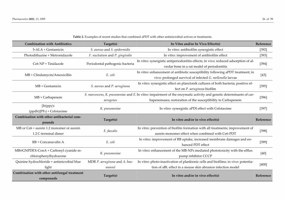
Pharmaceutics 2021, 13, 1995 26 of 59
Table 2. Examples of recent studies that combined aPDT with other antimicrobial actives or treatments.
Combination with Antibiotics Target(s) In Vitro and/or In Vivo Effect(s) Reference
5-ALA + Gentamicin S. aureus and S. epidermidis In vitro: antibiofilm synergistic effect [392]
Photodithazine + Metronidazole F. nucleatum and P. gingivalis In vitro: improvement of antibiofilm effect [393]
Ce6 NP + Tinidazole Periodontal pathogenic bacteria In vitro: synergistic antiperiodontitis effects; in vivo: reduced adsorption of al-
veolar bone in a rat model of periodontitis [394]
MB + Clindamycin/Amoxicillin E. coli In vitro: enhancement of antibiotic susceptibility following aPDT treatment; in
vivo: prolonged survival of infected G. mellonella larvae [43]
MB + Gentamicin S. aureus and P. aeruginosa In vitro: synergistic effect on planctonik cultures of both bacteria; positive ef-
fect on P. aeruginosa biofilm [395]
MB + Carbapenem S. marcescens, K. pneumoniae and E.
aerogenes
In vitro: impairment of the enzymatic activity and genetic determinants of car-
bapenemases; restoration of the susceptibility to Carbapenem [396]
[Ir(ppy)2
(ppdh)]PF6) + Cefotaxime K. pneumoniae In vitro: synergistic aPDI effect with Cefotaxime [397]
Combination with other antibacterial com-
pounds Target(s) In vitro and/or in vivo effect(s) Reference
MB or Ce6 + aurein 1.2 monomer or aurein
1.2 C-terminal dimer E. faecalis
In vitro: prevention of biofilm formation with all treatments; improvement of
aurein monomer effect when combined with Ce6-PDT [398]
RB + Concanavalin A E. coli In vitro: improvement of RB uptake, increased membrane damages and en-
hanced PDT effect [399]
MB@GNPDEX-ConA + Carbonyl cyanide m-
chlorophenylhydrazone K. pneumoniae
In vitro: enhancement of the MB-NPs mediated phototoxicity with the efflux
pump inhibitor CCCP [40]
Quinine hydrochloride + antimicrobial blue
light
MDR P. aeruginosa and A. bau-
mannii
In vitro: photo-inactivation of planktonic cells and biofilms; in vivo: potentia-
tion of aBL effect in a mouse skin abrasion infection model [400]
Combination with other antifungal treatment
compounds Target(s) In vitro and/or in vivo effect(s) Reference

Pharmaceutics 2021, 13, 1995 27 of 59
5-ALA + ITZ, itraconazole; TBF, terbinafine;
VOR, voriconazole
Candida species, dermatophytes, A.
fumigatus and F. monophora In vitro: reduction/improvement of lesions, disappearance of plaque [401]
Photodithazine + Nystatin Fluconazole-resistant C. albicans In vitro: reduction of fungal viability, decrease in oral lesions and inflamma-
tory reaction; in vivo: decrease in tongue lesions [54]
5-ALA + Itraconazole Trichosporon asahii In vitro: better elimination of planktonic and biofilms fungi than single ther-
apy [402]
Combination with immunotherapy Target(s) In vitro and/or in vivo effect(s) Reference
Schiff base complexes E. coli et S. aureus In vitro: blockage of the production of inflammatory TNFα cytokine [403]
Porphyrin + phtalocyanine HIV-infected cells In vitro: specific phototoxicity against infected cells [404]
Combination with sonodynamic therapy
(SDT) Target(s) In vitro and/or in vivo effect(s) Reference
Ce6 derivative Photodithazine + RB C. albicans In vitro: inactivation of biofilm (viability and total biomass) [405]
UCNPs + hematoporphyrin + SiO2-RB 1 Antibiotic-resistant bacteria In vitro: greater antibacterial effect with SDT and PDT at once [406]
Combination with electrochemotherapy Target(s) In vitro and/or in vivo effect(s) Reference
Hypericin E. coli and S. aureus In vitro: better bacterial inactivation with combined therapies [407]
Combination with viral NPs Target(s) In vitro and/or in vivo effect(s) Reference
TVP-A (luminogen) + PAP phage P. aeruginosa In vitro: synergistic bacterial recognizing and killing; in vivo: acceleration of
healing rates [408]
Pheophorbide A (chlorophyll) + JM-phage C. albicans In vitro: better specificity of PS targeting [409]
Ru(bpy2)phen-IA + Cowpea chlorotic mottle vi-
rus S. aureus In vitro: targeted bacterial photodynamic inactivation [410]
Combination of several PSs Target(s) In vitro and/or in vivo effect(s) Reference
Carboxypterin + MB K. pneumoniae In vitro: better biofilm eradication [411]
Phthalocyanines + Graphene QDs S. aureus In vitro: better bacterial photoinactivation [412]
ICG + Metformin + Curcumin E. faecalis In vitro: better biofilm eradication [35]
Porphyrin + Phthalocyanine Leishmania braziliensis In vitro: better assimilation of photo-inactivated parasites by macrophages [413]

Pharmaceutics 2021, 13, 1995 28 of 59
Combination with photothermal therapy
(PTT) Target(s) In vitro and/or in vivo effect(s) Reference
Ruthenium NPs Pathogenic bacteria In vitro: bacterial inhibition; in vivo: reduction of bacterial load and repair of
infected wounds [414]
Graphene oxide E. coli and S. aureus In vitro: efficient vector for both PDT and PTT [415]
ICG + SPIONs E. coli, K. pneumoniae, P. aeruginosa,
and S. epidermis In vitro: antimicrobial and antibiofilm activity at a low dose [148]
Ag-conjugated graphene QDs E. coli and S. aureus In vitro: efficient photoinactivation by PDT and PTT; in vivo: promoted heal-
ing in bacteria-infected rat wounds [280]
PDPPTT (photothermal agent) + MEH-PPV (PS)
2 E. coli In vitro: better inhibition rate than PTT/PDT systems used alone [324]
Mesoporous polydopamine NPs + ICG S. aureus In vivo: eradication of S. aureus biofilm on titanium implant [416]
Combination with NO phototherapy Target(s) In vitro and/or in vivo effect(s) Reference
N-(3-aminopropyl)-3-(trifluoromethyl)-4-nitro-
benzenamine + TMPyP/ZnPc E. coli In vitro: dual-mode photoantibacterial action [417]
Sulfonated polystyrene NPs (NO photodonor +
porphyrin/phthalocyanine) E. coli In vitro: strong antibacterial action [355]
[Ru(bpy)3]Cl2 P. aeruginosa In vitro: PDT/NO synergistic antibiofilm effect [418]
ALA, alanine; MB, methylene blue; RB, rose bengal; Ce6, chlorin e6; ICG, indocyanine green; SPION, superparamagnetic iron oxide NP. 1, hematoporphyrin monome-
thyl ether enclosed into yolk-structured up-conversion core and covalently linked RB on SiO2 shell; 2, photothermal agent poly(diketopyrrolopyrrole-thienothiophene)
(PDPPTT) and the photosensitizer poly(2-methoxy-5-((2-ethylhexyl)oxy)-p-phenylenevinylene) (MEH-PPV) in the presence of poly(styrene-co-maleic anhydride).
Pharmaceutics 2021, 13, 1995 29 of 59
5.1.“Basic” aPDT Combinations
5.1.1. Combination of Several PSs
The simplest combinatory aPDT approach probably consists in combining two or
more PSs in a single treatment. Thus, besides aPDT making use of a single PS, aPDT may
rely on the simultaneous use of several PSs in an attempt to obtain additive or synergistic
antimicrobial effects. The PSs combined may exhibit different photophysical characters
showing complementarities. For example, carboxypterin-based aPDT upon sunlight irra-
diation demonstrates a significant planktonic bacterial load reduction [419]. However,
eradication of biofilm formation needs a PS concentration 500 times higher than assays
performed with planktonic forms. When combined with MB, Tosato et al. showed that
reasonable concentrations of both PSs exert synergistic effect on both biofilm and plank-
tonic MDR bacteria [411]. In other studies, alternative dual-PSs aPDT systems have shown
even better antibacterial and antibiofilm properties [35,412].
5.1.2. Addition of Inorganic Salts
The combination of PSs with inorganic salts can modulate the PDT effectiveness, po-
tentiating or inhibiting the antimicrobial activity through the production of additional re-
active species or quenching 1O2 [420]. As an example, azide sodium can modulate aPDT
effectiveness by promoting or inhibiting the binding of bacteria with PSs, depending on
lipophilicity of the latter. Indeed, azide sodium is a 1O2 quencher, but can also produce
highly reactive azide radicals, via electron transfer from PS at the excited state. This has
been reported with many phenothiazinium dyes and also fullerenes [93,421,422]. Other
salts can amplify the bacterial killing mediated by MB-PDT such as potassium iodide (KI),
a very versatile salt, involved in the generation of short-lived reactive iodine radicals
(I•/I2•−) [423,424]; similar findings have been obtained when using KI with cationic BOD-
IPY derivatives [425] or porphyrin–Schiff base conjugates bearing basic amino groups
[426]. Potassium thiocyanate and potassium selenocyanate could also form reactive spe-
cies, such as the sulfur trioxide radical anion and selenocyanogen (SeCN)2, respectively
[427]. In addition, interactions between PSs and target microorganisms could be improved
thanks to inorganic salts. For example, calcium and magnesium cations can modulate the
electrostatic interaction between PSs and bacterial membranes [428]. It is worth noting
here that most of the above-mentioned studies were conducted under in vitro settings.
While inorganic salts can be useful to enhance in vitro or ex vivo aPDT effects, the use of
such additives in animals or human beings would need to be carefully examined, consid-
ering the dose to be used and potential concomitant side effects.
5.2. Combinations of aPDT with Other Antimicrobial Drugs or Antimicrobial Therapies
Various classes of antimicrobial drugs or other antimicrobial therapies, which action
does not depend on light, have been considered with regard to their aPDT compatibility.
5.2.1. Antibiotics
Antibiotherapy is an obvious complementary therapy to aPDT, which may allow ob-
taining stronger antimicrobial effects and/or restore antibiotic susceptibility. Combina-
tions of PSs with antibiotics have been investigated in numerous studies addressing vari-
ous infectious diseases, such as skin and mucosal infections as recently reviewed [429].
Aminosides are the most common antibiotics used in combination with a PS. For instance,
kanamycin, tobramycin, and gentamicin combined respectively to RB, porphyrin, and 5-
Alanine have been reported, showing potent effects against bacteria of clinical interest,
notably by improving biofilm clearance and reducing microbial loads [392,430–433]. In
addition, combinations with other antibiotic classes, such as nitroimidazoles (e.g., metro-
nidazole) or glycopeptides (e.g., vancomycine), also showed some effect against biofilms
of F. nucleatum and P. gingivalis [393], as well as S. aureus [434]. Further, PSs can also be
Pharmaceutics 2021, 13, 1995 30 of 59
useful to combine with antibiotics in order to restore the susceptibility of bacteria to the
latter, notably to last-resort antibiotics. As a recent example, Feng et al. reported the pho-
todynamic inactivation of bacterial carbapenemases, both restoring bacterial susceptibil-
ity to carbapenems and enhancing the effectiveness of these antibiotics [396]. However,
all combinations of PSs and antibiotics may not be effective, since in some cases antibiotics
can have an antagonistic effect on PS activity [435]. Finally, it is noteworthy that some
antibiotics can act themselves as PSs; Jiang et al. indeed reported light-excited antibiotics
for potentiating bacterial killing via ROS generation [436].
5.2.2. Antifungals
The combination of PSs with antifungals is a powerful approach to thwart growing
antifungal resistance, especially to fluconazole, which is commonly observed in C. albicans
strains [437]. Quiroga et al. showed that sublethal photoinactivation mediated by a tetra-
cationic tentacle porphyrin allowed to reduce the MIC of fluconazole in C. albicans [438].
Nystatin, a common antifungal used to prevent and treat candidiasis, was combined with
photodithazine-based PS and red light in C. albicans-infected mice. The combined therapy
reduced the fungal viability and decreased the oral lesions and the inflammatory reaction
[54]. Moreover, other antifungals (e.g., terbinafine, itraconazole and voriconazole) com-
bined to ALA reported promising results. These combinations could be alternative meth-
ods for the treatment of refractory and complex cases of chromoblastomycosis [401,439].
5.2.3. Other Antimicrobial Compounds
Many other antimicrobial compounds may be used in combination with PSs. Anti-
microbial peptides (AMPs) are oligopeptides (commonly consisting of 10–50 amino acids)
with high affinity for bacterial cells thanks to an overall positive charge. Their antimicro-
bial spectrum can be modulated through variation of their amino acids sequence. AMPs
encompass a large set of natural compounds that may be used in aPDT. For example, de
Freitas et al. reported that sub-lethal doses of PSs (either Ce6 or MB) and aurein peptides
(either aurein 1.2 monomer or aurein 1.2 C-terminal dimer) were able to prevent biofilm
development by E. faecalis [398]. In another study, a membrane-anchoring PS, named
TBD-anchor, demonstrated both bacterial membrane-anchoring abilities and ROS pro-
duction [440]. In a similar way to peptide therapy, LPS-binding proteins were used to
improve the contact of PSs with the cytoplasmic membrane of bacteria. For instance, the
antibacterial efficacy of a complex consisting of a combination of RB with the lectin conca-
navalin A (ConA) was demonstrated in a planktonic culture of E. coli; ConA-RB conjugates
increased membrane damages and enhanced the RB efficacy up to 117-fold [399]. In addi-
tion, coupling pump efflux inhibitors, such as CCCP EPI (carbonyl cyanide m-chloro-
phenylhydrazone), to PSs was also investigated. This highlighted the interest to combine
PSs with molecules acting on targets susceptible to induce resistance modulations [40].
Other antimicrobial molecules may be good candidates to be used in aPDT strategies. For
example, quinine used in combination with antimicrobial blue light was shown efficient
to photo-inactivate MDR P. aeruginosa and A. baumannii [400]. Some cationic molecules,
such as cationic lipids, can have a good affinity for bacterial cell membrane and a good
antibacterial activity [126,441]. Thus, PS-amphiphiles conjugates would also deserve to be
investigated for aPDT applications.
5.2.4. Viral NPs and Phagotherapy
In recent years, viral NPs (VNPs) deriving from phages, animal, or plant viruses have
been proposed as biological vehicles for delivery of PSs. Such carriers can exhibit a series
of advantageous properties including natural targeting, easy manufacture and good
safety profile [442]. Compared with nanomaterials used as PS carriers, VNPs are natural
protein-based NPs that may display higher biocompatibility and tissue specificity. In ad-
dition, VNPs can be tuned through genetic and synthetic engineering with appropriate

Pharmaceutics 2021, 13, 1995 31 of 59
biological and chemical modifications (e.g., surface decoration). Alternatively, the combi-
nation of aPDT with so-called phagotherapy may be useful for various reasons, following
different strategies. One study suggests that, following a PDT treatment, ROS damages
can cause quorum sensing and virulence pathway alterations rendering micro-organisms
more susceptible to other therapies. Among these, phagotherapy is known to be modu-
lated by multiple virulence factors [443]. Another approach can consist of conjugating
phages with PSs, in order to improve interaction/delivery of the latter into target micro-
organisms. For example, Dong et al. showed that a phage carrying the chlorophyll-based
PS pheophorbide efficiently induced apoptosis in C. albicans, thus demonstrating the po-
tential of phototherapeutic nanostructures for fungal inactivation [409]. However, such an
approach has to be prudently considered regarding the occurrence of phage resistance
already reported in numerous investigations [444].
5.3. Combinations of aPDT with Other Light-Based Treatments
Multiple modalities entirely controlled by light stimuli may be combined with aPDT
in multifunctional antimicrobial treatments. The following part reports some very recent
studies illustrating multiple light-based antimicrobial strategies combined to operate in-
dependently, with potential additive or synergistic effects and without reciprocal inter-
ferences. This may be obtained with a single PS displaying such multiple properties
and/or through combination of PSs with other light-activatable compounds exhibiting
complementary properties.
5.3.1. aPDT and Photothermal Therapy
Photothermal therapy (PTT) is a local treatment modality relying on the property of
a PS to absorb energy and convert it into heat upon stimulation with an electromagnetic
radiation, such as radiofrequency, microwaves, near-infrared irradiation, or visible light.
The localized hyperthermia can lead to various damages resulting in microbial inactiva-
tion in the treatment area. Following this principle, ruthenium NPs have been used for
PDT/PTT dual-modal phototherapeutic killing of pathogenic bacteria [414]. Moreover,
GO demonstrated antibacterial effect against E. coli and S. aureus as a result of both PDT
and PTT effects following irradiation with ultra-low doses (65 mW/cm2) of 630 nm light
[415]. Furthermore, combination of sonodynamic, photodynamic, and photothermal ther-
apies with an external controllable source recently reported against breast cancer [445]
may also show promising applications for treating bacterial infection. Mai et al. reported
a FDA-approved sinoporphyrin sodium (DVDMS) for photo- and sono-dynamic therapy
in cancer cells and photoinactivation of S. aureus strains, in in vitro and in vivo models.
However, no bacterial sonoinactivation by DVDMS was obtained [446].
5.3.2. aPDT and NO Phototherapy
Combination of aPDT with NO phototherapy is gaining increasing interest for anti-
microbial applications [447]. For instance, light-responsive dual NO and 1O2 releasing ma-
terials showed phototoxicities against E. coli [355,417]. More recently, Parisi et al. devel-
oped a molecular hybrid based on a BODIPY light-harvesting antenna producing simul-
taneously NO and 1O2 upon single photon excitation with green light for anticancer ap-
plications; according to the authors, this system may also act as an effective PS and NO
photodonor antibacterial agent [448].
5.3.3. aPDT and Low Laser Therapy
Photobiomodulation (PBM), also called low-level laser therapy, is a non-destructive
process that may alleviate pain and inflammation or promote tissue healing and regener-
ation. The use of this method coupled to aPDT is a very recent approach. A concomitant
use of aPDT and PBM was reported as an adjunct treatment for palatal ulcer [449]. In a

Pharmaceutics 2021, 13, 1995 32 of 59
clinic-laboratory study, aPDT and PBM showed similar improvement in gingival inflam-
matory and microbiological parameters compared with conventional treatment [450].
More recently, some benefits of this combined therapy were reported such as the modu-
lation of inflammatory state, pain relief, and acceleration of tissue repair of patients con-
tracting herpes simplex labialis virus or orofacial lesions in patients suffering from COVID-
19 [451–453].
5.4. Coupling of aPDT with Other Physical Treatments
5.4.1. aPDT and Sonodynamic Therapy
Sonodynamic therapy (SDT), combining so-called sonosensitizers (SS) and ultra-
sounds (US) is a relatively new approach for treating microbial infections [454,455]. The
ultrasonic waves have the property to induce a cavitation phenomenon thus enhancing
the efficacy of combined antimicrobial treatments. The rationale for combining PDT with
SDT relies on specific advantages of the latter, notably, a deeper propagation of US into
the tissue than light; therefore, PDT/SDT may be used to treat deeper lesions in vivo, alle-
viating the limitations of light propagation and delivery presented by aPDT [405,456]. An
approach combining PDT and SDT, called sonophotodynamic therapy (SPDT), has been
reported to improve microbial inactivation compared with individual aPDT or SDT [457].
Because of the complicated system of SPDT, its mechanisms have not been clearly re-
vealed yet. Some studies have demonstrated that sonoporation mechanism induced by
US improves the transfer of large molecules into the bacteria by forming transient pores.
Moreover, US waves could potentiate the microorganisms dispersion in the medium re-
sulting in (i) a better biodisponibility of therapeutic agent and light diffusion and (ii) a
reduction of microbial aggregation and networks, such as biofilm [457,458]. The mention
of a new class of PS characterized by the dual ability to be activated by both US and light,
for SPDT application, has been questioned. Indeed, Harris et al. recently suggested that
this specific PS/SS class could be useful for antimicrobial application – beside previously
reported anticancer application – with initial investigation using chlorins as dual PS/SS
agent [459]. Since this study by Harris et al., a few dual-activated PS/SS have been de-
scribed that would warrant further investigations [460,461].
5.4.2. aPDT and Electrochemotherapy
Electrochemotherapy, also called pulsed electromagnetic fields (PEMFs) or elec-
tropermeabilization, is a method consisting in applying an electrical field to cells in order
to enhance their permeability to therapeutic molecules (often chemical drugs or DNA).
Combination of PDT with electrochemotherapy has been used many times to treat cancer
diseases [14,462,463]. One study showed that, compared with aPDT used alone, hypericin
combined with electrochemotherapy allows to achieve more than 2 to 3 log10 CFU reduc-
tion in E. coli and S. aureus, respectively [407]. To our knowledge, no other study combin-
ing electrochemotherapy with aPDT was recorded since then. However, combination of
aPDT and a cell-permeabilization technique with a controllable toxicity degree may be
highly relevant since most PSs can act in extracellular medium without having a specific
target. Accordingly, electrochemotherapy may allow boosting aPDT activity by promot-
ing PSs internalization into target microorganisms.
5.5. aPDT and Other Antimicrobial-Related Therapies
Immunotherapeutic effects may be obtained as a result of PDT itself or due to other
treatments used in combination. For instance, Schiff base complexes with differential im-
mune-stimulatory and immune-modulatory activities were reported efficient to eliminate
both Gram(+) and Gram(−) bacteria. Furthermore, upon photoactivation, these complexes
blocked the production of the inflammatory cytokine TNFα, thereby allowing to treat at
once bacterial infections associated with damaging inflammation [403]. One recent study
proposed the first application of antimicrobial photoimmunotherapy (PIT) by developing

Pharmaceutics 2021, 13, 1995 33 of 59
a PS-antibody complex, selective to the HIV antigen anchored to the infected cell mem-
branes [404]. Such an approach supports the therapeutic applicability of PDT against an-
timicrobial infections, especially those mediated by intracellular pathogens. In addition,
photodynamic therapy using PSs at sub-lethal concentrations may exhibit interesting
properties for inflammatory and infectious conditions [464]. It was shown effective to alter
immune cell function and alleviate immune-mediated disease, to hasten the process of
wound healing, and to enhance antibacterial immunity. PDT thus appears as a promising
therapeutic modality in infectious and chronic inflammatory diseases such as inflamma-
tory bowel disease and arthritis.
6. Other aPDT Perspectives: New Strategies to Efficiently Target Bacteria
Irrespective of the biomedical applications, achieving a precise targeting is crucial to
guarantee both efficiency and specificity. For anticancer PDT, many targeting studies have
been done, notably for evaluating PSs covalently attached to molecules having affinity for
neoplasia or ligands for receptors expressed on tumors. By this way, PSs may be chosen
considering primarily their ability to achieve high PDT effects rather than depending on
their intrinsic targeting properties. Following the same rationale, aPDT-based combina-
tory systems can be developed, benefiting from earlier studies performed in multimodal
oncology [465].
6.1. Aggregation-Induced Emission (AIE) Luminogens
AIE luminogens exhibit, in the aggregated state, nonradiative decay and show bright
fluorescence due to the restriction of intramolecular motions [9]. Recently, their interests
for antimicrobial applications have been reported, showing the possibility to simultane-
ously perform detection and image-guided elimination of bacteria for theranostics appli-
cations [466]. In comparison with classical PSs, AIE luminogens in an aggregated state do
not exhibit self-quenched fluorescence and ROS production is better. For instance, Gao et
al. reported a tetraphenylethylene-based discrete organoplatinum(II) metallacycle electro-
statically assembled with a peptide-decorated virus coat protein. This assembly showed
strong membrane-intercalating ability, especially in Gram(−) bacteria, and behaved as a
potent AIE-PS upon light irradiation [467].
6.2. Photochemical Internalization (PCI)
PCI may be used to enhance cell internalization of diverse macromolecules. It con-
sists of PDT-induced disruption of endocytic vesicles and lysosomes improving the re-
lease of their payloads into the cytoplasm of target cells. Although most PCI applications
relate to cancer treatments, PCI could be extended to treat intracellular infections by de-
livering antimicrobials into infected cells [468]. For instance, Zhang et al. reported PCI as
an antibiotic delivery strategy allowing to enhance cytoplasmic release of Gentamicin, to
counter intracellular staphylococcal infection in eukaryotic cells and in zebrafish embryos
[469].
6.3. Genetically-Encoded PSs
Internalization of PSs inside target microorganisms could be facilitated thanks to
their conjugation with adjuvants, as mentioned before. Alternatively, it could be possible
to use genetically-encoded ROS-generating proteins (RGPs), also called genetically-en-
coded PSs. Such an approach represents a powerful way to “completely localize” PSs in-
side target microorganisms for highly specific antimicrobial phototoxicity. Furthermore,
in situ production of RGPs allows to enhance interaction with intracellular targets and
better control the biodistribution of PSs, while limiting side-effects for the host tissues and
environments [470]. To date, two groups of RGPs have been reported; those that belong
to the green fluorescent protein (GFP) family and form their chromophores auto catalyti-
cally, and those that use external ubiquitous co-factors (flavins) as chromophores [471].
Pharmaceutics 2021, 13, 1995 34 of 59
For example, Endres et al. compared eleven light-oxygen-voltage-based flavin binding
fluorescent proteins and showed that most were potent PSs for light-controlled killing of
bacteria [472].
6.4. pH-Sensitive aPDT
Some studies have reported smart photoactive systems consisting in PSs assembled
in nanoconjugates with acid-cleavable linkers. For instance, Staegemann et al. described
porphyrins conjugated with acid-labile benzacetal linkers and demonstrated the cleavage
of the active PS agents from the polymer carrier in the acidic bacterial environment [316].
In addition, photoacids may be useful to design pH-sensitive aPDT systems. Upon light
irradiation, such agents promote the spatial and temporal control of proton-release pro-
cesses and could provide a way to convert photoenergy into other types of energy [473].
Thus, proof-of-concept was reported for the use of reversible photo-switchable chemicals
as antimicrobials inducing MDR bacteria photoinactivation mediated by the acidification
of intercellular environment [474]. To our knowledge, no studies have yet reported the
potential of photoacids in combination with aPDT systems. However, some pH-sensitive
PSs can induce remarkable variations of antimicrobial photoinactivation levels under dif-
ferent environmental pH [475]. These observations suggest the potential of photoacids as
PDT potentiators for enhanced antimicrobial applications.
6.5. DNA Origami as PS Carriers
The quite recent development of DNA origami based on well-established DNA nan-
otechnology can serve as an excellent scaffold for the functionalization with different
kinds of molecules and could be a powerful tool, as described by Yang at al., to study in
a real-time conditions the assemble/disassemble of photo-controllable nanostructures
[476]. Oligonucleotides organized as DNA origami could thus be used as PS-carrying
nanostructures featuring numerous and dense intercalation sites. In addition, the tightly
packed double helices can avoid the degradation by DNA hydrolases in the cellular envi-
ronment. For instance, Zhuang et al. reported the uptake in tumor cells of a PS-loaded
DNA origami nanostructure where it generated free radicals, releasing PSs due to DNA
photocleavage, and induced cell apoptosis [477]. To our knowledge, such an approach has
not yet been investigated for antimicrobial purposes.
7. Discussion
Antimicrobial PDT has the potential to fight against a wide spectrum of infective
agents, including those resistant to conventional antimicrobials, under non-clinical and
clinical settings. Rather than replacement, aPDT may be a complementary approach to
reduce the use of current, especially last resort, antimicrobials. This review aims to give a
non-exhaustive overview of the diversity and richness of synthetic, natural, or hybrid sin-
gle PSs and aPDT nanosystems that were recently reported, with respect to their specific
advantages, limitations, and possible evolutions. It is noteworthy that many systems and
strategies primarily developed for anticancer PDT have been or could be applied—per se
or following adaptations—to antimicrobial applications.
Beyond the “chemical space” that can be explored with individual PSs, versatile com-
binations with other compounds can allow the design of multimodular/multimodal sys-
tems. Along this line, the various PSs available may be considered as “basic ingredients”.
Apart from offering alternative possibilities for overcoming the most common limitations
of PSs (i.e., solubility, delivery, and specificity), reasons for implementing PSs in complex
systems can be related to (i) the ability to target several types of pathogens at once (ex-
tended antimicrobial spectrum), (ii) synergistic antimicrobial effects, (iii) reduction in the
dose of each combined component, (iv) beneficial effects in severe poly-pathogenic infec-
tions, and (v) reduction in the risk of resistance emergence.

Pharmaceutics 2021, 13, 1995 35 of 59
In addition to the many possible variations concerning PSs, optimizations can also
consider other critical parameters in PDT, namely light irradiation and oxygenation. The
latter was considered for a long time as an indispensable component. However, control of
the oxygen level in the aPDT system is questionable. Indeed, additional oxygen-independ-
ent phototoxic mechanisms have been reported, for example with psoralens, which can
produce more effective aPDT without oxygen [21]. Furthermore, recent studies suggest
that various strategies could be used to reduce or bypass the limitations of oxygen and
light supplies (read below). All combined, optimizations targeting not only the PSs, but
also light irradiation and oxygen supply, could allow to evolve toward integrative, highly
sophisticated, antimicrobial photodynamic therapy.
Light irradiation and its various modalities have been reviewed in depth by different
authors [12,478–480]. Typically, the irradiation in PDT occurs in the UV (200–400 nm) or
in the visible light (400–700 nm) with a power of ≤ 100 mW [479]. However, the low light-
penetration depth (around mm) and possible occurrence of tissue photodamage limit the
applicability of this spectral range for PDT. This can be circumvented by application of a
near-infrared (NIR) irradiation (750–1100 nm), particularly via a two-photon excitation,
which is emerging for PDT applications [223,481]. Being a third-order nonlinear optic phe-
nomenon and corresponding to the simultaneous absorption of two photons with half the
resonant energy, it allows deeper penetration in biological tissues (around 2 cm), lower
scattering losses, and a three-dimensional spatial resolution [482]. Light sources are also
constantly improving. Laser is an exceptional source of radiation, capable of producing
extremely fine spectral bands, intense, coherent electromagnetic fields ranging from NIR
to UV. In comparison, LEDs feature other advantages, notably the possibility to be ar-
ranged in many ways, in large quantity, for irradiating wide areas while inducing negli-
gible heating [478,479].
Considering that light-emitted sources can become a brake to any PDT applications,
several promising options have been envisaged quite recently. Notably, Blum et al. have
identified a series of “self-exiting way” that allows to abolish the need of light to achieve
efficient PDT [483]. Among them, chemiluminescence was extensively investigated by us-
ing luminol or luciferase energy transfer to induce a chemiexcitation of PS as antibacterial
therapeutics [484]. In addition, other methods may be based on Cherenkov radiation,
which occurs when an emitted charged particle, such as an electron, moves with high
speed through a dielectric medium, such as water. Thus, the polarization of electrons in
the medium produces electromagnetic waves in the visible wavelengths that could acti-
vate PDT reaction. Another way to induce Cherenkov radiation is to use radioactive iso-
topes with high beta emissions (e.g., 18F, 64Cu, or 68Ga) as an electronic excitation source
[483]. The relevance of such approaches for the design of a self-induced aPDT system re-
mains to be evaluated. In addition, another external source of excitation, such as micro-
waves, could activate photoactive molecules such as Fe3O4 when they are complexed with
carbon nanotubes. This was recently evaluated by Qiao et al. for treating MRSA-infected
osteomyelitis [48]. Furthermore, X-ray as an activated source can also facilitate the activa-
tion of the PDT system by transferring energy harvested from X-ray irradiation to the PS
used [485]. Kamanli et al. compared pulse and superpulse radiation modes, showing that
the latter is more effective to produce 1O2 and S. aureus eradication than the former [486].
Oxygen is also a critical limiting factor that determines PDT efficiency, especially in
poorly oxygenated environments. However, it is noteworthy that PDT can be achieved in
cancers that typically feature a low oxygenation rate. To alleviate the possible limitations
of oxygen supply in PDT systems, several strategies have also been recently considered.
One is based on catalase grafting to achieve oxygen self-sufficient NPs in order to convert
H2O2 into available dissolved oxygen in the tumor environment. The abundant ROS in
tumors compared with normal tissues provide a coherent substrate for catalase and thus
allows an improvement of PDT activity [487]. Some multifunctional nanomaterials, called
nanozymes, can also be used in combination with PSs to achieve a catalase-like activity

Pharmaceutics 2021, 13, 1995 36 of 59
supplying an oxygen source for the PS functioning [488,489]. In addition, it was also re-
ported that noble metal NPs – such as Ru, Pt, and Au NPs – exhibit catalase-like nanozyme
activities [490]. The use of catalases or nanozymes may have the crucial role of oxygen
helpers in aPDT for treating deep infections.
The possibilities to design combinatorial aPDT strategies seem unlimited. Those pre-
sented in this review partly overlap with the description of aPDT systems, showing the
complexity of any classification process. Awareness and caution may be raised about
some sort of “paradox” or “dilemma”; indeed, while the seemingly boundless collection
of chemical options and modular tools to develop nanoscale aPDT therapeutics implicitly
defines extensive design flexibility, it staggeringly complicates and bewilders at the same
time the optimization process aiming to compare, rationalize, and identify the “best” op-
tion for each application. Along this line, data concerning structure-activity relationships
are usually missing, with not many studies yet dedicated to this matter [11,42,91,491].
Similar to the CLSI guidelines defined for antibiotics, uniform research methodologies
would be useful to assay aPDT systems under well-defined standards and guidelines,
considering notably (i) illumination settings, (ii) positive controls [492], (iii) microorgan-
isms and cell lines relevant for a given application, and (iv) assessment of antimicrobial
efficacy; this would guarantee better-conducted preclinical and clinical trials of aPDT sys-
tems used as mono or combinatory therapy. Furthermore, a public database compiling
the efficacy/side effects of various systems would also be useful, facilitating meta-analyses
for delineating (quantitative) structure/activity relationships and computational simula-
tions [493].
Finally, in view of translation to clinical practice, a series of precautions and potential
limitations must also be considered, especially when dealing with combinatory strategies.
Beside possible reciprocal interferences (antagonisms) between combined partners, safety
and specificity parameters must also be carefully examined. One main advantage of aPDT
is the possibility to control the production of ROS thanks to the use of nontoxic PSs trig-
gered with inducers (specific light and free oxygen) and/or enhancers. The vast majority
of PSs are considered safe and dark cytotoxic side effects toward non-target eukaryotic
cells are rarely reported. However, in many cases, more studies are needed for examin-
ing—beyond potential side-effects—other parameters such as bioavailability, biodisper-
sion, persistence in host cells and body, and elimination pathways. With regard to combi-
natory strategies, it is noteworthy that studies conducted to date were mostly based on in
vitro evaluation or using animal models bearing well-defined sites of infection (Table 2).
Thus, much more data in preclinical and clinical settings are required to support the actual
potential of such strategies. Moreover, considering that many microbial infections are sys-
temic, the use of modalities such as sonodynamic therapy or electrochemotherapy is at
present not realistically feasible. Therefore, in spite of significant progress and real prom-
ise, further important work/innovations are needed to effectively broaden the range of
infectious conditions that could be treated via aPDT approaches. Lastly, cost effectiveness
of multiplexed aPDT therapies over monomodal conventional antimicrobial agents is an-
other crucial point to be considered in view of clinical applications. Potent, but too expen-
sive solutions may fail to be used in practice, especially in under-developed countries
where conventional antimicrobials will continue to be used, allowing microorganisms to
increase in resistance.
8. Concluding Remarks
In conclusion, aPDT is a versatile approach that tends to evolve from quite a simple
method to reach much higher degrees of complexity, with several expected advantages,
but also possible drawbacks or undesirable effects. Among the latter, any direct/indirect
impact on AMR should be more thoroughly considered. It is noteworthy that aPDT clini-
cal trials conducted to date evaluated quite simple PSs. Any increase in the complexity of
therapeutic systems would lead to an increase in difficulty before being able to reach clin-

Pharmaceutics 2021, 13, 1995 37 of 59
ical applications. The development of effective and safe aPDT treatments requires exper-
tise in many fields of research, including biology (microbiology, cell biology, biochemis-
try, pharmacology), chemistry, physics (optical physics), and engineering. This is even
more the case with combinatory strategies involving different modalities as reviewed in
this article. Translation to practical applications also implies strong collaborations with
the different sectors of health care and pharmaceutical companies. In these conditions,
aPDT and its many therapeutic combinations could become a frontline routine treatment
to fight against microorganisms possibly responsible for the next healthcare crises [61].
Funding: This work was supported by grants from ANR/BMBF (TARGET-THERAPY; PIs: Tony Le
Gall and Holger Schönherr; ANR grant number: ANR-20-AMRB-0009, RPV21103NNA; BMBF
Förderkennzeichen: 16GW0342). We also thank the “Association de Transfusion Sanguine et de Bi-
ogénétique Gaétan Saleün” (France) and the “Conseil Régional de Bretagne” (France) for their fi-
nancial support. Raphaëlle Youf is recipient of a PhD fellowship from the French “Ministère de
l’Enseignement supérieur, de la Recherche et de l’Innovation” (Paris, France).
Conflicts of Interest: The authors declare no conflict of interest. The funders had no role in the
design of the study; in the collection, analyses, or interpretation of data; in the writing of the manu-
script, or in the decision to publish the results.
Abbreviations
AIE aggregation-induced emission
ALA alanine
AMP antimicrobial peptide
AMR antimicrobial resistance
aPDT antimicrobial PDT
ConA concanavalin A
CNTs carbon nanotubes
Ce6 chlorin e6
CFU colony forming unit
e− electron
ESKAPE Enterococcus faecium, Staphylococcus aureus, Klebsiella pneumoniae, Acinetobacter
baumannii, Pseudomonas aeruginosa, and Enterobacter spp.
IC internal conversion
ICG indocyanine green
ISC inter-system crossing
GO graphene oxide
H2O2 hydrogen peroxide
HO● hydroxyl radical
MB methylene blue
MDR multidrug resistance
MRSA methicillin resistant S. aureus
MOF metal organic framework
NIR near infrared
NO nitic oxide
NP nanoparticle
O2 dioxygen
O2●− superoxide anion radical 1O2 singlet oxygen 3O2 ground state molecular oxygen
PDT photodynamic therapy
PS photosensitizer
PS•− PS radical anion
Pharmaceutics 2021, 13, 1995 38 of 59
1PS PS in the ground state 1PS* PS in a first excited singlet state 3PS* PS in a triplet excited state
PEG poly(ethylene glycol)
PCI photochemical internalization
PCL poly(ε-caprolactone)
PLA poly(lactic acid)
pSi porous silicon
PTT photothermal therapy
QD quantum dot
R reduced molecule
R●+ oxidized molecule
RB rose bengal
ROS reactive oxygen species
SS sonosensitizer
SDT sonodynamic therapy
SPDT sonophotodynamic therapy
SPION superparamagnetic iron oxide NP
TB(O) toluidine blue
UCNP upconversion NP
UV ultraviolet
WHO world health organisation
References
1. Laxminarayan, R.; Matsoso, P.; Pant, S.; Brower, C.; Røttingen, J.-A.; Klugman, K.; Davies, S. Access to Effective Antimicrobials:
A Worldwide Challenge. Lancet 2016, 387, 168–175, doi:10.1016/S0140-6736(15)00474-2.
2. Mulani, M.S.; Kamble, E.E.; Kumkar, S.N.; Tawre, M.S.; Pardesi, K.R. Emerging Strategies to Combat ESKAPE Pathogens in the
Era of Antimicrobial Resistance: A Review. Front. Microbiol. 2019, 10, 539, doi:10.3389/fmicb.2019.00539.
3. Antoñanzas, F.; Goossens, H. The Economics of Antibiotic Resistance: A Claim for Personalised Treatments. Eur. J. Health Econ.
2019, 20, 483–485, doi:10.1007/s10198-018-1021-z.
4. Cassini, A.; Högberg, L.D.; Plachouras, D.; Quattrocchi, A.; Hoxha, A.; Simonsen, G.S.; Colomb-Cotinat, M.; Kretzschmar, M.E.;
Devleesschauwer, B.; Cecchini, M.; et al. Attributable Deaths and Disability-Adjusted Life-Years Caused by Infections with
Antibiotic-Resistant Bacteria in the EU and the European Economic Area in 2015: A Population-Level Modelling Analysis. Lan-
cet Infect. Dis. 2019, 19, 56–66, doi:10.1016/S1473-3099(18)30605-4.
5. Dadgostar, P. Antimicrobial Resistance: Implications and Costs. Infect. Drug Resist. 2019, 12, 3903–3910,
doi:10.2147/IDR.S234610.
6. Hutchings, M.I.; Truman, A.W.; Wilkinson, B. Antibiotics: Past, Present and Future. Curr. Opin. Microbiol. 2019, 51, 72–80,
doi:10.1016/j.mib.2019.10.008.
7. Wainwright, M.; Maisch, T.; Nonell, S.; Plaetzer, K.; Almeida, A.; Tegos, G.P.; Hamblin, M.R. Photoantimicrobials—Are We
Afraid of the Light? Lancet Infect. Dis. 2017, 17, e49–e55, doi:10.1016/S1473-3099(16)30268-7.
8. Daniell, M.D.; Hill, J.S. A History of Photodynamic Therapy. Aust. N. Z. J. Surg. 1991, 61, 340–348, doi:10.1111/j.1445-
2197.1991.tb00230.x.
9. Shi, X.; Zhang, C.Y.; Gao, J.; Wang, Z. Recent Advances in Photodynamic Therapy for Cancer and Infectious Diseases. WIREs
Nanomed. NanoBiotechnol. 2019, 11, e1560, doi:10.1002/wnan.1560.
10. Sabino, C.P.; Wainwright, M.; Ribeiro, M.S.; Sellera, F.P.; dos Anjos, C.; Baptista, M.d.S.; Lincopan, N. Global Priority Multidrug-
Resistant Pathogens Do Not Resist Photodynamic Therapy. J. Photochem. Photobiol. B Biol. 2020, 208, 111893, doi:10.1016/j.jpho-
tobiol.2020.111893.
11. Le Gall, T.; Lemercier, G.; Chevreux, S.; Tücking, K.-S.; Ravel, J.; Thétiot, F.; Jonas, U.; Schönherr, H.; Montier, T. Ruthenium(II)
Polypyridyl Complexes as Photosensitizers for Antibacterial Photodynamic Therapy: A Structure-Activity Study on Clinical
Bacterial Strains. ChemMedChem 2018, 13, 2229–2239, doi:10.1002/cmdc.201800392.
12. Cieplik, F.; Deng, D.; Crielaard, W.; Buchalla, W.; Hellwig, E.; Al-Ahmad, A.; Maisch, T. Antimicrobial Photodynamic Ther-
apy—What We Know and What We Don’t. Crit. Rev. Microbiol. 2018, 44, 571–589, doi:10.1080/1040841X.2018.1467876.
13. Hu, X.; Huang, Y.-Y.; Wang, Y.; Wang, X.; Hamblin, M.R. Antimicrobial Photodynamic Therapy to Control Clinically Relevant
Biofilm Infections. Front. Microbiol. 2018, 9, 1299. doi:10.3389/fmicb.2018.01299.

Pharmaceutics 2021, 13, 1995 39 of 59
14. Kwiatkowski, S.; Knap, B.; Przystupski, D.; Saczko, J.; Kędzierska, E.; Knap-Czop, K.; Kotlińska, J.; Michel, O.; Kotowski, K.;
Kulbacka, J. Photodynamic Therapy—Mechanisms, Photosensitizers and Combinations. Biomed. Pharmacother. 2018, 106, 1098–
1107, doi:10.1016/j.biopha.2018.07.049.
15. Wozniak, A.; Grinholc, M. Combined Antimicrobial Activity of Photodynamic Inactivation and Antimicrobials—State of the
Art. Front. Microbiol. 2018, 9, 930, doi:10.3389/fmicb.2018.00930.
16. Baier, J.; Maier, M.; Engl, R.; Landthaler, M.; Bäumler, W. Time-Resolved Investigations of Singlet Oxygen Luminescence in
Water, in Phosphatidylcholine, and in Aqueous Suspensions of Phosphatidylcholine or HT29 Cells. J. Phys. Chem. B 2005, 109,
3041–3046, doi:10.1021/jp0455531.
17. Maisch, T.; Baier, J.; Franz, B.; Maier, M.; Landthaler, M.; Szeimies, R.-M.; Bäumler, W. The Role of Singlet Oxygen and Oxygen
Concentration in Photodynamic Inactivation of Bacteria. Proc. Natl. Acad. Sci. USA 2007, 104, 7223–7228,
doi:10.1073/pnas.0611328104.
18. Ogilby, P.R. Singlet Oxygen: There Is Indeed Something New under the Sun. Chem. Soc. Rev. 2010, 39, 3181,
doi:10.1039/b926014p.
19. Foote, C.S. Definition of Type I and Type II Photosensitized Oxidation. Photochem. Photobiol. 1991, 54, 659–659, doi:10.1111/j.1751-
1097.1991.tb02071.x.
20. Baptista, M.d.S.; Cadet, J.; Di Mascio, P.; Ghogare, A.A.; Greer, A.; Hamblin, M.R.; Lorente, C.; Nunez, S.C.; Ribeiro, M.S.;
Thomas, A.H.; et al. Type I and II Photosensitized Oxidation Reactions: Guidelines and Mechanistic Pathways. Photochem. Pho-
tobiol. 2017, 93, 912–919, doi:10.1111/php.12716.
21. Hamblin, M.R.; Abrahamse, H. Oxygen-Independent Antimicrobial Photoinactivation: Type III Photochemical Mechanism?
Antibiotics 2020, 9, 53, doi:10.3390/antibiotics9020053.
22. Quiroga, E.D.; Cormick, M.P.; Pons, P.; Alvarez, M.G.; Durantini, E.N. Mechanistic Aspects of the Photodynamic Inactivation
of Candida Albicans Induced by Cationic Porphyrin Derivatives. Eur. J. Med. Chem. 2012, 58, 332–339,
doi:10.1016/j.ejmech.2012.10.018.
23. Wiehe, A.; O’Brien, J.M.; Senge, M.O. Trends and Targets in Antiviral Phototherapy. Photochem. Photobiol. Sci. 2019, 18, 2565–
2612, doi:10.1039/c9pp00211a.
24. Beirão, S.; Fernandes, S.; Coelho, J.; Faustino, M.A.F.; Tomé, J.P.C.; Neves, M.G.P.M.S.; Tomé, A.C.; Almeida, A.; Cunha, A.
Photodynamic Inactivation of Bacterial and Yeast Biofilms with a Cationic Porphyrin. Photochem. Photobiol. 2014, 90, 1387–1396,
doi:10.1111/php.12331.
25. Garcez, A.S.; Núñez, S.C.; Azambuja, N.; Fregnani, E.R.; Rodriguez, H.M.H.; Hamblin, M.R.; Suzuki, H.; Ribeiro, M.S. Effects
of Photodynamic Therapy on Gram-Positive and Gram-Negative Bacterial Biofilms by Bioluminescence Imaging and Scanning
Electron Microscopic Analysis. Photomed. Laser Surg. 2013, 31, 519–525, doi:10.1089/pho.2012.3341.
26. Alves, E.; Faustino, M.A.; Neves, M.G.; Cunha, A.; Tome, J.; Almeida, A. An Insight on Bacterial Cellular Targets of Photody-
namic Inactivation. Future Med. Chem. 2014, 6, 141–164, doi:10.4155/fmc.13.211.
27. Mesquita, M.Q.; Dias, C.J.; Neves, M.G.P.M.S.; Almeida, A.; Faustino, M.A.F. Revisiting Current Photoactive Materials for An-
timicrobial Photodynamic Therapy. Molecules 2018, 23, 2424, doi:10.3390/molecules23102424.
28. Lam, M.; Jou, P.C.; Lattif, A.A.; Lee, Y.; Malbasa, C.L.; Mukherjee, P.K.; Oleinick, N.L.; Ghannoum, M.A.; Cooper, K.D.; Baron,
E.D. Photodynamic Therapy with Pc 4 Induces Apoptosis of Candida Albicans. Photochem. Photobiol. 2011, 87, 904–909,
doi:10.1111/j.1751-1097.2011.00938.x.
29. Konopka, K.; Goslinski, T. Photodynamic Therapy in Dentistry. J. Dent. Res. 2007, 86, 694–707, doi:10.1177/154405910708600803.
30. Hirakawa, K.; Ota, K.; Hirayama, J.; Oikawa, S.; Kawanishi, S. Nile Blue Can Photosensitize DNA Damage through Electron
Transfer. Chem. Res. Toxicol. 2014, 27, 649–655, doi:10.1021/tx400475c.
31. Choi, S.S.; Lee, H.K.; Chae, H.S. In Vitro Photodynamic Antimicrobial Activity of Methylene Blue and Endoscopic White Light
against Helicobacter Pylori 26695. J. Photochem. Photobiol. B Biol. 2010, 101, 206–209, doi:10.1016/j.jphotobiol.2010.07.004.
32. Abdulrahman, H.; Misba, L.; Ahmad, S.; Khan, A.U. Curcumin Induced Photodynamic Therapy Mediated Suppression of
Quorum Sensing Pathway of Pseudomonas Aeruginosa: An Approach to Inhibit Biofilm In Vitro. Photodiagn. Photodyn. Ther. 2020,
30, 101645, doi:10.1016/j.pdpdt.2019.101645.
33. Boluki, E.; Moradi, M.; Azar, P.S.; Fekrazad, R.; Pourhajibagher, M.; Bahador, A. The Effect of Antimicrobial Photodynamic
Therapy against Virulence Genes Expression in Colistin-Resistance Acinetobacter Baumannii: APDT in Genes Expression of Col-
istin-Resistance A. Baumannii. Laser Ther. 2019, 28, 27–33, doi:10.5978/islsm.28_19-OR-03.
34. Ghorbanzadeh, R.; Assadian, H.; Chiniforush, N.; Parker, S.; Pourakbari, B.; Ehsani, B.; Alikhani, M.Y.; Bahador, A. Modulation
of Virulence in Enterococcus Faecalis Cells Surviving Antimicrobial Photodynamic Inactivation with Reduced Graphene Oxide-
Curcumin: An Ex Vivo Biofilm Model. Photodiagn. Photodyn. Ther. 2020, 29, 101643, doi:10.1016/j.pdpdt.2019.101643.
35. Pourhajibagher, M.; Plotino, G.; Chiniforush, N.; Bahador, A. Dual Wavelength Irradiation Antimicrobial Photodynamic Ther-
apy Using Indocyanine Green and Metformin Doped with Nano-Curcumin as an Efficient Adjunctive Endodontic Treatment
Modality. Photodiagn. Photodyn. Ther. 2020, 29, 101628, doi:10.1016/j.pdpdt.2019.101628.
36. Marasini, S.; Leanse, L.G.; Dai, T. Can Microorganisms Develop Resistance against Light Based Anti-Infective Agents? Adv.
Drug Deliv. Rev. 2021, 175, 113822, doi:10.1016/j.addr.2021.05.032.
37. Tschowri, N.; Lindenberg, S.; Hengge, R. Molecular Function and Potential Evolution of the Biofilm-Modulating Blue Light-
Signalling Pathway of Escherichia Coli. Mol. Microbiol. 2012, 85, 893–906, doi:10.1111/j.1365-2958.2012.08147.x.

Pharmaceutics 2021, 13, 1995 40 of 59
38. Prates, R.A.; Kato, I.T.; Ribeiro, M.S.; Tegos, G.P.; Hamblin, M.R. Influence of Multidrug Efflux Systems on Methylene Blue-
Mediated Photodynamic Inactivation of Candida Albicans. J. Antimicrob. Chemother. 2011, 66, 1525–1532, doi:10.1093/jac/dkr160.
39. Tegos, G.P.; Masago, K.; Aziz, F.; Higginbotham, A.; Stermitz, F.R.; Hamblin, M.R. Inhibitors of Bacterial Multidrug Efflux
Pumps Potentiate Antimicrobial Photoinactivation. Antimicrob. Agents Chemother. 2008, 52, 3202–3209, doi:10.1128/AAC.00006-
08.
40. Khan, S.; Khan, S.N.; Akhtar, F.; Misba, L.; Meena, R.; Khan, A.U. Inhibition of Multi-Drug Resistant Klebsiella Pneumoniae:
Nanoparticles Induced Photoinactivation in Presence of Efflux Pump Inhibitor. Eur. J. Pharm. Biopharm. 2020, 157, 165–174,
doi:10.1016/j.ejpb.2020.10.007.
41. Kashef, N.; Akbarizare, M.; Kamrava, S.K. Effect of Sub-Lethal Photodynamic Inactivation on the Antibiotic Susceptibility and
Biofilm Formation of Clinical Staphylococcus Aureus Isolates. Photodiagn. Photodyn. Ther. 2013, 10, 368–373,
doi:10.1016/j.pdpdt.2013.02.005.
42. Gollmer, A.; Felgenträger, A.; Bäumler, W.; Maisch, T.; Späth, A. A Novel Set of Symmetric Methylene Blue Derivatives Exhibits
Effective Bacteria Photokilling—A Structure–Response Study. Photochem. Photobiol. Sci. 2015, 14, 335–351,
doi:10.1039/C4PP00309H.
43. Garcez, A.S.; Kaplan, M.; Jensen, G.J.; Scheidt, F.R.; Oliveira, E.M.; Suzuki, S.S. Effects of Antimicrobial Photodynamic Therapy
on Antibiotic-Resistant Escherichia Coli. Photodiagn. Photodyn. Ther. 2020, 32, 102029, doi:10.1016/j.pdpdt.2020.102029.
44. Paziani, M.H.; Tonani, L.; de Menezes, H.D.; Bachmann, L.; Wainwright, M.; Braga, G.Ú.L.; von Zeska Kress, M.R. Antimicrobial
Photodynamic Therapy with Phenothiazinium Photosensitizers in Non-Vertebrate Model Galleria Mellonella Infected with
Fusarium Keratoplasticum and Fusarium Moniliforme. Photodiagn. Photodyn. Ther. 2019, 25, 197–203,
doi:10.1016/j.pdpdt.2018.12.010.
45. Santezi, C.; Reina, B.D.; Dovigo, L.N. Curcumin-Mediated Photodynamic Therapy for the Treatment of Oral Infections—A Re-
view. Photodiagn. Photodyn. Ther. 2018, 21, 409–415, doi:10.1016/j.pdpdt.2018.01.016.
46. Cieplik, F.; Buchalla, W.; Hellwig, E.; Al-Ahmad, A.; Hiller, K.-A.; Maisch, T.; Karygianni, L. Antimicrobial Photodynamic Ther-
apy as an Adjunct for Treatment of Deep Carious Lesions—A Systematic Review. Photodiagn. Photodyn. Ther. 2017, 18, 54–62,
doi:10.1016/j.pdpdt.2017.01.005.
47. Briggs, T.; Blunn, G.; Hislop, S.; Ramalhete, R.; Bagley, C.; McKenna, D.; Coathup, M. Antimicrobial Photodynamic Therapy—
a Promising Treatment for Prosthetic Joint Infections. Lasers Med. Sci. 2018, 33, 523–532, doi:10.1007/s10103-017-2394-4.
48. Qiao, Y.; Liu, X.; Li, B.; Han, Y.; Zheng, Y.; Yeung, K.W.K.; Li, C.; Cui, Z.; Liang, Y.; Li, Z.; et al. Treatment of MRSA-Infected
Osteomyelitis Using Bacterial Capturing, Magnetically Targeted Composites with Microwave-Assisted Bacterial Killing. Nat.
Commun. 2020, 11, 4446, doi:10.1038/s41467-020-18268-0.
49. Pappas, P.G.; Lionakis, M.S.; Arendrup, M.C.; Ostrosky-Zeichner, L.; Kullberg, B.J. Invasive Candidiasis. Nat. Rev. Dis. Primers
2018, 4, 18026, doi:10.1038/nrdp.2018.26.
50. Baltazar, L.M.; Ray, A.; Santos, D.A.; Cisalpino, P.S.; Friedman, A.J.; Nosanchuk, J.D. Antimicrobial Photodynamic Therapy: An
Effective Alternative Approach to Control Fungal Infections. Front. Microbiol. 2015, 6, 202, doi:10.3389/fmicb.2015.00202.
51. Cabrini Carmello, J.; Alves, F.; Basso, F.G.; de Souza Costa, C.A.; Tedesco, A.C.; Lucas Primo, F.; Mima, E.G.d.O.; Pavarina, A.C.
Antimicrobial Photodynamic Therapy Reduces Adhesion Capacity and Biofilm Formation of Candida Albicans from Induced
Oral Candidiasis in Mice. Photodiagn. Photodyn. Ther. 2019, 27, 402–407, doi:10.1016/j.pdpdt.2019.06.010.
52. Freire, F.; Ferraresi, C.; Jorge, A.O.C.; Hamblin, M.R. Photodynamic Therapy of Oral Candida Infection in a Mouse Model. J.
Photochem. Photobiol. B 2016, 159, 161–168, doi:10.1016/j.jphotobiol.2016.03.049.
53. Leanse, L.G.; Goh, X.S.; Dai, T. Quinine Improves the Fungicidal Effects of Antimicrobial Blue Light: Implications for the Treat-
ment of Cutaneous Candidiasis. Lasers Surg. Med. 2020, 52, 569–575, doi:10.1002/lsm.23180.
54. Janeth Rimachi Hidalgo, K.; Cabrini Carmello, J.; Carolina Jordão, C.; Aboud Barbugli, P.; de Sousa Costa, C.A.; Mima, E.G.d.O.;
Pavarina, A.C. Antimicrobial Photodynamic Therapy in Combination with Nystatin in the Treatment of Experimental Oral
Candidiasis Induced by Candida Albicans Resistant to Fluconazole. Pharmaceuticals 2019, 12, 140, doi:10.3390/ph12030140.
55. Shen, J.J.; Jemec, G.B.E.; Arendrup, M.C.; Saunte, D.M.L. Photodynamic Therapy Treatment of Superficial Fungal Infections: A
Systematic Review. Photodiagn. Photodyn. Ther. 2020, 31, 101774, doi:10.1016/j.pdpdt.2020.101774.
56. Liu, Z.; Tang, J.; Sun, Y.; Gao, L. Effects of Photodynamic Inactivation on the Growth and Antifungal Susceptibility of Rhizopus
Oryzae. Mycopathologia 2019, 184, 315–319, doi:10.1007/s11046-019-00321-2.
57. De Clercq, E.; Li, G. Approved Antiviral Drugs over the Past 50 Years. Clin. Microbiol. Rev. 2016, 29, 695–747,
doi:10.1128/CMR.00102-15.
58. Mohr, H.; Knüver-Hopf, J.; Gravemann, U.; Redecker-Klein, A.; Müller, T.H. West Nile Virus in Plasma Is Highly Sensitive to
Methylene Blue-Light Treatment. Transfusion 2004, 44, 886–890, doi:10.1111/j.1537-2995.2004.03424.x.
59. Ohtsuki, A.; Hasegawa, T.; Hirasawa, Y.; Tsuchihashi, H.; Ikeda, S. Photodynamic Therapy Using Light-Emitting Diodes for
the Treatment of Viral Warts. J. Dermatol. 2009, 36, 525–528, doi:10.1111/j.1346-8138.2009.00694.x.
60. Zhang, W.; Zhang, A.; Sun, W.; Yue, Y.; Li, H. Efficacy and Safety of Photodynamic Therapy for Cervical Intraepithelial Neo-
plasia and Human Papilloma Virus Infection. Medicine 2018, 97, doi:10.1097/MD.0000000000010864.
61. Almeida, A.; Faustino, M.A.F.; Neves, M.G.P.M.S. Antimicrobial Photodynamic Therapy in the Control of COVID-19. Antibiotics
2020, 9, E320, doi:10.3390/antibiotics9060320.

Pharmaceutics 2021, 13, 1995 41 of 59
62. Zarubaev, V.V.; Belousova, I.M.; Kiselev, O.I.; Piotrovsky, L.B.; Anfimov, P.M.; Krisko, T.C.; Muraviova, T.D.; Rylkov, V.V.;
Starodubzev, A.M.; Sirotkin, A.C. Photodynamic Inactivation of Influenza Virus with Fullerene C60 Suspension in Allantoic
Fluid. Photodiagn. Photodyn. Ther. 2007, 4, 31–35, doi:10.1016/j.pdpdt.2006.08.003.
63. Abada, Z.; Cojean, S.; Pomel, S.; Ferrié, L.; Akagah, B.; Lormier, A.T.; Loiseau, P.M.; Figadère, B. Synthesis and Antiprotozoal
Activity of Original Porphyrin Precursors and Derivatives. Eur. J. Med. Chem. 2013, 67, 158–165, doi:10.1016/j.ejmech.2013.06.002.
64. Espitia-Almeida, F.; Díaz-Uribe, C.; Vallejo, W.; Gómez-Camargo, D.; Romero Bohórquez, A.R. In Vitro Anti-Leishmanial Effect
of Metallic Meso-Substituted Porphyrin Derivatives against Leishmania Braziliensis and Leishmania Panamensis Promastigotes
Properties. Molecules 2020, 25, 1887, doi:10.3390/molecules25081887.
65. Alves, E.; Faustino, M.A.F.; Neves, M.G.P.M.S.; Cunha, Â.; Nadais, H.; Almeida, A. Potential Applications of Porphyrins in
Photodynamic Inactivation beyond the Medical Scope. J. Photochem. Photobiol. C Photochem. Rev. 2015, 22, 34–57,
doi:10.1016/j.jphotochemrev.2014.09.003.
66. de Souza, L.M.; Venturini, F.P.; Inada, N.M.; Iermak, I.; Garbuio, M.; Mezzacappo, N.F.; de Oliveira, K.T.; Bagnato, V.S. Curcu-
min in Formulations against Aedes Aegypti: Mode of Action, Photolarvicidal and Ovicidal Activity. Photodiagn. Photodyn. Ther.
2020, 31, 101840, doi:10.1016/j.pdpdt.2020.101840.
67. Khater, H.; Hendawy, N.; Govindarajan, M.; Murugan, K.; Benelli, G. Photosensitizers in the Fight against Ticks: Safranin as a
Novel Photodynamic Fluorescent Acaricide to Control the Camel Tick Hyalomma Dromedarii (Ixodidae). Parasitol. Res. 2016, 115,
3747–3758, doi:10.1007/s00436-016-5136-9.
68. Biel, M.A.; Pedigo, L.; Gibbs, A.; Loebel, N. Photodynamic Therapy of Antibiotic Resistant Biofilms in a Maxillary Sinus Model.
Int. Forum Allergy Rhinol. 2013, 3, 468–473, doi:10.1002/alr.21134.
69. Biel, M.A.; Sievert, C.; Usacheva, M.; Teichert, M.; Wedell, E.; Loebel, N.; Rose, A.; Zimmermann, R. Reduction of Endotracheal
Tube Biofilms Using Antimicrobial Photodynamic Therapy. Lasers Surg. Med. 2011, 43, 586–590, doi:10.1002/lsm.21103.
70. Karner, L.; Drechsler, S.; Metzger, M.; Hacobian, A.; Schädl, B.; Slezak, P.; Grillari, J.; Dungel, P. Antimicrobial Photodynamic
Therapy Fighting Polymicrobial Infections—A Journey from In Vitro to In Vivo. Photochem. Photobiol. Sci. 2020, 19, 1332–1343,
doi:10.1039/D0PP00108B.
71. Calzavara-Pinton, P.G.; Venturini, M.; Capezzera, R.; Sala, R.; Zane, C. Photodynamic Therapy of Interdigital Mycoses of the
Feet with Topical Application of 5-Aminolevulinic Acid. Photodermatol. Photoimmunol. Photomed. 2004, 20, 144–147,
doi:10.1111/j.1600-0781.2004.00095.x.
72. Lin, J.; Wan, M.T. Current Evidence and Applications of Photodynamic Therapy in Dermatology. Clin. Cosmet. Investig. Derma-
tol. 2014, 7, 145, doi:10.2147/CCID.S35334.
73. Mannucci, E.; Genovese, S.; Monami, M.; Navalesi, G.; Dotta, F.; Anichini, R.; Romagnoli, F.; Gensini, G. Photodynamic Topical
Antimicrobial Therapy for Infected Foot Ulcers in Patients with Diabetes: A Randomized, Double-Blind, Placebo-Controlled
Study—The D.A.N.T.E (Diabetic Ulcer Antimicrobial New Topical Treatment Evaluation) Study. Acta Diabetol. 2014, 51, 435–
440, doi:10.1007/s00592-013-0533-3.
74. Morley, S.; Griffiths, J.; Philips, G.; Moseley, H.; O’Grady, C.; Mellish, K.; Lankester, C.L.; Faris, B.; Young, R.J.; Brown, S.B.; et
al. Phase IIa Randomized, Placebo-Controlled Study of Antimicrobial Photodynamic Therapy in Bacterially Colonized, Chronic
Leg Ulcers and Diabetic Foot Ulcers: A New Approach to Antimicrobial Therapy. Br. J. Dermatol. 2013, 168, 617–624,
doi:10.1111/bjd.12098.
75. Tardivo, J.P.; Adami, F.; Correa, J.A.; Pinhal, M.A.S.; Baptista, M.S. A Clinical Trial Testing the Efficacy of PDT in Preventing
Amputation in Diabetic Patients. Photodiagn. Photodyn. Ther. 2014, 11, 342–350, doi:10.1016/j.pdpdt.2014.04.007.
76. Hamblin, M.R.; Viveiros, J.; Yang, C.; Ahmadi, A.; Ganz, R.A.; Tolkoff, M.J. Helicobacter Pylori Accumulates Photoactive Por-
phyrins and Is Killed by Visible Light. Antimicrob. Agents Chemother. 2005, 49, 2822–2827, doi:10.1128/AAC.49.7.2822-2827.2005.
77. Rahimi, R.; Fayyaz, F.; Rassa, M. The Study of Cellulosic Fabrics Impregnated with Porphyrin Compounds for Use as Photo-
Bactericidal Polymers. Mater. Sci. Eng. C 2016, 59, 661–668, doi:10.1016/j.msec.2015.10.067.
78. Fonseca, G.A.M.D.; Dourado, D.C.; Barreto, M.P.; Cavalcanti, M.F.X.B.; Pavelski, M.D.; Ribeiro, L.B.Q.; Frigo, L. Antimicrobial
Photodynamic Therapy (APDT) for Decontamination of High-Speed Handpieces: A Comparative Study. Photodiagn. Photodyn.
Ther. 2020, 30, 101686, doi:10.1016/j.pdpdt.2020.101686.
79. Abrahamse, H.; Hamblin, M.R. New Photosensitizers for Photodynamic Therapy. Biochem. J. 2016, 473, 347–364,
doi:10.1042/BJ20150942.
80. Ghorbani, J.; Rahban, D.; Aghamiri, S.; Teymouri, A.; Bahador, A. Photosensitizers in Antibacterial Photodynamic Therapy: An
Overview. Laser Ther. 2018, 27, 293–302, doi:10.5978/islsm.27_18-RA-01.
81. Polat, E.; Kang, K. Natural Photosensitizers in Antimicrobial Photodynamic Therapy. Biomedicines 2021, 9, 584, doi:10.3390/bio-
medicines9060584.
82. Ahmad, A. Phthalocyanines Derivatives as Control Approach for Antimicrobial Photodynamic Therapy. 2019, 2, 8.
83. Amos-Tautua, B.M.; Songca, S.P.; Oluwafemi, O.S. Application of Porphyrins in Antibacterial Photodynamic Therapy. Molecules
2019, 24, 2456, doi:10.3390/molecules24132456.
84. Jeon, Y.-M.; Lee, H.-S.; Jeong, D.; Oh, H.-K.; Ra, K.-H.; Lee, M.-Y. Antimicrobial Photodynamic Therapy Using Chlorin E6 with
Halogen Light for Acne Bacteria-Induced Inflammation. Life Sci. 2015, 124, 56–63, doi:10.1016/j.lfs.2014.12.029.
85. Li, X.; Lee, D.; Huang, J.-D.; Yoon, J. Phthalocyanine-Assembled Nanodots as Photosensitizers for Highly Efficient Type I Pho-
toreactions in Photodynamic Therapy. Angew. Chem. Int. Ed. 2018, 57, 9885–9890, doi:10.1002/anie.201806551.
Pharmaceutics 2021, 13, 1995 42 of 59
86. Terra Garcia, M.; Correia Pereira, A.H.; Figueiredo-Godoi, L.M.A.; Jorge, A.O.C.; Strixino, J.F.; Junqueira, J.C. Photodynamic
Therapy Mediated by Chlorin-Type Photosensitizers against Streptococcus Mutans Biofilms. Photodiagn. Photodyn. Ther. 2018, 24,
256–261, doi:10.1016/j.pdpdt.2018.08.012.
87. Zhang, Y.; Lovell, J.F. Recent Applications of Phthalocyanines and Naphthalocyanines for Imaging and Therapy. Wiley Interdis-
cip. Rev. Nanomed. Nanobiotechnol. 2017, 9, e1420, doi:10.1002/wnan.1420.
88. Hirose, M.; Yoshida, Y.; Horii, K.; Hasegawa, Y.; Shibuya, Y. Efficacy of Antimicrobial Photodynamic Therapy with Rose Bengal
and Blue Light against Cariogenic Bacteria. Arch Oral Biol 2021, 122, 105024, doi:10.1016/j.archoralbio.2020.105024.
89. Galdino, D.Y.T.; da Rocha Leódido, G.; Pavani, C.; Gonçalves, L.M.; Bussadori, S.K.; Benini Paschoal, M.A. Photodynamic
Optimization by Combination of Xanthene Dyes on Different Forms of Streptococcus Mutans: An In Vitro Study. Photodiagnosis
Photodyn Ther 2021, 33, 102191, doi:10.1016/j.pdpdt.2021.102191.
90. Silva, A.F.; Dos Santos, A.R.; Trevisan, D.A.C.; Bonin, E.; Freitas, C.F.; Batista, A.F.P.; Hioka, N.; Simões, M.; Graton Mikcha,
J.M. Xanthene Dyes and Green LED for the Inactivation of Foodborne Pathogens in Planktonic and Biofilm States. Photochem
Photobiol 2019, 95, 1230–1238, doi:10.1111/php.13104.
91. Mizuno, K.; Zhiyentayev, T.; Huang, L.; Khalil, S.; Nasim, F.; Tegos, G.P.; Gali, H.; Jahnke, A.; Wharton, T.; Hamblin, M.R.
Antimicrobial Photodynamic Therapy with Functionalized Fullerenes: Quantitative Structure-Activity Relationships. J. Nano-
med. Nanotechnol. 2011, 2, 1–9, doi:10.4172/2157-7439.1000109.
92. Sharma, S.K.; Chiang, L.Y.; Hamblin, M.R. Photodynamic Therapy with Fullerenes In Vivo: Reality or a Dream? Nanomedicine
2011, 6, 1813–1825, doi:10.2217/nnm.11.144.
93. Yin, R.; Hamblin, M. Antimicrobial Photosensitizers: Drug Discovery Under the Spotlight. Curr. Med. Chem. 2015, 22, 2159–2185,
doi:10.2174/0929867322666150319120134.
94. Ali, I.; Alsehli, M.; Scotti, L.; Tullius Scotti, M.; Tsai, S.-T.; Yu, R.-S.; Hsieh, M.F.; Chen, J.-C. Progress in Polymeric Nano-Medi-
cines for Theranostic Cancer Treatment. Polymers 2020, 12, 598, doi:10.3390/polym12030598.
95. Josefsen, L.B.; Boyle, R.W. Photodynamic Therapy and the Development of Metal-Based Photosensitisers. Met.-Based Drugs
2008, 2008, 1–23, doi:10.1155/2008/276109.
96. Frei, A. Metal Complexes, an Untapped Source of Antibiotic Potential? Antibiotics 2020, 9, 90, doi:10.3390/antibiotics9020090.
97. Frei, A.; Zuegg, J.; Elliott, A.G.; Baker, M.; Braese, S.; Brown, C.; Chen, F.; Dowson, C.G.; Dujardin, G.; Jung, N.; et al. Metal
Complexes as a Promising Source for New Antibiotics. Chem. Sci. 2020, 11, 2627–2639, doi:10.1039/C9SC06460E.
98. Morrison, C.N.; Prosser, K.E.; Stokes, R.W.; Cordes, A.; Metzler-Nolte, N.; Cohen, S.M. Expanding Medicinal Chemistry into
3D Space: Metallofragments as 3D Scaffolds for Fragment-Based Drug Discovery. Chem. Sci. 2020, 11, 1216–1225,
doi:10.1039/C9SC05586J.
99. Galloway, W.R.J.D.; Isidro-Llobet, A.; Spring, D.R. Diversity-Oriented Synthesis as a Tool for the Discovery of Novel Biologi-
cally Active Small Molecules. Nat. Commun. 2010, 1, 80, doi:10.1038/ncomms1081.
100. Hung, A.W.; Ramek, A.; Wang, Y.; Kaya, T.; Wilson, J.A.; Clemons, P.A.; Young, D.W. Route to Three-Dimensional Fragments
Using Diversity-Oriented Synthesis. Proc. Natl. Acad. Sci. USA 2011, 108, 6799–6804, doi:10.1073/pnas.1015271108.
101. Claudel, M.; Schwarte, J.V.; Fromm, K.M. New Antimicrobial Strategies Based on Metal Complexes. Chemistry 2020, 2, 849–899,
doi:10.3390/chemistry2040056.
102. Li, F.; Collins, J.G.; Keene, F.R. Ruthenium Complexes as Antimicrobial Agents. Chem. Soc. Rev. 2015, 44, 2529–2542,
doi:10.1039/C4CS00343H.
103. Deng, T.; Zhao, H.; Shi, M.; Qiu, Y.; Jiang, S.; Yang, X.; Zhao, Y.; Zhang, Y. Photoactivated Trifunctional Platinum Nanobiotics
for Precise Synergism of Multiple Antibacterial Modes. Small 2019, 15, 1902647, doi:10.1002/smll.201902647.
104. Kirakci, K.; Zelenka, J.; Rumlová, M.; Cvačka, J.; Ruml, T.; Lang, K. Cationic Octahedral Molybdenum Cluster Complexes Func-
tionalized with Mitochondria-Targeting Ligands: Photodynamic Anticancer and Antibacterial Activities. Biomater. Sci. 2019, 7,
1386–1392, doi:10.1039/C8BM01564C.
105. Sudhamani, C.N.; Bhojya Naik, H.S.; Sangeetha Gowda, K.R.; Girija, D.; Giridhar, M. DNA Binding, Prominent Photonuclease
Activity and Antibacterial PDT of Cobalt(II) Complexes of Phenanthroline Based Photosensitizers. Nucleosides Nucleotides Nu-
cleic Acids 2018, 37, 546–562, doi:10.1080/15257770.2018.1508691.
106. Wang, L.; Monro, S.; Cui, P.; Yin, H.; Liu, B.; Cameron, C.G.; Xu, W.; Hetu, M.; Fuller, A.; Kilina, S.; et al. Heteroleptic Ir(III)N 6
Complexes with Long-Lived Triplet Excited States and In Vitro Photobiological Activities. ACS Appl. Mater. Interfaces 2019, 11,
3629–3644, doi:10.1021/acsami.8b14744.
107. Munteanu, A.-C.; Uivarosi, V. Ruthenium Complexes in the Fight against Pathogenic Microorganisms. An Extensive Review.
Pharmaceutics 2021, 13, 874, doi:10.3390/pharmaceutics13060874.
108. Jain, A.; Garrett, N.T.; Malone, Z.P. Ruthenium-based Photoactive Metalloantibiotics †. Photochem. Photobiol. 2021, php.13435,
doi:10.1111/php.13435.
109. Frei, A.; Rubbiani, R.; Tubafard, S.; Blacque, O.; Anstaett, P.; Felgenträger, A.; Maisch, T.; Spiccia, L.; Gasser, G. Synthesis, Char-
acterization, and Biological Evaluation of New Ru(II) Polypyridyl Photosensitizers for Photodynamic Therapy. J. Med. Chem.
2014, 57, 7280–7292, doi:10.1021/jm500566f.
110. Hamblin, M.R.; Hasan, T. Photodynamic Therapy: A New Antimicrobial Approach to Infectious Disease? Photochem. Photobiol.
Sci. 2004, 3, 436–450, doi:10.1039/b311900a.

Pharmaceutics 2021, 13, 1995 43 of 59
111. Sun, W.; Jian, Y.; Zhou, M.; Yao, Y.; Tian, N.; Li, C.; Chen, J.; Wang, X.; Zhou, Q. Selective and Efficient Photoinactivation of
Intracellular Staphylococcus Aureus and MRSA with Little Accumulation of Drug Resistance: Application of a Ru(II) Complex
with Photolabile Ligands. J. Med. Chem. 2021, 64, 7359–7370, doi:10.1021/acs.jmedchem.0c02257.
112. Ibrahim, I.M.; Abdelmalek, D.H.; Elshahat, M.E.; Elfiky, A.A. COVID-19 Spike-Host Cell Receptor GRP78 Binding Site Predic-
tion. J. Infect. 2020, 80, 554–562, doi:10.1016/j.jinf.2020.02.026.
113. Wang, L.; Hu, C.; Shao, L. The Antimicrobial Activity of Nanoparticles: Present Situation and Prospects for the Future. Int. J.
Nanomed. 2017, 12, 1227–1249, doi:10.2147/IJN.S121956.
114. Eleraky, N.E.; Allam, A.; Hassan, S.B.; Omar, M.M. Nanomedicine Fight against Antibacterial Resistance: An Overview of the
Recent Pharmaceutical Innovations. Pharmaceutics 2020, 12, 142, doi:10.3390/pharmaceutics12020142.
115. Khorsandi, K.; Fekrazad, S.; Vahdatinia, F.; Farmany, A.; Fekrazad, R. Nano Antiviral Photodynamic Therapy: A Probable Bio-
physicochemical Management Modality in SARS-CoV-2. Expert Opin. Drug Deliv. 2021, 18, 265–272,
doi:10.1080/17425247.2021.1829591.
116. Perni, S.; Prokopovich, P.; Pratten, J.; Parkin, I.P.; Wilson, M. Nanoparticles: Their Potential Use in Antibacterial Photodynamic
Therapy. Photochem. Photobiol. Sci. 2011, 10, 712–720, doi:10.1039/c0pp00360c.
117. Basavegowda, N.; Baek, K.-H. Multimetallic Nanoparticles as Alternative Antimicrobial Agents: Challenges and Perspectives.
Molecules 2021, 26, 912, doi:10.3390/molecules26040912.
118. Zhou, Z.; Peng, S.; Sui, M.; Chen, S.; Huang, L.; Xu, H.; Jiang, T. Multifunctional Nanocomplex for Surface-Enhanced Raman
Scattering Imaging and near-Infrared Photodynamic Antimicrobial Therapy of Vancomycin-Resistant Bacteria. Colloids Surf. B
Biointerfaces 2018, 161, 394–402, doi:10.1016/j.colsurfb.2017.11.005.
119. Gupta, A.; Landis, R.F.; Rotello, V.M. Nanoparticle-Based Antimicrobials: Surface Functionality Is Critical. F1000Res. 2016, 5,
doi:10.12688/f1000research.7595.1.
120. Alfirdous, R.A.; Garcia, I.M.; Balhaddad, A.A.; Collares, F.M.; Martinho, F.C.; Melo, M.A.S. Advancing Photodynamic Therapy
for Endodontic Disinfection with Nanoparticles: Present Evidence and Upcoming Approaches. Appl. Sci. 2021, 11, 4759,
doi:10.3390/app11114759.
121. Trigo-Gutierrez, J.K.; Vega-Chacón, Y.; Soares, A.B.; Mima, E.G.d.O. Antimicrobial Activity of Curcumin in Nanoformulations:
A Comprehensive Review. Int. J. Mol. Sci. 2021, 22, 7130, doi:10.3390/ijms22137130.
122. Yetisgin, A.A.; Cetinel, S.; Zuvin, M.; Kosar, A.; Kutlu, O. Therapeutic Nanoparticles and Their Targeted Delivery Applications.
Molecules 2020, 25, E2193, doi:10.3390/molecules25092193.
123. Nair, A.B.; Morsy, M.A.; Shinu, P.; Kotta, S.; Chandrasekaran, M.; Tahir, A. Advances of Non-iron Metal Nanoparticles in Bio-
medicine. J. Pharm. Pharm. Sci. 2021, 24, 41–61, doi:10.18433/jpps31434.
124. Belekov, E.; Kholikov, K.; Cooper, L.; Banga, S.; Er, A.O. Improved Antimicrobial Properties of Methylene Blue Attached to
Silver Nanoparticles. Photodiagn. Photodyn. Ther. 2020, 32, 102012, doi:10.1016/j.pdpdt.2020.102012.
125. Franci, G.; Falanga, A.; Galdiero, S.; Palomba, L.; Rai, M.; Morelli, G.; Galdiero, M. Silver Nanoparticles as Potential Antibacterial
Agents. Molecules 2015, 20, 8856–8874, doi:10.3390/molecules20058856.
126. Mottais, A.; Berchel, M.; Sibiril, Y.; Laurent, V.; Gill, D.; Hyde, S.; Jaffrès, P.-A.; Montier, T.; Le Gall, T. Antibacterial Effect and
DNA Delivery Using a Combination of an Arsonium-Containing Lipophosphoramide with an N-Heterocyclic Carbene-Silver
Complex—Potential Benefits for Cystic Fibrosis Lung Gene Therapy. Int. J. Pharm. 2018, 536, 29–41,
doi:10.1016/j.ijpharm.2017.11.022.
127. Ribeiro, M.S.; de Melo, L.S.A.; Farooq, S.; Baptista, A.; Kato, I.T.; Núñez, S.C.; de Araujo, R.E. Photodynamic Inactivation As-
sisted by Localized Surface Plasmon Resonance of Silver Nanoparticles: In Vitro Evaluation on Escherichia Coli and Streptococcus
Mutans. Photodiagn. Photodyn. Ther. 2018, 22, 191–196, doi:10.1016/j.pdpdt.2018.04.007.
128. Yamamoto, M.; Shitomi, K.; Miyata, S.; Miyaji, H.; Aota, H.; Kawasaki, H. Bovine Serum Albumin-Capped Gold Nanoclusters
Conjugating with Methylene Blue for Efficient 1O2 Generation via Energy Transfer. J. Colloid Interface Sci. 2018, 510, 221–227,
doi:10.1016/j.jcis.2017.09.011.
129. Rizzi, V.; Vurro, D.; Placido, T.; Fini, P.; Petrella, A.; Semeraro, P.; Cosma, P. Gold-Chlorophyll a-Hybrid Nanoparticles and
Chlorophyll a/Cetyltrimethylammonium Chloride Self-Assembled-Suprastructures as Novel Carriers for Chlorophyll a Deliv-
ery in Water Medium: Photoactivity and Photostability. Colloids Surf. B Biointerfaces 2018, 161, 555–562,
doi:10.1016/j.colsurfb.2017.11.009.
130. Miyata, S.; Miyaji, H.; Kawasaki, H.; Yamamoto, M.; Nishida, E.; Takita, H.; Akasaka, T.; Ushijima, N.; Iwanaga, T.; Sugaya, T.
Antimicrobial Photodynamic Activity and Cytocompatibility of Au25(Capt)18 Clusters Photoexcited by Blue LED Light Irradia-
tion. Int. J. Nanomed. 2017, 12, 2703–2716, doi:10.2147/IJN.S131602.
131. Kubheka, G.; Uddin, I.; Amuhaya, E.; Mack, J.; Nyokong, T. Synthesis and Photophysicochemical Properties of BODIPY Dye
Functionalized Gold Nanorods for Use in Antimicrobial Photodynamic Therapy. J. Porphyr. Phthalocyanines 2016, 20, 1016–1024,
doi:10.1142/S108842461650070X.
132. Okamoto, I.; Miyaji, H.; Miyata, S.; Shitomi, K.; Sugaya, T.; Ushijima, N.; Akasaka, T.; Enya, S.; Saita, S.; Kawasaki, H. Antibac-
terial and Antibiofilm Photodynamic Activities of Lysozyme-Au Nanoclusters/Rose Bengal Conjugates. ACS Omega 2021, 6,
9279–9290, doi:10.1021/acsomega.1c00838.
133. Rigotto Caruso, G.; Tonani, L.; Marcato, P.D.; von Zeska Kress, M.R. Phenothiazinium Photosensitizers Associated with Silver
Nanoparticles in Enhancement of Antimicrobial Photodynamic Therapy. Antibiotics 2021, 10, 569, doi:10.3390/antibiot-
ics10050569.

Pharmaceutics 2021, 13, 1995 44 of 59
134. Morales-de-Echegaray, A.V.; Lin, L.; Sivasubramaniam, B.; Yermembetova, A.; Wang, Q.; Abutaleb, N.S.; Seleem, M.N.; Wei,
A. Antimicrobial Photodynamic Activity of Gallium-Substituted Haemoglobin on Silver Nanoparticles. Nanoscale 2020, 12,
21734–21742, doi:10.1039/C9NR09064A.
135. Parasuraman, P.; Thamanna, R.Y.; Shaji, C.; Sharan, A.; Bahkali, A.H.; Al-Harthi, H.F.; Syed, A.; Anju, V.T.; Dyavaiah, M.; Sid-
dhardha, B. Biogenic Silver Nanoparticles Decorated with Methylene Blue Potentiated the Photodynamic Inactivation of Pseu-
domonas Aeruginosa and Staphylococcus Aureus. Pharmaceutics 2020, 12, 709, doi:10.3390/pharmaceutics12080709.
136. Chen, J.; Yang, L.; Chen, J.; Liu, W.; Zhang, D.; Xu, P.; Dai, T.; Shang, L.; Yang, Y.; Tang, S.; et al. Composite of Silver Nanopar-
ticles and Photosensitizer Leads to Mutual Enhancement of Antimicrobial Efficacy and Promotes Wound Healing. Chem. Eng.
J. 2019, 374, 1373–1381, doi:10.1016/j.cej.2019.05.184.
137. Sen, P.; Nyokong, T. Enhanced Photodynamic Inactivation of Staphylococcus Aureus with Schiff Base Substituted Zinc Phthalo-
cyanines through Conjugation to Silver Nanoparticles. J. Mol. Struct. 2021, 1232, 130012, doi:10.1016/j.molstruc.2021.130012.
138. Shabangu, S.M.; Babu, B.; Soy, R.C.; Managa, M.; Sekhosana, K.E.; Nyokong, T. Photodynamic Antimicrobial Chemotherapy of
Asymmetric Porphyrin-Silver Conjugates towards Photoinactivation of Staphylococcus Aureus. J. Coord. Chem. 2020, 73, 593–608,
doi:10.1080/00958972.2020.1739273.
139. Wanarska, E.; Maliszewska, I. Gold Nanoparticles in an Enhancement of Antimicrobial Activity. Physicochem. Probl. Miner. Pro-
cess. 2020, 269–279, doi:10.37190/ppmp/130244.
140. Managa, M.; Antunes, E.; Nyokong, T. Conjugates of Platinum Nanoparticles with Gallium Tetra—(4-Carboxyphenyl) Porphy-
rin and Their Use in Photodynamic Antimicrobial Chemotherapy When in Solution or Embedded in Electrospun Fiber. Polyhe-
dron 2014, 76, 94–101, doi:10.1016/j.poly.2014.03.050.
141. Ermini, M.L.; Voliani, V. Antimicrobial Nano-Agents: The Copper Age. ACS Nano 2021, 15, 6008–6029,
doi:10.1021/acsnano.0c10756.
142. Zhen, X.; Chudal, L.; Pandey, N.K.; Phan, J.; Ran, X.; Amador, E.; Huang, X.; Johnson, O.; Ran, Y.; Chen, W.; et al. A Powerful
Combination of Copper-Cysteamine Nanoparticles with Potassium Iodide for Bacterial Destruction. Mater. Sci. Eng. C Mater.
Biol. Appl. 2020, 110, 110659, doi:10.1016/j.msec.2020.110659.
143. Sah, B.; Wu, J.; Vanasse, A.; Pandey, N.K.; Chudal, L.; Huang, Z.; Song, W.; Yu, H.; Ma, L.; Chen, W.; et al. Effects of Nanoparticle
Size and Radiation Energy on Copper-Cysteamine Nanoparticles for X-Ray Induced Photodynamic Therapy. Nanomaterials
2020, 10, E1087, doi:10.3390/nano10061087.
144. Shkodenko, L.; Kassirov, I.; Koshel, E. Metal Oxide Nanoparticles Against Bacterial Biofilms: Perspectives and Limitations.
Microorganisms 2020, 8, E1545, doi:10.3390/microorganisms8101545.
145. Namanga, J.; Foba, J.; Ndinteh, D.T.; Yufanyi, D.M.; Krause, R.W.M. Synthesis and Magnetic Properties of a Superparamagnetic
Nanocomposite “Pectin-Magnetite Nanocomposite.” J. Nanomater. 2013, 2013, 1–8, doi:10.1155/2013/137275.
146. Dadfar, S.M.; Camozzi, D.; Darguzyte, M.; Roemhild, K.; Varvarà, P.; Metselaar, J.; Banala, S.; Straub, M.; Güvener, N.; Engel-
mann, U.; et al. Size-Isolation of Superparamagnetic Iron Oxide Nanoparticles Improves MRI, MPI and Hyperthermia Perfor-
mance. J. Nanobiotechnol. 2020, 18, 22, doi:10.1186/s12951-020-0580-1.
147. Alves, E.; Rodrigues, J.M.M.; Faustino, M.A.F.; Neves, M.G.P.M.S.; Cavaleiro, J.A.S.; Lin, Z.; Cunha, Â.; Nadais, M.H.; Tomé,
J.P.C.; Almeida, A. A New Insight on Nanomagnet–Porphyrin Hybrids for Photodynamic Inactivation of Microorganisms. Dye.
Pigment. 2014, 110, 80–88, doi:10.1016/j.dyepig.2014.05.016.
148. Bilici, K.; Atac, N.; Muti, A.; Baylam, I.; Dogan, O.; Sennaroglu, A.; Can, F.; Yagci Acar, H. Broad Spectrum Antibacterial Pho-
todynamic and Photothermal Therapy Achieved with Indocyanine Green Loaded SPIONs under near Infrared Irradiation. Bi-
omater. Sci. 2020, 8, 4616–4625, doi:10.1039/D0BM00821D.
149. de Santana, W.M.O.S.; Caetano, B.L.; de Annunzio, S.R.; Pulcinelli, S.H.; Ménager, C.; Fontana, C.R.; Santilli, C.V. Conjugation
of Superparamagnetic Iron Oxide Nanoparticles and Curcumin Photosensitizer to Assist in Photodynamic Therapy. Colloids
Surf. B Biointerfaces 2020, 196, 111297, doi:10.1016/j.colsurfb.2020.111297.
150. Dube, E.; Soy, R.; Shumba, M.; Nyokong, T. Photophysicochemical Behaviour of Phenoxy Propanoic Acid Functionalised Zinc
Phthalocyanines When Grafted onto Iron Oxide and Silica Nanoparticles: Effects in Photodynamic Antimicrobial Chemother-
apy. J. Lumin. 2021, 234, 117939, doi:10.1016/j.jlumin.2021.117939.
151. Mapukata, S.; Nwahara, N.; Nyokong, T. The Photodynamic Antimicrobial Chemotherapy of Stapphylococcus Aureus Using an
Asymmetrical Zinc Phthalocyanine Conjugated to Silver and Iron Oxide Based Nanoparticles. J. Photochem. Photobiol. A Chem.
2020, 402, 112813, doi:10.1016/j.jphotochem.2020.112813.
152. Scanone, A.C.; Gsponer, N.S.; Alvarez, M.G.; Heredia, D.A.; Durantini, A.M.; Durantini, E.N. Magnetic Nanoplatforms for In
Situ Modification of Macromolecules: Synthesis, Characterization, and Photoinactivating Power of Cationic Nanoiman–Por-
phyrin Conjugates. ACS Appl. Bio Mater. 2020, 3, 5930–5940, doi:10.1021/acsabm.0c00625.
153. Scanone, A.C.; Santamarina, S.C.; Heredia, D.A.; Durantini, E.N.; Durantini, A.M. Functionalized Magnetic Nanoparticles with
BODIPYs for Bioimaging and Antimicrobial Therapy Applications. ACS Appl. Bio Mater. 2020, 3, 1061–1070,
doi:10.1021/acsabm.9b01035.
154. Sun, X.; Wang, L.; Lynch, C.D.; Sun, X.; Li, X.; Qi, M.; Ma, C.; Li, C.; Dong, B.; Zhou, Y.; et al. Nanoparticles Having Amphiphilic
Silane Containing Chlorin E6 with Strong Anti-Biofilm Activity against Periodontitis-Related Pathogens. J. Dent. 2019, 81, 70–
84, doi:10.1016/j.jdent.2018.12.011.

Pharmaceutics 2021, 13, 1995 45 of 59
155. Toledo, V.H.; Yoshimura, T.M.; Pereira, S.T.; Castro, C.E.; Ferreira, F.F.; Ribeiro, M.S.; Haddad, P.S. Methylene Blue-Covered
Superparamagnetic Iron Oxide Nanoparticles Combined with Red Light as a Novel Platform to Fight Non-Local Bacterial In-
fections: A Proof of Concept Study against Escherichia Coli. J. Photochem. Photobiol. B 2020, 209, 111956, doi:10.1016/j.jphoto-
biol.2020.111956.
156. Qi, M.; Chi, M.; Sun, X.; Xie, X.; Weir, M.D.; Oates, T.W.; Zhou, Y.; Wang, L.; Bai, Y.; Xu, H.H. Novel Nanomaterial-Based
Antibacterial Photodynamic Therapies to Combat Oral Bacterial Biofilms and Infectious Diseases. Int. J. Nanomed. 2019, 14,
6937–6956, doi:10.2147/IJN.S212807.
157. Collen Makola, L.; Nyokong, T.; Amuhaya, E.K. Impact of Axial Ligation on Photophysical and Photodynamic Antimicrobial
Properties of Indium (III) Methylsulfanylphenyl Porphyrin Complexes Linked to Silver-Capped Copper Ferrite Magnetic Na-
noparticles. Polyhedron 2021, 193, 114882, doi:10.1016/j.poly.2020.114882.
158. Makola, L.C.; Managa, M.; Nyokong, T. Enhancement of Photodynamic Antimicrobialtherapy through the Use of Cationic In-
dium Porphyrin Conjugated to Ag/CuFe2O4 Nanoparticles. Photodiagn. Photodyn. Ther. 2020, 30, 101736,
doi:10.1016/j.pdpdt.2020.101736.
159. Philip, S.; Kuriakose, S. Photodynamic Antifungal Activity of a Superparamagnetic and Fluorescent Drug Carrier System
against Antibiotic-Resistant Fungal Strains. Cellulose 2021, 28, 9091–9102, doi:10.1007/s10570-021-04107-y.
160. Sun, X.; Sun, J.; Sun, Y.; Li, C.; Fang, J.; Zhang, T.; Wan, Y.; Xu, L.; Zhou, Y.; Wang, L.; et al. Oxygen Self-Sufficient Nanoplatform
for Enhanced and Selective Antibacterial Photodynamic Therapy against Anaerobe-Induced Periodontal Disease. Adv. Funct.
Mater. 2021, 31, 2101040, doi:10.1002/adfm.202101040.
161. Siddiqi, K.S.; Ur Rahman, A.; Tajuddin; Husen, A. Properties of Zinc Oxide Nanoparticles and Their Activity Against Microbes.
Nanoscale Res. Lett. 2018, 13, 141, doi:10.1186/s11671-018-2532-3.
162. Gudkov, S.V.; Burmistrov, D.E.; Serov, D.A.; Rebezov, M.B.; Semenova, A.A.; Lisitsyn, A.B. A Mini Review of Antibacterial
Properties of ZnO Nanoparticles. Front. Phys. 2021, 9, 21, doi:10.3389/fphy.2021.641481.
163. Da Silva, B.L.; Abuçafy, M.P.; Manaia, E.B.; Junior, J.A.O.; Chiari-Andréo, B.G.; Pietro, R.C.R.; Chiavacci, L.A. Relationship
Between Structure and Antimicrobial Activity of Zinc Oxide Nanoparticles: An Overview. Int. J. Nanomed. 2019, 14, 9395–9410,
doi:10.2147/IJN.S216204.
164. Ziental, D.; Czarczynska-Goslinska, B.; Mlynarczyk, D.T.; Glowacka-Sobotta, A.; Stanisz, B.; Goslinski, T.; Sobotta, L. Titanium
Dioxide Nanoparticles: Prospects and Applications in Medicine. Nanomaterials 2020, 10, E387, doi:10.3390/nano10020387.
165. Joe, A.; Park, S.-H.; Shim, K.-D.; Kim, D.-J.; Jhee, K.-H.; Lee, H.-W.; Heo, C.-H.; Kim, H.-M.; Jang, E.-S. Antibacterial Mechanism
of ZnO Nanoparticles under Dark Conditions. J. Ind. Eng. Chem. 2017, 45, 430–439, doi:10.1016/j.jiec.2016.10.013.
166. Liou, J.-W.; Chang, H.-H. Bactericidal Effects and Mechanisms of Visible Light-Responsive Titanium Dioxide Photocatalysts on
Pathogenic Bacteria. Arch. Immunol. Ther. Exp. 2012, 60, 267–275, doi:10.1007/s00005-012-0178-x.
167. Oves, M.; Arshad, M.; Khan, M.S.; Ahmed, A.S.; Azam, A.; Ismail, I.M.I. Anti-Microbial Activity of Cobalt Doped Zinc Oxide
Nanoparticles: Targeting Water Borne Bacteria. J. Saudi Chem. Soc. 2015, 19, 581–588, doi:10.1016/j.jscs.2015.05.003.
168. Raut, A.V.; Yadav, H.M.; Gnanamani, A.; Pushpavanam, S.; Pawar, S.H. Synthesis and Characterization of Chitosan-TiO2:Cu
Nanocomposite and Their Enhanced Antimicrobial Activity with Visible Light. Colloids Surf. B Biointerfaces 2016, 148, 566–575,
doi:10.1016/j.colsurfb.2016.09.028.
169. Xu, X.; Dutta, A.; Khurgin, J.; Wei, A.; Shalaev, V.M.; Boltasseva, A. TiN@TiO2 Core–Shell Nanoparticles as Plasmon-Enhanced
Photosensitizers: The Role of Hot Electron Injection. Laser Photonics Rev. 2020, 14, 1900376, doi:10.1002/lpor.201900376.
170. Page, K.; Wilson, M.; Parkin, I.P. Antimicrobial Surfaces and Their Potential in Reducing the Role of the Inanimate Environment
in the Incidence of Hospital-Acquired Infections. J. Mater. Chem. 2009, 19, 3819–3831, doi:10.1039/B818698G.
171. Karthikeyan, K.T.; Nithya, A.; Jothivenkatachalam, K. Photocatalytic and Antimicrobial Activities of Chitosan-TiO2 Nanocom-
posite. Int. J. Biol. Macromol. 2017, 104, 1762–1773, doi:10.1016/j.ijbiomac.2017.03.121.
172. Pantaroto, H.N.; Ricomini-Filho, A.P.; Bertolini, M.M.; Dias da Silva, J.H.; Azevedo Neto, N.F.; Sukotjo, C.; Rangel, E.C.; Barão,
V.A.R. Antibacterial Photocatalytic Activity of Different Crystalline TiO2 Phases in Oral Multispecies Biofilm. Dent. Mater. 2018,
34, e182–e195, doi:10.1016/j.dental.2018.03.011.
173. Suketa, N.; Sawase, T.; Kitaura, H.; Naito, M.; Baba, K.; Nakayama, K.; Wennerberg, A.; Atsuta, M. An Antibacterial Surface on
Dental Implants, Based on the Photocatalytic Bactericidal Effect. Clin. Implant. Dent. Relat. Res. 2005, 7, 105–111,
doi:10.1111/j.1708-8208.2005.tb00053.x.
174. Kumar, S.G.; Devi, L.G. Review on Modified TiO2 Photocatalysis under UV/Visible Light: Selected Results and Related Mecha-
nisms on Interfacial Charge Carrier Transfer Dynamics. J. Phys. Chem. A 2011, 115, 13211–13241, doi:10.1021/jp204364a.
175. De Dicastillo, C.L.; Correa, M.G.; Martínez, F.B.; Streitt, C.; Galotto, M.J. Antimicrobial Effect of Titanium Dioxide Nanoparticles;
IntechOpen: London, UK, 2020; ISBN 978-1-83962-433-9.
176. Farkhonde Masoule, S.; Pourhajibagher, M.; Safari, J.; Khoobi, M. Base-Free Green Synthesis of Copper(II) Oxide Nanoparticles
Using Highly Cross-Linked Poly(Curcumin) Nanospheres: Synergistically Improved Antimicrobial Activity. Res. Chem. In-
termed. 2019, 45, 4449–4462, doi:10.1007/s11164-019-03841-0.
177. Varaprasad, K.; López, M.; Núñez, D.; Jayaramudu, T.; Sadiku, E.R.; Karthikeyan, C.; Oyarzúnc, P. Antibiotic Copper Oxide-
Curcumin Nanomaterials for Antibacterial Applications. J. Mol. Liq. 2020, 300, 112353, doi:10.1016/j.molliq.2019.112353.
178. Xiu, W.; Gan, S.; Wen, Q.; Qiu, Q.; Dai, S.; Dong, H.; Li, Q.; Yuwen, L.; Weng, L.; Teng, Z.; et al. Biofilm Microenvironment-
Responsive Nanotheranostics for Dual-Mode Imaging and Hypoxia-Relief-Enhanced Photodynamic Therapy of Bacterial Infec-
tions. Research 2020, 2020, 9426453, doi:10.34133/2020/9426453.

Pharmaceutics 2021, 13, 1995 46 of 59
179. Sambhaji, R.B.; Vijay, J.S. Folate Tethered Gd2O3 Nanoparticles Exhibit Photoactive Antimicrobial Effects and pH Responsive
Delivery of 5-Fluorouracil into MCF-7 Cells. Drug Deliv. Lett. 2019, 9, 58–68.
180. Hossain, M.K.; Khan, M.I.; El-Denglawey, A. A Review on Biomedical Applications, Prospects, and Challenges of Rare Earth
Oxides. Appl. Mater. Today 2021, 24, 101104, doi:10.1016/j.apmt.2021.101104.
181. Matea, C.T.; Mocan, T.; Tabaran, F.; Pop, T.; Mosteanu, O.; Puia, C.; Iancu, C.; Mocan, L. Quantum Dots in Imaging, Drug
Delivery and Sensor Applications. Int. J. Nanomed. 2017, 12, 5421–5431, doi:10.2147/IJN.S138624.
182. Tang, Y.; Qin, Z.; Yin, S.; Sun, H. Transition Metal Oxide and Chalcogenide-Based Nanomaterials for Antibacterial Activities:
An Overview. Nanoscale 2021, 13, 6373–6388, doi:10.1039/d1nr00664a.
183. Rajendiran, K.; Zhao, Z.; Pei, D.-S.; Fu, A. Antimicrobial Activity and Mechanism of Functionalized Quantum Dots. Polymers
2019, 11, 1670, doi:10.3390/polym11101670.
184. McCollum, C.R.; Levy, M.; Bertram, J.R.; Nagpal, P.; Chatterjee, A. Photoexcited Quantum Dots as Efficacious and Nontoxic
Antibiotics in an Animal Model. ACS Biomater. Sci. Eng. 2021, 7, 1863–1875, doi:10.1021/acsbiomaterials.0c01406.
185. Nain, A.; Wei, S.-C.; Lin, Y.-F.; Tseng, Y.-T.; Mandal, R.P.; Huang, Y.-F.; Huang, C.-C.; Tseng, F.-G.; Chang, H.-T. Copper Sulfide
Nanoassemblies for Catalytic and Photoresponsive Eradication of Bacteria from Infected Wounds. ACS Appl. Mater. Interfaces
2021, 13, 7865–7878, doi:10.1021/acsami.0c18999.
186. Jiang, C.; Scholle, F.; Ghiladi, R.A.; Dai, T.; Wu, M.X.; Popp, J. Mn-Doped Zn/S Quantum Dots as Photosensitizers for Antimi-
crobial Photodynamic Inactivation. In Photonic Diagnosis and Treatment of Infections and Inflammatory Diseases II; 2019; 10863,
doi:10.1117/12.2510934.
187. McCollum, C.R.; Bertram, J.R.; Nagpal, P.; Chatterjee, A. Photoactivated Indium Phosphide Quantum Dots Treat Multidrug-
Resistant Bacterial Abscesses In Vivo. ACS Appl. Mater. Interfaces 2021, 13, 30404–30419, doi:10.1021/acsami.1c08306.
188. Tian, X.; Sun, Y.; Fan, S.; Boudreau, M.D.; Chen, C.; Ge, C.; Yin, J.-J. Photogenerated Charge Carriers in Molybdenum Disulfide
Quantum Dots with Enhanced Antibacterial Activity. ACS Appl. Mater. Interfaces 2019, 11, 4858–4866,
doi:10.1021/acsami.8b19958.
189. Narband, N.; Mubarak, M.; Ready, D.; Parkin, I.P.; Nair, S.P.; Green, M.A.; Beeby, A.; Wilson, M. Quantum Dots as Enhancers
of the Efficacy of Bacterial Lethal Photosensitization. Nanotechnology 2008, 19, 445102, doi:10.1088/0957-4484/19/44/445102.
190. Nadeau, J.; Chibli, H.; Carlini, L. Photosensitization of InP/ZnS Quantum Dots for Anti-Cancer and Anti-Microbial Applica-
tions. In Proceedings of the Colloidal Nanocrystals for Biomedical Applications VII; SPIE: 2012; Volume 8232, pp. 87–95.
191. Liu, J.; Cheng, W.; Wang, Y.; Fan, X.; Shen, J.; Liu, H.; Wang, A.; Hui, A.; Nichols, F.; Chen, S. Cobalt-Doped Zinc Oxide Nano-
particle–MoS2 Nanosheet Composites as Broad-Spectrum Bactericidal Agents. ACS Appl. Nano Mater. 2021, 4, 4361–4370,
doi:10.1021/acsanm.0c02875.
192. Courtney, C.M.; Goodman, S.M.; McDaniel, J.A.; Madinger, N.E.; Chatterjee, A.; Nagpal, P. Photoexcited Quantum Dots for
Killing Multidrug-Resistant Bacteria. Nat. Mater. 2016, 15, 529–534, doi:10.1038/nmat4542.
193. Goodman, S.M.; Levy, M.; Li, F.-F.; Ding, Y.; Courtney, C.M.; Chowdhury, P.P.; Erbse, A.; Chatterjee, A.; Nagpal, P. Designing
Superoxide-Generating Quantum Dots for Selective Light-Activated Nanotherapy. Front. Chem. 2018, 6, 46,
doi:10.3389/fchem.2018.00046.
194. Ganguly, P.; Mathew, S.; Clarizia, L.; Kumar, R.S.; Akande, A.; Hinder, S.; Breen, A.; Pillai, S.C. Theoretical and Experimental
Investigation of Visible Light Responsive AgBiS2-TiO2 Heterojunctions for Enhanced Photocatalytic Applications. Appl. Catal. B
Environ. 2019, 253, 401–418, doi:10.1016/j.apcatb.2019.04.033.
195. Nazir, M.; Aziz, M.I.; Ali, I.; Basit, M.A. Revealing Antimicrobial and Contrasting Photocatalytic Behavior of Metal Chalco-
genide Deposited P25-TiO2 Nanoparticles. Photonics Nanostruct.—Fundam. Appl. 2019, 36, 100721, doi:10.1016/j.photon-
ics.2019.100721.
196. Suganya, M.; Balu, A.R.; Balamurugan, S.; Srivind, J.; Narasimman, V.; Manjula, N.; Rajashree, C.; Nagarethinam, V.S. Photo-
conductive, Photocatalytic and Antifungal Properties of PbS:Mo Nanoparticles Synthesized via Precipitation Method. Surf. In-
terfaces 2018, 13, 148–156, doi:10.1016/j.surfin.2018.09.005.
197. Schlachter, A.; Asselin, P.; Harvey, P.D. Porphyrin-Containing MOFs and COFs as Heterogeneous Photosensitizers for Singlet
Oxygen-Based Antimicrobial Nanodevices. ACS Appl. Mater. Interfaces 2021, 13, 26651–26672, doi:10.1021/acsami.1c05234.
198. Qian, S.; Song, L.; Sun, L.; Zhang, X.; Xin, Z.; Yin, J.; Luan, S. Metal-Organic Framework/Poly (ε-Caprolactone) Hybrid Electro-
spun Nanofibrous Membranes with Effective Photodynamic Antibacterial Activities. J. Photochem. Photobiol. A Chem. 2020, 400,
112626, doi:10.1016/j.jphotochem.2020.112626.
199. Nie, X.; Wu, S.; Mensah, A.; Wang, Q.; Huang, F.; Wei, Q. FRET as a Novel Strategy to Enhance the Singlet Oxygen Generation
of Porphyrinic MOF Decorated Self-Disinfecting Fabrics. Chem. Eng. J. 2020, 395, 125012, doi:10.1016/j.cej.2020.125012.
200. Yu, P.; Han, Y.; Han, D.; Liu, X.; Liang, Y.; Li, Z.; Zhu, S.; Wu, S. In-Situ Sulfuration of Cu-Based Metal-Organic Framework for
Rapid near-Infrared Light Sterilization. J. Hazard. Mater. 2020, 390, 122126, doi:10.1016/j.jhazmat.2020.122126.
201. Xiong, K.; Li, J.; Tan, L.; Cui, Z.; Li, Z.; Wu, S.; Liang, Y.; Zhu, S.; Liu, X. Ag2S Decorated Nanocubes with Enhanced Near-
Infrared Photothermal and Photodynamic Properties for Rapid Sterilization. Colloid Interface Sci. Commun. 2019, 33, 100201,
doi:10.1016/j.colcom.2019.100201.
202. Bagchi, D.; Bhattacharya, A.; Dutta, T.; Nag, S.; Wulferding, D.; Lemmens, P.; Pal, S.K. Nano MOF Entrapping Hydrophobic
Photosensitizer for Dual-Stimuli-Responsive Unprecedented Therapeutic Action against Drug-Resistant Bacteria. ACS Appl. Bio
Mater. 2019, 2, 1772–1780, doi:10.1021/acsabm.9b00223.
Pharmaceutics 2021, 13, 1995 47 of 59
203. Golmohamadpour, A.; Bahramian, B.; Khoobi, M.; Pourhajibagher, M.; Barikani, H.R.; Bahador, A. Antimicrobial Photodynamic
Therapy Assessment of Three Indocyanine Green-Loaded Metal-Organic Frameworks against Enterococcus Faecalis. Photodiagn.
Photodyn. Ther. 2018, 23, 331–338, doi:10.1016/j.pdpdt.2018.08.004.
204. Wang, M.; Nian, L.; Cheng, Y.; Yuan, B.; Cheng, S.; Cao, C. Encapsulation of Colloidal Semiconductor Quantum Dots into Metal-
Organic Frameworks for Enhanced Antibacterial Activity through Interfacial Electron Transfer. Chem. Eng. J. 2021, 426, 130832,
doi:10.1016/j.cej.2021.130832.
205. Hynek, J.; Zelenka, J.; Rathouský, J.; Kubát, P.; Ruml, T.; Demel, J.; Lang, K. Designing Porphyrinic Covalent Organic Frame-
works for the Photodynamic Inactivation of Bacteria. ACS Appl. Mater. Interfaces 2018, 10, 8527–8535,
doi:10.1021/acsami.7b19835.
206. Liang, G.; Wang, H.; Shi, H.; Wang, H.; Zhu, M.; Jing, A.; Li, J.; Li, G. Recent Progress in the Development of Upconversion
Nanomaterials in Bioimaging and Disease Treatment. J. Nanobiotechnol. 2020, 18, 154, doi:10.1186/s12951-020-00713-3.
207. Zhang, T.; Ying, D.; Qi, M.; Li, X.; Fu, L.; Sun, X.; Wang, L.; Zhou, Y. Anti-Biofilm Property of Bioactive Upconversion Nano-
composites Containing Chlorin E6 against Periodontal Pathogens. Molecules 2019, 24, 2692, doi:10.3390/molecules24152692.
208. Chen, B.; Wang, F. Emerging Frontiers of Upconversion Nanoparticles. Trends Chem. 2020, 2, 427–439,
doi:10.1016/j.trechm.2020.01.008.
209. Yao, J.; Huang, C.; Liu, C.; Yang, M. Upconversion Luminescence Nanomaterials: A Versatile Platform for Imaging, Sensing,
and Therapy. Talanta 2020, 208, 120157, doi:10.1016/j.talanta.2019.120157.
210. Li, Z.; Lu, S.; Liu, W.; Dai, T.; Ke, J.; Li, X.; Li, R.; Zhang, Y.; Chen, Z.; Chen, X. Synergistic Lysozyme-Photodynamic Therapy
Against Resistant Bacteria Based on an Intelligent Upconversion Nanoplatform. Angew. Chem. Int. Ed. 2021, 60, 19201–19206,
doi:10.1002/anie.202103943.
211. Wang, Z.; Liu, C.; Zhao, Y.; Hu, M.; Ma, D.; Zhang, P.; Xue, Y.; Li, X. Photomagnetic Nanoparticles in Dual-Modality Imaging
and Photo-Sonodynamic Activity against Bacteria. Chem. Eng. J. 2019, 356, 811–818, doi:10.1016/j.cej.2018.09.077.
212. Li, S.; Cui, S.; Yin, D.; Zhu, Q.; Ma, Y.; Qian, Z.; Gu, Y. Dual Antibacterial Activities of a Chitosan-Modified Upconversion
Photodynamic Therapy System against Drug-Resistant Bacteria in Deep Tissue. Nanoscale 2017, 9, 3912–3924,
doi:10.1039/c6nr07188k.
213. Zhang, Y.; Huang, P.; Wang, D.; Chen, J.; Liu, W.; Hu, P.; Huang, M.; Chen, X.; Chen, Z. Near-Infrared-Triggered Antibacterial
and Antifungal Photodynamic Therapy Based on Lanthanide-Doped Upconversion Nanoparticles. Nanoscale 2018, 10, 15485–
15495, doi:10.1039/C8NR01967C.
214. Sharma, B.; Kaur, G.; Chaudhary, G.R.; Gawali, S.L.; Hassan, P.A. High Antimicrobial Photodynamic Activity of Photosensitizer
Encapsulated Dual-Functional Metallocatanionic Vesicles against Drug-Resistant Bacteria S. Aureus. Biomater. Sci. 2020, 8, 2905–
2920, doi:10.1039/D0BM00323A.
215. Kaur, G.; Berwal, K.; Sharma, B.; Chaudhary, G.R.; Gawali, S.L.; Hassan, P.A. Enhanced Antimicrobial Photodynamic Activity
of Photosensitizer Encapsulated Copper Based Metallocatanionic Vesicles against E. coli Using Visible Light. J. Mol. Liq. 2021,
324, 114688, doi:10.1016/j.molliq.2020.114688.
216. Wang, D.; Niu, L.; Qiao, Z.-Y.; Cheng, D.-B.; Wang, J.; Zhong, Y.; Bai, F.; Wang, H.; Fan, H. Synthesis of Self-Assembled Por-
phyrin Nanoparticle Photosensitizers. ACS Nano 2018, 12, 3796–3803, doi:10.1021/acsnano.8b01010.
217. Park, Y.; Yoo, J.; Kang, M.-H.; Kwon, W.; Joo, J. Photoluminescent and Biodegradable Porous Silicon Nanoparticles for Biomed-
ical Imaging. J. Mater. Chem. B 2019, 7, 6271–6292, doi:10.1039/C9TB01042D.
218. Jabir, M.S.; Nayef, U.M.; Jawad, K.H.; Taqi, Z.J.; H., B.A.; Ahmed, N.R. Porous Silicon Nanoparticles Prepared via an Improved
Method: A Developing Strategy for a Successful Antimicrobial Agent against Escherichia Coli and Staphylococcus Aureus. IOP
Conf. Ser.: Mater. Sci. Eng. 2018, 454, 012077, doi:10.1088/1757-899X/454/1/012077.
219. Xiao, L.; Gu, L.; Howell, S.B.; Sailor, M.J. Porous Silicon Nanoparticle Photosensitizers for Singlet Oxygen and Their Phototox-
icity against Cancer Cells. ACS Nano 2011, 5, 3651–3659, doi:10.1021/nn1035262.
220. Secret, E.; Maynadier, M.; Gallud, A.; Gary-Bobo, M.; Chaix, A.; Belamie, E.; Maillard, P.; Sailor, M.J.; Garcia, M.; Durand, J.-O.;
et al. Anionic Porphyrin-Grafted Porous Silicon Nanoparticles for Photodynamic Therapy. Chem. Commun. 2013, 49, 4202–4204,
doi:10.1039/C3CC38837A.
221. Chaix, A.; Cheikh, K.E.; Bouffard, E.; Maynadier, M.; Aggad, D.; Stojanovic, V.; Knezevic, N.; Garcia, M.; Maillard, P.; Morère,
A.; et al. Mesoporous Silicon Nanoparticles for Targeted Two-Photon Theranostics of Prostate Cancer. J. Mater. Chem. B 2016, 4,
3639–3642, doi:10.1039/C6TB00690F.
222. Näkki, S.; Martinez, J.O.; Evangelopoulos, M.; Xu, W.; Lehto, V.-P.; Tasciotti, E. Chlorin E6 Functionalized Theranostic Multi-
stage Nanovectors Transported by Stem Cells for Effective Photodynamic Therapy. ACS Appl. Mater. Interfaces 2017, 9, 23441–
23449, doi:10.1021/acsami.7b05766.
223. Knežević, N.Ž.; Stojanovic, V.; Chaix, A.; Bouffard, E.; Cheikh, K.E.; Morère, A.; Maynadier, M.; Lemercier, G.; Garcia, M.; Gary-
Bobo, M.; et al. Ruthenium(II) Complex-Photosensitized Multifunctionalized Porous Silicon Nanoparticles for Two-Photon
near-Infrared Light Responsive Imaging and Photodynamic Cancer Therapy. J. Mater. Chem. B 2016, 4, 1337–1342,
doi:10.1039/C5TB02726H.
224. Capeletti, L.B.; de Oliveira, L.F.; Gonçalves, K.d.A.; de Oliveira, J.F.A.; Saito, Â.; Kobarg, J.; dos Santos, J.H.Z.; Cardoso, M.B.
Tailored Silica–Antibiotic Nanoparticles: Overcoming Bacterial Resistance with Low Cytotoxicity. Langmuir 2014, 30, 7456–7464,
doi:10.1021/la4046435.
Pharmaceutics 2021, 13, 1995 48 of 59
225. Guo, Y.; Rogelj, S.; Zhang, P. Rose Bengal-Decorated Silica Nanoparticles as Photosensitizers for Inactivation of Gram-Positive
Bacteria. Nanotechnology 2010, 21, 065102, doi:10.1088/0957-4484/21/6/065102.
226. Wang, C.; Chen, P.; Qiao, Y.; Kang, Y.; Yan, C.; Yu, Z.; Wang, J.; He, X.; Wu, H. PH Responsive Superporogen Combined with
PDT Based on Poly Ce6 Ionic Liquid Grafted on SiO2 for Combating MRSA Biofilm Infection. Theranostics 2020, 10, 4795–4808,
doi:10.7150/thno.42922.
227. Scanone, A.C.; Gsponer, N.S.; Alvarez, M.G.; Durantini, E.N. Photodynamic Properties and Photoinactivation of Microorgan-
isms Mediated by 5,10,15,20-Tetrakis(4-Carboxyphenyl)Porphyrin Covalently Linked to Silica-Coated Magnetite Nanoparti-
cles. J. Photochem. Photobiol. A Chem. 2017, 346, 452–461, doi:10.1016/j.jphotochem.2017.06.039.
228. Mesquita, M.Q.; Menezes, J.C.J.M.D.S.; Pires, S.M.G.; Neves, M.G.P.M.S.; Simões, M.M.Q.; Tomé, A.C.; Cavaleiro, J.A.S.; Cunha,
Â.; Daniel-da-Silva, A.L.; Almeida, A.; et al. Pyrrolidine-Fused Chlorin Photosensitizer Immobilized on Solid Supports for the
Photoinactivation of Gram Negative Bacteria. Dye. Pigment. 2014, 110, 123–133, doi:10.1016/j.dyepig.2014.04.025.
229. Baigorria, E.; Reynoso, E.; Alvarez, M.G.; Milanesio, M.E.; Durantini, E.N. Silica Nanoparticles Embedded with Water Insoluble
Phthalocyanines for the Photoinactivation of Microorganisms. Photodiagn. Photodyn. Ther. 2018, 23, 261–269,
doi:10.1016/j.pdpdt.2018.06.020.
230. Amin, A.; Kaduskar, D.V. Comparative Study on Photodynamic Activation of Ortho-Toluidine Blue and Methylene Blue
Loaded Mesoporous Silica Nanoparticles Against Resistant Microorganisms. Recent Pat. Drug Deliv. Formul. 2018, 12, 154–161,
doi:10.2174/1872211312666180627093316.
231. Paramanantham, P.; Siddhardha, B.; Sb, S.L.; Sharan, A.; Alyousef, A.A.; Dosary, M.S.A.; Arshad, M.; Syed, A. Antimicrobial
Photodynamic Therapy on Staphylococcus Aureus and Escherichia Coli Using Malachite Green Encapsulated Mesoporous Silica
Nanoparticles: An In Vitro Study. PeerJ 2019, 7, e7454, doi:10.7717/peerj.7454.
232. Parasuraman, P.; Antony, A.P.; B., S.L.S.; Sharan, A.; Siddhardha, B.; Kasinathan, K.; Bahkali, N.A.; Dawoud, T.M.S.; Syed, A.
Antimicrobial Photodynamic Activity of Toluidine Blue Encapsulated in Mesoporous Silica Nanoparticles against Pseudomonas
Aeruginosa and Staphylococcus Aureus. Biofouling 2019, 35, 89–103, doi:10.1080/08927014.2019.1570501.
233. Wysocka-Król, K.; Olsztyńska-Janus, S.; Plesch, G.; Plecenik, A.; Podbielska, H.; Bauer, J. Nano-Silver Modified Silica Particles
in Antibacterial Photodynamic Therapy. Appl. Surf. Sci. 2018, 461, 260–268, doi:10.1016/j.apsusc.2018.05.014.
234. Liu, Y.; Liu, X.; Xiao, Y.; Chen, F.; Xiao, F. A Multifunctional Nanoplatform Based on Mesoporous Silica Nanoparticles for
Imaging-Guided Chemo/Photodynamic Synergetic Therapy. RSC Adv. 2017, 7, 31133–31141, doi:10.1039/C7RA04549B.
235. Grüner, M.C.; Arai, M.S.; Carreira, M.; Inada, N.; de Camargo, A.S.S. Functionalizing the Mesoporous Silica Shell of Upconver-
sion Nanoparticles to Enhance Bacterial Targeting and Killing via Photosensitizer-Induced Antimicrobial Photodynamic Ther-
apy. ACS Appl. Bio Mater. 2018, 1, 1028–1036, doi:10.1021/acsabm.8b00224.
236. Mapukata, S.; Britton, J.; Osifeko, O.L.; Nyokong, T. The Improved Antibacterial Efficiency of a Zinc Phthalocyanine When
Embedded on Silver Nanoparticle Modified Silica Nanofibers. Photodiagn. Photodyn. Ther. 2021, 33, 102100,
doi:10.1016/j.pdpdt.2020.102100.
237. Al-Omari, S. Toward a Molecular Understanding of the Photosensitizer-Copper Interaction for Tumor Destruction. Biophys.
Rev. 2013, 5, 305–311, doi:10.1007/s12551-013-0112-4.
238. Eckl, D.B.; Dengler, L.; Nemmert, M.; Eichner, A.; Bäumler, W.; Huber, H. A Closer Look at Dark Toxicity of the Photosensitizer
TMPyP in Bacteria. Photochem. Photobiol. 2018, 94, 165–172, doi:10.1111/php.12846.
239. Ma, J.; Liu, C.; Yan, K. CQDs-MoS2 QDs Loaded on Dendritic Fibrous Nanosilica/Hydrophobic Waterborne Polyurethane Acry-
late for Antibacterial Coatings. Chem. Eng. J. 2022, 429, 132170, doi:10.1016/j.cej.2021.132170.
240. Zhao, Z.; Yan, R.; Wang, J.; Wu, H.; Wang, Y.; Chen, A.; Shao, S.; Li, Y.-Q. A Bacteria-Activated Photodynamic Nanosystem
Based on Polyelectrolyte-Coated Silica Nanoparticles. J. Mater. Chem. B 2017, 5, 3572–3579, doi:10.1039/C7TB00199A.
241. Planas, O.; Bresolí-Obach, R.; Nos, J.; Gallavardin, T.; Ruiz-González, R.; Agut, M.; Nonell, S. Synthesis, Photophysical Charac-
terization, and Photoinduced Antibacterial Activity of Methylene Blue-Loaded Amino- and Mannose-Targeted Mesoporous
Silica Nanoparticles. Molecules 2015, 20, 6284, doi:10.3390/molecules20046284.
242. Paramanantham, P.; Anju, V.T.; Dyavaiah, M.; Siddhardha, B. Applications of Carbon-Based Nanomaterials for Antimicrobial
Photodynamic Therapy. In Microbial Nanobionics: Volume 2, Basic Research and Applications; Prasad, R., Ed.; Nanotechnology in
the Life Sciences; Springer International Publishing: Cham, Switzerland, 2019; pp. 237–259; ISBN 978-3-030-16534-5.
243. Alavi, M.; Jabari, E.; Jabbari, E. Functionalized Carbon-Based Nanomaterials and Quantum Dots with Antibacterial Activity: A
Review. Expert Rev. Anti-Infect. Ther. 2021, 19, 35–44, doi:10.1080/14787210.2020.1810569.
244. Bacakova, L.; Pajorova, J.; Tomkova, M.; Matejka, R.; Broz, A.; Stepanovska, J.; Prazak, S.; Skogberg, A.; Siljander, S.; Kallio, P.
Applications of Nanocellulose/Nanocarbon Composites: Focus on Biotechnology and Medicine. Nanomaterials 2020, 10, 196,
doi:10.3390/nano10020196.
245. Agazzi, M.L.; Durantini, J.E.; Gsponer, N.S.; Durantini, A.M.; Bertolotti, S.G.; Durantini, E.N. Light-Harvesting Antenna and
Proton-Activated Photodynamic Effect of a Novel BODIPY−Fullerene C60 Dyad as Potential Antimicrobial Agent. Chem-
PhysChem 2019, 20, 1110–1125, doi:10.1002/cphc.201900181.
246. Agazzi, M.L.; Almodovar, V.A.S.; Gsponer, N.S.; Bertolotti, S.; Tomé, A.C.; Durantini, E.N. Diketopyrrolopyrrole–Fullerene C60
Architectures as Highly Efficient Heavy Atom-Free Photosensitizers: Synthesis, Photophysical Properties and Photodynamic
Activity. Org. Biomol. Chem. 2020, 18, 1449–1461, doi:10.1039/C9OB02487E.

Pharmaceutics 2021, 13, 1995 49 of 59
247. Agazzi, M.L.; Durantini, J.E.; Quiroga, E.D.; Alvarez, M.G.; Durantini, E.N. A Novel Tricationic Fullerene C60 as Broad-Spec-
trum Antimicrobial Photosensitizer: Mechanisms of Action and Potentiation with Potassium Iodide. Photochem. Photobiol. Sci.
2021, 20, 327–341, doi:10.1007/s43630-021-00021-1.
248. Huang, Y.-Y.; Sharma, S.K.; Yin, R.; Agrawal, T.; Chiang, L.Y.; Hamblin, M.R. Functionalized Fullerenes in Photodynamic Ther-
apy. J. Biomed. Nanotechnol. 2014, 10, 1918–1936, doi:10.1166/jbn.2014.1963.
249. Palacios, Y.B.; Durantini, J.E.; Heredia, D.A.; Martínez, S.R.; González de la Torre, L.; Durantini, A.M. Tuning the Polarity of
Fullerene C60 Derivatives for Enhanced Photodynamic Inactivation†. Photochem. Photobiol. 2021, n/a, doi:10.1111/php.13465.
250. Reynoso, E.; Durantini, A.M.; Solis, C.A.; Macor, L.P.; Otero, L.A.; Gervaldo, M.A.; Durantini, E.N.; Heredia, D.A. Photoactive
Antimicrobial Coating Based on a PEDOT-Fullerene C60 Polymeric Dyad. RSC Adv. 2021, 11, 23519–23532,
doi:10.1039/D1RA03417K.
251. Spesia, M.B.; Milanesio, M.E.; Durantini, E.N. Chapter 18—Fullerene Derivatives in Photodynamic Inactivation of Microorgan-
isms. In Nanostructures for Antimicrobial Therapy; Ficai, A., Grumezescu, A.M., Eds.; Micro and Nano Technologies; Elsevier:
Amsterdam, The Netherlands, 2017; pp. 413–433; ISBN 978-0-323-46152-8.
252. Anju, V.T.; Paramanantham, P.; S. B., S.L.; Sharan, A.; Syed, A.; Bahkali, N.A.; Alsaedi, M.H.; K., K.; Busi, S. Antimicrobial
Photodynamic Activity of Toluidine Blue-Carbon Nanotube Conjugate against Pseudomonas Aeruginosa and Staphylococcus Au-
reus—Understanding the Mechanism of Action. Photodiagn. Photodyn. Ther. 2019, 27, 305–316, doi:10.1016/j.pdpdt.2019.06.014.
253. Tondro, G.H.; Behzadpour, N.; Keykhaee, Z.; Akbari, N.; Sattarahmady, N. Carbon@polypyrrole Nanotubes as a Photosensi-
tizer in Laser Phototherapy of Pseudomonas Aeruginosa. Colloids Surf. B Biointerfaces 2019, 180, 481–486,
doi:10.1016/j.colsurfb.2019.05.020.
254. Anju, V.T.; Paramanantham, P.; Siddhardha, B.; Lal, S.S.; Sharan, A.; Alyousef, A.A.; Arshad, M.; Syed, A. Malachite Green-
Conjugated Multi-Walled Carbon Nanotubes Potentiate Antimicrobial Photodynamic Inactivation of Planktonic Cells and Bio-
films of Pseudomonas Aeruginosa and Staphylococcus Aureus. Int. J. Nanomed. 2019, 14, 3861–3874, doi:10.2147/IJN.S202734.
255. Parasuraman, P.; Anju, V.T.; Lal, S.S.; Sharan, A.; Busi, S.; Kaviyarasu, K.; Arshad, M.; Dawoud, T.M.S.; Syed, A. Synthesis and
Antimicrobial Photodynamic Effect of Methylene Blue Conjugated Carbon Nanotubes on E. Coli and S. Aureus. Photochem. Pho-
tobiol. Sci. 2019, 18, 563–576, doi:10.1039/C8PP00369F.
256. Vt, A.; Paramanantham, P.; Sb, S.L.; Sharan, A.; Alsaedi, M.H.; Dawoud, T.M.S.; Asad, S.; Busi, S. Antimicrobial Photodynamic
Activity of Rose Bengal Conjugated Multi Walled Carbon Nanotubes against Planktonic Cells and Biofilm of Escherichia Coli.
Photodiagn. Photodyn. Ther. 2018, 24, 300–310, doi:10.1016/j.pdpdt.2018.10.013.
257. Sah, U.; Sharma, K.; Chaudhri, N.; Sankar, M.; Gopinath, P. Antimicrobial Photodynamic Therapy: Single-Walled Carbon Nano-
tube (SWCNT)-Porphyrin Conjugate for Visible Light Mediated Inactivation of Staphylococcus Aureus. Colloids Surf. B Biointer-
faces 2018, 162, 108–117, doi:10.1016/j.colsurfb.2017.11.046.
258. Szunerits, S.; Barras, A.; Boukherroub, R. Antibacterial Applications of Nanodiamonds. Int. J. Environ. Res Public Health 2016,
13, 413, doi:10.3390/ijerph13040413.
259. Hurtado, C.R.; Hurtado, G.R.; de Cena, G.L.; Queiroz, R.C.; Silva, A.V.; Diniz, M.F.; dos Santos, V.R.; Trava-Airoldi, V.; Baptista,
M.d.S.; Tsolekile, N.; et al. Diamond Nanoparticles-Porphyrin MTHPP Conjugate as Photosensitizing Platform: Cytotoxicity
and Antibacterial Activity. Nanomaterials 2021, 11, 1393, doi:10.3390/nano11061393.
260. Openda, Y.I.; Nyokong, T. Enhanced Photo-Ablation Effect of Positively Charged Phthalocyanines-Detonation Nanodiamonds
Nanoplatforms for the Suppression of Staphylococcus Aureus and Escherichia Coli Planktonic Cells and Biofilms. J. Photochem.
Photobiol. A Chem. 2021, 411, 113200, doi:10.1016/j.jphotochem.2021.113200.
261. Openda, Y.I.; Nyokong, T. Detonation Nanodiamonds-Phthalocyanine Photosensitizers with Enhanced Photophysicochemical
Properties and Effective Photoantibacterial Activity. Photodiagn. Photodyn. Ther. 2020, 32, 102072,
doi:10.1016/j.pdpdt.2020.102072.
262. Openda, Y.I.; Ngoy, B.P.; Nyokong, T. Photodynamic Antimicrobial Action of Asymmetrical Porphyrins Functionalized Silver-
Detonation Nanodiamonds Nanoplatforms for the Suppression of Staphylococcus Aureus Planktonic Cells and Biofilms. Front.
Chem. 2021, 9, 97, doi:10.3389/fchem.2021.628316.
263. Gayen, B.; Palchoudhury, S.; Chowdhury, J. Carbon Dots: A Mystic Star in the World of Nanoscience. J. Nanomater. 2019, 2019,
e3451307, doi:10.1155/2019/3451307.
264. Knoblauch, R.; Geddes, C.D. Carbon Nanodots in Photodynamic Antimicrobial Therapy: A Review. Materials 2020, 13, 4004,
doi:10.3390/ma13184004.
265. Moniruzzaman, M.; Lakshmi, B.A.; Kim, S.; Kim, J. Preparation of Shape-Specific (Trilateral and Quadrilateral) Carbon Quan-
tum Dots towards Multiple Color Emission. Nanoscale 2020, 12, 11947–11959, doi:10.1039/D0NR02225J.
266. Dong, X.; Ge, L.; Abu Rabe, D.I.; Mohammed, O.O.; Wang, P.; Tang, Y.; Kathariou, S.; Yang, L.; Sun, Y.-P. Photoexcited State
Properties and Antibacterial Activities of Carbon Dots Relevant to Mechanistic Features and Implications. Carbon 2020, 170,
137–145, doi:10.1016/j.carbon.2020.08.025.
267. Knoblauch, R.; Harvey, A.; Geddes, C.D. Metal-Enhanced Photosensitization of Singlet Oxygen (ME1O2) from Brominated Car-
bon Nanodots on Silver Nanoparticle Substrates. Plasmonics 2021, 16, 1765–1772, doi:10.1007/s11468-021-01438-1.
268. Kováčová, M.; Špitalská, E.; Markovic, Z.; Špitálský, Z. Carbon Quantum Dots As Antibacterial Photosensitizers and Their
Polymer Nanocomposite Applications. Part. Part. Syst. Charact. 2020, 37, 1900348, doi:10.1002/ppsc.201900348.
269. Marković, Z.M.; Kováčová, M.; Humpolíček, P.; Budimir, M.D.; Vajďák, J.; Kubát, P.; Mičušík, M.; Švajdlenková, H.; Danko, M.;
Capáková, Z.; et al. Antibacterial Photodynamic Activity of Carbon Quantum Dots/Polydimethylsiloxane Nanocomposites

Pharmaceutics 2021, 13, 1995 50 of 59
against Staphylococcus Aureus, Escherichia Coli and Klebsiella Pneumoniae. Photodiagn. Photodyn. Ther. 2019, 26, 342–349,
doi:10.1016/j.pdpdt.2019.04.019.
270. Tejwan, N.; Saini, A.K.; Sharma, A.; Singh, T.A.; Kumar, N.; Das, J. Metal-Doped and Hybrid Carbon Dots: A Comprehensive
Review on Their Synthesis and Biomedical Applications. J. Control. Release 2021, 330, 132–150, doi:10.1016/j.jconrel.2020.12.023.
271. Zhang, J.; Lu, X.; Tang, D.; Wu, S.; Hou, X.; Liu, J.; Wu, P. Phosphorescent Carbon Dots for Highly Efficient Oxygen Photosen-
sitization and as Photo-Oxidative Nanozymes. ACS Appl. Mater. Interfaces 2018, 10, 40808–40814, doi:10.1021/acsami.8b15318.
272. Dong, Y.; Lin, J.; Chen, Y.; Fu, F.; Chi, Y.; Chen, G. Graphene Quantum Dots, Graphene Oxide, Carbon Quantum Dots and
Graphite Nanocrystals in Coals. Nanoscale 2014, 6, 7410–7415, doi:10.1039/c4nr01482k.
273. Tian, P.; Tang, L.; Teng, K.S.; Lau, S.P. Graphene Quantum Dots from Chemistry to Applications. Mater. Today Chem. 2018, 10,
221–258, doi:10.1016/j.mtchem.2018.09.007.
274. Cui, F.; Ye, Y.; Ping, J.; Sun, X. Carbon Dots: Current Advances in Pathogenic Bacteria Monitoring and Prospect Applications.
Biosens. Bioelectron. 2020, 156, 112085, doi:10.1016/j.bios.2020.112085.
275. Anand, A.; Unnikrishnan, B.; Wei, S.-C.; Chou, C.P.; Zhang, L.-Z.; Huang, C.-C. Graphene Oxide and Carbon Dots as Broad-
Spectrum Antimicrobial Agents—A Minireview. Nanoscale Horiz. 2019, 4, 117–137, doi:10.1039/c8nh00174j.
276. Karahan, H.E.; Wiraja, C.; Xu, C.; Wei, J.; Wang, Y.; Wang, L.; Liu, F.; Chen, Y. Graphene Materials in Antimicrobial Nanomed-
icine: Current Status and Future Perspectives. Adv. Healthc. Mater. 2018, 7, 1701406, doi:10.1002/adhm.201701406.
277. Kuo, W.-S.; Shao, Y.-T.; Huang, K.-S.; Chou, T.-M.; Yang, C.-H. Antimicrobial Amino-Functionalized Nitrogen-Doped Graphene
Quantum Dots for Eliminating Multidrug-Resistant Species in Dual-Modality Photodynamic Therapy and Bioimaging under
Two-Photon Excitation. ACS Appl. Mater. Interfaces 2018, 10, 14438–14446, doi:10.1021/acsami.8b01429.
278. Ristic, B.Z.; Milenkovic, M.M.; Dakic, I.R.; Todorovic-Markovic, B.M.; Milosavljevic, M.S.; Budimir, M.D.; Paunovic, V.G.; Dram-
icanin, M.D.; Markovic, Z.M.; Trajkovic, V.S. Photodynamic Antibacterial Effect of Graphene Quantum Dots. Biomaterials 2014,
35, 4428–4435, doi:10.1016/j.biomaterials.2014.02.014.
279. Sen, P.; Nwahara, N.; Nyokong, T. Photodynamic Antimicrobial Activity of Benzimidazole Substituted Phthalocyanine When
Conjugated to Nitrogen Doped Graphene Quantum Dots against Staphylococcus Aureus. Main Group Chem. 2021, 20, 175–191,
doi:10.3233/MGC-210030.
280. Yu, Y.; Mei, L.; Shi, Y.; Zhang, X.; Cheng, K.; Cao, F.; Zhang, L.; Xu, J.; Li, X.; Xu, Z. Ag-Conjugated Graphene Quantum Dots
with Blue Light-Enhanced Singlet Oxygen Generation for Ternary-Mode Highly-Efficient Antimicrobial Therapy. J. Mater.
Chem. B 2020, 8, 1371–1382, doi:10.1039/C9TB02300C.
281. Ferro, S.; Ricchelli, F.; Mancini, G.; Tognon, G.; Jori, G. Inactivation of Methicillin-Resistant Staphylococcus Aureus (MRSA) by
Liposome-Delivered Photosensitising Agents. J. Photochem. Photobiol. B Biol. 2006, 83, 98–104, doi:10.1016/j.jphoto-
biol.2005.12.008.
282. Jeong, S.; Lee, J.; Im, B.N.; Park, H.; Na, K. Combined Photodynamic and Antibiotic Therapy for Skin Disorder via Lipase-
Sensitive Liposomes with Enhanced Antimicrobial Performance. Biomaterials 2017, 141, 243–250, doi:10.1016/j.biomateri-
als.2017.07.009.
283. Miretti, M.; Juri, L.; Cosiansi, M.C.; Tempesti, T.C.; Baumgartner, M.T. Antimicrobial Effects of ZnPc Delivered into Liposomes
on Multidrug Resistant (MDR)-Mycobacterium Tuberculosis. ChemistrySelect 2019, 4, 9726–9730, doi:10.1002/slct.201902039.
284. Plenagl, N.; Seitz, B.S.; Duse, L.; Pinnapireddy, S.R.; Jedelska, J.; Brüßler, J.; Bakowsky, U. Hypericin Inclusion Complexes En-
capsulated in Liposomes for Antimicrobial Photodynamic Therapy. Int. J. Pharm. 2019, 570, 118666,
doi:10.1016/j.ijpharm.2019.118666.
285. Sobotta, L.; Dlugaszewska, J.; Ziental, D.; Szczolko, W.; Koczorowski, T.; Goslinski, T.; Mielcarek, J. Optical Properties of a Series
of Pyrrolyl-Substituted Porphyrazines and Their Photoinactivation Potential against Enterococcus Faecalis after Incorporation
into Liposomes. J. Photochem. Photobiol. A Chem. 2019, 368, 104–109, doi:10.1016/j.jphotochem.2018.09.015.
286. Wieczorek, E.; Mlynarczyk, D.T.; Kucinska, M.; Dlugaszewska, J.; Piskorz, J.; Popenda, L.; Szczolko, W.; Jurga, S.; Murias, M.;
Mielcarek, J.; et al. Photophysical Properties and Photocytotoxicity of Free and Liposome-Entrapped Diazepinoporphyrazines
on LNCaP Cells under Normoxic and Hypoxic Conditions. Eur. J. Med. Chem. 2018, 150, 64–73, doi:10.1016/j.ejmech.2018.02.064.
287. Babaie, S.; Bakhshayesh, A.R.D.; Ha, J.W.; Hamishehkar, H.; Kim, K.H. Invasome: A Novel Nanocarrier for Transdermal Drug
Delivery. Nanomaterials 2020, 10, 341, doi:10.3390/nano10020341.
288. Opatha, S.A.T.; Titapiwatanakun, V.; Chutoprapat, R. Transfersomes: A Promising Nanoencapsulation Technique for Trans-
dermal Drug Delivery. Pharmaceutics 2020, 12, 855, doi:10.3390/pharmaceutics12090855.
289. Park, H.; Lee, J.; Jeong, S.; Im, B.N.; Kim, M.-K.; Yang, S.-G.; Na, K. Lipase-Sensitive Transfersomes Based on Photosensi-
tizer/Polymerizable Lipid Conjugate for Selective Antimicrobial Photodynamic Therapy of Acne. Adv. Healthc. Mater. 2016, 5,
3139–3147, doi:10.1002/adhm.201600815.
290. Wang, Y.; Song, J.; Zhang, F.; Zeng, K.; Zhu, X. Antifungal Photodynamic Activity of Hexyl-Aminolevulinate Ethosomes
Against Candida Albicans Biofilm. Front. Microbiol. 2020, 11, 2052, doi:10.3389/fmicb.2020.02052.
291. Caruso, E.; Orlandi, V.T.; Malacarne, M.C.; Martegani, E.; Scanferla, C.; Pappalardo, D.; Vigliotta, G.; Izzo, L. Bodipy-Loaded
Micelles Based on Polylactide as Surface Coating for Photodynamic Control of Staphylococcus Aureus. Coatings 2021, 11, 223,
doi:10.3390/coatings11020223.
292. Castro, K.A.D.F.; Costa, L.D.; Prandini, J.A.; Biazzotto, J.C.; Tomé, A.C.; Hamblin, M.R.; da Graça, P.M.S. Neves, M.; Faustino,
M.A.F.; da Silva, R.S. The Photosensitizing Efficacy of Micelles Containing a Porphyrinic Photosensitizer and KI against Re-
sistant Melanoma Cells. Chem.—A Eur. J. 2021, 27, 1990–1994, doi:10.1002/chem.202004389.

Pharmaceutics 2021, 13, 1995 51 of 59
293. Dantas Lopes dos Santos, D.; Besegato, J.F.; de Melo, P.B.G.; Oshiro Junior, J.A.; Chorilli, M.; Deng, D.; Bagnato, V.S.; Rastelli,
A.N.d.S. Curcumin-Loaded Pluronic® F-127 Micelles as a Drug Delivery System for Curcumin-Mediated Photodynamic Ther-
apy for Oral Application. Photochem. Photobiol. 2021, 97, 1072-1088, doi:10.1111/php.13433.
294. Schoenmaker, L.; Witzigmann, D.; Kulkarni, J.A.; Verbeke, R.; Kersten, G.; Jiskoot, W.; Crommelin, D.J.A. MRNA-Lipid Nano-
particle COVID-19 Vaccines: Structure and Stability. Int. J. Pharm. 2021, 601, 120586, doi:10.1016/j.ijpharm.2021.120586.
295. Do Bonfim, C.M.; Monteleoni, L.F.; Calmon, M.d.F.; Cândido, N.M.; Provazzi, P.J.S.; Lino, V. de S.; Rabachini, T.; Sichero, L.;
Villa, L.L.; Quintana, S.M.; et al. Antiviral Activity of Curcumin-Nanoemulsion Associated with Photodynamic Therapy in
Vulvar Cell Lines Transducing Different Variants of HPV-16. Artif. Cells Nanomed. Biotechnol. 2020, 48, 515–524,
doi:10.1080/21691401.2020.1725023.
296. Osorno, L.L.; Brandley, A.N.; Maldonado, D.E.; Yiantsos, A.; Mosley, R.J.; Byrne, M.E. Review of Contemporary Self-Assembled
Systems for the Controlled Delivery of Therapeutics in Medicine. Nanomaterials 2021, 11, 278, doi:10.3390/nano11020278.
297. Zhu, S.; Wang, X.; Yang, Y.; Bai, H.; Cui, Q.; Sun, H.; Li, L.; Wang, S. Conjugated Polymer with Aggregation-Directed Intramo-
lecular Förster Resonance Energy Transfer Enabling Efficient Discrimination and Killing of Microbial Pathogens. Chem. Mater.
2018, 30, 3244–3253, doi:10.1021/acs.chemmater.8b00164.
298. Meng, Z.; Hou, W.; Zhou, H.; Zhou, L.; Chen, H.; Wu, C. Therapeutic Considerations and Conjugated Polymer-Based Photo-
sensitizers for Photodynamic Therapy. Macromol. Rapid Commun. 2018, 39, 1700614, doi:10.1002/marc.201700614.
299. Wang, S.; Wu, W.; Manghnani, P.; Xu, S.; Wang, Y.; Goh, C.C.; Ng, L.G.; Liu, B. Polymerization-Enhanced Two-Photon Photo-
sensitization for Precise Photodynamic Therapy. ACS Nano 2019, 13, 3095–3105, doi:10.1021/acsnano.8b08398.
300. Wu, W.; Mao, D.; Xu, S.; Kenry; Hu, F.; Li, X.; Kong, D.; Liu, B. Polymerization-Enhanced Photosensitization. Chem 2018, 4,
1937–1951, doi:10.1016/j.chempr.2018.06.003.
301. Xu, Q.; He, P.; Wang, J.; Chen, H.; Lv, F.; Liu, L.; Wang, S.; Yoon, J. Antimicrobial Activity of a Conjugated Polymer with Cationic
Backbone. Dye. Pigment. 2019, 160, 519–523, doi:10.1016/j.dyepig.2018.08.049.
302. Zhang, G.; Zhang, D. New Photosensitizer Design Concept: Polymerization-Enhanced Photosensitization. Chem 2018, 4, 2013–
2015, doi:10.1016/j.chempr.2018.08.027.
303. Zhou, T.; Hu, R.; Wang, L.; Qiu, Y.; Zhang, G.; Deng, Q.; Zhang, H.; Yin, P.; Situ, B.; Zhan, C.; et al. An AIE-Active Conjugated
Polymer with High ROS-Generation Ability and Biocompatibility for Efficient Photodynamic Therapy of Bacterial Infections.
Angew. Chem. Int. Ed. 2020, 59, 9952–9956, doi:10.1002/anie.201916704.
304. Huang, L.; Zhiyentayev, T.; Xuan, Y.; Azhibek, D.; Kharkwal, G.B.; Hamblin, M.R. Photodynamic Inactivation of Bacteria Using
Polyethylenimine–Chlorin(E6) Conjugates: Effect of Polymer Molecular Weight, Substitution Ratio of Chlorin(E6) and PH. La-
sers Surg. Med. 2011, 43, 313–323, doi:10.1002/lsm.21056.
305. Gavara, R.; de Llanos, R.; Pérez-Laguna, V.; Arnau del Valle, C.; Miravet, J.F.; Rezusta, A.; Galindo, F. Broad-Spectrum Photo-
Antimicrobial Polymers Based on Cationic Polystyrene and Rose Bengal. Front. Med. 2021, 8, 641646,
doi:10.3389/fmed.2021.641646.
306. Yang, Y.; Cai, Z.; Huang, Z.; Tang, X.; Zhang, X. Antimicrobial Cationic Polymers: From Structural Design to Functional Control.
Polym. J. 2018, 50, 33–44, doi:10.1038/pj.2017.72.
307. Ahmetali, E.; Sen, P.; Süer, N.C.; Aksu, B.; Nyokong, T.; Eren, T.; Şener, M.K. Enhanced Light-Driven Antimicrobial Activity of
Cationic Poly(Oxanorbornene)s by Phthalocyanine Incorporation into Polymer as Pendants. Macromol. Chem. Phys. 2020, 221,
2000386, doi:10.1002/macp.202000386.
308. Li, T.; Yan, L. Functional Polymer Nanocarriers for Photodynamic Therapy. Pharmaceuticals 2018, 11, 133,
doi:10.3390/ph11040133.
309. Sun, C.-Y.; Shen, S.; Xu, C.-F.; Li, H.-J.; Liu, Y.; Cao, Z.-T.; Yang, X.-Z.; Xia, J.-X.; Wang, J. Tumor Acidity-Sensitive Polymeric
Vector for Active Targeted SiRNA Delivery. J. Am. Chem. Soc. 2015, 137, 15217–15224, doi:10.1021/jacs.5b09602.
310. Zhou, J.; Li, T.; Zhang, C.; Xiao, J.; Cui, D.; Cheng, Y. Charge-Switchable Nanocapsules with Multistage PH-Responsive Behav-
iours for Enhanced Tumour-Targeted Chemo/Photodynamic Therapy Guided by NIR/MR Imaging. Nanoscale 2018, 10, 9707–
9719, doi:10.1039/C8NR00994E.
311. Calixto, G.M.F.; Bernegossi, J.; De Freitas, L.M.; Fontana, C.R.; Chorilli, M. Nanotechnology-Based Drug Delivery Systems for
Photodynamic Therapy of Cancer: A Review. Molecules 2016, 21, 342, doi:10.3390/molecules21030342.
312. Choudhary, S.; Gupta, L.; Rani, S.; Dave, K.; Gupta, U. Impact of Dendrimers on Solubility of Hydrophobic Drug Molecules.
Front. Pharmacol. 2017, 8, 261, doi:10.3389/fphar.2017.00261.
313. Svenson, S. Dendrimers as Versatile Platform in Drug Delivery Applications. Eur. J. Pharm. Biopharm. 2009, 71, 445–462,
doi:10.1016/j.ejpb.2008.09.023.
314. Tomalia, D.A.; Khanna, S.N. A Systematic Framework and Nanoperiodic Concept for Unifying Nanoscience: Hard/Soft Nano-
elements, Superatoms, Meta-Atoms, New Emerging Properties, Periodic Property Patterns, and Predictive Mendeleev-like Na-
noperiodic Tables. Chem. Rev. 2016, 116, 2705–2774, doi:10.1021/acs.chemrev.5b00367.
315. Caminade, A.-M.; Turrin, C.-O.; Majoral, J.-P. Biological Properties of Water-Soluble Phosphorhydrazone Dendrimers. Braz. J.
Pharm. Sci. 2013, 49, 33–44, doi:10.1590/S1984-82502013000700004.
316. Staegemann, M.H.; Gräfe, S.; Gitter, B.; Achazi, K.; Quaas, E.; Haag, R.; Wiehe, A. Hyperbranched Polyglycerol Loaded with
(Zinc-)Porphyrins: Photosensitizer Release Under Reductive and Acidic Conditions for Improved Photodynamic Therapy. Bi-
omacromolecules 2018, 19, 222–238, doi:10.1021/acs.biomac.7b01485.

Pharmaceutics 2021, 13, 1995 52 of 59
317. Staegemann, M.H.; Gitter, B.; Dernedde, J.; Kuehne, C.; Haag, R.; Wiehe, A. Mannose-Functionalized Hyperbranched Polyglyc-
erol Loaded with Zinc Porphyrin: Investigation of the Multivalency Effect in Antibacterial Photodynamic Therapy. Chem.—A
Eur. J. 2017, 23, 3918–3930, doi:10.1002/chem.201605236.
318. Zielińska, A.; Carreiró, F.; Oliveira, A.M.; Neves, A.; Pires, B.; Venkatesh, D.N.; Durazzo, A.; Lucarini, M.; Eder, P.; Silva, A.M.;
et al. Polymeric Nanoparticles: Production, Characterization, Toxicology and Ecotoxicology. Molecules 2020, 25, 3731,
doi:10.3390/molecules25163731.
319. Kubát, P.; Henke, P.; Raya, R.K.; Štěpánek, M.; Mosinger, J. Polystyrene and Poly(Ethylene Glycol)-b-Poly(ε-Caprolactone) Na-
noparticles with Porphyrins: Structure, Size, and Photooxidation Properties. Langmuir 2020, 36, 302–310, doi:10.1021/acs.lang-
muir.9b03468.
320. Gualdesi, M.S.; Vara, J.; Aiassa, V.; Alvarez Igarzabal, C.I.; Ortiz, C.S. New Poly(Acrylamide) Nanoparticles in the Development
of Third Generation Photosensitizers. Dye. Pigment. 2021, 184, 108856, doi:10.1016/j.dyepig.2020.108856.
321. Malá, Z.; Žárská, L.; Malina, L.; Langová, K.; Večeřová, R.; Kolář, M.; Henke, P.; Mosinger, J.; Kolářová, H. Photodynamic Effect
of TPP Encapsulated in Polystyrene Nanoparticles toward Multi-Resistant Pathogenic Bacterial Strains: AFM Evaluation. Sci.
Rep. 2021, 11, 6786, doi:10.1038/s41598-021-85828-9.
322. Ahmadi, H.; Haddadi-Asl, V.; Ghafari, H.-A.; Ghorbanzadeh, R.; Mazlum, Y.; Bahador, A. Shear Bond Strength, Adhesive Rem-
nant Index, and Anti-Biofilm Effects of a Photoexcited Modified Orthodontic Adhesive Containing Curcumin Doped Poly Lac-
tic-Co-Glycolic Acid Nanoparticles: An Ex-Vivo Biofilm Model of S. Mutans on the Enamel Slab Bonded Brackets. Photodiagn.
Photodyn. Ther.2020, 30, 101674, doi:10.1016/j.pdpdt.2020.101674.
323. Liang, Y.; Zhang, H.; Yuan, H.; Lu, W.; Li, Z.; Wang, L.; Gao, L.-H. Conjugated Polymer and Triphenylamine Derivative Codo-
ped Nanoparticles for Photothermal and Photodynamic Antimicrobial Therapy. ACS Appl. Bio Mater. 2020, 3, 3494–3499,
doi:10.1021/acsabm.0c00320.
324. Zhang, H.; Liang, Y.; Zhao, H.; Qi, R.; Chen, Z.; Yuan, H.; Liang, H.; Wang, L. Dual-Mode Antibacterial Conjugated Polymer
Nanoparticles for Photothermal and Photodynamic Therapy. Macromol. Biosci. 2020, 20, e1900301, doi:10.1002/mabi.201900301.
325. Ahmed, E.M. Hydrogel: Preparation, Characterization, and Applications: A Review. J. Adv. Res. 2015, 6, 105–121,
doi:10.1016/j.jare.2013.07.006.
326. Kirar, S.; Thakur, N.S.; Laha, J.K.; Banerjee, U.C. Porphyrin Functionalized Gelatin Nanoparticle-Based Biodegradable Photo-
theranostics: Potential Tools for Antimicrobial Photodynamic Therapy. ACS Appl. Bio Mater. 2019, 2, 4202–4212,
doi:10.1021/acsabm.9b00493.
327. Barbosa, P.M.; Campanholi, K.d.S.S.; Vilsinski, B.H.; Ferreira, S.B.d.S.; Gonçalves, R.S.; de Castro-Hoshino, L.V.; de Oliveira,
A.C.V.; Sato, F.; Baesso, M.L.; Bruschi, M.L.; et al. Aluminum Phthalocyanine Hydroxide-Loaded Thermoresponsive Biomedical
Hydrogel: A Design for Targeted Photosensitizing Drug Delivery. J. Mol. Liq. 2021, 341, 117421, doi:10.1016/j.molliq.2021.117421.
328. Zheng, Y.; Yu, E.; Weng, Q.; Zhou, L.; Li, Q. Optimization of Hydrogel Containing Toluidine Blue O for Photodynamic Therapy
in Treating Acne. Lasers Med. Sci. 2019, 34, 1535–1545, doi:10.1007/s10103-019-02727-2.
329. Feng, Y.; Xiao, K.; He, Y.; Du, B.; Hong, J.; Yin, H.; Lu, D.; Luo, F.; Li, Z.; Li, J.; et al. Tough and Biodegradable Polyurethane-
Curcumin Composited Hydrogel with Antioxidant, Antibacterial and Antitumor Properties. Mater. Sci. Eng. C 2021, 121, 111820,
doi:10.1016/j.msec.2020.111820.
330. de Souza, C.M.; Garcia, M.T.; de Barros, P.P.; Pedroso, L.L.C.; Ward, R.A.d.C.; Strixino, J.F.; Melo, V.M.M.; Junqueira, J.C. Chi-
tosan Enhances the Antimicrobial Photodynamic Inactivation Mediated by Photoditazine® against Streptococcus Mutans. Pho-
todiagn. Photodyn. Ther. 2020, 32, 102001, doi:10.1016/j.pdpdt.2020.102001.
331. Yilmaz Atay, H. Antibacterial Activity of Chitosan-Based Systems. In Functional Chitosan: Drug Delivery and Biomedical Applica-
tions; Jana, S., Jana, S., Eds.; Springer: Singapore, 2019; pp. 457–489; ISBN 9789811502637.
332. Pourhajibagher, M.; Rokn, A.R.; Rostami-Rad, M.; Barikani, H.R.; Bahador, A. Monitoring of Virulence Factors and Metabolic
Activity in Aggregatibacter Actinomycetemcomitans Cells Surviving Antimicrobial Photodynamic Therapy via Nano-Chitosan En-
capsulated Indocyanine Green. Front. Phys. 2018, 6, 124, doi:10.3389/fphy.2018.00124.
333. Rad, M.R.; Pourhajibagher, M.; Rokn, A.R.; Barikani, H.R.; Bahador, A. Effect of Antimicrobial Photodynamic Therapy Using
Indocyanine Green Doped with Chitosan Nanoparticles on Biofilm Formation-Related Gene Expression of Aggregatibacter Acti-
nomycetemcomitans. Front. Dent. 2019, 16, 187–193, doi:10.18502/fid.v16i3.1590.
334. Shrestha, A.; Kishen, A. Polycationic Chitosan-Conjugated Photosensitizer for Antibacterial Photodynamic Therapy. Photochem.
Photobiol. 2012, 88, 577–583, doi:10.1111/j.1751-1097.2011.01026.x.
335. Zhang, R.; Li, Y.; Zhou, M.; Wang, C.; Feng, P.; Miao, W.; Huang, H. Photodynamic Chitosan Nano-Assembly as a Potent Al-
ternative Candidate for Combating Antibiotic-Resistant Bacteria. ACS Appl. Mater. Interfaces 2019, 11, 26711–26721,
doi:10.1021/acsami.9b09020.
336. Rizzi, V.; Fini, P.; Fanelli, F.; Placido, T.; Semeraro, P.; Sibillano, T.; Fraix, A.; Sortino, S.; Agostiano, A.; Giannini, C.; et al.
Molecular Interactions, Characterization and Photoactivity of Chlorophyll a/Chitosan/2-HP-β-Cyclodextrin Composite Films
as Functional and Active Surfaces for ROS Production. Food Hydrocoll. 2016, C, 98–112, doi:10.1016/j.foodhyd.2016.02.012.
337. Sharma, M.; Dube, A.; Majumder, S.K. Antibacterial Photodynamic Activity of Photosensitizer-Embedded Alginate-Pectin-Car-
boxymethyl Cellulose Composite Biopolymer Films. Lasers Med. Sci. 2021, 36, 763–772, doi:10.1007/s10103-020-03083-2.
338. Hasanin, M.S.; Abdelraof, M.; Fikry, M.; Shaker, Y.M.; Sweed, A.M.K.; Senge, M.O. Development of Antimicrobial Laser-In-
duced Photodynamic Therapy Based on Ethylcellulose/Chitosan Nanocomposite with 5,10,15,20-Tetrakis(m-Hydroxy-
phenyl)Porphyrin. Molecules 2021, 26, 3551, doi:10.3390/molecules26123551.

Pharmaceutics 2021, 13, 1995 53 of 59
339. Mai, B.; Jia, M.; Liu, S.; Sheng, Z.; Li, M.; Gao, Y.; Wang, X.; Liu, Q.; Wang, P. Smart Hydrogel-Based DVDMS/BFGF Nanohybrids
for Antibacterial Phototherapy with Multiple Damaging Sites and Accelerated Wound Healing. ACS Appl. Mater. Interfaces 2020,
12, 10156–10169, doi:10.1021/acsami.0c00298.
340. Müller, A.; Preuß, A.; Bornhütter, T.; Thomas, I.; Prager, A.; Schulze, A.; Röder, B. Electron Beam Functionalized Photodynamic
Polyethersulfone Membranes—Photophysical Characterization and Antimicrobial Activity. Photochem. Photobiol. Sci. 2018, 17,
1346–1354, doi:10.1039/C8PP00254A.
341. Martínez, S.R.; Palacios, Y.B.; Heredia, D.A.; Aiassa, V.; Bartolilla, A.; Durantini, A.M. Self-Sterilizing 3D-Printed Polylactic Acid
Surfaces Coated with a BODIPY Photosensitizer. ACS Appl. Mater. Interfaces 2021, 13, 11597–11608, doi:10.1021/acsami.0c21723.
342. Santos, M.R.E.; Mendonça, P.V.; Branco, R.; Sousa, R.; Dias, C.; Serra, A.C.; Fernandes, J.R.; Magalhães, F.D.; Morais, P.V.; Coe-
lho, J.F.J. Light-Activated Antimicrobial Surfaces Using Industrial Varnish Formulations to Mitigate the Incidence of Nosocom-
ial Infections. ACS Appl. Mater. Interfaces 2021, 13, 7567–7579, doi:10.1021/acsami.0c18930.
343. Sautrot-Ba, P.; Contreras, A.; Andaloussi, S.A.; Coradin, T.; Hélary, C.; Razza, N.; Sangermano, M.; Mazeran, P.-E.; Malval, J.-
P.; Versace, D.-L. Eosin-Mediated Synthesis of Polymer Coatings Combining Photodynamic Inactivation and Antimicrobial
Properties. J. Mater. Chem. B 2017, 5, 7572–7582, doi:10.1039/C7TB01358B.
344. Kováčová, M.; Kleinová, A.; Vajďák, J.; Humpolíček, P.; Kubát, P.; Bodík, M.; Marković, Z.; Špitálský, Z. Photodynamic-Active
Smart Biocompatible Material for an Antibacterial Surface Coating. J. Photochem. Photobiol. B Biol. 2020, 211, 112012,
doi:10.1016/j.jphotobiol.2020.112012.
345. Wylie, M.P.; Irwin, N.J.; Howard, D.; Heydon, K.; McCoy, C.P. Hot-Melt Extrusion of Photodynamic Antimicrobial Polymers
for Prevention of Microbial Contamination. J. Photochem. Photobiol. B Biol. 2021, 214, 112098, doi:10.1016/j.jphotobiol.2020.112098.
346. Moor, K.J.; Osuji, C.O.; Kim, J.-H. Dual-Functionality Fullerene and Silver Nanoparticle Antimicrobial Composites via Block
Copolymer Templates. ACS Appl. Mater. Interfaces 2016, 8, 33583–33591, doi:10.1021/acsami.6b10674.
347. Mafukidze, D.M.; Sindelo, A.; Nyokong, T. Spectroscopic Characterization and Photodynamic Antimicrobial Chemotherapy of
Phthalocyanine-Silver Triangular Nanoprism Conjugates When Supported on Asymmetric Polymer Membranes. Spectrochim.
Acta A Mol. Biomol. Spectrosc. 2019, 219, 333–345, doi:10.1016/j.saa.2019.04.054.
348. Sun, J.; Fan, Y.; Zhang, P.; Zhang, X.; Zhou, Q.; Zhao, J.; Ren, L. Self-Enriched Mesoporous Silica Nanoparticle Composite Mem-
brane with Remarkable Photodynamic Antimicrobial Performances. J. Colloid Interface Sci. 2020, 559, 197–205,
doi:10.1016/j.jcis.2019.10.021.
349. Heredia, D.A.; Martínez, S.R.; Durantini, A.M.; Pérez, M.E.; Mangione, M.I.; Durantini, J.E.; Gervaldo, M.A.; Otero, L.A.; Du-
rantini, E.N. Antimicrobial Photodynamic Polymeric Films Bearing Biscarbazol Triphenylamine End-Capped Dendrimeric
Zn(II) Porphyrin. ACS Appl. Mater. Interfaces 2019, 11, 27574–27587, doi:10.1021/acsami.9b09119.
350. Peddinti, B.S.T.; Scholle, F.; Ghiladi, R.A.; Spontak, R.J. Photodynamic Polymers as Comprehensive Anti-Infective Materials:
Staying Ahead of a Growing Global Threat. ACS Appl. Mater. Interfaces 2018, 10, 25955–25959, doi:10.1021/acsami.8b09139.
351. Baigorria, E.; Milanesio, M.E.; Durantini, E.N. Synthesis, Spectroscopic Properties and Photodynamic Activity of Zn(II) Phthal-
ocyanine-Polymer Conjugates as Antimicrobial Agents. Eur. Polym. J. 2020, 134, 109816, doi:10.1016/j.eurpolymj.2020.109816.
352. Ambrósio, J.A.R.; Pinto, B.C.D.S.; da Silva, B.G.M.; Passos, J.C.d.S.; Beltrame Junior, M.; Costa, M.S.; Simioni, A.R. BSA Nano-
particles Loaded-Methylene Blue for Photodynamic Antimicrobial Chemotherapy (PACT): Effect on Both Growth and Biofilm
Formation by Candida Albicans. J. Biomater. Sci. Polym. Ed. 2020, 31, 2182–2198, doi:10.1080/09205063.2020.1795461.
353. Silva, E.P.O.; Ribeiro, N.M.; Cardoso, M.A.G.; Pacheco-Soares, C.; Jr, M.B. Photodynamic Therapy Using Silicon Phthalocyanine
Conjugated with Bovine Serum Albumin as a Drug Delivery System. Laser Phys. 2021, 31, 075601, doi:10.1088/1555-6611/abfe58.
354. Cheng, R.; Meng, F.; Deng, C.; Klok, H.-A.; Zhong, Z. Dual and Multi-Stimuli Responsive Polymeric Nanoparticles for Pro-
grammed Site-Specific Drug Delivery. Biomaterials 2013, 34, 3647–3657, doi:10.1016/j.biomaterials.2013.01.084.
355. Dolanský, J.; Henke, P.; Malá, Z.; Žárská, L.; Kubát, P.; Mosinger, J. Antibacterial Nitric Oxide- and Singlet Oxygen-Releasing
Polystyrene Nanoparticles Responsive to Light and Temperature Triggers. Nanoscale 2018, 10, 2639–2648,
doi:10.1039/c7nr08822a.
356. Ren, B.; Li, K.; Liu, Z.; Liu, G.; Wang, H. White Light-Triggered Zwitterionic Polymer Nanoparticles Based on an AIE-Active
Photosensitizer for Photodynamic Antimicrobial Therapy. J. Mater. Chem. B 2020, 8, 10754–10763, doi:10.1039/D0TB02272A.
357. Wang, H.; He, J.; Zhang, M.; Tao, Y.; Li, F.; Tam, K.C.; Ni, P. Biocompatible and Acid-Cleavable Poly(ε-Caprolactone)-Acetal-
Poly(Ethylene Glycol)-Acetal-Poly(ε-Caprolactone) Triblock Copolymers: Synthesis, Characterization and PH-Triggered Dox-
orubicin Delivery. J. Mater. Chem. B 2013, 1, 6596–6607, doi:10.1039/C3TB21170C.
358. Zhou, Q.; Zhang, L.; Yang, T.; Wu, H. Stimuli-Responsive Polymeric Micelles for Drug Delivery and Cancer Therapy. IJN 2018,
13, 2921–2942, doi:10.2147/IJN.S158696.
359. Mai, Y.; Eisenberg, A. Self-Assembly of Block Copolymers. Chem. Soc. Rev. 2012, 41, 5969–5985, doi:10.1039/C2CS35115C.
360. Shen, H.; Eisenberg, A. Morphological Phase Diagram for a Ternary System of Block Copolymer PS310-b-PAA52/Dioxane/H2O.
J. Phys. Chem. B 1999, 103, 9473–9487, doi:10.1021/jp991365c.
361. Zhang, L.; Eisenberg, A. Multiple Morphologies of “Crew-Cut” Aggregates of Polystyrene-b-Poly(Acrylic Acid) Block Copoly-
mers. Science 1995, 268, 1728–1731, doi:10.1126/science.268.5218.1728.
362. Lombardo, D.; Kiselev, M.A.; Caccamo, M.T. Smart Nanoparticles for Drug Delivery Application: Development of Versatile
Nanocarrier Platforms in Biotechnology and Nanomedicine. J. Nanomater. 2019, 2019, e3702518, doi:10.1155/2019/3702518.

Pharmaceutics 2021, 13, 1995 54 of 59
363. Won, Y.-Y.; Brannan, A.K.; Davis, H.T.; Bates, F.S. Cryogenic Transmission Electron Microscopy (Cryo-TEM) of Micelles and
Vesicles Formed in Water by Poly(Ethylene Oxide)-Based Block Copolymers. J. Phys. Chem. B 2002, 106, 3354–3364,
doi:10.1021/jp013639d.
364. Haas, S.; Chen, Y.; Fuchs, C.; Handschuh, S.; Steuber, M.; Schönherr, H. Amphiphilic Block Copolymer Vesicles for Active
Wound Dressings: Synthesis of Model Systems and Studies of Encapsulation and Release. Macromol. Symp. 2013, 328, 73–79,
doi:10.1002/masy.201350608.
365. Haas, S.; Hain, N.; Raoufi, M.; Handschuh-Wang, S.; Wang, T.; Jiang, X.; Schönherr, H. Enzyme Degradable Polymersomes from
Hyaluronic Acid-Block-Poly(ε-Caprolactone) Copolymers for the Detection of Enzymes of Pathogenic Bacteria. Biomacromole-
cules 2015, 16, 832–841, doi:10.1021/bm501729h.
366. Handschuh-Wang, S.; Wesner, D.; Wang, T.; Lu, P.; Tücking, K.-S.; Haas, S.; Druzhinin, S.I.; Jiang, X.; Schönherr, H. Determi-
nation of the Wall Thickness of Block Copolymer Vesicles by Fluorescence Lifetime Imaging Microscopy. Macromol. Chem. Phys.
2017, 218, 1600454, doi:10.1002/macp.201600454.
367. Tücking, K.-S.; Handschuh-Wang, S.; Schönherr, H.; Tücking, K.-S.; Handschuh-Wang, S.; Schönherr, H. Bacterial Enzyme Re-
sponsive Polymersomes: A Closer Look at the Degradation Mechanism of PEG-Block-PLA Vesicles. Aust. J. Chem. 2014, 67, 578–
584, doi:10.1071/CH13527.
368. Tang, Q.; Hu, P.; Peng, H.; Zhang, N.; Zheng, Q.; He, Y. Near-Infrared Laser-Triggered, Self-Immolative Smart Polymersomes
for in Vivo Cancer Therapy. Int. J. Nanomed. 2020, 15, 137–149, doi:10.2147/IJN.S224502.
369. Knop, K.; Mingotaud, A.-F.; El-Akra, N.; Violleau, F.; Souchard, J.-P. Monomeric Pheophorbide(a)-Containing Poly(Eth-
yleneglycol-b-ε-Caprolactone) Micelles for Photodynamic Therapy. Photochem. Photobiol. Sci. 2009, 8, 396–404,
doi:10.1039/B811248G.
370. Zhang, G.-D.; Harada, A.; Nishiyama, N.; Jiang, D.-L.; Koyama, H.; Aida, T.; Kataoka, K. Polyion Complex Micelles Entrapping
Cationic Dendrimer Porphyrin: Effective Photosensitizer for Photodynamic Therapy of Cancer. J. Control. Release 2003, 93, 141–
150, doi:10.1016/j.jconrel.2003.05.002.
371. Xiao-ying, Z.; Pei-ying, Z. Polymersomes in Nanomedicine—A Review. Curr. Nanosci. 2017, 13, 124–129.
372. Lanzilotto, A.; Kyropoulou, M.; Constable, E.C.; Housecroft, C.E.; Meier, W.P.; Palivan, C.G. Porphyrin-Polymer Nanocompart-
ments: Singlet Oxygen Generation and Antimicrobial Activity. J. Biol. Inorg. Chem. 2018, 23, 109–122, doi:10.1007/s00775-017-
1514-8.
373. Li, Z.; Fan, F.; Ma, J.; Yin, W.; Zhu, D.; Zhang, L.; Wang, Z. Oxygen- and Bubble-Generating Polymersomes for Tumor-Targeted
and Enhanced Photothermal–Photodynamic Combination Therapy. Biomater. Sci. 2021, 9, 5841-5853, doi:10.1039/D1BM00659B.
374. Avci, P.; Erdem, S.S.; Hamblin, M.R. Photodynamic Therapy: One Step Ahead with Self-Assembled Nanoparticles. J. Biomed.
Nanotechnol. 2014, 10, 1937–1952, doi:10.1166/jbn.2014.1953.
375. Gibot, L.; Demazeau, M.; Pimienta, V.; Mingotaud, A.-F.; Vicendo, P.; Collin, F.; Martins-Froment, N.; Dejean, S.; Nottelet, B.;
Roux, C.; et al. Role of Polymer Micelles in the Delivery of Photodynamic Therapy Agent to Liposomes and Cells. Cancers 2020,
12, 384, doi:10.3390/cancers12020384.
376. Karges, J.; Tharaud, M.; Gasser, G. Polymeric Encapsulation of a Ru(II)-Based Photosensitizer for Folate-Targeted Photody-
namic Therapy of Drug Resistant Cancers. J. Med. Chem. 2021, 64, 4612–4622, doi:10.1021/acs.jmedchem.0c02006.
377. Kashef, N.; Huang, Y.-Y.; Hamblin, M.R. Advances in Antimicrobial Photodynamic Inactivation at the Nanoscale. Nanophotonics
2017, 6, 853–879, doi:10.1515/nanoph-2016-0189.
378. Vilsinski, B.H.; Gerola, A.P.; Enumo, J.A.; Campanholi, K.d.S.S.; Pereira, P.C.d.S.; Braga, G.; Hioka, N.; Kimura, E.; Tessaro,
A.L.; Caetano, W. Formulation of Aluminum Chloride Phthalocyanine in Pluronic(TM) P-123 and F-127 Block Copolymer Mi-
celles: Photophysical Properties and Photodynamic Inactivation of Microorganisms. Photochem. Photobiol. 2015, 91, 518–525,
doi:10.1111/php.12421.
379. Tsai, T.; Yang, Y.-T.; Wang, T.-H.; Chien, H.-F.; Chen, C.-T. Improved Photodynamic Inactivation of Gram-Positive Bacteria
Using Hematoporphyrin Encapsulated in Liposomes and Micelles. Lasers Surg. Med. 2009, 41, 316–322, doi:10.1002/lsm.20754.
380. Teng, F.; Deng, P.; Song, Z.; Zhou, F.; Feng, R. Enhanced Effect in Combination of Curcumin- and Ketoconazole-Loaded Meth-
oxy Poly (Ethylene Glycol)-Poly (ε-Caprolactone) Micelles. Biomed. Pharmacother. 2017, 88, 43–51, doi:10.1016/j.bio-
pha.2017.01.033.
381. Yi, X.; Hu, J.-J.; Dai, J.; Lou, X.; Zhao, Z.; Xia, F.; Tang, B.Z. Self-Guiding Polymeric Prodrug Micelles with Two Aggregation-
Induced Emission Photosensitizers for Enhanced Chemo-Photodynamic Therapy. ACS Nano 2021, 15, 3026–3037,
doi:10.1021/acsnano.0c09407.
382. Demir, B.; Barlas, F.B.; Gumus, Z.P.; Unak, P.; Timur, S. Theranostic Niosomes as a Promising Tool for Combined Therapy and
Diagnosis: “All-in-One” Approach. ACS Appl. Nano Mater. 2018, 1, 2827–2835, doi:10.1021/acsanm.8b00468.
383. Fadel, M.A.; Tawfik, A.A. New Topical Photodynamic Therapy for Treatment of Hidradenitis Suppurativa Using Methylene
Blue Niosomal Gel: A Single-Blind, Randomized, Comparative Study. Clin. Exp. Dermatol. 2015, 40, 116–122,
doi:10.1111/ced.12459.
384. Belali, S.; Karimi, A.R.; Hadizadeh, M. Novel Nanostructured Smart, Photodynamic Hydrogels Based on Poly(N-Isopropy-
lacrylamide) Bearing Porphyrin Units in Their Crosslink Chains: A Potential Sensitizer System in Cancer Therapy. Polymer 2017,
109, 93–105, doi:10.1016/j.polymer.2016.12.041.

Pharmaceutics 2021, 13, 1995 55 of 59
385. Preis, E.; Anders, T.; Širc, J.; Hobzova, R.; Cocarta, A.-I.; Bakowsky, U.; Jedelská, J. Biocompatible Indocyanine Green Loaded
PLA Nanofibers for In Situ Antimicrobial Photodynamic Therapy. Mater. Sci. Eng. C 2020, 115, 111068,
doi:10.1016/j.msec.2020.111068.
386. Stoll, K.R.; Scholle, F.; Zhu, J.; Zhang, X.; Ghiladi, R.A. BODIPY-Embedded Electrospun Materials in Antimicrobial Photody-
namic Inactivation. Photochem. Photobiol. Sci. 2019, 18, 1923–1932, doi:10.1039/C9PP00103D.
387. Chen, W.; Chen, J.; Li, L.; Wang, X.; Wei, Q.; Ghiladi, R.A.; Wang, Q. Wool/Acrylic Blended Fabrics as Next-Generation Photo-
dynamic Antimicrobial Materials. ACS Appl. Mater. Interfaces 2019, 11, 29557–29568, doi:10.1021/acsami.9b09625.
388. Contreras, A.; Raxworthy, M.J.; Wood, S.; Schiffman, J.D.; Tronci, G. Photodynamically Active Electrospun Fibers for Antibiotic-
Free Infection Control. ACS Appl. Bio Mater. 2019, 2, 4258–4270, doi:10.1021/acsabm.9b00543.
389. Czapka, T.; Winkler, A.; Maliszewska, I.; Kacprzyk, R. Fabrication of Photoactive Electrospun Cellulose Acetate Nanofibers for
Antibacterial Applications. Energies 2021, 14, 2598, doi:10.3390/en14092598.
390. Managa, M.; Amuhaya, E.K.; Nyokong, T. Photodynamic Antimicrobial Chemotherapy Activity of (5,10,15,20-Tetrakis(4-(4-
Carboxyphenycarbonoimidoyl)Phenyl)Porphyrinato) Chloro Gallium(III). Spectrochim. Acta A Mol. Biomol. Spectrosc. 2015, 151,
867–874, doi:10.1016/j.saa.2015.06.088.
391. Sindelo, A.; Nyokong, T. Magnetic Nanoparticle—Indium Phthalocyanine Conjugate Embedded in Electrospun Fiber for Pho-
todynamic Antimicrobial Chemotherapy and Photodegradation of Methyl Red. Heliyon 2019, 5, e02352, doi:10.1016/j.heli-
yon.2019.e02352.
392. Barra, F.; Roscetto, E.; Soriano, A.A.; Vollaro, A.; Postiglione, I.; Pierantoni, G.M.; Palumbo, G.; Catania, M.R. Photodynamic
and Antibiotic Therapy in Combination to Fight Biofilms and Resistant Surface Bacterial Infections. Int. J. Mol. Sci. 2015, 16,
20417–20430, doi:10.3390/ijms160920417.
393. Tavares, L.J.; de Avila, E.D.; Klein, M.I.; Panariello, B.H.D.; Spolidório, D.M.P.; Pavarina, A.C. Antimicrobial Photodynamic
Therapy Alone or in Combination with Antibiotic Local Administration against Biofilms of Fusobacterium Nucleatum and Por-
phyromonas Gingivalis. J. Photochem. Photobiol. B Biol. 2018, 188, 135–145, doi:10.1016/j.jphotobiol.2018.09.010.
394. Li, Z.; Pan, W.; Shi, E.; Bai, L.; Liu, H.; Li, C.; Wang, Y.; Deng, J.; Wang, Y. A Multifunctional Nanosystem Based on Bacterial
Cell-Penetrating Photosensitizer for Fighting Periodontitis via Combining Photodynamic and Antibiotic Therapies. ACS Bio-
mater. Sci. Eng. 2021, 7, 772–786, doi:10.1021/acsbiomaterials.0c01638.
395. Pérez-Laguna, V.; García-Luque, I.; Ballesta, S.; Pérez-Artiaga, L.; Lampaya-Pérez, V.; Rezusta, A.; Gilaberte, Y. Photodynamic
Therapy Using Methylene Blue, Combined or Not with Gentamicin, against Staphylococcus Aureus and Pseudomonas Aeruginosa.
Photodiagn. Photodyn. Ther. 2020, 31, 101810, doi:10.1016/j.pdpdt.2020.101810.
396. Feng, Y.; Palanisami, A.; Ashraf, S.; Bhayana, B.; Hasan, T. Photodynamic Inactivation of Bacterial Carbapenemases Restores
Bacterial Carbapenem Susceptibility and Enhances Carbapenem Antibiotic Effectiveness. Photodiagn. Photodyn. Ther. 2020,
101693, doi:10.1016/j.pdpdt.2020.101693.
397. Núñez, C.; Palavecino, A.; González, I.A.; Dreyse, P.; Palavecino, C.E. Effective Photodynamic Therapy with Ir(III) for Virulent
Clinical Isolates of Extended-Spectrum Beta-Lactamase Klebsiella Pneumoniae. Pharmaceutics 2021, 13, 603, doi:10.3390/pharma-
ceutics13050603.
398. de Freitas, L.M.; Lorenzón, E.N.; Cilli, E.M.; de Oliveira, K.T.; Fontana, C.R.; Mang, T.S. Photodynamic and Peptide-Based Strat-
egy to Inhibit Gram-Positive Bacterial Biofilm Formation. Biofouling 2019, 35, 742–757, doi:10.1080/08927014.2019.1655548.
399. Cantelli, A.; Piro, F.; Pecchini, P.; Di Giosia, M.; Danielli, A.; Calvaresi, M. Concanavalin A-Rose Bengal Bioconjugate for Tar-
geted Gram-Negative Antimicrobial Photodynamic Therapy. J. Photochem. Photobiol. B Biol. 2020, 206, 111852, doi:10.1016/j.jpho-
tobiol.2020.111852.
400. Leanse, L.G.; Dong, P.-T.; Goh, X.S.; Lu, M.; Cheng, J.-X.; Hooper, D.C.; Dai, T. Quinine Enhances Photo-Inactivation of Gram-
Negative Bacteria. J. Infect. Dis. 2020, 221, 618–626, doi:10.1093/infdis/jiz487.
401. Hu, Y.; Huang, X.; Lu, S.; Hamblin, M.R.; Mylonakis, E.; Zhang, J.; Xi, L. Photodynamic Therapy Combined with Terbinafine
Against Chromoblastomycosis and the Effect of PDT on Fonsecaea Monophora In Vitro. Mycopathologia 2015, 179, 103–109,
doi:10.1007/s11046-014-9828-3.
402. Lan, Y.; Lu, S.; Zheng, B.; Tang, Z.; Li, J.; Zhang, J. Combinatory Effect of ALA-PDT and Itraconazole Treatment for Trichosporon
Asahii. Lasers Surg. Med. 2020, 53, 722-730, doi:10.1002/lsm.23343.
403. Ayaz, F.; Gonul, İ.; Demirbag, B.; Ocakoglu, K. Differential Immunomodulatory Activities of Schiff Base Complexes Depending
on Their Metal Conjugation. Inflammation 2019, 42, 1878–1885, doi:10.1007/s10753-019-01050-w.
404. Sadraeian, M.; Bahou, C.; da Cruz, E.F.; Janini, L.M.R.; Sobhie Diaz, R.; Boyle, R.W.; Chudasama, V.; Eduardo Gontijo
Guimarães, F. Photoimmunotherapy Using Cationic and Anionic Photosensitizer-Antibody Conjugates against HIV Env-Ex-
pressing Cells. Int. J. Mol. Sci. 2020, 21, 9151, doi:10.3390/ijms21239151.
405. Alves, F.; Pavarina, A.C.; Mima, E.G.d.O.; McHale, A.P.; Callan, J.F. Antimicrobial Sonodynamic and Photodynamic Therapies
against Candida Albicans. Biofouling 2018, 34, 357–367, doi:10.1080/08927014.2018.1439935.
406. Xu, F.; Hu, M.; Liu, C.; Choi, S.K. Yolk-Structured Multifunctional Up-Conversion Nanoparticles for Synergistic Photodynamic–
Sonodynamic Antibacterial Resistance Therapy. Biomater. Sci. 2017, 5, 678–685, doi:10.1039/C7BM00030H.
407. de Melo, W.d.C.M.A.; Lee, A.N.; Perussi, J.R.; Hamblin, M.R. Electroporation Enhances Antimicrobial Photodynamic Therapy
Mediated by the Hydrophobic Photosensitizer, Hypericin. Photodiagn. Photodyn. Ther. 2013, 10, 647–650,
doi:10.1016/j.pdpdt.2013.08.001.

Pharmaceutics 2021, 13, 1995 56 of 59
408. He, X.; Yang, Y.; Guo, Y.; Lu, S.; Du, Y.; Li, J.-J.; Zhang, X.; Leung, N.L.C.; Zhao, Z.; Niu, G.; et al. Phage-Guided Targeting,
Discriminative Imaging, and Synergistic Killing of Bacteria by AIE Bioconjugates. J. Am. Chem. Soc. 2020, 142, 3959–3969,
doi:10.1021/jacs.9b12936.
409. Dong, S.; Shi, H.; Zhang, X.; Chen, X.; Cao, D.; Mao, C.; Gao, X.; Wang, L. Difunctional Bacteriophage Conjugated with Photo-
sensitizers for Candida Albicans-Targeting Photodynamic Inactivation. Int. J. Nanomed. 2018, 13, 2199–2216,
doi:10.2147/IJN.S156815.
410. Suci, P.A.; Varpness, Z.; Gillitzer, E.; Douglas, T.; Young, M. Targeting and Photodynamic Killing of a Microbial Pathogen Using
Protein Cage Architectures Functionalized with a Photosensitizer. Langmuir 2007, 23, 12280–12286, doi:10.1021/la7021424.
411. Tosato, M.G.; Schilardi, P.; Lorenzo de Mele, M.F.; Thomas, A.H.; Lorente, C.; Miñán, A. Synergistic Effect of Carboxypterin
and Methylene Blue Applied to Antimicrobial Photodynamic Therapy against Mature Biofilm of Klebsiella Pneumoniae. Heliyon
2020, 6, e03522, doi:10.1016/j.heliyon.2020.e03522.
412. Openda, Y.I.; Sen, P.; Managa, M.; Nyokong, T. Acetophenone Substituted Phthalocyanines and Their Graphene Quantum Dots
Conjugates as Photosensitizers for Photodynamic Antimicrobial Chemotherapy against Staphylococcus Aureus. Photodiagn. Pho-
todyn. Ther. 2020, 29, 101607, doi:10.1016/j.pdpdt.2019.101607.
413. Sharma, R.; Viana, S.M.; Ng, D.K.P.; Kolli, B.K.; Chang, K.P.; de Oliveira, C.I. Photodynamic Inactivation of Leishmania
Braziliensis Doubly Sensitized with Uroporphyrin and Diamino-Phthalocyanine Activates Effector Functions of Macrophages
In Vitro. Sci. Rep. 2020, 10, 17065, doi:10.1038/s41598-020-74154-1.
414. Huang, X.; Chen, G.; Pan, J.; Chen, X.; Huang, N.; Wang, X.; Liu, J. Effective PDT/PTT Dual-Modal Phototherapeutic Killing of
Pathogenic Bacteria by Using Ruthenium Nanoparticles. J. Mater. Chem. B 2016, 4, 6258–6270, doi:10.1039/C6TB01122E.
415. Romero, M.P.; Marangoni, V.S.; de Faria, C.G.; Leite, I.S.; Silva, C. de C.C. e; Maroneze, C.M.; Pereira-da-Silva, M.A.; Bagnato,
V.S.; Inada, N.M. Graphene Oxide Mediated Broad-Spectrum Antibacterial Based on Bimodal Action of Photodynamic and
Photothermal Effects. Front. Microbiol. 2020, 10, 2995, doi:10.3389/fmicb.2019.02995.
416. Yuan, Z.; Tao, B.; He, Y.; Mu, C.; Liu, G.; Zhang, J.; Liao, Q.; Liu, P.; Cai, K. Remote Eradication of Biofilm on Titanium Implant
via Near-Infrared Light Triggered Photothermal/Photodynamic Therapy Strategy. Biomaterials 2019, 223, 119479,
doi:10.1016/j.biomaterials.2019.119479.
417. Dolanský, J.; Henke, P.; Kubát, P.; Fraix, A.; Sortino, S.; Mosinger, J. Polystyrene Nanofiber Materials for Visible-Light-Driven
Dual Antibacterial Action via Simultaneous Photogeneration of NO and O2((1)Δg). ACS Appl. Mater. Interfaces 2015, 7, 22980–
22989, doi:10.1021/acsami.5b06233.
418. Zhao, Z.; Li, H.; Tao, X.; Xie, Y.; Yang, L.; Mao, Z.; Xia, W. Light-Triggered Nitric Oxide Release by a Photosensitizer to Combat
Bacterial Biofilm Infections. Chem. Eur. J. 2021, 27, 5453–5460, doi:10.1002/chem.202004698.
419. Miñán, A.; Lorente, C.; Ipiña, A.; Thomas, A.H.; Fernández Lorenzo de Mele, M.; Schilardi, P.L. Photodynamic Inactivation
Induced by Carboxypterin: A Novel Non-Toxic Bactericidal Strategy against Planktonic Cells and Biofilms of Staphylococcus
Aureus. Biofouling 2015, 31, 459–468, doi:10.1080/08927014.2015.1055731.
420. Hamblin, M.R. Potentiation of Antimicrobial Photodynamic Inactivation by Inorganic Salts. Expert Rev. Anti-Infect. Ther. 2017,
15, 1059–1069, doi:10.1080/14787210.2017.1397512.
421. Huang, L.; St. Denis, T.G.; Xuan, Y.; Huang, Y.-Y.; Tanaka, M.; Zadlo, A.; Sarna, T.; Hamblin, M.R. Paradoxical Potentiation of
Methylene Blue-Mediated Antimicrobial Photodynamic Inactivation by Sodium Azide: Role of Ambient Oxygen and Azide
Radicals. Free Radic. Biol. Med. 2012, 53, 2062–2071, doi:10.1016/j.freeradbiomed.2012.09.006.
422. Kasimova, K.R.; Sadasivam, M.; Landi, G.; Sarna, T.; Hamblin, M.R. Potentiation of Photoinactivation of Gram-Positive and
Gram-Negative Bacteria Mediated by Six Phenothiazinium Dyes by Addition of Azide Ion. Photochem. Photobiol. Sci. 2014, 13,
1541–1548, doi:10.1039/C4PP00021H.
423. Vecchio, D.; Gupta, A.; Huang, L.; Landi, G.; Avci, P.; Rodas, A.; Hamblin, M.R. Bacterial Photodynamic Inactivation Mediated
by Methylene Blue and Red Light Is Enhanced by Synergistic Effect of Potassium Iodide. Antimicrob. Agents Chemother. 2015,
59, 5203–5212, doi:10.1128/AAC.00019-15.
424. Vieira, C.; Gomes, A.T.P.C.; Mesquita, M.Q.; Moura, N.M.M.; Neves, M.G.P.M.S.; Faustino, M.A.F.; Almeida, A. An Insight Into
the Potentiation Effect of Potassium Iodide on APDT Efficacy. Front. Microbiol. 2018, 9, 2665, doi:10.3389/fmicb.2018.02665.
425. Reynoso, E.; Quiroga, E.D.; Agazzi, M.L.; Ballatore, M.B.; Bertolotti, S.G.; Durantini, E.N. Photodynamic Inactivation of Micro-
organisms Sensitized by Cationic BODIPY Derivatives Potentiated by Potassium Iodide. Photochem. Photobiol. Sci. 2017, 16, 1524–
1536, doi:10.1039/C7PP00204A.
426. Pérez, M.E.; Durantini, J.E.; Reynoso, E.; Alvarez, M.G.; Milanesio, M.E.; Durantini, E.N. Porphyrin–Schiff Base Conjugates
Bearing Basic Amino Groups as Antimicrobial Phototherapeutic Agents. Molecules 2021, 26, 5877, doi:10.3390/mole-
cules26195877.
427. Hamblin, M.R.; Abrahamse, H. Inorganic Salts and Antimicrobial Photodynamic Therapy: Mechanistic Conundrums? Molecules
2018, 23, 3190, doi:10.3390/molecules23123190.
428. Gsponer, N.S.; Spesia, M.B.; Durantini, E.N. Effects of Divalent Cations, EDTA and Chitosan on the Uptake and Photoinactiva-
tion of Escherichia Coli Mediated by Cationic and Anionic Porphyrins. Photodiagn. Photodyn. Ther. 2015, 12, 67–75,
doi:10.1016/j.pdpdt.2014.12.004.
429. Pérez-Laguna, V.; Gilaberte, Y.; Millán-Lou, M.I.; Agut, M.; Nonell, S.; Rezusta, A.; Hamblin, M.R. A Combination of Photody-
namic Therapy and Antimicrobial Compounds to Treat Skin and Mucosal Infections: A Systematic Review. Photochem. Photobiol.
Sci. 2019, 18, 1020–1029, doi:10.1039/C8PP00534F.

Pharmaceutics 2021, 13, 1995 57 of 59
430. Cahan, R.; Swissa, N.; Gellerman, G.; Nitzan, Y. Photosensitizer–Antibiotic Conjugates: A Novel Class of Antibacterial Mole-
cules. Photochem. Photobiol. 2010, 86, 418–425, doi:10.1111/j.1751-1097.2009.00674.x.
431. Lu, Z.; Dai, T.; Huang, L.; Kurup, D.B.; Tegos, G.P.; Jahnke, A.; Wharton, T.; Hamblin, M.R. Photodynamic Therapy with a
Cationic Functionalized Fullerene Rescues Mice from Fatal Wound Infections. Nanomedicine 2010, 5, 1525–1533,
doi:10.2217/nnm.10.98.
432. Collins, T.L.; Markus, E.A.; Hassett, D.J.; Robinson, J.B. The Effect of a Cationic Porphyrin on Pseudomonas Aeruginosa Biofilms.
Curr. Microbiol. 2010, 61, 411–416, doi:10.1007/s00284-010-9629-y.
433. Almeida, J.; Tomé, J.P.C.; Neves, M.G.P.M.S.; Tomé, A.C.; Cavaleiro, J.A.S.; Cunha, Â.; Costa, L.; Faustino, M.A.F.; Almeida, A.
Photodynamic Inactivation of Multidrug-Resistant Bacteria in Hospital Wastewaters: Influence of Residual Antibiotics. Photo-
chem. Photobiol. Sci. 2014, 13, 626, doi:10.1039/c3pp50195g.
434. Di Poto, A.; Sbarra, M.S.; Provenza, G.; Visai, L.; Speziale, P. The Effect of Photodynamic Treatment Combined with Antibiotic
Action or Host Defence Mechanisms on Staphylococcus Aureus Biofilms. Biomaterials 2009, 30, 3158–3166, doi:10.1016/j.biomateri-
als.2009.02.038.
435. Tanaka, M.; Mroz, P.; Dai, T.; Huang, L.; Morimoto, Y.; Kinoshita, M.; Yoshihara, Y.; Shinomiya, N.; Seki, S.; Nemoto, K.; et al.
Linezolid and Vancomycin Decrease the Therapeutic Effect of Methylene Blue-Photodynamic Therapy in a Mouse Model of
MRSA Bacterial Arthritis. Photochem. Photobiol. 2013, 89, 679–682, doi:10.1111/php.12040.
436. Jiang, Q.; E., F.; Tian, J.; Yang, J.; Zhang, J.; Cheng, Y. Light-Excited Antibiotics for Potentiating Bacterial Killing via Reactive
Oxygen Species Generation. ACS Appl. Mater. Interfaces 2020, 12, 16150–16158, doi:10.1021/acsami.0c02647.
437. Morschhäuser, J. The Development of Fluconazole Resistance in Candida Albicans—An Example of Microevolution of a Fungal
Pathogen. J. Microbiol. 2016, 54, 192–201, doi:10.1007/s12275-016-5628-4.
438. Quiroga, E.D.; Mora, S.J.; Alvarez, M.G.; Durantini, E.N. Photodynamic Inactivation of Candida Albicans by a Tetracationic Ten-
tacle Porphyrin and Its Analogue without Intrinsic Charges in Presence of Fluconazole. Photodiagn. Photodyn. Ther. 2016, 13,
334–340, doi:10.1016/j.pdpdt.2015.10.005.
439. Hu, Y.; Qi, X.; Sun, H.; Lu, Y.; Hu, Y.; Chen, X.; Liu, K.; Yang, Y.; Mao, Z.; Wu, Z.; et al. Photodynamic Therapy Combined with
Antifungal Drugs against Chromoblastomycosis and the Effect of ALA-PDT on Fonsecaea In Vitro. PLoS Negl. Trop. Dis. 2019,
13, e0007849, doi:10.1371/journal.pntd.0007849.
440. Chen, H.; Li, S.; Wu, M.; Kenry; Huang, Z.; Lee, C.; Liu, B. Membrane-Anchoring Photosensitizer with Aggregation-Induced
Emission Characteristics for Combating Multidrug-Resistant Bacteria. Angew. Chem. Int. Ed. 2020, 59, 632–636,
doi:10.1002/anie.201907343.
441. Le Gall, T.; Berchel, M.; Le Hir, S.; Fraix, A.; Salaün, J.Y.; Férec, C.; Lehn, P.; Jaffrès, P.-A.; Montier, T. Arsonium-Containing
Lipophosphoramides, Poly-Functional Nano-Carriers for Simultaneous Antibacterial Action and Eukaryotic Cell Transfection.
Adv. Healthc. Mater. 2013, 2, 1513–1524, doi:10.1002/adhm.201200478.
442. Lin, S.; Liu, C.; Han, X.; Zhong, H.; Cheng, C. Viral Nanoparticle System: An Effective Platform for Photodynamic Therapy. Int.
J. Mol. Sci. 2021, 22, 1728, doi:10.3390/ijms22041728.
443. Hosseini, N.; Pourhajibagher, M.; Chiniforush, N.; Hosseinkhan, N.; Rezaie, P.; Bahador, A. Modulation of Toxin-Antitoxin
System Rnl AB Type II in Phage-Resistant Gammaproteobacteria Surviving Photodynamic Treatment. J. Lasers Med. Sci. 2018,
10, 21–28, doi:10.15171/jlms.2019.03.
444. Luong, T.; Salabarria, A.-C.; Roach, D.R. Phage Therapy in the Resistance Era: Where Do We Stand and Where Are We Going?
Clin. Ther. 2020, 42, 1659–1680, doi:10.1016/j.clinthera.2020.07.014.
445. Liu, Z.; Li, J.; Chen, W.; Liu, L.; Yu, F. Light and Sound to Trigger the Pandora’s Box against Breast Cancer: A Combination
Strategy of Sonodynamic, Photodynamic and Photothermal Therapies. Biomaterials 2020, 232, 119685, doi:10.1016/j.biomateri-
als.2019.119685.
446. Mai, B.; Wang, X.; Liu, Q.; Zhang, K.; Wang, P. The Application of DVDMS as a Sensitizing Agent for Sono-/Photo-Therapy.
Front Pharm. 2020, 11, 19, doi:10.3389/fphar.2020.00019.
447. Fraix, A.; Sortino, S. Combination of PDT Photosensitizers with NO Photodononors. Photochem. Photobiol. Sci. 2018, 17, 1709–
1727, doi:10.1039/C8PP00272J.
448. Parisi, C.; Failla, M.; Fraix, A.; Rescifina, A.; Rolando, B.; Lazzarato, L.; Cardile, V.; Graziano, A.C.E.; Fruttero, R.; Gasco, A.; et
al. A Molecular Hybrid Producing Simultaneously Singlet Oxygen and Nitric Oxide by Single Photon Excitation with Green
Light. Bioorganic Chem. 2019, 85, 18–22, doi:10.1016/j.bioorg.2018.12.027.
449. Maya, R.; Ladeira, L.L.C.; Maya, J.E.P.; Mail, L.M.G.; Bussadori, S.K.; Paschoal, M.A.B. The Combination of Antimicrobial Pho-
todynamic Therapy and Photobiomodulation Therapy for the Treatment of Palatal Ulcers: A Case Report. J. Lasers Med. Sci.
2020, 11, 228–233, doi:10.34172/jlms.2020.38.
450. Alqerban, A. Efficacy of Antimicrobial Photodynamic and Photobiomodulation Therapy against Treponema Denticola, Fuso-
bacterium Nucleatum and Human Beta Defensin-2 Levels in Patients with Gingivitis Undergoing Fixed Orthodontic Treatment:
A Clinic-Laboratory Study. Photodiagn. Photodyn. Ther. 2020, 29, 101659, doi:10.1016/j.pdpdt.2020.101659.
451. Fekrazad, R. Photobiomodulation and Antiviral Photodynamic Therapy as a Possible Novel Approach in COVID-19 Manage-
ment. Photobiomodulation Photomed. Laser Surg. 2020, 38, 255–257, doi:10.1089/photob.2020.4868.
452. Lago, A.D.N.; Fortes, A.B.C.; Furtado, G.S.; Menezes, C.F.S.; Gonçalves, L.M. Association of Antimicrobial Photodynamic Ther-
apy and Photobiomodulation for Herpes Simplex Labialis Resolution: Case Series. Photodiagn. Photodyn. Ther. 2020, 32, 102070,
doi:10.1016/j.pdpdt.2020.102070.

Pharmaceutics 2021, 13, 1995 58 of 59
453. Teixeira, I.S.; Leal, F.S.; Tateno, R.Y.; Palma, L.F.; Campos, L. Photobiomodulation Therapy and Antimicrobial Photodynamic
Therapy for Orofacial Lesions in Patients with COVID-19: A Case Series. Photodiagn. Photodyn. Ther. 2021, 34, 102281,
doi:10.1016/j.pdpdt.2021.102281.
454. Ma, X.; Pan, H.; Wu, G.; Yang, Z.; Yi, J. Ultrasound May Be Exploited for the Treatment of Microbial Diseases. Med. Hypotheses
2009, 73, 18–19, doi:10.1016/j.mehy.2009.01.033.
455. Serpe, L.; Giuntini, F. Sonodynamic Antimicrobial Chemotherapy: First Steps towards a Sound Approach for Microbe Inactiva-
tion. J. Photochem. Photobiol. B Biol. 2015, 150, 44–49, doi:10.1016/j.jphotobiol.2015.05.012.
456. Costley, D.; Ewan, C.M.; Fowley, C.; McHale, A.P.; Atchison, J.; Nomikou, N.; Callan, J.F. Treating Cancer with Sonodynamic
Therapy: A Review. Int. J. Hyperth. 2015, 31, 107–117, doi:10.3109/02656736.2014.992484.
457. Alves, F.; Gomes Guimarães, G.; Mayumi Inada, N.; Pratavieira, S.; Salvador Bagnato, V.; Kurachi, C. Strategies to Improve the
Antimicrobial Efficacy of Photodynamic, Sonodynamic, and Sonophotodynamic Therapies. Lasers Surg. Med. 2021, lsm.23383,
doi:10.1002/lsm.23383.
458. Erriu, M.; Blus, C.; Szmukler-Moncler, S.; Buogo, S.; Levi, R.; Barbato, G.; Madonnaripa, D.; Denotti, G.; Piras, V.; Orrù, G.
Microbial Biofilm Modulation by Ultrasound: Current Concepts and Controversies. Ultrason. Sonochemistry 2014, 21, 15–22,
doi:10.1016/j.ultsonch.2013.05.011.
459. Harris, F.; Dennison, S.R.; Phoenix, D.A. Using Sound for Microbial Eradication—Light at the End of the Tunnel? FEMS Micro-
biol. Lett. 2014, 356, 20–22, doi:10.1111/1574-6968.12484.
460. Pourhajibagher, M.; reza Rokn, A.; reza Barikani, H.; Bahador, A. Photo-Sonodynamic Antimicrobial Chemotherapy via Chi-
tosan Nanoparticles-Indocyanine Green against Polymicrobial Periopathogenic Biofilms: Ex Vivo Study on Dental Implants.
Photodiagn. Photodyn. Ther. 2020, 31, 101834, doi:10.1016/j.pdpdt.2020.101834.
461. Pourhajibagher, M.; Bahador, A. Attenuation of Aggregatibacter Actinomycetemcomitans Virulence Using Curcumin-Deco-
rated Nanophytosomes-Mediated Photo-Sonoantimicrobial Chemotherapy. Sci. Rep. 2021, 11, 6012, doi:10.1038/s41598-021-
85437-6.
462. Traitcheva, N.; Berg, H. Electroporation and Alternating Current Cause Membrane Permeation of Photodynamic Cytotoxins
Yielding Necrosis and Apoptosis of Cancer Cells. Bioelectrochemistry 2010, 79, 257–260, doi:10.1016/j.bioelechem.2010.02.005.
463. Weżgowiec, J.; Kulbacka, J.; Saczko, J.; Rossowska, J.; Chodaczek, G.; Kotulska, M. Biological Effects in Photodynamic Treatment
Combined with Electropermeabilization in Wild and Drug Resistant Breast Cancer Cells. Bioelectrochemistry 2018, 123, 9–18,
doi:10.1016/j.bioelechem.2018.04.008.
464. Reinhard, A.; Sandborn, W.J.; Melhem, H.; Bolotine, L.; Chamaillard, M.; Peyrin-Biroulet, L. Photodynamic Therapy as a New
Treatment Modality for Inflammatory and Infectious Conditions. Expert Rev. Clin. Immunol. 2015, 11, 637–657,
doi:10.1586/1744666X.2015.1032256.
465. McFarland, S.A.; Mandel, A.; Dumoulin-White, R.; Gasser, G. Metal-Based Photosensitizers for Photodynamic Therapy: The
Future of Multimodal Oncology? Curr. Opin. Chem. Biol. 2019, 56, 23–27, doi:10.1016/j.cbpa.2019.10.004.
466. Prasad, P.; Gupta, A.; Sasmal, P.K. Aggregation-Induced Emission Active Metal Complexes: A Promising Strategy to Tackle
Bacterial Infections. Chem. Commun. 2021, 57, 174–186, doi:10.1039/D0CC06037B.
467. Gao, S.; Yan, X.; Xie, G.; Zhu, M.; Ju, X.; Stang, P.J.; Tian, Y.; Niu, Z. Membrane Intercalation-Enhanced Photodynamic Inactiva-
tion of Bacteria by a Metallacycle and TAT-Decorated Virus Coat Protein. Proc. Natl. Acad. Sci. USA 2019, 116, 23437–23443,
doi:10.1073/pnas.1911869116.
468. Jerjes, W.; Theodossiou, T.A.; Hirschberg, H.; Høgset, A.; Weyergang, A.; Selbo, P.K.; Hamdoon, Z.; Hopper, C.; Berg, K. Pho-
tochemical Internalization for Intracellular Drug Delivery. From Basic Mechanisms to Clinical Research. J. Clin. Med. 2020, 9,
528, doi:10.3390/jcm9020528.
469. Zhang, X.; de Boer, L.; Heiliegers, L.; Man-Bovenkerk, S.; Selbo, P.K.; Drijfhout, J.W.; Høgset, A.; Zaat, S.A.J. Photochemical
Internalization Enhances Cytosolic Release of Antibiotic and Increases Its Efficacy against Staphylococcal Infection. J. Control.
Release 2018, 283, 214–222, doi:10.1016/j.jconrel.2018.06.004.
470. Hilgers, F.; Bitzenhofer, N.L.; Ackermann, Y.; Burmeister, A.; Grünberger, A.; Jaeger, K.-E.; Drepper, T. Genetically Encoded
Photosensitizers as Light-Triggered Antimicrobial Agents. Int. J. Mol. Sci. 2019, 20, 4608, doi:10.3390/ijms20184608.
471. Gorbachev, D.A.; Staroverov, D.B.; Lukyanov, K.A.; Sarkisyan, K.S. Genetically Encoded Red Photosensitizers with Enhanced
Phototoxicity. Int. J. Mol. Sci. 2020, 21, 8800, doi:10.3390/ijms21228800.
472. Endres, S.; Wingen, M.; Torra, J.; Ruiz-González, R.; Polen, T.; Bosio, G.; Bitzenhofer, N.L.; Hilgers, F.; Gensch, T.; Nonell, S.; et
al. An Optogenetic Toolbox of LOV-Based Photosensitizers for Light-Driven Killing of Bacteria. Sci. Rep. 2018, 8, 15021,
doi:10.1038/s41598-018-33291-4.
473. Liao, Y. Design and Applications of Metastable-State Photoacids. Acc. Chem. Res. 2017, 50, 1956–1964, doi:10.1021/acs.ac-
counts.7b00190.
474. Luo, Y.; Wang, C.; Peng, P.; Hossain, M.; Jiang, T.; Fu, W.; Liao, Y.; Su, M. Visible Light Mediated Killing of Multidrug-Resistant
Bacteria Using Photoacids. J. Mater. Chem. B 2013, 1, 997–1001, doi:10.1039/C2TB00317A.
475. Huang, T.-C.; Chen, C.-J.; Ding, S.-J.; Chen, C.-C. Antimicrobial Efficacy of Methylene Blue-Mediated Photodynamic Therapy
on Titanium Alloy Surfaces In Vitro. Photodiagn. Photodyn. Ther. 2019, 25, 7–16, doi:10.1016/j.pdpdt.2018.11.008.
476. Yang, Y.; Goetzfried, M.A.; Hidaka, K.; You, M.; Tan, W.; Sugiyama, H.; Endo, M. Direct Visualization of Walking Motions of
Photocontrolled Nanomachine on the DNA Nanostructure. Nano Lett. 2015, 15, 6672–6676, doi:10.1021/acs.nanolett.5b02502.

Pharmaceutics 2021, 13, 1995 59 of 59
477. Zhuang, X.; Ma, X.; Xue, X.; Jiang, Q.; Song, L.; Dai, L.; Zhang, C.; Jin, S.; Yang, K.; Ding, B.; et al. A Photosensitizer-Loaded
DNA Origami Nanosystem for Photodynamic Therapy. ACS Nano 2016, 10, 3486–3495, doi:10.1021/acsnano.5b07671.
478. Donnelly, R.F.; McCarron, P.A.; Tunney, M.M. Antifungal Photodynamic Therapy. Microbiol. Res. 2008, 163, 1–12,
doi:10.1016/j.micres.2007.08.001.
479. Pereira Rosa, L. Antimicrobial Photodynamic Therapy: A New Therapeutic Option to Combat Infections. J. Med. Microb. Diagn.
2014, 3, 1, doi:10.4172/2161-0703.1000158.
480. Kim, M.M.; Darafsheh, A. Light Sources and Dosimetry Techniques for Photodynamic Therapy. Photochem. Photobiol. 2020, 96,
280–294, doi:10.1111/php.13219.
481. Heinemann, F.; Karges, J.; Gasser, G. Critical Overview of the Use of Ru(II) Polypyridyl Complexes as Photosensitizers in One-
Photon and Two-Photon Photodynamic Therapy. Acc. Chem. Res. 2017, 50, 2727–2736, doi:10.1021/acs.accounts.7b00180.
482. Boca, S.C.; Four, M.; Bonne, A.; van der Sanden, B.; Astilean, S.; Baldeck, P.L.; Lemercier, G. An Ethylene-Glycol Decorated
Ruthenium(II) Complex for Two-Photon Photodynamic Therapy. Chem. Commun. 2009, 30, 4590–4592, doi:10.1039/b907143a.
483. Blum, N.T.; Zhang, Y.; Qu, J.; Lin, J.; Huang, P. Recent Advances in Self-Exciting Photodynamic Therapy. Front. Bioeng. Biotech-
nol. 2020, 8, 594491, doi:10.3389/fbioe.2020.594491.
484. Liu, S.; Yuan, H.; Bai, H.; Zhang, P.; Lv, F.; Liu, L.; Dai, Z.; Bao, J.; Wang, S. Electrochemiluminescence for Electric-Driven
Antibacterial Therapeutics. J. Am. Chem. Soc. 2018, 140, 2284–2291, doi:10.1021/jacs.7b12140.
485. Kamkaew, A.; Chen, F.; Zhan, Y.; Majewski, R.L.; Cai, W. Scintillating Nanoparticles as Energy Mediators for Enhanced Photo-
dynamic Therapy. ACS Nano 2016, 10, 3918–3935, doi:10.1021/acsnano.6b01401.
486. Kamanli, A.F.; Çetinel, G. Comparison of Pulse and Super Pulse Radiation Modes’ Singlet Oxygen Production Effect in Anti-
microbial Photodynamic Therapy (AmPDT). Photodiagn. Photodyn Ther. 2020, 30, 101706, doi:10.1016/j.pdpdt.2020.101706.
487. Hou, X.; Tao, Y.; Li, X.; Pang, Y.; Yang, C.; Jiang, G.; Liu, Y. CD44-Targeting Oxygen Self-Sufficient Nanoparticles for Enhanced
Photodynamic Therapy Against Malignant Melanoma. Int. J. Nanomed. 2020, 15, 10401–10416, doi:10.2147/IJN.S283515.
488. Liang, M.; Yan, X. Nanozymes: From New Concepts, Mechanisms, and Standards to Applications. Acc. Chem. Res. 2019, 52,
2190–2200, doi:10.1021/acs.accounts.9b00140.
489. Liu, X.; Liu, J.; Chen, S.; Xie, Y.; Fan, Q.; Zhou, J.; Bao, J.; Wei, T.; Dai, Z. Dual-Path Modulation of Hydrogen Peroxide to
Ameliorate Hypoxia for Enhancing Photodynamic/Starvation Synergistic Therapy. J. Mater. Chem. B 2020, 8, 9933–9942,
doi:10.1039/d0tb01556c.
490. Wang, D.; Wu, H.; Phua, S.Z.F.; Yang, G.; Qi Lim, W.; Gu, L.; Qian, C.; Wang, H.; Guo, Z.; Chen, H.; et al. Self-Assembled Single-
Atom Nanozyme for Enhanced Photodynamic Therapy Treatment of Tumor. Nat. Commun. 2020, 11, 357, doi:10.1038/s41467-
019-14199-7.
491. Nonell, S.; Ferreras, L.R.; Cañete, A.; Lemp, E.; Günther, G.; Pizarro, N.; Zanocco, A.L. Photophysics and Photochemistry of
Naphthoxazinone Derivatives. J. Org. Chem. 2008, 73, 5371–5378, doi:10.1021/jo800039r.
492. Lamarque, G.C.C.; Méndez, D.A.C.; Gutierrez, E.; Dionisio, E.J.; Machado, M.A.A.M.; Oliveira, T.M.; Rios, D.; Cruvinel, T.
Could Chlorhexidine Be an Adequate Positive Control for Antimicrobial Photodynamic Therapy in- In Vitro Studies? Photodiagn.
Photodyn. Ther. 2019, 25, 58–62, doi:10.1016/j.pdpdt.2018.11.004.
493. Pourhajibagher, M.; Bahador, A. Computational Biology Analysis of COVID-19 Receptor-Binding Domains: A Target Site for
Indocyanine Green Through Antimicrobial Photodynamic Therapy. J. Lasers Med. Sci. 2020, 11, 433–441,
doi:10.34172/jlms.2020.68.
Related Documents