Citation: Fernandes, V.; Cunha, E.; Nunes, T.; Silva, E.; Tavares, L.; Mateus, L.; Oliveira, M. Antimicrobial Resistance of Clinical and Commensal Escherichia coli Canine Isolates: Profile Characterization and Comparison of Antimicrobial Susceptibility Results According to Different Guidelines. Vet. Sci. 2022, 9, 284. https:// doi.org/10.3390/vetsci9060284 Academic Editor: Fabrizio Bertelloni Received: 16 May 2022 Accepted: 6 June 2022 Published: 9 June 2022 Publisher’s Note: MDPI stays neutral with regard to jurisdictional claims in published maps and institutional affil- iations. Copyright: © 2022 by the authors. Licensee MDPI, Basel, Switzerland. This article is an open access article distributed under the terms and conditions of the Creative Commons Attribution (CC BY) license (https:// creativecommons.org/licenses/by/ 4.0/). veterinary sciences Article Antimicrobial Resistance of Clinical and Commensal Escherichia coli Canine Isolates: Profile Characterization and Comparison of Antimicrobial Susceptibility Results According to Different Guidelines Vera Fernandes 1,† , Eva Cunha 1,2, * ,† , Telmo Nunes 1,2 , Elisabete Silva 1,2 , Luís Tavares 1,2 , Luísa Mateus 1,2 and Manuela Oliveira 1,2 1 CIISA—Centro de Investigação Interdisciplinar em Sanidade Animal, Faculdade de Medicina Veterinária, Universidade de Lisboa, Av. da Universidade Técnica de Lisboa, 1300-477 Lisbon, Portugal; [email protected] (V.F.); [email protected] (T.N.); [email protected] (E.S.); [email protected] (L.T.); [email protected] (L.M.); [email protected] (M.O.) 2 Laboratório Associado para Ciência Animal e Veterinária (AL4AnimalS), 1300-477 Lisbon, Portugal * Correspondence: [email protected] † These authors contributed equally to this work. Abstract: Background: Pyometra is a diestrual chronic disease frequently associated with Escherichia coli. Initial pyometra treatment involves empiric antimicrobial therapy whose suitability should be confirmed by antimicrobial susceptibility testing. Antimicrobial resistance is a major health issue for veterinary medicine, rendering surveillance studies essential. Our goal was to determine the susceptibility profile of E. coli isolates obtained from healthy and pyometra-presenting dogs and to compare the application of different antimicrobial susceptibility guidelines. Methods: The an- timicrobial susceptibility profile (ASP) of 74 E. coli isolates was determined by disk diffusion, using six antimicrobials commonly used in veterinary medicine. Profiles were assessed by CLSI VET01S, CLSI M100 and EUCAST guidelines. β-lactamases-encoding genes bla TEM , bla SHV and bla OXA were detected by multiplex PCR. Biofilm production ability was evaluated by pellicle formation assays in Luria–Bertani medium. Results: Variations in the resistance frequency were observed for amoxi- cillin/clavulanic acid, cephalexin and cefotaxime (29.7–54.1%, 10.8–16.2% and 1.4–4.1%, respectively). Results varied slightly between clinical and commensal isolates, as well as their biofilm-forming abil- ity. Genes bla TEM , bla SHV and bla OXA were detected in 25.5%, 11.8% and 9.8% of isolates, respectively. Conclusions: Results show the importance of ASP determination in veterinary isolates and the need for using standardized and validated testing methods and harmonized interpretive criteria. Keywords: antimicrobial resistance; evaluation criteria; Escherichia coli; pyometra 1. Introduction Antimicrobial resistance (AMR) is the main undesirable side effect of antimicrobial abuse and misuse in both human and veterinary medicine, making it a major public health threat, as infections by antimicrobial resistant pathogens are associated with poor clinical outcomes [1,2]. With prevalent and novel mechanisms of resistance constantly evolving, the levels of complexity of resistance mechanisms exhibited by the pathogens are increasing, often resulting in therapeutic failure of first-line treatments [3,4]. In addition to the development of new antimicrobials, other measures are needed to control AMR. Antimicrobial resistance surveillance is essential for acknowledgement of the com- plexity of AMR and for accessing the necessary information for implementing proper actions at the local, national and global levels [5]. It allows the identification of trends in pathogen and antimicrobial resistance incidence, including identification of emerging pathogens, identifying priority areas for interventions and monitoring the impact of those Vet. Sci. 2022, 9, 284. https://doi.org/10.3390/vetsci9060284 https://www.mdpi.com/journal/vetsci

Welcome message from author
This document is posted to help you gain knowledge. Please leave a comment to let me know what you think about it! Share it to your friends and learn new things together.
Transcript

Citation: Fernandes, V.; Cunha, E.;
Nunes, T.; Silva, E.; Tavares, L.;
Mateus, L.; Oliveira, M.
Antimicrobial Resistance of Clinical
and Commensal Escherichia coli
Canine Isolates: Profile
Characterization and Comparison of
Antimicrobial Susceptibility Results
According to Different Guidelines.
Vet. Sci. 2022, 9, 284. https://
doi.org/10.3390/vetsci9060284
Academic Editor: Fabrizio Bertelloni
Received: 16 May 2022
Accepted: 6 June 2022
Published: 9 June 2022
Publisher’s Note: MDPI stays neutral
with regard to jurisdictional claims in
published maps and institutional affil-
iations.
Copyright: © 2022 by the authors.
Licensee MDPI, Basel, Switzerland.
This article is an open access article
distributed under the terms and
conditions of the Creative Commons
Attribution (CC BY) license (https://
creativecommons.org/licenses/by/
4.0/).
veterinarysciences
Article
Antimicrobial Resistance of Clinical and CommensalEscherichia coli Canine Isolates: Profile Characterization andComparison of Antimicrobial Susceptibility Results Accordingto Different GuidelinesVera Fernandes 1,†, Eva Cunha 1,2,*,† , Telmo Nunes 1,2 , Elisabete Silva 1,2 , Luís Tavares 1,2, Luísa Mateus 1,2
and Manuela Oliveira 1,2
1 CIISA—Centro de Investigação Interdisciplinar em Sanidade Animal, Faculdade de Medicina Veterinária,Universidade de Lisboa, Av. da Universidade Técnica de Lisboa, 1300-477 Lisbon, Portugal;[email protected] (V.F.); [email protected] (T.N.); [email protected] (E.S.);[email protected] (L.T.); [email protected] (L.M.); [email protected] (M.O.)
2 Laboratório Associado para Ciência Animal e Veterinária (AL4AnimalS), 1300-477 Lisbon, Portugal* Correspondence: [email protected]† These authors contributed equally to this work.
Abstract: Background: Pyometra is a diestrual chronic disease frequently associated with Escherichiacoli. Initial pyometra treatment involves empiric antimicrobial therapy whose suitability should beconfirmed by antimicrobial susceptibility testing. Antimicrobial resistance is a major health issuefor veterinary medicine, rendering surveillance studies essential. Our goal was to determine thesusceptibility profile of E. coli isolates obtained from healthy and pyometra-presenting dogs andto compare the application of different antimicrobial susceptibility guidelines. Methods: The an-timicrobial susceptibility profile (ASP) of 74 E. coli isolates was determined by disk diffusion, usingsix antimicrobials commonly used in veterinary medicine. Profiles were assessed by CLSI VET01S,CLSI M100 and EUCAST guidelines. β-lactamases-encoding genes blaTEM, blaSHV and blaOXA weredetected by multiplex PCR. Biofilm production ability was evaluated by pellicle formation assaysin Luria–Bertani medium. Results: Variations in the resistance frequency were observed for amoxi-cillin/clavulanic acid, cephalexin and cefotaxime (29.7–54.1%, 10.8–16.2% and 1.4–4.1%, respectively).Results varied slightly between clinical and commensal isolates, as well as their biofilm-forming abil-ity. Genes blaTEM, blaSHV and blaOXA were detected in 25.5%, 11.8% and 9.8% of isolates, respectively.Conclusions: Results show the importance of ASP determination in veterinary isolates and the needfor using standardized and validated testing methods and harmonized interpretive criteria.
Keywords: antimicrobial resistance; evaluation criteria; Escherichia coli; pyometra
1. Introduction
Antimicrobial resistance (AMR) is the main undesirable side effect of antimicrobialabuse and misuse in both human and veterinary medicine, making it a major publichealth threat, as infections by antimicrobial resistant pathogens are associated with poorclinical outcomes [1,2]. With prevalent and novel mechanisms of resistance constantlyevolving, the levels of complexity of resistance mechanisms exhibited by the pathogens areincreasing, often resulting in therapeutic failure of first-line treatments [3,4]. In addition tothe development of new antimicrobials, other measures are needed to control AMR.
Antimicrobial resistance surveillance is essential for acknowledgement of the com-plexity of AMR and for accessing the necessary information for implementing properactions at the local, national and global levels [5]. It allows the identification of trendsin pathogen and antimicrobial resistance incidence, including identification of emergingpathogens, identifying priority areas for interventions and monitoring the impact of those
Vet. Sci. 2022, 9, 284. https://doi.org/10.3390/vetsci9060284 https://www.mdpi.com/journal/vetsci

Vet. Sci. 2022, 9, 284 2 of 13
interventions [6,7]. Routine surveillance is critical for creating and refining approachesto control antimicrobial resistance and guide treatment choices [6,7]. However, there arestill significant gaps in the surveillance of many bacterial pathogens that cause commoninfections, especially regarding companion animals. In fact, when compared to produc-tion animals, the antimicrobials administered to companion animals are more similar tothose applied in human medicine; therefore, resistance development in this group maypotentially have a more significant impact on public health [8,9]. As such, it is of extremeimportance to minimize and rationalize the use of antimicrobials in companion animalswithout affecting their clinical efficacy, in a prudent and rational manner, but avoidingimpairment of the clinical outcome. To do so, antimicrobial susceptibility testing (AST)is essential, as it predicts the resistance profile of the pathogen involved in the infectiousdisease and helps in the selection of the most appropriate drug for a specific treatment [10].Breakpoints for phenotypic AST are developed and allow the categorization of the activityof a drug against the isolated organism [11].
The Clinical Laboratory Standards Institute (CLSI) and the European Committee onAntimicrobial Susceptibility Testing (EUCAST) guidelines are the most popular breakpointguidelines worldwide, EUCAST being largely used in Europe [12]. However, it still doesnot have clinical breakpoints established for testing veterinary isolates. Standardizationof breakpoint guidelines and methods can ensure consistent clinical reporting of ASTresults and comparable data in antimicrobial resistance surveillance [12]. Given the currentconcerns about the emergence and trends in antimicrobial resistance dissemination inbacteria of animal origin, there is an urgent need for reliable interpretative criteria for opti-mum and controlled antimicrobial prescriptions, this requirement being dependent on thesurveillance of veterinary pathogens, for which specific guidelines are still insufficient [13].
Among the most common pathogens in veterinary medicine is Escherichia coli, acommensal microorganism present in the gastrointestinal tract of animals that is capable ofcausing several diseases, both intestinal and extraintestinal, using a variety of pathogenicmechanisms [14]. The emergence of antimicrobial resistance is threatening the therapeutictreatment of E. coli-related infections, with the prevalence of multidrug-resistant E. colistrains increasing worldwide [14].
Pyometra is one of the main uterine diseases diagnosed in bitches, affecting approxi-mately 25% of ovary-intact female canines before 10 years of age [15,16]. It is a diestrualchronic disease with multifactorial pathogenesis, being the presence of high progesteronelevels a keystone in disease development [15]. Pyometra is associated with severe bacterialinfection, being E. coli the most prevalent isolated microorganism [14,15]. Consequently,bitches may develop acute clinical signs that can be potentially life threatening if no actionis taken [14,15].
The treatment of pyometra should be established immediately after diagnosis, involv-ing empiric antimicrobial therapeutics independently if medical or surgical treatmentsaiming at controlling progesterone levels and emptying the uterus are implemented [15,16].In many cases, other infections caused by the same microorganism, such as urinary tractinfections or peritonitis, may be present, reinforcing the importance of antimicrobial ther-apy in these animals [16]. However, it is important to note that the use of antimicrobials inassociation with medical treatment may not be fully effective, being ovariohysterectomythe most recommended treatment option [15]. Some of the possible reasons are the in-ability of antimicrobials to diffuse into the pool of intrauterine fluid and possible biofilmformation [15]. Biofilms are microbial communities enclosed in an adherent extracellularpolymeric matrix constituted by polysaccharides, proteins and nucleic acids [17]. Microor-ganisms undergo significant changes during the transition from planktonic to biofilmgrowth, involving metabolic, physiological and phenotypic changes, coordinated by intra-cellular signaling pathways in response to environmental changes, thereby permitting theadaption to unfavorable conditions [18,19].
Biofilm formation is associated with the chronic nature of some infections due totheir inherent resistance to antimicrobial therapy [20]. Bacteria in biofilms are more

Vet. Sci. 2022, 9, 284 3 of 13
resistant to antimicrobials and evade more easily the hosts’ immune system defensemechanisms [20,21]. The reason is not fully understood, but it appears that this highertolerance to antimicrobials is due to the co-working of multiple factors [21]. In additionto biofilm production, there are several other mechanisms associated with antimicrobialresistance in Gram-negative bacteria, being the production of β-lactamases one of the mostimportant ones, as it is also the principal mechanism of resistance to β-lactam antimi-crobials, a group, which includes some of the most frequently prescribed antimicrobialsworldwide [2,22]. β-lactamases are remarkably diversified due continuous mutations, withmore than 400 β-lactamases having been documented [22,23]. TEM, SHV and OXA typeare among the most common classes of β-lactamases in companion animals. As manyclinical isolates now harbor more than one β-lactamase gene and due to the high diversityof β-lactamases, multiplex PCR assays are becoming widely used for their detection in epi-demiological surveys, as they confer a more rapid approach to identifying several classes,allowing the detection of more than one target in a single PCR reaction [23].
The main purpose of this study was to determine the susceptibility profile of Escherichiacoli isolates obtained from healthy and pyometra-presenting dogs and to compare theapplication of different antimicrobial susceptibility guidelines. The detection of biofilmformation capability and of β-lactamases-encoding genes blaTEM, blaSHV and blaOXA werealso performed.
2. Materials and Methods2.1. Bacterial Strains
In this study, a collection of 74 Escherichia coli isolates obtained from healthy (n = 41)and pyometra-presenting (n = 33) dogs were used as bacterial models. All isolates wereobtained using sterile swabs during diagnostic activities in the Teaching Veterinary Hospitalat the Faculty of Veterinary Medicine, University of Lisbon [24,25]. Pyometra isolates wererecovered from uterine swabs of bitches with pyometra, after ovariohysterectomy [24].Fecal isolates were obtained from healthy bitches with no clinical history of pyometra orurinary tract infections (UTI) [24]. The isolates’ phylogenetic group was determined inprevious studies by multiplex PCR [24,25]. Isolates were cryopreserved in buffered peptonewater plus 20% of glycerol and kept at −80 ◦C during this study.
2.2. Antimicrobial Susceptibility Testing
In order to analyze the suitability of the criteria established by three of the most usedantimicrobial susceptibility guidelines, namely CLSI VET01S, CLSI M100 and EUCAST,when applied to isolates of animal origin, antimicrobial susceptibility testing (AST) usingthe disc diffusion method was performed [13,26,27]. The disc diffusion tests were per-formed for each strain using six antimicrobials frequently used in veterinary medicine(OXOID, Hampshire, UK): amoxicillin/clavulanic acid (AMC, 30 µg), ampicillin (AMP,10 µg), enrofloxacin (ENR, 5 µg), cephalexin (CL, 30 µg), cefotaxime (CTX, 30 µg) andsulfamethoxazole/trimethoprim (SXT, 25 µg).
Assays were performed according to CLSI VET01-A4 [28]. Afterward, susceptibilityprofiles of the isolates were assessed using CLSI VET01S, CLSI M100 and EUCAST guide-lines, which allowed determining whether the organisms were susceptible, intermediateor resistant to each antimicrobial agent tested, according to the different susceptibilityinterpretative criteria [13,26,27]. An additional cefoxitin (30 µg) (OXOID, Hampshire, UK)disc diffusion test was performed regarding 18 of the 78 strains, in view of their resistanceprofile to ampicillin and amoxicillin/clavulanic acid, the synergy between the 2 antimi-crobials and a susceptible profile to cefotaxime. The percentage of multidrug-resistantEscherichia coli strains was evaluated by considering as a multidrug resistance isolate astrain resistant to antimicrobials from three or more classes [29].

Vet. Sci. 2022, 9, 284 4 of 13
2.3. Biofilm Formation in the Air–Liquid Interface
In order to study the biofilm formation capacity of the Escherichia coli isolates, pellicleformation assays in Luria–Bertani (LB) medium were performed [30]. LB medium wasprepared using Yeast Extract (OXOID, Hampshire, UK) and Bacto Tryptone (BD, NewJersey, EUA). Biofilm formation in the air–liquid interface was assessed by inoculationof 0.5 mL of an overnight culture in 4.5mL of LB medium without NaCl (1:10) in glasstubes, incubated at 28 ◦C for 8 days. Each isolate was visually examined every 24 h forbiofilm formation. Biofilm formation phenotypes were associated with the formation ofa pellicle containing a tight bacterial network on the air–liquid interface in LB medium.Three assays were performed for each isolate on separate days, and 10% of replicates werealso tested. Biofilm formation in the air–liquid interface was considered positive whenpellicle formation was observed in at least 2 independent assays.
2.4. Multiplex Polymerase Chain Reaction Assay of β-lactamases-Encoding Genes blaTEM, blaSHVand blaOXA
A multiplex polymerase chain reaction (PCR) assay targeting genes encoding TEM,SHV and OXA β-lactamases was performed for the isolates expressing a phenotypicresistance profile to ampicillin and/or amoxicillin/clavulanic acid. Thus, 51 E. coli isolates(21 pyometra isolates and 30 fecal isolates) were included in this analysis. The multiplexPCR protocol used in this assay was adapted from a previously published protocol [23].
2.4.1. DNA Extraction
Total DNA was extracted using a boiling procedure. To do so, 2 to 3 colonies werepicked from pure cultures in Mueller–Hinton agar (OXOID, England), incubated overnightat 37 ◦C and suspended in 50 µL of 0.5× TE buffer complemented with 0.1% of Tween 20(Merck, Lisbon, Portugal), followed by homogenization. TE buffer 0.5× was preparedusing a 10× stock solution of TBE buffer (NZYTech, Lisbon, Portugal) with a 1:20 dilutionin distilled water. Suspensions were incubated at 100 ◦C for 10 min, followed by a 5 minincubation on ice. Suspensions were then centrifuged at 11,250 rpm for 10 min. The DNAsupernatant obtained by centrifugation was used immediately or stored at −20 ◦C untilfurther use.
2.4.2. PCR Amplification
The multiplex PCR assay was performed using primers previously described, syn-thetized by STABVIDA (Table 1) [23].
Table 1. Nucleotide sequences of the primers used for the amplification of blaTEM/blaSHV/blaOXA
genes and size of the respective amplicons.
β-lactamases Targeted Sequence (5′–3′) Product Size (bp)
TEM variantsF_CATTTCCGTGTCGCCCTTATTC
800R_CGTTCATCCATAGTTGCCTGAC
SHV variantsF_AGCCGCTTGAGCAAATTAAAC
713R_ATCCCGCAGATAAATCACCACOXA-1, OXA-4 and
OXA-30F_GGCACCAGATTCAACTTTCAAG
564R_GACCCCAAGTTTCCTGTAAGTG
Each multiplex PCR mixture, with a final volume of 20 µL, contained 10 µL of SupremeNZYTaq 2x Green Master Mix (0.2 U/µL) (NZYTech, Lisbon, Portugal), 0.3 µL of eachspecific primer, 7.2 µL of sterile water and 1 µL of DNA. Positive controls and a negativecontrol with no DNA were used in all PCR reactions. The positive controls for TEM,SHV and OXA-type genes were kindly provided by Dr Lurdes Clemente of InstitutoNacional de Investigação Agrária e Veterinária (INIAV, Oeiras, Portugal). Amplificationwas carried out in a thermal cycler (MyCycler Thermal Cycler, Bio-Rad, Lisbon, Portugal)using the following conditions: initial denaturation step at 94 ◦C for 10 min; 30 cycles

Vet. Sci. 2022, 9, 284 5 of 13
consisting of denaturation at 94 ◦C for 40 s, annealing at 60 ◦C for 40 s and elongationat 72 ◦C for 1 min; and a final elongation step at 72 ◦C for 7 min. The amplicons werevisualized by conventional electrophoresis in a 2% agarose gel (NZYTech, Lisbon, Portugal)with 0.5% Tris/Boric Acid EDTA (NZYTech, Lisbon, Portugal) buffer stained with GelRed(Biotium, Fremont, CA, USA) at 90 V for 1 h 10 min. NZYDNA ladder VI (NZYTech,Lisbon, Portugal) was included as a molecular weight marker. Results were visualized bytransillumination under UV using the Molecular Imager® Gel Doc™ XR System (Bio-Rad,Lisbon, Portugal) and Image LabTM Software (Bio-Rad, Lisbon, Portugal).
2.5. Statistical Analysis
SPSS version 20.0 for Windows (SPSS Inc., Chicago, IL, USA) was used for statisticalanalyses. Cohen’s kappa was used to measure the agreement between the susceptibility pro-file results obtained by the three different guidelines. The interpretation of κ in the interval(0.1) was analyzed, as suggested by Landis and Koch [31]. A possible relation between theresistance profile and the phylogenetic group of the strains and a possible relation betweenthe antimicrobial susceptibility profile for each antimicrobial tested and the capability ofbiofilm formation were investigated using the Chi-square test, and a p-value ≤ 0.05 wasconsidered statistically significant. Pearson correlation was used to measure a possiblecorrelation between the resistance profile of the strains analyzed, measured as the numberof antimicrobials tested defined as resistant, and the biofilm formation capacity, measuredby the number of days until a certain isolate showed pellicle formation.
3. Results3.1. Antimicrobial Susceptibility Testing
The susceptibility results for all the isolates under study are summarized in Table 2,organized considering the strains’ origin and AST guideline interpretation.
Table 2. Antimicrobial susceptibility testing results for ampicillin, amoxicillin/clavulanic acid,enrofloxacin, cephalexin, cefotaxime and sulfamethoxazole/trimethoprim. The frequency of resistant,intermediate and susceptible strains according to the different AST guidelines, namely CLSI M100,CLSI VET01S and EUCAST, is represented. The differences in AST evaluation results are highlighted.
E. coli Total Strains(n = 74)
E. coli Strains fromPyometra-Presenting Dogs (n = 33) E. coli Commensal Strains (n = 41)
Drug/Interpretation (%) M V EUC M V EUC M V EUC
R 59.5 59.5 59.5 51.5 51.5 51.5 65.9 65.9 65.9
Ampicillin I 18.9 18.9 0.0 27.3 27.3 0.0 12.2 12.2 0.0
S 21.6 21.6 40.5 21.2 21.2 48.5 22.0 22.0 34.1
Amoxicillin R 29.7 29.7 54.1 21.2 21.2 51.5 36.6 36.6 56.1
clavulanic acid I 13.5 13.5 0.0 24.2 24.2 0.0 4.9 4.9 0.0
S 56.8 56.8 45.9 54.5 54.5 48.5 58.5 58.5 43.9
R - 5.4 - - 0.0 - - 9.8 -
Enrofloxacin I - 6.8 - - 3.0 - - 9.8 -
S - 87.8 - - 97.0 - - 80.5 -
R 16.2 16.2 10.8 18.2 18.2 15.2 14.6 14.6 7.3
Cephalexin I 32.4 32.4 0.0 30.3 30.3 0.0 34.1 34.1 0.0
S 51.4 51.4 89.2 51.5 51.5 84.8 51.2 51.2 92.7
R 4.1 - 1.4 3.0 - 0.0 4.9 - 2.4
Cefotaxime I 0.0 - 1.4 0.0 - 0.0 0.0 - 2.4
S 95.9 - 97.3 97.0 - 100 95.1 - 95.1
Sulfamethoxazole R 10.8 10.8 10.8 0.0 0.0 0.0 19.5 19.5 19.5
Trimethoprim I 1.4 1.4 1.4 3.0 3.0 3.0 0.0 0.0 0.0
S 87.8 87.8 87.8 97.0 97.0 97.0 80.5 80.5 80.5
Legend: M—CLSI M100; V—CLSI VET01S; EUC—EUCAST; R—resistant; I—intermediate; S—susceptible.

Vet. Sci. 2022, 9, 284 6 of 13
It is possible to note that there were differences in the susceptibility profile results(highlighted) when applying different antimicrobial susceptibility testing evaluation cri-teria. However, the susceptibility profiles determined based on CLSI M100 and CLSIVET01S standards are similar when AST evaluation criteria are present in both guide-lines, which can be explained by the fact that the CLSI veterinary guideline includeshuman-derived breakpoints.
The frequency of resistance, as determined between different guidelines, was similarfor ampicillin (59.5%), enrofloxacin (5.4%) and sulfamethoxazole/trimethoprim (10.8%);variations were observed for amoxicillin/clavulanic acid, cephalexin and cefotaxime(29.7–54.1%, 10.8–16.2% and 1.4–4.1%, respectively). There were slight variations in sus-ceptibility profiles between the Escherichia coli clinical strains isolated from pyometra-presenting animals and commensal strains obtained from fecal samples of healthy animals.From the 74 isolates under study, the susceptibility of 18 isolates against cefoxitin wasalso tested. Strains presenting a reduced susceptibility to cefoxitin can be presumptivelyconsidered as producers of AmpC enzymes, and as such, screening of plasmid-mediatedAmpC β-lactamases is recommended [32]. However, all 18 Escherichia coli strains weresusceptible to cefoxitin, and therefore, no further PMAβ testing was conducted.
3.1.1. Determination of Multidrug-Resistant Strains
None of the pyometra strains under study was considered MDR. However, the fre-quency of MDR strains among the commensal samples varied according to the antimicrobialsusceptibility testing guideline considered, amounting to 7.32%, 9.76% and 2.44% whenconsidering CLSI M100, CLSI VET01S and EUCAST version 6.0 standards, respectively.
3.1.2. Measurement of the Agreement between the Antimicrobial SusceptibilityTesting Guidelines
Cohen’s kappa was used to measure the agreement between antimicrobial suscep-tibility profiles assessed between the AST guidelines. Since enrofloxacin is only used inveterinary medicine, there are no AST standards in human-related guidelines. Therefore,the susceptibility profile for this agent was only assessed by CLSI VET01S standards, andthe measurement of agreement was not necessary. Additionally, as susceptibility profiles areequal when using CLSI M100 or CLSI VET01S for ampicillin, amoxicillin/clavulanic acid,cephalexin and sulfamethoxazole/trimethoprim, as human-derived breakpoints are usedfor these antimicrobial agents in CLSI VET01S standards, only the comparison betweenCLSI VET01S and EUCAST is shown in Figure 1.
Vet. Sci. 2022, 9, x FOR PEER REVIEW 7 of 14
Figure 1. Measurement of the agreement between AST guidelines for the different antimicrobials
tested. Cohen’s kappa takes values of (0.1). Clinical strains are represented in orange and commen‐
sals in blue.
It is possible to note that there is perfect agreement in both pyometra and commensal
strains for the sulfamethoxazole/trimethoprim susceptibility results when using CLSI
VET01S and EUCAST version 6.0 evaluation criteria standards. However, this is not true
for the remaining antimicrobials, with Cohen’s kappa value < 1. These differences in
agreement between AST results when using different guidelines leads to differences in
susceptibility profiles to the antimicrobials tested, which can lead to differences in thera‐
peutic options.
3.2. Antimicrobial Resistance Profile and Phylogenetic Group
The clinical strains tested belong mainly to phylogenetic group B2 (97.0%), with only
one strain (3.0%) belonging to group B1. As for the commensal strains, the phylogenetic
group was more diverse, with strains belonging to group B2, B1, A and D, with frequen‐
cies of 43.9%, 19.5%, 12.2% and 24.4%, respectively. Most of the strains under study belong
to phylogenetic group B2, which corresponds to 67.6% of the totality of samples. Strains
belonging to B2 are generally more virulent and have a higher number of virulence deter‐
minants among the phylogenetic groups [33]. A possible relation between the resistance
profile and the phylogenetic group of the strains was investigated using Pearson Chi‐
square test. No association between the phylogenetic groups and the number of re‐
sistances to the antimicrobials tested was observed (χ² = 18.821; p‐value = 0.222).
3.3. Biofilm Formation in the Air–Liquid Interface
Biofilm formation capacity of the E. coli strains from pyometra‐presenting and
healthy bitches was analyzed using pellicle formation assays in Luria–Bertani medium.
The frequency of biofilm formation among clinical and commensal isolates was 51.52%
and 57.14%, respectively. The average day of pellicle formation among the E. coli strains,
including clinical and commensal samples, is shown in Figure 2.
0.470.57
0.38
0.65
1.00
0.59
0.75
0.19
0.74
1.00
0.000.100.200.300.400.500.600.700.800.901.00
CLSI VET01‐S2/
EUCAST
CLSI VET01‐S2/
EUCAST
CLSI VET01‐S2/
EUCAST
CLSI M100‐S23/
EUCAST
CLSI VET01‐S2/
EUCAST
AMC AMP CL CTX SXT
Cohen`s kap
pa
Antimicrobial guidelines compared
Agreement between AST evaluation criteria applied to clinical and
commensal isolates
CLSI VET01S
EUCAST
CLSI M100
EUCAST
Figure 1. Measurement of the agreement between AST guidelines for the different antimicrobialstested. Cohen’s kappa takes values of (0.1). Clinical strains are represented in orange and commensalsin blue.

Vet. Sci. 2022, 9, 284 7 of 13
It is possible to note that there is perfect agreement in both pyometra and commensalstrains for the sulfamethoxazole/trimethoprim susceptibility results when using CLSIVET01S and EUCAST version 6.0 evaluation criteria standards. However, this is not true forthe remaining antimicrobials, with Cohen’s kappa value < 1. These differences in agreementbetween AST results when using different guidelines leads to differences in susceptibilityprofiles to the antimicrobials tested, which can lead to differences in therapeutic options.
3.2. Antimicrobial Resistance Profile and Phylogenetic Group
The clinical strains tested belong mainly to phylogenetic group B2 (97.0%), with onlyone strain (3.0%) belonging to group B1. As for the commensal strains, the phylogeneticgroup was more diverse, with strains belonging to group B2, B1, A and D, with frequenciesof 43.9%, 19.5%, 12.2% and 24.4%, respectively. Most of the strains under study belongto phylogenetic group B2, which corresponds to 67.6% of the totality of samples. Strainsbelonging to B2 are generally more virulent and have a higher number of virulence deter-minants among the phylogenetic groups [33]. A possible relation between the resistanceprofile and the phylogenetic group of the strains was investigated using Pearson Chi-squaretest. No association between the phylogenetic groups and the number of resistances to theantimicrobials tested was observed (χ2 = 18.821; p-value = 0.222).
3.3. Biofilm Formation in the Air–Liquid Interface
Biofilm formation capacity of the E. coli strains from pyometra-presenting and healthybitches was analyzed using pellicle formation assays in Luria–Bertani medium. The fre-quency of biofilm formation among clinical and commensal isolates was 51.52% and 57.14%,respectively. The average day of pellicle formation among the E. coli strains, includingclinical and commensal samples, is shown in Figure 2.
Vet. Sci. 2022, 9, x FOR PEER REVIEW 8 of 14
Figure 2. (A) Average day of pellicle formation in the air–liquid interface for the Escherichia coli
clinical strains; (B) Average day of pellicle formation in the air–liquid interface for the Escherichia
coli commensal strains from canines.
For the positive clinical strains, the frequency of pellicle formation was 11.76% (day
2), 47.06% (day 3), 17.65% (day 4) and 23.53% (day 5). For the positive commensal strains,
the frequency of pellicle formation was 4.17% (day 2), 20.83% (day 3), 50.00% (day 4),
16.67% (day 5) and 8.33% (day 6). The most prevalent days for biofilm formation were
different between clinical and commensal strains, those being the third and fourth days,
respectively.
3.4. Biofilm Formation and Antimicrobial Resistance Profile
Biofilm formation capability is a mechanism associated with an increase in tolerance
to antimicrobials [34]. A possible correlation between the number of resistances to antimi‐
crobials and the day of pellicle formation was investigated using the Pearson correlation
test. No significant correlation was observed for the commensal strains (r = 0.088) nor for
the clinical strains (r = − 0.099).
A possible relation between the strains’ antimicrobial susceptibility profile toward
each antimicrobial tested and their capability of biofilm formation were investigated using
the Pearson Chi‐square test. No significant relation was found between the biofilm for‐
mation capacity of the strains under study and the susceptibility profile to each antimi‐
crobial tested, namely amoxicillin/clavulanic acid (χ² = 0.397; p‐value = 0.820); ampicillin
(χ² = 0.242; p‐value = 0.886); enrofloxacin (χ² = 0.588; p‐value = 0.745); cephalexin (χ² = 4.796;
p‐value = 0.091); cefotaxime (χ² = 2.048; p‐value = 0.359); and sulfamethoxazole/trime‐
thoprim (χ² = 1.904; p‐value = 0.386).
3.5. Multiplex Polymerase Chain Reaction Assay of βlactamases‐Encoding Genes blaTEM, blaSHV
and blaOXA
The presence of the β‐lactamases‐encoding genes blaTEM, blaSHV and blaOXA was de‐
tected in 25.5%, 11.8% and 9.8% of the 51 strains tested, respectively. The frequency of
blaTEM, blaSHV and blaOXA among clinical and commensal isolates differed, with 14.3%, 14.3%
and 14.3%, respectively, in the clinical strains; and 33.3%, 10.0% and 6.7% in the commen‐
sal strains. In Figure 3, the positive controls for blaTEM, blaSHV and blaOXA, and the amplicons
of strains P 1, P 11, P 13 and P 14 are shown.
02
8
34
0 0 00
5
10
1 2 3 4 5 6 7 8
Number of strains
Day of pellicle formation
A ‐ Average day of pellicle formation of
the clinical strains
0 1
5
12
42
0 00
5
10
15
1 2 3 4 5 6 7 8
Number of strains
Day of pellicle formation
B‐ Average day of pellicle formation of
the commensal strains
Figure 2. (A) Average day of pellicle formation in the air–liquid interface for the Escherichia coliclinical strains; (B) Average day of pellicle formation in the air–liquid interface for the Escherichia colicommensal strains from canines.
For the positive clinical strains, the frequency of pellicle formation was 11.76% (day 2),47.06% (day 3), 17.65% (day 4) and 23.53% (day 5). For the positive commensal strains, thefrequency of pellicle formation was 4.17% (day 2), 20.83% (day 3), 50.00% (day 4), 16.67%(day 5) and 8.33% (day 6). The most prevalent days for biofilm formation were differentbetween clinical and commensal strains, those being the third and fourth days, respectively.
3.4. Biofilm Formation and Antimicrobial Resistance Profile
Biofilm formation capability is a mechanism associated with an increase in toleranceto antimicrobials [34]. A possible correlation between the number of resistances to antimi-crobials and the day of pellicle formation was investigated using the Pearson correlationtest. No significant correlation was observed for the commensal strains (r = 0.088) nor forthe clinical strains (r = −0.099).
Vet. Sci. 2022, 9, 284 8 of 13
A possible relation between the strains’ antimicrobial susceptibility profile towardeach antimicrobial tested and their capability of biofilm formation were investigated us-ing the Pearson Chi-square test. No significant relation was found between the biofilmformation capacity of the strains under study and the susceptibility profile to each an-timicrobial tested, namely amoxicillin/clavulanic acid (χ2 = 0.397; p-value = 0.820); ampi-cillin (χ2 = 0.242; p-value = 0.886); enrofloxacin (χ2 = 0.588; p-value = 0.745); cephalexin(χ2 = 4.796; p-value = 0.091); cefotaxime (χ2 = 2.048; p-value = 0.359); and sulfamethoxa-zole/trimethoprim (χ2 = 1.904; p-value = 0.386).
3.5. Multiplex Polymerase Chain Reaction Assay of βlactamases-Encoding Genes blaTEM, blaSHVand blaOXA
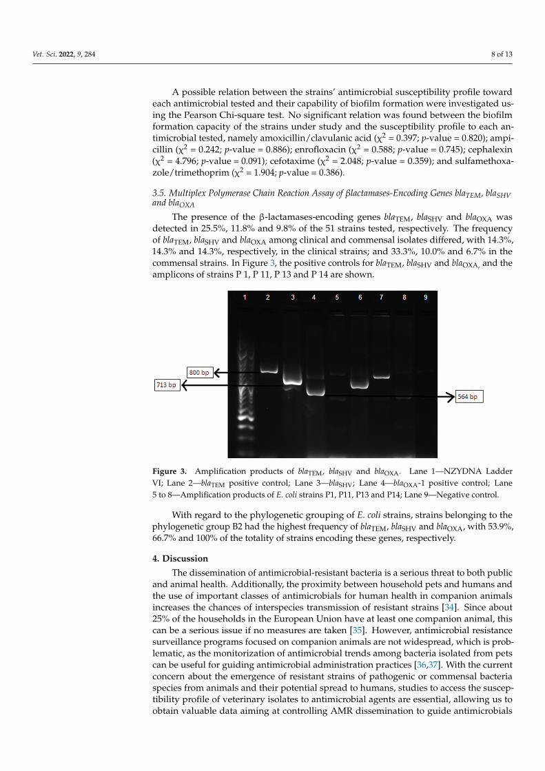
The presence of the β-lactamases-encoding genes blaTEM, blaSHV and blaOXA wasdetected in 25.5%, 11.8% and 9.8% of the 51 strains tested, respectively. The frequencyof blaTEM, blaSHV and blaOXA among clinical and commensal isolates differed, with 14.3%,14.3% and 14.3%, respectively, in the clinical strains; and 33.3%, 10.0% and 6.7% in thecommensal strains. In Figure 3, the positive controls for blaTEM, blaSHV and blaOXA, and theamplicons of strains P 1, P 11, P 13 and P 14 are shown.
Vet. Sci. 2022, 9, x FOR PEER REVIEW 9 of 14
Figure 3. Amplification products of blaTEM, blaSHV and blaOXA. Lane 1—NZYDNA Ladder VI; Lane
2—blaTEM positive control; Lane 3—blaSHV; Lane 4—blaOXA‐1 positive control; Lane 5 to 8—Amplifi‐
cation products of E. coli strains P1, P11, P13 and P14; Lane 9—Negative control.
With regard to the phylogenetic grouping of E. coli strains, strains belonging to the
phylogenetic group B2 had the highest frequency of blaTEM, blaSHV and blaOXA, with 53.9%,
66.7% and 100% of the totality of strains encoding these genes, respectively.
4. Discussion
The dissemination of antimicrobial‐resistant bacteria is a serious threat to both public
and animal health. Additionally, the proximity between household pets and humans and
the use of important classes of antimicrobials for human health in companion animals
increases the chances of interspecies transmission of resistant strains [34]. Since about 25%
of the households in the European Union have at least one companion animal, this can be
a serious issue if no measures are taken [35]. However, antimicrobial resistance surveil‐
lance programs focused on companion animals are not widespread, which is problematic,
as the monitorization of antimicrobial trends among bacteria isolated from pets can be
useful for guiding antimicrobial administration practices [36,37]. With the current concern
about the emergence of resistant strains of pathogenic or commensal bacteria species from
animals and their potential spread to humans, studies to access the susceptibility profile
of veterinary isolates to antimicrobial agents are essential, allowing us to obtain valuable
data aiming at controlling AMR dissemination to guide antimicrobials administration pol‐
icies in small animals veterinary practice and to assess the risk factors associated with the
emergence of multidrug‐resistant bacteria and interspecies transmission of these strains
[35,38].
Escherichia coli seems to be an ideal bacterial model for such studies, as it is one of the
main pathogens in dogs, a versatile organism and a commensal inhabitant of the intestinal
tract of both companion animals and humans [14,38]. E. coli has a high mutagenic capacity
and is capable of rapid cell division, which makes it able to adapt to various environments
and to the presence of antimicrobials compounds [39].
In this study, the susceptibility profile of E. coli strains from pyometra‐presenting and
healthy dogs was accessed, using antimicrobials which are among the most prescribed in
the initial treatment of pyometra [15]. The interpretation of AST was performed using
three of the most commonly used AST guidelines worldwide: CLSI M100 and EUCAST
version 6.0, two guidelines developed for human medicine but sometimes adapted for
veterinary medicine; and CLSI VET01S, an AST guideline for veterinary application.
Figure 3. Amplification products of blaTEM, blaSHV and blaOXA. Lane 1—NZYDNA LadderVI; Lane 2—blaTEM positive control; Lane 3—blaSHV; Lane 4—blaOXA-1 positive control; Lane5 to 8—Amplification products of E. coli strains P1, P11, P13 and P14; Lane 9—Negative control.
With regard to the phylogenetic grouping of E. coli strains, strains belonging to thephylogenetic group B2 had the highest frequency of blaTEM, blaSHV and blaOXA, with 53.9%,66.7% and 100% of the totality of strains encoding these genes, respectively.
4. Discussion
The dissemination of antimicrobial-resistant bacteria is a serious threat to both publicand animal health. Additionally, the proximity between household pets and humans andthe use of important classes of antimicrobials for human health in companion animalsincreases the chances of interspecies transmission of resistant strains [34]. Since about25% of the households in the European Union have at least one companion animal, thiscan be a serious issue if no measures are taken [35]. However, antimicrobial resistancesurveillance programs focused on companion animals are not widespread, which is prob-lematic, as the monitorization of antimicrobial trends among bacteria isolated from petscan be useful for guiding antimicrobial administration practices [36,37]. With the currentconcern about the emergence of resistant strains of pathogenic or commensal bacteriaspecies from animals and their potential spread to humans, studies to access the suscep-tibility profile of veterinary isolates to antimicrobial agents are essential, allowing us toobtain valuable data aiming at controlling AMR dissemination to guide antimicrobials

Vet. Sci. 2022, 9, 284 9 of 13
administration policies in small animals veterinary practice and to assess the risk factorsassociated with the emergence of multidrug-resistant bacteria and interspecies transmissionof these strains [35,38].
Escherichia coli seems to be an ideal bacterial model for such studies, as it is one of themain pathogens in dogs, a versatile organism and a commensal inhabitant of the intestinaltract of both companion animals and humans [14,38]. E. coli has a high mutagenic capacityand is capable of rapid cell division, which makes it able to adapt to various environmentsand to the presence of antimicrobials compounds [39].
In this study, the susceptibility profile of E. coli strains from pyometra-presenting andhealthy dogs was accessed, using antimicrobials which are among the most prescribed inthe initial treatment of pyometra [15]. The interpretation of AST was performed using threeof the most commonly used AST guidelines worldwide: CLSI M100 and EUCAST version6.0, two guidelines developed for human medicine but sometimes adapted for veterinarymedicine; and CLSI VET01S, an AST guideline for veterinary application.
Higher percentages of resistance to ampicillin and amoxicillin/clavulanic acid wereobserved. These profiles were expected, as these antimicrobials are highly used in theempirical treatment of pyometra and have been applied for longer in veterinary medicinethan more recent antimicrobials or antimicrobial combinations, such as enrofloxacin orsulfamethoxazole/trimethoprim [40].
E. coli associated with canine pyometra usually originates from the normal intestinalmicrobiota of dogs, not being related to any specific clonal type, and is characterized byharboring several virulence genes usually found in uropathogenic strains (UPEC) [24,41].The fact that E. coli strains associated with pyometra originate from commensal variantscan, in part, explain the similar rates of resistance to the antimicrobials tested in thisstudy. The resistance profiles of the commensal E. coli isolates from healthy dogs showthat the intestinal tract of healthy animals can constitute a reservoir of resistant strainsand their determinants [42]. However, regarding companion animals, there is still alack of monitorization programs at regional, national and international levels, whichare essential to guide policies and detect changes that require intervention strategies.As multidrug-resistant E. coli is capable of expressing resistance determinants, such asproduction of β-lactamases, and can be isolated from both healthy animals and clinicalcases, the surveillance of both types of strains is of vital importance [36].
In this study, it was possible to observe that the susceptibility profile of the strains var-ied when using different AST interpretation criteria. This can constitute a serious problemnot only in the selection of the antimicrobial therapeutic protocol but also in the surveillanceof antimicrobial resistance, as differences in data can compromise their comparison. Inorder to measure the degree of agreement between AST results when using different stan-dards, Cohen‘s kappa was calculated. As previously mentioned, the susceptibility profileswere similar when using CLSI M100 or CLSI VET01S evaluation criteria for ampicillin,amoxicillin/clavulanic acid, cephalexin and sulfamethoxazole/trimethoprim for Enterobac-teriaceae, as human-derived breakpoints are used in CLSI VET01S standards. The use ofsuch breakpoints constitutes a serious problem in the AST determination of veterinaryisolates, as the differences in pharmacodynamics (PD) and pharmacokinetics (PK) betweenhumans and other animals are not considered [43]. PK-PD studies are used in AST break-point development, as they describe the relationship between the effect of the antimicrobialon the infecting organism, typically measured in vitro (pharmacodynamics), and clinicaland microbiological outcomes in the host, measured in vivo (pharmacokinetics) [44].
Regarding the measured agreement between CLSI VET01S and EUCAST version6.0 evaluation criteria standards, although Cohen’s kappa revealed a perfect agreementregarding sulfamethoxazole/trimethoprim for the E. coli strains under study, this was notobserved for the remaining antimicrobials tested. In these cases, their Cohen’s kappa valuewas <1, ranging from 0.47–0.59 for AMC, 0.57–0.75 for AMP, 0.19–0.38 for CL and 0.65–0.75for CTX, among the clinical and commensal isolates. This fact may be explained by thedifferences in the breakpoint setting process between the two AST agencies that lead to

Vet. Sci. 2022, 9, 284 10 of 13
different interpretative criteria values for the same antimicrobial agent. Breakpoints set-ting is based on clinical, pharmacokinetic, microbiological and pharmacodynamic studies;however, the relative importance of each parameter is under constant discussion, and so,AST agencies can adopt different approaches [45]. CLSI and EUCAST are the two mainnongovernmental agencies in setting breakpoints standards for antimicrobial susceptibilitytesting [46]. However, these agencies differ in their approach to breakpoints setting, inter-nal organization, data collection establishment and also in their report obligations to theirgovernmental counterparts, explicitly the U.S. Food and Drug Administration (FDA) in thecase of CLSI and the European Medicines Agency (EMA) in the case of EUCAST [44,46].Other differences include the role of industry, funding and the availability of the stan-dards developed, as EUCAST standards are freely available while CLSI are not [44,46].As previously mentioned, setting or reviewing clinical breakpoints currently takes intoconsideration a combination between pharmacokinetic and pharmacodynamic data, clini-cal outcome data and minimum inhibitory concentrations (MIC) in vitro data [47]. Withbreakpoints for AST being expressed in either concentration or zone diameter, dependingon the testing method applied, and disk diffusion being one of the most widely used ASTmethods in routine clinical microbiology laboratories, there is a need to have zone diameterbreakpoints calibrated considering the MIC breakpoints [48]. This is particularly relevant inEurope, where a harmonizing process of AST methodologies was carried out by EUCASTon European MIC breakpoints [48]. Regarding AST in veterinary medicine, few species-specific interpretative breakpoints are available regarding MIC, and to an even greaterextent, disk diffusion breakpoints. Particularly in this study, when analyzing the clinicalbreakpoints available in CLSI-VET01S for Enterobacteriaceae, the bacterial family to whichE. coli belongs, it was possible to note that there were no species-specific disk diffusionbreakpoints for the studied antimicrobials, except for enrofloxacin, for which disk diffusionbreakpoints applicable to Enterobacteriaceae strains isolated from dogs are available. Assuch, the development of appropriate clinical breakpoints, both for MIC and disk diffusionfor veterinary isolates is vital for predicting treatment success of infections in companionanimals and guiding clinicians in the selection of the most adequate therapeutics [49].
To further characterize the E. coli strains under study in terms of resistance, biofilmproduction ability was accessed, as well as the presence of β-lactamases genes blaTEM,blaSHV and blaOXA, as biofilm production is a potential virulence trait, and the productionof β-lactamases is the main mechanism of resistance to β-lactams in Enterobacteriaceae [2,50].The biofilm formation capacity of the E. coli strains from pyometra-presenting and healthyanimals was analyzed using pellicle formation assays, with approximately half of theclinical strains and of the commensal isolates showing a biofilm formation phenotype;however, the majority of the clinical strains were able to form biofilms within a three-dayperiod, while most of the commensal strains required a four-day period to form biofilms,meaning that the E. coli strains from pyometra-presenting dogs, on average, displayed amore rapid biofilm formation process than the commensal isolates. Lopes et al. (2021) alsoevaluated the biofilm ability of E. coli isolates obtained from canine pyometra, observinga high prevalence of positive biofilm strains, reinforcing the importance of this virulentelement in infection [51]. It would be interesting to test biofilm-forming ability in thesepyometra isolates, simulating the conditions in the uterus of canines suffering from thisdisease, as in vivo conditions may influence this virulent factor [51].
Another important resistance determinant in E. coli is β-lactamases production, beingone of the main mechanisms of resistance to β-lactam antimicrobials among Enterobacteriaceaein both human and veterinary medicine but also frequently associated with resistanceto several other groups of antimicrobials, including fluoroquinolones, aminoglycosidesor sulfamethoxazole [30,52]. Since companion animals are a potential reservoir of β-lactamases-encoding genes, and their close contact with their owners facilitates resistancetransmission, surveillance studies to detect the prevalence of β-lactamases-encoding genesare important and needed in veterinary medicine [53,54]. Multiplex PCR approachesare suitable for the rapid detection of the emergence of β-lactamases in bacteria isolated

Vet. Sci. 2022, 9, 284 11 of 13
from animals, improving surveillance [23]. In a β-lactamase family type, the resistancephenotypes expressed can differ substantially among enzyme members due to changesin the enzyme structure and activity [55]. In fact, in this study, the strains identified ashaving β-lactamase-related genes of a certain β-lactamase family (e.g., TEM, SHV or OXA)showed different susceptibility profiles to the antimicrobials tested.
The identification of the β-lactamase(s) produced by the strains using a sequencingtechnique and the antimicrobial susceptibility testing of more β-lactams would provideextra knowledge of the specific resistance pattern. Furthermore, these enzymes are oftenplasmid mediated, which can facilitate their transmission among individuals and theirdissemination in the environment. Thus, the presence of β-lactamases-related genes inthe clinical and commensal E. coli strains under study, particularly plasmid-mediatedβ-lactamases, is of concern, as their transmission can occur frequently.
5. Conclusions
Antimicrobial-resistant bacteria are a serious threat to human and animal health. Sev-eral AST guidelines are available for the evaluation of the antimicrobial-resistant signaturesin human strains; however, specific guidelines for AMR evaluation of animal-origin isolatesand specific breakpoints are scarce. In this study, an antimicrobial susceptibility profilecharacterization of canine E. coli clinical and commensal isolates was performed. Resultsshowed the importance of determining the antimicrobial resistance profile of veterinaryisolates and the need for the implementation of national and international surveillanceprograms for companion animals. The low agreement between AST results when applyingdifferent guidelines to certain antimicrobials largely used in companion animals and the useof human-derived breakpoints can constitute a serious problem, possibly leading to poorclinical outcomes and resistance dissemination. In view of this, it is urgent to have stan-dardized and validated testing methods and harmonized interpretive criteria in veterinarymedicine, enabling the identification of multidrug-resistant bacteria, as they represent aclinical challenge for domestic animal medicine. Additionally, the presence of β-lactamases-related genes in the strains under study, and particularly plasmid-mediated β-lactamases,is of concern, as their transmission occurs frequently. Furthermore, biofilm-forming abilitybeing related to bacterial virulence, its association with antimicrobial resistance determi-nants is of great concern, particularly in companion animals due to close contact withhumans, possibly acting as a reservoir of bacteria-harboring resistance genes.
Author Contributions: Conceptualization, L.M. and M.O.; methodology, V.F. and E.C.; software,V.F. and T.N.; validation, M.O.; formal analysis, T.N. and M.O.; investigation, M.O., E.S., L.M.and V.F.; resources, L.M.; data curation, E.C.; writing—original draft preparation, V.F. and E.C.;writing—review and editing, M.O., E.C., T.N., L.T., E.S. and L.M.; visualization, E.C.; supervision,M.O.; project administration, M.O.; funding acquisition, L.T. and M.O. All authors have read andagreed to the published version of the manuscript.
Funding: Authors would like to acknowledge CIISA—Centro de Investigação Interdisciplinar emSanidade Animal, Faculdade de Medicina Veterinária, Universidade de Lisboa, Project UIDB/00276/2020(Funded by FCT); and the Laboratório Associado para Ciência Animal e Veterinária (LA/P/0059/2020—AL4AnimalS).
Institutional Review Board Statement: The animal study protocol was approved by the EthicalCommittee for Research and Teaching (CEIE) of the Faculty of Veterinary Medicine—University ofLisbon, Portugal (N/Refª CIISA 74/2013).
Informed Consent Statement: Informed consent was obtained from all subjects involved in the study.
Data Availability Statement: The datasets used in the current study are available from the corre-sponding author on reasonable request.
Acknowledgments: Authors would like to acknowledge Lurdes Clemente of Instituto Nacional deInvestigação Agrária e Veterinária (INIAV, Oeiras) for technical support.
Conflicts of Interest: The authors declare no conflict of interest.
Vet. Sci. 2022, 9, 284 12 of 13
References1. EFSA (European Food Safety Authority); ECDC (European Centre for Disease Prevention and Control). The European Union
summary report on antimicrobial resistance in zoonotic and indicator bacteria from humans, animals and food in 2015. EFSA J.2017, 15, e04694.
2. Shaikh, S.; Fatima, J.; Shakil, S.; Rizvi, S.; Kamal, M. Antibiotic resistance and extended spectrum beta-lactamases: Types,epidemiology and treatment. Saudi J. Biol. Sci. 2015, 22, 90–101. [CrossRef]
3. Tenover, F. Mechanisms of antimicrobial resistance in bacteria. Am. J. Infect. Control 2006, 34 (Suppl. 1), S3–S10. [CrossRef]4. Adekunle, O. Mechanisms of Antimicrobial Resistance in Bacteria, General Approach. Int. J. Pharm. Med. 2012, 1, 166–187.5. World Health Organization. Available online: http://www.who.int/antimicrobial-resistance/global-actionplan/surveillance/
glass/en/ (accessed on 12 July 2021).6. Masterton, R. The Importance and Future of Antimicrobial Surveillance Studies. Clin. Infect. Dis. 2008, 47, S21–S31. [CrossRef]7. Prestinaci, F.; Pezzotti, P.; Pantosti, A. Antimicrobial resistance: A global multifaceted phenomenon. Pathog. Glob. Health 2015,
109, 309–318. [CrossRef]8. Heuer, O.; Jensen, V.; Hammerum, A. Antimicrobial Drug Consumption in Companion Animals. Emerg. Infect. Dis. 2005,
11, 344–345. [CrossRef]9. Umber, J.; Bender, J. Pets and antimicrobial resistance. Vet. Clin. N. Am. Small Anim. Pract. 2009, 39, 279–292. [CrossRef]10. Guardabassi, L.; Jensen, L.B.; Kruse, H. Guide to Antimicrobial Use in Animals; Blackwell Pub.: Oxford, UK, 2008.11. Rodloff, A.; Bauer, T.; Ewig, S.; Kujath, P.; Müller, E. Susceptible, Intermediate, and Resistant—The Intensity of Antibiotic Action.
Dtsch. Arztebl. Int. 2008, 105, 657–662. [CrossRef]12. European Centre for Disease Prevention and Control. Antimicrobial Resistance Surveillance in Europe 2014. Annual Report of the
European Antimicrobial Resistance Surveillance Network (EARS-Net); ECDC: Stockholm, Sweden, 2015.13. European Committee on Antimicrobial Susceptibility Testing. Veterinary Committee on AST; EUCAST, Sweden. Available
online: https://www.eucast.org/fileadmin/src/media/PDFs/EUCAST_files/Breakpoint_tables/v_11.0_Breakpoint_Tables.pdf(accessed on 20 December 2020).
14. Allocati, N.; Masulli, M.; Alexeyev, M.; Di Illio, C. Escherichia coli in Europe: An Overview. Int. J. Environ. Res. Public Health 2013,10, 6235–6254. [CrossRef]
15. Johnston, S.D.; Kustritz, M.V.; Olson, P.S. Canine and Feline Theriogenology; Saunders: Philadelphia, PA, USA, 2001.16. Jitpean, S.; Ström-Holst, B.; Emanuelson, U.; Höglund, O.V.; Pettersson, A.; Alneryd-Bull, C.; Hagman, R. Outcome of pyometra
in female dogs and predictors of peritonitis and prolonged postoperative hospitalization in surgically treated cases. BMC Vet. Res.2014, 10, 6. [CrossRef]
17. Wood, T. Insights on Escherichia coli Biofilm Formation and Inhibition from Whole-Transcriptome Profiling. Environ. Microbiol.2009, 11, 1–15. [CrossRef]
18. Schembri, M.; Kjaergaard, K.; Klemm, P. Global gene expression in Escherichia coli biofilms. Mol. Microbiol. 2003, 48, 253–267.[CrossRef]
19. Allewell, N. Introduction to Biofilms Thematic Minireview Series. JBC 2016, 291, 12527–12528. [CrossRef]20. Stewart, P.; Costerton, J. Antibiotic resistance of bacteria in biofilms. Lancet 2001, 358, 135–138. [CrossRef]21. Rabin, N.; Zheng, Y.; Opoku-Temeng, C.; Du, Y.; Bonsu, E.; Sintim, H. Biofilm formation mechanisms and targets for developing
antibiofilm agents. Future Med. Chem. 2015, 7, 493–512. [CrossRef]22. Li, X.; Mehrotra, M.; Ghimire, S.; Adewoye, L. β-Lactam resistance and β-lactamases in bacteria of animal origin. Vet. Microbiol.
2007, 121, 197–214. [CrossRef]23. Dallence, C.; Da Costa, A.; Decré, D.; Favier, C.; Arlet, G. Development of a set of multiplex PCR assays for the detection of genes
encoding important β-lactamases in Enterobacteriaceae. J. Antimicrob. Chemother. 2010, 65, 490–495. [CrossRef]24. Mateus, L.; Henriques, S.; Merino, C.; Pomba, C.; Lopes da Costa, L.; Silva, E. Virulence genotypes of Escherichia coli canine
isolates from pyometra, cystitis and fecal origin. Vet. Microbiol. 2013, 166, 590–594. [CrossRef]25. Machado, I. Piometra na Cadela e na Gata: Diferenças e Semelhanças. Dissertação de Mestrado Integrado em Medicina Veterinária,
Faculdade de Medicina Veterinária, Universidade de Lisboa, Lisboa, Portugal, 2017.26. Clinical and Laboratory Standards Institute (CLSI). Performance Standards for Antimicrobial Disk and Dilution Susceptibility Tests for
Bacteria Isolated from Animals, 5th ed.; VET01S; Clinical and Laboratory Standards Institute (CLSI): Wayne, PA, USA, 2021.27. Clinical and Laboratory Standards Institute (CLSI). Performance Standards for Antimicrobial Susceptibility Tests, 31st ed.; M100;
Clinical and Laboratory Standards Institute (CLSI): Wayne, PA, USA, 2021.28. Clinical and Laboratory Standards Institute (CLSI). Performance Standards for Antimicrobial Disk and Dilution Susceptibility Tests for
Bacteria Isolated From Animals, 4th ed.; VET01-A4; Clinical and Laboratory Standards Institute (CLSI): Wayne, PA, USA, 2013.29. Magiorakos, A.; Srinivasan, A.; Carey, R.; Carmeli, Y.; Falagas, M.; Giske, C.; Harbarth, S.; Hindler, J.F.; Kahlmeter, G.; Olsson-
Liljequist, B.; et al. Multidrug-resistant, extensively drug-resistant and pandrugresistant bacteria: An international expertproposal for interim standard definitions for acquired resistance. Clin. Microbiol. Infect. 2012, 18, 268–281. [CrossRef]
30. Seixas, R.; Machado, J.; Bernardo, F.; Vilela, C.; Oliveira, M. Biofilm Formation by Salmonella Enterica Serovar 1,4,[5],12:i:—PortugueseIsolates: A Phenotypic, Genotypic, and Sociogeographic Analysis. Curr. Microbiol. 2014, 68, 670–677. [CrossRef]
31. Landis, J.; Koch, G. The measurement of observer agreement for categorical data. Biometrics 1977, 33, 159–174. [CrossRef]

Vet. Sci. 2022, 9, 284 13 of 13
32. Jones-Dias, D.; Manageiro, V.; Ferreira, E.; Louro, D.; Antibiotic Resistance Surveillance Program in Portugal (ARSIP) Participants;Caniça, M. Diversity of Extended-Pectrum and Plasmid-Mediated AmpC β-Lactamases in Enterobacteriaceae Isolates fromPortuguese Health Care Facilities. J. Microbiol. 2014, 52, 496–503. [CrossRef]
33. Picard, B.; Garcia, J.S.; Gouriou, S.; Duriez, P.; Brahimi, N.; Bingen, E.; Elion, J.; Denamur, E. The link between phylogeny andvirulence in Escherichia coli extraintestinal infection. Infect. Immun. 1999, 67, 546–553. [CrossRef]
34. Stewart, P. Antimicrobial Tolerance in Biofilms. Microbiol. Spectr. 2015, 3. [CrossRef]35. Weese, J. Antimicrobial resistance in companion animals. Anim. Health Res. Rev. 2008, 9, 169–176. [CrossRef]36. Cummings, K.J.; Aprea, V.A.; Altier, C. Antimicrobial resistance trends among canine Escherichia coli isolates obtained from
clinical samples in the northeastern USA, 2004–2011. Can. Vet. J. 2015, 56, 393–398.37. Guardabassi, L.; Schwarz, S.; Lloyd, D. Pet animals as reservoirs of antimicrobial-resistant bacteria. J. Antimicrob. Chemother. 2004,
54, 321–332. [CrossRef]38. Thungrat, K.; Price, S.; Carpenter, M.; Boothe, D. Antimicrobial susceptibility patterns of clinical Escherichia coli isolates from dogs
and cats in the United States: January 2008 through January 2013. Vet. Microbiol. 2015, 179, 287–295. [CrossRef]39. Boothe, D.; Smaha, T.; Carpenter, D.; Shaheen, B.; Hatchcock, T. Antimicrobial Resistance and Pharmacodynamics of Canine and
Feline Pathogenic E. coli in the United States. J. Am. Anim. Hosp. Assoc. 2012, 48, 379–389. [CrossRef]40. Tadesse, D.; Zhao, S.; Tong, E.; Ayers, S.; Singh, A.; Bartholomew, M.J.; McDermott, P.F. Antimicrobial drug resistance in Escherichia
coli from humans and food animals, United States, 1950–2002. Emerg. Infect. Dis. 2012, 18, 741–749. [CrossRef]41. Ettiger, S.J.; Feldman, E.C. Textbook of Veterinary Internal Medicine; Saunders: Philadelphia, PA, USA, 2010; Volume 2.42. Tenaillon, O.; Skurnik, D.; Picard, B.; Denamur, E. The population genetics of commensal Escherichia coli. Nat. Rev. Microbiol. 2010,
8, 207–217. [CrossRef]43. Lees, P.; Shojaee, F. Rational dosing of antimicrobial drugs: Animals versus humans. Int. J. Antimicrob. Agents 2002, 19, 269–284.
[CrossRef]44. Labreche, M.; Graber, C.; Nguyen, H. Recent Upgrades on the Role of Pharmacokinetic-pharmacodynamics in Antimicrobial
Susceptibility Testing as Applied to Clinical Practice. Clin. Infect. Dis. 2015, 61, 1446–1452.45. Jenkins, S.; Jerris, R. Critical Assessment of Issues Applicable to Development of Antimicrobial Susceptibility Testing Breakpoints.
J. Clin. Microbiol. 2011, 49, S5–S10. [CrossRef]46. Silley, P. Susceptibility testing methods, resistance and breakpoints: What do these terms really mean? Rev. Sci. Tech. Off. Int. Epiz.
2012, 31, 33–41. [CrossRef]47. Valsesia, G.; Roos, M.; Böttger, E.; Hombach, M. A Statistical Approach for Determination of Disk Diffusion-Based Cutoff Values
for Systematic Characterization of Wild-Type and Non-Wild-Type Bacterial Populations in Antimicrobial Susceptibility Testing.J. Clin. Microbiol. 2015, 53, 1812–1822. [CrossRef]
48. Matusckek, E.; Brown, D.; Kahlmeter, G. Development of the EUCAST disk diffusion antimicrobial susceptibility testing methodand its implementation in routine microbiology laboratories. Clin. Microbiol. Infect. 2014, 20, O255–O266. [CrossRef]
49. Toutain, P.L.; Bousquet-Mélou, A.; Damborg, P.; Ferran, A.A.; Mevius, D.; Pelligand, L.; Veldman, K.T.; Lees, P. En Route towardsEuropean Clinical Breakpoints for Veterinary Antimicrobial Susceptibility Testing: A Position Paper Explaining the VetCASTApproach. Front. Microbiol. 2017, 8, 2344. [CrossRef]
50. Nave, P.; Prado, G.; Huelves, L.; Gracia, M.; Ruiz, V.; Blanco, J.; Dahbi, G.; Blanco, M.; Mdel, C.P.; Soriano, F. Correlation betweenvirulence factors and in vitro biofilm formation by Escherichia coli strains. Microb. Pathog. 2008, 45, 86–91. [CrossRef]
51. Lopes, C.E.; De Carli, S.; Riboldi, C.I.; De Lorenzo, C.; Panziera, W.; Driemeier, D.; Siqueira, F.M. Pet Pyometra: CorrelatingBacteria Pathogenicity to Endometrial Histological Changes. Pathogens 2021, 10, 833. [CrossRef]
52. Mackenzie, K.; Palmer, M.; Köster, W.; White, A. Examining the Link between Biofilm Formation and the Ability of PathogenicSalmonella Strains to Colonize Multiple Host Species. Front. Vet. Sci. 2017, 4, 138. [CrossRef]
53. Clemente, L.; Manageiro, V.; Jones-Dias, D.; Correia, I.; Themudo, P.; Albuquerque, T.; Geraldes, M.; Matos, F.; Almendra, C.;Ferreira, E.; et al. Antimicrobial susceptibility and oxymino-β-lactam resistance mechanisms in Salmonella enterica and Escherichiacoli isolates from different animal sources. Res. Microbiol. 2015, 166, 574–583. [CrossRef]
54. Féria, C.; Ferreira, E.; Correia, J.; Gonçalves, J.; Caniça, M. Patterns and mechanisms of resistance to β-lactamase inhibitors inuropathogenic Escherichia coli isolated from dogs in Portugal. J. Antimicrob. Chemother. 2002, 49, 77–85. [CrossRef]
55. Ghafourian, S.; Sadegjifard, N.; Soheili, S.; Sekawi, Z. Extended Spectrum Beta-lactamases: Definition, Classification andEpidemiology. Curr. Issues Mol. Biol. 2015, 17, 11–22.
Related Documents


![UvA-DARE (Digital Academic Repository) Antimicrobial drug ... · around 50% [27]. Antimicrobial resistance in food-borne pathogens and in commensal bacteria of food animals is a major](https://static.cupdf.com/doc/110x72/5fb10ad3fe77fc3d170112e0/uva-dare-digital-academic-repository-antimicrobial-drug-around-50-27-antimicrobial.jpg)


