Antimicrobial photodynamic therapy in rat experimental candidiasis: evaluation of pathogenicity factors of Candida albicans Joyce da Silva Martins, MD, a Juliana Campos Junqueira, MD, PhD, b Raquel Lourdes Faria, c Naiara Fonseca Santiago, c Rodnei Dennis Rossoni, c Carlos Eduardo Dias Colombo, MD, PhD, d and Antonio Olavo Cardoso Jorge, MD, PhD, b São José dos Campos and Taubaté, Brazil SÃO PAULO STATE UNIVERSITY AND UNIVERSITY OF TAUBATÉ Objective. This study evaluated the effects of photodynamic therapy on pathogenicity of Candida albicans. Study design. Fifty-six rats were submitted to development of candidiasis on the tongue dorsum by C. albicans inoculations. After 5 days, different treatments were administered: laser and photosynthesizer methylene blue (LP); laser only (LP); photosensitizer only (LP); and physiologic solution only (LP). Samples of the oral cavity were collected for a count of colony-forming units per mL. Colonies were isolated for evaluation of proteinase and phospholipase activities. The rats were killed for microscopic analysis of the tongue dorsum. The data were analyzed by analysis of variance, Kruskal-Wallis, and Bonferroni tests. Results. The number of C. albicans recovered from the oral cavity of the rats was similar between the groups (P .106). The LP group showed fewer microscopic lesions of candidiasis than the LP group (P .001). The LP group presented lower proteinase activity compared with the other groups, with significant difference between the groups LP and LP (P .018). Conclusions. Photodynamic therapy reduced the microscopic lesions of experimental candidiasis in rats and inhibited the proteinase activity of C. albicans. (Oral Surg Oral Med Oral Pathol Oral Radiol Endod 2011;111:71-77) Fungi are important agents of human disease. Among the most important fungal pathogens are yeast species belonging to the genus Candida. These species can cause a wide range of human diseases, ranging from superficial mucosal infections, such as vulvovaginal and oropharyngeal candidiasis, to life-threatening inva- sive infections. 1-4 Several virulence factors contribute to the pathogenicity of C. albicans, including the ability to adhere to epithelial cells, the ability to form hyphae, and secretion of extracellular enzymes. 5-7 During the initial stages of superficial mucosal infection, C. albi- cans forms filamentous hyphae, which show thigmot- ropism, a phenomenon also known as contact guidance, in addition to releasing various hydrolytic enzymes, such as extracellular phospholipases and secretory as- partyl proteinases. 5 The widespread use of topical and systemic antifun- gal agents as the conventional treatment for oral can- didiasis has resulted in the development of resistance in C. albicans. 8 Therefore, the study of additional meth- ods for the control of C. albicans, such as photody- namic antimicrobial chemotherapy (PACT), has be- come essential. PACT is a process that combines light and a photosensitizing drug, promoting a phototoxic effect on the treated cells, in general via oxidative damage. The potential of PACT to promote microbial eradication is becoming progressively more accepted. The technique involves the production of highly cyto- toxic singlet oxygen and other reactive oxygen species, promoting photodynamic microbial damage. 9,10 As an antifungal therapy, PACT is very much a devel- oping science, and the vast majority of published work has understandably centered on in vitro laboratory investiga- tions. Various Candida species, photosensitizers, and ir- radiation protocols have been used. In most cases, com- plete killing of the yeast has been readily achieved. Critically, no reports on development of resistance to anti- fungal PACT currently exist, and the treatment has not been associated with mutagenic effects or genotoxicity in either fungi or cultured human cells. 2 The most extensively inves- tigated photosensitizer classes investigated in in vitro antifun- gal PACT studies have been the phenothiaziniums, 11-14 the porphyrins, 15,16 and the phthalocyanines. 17 Supported by the São Paulo Council of Research (FAPESP), Brazil (grant no. 07/58780-0). a Postgraduate student, Department of Biosciences and Oral Diagno- sis, School of Dentistry of São José dos Campos, São Paulo State University-UNESP, São Paulo, Brazil. b Professor, Graduate Student, School of Dentistry of São José dos Campos, São Paulo State University-UNESP, São Paulo, Brazil. c Graduate student, School of Dentistry of São José dos Campos, São Paulo State University-UNESP, São Paulo, Brazil. d Professor, Department of Dentistry, University of Taubaté/UNI- TAU, São Paulo, Brazil. Received for publication Jun. 17, 2010; returned for revision Jul. 28, 2010; accepted for publication Aug. 5, 2010. 1079-2104/$ - see front matter © 2011 Mosby, Inc. All rights reserved. doi:10.1016/j.tripleo.2010.08.012 71

Welcome message from author
This document is posted to help you gain knowledge. Please leave a comment to let me know what you think about it! Share it to your friends and learn new things together.
Transcript
Antimicrobial photodynamic therapy in rat experimental candidiasis:evaluation of pathogenicity factors of Candida albicans
Joyce da Silva Martins, MD,a Juliana Campos Junqueira, MD, PhD,b Raquel Lourdes Faria,c
Naiara Fonseca Santiago,c Rodnei Dennis Rossoni,c Carlos Eduardo Dias Colombo, MD, PhD,d
and Antonio Olavo Cardoso Jorge, MD, PhD,b São José dos Campos and Taubaté, BrazilSÃO PAULO STATE UNIVERSITY AND UNIVERSITY OF TAUBATÉ
Objective. This study evaluated the effects of photodynamic therapy on pathogenicity of Candida albicans.Study design. Fifty-six rats were submitted to development of candidiasis on the tongue dorsum by C. albicansinoculations. After 5 days, different treatments were administered: laser and photosynthesizer methylene blue (L�P�);laser only (L�P�); photosensitizer only (L�P�); and physiologic solution only (L�P�). Samples of the oral cavitywere collected for a count of colony-forming units per mL. Colonies were isolated for evaluation of proteinase andphospholipase activities. The rats were killed for microscopic analysis of the tongue dorsum. The data were analyzedby analysis of variance, Kruskal-Wallis, and Bonferroni tests.Results. The number of C. albicans recovered from the oral cavity of the rats was similar between the groups (P �.106). The L�P� group showed fewer microscopic lesions of candidiasis than the L�P� group (P � .001). The L�P�group presented lower proteinase activity compared with the other groups, with significant difference between thegroups L�P� and L�P� (P � .018).Conclusions. Photodynamic therapy reduced the microscopic lesions of experimental candidiasis in rats and inhibited
the proteinase activity of C. albicans. (Oral Surg Oral Med Oral Pathol Oral Radiol Endod 2011;111:71-77)Fungi are important agents of human disease. Amongthe most important fungal pathogens are yeast speciesbelonging to the genus Candida. These species cancause a wide range of human diseases, ranging fromsuperficial mucosal infections, such as vulvovaginaland oropharyngeal candidiasis, to life-threatening inva-sive infections.1-4 Several virulence factors contributeto the pathogenicity of C. albicans, including the abilityto adhere to epithelial cells, the ability to form hyphae,and secretion of extracellular enzymes.5-7 During theinitial stages of superficial mucosal infection, C. albi-cans forms filamentous hyphae, which show thigmot-ropism, a phenomenon also known as contact guidance,in addition to releasing various hydrolytic enzymes,
Supported by the São Paulo Council of Research (FAPESP), Brazil(grant no. 07/58780-0).aPostgraduate student, Department of Biosciences and Oral Diagno-sis, School of Dentistry of São José dos Campos, São Paulo StateUniversity-UNESP, São Paulo, Brazil.bProfessor, Graduate Student, School of Dentistry of São José dosCampos, São Paulo State University-UNESP, São Paulo, Brazil.cGraduate student, School of Dentistry of São José dos Campos, SãoPaulo State University-UNESP, São Paulo, Brazil.dProfessor, Department of Dentistry, University of Taubaté/UNI-TAU, São Paulo, Brazil.Received for publication Jun. 17, 2010; returned for revision Jul. 28,2010; accepted for publication Aug. 5, 2010.1079-2104/$ - see front matter© 2011 Mosby, Inc. All rights reserved.
doi:10.1016/j.tripleo.2010.08.012such as extracellular phospholipases and secretory as-partyl proteinases.5
The widespread use of topical and systemic antifun-gal agents as the conventional treatment for oral can-didiasis has resulted in the development of resistance inC. albicans.8 Therefore, the study of additional meth-ods for the control of C. albicans, such as photody-namic antimicrobial chemotherapy (PACT), has be-come essential. PACT is a process that combines lightand a photosensitizing drug, promoting a phototoxiceffect on the treated cells, in general via oxidativedamage. The potential of PACT to promote microbialeradication is becoming progressively more accepted.The technique involves the production of highly cyto-toxic singlet oxygen and other reactive oxygen species,promoting photodynamic microbial damage.9,10
As an antifungal therapy, PACT is very much a devel-oping science, and the vast majority of published work hasunderstandably centered on in vitro laboratory investiga-tions. Various Candida species, photosensitizers, and ir-radiation protocols have been used. In most cases, com-plete killing of the yeast has been readily achieved.Critically, no reports on development of resistance to anti-fungal PACT currently exist, and the treatment has not beenassociated with mutagenic effects or genotoxicity in eitherfungi or cultured human cells.2 The most extensively inves-tigated photosensitizer classes investigated in in vitro antifun-gal PACT studies have been the phenothiaziniums,11-14 the
porphyrins,15,16 and the phthalocyanines.1771
rming
OOOOE72 da Silva Martins et al. January 2011
The efficacy of PACT on yeasts of the Candidagenus has also been demonstrated by some in vivostudies.8,18,19 Teichert et al.18 tested several concentra-tions of methylene blue associated with a diode laser onbuccal candidiasis in immunosuppressed mice. Analy-sis of the results indicated the efficacy of PACT inreducing yeast, an effect that was directly proportionalto the photosensitizer concentration. Junqueira et al.19
evaluated the effects of photodynamic therapy (PDT)on buccal candidiasis in rats and verified that the ratstreated with a laser and with methylene blue developedmore discrete candidiasis lesions compared with thecontrol groups. Mima et al.8 observed that PACT withporphyrin and red light–emitting diode (LED) pro-moted significant reduction in the viability of C. albi-cans in buccal candidiasis in immunosuppressed mice.
Though many in vitro and in vivo studies haveshown inactivation of C. albicans by PACT, few stu-dies have focused on the effects of PACT on thevirulence factors of C. albicans. Munin et al.20 demon-strated that PDT using methylene blue as a photosen-sitizing drug inhibits both the growth and the germ tubeformation of C. albicans. Soares et al.21 verified thattoluidine blue with LED inhibits in vitro growth andadhesion of different Candida isolates to buccal epithe-lial cells. Regarding the effects of PACT on secretionof extracellular enzymes, which is an important viru-lence factor of C. albicans, there are no studies in the
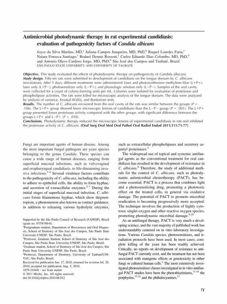
Fig. 1. Study design. L�P�: treated with laser and photophotosensitizer only; L�P�: control group. CFU, colony-fo
literature. Therefore, the objective of the present study
was to evaluate the effects of PDT on secretion of theextracellular enzyme by C. albicans using a murinemodel of buccal candidiasis.
MATERIAL AND METHODSExperimental animals
This study was approved by the Research EthicsCommittee of the São José dos Campos School ofDentistry under protocol no. 035/2007-PA/CEP. Fifty-six male rats (Rattus norvegicus, Albinus, Wistar) neg-ative for the Candida genus in the buccal cavity, weigh-ing �250 g (120 days old), were included in the study.The rats were distributed into the following experimen-tal groups: treated with laser and photosensitizer(L�P�); treated with laser only (L�P�); treated withphotosensitizer only (L�P�); and control (L�P�).Each group was composed of 14 rats: 10 with experi-mental candidiasis and 4 without experimental candi-diasis (Fig. 1).
Induction of experimental candidiasisThe rats were given a solution of 0.1% tetracycline
hydrochloride (Terramycin; Pfizer, São Paulo, Brazil)in their drinking water. This treatment was initiated 7days before the inoculation of C. albicans suspensionand was maintained throughout the experiment.22
A suspension of C. albicans containing 5 � 108
viable cells/mL was prepared according to Reed et al.23
zer; L�P�: treated with laser only; L�P�: treated withunits.
sensiti
A reference strain (ATCC 18,804) of C. albicans was
OOOOEVolume 111, Number 1 da Silva Martins et al. 73
used. For the inoculation of this suspension, the ratswere sedated via intramuscular injection of xylazinechloride solution (Vetbrands, São Paulo, Brazil) andketamine (Vetbrands) in the proportion 1:0.5 mL, at adose of 0.05 mL/100 g of body weight. The C. albicanssuspension (0.2 mL) was dropped into the mouths ofthe rats with the aid of a 1 mL syringe. The materialwas spread on the tongue dorsum with a swab that hadbeen previously soaked in the suspension. This proce-dure was repeated for 3 consecutive days.
Photosensitizer and laserA 0.1 mg/mL solution of methylene blue was used as
the photosensitizer and was prepared by dissolving thepowder (Sigma, São Paulo, Brazil) in a physiologicsolution of 0.85% sodium chloride (NaCl). The solutionwas filtered through a sterile membrane filter with0.22-�m-diameter pores (TPP, Trasadingen, Switzer-land) and stored in the dark.
The light source used was a gallium-aluminum-ar-senide (GaAlAs) laser (Photon laser III; DMC, SãoCarlos, Brazil) with a wavelength of 660 nm, outputpower of 100 mW, energy density of 245 J/cm2, andtime of 69 seconds. The irradiation laser was applied tothe tongue dorsum by contact.
Photodynamic therapyFive days after the last C. albicans inoculation, the
rats were anesthetized via intramuscular injection ofxylazine chloride solution (Bayer, São Paulo, Brazil)and ketamine (Virbac, São Paulo, Brazil) in the pro-portion 1:0.5 mL at a dose of 0.1 mL/100 g of bodyweight. Topical application of the methylene blue so-lution was performed on the tongue dorsum with asterilized swab. After 1 minute (preirradiation time), 2laser applications were made, 1 on the anterior portionof the tongue and the other in the region of the giantpapillae. The irradiation time for each application was69 second (L�P� group).
The effect of the photosensitizer alone was tested byapplying the methylene blue solution for the sameperiod as the L�P� group without the laser treatment(L�P� group). To verify the effect of the laser inisolation, the methylene blue solution was substitutedwith physiologic solution (L�P� group). In the fourthgroup, the rats received only an application of physio-logic solution in the absence of laser treatment (L�P�group). In each group, the treatments were performedon the tongue dorsum for 2 consecutive days.
Recovery of C. albicans from the tongue dorsumBefore and 1 day after the treatment of the groups
L�P�, L�P�, L�P�, and L�P�, samples were
collected from the tongue dorsum of the animals usinga swab, based on the methodology proposed by Freire-Garabal et al.24 and Takakura et al.25 The samples werespread into Sabouraud dextrose agar (Difco, Detroit,MI) with 0.1 mg/mL of chloramphenicol (Carlo Erba,Rio de Janeiro, Brazil) in duplicate and incubated for48 hours at 37°C. After incubation, the yeast colonycounts (colony-forming units [CFU]/mL) for each platewere quantified using a digital colony counter (Phoe-nix, São Paulo, Brazil).
Evaluation of pathogenicity factors of C. albicansAfter counting the number of C. albicans (CFU/mL)
grown on Sabouraud dextrose agar, 2 colonies peranimal were isolated to evaluate the phospholipase andproteinase activities.
Phospholipase production in the C. albicans isolateswas assayed using the egg yolk agar plate method ofPrice et al.26 Sabouraud dextrose agar plates containing57.3 g NaCl, 0.55 g CaCl2, and 8% sterile egg yolkemulsion were used. Test strains were spot inoculated(�6 mm) and plates were incubated at 37°C for up to5 days. Each isolate was tested in duplicate. The diam-eter of each colony and the total diameter of the colonyand precipitation zone (Pz) was measured, and phos-pholipase activity was scored according to the methoddescribed by Price et al.26 The Pz value, representingthe ratio of the colony alone to the diameter of thecolony plus the Pz, was determined. The results wereclassified as negative (Pz � 1 cm), positive (0.64 cm �Pz � 1 cm), and strongly positive (Pz � 0.64 cm).Thus, a high Pz value means low enzymatic activity.
The isolates were tested for proteinase secretion inbovine serum albumin (BSA) agar that contained yeastcarbon base (1.17%), yeast extract (0.01%), and BSA(0.2%), according to Ruchel et al.27 The medium wasadjusted to pH 5.0, sterilized by filtration, and added toautoclaved 2% agar. Test strains were spot inoculated(�6 mm), and plates were incubated at 37°C for up to5 days. Each isolate was tested in duplicate. Afterincubation, plates were stained with 0.5% amido blackand the zone of clearance around the colony was re-corded. Scoring was carried out by determination of thePz value, as for phospholipase activity.
Killing the ratsIn each experimental group, the rats were killed 1
day after the respective treatments, corresponding to 7days after experimental candidiasis induction. Thetongues were removed and analyzed by stereomicros-copy (Carl Zeiss, Jena, Germany).
Microscopic analysis of the tongue dorsumFor light microscopy analysis, the tongues were fixed
in 10% formalin for 24 hours and hemisected in the
OOOOE74 da Silva Martins et al. January 2011
sagittal plane. The tissue samples were mounted inparaffin, and 5-�m sections were stained with hema-toxylin-eosin and periodic acid–Schiff.
The description of histologic sections was performedby observation of yeasts and hyphae, presence of can-didiasis lesions, and possible damage caused by thephotosensitizer. The intensity of the candidiasis tissuelesions was evaluated according to epithelial alterationsand to the inflammatory response of the conjunctivetissue, following Junqueira et al.19
For the epithelial tissue, 7 tissue alterations were ana-lyzed: epithelial hyperplasia, disorganization of the basallayer, exocytosis, spongiosis, loss of filiform papillae,hyperparakeratosis, and the formation of intraepithelialmicroabscesses. Regarding chronic inflammatory infiltrateof the conjunctive tissue, the following scores were attrib-uted: 0 (absence of inflammatory cells), 1 (discrete inflam-matory infiltrate), 2 (moderate inflammatory infiltrate),and 3 (accentuated inflammatory infiltrate).
Statistical analysisStatistical analysis was performed using the Minitab
Program, using a 5% level of significance (P � .05).The CFU/mL(log) results and Pz values of enzymaticactivities were analyzed by analysis of variance(ANOVA) and the Tukey test. For evaluation of theresults obtained from the histologic analysis, theKruskal-Wallis and Bonferroni tests were used.
RESULTSThe number of C. albicans (CFU/mL) recovered
from the oral cavity after the experimental treatmentpresented reduced numbers of C. albicans comparedwith samples collected before the treatment for allgroups studied. ANOVA was applied to the differencevalue between the CFU/mL obtained before and after the
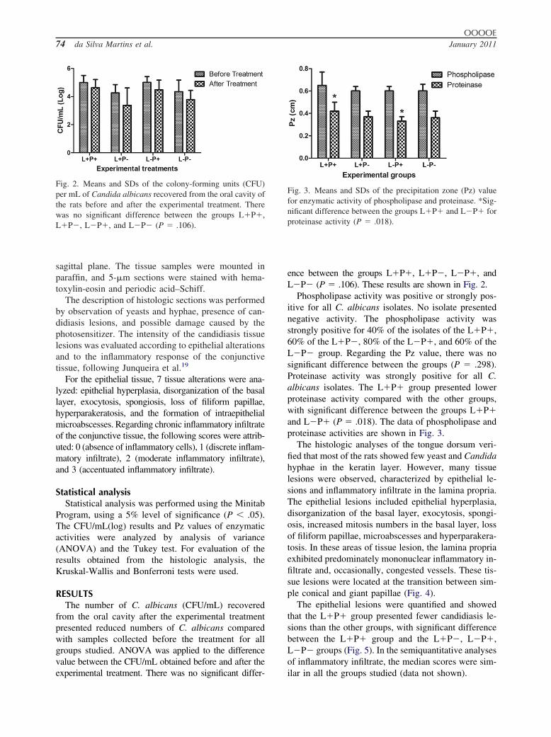
Fig. 2. Means and SDs of the colony-forming units (CFU)per mL of Candida albicans recovered from the oral cavity ofthe rats before and after the experimental treatment. Therewas no significant difference between the groups L�P�,L�P�, L�P�, and L�P� (P � .106).
experimental treatment. There was no significant differ-
ence between the groups L�P�, L�P�, L�P�, andL�P� (P � .106). These results are shown in Fig. 2.
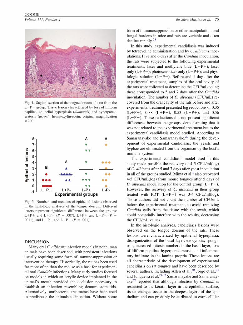
Phospholipase activity was positive or strongly pos-itive for all C. albicans isolates. No isolate presentednegative activity. The phospholipase activity wasstrongly positive for 40% of the isolates of the L�P�,60% of the L�P�, 80% of the L�P�, and 60% of theL�P� group. Regarding the Pz value, there was nosignificant difference between the groups (P � .298).Proteinase activity was strongly positive for all C.albicans isolates. The L�P� group presented lowerproteinase activity compared with the other groups,with significant difference between the groups L�P�and L�P� (P � .018). The data of phospholipase andproteinase activities are shown in Fig. 3.
The histologic analyses of the tongue dorsum veri-fied that most of the rats showed few yeast and Candidahyphae in the keratin layer. However, many tissuelesions were observed, characterized by epithelial le-sions and inflammatory infiltrate in the lamina propria.The epithelial lesions included epithelial hyperplasia,disorganization of the basal layer, exocytosis, spongi-osis, increased mitosis numbers in the basal layer, lossof filiform papillae, microabscesses and hyperparakera-tosis. In these areas of tissue lesion, the lamina propriaexhibited predominately mononuclear inflammatory in-filtrate and, occasionally, congested vessels. These tis-sue lesions were located at the transition between sim-ple conical and giant papillae (Fig. 4).
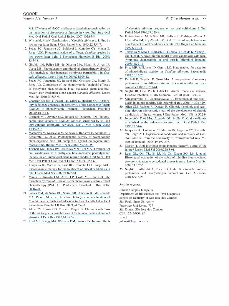
The epithelial lesions were quantified and showedthat the L�P� group presented fewer candidiasis le-sions than the other groups, with significant differencebetween the L�P� group and the L�P�, L�P�,L�P� groups (Fig. 5). In the semiquantitative analysesof inflammatory infiltrate, the median scores were sim-
Fig. 3. Means and SDs of the precipitation zone (Pz) valuefor enzymatic activity of phospholipase and proteinase. *Sig-nificant difference between the groups L�P� and L�P� forproteinase activity (P � .018).
ilar in all the groups studied (data not shown).
OOOOEVolume 111, Number 1 da Silva Martins et al. 75
DISCUSSIONMany oral C. albicans infection models in nonhuman
animals have been described, with persistent infectionsusually requiring some form of immunosuppression orintervention therapy. Historically, the rat has been usedfar more often than the mouse as a host for experimen-tal oral Candida infections. Many early studies focusedon models in which an acrylic device implanted in theanimal’s mouth provided the occlusion necessary toestablish an infection resembling denture stomatitis.Alternatively, antibacterial treatments have been used
Fig. 4. Sagittal section of the tongue dorsum of a rat from theL�P� group. Tissue lesion characterized by loss of filiformpapillae, epithelial hyperplasia (diamonds) and hyperparak-eratosis (arrow). hematoxylin-eosin; original magnification�200.
Fig. 5. Numbers and medians of epithelial lesions observedin the histologic analyses of the tongue dorsum. Differentletters represent significant difference between the groups:L�P� and L�P� (P � .007), L�P� and L�P� (P �0011), and L�P� and L�P� (P � .001).
to predispose the animals to infection. Without some
form of immunosuppression or other manipulation, oralfungal burdens in mice and rats are variable and oftendecline rapidly.28
In this study, experimental candidiasis was inducedby tetracycline administration and by C. albicans inoc-ulations. Five and 6 days after the Candida inoculation,the rats were subjected to the following experimentaltreatments: laser and methylene blue (L�P�); laseronly (L�P�); photosensitizer only (L�P�); and phys-iologic solution (L�P�). Before and 1 day after theexperimental treatment, samples of the oral cavity ofthe rats were collected to determine the CFU/mL count;these corresponded to 5 and 7 days after the Candidainoculation. The number of C. albicans (CFU/mL) re-covered from the oral cavity of the rats before and afterexperimental treatment presented log reductions of 0.35(L�P�), 0.88 (L�P�), 0.53 (L�P�), and 0.56(L�P�). These reductions did not present significantdifferences between the groups, demonstrating that itwas not related to the experimental treatment but to theexperimental candidiasis model studied. According toSamaranayake and Samaranayake,29 during the devel-opment of experimental candidiasis, the yeasts andhyphae are eliminated from the organism by the host’simmune system.
The experimental candidiasis model used in thisstudy made possible the recovery of 4-5 CFU/ml(log)of C. albicans after 5 and 7 days after yeast inoculationin all of the groups studied. Mima et al.8 also recovered4-5 CFU/mL(log) from mouse tongues after 5 days ofC. albicans inoculation for the control group (L�P�).However, the recovery of C. albicans in their grouptreated with PDT (L�P�) was 3-4 CFU/ml(log).Those authors did not count the number of CFU/mLbefore the experimental treatment, to avoid removingCandida cells from the tissue with the swab, whichcould potentially interfere with the results, decreasingthe CFU/mL values.
In the histologic analyses, candidiasis lesions wereobserved on the tongue dorsum of the rats. Theselesions were characterized by epithelial hyperplasia,disorganization of the basal layer, exocytosis, spongi-osis, increased mitosis numbers in the basal layer, lossof filiform papillae, hyperparakeratosis, and inflamma-tory infiltrate in the lamina propria. These lesions areall characteristic of the development of experimentalcandidiasis on rat tongues and have been described byseveral authors, including Allen et al.,30 Jorge et al.,31
and Junqueira et al.19,32 Samaranayake and Samaranay-ake29 reported that although infection by Candida isrestricted to the keratin layer in the epithelial surface,tissue changes occur in the deepest layers of the epi-
thelium and can probably be attributed to extracellular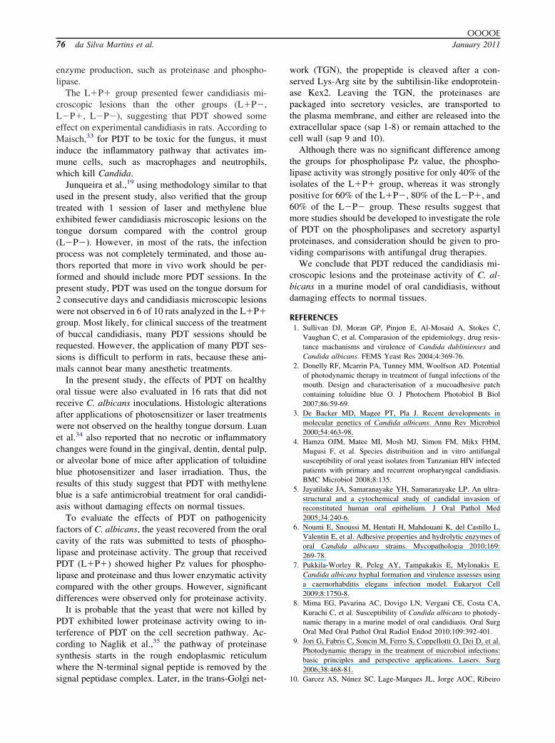
OOOOE76 da Silva Martins et al. January 2011
enzyme production, such as proteinase and phospho-lipase.
The L�P� group presented fewer candidiasis mi-croscopic lesions than the other groups (L�P�,L�P�, L�P�), suggesting that PDT showed someeffect on experimental candidiasis in rats. According toMaisch,33 for PDT to be toxic for the fungus, it mustinduce the inflammatory pathway that activates im-mune cells, such as macrophages and neutrophils,which kill Candida.
Junqueira et al.,19 using methodology similar to thatused in the present study, also verified that the grouptreated with 1 session of laser and methylene blueexhibited fewer candidiasis microscopic lesions on thetongue dorsum compared with the control group(L�P�). However, in most of the rats, the infectionprocess was not completely terminated, and those au-thors reported that more in vivo work should be per-formed and should include more PDT sessions. In thepresent study, PDT was used on the tongue dorsum for2 consecutive days and candidiasis microscopic lesionswere not observed in 6 of 10 rats analyzed in the L�P�group. Most likely, for clinical success of the treatmentof buccal candidiasis, many PDT sessions should berequested. However, the application of many PDT ses-sions is difficult to perform in rats, because these ani-mals cannot bear many anesthetic treatments.
In the present study, the effects of PDT on healthyoral tissue were also evaluated in 16 rats that did notreceive C. albicans inoculations. Histologic alterationsafter applications of photosensitizer or laser treatmentswere not observed on the healthy tongue dorsum. Luanet al.34 also reported that no necrotic or inflammatorychanges were found in the gingival, dentin, dental pulp,or alveolar bone of mice after application of toluidineblue photosensitizer and laser irradiation. Thus, theresults of this study suggest that PDT with methyleneblue is a safe antimicrobial treatment for oral candidi-asis without damaging effects on normal tissues.
To evaluate the effects of PDT on pathogenicityfactors of C. albicans, the yeast recovered from the oralcavity of the rats was submitted to tests of phospho-lipase and proteinase activity. The group that receivedPDT (L�P�) showed higher Pz values for phospho-lipase and proteinase and thus lower enzymatic activitycompared with the other groups. However, significantdifferences were observed only for proteinase activity.
It is probable that the yeast that were not killed byPDT exhibited lower proteinase activity owing to in-terference of PDT on the cell secretion pathway. Ac-cording to Naglik et al.,35 the pathway of proteinasesynthesis starts in the rough endoplasmic reticulumwhere the N-terminal signal peptide is removed by the
signal peptidase complex. Later, in the trans-Golgi net-work (TGN), the propeptide is cleaved after a con-served Lys-Arg site by the subtilisin-like endoprotein-ase Kex2. Leaving the TGN, the proteinases arepackaged into secretory vesicles, are transported tothe plasma membrane, and either are released into theextracellular space (sap 1-8) or remain attached to thecell wall (sap 9 and 10).
Although there was no significant difference amongthe groups for phospholipase Pz value, the phospho-lipase activity was strongly positive for only 40% of theisolates of the L�P� group, whereas it was stronglypositive for 60% of the L�P�, 80% of the L�P�, and60% of the L�P� group. These results suggest thatmore studies should be developed to investigate the roleof PDT on the phospholipases and secretory aspartylproteinases, and consideration should be given to pro-viding comparisons with antifungal drug therapies.
We conclude that PDT reduced the candidiasis mi-croscopic lesions and the proteinase activity of C. al-bicans in a murine model of oral candidiasis, withoutdamaging effects to normal tissues.
REFERENCES1. Sullivan DJ, Moran GP, Pinjon E, Al-Mosaid A, Stokes C,
Vaughan C, et al. Comparasion of the epidemiology, drug resis-tance machanisms and virulence of Candida dublinienses andCandida albicans. FEMS Yeast Res 2004;4:369-76.
2. Donelly RF, Mcarrin PA, Tunney MM, Woolfson AD. Potentialof photodynamic therapy in treatment of fungal infections of themouth. Design and characterisation of a mucoadhesive patchcontaining toluidine blue O. J Photochem Photobiol B Biol2007;86:59-69.
3. De Backer MD, Magee PT, Pla J. Recent developments inmolecular genetics of Candida albicans. Annu Rev Microbiol2000;54:463-98.
4. Hamza OJM, Matee MI, Mosh MJ, Simon FM, Mikx FHM,Mugusi F, et al. Species distribuition and in vitro antifungalsusceptibility of oral yeast isolates from Tanzanian HIV infectedpatients with primary and recurrent oropharyngeal candidiasis.BMC Microbiol 2008;8:135.
5. Jayatilake JA, Samaranayake YH, Samaranayake LP. An ultra-structural and a cytochemical study of candidal invasion ofreconstituted human oral epithelium. J Oral Pathol Med2005;34:240-6.
6. Noumi E, Snoussi M, Hentati H, Mahdouani K, del Castillo L,Valentin E, et al. Adhesive properties and hydrolytic enzymes oforal Candida albicans strains. Mycopathologia 2010;169:269-78.
7. Pukkila-Worley R, Peleg AY, Tampakakis E, Mylonakis E.Candida albicans hyphal formation and virulence assesses usinga caernorhabditis elegans infection model. Eukaryot Cell2009;8:1750-8.
8. Mima EG, Pavarina AC, Dovigo LN, Vergani CE, Costa CA,Kurachi C, et al. Susceptibility of Candida albicans to photody-namic therapy in a murine model of oral candidiasis. Oral SurgOral Med Oral Pathol Oral Radiol Endod 2010;109:392-401.
9. Jori G, Fabris C, Soncin M, Ferro S, Coppellotti O, Dei D, et al.Photodynamic therapy in the treatment of microbiol infections:basic principles and perspective applications. Lasers. Surg2006;38:468-81.
10. Garcez AS, Núnez SC, Lage-Marques JL, Jorge AOC, Ribeiro
OOOOEVolume 111, Number 1 da Silva Martins et al. 77
MS. Efficiency of NaOCl and laser assisted photosensitization onthe reduction of Enterococcus faecalis in vitro. Oral Surg OralMed Oral Pathol Oral Radiol Endod 2007;102:93-8.
11. Wilson M, Mia N. Sensitisation of Candida albicans to killing bylow-power laser light. J Oral Pathol Med 1993;22:354-7.
12. Souza SC, Junqueira JC, Balducci I, Koga-Ito CY, Munin E,Jorge AOC. Photosensitization of different Candida species bylow power laser light. J Photochem Photobiol B Biol 2006;83:34-8.
13. Giroldo LM, Felipe MP, de Oliveira MA, Munin E, Alves LP,Costa MS. Photodynamic antimicrobial chemotherapy (PACT)with methylene blue increases membrane permeability in Can-dida albicans. Lasers Med Sci 2009;24:109-12.
14. Souza RC, Junqueira JC, Rossoni RD, Cristiane CA, Munin E,Jorge AO. Comparison of the photodynamic fungicidal efficacyof methylene blue, toluidine blue, malachite green and low-power laser irradiation alone against Candida albicans. LasersMed Sci 2010;25:385-9.
15. Chabrier-Roselló Y, Foster TH, Mitra S, Haidaris CG. Respira-tory deficiency enhances the sensitivity of the pathogenic fungusCandida to photodynamic treatment. Photochem Photobiol2008;84:1141-8.
16. Cormick MP, Alvarez MG, Rovera M, Durantini EN. Photody-namic inactivations of Candida albicans sensitized by tri- andtetra-cationic porphyrin derivates. Eur J Med Chem 2009;44:1592-9.
17. Mantareva V, Kussovski V, Angelov I, Borisova E, Avramov L,Schnurpfeil G, et al. Photodynamic activity of water-solublephthalocyanine zinc (II) complexes against pathogenic mic-roorganisms. Bioorg Med Chem 2007;15:4829-35.
18. Teichert MC, Jones JW, Usacheva MN, Biel MA. Treatment oforal candidiasis with methylene blue–mediated photodynamictherapy in an immunodeficient murine model. Oral Surg OralMed Oral Pathol Oral Radiol Endod 2002;93:155-60.
19. Junqueira JC, Martins JS, Faria RL, Colombo CED, Jorge AOC.Photodynamic therapy for the treatment of buccal candidiasis inrats. Lasers Med Sci 2009;24:877-84.
20. Munin E, Giroldo LM, Alves LP, Costa MS. Study of tubeformation by Candida albicans after photodynamic antimicrobialchemotherapy (PACT). J Photochem Photobiol B Biol 2007;88:16-20.
21. Soares BM, da Silva DL, Sousa GR, Amorim JC, de ResendeMA, Pinotti M, et al. In vitro photodynamic inactivation ofCandida spp. growth and adhesion to buccal epithelial cells. JPhotochem Photobiol B Biol 2009;94:65-70.
22. Allen CM, Blozis GG, Rosen S, Bright JS. Chronic candidiasisof the rat tongue: a possible model for human median rhomboidglossitis. J Dent Res 1982;61:287-91.
23. Reed MF, Scragg MA, Williams DM, Soames JV. In vivo effects
of Candida albicans products on rat oral epithelium. J OralPathol Med 1990;19:326-9.
24. Freire-Garabal M, Núñez MJ, Balboa J, Rodríguez-Cobo A,López-Paz JM, Rey-Méndez M, et al. Effects of amphetamine ondevelopment of oral candidiasis in rats. Clin Diagn Lab Immunol1999;6:530-3.
25. Takakura N, Sato Y, Ishibashi H, Oshima H, Uchida K, Yamagu-chi H, et al. A novel murine model of oral candidiasis with localsymptoms characteristic of oral thrush. Microbiol Immunol2003;47:321-6.
26. Price MF, Wilkinson ID, Gentry LO. Plate method for detectionof phospholipase activity in Candida albicans. Sabouraudia1982;20:15-20.
27. Ruchell R, Tegeller R, Trost MA. A comparison of secretoryproteinases from different strains of Candida albicans. Sab-ouraudia 1982;20:233-44.
28. Naglik JR, Fidel PL Jr, Odds FC. Animal models of mucosalCandida infection. FEMS Microbiol Lett 2008;283:129-39.
29. Samaranayake YU, Samaranayake LP. Experimental oral candi-diasis in animal models. Clin Microbiol Rev 2001;14:398-429.
30. Allen CM, Paulson R, Duncan R. Clinical, histologic and scan-ning electron microscopic study of the development of chroniccandidiasis of the rat tongue. J Oral Pathol Med 1989;18:352-9.
31. Jorge AO, Totti MA, Almeida OP, Scully C. Oral candidosisestablished in the sialoadenectomized rat. J Oral Pathol Med1993;22:54-6.
32. Junqueira JC, Colombo CE, Martins JS, Koga Ito CY, CarvalhoYR, Jorge AO. Experimental candidosis and recovery of Can-dida albicans from the oral cavity of ovariectomized rats. Mi-crobiol Immunol 2005;49:199-207.
33. Maisch T. Anti-microbial photodynamic therapy: useful in thefuture? Lasers Med Sci 2006;22:83-91.
34. Luan XL, Qin YL, Bi LJ, Hu Cy, Zhang ZG, Lin J, et al.Histological evaluation of the safety of toluidine blue–mediatedphotosensitization to periodontal tissues in mice. Lasers Med Sci2009;24:162-6.
35. Naglik J, Albrecht A, Bader O, Hube B. Candida albicansproteinases and host/pathogen interactions. Cell Microbiol2004;6:915-26.
Reprint requests:
Juliana Campos JunqueiraDepartment of Biosciences and Oral DiagnosisSchool of Dentistry of São José dos CamposSão Paulo State UniversityFrancisco José Longo 777São Dimas, São José dos CamposCEP 12245-000, SPBrazil
[email protected]
Related Documents