diagnostics Article Analysis of Residual Ridge Morphology in a Group of Edentulous Patients Seeking NHS Dental Implant Provision—A Retrospective Observational Lateral Cephalometric Study Rafif Alshenaiber 1,2, * , Callum Cowan 1 , Craig Barclay 3 and Nikolaos Silikas 1 Citation: Alshenaiber, R.; Cowan, C.; Barclay, C.; Silikas, N. Analysis of Residual Ridge Morphology in a Group of Edentulous Patients Seeking NHS Dental Implant Provision—A Retrospective Observational Lateral Cephalometric Study. Diagnostics 2021, 11, 2348. https://doi.org/10.3390/ diagnostics11122348 Academic Editor: Timo Sorsa Received: 25 November 2021 Accepted: 9 December 2021 Published: 13 December 2021 Publisher’s Note: MDPI stays neutral with regard to jurisdictional claims in published maps and institutional affil- iations. Copyright: © 2021 by the authors. Licensee MDPI, Basel, Switzerland. This article is an open access article distributed under the terms and conditions of the Creative Commons Attribution (CC BY) license (https:// creativecommons.org/licenses/by/ 4.0/). 1 Division of Dentistry, Faculty of Biology, Medicine and Health, Coupland 3 Building, University of Manchester, Manchester M13 9PL, UK; [email protected] (C.C.); [email protected] (N.S.) 2 Prosthetic Dental Sciences Department, College of Dentistry, Prince Sattam Bin Abdulaziz University, AlKharj 16278, Saudi Arabia 3 Manchester Dental Hospital, Higher Cambridge Street, Manchester M15 6FH, UK; [email protected] * Correspondence: rafi[email protected]; Tel.: +44-7477-426-007 Abstract: A convenience sample of 154 edentulous patients referred for implant provision at a Regional National Health Service Dental Hospital in the North West of England were identified. The cephalometric radiographs that were taken as part of the patient baseline investigation were assessed. Digital tracing was used to measure the anterior maxillary and mandibular bone height and ridge angle with respect to the maxillary and mandibular planes. The mean height of the bone in the maxilla was found to be 14 mm, and the mean ridge angle for the anterior maxillary residual ridge is 104 ◦ . The mean height of bone in the mandible was 18 mm, while the mean ridge angle for the anterior mandibular residual ridge was 77 ◦ . Using the Cawood and Howell classification demonstrated that class VI mandibles were the most common. The cross-sectional shape of the mandible varied, with the triangular shape most common. Although there was adequate bone stock for implant placement in these cases, the mandibular residual ridge resorption presents a lingual inclination to the residual bone. The limited residual ridge position and inclination would dictate that conventional implant placement could be challenging. Keywords: resorption; edentulous; lateral cephalometric radiograph; residual ridge; mandible; maxilla 1. Introduction The number of patients in the UK that are edentulous is falling. The Adult Dental Health Survey in 2009 revealed that 94% of adults in England, Wales and Northern Ire- land retained at least one natural tooth. In the same study, it was noted that there are still about 2.7 million edentulous adults [1]. Therefore, dentists will continue to need conventional complete denture skills for the foreseeable future [2]. It is well established in the classical studies by Tallgren and Carlsson that tooth extraction results in alveolar bone resorption [3,4]. Subsequent studies have failed to agree on the rate, volume and distribution of this resorption, and there appears to be a high degree of variation between individuals. A range of factors are thought to impact the rate of resorption, such as age, gender and metabolic disorders [5]. Case-control studies contrasting denture wearers with non-denture wearers suggest that the wearing of a prosthesis itself will increase the rate of bone resorption [6,7]. If the literature were to be broadly summarised, resorption is documented to occur most rapidly in the first 3–6 months after tooth extraction with up to a 50% reduction in ridge width at 12 months [8,9]. Both horizontal and vertical ridge resorption occurs, but most studies suggest that there is a greater horizontal than vertical component [10,11]. Resorption is thought to occur at up to four times greater rate Diagnostics 2021, 11, 2348. https://doi.org/10.3390/diagnostics11122348 https://www.mdpi.com/journal/diagnostics

Welcome message from author
This document is posted to help you gain knowledge. Please leave a comment to let me know what you think about it! Share it to your friends and learn new things together.
Transcript

diagnostics
Article
Analysis of Residual Ridge Morphology in a Group ofEdentulous Patients Seeking NHS Dental Implant Provision—ARetrospective Observational Lateral Cephalometric Study
Rafif Alshenaiber 1,2,* , Callum Cowan 1, Craig Barclay 3 and Nikolaos Silikas 1
�����������������
Citation: Alshenaiber, R.; Cowan, C.;
Barclay, C.; Silikas, N. Analysis of
Residual Ridge Morphology in a
Group of Edentulous Patients
Seeking NHS Dental Implant
Provision—A Retrospective
Observational Lateral Cephalometric
Study. Diagnostics 2021, 11, 2348.
https://doi.org/10.3390/
diagnostics11122348
Academic Editor: Timo Sorsa
Received: 25 November 2021
Accepted: 9 December 2021
Published: 13 December 2021
Publisher’s Note: MDPI stays neutral
with regard to jurisdictional claims in
published maps and institutional affil-
iations.
Copyright: © 2021 by the authors.
Licensee MDPI, Basel, Switzerland.
This article is an open access article
distributed under the terms and
conditions of the Creative Commons
Attribution (CC BY) license (https://
creativecommons.org/licenses/by/
4.0/).
1 Division of Dentistry, Faculty of Biology, Medicine and Health, Coupland 3 Building, University ofManchester, Manchester M13 9PL, UK; [email protected] (C.C.);[email protected] (N.S.)
2 Prosthetic Dental Sciences Department, College of Dentistry, Prince Sattam Bin Abdulaziz University,AlKharj 16278, Saudi Arabia
3 Manchester Dental Hospital, Higher Cambridge Street, Manchester M15 6FH, UK;[email protected]
* Correspondence: [email protected]; Tel.: +44-7477-426-007
Abstract: A convenience sample of 154 edentulous patients referred for implant provision at aRegional National Health Service Dental Hospital in the North West of England were identified.The cephalometric radiographs that were taken as part of the patient baseline investigation wereassessed. Digital tracing was used to measure the anterior maxillary and mandibular bone heightand ridge angle with respect to the maxillary and mandibular planes. The mean height of the bonein the maxilla was found to be 14 mm, and the mean ridge angle for the anterior maxillary residualridge is 104◦. The mean height of bone in the mandible was 18 mm, while the mean ridge anglefor the anterior mandibular residual ridge was 77◦. Using the Cawood and Howell classificationdemonstrated that class VI mandibles were the most common. The cross-sectional shape of themandible varied, with the triangular shape most common. Although there was adequate bone stockfor implant placement in these cases, the mandibular residual ridge resorption presents a lingualinclination to the residual bone. The limited residual ridge position and inclination would dictatethat conventional implant placement could be challenging.
Keywords: resorption; edentulous; lateral cephalometric radiograph; residual ridge;mandible; maxilla
1. Introduction
The number of patients in the UK that are edentulous is falling. The Adult DentalHealth Survey in 2009 revealed that 94% of adults in England, Wales and Northern Ire-land retained at least one natural tooth. In the same study, it was noted that there arestill about 2.7 million edentulous adults [1]. Therefore, dentists will continue to needconventional complete denture skills for the foreseeable future [2]. It is well establishedin the classical studies by Tallgren and Carlsson that tooth extraction results in alveolarbone resorption [3,4]. Subsequent studies have failed to agree on the rate, volume anddistribution of this resorption, and there appears to be a high degree of variation betweenindividuals. A range of factors are thought to impact the rate of resorption, such as age,gender and metabolic disorders [5]. Case-control studies contrasting denture wearerswith non-denture wearers suggest that the wearing of a prosthesis itself will increase therate of bone resorption [6,7]. If the literature were to be broadly summarised, resorptionis documented to occur most rapidly in the first 3–6 months after tooth extraction withup to a 50% reduction in ridge width at 12 months [8,9]. Both horizontal and verticalridge resorption occurs, but most studies suggest that there is a greater horizontal thanvertical component [10,11]. Resorption is thought to occur at up to four times greater rate
Diagnostics 2021, 11, 2348. https://doi.org/10.3390/diagnostics11122348 https://www.mdpi.com/journal/diagnostics
Diagnostics 2021, 11, 2348 2 of 8
in the mandible when compared to the maxilla [12,13]. This is complicated by the factthat the receptive pattern of the maxilla results in the residual bone stock position beingupward and inward, and in the mandible the bone resorbs downward and outward, oftenincreasing any baseline skeletal discrepancies [14].
The interplay between the clinical and technical aspects of denture constructionand the patient’s psychosocial state dictates that success with complete dentures can beunpredictable. This can be frustrating and disheartening for the treating clinician. Althoughresearch has not demonstrated a strong correlation between anatomic conditions, denturequality and denture success, conventional wisdom dictates that successful dentures aremore difficult to achieve in patients with severely resorbed ridges [15]. The McGill and Yorkconsensus statements have clearly defined that a two-implant overdenture is the minimumstandard of care for the edentulous mandible [16,17]. Due to financial constraints, thisstandard of care has not been achieved for most. In those complete denture wearersthat do receive dental implants, the use of prosthetic componentry to attach a dentureto the implant will increase its retention. Depending on the number of implants placedand prosthetic componentry used, they can also provide significant support and, hence,compensate for poor supporting structures such and thin atrophied ridges and soft tissues.In addition, implants slow or prevent further alveolar bone resorption [18]. With implantosseointegration, and stimulation of the remaining alveolar bone, the resorptive processis reduced. This is hugely advantageous, with early and simple intervention potentiallypreventing progressive bone loss and, therefore, the need for the significant surgicalintervention that is often needed to definitively address the severely atrophic mandible.
At Manchester dental hospital, patients who are referred after failing to tolerateconventional denture construction will be assessed at a Joint Implant Clinic. If it is agreedthat the conventional options for treatment have been exhausted, the patient does notsmoke and they are medically fit enough to undergo surgical treatment, funding to placedental implants will be applied for through the National Commissioning Service (NHS).The final implant plan will vary depending on the needs of the patient and the bone stockavailable. As part of the patients’ workup for the Joint Implant Clinic, they will havea panoramic tomography and lateral cephalometric radiographs taken. These specialinvestigations are often sufficient to determine if adequate bone height is present to placedental implants.
The aim of the study was to assess the residual bone morphology that was presentin the anterior maxilla and mandible in a cohort of patients who had failed to tolerateconventional complete dentures and were seeking NHS dental implant provision.
2. Materials and Methods
This is a single centre retrospective descriptive observational study. The protocolwas granted ethical approval by the Health Research Authority. It was given the Inte-grated Research Approval System number 238472 for NHS organisations in England anda University of Manchester sponsor reference number NHS001339. The principles of the“Declaration of Helsinki”, Good Clinical Practice (GCP) and the laws and regulations ofthe country in which this research was conducted were adhered to.
2.1. Patient Selection
All edentulous patients at Manchester Dental Hospital, Manchester, UK who hadbeen treated with dental implants or had dental implant funding approved and wereawaiting treatment between June 2013 and August 2017 were assessed, and a total of743 patients were identified. Patients were excluded from the study for the followingreasons: (1) No cephalometric radiograph was taken for the patient; (2) The cut or positionof the cephalometric radiograph prevented complete landmark analysis; (3) There was evi-dence of residual dentition or dental implants on the cephalometric radiograph; (4) Therewas evidence of mini-screws or plates on the radiograph indicating previous trauma orbone grafting.
Diagnostics 2021, 11, 2348 3 of 8
This resulted in a convenience sample of 154 patients. A total of 146 maxillary and152 mandibular edentulous arches were included in the study.
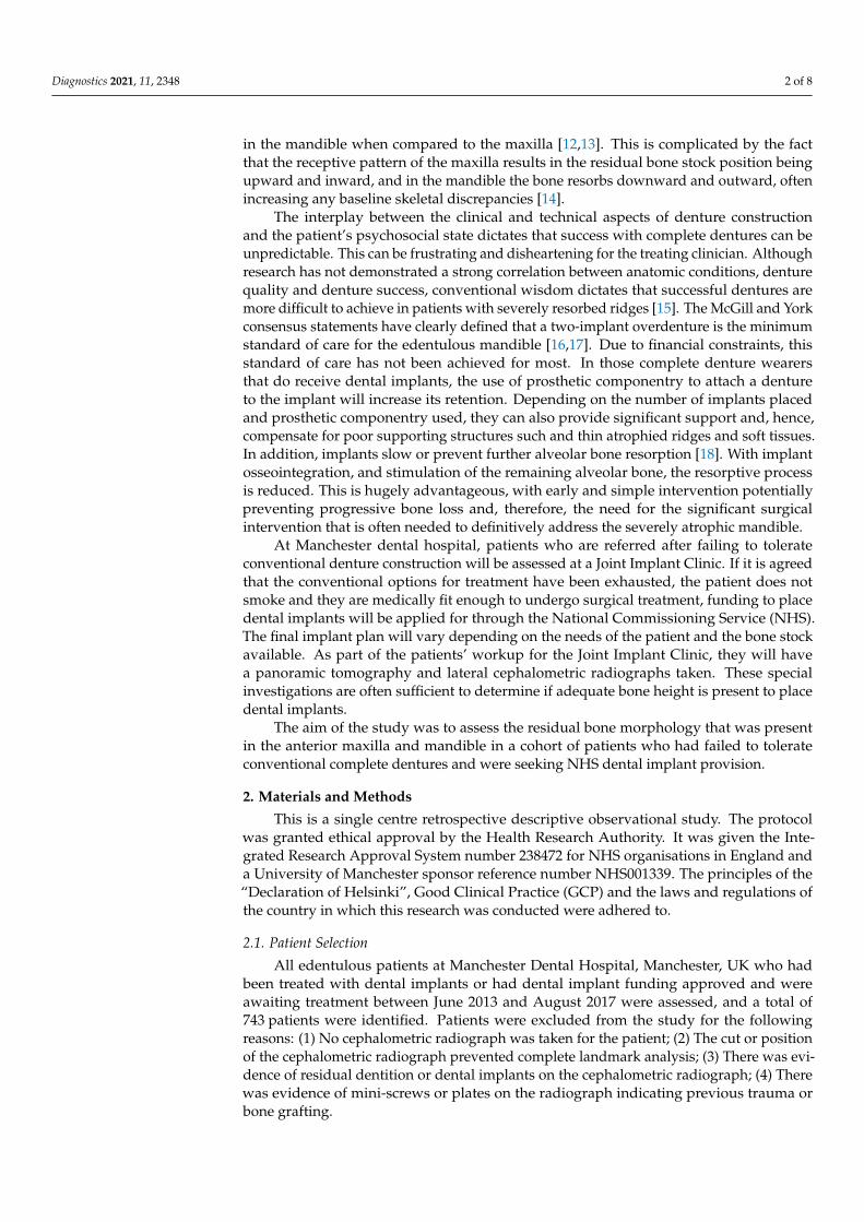
Each lateral cephalometric radiograph was taken using Planmeca Promax® (PlanmecaUK Limited, Coventry, UK), they were anonymised before being traced by a single examiner.This was completed on the Picture Archiving and Communication System (PACS) imagingsystem (Insignia Medical System, Basingstocke, UK) using the annotation functions. Thekey anatomical landmarks were identified and, using the dentate cephalometric tracing,tracing lines were also added to the tracing presented in Table 1. An example tracingtemplate is shown in Figure 1.
Table 1. Landmarks added by using the lateral cephalometric dentate tracing as guidance.
Tracing Lines Reference
A line bisecting the maxillary residual ridge crest whichrepresent the highest midline point of the anterior
maxillary ridge; this mimics taxis of the maxillary incisor(UInc) in dentate subjects.
This line is the preferable maxillary implantposition.
Maxillary mid-ridge inclination which represents the anglebetween the maxillary plane (MxPl) and the line bisecting
the maxillary residual ridge.
The maxillary incisal inclination in dentatetracing.
A line bisecting the mandibular residual ridge whichrepresent the midline point on the anterior mandibular
ridge; this mimics the axis of the mandibular incisor (LInc)in dentate subjects.
This line is the preferable mandibular implantposition
Mandibular mid-ridge inclination which represents theangle between the mandibular plane (MnPl) and the line
bisecting the mandibular residual ridge.
The mandibular incisal inclination in dentatetracing.
Diagnostics 2021, 11, x FOR PEER REVIEW 3 of 9
cephalometric radiograph prevented complete landmark analysis; (3) There was evidence
of residual dentition or dental implants on the cephalometric radiograph; (4) There was
evidence of mini-screws or plates on the radiograph indicating previous trauma or bone
grafting.
This resulted in a convenience sample of 154 patients. A total of 146 maxillary and
152 mandibular edentulous arches were included in the study.
Each lateral cephalometric radiograph was taken using Planmeca Promax®
(Planmeca UK Limited, Coventry, UK), they were anonymised before being traced by a
single examiner. This was completed on the Picture Archiving and Communication
System (PACS) imaging system (Insignia Medical System, Basingstocke, UK) using the
annotation functions. The key anatomical landmarks were identified and, using the
dentate cephalometric tracing, tracing lines were also added to the tracing presented in
Table 1. An example tracing template is shown in Figure 1.
Figure 1. Tracing template used in this study: the red line in the maxillae connecting the anterior
and posterior nasal spine (ANS-PNS line) represents the maxillary plane reference (MxPl); the red
line in the mandible connecting the menton and the gonion (Me-Go line) represents the mandibular
plane reference (MnPl); the yellow line in the maxillae is the line bisecting the maxillary residual
ridge; the yellow line in the mandible is the line bisecting the mandibular residual ridge. The angles
in green represent the maxillary and the mandibular incisal inclination adopted from dentate
tracing.
Table 1. Landmarks added by using the lateral cephalometric dentate tracing as guidance.
Tracing Lines Reference
A line bisecting the maxillary residual ridge crest which repre-
sent the highest midline point of the anterior maxillary ridge;
this mimics taxis of the maxillary incisor (UInc) in dentate sub-
jects.
This line is the preferable
maxillary implant posi-
tion.
Maxillary mid-ridge inclination which represents the angle be-
tween the maxillary plane (MxPl) and the line bisecting the
maxillary residual ridge.
The maxillary incisal in-
clination in dentate trac-
ing.
Figure 1. Tracing template used in this study: the red line in the maxillae connecting the anterior andposterior nasal spine (ANS-PNS line) represents the maxillary plane reference (MxPl); the red line inthe mandible connecting the menton and the gonion (Me-Go line) represents the mandibular planereference (MnPl); the yellow line in the maxillae is the line bisecting the maxillary residual ridge; theyellow line in the mandible is the line bisecting the mandibular residual ridge. The angles in greenrepresent the maxillary and the mandibular incisal inclination adopted from dentate tracing.
Diagnostics 2021, 11, 2348 4 of 8
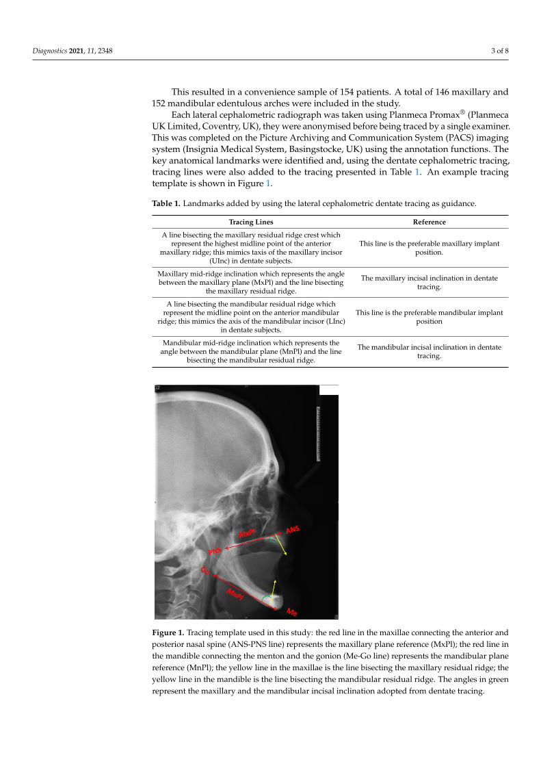
A line was then drawn from the maximum midpoint of the ridge to the maxillaryand mandibular plane angles. This line would transect the point on the residual alveolarridge of maximum height (Figure 2). The difference in height between the maxillary ormandibular plane and the maximum height of the residual ridge was then measured.
Diagnostics 2021, 11, x FOR PEER REVIEW 4 of 9
A line bisecting the mandibular residual ridge which repre-
sent the midline point on the anterior mandibular ridge; this
mimics the axis of the mandibular incisor (LInc) in dentate
subjects.
This line is the preferable
mandibular implant posi-
tion
Mandibular mid-ridge inclination which represents the angle
between the mandibular plane (MnPl) and the line bisecting
the mandibular residual ridge.
The mandibular incisal
inclination in dentate
tracing.
A line was then drawn from the maximum midpoint of the ridge to the maxillary and
mandibular plane angles. This line would transect the point on the residual alveolar ridge
of maximum height (Figure 2). The difference in height between the maxillary or
mandibular plane and the maximum height of the residual ridge was then measured.
Figure 2. The red lines represent the height of the anterior part of maxillary (top) and mandibular
arches (bottom).
2.2. Statistical Analysis
All data were analysed using IBM SPSS Version 22.0 (IBM Corp. Released 2013. IBM
SPSS Statistics for Windows, Version 22.0 Armonk, NY, USA: IBM Corp). The data were
assessed for normality using a Kolmogorov-Smirnov test and histograms. Intra-operator
reliability was assessed using Cohen’s kappa correlation coefficient.
3. Results
These tests showed the data to be normally distributed. Summary statistics are
therefore displayed as mean and standard deviation.
Intra-operator reliability was measured using a test–retest process. Each anonymised
Cephalometric tracing was allocated a number. Three weeks after the initial tracings, a
random number generator was used to select 30 cases from the original 154. The
radiographs were then retraced and compared to the original. Results reliability was
demonstrated using a correlation coefficient (Table 2).
Figure 2. The red lines represent the height of the anterior part of maxillary (top) and mandibulararches (bottom).
2.2. Statistical Analysis
All data were analysed using IBM SPSS Version 22.0 (IBM Corp. Released 2013. IBMSPSS Statistics for Windows, Version 22.0 Armonk, NY, USA: IBM Corp). The data wereassessed for normality using a Kolmogorov-Smirnov test and histograms. Intra-operatorreliability was assessed using Cohen’s kappa correlation coefficient.
3. Results
These tests showed the data to be normally distributed. Summary statistics aretherefore displayed as mean and standard deviation.
Intra-operator reliability was measured using a test–retest process. Each anonymisedCephalometric tracing was allocated a number. Three weeks after the initial tracings, a ran-dom number generator was used to select 30 cases from the original 154. The radiographswere then retraced and compared to the original. Results reliability was demonstratedusing a correlation coefficient (Table 2).
Table 2. The intra-examiner agreement for all landmarks and references.
Landmarks and References Mean Kappa Value
The mean length of the anterior residual ridges of the maxilla 0.89
The mean length of the anterior residual ridges of the mandible 0.98
The mean implant angle of placement for the anterior residualridge of the maxilla 0.88
The mean implant angle of placement for the anterior residualridge of the mandible 0.92
Diagnostics 2021, 11, 2348 5 of 8
3.1. Residual Alveolar Ridge Height
The mean residual alveolar ridge height of the maxilla was 14.09 mm ± 3.78 and18.94 mm ± 5.57 for the mandible.
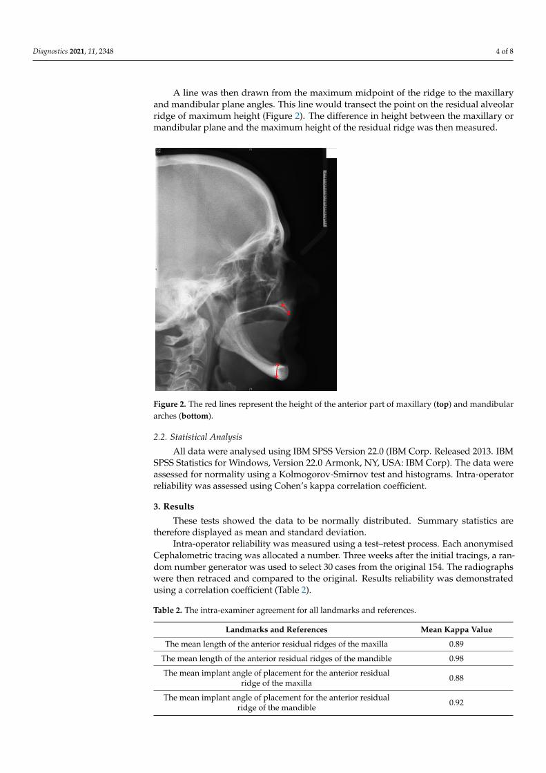
In order to give a more clinical context to the results, the measurements have beensubclassified according to the Cawood and Howell classification (Figure 3). Table 3 showsthe percentage of Cawood and Howell Subgroups in the maxilla and mandible.
Diagnostics 2021, 11, x FOR PEER REVIEW 5 of 9
Table 2. The intra-examiner agreement for all landmarks and references.
Landmarks and References Mean Kappa
Value
The mean length of the anterior residual ridges of the maxilla 0.89
The mean length of the anterior residual ridges of the mandible 0.98
The mean implant angle of placement for the anterior residual ridge of
the maxilla 0.88
The mean implant angle of placement for the anterior residual ridge of
the mandible 0.92
3.1. Residual Alveolar Ridge Height
The mean residual alveolar ridge height of the maxilla was 14.09 mm ± 3.78 and 18.94
mm ± 5.57 for the mandible.
In order to give a more clinical context to the results, the measurements have been
subclassified according to the Cawood and Howell classification (Figure 3). Table 3 shows
the percentage of Cawood and Howell Subgroups in the maxilla and mandible.
Table 3. Percentage and number of cases in each subgroup of the Cawood and Howell
Classification.
Class II Class III Class IV Class V Class VI
Maxilla 14.4% 7.5% 20.5% 29.5% 28.1%
Mandible 5.9% 0.7% 5.9% 13.8% 73.7%
Figure 3. Diagrammatic summary of the Cawood and Howell Classification for the Maxilla (above)
and the Mandible (below).
3.2. Residual Alveolar Ridge Inclination
The mandibular residual ridge/mandibular plane angle and maxillary residual
ridge/maxillary plane angle are also to be calculated (Table 4).
Figure 3. Diagrammatic summary of the Cawood and Howell Classification for the Maxilla (above)and the Mandible (below).
Table 3. Percentage and number of cases in each subgroup of the Cawood and Howell Classification.
Class II Class III Class IV Class V Class VI
Maxilla 14.4% 7.5% 20.5% 29.5% 28.1%
Mandible 5.9% 0.7% 5.9% 13.8% 73.7%
3.2. Residual Alveolar Ridge Inclination
The mandibular residual ridge/mandibular plane angle and maxillary residual ridge/maxillary plane angle are also to be calculated (Table 4).
Table 4. The mean inclination angle of the anterior residual ridges with the mean correction anglesand standard deviations.
Mean Inclination Angle forthe Anterior Residual Ridge Standard Deviation
Maxillary 104.26◦ 7.65
Mandibular 77.46◦ 9.11
3.3. Mandibular Alveolar Bone Cross-Section
Figure 4 shows the three distinct cross-sectional shapes of the anterior mandible. Themajority of the mandibles assessed presented with the classic triangular cross-section. This
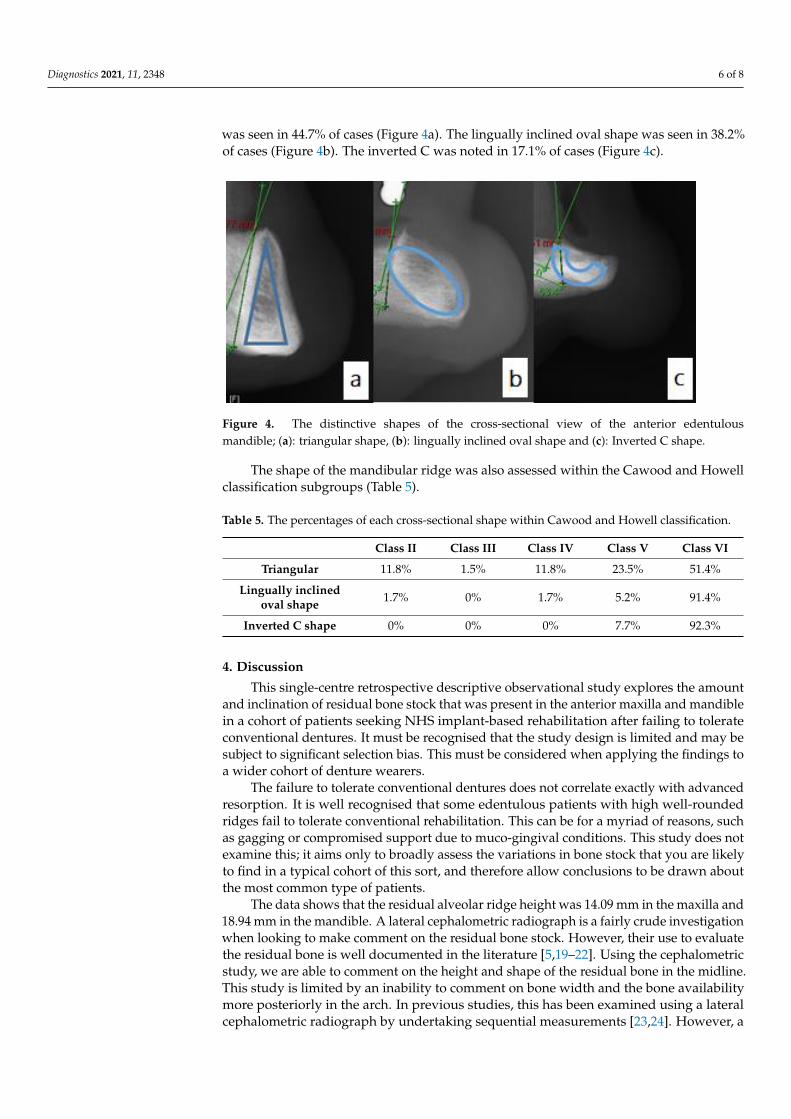
Diagnostics 2021, 11, 2348 6 of 8
was seen in 44.7% of cases (Figure 4a). The lingually inclined oval shape was seen in 38.2%of cases (Figure 4b). The inverted C was noted in 17.1% of cases (Figure 4c).
Diagnostics 2021, 11, x FOR PEER REVIEW 6 of 9
Table 4. The mean inclination angle of the anterior residual ridges with the mean correction angles
and standard deviations.
Mean Inclination Angle for the Anterior Residual
Ridge Standard Deviation
Maxillary 104.26° 7.65
Mandibular 77.46° 9.11
3.3. Mandibular Alveolar Bone Cross-section
Figure 4 shows the three distinct cross-sectional shapes of the anterior mandible. The
majority of the mandibles assessed presented with the classic triangular cross-section.
This was seen in 44.7% of cases (Figure 4a). The lingually inclined oval shape was seen in
38.2% of cases (Figure 4b). The inverted C was noted in 17.1% of cases (Figure 4c).
Figure 4. The distinctive shapes of the cross-sectional view of the anterior edentulous mandible; (a):
triangular shape, (b): lingually inclined oval shape and (c): Inverted C shape.
The shape of the mandibular ridge was also assessed within the Cawood and Howell
classification subgroups (Table 5).
Table 5. The percentages of each cross-sectional shape within Cawood and Howell classification.
Class II Class III Class IV Class V Class VI
Triangular 11.8% 1.5% 11.8% 23.5% 51.4%
Lingually inclined oval
shape 1.7% 0% 1.7% 5.2% 91.4%
Inverted C shape 0% 0% 0% 7.7% 92.3%
4. Discussion
This single-centre retrospective descriptive observational study explores the amount
and inclination of residual bone stock that was present in the anterior maxilla and
mandible in a cohort of patients seeking NHS implant-based rehabilitation after failing to
tolerate conventional dentures. It must be recognised that the study design is limited and
may be subject to significant selection bias. This must be considered when applying the
findings to a wider cohort of denture wearers.
The failure to tolerate conventional dentures does not correlate exactly with
advanced resorption. It is well recognised that some edentulous patients with high well-
rounded ridges fail to tolerate conventional rehabilitation. This can be for a myriad of
reasons, such as gagging or compromised support due to muco-gingival conditions. This
study does not examine this; it aims only to broadly assess the variations in bone stock
that you are likely to find in a typical cohort of this sort, and therefore allow conclusions
to be drawn about the most common type of patients.
Figure 4. The distinctive shapes of the cross-sectional view of the anterior edentulousmandible; (a): triangular shape, (b): lingually inclined oval shape and (c): Inverted C shape.
The shape of the mandibular ridge was also assessed within the Cawood and Howellclassification subgroups (Table 5).
Table 5. The percentages of each cross-sectional shape within Cawood and Howell classification.
Class II Class III Class IV Class V Class VI
Triangular 11.8% 1.5% 11.8% 23.5% 51.4%
Lingually inclinedoval shape 1.7% 0% 1.7% 5.2% 91.4%
Inverted C shape 0% 0% 0% 7.7% 92.3%
4. Discussion
This single-centre retrospective descriptive observational study explores the amountand inclination of residual bone stock that was present in the anterior maxilla and mandiblein a cohort of patients seeking NHS implant-based rehabilitation after failing to tolerateconventional dentures. It must be recognised that the study design is limited and may besubject to significant selection bias. This must be considered when applying the findings toa wider cohort of denture wearers.
The failure to tolerate conventional dentures does not correlate exactly with advancedresorption. It is well recognised that some edentulous patients with high well-roundedridges fail to tolerate conventional rehabilitation. This can be for a myriad of reasons, suchas gagging or compromised support due to muco-gingival conditions. This study does notexamine this; it aims only to broadly assess the variations in bone stock that you are likelyto find in a typical cohort of this sort, and therefore allow conclusions to be drawn aboutthe most common type of patients.
The data shows that the residual alveolar ridge height was 14.09 mm in the maxilla and18.94 mm in the mandible. A lateral cephalometric radiograph is a fairly crude investigationwhen looking to make comment on the residual bone stock. However, their use to evaluatethe residual bone is well documented in the literature [5,19–22]. Using the cephalometricstudy, we are able to comment on the height and shape of the residual bone in the midline.This study is limited by an inability to comment on bone width and the bone availabilitymore posteriorly in the arch. In previous studies, this has been examined using a lateralcephalometric radiograph by undertaking sequential measurements [23,24]. However, a
Diagnostics 2021, 11, 2348 7 of 8
Cone-beam computed tomography (CBCT) would be needed to comment accurately onthese parameters. In this cohort, a CBCT was not justified as part of their initial assessment.
No information is provided regarding the prosthetic plans for the patients. Overallassessment of the residual bone stock is challenging to comment on without putting it incontext with a stent with radio-opaque teeth or the use of digital implant planning softwarewith StLs or Surface Density Models of the proposed tooth position. This is a limitation ofthe study and provides grounds for future research and analysis. The data shows that thereis great variability in the cross-sectional shape of the mandible. As the mandible becomesmore resorbed, the C-shape cross-section becomes more prevalent. This is logical; with theincreasing loss of the alveolar bone, the basal bone becomes more prominent, taking ona relatively predictable pattern. That said, we need to be careful not to assume this willalways be the case, with a high risk of lingual perforation possible if trying to performflapless surgery in a mandible that is significantly resorbed but with a lingual inclinationto the residual bone. Such inclination was apparent as the mean inclination angle for theanterior residual mandibular ridge was at an acute angle of 77.46◦.
This result was compared to the angle formed between the mandibular plane and axisof the mandibular incisors in dentate subjects adopted from the Eastman Cephalometricstandards: for Caucasians skeletal class I dentate subjects, mandibular incisal inclination is93 ± 6◦. On the other hand, the mean inclination of the maxilla as measured was 104.26◦,compared to the Eastman Cephalometric standards maxillary incisal inclination of 109 ± 6◦.Hence, to place a straight implant, with respect to such inclinations, with the standardprosthetic envelope in the long axis of the prosthetic supra-structure, a correction angleof about 14.26◦ buccally and 12.54◦ lingually in the maxilla and the mandible may beneeded, respectively.
The mean height of bone stock available in this patient group varies. The majority ofmandibles in this study were a Cawood and Howell class VI. Although there was adequatebone stock for implant placement in these cases, the limited residual ridge position andinclination would dictate that conventional implant placement could be challenging. Aprosthetically-driven approach that utilises guided implant placement in such cases maybe critical. Considerations to use shorter implants or angled components may present moreconservative treatment options. In addition, the support available from these severelyatrophied ridges may well be beyond the point that a two-implant overdenture will besufficient to adequately address a patient’s functional needs.
Author Contributions: Ethical acquisition, Investigation, Writing—Original draft prepration, R.A.;Formal analysis, R.A. and C.C. Writing—Revew and Editing, C.C.; Conceptualisation, Methodology,C.B. Project administration, Supervison, C.B. and N.S. All authors have read and agreed to thepublished version of the manuscript.
Funding: This research is part of PhD project sponsored by Saudi Cultural Bureau in London, UK.
Institutional Review Board Statement: The protocol was granted ethical approval by the HealthResearch Authority. It was given the Integrated Research Approval System number 238472 for NHSorganisations in England and a University of Manchester sponsor reference number NHS001339on 6 March 2018. The principles of the “Declaration of Helsinki”, Good Clinical Practice (GCP) andthe laws and regulations of the country in which this research was conducted were adhered to.
Informed Consent Statement: Patients consent was waived as the study was carried retrospectivelyand lateral cephalometric radiographs were obtained from Joint Implant Clinic at Manchester Den-tal Hospital were all patients receiving treatment sign an informed consent upon their first visit,including the use of their diagnostic data anonymously in future studies.
Data Availability Statement: Data are available upon request from the corresponding author.
Conflicts of Interest: The authors declare no conflict of interest.
Diagnostics 2021, 11, 2348 8 of 8
References1. Steele, J.G.; Treasure, E.T.; O’Sullivan, I.; Morris, J.; Murray, J.J. Adult dental health survey 2009: Transformations in British oral
health 1968–2009. Br. Dent. J. 2012, 213, 523–527. [CrossRef] [PubMed]2. Musacchio, E.; Perissinotto, E.; Binotto, P.; Sartori, L.; Silva-Netto, F.; Zambon, S.; Manzato, E.; Corti, M.C.; Baggio, G.; Crepaldi,
G. Tooth loss in the elderly and its association with nutritional status, socio-economic and lifestyle factors. Acta Odontol. Scand.2007, 65, 78–86. [CrossRef] [PubMed]
3. Tallgren, A. The Reduction in Face Height of Edentulous and Partially Edentulous Subjects During Long-Term Denture Wear aLongitudinal Roentgenographic Cephalometric STUDY. Acta Odontol. Scand. 1966, 24, 195–239. [CrossRef] [PubMed]
4. Carlsson, G.E.; Persson, G. Morphologic changes of the mandible after extraction and wearing of dentures. A longitudinal,clinical, and X-ray cephalometric study covering 5 years. Odontol Rev. 1967, 18, 27–54.
5. Atwood, D.A.; Coy, W.A. Clinical, cephalometric, and densitometric study of reduction of residual ridges. J. Prosthet. Dent. 1971,26, 280–295. [CrossRef]
6. Tuncay, O.C.; Thomson, S.; Abadi, B.; Ellinger, C. Cephalometric evaluation of the changes in patients wearing complete dentures.A ten-year longitudinal study. J. Prosthet. Dent. 1984, 51, 169–180. [CrossRef]
7. Rissin, L.; House, J.E.; Manly, R.S.; Kapur, K.K. Clinical comparison of masticatory performance and electromyographic activityof patients with complete dentures, overdentures, and natural teeth. J. Prosthet. Dent. 1978, 39, 508–511. [CrossRef]
8. Schropp, L.; Wenzel, A.; Kostopoulos, L.; Karring, T. Bone healing and soft tissue contour changes following single-toothextraction: A clinical and radiographic 12-month prospective study. Int. J. Periodontics Restor. Dent. 2003, 23, 313–323.
9. Amler, M.H.; Johnson, P.L.; Salman, I. Histological and histochemical investigation of human alveolar socket healing inundisturbed extraction wounds. J. Am. Dent. Assoc. 1960, 61, 32–44. [CrossRef]
10. Lekovic, V.; Camargo, P.M.; Klokkevold, P.R.; Weinlaender, M.; Kenney, E.B.; Dimitrijevic, B.; Nedic, M. Preservation of AlveolarBone in Extraction Sockets Using Bioabsorbable Membranes. J. Periodontol. 1998, 69, 1044–1049. [CrossRef]
11. Tan, W.L.; Wong, T.L.T.; Wong, M.C.M.; Lang, N.P. A systematic review of post-extractional alveolar hard and soft tissuedimensional changes in humans. Clin. Oral Implant. Res. 2012, 23, 1–21. [CrossRef]
12. Nemcovsky, C.E.; Serfaty, V. Alveolar ridge preservation following extraction of maxillary anterior teeth. Report on 23 consecutivecases. J. Periodontol. 1996, 67, 390–395. [CrossRef]
13. Tallgren, A. The continuing reduction of the residual alveolar ridges in complete denture wearers: A mixed-longitudinal studycovering 25 years. J. Prosthet. Dent. 2003, 89, 427–435. [CrossRef]
14. Douglass, J.B.; Meader, L.; Kaplan, A.; Ellinger, C.W. Cephalometric evaluation of the changes in patients wearing completedentures: A 20-year study. J. Prosthet. Dent. 1993, 69, 270–275. [CrossRef]
15. Huumonen, S.; Haikola, B.; Oikarinen, K.; Söderholm, A.L.; Remes-Lyly, T.; Sipilä, K. Residual ridge resorption, lower denturestability and subjective complaints among edentulous individuals. J. Oral Rehabil. 2012, 39, 384–390. [CrossRef]
16. Thomason, J.M.; Feine, J.; Exley, C.; Moynihan, P.; Müller, F.; Naert, I.; Ellis, J.S.; Barclay, C.; Butterworth, C.; Scott, B.; et al.Mandibular two implant-supported overdentures as the first choice standard of care for edentulous patients—The york consensusstatement. Br. Dent. J. 2009, 207, 185–186. [CrossRef]
17. Feine, J.S.; Carlsson, G.E.; Awad, M.A.; Chehade, A.; Duncan, W.J.; Gizani, S.; Head, T.; Heydecke, G.; Lund, J.P.; MacEntee, M.;et al. The McGill consensus statement on overdentures. Quintessence Int. 2003, 34, 78–79. [CrossRef]
18. Lindquist, L.W.; Rockler, B.; Carlsson, G.E. Bone resorption around fixtures in edentulous patients treated with mandibular fixedtissue-integrated prostheses. J. Prosthet. Dent. 1988, 59, 59–63. [CrossRef]
19. Atwood, D.A. A cephalometric study of the clinical rest position of the mandible. J. Prosthet. Dent. 1956, 6, 504–519. [CrossRef]20. Scott, R.F.; Barber, H.D.; Maxson, B.B. A technique for evaluating bony changes in the anterior edentulous maxilla: A modification
of a cephalometric analysis. Oral Surg. Oral Med. Oral Pathol. Oral Radiol. 1991, 71, 250–251. [CrossRef]21. Atwood, D.A. Some clinical factors related to rate of resorption of residual ridges. J. Prosthet. Dent. 2001, 86, 119–125. [CrossRef]22. Joanna, K.; Teresa, S.; Maria, G. Evaluation of functional parameters in the occlusion of complete denture wearers before and after
prosthetic treatment. J. Prosthodont. Res. 2017, 61, 480–490. [CrossRef]23. Tallgren, A.; Lang, B.R.; Walker, G.F.; Ash, M.M. Roentgen cephalometric analysis of ridge resorption and changes in jaw and
occlusal relationships in immediate complete denture wearers. J. Oral Rehabil. 1980, 7, 77–94. [CrossRef]24. Atwood, D.A. Postextraction changes in the adult mandible as illustrated by microradiographs of midsagittal sections and serial
cephalometric roentgenograms. J. Prosthet. Dent. 1963, 13, 810–824. [CrossRef]
Related Documents


