ORIGINAL ARTICLE Analysis of genetic variability, antimicrobial susceptibility and virulence markers in Helicobacter pylori identified in Central Italy LUIGINA CELLINI 1 , ROSSELLA GRANDE 1 , EMANUELA DI CAMPLI 1 , SORAYA DI BARTOLOMEO 1 , SIMONA CAPODICASA 2 & LEONARDO MARZIO 2 1 Department of Biomedical Sciences, and 2 Department of Medicine, University ‘‘G. d’Annunzio’’, Chieti, Italy Abstract Objective. To assess the relationship between the presence of mixed infection of Helicobacter pylori and both antimicrobial susceptibility and virulence markers. Material and methods. Thirty-six patients with H. pylori infection were included in the study. Three colonies were selected from each positive biopsy sample collected from each host for a total of 108 H. pylori strains. The genetic variability was evaluated through the amplified fragment length polymorphism (AFLP) analysis; the antibiotic susceptibility to amoxicillin, clarithromycin, moxifloxacin, rifabutin and tinidazole was determined using the minimum inhibitory concentrations (MICs) with the agar dilution method. Moreover, the vac A, cag A, ice A and bab A2 status were detected by polymerase chain reaction (PCR). Results. There was a strong connection between mixed H. pylori infection and antimicrobial resistance. In particular, H. pylori strains with genetic variability, in the same host, expressed more resistance to clarithromycin, moxifloxacin and tinidazole than that expressed in strains with a unique genetic host pattern. Vac A s1m1/s1m2 genotypes were found in 70% of strains isolated in mixed infection, whereas the same allelic combinations were found in 42% of strains, isolated in single infection. The cag A ! status prevailed both in patients with mixed (97%) and in those with single infection (85%) without significant differences. The ice A1 status was more commonly found in patients with mixed infection, whereas the bab A2 status was significantly prevalent in single H. pylori infection. Conclusions. Mixed H. pylori infection harbouring in one patient is significantly related to strains that are more resistant to antibiotics and with a more virulent genotype (vac A s1m1/s1m2, cag A, ice A1) than strains responsible for single infection. Key Words: AFLP analysis, antimicrobial agents, Helicobacter pylori, mixed and single infections, virulence markers Introduction Helicobacter pylori (H. pylori ) is a Gram-negative bacterium that colonizes the human stomach early in life, although the related pathology may be expressed later. Both the spiral-shaped bacterium and the coccoid form persisting within the gastric mucus layer can cause significant alterations of the gastric mucosa. Half of all people world-wide are carriers of this microorganism but disease occurs in only about 15%, with development of gastritis, peptic ulcer, gastric adenocarcinoma (the fourth most common malignancy in the world) and mucosa-associated lymphoid tissue (MALT) lymphoma [1 /4]. Genetic diversity among strains of H. pylori is more relevant than for other bacterial species and it is virtually impossible to find two identical DNA patterns in microorganisms isolated from different hosts [5,6]. Moreover, one individual can harbour either more than one isolate or a micro-evolutionary change among strains originating from a unique microorganism. This scenario may offer a condition for a more efficacious bacterium /host association during long-term colonization [6 /9]. Several viru- lence factors contribute to the pathogenicity of H. pylori [10 /12]. Vac A encodes a vacuolating toxin that causes target cell degeneration by interfering with intracel- lular membrane fusion [13]. Mosaicism in vac A alleles is expressed by two families of allelic variation of the signal sequence region (s1, s2) and of the mid- region (m1, m2) [14]. The mosaic combination of s Correspondence: Luigina Cellini, Department of Biomedical Sciences, University ‘‘G. d’Annunzio’’, Via dei Vestini, 31, IT-66100 Chieti, Italy. Tel: ! /39 0871 3555 289. Fax: ! /39 0871 3555 282. E-mail: [email protected] Scandinavian Journal of Gastroenterology, 2006; 41: 280 /287 (Received 17 March 2005; accepted 14 June 2005) ISSN 0036-5521 print/ISSN 1502-7708 online # 2006 Taylor & Francis DOI: 10.1080/00365520510024223

Welcome message from author
This document is posted to help you gain knowledge. Please leave a comment to let me know what you think about it! Share it to your friends and learn new things together.
Transcript
ORIGINAL ARTICLE
Analysis of genetic variability, antimicrobial susceptibility andvirulence markers in Helicobacter pylori identified in Central Italy
LUIGINA CELLINI1, ROSSELLA GRANDE1, EMANUELA DI CAMPLI1, SORAYA DI
BARTOLOMEO1, SIMONA CAPODICASA2 & LEONARDO MARZIO2
1Department of Biomedical Sciences, and 2Department of Medicine, University ‘‘G. d’Annunzio’’, Chieti, Italy
AbstractObjective. To assess the relationship between the presence of mixed infection of Helicobacter pylori and both antimicrobialsusceptibility and virulence markers. Material and methods. Thirty-six patients with H. pylori infection were included inthe study. Three colonies were selected from each positive biopsy sample collected from each host for a total of 108H. pyloristrains. The genetic variability was evaluated through the amplified fragment length polymorphism (AFLP) analysis; theantibiotic susceptibility to amoxicillin, clarithromycin, moxifloxacin, rifabutin and tinidazole was determined using theminimum inhibitory concentrations (MICs) with the agar dilution method. Moreover, the vacA, cagA, iceA and babA2status were detected by polymerase chain reaction (PCR). Results. There was a strong connection between mixedH. pylori infection and antimicrobial resistance. In particular, H. pylori strains with genetic variability, in the same host,expressed more resistance to clarithromycin, moxifloxacin and tinidazole than that expressed in strains with a unique genetichost pattern. VacA s1m1/s1m2 genotypes were found in 70% of strains isolated in mixed infection, whereas the same alleliccombinations were found in 42% of strains, isolated in single infection. The cagA status prevailed both in patients withmixed (97%) and in those with single infection (85%) without significant differences. The iceA1 status was more commonlyfound in patients with mixed infection, whereas the babA2 status was significantly prevalent in single H. pyloriinfection. Conclusions. Mixed H. pylori infection harbouring in one patient is significantly related to strains that aremore resistant to antibiotics and with a more virulent genotype (vacA s1m1/s1m2, cagA, iceA1) than strains responsible forsingle infection.
Key Words: AFLP analysis, antimicrobial agents, Helicobacter pylori, mixed and single infections, virulence markers
Introduction
Helicobacter pylori (H. pylori) is a Gram-negative
bacterium that colonizes the human stomach early in
life, although the related pathology may be expressed
later. Both the spiral-shaped bacterium and the
coccoid form persisting within the gastric mucus
layer can cause significant alterations of the gastric
mucosa. Half of all people world-wide are carriers of
this microorganism but disease occurs in only about
15%, with development of gastritis, peptic ulcer,
gastric adenocarcinoma (the fourth most common
malignancy in the world) and mucosa-associated
lymphoid tissue (MALT) lymphoma [1 /4].
Genetic diversity among strains of H. pylori is
more relevant than for other bacterial species and it
is virtually impossible to find two identical DNA
patterns in microorganisms isolated from different
hosts [5,6]. Moreover, one individual can harbour
either more than one isolate or a micro-evolutionary
change among strains originating from a unique
microorganism. This scenario may offer a condition
for a more efficacious bacterium /host association
during long-term colonization [6 /9]. Several viru-
lence factors contribute to the pathogenicity of
H. pylori [10 /12].
VacA encodes a vacuolating toxin that causes
target cell degeneration by interfering with intracel-
lular membrane fusion [13]. Mosaicism in vacA
alleles is expressed by two families of allelic variation
of the signal sequence region (s1, s2) and of the mid-
region (m1, m2) [14]. The mosaic combination of s
Correspondence: Luigina Cellini, Department of Biomedical Sciences, University ‘‘G. d’Annunzio’’, Via dei Vestini, 31, IT-66100 Chieti, Italy. Tel: /39 0871
3555 289. Fax: /39 0871 3555 282. E-mail: [email protected]
Scandinavian Journal of Gastroenterology, 2006; 41: 280 /287
(Received 17 March 2005; accepted 14 June 2005)
ISSN 0036-5521 print/ISSN 1502-7708 online # 2006 Taylor & Francis
DOI: 10.1080/00365520510024223
and m allelic types is related to the production of
vacuolating cytotoxin. Type s1m1 and type s1m2
strains produce a large and moderate level of toxin,
respectively, while type s2m2 strains produce little or
no toxins [14].
The CagA, which is the marker for the presence of
the pathogenicity island (Cag PAI) [15], is the most
studied putative virulence factor. The cagA gene is
detected from biopsies of patient with peptic ulcer
disease and chronic gastritis [16].
The iceA gene (induced by contact with the
epithelium), has two main allelic variants, iceA1
and iceA2 [17]. Carriage of iceA1 strains is fre-
quently associated with the presence of peptic ulcer
and it increases the production of IL-8 [18,19],
whereas iceA2 strains are more commonly associated
with patients with non-ulcer dyspepsia.
babA and babB genes encode a protein able to
blind the human blood group antigen Lewis b (Leb)
to human gastric epithelial cells [20]. Furthermore,
the chemical structure of LPS of some strains of
H. pylori mimic Lewis x and Lewis y blood group
antigens expressed in the gastric mucosa; this could
down-regulate the immune response in patients with
acute and chronic infections [20]. Although three
bab alleles were identified (babA1, babA2, babB),
the protein which encodes the babA2 gene is a
determinant for the Leb binding activity [21].
Many studies have been carried out to find a
relationship among some virulence markers and
diseases, and have produced different results that
are also related to the investigated geographic areas
[9,22 /24].
The aim of this study was to analyse the genoty-
pical variations, the susceptibility to antimicrobial
agents and the virulence markers of H. pylori strains
isolated from individuals from Central Italy. In
particular, the DNA plasticity, studied through
amplified fragment length polymorphism (AFLP)
analysis among strains isolated from the same host,
was compared with both antibiotics susceptibility
such as amoxicillin, clarithromycin, moxifloxacin,
rifabutin and tinidazole and the virulence factors
vacA, cagA, iceA1, iceA2 and babA2. Finally, the
antimicrobial susceptibility was also paralleled with
the virulence markers.
Overall, this paper furnishes data to confirm that
in mixed H. pylori infection, cohabition exists among
more resistant and more virulent strains.
Material and methods
Patients and H. pylori strains
A total of 36 H. pylori-positive patients living in
Central Italy (12 M, 24 F, aged 30 /71 years) with
dyspeptic symptoms were included in this study.
These patients were identified among a group of
113 subjects submitted to upper gastrointestinal
endoscopy in which two biopsies from the antrum
were taken, one for rapid urease test (CP test) and
the other for H. pylori culture. The CP test had to
be positive within 30 min; bacterial cultures were
performed only when the CP test gave positive
results. Exclusion criteria were: age B/18 or /90
years, gastrointestinal malignancy, use of proton-
pump inhibitors (PPIs) within the previous 4 weeks
and severe concomitant diseases, a history of
allergy to any of the substances used in the study,
previous gastric surgery, pregnancy or lactation,
alcohol abuse, drug addiction, chronic use of
corticosteroids or non-steroidal anti-inflammatory
drugs.
Patients gave their written informed consent to
participate in the study, which was approved by the
Ethics Committee of the ‘‘G. d’Annunzio’’ Univer-
sity, Chieti, Italy.
Endoscopic diagnoses were as follows: 13 patients
had gastro-oesophageal reflux diseases (GORD);
13 patients had non-ulcer dyspepsia (NUD) while
10 patients had peptic ulcer diseases (PUD). GORD
was defined as the presence of predominant symp-
toms of reflux, e.g. heartburn, acid regurgitation
and/or the presence of any length of mucosal break
in the oesophagus due to gastro-oesophageal reflux.
NUD was defined as patients with no history of
GORD or endoscopic evidence of organic patholo-
gies. PUD refers to patients who were either
diagnosed upon endoscopy as suffering from gastric
ulcers (ulcers at the corpus) or duodenal ulcers
(ulcers at the antrum).
Stomach samples were collected in Portagerm-
Pylori (Bio-Merieux, Marcy L’Etoile, France) and
processed microbiologically within 24 h. Biopsies
were trimmed with a razor, homogenized and
cultured on Chocolate agar plus 1% IsoVitaleX
(CA, Becton Dickinson & Co., Cockeisville, Md.,
USA) and Campylobacter selective medium (CP,
Unipath Ltd., Basingstoke, UK). Plates were in-
cubated in a microaerophilic atmosphere at 378Cfor 5 /7 days. H. pylori colonies were identified on
the basis of their colony morphology, Gram staining
and positive reaction with urease, catalase and
oxidase. Three colonies from each positive sample
were picked up randomly from the primary culture,
transferred to CA and incubated in a microaero-
philic atmosphere for 3 days at 378C. Isolated
clones were collected and stored at /808C until
needed, using the Drumm & Sherman method
[25]. A total of 108 H. pylori strains were examined
for the study.
Variability, susceptibility and virulence in H. pylori 281

Antibiotic susceptibility tests
Standard laboratory powders were supplied as fol-
lows: amoxicillin (Sigma, Milan, Italy), clarithromy-
cin (Abbott Laboratories, North Chicago, Ill.,
USA), moxifloxacin (FLUKA-Biochemika, Buchs,
Switzerland), rifabutin (Pharmacia & Upjohn, Ascoli
Piceno, Italy), and tinidazole (Sigma). Powders were
reconstituted following the manufacturer’s instruc-
tions. Minimum inhibitory concentrations (MICs)
were evaluated by the standard agar dilution method
according to the National Committee for Clinical
Laboratory Standards (NCCLS) [26] guidelines,
using Mueller-Hinton agar (Oxoid) with 7% of
defibrinated horse blood. Two-fold dilutions of the
antibiotics were added in order to obtain the
following final concentrations: from 4 to 0.03 mg/ml
for amoxicillin, from 8 to 0.06 mg/ml for clarithro-
mycin, from 64 to 0.5 mg/ml for moxifloxacin and
tinidazole and from 0.5 to 0.003 mg/ml for rifabutin.
Each dilution was incorporated in appropriate
melted agar medium ad poured into a Petri dish.
Agar plates were inoculated using a Steers replicator
delivering a bacterial suspension of approximately of
5 /104 colony-forming units (cfu)/spot. Test plates
were incubated for 5 /7 days at 378C under micro-
aerophilic conditions. MIC was defined as the lowest
concentration of the antibiotics inhibiting the visible
growth. Bacteria were considered resistant when the
MIC was greater than 0.5 mg/ml for amoxicillin,
1 mg/ml for clarithromycin, 5 mg/ml for moxifloxacin
and tinidazole and 0.05 mg/ml for rifabutin [27 /30].
The reference strain H. pylori ATCC 43629 was
inserted in the experiments as control. All tests were
performed in duplicate.
Amplified fragment length polymorphism
The chromosomal DNA was extracted from each
isolated colony using Qiamp Tissue DNA isolation
minikit (QIAGEN S.p.A., Milan, Italy). The AFLP
fingerprintings of H. pylori strains were generated by
HindIII-AFLP (HindIII digestion and AFLP analy-
sis) following the methodology reported by Gibson
et al. [31].
Briefly, an aliquot containing 1 mg DNA was
digested overnight (16 h) at 378C with 24 U HindIII
(Roche Diagnostic, Milan, Italy) according to the
manufacturer’s instructions. A 10 ml aliquot of
digested DNA was used in a ligation reaction
containing a final volume of 20 ml: 0.2 mg of each
of the adapter oligonucleotides (ADH1 and ADH2),
1 U of T4 DNA ligase (Roche) and single-strength
ligase buffer. The reaction was performed at 378Cfor 3.5 h. The ligated DNA sample was heated
to 808C for 10 min to stop the T4 enzyme activity
and then diluted (1/4) in distilled water. HindIII
restriction fragments tagged with the specific adap-
tors ADH1 and ADH2 were used as template DNA
for selective polymerase chain reaction (PCR) am-
plification. Adapter ologonucleotides ADH1 and
ADH2 and the specific primers ADH2 and HI-A
are listed in Table I.
Amplification reaction was performed in a 2700
Thermocycler (PE-Applied Biosystems, Foster City,
Calif., USA) in 50 ml of total volume containing 5 mlof template DNA, 20 mM of each dNTP, 20 mM of
each primer, 1 U of AmpliTaq DNA polymerase
(Applera Italia, Milan, Italy) and 2.5 mM of MgCl2in standard PCR buffer.
The amplification consisted of 4 min at 948C and
then 33 cycles of 1 min at 948C, 1 min at 608C and
2.5 min at 728C. Amplified DNA products were
separated by electrophoresis in 2% (w/v) agarose gel
at 30 V for 18 h.
Gels were stained with ethidium bromide and
photographed. The AFLP-PCR fingerprintings of
the H. pylori strains were analysed with Gel Compar
Software, Windows version 4.1 (Applied Math,
Kortrijk, Belgium) [32]. AFLP patterns were nor-
malized using the 0.1-kbp standard molecular size.
The similarity coefficient indicates the relationship
of the strains and was calculated using band posi-
tions (coefficient of Jaccard) per the Gel Compar
program. Briefly, for each couple of tracks, the
coefficient of Jaccard [S1] divides the number of
corresponding bands by the total number of bands in
both tracks using the formula S1 /nAB /[(nA /
nB) /nAB], where nAB is the number of bands
common for A and B , nA is the total number of
bands in A , and nB is the total number of bands in B
[32]. A similarity coefficient of B/70% was consid-
ered significant for mixed infection.
Virulence factors genotyping
PCR reactions were performed in a 2700 Thermo-
cycler (PE-Applied Biosystems) with oligonucleotide
primers listed in Table I in a total volume of 25 mlcontaining 2.5 ml of 10 /PCR buffer, 1.5 mM
MgCl2, 200 mM (each) deoxinucleotide tripho-
sphates (dNTPs), 2 U of Amplitaq DNA polymer-
ase, 20 mM of each primer and 50 ng of H. pylori
DNA.
After 5 min of denaturation, each reaction was
amplified for 35 cycles as follows: 30 s at 948C,
1 min of annealing at 558C and 1 min and 30 s of
extension at 728C (for the analysis of vacAs/m
regions, iceA1, iceA2 and babA2 genes); 1 min at
948C, 1 min at 558C and 1 min at 728C for the cagA
gene.
After the last cycle, the extension was continued
for 5 min. For the analysis of the vacA/s region,
282 L. Cellini et al.
primers VA1-F, VA1-R that yielded a fragment of
259 bp for the s1 variant and a fragment of 286 bp
for the s2 variant were used [33]. For analysis of the
vacA/m region, primers VAG-F and VAG-R which
yielded a fragment of 567 bp for m1 variants and a
fragment of 642 bp m2 variant were used [33]. For
the analysis of the cagA region, primers D008 and
R008 which yielded a fragment of 298 bp were used
[34]. For analysis of the iceA genotype, primers
iceA1-F, iceA1-R /M.Hpy1-R, iceA2-F /CysS-F
iceA2-R which yielded a fragment of 600 bp and of
700 bp, respectively, were used [18]. For the analysis
of the babA2 genotype, primers babA2-F /sense,
babA2-R /antisense which yielded a fragment of
700 /800 bp were used [35]. Each oligonucleotide
sequence used in this work was synthesized by
Primm, (Milan, Italy).
The PCR products were examined by electro-
phoresis in 2% (w/v) agarose gel at 100 V for 30 min.
Gels were stained with ethidium bromide and
photographed.
Statistical analysis
The association between H. pylori mixed or single
infections with both antibiotic resistance and viru-
lence markers was evaluated using either the x2 test
with Yates’s continuity correction or Fisher’s exact
test, as appropriate. Probability levels of B/0.05 were
considered statistically significant.
Results
The analysis of the DNA fingerprintings obtained
through the AFLP technique confirmed the total
heterogeneity among sets of strains isolated from
different individuals. In fact, there were no cases
where identical patterns were observed in sets of
isolates from two different hosts. Repeated AFLP
analysis (three different assays on different days)
showed that the fingerprintings were stable and
reproducible (not shown). Among the 36 analysed
patients, 10 (27.7%) of them had H. pylori colonies
with different DNA patterns. In all these cases the
three colonies were different from each other by a
similarity coefficient B/70%. This value was ob-
tained through the analysis of the AFLP fingerprint-
ings with Gel Compar Software. Representative
AFLP profiles of patients with mixed infection are
shown in Figure 1a in which the same host (patients
36, 62, 63) is colonized by strains with different
fingerprintings (lines 1-2-3). Figure 1b shows repre-
sentative patterns of equal strains harbouring in the
same individual (patients 4, 71, 81).
Regarding the H. pylori resistance to antimicro-
bials, our data show a high presence of resistant
strains. Among the 108 analysed strains, only 36
were colonized by H. pylori cells which were totally
susceptible to drugs tested in the study. In addition,
these susceptible strains were prevalent (33/36) in
single infection (Table II). In 90% of mixed infec-
tion, patients were colonized by resistant strains,
whereas in cases of single infection, resistant strains
were present in 58% of cases.
Regarding the susceptibility towards each antibio-
tic tested in this study (Table III), when different
bacteria were isolated in the host, they showed a
resistance to clarithromycin and tinidazole (80%)
that was higher than that of bacteria in single
infection (48% and 35%; pB/0.001). In particular,
in 3 patients with mixed infection, both susceptible
and resistant strains to clarithromycin were isolated
(not shown).
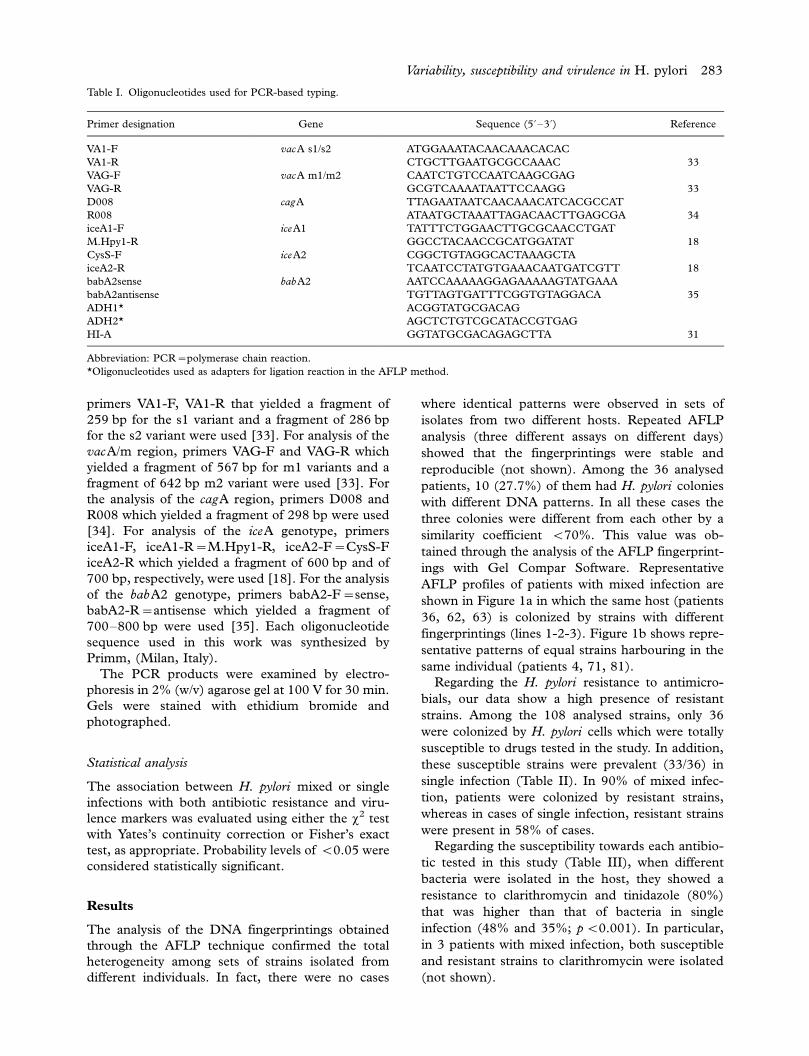
Table I. Oligonucleotides used for PCR-based typing.
Primer designation Gene Sequence (5? /3?) Reference
VA1-F vacA s1/s2 ATGGAAATACAACAAACACAC
VA1-R CTGCTTGAATGCGCCAAAC 33
VAG-F vacA m1/m2 CAATCTGTCCAATCAAGCGAG
VAG-R GCGTCAAAATAATTCCAAGG 33
D008 cagA TTAGAATAATCAACAAACATCACGCCAT
R008 ATAATGCTAAATTAGACAACTTGAGCGA 34
iceA1-F iceA1 TATTTCTGGAACTTGCGCAACCTGAT
M.Hpy1-R GGCCTACAACCGCATGGATAT 18
CysS-F iceA2 CGGCTGTAGGCACTAAAGCTA
iceA2-R TCAATCCTATGTGAAACAATGATCGTT 18
babA2sense babA2 AATCCAAAAAGGAGAAAAAGTATGAAA
babA2antisense TGTTAGTGATTTCGGTGTAGGACA 35
ADH1* ACGGTATGCGACAG
ADH2* AGCTCTGTCGCATACCGTGAG
HI-A GGTATGCGACAGAGCTTA 31
Abbreviation: PCR /polymerase chain reaction.
*Oligonucleotides used as adapters for ligation reaction in the AFLP method.
Variability, susceptibility and virulence in H. pylori 283

Against moxifloxacin, H. pylori strains isolated in
mixed infections expressed a significantly higher
percentage of resistance (20%) in comparison with
strains with similar fingerprintings in one patient
(6%, pB/0.05).
Finally, the very low increase of amoxicillin and
rifabutin resistance in mixed infection strains (10%)
compared with single infection strains (4%) was not
significant (p /0.05). In particular, amoxicillin was
ineffective against six H. pylori strains isolated from
2 patients with single and mixed infection, respec-
tively. For strains isolated in single infection, the
detected MIC value was 1 mg/ml, whereas in mixed
infection MIC values were 2 mg/ml for one strain and
one step lower for the other two strains.
Significant differences were also recorded when
H. pylori strains isolated in patients with mixed and
single infections were studied for their virulence
factors (Table IV). Among H. pylori strains isolated
in mixed infection, 21 (70%) of them displayed vacA
s1m1/s1m2 genotypes and 9 (30%) of them showed
vacA s2m2 genotype, whereas 33 (42%) H. pylori
strains in single infection had the s1m1/s1m2 geno-
type and 45 (58%) displayed the non-toxin-produ-
cing vacA s2m2 genotype. Among the 108 H. pylori
analysed, there was a significant association between
the vacA s1m1/s1m2 genotypes and mixed infection
(pB/0.01).
The cagA status was recorded in 29 (97%) to 30
strains in mixed infection and in 66 (85%) of the 78
strains showing equal fingerprintings in the same
individual. There was no significant difference
(p /0.05) among the presence of cagA microor-
ganisms in the single and mixed infections.
The iceA allelic types (iceA1 and iceA2) displayed
a total presence of strains iceA1 (100%) in mixed
infection without the iceA2 (0%) allele, whereas
single infection had the iceA1 and iceA2 alleles in
74% and 26% of cases, respectively. The distribution
of iceA1 and iceA2 alleles among strains causing
mixed and single infection showed significant differ-
ences (PB/0.01).
Concerning the babA2 genotype, there was a
significant prevalence of this condition in isolates in
single infection (31/78, 40%) in comparison with
isolates in mixed infection (5/30, 17%; pB/0.025).
When the 108 H. pylori strains analysed were
compared for their susceptibility to antibiotics and
the presence of the virulence markers (Table V), no
correlation was observed among the vacA, cagA,
iceA1/iceA2 and babA2 status and the susceptibility
Figure 1. Representative amplified fragment length polymorphism (AFLP) profiles of Helicobacter pylori isolates from 36 biopsy specimens
obtained from (a) patients (36, 62, 63) with mixed infection and (b) patients (4, 71, 81) with single infection. For each biopsy, 3 colonies
(lanes 1-2-3) were collected to compare the fingerprintings. DNA size standards (0.1 Kbp marker) are in the lanes marked ‘‘M’’.
Table II. Association between mixed and single Helicobacter pylori
infections and antimicrobial agents susceptibility.
Helicobacter pylori infection Susceptibility (%) Resistance* (%)
Multiple 3 (10) 27 (90)
Single 33 (42) 45 (58)
*Referred to strains which showed at least one resistance to
antimicrobial agents tested.
Table III. Association between Helicobacter pylori strains isolated
in patients with mixed and single infections and antimicrobial
agent susceptibility.
Mixed infection Single infection
Antimicrobial agents S* (%) R** (%) S (%) R (%)
Amoxicillin 27 (90) 3 (10) 75 (96) 3 (4)
Clarithromycin 6 (20) 24 (80) 48 (62) 30 (48)
Moxifloxacin 24 (80) 6 (20) 73 (94) 5 (6)
Rifabutin 27 (90) 3 (10) 75 (96) 3 (4)
Tinidazole 6 (20) 24 (80) 51 (65) 27 (35)
S* /susceptible strains; R** /resistant strains.
284 L. Cellini et al.

or resistance of at least one antibiotic tested in this
work (P /0.05).
Conclusions
This work is aimed at finding significant differences
between strains with or without genetic variability in
the same host, and susceptibility of antimicrobial
agents and virulence markers.
The high degree of genomic diversity among
H. pylori strains in one individual, which can be
due to mechanisms such as free recombination [36]
or co-infection with mixed H . pylori strains [6,37],
has been confirmed in this study through the AFLP
analysis. Among the analysed strains, this technique
displayed reproducible and discriminatory patterns.
In particular, among the 36 patients, the DNA
fingerprintings were different from each other and
these results are consistent with those of other
investigators [5,38] who asserted that polymorphism
has spread globally during the long evolutionary
history of H . pylori and has led to unprecedented
allelic diversity [9].
Moreover, significant differences among H . pylori
DNA patterns in a single host were also detected,
emphasizing the ability of the microorganism to
develop conditions for a more efficient colonization;
mixed infections were found in 27.7% of the cases
analysed. Various studies reported on the coexis-
tence of different strains of H . pylori in the same
patient detected by different methods [17,36,39,40].
In particular, Van Doorn et al. [17] identified mixed
infection of H. pylori in 20.2% of the cases analysed,
using a rapid and efficient method directly from
gastric biopsies based on the detection of mixed
vacA and/or iceA genotypes. In this work, we found
mixed infection in the same individual which in some
cases showed identical virulence marker profiles.
Therefore, the prevalence of mixed infection, as
reported earlier, could be underestimated. In our
opinion, the AFLP method represents a valid
technique for detection of mixed H. pylori infection.
Moreover, in our study, mixed infection was inde-
pendent of age and gender (not shown) which
suggests that relevant polymorphisms in one host
are not associated with the duration of H . pylori
infection.
When mixed or single colonization was compared
with antimicrobial susceptibility, the presence of
more resistant strains was consistent with the co-
infection. In fact, in mixed infection there is a
significant higher presence of resistant microorgan-
isms in respect of single H. pylori infections. The
presence in 90% of strains in mixed infections with
at least one resistance to the antimicrobial agents
tested emphasizes the need for more accurate studies
based on new strategies in H . pylori eradication
therapy. On the other hand, H . pylori resistance to
antimicrobials is the biggest cause of failure in
eradication therapy. In our study, in some cases of
mixed infection, H . pylori cells were found to be
either susceptible or resistant to clarithromycin in
the same host, which suggests the need to perform
an antimicrobial susceptibility test of at least two
different H . pylori colonies. This H . pylori hetero-
resistance has also been demonstrated by taking two
biopsies from two different sites in patients with
secondary resistance in previous studies [29,41]. All
these data emphasize the need for a careful H . pylori
antimicrobial surveillance to improve management
of H . pylori infection.
In particular, in this study, we found clarithromy-
cin and tinidazole resistance values, in different co-
infectant strains, that were higher than those of
single infection. These data confirm the ability of
these strains to better adapt themselves to the host
and highlight the worrying problem of clarithromy-
cin resistance found in our geographic area [29].
In this study, we found a significant relationship
when mixed and single infections were compared
with the main virulence factors, with the exception of
Table V. Association between Helicobacter pylori strains suscept-
ibility and virulence markers.
Genotype Susceptibility (%) Resistance* (%)
vacA s1m1/s1m2 15 (28) 39 (72)
vacA s2m2 21 (40) 33 (60)
cagA / 32 (34) 4 (66)
cagA / 4 (30) 8 (70)
iceA1 / 32 (35) 61 (65)
iceA1 / 4 (25) 11 (75)
iceA2 / 7 (29) 18 (71)
iceA2 / 29 (35) 54 (65)
babA2 / 12 (31) 26 (69)
babA2 / 24 (34) 46 (66)
*Referred to strains which showed at least one resistance to
antimicrobial agents tested.
Table IV. Association between Helicobacter pylori strains isolated
in patients with mixed and single infections and virulence markers.
Genotype
Mixed infection
isolates (%)
Single infection
isolates (%)
vacA s1m1/s1m2 21 (70) 33 (42)
vacA s2m2 9 (30) 45 (58)
cagA / 29 (97) 66 (85)
cagA / 1 (3) 12 (15)
iceA1 / 30 (100) 58 (74)
iceA1 / 0 (0) 20 (26)
iceA2 / 0 (0) 20 (26)
iceA2 / 30 (100) 58 (74)
babA2 / 5 (17) 31 (40)
babA2 / 25 (83) 47 (60)
Variability, susceptibility and virulence in H. pylori 285

the cagA gene. Therefore, co-infected hosts har-
boured strains that were characterized by genotypes
which encode proteins able to display the most
relevant damage.
However, the combination of vacA, cagA, iceA
status was not always helpful in predicting the
clinical state of patients [19,42] with controversial
reports on the presence of virulence markers and
clinical outcome [13,17,19,42]. This suggests that
other factors must play a role in disease pathogen-
esis. In fact, the interaction between bacterium /host
involves complex mechanisms that can balance or
emphasize the effect of virulence factors together
with environmental and dietary factors.
Our study presents a new scenario in which co-
infected hosts are colonized by H . pylori strains that
are more difficult to eradicate, and are more virulent.
Acknowledgements
We thank Professor Ray Pizzuto for help with editing
the manuscript. This study was supported by a grant
awarded by the Ministero Istruzione, Universita e
Ricerca, Rome, Italy.
References
[1] Cover TL, Berg DE, Blaser MJ. H. pylori pathogenesis. In:
Groisman EA, editor. Principles of bacterial pathogenesis.
New York NY: Academic Press; 2001. pp 509 /58.
[2] Frenck RW Jr, Clemens J. Helicobacter in the developing
world. Microbes Infect 2003;/5:/705 /13.
[3] Mobley HLT, Mendez GL, Hazell SL. Helicobacter pylori :
physiology and genetics. Washington DC: ASM Press; 2001.
[4] Suerbaum S, Michetti P. Helicobacter pylori infection. N Engl
J Med 2002;/347:/1175 /86.
[5] Suerbaum S. Genetic variability within Helicobacter pylori .
Int J Med Microbiol 2000;/290:/175 /81.
[6] Cellini L, Di Campli E, Di Candia M, Marzio L. Molecular
fingerprinting of Helicobacter pylori strains from duodenal
ulcer patients. Lett Appl Microbiol 2003;/36:/222 /6.
[7] Jorgensen M, Daskalopoulos G, Waburton V, Mitchell HM,
Hazell SL. Mixed strain colonization and metronidazole
resistence in Helicobacter pylori -infected patients: identifica-
tion from sequential and mixed biopsy specimens. J Infect
Dis 1996;/174: /631 /5.
[8] Kersulyte D, Mukhopadhyay AK, Velapatino B, Su W, Pan
Z, Garcia C, et al. Differences in genotypes of Helicobacter
pylori from different human populations. J Bacteriol 2000;/
182:/3210 /8.
[9] Wong BC, Wang WH, Berg DE, Fung FM, Wong KW, Wong
WM, Lai KC, et al. High prevalence of mixed infections by
Helicobacter pylori in Hong Kong: metronidazole sensitivity
and overall genotype. Aliment Pharmacol Ther 2001;/15:/
493 /503.
[10] Cellini L, Donelli G. Virulence factors of Helicobacter pylori .
Microb Ecol Health Dis 2000;/Suppl 2:/259 /62.
[11] Datta S, Chattopadhyay S, Balakrish Nair G, Mukhopad-
hyay AK, Hembram J, Berg DE, et al. Virulence genes and
neutral DNA markers of Helicobacter pylori isolates from
different ethnic communities of West Bengal, India. J Clin
Microbiol 2003;/41:/3737 /43.
[12] Lehours P, Menard A, Dupouy S, Bergey B, Richy F, Zerbib
F, et al. Evaluation of the association of nine Helicobacter
pylori virulence factors with strains involved in low-grade
gastric mucosa-associated lymphoid tissue lymphoma. Infec
Immun 2004;/72:/880 /8.
[13] Atherton JC, Peek RM Jr, Tham KT, Cover TL, Blaser MJ.
Clinical and pathological importance of heterogeneity in
vacA, the vacuolating cytotoxin gene of Helicobacter pylori .
Gastroenterology 1997;/112:/92 /9.
[14] Atherton JC, Cao P, Peek RM Jr, Tummuru MK, Blaser MJ,
Cover TL, et al. Mosaicism in vacuolating cytotoxin alleles
of Helicobacter pylori . Association of specific vacA types with
cytotoxin production and peptic ulceration. J Biol Chem
1995;/270:/17771 /7.
[15] Covacci A, Censini S, Bugnoli M, Petracca R, Burroni D,
Macchia G, et al. Molecular characterization of the 128-kDa
immunodominant antigen of Helicobacter pylori associated
with cytotoxicity and duodenal ulcer. PNAS USA 1993;/90:/
5791 /5.
[16] Awakawa T, Sugiyama T, Hisano K, Karita M, Yachi A.
Detection and identification of cagA of Helicobacter pylori by
polymerase chain reaction. Eur J Gastroenterol Hepatol
1995;/Suppl 1:/S75 /8.
[17] van Doorn LJ, Figueiredo C, Sanna R, Plaisier A, Schnee-
berger P, de Boer W, et al. Clinical relevance of the cagA,
vacA, and iceA status of Helicobacter pylori . Gastroenterol-
ogy 1998;/115:/58 /66.
[18] Mukhopadhyay AK, Kersulyte D, Jeong JY, Datta S, Ito Y,
Chowdhury A, et al. Distinctiveness of genotypes of Helico-
bacter pylori in Calcutta, India. J Bacteriol 2000;/182:/3219 /
27.
[19] Yamaoka Y, Kodama T, Gutierrez O, Kim JG, Kashima K,
Graham DY, et al. Relationship between Helicobacter pylori
iceA, cagA, and vacA status and clinical outcome: studies in
four different countries. J Clin Microbiol 1999;/37:/2274 /9.
[20] Ilver D, Arnqvist A, Ogren J, Frick IM, Kersulyte D, Incecik
ET, et al. Helicobacter pylori adhesin binding fucosylated
histo-blood group antigens revealed by retagging. Science
1998;/279:/373 /7.
[21] Appelmelk BJ, Negrini R, Moran AP, Kuipers EJ. Molecular
mimicry between Helicobacter pylori and the host. Trends
Microbiol 1997;/5: /70 /3.
[22] Kidd M, Atherton JC, Lastovica AJ, Louw JA. Clustering of
South African Helicobacter pylori isolates from peptic ulcer
disease patients is demonstrated by repetitive extragenic
palindromic-PCR fingerprinting. J Clin Microbiol 2001;/39:/
1833 /9.
[23] Kidd M, Lastovica AJ, Atherton JC, Louw JA. Heterogeneity
in the Helicobacter pylori vacA and cagA genes: association
with gastroduodenal disease in South Africa? Gut 1999;/45:/
499 /502.
[24] Kidd M, Lastovica AJ, Atherton JC. Specific genotypes of
Helicobacter pylori vacA and cagA, but not presence of cagA,
are associated with gastroduodenal disease in South Africa.
Gastroenterology 1999;/116:/G0928.
[25] Drumm B, Sherman P. Long-term storage of Campylobacter
pylori . J Clin Microbiol 1989;/27:/1655 /6.
[26] National Committee for Clinical Laboratory Standards.
Methods for dilution antimicrobial susceptibility tests for
bacteria that grow aerobically. Approved standard M7-A5.
National Committee for Clinical Laboratory Standards,
Villanova, Pa: 2000.
[27] Andrews JM, Ashby JP, Jevons GM, Wise R. Tentative
minimum inhibitory concentration and zone diameter break-
points for moxifloxacin using BSAC criteria. J Antimicrob
Chemother 1999 Dec;/44:/819 /22.
286 L. Cellini et al.

[28] Aydemir S, Boyacioglu S, Gur G, Demirbilek M, Can FK,
Korkmaz M, et al. Helicobacter pylori infection in hemodia-
lysis patients: susceptibility to amoxicillin and clarithromy-
cin. World J Gastroenterol 2005;/11:/842 /5.
[29] Toracchio S, Capodicasa S, Soraja DB, Cellini L, Marzio L.
Rifabutin based triple therapy for eradication of H. pylori
primary and secondary resistant to tinidazole and clarithro-
mycin. Dig Liver Dis 2005;/37:/33 /8.
[30] Toracchio S, Cellini L, Di Campli E, Cappello G, Malatesta
MG, Ferri A, et al. Role of antimicrobial susceptibility
testing on efficacy of triple therapy in Helicobacter pylori
eradication. Aliment Pharmacol Ther 2000;/14:/1639 /43.
[31] Gibson JR, Slater E, Xerry J, Tompkins DS, Owen RJ. Use
of an amplified-fragment length polymorphism technique to
fingerprint and differentiate isolates of Helicobacter pylori . J
Clin Microbiol 1998;/36:/2580 /5.
[32] Gerner-Smidt P, Graves LM, Hunter S, Swaminathan B.
Computerized analysis of restriction fragment length poly-
morphism patterns: comparative evaluation of two commer-
cial software packages. J Clin Microbiol 1998;/36:/1318 /23.
[33] Chisholm SA, Teare EL, Patel B, Owen RJ. Determination
of Helicobacter pylori vacA allelic types by single-step mixed
PCR. Lett Appl Microbiol 2002;/35:/42 /6.
[34] Covacci A, Rappuoli R. PCR amplification of gene se-
quences Helicobacter pylori : techniques for clinical diagnosis
and basic research. In: Lee A, Megraud F, editors. PCR
amplification of gene sequences from H. pylori strains.
London: WB Saunders; 1996. pp 94 /109.
[35] Gerhard M, Lehn N, Neumayer N, Boren T, Rad R, Schepp
W, et al. Clinical relevance of the Helicobacter pylori gene for
blood-group antigen-binding adhesin. Proc Natl Acad Sci
USA 1999;/96:/12778 /83.
[36] Suerbaum S, Smith JM, Bapumia K, Morelli G, Smith NH,
Kunstmann E, et al. Free recombination within Helicobacter
pylori . Proc Natl Acad Sci USA 1998;/95:/12619 /24.
[37] Prewett EJ, Bickley J, Owen RJ, et al. DNA patterns of
Helicobacter pylori isolated from gastric antrum, body, and
duodenum. Gastroenterology 1992;/102:/829 /33.
[38] Owen RJ, Ferrus M, Gibson J. Amplified fragment length
polymorphism genotyping of metronidazole-resistant Helico-
bacter pylori infecting dyspeptics in England. Clin Microbiol
Infect 2001;/7:/244 /53.
[39] Owen RJ, Bickley J, Hurtado A, Fraser A, Pounder RE.
Comparison of PCR-based restriction length polymorphism
analysis of urease genes with rRNA gene profiling for
monitoring Helicobacter pylori infections in patients on triple
therapy. J Clin Microbiol 1994;/32:/1203 /10.
[40] Owen RJ, Desai M, Figura N, Bayeli PF, Di Gregorio L,
Russi M, et al. Comparisons between degree of histological
gastritis and DNA fingerprints, cytotoxicity and adhesivity of
Helicobacter pylori from different gastric sites. Eur J Epide-
miol 1993;/9:/315 /21.
[41] Kim JJ, Kim JG, Kwon DH. Mixed-infection of antibiotic
susceptible and resistantHelicobacter pylori isolates in a single
patient and underestimation of antimicrobial susceptibility
testing. Helicobacter 2003;/8:/202 /6.
[42] Ho YW, Ho KY, Ascencio F. Neither gastric topological
distribution nor principle virulence genes of Helicobacter
pylori contributes to clinical outcomes. World J Gastroen-
terol 2004;/10:/3274 /7.
Variability, susceptibility and virulence in H. pylori 287
Related Documents