Downloaded from www.microbiologyresearch.org by IP: 54.90.167.105 On: Wed, 01 Jun 2016 11:06:42 Inhibitors of Helicobacter pylori ATPase Caga block CagA transport and cag virulence Markus Hilleringmann, 1 Werner Pansegrau, 1 Michael Doyle, 2 Susan Kaufman, 2 Mary Lee MacKichan, 2 Claudia Gianfaldoni, 3 Paolo Ruggiero 3 and Antonello Covacci 1 Correspondence Antonello Covacci [email protected] 1,3 Cellular Microbiology and Bioinformatics Unit 1 , and Serology and Animal Model Unit 3 , Immunological Research Institute Siena (IRIS), Novartis Vaccines S.r.l., Via Fiorentina 1, I-53100 Siena, Italy 2 Research and Development, Biopharmaceuticals, Novartis, 4560 Horton St M/S 4.4, Emeryville, CA 94608-2916, USA Received 10 March 2006 Revised 16 June 2006 Accepted 22 June 2006 With the steadily increasing occurrence of antibiotic resistance in bacteria, there is a great need for new antibacterial compounds. The approach described here involves targeting virulence- related bacterial type IV secretion systems (TFSSs) with small-molecule inhibitors. The cag TFSS of Helicobacter pylori was chosen as a model, and novel inhibitors directed against the cag VirB11- type ATPase Caga were identified. The cag genes encode proteins that are components of a contact-dependent secretion system used by the bacterium to translocate the effector molecule CagA into host cells. Translocated CagA is associated with severe gastritis, and carcinoma. Furthermore, functional TFSSs and immunodominant CagA play a role in interleukin (IL)-8 induction, which is an important factor for chronic inflammation. Inhibitors of Caga were identified by high- throughput screening of chemical libraries that comprised 524 400 small molecules. The ATPase activity of Caga was inhibited by the selected compounds in an in vitro enzymic assay using the purified enzyme. The most active compound, CHIR-1, reduced TFSS function to an extent that cellular effects on AGS cells mediated by CagA were virtually undetectable, while reduced levels of IL-8 induction were observed. Gastric colonization by CHIR-1-pre-treated bacteria was found to be impaired in a dose-dependent manner using a mouse model of infection. Small-molecule Caga inhibitors, the first described inhibitors of a TFSS, are potential candidates for the development of new antibacterial compounds that may lead to alternative medical treatments. The compounds are expected to impose weak selective pressure, since they target virulence functions. Moreover, the targeted virulence protein is conserved in a variety of bacterial pathogens. Additionally, TFSS inhibitors are potent tools to study the biology of TFSSs. INTRODUCTION Widespread emergence of bacterial pathogens that are resis- tant to common antibiotics calls for innovative antibacterial drugs. Targeting virulence functions, instead of enzymic activities that are essential in vitro, is a new approach and may represent a way to reduce the rate of resistance formation. Virulence genes often encode functions involved in host–pathogen relationships, such as persistence and invasion, and therefore they are non-essential under laboratory conditions, but they are required for successful propagation of a pathogen within the host. Hence, the resulting selective pressure that could give rise to resistance is expected to be weaker than for housekeeping genes. Type IV secretion systems (TFSSs) have been recognized as major virulence determinants in bacteria that are pathogenic to animals and plants (Agrobacterium, Brucella, Bartonella, Bordetella, Helicobacter, Legionella, Rickettsia and Wolba- chia). TFSSs are required for delivery of effector molecules, either directly into host cells or to the bacterial environment (Ding et al., 2003; Christie et al., 2005; Bourzac & Guillemin, 2005). We have chosen the well-studied human gastroduodenal pathogen Helicobacter pylori as a model system to demon- strate that virulence functions encoded by TFSSs can be targeted for use in antibacterial therapy. While it is estimated that about half of the world population is infected Abbreviations: ATPase, adenosine triphosphate hydrolase; HTS, high- throughput screening; IL, interleukin; NTPase, nucleoside triphosphate hydrolase; PAI, pathogenicity island; TFSS, type IV secretion system. 0002-8984 G 2006 SGM Printed in Great Britain 2919 Microbiology (2006), 152, 2919–2930 DOI 10.1099/mic.0.28984-0

Welcome message from author
This document is posted to help you gain knowledge. Please leave a comment to let me know what you think about it! Share it to your friends and learn new things together.
Transcript
Downloaded from www.microbiologyresearch.org by
IP: 54.90.167.105
On: Wed, 01 Jun 2016 11:06:42
Inhibitors of Helicobacter pylori ATPase Cagablock CagA transport and cag virulence
Markus Hilleringmann,1 Werner Pansegrau,1 Michael Doyle,2
Susan Kaufman,2 Mary Lee MacKichan,2 Claudia Gianfaldoni,3
Paolo Ruggiero3 and Antonello Covacci1
Correspondence
Antonello Covacci
1,3Cellular Microbiology and Bioinformatics Unit1, and Serology and Animal Model Unit3,Immunological Research Institute Siena (IRIS), Novartis Vaccines S.r.l., Via Fiorentina 1,I-53100 Siena, Italy
2Research and Development, Biopharmaceuticals, Novartis, 4560 Horton St M/S 4.4,Emeryville, CA 94608-2916, USA
Received 10 March 2006
Revised 16 June 2006
Accepted 22 June 2006
With the steadily increasing occurrence of antibiotic resistance in bacteria, there is a great
need for new antibacterial compounds. The approach described here involves targeting virulence-
related bacterial type IV secretion systems (TFSSs) with small-molecule inhibitors. The cag TFSS of
Helicobacter pylori was chosen as a model, and novel inhibitors directed against the cag VirB11-
type ATPase Caga were identified. The cag genes encode proteins that are components of a
contact-dependent secretion system used by the bacterium to translocate the effector molecule
CagA into host cells. Translocated CagA is associated with severe gastritis, and carcinoma.
Furthermore, functional TFSSs and immunodominant CagA play a role in interleukin (IL)-8 induction,
which is an important factor for chronic inflammation. Inhibitors of Caga were identified by high-
throughput screening of chemical libraries that comprised 524 400 small molecules. The ATPase
activity of Caga was inhibited by the selected compounds in an in vitro enzymic assay using the
purified enzyme. The most active compound, CHIR-1, reduced TFSS function to an extent that
cellular effects on AGS cells mediated by CagA were virtually undetectable, while reduced levels of
IL-8 induction were observed. Gastric colonization by CHIR-1-pre-treated bacteria was found to be
impaired in a dose-dependent manner using a mouse model of infection. Small-molecule Caga
inhibitors, the first described inhibitors of a TFSS, are potential candidates for the development of
new antibacterial compounds that may lead to alternative medical treatments. The compounds are
expected to impose weak selective pressure, since they target virulence functions. Moreover, the
targeted virulence protein is conserved in a variety of bacterial pathogens. Additionally, TFSS
inhibitors are potent tools to study the biology of TFSSs.
INTRODUCTION
Widespread emergence of bacterial pathogens that are resis-tant to common antibiotics calls for innovative antibacterialdrugs. Targeting virulence functions, instead of enzymicactivities that are essential in vitro, is a new approach andmay represent a way to reduce the rate of resistanceformation. Virulence genes often encode functions involvedin host–pathogen relationships, such as persistence andinvasion, and therefore they are non-essential underlaboratory conditions, but they are required for successfulpropagation of a pathogen within the host. Hence, the
resulting selective pressure that could give rise to resistanceis expected to be weaker than for housekeeping genes.
Type IV secretion systems (TFSSs) have been recognized asmajor virulence determinants in bacteria that are pathogenicto animals and plants (Agrobacterium, Brucella, Bartonella,Bordetella, Helicobacter, Legionella, Rickettsia and Wolba-chia). TFSSs are required for delivery of effector molecules,either directly into host cells or to the bacterial environment(Ding et al., 2003; Christie et al., 2005; Bourzac & Guillemin,2005).
We have chosen the well-studied human gastroduodenalpathogen Helicobacter pylori as a model system to demon-strate that virulence functions encoded by TFSSs can betargeted for use in antibacterial therapy. While it isestimated that about half of the world population is infected
Abbreviations: ATPase, adenosine triphosphate hydrolase; HTS, high-throughput screening; IL, interleukin; NTPase, nucleoside triphosphatehydrolase; PAI, pathogenicity island; TFSS, type IV secretion system.
0002-8984 G 2006 SGM Printed in Great Britain 2919
Microbiology (2006), 152, 2919–2930 DOI 10.1099/mic.0.28984-0

Downloaded from www.microbiologyresearch.org by
IP: 54.90.167.105
On: Wed, 01 Jun 2016 11:06:42
with H. pylori (Rothenbacher & Brenner, 2003), it has beenrecognized that severe outcomes of the infection, e.g. acutegastritis, peptic ulceration, formation of MALT lymphomaand adenocarcinoma, are associated with the presence ofthe cag pathogenicity island (PAI) (Bourzac & Guillemin,2005; Censini et al., 1996) in the H. pylori chromosome. Thepresence of the cag PAI has been linked to virulent type Istrains, whereas strains lacking the PAI are less virulent, andclassified as type II strains (Censini et al., 1996). The cag PAIencodes protein components of a TFSS that has been shownto play a major role in H. pylori virulence (Covacci et al.,1997; Censini et al., 1996). To date, the only known effectormolecule that has been identified to be secreted by cag is theCagA protein (Stein et al., 2002, 2000; Backert et al., 2000;Odenbreit et al., 2000).
CagA is targeted to epithelial cells, where it is tyrosinephosphorylated at EPIYA sites by c-Src and Lyn host kinases(Stein et al., 2002). Phosphorylated CagA triggers a signalcascade that promotes cell motility and proliferation, andleads to the characteristic ‘hummingbird’ phenotype (Segalet al., 1999), involving formation of lamellipodia andfilopodia. Another CagA-mediated effect results from thefact that translocated CagA localizes to the cell membrane,and promotes redistribution of the proteins ZO-1 and JAM,which are normally found at tight cell junctions (Amievaet al., 2003). This redistribution occurs independently ofthe phosphorylation state of CagA phosphorylation stateand leads to leakiness of the junctions (Amieva et al., 2003).
The presence of a functional cag TFSS results in NF-kBactivation in infected gastric epithelial cells, and leads topro-inflammatory responses, such as IL-8 release (Crabtreeet al., 1995). Although the mechanism by which a functionalcag TFSS induces pro-inflammatory responses is still notfully understood, there are two candidate mechanisms, bothof which may contribute to the observed effects: (i) thepresence of cag facilitates entry of peptidoglycan fragmentsinto host cells, and these are sensed by the intracellularreceptor Nod1, resulting in NF-kB activation (Viala et al.,2004); and (ii) CagA itself seems to be capable of potentiat-ing pro-inflammatory responses, as shown by gene replace-ment and mutagenesis studies (Brandt et al., 2005).
The cag TFSS of H. pylori encodes several proteins that showsignificant similarity with gene products of the paradigmaticvir TFSS of the Agrobacterium tumefaciens T-DNA transfersystem (Akopyants et al., 1998; Christie & Vogel, 2000;Christie, 2001; Christie et al., 2005). The vir TFSS is formedby 12 proteins (VirB1–11, plus the coupling protein VirD4)(Christie, 2001).
The assembly process is not yet defined at the molecularlevel, but mutations within the virB operon have resulted inlack of polymerization, and absence of functions. Mutants ofVirB4, VirB7, VirB9, VirB10 and VirB11 all show functionalimpairment, and lack polymerization of the conduit usedfor delivery of the effectors (Fernandez et al., 1996; Berger &Christie, 1994).
Three proteins that contain NTP-binding motifs have beenidentified: the prototypes are VirB4, VirB11 and VirD4. Todate, in vitro NTPase activity has been shown for the VirB11-type proteins (Krause et al., 2000b; Machon et al., 2002) andfor one member of the VirD4 family, TrwB (Tato et al.,2005). For several VirB11-type proteins, a ring-like hexame-ric structure has been demonstrated (Krause et al., 2000a),and this has been confirmed by X-ray structural analysis ofthe H. pylori Caga NTPase (Yeo & Waksman, 2004; Yeo et al.,2000).
Although VirB11-type NTPases have been very well char-acterized structurally (Krause et al., 2000a; Savvides et al.,2003; Yeo et al., 2000) and biochemically (Krause et al.,2000b; Machon et al., 2002; Rivas et al., 1997), their functionin TFSSs remains unclear. Members of this class of enzymesare also present in the type II secretion systems, and type 4pilus systems (Planet et al., 2001; Krause et al., 2000a; Rivaset al., 1997), and a role in the formation of the pilus-like cellappendages, which are commonly associated with TFSS,has been suggested (Sagulenko et al., 2001). VirB11-typeproteins are essential for TFSS function, as shown bymutagenesis studies (Berger & Christie, 1994), and theirNTPase activity is detectable in vitro using purified protein,without the requirement of complex cofactors (Krause et al.,2000b; Rivas et al., 1997); these two properties make VirB11-type proteins, such as H. pylori Caga, ideal targets for use inhigh-throughput screening (HTS) to search for compoundsthat specifically inhibit TFSS function.
Here, we describe the identification and validation of threespecific inhibitors of Caga, which is the VirB11 homologueof cag. The compounds efficiently inhibit Caga ATPaseactivity in vitro. We demonstrate that one of the compoundsreduces cag activity to the limits of detection, as measured bytransfer of CagA protein from H. pylori to eukaryotic hostcells. In a mouse model of infection, bacteria that had beenpre-treated with the inhibitor showed impaired ability tocolonize the gastric mucosa. Implications for application ofTFSS inhibitors as a new type of anti-infective, representingthe first described inhibitor of a TFSS, as well as a new toolfor studying the mechanism of action of TFSSs, arediscussed.
METHODS
Bacterial strains, cell lines and culture conditions. H. pylori
strains G27 and SPM 326, and mutants G27DHP527 (cagY) and
G27DcagA, have been described (Marchetti et al., 1995). Strains were
cultivated on Columbia agar base (Oxoid) containing 5 % (v/v) defi-
brinated horse blood or 5 % (v/v) fetal bovine serum (FBS), and
Dent’s antibiotic supplement (Oxoid). BHI and Brucella broth (BB)
media were supplemented with 5 % FBS and appropriate antibiotics.
Bacitracin (Sigma-Aldrich) was used at a concentration of 200 mg
ml21. Bacteria were incubated at 37 uC, under microaerophilic con-
ditions. AGS cells (European Collection of Cell Cultures no.
89090402) were grown at 37 uC in 5 % CO2/95 % air in Dulbecco’s
modified Eagle’s medium (DMEM), with 10 % (v/v) FBS and
1?5 mM L-glutamine.
2920 Microbiology 152
M. Hilleringmann and others

Downloaded from www.microbiologyresearch.org by
IP: 54.90.167.105
On: Wed, 01 Jun 2016 11:06:42
Caga in vitro assay. Native Caga protein (HP0525) was obtainedfrom induced Escherichia coli SCS1 (pWP4525) cells, as describedpreviously (Krause et al., 2000b). The ATPase activity of Caga wasassessed using a chromogenic reagent based on the classical mala-chite green reagent for detection of Pi (BIOMOL Green reagent).Briefly, Caga (approx. 0?4 mg in a total reaction volume of 40 ml)was incubated for 45 min at 37 uC in reaction buffer (20 mM Tris/HCl, pH 8?0, 50 mM NaCl, 2?5 mM MgCl2, 0?1 mM EDTA, and0?05 % Brij-35) in the presence of 1 mM ATP. Following the incu-bation period, 60 ml reaction buffer was added, followed by 1 mlBIOMOL Green reagent. The concentration of Pi was determined bymeasuring the absorbance at 620 nm, after an incubation period of30 min at 25 uC.
Chemical library. The compound collection belonged to the ChironCorporation (Emeryville) substance libraries. For HTS, 524 400diverse chemicals were selected, all of which were small molecules(Mr <600), and of diverse structure. Five hundred thousand of thecompounds were derived from Chiron’s combined chemical libraries(CombiChem). We did not use a focused library, such as one con-taining analogues of ATP or ADP.
Assay methodology for Caga inhibitors (HTS). Test com-pounds were added to DMSO at 406 final concentration, and dis-pensed into 384-well polypropylene plates (1 ml per well). Using aMultidrop dispenser (Thermo), 20 ml buffer/salt solution (20 mMTris, pH 8?0, 50 mM NaCl, 5 mM MgCl2, and 200 mM ATP; RocheApplied Sciences) was added to each well, and the plates were shakenbriefly to mix in the compounds. The Caga enzyme (HP0525) wasdiluted to 2?25 mg ml21 in a buffer/salt solution consisting of20 mM Tris, pH 8?0, 50 mM NaCl, and 0?1 % Brij-35. Again usingthe Multidrop dispenser, 20 ml of the enzyme solution was added tothe plates to start the reaction. The plates were incubated at 37 uCfor 45 min, at which time the reaction was terminated, and detec-tion of free phosphate was initiated by the addition of 60 ml Greenreagent. After 30 min at room temperature, the plates were stacked,ten at a time, onto two Victor 2 plate readers (Perkin Elmer), andabsorbance at 625 nm was measured. Two readers were used foreach set of 20 plates, so that all were read within 1 h of the additionof Green reagent. Data were analysed using ActivityBase software(IDBS).
Hit evaluation (re-screening of hit candidates). Evaluation ofhits was performed by re-screening the compounds in a dose–response assay. The most potent compounds were analysed by LC/MS to determine purity, and to confirm the intended structure. Forfurther study, selected compounds were repurified by HPLC andreassayed to calculate their IC50 values. Some compounds were con-taminated with heavy metals; therefore, 0?1 mM EDTA was includedin follow-up assays to avoid inhibition of Caga activity by heavymetal ions. Inhibitory activity was crosschecked against non-VirB11-type ATPases, e.g. E. coli RecA protein (Sigma-Aldrich) in the pre-sence of ssDNA, and canine Na+/K+-ATPase (EC 3.6.3.9).
Inhibitor studies. H. pylori Caga inhibitor CHIR-1 was used atconcentrations of 0–25 mM (100 mM stock solution in DMSO) forthe in vitro enzymic assay. For cell culture experiments with AGScells, 25, 50 and 75 mM solutions of CHIR-1 were applied. All con-trols were performed with the respective amount of DMSO. Preced-ing the infection protocol, overnight cultures of H. pylori or AGScells were pre-incubated with CHIR-1 at 37 uC for up to 30 min;otherwise, the bacteria were added to the monolayer simultaneouslywith the supplemented medium. Cell viability was tested using thecell proliferation reagent WST-1 (Roche), according to the manufac-turer’s instructions.
Infection and fractionation of AGS cells. AGS cells (seeded in10 cm tissue-culture dishes) were infected with an overnight culture
of H. pylori (in BB supplemented with antibiotics, or DMEM) for3–5 h at an m.o.i. of about 100 : 1. Fractionation of AGS cells intoan RIPA-buffer-soluble fraction (0?15 mM NaCl, 0?05 mM Tris/HCl, pH 7?2, 1 % Triton X-100, 1 % sodium deoxycholate, 0?1 %SDS, containing 200 mM vanadate and 5 mg protease inhibitorsml21), and a membrane fraction, was performed as described byStein et al. (2000).
Antibodies and immunoprecipitation. Immunoprecipitationwith anti-phosphotyrosine (PY99; Santa Cruz Biotechnology) wascarried out according to the protocols provided by the supplier.Anti-tubulin antibody (T-9026) was obtained from Sigma. Anti-bodies against G27 CagA (rabbit) and G27 urease (rabbit) were pro-duced in our laboratory (F. Bagnoli, L. Buti, S. Censini, W.Pansegrau, I. Russo & M. Stein, unpublished results; Stein et al.,2000). Secondary antibodies were obtained from Bio-Rad.
Assay for IL-8 induction by epithelial cells. AGS cells werecultivated in 75 ml tissue-culture flasks in a medium containingDMEM supplemented with L-glutamine and 5 % FBS. Cells wereharvested by trypsinization, and resuspended in supplementedDMEM to a concentration of 26105 cells ml21. A 2 ml volume ofthe cell suspension was added to each well of a six-well plastictissue-culture plate, and incubated until the monolayer was conflu-ent to about 80 %. A H. pylori G27 culture grown overnight was har-vested in PBS, and resuspended to a density of approximately108 c.f.u. ml21. Dilutions of 1 : 3 and 1 : 10 were prepared. Whereindicated, bacteria were pre-incubated with DMEM/CHIR-1 at 37 uC,before adding 1?5 ml of the suspension to the monolayer. Otherwise,bacteria were added simultaneously with the respective medium tothe AGS cells. After 4 and 24 h microaerophilic incubation, samples(500 ml) were taken and centrifuged (15 000 g), before freezing at280 uC, until assayed. The volume removed was replaced by freshDMEM/CHIR-1, and phenotypic changes of AGS cells were esti-mated by light microscopy. Additionally, 20 ml samples wereremoved and incubated under microaerophilic conditions in 1 mlBB medium for up to 5 days to control the viability of H. pyloriG27.
Cytokine assay. The level of IL-8 in cell culture supernatants wasdetermined by use of a commercially available human IL-8 ELISAkit (OptEIA; BD Biosciences), and expressed in pg ml21.
Confocal immunofluorescence. Infected AGS monolayers onglass coverslips were fixed with 3 % formaldehyde in PBS, with1 mM CaCl2 and 1 mM MgCl2. Following permeabilization with0?2 % Triton X-100, anti-H. pylori antibodies (a-CagA, rabbit) wereapplied to the coverslips. Green Alexa Fluor 488 goat anti-rabbit IgG(A-11034; Molecular Probes) was used as a secondary antibody.
Mouse colonization assays. Specific-pathogen-free CD1 mice(Charles River Laboratories) were housed and treated in compliancewith current Italian law. After a 16-h starvation period, 6-week-oldCD1 mice (10 animals per group) were infected, as previouslyreported (Marchetti et al., 1995), with minor modifications. Imme-diately before challenge, bacteria were harvested from plates by asterile cotton swab, resuspended in supplemented BHI medium, andtheir concentration was determined spectrophotometrically. AnOD535 of 1 in a cuvette with a 1 cm path length corresponds to109 c.f.u. ml21. H. pylori exists as an actively dividing, highly motile,spiral form, and a non-culturable, but viable, metabolizing coccoidform. Others (Cole et al., 1997) have shown that the coccoid formof H. pylori, in contrast to the spiral form, binds poorly to gastricepithelial cells. Therefore, the phenotype of bacterial cultures, i.e.motility and spiral shape, was monitored microscopically so that wewere able to initiate infection experiments with motile spiral cells.Mice were given 0?25 ml of 0?2 M NaHCO3, intragastrically, to neu-tralize acidity, about 15 min before challenge. Through the same
http://mic.sgmjournals.org 2921
Inhibitors of H. pylori VirB11-type ATPase Caga

Downloaded from www.microbiologyresearch.org by
IP: 54.90.167.105
On: Wed, 01 Jun 2016 11:06:42
route, either 109 (high dose of H. pylori) or 108 (standard dose of H.pylori) c.f.u. in 100 ml saline was delivered directly into the stomachof each mouse. Where indicated, bacteria were pre-treated with 25or 50 mM CHIR-1 in DMSO (3?5 and 7 mM, respectively) forapproximately 30 min before challenge (controls received 7 mMDMSO). The number of c.f.u. in inocula was determined by subse-quent plate culture. Groups that received the standard infecting doseof H. pylori were further infected with the same bacterial dose2 days later; infection was not repeated in mice that received thehigh dose. Forty-eight hours after each infection, a second dose ofinhibitor (in 150 mM NaCl) was administered. Four and elevendays after infection with high and low dose, respectively, the animalswere euthanized by carbon dioxide asphyxiation and their stomachswere removed immediately. Gastric mucosa was scraped from thestomach of each mouse, and, after serial dilution, this was spread onselective plates, which had been prepared as described above, withaddition of 0?2 mg bacitracin ml21. H. pylori c.f.u. were countedafter 6 days.
Statistical analysis. The statistical significance of the data wasevaluated by a one-tailed Mann–Whitney U test or Fisher’s exacttest. For all comparisons, probability values less than 5 % (P<0?05)were considered statistically significant.
RESULTS
HTS of Caga identifies three potentsmall-molecule inhibitors
The ATPase activity of Caga was measured by assessing thePi liberated during the Caga in vitro assay. Quantification offree phosphate was performed by measuring absorbance at620 nm using BIOMOL Green reagent. The laboratory-scaleenzymic assay was adapted to HTS in 384-well plates (seeMethods). A chemical library of 524 400 compounds wasscreened to identify inhibitors of Caga ATPase activity.CombiChem compounds were tested in pools, at a finalconcentration of 1 mM for each substance; other com-pounds were added at a final concentration of 5 mM to theassay mixture, either as single compounds or as mixtures offive compounds. In an initial screen, 675 compounds werefound to inhibit Caga ATPase activity by at least 50 %. Re-screening and verification of primary hits was performed indose–response assays, resulting in 200 confirmed hits thathad preliminary IC50 values of less than 10 mM. Afterdeconvolution of any pooled compound hits, 33 singlecompounds, thereof, showed IC50 values of 5 mM or less,and 20 showed values of 1 mM or less. Structural con-siderations to exclude undesirable structures with reactivegroups, or structures difficult to modify, led to a selection ofseven compounds. These compounds were analysed byLC/MS for purity, and to confirm their intended structure,followed by further studies after repurification by HPLC.The seven compounds considered for more comprehensiveevaluation had not been identified as significant kinaseinhibitors in other screens, nor had they shown inhibitoryactivity in other HTS assays.
Three of the seven compounds (CHIR-1, -2 and -3)retained IC50 values of <1 mM (Table 1) in further screens.Remarkably, two compounds, CHIR-1 and CHIR-2, were
structurally closely related, falling into the class ofthiadiazolidine-3,5-diones. The compound with the lowestIC50 value, CHIR-1, was acquired in larger amounts toconfirm the inhibition properties of the substance, and toperform the experiments described below. Crosschecking itsin vitro activity against non-VirB11-type ATPases [a mam-malian ATPase (canine Na+/K+-ATPase), and bacterialRecA protein] revealed 10- and 5-fold higher IC50 values,respectively (data not shown).
CHIR-1 suppresses the hummingbird phenotypein infected AGS cells
Infection of gastric cell lines by type I strains of H. pyloriinduces the so-called hummingbird phenotype, character-ized by lamellipodia and filopodia, with spreading andelongated growth of the cells (Segal et al., 1999). Weinvestigated the effect of CHIR-1 on H. pylori in an infectionexperiment in which the translocation of CagA, and theresulting hummingbird phenotype, were indicators of thefunctionality of the H. pylori TFSS.
AGS cells (Fig. 1a) were infected with the wild-type strainG27, or with G27DcagA as a negative control. To study theinfluence of CHIR-1, AGS cells were co-cultivated with thewild-type strain pre-incubated in the presence of CHIR-1.Wild-type H. pylori G27 induced cytoskeletal rearrange-ment, resulting in the hummingbird phenotype (Fig. 1b),whereas the DcagA mutant of G27 did not (Fig. 1c).Following pre-incubation of H. pylori for 30 min in thepresence of 50 mM CHIR-1, and co-cultivation with AGScells in the presence of 50 mM CHIR-1 for 4 h, a reduction inthe number of cells showing the hummingbird phenotype,which was comparable with that for the DcagA mutant, wasobserved (Fig. 1e). A similar reduction of the hummingbirdphenotype was not observed with bacterial cells that had notbeen pre-incubated with CHIR-1 (Fig. 1d).
The MIC value of CHIR-1 with H. pylori G27 was calculatedto be 90 mM, as determined by serial dilution of the inhibitorin a bacterial suspension. Human AGS cell toxicity ofCHIR-1 was tested using a colorimetric assay, based on thecleavage of the tetrazolium salt WST-1 by mitochondrialdehydrogenases. AGS cell viability was not affected byCHIR-1 concentrations up to 100 mM (data not shown).
Table 1. Small-molecule inhibitors of H. pylori Caga
Inhibitor Structure IC50 (mM) Mr
CHIR-1 0?45 292?2
CHIR-2 0?78 316?4
CHIR-3 0?55 280?7
2922 Microbiology 152
M. Hilleringmann and others

Downloaded from www.microbiologyresearch.org by
IP: 54.90.167.105
On: Wed, 01 Jun 2016 11:06:42
CHIR-1 causes accumulation of CagA protein inH. pylori during AGS cell infection
The effect of CHIR-1 on CagA translocation was studied byconfocal fluorescence microscopy (Fig. 2). AGS cells wereinfected with wild-type H. pylori, and CagA protein wasvisualized in permeabilized cells using a polyclonal anti-CagA antibody. A secondary antibody conjugated withGreen Alexa Fluor 488 was used for CagA detection. AGScells (60 % confluent) were infected with 107 c.f.u wild-typeH. pylori in 12-well culture plates. In the untreated controls,the signal intensity of CagA within the bacterial cells wasrelatively low, while in the AGS cells, a faint and diffusefluorescence signal, due to CagA translocation, was observed(Fig. 2a–c). However, upon co-cultivation of AGS cells withH. pylori cells that had been pre-cultivated in the presenceof 50 mM CHIR-1, CagA was visible virtually exclusivelywithin the bacterial bodies, in which the protein accumu-lated in significant amounts (Fig. 2d–f).
As demonstrated by fluorescence intensity, at least athreefold higher concentration of CagA was demonstratedin H. pylori cells in AGS–H.-pylori co-cultivates that weretreated with CHIR-1, compared with untreated co-culti-vates (Fig. 2g). Correspondingly, as compared with theuntreated control, a similar (three- to fourfold) decrease inmean fluorescence intensity was observed in AGS cells in
AGS–H.-pylori co-cultivates treated with CHIR-1 (Fig. 2g).These findings demonstrate that translocation of CagA ismuch reduced by the action of CHIR-1. The effect wascomparable with that seen with H. pylori cag mutantsdefective in Caga (VirB11) (Backert et al., 2000).
CagA translocation into AGS cells is blocked byCHIR-1
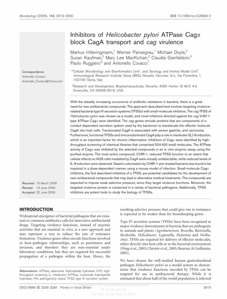
To quantify the inhibitory effect of CHIR-1 on CagA trans-location more precisely, we took advantage of the fact thatCagA becomes tyrosine phosphorylated in the host cell.Therefore, the amount of tyrosine-phosphorylated CagA(pCagA) can be used as a readout for CagA translocation.Following co-cultivation of AGS cells with H. pylori G27,infected cells were harvested together with the attachedbacteria, and a total extract was prepared. Phosphorylatedproteins were enriched by immunoprecipitation from thetotal cell extract using anti-phosphotyrosine antibodies.Phosphorylated CagA (pCagA) protein was detected in theprecipitate of untreated cells by Western blot analysis(Fig. 3, lanes 2 and 3). Again, 50 mM CHIR-1 was sufficientto inhibit CagA translocation into AGS cells to the limitsof detection for our study (Fig. 3, lanes 6 and 7). Once more,a short period of pre-incubation of H. pylori cells in thepresence of the inhibitor was necessary to achieve complete
Fig. 1. Interference contrast microscopy of infected AGS cells. AGS cells were incubated for 4 h in the presence of H. pylori
strains, as described in Methods. Where indicated, co-cultivation was performed in the presence of CHIR-1; for oneexperiment, bacteria were pre-incubated for 30 min with CHIR-1. (a) AGS cells; (b) AGS cells incubated with H. pylori G27;(c) AGS cells incubated with G27DcagA; (d) AGS cells incubated with G27 in the presence of 50 mM CHIR-1; (e) G27pre-incubated for 30 min in the presence of 50 mM CHIR-1, and AGS cells incubated with the pre-treated G27 cells (*) in thepresence of 50 mM CHIR-1. Unstained cells were examined by using interference contrast microscopy.
http://mic.sgmjournals.org 2923
Inhibitors of H. pylori VirB11-type ATPase Caga

Downloaded from www.microbiologyresearch.org by
IP: 54.90.167.105
On: Wed, 01 Jun 2016 11:06:42
inhibition of CagA transfer. Without pre-incubation, adetectable amount of CagA reached the recipient cells(Fig. 3, lanes 4 and 5). Importantly, the total amount ofCagA remained constant, indicating that the differentquantities of pCagA measured were not due to variableoverall amounts of CagA resulting from CHIR-1 treatment(Fig. 3, lower panel). Intracellular concentrations of H.pylori a-urease and AGS a-tubulin were not altered duringa 4 h incubation period in the presence of 50 mM CHIR-1,when compared with a control without CHIR-1 (data notshown).
To determine the minimum period of time required for pre-incubation, and to compare it with the time required forCagA transfer, H. pylori G27 cells were exposed to CHIR-1.
Aliquots of the treated H. pylori cells were applied inco-cultivation experiments with AGS cells, in the presenceof CHIR-1, and the amount of phosphorylated CagA(pCagA) was determined as described above (Fig. 3). CagAtransfer activity was reduced significantly between 1 and3 min of pre-incubation (Fig. 4, bars 6 and 7). Theincubation period required for this effect to occur wasshort (<3 min) compared with the time required for CagAtransfer, which was in the range of 30 min. The finding thata significant quantity of CagA reaches the AGS cells in thepresence of CHIR-1, without the pre-incubation step,suggests that the inhibitor is effective only for a shorttime period at the beginning of the secretion process, andthat transfer of CagA then becomes resistant to the action ofCHIR-1. This might be related to the observation that
Fig. 2. Inhibition of Caga activity by CHIR-1 results in CagA accumulation in H. pylori. AGS cells were incubated in thepresence of H. pylori G27, as described in Methods. CagA protein was labelled by immunofluorescence using polyclonalanti-CagA antibodies and Green Alexa-Fluor-488-labelled IgG, as primary and secondary antibodies, respectively. (c, f)Phase-contrast images (a, d) were overlaid with the corresponding confocal fluorescent images (b, e) showing the distributionof CagA. Bar, 50 mm. (a–c) No inhibitor was added; (d–f) G27 was pre-incubated for 30 min in the presence of 50 mMCHIR-1, followed by co-cultivation of AGS cells with G27 in the presence of 50 mM CHIR-1. (g) Quantification of CagA-protein-related fluorescence within H. pylori and AGS cells, respectively. Mean fluorescence intensities were measured inselected regions of digital image sets corresponding to either AGS or H. pylori cells. Intensities are given in arbitrary units.Error bars indicate SD.
2924 Microbiology 152
M. Hilleringmann and others
Downloaded from www.microbiologyresearch.org by
IP: 54.90.167.105
On: Wed, 01 Jun 2016 11:06:42
contact establishment between AGS cells and H. pylori isa fast process that requires less than 5 min (F. Bagnoli,personal communication). Pre-incubation of the AGScells with the inhibitor had no effect on CagA transfer(Fig. 4, bar 2).
CHIR-1 reduces IL-8 secretion in infected AGScells
IL-8 induction by H. pylori in epithelial cells is dependent onthe presence of a functional TFSS (Fischer et al., 2001;Selbach et al., 2002), and is influenced by mediators such asCagA (Brandt et al., 2005) and peptidoglycan (Viala et al.,2004). The function of Caga (VirB11) has been shown to benecessary for IL-8 induction (Fischer et al., 2001). Further-more, systematic mutagenesis of the cag PAI revealed thatthe mechanism is independent of Cagb (VirD4), which isanother TFSS protein that contains an NTP-binding motif.We tested Caga-ATPase inhibitor CHIR-1 for its effect onIL-8 secretion. It has been shown that IL-8 secretion ofinfected AGS cells is strongly enhanced after co-cultivationfor 24–36 h (Brandt et al., 2005). In particular, the
potentiating effect of CagA on IL-8 production can beobserved only after prolonged periods of co-cultivation(Brandt et al., 2005). In our experiment, we quantified theamount of IL-8 release by AGS cells infected with CHIR-1-treated H. pylori G27 by ELISA after 24 h (Fig. 5).
IL-8 production by infected AGS cells was found to beinhibited by CHIR-1 in a concentration-dependent manner(Fig. 5). The effect varied with the amount of viable bacteriaadded (not shown), being most evident for a 1 : 10 dilutionof the stock suspension. Interestingly, inhibition of IL-8production was more pronounced after pre-incubation ofinfecting bacteria with CHIR-1. In the presence of 50 mMCHIR-1, the amount of secreted IL-8 dropped by 19?8 %.Using H. pylori cells that had been pre-incubated for 15 minwith 50 mM CHIR-1, IL-8 secretion dropped by 36?6 %. Inboth cases, IL-8 concentrations were measured after 24 hco-cultivation, and the viability of the treated bacteria wastested by incubating an aliquot in BB medium at 37 uC forseveral days, under microaerophilic conditions. It was foundthat the CHIR-1 treatment did not impair viability of H.pylori under the conditions described (not shown).
Fig. 3. Complete inhibition of CagA translocation requires pre-incubation of H. pylori G27 with CHIR-1. AGS cells were co-cultivated with H. pylori G27. Upper panel, transfer of CagAprotein to AGS cells was analysed by immunoprecipitation ofphosphotyrosine-containing proteins from the cell extracts withanti-phosphotyrosine IgGs, separation on a SDS-polyacrylamidegel, and blotting to a nitrocellulose membrane. Hence, onlyCagA protein that was phosphorylated after transfer to AGScells (pCagA) is seen. Immunoprecipitated pCagA protein wasdetected using polyclonal anti-CagA serum and the ECL system.Lower panel, CagA protein was detected with polyclonal anti-CagA serum, following electrophoresis of total cell extracts andblotting. Lanes: 1, AGS cells were co-cultivated with G27DcagY
(virB10); 2 and 3, AGS cells were co-cultivated with G27in the absence of the inhibitor; 4 and 5, AGS cells were co-cultivated with G27 in the presence of 50 mM CHIR-1; 6 and7, G27 cells were pre-incubated for 30 min in the presenceof 50 mM CHIR-1. AGS cells were co-cultivated with the pre-treated G27 in the presence of 50 mM CHIR-1.
Fig. 4. Kinetics of CagA translocation inhibition by CHIR-1. H.
pylori cells were incubated for various periods of time in thepresence of 50 mM CHIR-1. Following pre-incubation, bacteriawere co-cultivated with AGS cells and 50 mM CHIR-1.Phosphorylated proteins were immunoprecipitated with anti-phosphotyrosine antibodies, separated on a polyacrylamide gel,and blotted to a nitrocellulose membrane. pCagA protein (phos-phorylated following transfer to AGS cells) was detected usinga polyclonal anti-CagA antiserum, and quantified densitometri-cally. Relative amounts of pCagA are given in arbitrary units.Internal controls for quantification: total CagA protein anda-urease for H. pylori; a-tubulin for the AGS cells (not shown).Bars: 1, AGS cells were co-cultivated with H. pylori G27DcagA;2, AGS cells were pre-incubated with CHIR-1 for 15 min (*)before co cultivation with G27; 3, AGS cells were co-cultivatedwith H. pylori G27; 4–9, H. pylori G27 was pre-incubated withCHIR-1 for the indicated periods of time, before co-cultivationwith AGS cells (*). Grey bars, data points for the kineticassays; white bars, control experiments. Quantifications wereperformed in triplicate. The results are means±SD.
http://mic.sgmjournals.org 2925
Inhibitors of H. pylori VirB11-type ATPase Caga

Downloaded from www.microbiologyresearch.org by
IP: 54.90.167.105
On: Wed, 01 Jun 2016 11:06:42
CHIR-1 impairs gastric colonization of mice byH. pylori
To study the effect of the Caga inhibitor on early phase ofcolonization, we inoculated CD1 mice intragastrically withH. pylori strain SPM 326 that had been pre-treated withCHIR-1 (Marchetti et al., 1995). SPM 326 is a mouse-adapted type I strain, which responds to CHIR-1 in a waythat is similar to G27 in various assays (data not shown).Mice were given roughly equal numbers of c.f.u. of non-treated and CHIR-1-treated bacteria. All animals testedsurvived treatment with bacteria plus Caga inhibitor atconcentrations as high as 50 mM.
Infection/colonization was quantified by determining thenumber of bacteria in the stomach of each mouse afterscraping gastric mucosa, and plating out serial dilutions.Increasing concentrations of CHIR-1 resulted in decreasedcolonization by H. pylori SPM 326, as measured by thenumber of c.f.u. recovered, in comparison with the c.f.u.recovered in the corresponding infected controls. When thestomachs of mice that received standard bacterial dose wereanalysed after sacrifice, 8 out of 10 mice were found to beH. pylori-positive in the infected control group, while asignificantly lower number of H. pylori-positive mice (2out of 10, P=0?011) were found infected in the group thatconcomitantly received 50 mM CHIR-1; the group thatreceived 25 mM CHIR-1 showed a non-significant reductionin the number of infected mice (6 out of 10, P=0?31). Interms of bacterial colonization level, the group that receivedthe standard bacterial dose showed a highly significantdecrease in bacterial colonization (the geometric mean ofc.f.u. per stomach was more than 2 log units lower thancontrol group, P=0?0014) when concomitantly treated with50 mM CHIR-1, and only a marginal reduction (P=0?139)when treated with 25 mM CHIR-1. The group that receivedthe high bacterial dose showed a similar trend after 4 days ofinfection, although the differences with the control groupwere not significant: 9 out of 10 mice were found to beinfected in the control group, 5 out of 10 (P=0?07) treatedwith 50 mM CHIR-1 were infected, and 8 out of 10 (P=0?5)treated with 25 mM CHIR-1 were infected; in quantitativeterms, the reduction of infection almost reached a signi-ficant level (a geometric mean of more than 1 log unit lowerthan the control group, P=0?052) with 50 mM CHIR-1, andit was not significant (P=0?3) with 25 mM CHIR-1 (Fig. 6).
DISCUSSION
Using the well-studied organism H. pylori as a model, wesought to find small molecules that inhibited the activity ofthe cag-encoded TFSS, which is an important factor in thepathogenicity of the bacterium (Bourzac & Guillemin, 2005;Censini et al., 1996). A HTS involving 524 400 moleculesidentified 53 compounds that inhibited the TFSS-encodedATPase Caga in vitro, with IC50 values below 5 mM. Threeselected compounds (CHIR-1, -2 and -3) had IC50 valuesbelow 1 mM, and we considered these to be a reasonablestarting point for identifying candidates as lead structures(Table 1). The most potent substance, CHIR-1 (IC50
0?45 mM), was chosen for further characterization.
The potential of CHIR-1 to block cag activity was assessedin several cell-based assays. We used CagA transfer andphosphorylation as early indicators of TFSS functionality,since phosphorylated CagA can be detected in AGS cells asearly as 30 min after the start of co-cultivation (Selbach et al.,2003). Furthermore, it has been verified recently that alltranslocated CagA is phosphorylated at tyrosine residues,before being processed in the Golgi apparatus (A. Covacci, F.Bagnoli & L. Buti, unpublished observations).
Fig. 5. CHIR-1 inhibits IL-8 secretion by infected AGS cells.IL-8 secretion was induced in AGS cells, as described inMethods, and the amount of secreted IL-8 was detected byELISA. Bars: 1, AGS cells were co-cultivated with H. pylori
G27; 2–4, co-cultivation was performed in the presence of25, 50 and 75 mM CHIR-1, respectively; 5, G27* was pre-incubated for 15 min with 50 mM CHIR-1, and co-cultivationwith AGS cells was in the presence of the same concentrationof CHIR-1; 6, G27 in the absence of AGS cells or CHIR-1;7, AGS cells in the absence of G27 or CHIR-1; 8, AGS cellsin the presence of 50 mM CHIR-1. A square bracket joins datapoints obtained with and without pre-incubation of bacteria (PI)in the presence of 50 mM CHIR-1. IL-8 concentration in theculture supernatant was determined after 24 h co-cultivation.Quantifications were performed in triplicate. The results aremeans±SD.
2926 Microbiology 152
M. Hilleringmann and others

Downloaded from www.microbiologyresearch.org by
IP: 54.90.167.105
On: Wed, 01 Jun 2016 11:06:42
In this work, we did not use caga (virB11) mutants ascontrols. Although this seems to be an obvious control touse, Caga mutants do not reflect the action of the inhibitor.TFSSs are multi-protein complexes consisting of a core often or more essential components. If any of thesecomponents is missing, the entire complex is absent fromthe cells. Caga is one of the essential components, and,consequently, using a Caga mutant as a control would notonly eliminate the target for the inhibitor, but destroy theentire TFSS. Hence, one would neither see any transfer ofCagA, nor see the phenotypic effects caused by eitherphosphorylated CagA or the mere presence of the TFSS.These effects are well studied, and described extensivelyin the literature (Christie et al., 2005; Fischer et al., 2001).Exposing cells that bear a functional TFSS to an inhibitorsuch as CHIR-1 is a completely different situation. The effectis directed against the NTPase activity of VirB11, but theTFSS complex is present, and the effect is transitory.Nevertheless, in this situation, which is not comparable withthat of a VirB11 mutant, we see that in the presence ofCHIR-1, CagA transfer can be virtually abolished.
We demonstrated that CHIR-1 (i) inhibits CagA secretioninto AGS cells, as shown by the marked reduction ofintracellular CagA in AGS cells, and accumulation ofCagA protein in bacterial cells when co-cultivated with AGScells (Fig. 2); (ii) reduces the amount of phosphorylatedCagA in infected AGS cells to the limits of detection(Fig. 3); and (iii) strongly reduces the number of cellsshowing the hummingbird phenotype, which is known tobe a consequence of the presence of phosphorylated CagAin AGS cells (Fig. 1).
We did not observe a reduction of viability in bacteriaafter several hours of incubation in the presence of 50 mMCHIR-1. In addition to general viability tests, the intracel-lular concentrations of two chosen proteins of H. pylori,
CagA and a-urease, were found to be unaltered during a4 h incubation period in the presence of 50 mM CHIR-1,when compared with an untreated control (Fig. 3, and datanot shown). Furthermore, there was no influence on AGScell viability at CHIR-1 concentrations of up to 100 mM,when using the cell proliferation reagent WST-1.
A complete block of the TFSS-driven CagA translocation byinhibition of Caga ATPase activity was dependent on pre-incubation of the bacteria with CHIR-1 for a short period oftime before allowing the bacteria to come into contact withthe recipient cells (Figs 3 and 4). Without a pre-incubationstep, CagA translocation into AGS cells was not totallyblocked, even in the presence of CHIR-1, and the humming-bird phenotype (Fig. 1) was observed. These findingsindicate that the inhibitor interferes with a very early stepin the TFSS mechanism, but not with the translocation stepof the secretion process itself, or with other processesresulting in CagA-induced cellular changes.
In the common model of the TFSS machinery, it is gener-ally assumed that VirB11-like proteins, such as Caga, arerequired for pilus assembly, functioning either as chaper-ones for pilus subunits or as a pump that drives pilussubunits across the inner membrane and assembles theminto filamentous structures (Savvides et al., 2003; Sagulenkoet al., 2001). Interestingly, NTPases related to VirB11-typeproteins are also found in type-4 pilus systems, in which theyare supposed to be involved in active pilus extrusion andretraction (Planet et al., 2001). Using electron microscopyto observe TFSSs during the initial phase of the transferprocess, pilus structures have been seen to make the firstcontact between donor and recipient cells (Rohde et al.,2003). Caga has also been found to be structurally similar tothe p97 AAA ATPase, a protein involved in membranefusion and organelle biogenesis (Atmakuri et al., 2004; Yeo& Waksman, 2004). Therefore, one aspect of Caga activity
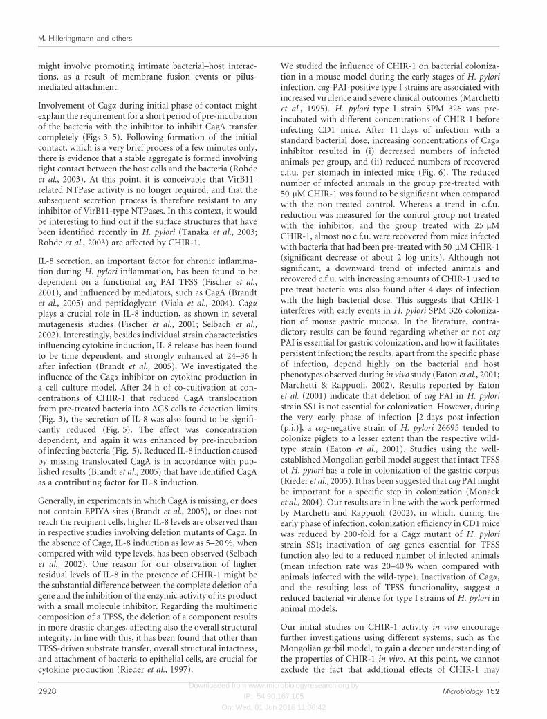
Fig. 6. Dependence of c.f.u. recovered in amouse model of infection on CHIR-1 pre-treatment of H. pylori. CD1 mice (10 animalsper group) were infected twice with 16108
(a) or once with 16109 (b) H. pylori SPM326 that had been pre-treated for 30 minwith different concentrations of CHIR-1 (0,25 and 50 mM). A second dose of CHIR-1was given 48 h p.i. Eleven and four daysp.i., for low- and high-infection doses,respectively, groups were euthanized, andgastric mucosa was assessed for coloniza-tion by inoculating serial dilutions of mucosalcells on selective blood agar plates. Statisti-cal analysis was performed using a Mann–Whitney U test. Symbols represent valuesfor individual animals in a group, and hori-zontal bars represent geometric means. *, Asignificant reduction in c.f.u. compared withthe non-treated control (P=0?0014).
http://mic.sgmjournals.org 2927
Inhibitors of H. pylori VirB11-type ATPase Caga
Downloaded from www.microbiologyresearch.org by
IP: 54.90.167.105
On: Wed, 01 Jun 2016 11:06:42
might involve promoting intimate bacterial–host interac-tions, as a result of membrane fusion events or pilus-mediated attachment.
Involvement of Caga during initial phase of contact mightexplain the requirement for a short period of pre-incubationof the bacteria with the inhibitor to inhibit CagA transfercompletely (Figs 3–5). Following formation of the initialcontact, which is a very brief process of a few minutes only,there is evidence that a stable aggregate is formed involvingtight contact between the host cells and the bacteria (Rohdeet al., 2003). At this point, it is conceivable that VirB11-related NTPase activity is no longer required, and that thesubsequent secretion process is therefore resistant to anyinhibitor of VirB11-type NTPases. In this context, it wouldbe interesting to find out if the surface structures that havebeen identified recently in H. pylori (Tanaka et al., 2003;Rohde et al., 2003) are affected by CHIR-1.
IL-8 secretion, an important factor for chronic inflamma-tion during H. pylori inflammation, has been found to bedependent on a functional cag PAI TFSS (Fischer et al.,2001), and influenced by mediators, such as CagA (Brandtet al., 2005) and peptidoglycan (Viala et al., 2004). Cagaplays a crucial role in IL-8 induction, as shown in severalmutagenesis studies (Fischer et al., 2001; Selbach et al.,2002). Interestingly, besides individual strain characteristicsinfluencing cytokine induction, IL-8 release has been foundto be time dependent, and strongly enhanced at 24–36 hafter infection (Brandt et al., 2005). We investigated theinfluence of the Caga inhibitor on cytokine production ina cell culture model. After 24 h of co-cultivation at con-centrations of CHIR-1 that reduced CagA translocationfrom pre-treated bacteria into AGS cells to detection limits(Fig. 3), the secretion of IL-8 was also found to be signifi-cantly reduced (Fig. 5). The effect was concentrationdependent, and again it was enhanced by pre-incubationof infecting bacteria (Fig. 5). Reduced IL-8 induction causedby missing translocated CagA is in accordance with pub-lished results (Brandt et al., 2005) that have identified CagAas a contributing factor for IL-8 induction.
Generally, in experiments in which CagA is missing, or doesnot contain EPIYA sites (Brandt et al., 2005), or does notreach the recipient cells, higher IL-8 levels are observed thanin respective studies involving deletion mutants of Caga. Inthe absence of Caga, IL-8 induction as low as 5–20 %, whencompared with wild-type levels, has been observed (Selbachet al., 2002). One reason for our observation of higherresidual levels of IL-8 in the presence of CHIR-1 might bethe substantial difference between the complete deletion of agene and the inhibition of the enzymic activity of its productwith a small molecule inhibitor. Regarding the multimericcomposition of a TFSS, the deletion of a component resultsin more drastic changes, affecting also the overall structuralintegrity. In line with this, it has been found that other thanTFSS-driven substrate transfer, overall structural intactness,and attachment of bacteria to epithelial cells, are crucial forcytokine production (Rieder et al., 1997).
We studied the influence of CHIR-1 on bacterial coloniza-tion in a mouse model during the early stages of H. pyloriinfection. cag-PAI-positive type I strains are associated withincreased virulence and severe clinical outcomes (Marchettiet al., 1995). H. pylori type I strain SPM 326 was pre-incubated with different concentrations of CHIR-1 beforeinfecting CD1 mice. After 11 days of infection with astandard bacterial dose, increasing concentrations of Caga
inhibitor resulted in (i) decreased numbers of infectedanimals per group, and (ii) reduced numbers of recoveredc.f.u. per stomach in infected mice (Fig. 6). The reducednumber of infected animals in the group pre-treated with50 mM CHIR-1 was found to be significant when comparedwith the non-treated control. Whereas a trend in c.f.u.reduction was measured for the control group not treatedwith the inhibitor, and the group treated with 25 mMCHIR-1, almost no c.f.u. were recovered from mice infectedwith bacteria that had been pre-treated with 50 mM CHIR-1(significant decrease of about 2 log units). Although notsignificant, a downward trend of infected animals andrecovered c.f.u. with increasing amounts of CHIR-1 used topre-treat bacteria was also found after 4 days of infectionwith the high bacterial dose. This suggests that CHIR-1interferes with early events in H. pylori SPM 326 coloniza-tion of mouse gastric mucosa. In the literature, contra-dictory results can be found regarding whether or not cagPAI is essential for gastric colonization, and how it facilitatespersistent infection; the results, apart from the specific phaseof infection, depend highly on the bacterial and hostphenotypes observed during in vivo study (Eaton et al., 2001;Marchetti & Rappuoli, 2002). Results reported by Eatonet al. (2001) indicate that deletion of cag PAI in H. pyloristrain SS1 is not essential for colonization. However, duringthe very early phase of infection [2 days post-infection(p.i.)], a cag-negative strain of H. pylori 26695 tended tocolonize piglets to a lesser extent than the respective wild-type strain (Eaton et al., 2001). Studies using the well-established Mongolian gerbil model suggest that intact TFSSof H. pylori has a role in colonization of the gastric corpus(Rieder et al., 2005). It has been suggested that cag PAI mightbe important for a specific step in colonization (Monacket al., 2004). Our results are in line with the work performedby Marchetti and Rappuoli (2002), in which, during theearly phase of infection, colonization efficiency in CD1 micewas reduced by 200-fold for a Caga mutant of H. pyloristrain SS1; inactivation of cag genes essential for TFSSfunction also led to a reduced number of infected animals(mean infection rate was 20–40 % when compared withanimals infected with the wild-type). Inactivation of Caga,and the resulting loss of TFSS functionality, suggest areduced bacterial virulence for type I strains of H. pylori inanimal models.
Our initial studies on CHIR-1 activity in vivo encouragefurther investigations using different systems, such as theMongolian gerbil model, to gain a deeper understanding ofthe properties of CHIR-1 in vivo. At this point, we cannotexclude the fact that additional effects of CHIR-1 may
2928 Microbiology 152
M. Hilleringmann and others

Downloaded from www.microbiologyresearch.org by
IP: 54.90.167.105
On: Wed, 01 Jun 2016 11:06:42
contribute to the measured reduction of colonizationduring early stages of infection. These effects include inter-action with other VirB11-like proteins in H. pylori, such asthe VirB11-homologue located within the flagellar exportlocus (Porwollik et al., 1999), which has been found to beessential for gastric colonization in a Mongolian gerbilmodel of infection (Kavermann et al., 2003).
For TFSS Cagb, which is another protein containing NTP-binding motifs, the effect of CHIR-1 could not be tested,since we did not succeed in demonstrating the NTPaseactivity in vitro. However, the primary sequences, includingthe NTP-binding motifs of Caga and b, differ markedly fromeach other; therefore, it is very unlikely that a moleculeselected for its activity on Caga would also interact withCagb.
Our study formally demonstrates that it is possible toidentify inhibitors of virulence-related TFSSs, as shown inour model of study H. pylori. Nevertheless, many additionalquestions need to be addressed before an enzymic inhibitorcan be transformed into an efficient antibiotic. Studieson the suitability of a compound for use as a drug (e.g.solubility and uptake), and its toxicity and pharmacoki-netics, together with further optimization of lead com-pounds, must be performed in order to obtain the desiredproperties.
Presently, we are studying the interaction between Caga andCHIR-1 using co-crystallization and molecular modellingtechniques. Detailed structural information might allowrational modifications of the lead structure of the inhibitorto further improve its efficacy. The crystal structure ofCaga has been solved by Yeo et al. (2000). In addition,the synthesis of radioactively labelled CHIR-1 might behelpful for a detailed investigation of the uptake, andfurther interaction of the inhibitor with Caga, in the cellularsystem.
To our knowledge, this is the first report that describes aspecific inhibitor of a TFSS that is capable of interfering withan essential enzymic function of the injectosome. Thecompound efficiently inhibited the function of a singlecomponent of the H. pylori cag TFSS, Caga, and therebyblocked the biological activity of the system as a whole.Initial in vivo evaluation studies indicated that CHIR-1mediated the reduction of pathogenic effects of H. pylori inmice. Recently, for several conjugative systems, inhibitionby unsaturated fatty acids was demonstrated, although themechanism of action of these compounds remains to beelucidated (Fernandez-Lopez et al., 2005). Others haveshown that type III secretion systems are also potentialtargets for new antimicrobials (Nordfelth et al., 2005;Kauppi et al., 2003). Our ongoing work will extend thesestudies to other pathogens in which TFSSs are requiredvirulence factors. Future experiments could give moreinformation on the specificity of the inhibitors identifiedusing H. pylori Caga that, in principle, might also be activeon similar NTPases from other pathogens that carry a TFSS.
ACKNOWLEDGEMENTS
We grateful acknowledge A. Harris and S. Harrison for providinginhibitors CHIR-1, -2 and -3, J. Bach for carrying out many of theassays, and K. Dungca for technical assistance. We are indebted toL. Pancotto and the animal care team for excellent help during in vivoexperiments, and to M. de Bernard (University of Padua) for valuablehelp with confocal microscopy. We would like to thank S. Baroneand F. Bagnoli for helpful discussions, and providing protocols, andthe other members of our laboratory for the supportive comments,and supplying various reagents and antibodies. Finally, we are gratefulto G. Corsi for his support doing the artwork. M. H. is supported by aMarie Curie Fellowship of the European Commission (contractnumber QLK2-CT-2001-51028). This work was supported by grantQLRT-2001-01200 (TFSS) of the European Union framework 5program.
REFERENCES
Akopyants, N. S., Clifton, S. W., Kersulyte, D. & 7 other authors(1998). Analyses of the cag pathogenicity island of Helicobacterpylori. Mol Microbiol 28, 37–53.
Amieva, M. R., Vogelmann, R., Covacci, A., Tompkins, L. S., Nelson,W. J. & Falkow, S. (2003). Disruption of the epithelial apical-junctionalcomplex by Helicobacter pylori CagA. Science 300, 1430–1434.
Atmakuri, K., Cascales, E. & Christie, P. J. (2004). Energetic com-ponents VirD4, VirB11 and VirB4 mediate early DNA transferreactions required for bacterial type IV secretion. Mol Microbiol 54,1199–1211.
Backert, S., Ziska, E., Brinkmann, V., Zimny-Arndt, U., Fauconnier,A., Jungblut, P. R., Naumann, M. & Meyer, T. F. (2000). Translocationof the Helicobacter pylori CagA protein in gastric epithelial cells by atype IV secretion apparatus. Cell Microbiol 2, 155–164.
Berger, B. R. & Christie, P. J. (1994). Genetic complementationanalysis of the Agrobacterium tumefaciens virB operon: virB2 throughvirB11 are essential virulence genes. J Bacteriol 176, 3646–3660.
Bourzac, K. M. & Guillemin, K. (2005). Helicobacter pylori–host cellinteractions mediated by type IV secretion. Cell Microbiol 7, 911–919.
Brandt, S., Kwok, T., Hartig, R., Konig, W. & Backert, S. (2005).NF-kB activation and potentiation of proinflammatory responses bythe Helicobacter pylori CagA protein. Proc Natl Acad Sci U S A 102,9300–9305.
Censini, S., Lange, C., Xiang, Z., Crabtree, J. E., Ghiara, P.,Borodovsky, M., Rappuoli, R. & Covacci, A. (1996). cag, a pathogenicityisland of Helicobacter pylori, encodes type I-specific and disease-associated virulence factors. Proc Natl Acad Sci U S A 93, 14648–14653.
Christie, P. J. (2001). Type IV secretion: intercellular transfer ofmacromolecules by systems ancestrally related to conjugationmachines. Mol Microbiol 40, 294–305.
Christie, P. J. & Vogel, J. P. (2000). Bacterial type IV secretion:conjugation systems adapted to deliver effector molecules to hostcells. Trends Microbiol 8, 354–360.
Christie, P. J., Atmakuri, K., Krishnamoorthy, V., Jakubowski, S. &Cascales, E. (2005). Biogenesis, architecture, and function ofbacterial Type IV secretion systems. Annu Rev Microbiol
Cole, S. P., Cirillo, D., Kagnoff, M. F., Guiney, D. G. & Eckmann, L.(1997). Coccoid and spiral Helicobacter pylori differ in their abilitiesto adhere to gastric epithelial cells and induce interleukin-8secretion. Infect Immun 65, 843–846.
Covacci, A., Falkow, S., Berg, D. E. & Rappuoli, R. (1997). Did theinheritance of a pathogenicity island modify the virulence ofHelicobacter pylori? Trends Microbiol 5, 205–208.
http://mic.sgmjournals.org 2929
Inhibitors of H. pylori VirB11-type ATPase Caga

Downloaded from www.microbiologyresearch.org by
IP: 54.90.167.105
On: Wed, 01 Jun 2016 11:06:42
Crabtree, J. E., Covacci, A., Farmery, S. M., Xiang, Z., Tompkins,D. S., Perry, S., Lindley, I. J. & Rappuoli, R. (1995). Helicobacter
pylori induced interleukin-8 expression in gastric epithelial cells is
associated with CagA positive phenotype. J Clin Pathol 48, 41–45.
Ding, Z., Atmakuri, K. & Christie, P. J. (2003). The outs and ins of
bacterial type IV secretion substrates. Trends Microbiol 11, 527–535.
Eaton, K. A., Kersulyte, D., Mefford, M., Danon, S. J., Krakowka, S. &Berg, D. E. (2001). Role of Helicobacter pylori cag region genes in
colonization and gastritis in two animal models. Infect Immun 69,
2902–2908.
Fernandez, D., Spudich, G. M., Zhou, X. R. & Christie, P. J. (1996).The Agrobacterium tumefaciens VirB7 lipoprotein is required for
stabilization of VirB proteins during assembly of the T-complex
transport apparatus. J Bacteriol 178, 3168–3176.
Fernandez-Lopez, R., Machon, C., Longshaw, C. M., Martin, S.,Molin, S., Zechner, E. L., Espinosa, M., Lanka, E. & de la Cruz, F.(2005). Unsaturated fatty acids are inhibitors of bacterial conjuga-
tion. Microbiology 151, 3517–3526.
Fischer, W., Puls, J., Buhrdorf, R., Gebert, B., Odenbreit, S. & Haas, R.(2001). Systematic mutagenesis of the Helicobacter pylori cag patho-
genicity island: essential genes for CagA translocation in host cells and
induction of interleukin-8. Mol Microbiol 42, 1337–1348.
Kauppi, A. M., Nordfelth, R., Uvell, H., Wolf-Watz, H. & Elofsson, M.(2003). Targeting bacterial virulence: inhibitors of type III secretion
in Yersinia. Chem Biol 10, 241–249.
Kavermann, H., Burns, B. P., Angermuller, K., Odenbreit, S.,Fischer, W., Melchers, K. & Haas, R. (2003). Identification and
characterization of Helicobacter pylori genes essential for gastric
colonization. J Exp Med 197, 813–822.
Krause, S., Barcena, M., Pansegrau, W., Lurz, R., Carazo, J. M. &Lanka, E. (2000a). Sequence-related protein export NTPases encoded
by the conjugative transfer region of RP4 and by the cag patho-
genicity island of Helicobacter pylori share similar hexameric ring
structures. Proc Natl Acad Sci U S A 97, 3067–3072.
Krause, S., Pansegrau, W., Lurz, R., de la Cruz, F. & Lanka, E.(2000b). Enzymology of type IV macromolecule secretion systems:
the conjugative transfer regions of plasmids RP4 and R388 and the
cag pathogenicity island of Helicobacter pylori encode structurally
and functionally related nucleoside triphosphate hydrolases. J
Bacteriol 182, 2761–2770.
Machon, C., Rivas, S., Albert, A., Goni, F. M. & de la Cruz, F. (2002).TrwD, the hexameric traffic ATPase encoded by plasmid R388,
induces membrane destabilization and hemifusion of lipid vesicles.
J Bacteriol 184, 1661–1668.
Marchetti, M. & Rappuoli, R. (2002). Isogenic mutants of the cag
pathogenicity island of Helicobacter pylori in the mouse model
of infection: effects on colonization efficiency. Microbiology 148,
1447–1456.
Marchetti, M., Arico, B., Burroni, D., Figura, N., Rappuoli, R. &Ghiara, P. (1995). Development of a mouse model of Helicobacter
pylori infection that mimics human disease. Science 267, 1655–1658.
Monack, D. M., Mueller, A. & Falkow, S. (2004). Persistent bacterial
infections: the interface of the pathogen and the host immune
system. Nat Rev Microbiol 2, 747–765.
Nordfelth, R., Kauppi, A. M., Norberg, H. A., Wolf-Watz, H. &Elofsson, M. (2005). Small-molecule inhibitors specifically targeting
type III secretion. Infect Immun 73, 3104–3114.
Odenbreit, S., Puls, J., Sedlmaier, B., Gerland, E., Fischer, W. &Haas, R. (2000). Translocation of Helicobacter pylori CagA into
gastric epithelial cells by type IV secretion. Science 287, 1497–1500.
Planet, P. J., Kachlany, S. C., Desalle, R. & Figurski, D. H. (2001).Phylogeny of genes for secretion NTPases: identification of the
widespread tadA subfamily and development of a diagnostic key for
gene classification. Proc Natl Acad Sci U S A 98, 2503–2508.
Porwollik, S., Noonan, B. & O’Toole, P. W. (1999). Molecular char-
acterization of a flagellar export locus of Helicobacter pylori. Infect
Immun 67, 2060–2070.
Rieder, G., Hatz, R. A., Moran, A. P., Walz, A., Stolte, M. & Enders, G.
(1997). Role of adherence in interleukin-8 induction in Helicobacter
pylori-associated gastritis. Infect Immun 65, 3622–3630.
Rieder, G., Merchant, J. L. & Haas, R. (2005). Helicobacter pylori cag-type
IV secretion system facilitates corpus colonization to induce precancer-
ous conditions in Mongolian gerbils. Gastroenterology 128, 1229–1242.
Rivas, S., Bolland, S., Cabezon, E., Goni, F. M. & de la Cruz, C. F.
(1997). TrwD, a protein encoded by the IncW plasmid R388,
displays an ATP hydrolase activity essential for bacterial conjugation.
J Biol Chem 272, 25583–25590.
Rohde, M., Puls, J., Buhrdorf, R., Fischer, W. & Haas, R. (2003). A
novel sheathed surface organelle of the Helicobacter pylori cag type IV
secretion system. Mol Microbiol 49, 219–234.
Rothenbacher, D. & Brenner, H. (2003). Burden of Helicobacter
pylori and H. pylori-related diseases in developed countries: recent
developments and future implications. Microbes Infect 5, 693–703.
Sagulenko, E., Sagulenko, V., Chen, J. & Christie, P. J. (2001). Role
of Agrobacterium VirB11 ATPase in T-pilus assembly and substrate
selection. J Bacteriol 183, 5813–5825.
Savvides, S. N., Yeo, H. J., Beck, M. R. & 7 other authors (2003).VirB11 ATPases are dynamic hexameric assemblies: new insights into
bacterial type IV secretion. EMBO J 22, 1969–1980.
Segal, E. D., Cha, J., Lo, J., Falkow, S. & Tompkins, L. S. (1999).Altered states: involvement of phosphorylated CagA in the induction
of host cellular growth changes by Helicobacter pylori. Proc Natl Acad
Sci U S A 96, 14559–14564.
Selbach, M., Moese, S., Meyer, T. F. & Backert, S. (2002).
Functional analysis of the Helicobacter pylori cag pathogenicity
island reveals both VirD4-CagA-dependent and VirD4-CagA-inde-
pendent mechanisms. Infect Immun 70, 665–671.
Selbach, M., Moese, S., Hurwitz, R., Hauck, C. R., Meyer, T. F. &
Backert, S. (2003). The Helicobacter pylori CagA protein induces
cortactin dephosphorylation and actin rearrangement by c-Src
inactivation. EMBO J 22, 515–528.
Stein, M., Rappuoli, R. & Covacci, A. (2000). Tyrosine phosphoryla-
tion of the Helicobacter pylori CagA antigen after cag-driven host cell
translocation. Proc Natl Acad Sci U S A 97, 1263–1268.
Stein, M., Bagnoli, F., Halenbeck, R., Rappuoli, R., Fantl, W. J. &Covacci, A. (2002). c-Src/Lyn kinases activate Helicobacter pylori
CagA through tyrosine phosphorylation of the EPIYA motifs. Mol
Microbiol 43, 971–980.
Tanaka, J., Suzuki, T., Mimuro, H. & Sasakawa, C. (2003). Structural
definition on the surface of Helicobacter pylori type IV secretion
apparatus. Cell Microbiol 5, 395–404.
Tato, I., Zunzunegui, S., de la Cruz, F. & Cabezon, E. (2005). TrwB,
the coupling protein involved in DNA transport during bacterial
conjugation, is a DNA-dependent ATPase. Proc Natl Acad Sci U S A
102, 8156–8161.
Viala, J., Chaput, C., Boneca, I. G. & 13 other authors (2004). Nod1
responds to peptidoglycan delivered by the Helicobacter pylori cag
pathogenicity island. Nat Immunol 5, 1166–1174.
Yeo, H. J. & Waksman, G. (2004). Unveiling molecular scaffolds of
the type IV secretion system. J Bacteriol 186, 1919–1926.
Yeo, H. J., Savvides, S. N., Herr, A. B., Lanka, E. & Waksman, G.
(2000). Crystal structure of the hexameric traffic ATPase of the
Helicobacter pylori type IV secretion system. Mol Cell 6, 1461–1472.
2930 Microbiology 152
M. Hilleringmann and others
Related Documents