Activation of P2X7 Promotes Cerebral Edema and Neurological Injury after Traumatic Brain Injury in Mice Donald E. Kimbler 1 , Jessica Shields 1 , Nathan Yanasak 2 , John R. Vender 1 , Krishnan M. Dhandapani 1 * 1 Department of Neurosurgery, Georgia Health Sciences University, Augusta, Georgia, United States of America, 2 Department of Radiology, Georgia Health Sciences University, Augusta, Georgia, United States of America Abstract Traumatic brain injury (TBI) is a leading cause of death and disability worldwide. Cerebral edema, the abnormal accumulation of fluid within the brain parenchyma, contributes to elevated intracranial pressure (ICP) and is a common life- threatening neurological complication following TBI. Unfortunately, neurosurgical approaches to alleviate increased ICP remain controversial and medical therapies are lacking due in part to the absence of viable drug targets. In the present study, genetic inhibition (P2X72/2 mice) of the purinergic P2x7 receptor attenuated the expression of the pro- inflammatory cytokine, interleukin-1b (IL-1b) and reduced cerebral edema following controlled cortical impact, as compared to wild-type mice. Similarly, brilliant blue G (BBG), a clinically non-toxic P2X7 inhibitor, inhibited IL-1b expression, limited edemic development, and improved neurobehavioral outcomes after TBI. The beneficial effects of BBG followed either prophylactic administration via the drinking water for one week prior to injury or via an intravenous bolus administration up to four hours after TBI, suggesting a clinically-implementable therapeutic window. Notably, P2X7 localized within astrocytic end feet and administration of BBG decreased the expression of glial fibrillary acidic protein (GFAP), a reactive astrocyte marker, and attenuated the expression of aquaporin-4 (AQP4), an astrocytic water channel that promotes cellular edema. Together, these data implicate P2X7 as a novel therapeutic target to prevent secondary neurological injury after TBI, a finding that warrants further investigation. Citation: Kimbler DE, Shields J, Yanasak N, Vender JR, Dhandapani KM (2012) Activation of P2X7 Promotes Cerebral Edema and Neurological Injury after Traumatic Brain Injury in Mice. PLoS ONE 7(7): e41229. doi:10.1371/journal.pone.0041229 Editor: Christoph Kleinschnitz, Julius-Maximilians-Universita ¨t Wu ¨ rzburg, Germany Received March 9, 2012; Accepted June 19, 2012; Published July 17, 2012 Copyright: ß 2012 Kimbler et al. This is an open-access article distributed under the terms of the Creative Commons Attribution License, which permits unrestricted use, distribution, and reproduction in any medium, provided the original author and source are credited. Funding: Financial support for this study was provided by grants from the National Institutes of Health (NS065172) and from the TriServices Nursing Research Program (HU0001-10-1-TS11). The content and views expressed herein do not necessarily represent the views of the Department of Defense, the TriServices Nursing Research Program, Uniformed Services University of the Health Sciences, or the United States Government. The funders had no role in study design, data collection and analysis, decision to publish, or preparation of the manuscript. Competing Interests: Co-author Krishnan M. Dhandapani serves as a PLoS ONE Editorial Board member. This relationship does not alter the authors’ adherence to all the PLoS ONE policies on sharing data and materials. * E-mail: [email protected] Introduction Traumatic brain injury (TBI), a leading cause of mortality and morbidity worldwide, affects over 1.7 million Americans annually [1]. In contrast to primary injuries that occur at the time of impact, secondary pathological processes develop while under supervised medical care and profoundly influence patient out- comes [2]. Cerebral edema, the abnormal accumulation of fluid within the brain, is a life-threatening neurological complication that promotes elevated intracranial pressure (ICP) and leads to clinical deterioration in the hours and days after the initial traumatic event [3,4]. Increased ICP subsequently promotes brain herniation, limits cerebral blood flow, reduces brain oxygenation, and contributes to poor clinical outcomes [5,6,7,8]; however, the efficacy of neurosurgical approaches to alleviate increased ICP and improve patient prognoses remain limited [9]. Furthermore, effective medical therapies to control ICP are lacking, in part, due to the poorly defined mechanisms that underlie edemic development after TBI. The innate immune system provides immediate, non-specific defense following infection or tissue injury, although controversy remains as to whether theses response are protective or detrimental after injury. Glia constitutively express receptors involved in cerebral innate immune responses and upon activa- tion, may secrete pro-inflammatory mediators to recruit peripheral immune cells to the site of injury [10]; however, the functional significance and cellular mediators of cerebral innate immune activation remains unresolved. Cellular necrosis correlates with the development of peri-contusional brain edema after TBI and surgical excision of necrotic tissue reduces ICP, decreases patient mortality, and improves neurological outcomes in neurotrauma patients [11,12,13]. Thus, necrotic cell death may initiate post- traumatic immune responses. Damage-associated molecular pat- tern molecules (DAMPs) are multi-functional host proteins that trigger innate immune activation after necrotic injuries. Adenosine 59-triphosphate (ATP), an important intracellular energy source, is rapidly released into the extracellular space following traumatic or ischemic injuries to function as a non-proteinaceous DAMP [14,15,16,17]. Notably, the accumulation of ATP metabolites within the cerebrospinal fluid (CSF) directly correlated with edemic development and elevated ICP in a neurotrauma patient [18], implicating ATP as an initiator of secondary brain injury after TBI. Purinergic P2X7 receptors mediate, at least in part, the biological actions of extracellular ATP [19]. Sustained activation of P2X7 with high concentrations of ATP induced the release of PLoS ONE | www.plosone.org 1 July 2012 | Volume 7 | Issue 7 | e41229

Welcome message from author
This document is posted to help you gain knowledge. Please leave a comment to let me know what you think about it! Share it to your friends and learn new things together.
Transcript
Activation of P2X7 Promotes Cerebral Edema andNeurological Injury after Traumatic Brain Injury in MiceDonald E. Kimbler1, Jessica Shields1, Nathan Yanasak2, John R. Vender1, Krishnan M. Dhandapani1*
1Department of Neurosurgery, Georgia Health Sciences University, Augusta, Georgia, United States of America, 2Department of Radiology, Georgia Health Sciences
University, Augusta, Georgia, United States of America
Abstract
Traumatic brain injury (TBI) is a leading cause of death and disability worldwide. Cerebral edema, the abnormalaccumulation of fluid within the brain parenchyma, contributes to elevated intracranial pressure (ICP) and is a common life-threatening neurological complication following TBI. Unfortunately, neurosurgical approaches to alleviate increased ICPremain controversial and medical therapies are lacking due in part to the absence of viable drug targets. In the presentstudy, genetic inhibition (P2X72/2 mice) of the purinergic P2x7 receptor attenuated the expression of the pro-inflammatory cytokine, interleukin-1b (IL-1b) and reduced cerebral edema following controlled cortical impact, as comparedto wild-type mice. Similarly, brilliant blue G (BBG), a clinically non-toxic P2X7 inhibitor, inhibited IL-1b expression, limitededemic development, and improved neurobehavioral outcomes after TBI. The beneficial effects of BBG followed eitherprophylactic administration via the drinking water for one week prior to injury or via an intravenous bolus administration upto four hours after TBI, suggesting a clinically-implementable therapeutic window. Notably, P2X7 localized within astrocyticend feet and administration of BBG decreased the expression of glial fibrillary acidic protein (GFAP), a reactive astrocytemarker, and attenuated the expression of aquaporin-4 (AQP4), an astrocytic water channel that promotes cellular edema.Together, these data implicate P2X7 as a novel therapeutic target to prevent secondary neurological injury after TBI,a finding that warrants further investigation.
Citation: Kimbler DE, Shields J, Yanasak N, Vender JR, Dhandapani KM (2012) Activation of P2X7 Promotes Cerebral Edema and Neurological Injury afterTraumatic Brain Injury in Mice. PLoS ONE 7(7): e41229. doi:10.1371/journal.pone.0041229
Editor: Christoph Kleinschnitz, Julius-Maximilians-Universitat Wurzburg, Germany
Received March 9, 2012; Accepted June 19, 2012; Published July 17, 2012
Copyright: � 2012 Kimbler et al. This is an open-access article distributed under the terms of the Creative Commons Attribution License, which permitsunrestricted use, distribution, and reproduction in any medium, provided the original author and source are credited.
Funding: Financial support for this study was provided by grants from the National Institutes of Health (NS065172) and from the TriServices Nursing ResearchProgram (HU0001-10-1-TS11). The content and views expressed herein do not necessarily represent the views of the Department of Defense, the TriServicesNursing Research Program, Uniformed Services University of the Health Sciences, or the United States Government. The funders had no role in study design, datacollection and analysis, decision to publish, or preparation of the manuscript.
Competing Interests: Co-author Krishnan M. Dhandapani serves as a PLoS ONE Editorial Board member. This relationship does not alter the authors’ adherenceto all the PLoS ONE policies on sharing data and materials.
* E-mail: [email protected]
Introduction
Traumatic brain injury (TBI), a leading cause of mortality and
morbidity worldwide, affects over 1.7 million Americans annually
[1]. In contrast to primary injuries that occur at the time of
impact, secondary pathological processes develop while under
supervised medical care and profoundly influence patient out-
comes [2]. Cerebral edema, the abnormal accumulation of fluid
within the brain, is a life-threatening neurological complication
that promotes elevated intracranial pressure (ICP) and leads to
clinical deterioration in the hours and days after the initial
traumatic event [3,4]. Increased ICP subsequently promotes brain
herniation, limits cerebral blood flow, reduces brain oxygenation,
and contributes to poor clinical outcomes [5,6,7,8]; however, the
efficacy of neurosurgical approaches to alleviate increased ICP and
improve patient prognoses remain limited [9]. Furthermore,
effective medical therapies to control ICP are lacking, in part,
due to the poorly defined mechanisms that underlie edemic
development after TBI.
The innate immune system provides immediate, non-specific
defense following infection or tissue injury, although controversy
remains as to whether theses response are protective or
detrimental after injury. Glia constitutively express receptors
involved in cerebral innate immune responses and upon activa-
tion, may secrete pro-inflammatory mediators to recruit peripheral
immune cells to the site of injury [10]; however, the functional
significance and cellular mediators of cerebral innate immune
activation remains unresolved. Cellular necrosis correlates with the
development of peri-contusional brain edema after TBI and
surgical excision of necrotic tissue reduces ICP, decreases patient
mortality, and improves neurological outcomes in neurotrauma
patients [11,12,13]. Thus, necrotic cell death may initiate post-
traumatic immune responses. Damage-associated molecular pat-
tern molecules (DAMPs) are multi-functional host proteins that
trigger innate immune activation after necrotic injuries. Adenosine
59-triphosphate (ATP), an important intracellular energy source, is
rapidly released into the extracellular space following traumatic or
ischemic injuries to function as a non-proteinaceous DAMP
[14,15,16,17]. Notably, the accumulation of ATP metabolites
within the cerebrospinal fluid (CSF) directly correlated with
edemic development and elevated ICP in a neurotrauma patient
[18], implicating ATP as an initiator of secondary brain injury
after TBI.
Purinergic P2X7 receptors mediate, at least in part, the
biological actions of extracellular ATP [19]. Sustained activation
of P2X7 with high concentrations of ATP induced the release of
PLoS ONE | www.plosone.org 1 July 2012 | Volume 7 | Issue 7 | e41229
biologically-active interleukin-1b (IL-1b) [20,21], a potent pro-
inflammatory cytokine. Notably, IL-1b exhibited a prolonged
induction in multiple pre-clinical models of TBI
[22,23,24,25,26,27] and increased CSF and brain content of IL-
1b positively correlated with elevated ICP and unfavorable
outcomes in TBI patients [28,29,30]. Furthermore, we and others
demonstrated that genetic or pharmacological inhibition of IL-1battenuated both cerebral edema and secondary injury after TBI
[26,31,32,33,34], indicative of a deleterious role for IL-1b after
head trauma. However, a mechanistic understanding of post-
traumatic IL-1b production remains undetermined and once
elucidated, may provide novel opportunities for therapeutic
development. As a robust inflammatory response clinically
correlates with secondary neurovascular injury after TBI, we
hypothesized that activation of P2X7 mediates neurological
demise following TBI. Our results implicate P2X7 as a novel
therapeutic target to prevent secondary injury after TBI, a finding
that warrants further investigation.
Results
BBG reduces post-traumatic cerebral edema with anextended therapeutic windowBrain water content, a sensitive measure of cerebral edema, was
significantly increased within the ipsilateral cortex at 24h post-TBI
(83.660.4% brain water content after TBI vs. 77.960.2% in
sham, p,0.001 vs. sham). A single, intravenous injection of
50 mg/kg BBG at 15 minutes prior to injury attenuated brain
water content after TBI (80.660.5%; p,0.01 vs. TBI) whereas
administration of 25 mg/kg BBG did not significantly reduce
edema (83.3%60.3%; not significantly different from TBI)
(Figure 1a). Notably, the ability of 100 mg/kg BBG to reduce
edema was not significantly different from administration of
50 mg/kg (81.060.2%; p,0.001 vs. TBI), suggesting 50 mg/kg
was the lowest efficacious dose to limit edemic development after
TBI (Figure 1b). For all studies, brain water content within the
contralateral (uninjured) cortices did not significantly differ
between any of the treatment groups (data not shown). Further-
more, administration of BBG alone (50 mg/kg, i.v., 15 minute
pre-treatment) did not significantly affect brain water content, as
compared to placebo-treated, sham-operated mice (Figure 1a),consistent with an injury-specific reduction in edema.
The therapeutic window whereby BBG reduced edemic de-
velopment was next established. A 1h post-treatment with 50 mg/
kg significantly reduced cerebral edema (81.360.2%, p,0.05 vs.
TBI) to a similar extent as pre-treatment (Figure 1c; see
Figure 1a for comparison). Similarly, a 4h post-treatment
effectively attenuated post-traumatic edema (81.460.4%,
p,0.05 vs. TBI, not significantly different from 1h post-
treatment). In contrast, 8h post-treatment with 50 mg/kg was
ineffective at reducing edema, as compared to TBI
(83.2%60.2%), suggesting a 4h post-injury therapeutic window.
We next determined whether prophylactic administration of
BBG reduces edema. Oral administration of 25 mg/mL BBG via
the drinking water for one week prior to injury effectively
decreased brain edema after TBI (80.960.2%, p,0.01 vs. TBI)
(Figure 1d). In contrast, 10 mg/mL BBG via the drinking water
did not significantly reduce edema, as compared to mice receiving
water containing only placebo. As a whole, either prophylactic
oral administration or post-injury intravenous administration of
BBG effective attenuates brain edema after TBI.
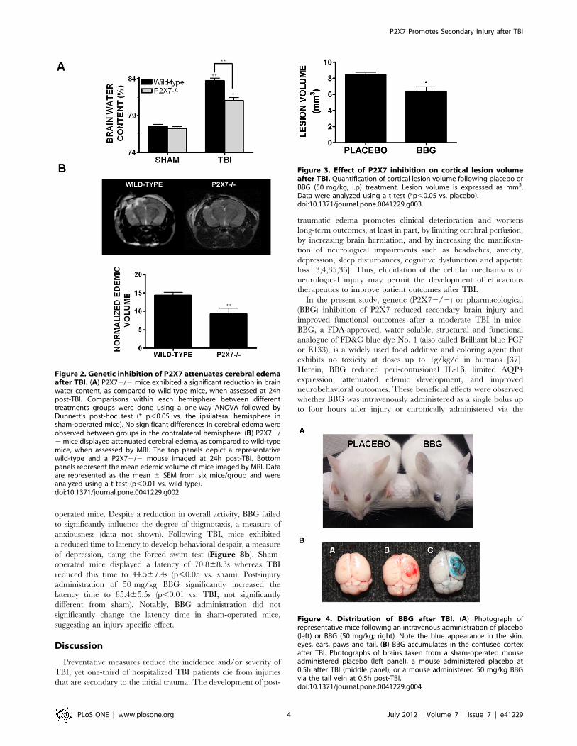
P2X72/2 mice exhibit reduced cerebral edema after TBIBBG is a highly selective inhibitor of P2X7; however,
pharmacological agents often exhibit ‘‘off-target’’ or non-specific
effects. To validate P2X7 as a potential therapeutic target to
reduce brain edema, P2X72/2 mice were utilized. Consistent
with data collected after BBG administration, P2X72/2 mice
significantly reduced brain water content, as compared to wild-
type mice, following TBI (81.060.4% in P2X72/2 vs.
83.760.3% in wild-type; p,0.01). These findings were supported
by the measurement of edemic volume using MRI. P2X72/2
mice exhibited a 36% reduction in edemic volume after TBI, as
compared to wild-type mice (14.460.7 mm3 in wild-type mice vs.
9.261.5 mm3 in P2X72/2 mice; p,0.01 vs. wild-type)
(Figure 2b). Brain water content was not significantly different
either in sham-operated mice (Figure 2a) or in the contralateral
hemisphere of wild-type or P2X72/2 mice (data not shown). In
line with the reduction in brain edema, inhibition of P2X7
significantly reduced cortical lesion volume after TBI (Figure 3).Specifically, lesion volume was decreased from 8.560.3 mm3 in
placebo-treated mice to 6.460.6 mm3 (p,0.05).
Brain expression of P2X7 after TBIPeripheral administration of BBG reduced brain edema,
although the potential tissue and cellular targets of BBG remained
unclear. Intravenous administration of 50 mg/kg BBG produced
a transient deep blue color over the first 24h within the eyes, nose,
ears, and paws (Figure 4a), demonstrating wide peripheral
distribution throughout the circulatory system. No trace of blue
color remained by 72h post-administration. Similarly, oral
administration of BBG for one week via the drinking water also
produced a faint blue hue in the paws and eyes, albeit to a far
lesser extent, as compared to intravenous administration. Consis-
tent with the observed blue appearance, serum levels of BBG
reached 383633.3 mM and 1.7360.07 mM following intravenous
administration of 50 mg/kg and 100 mg/kg, respectively. Wheth-
er BBG acted peripherally or centrally after TBI remained
unclear. In line with a potential direct effect within the CNS, the
brains of mice administered BBG appeared greyish-blue, with blue
color observed throughout the cerebral vasculature and brain
tissue. Most notably, the contused cortex exhibited a distinct blue
color (Figure 4b); suggesting BBG can enter the brain and
preferentially accumulates at high levels around damaged tissue,
presumably following blood-brain barrier disruption.
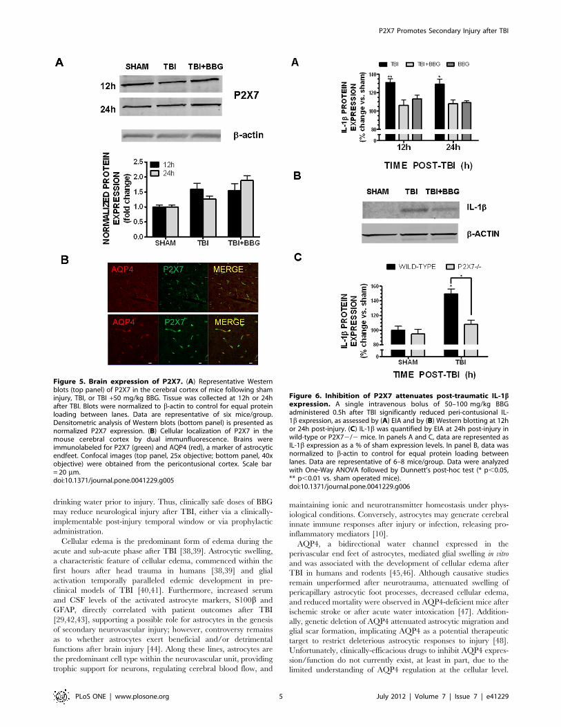
The cellular expression pattern of P2X7, the presumed cellular
target of BBG action, was next assessed within the brain. P2X7
was basally expressed within the cerebral cortex, as demonstrated
by Western blotting; however, expression was not increased
following TBI, as compared to sham-operated mice (Figure 5a).Immunohistochemical analysis revealed that P2X7 strongly co-
localized with the astrocytic end foot marker, aquaporin-4 (AQP4)
(Figure 5b) whereas dual labeling was not observed with either
markers of microglia or neurons (data not shown). These data
implicate astrocytes as key mediators of the biological actions of
P2X7 and as a possible cellular target of BBG after TBI.
P2X7 mediates IL-1b expression after TBIIncreased expression of the pro-inflammatory cytokine, IL-1b,
clinically correlates with the development of cerebral edema after
brain injury. Consistent with the ability of BBG to attenuate post-
traumatic edema, post-treatment with 50 mg/kg BBG significantly
reduced the expression of biologically mature IL-1bwithin the
pericontusional cortex at 12h and 24h after injury, as assessed by
either enzyme-linked immunoassay (EIA) (Figure 6a) or by
Western blotting (Figure 6b). TBI increased IL-1b expression by
P2X7 Promotes Secondary Injury after TBI
PLoS ONE | www.plosone.org 2 July 2012 | Volume 7 | Issue 7 | e41229
30.961.1% and 28.865.5% over sham-operated mice at 12h
(p,0.01 vs. sham) and 24h (p,0.05 vs. sham), respectively. Post-
treatment with 50 mg/kg BBG reduced the post-traumatic
expression of IL-1b to 6.866.3% and 8.064.0% over sham
(p,0.01 and p,0.05 vs. TBI at 12h and 24h, respectively; not
significantly different from sham at either timepoint). Notably,
administration of 50 mg/kg BBG alone had no significant effect
on the basal expression of IL-1b, as compared to sham-operated
mice. In line with these observations, a significant reduction in
post-traumatic IL-1b expression was observed in the pericontu-
sional cortex of P2X72/2 mice, as compared with wild-type mice
(Figure 6c). Specifically, IL-1b expression was increased by
49.367.1% at 24h post-TBI in wild-type mice (p,0.05 vs. sham).
In contrast, a 7.865.9% increase was observed in P2X72/2mice
(p,0.05 vs. wild-type TBI; not significantly different from sham).
No significant differences in basal IL-1b expression were detected
between genotypes, suggesting an injury-specific effect of P2X7
knockout.
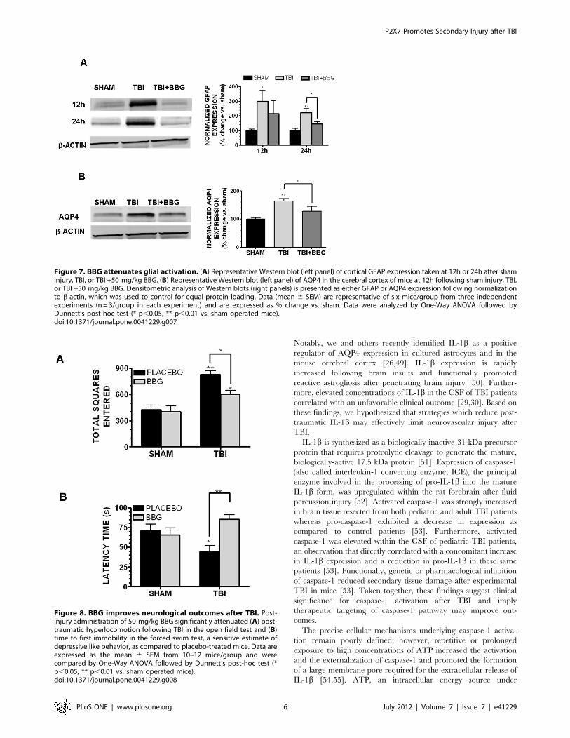
P2X7 mediates glial reactivity after TBIIL-1b induces reactive astrogliosis after TBI; therefore, the
ability of BBG to attenuate the expression of GFAP, a hallmark of
gliosis, was next assessed. GFAP expression was significantly
increased by 299.7672.2% within the peri-contusional cortex
(p,0.05 vs. sham) and 222.0628.6% (p,0.01 vs. sham) of sham-
operated mice at 12h and 24h post-TBI, respectively (Figure 7a).Post-treatment with 50 mg/kg BBG reduced GFAP expression to
216.1688.6% (not significantly different from either sham or TBI)
and 145.3615.2% (p,0.05 vs. TBI, not significantly different
from sham) of expression levels in sham-operated mice at 12h and
24h, respectively.
Consistent with the inhibitory effect of BBG on post-traumatic
cerebral edema and glial reactivity, BBG attenuated the expression
of the astrocytic water channel, AQP4, after TBI. AQP4 protein
expression was increased within the pericontusional context at 12h
(1.760.1 fold increase; p,0.01 vs. sham) and at 24h (1.560.1 fold
increase; p,0.05 vs. sham) after TBI. Intravenous administration
of 50 mg/kg BBG at 0.5h post-injury attenuated the TBI-induced
increases in AQP4 expression (1.360.2 and 1.160.1 fold increase
vs. sham at 12h and 24h, respectively; p,0.05 vs. TBI, not
significant different from sham) (Figure 7b).
BBG improves neurobehavioral outcomes after TBIDepression, anxiety, and cognitive dysfunction are frequent co-
morbidities after a TBI. A significant increase in open-field
hyperlocomotion (total number of squares entered) was observed
following TBI (p,0.01 vs. sham) (Figure 8a). Administration of
50 mg/kg BBG partially attenuated post-traumatic hyperlocomo-
tion by ,50% (p,0.05 vs. sham and TBI). In contrast, BBG
administration had no significant effect on basal activity in sham-
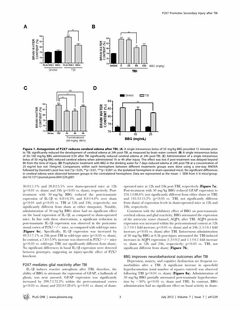
Figure 1. Antagonism of P2X7 reduces cerebral edema after TBI. (A) A single intravenous bolus of 50 mg/kg BBG provided 15 minutes priorto TBI, significantly reduced the development of cerebral edema at 24h post-TBI, as measured by brain water content. (B) A single intravenous bolusof 50–100 mg/kg BBG administered 0.5h after TBI significantly reduced cerebral edema at 24h post-TBI. (C) Administration of a single intravenousbolus of 50 mg/kg BBG reduced cerebral edema when administered 1h or 4h after injury. This effect was lost if post-treatment was delayed beyond8h from the time of injury. (D) Prophylactic treatment with BBG in the drinking water for 7 days reduced edema at 24h post-TBI at a concentration of25 mg/ml but not 10mg/ml. Comparisons within each hemisphere between different treatments groups were done using a one-way ANOVAfollowed by Dunnett’s post-hoc test (*p,0.05, **p,0.01, ***p,0.001 vs. the ipsilateral hemisphere in sham-operated mice). No significant differencesin cerebral edema were observed between groups in the contralateral hemisphere. Data are represented as the mean 6 SEM from 5–6 mice/group.doi:10.1371/journal.pone.0041229.g001
P2X7 Promotes Secondary Injury after TBI
PLoS ONE | www.plosone.org 3 July 2012 | Volume 7 | Issue 7 | e41229
operated mice. Despite a reduction in overall activity, BBG failed
to significantly influence the degree of thigmotaxis, a measure of
anxiousness (data not shown). Following TBI, mice exhibited
a reduced time to latency to develop behavioral despair, a measure
of depression, using the forced swim test (Figure 8b). Sham-
operated mice displayed a latency of 70.868.3s whereas TBI
reduced this time to 44.567.4s (p,0.05 vs. sham). Post-injury
administration of 50 mg/kg BBG significantly increased the
latency time to 85.465.5s (p,0.01 vs. TBI, not significantly
different from sham). Notably, BBG administration did not
significantly change the latency time in sham-operated mice,
suggesting an injury specific effect.
Discussion
Preventative measures reduce the incidence and/or severity of
TBI, yet one-third of hospitalized TBI patients die from injuries
that are secondary to the initial trauma. The development of post-
traumatic edema promotes clinical deterioration and worsens
long-term outcomes, at least in part, by limiting cerebral perfusion,
by increasing brain herniation, and by increasing the manifesta-
tion of neurological impairments such as headaches, anxiety,
depression, sleep disturbances, cognitive dysfunction and appetite
loss [3,4,35,36]. Thus, elucidation of the cellular mechanisms of
neurological injury may permit the development of efficacious
therapeutics to improve patient outcomes after TBI.
In the present study, genetic (P2X72/2) or pharmacological
(BBG) inhibition of P2X7 reduced secondary brain injury and
improved functional outcomes after a moderate TBI in mice.
BBG, a FDA-approved, water soluble, structural and functional
analogue of FD&C blue dye No. 1 (also called Brilliant blue FCF
or E133), is a widely used food additive and coloring agent that
exhibits no toxicity at doses up to 1g/kg/d in humans [37].
Herein, BBG reduced peri-contusional IL-1b, limited AQP4
expression, attenuated edemic development, and improved
neurobehavioral outcomes. These beneficial effects were observed
whether BBG was intravenously administered as a single bolus up
to four hours after injury or chronically administered via the
Figure 2. Genetic inhibition of P2X7 attenuates cerebral edemaafter TBI. (A) P2X72/2 mice exhibited a significant reduction in brainwater content, as compared to wild-type mice, when assessed at 24hpost-TBI. Comparisons within each hemisphere between differenttreatments groups were done using a one-way ANOVA followed byDunnett’s post-hoc test (* p,0.05 vs. the ipsilateral hemisphere insham-operated mice). No significant differences in cerebral edema wereobserved between groups in the contralateral hemisphere. (B) P2X72/2mice displayed attenuated cerebral edema, as compared to wild-typemice, when assessed by MRI. The top panels depict a representativewild-type and a P2X72/2 mouse imaged at 24h post-TBI. Bottompanels represent the mean edemic volume of mice imaged by MRI. Dataare represented as the mean 6 SEM from six mice/group and wereanalyzed using a t-test (p,0.01 vs. wild-type).doi:10.1371/journal.pone.0041229.g002
Figure 3. Effect of P2X7 inhibition on cortical lesion volumeafter TBI. Quantification of cortical lesion volume following placebo orBBG (50 mg/kg, i.p) treatment. Lesion volume is expressed as mm3.Data were analyzed using a t-test (*p,0.05 vs. placebo).doi:10.1371/journal.pone.0041229.g003
Figure 4. Distribution of BBG after TBI. (A) Photograph ofrepresentative mice following an intravenous administration of placebo(left) or BBG (50 mg/kg; right). Note the blue appearance in the skin,eyes, ears, paws and tail. (B) BBG accumulates in the contused cortexafter TBI. Photographs of brains taken from a sham-operated mouseadministered placebo (left panel), a mouse administered placebo at0.5h after TBI (middle panel), or a mouse administered 50 mg/kg BBGvia the tail vein at 0.5h post-TBI.doi:10.1371/journal.pone.0041229.g004
P2X7 Promotes Secondary Injury after TBI
PLoS ONE | www.plosone.org 4 July 2012 | Volume 7 | Issue 7 | e41229
drinking water prior to injury. Thus, clinically safe doses of BBG
may reduce neurological injury after TBI, either via a clinically-
implementable post-injury temporal window or via prophylactic
administration.
Cellular edema is the predominant form of edema during the
acute and sub-acute phase after TBI [38,39]. Astrocytic swelling,
a characteristic feature of cellular edema, commenced within the
first hours after head trauma in humans [38,39] and glial
activation temporally paralleled edemic development in pre-
clinical models of TBI [40,41]. Furthermore, increased serum
and CSF levels of the activated astrocyte markers, S100b and
GFAP, directly correlated with patient outcomes after TBI
[29,42,43], supporting a possible role for astrocytes in the genesis
of secondary neurovascular injury; however, controversy remains
as to whether astrocytes exert beneficial and/or detrimental
functions after brain injury [44]. Along these lines, astrocytes are
the predominant cell type within the neurovascular unit, providing
trophic support for neurons, regulating cerebral blood flow, and
maintaining ionic and neurotransmitter homeostasis under phys-
iological conditions. Conversely, astrocytes may generate cerebral
innate immune responses after injury or infection, releasing pro-
inflammatory mediators [10].
AQP4, a bidirectional water channel expressed in the
perivascular end feet of astrocytes, mediated glial swelling in vitro
and was associated with the development of cellular edema after
TBI in humans and rodents [45,46]. Although causative studies
remain unperformed after neurotrauma, attenuated swelling of
pericapillary astrocytic foot processes, decreased cellular edema,
and reduced mortality were observed in AQP4-deficient mice after
ischemic stroke or after acute water intoxication [47]. Addition-
ally, genetic deletion of AQP4 attenuated astrocytic migration and
glial scar formation, implicating AQP4 as a potential therapeutic
target to restrict deleterious astrocytic responses to injury [48].
Unfortunately, clinically-efficacious drugs to inhibit AQP4 expres-
sion/function do not currently exist, at least in part, due to the
limited understanding of AQP4 regulation at the cellular level.
Figure 5. Brain expression of P2X7. (A) Representative Westernblots (top panel) of P2X7 in the cerebral cortex of mice following shaminjury, TBI, or TBI +50 mg/kg BBG. Tissue was collected at 12h or 24hafter TBI. Blots were normalized to b-actin to control for equal proteinloading between lanes. Data are representative of six mice/group.Densitometric analysis of Western blots (bottom panel) is presented asnormalized P2X7 expression. (B) Cellular localization of P2X7 in themouse cerebral cortex by dual immunfluorescence. Brains wereimmunolabeled for P2X7 (green) and AQP4 (red), a marker of astrocyticendfeet. Confocal images (top panel, 25x objective; bottom panel, 40xobjective) were obtained from the pericontusional cortex. Scale bar= 20 mm.doi:10.1371/journal.pone.0041229.g005
Figure 6. Inhibition of P2X7 attenuates post-traumatic IL-1bexpression. A single intravenous bolus of 50–100 mg/kg BBGadministered 0.5h after TBI significantly reduced peri-contusional IL-1b expression, as assessed by (A) EIA and by (B) Western blotting at 12hor 24h post-injury. (C) IL-1b was quantified by EIA at 24h post-injury inwild-type or P2X72/2 mice. In panels A and C, data are represented asIL-1b expression as a % of sham expression levels. In panel B, data wasnormalized to b-actin to control for equal protein loading betweenlanes. Data are representative of 6–8 mice/group. Data were analyzedwith One-Way ANOVA followed by Dunnett’s post-hoc test (* p,0.05,** p,0.01 vs. sham operated mice).doi:10.1371/journal.pone.0041229.g006
P2X7 Promotes Secondary Injury after TBI
PLoS ONE | www.plosone.org 5 July 2012 | Volume 7 | Issue 7 | e41229
Notably, we and others recently identified IL-1b as a positive
regulator of AQP4 expression in cultured astrocytes and in the
mouse cerebral cortex [26,49]. IL-1b expression is rapidly
increased following brain insults and functionally promoted
reactive astrogliosis after penetrating brain injury [50]. Further-
more, elevated concentrations of IL-1b in the CSF of TBI patients
correlated with an unfavorable clinical outcome [29,30]. Based on
these findings, we hypothesized that strategies which reduce post-
traumatic IL-1b may effectively limit neurovascular injury after
TBI.
IL-1b is synthesized as a biologically inactive 31-kDa precursor
protein that requires proteolytic cleavage to generate the mature,
biologically-active 17.5 kDa protein [51]. Expression of caspase-1
(also called interleukin-1 converting enzyme; ICE), the principal
enzyme involved in the processing of pro-IL-1b into the mature
IL-1b form, was upregulated within the rat forebrain after fluid
percussion injury [52]. Activated caspase-1 was strongly increased
in brain tissue resected from both pediatric and adult TBI patients
whereas pro-caspase-1 exhibited a decrease in expression as
compared to control patients [53]. Furthermore, activated
caspase-1 was elevated within the CSF of pediatric TBI patients,
an observation that directly correlated with a concomitant increase
in IL-1b expression and a reduction in pro-IL-1b in these same
patients [53]. Functionally, genetic or pharmacological inhibition
of caspase-1 reduced secondary tissue damage after experimental
TBI in mice [53]. Taken together, these findings suggest clinical
significance for caspase-1 activation after TBI and imply
therapeutic targeting of caspase-1 pathway may improve out-
comes.
The precise cellular mechanisms underlying caspase-1 activa-
tion remain poorly defined; however, repetitive or prolonged
exposure to high concentrations of ATP increased the activation
and the externalization of caspase-1 and promoted the formation
of a large membrane pore required for the extracellular release of
IL-1b [54,55]. ATP, an intracellular energy source under
Figure 7. BBG attenuates glial activation. (A) Representative Western blot (left panel) of cortical GFAP expression taken at 12h or 24h after shaminjury, TBI, or TBI +50 mg/kg BBG. (B) Representative Western blot (left panel) of AQP4 in the cerebral cortex of mice at 12h following sham injury, TBI,or TBI +50 mg/kg BBG. Densitometric analysis of Western blots (right panels) is presented as either GFAP or AQP4 expression following normalizationto b-actin, which was used to control for equal protein loading. Data (mean 6 SEM) are representative of six mice/group from three independentexperiments (n = 3/group in each experiment) and are expressed as % change vs. sham. Data were analyzed by One-Way ANOVA followed byDunnett’s post-hoc test (* p,0.05, ** p,0.01 vs. sham operated mice).doi:10.1371/journal.pone.0041229.g007
Figure 8. BBG improves neurological outcomes after TBI. Post-injury administration of 50 mg/kg BBG significantly attenuated (A) post-traumatic hyperlocomotion following TBI in the open field test and (B)time to first immobility in the forced swim test, a sensitive estimate ofdepressive like behavior, as compared to placebo-treated mice. Data areexpressed as the mean 6 SEM from 10–12 mice/group and werecompared by One-Way ANOVA followed by Dunnett’s post-hoc test (*p,0.05, ** p,0.01 vs. sham operated mice).doi:10.1371/journal.pone.0041229.g008
P2X7 Promotes Secondary Injury after TBI
PLoS ONE | www.plosone.org 6 July 2012 | Volume 7 | Issue 7 | e41229
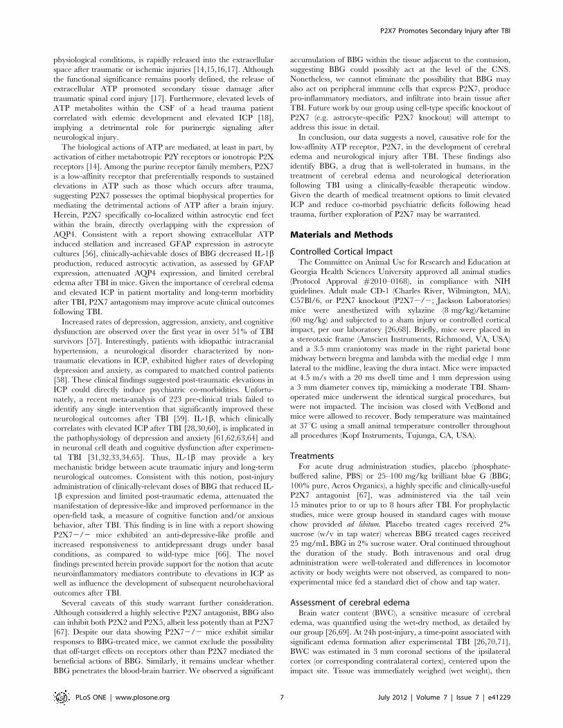
physiological conditions, is rapidly released into the extracellular
space after traumatic or ischemic injuries [14,15,16,17]. Although
the functional significance remains poorly defined, the release of
extracellular ATP promoted secondary tissue damage after
traumatic spinal cord injury [17]. Furthermore, elevated levels of
ATP metabolites within the CSF of a head trauma patient
correlated with edemic development and elevated ICP [18],
implying a detrimental role for purinergic signaling after
neurological injury.
The biological actions of ATP are mediated, at least in part, by
activation of either metabotropic P2Y receptors or ionotropic P2X
receptors [14]. Among the purine receptor family members, P2X7
is a low-affinity receptor that preferentially responds to sustained
elevations in ATP such as those which occurs after trauma,
suggesting P2X7 possesses the optimal biophysical properties for
mediating the detrimental actions of ATP after a brain injury.
Herein, P2X7 specifically co-localized within astrocytic end feet
within the brain, directly overlapping with the expression of
AQP4. Consistent with a report showing extracellular ATP
induced stellation and increased GFAP expression in astrocyte
cultures [56], clinically-achievable doses of BBG decreased IL-1bproduction, reduced astrocytic activation, as assessed by GFAP
expression, attenuated AQP4 expression, and limited cerebral
edema after TBI in mice. Given the importance of cerebral edema
and elevated ICP in patient mortality and long-term morbidity
after TBI, P2X7 antagonism may improve acute clinical outcomes
following TBI.
Increased rates of depression, aggression, anxiety, and cognitive
dysfunction are observed over the first year in over 51% of TBI
survivors [57]. Interestingly, patients with idiopathic intracranial
hypertension, a neurological disorder characterized by non-
traumatic elevations in ICP, exhibited higher rates of developing
depression and anxiety, as compared to matched control patients
[58]. These clinical findings suggested post-traumatic elevations in
ICP could directly induce psychiatric co-morbidities. Unfortu-
nately, a recent meta-analysis of 223 pre-clinical trials failed to
identify any single intervention that significantly improved these
neurological outcomes after TBI [59]. IL-1b, which clinically
correlates with elevated ICP after TBI [28,30,60], is implicated in
the pathophysiology of depression and anxiety [61,62,63,64] and
in neuronal cell death and cognitive dysfunction after experimen-
tal TBI [31,32,33,34,65]. Thus, IL-1b may provide a key
mechanistic bridge between acute traumatic injury and long-term
neurological outcomes. Consistent with this notion, post-injury
administration of clinically-relevant doses of BBG that reduced IL-
1b expression and limited post-traumatic edema, attenuated the
manifestation of depressive-like and improved performance in the
open-field task, a measure of cognitive function and/or anxious
behavior, after TBI. This finding is in line with a report showing
P2X72/2 mice exhibited an anti-depressive-like profile and
increased responsiveness to antidepressant drugs under basal
conditions, as compared to wild-type mice [66]. The novel
findings presented herein provide support for the notion that acute
neuroinflammatory mediators contribute to elevations in ICP as
well as influence the development of subsequent neurobehavioral
outcomes after TBI.
Several caveats of this study warrant further consideration.
Although considered a highly selective P2X7 antagonist, BBG also
can inhibit both P2X2 and P2X5, albeit less potently than at P2X7
[67]. Despite our data showing P2X72/2 mice exhibit similar
responses to BBG-treated mice, we cannot exclude the possibility
that off-target effects on receptors other than P2X7 mediated the
beneficial actions of BBG. Similarly, it remains unclear whether
BBG penetrates the blood-brain barrier. We observed a significant
accumulation of BBG within the tissue adjacent to the contusion,
suggesting BBG could possibly act at the level of the CNS.
Nonetheless, we cannot eliminate the possibility that BBG may
also act on peripheral immune cells that express P2X7, produce
pro-inflammatory mediators, and infiltrate into brain tissue after
TBI. Future work by our group using cell-type specific knockout of
P2X7 (e.g. astrocyte-specific P2X7 knockout) will attempt to
address this issue in detail.
In conclusion, our data suggests a novel, causative role for the
low-affinity ATP receptor, P2X7, in the development of cerebral
edema and neurological injury after TBI. These findings also
identify BBG, a drug that is well-tolerated in humans, in the
treatment of cerebral edema and neurological deterioration
following TBI using a clinically-feasible therapeutic window.
Given the dearth of medical treatment options to limit elevated
ICP and reduce co-morbid psychiatric deficits following head
trauma, further exploration of P2X7 may be warranted.
Materials and Methods
Controlled Cortical ImpactThe Committee on Animal Use for Research and Education at
Georgia Health Sciences University approved all animal studies
(Protocol Approval #2010–0168), in compliance with NIH
guidelines. Adult male CD-1 (Charles River, Wilmington, MA),
C57Bl/6, or P2X7 knockout (P2X72/2; Jackson Laboratories)
mice were anesthetized with xylazine (8 mg/kg)/ketamine
(60 mg/kg) and subjected to a sham injury or controlled cortical
impact, per our laboratory [26,68]. Briefly, mice were placed in
a stereotaxic frame (Amscien Instruments, Richmond, VA, USA)
and a 3.5 mm craniotomy was made in the right parietal bone
midway between bregma and lambda with the medial edge 1 mm
lateral to the midline, leaving the dura intact. Mice were impacted
at 4.5 m/s with a 20 ms dwell time and 1 mm depression using
a 3 mm diameter convex tip, mimicking a moderate TBI. Sham-
operated mice underwent the identical surgical procedures, but
were not impacted. The incision was closed with VetBond and
mice were allowed to recover. Body temperature was maintained
at 37uC using a small animal temperature controller throughout
all procedures (Kopf Instruments, Tujunga, CA, USA).
TreatmentsFor acute drug administration studies, placebo (phosphate-
buffered saline, PBS) or 25–100 mg/kg brilliant blue G (BBG;
100% pure, Acros Organics), a highly specific and clinically-useful
P2X7 antagonist [67], was administered via the tail vein
15 minutes prior to or up to 8 hours after TBI. For prophylactic
studies, mice were group housed in standard cages with mouse
chow provided ad libitum. Placebo treated cages received 2%
sucrose (w/v in tap water) whereas BBG treated cages received
25 mg/mL BBG in 2% sucrose water. Oral continued throughout
the duration of the study. Both intravenous and oral drug
administration were well-tolerated and differences in locomotor
activity or body weights were not observed, as compared to non-
experimental mice fed a standard diet of chow and tap water.
Assessment of cerebral edemaBrain water content (BWC), a sensitive measure of cerebral
edema, was quantified using the wet-dry method, as detailed by
our group [26,69]. At 24h post-injury, a time-point associated with
significant edema formation after experimental TBI [26,70,71],
BWC was estimated in 3 mm coronal sections of the ipsilateral
cortex (or corresponding contralateral cortex), centered upon the
impact site. Tissue was immediately weighed (wet weight), then
P2X7 Promotes Secondary Injury after TBI
PLoS ONE | www.plosone.org 7 July 2012 | Volume 7 | Issue 7 | e41229
dehydrated at 65uC. The sample was reweighed 48h later to
obtain a dry weight. The percentage of tissue water content was
calculated using the following formula: BWC = [(wet weight)-(dry
weight)/wet weight] * 100.
Determination of lesion sizeCortical lesion area was quantified by an investigator blinded to
experimental conditions, as described by our laboratory [26].
Briefly, serial coronal sections were digitized using a Zeiss
Axiophot microscope using a 2.5X objective and imported into
the OsiriX v2.7.5 32-bit program. A region of interest was drawn
along the perimeter of the injured cortex and lesion volume was
calculated and expressed as mm3.
Magnetic resonance imaging (MRI)Non-invasive determination of brain edema was performed
using a horizontal 7.0T BioSpec MRI spectrometer (Bruker
Instruments) equipped with a 8.9 cm micro-imaging gradient
insert (100 gauss/cm). For all studies, anesthetized mice were
positioned with the MR scanner. Breathing was controlled at 35
respirations/minutes and core body temperature was maintained
at 37uC using a recirculating water bath. High-resolution T2-
weighted (T2W) images and diffusion-weighted images (DWI)
were acquired during each session, using a surface coil developed
in-house. Two T2W image volumes were acquired using a 2D
RARE sequence (TR/TEeffective = 2800/56ms; RARE fac-
tor = 8; FOV=25.6625.6mm; Matrix = 2566256; slice thick-
ness = 0.5 mm; slice gap= 0.5mm; 20 slices; 5 NEX), offset by
0.5 mm to allow for collection of data without a gap. A diffusion
tensor imaging (DTI) sequence was used for calculation of an
apparent diffusion coefficient (ADC) map (TR/TE=2800/60ms;
FOV=25.6625.6mm; Matrix = 1286128; slice thick-
ness = 0.8mm; slice gap= 0.2mm; 3 b= 0 images; six directions;
Delta = 14 ms; delta = 7 ms; b value = 1200 sec mm2; 12 slices; 16
repetitions). Apparent diffusion coefficient (ADC) values, which
are rotationally invariant, reduces systemic bias that may be
introduced by variation in animal orientation. After normalizing
image intensities and ADC values between pre- and post-TBI
images, T2W images were segmented according to brightness to
identify regions of edema, and the edemic volume was computed.
A Region of Interest (ROI) was superimposed on the ADC map to
characterize the edema as cellular or vasogenic.
Assessment of neurological injuryMice were housed on a 12-hour light-dark cycle in a climate-
controlled vivarium throughout the experiment. At 72h after
injury, a 2h undisturbed habituation period was implemented
prior to the initiation of behavioral testing. All tests were digitally
recorded using a video camera fixed above the floor and scored by
an investigator who was blinded to experimental conditions. For
the open field test, animals were placed into a 14614 inch box
with 262 inch squares, as detailed by our laboratory [26,69,72].
Locomotion, as assessed by the number of squares entered over
the 5-minute testing period, was defined as the nose and two
forepaws entering a new square. Directly following the open field
test, mice were placed in a cylinder filled with 13 cm of 37uCwater. Behavioral despair, defined as the time when an animal
ceases to attempt to escape from a stressful situation, was
quantified as the latency time to the first total immobility in the
forced swim. This event was defined as all 4 paws and head
remaining motionless in the water.
Western blottingWhole cell lysates were prepared from 3 mm coronal sections
centered upon the site of impact. A 1-mm micropunch was
collected from the pericontusional cortex or from the correspond-
ing contralateral hemisphere. Tissue was placed in complete RIPA
buffer, sonicated, and centrifuged for 10 minutes at 14,0006g at
4uC. Protein concentrations were quantified using a BCA protein
assay kit (Pierce, Rockford, IL). 30 mg of protein were resolved on
a 4–20% sodium dodecyl sulfate-polyacrylamide gel and trans-
ferred onto a polyvinylidene difluoride (PVDF) membrane. Blots
were incubated overnight at 4uC in primary antibody [(1:200 anti-
P2X7, Alomone Labs, Jerusalem, Israel), (1:400 anti-AQP4
antibody, Santa Cruz Biotechnology, Santa Cruz, CA), (1:200
anti-GFAP antibody, Dako, Carpinteria, CA), (1:5000 anti-IL-1bantibody, National Cancer Institute, clone 3ZD, lot 1), or (1:2000
anti-b actin, Abcam, Cambridge, MA) followed by a 2h in-
cubation with an Alexa Fluor-tagged secondary antibody at room
temperature, per our laboratory [73,74,75,76,77]. Blots were
visualized using a Li-Cor Odyssey near-infrared imaging system
and densitometry analysis was performed using Quantity One
software (Bio-Rad, Foster City, CA).
ImmunohistochemistryDeeply anesthetized mice were perfused with saline, followed by
fixation with 4% paraformaldehyde in 0.1 M phosphate buffer
(pH 7.4). Brains were post-fixed overnight in paraformaldehyde
followed by cryoprotection with 30% sucrose (pH 7.4) until brains
permeated. Serial coronal sections (12 mM) were prepared using
a cryostat microtome (Leica, Wetzlar, Germany) and directly
mounted onto glass slides. Sections were incubated at room
temperature with 10% normal donkey serum in phosphate-
buffered saline containing 0.4% Triton X-100 for 1 h, followed by
incubation with the primary antibody [P2X7 (1:100), AQP4
(1:200), or GFAP (1:500)] overnight at 4uC. Sections were then
washed and incubated with the appropriate Alexa Fluor-tagged
secondary antibody. Omission of primary antibody served as
a negative control.
Confocal microscopyImmunofluorescence was determined using a LSM510 Meta
confocal laser microscope (Carl Zeiss), as described by our group
[26,68]. Cellular co-localization was determined in Z-stack mode
using 63X oil immersion Neofluor objective (NA 1.3) with the
image size set at 5126512 pixels. The following excitation lasers/
emission filters settings were used for various chromophores:
argon2 laser was used for Alexa Fluor 488, with excitation maxima
at 490 nm and emission in the range 505–530 nm. A HeNe1 laser
was used for Alexa Fluor 594 with excitation maxima at 543 nm
and emission in the range 568–615 nm. Z-stacks (20 optical slices)
were collected at optimal pinhole diameter at 12-bit pixel depth
and converted into three-dimensional projection images using
LSM510 Meta imaging software.
Statistical analysisThe effects of treatments were analyzed using a one-way
analysis of variance (ANOVA) followed by Dunnett’s post hoc test.
Results are expressed as mean 6 SEM. A p,0.05 was considered
to be statistically significant.
Acknowledgments
The authors would like to acknowledge the technical assistance of Tim
Kurtz and Christopher Middleton.
P2X7 Promotes Secondary Injury after TBI
PLoS ONE | www.plosone.org 8 July 2012 | Volume 7 | Issue 7 | e41229
Author Contributions
Conceived and designed the experiments: DEK KMD. Performed the
experiments: DEK JS NY. Analyzed the data: DEK JS NY JRV KMD.
Wrote the paper: DEK KMD.
References
1. Nortje J, Menon DK (2004) Traumatic brain injury: physiology, mechanisms,
and outcome. Curr Opin Neurol 17: 711–718.
2. Bramlett HM, Dietrich WD (2007) Progressive damage after brain and spinal
cord injury: pathomechanisms and treatment strategies. Prog Brain Res 161:
125–141.
3. Levin HS, Eisenberg HM, Gary HE, Marmarou A, Foulkes MA, et al. (1991)
Intracranial hypertension in relation to memory functioning during the first year
after severe head injury. Neurosurgery 28: 196–199; discussion 200.
4. Saul TG, Ducker TB (1982) Effect of intracranial pressure monitoring and
aggressive treatment on mortality in severe head injury. J Neurosurg 56: 498–
503.
5. Catala-Temprano A, Claret Teruel G, Cambra Lasaosa FJ, Pons Odena M,
Noguera Julian A, et al. (2007) Intracranial pressure and cerebral perfusion
pressure as risk factors in children with traumatic brain injuries. J Neurosurg
106: 463–466.
6. Narotam PK, Morrison JF, Nathoo N (2009) Brain tissue oxygen monitoring in
traumatic brain injury and major trauma: outcome analysis of a brain tissue
oxygen-directed therapy. J Neurosurg 111: 672–682.
7. (2000) The Brain Trauma Foundation. The American Association of
Neurological Surgeons. The Joint Section on Neurotrauma and Critical Care.
Guidelines for cerebral perfusion pressure. J Neurotrauma 17: 507–511.
8. Eisenberg HM, Gary HE Jr, Aldrich EF, Saydjari C, Turner B, et al. (1990)
Initial CT findings in 753 patients with severe head injury. A report from the
NIH Traumatic Coma Data Bank. J Neurosurg 73: 688–698.
9. Sahuquillo J, Arikan F (2006) Decompressive craniectomy for the treatment of
refractory high intracranial pressure in traumatic brain injury. Cochrane
Database Syst Rev: CD003983.
10. Farina C, Aloisi F, Meinl E (2007) Astrocytes are active players in cerebral innate
immunity. Trends Immunol 28: 138–145.
11. Katayama Y, Kawamata T (2003) Edema fluid accumulation within necrotic
brain tissue as a cause of the mass effect of cerebral contusion in head trauma
patients. Acta Neurochir Suppl 86: 323–327.
12. Kawamata T, Katayama Y (2006) Surgical management of early massive edema
caused by cerebral contusion in head trauma patients. Acta Neurochir Suppl 96:
3–6.
13. Kawamata T, Katayama Y (2007) Cerebral contusion: a role model for lesion
progression. Prog Brain Res 161: 235–241.
14. Ralevic V, Burnstock G (1998) Receptors for purines and pyrimidines.
Pharmacol Rev 50: 413–492.
15. Khakh BS, North RA (2006) P2X receptors as cell-surface ATP sensors in health
and disease. Nature 442: 527–532.
16. Latini S, Corsi C, Pedata F, Pepeu G (1996) The source of brain adenosine
outflow during ischemia and electrical stimulation. Neurochem Int 28: 113–118.
17. Peng W, Cotrina ML, Han X, Yu H, Bekar L, et al. (2009) Systemic
administration of an antagonist of the ATP-sensitive receptor P2X7 improves
recovery after spinal cord injury. Proc Natl Acad Sci U S A 106: 12489–12493.
18. Cristofori L, Tavazzi B, Gambin R, Vagnozzi R, Signoretti S, et al. (2005)
Biochemical analysis of the cerebrospinal fluid: evidence for catastrophic energy
failure and oxidative damage preceding brain death in severe head injury: a case
report. Clin Biochem 38: 97–100.
19. Surprenant A, Rassendren F, Kawashima E, North RA, Buell G (1996) The
cytolytic P2Z receptor for extracellular ATP identified as a P2X receptor
(P2X7). Science 272: 735–738.
20. Calogero S, Grassi F, Aguzzi A, Voigtlander T, Ferrier P, et al. (1999) The lack
of chromosomal protein Hmg1 does not disrupt cell growth but causes lethal
hypoglycaemia in newborn mice. Nat Genet 22: 276–280.
21. Takenouchi T, Sugama S, Iwamaru Y, Hashimoto M, Kitani H (2009)
Modulation of the ATP-lnduced release and processing of IL-1beta in microglial
cells. Crit Rev Immunol 29: 335–345.
22. Folkersma H, Breve JJ, Tilders FJ, Cherian L, Robertson CS, et al. (2008)
Cerebral microdialysis of interleukin (IL)-1beta and IL-6: extraction efficiency
and production in the acute phase after severe traumatic brain injury in rats.
Acta Neurochir (Wien) 150: 1277–1284; discussion 1284.
23. Herx LM, Rivest S, Yong VW (2000) Central nervous system-initiated
inflammation and neurotrophism in trauma: IL-1 beta is required for the
production of ciliary neurotrophic factor. J Immunol 165: 2232–2239.
24. Kamm K, Vanderkolk W, Lawrence C, Jonker M, Davis AT (2006) The effect of
traumatic brain injury upon the concentration and expression of interleukin-
1beta and interleukin-10 in the rat. J Trauma 60: 152–157.
25. Kinoshita K, Chatzipanteli K, Vitarbo E, Truettner JS, Alonso OF, et al. (2002)
Interleukin-1beta messenger ribonucleic acid and protein levels after fluid-
percussion brain injury in rats: importance of injury severity and brain
temperature. Neurosurgery 51: 195–203; discussion 203.
26. Laird MD, Sukumari-Ramesh S, Swift AE, Meiler SE, Vender JR, et al. (2010)
Curcumin attenuates cerebral edema following traumatic brain injury in mice:
a possible role for aquaporin-4? J Neurochem 113: 637–648.
27. Taupin V, Toulmond S, Serrano A, Benavides J, Zavala F (1993) Increase in IL-
6, IL-1 and TNF levels in rat brain following traumatic lesion. Influence of pre-
and post-traumatic treatment with Ro5 4864, a peripheral-type (p site)
benzodiazepine ligand. J Neuroimmunol 42: 177–185.
28. Shiozaki T, Hayakata T, Tasaki O, Hosotubo H, Fuijita K, et al. (2005)
Cerebrospinal fluid concentrations of anti-inflammatory mediators in early-
phase severe traumatic brain injury. Shock 23: 406–410.
29. Hayakata T, Shiozaki T, Tasaki O, Ikegawa H, Inoue Y, et al. (2004) Changes
in CSF S100B and cytokine concentrations in early-phase severe traumatic brain
injury. Shock 22: 102–107.
30. Chiaretti A, Genovese O, Aloe L, Antonelli A, Piastra M, et al. (2005)
Interleukin 1beta and interleukin 6 relationship with paediatric head trauma
severity and outcome. Childs Nerv Syst 21: 185–193; discussion 194.
31. Clausen F, Hanell A, Bjork M, Hillered L, Mir AK, et al. (2009) Neutralization
of interleukin-1beta modifies the inflammatory response and improves
histological and cognitive outcome following traumatic brain injury in mice.
Eur J Neurosci 30: 385–396.
32. Jones NC, Prior MJ, Burden-Teh E, Marsden CA, Morris PG, et al. (2005)
Antagonism of the interleukin-1 receptor following traumatic brain injury in the
mouse reduces the number of nitric oxide synthase-2-positive cells and improves
anatomical and functional outcomes. Eur J Neurosci 22: 72–78.
33. Tehranian R, Andell-Jonsson S, Beni SM, Yatsiv I, Shohami E, et al. (2002)
Improved recovery and delayed cytokine induction after closed head injury in
mice with central overexpression of the secreted isoform of the interleukin-1
receptor antagonist. J Neurotrauma 19: 939–951.
34. Toulmond S, Rothwell NJ (1995) Interleukin-1 receptor antagonist inhibits
neuronal damage caused by fluid percussion injury in the rat. Brain Res 671:
261–266.
35. Whelan-Goodinson R, Ponsford J, Johnston L, Grant F (2009) Psychiatric
disorders following traumatic brain injury: their nature and frequency. J Head
Trauma Rehabil 24: 324–332.
36. Rogers JM, Read CA (2007) Psychiatric comorbidity following traumatic brain
injury. Brain Inj 21: 1321–1333.
37. Federal Register, Government Printing Office website. Available: http://
edocket.access.gpo.gov/2006/06-3307.htm. Accessed 2012 Jun 1.
38. Ito J, Marmarou A, Barzo P, Fatouros P, Corwin F (1996) Characterization of
edema by diffusion-weighted imaging in experimental traumatic brain injury.
J Neurosurg 84: 97–103.
39. Narayan RK, Michel ME, Ansell B, Baethmann A, Biegon A, et al. (2002)
Clinical trials in head injury. J Neurotrauma 19: 503–557.
40. Hinkle DA, Baldwin SA, Scheff SW, Wise PM (1997) GFAP and S100beta
expression in the cortex and hippocampus in response to mild cortical contusion.
J Neurotrauma 14: 729–738.
41. Dietrich WD, Truettner J, Zhao W, Alonso OF, Busto R, et al. (1999) Sequential
changes in glial fibrillary acidic protein and gene expression following
parasagittal fluid-percussion brain injury in rats. J Neurotrauma 16: 567–581.
42. Pelinka LE, Kroepfl A, Leixnering M, Buchinger W, Raabe A, et al. (2004)
GFAP versus S100B in serum after traumatic brain injury: relationship to brain
damage and outcome. J Neurotrauma 21: 1553–1561.
43. Pelinka LE, Kroepfl A, Schmidhammer R, Krenn M, Buchinger W, et al. (2004)
Glial fibrillary acidic protein in serum after traumatic brain injury and multiple
trauma. J Trauma 57: 1006–1012.
44. Laird MD, Wakade C, Alleyne CH Jr, Dhandapani KM (2008) Hemin-induced
necroptosis involves glutathione depletion in mouse astrocytes. Free Radic Biol
Med 45: 1103–1114.
45. Oprica M, Zhu S, Goiny M, Pham TM, Mohammed AH, et al. (2005)
Transgenic overexpression of interleukin-1 receptor antagonist in the CNS
influences behaviour, serum corticosterone and brain monoamines. Brain Behav
Immun 19: 223–234.
46. Badaut J, Ashwal S, Adami A, Tone B, Recker R, et al. (2011) Brain water
mobility decreases after astrocytic aquaporin-4 inhibition using RNA in-
terference. J Cereb Blood Flow Metab 31: 819–831.
47. Manley GT, Fujimura M, Ma T, Noshita N, Filiz F, et al. (2000) Aquaporin-4
deletion in mice reduces brain edema after acute water intoxication and
ischemic stroke. Nat Med 6: 159–163.
48. Saadoun S, Papadopoulos MC, Watanabe H, Yan D, Manley GT, et al. (2005)
Involvement of aquaporin-4 in astroglial cell migration and glial scar formation.
J Cell Sci 118: 5691–5698.
49. Ito H, Yamamoto N, Arima H, Hirate H, Morishima T, et al. (2006)
Interleukin-1beta induces the expression of aquaporin-4 through a nuclear
factor-kappaB pathway in rat astrocytes. J Neurochem 99: 107–118.
P2X7 Promotes Secondary Injury after TBI
PLoS ONE | www.plosone.org 9 July 2012 | Volume 7 | Issue 7 | e41229
50. Lin HW, Basu A, Druckman C, Cicchese M, Krady JK, et al. (2006) Astrogliosis
is delayed in type 1 interleukin-1 receptor-null mice following a penetratingbrain injury. J Neuroinflammation 3: 15.
51. Perregaux DG, Gabel CA (1998) Post-translational processing of murine IL-1:
evidence that ATP-induced release of IL-1 alpha and IL-1 beta occurs viaa similar mechanism. J Immunol 160: 2469–2477.
52. Yakovlev AG, Knoblach SM, Fan L, Fox GB, Goodnight R, et al. (1997)Activation of CPP32-like caspases contributes to neuronal apoptosis and
neurological dysfunction after traumatic brain injury. J Neurosci 17: 7415–7424.
53. Satchell MA, Lai Y, Kochanek PM, Wisniewski SR, Fink EL, et al. (2005)Cytochrome c, a biomarker of apoptosis, is increased in cerebrospinal fluid from
infants with inflicted brain injury from child abuse. J Cereb Blood Flow Metab25: 919–927.
54. Mariathasan S, Weiss DS, Newton K, McBride J, O’Rourke K, et al. (2006)Cryopyrin activates the inflammasome in response to toxins and ATP. Nature
440: 228–232.
55. Laliberte RE, Eggler J, Gabel CA (1999) ATP treatment of human monocytespromotes caspase-1 maturation and externalization. J Biol Chem 274: 36944–
36951.56. Neary JT, Baker L, Jorgensen SL, Norenberg MD (1994) Extracellular ATP
induces stellation and increases glial fibrillary acidic protein content and DNA
synthesis in primary astrocyte cultures. Acta Neuropathol 87: 8–13.57. Fann JR, Burington B, Leonetti A, Jaffe K, Katon WJ, et al. (2004) Psychiatric
illness following traumatic brain injury in an adult health maintenanceorganization population. Arch Gen Psychiatry 61: 53–61.
58. Kleinschmidt JJ, Digre KB, Hanover R (2000) Idiopathic intracranialhypertension: relationship to depression, anxiety, and quality of life. Neurology
54: 319–324.
59. Wheaton P, Mathias JL, Vink R (2011) Impact of pharmacological treatmentson outcome in adult rodents after traumatic brain injury: a meta-analysis.
J Psychopharmacol.60. Holmin S, Hojeberg B (2004) In situ detection of intracerebral cytokine
expression after human brain contusion. Neurosci Lett 369: 108–114.
61. Norman GJ, Karelina K, Zhang N, Walton JC, Morris JS, et al. (2010) Stressand IL-1beta contribute to the development of depressive-like behavior following
peripheral nerve injury. Mol Psychiatry 15: 404–414.62. Koo JW, Duman RS (2009) Evidence for IL-1 receptor blockade as a therapeutic
strategy for the treatment of depression. Curr Opin Investig Drugs 10: 664–671.63. Koo JW, Duman RS (2008) IL-1beta is an essential mediator of the
antineurogenic and anhedonic effects of stress. Proc Natl Acad Sci U S A
105: 751–756.64. Koo JW, Duman RS (2009) Interleukin-1 receptor null mutant mice show
decreased anxiety-like behavior and enhanced fear memory. Neurosci Lett 456:39–43.
65. Sanderson KL, Raghupathi R, Saatman KE, Martin D, Miller G, et al. (1999)
Interleukin-1 receptor antagonist attenuates regional neuronal cell death and
cognitive dysfunction after experimental brain injury. J Cereb Blood Flow Metab
19: 1118–1125.
66. Basso AM, Bratcher NA, Harris RR, Jarvis MF, Decker MW, et al. (2009)
Behavioral profile of P2X7 receptor knockout mice in animal models of
depression and anxiety: relevance for neuropsychiatric disorders. Behav Brain
Res 198: 83–90.
67. Jiang LH, Mackenzie AB, North RA, Surprenant A (2000) Brilliant blue G
selectively blocks ATP-gated rat P2X(7) receptors. Mol Pharmacol 58: 82–88.
68. Wakade C, Sukumari-Ramesh S, Laird MD, Dhandapani KM, Vender JR
(2010) Delayed reduction in hippocampal postsynaptic density protein-95
expression temporally correlates with cognitive dysfunction following controlled
cortical impact in mice. J Neurosurg 113: 1195–1201.
69. Wakade C, King MD, Laird MD, Alleyne CH Jr, Dhandapani KM (2009)
Curcumin attenuates vascular inflammation and cerebral vasospasm after
subarachnoid hemorrhage in mice. Antioxid Redox Signal 11: 35–45.
70. Kiening KL, van Landeghem FK, Schreiber S, Thomale UW, von Deimling A,
et al. (2002) Decreased hemispheric Aquaporin-4 is linked to evolving brain
edema following controlled cortical impact injury in rats. Neurosci Lett 324:
105–108.
71. Zweckberger K, Eros C, Zimmermann R, Kim SW, Engel D, et al. (2006) Effect
of early and delayed decompressive craniectomy on secondary brain damage
after controlled cortical impact in mice. J Neurotrauma 23: 1083–1093.
72. Wakade C, Sukumari-Ramesh S, Laird MD, Dhandapani KM, Vender JR
(2010) Delayed reduction in hippocampal postsynaptic density protein-95
expression temporally correlates with cognitive dysfunction following controlled
cortical impact in mice. J Neurosurg 113: 1195–1201.
73. Dhandapani K, Brann D (2003) Neuroprotective effects of estrogen and
tamoxifen in vitro: a facilitative role for glia? Endocrine 21: 59–66.
74. Dhandapani KM, Hadman M, De Sevilla L, Wade MF, Mahesh VB, et al.
(2003) Astrocyte protection of neurons: role of transforming growth factor-beta
signaling via a c-Jun-AP-1 protective pathway. J Biol Chem 278: 43329–43339.
75. Dhandapani KM, Wade FM, Mahesh VB, Brann DW (2005) Astrocyte-derived
transforming growth factor-{beta} mediates the neuroprotective effects of
17{beta}-estradiol: involvement of nonclassical genomic signaling pathways.
Endocrinology 146: 2749–2759.
76. Fink KB, Andrews LJ, Butler WE, Ona VO, Li M, et al. (1999) Reduction of
post-traumatic brain injury and free radical production by inhibition of the
caspase-1 cascade. Neuroscience 94: 1213–1218.
77. Pillai A, Dhandapani KM, Pillai BA, Terry AV Jr, Mahadik SP (2007)
Erythropoietin Prevents Haloperidol Treatment-Induced Neuronal Apoptosis
through Regulation of BDNF. Neuropsychopharmacology.
P2X7 Promotes Secondary Injury after TBI
PLoS ONE | www.plosone.org 10 July 2012 | Volume 7 | Issue 7 | e41229
Related Documents