P2X7 Receptors Trigger ATP Exocytosis and Modify Secretory Vesicle Dynamics in Neuroblastoma Cells * □ S Received for publication, April 29, 2010, and in revised form, January 31, 2011 Published, JBC Papers in Press, February 3, 2011, DOI 10.1074/jbc.M110.139410 Yolanda Gutie ´ rrez-Martín ‡§1 , Diego Bustillo ‡§2 , Rosa Go ´ mez-Villafuertes §¶2 , Jesu ´ s Sa ´ nchez-Nogueiro §¶ , Cristina Torregrosa-Hetland , Thomas Binz**, Luis Miguel Gutie ´ rrez , Marı ´a Teresa Miras-Portugal §¶ , and Antonio R. Artalejo ‡§3 From the ‡ Departamento de Toxicología y Farmacología, ¶ Bioquímica y Biología Molecular, and § Instituto Universitario de Investigacio ´n en Neuroquímica, Universidad Complutense de Madrid, 28040 Madrid, Spain, the Instituto de Neurociencias, Centro Mixto Consejo Superior de Investigaciones Científicas-Universidad Miguel Herna ´ndez, 03550 Sant Joan d’Alacant, Spain, and the **Institut fu ¨r Biochemie, Medizinische Hochschule Hannover, 30625 Hannover, Germany Previously, we reported that purinergic ionotropic P2X7 re- ceptors negatively regulate neurite formation in Neuro-2a (N2a) mouse neuroblastoma cells through a Ca 2 /calmodulin-depen- dent kinase II-related mechanism. In the present study we used this cell line to investigate a parallel though faster P2X7 recep- tor-mediated signaling pathway, namely Ca 2 -regulated exocy- tosis. Selective activation of P2X7 receptors evoked exocytosis as assayed by high resolution membrane capacitance measure- ments. Using dual-wavelength total internal reflection micros- copy, we have observed both the increase in near-membrane Ca 2 concentration and the exocytosis of fluorescently labeled vesicles in response to P2X7 receptor stimulation. Moreover, activation of P2X7 receptors also affects vesicle motion in the vertical and horizontal directions, thus, involving this receptor type in the control of early steps (docking and priming) of the secretory pathway. Immunocytochemical and RT-PCR experi- ments evidenced that N2a cells express the three neuronal SNAREs as well as vesicular nucleotide and monoamine (VMAT-1 and VMAT-2) transporters. Biochemical measure- ments indicated that ionomycin induced a significant release of ATP from N2a cells. Finally, P2X7 receptor stimulation and ionomycin increased the incidence of small transient inward currents, reminiscent of postsynaptic quantal events observed at synapses. Small transient inward currents were dependent on extracellular Ca 2 and were abolished by Brilliant Blue G, sug- gesting they were mediated by P2X7 receptors. Altogether, these results suggest the existence of a positive feedback mech- anism mediated by P2X7 receptor-stimulated exocytotic release of ATP that would act on P2X7 receptors on the same or neighbor cells to further stimulate its own release and neg- atively control N2a cell differentiation. Adenine nucleotides are well recognized extracellular signal- ing molecules mediating many physiological functions via the P2-type of purinergic receptors. P2 receptors are subdivided into ionotropic P2X and metabotropic P2Y subtypes (1). Up to now, seven mammalian P2X-receptor subtypes (P2X1–7) and eight mammalian P2Y-receptor subtypes (P2Y1, -2, -4, -6, -11, -12, -13, and -14) have been cloned and functionally character- ized (2– 4). P2X receptors are non-selective cation channels mediating Na and Ca 2 influx across the cell membrane, which leads to cell depolarization and the ensued activation of voltage-gated channels (5). Purinergic signaling is already present at early stages of embryogenesis wherein it is involved in proliferation, migra- tion, and differentiation of cells from multiple structures (6 –9). Likewise, different purinergic receptors have been identified in a variety of both primary tumors and cell lines (10, 11). In this respect, recent data from our laboratory have shown that mouse neuroblastoma Neuro2-a cells (henceforth referred to as N2a 4 cells) express ionotropic P2X7 receptors mediating cation currents that are selectively blocked by Brilliant Blue G (BBG) (12). As is also the case for hippocampal neurons (13), P2X7 receptors negatively regulate neurite formation in N2a cells through a Ca 2 /calmodulin-dependent kinase II-related mech- anism (12). Therefore, either by inhibiting P2X7 receptor activ- ity with BBG or silencing its expression with shRNA, an increased neuritogenesis could be observed in N2a cell cultures. On the contrary, P2X7 receptor overexpression resulted in a significant reduction in the formation of neurites. These results imply the existence of an endogenous purinergic tone acting on P2X7 receptors to control neuronal differentiation of N2a cells. Interestingly, neuroblastoma cells are capable of releasing a variety of mediators including substance P and ATP itself (14, 15), and P2X7 receptor stimulation is also coupled to ATP release from human ACN neuroblastoma cells (16). Neverthe- less, very little is known about the mechanisms involved in such a release process and, particularly, as to whether P2X7 recep- tors could be coupled to exocytosis from this sort of cells. In the * This work was supported by Ministerio de Ciencia e Innovacio ´ n Grants BFU2008-02699 and BFU2005-0634, Comunidad de Madrid Grant S-SAL- 0253-2006, the Fundacio ´ n Marcelino Botín, and “Spanish Ion Channel Ini- tiative” (SICI) Grant CSD2008-00005. Author’s Choice—Final version full access. □ S The on-line version of this article (available at http://www.jbc.org) contains supplemental Videos 1–5 and Figs. S1–S7. 1 Supported by Comunidad de Madrid Postdoctoral Contract S-SAL-0253-2006. 2 Supported by SICI. 3 To whom correspondence should be addressed: Dept. Toxicología y Farma- cología, Facultad de Veterinaria, Universidad Complutense, Avda. Puerta de Hierro s/n, 28040 Madrid, Spain. Tel./Fax.: 34-91-394-3851; E-mail: [email protected]. 4 The abbreviations used are: N2a, Neuro-2a; BoNT/A, botulinum neurotoxin A; BBG, Brilliant Blue G; BzATP, 2,3-O-(4-benzoyl)-benzoyl ATP; NEM, N-ethylmaleimide; STIC, small transient inward current; TIRFM, total inter- nal reflection fluorescence microscopy; SNARE, soluble N-ethylmaleimide factor attachment protein (NSF) receptor; fF, femtofaraday; LDCV, large dense-core vesicle. THE JOURNAL OF BIOLOGICAL CHEMISTRY VOL. 286, NO. 13, pp. 11370 –11381, April 1, 2011 Author’s Choice © 2011 by The American Society for Biochemistry and Molecular Biology, Inc. Printed in the U.S.A. 11370 JOURNAL OF BIOLOGICAL CHEMISTRY VOLUME 286 • NUMBER 13 • APRIL 1, 2011 at UNIVERSIDAD COMPLUTENSE DE MADRID, on March 31, 2011 www.jbc.org Downloaded from http://www.jbc.org/content/suppl/2011/02/03/M110.139410.DC1.html Supplemental Material can be found at:

Welcome message from author
This document is posted to help you gain knowledge. Please leave a comment to let me know what you think about it! Share it to your friends and learn new things together.
Transcript
P2X7 Receptors Trigger ATP Exocytosis and Modify SecretoryVesicle Dynamics in Neuroblastoma Cells*□S
Received for publication, April 29, 2010, and in revised form, January 31, 2011 Published, JBC Papers in Press, February 3, 2011, DOI 10.1074/jbc.M110.139410
Yolanda Gutierrez-Martín‡§1, Diego Bustillo‡§2, Rosa Gomez-Villafuertes§¶2, Jesus Sanchez-Nogueiro§¶,Cristina Torregrosa-Hetland�, Thomas Binz**, Luis Miguel Gutierrez�, Marıa Teresa Miras-Portugal§¶,and Antonio R. Artalejo‡§3
From the ‡Departamento de Toxicología y Farmacología, ¶Bioquímica y Biología Molecular, and §Instituto Universitario deInvestigacion en Neuroquímica, Universidad Complutense de Madrid, 28040 Madrid, Spain, the �Instituto de Neurociencias, CentroMixto Consejo Superior de Investigaciones Científicas-Universidad Miguel Hernandez, 03550 Sant Joan d’Alacant, Spain, and the**Institut fur Biochemie, Medizinische Hochschule Hannover, 30625 Hannover, Germany
Previously, we reported that purinergic ionotropic P2X7 re-ceptors negatively regulate neurite formation inNeuro-2a (N2a)mouse neuroblastoma cells through a Ca2�/calmodulin-depen-dent kinase II-related mechanism. In the present study we usedthis cell line to investigate a parallel though faster P2X7 recep-tor-mediated signaling pathway, namely Ca2�-regulated exocy-tosis. Selective activation of P2X7 receptors evoked exocytosisas assayed by high resolution membrane capacitance measure-ments. Using dual-wavelength total internal reflection micros-copy, we have observed both the increase in near-membraneCa2� concentration and the exocytosis of fluorescently labeledvesicles in response to P2X7 receptor stimulation. Moreover,activation of P2X7 receptors also affects vesicle motion in thevertical and horizontal directions, thus, involving this receptortype in the control of early steps (docking and priming) of thesecretory pathway. Immunocytochemical and RT-PCR experi-ments evidenced that N2a cells express the three neuronalSNAREs as well as vesicular nucleotide and monoamine(VMAT-1 and VMAT-2) transporters. Biochemical measure-ments indicated that ionomycin induced a significant release ofATP from N2a cells. Finally, P2X7 receptor stimulation andionomycin increased the incidence of small transient inwardcurrents, reminiscent of postsynaptic quantal events observedat synapses. Small transient inward currents were dependent onextracellular Ca2� and were abolished by Brilliant Blue G, sug-gesting they were mediated by P2X7 receptors. Altogether,these results suggest the existence of a positive feedbackmech-anism mediated by P2X7 receptor-stimulated exocytoticrelease of ATP that would act on P2X7 receptors on the sameor neighbor cells to further stimulate its own release and neg-atively control N2a cell differentiation.
Adenine nucleotides are well recognized extracellular signal-ing molecules mediating many physiological functions via theP2-type of purinergic receptors. P2 receptors are subdividedinto ionotropic P2X and metabotropic P2Y subtypes (1). Up tonow, seven mammalian P2X-receptor subtypes (P2X1–7) andeight mammalian P2Y-receptor subtypes (P2Y1, -2, -4, -6, -11,-12, -13, and -14) have been cloned and functionally character-ized (2–4). P2X receptors are non-selective cation channelsmediating Na� and Ca2� influx across the cell membrane,which leads to cell depolarization and the ensued activation ofvoltage-gated channels (5).Purinergic signaling is already present at early stages of
embryogenesis wherein it is involved in proliferation, migra-tion, and differentiation of cells frommultiple structures (6–9).Likewise, different purinergic receptors have been identified ina variety of both primary tumors and cell lines (10, 11). In thisrespect, recent data from our laboratory have shown thatmouse neuroblastomaNeuro2-a cells (henceforth referred to asN2a4 cells) express ionotropic P2X7 receptorsmediating cationcurrents that are selectively blocked by Brilliant Blue G (BBG)(12). As is also the case for hippocampal neurons (13), P2X7receptors negatively regulate neurite formation in N2a cellsthrough aCa2�/calmodulin-dependent kinase II-relatedmech-anism (12). Therefore, either by inhibiting P2X7 receptor activ-ity with BBG or silencing its expression with shRNA, anincreased neuritogenesis could be observed inN2a cell cultures.On the contrary, P2X7 receptor overexpression resulted in asignificant reduction in the formation of neurites. These resultsimply the existence of an endogenous purinergic tone acting onP2X7 receptors to control neuronal differentiation of N2a cells.Interestingly, neuroblastoma cells are capable of releasing avariety of mediators including substance P and ATP itself (14,15), and P2X7 receptor stimulation is also coupled to ATPrelease from human ACN neuroblastoma cells (16). Neverthe-less, very little is known about themechanisms involved in sucha release process and, particularly, as to whether P2X7 recep-tors could be coupled to exocytosis from this sort of cells. In the
* This work was supported by Ministerio de Ciencia e Innovacion GrantsBFU2008-02699 and BFU2005-0634, Comunidad de Madrid Grant S-SAL-0253-2006, the Fundacion Marcelino Botín, and “Spanish Ion Channel Ini-tiative” (SICI) Grant CSD2008-00005.Author’s Choice—Final version full access.
□S The on-line version of this article (available at http://www.jbc.org) containssupplemental Videos 1–5 and Figs. S1–S7.
1 Supported by Comunidad de Madrid Postdoctoral Contract S-SAL-0253-2006.2 Supported by SICI.3 To whom correspondence should be addressed: Dept. Toxicología y Farma-
cología, Facultad de Veterinaria, Universidad Complutense, Avda. Puertade Hierro s/n, 28040 Madrid, Spain. Tel./Fax.: 34-91-394-3851; E-mail:[email protected].
4 The abbreviations used are: N2a, Neuro-2a; BoNT/A, botulinum neurotoxinA; BBG, Brilliant Blue G; BzATP, 2�,3�-O-(4-benzoyl)-benzoyl ATP; NEM,N-ethylmaleimide; STIC, small transient inward current; TIRFM, total inter-nal reflection fluorescence microscopy; SNARE, soluble N-ethylmaleimidefactor attachment protein (NSF) receptor; fF, femtofaraday; LDCV, largedense-core vesicle.
THE JOURNAL OF BIOLOGICAL CHEMISTRY VOL. 286, NO. 13, pp. 11370 –11381, April 1, 2011Author’s Choice © 2011 by The American Society for Biochemistry and Molecular Biology, Inc. Printed in the U.S.A.
11370 JOURNAL OF BIOLOGICAL CHEMISTRY VOLUME 286 • NUMBER 13 • APRIL 1, 2011
at UN
IVE
RS
IDA
D C
OM
PLU
TE
NS
E D
E M
AD
RID
, on March 31, 2011
ww
w.jbc.org
Dow
nloaded from
http://www.jbc.org/content/suppl/2011/02/03/M110.139410.DC1.html Supplemental Material can be found at:
present studywe have addressed this issue by identifying inN2acells several essential proteins of the exocytotic apparatus ofsympathetic neurons, including the vesicular catecholamineand nucleotide transporters, and by measuring exocytosis withboth electrophysiological and imaging techniques in the pres-ence of selective P2X7 receptor agents. Biochemical measure-ments based on the luciferin-luciferase reaction indicated that,in effect, N2a cells release ATP. Moreover, in clusters of N2acells we report the detection of small transient inward currents(STICs), which are likely generated by the quantal release ofATP. Altogether, our results indicate that P2X7 receptors arecoupled to exocytosis and to the regulation of secretory vesicledynamics in N2a cells, and they suggest that the exocytoticrelease of ATP induced by P2X7 receptor stimulation couldmediate the tonic inhibition of neurite formation exerted byP2X7 receptors in N2a cells.
EXPERIMENTAL PROCEDURES
Chemicals and Antibodies—2�,3�-O-(4-Benzoyl)-benzoylATP (BzATP), BBG, ionomycin, bovine serum albumin, quin-acrine, and N-ethylmaleimide (NEM) were purchased fromSigma. ARL 67156 was from Tocris Bioscience (Bristol, UK).Fura-2 AM, fluo-4 AM, rhod-2 AM, and Lysotracker redDND-99 were fromMolecular Probes (Invitrogen). Botulinumneurotoxin A (BoNT/A) was purified as previously described(17).Cell Culture—Murine Neuro-2a (ATCC� number CCL-
131TM) cells were cultured in Dulbecco modified Eagle’smedium (Sigma) supplemented with Glutamax� (Invitrogen),penicillin/streptomycin (Invitrogen), and 10% heat-inactivatedfetal bovine serum (EuroClone, Padova, Italy). Cellswere grownat 37 °C in a humidified atmosphere containing 5% CO2.Electrophysiological Recordings—Electrophysiological re-
cordings were performed with an EPC9 patch clamp amplifierusing the PatchMaster software (HEKA Electronic, Lambrecht,Germany). Pipettes were pulled from Kimax borosilicate glass(Witz Scientific, Holland, OH) and subsequently wax-coatedand fire-polished to obtain a final resistance of 2–3 megaohmswhen filled with standard solutions. The standard extracellularsolution (Mg2�-free Locke’s buffer) had the following compo-sition: 140 mM NaCl, 4.7 mM KCl, 1.2 mM KH2PO4, 2.5 mM
CaCl2, 10 mM HEPES, and 5.5 mM glucose (pH 7.4, adjustedwithNaOH; 294mosM). External Ca2�-free solution had CaCl2replaced by 1 mM MgCl2. Recording pipettes were filled with asolution containing 140 mM KCl, 1 mM MgCl2, and 10 mM
HEPES (pH 7.2, adjusted with KOH; 290 mosM). Cells attachedto glass coverslips (104 cells/ml) were transferred to a recordingchamber placed on the stage of an inverted Zeiss Axiovert 100microscope and continuously superfused with Locke’s buffer(perfusion rate of 1 ml min�1). Membrane currents were mea-sured in the whole-cell configuration of the patch clamp tech-nique (18), filtered at 2 kHz, and sampled at 10 KHz. Seriesresistancewas compensated by 80% andmonitored throughoutthe experiment together with the cell membrane capacitance.STICs were analyzed off-line using PatchMaster to calculatepeak amplitude, rise time (20–80% of the peak amplitude), andhalf-decay time (to 50% of peak amplitude). Such events weredetected using amplitude threshold set to 3 times the S.D. of the
noise, and each of them was visually inspected and additionallyfiltered to avoid artifactual noise components.Exocytosis was estimated by the membrane capacitance
increment according to the Lindau-Neher technique imple-mented as the “Sine � D” feature of the PatchMaster lock-inmodule (19, 20). A 1-kHz, 70-mV peak-to-peak amplitudesinusoidal function was superimposed onto the holding voltage(Vh; �70 mV). To determine the change in capacitance values,membrane capacitance was first averaged over 2 s precedingpurinergic agonist (BzATP) or plain Locke’s solution applica-tion to give a base-line value; this was subtracted from the valueestimated after drug or saline administration averaged over a10-s window, excluding the first 1 s to avoid contamination bynonsecretory capacitative or purinergic currents. Experimentswere performed at room temperature (22–24 °C).Modified solutions and drugs were applied onto the cell
under investigation by means of a gravity-driven perfusion sys-tem with 5 independent lines controlled by electronic valves(The Lee Co., Westbrook, CO). This system allowed theexchange of themedium surrounding a cell in less than 200ms.In experiments involving the recording of STICs, drugs werelocally applied by pressure ejection from a patch-pipette (2–5�m tip diameter) placed few �m away from a cell cluster. Pres-sure pulses were delivered by an electronically controlled airpump (MPCU, List Electronic, Darmstadt, Germany) at lowamplitudes (5–10 cm H2O) to reduce bulk movement of theextracellular fluid. Stock solutions of drugswere diluted daily inextracellular saline and incorporated into the perfusion systema few minutes before starting the experiments.Total Internal Reflection Fluorescence Microscopy (TIRFM)
Studies of Vesicle Fusion and Motion—The TIRF system wasbased on a IX81 microscope (Olympus) equipped with a 100�,1.45 NA, Plan Apochromat Olympus objective (pixel size, 80nm). Two laser beams emitting at 488 and 561 nmwere used toalternately illuminate the cell footprint, and the correspondingfluorescence signals were band-pass-filtered with a dual-band(emission between 500–550 nm and 580–620 nm) filter(Chroma Technology Corp., Rockingham, VT). A filter wheel-based epifluorescence illumination system (MT20; Olympus)was also coupled to the setup by using an Olympus TIRFMIX2-RFACA attachment system. Imaging was performedwith a C9100 EM-CCD digital camera from Hamamatsu(Hamamatsu) controlled by CellR software (Olympus). Sepa-rated images corresponding to each of the laser illuminationsources were acquired at a rate of 20 Hz to detect vesicle fusionor 1Hz tomonitor vesiclemotion. Exposure timewas 10 and 50ms, respectively. TIRFM calibration was performed accordingtoWu et al. (21), and decay constant of the evanescent field (1/edepth) was determined to be 160 � 28 nm.
Different combinations of fluorescent dyeswere used to labelthe cells. In experiments aimed at simultaneously determiningvesicle fusion and the intracellular calcium concentration([Ca2�]i), N2a cells plated on coverslips inside 35-mm cell cul-ture dishes (2.5� 105 cells/dish)were loadedwith quinacrine (2�M; excited by 488-nm light) and rhod-2AM (10�M; excited by561-nm light) for 20 min at 37 °C. At variance, N2a cells wereincubated with Lysotracker red (2 �M; excited by 561-nm light)and fluo-4 AM (10 �M; excited by 488 nm light) in experiments
P2X7 Receptor-mediated ATP Exocytosis in Neuroblastoma Cells
APRIL 1, 2011 • VOLUME 286 • NUMBER 13 JOURNAL OF BIOLOGICAL CHEMISTRY 11371
at UN
IVE
RS
IDA
D C
OM
PLU
TE
NS
E D
E M
AD
RID
, on March 31, 2011
ww
w.jbc.org
Dow
nloaded from
designed tomonitor vesicular motion as well as [Ca2�]i. Fusionof a vesicle and release of its content into the extracellular spacewas detected as a rapid loss of the quinacrine signal (in less than500 ms). During the experiments, cells were continuouslysuperfused with Locke’s medium at a rate of 1.5 ml min�1, andP2X7 receptor-selective agents were applied by bath superfu-sion. Perfusion was gravity-driven, and solution exchange wasperformed by manually operating the electronic valves of aVC-6 drug application system (Warner Instruments). Experi-ments were performed at 37 °C.Images were processed using the ImageJ software (public
domain image processing program developed by Wayne Ras-band at the National Institutes of Health, Bethesda, Maryland)with plug-ins for image average, region of interest measure-ments, and particle centroid tracking. Further analysis such asdetermination of mean square displacement according to Qianet al. (22) and z distances according to Johns et al. (23) wereperformed using homemademacros for IgorPro (WaveMetricsInc., Lake Oswego, OR). [Ca2�]i-related signals are presentedas relative fluorescent intensities (F/F0) rather than absoluteconcentrations.Luciferin-Luciferase Luminescence Assay—Extracellular
ATP concentration was measured using ENLITEN�rLuciferase/Luciferin reagent (Promega, Madison, WI). N2acells were grown in 6-well plates till confluence. Culturemedium (100 �l) was collected under various experimentalconditions and centrifuged at 600 � g for 5 min at 4 °C, and10-�l aliquots of supernatant were transferred to wells of a96-well plate placed on ice. The 96-well plate was set in aFLUOstar OPTIMA Microplate Luminometer (BMGLABTECH GmbH, Offenburg, Germany), and 100 �l of rLu-ciferase/Luciferin reagent was automatically injected into eachwell at room temperature (�25 °C). Immediately before thestart of the experiments, N2a cells were bathed in Mg2�-freeLocke’s buffer for 1 h at 37 °C. Then cells were exposed for 5min to either plain extracellular Locke’s buffer or Locke’s buffersupplemented with 100 �MARL 67156, a competitive inhibitorof ecto-ATPases (24), with 500 �M NEM or with the two com-pounds, and their mediumwas collected to measure basal ATPconcentration. Thereafter, cells were stimulated by addingionomycin (10 �M, final concentration in extracellularmedium) prepared in either plain Locke’s buffer or Locke’sbuffer containing the above-mentioned supplements. Fivemin-utes later, extracellular mediumwas again collected tomeasureevoked ATP concentration. In another set of experiments, cellswere grown for 72 h in the absence or the presence of BoNT/A(30 nM). Both controls and toxin-treated cells were incubatedwith ARL 67156 (100 �M; 5 min) and subsequently challengedwith ionomycin (10 �M; 5 min) to elicit ATP release. ATP con-centration was determined by comparison with a calibrationcurve generated with ATP standards diluted in the same bufferas the samples.DataAnalysis—Pooled data are shown as themeans� S.E.; n
denotes the number of individual cells, vesicles, exocytoticevents, or STICs considered in each particular analysis. Statis-tical differences were determined by the Student’s t test forunpaired samples. A p value equal or smaller than 0.05 wastaken as the limit of significance.
RESULTS
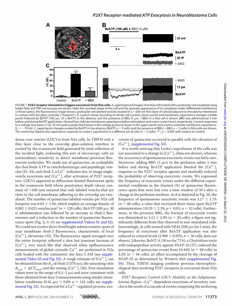
P2X7 Receptors Trigger Exocytosis as Assayed by MembraneCapacitance Measurements—It has been reported that N2acells transiently transfected with pro-opiomelanocortin un-dergo calcium-regulated release of �-endorphin located todense-core granules (25, 26). On the other hand, N2a cellsexpress ionotropic purinergic P2X7 receptors whose activationpromotes Ca2� entry into the cell and the ensued increase in[Ca2�]i (12). Thus, the question arises as to whether P2X7receptorsmight be coupled to exocytosis in this neuroblastomacell line.We first addressed this issue by determining the poten-tial change in membrane capacitance elicited by P2X7 receptorstimulation. Biological membranes behave as electrical capaci-tors whose capacitance is in direct relation to their surface.Because exocytosis involves the fusion of the vesicle membranewith the plasma membrane, it implies an increase in cellularsurface, which can be detected as a change in the capacitance ofthe cell by using thewhole-cell configuration of the patch clamptechnique (20, 27). In these experiments we employedN2a cellsseeded at a low density (104 cells/ml) and systematically chosecells with a rounded shape and isolated from neighbors (Fig.1A). Resting membrane capacitance of N2a cells was 10.03 �0.33 picofarads (n � 19). Selective activation of P2X7 receptorswith BzATP (100 �M for 10 s) induced inwardly directed non-desensitizing currents that were consistently associatedwith anincrease in membrane capacitance (Fig. 1B). The mean chargeof the currents evoked by BzATP was 93.15 � 25.47 picocou-lombs, and the capacitance increase was 84.77� 9.46 femtofar-ads (n� 9 cells) (Fig. 1, B andC). None of these effects could bedue to an application artifact, as perfusing Locke’s solution dur-ing 10 s did not cause any consistent change in whole-cell cur-rent and was associated to a capacitance increase of only 2.5 �2.5 femtofarads (n � 5 cells). On the contrary, it can be attrib-uted to the activation of P2X7 receptors, as treatmentwith BBG(5 �M, 3 min before and during agonist application), a selectiveP2X7 receptor antagonist (28), reduced the currents (12.58 �2.94 picocoulombs) and the capacitance increase (22.2 � 12.23femtofarads) evoked byBzATPby 85 and 73%, respectively (n�5 cells) (Fig. 1, B and C). Calcium-dependent neuroexocytosisrequires the formation of the SNARE (soluble N-ethylmaleim-ide-sensitive fusion protein (NSF) attachment protein recep-tor)membrane fusion complex between two plasmamembraneproteins, syntaxin and SNAP-25, and one vesicular protein,synaptobrevin/VAMP (29). SNARE complexes are disassem-bled due to the ATPase activity of NSF, which allows recoveryof the components of the fusion complex for subsequentrounds of exocytosis (30). Accordingly, NEM has been used astool to inhibit vesicle fusion underlying the exocytosis of a vari-ety ofmediators includingATP (31). Interestingly, treatment ofN2a cells with 500�MNEM for 2min abolished the capacitanceincrease elicited by BzATP without reducing P2X7 receptor-mediated currents (supplemental Fig. S1). Altogether, theseresults indicate that P2X7 receptors are coupled to exocytosisin N2a cells.P2X7 Receptors Trigger Exocytosis as Assayed by TIRFM
Imaging—To corroborate electrophysiological results, weemployed TIRFM to visualize exocytosis of individual large
P2X7 Receptor-mediated ATP Exocytosis in Neuroblastoma Cells
11372 JOURNAL OF BIOLOGICAL CHEMISTRY VOLUME 286 • NUMBER 13 • APRIL 1, 2011
at UN
IVE
RS
IDA
D C
OM
PLU
TE
NS
E D
E M
AD
RID
, on March 31, 2011
ww
w.jbc.org
Dow
nloaded from
dense-core vesicles (LDCVs) from N2a cells. In TIRFM only athin layer close to the coverslip glass-solution interface isexcited by the evanescent field generated by total reflection ofthe incident light, endowing this sort of microscopy with anextraordinary sensitivity to detect membrane-proximal fluo-rescent molecules. We made use of quinacrine, an acidophilicdye that binds ATP in catecholaminergic and peptidergic vesi-cles (32–34), and rhod-2, a Ca2� indicator dye, to image single-vesicle exocytosis and [Ca2�]i after activation of P2X7 recep-tors. LDCVs appeared as diffraction-limited fluorescent spotsin the evanescent field whose penetration depth (decay con-stant of �160 nm) ensured that only labeled vesicles that areclose to the cell membrane adhering to the coverslip are visu-alized. The number of quinacrine-labeled vesicles per N2a cellfootprint was 8.03 � 1.94, which implies an average density of0.082 � 0.021 vesicles/�m2 (n � 120 cells). BzATP (100 �M, 30s) administration was followed by an increase in rhod-2 fluo-rescence and a reduction in the number of quinacrine fluores-cence spots (Fig. 2, A–D; see supplemental Videos S1 and S2).We could not resolve short-lived bright submicrometer spots ofnear membrane rhod-2 fluorescence, characteristic of local[Ca2�]i elevations (35). The fluorescence signal averaged overthe entire footprint reflected a slow but transient increase of[Ca2�]i very much like that observed when epifluorescencemeasurements of global cytosolic Ca2� are performed in N2acells loaded with the ratiometric dye fura-2 AM (see supple-mental Video S3 and Fig. S2). A rough estimate of [Ca2�]i canbe obtained from theKd of rhod-2 (570 nM), the saturating ratioRmax � F/Fmax, and the resting [Ca2�]i (36). Post-simulationvalues were in the range of 0.2–1 �M and were consistent withthose obtained from fura-2 measurements using similar stimu-lation conditions (0.41 �M � 0.09; n � 141 cells; see supple-mental Fig. S2). As expected for a Ca2�-regulated process, exo-
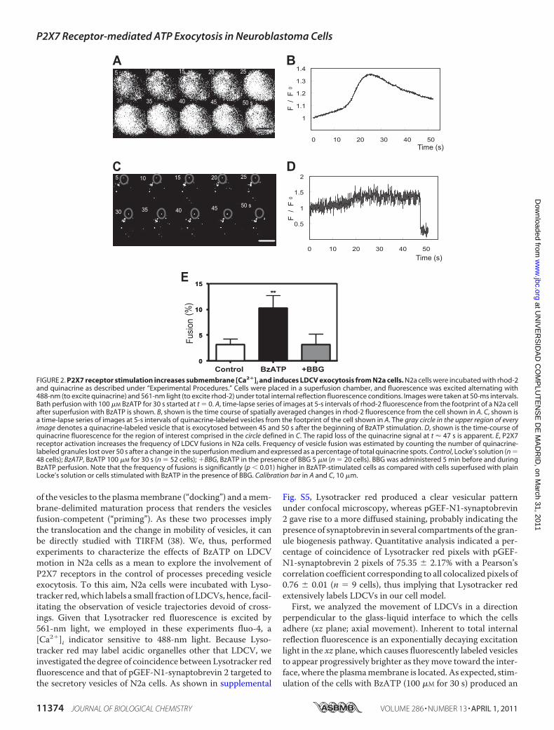
cytosis of quinacrine occurred in parallel with the elevation of[Ca2�]i (supplemental Fig. S3).
It is worth noticing that Locke’s superfusion of the cells wasnot associated to a change in [Ca2�]i (data not shown), whereasthe occurrence of spontaneous exocytotic eventswas fairly rare.Moreover, adding BBG (5 �M) to the perfusion saline 5 minbefore and during BzATP application blunted the [Ca2�]iresponse to the P2X7 receptor agonist and markedly reducedthe probability of observing exocytotic events. We expressedthe frequency of exocytotic events under the different experi-mental conditions as the fraction (%) of quinacrine fluores-cence spots that were lost over a time window of 50 s after achange in the perfusionmedium. As it is depicted in Fig. 2E, thefrequency of spontaneous exocytotic events was 3.17 � 1.1%(n � 48 cells), a value that increased three times upon BzATPadministration (10.29 � 2.3%; p 0.01; n � 52 cells). Further-more, in the presence BBG, the fraction of exocytotic eventswas diminished to 3.12 � 2.0% (n � 20 cells), a figure not sig-nificantly different from that observed in non-stimulated cells.Interestingly, in cells treated with NEM (500 �M for 2 min), thefrequency of exocytosis after BzATP application was alsoreduced to control levels (2.988 � 0.85%; n � 36 cells; data notshown). Likewise, BoNT/A (30 nM for 72 h), aClostridium toxinwith endopeptidase activity against SNAP-25 (37), reduced thepercentage of quinacrine events from 10.44% (n � 48 cells) to4.2% (n � 44 cells), an effect accompanied by the cleavage ofSNAP-25 as determined by Western blot (supplemental Fig.S4). Thus, TIRFM imaging confirms previous electrophysi-ological data involving P2X7 receptors in exocytosis from N2acells.P2X7 Receptors Control LDCV Mobility at the Subplasma-
lemma Region—Ca2�-dependent exocytosis of secretory vesi-cles is the result of a cascade of events comprising the anchoring
FIGURE 1. P2X7 receptor stimulation triggers exocytosis from N2a cells. A, superimposed images of an N2a cell loaded with Lysotracker red visualized usingbright field and TIRF microscopy are shown. Note the rounded shape of the cell and the granular appearance of its cytoplasm under differential interferencecontrast optics; the fluorescence image shows Lysotracker red-labeled vesicles located in a �200-nm thin layer of cytosol adjacent to the plasma membranein contact with the glass coverslip (“footprint”). B, a patch clamp recording of whole-cell currents (lower panel) and membrane capacitance changes (middlepanel) induced by BzATP (100 �M, 10 s; BzATP) in the absence and the presence of BBG (5 �M; �BBG) in a N2a cell is shown. BBG was administered 3 minbefore and during BzATP application. Dashed lines indicate membrane capacitance before stimulation and a zero current level, respectively. Current responsesto a voltage sine wave (1-Hz, 70-mV peak to peak; black boxes in the voltage protocol shown in the upper panel) were used to estimate membrane capacitance.Vh � �70 mV. C, capacitance changes evoked by BzATP in the absence (BzATP; n � 9 cells) and the presence of BBG (�BBG; n � 5 cells) in N2a cells are shown.The control bar depicts the capacitance response to Locke’s superfusion in a different set of cells (n � 5 cells). ***, p 0.005 with respect to control.
P2X7 Receptor-mediated ATP Exocytosis in Neuroblastoma Cells
APRIL 1, 2011 • VOLUME 286 • NUMBER 13 JOURNAL OF BIOLOGICAL CHEMISTRY 11373
at UN
IVE
RS
IDA
D C
OM
PLU
TE
NS
E D
E M
AD
RID
, on March 31, 2011
ww
w.jbc.org
Dow
nloaded from
of the vesicles to the plasmamembrane (“docking”) and amem-brane-delimited maturation process that renders the vesiclesfusion-competent (“priming”). As these two processes implythe translocation and the change in mobility of vesicles, it canbe directly studied with TIRFM (38). We, thus, performedexperiments to characterize the effects of BzATP on LDCVmotion in N2a cells as a mean to explore the involvement ofP2X7 receptors in the control of processes preceding vesicleexocytosis. To this aim, N2a cells were incubated with Lyso-tracker red,which labels a small fraction of LDCVs, hence, facil-itating the observation of vesicle trajectories devoid of cross-ings. Given that Lysotracker red fluorescence is excited by561-nm light, we employed in these experiments fluo-4, a[Ca2�]i indicator sensitive to 488-nm light. Because Lyso-tracker red may label acidic organelles other that LDCV, weinvestigated the degree of coincidence between Lysotracker redfluorescence and that of pGEF-N1-synaptobrevin 2 targeted tothe secretory vesicles of N2a cells. As shown in supplemental
Fig. S5, Lysotracker red produced a clear vesicular patternunder confocal microscopy, whereas pGEF-N1-synaptobrevin2 gave rise to a more diffused staining, probably indicating thepresence of synaptobrevin in several compartments of the gran-ule biogenesis pathway. Quantitative analysis indicated a per-centage of coincidence of Lysotracker red pixels with pGEF-N1-synaptobrevin 2 pixels of 75.35 � 2.17% with a Pearson’scorrelation coefficient corresponding to all colocalized pixels of0.76 � 0.01 (n � 9 cells), thus implying that Lysotracker redextensively labels LDCVs in our cell model.First, we analyzed the movement of LDCVs in a direction
perpendicular to the glass-liquid interface to which the cellsadhere (xz plane; axial movement). Inherent to total internalreflection fluorescence is an exponentially decaying excitationlight in the xz plane, which causes fluorescently labeled vesiclesto appear progressively brighter as they move toward the inter-face, where the plasmamembrane is located. As expected, stim-ulation of the cells with BzATP (100 �M for 30 s) produced an
FIGURE 2. P2X7 receptor stimulation increases submembrane [Ca2�]i and induces LDCV exocytosis from N2a cells. N2a cells were incubated with rhod-2and quinacrine as described under “Experimental Procedures.” Cells were placed in a superfusion chamber, and fluorescence was excited alternating with488-nm (to excite quinacrine) and 561-nm light (to excite rhod-2) under total internal reflection fluorescence conditions. Images were taken at 50-ms intervals.Bath perfusion with 100 �M BzATP for 30 s started at t � 0. A, time-lapse series of images at 5-s intervals of rhod-2 fluorescence from the footprint of a N2a cellafter superfusion with BzATP is shown. B, shown is the time course of spatially averaged changes in rhod-2 fluorescence from the cell shown in A. C, shown isa time-lapse series of images at 5-s intervals of quinacrine-labeled vesicles from the footprint of the cell shown in A. The gray circle in the upper region of everyimage denotes a quinacrine-labeled vesicle that is exocytosed between 45 and 50 s after the beginning of BzATP stimulation. D, shown is the time-course ofquinacrine fluorescence for the region of interest comprised in the circle defined in C. The rapid loss of the quinacrine signal at t � 47 s is apparent. E, P2X7receptor activation increases the frequency of LDCV fusions in N2a cells. Frequency of vesicle fusion was estimated by counting the number of quinacrine-labeled granules lost over 50 s after a change in the superfusion medium and expressed as a percentage of total quinacrine spots. Control, Locke’s solution (n �48 cells); BzATP, BzATP 100 �M for 30 s (n � 52 cells); �BBG, BzATP in the presence of BBG 5 �M (n � 20 cells). BBG was administered 5 min before and duringBzATP perfusion. Note that the frequency of fusions is significantly (p 0.01) higher in BzATP-stimulated cells as compared with cells superfused with plainLocke’s solution or cells stimulated with BzATP in the presence of BBG. Calibration bar in A and C, 10 �m.
P2X7 Receptor-mediated ATP Exocytosis in Neuroblastoma Cells
11374 JOURNAL OF BIOLOGICAL CHEMISTRY VOLUME 286 • NUMBER 13 • APRIL 1, 2011
at UN
IVE
RS
IDA
D C
OM
PLU
TE
NS
E D
E M
AD
RID
, on March 31, 2011
ww
w.jbc.org
Dow
nloaded from

increase in fluo-4 fluorescence, reflecting the rise in [Ca2�]i(Fig. 3,A andB; supplemental Video S4). Thiswas accompaniedby changes in the fluorescence intensity of Lysotracker red-tagged vesicles (Fig. 3, C and D; supplemental Video S5) suchthat a number of vesicles became brighter (see vesicle 1 in Fig.3C and the dashed line in Fig. 3D), whereas others underwentsmall fluorescence decreases (vesicle 2 in Fig. 3C and the dottedline in Fig. 3D), and still others did not show any significantchange in fluorescence (vesicle 3 in Fig. 3C and the continuousline in Fig. 3D). Such a differential behavior in vesicle fluores-cence led us to classify LCDVs into three groups according totheir axialmovement; 1) vesicles approaching the plasmamem-brane, 2) vesicles moving away from it, and 3), vesicles keepingtheir position with respect to the membrane. We have evalu-ated the relative distribution of the three vesicle populations innon-stimulated cells, BzATP-stimulated cells, and BzATP-stimulated cells in the presence of BBG (Fig. 3E). Because the
loading with Lysotracker red showed variability between cellsand between LDCVs of the same cell and also because of thepossible existence of infoldings in the plasma membrane, wedid not infer the absolute z position of the vesicles from itsfluorescence intensity but only estimated thez change.We seta vertical displacement of �15 nm from the initial position asthe minimum that a vesicle should move for being assigned toone of the two first groups (namely, vesicles approaching orleaving themembrane). BzATP stimulation increased by 4-foldthe percentage of vesicles approaching themembrane (11.91�1.9%; n � 57 cells) in comparison with non-stimulated cells(3.0 � 1.0%; n � 42 cells) or cells stimulated with BzATP in thepresence of BBG (3.5 � 1.9%; n � 18 cells). The population ofvesicles moving away from themembrane was also aminor one(7–12%), although its importance did not vary significantlyamong the three conditions considered. Last, vesicles keepingtheir position relative to the plasma membrane constituted the
FIGURE 3. P2X7 receptor stimulation modifies axial movement of LDCVs. Cells were loaded with Lysotracker red and fluo-4 as indicated under “Experi-mental Procedures,” and TIRFM images were taken at 1-s intervals. A, shown is a time-lapse series of images at 5-s intervals of fluo-4 fluorescence from thefootprint of a N2a cell after superfusion with BzATP (100 �M for 30 s; initiated at t � 0). B, shown is the time-course of spatially averaged changes in fluo-4fluorescence from the cell shown in A. C, shown is a time-lapse series of images at 5-s intervals of a group of Lysotracker red-tagged vesicles from the footprintof the cell shown in A. Changes in vesicle fluorescence over time reflect the movement of the vesicles toward and away from the plasma membrane. Threevesicles representative of different motion behaviors appear encircled and numbered (1, 2, and 3). D, shown is the time-course of changes in fluorescence ofthe regions of interest comprised in the circles drawn in C. The dashed line reflects the axial movement of vesicle 1 that approaches the plasma membrane, thedotted line reports on vesicle 2 that moves away from the membrane, and the continuous line refers to vesicle 3 that keeps its distance to the membrane. E, P2X7receptor stimulation increases the fraction of LDCVs approaching the plasma membrane. Relative distribution in non-stimulated N2a cells (Control, 199 vesiclesfrom 42 cells), N2a cells stimulated with 100 �M BzATP (Bz-ATP, 284 vesicles from 57 cells), and N2a cells stimulated with 100 �M BzATP in the presence of 5 �M
BBG (�BBG, 95 vesicles from 18 cells) of three groups of LDCVs defined according to their type of movement in the xz plane; left bars, vesicles approaching themembrane; middle bars, vesicles moving away from the plasma membrane; right bars, vesicles keeping their distance to the membrane. BBG was administered5 min before and during BzATP perfusion. Note that the population of vesicles that gets closer to the membrane in BzATP-stimulated cells is significantly (p 0.005) larger than in control cells or BzATP-stimulated cells in the presence of BBG. F, z distance covered by vesicles within the three different axial movementgroups from non-stimulated N2a cells (Control), N2a cells stimulated with 100 �M BzATP (Bz-ATP), and N2a cells stimulated with 100 �M BzATP in the presenceof 5 �M BBG (�BBG) is shown. LDCVs analyzed were the same as in E. Calibration bar in A and C, 10 �m.
P2X7 Receptor-mediated ATP Exocytosis in Neuroblastoma Cells
APRIL 1, 2011 • VOLUME 286 • NUMBER 13 JOURNAL OF BIOLOGICAL CHEMISTRY 11375
at UN
IVE
RS
IDA
D C
OM
PLU
TE
NS
E D
E M
AD
RID
, on March 31, 2011
ww
w.jbc.org
Dow
nloaded from
largest population, contributing 80–90% of the vesicles taggedwith Lysotracker red irrespective of the experimental condi-tions. Interestingly, the average displacement (z distance) ofthe vesicles within each particular group was quite constantacross the three experimental situations (Fig. 3F).Next, we studied vesicle motion in the plane parallel to the
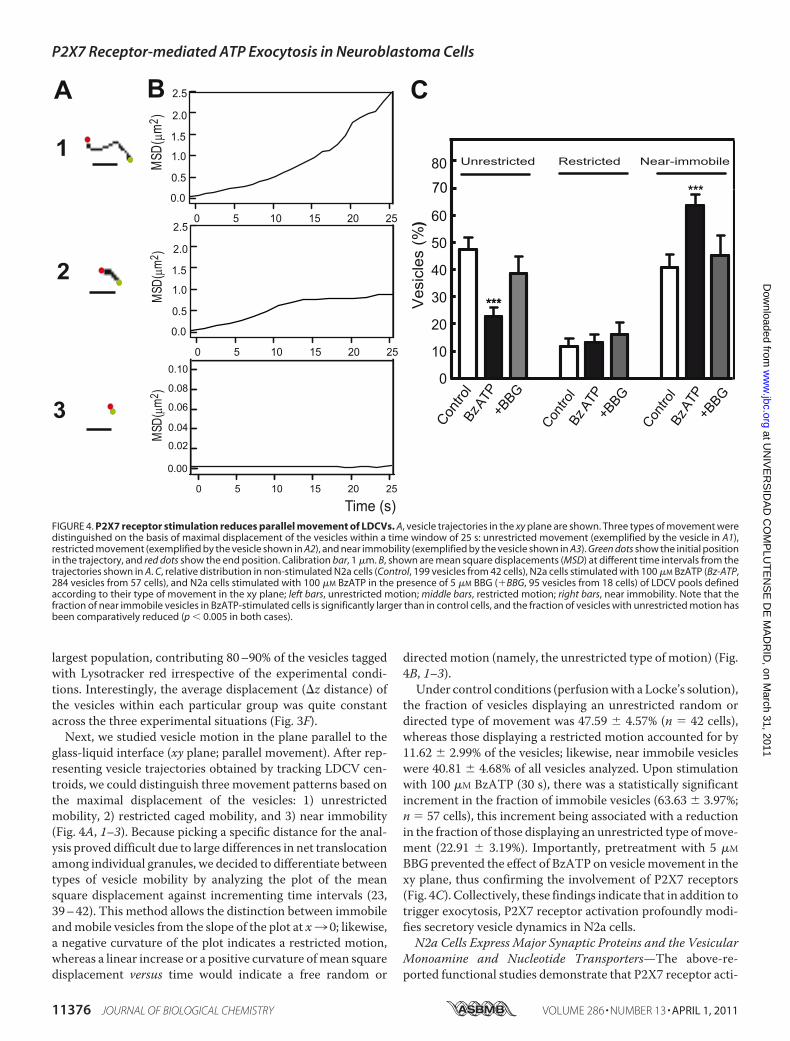
glass-liquid interface (xy plane; parallel movement). After rep-resenting vesicle trajectories obtained by tracking LDCV cen-troids, we could distinguish three movement patterns based onthe maximal displacement of the vesicles: 1) unrestrictedmobility, 2) restricted caged mobility, and 3) near immobility(Fig. 4A, 1–3). Because picking a specific distance for the anal-ysis proved difficult due to large differences in net translocationamong individual granules, we decided to differentiate betweentypes of vesicle mobility by analyzing the plot of the meansquare displacement against incrementing time intervals (23,39–42). This method allows the distinction between immobileandmobile vesicles from the slope of the plot at x3 0; likewise,a negative curvature of the plot indicates a restricted motion,whereas a linear increase or a positive curvature ofmean squaredisplacement versus time would indicate a free random or
directedmotion (namely, the unrestricted type of motion) (Fig.4B, 1–3).Under control conditions (perfusionwith a Locke’s solution),
the fraction of vesicles displaying an unrestricted random ordirected type of movement was 47.59 � 4.57% (n � 42 cells),whereas those displaying a restricted motion accounted for by11.62 � 2.99% of the vesicles; likewise, near immobile vesicleswere 40.81 � 4.68% of all vesicles analyzed. Upon stimulationwith 100 �M BzATP (30 s), there was a statistically significantincrement in the fraction of immobile vesicles (63.63 � 3.97%;n � 57 cells), this increment being associated with a reductionin the fraction of those displaying an unrestricted type ofmove-ment (22.91 � 3.19%). Importantly, pretreatment with 5 �M
BBGprevented the effect of BzATP on vesiclemovement in thexy plane, thus confirming the involvement of P2X7 receptors(Fig. 4C). Collectively, these findings indicate that in addition totrigger exocytosis, P2X7 receptor activation profoundly modi-fies secretory vesicle dynamics in N2a cells.N2a Cells Express Major Synaptic Proteins and the Vesicular
Monoamine and Nucleotide Transporters—The above-re-ported functional studies demonstrate that P2X7 receptor acti-
FIGURE 4. P2X7 receptor stimulation reduces parallel movement of LDCVs. A, vesicle trajectories in the xy plane are shown. Three types of movement weredistinguished on the basis of maximal displacement of the vesicles within a time window of 25 s: unrestricted movement (exemplified by the vesicle in A1),restricted movement (exemplified by the vesicle shown in A2), and near immobility (exemplified by the vesicle shown in A3). Green dots show the initial positionin the trajectory, and red dots show the end position. Calibration bar, 1 �m. B, shown are mean square displacements (MSD) at different time intervals from thetrajectories shown in A. C, relative distribution in non-stimulated N2a cells (Control, 199 vesicles from 42 cells), N2a cells stimulated with 100 �M BzATP (Bz-ATP,284 vesicles from 57 cells), and N2a cells stimulated with 100 �M BzATP in the presence of 5 �M BBG (�BBG, 95 vesicles from 18 cells) of LDCV pools definedaccording to their type of movement in the xy plane; left bars, unrestricted motion; middle bars, restricted motion; right bars, near immobility. Note that thefraction of near immobile vesicles in BzATP-stimulated cells is significantly larger than in control cells, and the fraction of vesicles with unrestricted motion hasbeen comparatively reduced (p 0.005 in both cases).
P2X7 Receptor-mediated ATP Exocytosis in Neuroblastoma Cells
11376 JOURNAL OF BIOLOGICAL CHEMISTRY VOLUME 286 • NUMBER 13 • APRIL 1, 2011
at UN
IVE
RS
IDA
D C
OM
PLU
TE
NS
E D
E M
AD
RID
, on March 31, 2011
ww
w.jbc.org
Dow
nloaded from
vation triggers exocytosis from N2a cells. However, both theproteins mediating vesicle fusion and the type of cargo storedand released are not known. Because neuroblastoma cells arisefrom neural crest precursors of the sympathetic nervous sys-tem, we set out to investigate whether N2a cells express prom-inentmembers of the secretorymachinery involved in calcium-dependent neuroexocytosis. Hence, the expression of the threemembers, SNAP-25, syntaxin 1, and synaptobrevin/VAMP, ofthe neuronal SNAREmembrane fusion complex and other pro-teins involved in synaptic exocytosis was analyzed by immuno-cytochemistry or RT-PCR (supplemental Fig. S6). In N2a cells,SNAP-25 and syntaxin 1A were predominantly localized at theplasma membrane as already reported (26). At variance, vesi-cle-resident proteins such as synaptophysin and synaptobrevinshowed a broad intracellular distribution. Moreover, synapsin1, a protein involved in regulating the number of synaptic ves-icles available for exocytotic release (43), also showed a cytoso-lic location in N2a cells. Likewise, the expression of vesicularmonoamine transporters 1 and 2 (VMAT-1 and VMAT-2) asmarkers of catecholamine-storing vesicles was investigated.Both transporter proteins were detected in N2a cells and local-ized in the cytosolic compartment (supplemental Fig. S6A).Interestingly, a high level of the vesicular nucleotide trans-porter transcript was also observed in N2A cells (supplementalFig. S6B), suggesting that ATP and other nucleotidic com-pounds could be co-storedwith catecholamines and released byexocytosis in a Ca2�-dependent manner.N2aCells Release ATP—Toconfirm the ability ofN2a cells to
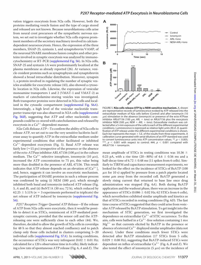
release ATP, we set out to use the very sensitive luciferin-lucif-erase assay to quantify ATP in the extracellular medium underexperimental conditions that either facilitated or inhibitedCa2�-dependent exocytosis (Fig. 5). Basal ATP release wasfairly low (�15 pM) irrespective of the presence or the absenceof the ecto-ATPase inhibitorARL67156 (100�M) in the culturemedium. The Ca2�-selective ionophore, ionomycin (10 �M),increased the ATP concentration to 75 pM, this value beingmore than doubled in the presence of ARL 67156. This resultindicates that ATP release depends on an elevation of [Ca2�]iand, hence, suggests it can involve an exocytotic mechanism.The participation of SNARE proteins in such a release processwas confirmed by using (i) NEM (500 �M), which stronglyinhibited both basal and ionomycin-induced ATP release (Fig.5, A and B), and (ii) BoNT/A (30 nM; 72 h), which reduced by62.25 � 5.11% (n � 3 experiments performed in triplicate) thenet release of ATP induced by ionomycin (supplemental Fig.S4).P2X7 Receptors Trigger Quantal ATP Release—If the release
of ATP from N2a cells were exocytotic, then it would be possi-ble to detect it as STICs, reminiscent of ATP-mediated post-synaptic currents, provided that the sensor cell and the ATP-releasing one were sufficiently close to each other (44). We,therefore, decided to allow the growth of N2a cells in coverslipsfor 48 h so that they almost reached confluency and to patchclamp only those cells included in clusters comprising 5–20individual cells (supplemental Fig. S7A). In resting conditions,the occurrence of STICs was very infrequent (0.02 � 0.01 Hz;calculated for a 120 s observation time in 6 cells), likely indicat-ing a low rate of spontaneous ATP release (Fig. 6,A and E). The
mean amplitude of STICs in resting conditions was 10.36 �0.23 pA, with a rise time (20–80%) of 4.6 � 0.56 ms and ahalf-decay time of 6.72 � 0.48 ms (11 spikes from 6 cells). Sim-ilarly toTIRFMand capacitancemeasurement experiments, welooked for the effect on the incidence of STICs of BzATP (100�M for 10 s) applied by pressure from a patch pipette locatedsome �m away from the recorded cell. BzATP generated aslowly rising current that returned to base line once drugadministration was stopped (Fig. 6A). Both during BzATPapplication and the washout phase, there was an increase in theappearance of STICs (0.086 � 0.02 Hz; 62 STICs from 6 cells),which nevertheless exhibited similar amplitude and kinetics asthat of STICs recorded in resting conditions (Fig. 6D). The fasttime course of STICs suggested that they could arise fromvesic-ularATP released byBzATP stimulation. To get insight into themechanism of STIC generation, we first investigated thedependence on extracellular Ca2� of STIC occurrence. To thisaim, cells were bathed in a Ca2�-free solution containing 1 mM
Mg2� so that currents elicited by BzATP in the presence orabsence of external Ca2�displayed similar amplitudes (data notshown). Under these conditions much fewer STICs weredetected after BzATP stimulation (17 STICs from 5 cells;0.029 � 0.08 Hz), suggesting that BzATP-induced STICs weredependent on influx of extracellular Ca2� (Fig. 6, B and E). Wealso tested the effect of ionomycin (5 �M) on the incidence of
FIGURE 5. N2a cells release ATP by a NEM-sensitive mechanism. A, shownare representative records of luminescence evoked by ATP released into theextracellular medium of N2a cells before (Control) and after ionomycin (10�M) stimulation in the absence (ionomycin) or presence of the ecto-ATPaseinhibitor ARL67156 (100 �M; ARL � Iono) or ARL67156 plus the exocytosisinhibitor NEM (500 �M; NEM � ARL � Iono). Extracellular medium was col-lected after a 5-min exposure of the cells to each of the different experimentalconditions. Luminescence is expressed as relative light units (RLU). B, quanti-fication of ATP release under the different experimental conditions is shown.Each bar represents the mean � S.E. of the results from three experiments. Acalibration curve generated with serial dilutions of an ATP standard was usedto convert luminescence units into ATP concentration values. **, p 0.005;***, p 0.001 with respect to control; ###, p 0.001 compared withARL67156 � ionomycin.
P2X7 Receptor-mediated ATP Exocytosis in Neuroblastoma Cells
APRIL 1, 2011 • VOLUME 286 • NUMBER 13 JOURNAL OF BIOLOGICAL CHEMISTRY 11377
at UN
IVE
RS
IDA
D C
OM
PLU
TE
NS
E D
E M
AD
RID
, on March 31, 2011
ww
w.jbc.org
Dow
nloaded from
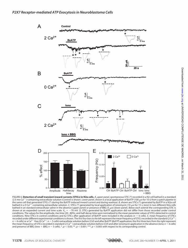
FIGURE 6. Detection of small transient inward currents (STICs) in N2a cells. A, upper panel, spontaneous STIC (*) recorded in a N2 cell bathed in a standard(2.5 mM Ca2�)-containing extracellular solution (Control) is shown. Lower panel, shown is a local application of BzATP (100 �M for 10 s) from a patch pipette tothe same cell that generated STICs (*) during the BzATP-induced inward current and during washout. B, shown are STICs (*) generated by BzATP in a N2a cellbathed in a 0 Ca2�-containing extracellular solution. C, STICs (*) generated by local application of ionomycin (10 �M for 15 s; Iono) in two different N2a cellsbathed in an standard extracellular saline in the absence (upper panel) or presence of BBG (5 �M) (lower panel). Below each asterisk the corresponding STIC isshown at an expanded current and time scale. Vh � �70 mV. D, STICs generated by BzATP application did not differ from those recorded under controlconditions. The values for the amplitude, rise time (20 – 80%), and half-decay time were normalized to the mean parameter values of STICs detected in controlconditions. Nine STICs in control conditions and 62 STICs after application of BzATP were included in the analysis (n � 6 cells). E, mean frequency of STICsrecorded under different experimental conditions is shown. The first four bars to the left represent the mean frequency of STICs recorded in the standard (2 Ca2�;n � 6 cells) or a Ca2�-free (0 Ca2�; n � 5 cells) extracellular solution before (Ctrl) and after BzATP (BzATP) application; the first three bars from the right representthe mean frequency of STICs recorded in standard (2 Ca2�) extracellular solution before (Ctrl) and after ionomycin application in the absence (Iono; n � 6 cells)and presence of BBG (Iono � BBG; n � 5 cells). *, p 0.05; **, p 0.001; ***, p 0.005 with respect to its corresponding control.
P2X7 Receptor-mediated ATP Exocytosis in Neuroblastoma Cells
11378 JOURNAL OF BIOLOGICAL CHEMISTRY VOLUME 286 • NUMBER 13 • APRIL 1, 2011
at UN
IVE
RS
IDA
D C
OM
PLU
TE
NS
E D
E M
AD
RID
, on March 31, 2011
ww
w.jbc.org
Dow
nloaded from
STICs. As Fig. 6, C and E show, besides eliciting a long-lastingoutward current possibly reflecting the activation of a Ca2�-de-pendent K� conductance (45), ionomycin increased the fre-quency of STICs (0.077� 0.012Hz; 55 STICs from6 cells), thusimplying a relation with an elevation of [Ca2�]i. Confirmationthat STICs were indeed mediated by P2X7 receptors wasobtained by adding BBG to the bath solution. Consistent withour expectations, treatment with BBG (5 �M; added to the bathand puffer pipette solutions) abolished STICs evoked by iono-mycin (0.0085 � 0.0002 Hz; 5 cells) (Fig. 6, C and E). It is alsoworth mentioning that no significant differences in size andkinetics were observed among STICs detected under the differ-ent experimental conditions (supplemental Fig. S6, B–D).Moreover, no correlation was found between the amplitudeand decay time of the STICs detected in all experiments (r �0.61). Altogether, these results indicate that endogenous ATPacting on P2X7 receptors generates STICs and that P2X7receptor stimulation can elicit vesicular ATP release.
DISCUSSION
ATP and its metabolites have now achieved a full and possi-bly unique status as ubiquitous extracellular messengers. In thenervous system, adenine nucleotides have been involved inintercellular communication between neurons and betweenneurons and glial cells as well as in the control of cell prolifera-tion, migration, and differentiation (46). The latter roles areessential in supporting brain development, maturation, andrepair after either acute injuries or chronic degenerative pro-cesses (47). Given the ubiquitous distribution of ATP, an ade-quate performance of such a variety of functions requires theexistence of not only a rich repertoire of purinergic receptorswith differential affinity and coupling to intracellular signalingpathways but also a tight control of the availability of the sig-naling molecule in the extracellular space. On the other hand,cell heterogeneity of primary neuronal cultures and the need toreduce animal use in biomedical research havemade clonal celllines from neuroectodermal origin a convenient experimentalmodel to study basic mechanisms in neural cell signaling.Recently, we have reported that P2X7 receptor inhibition pro-motes neuritogenesis from N2a murine neuroblastoma cellsand axonal growth in rat hippocampal neurons (12, 13). Byallowing the entrance of small cations into the cell, P2X recep-tors may also be coupled to some rapid signaling pathways, likethose responsible for the control of membrane excitability andthe exocytotic release of neurotransmitters. Interestingly, P2X7receptors are the onlymembers of the ionotropic family of puri-nergic receptors functionally expressed in N2a cells (12, 48), acell type that does not show voltage-gated Ca2� entry whenkept in a non-differentiated state.5 These two facts make thiscell system an ideally suited model to study the direct couplingof P2X7 receptors to exocytosis. In the present study, byemploying high resolution techniques (patch clamp andTIRFM imaging), we have been able to demonstrate that P2X7receptor activation not only triggers exocytosis but also pro-
foundlymodifies secretory vesicle dynamics inN2a cells.More-over, immunocytochemical andmolecular biological data indi-cate that these cells express essential proteins involved inexocytosis and have the capability of storing catecholaminesand nucleotides in secretory vesicles. Likewise, biochemicalmeasurements demonstrate that N2a cells secrete ATP by anSNARE-dependent exocytotic mechanism. Last, P2X7 recep-tors have also been shown to mediate the generation of STICsreflecting quantal ATP release, whose incidence is increased byagents that elevate [Ca2�]i (ionomycin and the P2X7 receptoragonist, BzATP) and diminished in the absence of extracellularCa2�.Membrane capacitance measurements are commonly used
as a single-cell assay of exocytosis. Our results indicate thatshort (few seconds) pulses of BzATP trigger capacitanceincreases in the 10s of femtofarads range. Taken the value of 1.9femtofarads for the capacitance of a single chromaffin granule(49), the catecholamine and ATP-storing LDCV from adre-nomedullary chromaffin cells, we estimate that on average ourstimulation paradigm induces the exocytosis of �40 LDCVsfrom N2a cells. Interestingly, such a response, which involvesvesicular fusion due to its sensitivity to NEM, can be attributedto the activation of P2X7 receptors as it is not observed underbasal (saline superfusion) conditions and, for its major part, isprevented when BzATP-activated currents are inhibited byBBG, a P2X7 receptor antagonist. In comparison with previouswork on human neuroblastoma cells reporting small increases(3–4 times the basal values) in substance P release after 2 h ofstimulation with nucleotides (15), the results presented here,besides demonstrating a direct coupling (our recording condi-tions effectively isolate P2X7 receptor-mediated currents)between P2X7 receptors and exocytosis in N2a cells, illustratethe ability of this cell line to rapidly and efficiently undergoexocytosis.Electrophysiological data were complemented by TIRFM
imaging, which allows the observation of individual secretoryvesicles in real time during exocytosis. After loading N2a cellswith quinacrine, we were able to detect diffraction-limited(�400-nm diameter) fluorescent spots representing ATP-con-taining vesicles (32, 34, 50) located in the submembrane regionof the cell. Moreover, by simultaneously loading the cells withrhod-2, a [Ca2�]i indicator dye, we could also monitor changesin [Ca2�]i in the cell regionwhere exocytosis occurs. Exocytosiswas detected by the rapid loss of quinacrine fluorescence, andrelease efficacy was estimated as the fraction of labeled-vesiclesexocytosed in a given time period (50 s). Under control condi-tions, there were not significant changes in rhod-2 fluores-cence, whereas the fraction of vesicles exocytosed was of 3%.Importantly, upon BzATP application, both a rise in submem-brane [Ca2�]i and a 3-fold increase in the number of exocytoticevents were observed. It should be noted that the idea that thecell region in contact with the culture support (the footprint)can show exocytosis is consistent with a wealth of informationderived from TIRFM imaging employing vesicular membranemarkers (51) and with electron microscopy studies (52) and isalso in linewith our results on theBoNT/AandNEMsensitivityof the quinacrine-fluorescence loss events. Both submembrane[Ca2�]i increases and the accompanying vesicular exocytosis
5 Y. Gutierrez-Martín, D. Bustillo, R. Gomez-Villafuertes, J. Sanchez-Nogueiro,C. Torregrosa-Hetland, T. Binz, L. M. Gutierrez, M. T. Miras-Portugal, andA. R. Artalejo, unpublished data.
P2X7 Receptor-mediated ATP Exocytosis in Neuroblastoma Cells
APRIL 1, 2011 • VOLUME 286 • NUMBER 13 JOURNAL OF BIOLOGICAL CHEMISTRY 11379
at UN
IVE
RS
IDA
D C
OM
PLU
TE
NS
E D
E M
AD
RID
, on March 31, 2011
ww
w.jbc.org
Dow
nloaded from
can be directly attributed to P2X7 receptor activation, as theywere reduced to near control values in the presence of BBG.The observation that only about 10% of the LDCVs that areexposed to a submembrane [Ca2�]i elevation is released indi-cates that proximity to the plasma membrane does not ensurerelease readiness and that vesicles have to undergo docking anda priming process to achieve full release competence (53–55).Both processes can be directly studied by TIRFM, as they havebeen related to changes in vesicle mobility (53). Therefore,docking has been shown to correspond to changes in the axialmobility (56, 57), whereas priming is associated with restrictedlateral motion (40). Similarly to what has been reported forembryonic mouse chromaffin cells (56), most of the LDCVsseen at the footprint ofN2a cells are stably docked at the plasmamembrane. Axial movement leading to either vesicle docking(movement toward the membrane) or undocking (movementaway from the membrane) was observed for a relatively smallfraction of the vesicles. Noteworthy, P2X7 receptor stimulationwas associated with a significant increase in the fraction of ves-icles that approached the plasmamembrane. This can be inter-preted as if P2X7 receptors could control the rate of vesicledelivery from the cell interior, this effect being crucial to sup-port secretion over sustained stimulation. Interestingly, vesiclesupply and docking at the plasmamembrane are two processesinterlinked by the action of Munc 18-1, a cytosolic proteinknown to enhance morphological vesicle docking by interact-ing both with the plasma membrane SNARE, syntaxin, and theactin cytoskeleton (58–60). In neurons and neuroendocrinecells, docked vesicles need to undergo priming before they canbe released. Primed vesicles constitute the readily releasablepool of vesicles and can be rapidly exocytosed in response to anelevation in [Ca2�]i. Priming requires the formation of the ter-nary SNARE complex between syntaxin, SNAP-25, and synap-tobrevin/VAMP (53) and has been associated with vesicleimmobilization (40, 61). In resting N2a cells, about 45% of thevesicles visualized at the footprint appear as near immobile inthe plane parallel to the membrane. P2X7 receptor stimulationleads to an increase in the fraction of immobile vesicles and aconcomitant reduction of those exhibiting unrestricted mobil-ity. These two effects imply an overall decrease in lateral mobil-ity of the vesicles locatedwithin the evanescent field and, there-fore, are consistent with enhanced vesicle priming. Our resultsalso point to a concomitant increase in the fraction of immobilevesicles and in the rate of exocytosis. Both effects are mutuallycompatible by considering the different dependence on [Ca2�]iof vesicle priming and exocytosis (62) and that the former isaccelerated at physiological temperature (63). Hence, underour experimental condition of 37 °C and with the moderate[Ca2�]i elevation elicited by BzATP, it is feasible to induce anoverfilling of the readily releasable pool, whereas the cell exhib-its a modest secretory response (64). Altogether, the functionalresults so far discussed indicate that P2X7 receptor stimulationaffects multiple steps (vesicle delivery, docking, priming, andexocytosis) in the regulated-secretion pathway of N2a cells.Most likely, this is a consequence of the ability of [Ca2�]i, whichis elevated after P2X7 receptor activation, to control secretionin a way that ensures both a fast secretory response due to therelease of the readily releasable pool of vesicles and a sustained
secretion by promoting the delivery of vesicles to the exocytoticsites at the plasma membrane as well and their priming toachieve release competence (65–67).Our results add to the existing evidence implicating P2X
receptors in neurotransmitter and hormone release from neu-rons and neuroendocrine cells (68). N2a cells express the threeneuronal SNAREs, which are involved in the docking, priming,and fusion steps of regulated exocytosis (69–71). Moreover, inaccordance with their origin from precursors of sympatheticneurons, they are endowed with LDCVs specialized in the stor-age of catecholamines and ATP. Our experiments also supportthe notion that ATP transported into secretory granules (72)can be released by exocytosis after a rise in [Ca2�]i broughtabout by ionomycin and by P2X7 receptor activation. We havereached this conclusion by a biochemical determination ofATPrelease but also by using N2a cells as biosensors to detect theirown ATP release. Hence, in response to stimulation by eitherionomycin or BzATP, we recorded STICs that were mediatedby ATP acting on P2X7 receptors due to their sensitivity toBBG. In addition, the fast kinetics of STICs and the non-desen-sitizing characteristics of P2X receptor-mediated responsessuggest that STICs are based on non-equilibrium ATP gradi-ents like those expected to arise from a vesicular type of release.Furthermore, the extracellular Ca2� dependence of STICoccurrence is fully in accordance with the involvement of aclassical exocytoticmechanism. Taking into account our previ-ous results indicating that P2X7 receptors activated by endog-enously released ATP exert a tonic inhibition of neuron-likedifferentiation of N2a cells (12, 13), the present results impli-cating P2X7 receptors in exocytosis prompt us to propose theexistence of an autocrine/paracrine positive feedback mecha-nism by which P2X7 receptors would mediate the exocytoticrelease ofATP that, in turn, would act on P2X7 receptors on thesame or neighbor cells to further stimulate its own release andnegatively control cell differentiation. Likewise, our resultspoint out the suitability of N2a cells as a model system to char-acterize the physiology of vesicular release of ATP and toexplore many of the complex intra- and intercellular signalingnetworks controlled by ATP in both the central and peripheralnervous system.
REFERENCES1. Burnstock, G., and Kennedy, C. (1985) Gen. Pharmacol. 16, 433–4402. von Kugelgen, I., and Wetter, A. (2000) Naunyn-Schmiedebergs Arch.
Pharmacol. 362, 310–3233. Khakh, B. S., Burnstock, G., Kennedy, C., King, B. F., North, R. A., Seguela,
P., Voigt, M., and Humphrey, P. P. (2001) Pharmacol. Rev. 53, 107–1184. North, R. A. (2002) Physiol. Rev. 82, 1013–10675. Jarvis, M. F., and Khakh, B. S. (2009) Neuropharmacology 56, 208–2156. Huang, N., Wang, D. J., and Heppel, L. A. (1989) Proc. Natl. Acad. Sci.
U.S.A. 86, 7904–79087. Brandle, U., Zenner, H. P., and Ruppersberg, J. P. (1999) Neurosci. Lett.
273, 105–1088. Adrian, K., Bernhard, M. K., Breitinger, H. G., and Ogilvie, A. (2000)
Biochim. Biophys. Acta 1492, 127–1389. Heo, J. S., and Han, H. J. (2006) Stem Cells 24, 2637–264810. Deli, T., and Csernoch, L. (2008) Pathol. Oncol. Res. 14, 219–23111. White, N., and Burnstock, G. (2006)Trends J. Pharmacol. Sci. 27, 211–21712. Gomez-Villafuertes, R., del Puerto, A., Díaz-Hernandez, M., Bustillo, D.,
Díaz-Hernandez, J. I., Huerta, P. G., Artalejo, A. R., Garrido, J. J., andMiras-Portugal, M. T. (2009) FEBS J. 276, 5307–5325
P2X7 Receptor-mediated ATP Exocytosis in Neuroblastoma Cells
11380 JOURNAL OF BIOLOGICAL CHEMISTRY VOLUME 286 • NUMBER 13 • APRIL 1, 2011
at UN
IVE
RS
IDA
D C
OM
PLU
TE
NS
E D
E M
AD
RID
, on March 31, 2011
ww
w.jbc.org
Dow
nloaded from

13. Díaz-Hernandez,M., del Puerto, A., Díaz-Hernandez, J. I., Diez-Zaera,M.,Lucas, J. J., Garrido, J. J., and Miras-Portugal, M. T. (2008) J. Cell Sci. 121,3717–3728
14. Munoz, M., Rosso, M., Perez, A., Covenas, R., Rosso, R., Zamarriego, C.,and Piruat, J. I. (2005) Neuropeptides 39, 427–432
15. Raffaghello, L., Chiozzi, P., Falzoni, S., Di Virgilio, F., and Pistoia, V. (2006)Cancer Res. 66, 907–914
16. Pellegatti, P., Falzoni, S., Pinton, P., Rizzuto, R., and Di Virgilio, F. (2005)Mol. Biol. Cell 16, 3659–3665
17. Rummel, A.,Mahrhold, S., Bigalke, H., and Binz, T. (2004)Mol.Microbiol.51, 631–643
18. Hamill, O. P., Marty, A., Neher, E., Sakmann, B., and Sigworth, F. J. (1981)Pflugers Arch. 391, 85–100
19. Lindau, M., and Neher, E. (1988) Pflugers Arch. 411, 137–14620. Carabelli, V., Giancippoli, A., Baldelli, P., Carbone, E., and Artalejo, A. R.
(2003) Biophys. J. 85, 1326–133721. Wu, Z. X., Xia, S., Xu, L., Bai, L., and Xu, T. (2003) Sheng wu hua xue yu
sheng wu wu li xue bao Acta biochimica et biophysica Sinica 35, 381–38622. Qian, H., Sheetz, M. P., and Elson, E. L. (1991) Biophys. J. 60, 910–92123. Johns, L. M., Levitan, E. S., Shelden, E. A., Holz, R. W., and Axelrod, D.
(2001) J. Cell Biol. 153, 177–19024. Levesque, S. A., Lavoie, E. G., Lecka, J., Bigonnesse, F., and Sevigny, J.
(2007) Br. J. Pharmacol. 152, 141–15025. Chevrier, D., Fournier, H., Nault, C., Zollinger, M., Crine, P., and Boileau,
G. (1991)Mol. Cell. Endocrinol. 79, 109–11826. Koticha, D. K., McCarthy, E. E., and Baldini, G. (2002) J. Cell Sci. 115,
3341–335127. Neher, E., and Marty, A. (1982) Proc. Natl. Acad. Sci. U.S.A. 79,
6712–671628. Jiang, L. H.,Mackenzie, A. B., North, R. A., and Surprenant, A. (2000)Mol.
Pharmacol. 58, 82–8829. Sollner, T., Whiteheart, S. W., Brunner, M., Erdjument-Bromage, H., Ge-
romanos, S., Tempst, P., and Rothman, J. E. (1993) Nature 362, 318–32430. Xu, T., Ashery, U., Burgoyne, R. D., and Neher, E. (1999) EMBO J. 18,
3293–330431. Luna, C., Li, G., Qiu, J., Challa, P., Epstein, D. L., and Gonzalez, P. (2009)
Invest. Ophthalmol. Vis. Sci. 50, 5805–581032. Bodin, P., andBurnstock,G. (2001) J. Cardiovasc. Pharmacol.38, 900–90833. Coco, S., Calegari, F., Pravettoni, E., Pozzi, D., Taverna, E., Rosa, P., Mat-
teoli, M., and Verderio, C. (2003) J. Biol. Chem. 278, 1354–136234. Pangrsic, T., Potokar, M., Stenovec, M., Kreft, M., Fabbretti, E., Nistri, A.,
Pryazhnikov, E., Khiroug, L., Giniatullin, R., and Zorec, R. (2007) J. Biol.Chem. 282, 28749–28758
35. Becherer, U., Moser, T., Stuhmer, W., and Oheim, M. (2003)Nat. Neuro-sci. 6, 846–853
36. Lev-Ram, V., Miyakawa, H., Lasser-Ross, N., and Ross, W. N. (1992)J. Neurophysiol. 68, 1167–1177
37. Blasi, J., Chapman, E. R., Link, E., Binz, T., Yamasaki, S., De Camilli, P.,Sudhof, T. C., Niemann, H., and Jahn, R. (1993) Nature 365, 160–163
38. Becherer, U., Pasche, M., Nofal, S., Hof, D., Matti, U., and Rettig, J. (2007)PLoS ONE 2, e505
39. Giner, D., Lopez, I., Villanueva, J., Torres, V., Viniegra, S., and Gutierrez,
L. M. (2007) Neuroscience 146, 659–66940. Nofal, S., Becherer, U., Hof, D., Matti, U., and Rettig, J. (2007) J. Neurosci.
27, 1386–139541. Oheim, M., and Stuhmer, W. (2000) Eur. Biophys. J. 29, 67–8942. Steyer, J. A., and Almers, W. (1999) Biophys. J. 76, 2262–227143. Yamamoto, H.,Matsumoto, K., Araki, E., andMiyamoto, E. (2003) J. Phar-
macol. Sci. 93, 30–3444. Fabbro, A., Skorinkin, A., Grandolfo, M., Nistri, A., and Giniatullin, R.
(2004) J. Physiol. 560, 505–51745. Leinders, T., and Vijverberg, H. P. (1992) Pflugers Arch. 422, 223–23246. Burnstock, G. (2006) Pharmacol. Rev. 58, 58–8647. Burnstock, G. (2008) Nat. Rev. Drug Discov. 7, 575–59048. Wu, P. Y., Lin, Y. C., Chang, C. L., Lu, H. T., Chin, C. H., Hsu, T. T., Chu,
D., and Sun, S. H. (2009) Cell. Signal. 21, 881–89149. Dernick, G., Gong, L. W., Tabares, L., Alvarez de Toledo, G., and Lindau,
M. (2005) Nat. Methods 2, 699–70850. Orriss, I. R., Knight, G. E., Utting, J. C., Taylor, S. E., Burnstock, G., and
Arnett, T. R. (2009) J. Cell. Physiol. 220, 155–16251. Holz, R. W. (2006) Cell. Mol. Neurobiol. 26, 439–44752. Plattner, H., Artalejo, A. R., and Neher, E. (1997) J. Cell Biol. 139,
1709–171753. Becherer, U., and Rettig, J. (2006) Cell Tissue Res. 326, 393–40754. Rettig, J., and Neher, E. (2002) Science 298, 781–78555. Xu, T., Rammner, B., Margittai, M., Artalejo, A. R., Neher, E., and Jahn, R.
(1999) Cell 99, 713–72256. Toonen, R. F., Kochubey, O., de Wit, H., Gulyas-Kovacs, A., Konijnen-
burg, B., Sørensen, J. B., Klingauf, J., and Verhage, M. (2006) EMBO J. 25,3725–3737
57. Verhage, M., and Sørensen, J. B. (2008) Traffic 9, 1414–142458. Bhaskar, K., Shareef, M. M., Sharma, V. M., Shetty, A. P., Ramamohan, Y.,
Pant, H. C., Raju, T. R., and Shetty, K. T. (2004)Neurochem. Int. 44, 35–4459. Rizo, J., and Sudhof, T. C. (2002) Nat. Rev. Neurosci. 3, 641–65360. Voets, T., Toonen, R. F., Brian, E. C., de Wit, H., Moser, T., Rettig, J.,
Sudhof, T. C., Neher, E., and Verhage, M. (2001) Neuron 31, 581–59161. Yizhar, O., and Ashery, U. (2008) PloS one 3, e269462. Neher, E., and Sakaba, T. (2008) Neuron 59, 861–87263. Dinkelacker, V., Voets, T., Neher, E., andMoser, T. (2000) J. Neurosci. 20,
8377–838364. von Ruden, L., and Neher, E. (1993) Science 262, 1061–106565. Smith, C., Moser, T., Xu, T., and Neher, E. (1998) Neuron 20, 1243–125366. Voets, T. (2000) Neuron 28, 537–54567. Voets, T., Neher, E., and Moser, T. (1999) Neuron 23, 607–61568. Sperlagh, B., Heinrich, A., and Csolle, C. (2007) Purinergic Signal. 3,
269–28469. Borisovska,M., Zhao, Y., Tsytsyura, Y., Glyvuk,N., Takamori, S.,Matti, U.,
Rettig, J., Sudhof, T., and Bruns, D. (2005) EMBO J. 24, 2114–212670. Gil, A., Gutierrez, L. M., Carrasco-Serrano, C., Alonso, M. T., Viniegra, S.,
and Criado, M. (2002) J. Biol. Chem. 277, 9904–991071. Han, X., Wang, C. T., Bai, J., Chapman, E. R., and Jackson, M. B. (2004)
Science 304, 289–29272. Gualix, J., Abal, M., Pintor, J., andMiras-Portugal, M. T. (1996) FEBS Lett.
391, 195–198
P2X7 Receptor-mediated ATP Exocytosis in Neuroblastoma Cells
APRIL 1, 2011 • VOLUME 286 • NUMBER 13 JOURNAL OF BIOLOGICAL CHEMISTRY 11381
at UN
IVE
RS
IDA
D C
OM
PLU
TE
NS
E D
E M
AD
RID
, on March 31, 2011
ww
w.jbc.org
Dow
nloaded from
Related Documents


