Purinergic P2X 7 Receptors Mediate ATP-induced Saliva Secretion by the Mouse Submandibular Gland * Received for publication, November 12, 2008 Published, JBC Papers in Press, December 19, 2008, DOI 10.1074/jbc.M808597200 Tetsuji Nakamoto ‡§1,2 , David A. Brown ‡1 , Marcelo A. Catala ´n ‡1 , Mireya Gonzalez-Begne ‡ , Victor G. Romanenko ‡§ , and James E. Melvin ‡§3 From the ‡ Center for Oral Biology and the § Department of Pharmacology & Physiology, University of Rochester Medical Center, Rochester, New York 14642 Salivary glands express multiple isoforms of P2X and P2Y nucleotide receptors, but their in vivo physiological roles are unclear. P2 receptor agonists induced salivation in an ex vivo submandibular gland preparation. The nucleotide selectivity sequence of the secretion response was BzATP ATP > ADP UTP, and removal of external Ca 2 dramatically sup- pressed the initial ATP-induced fluid secretion (85%). Together, these results suggested that P2X receptors are the major purinergic receptor subfamily involved in the fluid secre- tion process. Mice with targeted disruption of the P2X 7 gene were used to evaluate the role of the P2X 7 receptor in nucleoti- de-evoked fluid secretion. P2X 7 receptor protein and BzATP- activated inward cation currents were absent, and importantly, purinergic receptor agonist-stimulated salivation was sup- pressed by more than 70% in submandibular glands from P2X 7 - null mice. Consistent with these observations, the ATP-induced increases in [Ca 2 ] i were nearly abolished in P2X 7 / subman- dibular acinar and duct cells. ATP appeared to also act through the P2X 7 receptor to inhibit muscarinic-induced fluid secretion. These results demonstrate that the ATP-sensitive P2X 7 recep- tor regulates fluid secretion in the mouse submandibular gland. Salivation is a Ca 2 -dependent process (1, 2) primarily asso- ciated with the neurotransmitters norepinephrine and acetyl- choline, release of which stimulates -adrenergic and musca- rinic receptors, respectively. Both types of receptors are coupled to G proteins that activate phospholipase C (PLC) during salivary gland stimulation. PLC activation cleaves phosphatidylinositol 1,4-bisphosphate resulting in diacylglyc- erol and inositol 1,4,5-trisphosphate (InsP 3 ) production. Acti- vation of Ca 2 -selective InsP 3 receptor channels localized to the endoplasmic reticulum of salivary acinar cells increases the intracellular free calcium concentration ([Ca 2 ] i ). 4 Depletion of the endoplasmic reticulum Ca 2 pool triggers extracellular Ca 2 influx and a sustained elevation in [Ca 2 ] i . This increase in [Ca 2 ] i activates Ca 2 -dependent K and Cl channels pro- moting Cl secretion across the apical membrane and a lumen negative, electrochemical gradient that supports Na efflux into the lumen. The accumulation of NaCl creates an osmotic gradient which drives water movement into the lumen, thus generating isotonic primary saliva. This primary fluid is then modified by the ductal system, which reabsorbs NaCl and secretes KHCO 3 producing a final saliva that is hypotonic (1, 2). Salivation also has a non-cholinergic, non-adrenergic com- ponent, the origin of which is unclear (3). In addition to mus- carinic and -adrenergic receptors, salivary acinar cells express other receptors that are coupled to an increase in [Ca 2 ] i such as purinergic P2 and substance P receptors. Like muscarinic and -adrenergic receptors, P2 receptor activation leads to a sustained increase in [Ca 2 ] i in salivary acinar cells (4). In con- trast, substance P receptor activation rapidly desensitizes and therefore generates only a relatively transient increase in [Ca 2 ] i (5) that is unlikely to appreciably contribute to fluid secretion. The purinergic P2 receptor family is comprised of G protein-coupled P2Y and ionotropic P2X receptors activated by extracellular di- and triphosphate nucleotides. Activation of both subfamilies of P2 receptors causes an increase in [Ca 2 ] i . P2Y receptors increase [Ca 2 ] i via InsP 3 -induced Ca 2 mobili- zation from intracellular stores (similar to -adrenergic and muscarinic receptors) while P2X receptors act as ligand-gated, non-selective cation channels that mediate extracellular Ca 2 influx (6). Salivary gland tissues express at least four isoforms of P2X (P2X 4 and P2X 7 ) and P2Y (P2Y 1 and P2Y 2 ) subtypes; how- ever, their in vivo physiological significance has yet to be char- acterized (7–11). Our results revealed that ATP acts in isolation to stimulate fluid secretion from the mouse submandibular gland, but secre- tion is inhibited when ATP is simultaneously presented with a muscarinic receptor agonist. Ablation of the P2X 7 gene had no effect on the salivary flow rate evoked by muscarinic receptor activation, but markedly reduced ATP-mediated fluid secretion and rescued the inhibitory effects of ATP on muscarinic recep- tor activation. Submandibular gland acinar cells from P2X 7 / * This work was supported, in whole or in part, by National Institutes of Health Grants DE09692 and DE08921 (to J. E. M.) and NIDCR Training Grant T32- DE07202 (to D. A. B. and M. G.-B.). The costs of publication of this article were defrayed in part by the payment of page charges. This article must therefore be hereby marked “advertisement” in accordance with 18 U.S.C. Section 1734 solely to indicate this fact. 1 These authors contributed equally to this work. 2 Present address: Dept. of Oral Reconstruction and Rehabilitation, Kyushu Dental College, 2-6-1 Manazuru, Kokurakita-ku, Kitakyushu City 803-8580, Japan. 3 To whom correspondence should be addressed: Center for Oral Biology, Box 611, University of Rochester Medical Center, 601 Elmwood Ave., Rochester, NY 14642. Tel.: 585-275-3441; Fax: 585-506-0190; E-mail: james_melvin@ urmc.rochester.edu. 4 The abbreviations used are: [Ca 2 ] i , intracellular free calcium concentration; BzATP, 2,3-O-(4-benzoylbenzoyl)adenosine 5-triphosphate; SMG, sub- mandibular gland(s); CCh, carbachol; GPCR, G protein-coupled receptors; SLG, sublingual; PG, parotid; ANOVA, analysis of variance; BSA, bovine serum albumin. THE JOURNAL OF BIOLOGICAL CHEMISTRY VOL. 284, NO. 8, pp. 4815–4822, February 20, 2009 © 2009 by The American Society for Biochemistry and Molecular Biology, Inc. Printed in the U.S.A. FEBRUARY 20, 2009 • VOLUME 284 • NUMBER 8 JOURNAL OF BIOLOGICAL CHEMISTRY 4815 by guest on April 13, 2015 http://www.jbc.org/ Downloaded from

Welcome message from author
This document is posted to help you gain knowledge. Please leave a comment to let me know what you think about it! Share it to your friends and learn new things together.
Transcript

Purinergic P2X7 Receptors Mediate ATP-induced SalivaSecretion by the Mouse Submandibular Gland*
Received for publication, November 12, 2008 Published, JBC Papers in Press, December 19, 2008, DOI 10.1074/jbc.M808597200
Tetsuji Nakamoto‡§1,2, David A. Brown‡1, Marcelo A. Catalan‡1, Mireya Gonzalez-Begne‡, Victor G. Romanenko‡§,and James E. Melvin‡§3
From the ‡Center for Oral Biology and the §Department of Pharmacology & Physiology, University of Rochester Medical Center,Rochester, New York 14642
Salivary glands express multiple isoforms of P2X and P2Ynucleotide receptors, but their in vivo physiological roles areunclear. P2 receptor agonists induced salivation in an ex vivosubmandibular gland preparation. The nucleotide selectivitysequence of the secretion response was BzATP �� ATP >ADP �� UTP, and removal of external Ca2� dramatically sup-pressed the initial ATP-induced fluid secretion (�85%).Together, these results suggested that P2X receptors are themajor purinergic receptor subfamily involved in the fluid secre-tion process. Mice with targeted disruption of the P2X7 genewere used to evaluate the role of the P2X7 receptor in nucleoti-de-evoked fluid secretion. P2X7 receptor protein and BzATP-activated inward cation currents were absent, and importantly,purinergic receptor agonist-stimulated salivation was sup-pressed by more than 70% in submandibular glands from P2X7-nullmice. Consistent with these observations, theATP-inducedincreases in [Ca2�]i were nearly abolished in P2X7
�/� subman-dibular acinar and duct cells. ATP appeared to also act throughtheP2X7 receptor to inhibitmuscarinic-induced fluid secretion.These results demonstrate that the ATP-sensitive P2X7 recep-tor regulates fluid secretion in themouse submandibular gland.
Salivation is a Ca2�-dependent process (1, 2) primarily asso-ciated with the neurotransmitters norepinephrine and acetyl-choline, release of which stimulates �-adrenergic and musca-rinic receptors, respectively. Both types of receptors arecoupled to G proteins that activate phospholipase C� (PLC�)during salivary gland stimulation. PLC� activation cleavesphosphatidylinositol 1,4-bisphosphate resulting in diacylglyc-erol and inositol 1,4,5-trisphosphate (InsP3) production. Acti-vation of Ca2�-selective InsP3 receptor channels localized tothe endoplasmic reticulum of salivary acinar cells increases the
intracellular free calcium concentration ([Ca2�]i).4 Depletionof the endoplasmic reticulum Ca2� pool triggers extracellularCa2� influx and a sustained elevation in [Ca2�]i. This increasein [Ca2�]i activates Ca2�-dependent K� and Cl� channels pro-moting Cl� secretion across the apical membrane and a lumennegative, electrochemical gradient that supports Na� effluxinto the lumen. The accumulation of NaCl creates an osmoticgradient which drives water movement into the lumen, thusgenerating isotonic primary saliva. This primary fluid is thenmodified by the ductal system, which reabsorbs NaCl andsecretes KHCO3 producing a final saliva that is hypotonic (1, 2).Salivation also has a non-cholinergic, non-adrenergic com-
ponent, the origin of which is unclear (3). In addition to mus-carinic and �-adrenergic receptors, salivary acinar cells expressother receptors that are coupled to an increase in [Ca2�]i suchas purinergic P2 and substance P receptors. Like muscarinicand �-adrenergic receptors, P2 receptor activation leads to asustained increase in [Ca2�]i in salivary acinar cells (4). In con-trast, substance P receptor activation rapidly desensitizes andtherefore generates only a relatively transient increase in[Ca2�]i (5) that is unlikely to appreciably contribute to fluidsecretion. The purinergic P2 receptor family is comprised of Gprotein-coupled P2Y and ionotropic P2X receptors activatedby extracellular di- and triphosphate nucleotides. Activation ofboth subfamilies of P2 receptors causes an increase in [Ca2�]i.P2Y receptors increase [Ca2�]i via InsP3-induced Ca2� mobili-zation from intracellular stores (similar to �-adrenergic andmuscarinic receptors) while P2X receptors act as ligand-gated,non-selective cation channels that mediate extracellular Ca2�
influx (6). Salivary gland tissues express at least four isoforms ofP2X (P2X4 and P2X7) and P2Y (P2Y1 and P2Y2) subtypes; how-ever, their in vivo physiological significance has yet to be char-acterized (7–11).Our results revealed that ATP acts in isolation to stimulate
fluid secretion from themouse submandibular gland, but secre-tion is inhibited when ATP is simultaneously presented with amuscarinic receptor agonist. Ablation of the P2X7 gene had noeffect on the salivary flow rate evoked by muscarinic receptoractivation, butmarkedly reducedATP-mediated fluid secretionand rescued the inhibitory effects of ATP onmuscarinic recep-tor activation. Submandibular gland acinar cells from P2X7
�/�
* This work was supported, in whole or in part, by National Institutes of HealthGrants DE09692 and DE08921 (to J. E. M.) and NIDCR Training Grant T32-DE07202 (to D. A. B. and M. G.-B.). The costs of publication of this articlewere defrayed in part by the payment of page charges. This article musttherefore be hereby marked “advertisement” in accordance with 18 U.S.C.Section 1734 solely to indicate this fact.
1 These authors contributed equally to this work.2 Present address: Dept. of Oral Reconstruction and Rehabilitation, Kyushu
Dental College, 2-6-1 Manazuru, Kokurakita-ku, Kitakyushu City 803-8580,Japan.
3 To whom correspondence should be addressed: Center for Oral Biology, Box611, University of Rochester Medical Center, 601 Elmwood Ave., Rochester,NY 14642. Tel.: 585-275-3441; Fax: 585-506-0190; E-mail: [email protected].
4 The abbreviations used are: [Ca2�]i, intracellular free calcium concentration;BzATP, 2�,3�-O-(4-benzoylbenzoyl)adenosine 5�-triphosphate; SMG, sub-mandibular gland(s); CCh, carbachol; GPCR, G protein-coupled receptors;SLG, sublingual; PG, parotid; ANOVA, analysis of variance; BSA, bovineserum albumin.
THE JOURNAL OF BIOLOGICAL CHEMISTRY VOL. 284, NO. 8, pp. 4815–4822, February 20, 2009© 2009 by The American Society for Biochemistry and Molecular Biology, Inc. Printed in the U.S.A.
FEBRUARY 20, 2009 • VOLUME 284 • NUMBER 8 JOURNAL OF BIOLOGICAL CHEMISTRY 4815
by guest on April 13, 2015
http://ww
w.jbc.org/
Dow
nloaded from

animals had dramatically impaired ATP-activated Ca2� signal-ing, consistent with this being the mechanism responsible forthe reduction in ATP-mediated fluid secretion in these mice.Together, these results demonstrated that ATP regulates sali-vation, acting mainly through the P2X7 receptor. Activation ofthe P2X7 receptor may play a major role in non-adrenergic,non-cholinergic stimulated fluid secretion.
EXPERIMENTAL PROCEDURES
General Methods—Mice were housed in microisolator cageswith ad libitum access to laboratory chow and water during12-hour light/dark cycles. An equal number of gender- and age-matched (2–6-month-old) animals were utilized. Black Swiss/129 SvJ hybrid (Rochester colony) and C57BL/6 strain micewere obtained from Jackson Laboratories (Bar Harbor, ME),while P2X7
�/� C57BL/6 mice were obtained from Pfizer (Ben-ton, CT) and used as indicated. All experimental protocolswereapproved by the University of Rochester Animal ResourcesCommittee. Reagents were obtained from Sigma unless other-wise specified.Ex Vivo Submandibular Gland (SMG) Perfusion—Ex vivo
SMG perfusion was performed as previously reported (12, 13).In brief, mice were anesthetized with an intraperitoneal injec-tion of chloral hydrate (400mg/kg bodyweight). Following liga-tion of all branches of the common carotid artery except theSMG artery, the SMG was removed, cannulated, and perfused.The ex vivo perfusion solution contained (in mM): 4.3 KCl, 120NaCl, 25 NaHCO3, 5 glucose, 10 HEPES, 1 CaCl2, 1 MgCl2, pH7.4. Extracellular Ca2�-free solutions were made by removingCaCl2. Solutions maintained at 37 °C were gassed with 95%O2,5% CO2, and perfused at 0.8 ml/min using a peristaltic pump.When stimulating with only purinergic receptor agonists, mus-carinic and �-adrenergic receptor antagonists were included(0.5 �M atropine and 20 �M propranolol, respectively).
Once the gland began to secrete fluid (defined as time 0),stimulation was continued for an additional 10 min. Saliva wascollected in pre-calibrated capillary tubes (Sigma-Aldrich), andvolumes were recorded every 0.5 or 1 min to calculate the flowrate (�l/min). Following saliva collection, the gland was blotdried and weighed. Saliva samples were stored at �86 °C untilfurther analysis. Na� and K� concentrations were analyzed byatomic absorption using a Perkin-Elmer 3030 spectrophotom-eter. The Cl� concentration was analyzed with an ExpandableIon Analyzer EA 940 (Orion Research), and the pH and theosmolality were measured with a pH-sensitive electrode(Thermo Scientific, Beverly, MA) and a Wescor 5500 VaporPressure Osmometer (Logan, Utah), respectively.In Vitro [Ca2�]i Measurement—SMG ductal and acinar cells
were prepared by enzyme digestion as previously reported (14).In brief, mice were euthanized by 100% CO2 exposure followedby cardiac puncture. Acinar cells were dispersed in MinimumEssential Medium (SMEM, Invitrogen) supplemented with 1%bovine serum albumin (BSA), 0.17 mg/ml Liberase-RI (RocheApplied Science), and 2 mM L-glutamine, whereas duct cellswere dispersed in Minimum Essential Medium (SMEM,Invitrogen) supplemented with 1% BSA, 0.012% trypsin, 0.05mM EDTA, and 2 mM L-glutamine. Trypsin digestion wasstopped with 2 mg/ml of soybean trypsin inhibitor. Duct cells
were further dispersed by additional digestion in MinimumEssential Medium (SMEM) supplemented with 1% BSA, 0.15units/ml Liberase-RI, and 2 mM L-glutamine. Following diges-tion, acinar and duct cells were rinsed in Basal Medium Eagle(BME, Invitrogen) supplemented with 1% BSA.The fluorescent dye Fura-2 was used for [Ca2�]i measure-
ment. Cells were loaded by incubation with 2 �M Fura-2 AM(Invitrogen) for 20 min at room temperature. Imaging was per-formed using an inverted microscope (Nikon Diaphot 200)equipped with an imaging system (Till Photonics, Pleasanton,CA). Images from Fura-2-loaded cells were acquired at a rate of1 Hz by alternate excitation of light at 340 nm and 380 nm, andemission was captured at 510 nm using a high speed digitalcamera (Till Photonics). Chamber volume was maintained at�400�l. Cells were superfused at a rate of 4ml/minwith the exvivo perfusion solution (37 °C). The fluorescence ratio of 340nmover 380nmwas calculated, and all data are presented as thechange in ratio units.Enrichment of Biotinylated Plasma Membrane Proteins—
Isolated SMG acinar cells (14) were biotinylated according tothe manufacturer’s instructions (Pierce), and the plasmamem-brane proteins enriched as previously described (15). In brief,biotinylated cells were collected by centrifugation at 1,000 � gfor 45 s, and the pellet homogenized twice in ice-cold homog-enizing buffer containing: 250 mM sucrose, 10 mM triethanol-amine, 1 �g/ml leupeptin, and 0.1 mg/ml phenylmethanesulfo-nyl fluoride. Unbroken cells and nuclei were pelleted at 4,000�g for 10 min at 4 °C and discarded. The supernatants were cen-trifuged at 22,000 � g for 20 min at 4 °C. The resulting pelletwas resuspended in homogenization buffer, centrifuged at46,000 � g (Beckman SW28 rotor) for 30 min at 4 °C, and thecrude pellet was resuspended in 1 ml of hypotonic buffer (100mM NH4HCO3, pH 7.5, 5 mM MgCl2) followed by incubationovernight with 200 �l of Dynabeads M-280 streptavidin(Invitrogen Dynal AS; Carlsbad, CA) at 4 °C. Beads were col-lected with amagnetic plate and washed with hypotonic buffer.Streptavidin beads carrying the enriched plasma membranefractions were suspended in 100 mM dithiothreitol for 2 h, cen-trifuged at 10,600 � g for 3 min, and the supernatants collectedand used for immunoblotting.Electrophoresis and Immunoblot Analysis—Protein samples
(30 �g) were boiled for 5 min prior to separation in a 10% SDS-PAGE Tris-glycine mini-gel (Bio-Rad). Protein was transferredovernight at 4 °C onto polyvinylidene difluoride membranes(Invitrogen) as described previously (15). Membranes wereblocked overnight at 4 °Cwith 5%nonfat drymilk in 25mMTrispH 7.5, 150 mM NaCl (TBS) and then incubated at 4 °C over-night with primary antibody raised against the 576–595 C ter-minus of the rat P2X7 receptor (Millipore-Chemicon Interna-tional Inc., Temecula, CA) at a dilution of 1:300 in a 2.5% nonfatdry milk solution. After washing with TBS containing 0.05%Tween-20 (TBS-T), the membranes were incubated withhorseradish peroxidase-conjugated goat anti-rabbit IgG sec-ondary antibody (Pierce) at a dilution of 1:2,500 in TBS-T/2.5%nonfat dry milk for 1 h at room temperature. Proteins werevisualized using enhanced chemiluminescence (GE-AmershamBiosciences, Piscataway, NJ).
ATP-stimulated Fluid Secretion
4816 JOURNAL OF BIOLOGICAL CHEMISTRY VOLUME 284 • NUMBER 8 • FEBRUARY 20, 2009
by guest on April 13, 2015
http://ww
w.jbc.org/
Dow
nloaded from

Electrophysiological Recordings—Single SMG acinar cellswere prepared by enzymatic digestion (13). In brief, SMG aci-nar cells were digested for 15 min in SMEM containing 0.02%trypsin (Invitrogen), then centrifuged and resuspended inmedium containing soybean trypsin inhibitor (Type 1-S,Sigma), followed by 2 sequential digestions for 25 min each in0.17 mg/ml Liberase RI Enzyme (Roche Applied Science, Indi-anapolis, IN). The cell suspension was gently centrifuged andthe supernatant filtrated through a 53-�mnylon mesh. Finally,the suspension was centrifuged, and the cell pellet was resus-pended in BME supplemented with 2mM L-glutamine (Invitro-gen). Cells were maintained at 37 °C in a 5% CO2 humidifiedincubator until use.Electrophysiological data were acquired at room tempera-
ture using a PC-501A amplifier (Warner Instrument, Hamden,CT) or anAxopatch 200B amplifier (MolecularDevices, Sunny-vale, CA). Voltage pulses were generated with Clampex 9 soft-ware through a Digidata 1320A interface (Molecular Devices),which also served to acquire the currents. Voltage clamp exper-iments were performed using the standard whole-cell configu-ration of the patch clamp technique. Glass pipettes (WarnerInstrument) were pulled to give a resistance of 2–3 M� in thesolutions described below. The external solution contained (inmM): 150NaCl, 1CaCl2, 1MgCl2, 10HEPES, 20 sucrose, pH7.4.The internal pipette solution contained (in mM): 130 Cs gluta-mate (130 CsOH � 130 glutamic acid), 10 NaCl, 1 MgCl2, 1.5EGTA, 10 HEPES, pH 7.3. Single mouse SMG acinar cells werevoltage clamped at ECl (�63.8 mV). Liquid junction potentialwas calculated to be 17.3mV, and the correction was applied tothe voltage.Statistical Analysis—Results are presented as the mean �
S.E. Statistical significance was determined using Student’s ttest or ANOVA analysis, followed by a Bonferroni’s test formultiple comparisons with Origin 7.0 Software (OriginLab,Northampton, MA). p values of less than 0.05 were consideredstatistically significant. All experiments were performed usingthree or more separate preparations.
RESULTS
Purinergic Receptor Agonists Evoke Fluid Secretion from theex Vivo SMG—Salivary glands express several types of puriner-gic receptors which belong to both the P2X (P2X4 and P2X7)and P2Y (P2Y1 and P2Y2) receptor subfamilies (10, 11). Giventhat activation of P2X and P2Y receptors evokes an increase in[Ca2�]i in salivary gland cells, purinergic stimulation of eithersubfamily might be expected to result in fluid secretion (2).However, purinergic receptor activation has not been previ-ously performed in the intact gland. To test this hypothesis weemployed an ex vivo, perfused mouse SMG organ system. Thisex vivo technique allows for precise control of the content of thevascular perfusate and ameliorates the rapid degradation ofpurinergic receptor agonists observed in vivo.Most G protein-coupled P2Y receptors are sensitive to UTP
whereas the ionotropic P2X receptors are not, while bothclasses of P2 receptors are generally activated by ATP and ADP(6). To determine if purinergic receptor activation producedsaliva secretion in the ex vivo SMG preparation we initially uti-lized the ubiquitous P2 receptor activator ATP. Fig. 1A shows
that the time course of the secretion generated by stimulationof the ex vivo SMG by ATP (1mM) was best described as havingan initial peak during the first 2 min followed over the next 8min by a gradual decline to a relatively sustained fluid secretionrate (Fig. 1A, black squares). A relatively rapid loss of secretionwas observed after the removal of ATP from the perfusate. Tofurther characterize which P2 receptor was likely involved wealso tested the ability of ADP and UTP to stimulate secretion.ADP (1 mM) produced a similar pattern as observed duringATP stimulation (Fig. 1A, gray squares), but the total amount ofsaliva produced during a 10-min stimulation was significantlyless (Fig. 1B, ADP; 15.2 � 2.8 �l/10 min versusATP; 43.7 � 3.5�l/10 min, p 0.001). Stimulation with UTP (1 mM) produceda transient flow (Fig. 1A,white squares) which generatedmark-edly less fluid, �6% of the saliva secreted with an identical con-centration of ATP (Fig. 1B, UTP; 2.7 � 0.7 �l/10 min versusATP; 43.7 � 3.5 �l/10 min, p 0.001).Extracellular Ca2� Depletion Suppresses ATP-induced Sali-
vation by the ex Vivo SMG—UTP does not activate P2X recep-tors (6). Accordingly, because UTP caused minimal fluidsecretion (Fig. 1), it appeared that P2Y receptors do not playa major role in the purinergic-induced production of saliva.Therefore, we next evaluated the role of P2X receptors inATP-mediated, Ca2�-dependent fluid secretion. Althoughboth couple to an increase in [Ca2�]i, one fundamentaldifference between P2Y and P2X receptors is that P2X re-ceptors mediate extracellular Ca2� entry, whereas P2Y re-ceptors initially trigger intracellular Ca2� release. Conse-quently, acute removal of extracellular Ca2� effectivelyeliminates Ca2� influx via P2X channels, but the intracellu-lar Ca2� release mediated by P2Y receptors is resistant tothis maneuver.Using the muscarinic agonist carbachol (CCh), which acti-
vates an identical Ca2� release GPCR pathway as P2Y recep-tors, we show that initial secretion by the ex vivo SMG was notsignificantly affected by acute removal of extracellular Ca2�.CCh-evoked (0.3 �M) SMG fluid secretion in the absence ofextracellular Ca2� (black squares) was similar to secretion inCa2�-containing experiments (gray squares) during the first
FIGURE 1. Purinergic P2 receptor agonists evoked fluid secretion in the exvivo SMG. Agonists (1 mM) were applied to the ex vivo, perfused SMG for 10min as indicated by the bar. A, flow rate following stimulation with ATP (blacksquares), ADP (gray squares), or UTP (white squares). B, summary of the resultsshown in panel A as the total volume of fluid secreted over the 10-min stim-ulation period. Data were from an equal number of male and female animalsfor ATP, ADP, and UTP (n 12, n 10, and n 10 glands, respectively; *, p 0.001). Black Swiss/129 SvJ mice were used for these experiments.
ATP-stimulated Fluid Secretion
FEBRUARY 20, 2009 • VOLUME 284 • NUMBER 8 JOURNAL OF BIOLOGICAL CHEMISTRY 4817
by guest on April 13, 2015
http://ww
w.jbc.org/
Dow
nloaded from
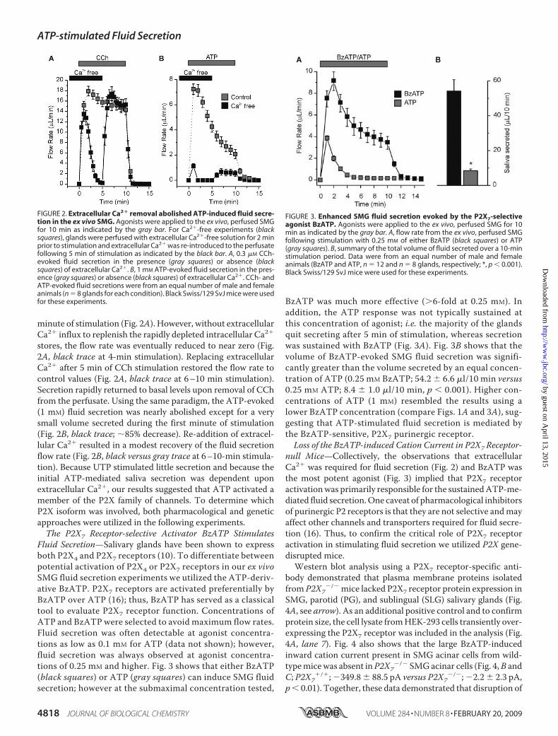
minute of stimulation (Fig. 2A). However, without extracellularCa2� influx to replenish the rapidly depleted intracellular Ca2�
stores, the flow rate was eventually reduced to near zero (Fig.2A, black trace at 4-min stimulation). Replacing extracellularCa2� after 5 min of CCh stimulation restored the flow rate tocontrol values (Fig. 2A, black trace at 6–10 min stimulation).Secretion rapidly returned to basal levels upon removal of CChfrom the perfusate. Using the same paradigm, the ATP-evoked(1 mM) fluid secretion was nearly abolished except for a verysmall volume secreted during the first minute of stimulation(Fig. 2B, black trace; �85% decrease). Re-addition of extracel-lular Ca2� resulted in a modest recovery of the fluid secretionflow rate (Fig. 2B, black versus gray trace at 6–10-min stimula-tion). Because UTP stimulated little secretion and because theinitial ATP-mediated saliva secretion was dependent uponextracellular Ca2�, our results suggested that ATP activated amember of the P2X family of channels. To determine whichP2X isoform was involved, both pharmacological and geneticapproaches were utilized in the following experiments.The P2X7 Receptor-selective Activator BzATP Stimulates
Fluid Secretion—Salivary glands have been shown to expressboth P2X4 and P2X7 receptors (10). To differentiate betweenpotential activation of P2X4 or P2X7 receptors in our ex vivoSMG fluid secretion experiments we utilized the ATP-deriv-ative BzATP. P2X7 receptors are activated preferentially byBzATP over ATP (16); thus, BzATP has served as a classicaltool to evaluate P2X7 receptor function. Concentrations ofATP and BzATP were selected to avoid maximum flow rates.Fluid secretion was often detectable at agonist concentra-tions as low as 0.1 mM for ATP (data not shown); however,fluid secretion was always observed at agonist concentra-tions of 0.25 mM and higher. Fig. 3 shows that either BzATP(black squares) or ATP (gray squares) can induce SMG fluidsecretion; however at the submaximal concentration tested,
BzATP was much more effective (�6-fold at 0.25 mM). Inaddition, the ATP response was not typically sustained atthis concentration of agonist; i.e. the majority of the glandsquit secreting after 5 min of stimulation, whereas secretionwas sustained with BzATP (Fig. 3A). Fig. 3B shows that thevolume of BzATP-evoked SMG fluid secretion was signifi-cantly greater than the volume secreted by an equal concen-tration of ATP (0.25 mM BzATP; 54.2 � 6.6 �l/10 min versus0.25 mM ATP; 8.4 � 1.0 �l/10 min, p 0.001). Higher con-centrations of ATP (1 mM) resembled the results using alower BzATP concentration (compare Figs. 1A and 3A), sug-gesting that ATP-stimulated fluid secretion is mediated bythe BzATP-sensitive, P2X7 purinergic receptor.Loss of the BzATP-induced Cation Current in P2X7 Receptor-
null Mice—Collectively, the observations that extracellularCa2� was required for fluid secretion (Fig. 2) and BzATP wasthe most potent agonist (Fig. 3) implied that P2X7 receptoractivationwas primarily responsible for the sustainedATP-me-diated fluid secretion.One caveat of pharmacological inhibitorsof purinergic P2 receptors is that they are not selective andmayaffect other channels and transporters required for fluid secre-tion (16). Thus, to confirm the critical role of P2X7 receptoractivation in stimulating fluid secretion we utilized P2X gene-disrupted mice.Western blot analysis using a P2X7 receptor-specific anti-
body demonstrated that plasma membrane proteins isolatedfrom P2X7
�/� mice lacked P2X7 receptor protein expression inSMG, parotid (PG), and sublingual (SLG) salivary glands (Fig.4A, see arrow). As an additional positive control and to confirmprotein size, the cell lysate fromHEK-293 cells transiently over-expressing the P2X7 receptor was included in the analysis (Fig.4A, lane 7). Fig. 4 also shows that the large BzATP-inducedinward cation current present in SMG acinar cells from wild-typemicewas absent inP2X7
�/� SMGacinar cells (Fig. 4,B andC; P2X7
�/�;�349.8� 88.5 pA versus P2X7�/�;�2.2� 2.3 pA,
p 0.01). Together, these data demonstrated that disruption of
FIGURE 2. Extracellular Ca2� removal abolished ATP-induced fluid secre-tion in the ex vivo SMG. Agonists were applied to the ex vivo, perfused SMGfor 10 min as indicated by the gray bar. For Ca2�-free experiments (blacksquares), glands were perfused with extracellular Ca2�-free solution for 2 minprior to stimulation and extracellular Ca2� was re-introduced to the perfusatefollowing 5 min of stimulation as indicated by the black bar. A, 0.3 �M CCh-evoked fluid secretion in the presence (gray squares) or absence (blacksquares) of extracellular Ca2�. B, 1 mM ATP-evoked fluid secretion in the pres-ence (gray squares) or absence (black squares) of extracellular Ca2�. CCh- andATP-evoked fluid secretions were from an equal number of male and femaleanimals (n 8 glands for each condition). Black Swiss/129 SvJ mice were usedfor these experiments.
FIGURE 3. Enhanced SMG fluid secretion evoked by the P2X7-selectiveagonist BzATP. Agonists were applied to the ex vivo, perfused SMG for 10min as indicated by the gray bar. A, flow rate from the ex vivo, perfused SMGfollowing stimulation with 0.25 mM of either BzATP (black squares) or ATP(gray squares). B, summary of the total volume of fluid secreted over a 10-minstimulation period. Data were from an equal number of male and femaleanimals (BzATP and ATP, n 12 and n 8 glands, respectively; *, p 0.001).Black Swiss/129 SvJ mice were used for these experiments.
ATP-stimulated Fluid Secretion
4818 JOURNAL OF BIOLOGICAL CHEMISTRY VOLUME 284 • NUMBER 8 • FEBRUARY 20, 2009
by guest on April 13, 2015
http://ww
w.jbc.org/
Dow
nloaded from

the P2X7 gene results in loss of both P2X7 receptor proteinexpression and nucleotide-gated channel activity.P2X7 Receptor DisruptionHas No Effect on CCh-evoked Fluid
Secretion and [Ca2�]i Signals—We next determined if disrup-tion of P2X7 receptors in the SMG had nonspecific effects onthe fluid secretion machinery. To evaluate this we utilized themuscarinic receptor agonistCCh, the response towhich shouldbeunaffected in P2X7-null mice. Fig. 5A shows that the ex vivo sali-vary flow rates in P2X7
�/� (black squares) and P2X7�/� (gray
squares) SMGwere effectively identical in response to CCh stim-ulation. There were only subtle differences in the total volumessecreted in 10min (P2X7
�/�; 135 � 5 �l/10 min versus P2X7�/�;
144� 6�l/10min, p 0.25), saliva ion compositions (Table 1), orosmolalities (P2X7
�/�; 183.9 � 4.5 mOsm versus P2X7�/�;
186.0 � 3.3 mOsm, p 0.74). The lack of effect of P2X7 receptorablation on the CCh-stimulated response demonstrated that ionchannels and transporters responsible for fluid secretion in SMGare not affected when the P2X7 gene is disrupted.Given that salivary gland fluid secretion is dependent on an
elevation of [Ca2�]i, we expected that disruption of P2X7 recep-tors would also have no effect on CCh-evoked Ca2� signals inSMG acinar cells. To confirm this, SMG acini were isolated andloaded with the Ca2�-sensitive dye Fura-2. In support of thefluid secretion data, Fig. 5B shows that the CCh-induced Ca2�
signals in P2X7�/� (black trace) and P2X7
�/� (gray trace) iso-lated SMGacinar cells were essentially identical. Analysis of thedata showed that the average peak value over baseline of theCCh-induced Ca2� signal for P2X7
�/� cells was not signifi-cantly different from that for P2X7
�/� cells (Fig. 5B, P2X7�/�;
0.30 � 0.02 ratio units versus P2X7�/�; 0.33 � 0.03 ratio units,
p 0.41). In addition, the average plateau value over baseline(taken at 1.5 min into the 3-min stimulation) of the CCh-in-duced Ca2� signal for P2X7
�/� cells was also similar to that forP2X7
�/� cells (P2X7�/�; 0.17 � 0.01 ratio units versus
P2X7�/�; 0.21 � 0.02 ratio units, p 0.07). Thus, disruption of
P2X7 receptors had no significant effect on either CCh-medi-ated fluid secretion or Ca2� signaling.
FIGURE 4. SMG isolated from P2X7�/� mice lacked P2X7 protein expres-
sion and BzATP-activated cation currents. A, immunoblot of biotiny-lated membrane protein samples probed with a P2X7 receptor specificantibody. P2X7 receptor protein expression was absent in all three salivarygland tissues from P2X7
�/� animals. HEK-293 cells were transiently trans-fected as previously described (9), and cell lysates from mock-transfectedand P2X7 receptor-expressing cells were included as controls and to con-firm P2X7 protein size (see arrow). B, representative traces of BzATP (0.25mM) induced inward cation currents from SMG acinar cells isolated fromP2X7
�/� (upper panel) and P2X7�/� (lower panel) mice. C, data summary
shows the average inward currents following BzATP stimulation (P2X7�/�
versus P2X7�/�, n 9 and n 8, respectively; *, p 0.003). Data were from
experiments from at least three animals. P2X7�/� or P2X7
�/� C57Bl/6Jmice were used for these experiments.
FIGURE 5. CCh evoked SMG fluid secretion and [Ca2�]i signals in P2X7�/�
and P2X7�/� mice. A, ex vivo SMG flow rate (left panel), total saliva volume
over a 10-min stimulation period (middle panel), and the osmolality (rightpanel) in response to CCh (0.3 �M). There was no significant differencebetween P2X7
�/� and P2X7�/� animals for the total volume secreted
(P2X7�/� versus P2X7
�/�, n 20 glands and n 17 glands, respectively, p 0.23) or osmolality (P2X7
�/� versus P2X7�/�, n 12 and n 8 respectively, p
0.74). B, Fura-2 loaded SMG acinar cells were stimulated with CCh (0.3 �M) for3 min. The representative [Ca2�]i response is shown as a change in ratio units(left panel). The data summary (right panel) shows that there was no signifi-cant difference in either the average peak value over baseline [P2X7
�/�;0.30 � 0.02 ratio units versus P2X7
�/�; 0.33 � 0.03 ratio units, n 9 (33 cells)and n 9 (34 cells), respectively, p 0.41] or average plateau value overbaseline (taken at 1.5 min into the 3-min stimulation) [P2X7
�/�; 0.17 � 0.01ratio units versus P2X7
�/�; 0.21 � 0.02 ratio units, n 9 (33 cells) and n 9 (34cells), respectively, p 0.07]. P2X7
�/� and P2X7�/� C57Bl/6J mice were used
for these experiments.
TABLE 1Carbachol and ATP stimulated fluid secretion and ion composition inthe ex vivo SMG of wild type and P2X7
�/� miceANOVA analyses, followed by a Bonferroni test, were performed for multiple com-parisons (see “Experimental Procedures”). The numbers in parentheses equal thenumber of glands tested for each condition.
P2X7�/� P2X7
�/�
CCh CCh � ATP CCh CCh � ATPNa 37.4 � 2.6 (20) 44.1 � 3.3 (21) 39.9 � 4.6 (17) 47.9 � 5.8 (10)K 50.4 � 3.3 (20) 59.6 � 2.2a (21) 47.7 � 2.8b (17) 53.8 � 3.9 (10)Cl 89.2 � 4.6 (20) 85.2 � 1.7 (21) 82.9 � 4.3a (17) 100.9 � 3.3b (10)pH 7.85 � 0.07 (8) 7.99 � 0.09a (8) 7.71 � 0.03b (9) 7.71 � 0.06b (10)�l 135 � 5a (20) 90 � 7b (21) 144 � 6a (17) 130 � 6a (10)
a Values are different from those labeled b by at least p 0.03.
ATP-stimulated Fluid Secretion
FEBRUARY 20, 2009 • VOLUME 284 • NUMBER 8 JOURNAL OF BIOLOGICAL CHEMISTRY 4819
by guest on April 13, 2015
http://ww
w.jbc.org/
Dow
nloaded from
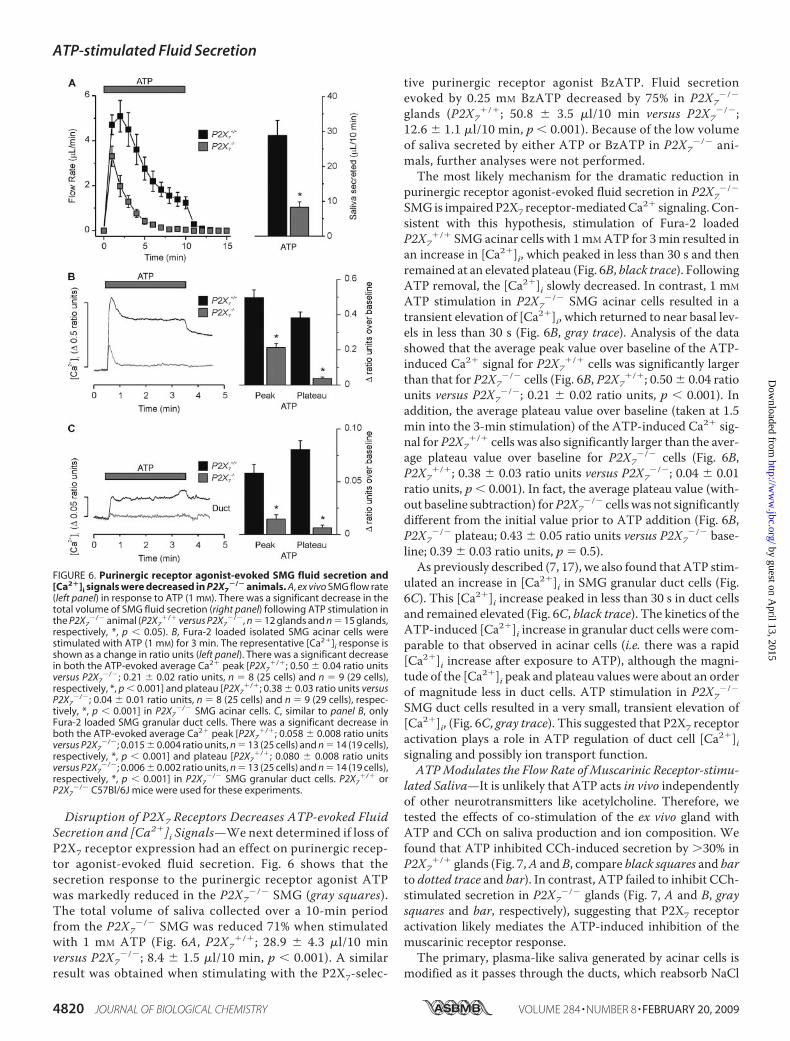
Disruption of P2X7 Receptors Decreases ATP-evoked FluidSecretion and [Ca2�]i Signals—We next determined if loss ofP2X7 receptor expression had an effect on purinergic recep-tor agonist-evoked fluid secretion. Fig. 6 shows that thesecretion response to the purinergic receptor agonist ATPwas markedly reduced in the P2X7
�/� SMG (gray squares).The total volume of saliva collected over a 10-min periodfrom the P2X7
�/� SMG was reduced 71% when stimulatedwith 1 mM ATP (Fig. 6A, P2X7
�/�; 28.9 � 4.3 �l/10 minversus P2X7
�/�; 8.4 � 1.5 �l/10 min, p 0.001). A similarresult was obtained when stimulating with the P2X7-selec-
tive purinergic receptor agonist BzATP. Fluid secretionevoked by 0.25 mM BzATP decreased by 75% in P2X7
�/�
glands (P2X7�/�; 50.8 � 3.5 �l/10 min versus P2X7
�/�;12.6 � 1.1 �l/10 min, p 0.001). Because of the low volumeof saliva secreted by either ATP or BzATP in P2X7
�/� ani-mals, further analyses were not performed.The most likely mechanism for the dramatic reduction in
purinergic receptor agonist-evoked fluid secretion in P2X7�/�
SMG is impairedP2X7 receptor-mediatedCa2� signaling. Con-sistent with this hypothesis, stimulation of Fura-2 loadedP2X7
�/� SMG acinar cells with 1mMATP for 3min resulted inan increase in [Ca2�]i, which peaked in less than 30 s and thenremained at an elevated plateau (Fig. 6B, black trace). FollowingATP removal, the [Ca2�]i slowly decreased. In contrast, 1 mMATP stimulation in P2X7
�/� SMG acinar cells resulted in atransient elevation of [Ca2�]i, which returned to near basal lev-els in less than 30 s (Fig. 6B, gray trace). Analysis of the datashowed that the average peak value over baseline of the ATP-induced Ca2� signal for P2X7
�/� cells was significantly largerthan that for P2X7
�/� cells (Fig. 6B, P2X7�/�; 0.50 � 0.04 ratio
units versus P2X7�/�; 0.21 � 0.02 ratio units, p 0.001). In
addition, the average plateau value over baseline (taken at 1.5min into the 3-min stimulation) of the ATP-induced Ca2� sig-nal for P2X7
�/� cells was also significantly larger than the aver-age plateau value over baseline for P2X7
�/� cells (Fig. 6B,P2X7
�/�; 0.38 � 0.03 ratio units versus P2X7�/�; 0.04 � 0.01
ratio units, p 0.001). In fact, the average plateau value (with-out baseline subtraction) forP2X7
�/� cells was not significantlydifferent from the initial value prior to ATP addition (Fig. 6B,P2X7
�/� plateau; 0.43 � 0.05 ratio units versus P2X7�/� base-
line; 0.39 � 0.03 ratio units, p 0.5).As previously described (7, 17), we also found that ATP stim-
ulated an increase in [Ca2�]i in SMG granular duct cells (Fig.6C). This [Ca2�]i increase peaked in less than 30 s in duct cellsand remained elevated (Fig. 6C, black trace). The kinetics of theATP-induced [Ca2�]i increase in granular duct cells were com-parable to that observed in acinar cells (i.e. there was a rapid[Ca2�]i increase after exposure to ATP), although the magni-tude of the [Ca2�]i peak and plateau values were about an orderof magnitude less in duct cells. ATP stimulation in P2X7
�/�
SMG duct cells resulted in a very small, transient elevation of[Ca2�]i, (Fig. 6C, gray trace). This suggested that P2X7 receptoractivation plays a role in ATP regulation of duct cell [Ca2�]isignaling and possibly ion transport function.ATPModulates the Flow Rate of Muscarinic Receptor-stimu-
lated Saliva—It is unlikely that ATP acts in vivo independentlyof other neurotransmitters like acetylcholine. Therefore, wetested the effects of co-stimulation of the ex vivo gland withATP and CCh on saliva production and ion composition. Wefound that ATP inhibited CCh-induced secretion by �30% inP2X7
�/� glands (Fig. 7,A andB, compare black squares and barto dotted trace and bar). In contrast, ATP failed to inhibit CCh-stimulated secretion in P2X7
�/� glands (Fig. 7, A and B, graysquares and bar, respectively), suggesting that P2X7 receptoractivation likely mediates the ATP-induced inhibition of themuscarinic receptor response.The primary, plasma-like saliva generated by acinar cells is
modified as it passes through the ducts, which reabsorb NaCl
FIGURE 6. Purinergic receptor agonist-evoked SMG fluid secretion and[Ca2�]i signals were decreased in P2X7
�/� animals. A, ex vivo SMG flow rate(left panel) in response to ATP (1 mM). There was a significant decrease in thetotal volume of SMG fluid secretion (right panel) following ATP stimulation inthe P2X7
�/� animal (P2X7�/� versus P2X7
�/�, n 12 glands and n 15 glands,respectively, *, p 0.05). B, Fura-2 loaded isolated SMG acinar cells werestimulated with ATP (1 mM) for 3 min. The representative [Ca2�]i response isshown as a change in ratio units (left panel). There was a significant decreasein both the ATP-evoked average Ca2� peak [P2X7
�/�; 0.50 � 0.04 ratio unitsversus P2X7
�/�; 0.21 � 0.02 ratio units, n 8 (25 cells) and n 9 (29 cells),respectively, *, p 0.001] and plateau [P2X7
�/�; 0.38 � 0.03 ratio units versusP2X7
�/�; 0.04 � 0.01 ratio units, n 8 (25 cells) and n 9 (29 cells), respec-tively, *, p 0.001] in P2X7
�/� SMG acinar cells. C, similar to panel B, onlyFura-2 loaded SMG granular duct cells. There was a significant decrease inboth the ATP-evoked average Ca2� peak [P2X7
�/�; 0.058 � 0.008 ratio unitsversus P2X7
�/�; 0.015 � 0.004 ratio units, n 13 (25 cells) and n 14 (19 cells),respectively, *, p 0.001] and plateau [P2X7
�/�; 0.080 � 0.008 ratio unitsversus P2X7
�/�; 0.006 � 0.002 ratio units, n 13 (25 cells) and n 14 (19 cells),respectively, *, p 0.001] in P2X7
�/� SMG granular duct cells. P2X7�/� or
P2X7�/� C57Bl/6J mice were used for these experiments.
ATP-stimulated Fluid Secretion
4820 JOURNAL OF BIOLOGICAL CHEMISTRY VOLUME 284 • NUMBER 8 • FEBRUARY 20, 2009
by guest on April 13, 2015
http://ww
w.jbc.org/
Dow
nloaded from

and secrete KHCO3 (1, 2). ATP stimulated a very modestincrease in [Ca2�]i in SMGgranular duct cells (Fig. 6C) suggest-ing thatATPhas little influence onduct cell function. Indeed, inP2X7
�/� mice, ATP did not significantly alter the Na� or Cl�concentration of the saliva induced by CCh stimulation (Table1); while the pH and the K� concentration of saliva increasedslightly. Consistent with these observations, ATP induced onlymodest changes in the ion composition of saliva from P2X7
�/�
submandibular glands. Taken together, these results suggestedthat the ATP-stimulated increase in [Ca2�]i in SMG duct cellsis unlikely to play a major role in the regulation of ductalfunction.
DISCUSSION
Salivary tissues express multiple types of purinergic P2receptors. Activation of these receptors increases [Ca2�]i sug-gesting a possible role for P2 receptors in fluid secretion (4, 7, 8,10, 18, 19), although this has never been directly tested.Hereweshow that purinergic stimulation results in a sustained, extra-cellular Ca2�-dependent secretion of saliva in an intact mousesubmandibular salivary gland preparation. The sensitivity ofthis response to different purinergic agonists was consistentwith P2X7 receptor activation (BzATP �� ATP � ADP ��UTP). Indeed, a �70% decrease was observed in BzATP- andATP-evoked fluid secretions from the SMG in P2X7-null ani-mals. These results demonstrate that P2X7 receptors are essen-tial for ATP-mediated saliva production in the mouse subman-dibular gland.Our results highlight the significant contribution puriner-
gic receptors may have on the regulation of salivary gland
fluid secretion. Earlier reports describe a non-adrenergic,non-cholinergic mediated fluid secretion (3, 20–24). Insheep, electrical stimulation of the parotid parasympatheticnerve in the presence of atropine produced a significantincrease in the flow rate (25). The authors concluded that theflow rate was produced by vasoactive intestinal peptide (VIP)release (25), however this seems unlikely since VIP receptoractivation is linked to an increase in cAMP, not [Ca2�]i.Indeed, Ekstrom et al. (26) reported that VIP treatmentcaused little to no fluid secretion in feline parotid glands.Interestingly, the electrically evoked “atropine-resistant”fluid secretion was �30% of the response prior to muscarinicreceptor inhibition in the ferret submandibular gland (3).This observation is essentially identical to our results; i.e. themaximal volume of ATP-induced fluid secretion was about30% of that produced by muscarinic receptor stimulation(Fig. 1 versus Fig. 5). Thus, our results are consistent withATP-mediated fluid secretion playing a physiological role innon-adrenergic, non-cholinergic mediated salivation.Although both P2X4 and P2X7 receptors are expressed in
salivary glands, the greater secretion induced by BzATP (Fig. 3)and the severe impairment of secretion in P2X7-null mice (Fig.6) implied that the P2X7 receptor channel contributes most tosalivation. Adding to P2X receptor signaling complexity, func-tional P2X receptors form heteromeric channels comprised ofthree P2X subunits (16, 27, 28). Until recently P2X7 receptorswere the only P2X isoform thought not to form a heteromericchannel. However, recent reports have identified P2X4/P2X7heteromeric channels (29, 30). These latter observations sug-gest that salivary gland cells might contain both P2X4 and P2X7homotrimers, as well as P2X4/P2X7 heterotrimers. The func-tional significance of P2X4/P2X7 heterotrimer formation isunclear, but genetic disruption of P2X7 receptors might alsointerfere with P2X4 targeting and/or overall function. Never-theless, while a P2X4/P2X7 heterotrimer is possible, there is noevidence of P2X4/P2X7 heterotrimer formation in native tissue.Future studies should be directed to determine the relationship,if any, between P2X4 and P2X7 homo- and heterotrimers insalivary gland function.P2X7 receptors have been previously localized to the apical
membrane of mouse parotid acinar and duct cells (8). P2X7receptor immunostaining was also noted in mouse SMGduct cells, which suggests that this ligand-gated channel mayparticipate in modification of the electrolyte content (31). Liet al. (8) proposed that preassembled P2X7 receptors in ductcells sense upstream secretion by acinar cells (via ATPrelease) and respond by augmenting HCO3
� secretion. Inagreement with this model, we found that ATP produced amodest increase in the pH of CCh-induced secretions, sug-gesting an increase in the [HCO3
�], and this effect disap-peared in P2X7-null mice (Table 1). Given that ATP is notlikely to be released independent of other agonists in vivo,ATP might be expected to modulate the response of salivaryglands to muscarinic receptor stimulation. Indeed, we alsofound that the volume of fluid secreted during muscarinicreceptor activation was decreased by co-stimulation withATP and CCh in the ex vivo SMG. These results are consist-ent with the previous observation that ATP inhibits musca-
FIGURE 7. Purinergic receptor agonist induced changes in muscarinicreceptor-activated SMG fluid secretion. A, flow rate from the ex vivo, per-fused SMG from P2X7
�/� (black squares) or P2X7�/� (gray squares) mice during
stimulation with 0.3 �M CCh � 1 mM ATP. For comparison the dotted linerepresents data from P2X7
�/� mice using 0.3 �M CCh alone from Fig. 5A.B, summary of the total volume of fluid secreted over a 10-min stimulationperiod. There was no difference in the total volume of SMG fluid secretionbetween P2X7
�/� mice stimulated with CCh only (dotted bar) and P2X7�/�
animals stimulated with CCh � ATP (gray bar) [P2X7�/� with CCh only; 135 �
5 �l/10 min, versus P2X7�/� with CCh � ATP; 130 � 6 �l/10 min, n 20 and
n 10 glands, respectively], but both were significantly greater than the totalvolume of SMG fluid secretion in P2X7
�/� mice stimulated with CCh � ATP(black bar) [P2X7
�/� with CCh � ATP; 90 � 7 �l/10 min, n 21, *, p 0.01].P2X7
�/� and P2X7�/� C57Bl/6J mice were used for these experiments.
ATP-stimulated Fluid Secretion
FEBRUARY 20, 2009 • VOLUME 284 • NUMBER 8 JOURNAL OF BIOLOGICAL CHEMISTRY 4821
by guest on April 13, 2015
http://ww
w.jbc.org/
Dow
nloaded from

rinic receptor induced Ca2� mobilization in rat parotid andsubmandibular gland acinar cells (32–34). The inhibition ofthe CCh-induced response by ATP was absent in P2X7-nullmice (Fig. 7). Additional studies will be required to confirmthe mechanism(s) responsible for these observations.Regardless of the mechanism, our results suggest that ATPcan stimulate secretion on its own, and when co-releasedwith a muscarinic agonist, ATP likely modulates the musca-rinic receptor-stimulated response of salivary glands. More-over, the in vivo physiologically relevant source of ATPrelease is unknown. It is well known that ATP is co-releasedfrom nerve terminals with acetylcholine and other neuro-transmitters (35). Alternatively, ATP is also released fromthe secretory granules of exocrine acinar cells in response toagonists that stimulate granule fusion (36). The release ofATP from these two distinct sites is likely to have very dif-ferent functional consequences.In summary, our results demonstrate that ATP regulates
fluid secretion in the mouse submandibular gland. This mech-anism is extracellularCa2�-dependent, sensitive toBzATP, andinsensitive to UTP, suggesting activation of a P2X receptorfamily member. Indeed, ablation of the P2X7 gene resulted in adramatic reduction in the amount of fluid secreted during ATPexposure. These new findings contribute to our understandingof the role of purinergic P2 receptors in salivary glands andprovide insight into future studies. ATP-mediated fluid secre-tion is likely an importantmechanism for “tuning” the resultantfluid secretion under different physiological conditions.
Acknowledgments—We thank Laurie Koek, Jennifer Scantlin, YasnaJaramillo, and Mark Wagner for excellent technical assistance.
REFERENCES1. Cook, D. I., Van Lennep, E. W., Roberts, M. L., and Young, J. A. (1994)
Secretion by the Major Salivary Glands, 3rd Ed., Raven Press, New York2. Melvin, J. E., Yule, D., Shuttleworth, T., and Begenisich, T. (2005) Annu.
Rev. Physiol. 67, 445–4693. Ekstrom, J., Mansson, B., Olgart, L., and Tobin, G. (1988) Quart. J. Exp.
Physiol. 73, 163–1734. Soltoff, S. P.,McMillian,M.K., Cragoe, E. J., Jr., Cantley, L. C., andTalamo,
B. R. (1990) J. Gen. Physiol. 95, 319–3465. Merritt, J. E., and Rink, T. J. (1987) J. Biol. Chem. 262, 14912–149166. Burnstock, G. (2007) Physiol. Rev. 87, 659–7977. Lee, M. G., Zeng, W., and Muallem, S. (1997) J. Biol. Chem. 272,
32951–329558. Li, Q., Luo, X., Zeng, W., and Muallem, S. (2003) J. Biol. Chem. 278,
47554–475619. Brown, D. A., Bruce, J. I., Straub, S. V., and Yule, D. I. (2004) J. Biol. Chem.
279, 39485–3949410. Turner, J. T., Landon, L. A., Gibbons, S. J., and Talamo, B. R. (1999) Crit.
Rev. Oral. Biol. Med. 10, 210–22411. Novak, I. (2003) News Physiol. Sci. 18, 12–1712. Nakamoto, T., Romanenko, V. G., Takahashi, A., Begenisich, T., and
Melvin, J. E. (2008) Am. J. Physiol. Cell Physiol. 294, C810–C81913. Romanenko, V. G., Nakamoto, T., Srivastava, A., Begenisich, T., and
Melvin, J. E. (2007) J. Physiol. 581, 801–81714. Romanenko, V., Nakamoto, T., Srivastava, A., Melvin, J. E., and Bege-
nisich, T. (2006) J. Biol. Chem. 281, 27964–2797215. Gonzalez-Begne, M., Nakamoto, T., Nguyen, H. V., Stewart, A. K., Alper,
S. L., and Melvin, J. E. (2007) J. Biol. Chem. 282, 35125–3513216. North, R. A. (2002) Physiol. Rev. 82, 1013–106717. Pochet, S., Garcia-Marcos, M., Seil, M., Otto, A., Marino, A., and Dehaye,
J. P. (2007) Cell Signal. 19, 2155–216418. Gallacher, D. V. (1982) Nature 296, 83–8619. Soltoff, S. P.,McMillian,M.K., Lechleiter, J. D., Cantley, L. C., andTalamo,
B. R. (1990) Ann. N. Y. Acad. Sci. 603, 76–90; discussion 91–7220. Asztely, A., Tobin, G., and Ekstrom, J. (1994) Acta Physiol. Scand. 151,
373–37621. Ekstrom, J. (1998) Exp. Physiol. 83, 697–70022. Ekstrom, J. (2001) Exp. Physiol. 86, 475–48023. Ekstrom, J., Asztely, A., and Tobin, G. (1998) Eur. J. Morphol. 36, (suppl.),
208–21224. Ekstrom, J., Helander, H. F., and Tobin, G. (1993) J. Physiol. 472,
233–24425. Reid, A. M., and Titchen, D. A. (1988) Q J. Exp. Physiol. 73, 413–42426. Ekstrom, J., Asztely, A., Helander, H. F., andTobin, G. (1994)Acta Physiol.
Scand. 150, 83–8827. Nicke, A., Baumert, H. G., Rettinger, J., Eichele, A., Lambrecht, G., Mut-
schler, E., and Schmalzing, G. (1998) EMBO J. 17, 3016–302828. Torres, G. E., Egan, T. M., and Voigt, M. M. (1999) J. Biol. Chem. 274,
6653–665929. Dubyak, G. R. (2007)Mol. Pharmacol. 72, 1402–140530. Guo, C., Masin, M., Qureshi, O. S., and Murrell-Lagnado, R. D. (2007)
Mol. Pharmacol. 72, 1447–145631. Sim, J. A., Young, M. T., Sung, H. Y., North, R. A., and Surprenant, A.
(2004) J. Neurosci. 24, 6307–631432. Fukushi, Y. (1999) Eur. J. Pharmacol. 364, 55–6433. Jorgensen, T. D., Gromada, J., Tritsaris, K., Nauntofte, B., and Dissing, S.
(1995) Biochem. J. 312, 457–46434. Metioui, M., Amsallem, H., Alzola, E., Chaib, N., Elyamani, A., Moran, A.,
Marino, A., and Dehaye, J. P. (1996) J. Cell Physiol. 168, 462–47535. Richardson, P. J., and Brown, S. J. (1987) J. Neurochem. 48, 622–63036. Sorensen, C. E., and Novak, I. (2001) J. Biol. Chem. 276, 32925–32932
ATP-stimulated Fluid Secretion
4822 JOURNAL OF BIOLOGICAL CHEMISTRY VOLUME 284 • NUMBER 8 • FEBRUARY 20, 2009
by guest on April 13, 2015
http://ww
w.jbc.org/
Dow
nloaded from

G. Romanenko and James E. MelvinA. Catalán, Mireya Gonzalez-Begne, Victor Tetsuji Nakamoto, David A. Brown, Marcelo Mouse Submandibular GlandATP-induced Saliva Secretion by the
Receptors Mediate7Purinergic P2Xand Biogenesis:Membrane Transport, Structure, Function,
doi: 10.1074/jbc.M808597200 originally published online December 19, 20082009, 284:4815-4822.J. Biol. Chem.
10.1074/jbc.M808597200Access the most updated version of this article at doi:
.JBC Affinity SitesFind articles, minireviews, Reflections and Classics on similar topics on the
Alerts:
When a correction for this article is posted•
When this article is cited•
to choose from all of JBC's e-mail alertsClick here
http://www.jbc.org/content/284/8/4815.full.html#ref-list-1
This article cites 35 references, 19 of which can be accessed free at
by guest on April 13, 2015
http://ww
w.jbc.org/
Dow
nloaded from
Related Documents