A Potential Role for P2X 7 R in Allergic Airway Inflammation in Mice and Humans Tobias Mu ¨ller 1 *, Rodolfo Paula Vieira 1 *, Melanie Grimm 1 , Thorsten Du ¨rk 1 , Sanja Cicko 1 , Robert Zeiser 2 , Thilo Jakob 3 , Stefan F. Martin 3 , Britta Blumenthal 1 , Stephan Sorichter 1 , Davide Ferrari 4 , Francesco Di Virgillio 4 , and Marco Idzko 1 1 Department of Pneumology, 2 Department of Oncology, and 3 Allergy Research Group, University Medical Center Freiburg, Freiburg, Germany; and 4 Department of General Pathology, University of Ferrara, Italy P2X 7 R deficiency is associated with a less severe outcome in acute and chronic inflammatory disorders. Recently, we demonstrated that extracellular adenosine triphosphate is involved in the patho- genesis of asthma by modulating the function of dendritic cells (DCs). However, the role of the purinergic receptor subtype P2X 7 is unknown. To elucidate the role of P2X 7 R in allergic airway inflam- mation (AAI) in vitro and in vivo, P2X 7 R expression was measured in lung tissue and immune cells of mice or in humans with allergic asthma. By using a specific P2X 7 R-antagonist and P2X 7 R-deficient animals, the role of this receptor in acute and chronic experimental asthma was explored. P2X 7 R was found to be up-regulated during acute and chronic asthmatic airway inflammation in mice and humans. In vivo experiments revealed the functional relevance of this finding because selective P2X 7 R inhibition or P2X 7 R deficiency was associated with reduced features of acute and chronic asthma in the ovalbumin-alum or HDM model of AAI. Experiments with bone marrow chimeras emphasized that P2X 7 R expression on hemato- poietic cells is responsible for the proasthmatic effects of P2X 7 R signaling. In the DC-driven model of AAI, P2X 7 R-deficient DCs showed a reduced capacity to induce Th2 immunity in vivo. Up- regulation of P2X 7 R on BAL macrophages and blood eosinophils could be observed in patients with chronic asthma. Our data suggest that targeting P2X 7 R on hematopoietic cells (e.g., DCs or eosino- phils) might be a new therapeutic option for the treatment of asthma. Keywords: asthma; P2X 7 ; ATP; dendritic cells; eosinophils Asthma is a chronic inflammatory airway disease clinically characterized by variable airway obstruction, excessive mucus production, and hyperresponsiveness to nonspecific stimuli. The nonresolved chronic inflammation is orchestrated by eosinophils, mast cells, Th2 lymphocytes, and dendritic cells (DCs), resulting in airway remodelling (1). Compelling recent evidence points to an important role for myeloid DCs in initiating and maintaining allergic airway inflammation (2–6). Adenosine triphosphate (ATP) has been implicated as an important mediator and ‘‘danger signal’’ in acute and chronic inflammation and can be released in large amounts from various cell types after cellular stress or tissue injury (4, 7–10). Extracel- lular ATP and other nucleotides (e.g., ADP, UTP, and UDP) exert their effects via binding to purinergic P2-receptors, which can be subdivided into two families: (1) the G-protein–coupled P2YR (P2Y 1–14 ) and (2) the ligand-gated ion channels P2XR (P2X 1–7 ). Among the P2XR subtypes, P2X 7 R is particularly highly expressed in immune cells (11–13), and its behavior is unique in that, upon repetitive or prolonged exposure to high concentrations of ATP, it can form a large membrane pore permeable to hydrophilic solutes of molecular mass up to 900 kD (14). Activation of the P2X 7 R on immune cells, including den- dritic cells, macrophages, and neutrophils, has been involved in various proinflammatory cell responses, such as the release of reactive oxygen species, matrix metalloproteinase-9, and cyto- kines (IL-2, IL-6, and IL-18) and via linking to the NALP3- inflammasom pathway in production and release of mature IL-1b (14–16), which has been shown to play an important role in asthma and other chronic diseases (17). Several studies dem- onstrated that the absence or blocking of P2X 7 Rs are associated with less severe outcomes in chronic inflammatory diseases and neuropathic pain (9, 13, 18–21). Recent evidence suggests that uric acid, another danger signal also activating the NALP3- inflammasome pathway, plays a role in the induction and maintenance of allergic diseases (22, 23). Recently we demonstrated that BALF ATP levels are in- creased after allergen challenge in humans and mice and that neutralizing intrapulmonary ATP levels or the application of unselective purinergic receptor antagonists can abrogate all cardinal features of experimental asthma in mice (4). However, studies exploring the contribution of P2X 7 Rs to the pathophys- iology of allergic airway inflammation in vivo are lacking. The aim of the current study was to investigate the role of P2X 7 R signaling in the pathogenesis of allergic airway inflamma- tion: P2X 7 receptor expression in lung tissue and immune cells of mice with acute and chronic allergic airway inflammation and in patients with asthma was measured. In addition, we explored the functional relevance of P2X 7 R signaling for allergic airway inflammation in vivo. Finally, the importance of P2X 7 R expres- sion on the capacity of DCs to induce Th2 priming in vivo was elucidated. Our data provide evidence that selective targeting of the P2X 7 R on hematopoietic cells might be a new therapeutic option for the treatment of asthma. MATERIALS AND METHODS Mice Balb/c mice, ovalbumin–T-cell receptor (OVA-TCR) transgenic mice (DO11.10) on a Balb/c background and C57/Bl6 mice (6–8 wk old) CLINICAL RELEVANCE Bronchial asthma is a major health burden in western society. Our data showing the involvement of P2X 7 R signaling in asthma pathophysiology could be the basis for a new therapeutic approach. (Received in original form March 29, 2010 and in final form April 22, 2010) This work was supported by an Emmy-Noether grant (M.I.) from the German Research Foundation (DFG, ID 7/4–1). * These authors contributed equally to this work. Correspondence and requests for reprints should be addressed to Dr. Marco Idzko, M.D., University Medical Center Freiburg, Department of Pneumology, Killianstrasse 5, 79106 Freiburg, Germany. E-mail: marco.idzko@uniklinink- freiburg.de This article has an online supplement, which is accessible from this issue’s table of contents at www.atsjournals.org Am J Respir Cell Mol Biol Vol 44. pp 456–464, 2011 Originally Published in Press as DOI: 10.1165/rcmb.2010-0129OC on May 27, 2010 Internet address: www.atsjournals.org

Welcome message from author
This document is posted to help you gain knowledge. Please leave a comment to let me know what you think about it! Share it to your friends and learn new things together.
Transcript

A Potential Role for P2X7R in Allergic AirwayInflammation in Mice and Humans
Tobias Muller1*, Rodolfo Paula Vieira1*, Melanie Grimm1, Thorsten Durk1, Sanja Cicko1, Robert Zeiser2,Thilo Jakob3, Stefan F. Martin3, Britta Blumenthal1, Stephan Sorichter1, Davide Ferrari4,Francesco Di Virgillio4, and Marco Idzko1
1Department of Pneumology, 2Department of Oncology, and 3Allergy Research Group, University Medical Center Freiburg, Freiburg, Germany;
and 4Department of General Pathology, University of Ferrara, Italy
P2X7R deficiency is associated with a less severe outcome in acuteand chronic inflammatory disorders. Recently, we demonstratedthat extracellular adenosine triphosphate is involved in the patho-genesis of asthma by modulating the function of dendritic cells(DCs). However, the role of the purinergic receptor subtype P2X7 isunknown. To elucidate the role of P2X7R in allergic airway inflam-mation (AAI) in vitro and in vivo, P2X7R expression was measured inlung tissue and immune cells of mice or in humans with allergicasthma. By using a specific P2X7R-antagonist and P2X7R-deficientanimals, the role of this receptor in acute and chronic experimentalasthma was explored. P2X7R was found to be up-regulated duringacute and chronic asthmatic airway inflammation in mice andhumans. In vivo experiments revealed the functional relevance ofthis finding because selective P2X7R inhibition or P2X7R deficiencywas associated with reduced features of acute and chronic asthma inthe ovalbumin-alum or HDM model of AAI. Experiments with bonemarrow chimeras emphasized that P2X7R expression on hemato-poietic cells is responsible for the proasthmatic effects of P2X7Rsignaling. In the DC-driven model of AAI, P2X7R-deficient DCsshowed a reduced capacity to induce Th2 immunity in vivo. Up-regulation of P2X7R on BAL macrophages and blood eosinophilscould be observed in patients with chronic asthma. Our data suggestthat targeting P2X7R on hematopoietic cells (e.g., DCs or eosino-phils) might be a new therapeutic option for the treatment ofasthma.
Keywords: asthma; P2X7; ATP; dendritic cells; eosinophils
Asthma is a chronic inflammatory airway disease clinicallycharacterized by variable airway obstruction, excessive mucusproduction, and hyperresponsiveness to nonspecific stimuli. Thenonresolved chronic inflammation is orchestrated by eosinophils,mast cells, Th2 lymphocytes, and dendritic cells (DCs), resultingin airway remodelling (1). Compelling recent evidence points toan important role for myeloid DCs in initiating and maintainingallergic airway inflammation (2–6).
Adenosine triphosphate (ATP) has been implicated as animportant mediator and ‘‘danger signal’’ in acute and chronicinflammation and can be released in large amounts from variouscell types after cellular stress or tissue injury (4, 7–10). Extracel-lular ATP and other nucleotides (e.g., ADP, UTP, and UDP)exert their effects via binding to purinergic P2-receptors, which
can be subdivided into two families: (1) the G-protein–coupledP2YR (P2Y1–14) and (2) the ligand-gated ion channels P2XR(P2X1–7). Among the P2XR subtypes, P2X7R is particularly highlyexpressed in immune cells (11–13), and its behavior is unique inthat, upon repetitive or prolonged exposure to high concentrationsof ATP, it can form a large membrane pore permeable tohydrophilic solutes of molecular mass up to 900 kD (14).
Activation of the P2X7R on immune cells, including den-dritic cells, macrophages, and neutrophils, has been involved invarious proinflammatory cell responses, such as the release ofreactive oxygen species, matrix metalloproteinase-9, and cyto-kines (IL-2, IL-6, and IL-18) and via linking to the NALP3-inflammasom pathway in production and release of matureIL-1b (14–16), which has been shown to play an important rolein asthma and other chronic diseases (17). Several studies dem-onstrated that the absence or blocking of P2X7Rs are associatedwith less severe outcomes in chronic inflammatory diseases andneuropathic pain (9, 13, 18–21). Recent evidence suggests thaturic acid, another danger signal also activating the NALP3-inflammasome pathway, plays a role in the induction andmaintenance of allergic diseases (22, 23).
Recently we demonstrated that BALF ATP levels are in-creased after allergen challenge in humans and mice and thatneutralizing intrapulmonary ATP levels or the application ofunselective purinergic receptor antagonists can abrogate allcardinal features of experimental asthma in mice (4). However,studies exploring the contribution of P2X7Rs to the pathophys-iology of allergic airway inflammation in vivo are lacking.
The aim of the current study was to investigate the role ofP2X7R signaling in the pathogenesis of allergic airway inflamma-tion: P2X7 receptor expression in lung tissue and immune cells ofmice with acute and chronic allergic airway inflammation and inpatients with asthma was measured. In addition, we explored thefunctional relevance of P2X7R signaling for allergic airwayinflammation in vivo. Finally, the importance of P2X7R expres-sion on the capacity of DCs to induce Th2 priming in vivo waselucidated. Our data provide evidence that selective targeting ofthe P2X7R on hematopoietic cells might be a new therapeuticoption for the treatment of asthma.
MATERIALS AND METHODS
Mice
Balb/c mice, ovalbumin–T-cell receptor (OVA-TCR) transgenic mice(DO11.10) on a Balb/c background and C57/Bl6 mice (6–8 wk old)
CLINICAL RELEVANCE
Bronchial asthma is a major health burden in westernsociety. Our data showing the involvement of P2X7Rsignaling in asthma pathophysiology could be the basisfor a new therapeutic approach.
(Received in original form March 29, 2010 and in final form April 22, 2010)
This work was supported by an Emmy-Noether grant (M.I.) from the German
Research Foundation (DFG, ID 7/4–1).
* These authors contributed equally to this work.
Correspondence and requests for reprints should be addressed to Dr. Marco
Idzko, M.D., University Medical Center Freiburg, Department of Pneumology,
Killianstrasse 5, 79106 Freiburg, Germany. E-mail: marco.idzko@uniklinink-
freiburg.de
This article has an online supplement, which is accessible from this issue’s table of
contents at www.atsjournals.org
Am J Respir Cell Mol Biol Vol 44. pp 456–464, 2011
Originally Published in Press as DOI: 10.1165/rcmb.2010-0129OC on May 27, 2010
Internet address: www.atsjournals.org
were bred at the animal facilities at the University Hospital Freiburg.P2X7R-deficient mice (P2X7R2/2) on C57Bl/6 background werekindly provided by GlaxoSmithKline (London, UK) and backcrossedwith our C57/Bl6 strains (24). All experiments were performedaccording to institutional guidelines of the animal ethics committeefrom the German government.
OVA-Alum Model of Allergic Airway Inflammation
For the acute model, different mice strains were sensitized to OVA(Worthington Biochemical, Lakewood, NJ) via intraperitoneal injec-tion of OVA/alum on Days 0 and 7 and were challenged with OVAaerosols on Days 19 to 21 delivered from a jet nebulizer delivering 1%OVA in PBS for 30 minutes. Twenty-four hours after the last OVAexposure, bronchoalveolar lavage (BAL) was performed with 3 3 1 mlof Ca21-free and Mg21-free PBS (Invitrogen, Carlsbad, CA) supple-mented with 0.1 mM sodium EDTA, followed by lung resection andstorage in OCT freezing medium.
In the chronic model, female BALB/c mice were sensitized asdescribed above and challenged with OVA aerosols 3 d/wk over 6weeks. During the last 2 weeks, animals were treated intratracheallywith vehicle or KN62 before allergen challenge.
House Dust Mite–Induced Allergic Airway Inflammation
Female BALB/c mice (6–9 wk old) were anesthetized using ketamine/xylazine and injected intratracheally with 100 mg Dermatophagoidespteronyssinus extracts (Greer Laboratories, Lenoir, NC) or vehicle(PBS) as a negative control on Day 0. On Days 7 and 14, animalsreceived an intratracheal injection of house dust mite (HDM) (100 mg)or HDM admixed with KN62. Animals were assessed for the classicalfeatures of allergic airway inflammation (BAL eosinophilia, histology)on Day 17, as previously described (25).
Th2 Sensitization Induced by Intratracheal Injection of Bone
Marrow–Derived DCs
DCs were prepared as previously described (3) (details are provided inthe online supplement). Cells were pulsed overnight with 100 mg/mlLPS-low OVA (Worthington Biochemicals, Lakewood, NJ) or vehicle.After antigen pulsing, nonadherent DCs were collected and washed toremove free OVA. For in vivo experiments, C57Bl/6 mice wereanesthetized on Day 0 with ketamin/xylazine, and 1 3 106 vehicle-wildtype (wt)-DCs, OVA-wt-DCs, vehicle-P2X7R2/2 DCs, or OVA-P2X7R2/2 DCs were instilled through the vocal cords as described(3). On Days 10 through 12, mice were exposed to OVA aerosols (30min). Mice were killed 24 hours after the last aerosol.
BAL Fluid Collection from Patients with Asthma and Healthy
Control Subjects
BAL fluid (BALF) of 10 patients with mild allergic asthma and 10healthy control subjects was collected at the University of Freiburg (seeTable E2 in the online supplement for patient characteristics). Patientswith malignancies or signs or symptoms of acute infections wereexcluded. In the control group, patients taking cardiac or pulmonarymedications were excluded. Patients with asthma were diagnosedaccording to the current guidelines, and inhaled corticosteroids werewithheld for 4 weeks before bronchoscopy. The study was approved bythe local ethics committee of Freiburg, and all participants gave theirwritten informed consent. BALF was collected and processed asdescribed (26).
Blood Donors for Eosinophils
Eight patients with a diagnosis of allergic asthma were recruited on thebasis of airway hyperresponsiveness, positive allergen skin-prick tests,or elevated total or specific IgE concentrations. Eight subjects withoutany history of allergic signs or symptoms, a negative skin prick test toa large panel of allergens, and normal IgE levels served as controlsubjects (Table E2). Participants gave their written informed consent.The study was approved by the local ethics committee. Inhaledcorticosteroids were withdrawn at least 14 days before blood wastaken. None of the patients took systemic corticosteroids.
Statistical Analysis
If not stated otherwise for most experiments, the statistical significanceof differences between samples was calculated using ANOVA, fol-lowed by Bonferroni comparison test. Differences were consideredsignificant at P , 0.05.
RESULTS
Up-Regulation of P2X7R Expression in Acute and Chronic
Allergic Airway Inflammation
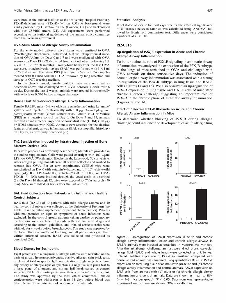
To better define the role of P2X7R signaling in asthmatic airwayinflammation, we analyzed the expression of the P2X7R subtypein the lungs of mice sensitized to OVA and challenged withOVA aerosols on three consecutive days. The induction ofacute allergic airway inflammation was associated with a strongup-regulation of the P2X7R subtype in lung tissue and BALFcells (Figures 1a and 1b). We also observed an up-regulation ofP2X7R expression in lung tissue and BALF cells of mice afterchronic allergen challenge, suggesting an important role ofP2X7R in the chronic phase of asthmatic airway inflammation(Figures 1c and 1d).
Effect of Selective P2X7R Blockade on Acute and Chronic
Allergic Airway Inflammation in Mice
To determine whether blocking of P2X7R during allergenchallenge could influence the development of acute allergic lung
Figure 1. Up-regulation of P2X7R expression in acute and chronic
allergic airway inflammation. Acute and chronic allergic airways in
BALB/c animals were induced as described in MATERIALS AND METHODS.After the last allergen challenge, animals were killed, bronchoalveolar
lavage fluid (BALF) and whole lungs were collected, and RNA was
isolated. Relative expression of P2X7R in sensitized compared with
nonsensitized animals was analyzed using quantitative RT-PCR: P2X7Rexpression on total lung tissue of animals with (b) acute and (d ) chronic
allergic airway inflammation and control animals. P2X7R expression on
BALF cells from animals with (a) acute or (c) chronic allergic airway
inflammation and control animals. Data are shown as mean 6 SEM(n 5 5–8 mice per group). *P , 0.05. Data from one representative
experiment out of three are shown. OVA 5 ovalbumin.
Muller, Vieira, Grimm, et al.: P2X7R and Asthma 457
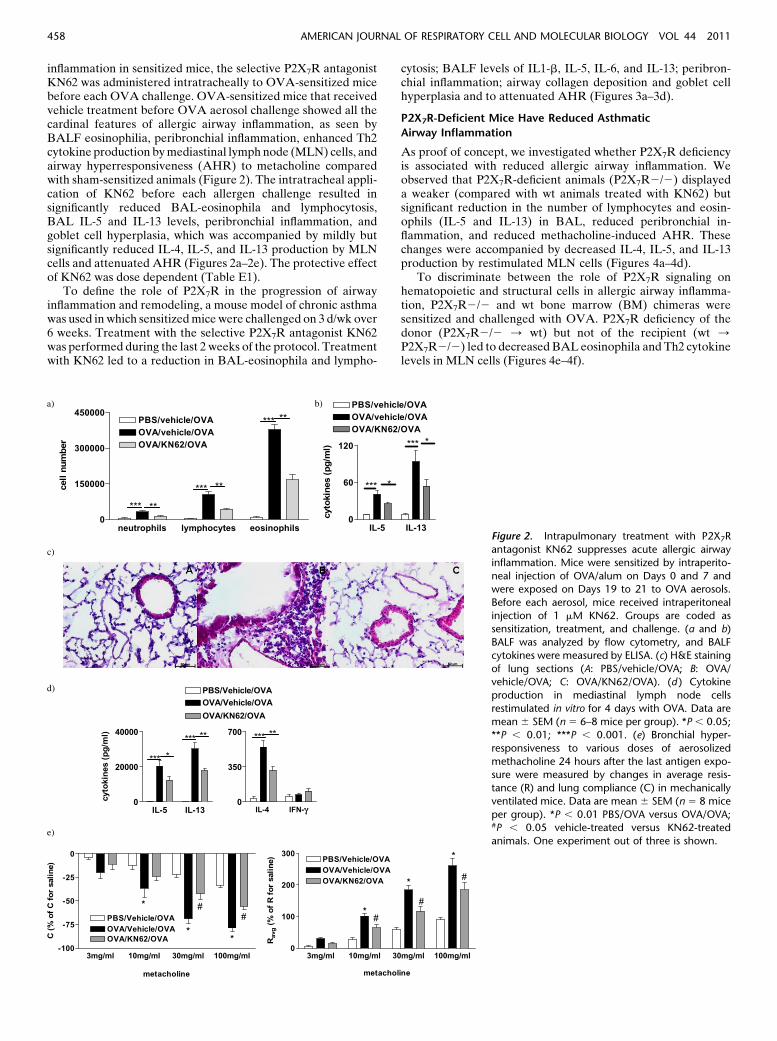
inflammation in sensitized mice, the selective P2X7R antagonistKN62 was administered intratracheally to OVA-sensitized micebefore each OVA challenge. OVA-sensitized mice that receivedvehicle treatment before OVA aerosol challenge showed all thecardinal features of allergic airway inflammation, as seen byBALF eosinophilia, peribronchial inflammation, enhanced Th2cytokine production by mediastinal lymph node (MLN) cells, andairway hyperresponsiveness (AHR) to metacholine comparedwith sham-sensitized animals (Figure 2). The intratracheal appli-cation of KN62 before each allergen challenge resulted insignificantly reduced BAL-eosinophila and lymphocytosis,BAL IL-5 and IL-13 levels, peribronchial inflammation, andgoblet cell hyperplasia, which was accompanied by mildly butsignificantly reduced IL-4, IL-5, and IL-13 production by MLNcells and attenuated AHR (Figures 2a–2e). The protective effectof KN62 was dose dependent (Table E1).
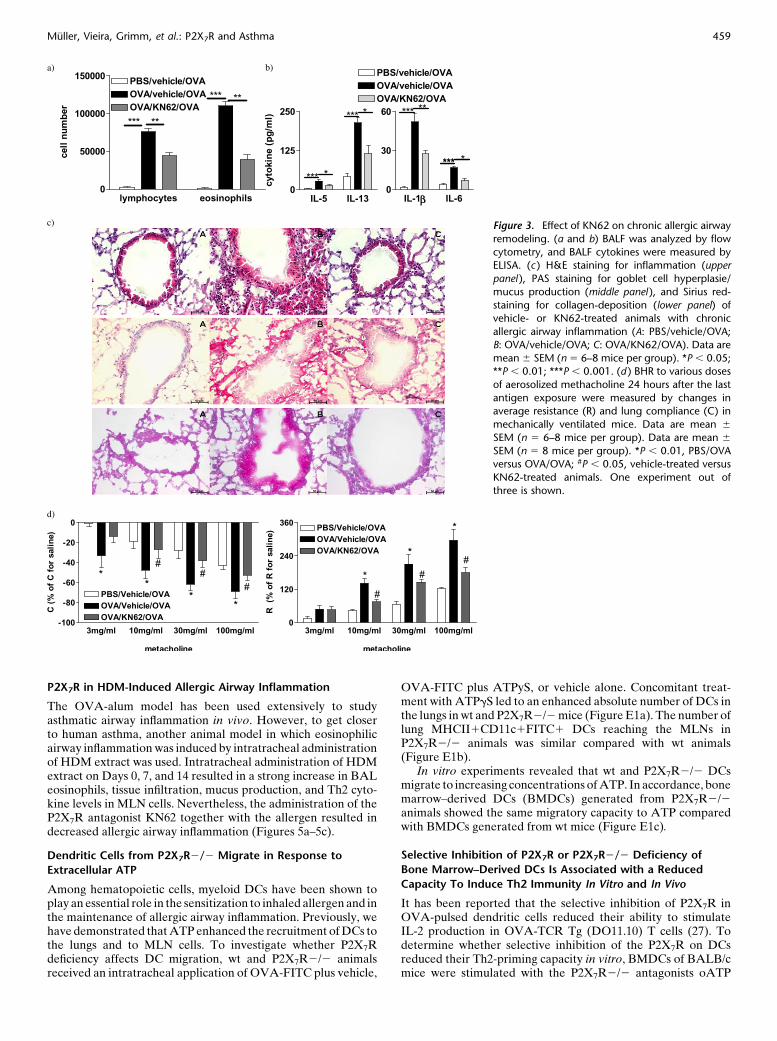
To define the role of P2X7R in the progression of airwayinflammation and remodeling, a mouse model of chronic asthmawas used in which sensitized mice were challenged on 3 d/wk over6 weeks. Treatment with the selective P2X7R antagonist KN62was performed during the last 2 weeks of the protocol. Treatmentwith KN62 led to a reduction in BAL-eosinophila and lympho-
cytosis; BALF levels of IL1-b, IL-5, IL-6, and IL-13; peribron-chial inflammation; airway collagen deposition and goblet cellhyperplasia and to attenuated AHR (Figures 3a–3d).
P2X7R-Deficient Mice Have Reduced Asthmatic
Airway Inflammation
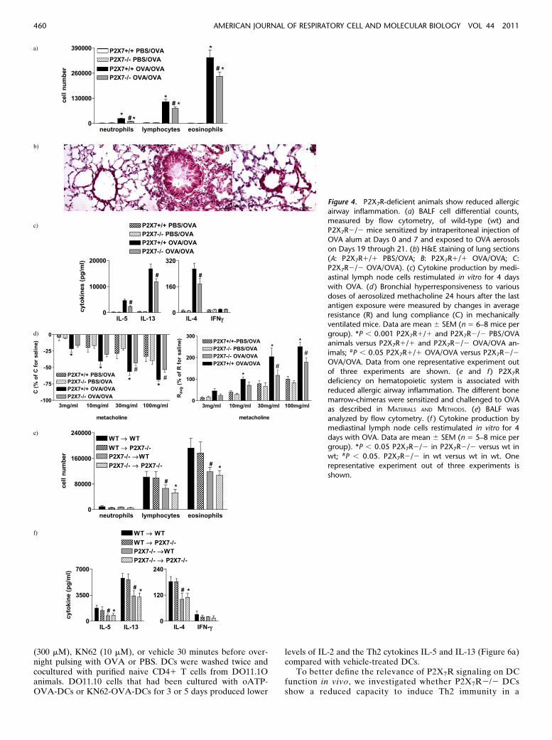
As proof of concept, we investigated whether P2X7R deficiencyis associated with reduced allergic airway inflammation. Weobserved that P2X7R-deficient animals (P2X7R2/2) displayeda weaker (compared with wt animals treated with KN62) butsignificant reduction in the number of lymphocytes and eosin-ophils (IL-5 and IL-13) in BAL, reduced peribronchial in-flammation, and reduced methacholine-induced AHR. Thesechanges were accompanied by decreased IL-4, IL-5, and IL-13production by restimulated MLN cells (Figures 4a–4d).
To discriminate between the role of P2X7R signaling onhematopoietic and structural cells in allergic airway inflamma-tion, P2X7R2/2 and wt bone marrow (BM) chimeras weresensitized and challenged with OVA. P2X7R deficiency of thedonor (P2X7R2/2 / wt) but not of the recipient (wt /P2X7R2/2) led to decreased BAL eosinophila and Th2 cytokinelevels in MLN cells (Figures 4e–4f).
Figure 2. Intrapulmonary treatment with P2X7R
antagonist KN62 suppresses acute allergic airway
inflammation. Mice were sensitized by intraperito-
neal injection of OVA/alum on Days 0 and 7 andwere exposed on Days 19 to 21 to OVA aerosols.
Before each aerosol, mice received intraperitoneal
injection of 1 mM KN62. Groups are coded assensitization, treatment, and challenge. (a and b)
BALF was analyzed by flow cytometry, and BALF
cytokines were measured by ELISA. (c) H&E staining
of lung sections (A: PBS/vehicle/OVA; B: OVA/vehicle/OVA; C: OVA/KN62/OVA). (d ) Cytokine
production in mediastinal lymph node cells
restimulated in vitro for 4 days with OVA. Data are
mean 6 SEM (n 5 6–8 mice per group). *P , 0.05;**P , 0.01; ***P , 0.001. (e) Bronchial hyper-
responsiveness to various doses of aerosolized
methacholine 24 hours after the last antigen expo-sure were measured by changes in average resis-
tance (R) and lung compliance (C) in mechanically
ventilated mice. Data are mean 6 SEM (n 5 8 mice
per group). *P , 0.01 PBS/OVA versus OVA/OVA;#P , 0.05 vehicle-treated versus KN62-treated
animals. One experiment out of three is shown.
458 AMERICAN JOURNAL OF RESPIRATORY CELL AND MOLECULAR BIOLOGY VOL 44 2011
P2X7R in HDM-Induced Allergic Airway Inflammation
The OVA-alum model has been used extensively to studyasthmatic airway inflammation in vivo. However, to get closerto human asthma, another animal model in which eosinophilicairway inflammation was induced by intratracheal administrationof HDM extract was used. Intratracheal administration of HDMextract on Days 0, 7, and 14 resulted in a strong increase in BALeosinophils, tissue infiltration, mucus production, and Th2 cyto-kine levels in MLN cells. Nevertheless, the administration of theP2X7R antagonist KN62 together with the allergen resulted indecreased allergic airway inflammation (Figures 5a–5c).
Dendritic Cells from P2X7R2/2 Migrate in Response to
Extracellular ATP
Among hematopoietic cells, myeloid DCs have been shown toplay an essential role in the sensitization to inhaled allergen and inthe maintenance of allergic airway inflammation. Previously, wehave demonstrated that ATP enhanced the recruitment of DCs tothe lungs and to MLN cells. To investigate whether P2X7Rdeficiency affects DC migration, wt and P2X7R2/2 animalsreceived an intratracheal application of OVA-FITC plus vehicle,
OVA-FITC plus ATPyS, or vehicle alone. Concomitant treat-ment with ATPgS led to an enhanced absolute number of DCs inthe lungs in wt and P2X7R2/2 mice (Figure E1a). The number oflung MHCII1CD11c1FITC1 DCs reaching the MLNs inP2X7R2/2 animals was similar compared with wt animals(Figure E1b).
In vitro experiments revealed that wt and P2X7R2/2 DCsmigrate to increasing concentrations of ATP. In accordance, bonemarrow–derived DCs (BMDCs) generated from P2X7R2/2animals showed the same migratory capacity to ATP comparedwith BMDCs generated from wt mice (Figure E1c).
Selective Inhibition of P2X7R or P2X7R2/2 Deficiency of
Bone Marrow–Derived DCs Is Associated with a Reduced
Capacity To Induce Th2 Immunity In Vitro and In Vivo
It has been reported that the selective inhibition of P2X7R inOVA-pulsed dendritic cells reduced their ability to stimulateIL-2 production in OVA-TCR Tg (DO11.10) T cells (27). Todetermine whether selective inhibition of the P2X7R on DCsreduced their Th2-priming capacity in vitro, BMDCs of BALB/cmice were stimulated with the P2X7R2/2 antagonists oATP
Figure 3. Effect of KN62 on chronic allergic airway
remodeling. (a and b) BALF was analyzed by flow
cytometry, and BALF cytokines were measured byELISA. (c ) H&E staining for inflammation (upper
panel ), PAS staining for goblet cell hyperplasie/
mucus production (middle panel ), and Sirius red-
staining for collagen-deposition (lower panel) ofvehicle- or KN62-treated animals with chronic
allergic airway inflammation (A: PBS/vehicle/OVA;
B: OVA/vehicle/OVA; C: OVA/KN62/OVA). Data are
mean 6 SEM (n 5 6–8 mice per group). *P , 0.05;**P , 0.01; ***P , 0.001. (d ) BHR to various doses
of aerosolized methacholine 24 hours after the last
antigen exposure were measured by changes inaverage resistance (R) and lung compliance (C) in
mechanically ventilated mice. Data are mean 6
SEM (n 5 6–8 mice per group). Data are mean 6
SEM (n 5 8 mice per group). *P , 0.01, PBS/OVAversus OVA/OVA; #P , 0.05, vehicle-treated versus
KN62-treated animals. One experiment out of
three is shown.
Muller, Vieira, Grimm, et al.: P2X7R and Asthma 459
(300 mM), KN62 (10 mM), or vehicle 30 minutes before over-night pulsing with OVA or PBS. DCs were washed twice andcocultured with purified naive CD41 T cells from DO11.1Oanimals. DO11.10 cells that had been cultured with oATP-OVA-DCs or KN62-OVA-DCs for 3 or 5 days produced lower
levels of IL-2 and the Th2 cytokines IL-5 and IL-13 (Figure 6a)compared with vehicle-treated DCs.
To better define the relevance of P2X7R signaling on DCfunction in vivo, we investigated whether P2X7R2/2 DCsshow a reduced capacity to induce Th2 immunity in a
Figure 4. P2X7R-deficient animals show reduced allergic
airway inflammation. (a) BALF cell differential counts,
measured by flow cytometry, of wild-type (wt) and
P2X7R2/2 mice sensitized by intraperitoneal injection ofOVA alum at Days 0 and 7 and exposed to OVA aerosols
on Days 19 through 21. (b) H&E staining of lung sections
(A: P2X7R1/1 PBS/OVA; B: P2X7R1/1 OVA/OVA; C:P2X7R2/2 OVA/OVA). (c) Cytokine production by medi-
astinal lymph node cells restimulated in vitro for 4 days
with OVA. (d ) Bronchial hyperresponsiveness to various
doses of aerosolized methacholine 24 hours after the lastantigen exposure were measured by changes in average
resistance (R) and lung compliance (C) in mechanically
ventilated mice. Data are mean 6 SEM (n 5 6–8 mice per
group). *P , 0.001 P2X7R1/1 and P2X7R2/2 PBS/OVAanimals versus P2X7R1/1 and P2X7R2/2 OVA/OVA an-
imals; #P , 0.05 P2X7R1/1 OVA/OVA versus P2X7R2/2
OVA/OVA. Data from one representative experiment out
of three experiments are shown. (e and f ) P2X7Rdeficiency on hematopoietic system is associated with
reduced allergic airway inflammation. The different bone
marrow-chimeras were sensitized and challenged to OVAas described in MATERIALS AND METHODS. (e) BALF was
analyzed by flow cytometry. (f ) Cytokine production by
mediastinal lymph node cells restimulated in vitro for 4
days with OVA. Data are mean 6 SEM (n 5 5–8 mice pergroup). *P , 0.05 P2X7R2/2 in P2X7R2/2 versus wt in
wt; #P , 0.05. P2X7R2/2 in wt versus wt in wt. One
representative experiment out of three experiments is
shown.
460 AMERICAN JOURNAL OF RESPIRATORY CELL AND MOLECULAR BIOLOGY VOL 44 2011
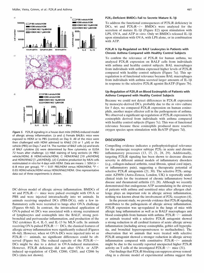
DC-driven model of allergic airway inflammation. BMDCs ofwt and P2X7R2/2 mice were pulsed overnight with OVA orPBS and were injected intratracheally into wt animals. Inanimals receiving unpulsed DCs (PBS-DCs), only a few in-flammatory cells were recruited to lungs after OVA challenge(Figures 6b–6d). In contrast, the intratracheal application ofOVA-pulsed wt DCs was associated with a strong recruitmentof lymphocytes and eosinophils into the BALF, strong peri-bronchial and perivascular inflammation, and production of theTh2 cytokines IL-4, IL-5, and IL-13 by MLN cells. In animalsreceiving OVA-pulsed P2X7R2/2 DCs, the classical features ofallergic airway inflammation were significantly reduced (Figures6b–6d). However, when wt OVA-DCs were injected into wt orP2X7R2/2 animals, no significant differences could be ob-served (Figure 6e). The reduced capacity of the P2X7R2/2DCs might be due to a defect in OVA-induced maturation.However, P2X7R deficiency did not alter OVA- or ATP-induced up-regulation of CD40, CD80, CD83, and CD86 byDCs (data not shown).
P2X7-Deficient BMDCs Fail to Secrete Mature IL-1b
To address the functional consequences of P2X7R deficiency inDCs, wt and P2X7R2/2 BMDCs were analyzed for thesecretion of mature IL-1b (Figure E2) upon stimulation withLPS, OVA, and ATP in vitro. Only wt BMDCs released IL-1b
upon stimulation with OVA, with LPS alone, or in combinationwith ATP.
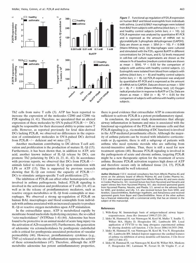
P2X7R Is Up-Regulated on BALF Leukocytes in Patients with
Chronic Asthma Compared with Healthy Control Subjects
To confirm the relevance of P2X7R for human asthma, weanalyzed P2X7R expression on BALF cells from individualswith asthma and healthy control subjects. BAL macrophagesfrom individuals with asthma expressed higher levels of P2X7Rcompared with healthy control subjects (Figure 7a). This up-regulation is of functional relevance because BAL macrophagesfrom individuals with asthma secreted larger amounts of IL-1b
in response to the selective P2X7R agonist BzATP (Figure 7b).
Up-Regulation of P2X7R on Blood Eosinophils of Patients with
Asthma Compared with Healthy Control Subjects
Because we could not detect differences in P2X7R expressionby monocyte-derived DCs, probably due to the in vitro culturefor 5 days, we compared P2X7R expression on human eosino-phils, another major effector cell in the pathogenesis of asthma.We observed a significant up-regulation of P2X7R expression byeosinophils derived from individuals with asthma comparedwith healthy control subjects (Figure 7c). This was of functionalrelevance because these eosinophils produced more reactiveoxygen species upon stimulation with BzATP (Figure 7d).
DISCUSSION
Compelling evidence indicates a pathophysiological relevancefor the purinergic receptor subtype P2X7 in acute and chronicinflammatory processes (13, 15, 21, 28, 29). Additionally,targeting P2X7R signaling has been shown to decrease diseaseseverity in different animal models of inflammatory disorders(e.g., collagen-induced arthritis, renal fibrosis, spinal cord injury,or inflammatory pain), suggesting a therapeutic potential ofselective P2X7R antagonists (21, 30). The selective P2X7 antag-onist AZ9056 (Astra-Zeneca, London, UK) is reportedly underclinical trials for the treatment of chronic inflammatory boweldisease and rheumatoid arthritis (13, 28). Although we recentlydemonstrated that endogenous ATP accumulating in the airwaysof patients with asthma and sensitized mice after allergen chal-lenge plays an important role in allergic airway inflammation,nothing was known about the involvement of P2X7 receptors (4).
In the present study, we provide evidence that P2X7R signalingcontributes to the pathogenesis of allergic airway inflammation.P2X7R expression was up-regulated in the lungs of mice withallergic lung inflammation as well as in BALF macrophages andblood eosinophils from humans with asthma. P2X7R2/2 animalsor animals treated with a selective P2X7R antagonist showeda strong reduction in all cardinal features of acute allergic airwayinflammation (including airway eosinophilia, goblet cell hyperpla-sia, and bronchial hyperresponsiveness to methacholine). Theobservation that wt animals that were treated with selectiveP2X7R antagonist showed a stronger reduction in allergic airwayinflammation compared with constitutive P2X7R2/2 animalsmight be due to the recently reported unexpected higher P2X7Ractivity on T cells of the investigated P2X7R2/2 mice (31).
Our findings that P2X7R blocking can suppress airway remod-eling in a chronic model of experimental asthma suggest that
Figure 5. P2X7R signaling in a house dust mite (HDM)-induced modelof allergic airway inflammation. (a and c) Female BALB/c mice were
exposed to HDM or to PBS (control) on Day 0. All of the mice were
then challenged with HDM admixed to KN62 (10 or 1 mM) or the
vehicle (PBS) on Days 7 and 14. The number of BALF cells (a) and levelsof BALF cytokine (b) were determined by flow cytometry or ELISA
72 hours after challenge. (c) H&E staining of lung sections (A: PBS/
vehicle/HDM; B: HDM/vehicle/HDM; C: HDM/KN62 [10 mM/HDM]
and HDM/KN62 [1 mM/HDM]). (d ) Cytokine production by MLN cellsrestimulated in vitro for 4 days with HDM. Data are means 6 SEM (n 5
6–8 mice per group). *P , 0.01 PBS/HDM versus HDM/HDM; #P ,
0.05 HDM/vehicle/HDM versus HDM/KN62/HDM. One representative
data out of three experiments is shown.
Muller, Vieira, Grimm, et al.: P2X7R and Asthma 461

targeting P2X7R be a new option for the treatment of chronicairway inflammation. This hypothesis is further supported byour data showing that, compared with healthy control subjects,BALF macrophages from individuals with asthma producehigher levels of IL-1b, a cytokine involved in asthma pathogen-esis (32–34), upon stimulation with the P2X7R agonist BzATP.We have recently shown that P2X7R expression is functionallyup-regulated in neutrophils and macrophages of patients withCOPD and that selective inhibition of P2X7R strongly suppressessmoke-induced lung inflammation and the development ofemphysema (10 and Lucattelli and colleagues, unpublished).
Because P2X7R expression was up-regulated in lung tissuefrom mice with allergic airway inflammation, P2X7R signal-
ing on structural cells might also be involved in the patho-genesis of allergic airway diseases. However, studies with bonemarrow chimeras revealed that P2X7R deficiency on hematopoietic(P2X7R2/2 / wt) but not on structural (wt / P2X7R2/2) cellswas associated with reduced allergic lung inflammation. In linewith these findings, the importance of P2X7R expression onhematopoietic cells has also been shown in smoke-induced lunginflammation (Lucattelli and colleagues, unpublished).
Increasing evidence points to an essential role of DCs in thepathogenesis of allergic airway inflammation (6). Several stud-ies have demonstrated the functional expression of P2-receptorsubtypes, including P2X7R, on human and murine DCs (4, 12,27, 35–38), and we recently reported that some of the proasth-matic effects of ATP in vivo are mediated via activation ofpurinergic receptors on myeloid DCs (2). Recruitment ofmyeloid DCs from the lung to the draining lymph nodes is anintegral part of the primary and secondary immune responses toinhaled allergens (5, 39). Thus, we addressed whether functionalP2X7R expression is necessary for the ATP-induced recruit-ment of blood DCs to the lungs and antigen-loaded DCs fromthe lung to the draining lymph nodes in vivo. The migratorycapacity of DCs in response to ATP was not affected by P2X7Rdeficiency, in accordance with previous studies showing that theATP-induced migration of various cells types is linked to thepurinergic receptor subtype P2Y2 (7, 8, 10, 40).
Priming of naive T cells into Th2 effector cells by myeloid DCsis a crucial step in asthma pathogenesis (5, 6), and ATP has beenreported to enhance the Th2 priming capacity of DCs (4, 41). Inline with a previous report by Mutini and colleagues (27),selective blocking of P2X7R or P2X7R deficiency of OVA-pulsedDCs was associated with a reduced capacity to prime OVA-specific naive T cells, suggesting an involvement of P2X7Rsignaling on DC in T-cell priming in allergic airway inflammation.To further support this hypothesis, we used a model in whichallergic airway inflammation is induced by adoptive transfer ofOVA-pulsed DCs to the lungs of naive animals followed by OVAchallenge. Although wt animals receiving OVA-pulsed P2X7R2/2DCs showed attenuated allergic airway inflammation comparedwith animals receiving OVA-pulsed wt DCs, there was nodifference in P2X7R2/2 animals receiving OVA-pulsed wt DCs.
It is well established that stimulation of T cells by DCs viathe CD80/CD86 complex is essential for the differentiation of
Figure 6. Involvement of functional P2X7R in T-cell priming capacity ofdendritic cells (DCs) in vitro and in vivo. (a) OVA-DCs were incubated
with different P2X7R antagonists (KN62 1 mM or oATP 300 mM) or
vehicle before coculture with DO.11.10 T cells for 3 and 5 days in vitro.
The levels of IL-2, IL-5, and IL-13 were measured in the supernants.Data are means 6 SEM. *P , 0.01 vehicle-OVA DC versus P2X7R
antagonist-OVA-DCs. (b–d ) On Day 0, mice received intracheal in-
jections of WT or P2X7R2/2 OVA-DCs or unpulsed WT or P2X7R2/2
DCs. From Days 10 to 13, all mice were exposed to OVA aerosols. (b)BALF was analyzed by flow cytometry. (c) H&E staining of lung sections
(A: P2X7R1/1 PBS DCs/OVA; B: P2X7R2/2 PBS/OVA; C: P2X7R1/1
DCs OVA/OVA; D: P2X7R2/2 DCs OVA/OVA). (d ) Cytokine productionby MLN cells restimulated in vitro for 4 days with OVA. Data are mean 6
SEM (n 5 6–8 mice per group). *P , 0.01 PBS-DCs versus OVA-DCs;#P , 0.01 WT-OVA-DCs versus P2X7R2/2 OVA-DCs. Representative
data from three experiments are shown. (e) OVA-DCs from WT orP2X7R2/2 animals were injected intratracheally in WT or P2X7R2/2
animals. The cells differentiation in the BALF was analyzed by flow
cytometry. Data are mean 6 SEM (n 5 6 mice per group). *P , 0.01
P2X7R2/2 OVA-DCs in wt versus WT-OVA-DCs in wt; #P , 0.01P2X7R2/2 OVA DCs in P2X7R2/2 versus WT-OVA-DCs in P2X7R2/2.
One representative experiment out of three experiments is shown.
b
462 AMERICAN JOURNAL OF RESPIRATORY CELL AND MOLECULAR BIOLOGY VOL 44 2011
Th2 cells from naive T cells (3). ATP has been reported toincrease the expression of the molecules CD80 and CD86 viaP2R signaling (4, 41). Therefore, we speculated that an alteredexpression of these molecules by OVA-pulsed P2X7R2/2 DCsmight be responsible for their decreased ability to prime naive Tcells. However, as reported previously for fetal skin–derivedDCs lacking P2X7R, we observed no differences in the expres-sion of costimulatory molecules in OVA-pulsed DCs derivedfrom P2X7R2/2 deficient and wt mice (27).
Another mechanism contributing to DC-driven T-cell acti-vation and proliferation is the production of mature IL-1b (15).Furthermore, it has been shown that, in addition to ATP, uricacid, another known inducer of IL-1b release by DCs, canpromote Th2 polarizing by DCs (4, 23, 41, 42). In accordancewith previous reports, we observed that DCs from P2X7R2/2animals failed to release mature IL-1b upon stimulation withLPS or ATP (15). This is supported by previous researchshowing that IL-1b can restore the capacity of P2X7R2/2DCs to stimulate antigen-specific T-cell proliferation (27).
The inhibition of P2X7R can affect other hematopoietic cellsinvolved in asthma pathogenesis. Indeed, P2X7R signaling isinvolved in the activation and proliferation of T cells (18, 43) aswell as in the release of proinflammatory mediators, such asreactive oxygen metabolites or IL-1b from eosinophils or mac-rophages. We observed a strong up-regulation of P2X7R onhuman BAL macrophages and blood eosinophils from individ-uals with asthma associated with an increased capacity to produceIL-1b or reactive oxygen metabolites upon ATP stimulation.
In the extracellular space, ATP is metabolized rapidly bymembrane-bound nucleotide-hydrolyzing enzymes, the so-called‘‘ecto-nucleotidases’’ (NTPDase 1–8) (44). Adenosine has beenfound to be protective in an animal model of acute lung injury viathe activation of A2b receptors (45). Furthermore, the generationof adenosine via ectonucleotidases by posthypoxic endothelialcells is critical for posthypoxia-associated protection of vascularpermeability (46). However, proinflammatory cytokines (e.g.,TNF-a) released at the site of inflammation suppress the activityof these ectonucleotidases (47). Therefore, although the ATPmetabolite adenosine has potent antiinflammatory properties,
there is good evidence that extracellular ATP in concentrationssufficient to activate P2X7R is a potent proinflammatory signal.
In conclusion, the present study demonstrates that allergicairway inflammation in humans and mice is associated with thefunctional up-regulation of the P2X7R on immune cells and thatP2X7R signaling (e.g., via modulating of DC function) is involvedin the ATP-mediated proasthmatic effects. Although the major-ity of asthma patients can be treated with inhalative corticoste-roids or b2-agonists, there is still a subgroup of patients withasthma who need systemic steroids who are suffering fromsteroid-insensitive asthma. Thus, there is still a need for newtreatment options (48). Because P2X7R signaling is involved inthe pathogenesis of acute and chronic asthma, targeting P2X7Rmight be a new therapeutic option for the treatment of severeasthma. Because P2X7R activation requires high doses of ATPand therefore occurs only in inflamed tissue (14, 15), P2X7Rantagonists should be well tolerated.
Author Disclosure: F.D.V. received consultancy fees from Affectis Pharma AG andserves on the advisory board of Affectis Pharma AG and Cordex Pharma Inc.F.D.V. also received a sponsored grant from Affectis Pharma AG and owns stocksin Cordex Pharma Inc. M.I. received lecture fees from Nycomed and Novartis andexpert witness fees from Nycomed Pharma. M.I. also received sponsored grantsfrom Nycomed Pharma, Neurim, and Phedia. S.S. served on the advisory boardfor MSD, and Actelion and Lilly. S.S. also received lecture fees from MSD, andActelion, GlaxoSmithKline, Boehringer Ingelheim, and AstraZeneca and receivedsponsored grants from Bayer and GlaxoSmithKline. None of the other authorshas a financial relationship with a commercial entity that has an interest in thesubject of this manuscript.
References
1. Wills-Karp M. Immunologic basis of antigen-induced airway hyper-
responsiveness. Annu Rev Immunol 1999;17:255–281.2. Idzko M, Hammad H, van Nimwegen M, Kool M, Muller T, Soullie T,
Willart MA, Hijdra D, Hoogsteden HC, Lambrecht BN. Localapplication of FTY720 to the lung abrogates experimental asthmaby altering dendritic cell function. J Clin Invest 2006;116:2935–2944.
3. Idzko M, Hammad H, van Nimwegen M, Kool M, Vos N, Hoogsteden
HC, Lambrecht BN. Inhaled iloprost suppresses the cardinal featuresof asthma via inhibition of airway dendritic cell function. J Clin Invest2007;117:464–472.
4. Idzko M, Hammad H, van Nimwegen M, Kool M, Willart MA, Muskens
F, Hoogsteden HC, Luttmann W, Ferrari D, Di Virgilio F, et al.
Figure 7. Functional up-regulation of P2X7R expression
on human BALF and blood eosinophils from individuals
with asthma. (a and b) BALF macrophages were isolatedfrom BALF from subjects with asthma (black bars; n 5 10)
and healthy control subjects (white bars; n 5 10). (a)
P2X7R expression was analyzed by quantitative RT-PCR
and is expressed as the amount of mRNA net toglyceraldehyde 3-phosphate dehydrogenase (GAPDH).
Data are shown as mean 6 SEM (n 5 10). P , 0.008
(Mann-Whitney test). (b) Macrophages were cultured
and stimulated with the P2X7 agonist BzATP in differentconcentrations for 24 hours, and IL-1b levels measured
in cell-free cell culture supernatants are shown as the
release in % of baseline (medium control data are shownas mean 6 SEM). *P , 0.05 for the comparison of
subjects with asthma with healthy control subjects. (c)
Eosinophils were isolated from blood from subjects with
asthma (black bars; n 5 8) and healthy control subjects(white bars; n 5 8). (a) P2X7R expression was analyzed
by quantitative RT-PCR and is expressed as the amount
of mRNA net to GAPDH. Data are shown as mean 6 SEM
(n 5 8). P , 0.004 (Mann-Whitney test). (d ) Oxygenradical production in response to BzATP or C5a. Data are
shown as mean 6 SEM (n 5 8). *P , 0.05 for the
comparison of subjects with asthma with healthy controlsubjects.
Muller, Vieira, Grimm, et al.: P2X7R and Asthma 463
Extracellular atp triggers and maintains asthmatic airway inflamma-tion by activating dendritic cells. Nat Med 2007;13:913–919.
5. Lambrecht BN, Hammad H. Biology of lung dendritic cells at the originof asthma. Immunity 2009;31:412–424.
6. van Rijt LS, Jung S, Kleinjan A, Vos N, Willart M, Duez C, HoogstedenHC, Lambrecht BN. In vivo depletion of lung CD11c1 dendritic cellsduring allergen challenge abrogates the characteristic features ofasthma. J Exp Med 2005;201:981–991.
7. Chen Y, Corriden R, Inoue Y, Yip L, Hashiguchi N, Zinkernagel A,Nizet V, Insel PA, Junger WG. ATP release guides neutrophilchemotaxis via P2Y2 and A3 receptors. Science 2006;314:1792–1795.
8. Elliott MR, Chekeni FB, Trampont PC, Lazarowski ER, Kadl A, WalkSF, Park D, Woodson RI, Ostankovich M, Sharma P, et al. Nucleo-tides released by apoptotic cells act as a find-me signal to promotephagocytic clearance. Nature 2009;461:282–286.
9. Khakh BS, North RA. P2X receptors as cell-surface ATP sensors inhealth and disease. Nature 2006;442:527–532.
10. Lommatzsch M, Cicko S, Muller T, Lucattelli M, Bratke K, Stoll P,Grimm M, Durk T, Zissel G, Ferrari D, et al. Extracellular adenosinetriphosphate and chronic obstructive pulmonary disease. Am. J. Respir.Crit. Care Med. 2010;181:928–934.
11. Bours MJ, Swennen EL, Di Virgilio F, Cronstein BN, Dagnelie PC.Adenosine 59-triphosphate and adenosine as endogenous signaling mol-ecules in immunity and inflammation. Pharmacol Ther 2006;112:358–404.
12. Di Virgilio F, Chiozzi P, Ferrari D, Falzoni S, Sanz JM, Morelli A, TorboliM, Bolognesi G, Baricordi OR. Nucleotide receptors: an emergingfamily of regulatory molecules in blood cells. Blood 2001;97:587–600.
13. Surprenant A, North RA. Signaling at purinergic P2X receptors. AnnuRev Physiol 2009;71:333–359.
14. Ferrari D, Chiozzi P, Falzoni S, Dal Susino M, Melchiorri L, BaricordiOR, Di Virgilio F. Extracellular atp triggers IL-1 beta release byactivating the purinergic P2Z receptor of human macrophages.J Immunol 1997;159:1451–1458.
15. Ferrari D, Pizzirani C, Adinolfi E, Lemoli RM, Curti A, Idzko M,Panther E, Di Virgilio F. The P2X7 receptor: a key player in IL-1processing and release. J Immunol 2006;176:3877–3883.
16. Franchi L, Kanneganti TD, Dubyak GR, Nunez G. Differential re-quirement of P2X7 receptor and intracellular K1 for caspase-1activation induced by intracellular and extracellular bacteria. J BiolChem 2007;282:18810–18818.
17. Nakae S, Komiyama Y, Yokoyama H, Nambu A, Umeda M, Iwase M,Homma I, Sudo K, Horai R, Asano M, et al. IL-1 is required forallergen-specific Th2 cell activation and the development of airwayhypersensitivity response. Int Immunol 2003;15:483–490.
18. Labasi JM, Petrushova N, Donovan C, McCurdy S, Lira P, Payette MM,Brissette W, Wicks JR, Audoly L, Gabel CA. Absence of the P2X7
receptor alters leukocyte function and attenuates an inflammatoryresponse. J Immunol 2002;168:6436–6445.
19. Goncalves RG, Gabrich L, Rosario A Jr, Takiya CM, Ferreira ML,Chiarini LB, Persechini PM, Coutinho-Silva R, Leite M Jr. The roleof purinergic P2X7 receptors in the inflammation and fibrosis ofunilateral ureteral obstruction in mice. Kidney Int 2006;70:1599–1606.
20. Peng W, Cotrina ML, Han X, Yu H, Bekar L, Blum L, Takano T, TianGF, Goldman SA, Nedergaard M. Systemic administration of anantagonist of the atp-sensitive receptor P2X7 improves recovery afterspinal cord injury. Proc Natl Acad Sci USA 2009;106:12489–12493.
21. Carroll WA, Donnelly-Roberts D, Jarvis MF. Selective P2X(7) receptor an-tagonists for chronic inflammation and pain. Purinergic Signal 2009;5:63–73.
22. Eisenbarth SC, Colegio OR, O’Connor W, Sutterwala FS, Flavell RA.Crucial role for the Nalp3 inflammasome in the immunostimulatoryproperties of aluminium adjuvants. Nature 2008;453:1122–1126.
23. Kool M, Soullie T, van Nimwegen M, Willart MA, Muskens F, Jung S,Hoogsteden HC, Hammad H, Lambrecht BN. Alum adjuvant boostsadaptive immunity by inducing uric acid and activating inflammatorydendritic cells. J Exp Med 2008;205:869–882.
24. Chessell IP, Hatcher JP, Bountra C, Michel AD, Hughes JP, Green P,Egerton J, Murfin M, Richardson J, Peck WL, et al. Disruption of theP2X7 purinoceptor gene abolishes chronic inflammatory and neuro-pathic pain. Pain 2005;114:386–396.
25. Hammad H, Chieppa M, Perros F, Willart MA, Germain RN, LambrechtBN. House dust mite allergen induces asthma via toll-like receptor 4triggering of airway structural cells. Nat Med 2009;15:410–416.
26. Myrtek D, Muller T, Geyer V, Derr N, Ferrari D, Zissel G, Durk T,Sorichter S, Luttmann W, Kuepper M, et al. Activation of humanalveolar macrophages via P2 receptors: coupling to intracellular Ca21
increases and cytokine secretion. J Immunol 2008;181:2181–2188.
27. Mutini C, Falzoni S, Ferrari D, Chiozzi P, Morelli A, Baricordi OR,Collo G, Ricciardi-Castagnoli P, Di Virgilio F. Mouse dendritic cellsexpress the P2X7 purinergic receptor: characterization and possibleparticipation in antigen presentation. J Immunol 1999;163:1958–1965.
28. Solle M, Labasi J, Perregaux DG, Stam E, Petrushova N, Koller BH,Griffiths RJ, Gabel CA. Altered cytokine production in mice lackingP2X(7) receptors. J Biol Chem 2001;276:125–132.
29. Kanneganti TD, Lamkanfi M, Kim YG, Chen G, Park JH, Franchi L,Vandenabeele P, Nunez G. Pannexin-1-mediated recognition ofbacterial molecules activates the cryopyrin inflammasome indepen-dent of toll-like receptor signaling. Immunity 2007;26:433–443.
30. Friedle SA, Curet MA, Watters JJ. Recent patents on novel P2X(7)receptor antagonists and their potential for reducing central nervoussystem inflammation. Recent Patents CNS Drug Discov 2010;5:35–45.
31. Taylor SR, Gonzalez-Begne M, Sojka DK, Richardson JC, SheardownSA, Harrison SM, Pusey CD, Tam FW, Elliott JI. Lymphocytes fromP2X7-deficient mice exhibit enhanced P2X7 responses. J Leukoc Biol2009;85:978–986.
32. Okada S, Inoue H, Yamauchi K, Iijima H, Ohkawara Y, Takishima T,Shirato K. Potential role of interleukin-1 in allergen-induced lateasthmatic reactions in guinea pigs: Suppressive effect of interleukin-1receptor antagonist on late asthmatic reaction. J Allergy Clin Immu-nol 1995;95:1236–1245.
33. Tillie-Leblond I, Pugin J, Marquette CH, Lamblin C, Saulnier F, BrichetA, Wallaert B, Tonnel AB, Gosset P. Balance between proinflamma-tory cytokines and their inhibitors in bronchial lavage from patientswith status asthmaticus. Am J Respir Crit Care Med 1999;159:487–494.
34. Wanderer AA. Interleukin-1beta targeted therapy in severe persistentasthma (SPA) and chronic obstructive pulmonary disease (COPD):proposed similarities between biphasic pathobiology of SPA/COPDand ischemia-reperfusion injury. Isr Med Assoc J 2008;10:837–842.
35. Qu Y, Ramachandra L, Mohr S, Franchi L, Harding CV, Nunez G,Dubyak GR. P2X7 receptor-stimulated secretion of mhc class II–containing exosomes requires the asc/nlrp3 inflammasome but isindependent of caspase-1. J Immunol 2009;182:5052–5062.
36. Baroni M, Pizzirani C, Pinotti M, Ferrari D, Adinolfi E, Calzavarini S,Caruso P, Bernardi F, Di Virgilio F. Stimulation of P2 (P2X7)receptors in human dendritic cells induces the release of tissuefactor-bearing microparticles. FASEB J 2007;21:1926–1933.
37. Di Virgilio F. Purinergic mechanism in the immune system: a signal ofdanger for dendritic cells. Purinergic Signal 2005;1:205–209.
38. la Sala A, Ferrari D, Di Virgilio F, Idzko M, Norgauer J, Girolomoni G.Alerting and tuning the immune response by extracellular nucleo-tides. J Leukoc Biol 2003;73:339–343.
39. Harris NL, Watt V, Ronchese F, Le Gros G. Differential T cell functionand fate in lymph node and nonlymphoid tissues. J Exp Med 2002;195:317–326.
40. Myrtek D, Idzko M. Chemotactic activity of extracellular nucleotidesonhuman immune cells. Purinergic Signal 2007;3:5–11.
41. la Sala A, Sebastiani S, Ferrari D, Di Virgilio F, Idzko M, Norgauer J,Girolomoni G. Dendritic cells exposed to extracellular adenosinetriphosphate acquire the migratory properties of mature cells andshow a reduced capacity to attract type 1 T lymphocytes. Blood 2002;99:1715–1722.
42. Behrens MD, Wagner WM, Krco CJ, Erskine CL, Kalli KR, Krempski J,Gad EA, Disis ML, Knutson KL. The endogenous danger signal,crystalline uric acid, signals for enhanced antibody immunity. Blood2008;111:1472–1479.
43. Schenk U, Westendorf AM, Radaelli E, Casati A, Ferro M, FumagalliM, Verderio C, Buer J, Scanziani E, Grassi F. Purinergic control of Tcell activation by ATP released through pannexin-1 hemichannels. SciSignal 2008;1:ra6.
44. Zimmermann H. Extracellular metabolism of ATP and other nucleo-tides. Naunyn Schmiedebergs Arch Pharmacol 2000;362:299–309.
45. Eckle T, Grenz A, Laucher S, Eltzschig HK. A2b adenosine receptorsignaling attenuates acute lung injury by enhancing alveolar fluidclearance in mice. J Clin Invest 2008;118:3301–3315.
46. Eltzschig HK, Ibla JC, Furuta GT, Leonard MO, Jacobson KA, Enjyoji K,Robson SC, Colgan SP. Coordinated adenine nucleotide phosphohydrol-ysis and nucleoside signaling in posthypoxic endothelium: role of ectonu-cleotidases and adenosine A2b receptors. J Exp Med 2003;198:783–796.
47. Robson SC, Kaczmarek E, Siegel JB, Candinas D, Koziak K, Millan M,Hancock WW, Bach FH. Loss of atp diphosphohydrolase activitywith endothelial cell activation. J Exp Med 1997;185:153–163.
48. Barnes PJ, Adcock IM. Glucocorticoid resistance in inflammatorydiseases. Lancet 2009;373:1905–1917.
464 AMERICAN JOURNAL OF RESPIRATORY CELL AND MOLECULAR BIOLOGY VOL 44 2011
Related Documents

![Research Paper Deguelin Attenuates Allergic Airway ... · Research Paper Deguelin Attenuates Allergic Airway Inflammation via ... pathophysiology of asthma [4]. ... schematic diagram](https://static.cupdf.com/doc/110x72/5b396a827f8b9ab9068e82d4/research-paper-deguelin-attenuates-allergic-airway-research-paper-deguelin.jpg)
![Research Paper Deguelin Attenuates Allergic Airway ...Asthma is a chronic respiratory disease characterized by airway inflammation and remodeling, ... pathophysiology of asthma [4].](https://static.cupdf.com/doc/110x72/6021eed39e87047b88365ced/research-paper-deguelin-attenuates-allergic-airway-asthma-is-a-chronic-respiratory.jpg)