This is an Open Access article distributed under the terms of the Creative Commons Attribution Non-Commercial License (http://creativecommons.org/licenses/by-nc/4.0/) which permits unrestricted non-commercial use, distribution, and reproduction in any medium, provided the original work is properly cited. Copyright © 2017. Anatomy & Cell Biology ing bone’s growth and development, it is affected by intrinsic factors under genetic constraint and hormone. Extrinsic fac- tors include biomechanical effects of load bearing and muscle forces acting on bone, nutritional status, activity levels and body mass [1]. The most reliable bones used for sex estima- tion are pelvis and skull, with an accuracy of 90% and 80%, respectively [2]. However, skeleton remains derived from forensic and archaeological contexts are rarely complete and undamaged [3]. Therefore, it is important to establish meth- ods for sex estimation from other parts of skeleton. Cranial base was reported to be a survived structure in archeological skeletal remains [4]. Bony and dental structures of the palate are often preserved well in forensic contexts [5]. Maxillary sutures are one of the cranial sutures which are Introduction Human skeletal remains provide important sources of information about sex, age, race, and stature, which are the major components of forensic identifications. Sex estimation screens about 50% of all humans. Sex dimorphism is seen in bone according to both intrinsic and extrinsic factors. Dur- Original Article https://doi.org/10.5115/acb.2017.50.4.261 pISSN 2093-3665 eISSN 2093-3673 Corresponding author: Pasuk Mahakkanukrauh Excellence Center in Osteology Research and Training Center (ORTC), Chiang Mai University, Chiang Mai 50200, Thailand Tel: +66-53-949-474, Fax: +66-53-945-304, E-mail: pasuk034@gmail. com A new method for sex estimation from maxillary suture length in a Thai population Apichat Sinthubua 1,2,3 , Sittiporn Ruengdit 1 , Srijit Das 4 , Pasuk Mahakkanukrauh 1,2,3 1 Forensic Osteology Research Center, Faculty of Medicine, Chiang Mai University, Chiang Mai, 2 Excellence in Osteology Research and Training Center (ORTC), Chiang Mai University, Chiang Mai, 3 Department of Anatomy, Faculty of Medicine, Chiang Mai University, Chiang Mai, Thailand, 4 Department of Anatomy, Universiti Kebangsaan Malaysia Medical Centre, Kuala Lumpur, Malaysia Abstract: Sex estimation is one of the crucial procedures in the biological profile identification of human skeletal remains. Knowing sex of unknown case can lead to accurate and appropriate methods for predicting age, stature, ancestry, or even personal identification. Skull is one of the most reliable one among other skeletons and it is usually retained for both archaeological and forensic contexts. Although many morphological features and metric measurements of skull have been studied for sexing, but to the best of our knowledge is no study on maxillary suture length for sex estimation. Therefore, this study aims to develop a new sex estimation method for a Thai population by determining three maxillary suture lengths: anterior, transverse, and posterior maxillary suture, by computerizing amount of pixel obtained from photographs of these sutures. The present study was conducted on 190 Thai bone samples of which 96 were males and 94 were females. Independent t test revealed statistically significant difference (P<0.01) between males and females in all maxillary suture measurements. Equations derived from prediction model, which required three maxillary suture lengths gave 76.8421% accuracy from the leave-one-out cross validation in estimating sex percentage accuracies in predicting sex from these equations, which were relatively moderate. This study provides a novel and objective sex estimation method for Thais. It suggests that maxillary suture length can be applied for sex estimation. The new computerized technique will contribute basis knowledge and method for sex estimation, especially when only base of skull is available in forensic circumstance. Key words: Sex determination analysis, Maxillary, Cranial sutures, Thailand Received July 22, 2017; Revised September 10, 2017; Accepted October 13, 2017

Welcome message from author
This document is posted to help you gain knowledge. Please leave a comment to let me know what you think about it! Share it to your friends and learn new things together.
Transcript
This is an Open Access article distributed under the terms of the Creative Commons Attribution Non-Commercial License (http://creativecommons.org/licenses/by-nc/4.0/) which permits unrestricted non-commercial use, distribution, and reproduction in any medium, provided the original work is properly cited.
Copyright © 2017. Anatomy & Cell Biology
ing bone’s growth and development, it is affected by intrinsic factors under genetic constraint and hormone. Extrinsic fac-tors include biomechanical effects of load bearing and muscle forces acting on bone, nutritional status, activity levels and body mass [1]. The most reliable bones used for sex estima-tion are pelvis and skull, with an accuracy of 90% and 80%, respectively [2]. However, skeleton remains derived from forensic and archaeological contexts are rarely complete and undamaged [3]. Therefore, it is important to establish meth-ods for sex estimation from other parts of skeleton. Cranial base was reported to be a survived structure in archeological skeletal remains [4]. Bony and dental structures of the palate are often preserved well in forensic contexts [5].
Maxillary sutures are one of the cranial sutures which are
Introduction
Human skeletal remains provide important sources of information about sex, age, race, and stature, which are the major components of forensic identifications. Sex estimation screens about 50% of all humans. Sex dimorphism is seen in bone according to both intrinsic and extrinsic factors. Dur-
Original Articlehttps://doi.org/10.5115/acb.2017.50.4.261pISSN 2093-3665 eISSN 2093-3673
Corresponding author: Pasuk Mahakkanukrauh Excellence Center in Osteology Research and Training Center (ORTC), Chiang Mai University, Chiang Mai 50200, ThailandTel: +66-53-949-474, Fax: +66-53-945-304, E-mail: [email protected]
A new method for sex estimation from maxillary suture length in a Thai populationApichat Sinthubua1,2,3, Sittiporn Ruengdit1, Srijit Das4, Pasuk Mahakkanukrauh1,2,3
1Forensic Osteology Research Center, Faculty of Medicine, Chiang Mai University, Chiang Mai, 2Excellence in Osteology Research and Training Center (ORTC), Chiang Mai University, Chiang Mai, 3Department of Anatomy, Faculty of Medicine, Chiang Mai University, Chiang Mai, Thailand, 4Department of Anatomy, Universiti Kebangsaan Malaysia Medical Centre, Kuala Lumpur, Malaysia
Abstract: Sex estimation is one of the crucial procedures in the biological profile identification of human skeletal remains. Knowing sex of unknown case can lead to accurate and appropriate methods for predicting age, stature, ancestry, or even personal identification. Skull is one of the most reliable one among other skeletons and it is usually retained for both archaeological and forensic contexts. Although many morphological features and metric measurements of skull have been studied for sexing, but to the best of our knowledge is no study on maxillary suture length for sex estimation. Therefore, this study aims to develop a new sex estimation method for a Thai population by determining three maxillary suture lengths: anterior, transverse, and posterior maxillary suture, by computerizing amount of pixel obtained from photographs of these sutures. The present study was conducted on 190 Thai bone samples of which 96 were males and 94 were females. Independent t test revealed statistically significant difference (P<0.01) between males and females in all maxillary suture measurements. Equations derived from prediction model, which required three maxillary suture lengths gave 76.8421% accuracy from the leave-one-out cross validation in estimating sex percentage accuracies in predicting sex from these equations, which were relatively moderate. This study provides a novel and objective sex estimation method for Thais. It suggests that maxillary suture length can be applied for sex estimation. The new computerized technique will contribute basis knowledge and method for sex estimation, especially when only base of skull is available in forensic circumstance.
Key words: Sex determination analysis, Maxillary, Cranial sutures, Thailand
Received July 22, 2017; Revised September 10, 2017; Accepted October 13, 2017

Anat Cell Biol 2017;50:261-264 Apichat Sinthubua, et al262
www.acbjournal.orghttps://doi.org/10.5115/acb.2017.50.4.261
used for age estimation. Maxillary suture closure has been studied by Mann et al. [6]. A total of 36 skulls from the An-thropology Department of the University of Tennessee were examined by macroscopic method. Results showed that there was correlation between maxillary suture closure and age. There were also evidences of difference between the maxillary suture closure in males and females. These facts strongly sug-gested that sexual dimorphism presents in maxillary suture closure pattern.
Because of the fact that maxillary sutures are difficult to examine, according to their fine characteristic and position in the small area of maxilla, macroscopic assessment may be inappropriate method to examine such. Hence, photograph-ing and computerizing might be more suitable method which can provide more accurate information from the sutures. In addition, there are no studies on these sutures in a Thai popu-lation. Thus, the present study aimed to investigate maxillary sutures by a new method in this population.
Materials and Methods
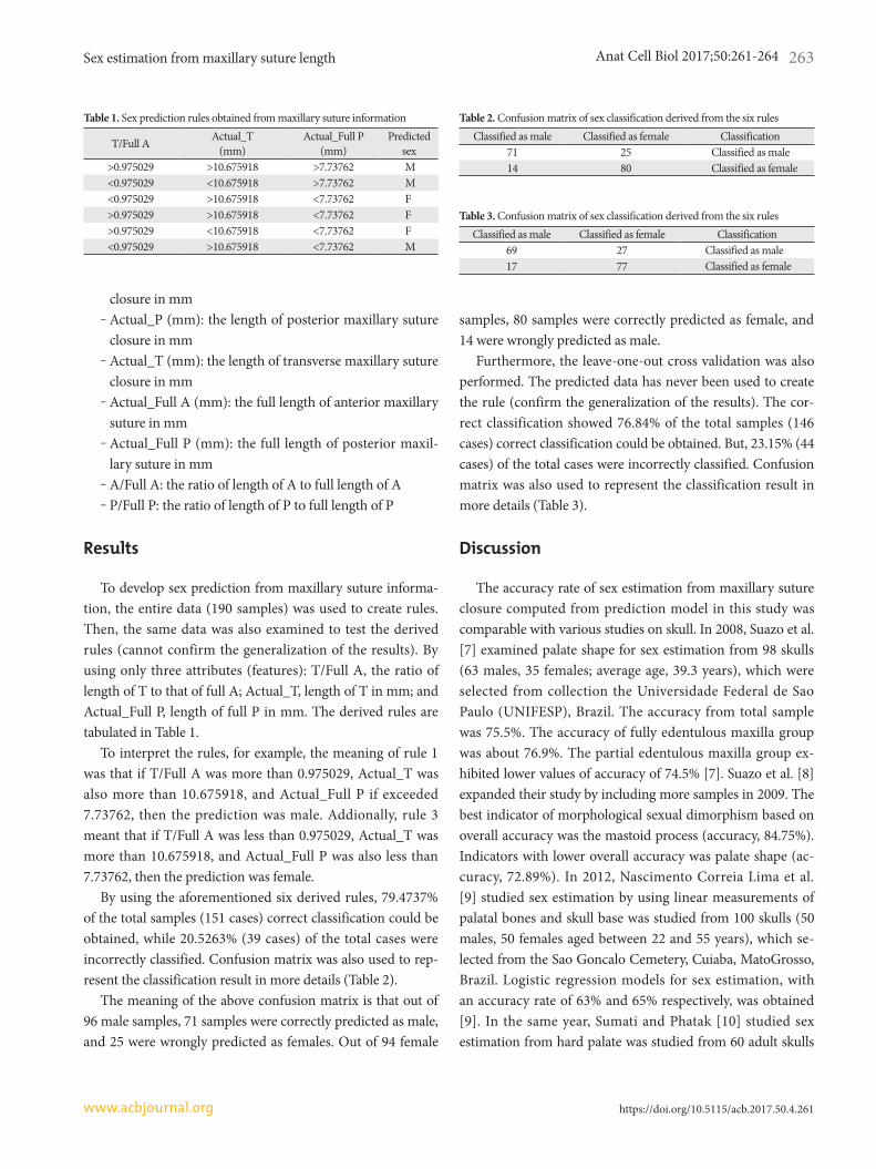
The skulls of 190 Thai individuals of known sex, and age at death ranging from 15 to 93 years, were obtained from the Forensic Osteology Research Center (FORC), Faculty of Med-icine, and Chiang Mai University. Skulls exhibiting fractures or pathologies of palate were excluded from this study. Three parts of maxillary suture (Fig. 1), anterior (A), transverse (T), and posterior (P) maxillary sutures were photographed using a Canon EOS camera (Tokyo, Japan).
An expert manually delineated each part of maxillary su-ture in each original image (Fig. 1) with different colors, i.e., A,
T, and P were marked with blue, red, and green, respectively. Length of anterior maxillary suture that was still open was represented as A. On the other hand, a total length of anterior maxillary suture—where it started from the posterior edge of incisive foramen to the point that it met transverse maxillary suture—was defined as Full A. Actual A represented closure of anterior maxillary suture which was obtained by subtract-ing A from Full A. Also, P and Full P were defined as length of open suture and total length of posterior maxillary suture, which started from the point that it met transverse suture to posterior nasal spine, respectively and T represented length of opened transverse maxillary suture. Actual P was calculated by subtracting P from Full P. Actual T was obtained by sub-tracting T from Full T. To enable the mapping of each length to its real-world value, the expert also marked two cyan dots on the measurement scale with 5 cm apart. Then, pixel value of 1 cm was calculated in order to use as a reference of mea-surement for suture length in mm.
Considering the length of suture obtained from the photo-graph, it does not make sense to represent the length of each suture in pixels. This is due to the distance variation between the camera and the skull. To map the length in pixels to the real-world unit, we used the two cyan dots marked by the expert with the prior knowledge that the distance separation was 5 cm. We considered the two detected cyan dots. Each of the dots would have different shape and area. Hence, we com-puted the centroid of each dot to represent its location. The Euclidean distance between each pair was computed, and it was used to compute the resolution of each image as follows:
Resolution (mm/pixel)=
50Euclidean distance between two cyan dots
Hence, for each image, we could map the length in pixels to the real-world unit in millimeter by simply multiplying the length in pixels by the derived resolution.
The other two pairs of dots, i.e., blue dots and green dots were detected to represent the full lengths of A and P. The centroid of each dot was once again used to represent its loca-tion. The full lengths of A and P were calculated by using the Euclidean distance between each corresponding pair of dots.
Finally, the following eight features/attributes were extract-ed for the prediction model:
‒ Sex_val: ‒1 for male and +1 for female‒ Actual_A (mm): the length of anterior maxillary suture
Transverse (red)Anterior (blue)
Posterior (green)
Fig. 1. Original image with expert’s delineations and markers for anterior (blue), transverse (red), and posterior (green).
Sex estimation from maxillary suture length
https://doi.org/10.5115/acb.2017.50.4.261
Anat Cell Biol 2017;50:261-264 263
www.acbjournal.org
closure in mm‒ Actual_P (mm): the length of posterior maxillary suture
closure in mm‒ Actual_T (mm): the length of transverse maxillary suture
closure in mm‒ Actual_Full A (mm): the full length of anterior maxillary
suture in mm‒ Actual_Full P (mm): the full length of posterior maxil-
lary suture in mm‒ A/Full A: the ratio of length of A to full length of A‒ P/Full P: the ratio of length of P to full length of P
Results
To develop sex prediction from maxillary suture informa-tion, the entire data (190 samples) was used to create rules. Then, the same data was also examined to test the derived rules (cannot confirm the generalization of the results). By using only three attributes (features): T/Full A, the ratio of length of T to that of full A; Actual_T, length of T in mm; and Actual_Full P, length of full P in mm. The derived rules are tabulated in Table 1.
To interpret the rules, for example, the meaning of rule 1 was that if T/Full A was more than 0.975029, Actual_T was also more than 10.675918, and Actual_Full P if exceeded 7.73762, then the prediction was male. Addionally, rule 3 meant that if T/Full A was less than 0.975029, Actual_T was more than 10.675918, and Actual_Full P was also less than 7.73762, then the prediction was female.
By using the aforementioned six derived rules, 79.4737% of the total samples (151 cases) correct classification could be obtained, while 20.5263% (39 cases) of the total cases were incorrectly classified. Confusion matrix was also used to rep-resent the classification result in more details (Table 2).
The meaning of the above confusion matrix is that out of 96 male samples, 71 samples were correctly predicted as male, and 25 were wrongly predicted as females. Out of 94 female
samples, 80 samples were correctly predicted as female, and 14 were wrongly predicted as male.
Furthermore, the leave-one-out cross validation was also performed. The predicted data has never been used to create the rule (confirm the generalization of the results). The cor-rect classification showed 76.84% of the total samples (146 cases) correct classification could be obtained. But, 23.15% (44 cases) of the total cases were incorrectly classified. Confusion matrix was also used to represent the classification result in more details (Table 3).
Discussion
The accuracy rate of sex estimation from maxillary suture closure computed from prediction model in this study was comparable with various studies on skull. In 2008, Suazo et al. [7] examined palate shape for sex estimation from 98 skulls (63 males, 35 females; average age, 39.3 years), which were selected from collection the Universidade Federal de Sao Paulo (UNIFESP), Brazil. The accuracy from total sample was 75.5%. The accuracy of fully edentulous maxilla group was about 76.9%. The partial edentulous maxilla group ex-hibited lower values of accuracy of 74.5% [7]. Suazo et al. [8] expanded their study by including more samples in 2009. The best indicator of morphological sexual dimorphism based on overall accuracy was the mastoid process (accuracy, 84.75%). Indicators with lower overall accuracy was palate shape (ac-curacy, 72.89%). In 2012, Nascimento Correia Lima et al. [9] studied sex estimation by using linear measurements of palatal bones and skull base was studied from 100 skulls (50 males, 50 females aged between 22 and 55 years), which se-lected from the Sao Goncalo Cemetery, Cuiaba, MatoGrosso, Brazil. Logistic regression models for sex estimation, with an accuracy rate of 63% and 65% respectively, was obtained [9]. In the same year, Sumati and Phatak [10] studied sex estimation from hard palate was studied from 60 adult skulls
Table 1. Sex prediction rules obtained from maxillary suture information
T/Full A Actual_T (mm)
Actual_Full P (mm)
Predicted sex
>0.975029 >10.675918 >7.73762 M<0.975029 <10.675918 >7.73762 M<0.975029 >10.675918 <7.73762 F>0.975029 >10.675918 <7.73762 F>0.975029 <10.675918 <7.73762 F<0.975029 >10.675918 <7.73762 M
Table 2. Confusion matrix of sex classification derived from the six rulesClassified as male Classified as female Classification
71 25 Classified as male14 80 Classified as female
Table 3. Confusion matrix of sex classification derived from the six rulesClassified as male Classified as female Classification
69 27 Classified as male17 77 Classified as female
Anat Cell Biol 2017;50:261-264 Apichat Sinthubua, et al264
www.acbjournal.orghttps://doi.org/10.5115/acb.2017.50.4.261
(30 males, 30 females) of North Indian population, which were selected from the Department of Anatomy and Forensic Medicine, Government Medical College, Patiala. The results showed that the correct classification rate in stepwise analysis decreased from 70.0% (discriminant analysis) to 66.7% (logis-tic method) [10]. Skrzat et al. [11] reported that there was a significant difference of the length of mid-palatal suture—also known as posterior maxillary suture in the present study—between sexes. Females demonstrated significant correlation between the total length of anterior and posterior maxillary, and the length of posterior maxillary suture while males did not show this kind of relationship.
Besides from sexual difference in palatal shape, several studies also reported the difference of closure rate of maxil-lary sutures between males and females [12, 13]. Furthermore, Beauthier et al. [13] proposed that males showed higher rate of maxillary suture fusion than that of females in French and Belgian samples. This was consistent with finding reported by Mann et al. [6]. White and Black skeletal samples showed the evidence that males typically display more progressive rate of maxillary suture obliteration than that of females [12]. In contrast, Apostolidou et al. [14] reported that there was insig-nificant sex difference between closures of maxillary suture in Greek population.
In this study, we came out with a new method of predict-ing sex from measurement of maxillary sutures as depicted in Table 1. We also wish to highlight that by simply measuring the maxillary sutures in photographs, one can estimate the sex.
Although maxillary suture closures have been long studied for age estimation, but they were also valuable for sex estima-tion. This study presented the sexual dimorphism of maxil-lary suture closure and illustrated that they were able to be as sex indicator. The present study also provided the sex estima-tion model which can be applied for Thai population. This finding is useful for both sex and age estimation when only skull was available in forensic situations. This study may con-tribute as a basis knowledge and method for further study of sex estimation in archaeological and forensic anthropological contexts, especially when only skull or skull base are found.
Acknowledgements
The authors are grateful to Forensic Osteology Research Center. The authors also thank research funding received
from Excellence Center in Osteology Research and Training Center (ORTC). The authors would also like to thank Ms. Phruksachat Singsuwan, Ph.D. candidate in Forensic Osteol-ogy for preparing a manuscript formatting.
References
1. DiGangi EA, Moore MK. Research methods in human skeletal biology. Oxford: Elsevier; 2013.
2. Mays S, Cox M. Sex determination in skeleton remains. In: Cox M, Mays S, editors. Human Osteology in Archaeology and Foren sic Science. London: Cambridge University Press; 2000. p.117-30.
3. Macaluso PJ Jr. Metric sex determination from the basal region of the occipital bone in a documented French sample. Bull Mem Soc Anthropol Paris 2011;23:19-26.
4. Kamath V, Asif M, Shetty R, Avadhani R. Binary logistic regres-sion analysis of hard palate dimensions for sexing human crania. Anat Cell Biol 2016;49:151-9.
5. Wood JK. Direction and type of the transverse palatine suture and its relation to the form of the hard palate. Am J Phys An-thropol 1949;7:385-99.
6. Mann RW, Symes SA, Bass WM. Maxillary suture obliteration: aging the human skeleton based on intact or fragmentary max-illa. J Forensic Sci 1987;32:148-57.
7. Suazo GI, Zavando MD, Smith RL. Accuracy of palate shape as sex indicator in human skull with maxillary teeth loss. Int J Mor-phol 2008;26:989-93.
8. Suazo GI, Zavando MD, Smith RL. Performance evaluation as a diagnostic test for traditional methods for forensic identification of sex. Int J Morphol 2009;27:381-6.
9. Nascimento Correia Lima N, Fortes de Oliveira O, Sassi C, Picapedra A, Francesquini L Jr, Daruge E Jr. Sex determination by linear measurements of palatal bones and skull base. J Foren-sic Odontostomatol 2012;30:38-44.
10. Sumati PV, Phatak A. Determination of sex from hard palate by discriminant function analysis. Int J Basic Appl Med Sci 2012;2: 243-51.
11. Skrzat J, Holiat D, Walocha J. A morphometrical study of the hu-man palatine sutures. Folia Morphol (Warsz) 2003;62:123-7.
12. Mann RW, Jantz RL, Bass WM, Willey PS. Maxillary suture obliteration: a visual method for estimating skeletal age. J Foren-sic Sci 1991;36:781-91.
13. Beauthier JP, Lefevre P, Meunier M, Orban R, Polet C, Werquin JP, Quatrehomme G. Palatine sutures as age indicator: a con-trolled study in the elderly. J Forensic Sci 2010;55:153-8.
14. Apostolidou C, Eleminiadis I, Koletsa T, Natsis K, Dalampiras S, Psaroulis D, Apostolidis S, Psifidis A, Tsikaras P, Njau SN. Appli-cation of the maxillary suture obliteration method for estimating age at death in Greek population. Open Forensic Sci J 2011;4:15-9.
Related Documents