A Modular Approach to Cardiac Tissue Engineering Brendan M. Leung, MASc., 1 and Michael V. Sefton, Sc.D. 1,2 Functional cardiac tissue was prepared using a modular tissue engineering approach with the goal of creating vascularized tissue. Rat aortic endothelial cells (RAEC) were seeded onto submillimeter-sized modules made of type I bovine collagen supplemented with MatrigelÔ (25% v/v) embedded with cardiomyocyte (CM)-enriched neonatal rat heart cells and assembled into a contractile, macroporous, sheet-like construct. Modules (without RAEC) cultured in 10% bovine serum (BS) were more contractile and responsive to external stimulus (lower excitation threshold, higher maximum capture rate, and greater en face fractional area changes) than modules cultured in 10% fetal BS. Incorporating 25% Matrigel in the matrix reduced the excitation threshold and in- creased the fractional area change relative to collagen only modules (without RAEC). A coculture medium, containing 10% BS, low Mg 2þ (0.814 mM), and normal glucose (5.5 mM), was used to maintain RAEC junction morphology (VE-cadherin) and CM contractility, although the responsiveness of CM was attenuated with RAEC on the modules. Macroporous, sheet-like module constructs were assembled by partially immobilizing a layer of modules in alginate gel until day 8, with or without RAEC. RAEC/CM module sheets were electrically re- sponsive; however, like modules with RAEC this responsiveness was attenuated relative to CM-only sheets. Muscle bundles coexpressing cardiac troponin I and connexin-43 were evident near the perimeter of modules and at intermodule junctions. These results suggest the potential of the modular approach as a platform for building vascularized cardiac tissue. Introduction C ongestive heart failure is a major cause of death in both the developed and developing world. Although the pathophysiologies relating to congestive heart failure are complex, the condition is ultimately caused by the weakening of heart muscle tissue resulting in lowered car- diac output. The decrease in cardiac output causes chronic systemic ischemia that leads to decompensation responses such as ventricular hypertrophy, increased blood volume, and increased blood pressure, which in turn increases car- diac load and leads to further damage to cardiac muscle tissue. As a result, there is considerable interest in fabri- cating a cardiac tissue substitute using cardiomyocytes (CMs) in vitro. The resulting tissue-engineered cardiac muscle would be expected, at a minimum, to possess sim- ilar mechanical and functional properties as native cardiac tissue, but also have the capability to be integrated into native tissue, thus preventing long-term cardiac tissue re- modeling. 1 Attempts to fabricate functional cardiac tissues in vitro have yielded promising results. 2,3 A crucial factor in creating viable three-dimensional tissue in vitro is to achieve adequate perfusion throughout the tissue. This is especially true for cardiac tissue due to its high metabolic rate and oxygen demand. With a view to improving engineered tissue per- fusion, Radisic et al. 4 incorporated oxygen carriers and flow channels into neonatal rat CM-seeded scaffolds. Aside from nutrients, environmental factors including mechanical 5 and electrical 6 stimuli are also needed for the proper develop- ment of cardiac tissue. Many of these concepts have been incorporated into recent culture systems 7 in which CM are incorporated with other materials to form functional tissues. 8 For example, a three-dimensional model was used by Zim- mermann et al., 5 where they embedded a neonatal rat CM- enriched cell population (also containing fibroblasts and endothelial cells [EC]) into a MatrigelÔ/collagen gel. In their model, the EC in culture facilitated angiogenesis and host vasculature coupling. Other three-dimensional culture sys- tems rely on cell-secreted extracellular matrix as the major matrix component. Kelm et al. 9,10 created microtissues con- taining CM in a hanging drop culture system. The scale of these microtissues (*100 mm) has been shown to support core tissue viability. In addition, contractile CM sheets have been cultivated and assembled into multilayer tissues by Shimizu et al. 11 These strategies improve tissue perfusion by promoting either angiogenesis of surrounding blood vessels, or neovasculogenesis of embedded EC (part of 1 Institute of Biomaterials and Biomedical Engineering and 2 Department of Chemical Engineering and Applied Chemistry, University of Toronto, Toronto, Canada. TISSUE ENGINEERING: Part A Volume 16, Number 10, 2010 ª Mary Ann Liebert, Inc. DOI: 10.1089/ten.tea.2009.0746 3207

Welcome message from author
This document is posted to help you gain knowledge. Please leave a comment to let me know what you think about it! Share it to your friends and learn new things together.
Transcript
A Modular Approach to Cardiac Tissue Engineering
Brendan M. Leung, MASc.,1 and Michael V. Sefton, Sc.D.1,2
Functional cardiac tissue was prepared using a modular tissue engineering approach with the goal of creatingvascularized tissue. Rat aortic endothelial cells (RAEC) were seeded onto submillimeter-sized modules made oftype I bovine collagen supplemented with Matrigel� (25% v/v) embedded with cardiomyocyte (CM)-enrichedneonatal rat heart cells and assembled into a contractile, macroporous, sheet-like construct. Modules (withoutRAEC) cultured in 10% bovine serum (BS) were more contractile and responsive to external stimulus (lowerexcitation threshold, higher maximum capture rate, and greater en face fractional area changes) than modulescultured in 10% fetal BS. Incorporating 25% Matrigel in the matrix reduced the excitation threshold and in-creased the fractional area change relative to collagen only modules (without RAEC). A coculture medium,containing 10% BS, low Mg2þ (0.814 mM), and normal glucose (5.5 mM), was used to maintain RAEC junctionmorphology (VE-cadherin) and CM contractility, although the responsiveness of CM was attenuated with RAECon the modules. Macroporous, sheet-like module constructs were assembled by partially immobilizing a layer ofmodules in alginate gel until day 8, with or without RAEC. RAEC/CM module sheets were electrically re-sponsive; however, like modules with RAEC this responsiveness was attenuated relative to CM-only sheets.Muscle bundles coexpressing cardiac troponin I and connexin-43 were evident near the perimeter of modulesand at intermodule junctions. These results suggest the potential of the modular approach as a platform forbuilding vascularized cardiac tissue.
Introduction
Congestive heart failure is a major cause of death inboth the developed and developing world. Although
the pathophysiologies relating to congestive heart failureare complex, the condition is ultimately caused by theweakening of heart muscle tissue resulting in lowered car-diac output. The decrease in cardiac output causes chronicsystemic ischemia that leads to decompensation responsessuch as ventricular hypertrophy, increased blood volume,and increased blood pressure, which in turn increases car-diac load and leads to further damage to cardiac muscletissue. As a result, there is considerable interest in fabri-cating a cardiac tissue substitute using cardiomyocytes(CMs) in vitro. The resulting tissue-engineered cardiacmuscle would be expected, at a minimum, to possess sim-ilar mechanical and functional properties as native cardiactissue, but also have the capability to be integrated intonative tissue, thus preventing long-term cardiac tissue re-modeling.1
Attempts to fabricate functional cardiac tissues in vitrohave yielded promising results.2,3 A crucial factor in creatingviable three-dimensional tissue in vitro is to achieve adequateperfusion throughout the tissue. This is especially true for
cardiac tissue due to its high metabolic rate and oxygendemand. With a view to improving engineered tissue per-fusion, Radisic et al.4 incorporated oxygen carriers and flowchannels into neonatal rat CM-seeded scaffolds. Aside fromnutrients, environmental factors including mechanical5 andelectrical6 stimuli are also needed for the proper develop-ment of cardiac tissue. Many of these concepts have beenincorporated into recent culture systems7 in which CM areincorporated with other materials to form functional tissues.8
For example, a three-dimensional model was used by Zim-mermann et al.,5 where they embedded a neonatal rat CM-enriched cell population (also containing fibroblasts andendothelial cells [EC]) into a Matrigel�/collagen gel. In theirmodel, the EC in culture facilitated angiogenesis and hostvasculature coupling. Other three-dimensional culture sys-tems rely on cell-secreted extracellular matrix as the majormatrix component. Kelm et al.9,10 created microtissues con-taining CM in a hanging drop culture system. The scale ofthese microtissues (*100 mm) has been shown to supportcore tissue viability. In addition, contractile CM sheetshave been cultivated and assembled into multilayer tissuesby Shimizu et al.11 These strategies improve tissue perfusionby promoting either angiogenesis of surrounding bloodvessels, or neovasculogenesis of embedded EC (part of
1Institute of Biomaterials and Biomedical Engineering and 2Department of Chemical Engineering and Applied Chemistry, University ofToronto, Toronto, Canada.
TISSUE ENGINEERING: Part AVolume 16, Number 10, 2010ª Mary Ann Liebert, Inc.DOI: 10.1089/ten.tea.2009.0746
3207
the CM mixed cell population) via cytokine and chemokinesignaling.
The overall goal of this work was to create functional,cardiac tissue using a modular tissue engineering approachwith neonatal rat CM embedded in a collagen/Matrigel gel(the module) and seeded with rat aortic EC (RAEC). Moduleshad a postcontraction length of *500mm and diameter of300 mm. When assembled into a construct, the resulting in-terconnected channels are lined with RAEC and perfuseable,mimicking the required vasculature. The small diameter ofeach module ensures sufficient nutrient delivery to the centerby diffusion. Modules may be assembled into larger con-structs in vitro followed by implantation, or they may bedeployed in vivo and allowed to be remodeled by host tissue.We hypothesized that the modular tissue engineering ap-proach12,13 can be used to produce vascularized cardiac tis-sue by assembling modules into a macroporous sheet, whereEC in the intermodular space can form vessel-like structures.This approach results in uniform, scalable, and vascularizedconstructs13 and is being explored elsewhere for its utility inpancreatic islet transplantation and fat reconstruction.
In work reported here, we have characterized the in vitrophenotype and functional response of a CM/RAEC cocul-ture system comprised of both individual modules andmodules in the form of a contractile sheet. We examined theresponses of CM only and RAEC/CM coculture modules toexternal electrical stimulation, as well as the effect of surface-coated RAEC on selected features of the embedded CM. Keyaspects of this study were the definition of a suitable me-dium for coculture of CM and EC and a means of preparingsheet-like structures from the modules.
Materials and Methods
CM isolation and module fabrication
The protocol for cell isolation was similar to that reportedby Radisic et al.6 Hearts from neonatal rat pups (1–2 daysold) were harvested and kept in ice-cold Hanks balanced saltsolution (HBSS; Gibco). The aortas were trimmed and theremaining heart tissues were quartered followed by serialdigestion with bovine pancreas trypsin (0.6 mg/mL in HBSS;Sigma) and collagenase II (1 mg/mL in HBSS; WorthingtonBiochemical). The extracted cell mixture was preplated ontotissue culture polystyrene for 1 h to enrich its CM content.Harvested cells were maintained in native CM mediumconsisting of high-glucose (25 mM) Dulbecco’s modified Ea-gle’s medium (Gibco) supplemented with 10% fetal bovineserum (FBS; or 10% bovine serum [BS]; Sigma), 1% HEPESbuffer (10 mM; Gibco), and 1% penicillin/streptomycin(Gibco). Each CM preparation consisted of cells from anentire litter (10–13 neonatal rats) to enhance consistency.Over the course of the study several litters were used in eachset of data. The number of experiments reflects the use ofthese replicate litters.
The CM-enriched cell mixture was loaded onto a Cyto-spin� slide and stained for troponin I and prolyl-4-hydroxy-lase (see below) to identify CM and fibroblasts, respectively:the mixture consisted of 50% CMs and 40% fibroblasts. Theremaining 10% was presumed to consist of other cardiac celltypes, including EC. The CM-rich cell mixture was embed-ded in a type I bovine collagen matrix supplemented with25% Matrigel (v/v; BD Bioscience), and cast into modules
using methods as described previously.13 Viability of em-bedded cells was determined using Live/Dead� assay kit(Molecular Probes).
EC seeding
RAEC were purchased and cultured in an MCDB-131-based native EC medium (VEC Technologies), supplementedwith 10% FBS. In some experiments, RAEC were transducedwith a lentiviral-enhanced green fluorescent protein (eGFP)vector14 to enable tracking over time (in collaboration withDr. J. Medin, Ontario Cancer Institute).
CM-embedded modules were cultured in the native CMmedium supplemented with 10% BS for 3 days before ECseeding. To coat the surface of modules, 2–3 million RAECwere suspended in a seeding medium consisting of a 50:50mixture of the native CM (10% BS) and native EC (10% FBS)medium, and mixed with 1 mL of CM-embedded modulesinside a 15 mL centrifuge tube. The tube was agitated for30 min on a uniaxial rocker at low speed. The mixture wastransferred to a nontissue culture-treated six-well plate andkept in th seeding medium overnight to promote RAEC at-tachment. The seeding medium was used to transit the CMand EC from their separate native media to the coculturemedium, described in Table 1. After 1 day, modules weretransferred into a new nontissue culture-treated six-well plateto remove any RAEC that did not attach to modules, and theseeding medium was changed to the coculture medium.Modules were maintained in this medium until assayed.
Module sheet
Four days after fabrication, CM-embedded modules wereassembled into a monolayer sheet. For RAEC/CM modularsheets, CM-embedded modules (3 days after fabrication)were seeded with RAEC and assembled into sheets the nextday. Using a 10 mL serological pipette, modules, with orwithout RAEC, were placed over a sterile nylon mesh (poresize¼ 100mm; Millipore) to form a single layer. A dilute al-ginate solution (1.2% w/w; Sigma) was dripped over themodular sheet followed by a few drops of 50mM calciumchloride to crosslink the alginate. Once the alginate formed asoft gel (*5–10 s), the sheet was rinsed in phosphate-buffered saline (1.06 mM KH2PO4, 155.17 mM NaCl, and2.97 mM Na2HPO4-7H2O) and transferred into the CM orcoculture medium. These alginate-embedded sheets werestimulated (see below) for 10 days and then the alginate gelwas removed before assay by brief incubation in 100mMcitrate buffer at room temperature.
Electrical response assessment and field stimulation
CM-embedded modules were equilibrated in 1�Tyrode’ssalt solution (Sigma) at 378C for 1 h before measurement.Electrical responses of modules were assessed using custom-made testing chambers. Modules were paced between a pairof graphite electrodes kept 1 cm apart by a polycarbonatebracket. Using a six-well plate made from tissue culturepolystyrene, modules and electrodes were placed in an in-dividual well with 1.5 mL of Tyrode’s salt solution, whichwas enough to partially submerge the electrodes. The smallchamber volume ensured that most of the modules werelocated between the electrodes.
3208 LEUNG AND SEFTON
Module sheets were assayed using a polydimethylsiloxane(PDMS)/glass testing chamber. A pair of graphite electrodeswere kept 1 cm apart by a PDMS bracket and placed over alayer of PDMS poured into a 60 mm glass Petri dish. Thisallowed the module sheets to be fixed in position relative tothe electrode.
For both systems, a signal generator (S88X stimulator;Grass Technologies) connected to the electrodes provided thedesired electrical stimulation (biphasic, 2 ms square pulse, upto 12 V at various frequencies). The samples were kept at378C during the experiment using a solid-state microscopestage heater. Contractions of modules and module sheetswere recorded with a CCD camera (Hitachi Denshi, modelKP-M1U) on an inverted phase-contrast microscope (Olym-pus CX2). The image sequences were analyzed using ImageJ(NIH, version 1.38x).
Excitation threshold (ET) was defined as the minimumvoltage required for synchronous contraction at 1 Hz. Max-imum capture rate (MCR) was defined as the maximum rateof synchronous contraction under an electric field equivalentto twice the ET. The contractility of modules was defined asthe fractional en face area change during one contractioncycle using frame-by-frame video analysis in ImageJ.
Module sheets were electrically conditioned before assay.Briefly, CM-embedded modules were maintained in the na-tive CM medium with 10% BS for 3 days. Then, the moduleswere fabricated into sheets (see above) and transferred into awell, fitted with a pair of graphite electrodes as above, andstimulated continuously with biphasic pulses at 5 V/cm and1 Hz for 10 days. The medium was supplemented with10 mM of ascorbic acid and changed daily to minimize theeffect of reactive oxygen species generated at the electrodes.Individual modules were not electrically conditioned be-cause they could not be fixed in position relative to theelectrical field over the course of an experiment.
Immunofluorescence staining
Freshly harvested, CM-enriched cell mixtures were ana-lyzed by Cytospin. Slides were stained with rabbit anti-rattroponin I (Santa Cruz Biotechnology) and mouse anti-ratprolyl 4-hydroxylase (Sigma) antibodies to identify CMs andfibroblasts, respectively.
RAEC coverage on modules was determined by wholemount immunofluorescence staining of goat anti-rat VE-cadherin primary antibody (Santa Cruz Biotechnology)
followed by rabbit anti-goat immunoglobulin (IgG) Alexa-Fluor� 488 secondary antibody (Molecular Probes). Moduleswere fixed in 4% paraformaldehyde solution and permeabi-lized using 0.2% Triton-X100 solution. On immunohisto-chemical sections, CMs were stained using rabbit anti-rattroponin I (Santa Cruz Biotechnology) followed by goat anti-rabbit IgG secondary antibodies conjugated with AlexaFluor488 (Molecular Probes). Cardiac fibroblasts were stained withmouse anti-rat vimentin antibody (Sigma) followed by goatanti-mouse IgG AlexaFluor 568 (Molecular Probes). In sectionsthat were triple-stained (troponin I, vimentin, and connexin-43), connexin-43 was stained before immunofluorescencestaining using rabbit anti-rat Cx43 antibody (Chemicon) cou-pled by a three-step immunohistochemistry (IHC) chromo-genic method. In some experiments, surface-seeded RAECwere tracked by eGFP, where cell nuclei were counterstainedwith Hoechst nuclear stain (Sigma). Samples were observedusing a Zeiss LSM510 confocal microscope.
Statistical analysis
Statistical analyses, including calculations of standarderror of means and mean comparisons (t-test or one-wayanalysis of variance), were performed using Prism (Version5.0; Graphpad Software).
Results
Contractility of CM-embedded modules
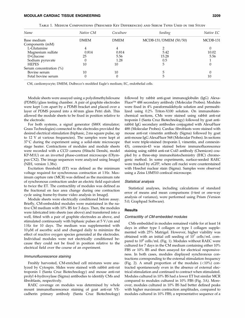
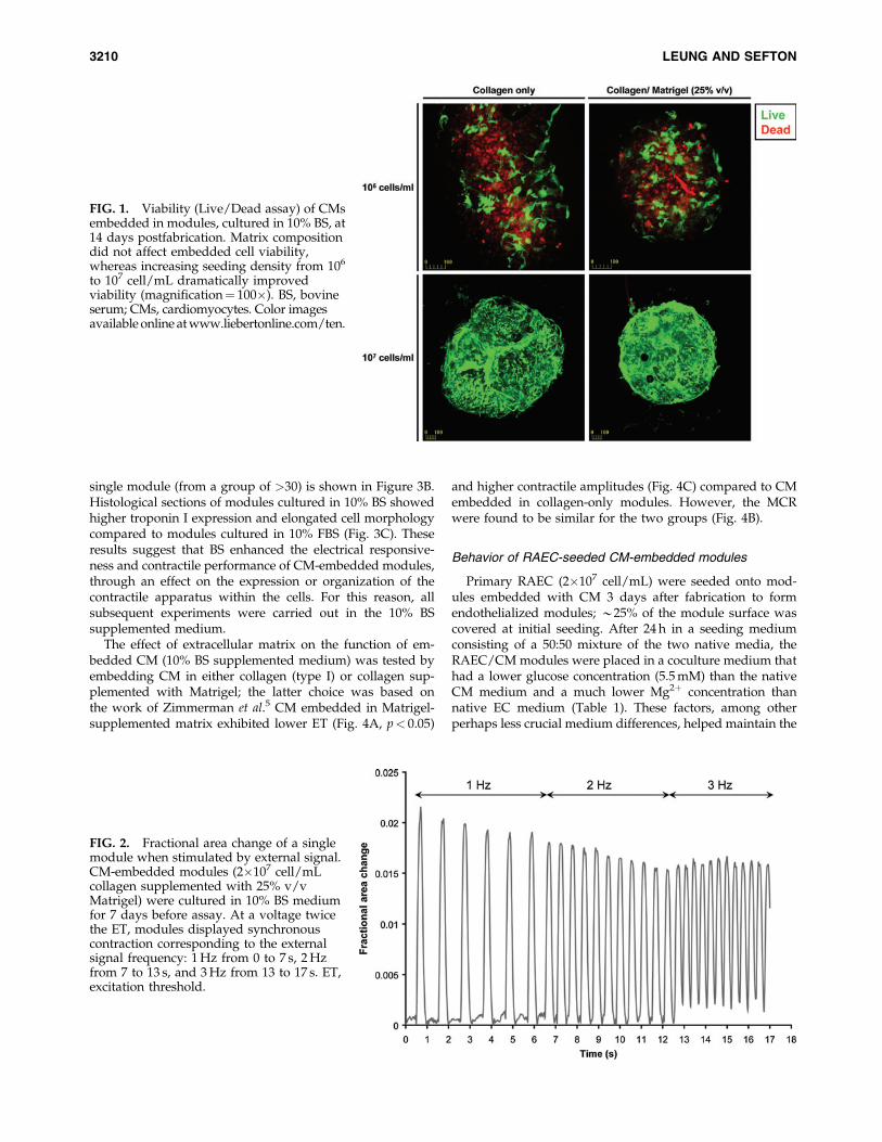
CMs embedded in modules remained viable for at least 14days in either type I collagen or type I collagen supple-mented with 25% Matrigel. However, higher viability wasobtained with an initial cell seeding of 107 cells/mL com-pared to 106 cells/mL (Fig. 1). Modules without RAEC werecultured for 7 days in the CM medium containing either 10%FBS or 10% BS and then assayed for electrical responsive-ness. In both cases, modules displayed synchronous con-tractions corresponding to the external stimulation frequency(Fig. 2). A small proportion of the modules (<10%) con-tracted spontaneously even in the absence of external elec-trical stimulation and continued to contract when stimulated.Modules cultured in 10% BS had a lower ET but similar MCRcompared to modules cultured in 10% FBS (Fig. 3A). More-over, modules cultured in 10% BS had better defined peakswith higher maximum contraction amplitudes, compared tomodules cultured in 10% FBS; a representative sequence of a
Table 1. Medium Compositions (Presumed Key Differences) and Serum Types Used in the Study
Name Native CM Coculture Seeding Native EC
Base medium DMEM DMEM MCDB-131/DMEM (50/50) MCDB-131Components (mM)
L-Glutamine 4 4 2 0Magnesium sulfate 0.814 0.814 5.42 10.02D-Glucose 25 5.56 15.28 5.56Sodium pyruvate 0 1.28 0.5 1HEPES 10 10 5 0
Serum concentration (%)Bovine serum 10 10 5 0Fetal bovine serum 0 0 5 10
CM, cardiomyocyte; DMEM, Dulbecco’s modified Eagle’s medium; EC, endothelial cells.
MODULAR CARDIAC TISSUE ENGINEERING 3209
single module (from a group of >30) is shown in Figure 3B.Histological sections of modules cultured in 10% BS showedhigher troponin I expression and elongated cell morphologycompared to modules cultured in 10% FBS (Fig. 3C). Theseresults suggest that BS enhanced the electrical responsive-ness and contractile performance of CM-embedded modules,through an effect on the expression or organization of thecontractile apparatus within the cells. For this reason, allsubsequent experiments were carried out in the 10% BSsupplemented medium.
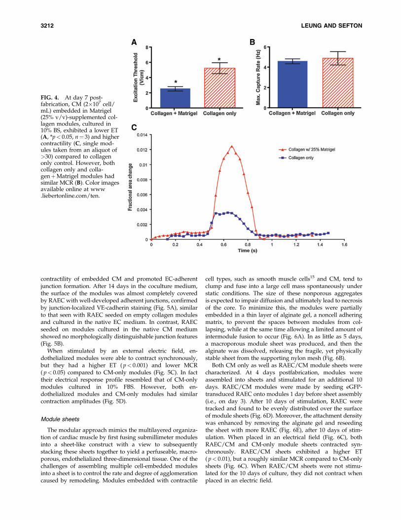
The effect of extracellular matrix on the function of em-bedded CM (10% BS supplemented medium) was tested byembedding CM in either collagen (type I) or collagen sup-plemented with Matrigel; the latter choice was based onthe work of Zimmerman et al.5 CM embedded in Matrigel-supplemented matrix exhibited lower ET (Fig. 4A, p< 0.05)
and higher contractile amplitudes (Fig. 4C) compared to CMembedded in collagen-only modules. However, the MCRwere found to be similar for the two groups (Fig. 4B).
Behavior of RAEC-seeded CM-embedded modules
Primary RAEC (2�107 cell/mL) were seeded onto mod-ules embedded with CM 3 days after fabrication to formendothelialized modules; *25% of the module surface wascovered at initial seeding. After 24 h in a seeding mediumconsisting of a 50:50 mixture of the two native media, theRAEC/CM modules were placed in a coculture medium thathad a lower glucose concentration (5.5 mM) than the nativeCM medium and a much lower Mg2þ concentration thannative EC medium (Table 1). These factors, among otherperhaps less crucial medium differences, helped maintain the
FIG. 1. Viability (Live/Dead assay) of CMsembedded in modules, cultured in 10% BS, at14 days postfabrication. Matrix compositiondid not affect embedded cell viability,whereas increasing seeding density from 106
to 107 cell/mL dramatically improvedviability (magnification¼ 100�). BS, bovineserum; CMs, cardiomyocytes. Color imagesavailable online at www.liebertonline.com/ten.
FIG. 2. Fractional area change of a singlemodule when stimulated by external signal.CM-embedded modules (2�107 cell/mLcollagen supplemented with 25% v/vMatrigel) were cultured in 10% BS mediumfor 7 days before assay. At a voltage twicethe ET, modules displayed synchronouscontraction corresponding to the externalsignal frequency: 1 Hz from 0 to 7 s, 2 Hzfrom 7 to 13 s, and 3 Hz from 13 to 17 s. ET,excitation threshold.
3210 LEUNG AND SEFTON
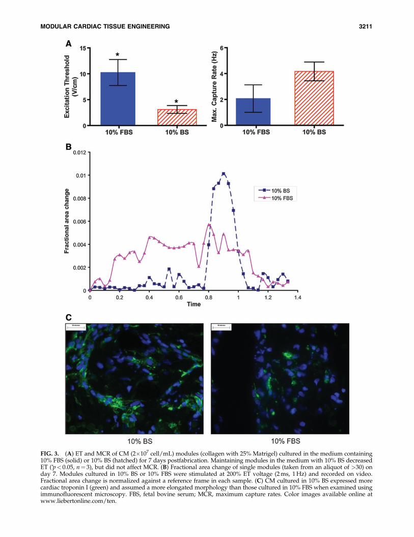
FIG. 3. (A) ET and MCR of CM (2�107 cell/mL) modules (collagen with 25% Matrigel) cultured in the medium containing10% FBS (solid) or 10% BS (hatched) for 7 days postfabrication. Maintaining modules in the medium with 10% BS decreasedET (*p< 0.05, n¼ 3), but did not affect MCR. (B) Fractional area change of single modules (taken from an aliquot of >30) onday 7. Modules cultured in 10% BS or 10% FBS were stimulated at 200% ET voltage (2 ms, 1 Hz) and recorded on video.Fractional area change is normalized against a reference frame in each sample. (C) CM cultured in 10% BS expressed morecardiac troponin I (green) and assumed a more elongated morphology than those cultured in 10% FBS when examined usingimmunofluorescent microscopy. FBS, fetal bovine serum; MCR, maximum capture rates. Color images available online atwww.liebertonline.com/ten.
MODULAR CARDIAC TISSUE ENGINEERING 3211
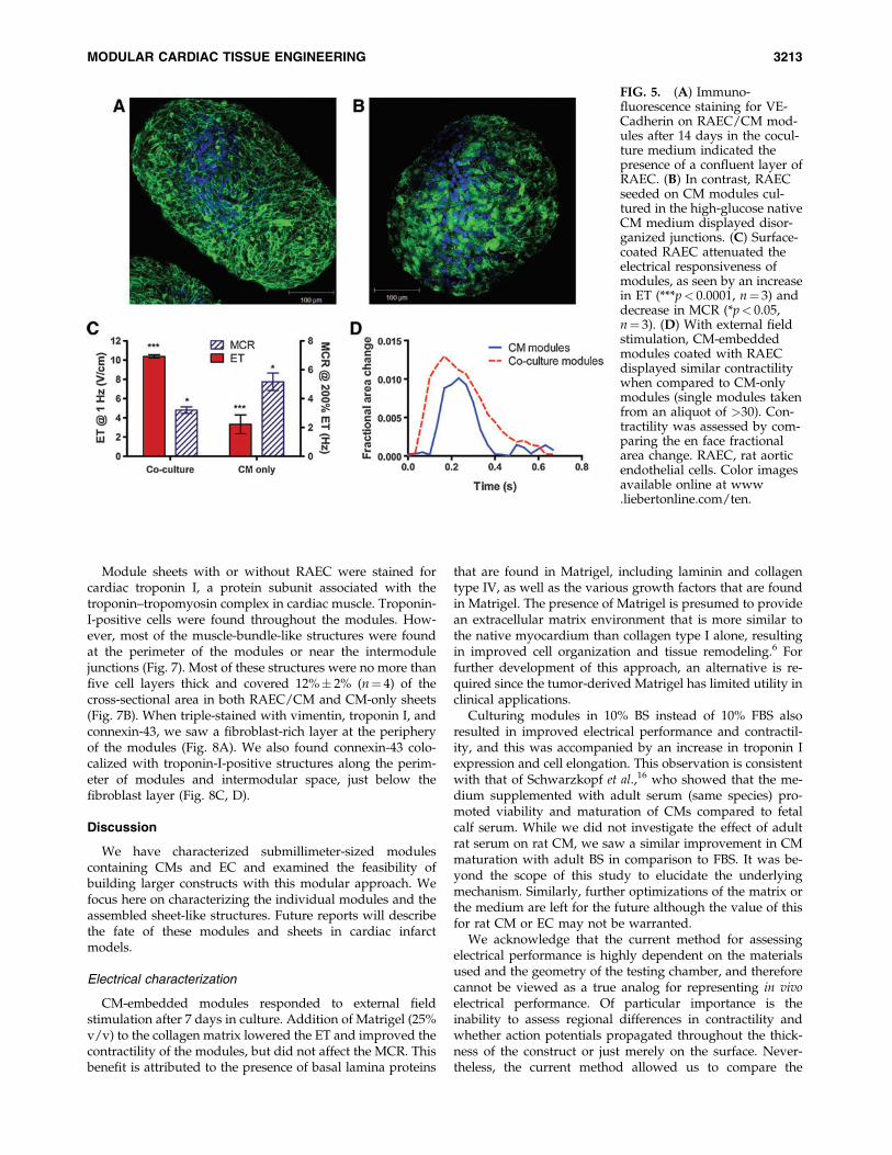
contractility of embedded CM and promoted EC-adherentjunction formation. After 14 days in the coculture medium,the surface of the modules was almost completely coveredby RAEC with well-developed adherent junctions, confirmedby junction-localized VE-cadherin staining (Fig. 5A), similarto that seen with RAEC seeded on empty collagen modulesand cultured in the native EC medium. In contrast, RAECseeded on modules cultured in the native CM mediumshowed no morphologically distinguishable junction features(Fig. 5B).
When stimulated by an external electric field, en-dothelialized modules were able to contract synchronously,but they had a higher ET ( p< 0.001) and lower MCR( p< 0.05) compared to CM-only modules (Fig. 5C). In facttheir electrical response profile resembled that of CM-onlymodules cultured in 10% FBS. However, both en-dothelialized modules and CM-only modules had similarcontraction amplitudes (Fig. 5D).
Module sheets
The modular approach mimics the multilayered organiza-tion of cardiac muscle by first fusing submillimeter modulesinto a sheet-like construct with a view to subsequentlystacking these sheets together to yield a perfuseable, macro-porous, endothelialized three-dimensional tissue. One of thechallenges of assembling multiple cell-embedded modulesinto a sheet is to control the rate and degree of agglomerationcaused by remodeling. Modules embedded with contractile
cell types, such as smooth muscle cells15 and CM, tend toclump and fuse into a large cell mass spontaneously understatic conditions. The size of these nonporous aggregatesis expected to impair diffusion and ultimately lead to necrosisof the core. To minimize this, the modules were partiallyembedded in a thin layer of alginate gel, a noncell adheringmatrix, to prevent the spaces between modules from col-lapsing, while at the same time allowing a limited amount ofintermodule fusion to occur (Fig. 6A). In as little as 5 days,a macroporous module sheet was produced, and then thealginate was dissolved, releasing the fragile, yet physicallystable sheet from the supporting nylon mesh (Fig. 6B).
Both CM only as well as RAEC/CM module sheets werecharacterized. At 4 days postfabrication, modules wereassembled into sheets and stimulated for an additional 10days. RAEC/CM modules were made by seeding eGFP-transduced RAEC onto modules 1 day before sheet assembly(i.e., on day 3). After 10 days of stimulation, RAEC weretracked and found to be evenly distributed over the surfaceof module sheets (Fig. 6D). Moreover, the attachment densitywas enhanced by removing the alginate gel and reseedingthe sheet with more RAEC (Fig. 6E), after 10 days of stim-ulation. When placed in an electrical field (Fig. 6C), bothRAEC/CM and CM-only module sheets contracted syn-chronously. RAEC/CM sheets exhibited a higher ET( p< 0.01), but a roughly similar MCR compared to CM-onlysheets (Fig. 6C). When RAEC/CM sheets were not stimu-lated for the 10 days of culture, they did not contract whenplaced in an electric field.
FIG. 4. At day 7 post-fabrication, CM (2�107 cell/mL) embedded in Matrigel(25% v/v)-supplemented col-lagen modules, cultured in10% BS, exhibited a lower ET(A, *p< 0.05, n¼ 3) and highercontractility (C, single mod-ules taken from an aliquot of>30) compared to collagenonly control. However, bothcollagen only and colla-genþMatrigel modules hadsimilar MCR (B). Color imagesavailable online at www.liebertonline.com/ten.
3212 LEUNG AND SEFTON
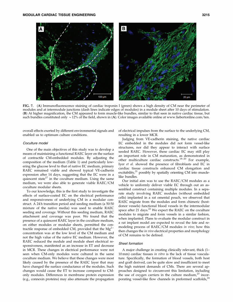
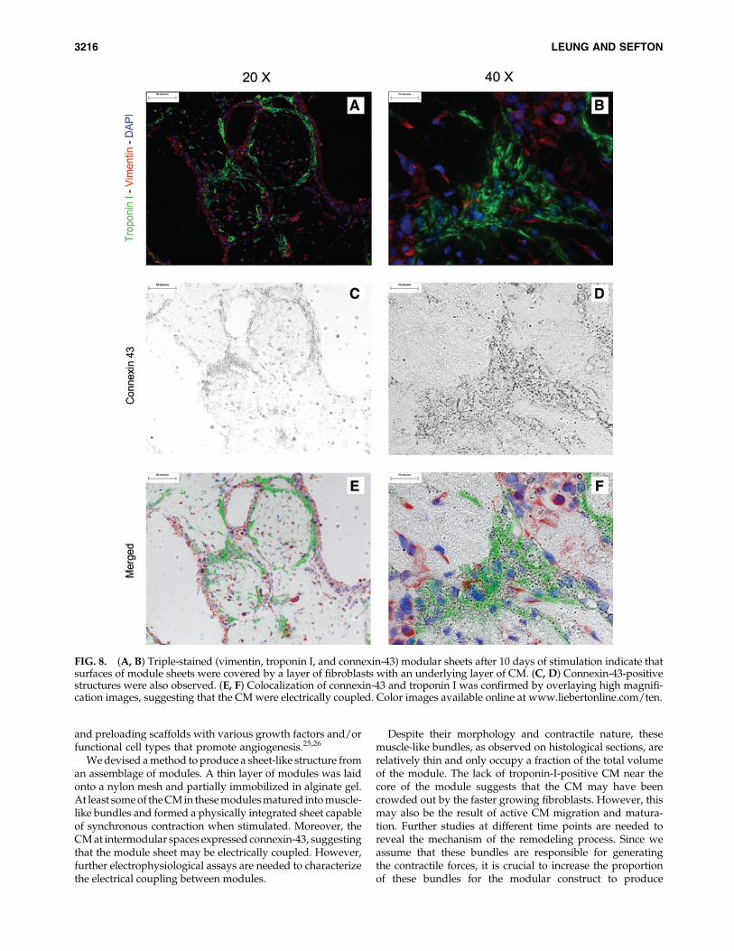
Module sheets with or without RAEC were stained forcardiac troponin I, a protein subunit associated with thetroponin–tropomyosin complex in cardiac muscle. Troponin-I-positive cells were found throughout the modules. How-ever, most of the muscle-bundle-like structures were foundat the perimeter of the modules or near the intermodulejunctions (Fig. 7). Most of these structures were no more thanfive cell layers thick and covered 12%� 2% (n¼ 4) of thecross-sectional area in both RAEC/CM and CM-only sheets(Fig. 7B). When triple-stained with vimentin, troponin I, andconnexin-43, we saw a fibroblast-rich layer at the peripheryof the modules (Fig. 8A). We also found connexin-43 colo-calized with troponin-I-positive structures along the perim-eter of modules and intermodular space, just below thefibroblast layer (Fig. 8C, D).
Discussion
We have characterized submillimeter-sized modulescontaining CMs and EC and examined the feasibility ofbuilding larger constructs with this modular approach. Wefocus here on characterizing the individual modules and theassembled sheet-like structures. Future reports will describethe fate of these modules and sheets in cardiac infarctmodels.
Electrical characterization
CM-embedded modules responded to external fieldstimulation after 7 days in culture. Addition of Matrigel (25%v/v) to the collagen matrix lowered the ET and improved thecontractility of the modules, but did not affect the MCR. Thisbenefit is attributed to the presence of basal lamina proteins
that are found in Matrigel, including laminin and collagentype IV, as well as the various growth factors that are foundin Matrigel. The presence of Matrigel is presumed to providean extracellular matrix environment that is more similar tothe native myocardium than collagen type I alone, resultingin improved cell organization and tissue remodeling.6 Forfurther development of this approach, an alternative is re-quired since the tumor-derived Matrigel has limited utility inclinical applications.
Culturing modules in 10% BS instead of 10% FBS alsoresulted in improved electrical performance and contractil-ity, and this was accompanied by an increase in troponin Iexpression and cell elongation. This observation is consistentwith that of Schwarzkopf et al.,16 who showed that the me-dium supplemented with adult serum (same species) pro-moted viability and maturation of CMs compared to fetalcalf serum. While we did not investigate the effect of adultrat serum on rat CM, we saw a similar improvement in CMmaturation with adult BS in comparison to FBS. It was be-yond the scope of this study to elucidate the underlyingmechanism. Similarly, further optimizations of the matrix orthe medium are left for the future although the value of thisfor rat CM or EC may not be warranted.
We acknowledge that the current method for assessingelectrical performance is highly dependent on the materialsused and the geometry of the testing chamber, and thereforecannot be viewed as a true analog for representing in vivoelectrical performance. Of particular importance is theinability to assess regional differences in contractility andwhether action potentials propagated throughout the thick-ness of the construct or just merely on the surface. Never-theless, the current method allowed us to compare the
FIG. 5. (A) Immuno-fluorescence staining for VE-Cadherin on RAEC/CM mod-ules after 14 days in the cocul-ture medium indicated thepresence of a confluent layer ofRAEC. (B) In contrast, RAECseeded on CM modules cul-tured in the high-glucose nativeCM medium displayed disor-ganized junctions. (C) Surface-coated RAEC attenuated theelectrical responsiveness ofmodules, as seen by an increasein ET (***p< 0.0001, n¼ 3) anddecrease in MCR (*p< 0.05,n¼ 3). (D) With external fieldstimulation, CM-embeddedmodules coated with RAECdisplayed similar contractilitywhen compared to CM-onlymodules (single modules takenfrom an aliquot of >30). Con-tractility was assessed by com-paring the en face fractionalarea change. RAEC, rat aorticendothelial cells. Color imagesavailable online at www.liebertonline.com/ten.
MODULAR CARDIAC TISSUE ENGINEERING 3213
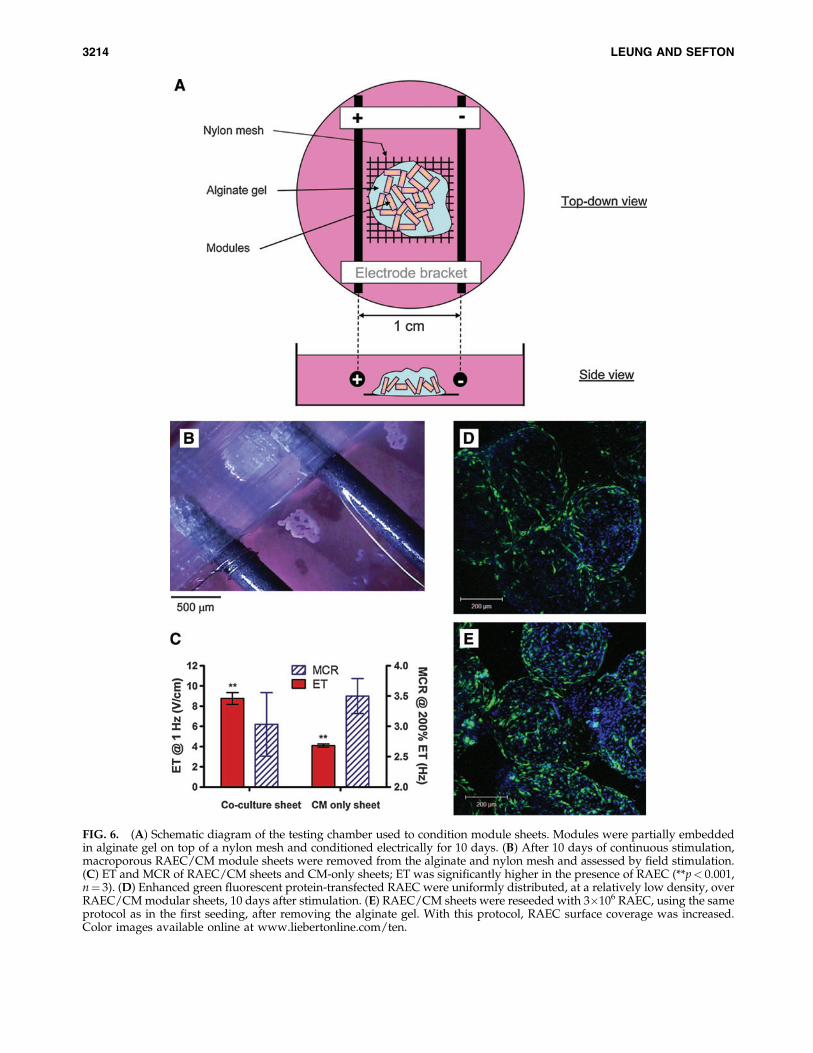
FIG. 6. (A) Schematic diagram of the testing chamber used to condition module sheets. Modules were partially embeddedin alginate gel on top of a nylon mesh and conditioned electrically for 10 days. (B) After 10 days of continuous stimulation,macroporous RAEC/CM module sheets were removed from the alginate and nylon mesh and assessed by field stimulation.(C) ET and MCR of RAEC/CM sheets and CM-only sheets; ET was significantly higher in the presence of RAEC (**p< 0.001,n¼ 3). (D) Enhanced green fluorescent protein-transfected RAEC were uniformly distributed, at a relatively low density, overRAEC/CM modular sheets, 10 days after stimulation. (E) RAEC/CM sheets were reseeded with 3�106 RAEC, using the sameprotocol as in the first seeding, after removing the alginate gel. With this protocol, RAEC surface coverage was increased.Color images available online at www.liebertonline.com/ten.
3214 LEUNG AND SEFTON
overall effects exerted by different environmental signals andenabled us to optimum culture conditions.
Coculture model
One of the main objectives of this study was to develop ameans of maintaining a functional RAEC layer on the surfaceof contractile CM-embedded modules. By adjusting thecomposition of the medium (Table 1) and particularly low-ering the glucose level to that of native EC medium, primaryRAEC remained viable and showed typical VE-cadherinexpression after 14 days, suggesting that the EC were in aquiescent state17 in the coculture medium. Using the samemedium, we were also able to generate viable RAEC/CMcoculture modular sheets.
To our knowledge, this is the first study to investigate theeffects of surface-coated EC on the electrical performanceand responsiveness of underlying CM in a modular con-struct. A 24 h transition period and seeding medium (a 50:50mixture of the native media) was used to enable RAECseeding and coverage. Without this seeding medium, RAECattachment and coverage was poor. We found that thepresence of a quiescent RAEC layer in the coculture medium,on either modules or modular sheets, permitted the con-tractile response of embedded CM, provided that the Mg2þ
concentration was at the low level of the CM medium andnot the high value of the native EC medium. However, theRAEC reduced the module and module sheet electrical re-sponsiveness, manifested as an increase in ET and decreasein MCR. These changes in electrical performance were notseen when CM-only modules were cultured in the samecoculture medium. We believe that these changes were mostlikely caused by the presence of the RAEC layer that mayhave changed the surface conductance of the modules. Suchchanges would cause the ET to increase compared to CM-only modules. Differences in membrane protein expression(e.g., connexin proteins) may also attenuate the propagation
of electrical impulses from the surface to the underlying CM,resulting in a lower MCR.
Judging from VE-cadherin staining, the native cardiacEC embedded in the modules did not form vessel-likestructures, nor did they appear to interact with surfaceseeded RAEC. However, these cardiac EC may still playan important role in CM maturation, as demonstrated inother multiculture cardiac constructs.18–20 For example,Iyer et al. showed the presence of fibroblasts and EC incardiac tissue constructs enhanced CM elongation andexcitability,21 possibly by spatially orienting CM into muscle-like bundles.
Our initial aim was to use the RAEC/CM modules as avehicle to uniformly deliver viable EC through out an as-sembled construct containing multiple modules. In a sepa-rate study involving RAEC modules (without embeddedcells) implanted in a rat omental pouch, we observed thatRAEC migrate from the modules and form chimeric (host-donor vessels) functional blood vessels in the intermodularspace after 21 days.22 We expect the RAEC on the coculturemodules to migrate and form vessels in a similar fashion,when implanted. Plans to evaluate the modular construct ina rat implant model are expected to reveal the fate and re-modeling process of RAEC/CM modules in vivo; how thisthen changes the in vivo electrical properties and morphologyof CM remains to be defined.
Sheet formation
A major challenge in creating clinically relevant, thick (1–10 mm) cardiac tissues in vitro is the lack of tissue vascula-ture. Specifically, the formation of blood vessels, both hostand graft derived, can be quite slow and insufficient to meetthe high nutrient demands of CMs. There are several ap-proaches designed to circumvent this limitation, includingthe use of oxygen carriers in the culture medium,23 incor-porating vessel-like flow channels in preformed scaffolds,24
FIG. 7. (A) Immunofluorescence staining of cardiac troponin I (green) shows a high density of CM near the perimeter ofmodules and at intermodule junctions (dash lines indicate edges of modules) in a module sheet after 10 days of stimulation.(B) At higher magnification, the CM appeared to form muscle-like bundles, similar to that seen in native cardiac tissue, butsuch bundles constituted only *12% of the field, shown in (A). Color images available online at www.liebertonline.com/ten.
MODULAR CARDIAC TISSUE ENGINEERING 3215
and preloading scaffolds with various growth factors and/orfunctional cell types that promote angiogenesis.25,26
We devised a method to produce a sheet-like structure froman assemblage of modules. A thin layer of modules was laidonto a nylon mesh and partially immobilized in alginate gel.At least some of the CM in these modules matured into muscle-like bundles and formed a physically integrated sheet capableof synchronous contraction when stimulated. Moreover, theCM at intermodular spaces expressed connexin-43, suggestingthat the module sheet may be electrically coupled. However,further electrophysiological assays are needed to characterizethe electrical coupling between modules.
Despite their morphology and contractile nature, thesemuscle-like bundles, as observed on histological sections, arerelatively thin and only occupy a fraction of the total volumeof the module. The lack of troponin-I-positive CM near thecore of the module suggests that the CM may have beencrowded out by the faster growing fibroblasts. However, thismay also be the result of active CM migration and matura-tion. Further studies at different time points are needed toreveal the mechanism of the remodeling process. Since weassume that these bundles are responsible for generatingthe contractile forces, it is crucial to increase the proportionof these bundles for the modular construct to produce
FIG. 8. (A, B) Triple-stained (vimentin, troponin I, and connexin-43) modular sheets after 10 days of stimulation indicate thatsurfaces of module sheets were covered by a layer of fibroblasts with an underlying layer of CM. (C, D) Connexin-43-positivestructures were also observed. (E, F) Colocalization of connexin-43 and troponin I was confirmed by overlaying high magnifi-cation images, suggesting that the CM were electrically coupled. Color images available online at www.liebertonline.com/ten.
3216 LEUNG AND SEFTON
physiologically relevant force. For instance, in future im-plementations it may be beneficial to reduce the diameter ofmodules so that the surface-to-volume ratio is increased, thusincreasing the contractile force on a per module basis. It isalso conceivable that over a longer incubation period in vitroor when implanted in vivo, more CM may migrate to thesurface, thus increasing the thickness of this layer. These is-sues will be explored in future studies.
In recent years, there has been interest in building cardiacconstructs using small functional subunits: for example, mi-crotissues,9 cardiac sheets,11 or other components.27 In theapproach most conceptually similar to modules, Kelm et al.demonstrated that human umbilical vein EC-coated micro-tissue (a ‘‘spheroid’’ without collagen gel) with embeddedCM was able to agglomerate to form a patch with functionalhuman umbilical vein EC-lined capillaries.10,28 In all cases,the goal was to create uniform, scalable, vascularized, andfunctional cardiac tissue, and the merits of one approachrelative to the other will be determined by the success ofmeeting these goals based on in vivo performance. Of par-ticular significance in this work is that coculture conditionsthat maintained both CM function and RAEC junctionmorphology were devised. We found, for example, thatRAEC/CM sheets that were not stimulated during culturefor 10 days did not contract when subject to external fieldstimulation.
We chose to embed modules in alginate not only becauseof its ease of application and removal by changing localcalcium concentration, but also because it is cell compatibleand noncell adherent. A single-module thickness across thesheet ensured that sufficient nutrients could diffuse into thecore of each module, at least in vitro. These macroporousmodule sheets will serve as intermediate components foreventually building thicker tissues in a scalable fashion,where the tissue maturation process can be monitored. Whenthe modules were assembled into sheets, there was a smalldecrease in the electrical responsiveness relative to modulesalone. This is likely the result of differing geometries of thestimulation apparatus resulting in differences in the fieldperceived by the CM. It is also conceivable that intermodularinteractions of surface-seeded RAEC may have lead to achange in tissue conductance.
Fabrication of functional cardiac tissue in vitro depends onmany parameters, including medium composition, extracel-lular matrix geometry, and cell seeding sequence. Nativemyocardium consists of CMs that form mechanically andelectrically coupled bundles. These bundles are encased inextracellular matrix secreted by cardiac fibroblasts, andmaintained by capillaries lined with EC. The three cell typesexist in their respective microenvironments. The RAEC/CMcoculture system described in this study consists of morethan two cell types, since the primary CM-enriched cellpopulation contained a significant quantity of fibroblasts andcardiac EC. While in this study, the electrical responsivenessof CM and the adherent junction morphology of RAEC werechosen as the primary endpoints, we must also consider thecontribution of cardiac fibroblasts and EC to these mea-surements. The data presented here suggest that cardiac fi-broblasts actively segregate and migrate to the surface ofthe modular sheet, and potentially play an important role inthe formation and remodeling of the modular sheet since thespatio-temporal relationships between CM and the various
supporting cell types have been shown to enhance CMmaturation and the overall functionality of construct.21,29
Therefore, aside from being a viable platform for in vitrotissue formation, the module system may also serve as a celldelivery vehicle that is capable of providing a supportiveenvironment to multiple cell types in a predeterminedseeding ratio and geometry.
Conclusion
We have created RAEC-coated, neonatal rat CM-embeddedmodules as well as module sheets. In both cases, embed-ded cells formed troponin-I-positive, native-muscle-likestructures (although to a limited extent), and contractedwith external field stimulation. In the presence of EC in anappropriate coculture medium, the construct became lessresponsive, although it continued to be excitable and con-tractile. While an in vivo study will be needed to elucidatethe fate of these modules, this study has demonstrated thefeasibility of creating functional cardiac tissue using themodular approach.
Acknowledgments
We acknowledge the financial support from the Cana-dian Institutes of Health Research through a team grantto R.D. Weisel (The Cardiac Regeneration Project), theNational Institutes of Health (EB006903), and through theTraining Program in Regenerative Medicine (PI: G.A. Levy).We acknowledge the technical assistance of Chuen Lo inharvesting the necessary tissues. We also acknowledgethe many helpful discussions with M. Radisic and herlaboratory.
Disclosure Statement
No competing financial interests exist.
References
1. Schmidt, D., Mol, A., Neuenschwander, S., Breymann, C.,Gossi, M., Zund, G., Turina, M., and Hoerstrup, S.P. Livingpatches engineered from human umbilical cord derived fi-broblasts and endothelial progenitor cells. Eur J Cardi-othorac Surg 27, 795, 2005.
2. Carrier, R.L., Papadaki, M., Rupnick, M., Schoen, F.J., Bur-sac, N., Langer, R., Freed, L.E., and Vunjak-Novakovic, G.Cardiac tissue engineering: cell seeding, cultivation param-eters, and tissue construct characterization. BiotechnolBioeng 64, 580, 1999.
3. Li, R.K., Jia, Z.Q., Weisel, R.D., Mickle, D.A., Choi, A., andYau, T.M. Survival and function of bioengineered cardiacgrafts. Circulation 100, II63, 1999.
4. Radisic, M., Park, H., Chen, F., Salazar-Lazzaro, J.E., Wang,Y., Dennis, R., Langer, R., Freed, L.E., and Vunjak-Novakovic, G. Biomimetic approach to cardiac tissue engi-neering: oxygen carriers and channeled scaffolds. Tissue Eng12, 2077, 2006.
5. Zimmermann, W.H., Fink, C., Kralisch, D., Remmers, U.,Weil, J., and Eschenhagen, T. Three-dimensional engineeredheart tissue from neonatal rat cardiac myocytes. BiotechnolBioeng 68, 106, 2000.
6. Radisic, M., Park, H., Shing, H., Consi, T., Schoen, F.J.,Langer, R., Freed, L.E., and Vunjak-Novakovic, G. Func-tional assembly of engineered myocardium by electrical
MODULAR CARDIAC TISSUE ENGINEERING 3217

stimulation of cardiac myocytes cultured on scaffolds. ProcNatl Acad Sci U S A 101, 18129, 2004.
7. Grayson, W.L., Martens, T.P., Eng, G.M., Radisic, M., andVunjak-Novakovic, G. Biomimetic approach to tissue engi-neering. Semin Cell Dev Biol 20, 665, 2009.
8. Vunjak-Novakovic, G., Tandon, N., Godier, A., Maidhof, R.,Marsano, A., Martens, T., and Radisic, M. Challenges in car-diac tissue engineering. Tissue Eng Part B Rev 16, 169, 2010.
9. Kelm, J.M., Ehler, E., Nielsen, L.K., Schlatter, S., Perriard,J.C., and Fussenegger, M. Design of artificial myocardialmicrotissues. Tissue Eng 10, 201, 2004.
10. Kelm, J.M., Djonov, V., Ittner, L.M., Fluri, D., Born, W.,Hoerstrup, S.P., and Fussenegger, M. Design of custom-shaped vascularized tissues using microtissue spheroids asminimal building units. Tissue Eng 12, 2151, 2006.
11. Shimizu, T., Yamato, M., Isoi, Y., Akutsu, T., Setomaru, T.,Abe, K., Kikuchi, A., Umezu, M., and Okano, T. Fabricationof pulsatile cardiac tissue grafts using a novel 3-dimensionalcell sheet manipulation technique and temperature-responsivecell culture surfaces. Circ Res 90, e40, 2002.
12. McGuigan, A.P., and Sefton, M.V. Design and fabricationof a vascularized tissue engineered construct. CardiovascPathol 13, S182, 2004.
13. McGuigan, A.P., and Sefton, M.V. Vascularized organoidengineered by modular assembly enables blood perfusion.Proc Natl Acad Sci U S A 103, 11461, 2006.
14. Yoshimitsu, M., Higuchi, K., Ramsubir, S., Nonaka, T., Ra-saiah, V.I., Siatskas, C., Liang, S.B., Murray, G.J., Brady,R.O., and Medin, J.A. Efficient correction of Fabry mice andpatient cells mediated by lentiviral transduction of hemato-poietic stem/progenitor cells. Gene Ther 14, 256, 2007.
15. Leung, B.M., and Sefton, M.V. A modular tissue engineeringconstruct containing smooth muscle cells and endothelialcells. Ann Biomed Eng 35, 2039, 2007.
16. Schwarzkopf, R., Shachar, M., Dvir, T., Dayan, Y., Holbova,R., Leor, J., and Cohen, S. Autospecies and post-myocardialinfarction sera enhance the viability, proliferation, and mat-uration of 3D cardiac cell culture. Tissue Eng 12, 3467, 2006.
17. Scharpfenecker, M., Fiedler, U., Reiss, Y., and Augustin,H.G. The Tie-2 ligand angiopoietin-2 destabilizes quiescentendothelium through an internal autocrine loop mechanism.J Cell Sci 118, 771, 2005.
18. Kobayashi, H., Shimizu, T., Yamato, M., Tono, K., Masuda,H., Asahara, T., Kasanuki, H., and Okano, T. Fibroblastsheets co-cultured with endothelial progenitor cells improvecardiac function of infarcted hearts. J Artif Organs 11, 141,2008.
19. Iyer, R.K., Chui, J., and Radisic, M. Spatiotemporal trackingof cells in tissue-engineered cardiac organoids. J Tissue EngRegen Med 3, 196, 2009.
20. Lesman, A., Habib, M., Caspi, O., Gepstein, A., Arbel, G.,Levenberg, S., and Gepstein, L. Transplantation of a tissue-engineered human vascularized cardiac muscle. Tissue EngPart A 16, 115, 2010.
21. Iyer, R.K., Chiu, L.L., and Radisic, M. Microfabricatedpoly(ethylene glycol) templates enable rapid screening oftriculture conditions for cardiac tissue engineering. J BiomedMater Res A 89, 616, 2009.
22. Chamberlain, M.D., Gupta, R., and Sefton, M.V. Chimericvessels in allogenic rat tissue engineering driven by en-dothelialized modules. 2009. (Submitted).
23. Iyer, R.K., Radisic, M., Cannizzaro, C., and Vunjak-Novakovic, G. Synthetic oxygen carriers in cardiac tissueengineering. Artif Cells Blood Substit Immobil Biotechnol35, 135, 2007.
24. Radisic, M., Marsano, A., Maidhof, R., Wang, Y., andVunjak-Novakovic, G. Cardiac tissue engineering usingperfusion bioreactor systems. Nat Protoc 3, 719, 2008.
25. Kaigler, D., Wang, Z., Horger, K., Mooney, D.J., andKrebsbach, P.H. VEGF scaffolds enhance angiogenesis andbone regeneration in irradiated osseous defects. J Bone Mi-ner Res 21, 735, 2006.
26. Chen, L., He, Z., Chen, B., Yang, M., Zhao, Y., Sun, W., Xiao,Z., Zhang, J., and Dai, J. Loading of VEGF to the heparincross-linked demineralized bone matrix improves vascular-ization of the scaffold. J Mater Sci Mater Med 21, 309, 2010.
27. Naito, H., Melnychenko, I., Didie, M., Schneiderbanger, K.,Schubert, P., Rosenkranz, S., Eschenhagen, T., and Zim-mermann, W.H. Optimizing engineered heart tissue fortherapeutic applications as surrogate heart muscle. Circula-tion 114, I72, 2006.
28. Kelm, J.M., Djonov, V., Hoerstrup, S.P., Guenter, C.I., Ittner,L.M., Greve, F., Hierlemann, A., Sanchez-Bustamante,C.D., Perriard, J.C., Ehler, E., and Fussenegger, M. Tissue-transplant fusion and vascularization of myocardial micro-tissues and macrotissues implanted into chicken embryosand rats. Tissue Eng 12, 2541, 2006.
29. van Luyn, M.J., Tio, R.A., van Seijen, X.J., Plantinga, J.A., deLeij, L.F., DeJongste, M.J., and van Wachem, P.B. Cardiactissue engineering: characteristics of in unison contractingtwo- and three-dimensional neonatal rat ventricle cell (co)-cultures. Biomaterials 23, 4793, 2002.
Address correspondence to:Michael V. Sefton, Sc.D.
Department of Chemical Engineering and Applied ChemistryInstitute of Biomaterials and Biomedical Engineering
164 College St., Room 407University of Toronto
Toronto, M5S 3G9Ontario, Canada
E-mail: [email protected]
Received: November 17, 2009Accepted: May 24, 2010
Online Publication Date: June 23, 2010
3218 LEUNG AND SEFTON
Related Documents