REVIEW published: 11 August 2020 doi: 10.3389/fbioe.2020.00955 Edited by: Susanna Sartori, Politecnico di Torino, Italy Reviewed by: Karina Nakayama, Oregon Health and Science University, United States Diana Massai, Politecnico di Torino, Italy *Correspondence: Manuel M. Mazo [email protected] † These authors have contributed equally to this work Specialty section: This article was submitted to Tissue Engineering and Regenerative Medicine, a section of the journal Frontiers in Bioengineering and Biotechnology Received: 10 April 2020 Accepted: 23 July 2020 Published: 11 August 2020 Citation: Montero P, Flandes-Iparraguirre M, Musquiz S, Pérez Araluce M, Plano D, Sanmartín C, Orive G, Gavira JJ, Prosper F and Mazo MM (2020) Cells, Materials, and Fabrication Processes for Cardiac Tissue Engineering. Front. Bioeng. Biotechnol. 8:955. doi: 10.3389/fbioe.2020.00955 Cells, Materials, and Fabrication Processes for Cardiac Tissue Engineering Pilar Montero 1† , María Flandes-Iparraguirre 1† , Saioa Musquiz 1,2 , María Pérez Araluce 1,3 , Daniel Plano 3,4 , Carmen Sanmartín 3,4 , Gorka Orive 2,4,5,6 , Juan José Gavira 4,7 , Felipe Prosper 1,4,8 and Manuel M. Mazo 1,4,8 * 1 Regenerative Medicine Program, Cima Universidad de Navarra, Foundation for Applied Medical Research, Pamplona, Spain, 2 NanoBioCel Group, Laboratory of Pharmaceutics, School of Pharmacy, University of the Basque Country – UPV/EHU, Vitoria-Gasteiz, Spain, 3 Department of Pharmaceutical Technology and Chemistry, University of Navarra, Pamplona, Spain, 4 IdiSNA, Navarra Institute for Health Research, Pamplona, Spain, 5 University Institute for Regenerative Medicine and Oral Implantology – UIRMI (UPV/EHU – Fundación Eduardo Anitua), Vitoria-Gasteiz, Spain, 6 Singapore Eye Research Institute, Singapore, Singapore, 7 Cardiology Department, Clínica Universidad de Navarra, Pamplona, Spain, 8 Hematology and Cell Therapy Area, Clínica Universidad de Navarra, Pamplona, Spain Cardiovascular disease is the number one killer worldwide, with myocardial infarction (MI) responsible for approximately 1 in 6 deaths. The lack of endogenous regenerative capacity, added to the deleterious remodelling programme set into motion by myocardial necrosis, turns MI into a progressively debilitating disease, which current pharmacological therapy cannot halt. The advent of Regenerative Therapies over 2 decades ago kick-started a whole new scientific field whose aim was to prevent or even reverse the pathological processes of MI. As a highly dynamic organ, the heart displays a tight association between 3D structure and function, with the non-cellular components, mainly the cardiac extracellular matrix (ECM), playing both fundamental active and passive roles. Tissue engineering aims to reproduce this tissue architecture and function in order to fabricate replicas able to mimic or even substitute damaged organs. Recent advances in cell reprogramming and refinement of methods for additive manufacturing have played a critical role in the development of clinically relevant engineered cardiovascular tissues. This review focuses on the generation of human cardiac tissues for therapy, paying special attention to human pluripotent stem cells and their derivatives. We provide a perspective on progress in regenerative medicine from the early stages of cell therapy to the present day, as well as an overview of cellular processes, materials and fabrication strategies currently under investigation. Finally, we summarise current clinical applications and reflect on the most urgent needs and gaps to be filled for efficient translation to the clinical arena. Keywords: cardiac tissue engineering, human pluripotent stem cells, material properties, cell differentiation, fabrication strategies Frontiers in Bioengineering and Biotechnology | www.frontiersin.org 1 August 2020 | Volume 8 | Article 955

Welcome message from author
This document is posted to help you gain knowledge. Please leave a comment to let me know what you think about it! Share it to your friends and learn new things together.
Transcript

fbioe-08-00955 August 9, 2020 Time: 12:3 # 1
REVIEWpublished: 11 August 2020
doi: 10.3389/fbioe.2020.00955
Edited by:Susanna Sartori,
Politecnico di Torino, Italy
Reviewed by:Karina Nakayama,
Oregon Health and ScienceUniversity, United States
Diana Massai,Politecnico di Torino, Italy
*Correspondence:Manuel M. Mazo
†These authors have contributedequally to this work
Specialty section:This article was submitted to
Tissue Engineering and RegenerativeMedicine,
a section of the journalFrontiers in Bioengineering and
Biotechnology
Received: 10 April 2020Accepted: 23 July 2020
Published: 11 August 2020
Citation:Montero P,
Flandes-Iparraguirre M, Musquiz S,Pérez Araluce M, Plano D,
Sanmartín C, Orive G, Gavira JJ,Prosper F and Mazo MM (2020) Cells,Materials, and Fabrication Processes
for Cardiac Tissue Engineering.Front. Bioeng. Biotechnol. 8:955.
doi: 10.3389/fbioe.2020.00955
Cells, Materials, and FabricationProcesses for Cardiac TissueEngineeringPilar Montero1†, María Flandes-Iparraguirre1†, Saioa Musquiz1,2, María Pérez Araluce1,3,Daniel Plano3,4, Carmen Sanmartín3,4, Gorka Orive2,4,5,6, Juan José Gavira4,7,Felipe Prosper1,4,8 and Manuel M. Mazo1,4,8*
1 Regenerative Medicine Program, Cima Universidad de Navarra, Foundation for Applied Medical Research, Pamplona,Spain, 2 NanoBioCel Group, Laboratory of Pharmaceutics, School of Pharmacy, University of the Basque Country –UPV/EHU, Vitoria-Gasteiz, Spain, 3 Department of Pharmaceutical Technology and Chemistry, University of Navarra,Pamplona, Spain, 4 IdiSNA, Navarra Institute for Health Research, Pamplona, Spain, 5 University Institute for RegenerativeMedicine and Oral Implantology – UIRMI (UPV/EHU – Fundación Eduardo Anitua), Vitoria-Gasteiz, Spain, 6 Singapore EyeResearch Institute, Singapore, Singapore, 7 Cardiology Department, Clínica Universidad de Navarra, Pamplona, Spain,8 Hematology and Cell Therapy Area, Clínica Universidad de Navarra, Pamplona, Spain
Cardiovascular disease is the number one killer worldwide, with myocardial infarction(MI) responsible for approximately 1 in 6 deaths. The lack of endogenous regenerativecapacity, added to the deleterious remodelling programme set into motion bymyocardial necrosis, turns MI into a progressively debilitating disease, which currentpharmacological therapy cannot halt. The advent of Regenerative Therapies over 2decades ago kick-started a whole new scientific field whose aim was to prevent oreven reverse the pathological processes of MI. As a highly dynamic organ, the heartdisplays a tight association between 3D structure and function, with the non-cellularcomponents, mainly the cardiac extracellular matrix (ECM), playing both fundamentalactive and passive roles. Tissue engineering aims to reproduce this tissue architectureand function in order to fabricate replicas able to mimic or even substitute damagedorgans. Recent advances in cell reprogramming and refinement of methods for additivemanufacturing have played a critical role in the development of clinically relevantengineered cardiovascular tissues. This review focuses on the generation of humancardiac tissues for therapy, paying special attention to human pluripotent stem cellsand their derivatives. We provide a perspective on progress in regenerative medicinefrom the early stages of cell therapy to the present day, as well as an overview of cellularprocesses, materials and fabrication strategies currently under investigation. Finally, wesummarise current clinical applications and reflect on the most urgent needs and gapsto be filled for efficient translation to the clinical arena.
Keywords: cardiac tissue engineering, human pluripotent stem cells, material properties, cell differentiation,fabrication strategies
Frontiers in Bioengineering and Biotechnology | www.frontiersin.org 1 August 2020 | Volume 8 | Article 955
fbioe-08-00955 August 9, 2020 Time: 12:3 # 2
Montero et al. Human Cardiac Tissue Engineering
A PERSPECTIVE ON CARDIAC DISEASEAND REGENERATIVE MEDICINE
Organ transplantation is one of the greatest medicalachievements of the 20th century. However, its applicabilityis hampered by donor shortage, life-long immunosuppressionand its success rates are linked to the experience of the surgicalteam. It requires a well-coordinated national effort, which issometimes hindered by ethical issues (Prabhu, 2019). The searchfor novel ways to approach organ repair inspired the field ofregenerative medicine, with Stem Cell Therapy as one of the mostrepresentative examples. Since this began, stem cells have beendiscovered even in low turnover adult tissues, such as the centralnervous system (Doetsch et al., 1999), the lung, (Rock et al., 2009;Barkauskas et al., 2013) or the heart, (Beltrami et al., 2003) andhave been widely assayed in animal models of disease, quicklyreaching clinical trials. This swift progression in general met withrapid failure, but on the bright side, it also enabled specialists togain immense insights into their mechanisms and ways of action.
Nothing exemplifies this journey better than the cardiac field.Cardiovascular diseases are well recognised as the leading causeof death worldwide, accounting for almost 1 in 2 deaths in Europeand causing 3.9 million deaths per year (Townsend et al., 2016).Ischemic heart disease (IHD) is one shade on this spectrum. Itis generally caused by the clotting of a coronary vessel, whichin turn leads to the death of a portion of the myocardium andthe subsequent functional impairment of the organ. Being mostlynon-regenerative, the heart is chronically impaired (Eschenhagenet al., 2017). A real epidemic, IHD is the leading single causeof death globally, responsible for over 15 million deaths in2016, ranking first in high- and lower-middle-income countries,and third in low-income countries (World Health Organization[WHO], 2018). As per 2015, over 22 million EU citizens wereliving with the disease, with approximately 3 million new casesyearly. IHD imposes an enormous burden on society as affectedpatients must be cared for by health systems, requiring lifelonghighly specialised medical attention and multimedication. It alsojeopardises the structure of the workforce and puts significantpressure on families. In terms of economic cost, the total burdenof IHD for EU economies is estimated at €59 billion/year. Ofthese, €19 billion is directly related to healthcare costs, while€20 billion is linked to productivity losses and the remaining€20 billion to the cost of indirect care. Estimations sketch outa grim future. In the United States, heart attacks are projectedto contribute more than $818 billion to annual healthcare costsand lost productivity by 2030, (Nowbar et al., 2019) while in theSouth Asia region, direct medical costs for CVD are estimatedto reach US $16.6 billion in 2021 (Walker et al., 2018). Organtransplant cannot match this overwhelming demand and becomea widespread therapeutic option (Stehlik et al., 2011).
It was hoped that cell therapy would provide new meansto regenerate the scarred myocardium. Remarkable discoveriesencouraged rapid progression towards clinical trials, with thefirst one launched in record time (Behfar et al., 2014). After theBOOST and REPAIR-AMI trials reported significant benefits incardiac function, (Wollert et al., 2004; Schächinger et al., 2006)hundreds of patients were recruited often in individual efforts
mostly based on local experience. This led to the first voicing ofconcerns. The primary initial objective, improvement of cardiacfunction, failed to be met in many cases. Although statisticallysignificant benefits in cardiac function were sometimes found,their magnitude was not as great as had been expected(reviewed in Gyöngyösi et al., 2016; Menasché, 2018). Thisreversal of fortunes coincided also with a remarkable twist inour understanding of what basic science and animal modelsconveyed: no true regeneration of the myocardium was achievedby adult stem cells. The underlying effects were mostly dueto the paracrine secretion of beneficial molecules (Hodgkinsonet al., 2016). Although this general perspective is valid for mostadult stem cells, use of their embryonic counterpart, althoughboosted by their capacity to give rise to new tissue oncetransplanted, was still marred by some common issues such aslack of proper engraftment, and crucially by safety concerns asteratoma formation and need for immune suppression, as wellas ethical issues.
Did stem cell regenerative medicine approaches fail? Theyobviously did not achieve the initial aims, but looking back,those can no doubt be branded as overambitious (Pagano et al.,2019). It did succeed in gathering a whole new compendiumof knowledge, which has led to renewed and better effortsin the regenerative direction and, importantly, has built aworldwide network of excellence encompassing not only differentnationalities, but also very diverse scientific disciplines. Oneof the greatest and perhaps now evident realisations in thefield is the notion that cells are not alone in a tissue, andthat the extracellular matrix (ECM) has a predominant role,not only as a passive architectural element, but crucially as asignal transducer and determinant of functionality (Majkut et al.,2013; Crowder et al., 2016; Kumar et al., 2017). Specifically inthe myocardium, the ECM is highly dynamic, changing duringdevelopment and disease. This latter change is bidirectional, asdisease induces pathological ECM deposition but an abnormalmatrix is able to produce malfunction (Frangogiannis, 2019).In consequence, it is now recognised that a cell-based cardiacregeneration without an adequate ECM is not viable. Generatingnew myocardium thus requires the participation of the mostpromising cells, with a surrounding matrix able to replicate theconditions of the native tissue and the proper 3D architecture.This is precisely one of the main directions of cardiacTissue Engineering.
Cardiac Tissue Engineering (cTE) is a highly interdisciplinaryscientific discipline, aiming at reproducing as accurately aspossible the function and biology of cardiac muscle, duringdevelopment or maturity, health or disease. Although itsfirst objective was focused on meeting the needs of cardiacregenerative Medicine, as knowledge and experience on how theECM influences cardiac cell biology increased and the fabricationcapacities widened, its scope has greatly expanded into areas suchas disease modelling, drug testing and personalised medicineamongst others (Feric et al., 2019; Noor et al., 2019; Mastikhinaet al., 2020). This review aims at presenting the reader withan overview of the specific characteristics of the myocardiumthat determine the needs regenerative cTE has to meet, as wellas providing a non-exhaustive revision of what the field has
Frontiers in Bioengineering and Biotechnology | www.frontiersin.org 2 August 2020 | Volume 8 | Article 955

fbioe-08-00955 August 9, 2020 Time: 12:3 # 3
Montero et al. Human Cardiac Tissue Engineering
delivered to attain this end, with a focus on human myocardiumand human pluripotent stem cells.
THE HEART
The mammalian heart is an incredible organ. Its main role isto provide a continuous unidirectional supply of blood to theorganism. This comes at a stringent metabolic cost, consumingthe equivalent of 6 kg of ATP per day, with a complete renewalof its ATP pool every 10 seconds. Most of this energy is obtainedthrough the oxidation of fatty acids in adulthood, though cardiacmetabolism is dependent on glucose during embryonic stages,being able to employ lactate as a metabolic substrate (Neejy,1974). Correct function is achieved through a specialised organarchitecture, dividing the heart into 4 chambers: atria, whichare smaller in size and muscular mass, receive blood and pushit out into the ventricles, which in turn pump either towardsthe lungs or the body. In consequence, the left ventricle islarger and has a thicker muscular wall than its right counterpart.Chambers, inlets and outlets are separated by valves impedingback flow. The heart is the first organ to function, around day8 in mice and the 4th gestational week in humans (Brand, 2003).It pumps continuously throughout life, efficiently ejecting bloodthrough an exquisite 3D structure, (Buckberg, 2002) establishedby a complex set of embryonic movements, cellular growthand incorporation (Günthel et al., 2018). Disruption of thisstructure is seen in disease and can be in itself the cause of organmalfunction: cardiac congenital defects and malformation are amain cause of perinatal death, (Bressan et al., 2013) but also giverise to many cardiomyopathies (McKenna et al., 2017).
Cardiac Embryonic Development: A BriefOverviewThe formation of the mammalian four-chambered heartencompasses a series of tightly coordinated morphological,cellular and molecular events (reviewed in Vincent andBuckingham, 2010; Meilhac et al., 2014; Ruiz-Villalba et al.,2016). Different pools of cardiac and extracardiac progenitorsare involved, including the mesoderm-derived First, Second andThird Heart Fields (FHF, SHF and THF respectively) and theCardiac Neural Crest Cells (CNCCs). Cells of the FHF contributeprimarily to the left ventricle (LV) but there is also a smallcontribution to the atria; SHF will form the right ventricle(RV), outflow tract (OFT), atria and part of inflow tract (IFT);(Buckingham et al., 2005). THF cells contribute to the sinusnode, some regions in the caval myocardium, and the Pro-Epicardial Organ (PEO) (Mommersteeg et al., 2010; Bressanet al., 2013). CNCCs arise from the dorsal neural tube, contributeto the parasympathetic innervation of the heart, valves andplay a pivotal role in OFT patterning and optimal septation(Keyte et al., 2014).
Early precardiac progenitors from the lateral mesodermhave been mapped into the mid-anterior region of theprimitive streak, characterised by the presence of both anteriorNodal/Activin and posterior bone morphogenetic protein (BMP)signalling at low levels, (Zhang et al., 2008; Vallier et al., 2009;
Yamauchi et al., 2010; Yu et al., 2011) promoting the emergenceof cardiogenic mesodermal MIXL1 + KDR + cells. As a resultof these signalling gradients set in gastrulation, multipotentcardiovascular progenitor (M) expressing the cardiac masterregulator MESP1 move in an anterior-lateral direction, forminga horseshoe-like region termed the cardiac crescent or FHF(Bondue et al., 2008; Chan S. S. K. et al., 2013). At amolecular level, MESP1 induces the expression of the minimalcore of the essential cardiogenic transcription factors includingISL1, TBX5, NKX2.5, and GATA4, in combination with thechromatin remodeller SMARCD3 (BAF60C), which further drivecardiomyogenesis (Vincent and Buckingham, 2010; Meilhacet al., 2014; Meilhac and Buckingham, 2018). The cardiac crescentfuses at the midline, forming the linear heart tube, which consistsof an interior layer of endocardial cells and an exterior layerof myocardial cells separated by an acellular, ECM-rich space,the cardiac jelly. Located central and dorsal to the FHF, SHFcells remain in contact with the pharyngeal endoderm and in aproliferative state as undifferentiated ISL1 + MEF2C + cells. Asdevelopment proceeds, SHF cells are added to the poles of theheart tube, with the tube looping to position the different regionsinto place. Chambers balloon out as a result of the differentialproliferation rates of CMs (Jong et al., 1997; Christoffels et al.,2004). As already mentioned, THF cells (TBX18 + NKX2.5-)contribute to the sinus node, caval myocardial cells and the PEO.PEO-cells give rise to the epicardium, and some cells of this layerundergo epithelial-to-mesenchymal transition to form epicardialderived cells (EPDC), which will differentiate into vascular cells(including the coronaries) as well as interstitial fibroblasts andvalvular cells, being essential for compaction (Pérez-Pomareset al., 2002; Weeke-Klimp et al., 2010; Katz et al., 2012). Lastly,CNCCs originate by delamination from the neuroectoderm,(Hildreth et al., 2008) initially contributing to smooth musclecells and CMs, (Mjaatvedt et al., 2001) and making a significantcontribution to the innervation of the organ and to the OFT(Hildreth et al., 2008; Sizarov et al., 2012). We refer the reader toTable 1 for a full description of the mentioned gene abbreviations.
Post-birth Cardiac Development: FoetalCMs vs. Adult CMsAside from the formation of the mammalian heart, CMs continueto develop postnatally (Guo and Pu, 2020). Embryonic CMscan beat spontaneously, express sarcomeric proteins and ionchannels, and exhibit action potentials and calcium transientswhich are significantly distinctive from their adult counterpart(Vincent and Buckingham, 2010; Meilhac et al., 2014). Humanand rodent embryonic CMs are around 30-40 fold less in sizeand feature an irregular shape, in comparison with adult CMs(Yang et al., 2014). These are characterised by an ultrastructuralorganisation with a large mitochondrial volume and specificmitochondria positioning between myofibrils. Sarcomeres inpostnatal CMs are long and well-aligned, in contrast to shorterand disarrayed ones found in foetal CM. At a metabolic level,embryonic CMs rely on glycolysis, whereas adult myocytespreferentially consume fatty acids, a much more efficient energysource. Myofibrillar protein isoform undergoes switching, being
Frontiers in Bioengineering and Biotechnology | www.frontiersin.org 3 August 2020 | Volume 8 | Article 955

fbioe-08-00955 August 9, 2020 Time: 12:3 # 4
Montero et al. Human Cardiac Tissue Engineering
TABLE 1 | Full description of genes names.
Abbreviation Description
MIXL1 Mix Paired-Like Homeobox
KDR Kinase Insert Domain Receptor
MESP1 Mesoderm Posterior BHLH Transcription Factor 1
ISL1 Islet-1 LIM Homeobox
TBX5 T-Box Transcription Factor 5
NKX2.5 NK2 homeobox 5
GATA4 GATA Binding Protein 4
SMARCD3 SWI/SNF Related, Matrix Associated, ActinDependent Regulator Of Chromatin, Subfamily D,Member 3
BX18 T-Box Transcription Factor 18
myosin heavy chain 7 (MYH7), myosin light chain 2 ventricularisoform (MLC2v), cardiac troponin I3 (TNNI3) and a shorterand stiffer Titin isoform, preferentially expressed in adult CMs,in contrast to myosin heavy chain 6 (MYH6), myosin light chain2 atrial isoform (MLC2a), and slow skeletal-type troponin I1(TNNI1) on foetal CMs (Bedada et al., 2014). All these differencesdirectly correlate with contractile capacity, with adult CMs ableto generate more force than embryonic ones (Vincent andBuckingham, 2010; Meilhac et al., 2014; Tan and Ye, 2018). Forexample, strips of adult rat myocardium have been reported toproduce a peak twitch tension of 56.4± 44 mN/mm2, (Hasenfusset al., 1991) whereas collagen constructs with neonatal rat CMsgenerated 0.4-0.8 mN/mm2 (Zimmermann et al., 2002). Thesame difference in magnitude is believed to exist for humancells, as comparisons with primary foetal human CMs are rare(Yang et al., 2014).
The cardiac action potential and associated channels andcurrents also distinguishes adult and foetal CMs. In immatureCMs, the expression of channels involved in repolarisation,including potassium transient outward channels, L-type calciumcurrents and the rectifying K + current (encoded mainly byKCNJ2), is lower than in adult cells resulting in a less negativeresting membrane potential (−50mv ∼ −60 mv in embryonicCMs) compared to normal (–85mv ∼ –90 mv in adult CMs)(Zhang et al., 2009). Also, the pacemaker current If is presentin embryonic CMs but does not occur in adult myocytes(Sartiani et al., 2007). The distribution of the gap junctionprotein connexin 43 (Cx43) also plays an important role inregulating electrical activity. While Cx43 concentrates at theintercalated disc of adult CMs, it is circumferentially distributedin immature CMs, which is not optimal for longitudinal electricalpropagation (Vreeker et al., 2014; Jiang et al., 2018). AdultCMs have a well-developed sarcoplasmic reticulum (SR) witha high level of SR-specific proteins like ryanodine receptor 2(RYR2) and sarcoplasmic/endoplasmic reticulum Ca2 + ATPase2a (SERCA2), (Ivashchenko et al., 2013) which, coupled withthe presence of transverse tubules (t-tubules), leads to a highlycoordinated Ca-induced-Ca-release and hence faster Ca transientkinetics and amplitude when compared to foetal CMs (Louchet al., 2015). Finally, where embryonic CMs are diploid, adultCMs present different degrees of polyploidy, achieved through
DNA-synthesis without karyokinesis (Adler and Costabel, 1975;Herget et al., 1997). Understanding how an embryonic CMevolves into a mature cell is already proving fundamental inhuman cTE. As the bioartificial tissues developed so far resemblemore their foetal counterpart, this insight is being incorporatedinto the effort of driving engineered tissues towards an adult-likefunctionality (Karbassi et al., 2020).
Heart Characteristics: What We Aim toEngineerGenerating human myocardial surrogates in the laboratoryrequires knowing what the natural composition and propertiesof the organ are. The following paragraphs provide an overviewof what nature has achieved, specifically, what the main cellularand extracellular components of the heart are, how they arearranged in space, and importantly, what this means regardingthe resulting material properties (Figure 1).
Cellular CompositionAs already explained, most cells forming the structure of the heartare of mesodermal origin: CMs, vascular (endothelial and smoothmuscle) cells and fibroblasts. Others reside in the tissue butare formed elsewhere, as immune cells, which play a significantrole in organ surveillance and disease. Deciphering the cellularcomposition of the heart has been a very controversial subject,be it in rodents or humans (Zhou and Pu, 2016). Histology candetermine that CMs are the largest fraction by volume. However,numbers vary, with reports of murine myocytes being the largestpopulation by number [56/27/7 for CMs/fibroblasts/endothelialcells respectively (Banerjee et al., 2007)] and others attributinggreater numbers to endothelial cells [43.6% vs 31% of CMs (Pintoet al., 2016)]. Human proportions are similarly contradictory,with some publications showing non-CM/endothelial cells arethe most abundant (Bergmann et al., 2015) and others endothelialcells (Anversa et al., 1978). Furthermore, several studies havereported varying cell proportions throughout the anatomicalregions of the organ (Sussman et al., 2002; Gaudesius et al.,2003; Camelliti et al., 2004, 2005; Kohl, 2004; Baudino et al.,2006). Things become more complicated if we take into accountthe age of the individual, as some claim the final number ofmyocytes is reached by one month, remaining constant over thelifetime of the individual (Bergmann et al., 2015) whereas othershave reported a 3.4-fold increase in CM number between 1 and20 years of age (Mollova et al., 2013). Other cell type numberschange dynamically over time, with a reported 6.5-fold increasein endothelial cells and an 8.2-fold increase for mesenchymalcells (including fibroblasts) during heart growth. Interactionsbetween these cells are multidirectional and exert great influenceover crucial aspects of cardiac biology. Both endothelial cellsand fibroblasts are key for tissue function and homeostasis.Aside from delivering oxygen and nutrients to the metabolicallydemanding CMs, the endothelium is fundamental for tissuehypertrophy and post-disease remodelling, (Holopainen et al.,2015) and maturation, (Giacomelli et al., 2020) displaying astrong paracrine influence (reviewed in Leucker and Jones,2014). Fibroblasts also affect organ function and cell maturity,(Woodall et al., 2016; Wang Y. et al., 2020) whilst other cell
Frontiers in Bioengineering and Biotechnology | www.frontiersin.org 4 August 2020 | Volume 8 | Article 955

fbioe-08-00955 August 9, 2020 Time: 12:3 # 5
Montero et al. Human Cardiac Tissue Engineering
FIGURE 1 | Main components of the mammalian heart. The two main constituents of the myocardium, cardiac cells and the surrounding ECM, both contribute toand modulate the specific material properties of the tissue.
types, such as immune cells, have been reported to display directand significant interactions with CMs, as is the case with theelectrical coupling of macrophages with atrioventricular nodecells (Hulsmans et al., 2017). All in all, the general consensussupported by unambiguous histological evidence is that CMs arethe largest fraction by volume, each nurtured by a median of 3capillaries, where fibroblasts constantly keep the ECM through adegradation-deposition equilibrium. This brings us to our nextplayer: the cardiac ECM.
ECM CompositionThe cardiac cell types discussed above are arranged within aglycoprotein matrix which supports and provides them with astructure. Moreover, the cardiac ECM also has an active role intransmitting contraction and avoiding hyper-stretching of CMs.Its principal component is collagen, which accounts for 2–5% of
the total weight of the heart, mainly types I (89%) and III (11%).Collagen type IV is present in the basement membranes, andcollagen type V is located in the pericellular space (Weber, 1989;Eghbali and Weber, 1990; Sommer et al., 2015a). The collagenmatrix has classically been categorised depending on whichelements it tethers together into endomysium (binds adjoiningCMs), perimysium (aggregates myocytes into myofibrils) andepimysium (present at the epicardial and endocardial surfaces).Cardiac fibroblasts have been identified as the main cell typeresponsible for secreting and remodelling the collagen matrix,although CMs seem to contribute to collagen type IV deposition(Eghbali et al., 1988). Apart from the structural function, thecollagen network makes an important contribution to the wholemyocardial tensile properties (Fomovsky et al., 2010).
Another key element of the cardiac extracellular matrix iselastic fibres. These are composites, made of an elastin core
Frontiers in Bioengineering and Biotechnology | www.frontiersin.org 5 August 2020 | Volume 8 | Article 955

fbioe-08-00955 August 9, 2020 Time: 12:3 # 6
Montero et al. Human Cardiac Tissue Engineering
surrounded by a myriad of microfibrils. They provide elasticproperties, by stretching upon mechanical demand and goingback to their original length once the load is removed. Hence theimportance of elastic fibres in tissues which have to accommodatetheir structure, such as skin, arteries or lungs or the heart.However, although elastic fibres are paramount for the heart’selasticity, other factors are known to have an influence uponit, namely the proportion of muscle bundles to fibrotic tissue,and the density of collagen crosslinking (Forrest and Jackson,1971; Parmley et al., 1973). In fact, elastic fibres are found inmost cases close to the collagenous network and in intimateassociation with it (Sato et al., 1983). Of note, mature elasticfibres show slight architectural differences depending on thetissue (Kielty et al., 2002). As aforementioned, elastin formsthe core of elastic fibres. Unlike most matrix proteins, whichundergo a constant/continuous deposition and turnover, inhealthy conditions elastin is synthesised only until adolescence(Dubick et al., 1981; Burnett et al., 1982; Davidson et al.,1982; Myers et al., 1985; Sephel et al., 1987; Parks et al.,1988; Pollock et al., 1990; Ritz-Timme et al., 2003). Otherfundamental components of the cardiac matrix include, to a lesserextent, laminin, fibronectin, proteoglycans and glycoproteins(Fan et al., 2012). Laminin molecules are part of the basementmembrane and are thus in close contact with the cell, playingan active role in modulating cell behaviour, including migration,differentiation and phenotype stabilisation (Yap et al., 2019).Fibronectin, besides promoting cell attachment, acts as an ECMorganiser and is involved in collagen deposition (Valiente-Alandiet al., 2018). However, all components are crucial for tissueintegrity and function.
Many, if not all, cardiovascular diseases have repercussionsfor the cardiac ECM. The reverse is also true. For instance,infarction studies in pigs show that the collagenous networkstarts to become disarranged after just 20 min of coronaryocclusion, whilst elastin begins to disappear after 40 min, andboth components appear detached from the basement membraneafter 120 min. The balance of collagens I and III has beenwidely studied, revealing a significant increase in type III collagenafter myocardial infarction (MI) (Sato et al., 1983). Dilatedcardiomyopathies, in which the shape of the cardiac cavityis abnormal, are at least partly related to aberrant collagenremodelling, with less thick collagen and thinner fibres, whichresults in weaker tensile properties, more muscle slippage andwall thinning (Weber et al., 1988; Weber, 1989). Ventricularhypertrophy consists of the thickening of the ventricular wallassociated with some conditions like hypertension, and it is foundtogether with overexpression of collagen in the form of interstitialfibrosis (Eghbali and Weber, 1990).
Cardiac ArchitectureIn most tissues, structure and function are closely intertwined,and the heart is no exception. However, certain aspects ofthis relationship are still under debate. The overall mannerin which the heart contracts and pumps blood is known,as is the arrangement of the tissue microstructure. The gaplies in providing a theory that explains how the differentarchitectural elements interact to produce the global behaviour.
For example, there is an ongoing debate about whether themyocardium forms a single myocardial band, (Buckberg et al.,2015a,b) or the so-called myocardial mesh model is moreaccurate (MacIver et al., 2018a,b). The controversy has twoaspects. On the one hand, there is no consensus on whetherthe basic functional unit is the CM, or groupings of thisinto bundles (groups of CMs), sheetlets (groups of bundles),sheets (groups of sheetlets) or even laminae (groups of sheets).On the other hand, although imaging techniques allow us tovisualise phenomena across the whole myocardium, it is notfeasible to ascertain the distinct contribution of the individualfunctional units to producing the global outcome. Accordingto Buckberg et al., the CM can undergo six functional events:shortening, lengthening, narrowing, widening, twisting anduncoiling (Buckberg et al., 2015a, 2018). There are an estimated2.5-10 billion cells (Bergmann et al., 2015) in the heart, eachof them performing one or more of these six actions in thesame or a different direction, and all we are able to see is themacroscopic effect: a torsion-contraction movement of the organ.Still under controversy, there are at least 7 proposed modelsto accurately describe cardiac architecture, (Gilbert et al., 2007)which is widely recognised to have a profound effect, whetherat a mechanical (LeGrice et al., 1995; Zócalo et al., 2008) orelectrical (Roberts et al., 1979; Taccardi et al., 2008) level. Duringdisease, myocardial architecture is severely disarranged, leadingto inefficient contraction (Roberts et al., 1987; Wickline et al.,1992). It is expected that the application of advanced technologylike diffusion tensor MRI (DT-MRI), which can obtain highlydetailed information on fibre architecture, will soon shed light onthis debate (Scollan et al., 1998; Poveda et al., 2013).
At a simpler histological level, CMs (and CMbundles/sheetlets) are arranged in different orientationsdepending on their location in the organ, which in turndetermines the direction of the stress produced. Myocytes arealways in intimate contact with capillaries, which no doubt stemsfrom the high metabolic demand of an ever-working musculartissue: capillaries are located within 20 µm of CMs. Each CM issurrounded by a basement membrane containing laminin andcollagen type IV, amongst others, and embedded in a highlystructured ECM where collagen type I, as already discussed, is themain component (Figure 2). CMs connect to each other mainlyby intercalated disks at their ends, but also through side branches,coupled to at least 2 CMs on the long axis and 1 laterally (Spachand Heidlage, 1995). Intercalated disks contain gap junctions,allowing fast current flow between neighbouring cells (Klabunde,2012). As mentioned above, both individual CMs and groupingsof these are surrounded by enveloping collagen. Fibroblasts donot participate in the electrical syncytium formed by the CMs,but rather lie in the interstitial space.
Cardiac Biophysical PropertiesWhen attempting to engineer a tissue, it is essential to carefullyrecapitulate not only the cellular-extracellular components andtheir architecture, but also the resulting biophysical properties.These must reliably mimic those of their natural counterpart.In the heart, material properties are very complex: not onlyare they direction-dependent, but they also vary within the
Frontiers in Bioengineering and Biotechnology | www.frontiersin.org 6 August 2020 | Volume 8 | Article 955

fbioe-08-00955 August 9, 2020 Time: 12:3 # 7
Montero et al. Human Cardiac Tissue Engineering
FIGURE 2 | Cardiac structure. The endocardial-to-pericardial structure is outlined, with the main cellular and extracellular components.
anatomical regions and the stage of the cardiac cycle. As anexample, the literature reports variations in stiffness betweenthe beginning and the end of diastole higher than an order ofmagnitude. For human LV, the reported values are 10-20 kPa atthe beginning of the diastole, and 200-500 kPa at the end (Chenet al., 2008). Contraction itself results in a significant stiffening:from 0.5 to > 10 kPa. This magnitude is species-dependent,with a reported 3-fold increase in mouse, (Jacot et al., 2010) 2-fold in rat, (Prakash et al., 1999) and over 20 times in zebrafish(Krieg et al., 2008). Development also leads to a stiffening in thetissue, which arises from a relatively soft mesodermal layer (Krieget al., 2008). Disease severely stiffens the organ, mostly due tothe excessive deposition of collagen (scar for MI, interstitial inhypertension or other conditions), with values of over 50-100 kPa(Engler et al., 2008). Stiffness itself has a fundamental influenceon how efficient CM contraction is, with CMs on softer- orstiffer-than-normal substrates doing little work or overstrainingthemselves, respectively (Engler et al., 2008). Reports on cardiacmechanical properties are extremely variable. This stems from
a mix-up of animal vs human, fresh vs fixed, and healthy vsdiseased data. In general, it is now accepted that most of theheart’s passive mechanical properties are due to the collagenin the matrix, (Sommer et al., 2015a) but at short sarcomerelengths the protein titin is the predominant contributor (Nguyen-Truong and Wang, 2018). Quoting Sommer et al., ‘results suggestthat the passive human LV myocardium under quasi-staticand dynamic multiaxial loadings is a non-linear, anisotropic(orthotropic), viscoelastic and history-dependent soft biologicalmaterial undergoing large deformations’ (Sommer et al., 2015b).Or in simpler words: it is very complex and with multiplecontributions from cellular/extracellular components. Added tothis, scales differ, depending whether the tissue is macroscopicallycharacterised using biaxial mechanical tests, (Sommer et al.,2015b) or whether isolated CMs are probed at a cell-relevantscale with Atomic Force Microscopy (AFM) (Andreu et al., 2014).Furthermore, some conflicting results have been reported, fromreports showing force production from CMs to be increasedwith increasing stiffness, (Bhana et al., 2010) to stiffness having
Frontiers in Bioengineering and Biotechnology | www.frontiersin.org 7 August 2020 | Volume 8 | Article 955

fbioe-08-00955 August 9, 2020 Time: 12:3 # 8
Montero et al. Human Cardiac Tissue Engineering
no influence at all (Jacot et al., 2008). In fact and as explainedby Domian et al. (2017), it might even be the case that thematerial properties of cardiac tissue are not the main actor in themyocardial scenario, but this role is rather played by chamberpressure. More experimental and theoretical work needs to bedone in this area before we reach a definitive conclusion.
Adding another layer of complexity, the heart has constant,potent and highly relevant electrical activity. Sparking at a smalland specialised region called the sinoatrial node, the electricalwave travels through the auricles, reaching the atrioventricularnode where it is delayed (allowing for the filling of theventricles), and then spreads apex-to-base through the ventriclesin a coordinated manner. All this process is controlled by asingular CM type, termed pacemaker cell, displaying disarrayedsarcomeres and low work generation capacity, but able toautonomously start the cardiac action potential. Ultimately, theaction potential results in the entry of Ca+2 ions into theCM, releasing the sarcoplasmic stores of Ca+2 and freeingmyosin of the inhibitory action of troponin I. CMs are alsoelectrically connected through connexins, which form bridgesbetween the cytoplasm of adjacent myocytes, effectively makingthe myocardium an electrical syncytium. However, it is ananisotropic one, with faster propagation in the direction of thefibres as opposed to the transverse direction (Chung et al., 2007).Conduction velocity is the speed with which the cardiac impulsetravels from one point in the tissue to another. In adulthood,it lies in the range of 0.3-1 m/s, but developmental stage anddisease will affect it (Yang et al., 2014). Achieving a similar valuein any cardiac engineered tissue is paramount, given the factthat a mismatch between conduction velocities may give riseto potentially fatal electrical abnormalities such as arrhythmias(Kadota et al., 2013; Zhang et al., 2018). It is interesting thatone of the foci in cTE is towards providing material-basedelectronic conductivity, although cardiac cells do not function bytransmitting electrons but ions.
As mentioned already, both the mechanical and electricalproperties exert a strong influence upon myocardial biology andfunction, in both health and disease. For example, increasedfibrosis due to pathological conditions like MI or hypertensionsignificantly stiffens cardiac muscle and affects CM contraction(Sessions and Engler, 2016). Ventricle loading induces CMelongation which, as explained by the Frank-Starling law,renders higher stroke with increase diastolic filling (Solaro,2007). The coordinated conduction of the depolarisation wavethroughout the organ, including the atrioventricular delayand the apex-to-base transmission, all contribute to optimalfunctionality and must be taken into account when engineeringa human myocardium.
ENGINEERING CARDIAC TISSUE: THEBUILDING BLOCKS
cTE aims to generate tissue surrogates, either micro or macro,for various purposes, from developmental biology, (Young andEngler, 2011) to therapy (Miyagawa et al., 2018). In the followingparagraphs, we will outline the main cells and materials, as well as
the different fabrication technologies assayed in the field and theprocedures for their maturation. Table 2 summarises some of themost relevant engineered myocardium examples, with a focus onhuman cardiac tissue.
CellsThe capacity to obtain human cardiac cell phenotypes in thelaboratory began with the derivation of human embryonic stemcells (hESC) by Thomson and colleagues in 1998, (Thomsonet al., 1998) which was soon followed by the first protocols fordifferentiation towards CMs (Mummery et al., 2003; Kattmanet al., 2006). In 2006, thanks to the breakthrough of thereprogramming technology (Takahashi and Yamanaka, 2006; Yuet al., 2007) it became possible to relieve the field of some ofits most notorious encumbrances, including the ethical ones.Both, hESCs and human induced pluripotent stem cells (hiPSC)fall within the wider category of human pluripotent stem cells(hPSC). Current methods, including scaling up protocols, (Serraet al., 2011) have paved the way for their widespread use. Ingeneral direct, efficient and reproducible hPSCs differentiationmethods try to recapitulate embryonic development, from theinduction of cardiac mesoderm, to CM, endothelial cells (ECs),cardiac fibroblast (CFs) or smooth muscle cells (SMCs) in vitrospecification and maturation (Figure 3) (Burridge et al., 2015).
According to the culture format employed, derivation ofCMs, CFs, ECs and pericytes/SMCs from hPSCs can becategorised into 3 main approaches: (i) inductive co-culturewith visceral endodermal-like cells, (ii) suspension aggregatessuch as three dimensional (3D) embryoid bodies (EBs) and (iii)two-dimensional (2D) cell monolayer differentiation (Mummeryet al., 2003; Kattman et al., 2006; Laflamme et al., 2007; Morettiet al., 2010). Early reports showed that co-culturing hPSCs withthe mouse endodermal cell line END2 was able to induce beatingfoci (MacIver et al., 2018a). The low efficiency of this method,as well as the need for xenogenic co-culture, precluded itswidespread application. EBs are formed by culturing dissociatedhPSC in non-adherent plastic dishes and partially recapitulatethe 3D structure and interactions of a developing embryo. hESC-EBs differentiate to derivatives of the three primary germ layers,resulting in spontaneously contracting outgrowths of human CM(Kehat et al., 2001). Based on EB differentiation protocols, CMfrom a variety of hESC and hiPSC lines have been generated,usually with a purity of < 10% (Zhang et al., 2009). ECs canalso be isolated from spontaneously differentiating EBs, at asimilarly low yield (≈2%) (Levenberg et al., 2002). In bothcases, early reports explored the addition of cardiac mesoderm-inducing growth factors, including FGF2, VEGF BMP4, ActivinA, Wnt agonists (WNT3A) or antagonists (DKK1), amongstothers (Yuasa et al., 2005; Kattman et al., 2006, 2011; Yang et al.,2008; Tran et al., 2009; James et al., 2010). In general, however,EB-based differentiations have lost ground to more advancedand defined procedures, as the former are generally inefficientand render a mixture of cardiac cells with other non-cardiacphenotypes, requiring additional purification.
Monolayer-based differentiation is nowadays the most usuallyapplied method. Cytokine-based protocols were developed first(Taccardi et al., 2008). These have been progressively modified
Frontiers in Bioengineering and Biotechnology | www.frontiersin.org 8 August 2020 | Volume 8 | Article 955

fbioe-08-00955A
ugust9,2020Tim
e:12:3#
9
Montero
etal.H
uman
Cardiac
TissueE
ngineering
TABLE 2 | Summary of materials, cells and methods employed to engineer cardiac tissues, their biomimicry and resulting outcome.
REF Materials Fabrication Cellular mimicry Material mimicry Maturation Benefit of the selected approach?
Hydrogel Fibres CM EC SMC CF Mech. Elect. Align Mech Elec vs. Gene exp. Structure Function
Godier-Furnémont et al., 2015 Col I – Mould casting Rat neonatal Yes No No + + NM nd + +
Hirt et al., 2014 Fibrin . Mould casting Rat neonatal Yes No No + + EHT + + +
Jackman et al., 2018 FGN – Mould casting Rat neonatal Yes No No + – NM + + +
Amdursky et al., 2018 Albumin – Mould casting Rat neonatal Yes No No – – 2D + + –
Nunes et al., 2013 Col I – Mould casting hPSC + – – – + NM + + +
Ruan et al., 2016 Col I – Mould casting hPSC + – – + + EHT + + +
Tiburcy et al., 2017 Col I – Mould casting hPSC FK + – – + – 2D nd + +
Valls-Margarit et al., 2019 Col I + ELN – Mould casting hPSC FK + – – + + 2D nd + +
Zhang et al., 2013 Fibrin – Mould casting hPSC nd – – – – 2D + + +
Hirt et al., 2014 Fibrin . Mould casting hPSC + – – + + EHT + + +
Weinberger et al., 2016 Fibrin – Mould casting hPSC hPSC + – – + – NM nd – –
Ulmer et al., 2018 Fibrin – Mould casting hPSC + – – + – 2D – + nd
Ronaldson-Bouchard et al., 2018 Fibrin – Mould casting hPSC DF + – – + + 2D + nd nd
Jackman et al., 2016 FGN – Mould casting hPSC + – – + – NM nd + +
Shadrin et al., 2017 FGN – Mould casting hPSC hPSC hPSC + – – + – NM + – +
Dattola et al., 2019 PVA* – Foaming + FD hPSC + – – – – 2D nd – nd
Han et al., 2016 – PCL SE hPSC – – + – – 2D + + –
Joanne et al., 2016 – Col I SE hPSC + – – – – NM nd + +
Sireesha et al., 2015 – POCS–FGN SE hCM + – – – – 2D nd + nd
Khan et al., 2015 – PLGA SE hPSC – – + – – 2D – + –
Roshanbinfar et al., 2020 – Col I/HA/PANi SE hPSC + + – – – EHT nd + +
Macqueen et al., 2018 – PCL/Gelat Pull spinning hPSC + – + – – 2D nd + +
Castilho et al., 2018 Col I PCL MEW hPSC – – – – – 2D + + nd
Vaithilingam et al., 2019 PETra + MWCNT 3DP–SLA hPSC No + + – – 2D nd + nd
Lee et al., 2019 Col I – 3DbioP hPSC CF + – – – – NM nd + –
Maiullari et al., 2018 FGN/PEG – 3DbioP hPSC HUVEC nd – – – – EHT nd + nd
Noor et al., 2019 dECM – 3DbioP hPSC hPSC + – – – – NM nd + +
Arai et al., 2018 – – 3DbioP hPSC HUVEC DF nd – – – – EHT nd + nd
REF, Reference; CM, Cardiomyocyte; EC, Endothelial cell; SMC, Smooth muscle cell; CF, Cardiac fibroblast; Mech, mechanical; Elec, electrical; Gene exp., Gene expression; Col I, collagen type I; FGN, Fibrinogen; hPSC,human pluripotent stem cell; FK, Foreskin fibroblast; DF, Dermal fibroblast; PVA, poly-vinil-alcohol; FD, Freeze-drying; ∗, foam; SE, Solution electrospinning; POCS, poly[1,8-octanediol-co-(citric acid)-co-(sebacic acid)];hCM, primary human CM; PLGA, polylactide-co-glycolide; PCL, polycaprolactone; Gelat, Gelatin; PETrA, pentaerythritol triacrylate; MWCNT, Multi-walled carbon nanotubes; 3DP,3D Printing; SLA, Stereolithography;PEG, polyethylene glycol; ALG, Alginate; 3DbioP, 3D bioprinting; nd, not described; dECM, decellularised extracellular matrix; ELN, elastin; HA, Hyaluronic acid; PANi, polyaniline; NM, native myocardium; EHT,engineered heart tissue.
Frontiersin
Bioengineering
andB
iotechnology|w
ww
.frontiersin.org9
August2020
|Volume
8|A
rticle955

fbioe-08-00955 August 9, 2020 Time: 12:3 # 10
Montero et al. Human Cardiac Tissue Engineering
FIGURE 3 | Cardiac differentiation of hPSC. hPSC differentiation in vitro mimics embryonic development. Induction signals, main molecular pathways and lineagemarkers are outlined.
Frontiers in Bioengineering and Biotechnology | www.frontiersin.org 10 August 2020 | Volume 8 | Article 955

fbioe-08-00955 August 9, 2020 Time: 12:3 # 11
Montero et al. Human Cardiac Tissue Engineering
by the discovery of Wnt signals playing a biphasic role in cardiacdifferentiation in vivo, (Marvin et al., 2001; Ueno et al., 2007) withearly signals directing hPSCs towards cardiac fate, whilst laterinhibition of those signals is a prerequisite for CM specification.Almost 10 years ago, this concept was incorporated into the CMdifferentiation from hPSCs, (Lian et al., 2012) paving the way forthe grounding of a chemically defined procedure (Burridge et al.,2014). Based on small molecules rather than cytokines, and thusless costly, this protocol is now widely applied, providing highlypure yields of hPSC-derived CMs when in combination with ametabolic-based selection (Tohyama et al., 2013). This meansthat complicated and inefficient EB-forming procedures orexpensive and time-consuming immune-selection protocols havenow been discarded (Burridge et al., 2007; Hattori et al., 2010;Elliott et al., 2011; Uosaki et al., 2011). However, even this latestprotocol still requires a degree of set up to avoid inconsistentefficiencies amongst cell lines and experimental repeats, mostlyrelated to different patterns of endogenous early canonical Wntexpression (Paige et al., 2010). In general, CMs obtained fromthese protocols consist of a mixture of pacemaker, atrial andventricular myocytes, though some researchers consider that thisis open to question, as hPSC-CMs are immature and intrinsicallyplastic (Du et al., 2015).
The derivation of other cardiac phenotypes has been alsoachieved, with a variety of protocols now available. Palpant et al.reported the generation of CMs, cardiac- or hemogenic-derivedECs as well as blood cells by finely dosing BMP4 and ActivinA in order to pattern hPSC towards different mesodermal fates(Palpant et al., 2017). Others have employed a mixture of smallmolecules and cytokines to derive vascular cells from hPSCs inmonolayer culture with high efficiency (Orlova et al., 2014; Patschet al., 2015). Global gene transcription analysis has demonstratedlow variability between ECs differentiated via cytokine-basedmethods from multiple lines of hPSCs (White et al., 2013). CFs,have been increasingly recognised as major players in cardiacdevelopment and homeostasis, having a similarly significanteffect upon the capacity to build cardiac tissues in the lab.Recently, two independent groups have reported the generationof hPSC-derived CFs, giving also proof of their capacity to affecthPSC-CM function (Zhang H. et al., 2019; Zhang J. et al., 2019).Epicardial cells have similarly been derived, (Witty et al., 2014)demonstrating their ability to increase the therapeutic capacityof hPSC-CMs in vivo (Bargehr et al., 2019). Finally, sinoatrialnode pacemaker CMs have been obtained from hPSC, and theircapacity to pace tissues in vivo has been reported (Protze et al.,2017). Other approaches to the differentiation of cardiac lineagesinclude the generation of CVPs, (Blin et al., 2010; Birket et al.,2015; Zhang Y. et al., 2016) or direct reprogramming strategies,(Mohamed et al., 2017) but they have rarely been explored in cTE.
MaterialsIn parallel to the way differentiation of hPSC mimics the naturalembryonic development, the current view is that the more amaterial replicates the properties of cardiac tissue, the higher thechances of success. Development over the last 15 years has yieldeda wide portfolio of materials and biomaterials. Classificationsare numerous, be it by origin (natural, synthetic or hybrid),
crosslinking (chemical vs physical), size (macro, micro or nano),polymerisation mechanism (enzymatic, light-triggered or pH-responsive) or whether they are or not reinforced with otherstructures like fibres. For specific insight into these classifications,we direct the reader towards some of the excellent latest papers(Peña et al., 2018; Liu et al., 2019; Xu et al., 2019). One of the mostrelevant classifications is, however, on the physical consistencyof the applied material, where we can differentiate (i) injectablematerials and hydrogels, (ii) solid or fibrous scaffolds and (iii)composite systems.
Hydrogels are probably the most widely explored type ofmaterial in cTE. Collagen, being the main component of thecardiac ECM, has been widely employed (see the followingsections). It can be readily isolated from animal or even humantissues in sufficient quantities, extracted and solubilised, althoughit requires acidic pH for this. Therefore, a careful control overpH is needed for optimal polymerisation and embedded cellsurvival. Gelatin, being denatured/digested collagen, has alsobeen intensely explored and the basis for the generation of someof the most applied semi-synthetic materials, such as gelatinmethacryloyl (GelMA)(Yue et al., 2015) and several biorthogonalderivatives (Koshy et al., 2016; Bertlein et al., 2017). Alginate, asugar-based natural hydrogel obtained from algae, (Orive et al.,2006) has been employed due to their tailorable mechanicalproperties and simple polymerisation, mediated by cations suchas Ca or Mg, albeit lacking biological binding motifs. Silk andits derivatives have also been processed into hydrogels, (Hollandet al., 2019) and modified to incorporate electrically activeparticles (Barreiro et al., 2019) or photocrosslinkable chemicalgroups (Cui et al., 2020). Allergic and anti-inflammatory sidereactions have been reported with some silk derivatives, so careshould be taken when incorporating them into an engineeredtissue. Amongst natural hydrogels, decellularised ECM (dECM)has attracted significant interest since the breakthrough discoveryof the process, as applied to the building of tissues (Ott et al., 2008;Belviso et al., 2020). In principle, dECM retains all components ofthe ECM of origin, thus creating a more complex and biomimeticenvironment. Garreta et al. obtained human dECM slices onwhich they cultured hPSC-CMs. The human CMs demonstratedenhanced conduction velocity and gene expression of relatedgenes (SERCA, KCNJ2, CACNA1C or SCN5A amongst others)(Garreta et al., 2016). The group of Lior Gepstein employeddECM-chitosan mixtures in combination with hiPSC-CMs. Theresulting engineered myocardium displayed enhanced maturityas compared with cells cultured in 2D, showing tissue-like drugresponses. Their work also provided proof-of-concept of thecapacity of this system to model human cardiac diseases (LongQT syndrome) and arrhythmias (Goldfracht et al., 2019). Onthe synthetic side, polyethylene glycol (PEG) and its severalmodifications have been extensively studied in the TE field(Iyer et al., 2009).
Most of the materials so far outlined in this section canbe processed into fibres employing strategies explained in thenext section. Thermoplastics have also been applied to cTE,mostly as fibrous scaffolds. Examples include poly-ε-caprolactone(PCL), (Woodruff and Hutmacher, 2010) or elastomers like poly(glycerol-sebacate) (PGS) (Kharaziha et al., 2013). Conductive
Frontiers in Bioengineering and Biotechnology | www.frontiersin.org 11 August 2020 | Volume 8 | Article 955
fbioe-08-00955 August 9, 2020 Time: 12:3 # 12
Montero et al. Human Cardiac Tissue Engineering
polymers have only recently began to be applied to the field,though some of the most remarkable examples have notincorporated the use of cells and would therefore not qualifyas engineered tissues (Mawad et al., 2016; Kapnisi et al., 2018).Finally, given the low mechanical properties displayed by mosthydrogels, composite fibre-reinforced materials are also beingdeveloped, (Bas et al., 2015) with some examples explained in thefollowing section.
Maturation StimuliCells derived from hPSC are immature (see Karbassi et al., 2020for a review). Although not the direct focus of this work, neonatalmyocytes, which are another cell source commonly employed,also suffer from this drawback. cTE has long been aware of thislimitation and has applied three main stimuli, namely physical,mechanical and electrical, and combinations thereof Parsa et al.(2016) and Stoppel et al. (2016). Perfusion is able to improveengineered tissues’ properties, as it will boost nutrient access andrenewal, as has been shown for neonatal cells (Radisic et al.,2004b) as well as hiPSC-cardiac derivatives (Valls-Margarit et al.,2019). Materials’ physical properties (e.g., stiffness) are able toinduce maturation features in these CMs, or at least preserveprimary CMs from dedifferentiation (Amdursky et al., 2018). Ingeneral, most hydrogels are able to replicate the right myocardial-like properties. For example, Feaster and colleagues found thatplating hiPSC-CMs on thick Matrigel induced a certain degreeof molecular and functional maturation, in comparison to thin,diluted hydrogel coating, which essentially transmits the rigidityof the underlying plastic (Feaster et al., 2015). Herron et al.employed soft (albeit supra-cardiac) substrates and comparedthem with glass in their capacity to influence hiPSC-CMs, witha significant effect on maturation, showing improvement in theexpression of Na and K channels, as well as on the degree ofbinucleation, cell cycle exit and hypertrophy (Herron et al., 2016).Although not purely based on engineered tissues, they and othersgive proof of the crucial role stiffness plays in cardiac maturation.
Mechanical stimulation has a leading role in cardiacdevelopment and aging (Happe and Engler, 2016; Sessionsand Engler, 2016). It can be isometric, isotonic or auxotonic(Liaw and Zimmermann, 2016). In isometric stimulation, theconstruct is preloaded and must exert force against a staticload. In isotonic stimulation, a device will cyclically exertactive elongation on the engineered tissue. Finally, auxotonicstimulation occurs when the myocardial tissue has to contractagainst a resilient load. In principle, the auxotonic mode confersa more physiological stimulation, although all three are reportedto deliver maturation upon engineered myocardium (Godier-Furnémont et al., 2015; Lux et al., 2016; Ruan et al., 2016;Ulmer et al., 2018). Although the exact mechanisms by whichmechanical stimulation matures the cardiac engineered tissue arenot known in depth, it is presumed that they will operate by thesame ones occurring during cardiac development or physiologichypertrophy (Nakamura and Sadoshima, 2018). After all, beinga striated muscle, the myocardium can undergo hypertrophy ifexercised (Kim et al., 2008).
As an electro-sensitive organ, the heart can be stimulated byelectrical pulses, which can help maintain its function ex vivo
(Watson et al., 2019). Electrical stimulation is known to havea relevant effect on hPSC-CM differentiation and maturation.Over 20 years ago, Sauer et al. established a relationship betweenthis stimulation and mouse ESC differentiation to CM (EBmethod). They showed the effect was at least partly mediatedby reactive oxygen species (ROS) generation and NF-κB, andcould be replicated by ROS from H2O2 incubation (Saueret al., 1999). Serena et al. analysed the effects of the type ofelectrode and stimulation length on CM differentiation via theEB method from hESC, finding a role for ROS, though theeffect on the efficiency of differentiation was not determined(Serena et al., 2009). Hernández et al. employed brief (5 min)electrical stimulation, of hiPSC-EBs, finding an increase in thepercentage of cardiac differentiation (% of beating EBs) after14 days (Hernández et al., 2016). However, the use of theEB method complicates findings, as the effect could also bemediated by other cell types within the EB. Electrical stimulationhas also been shown to enhance hPSC-CM maturation at thegene expression and functional levels (Ca transients), as well aspromoting the ventricular phenotype (Chan Y. C. et al., 2013).Reasoning that exogenous electrical stimulation would act asan artificial pacemaker, Richards and coworkers evaluated theimplementation of electrically conductive silicon nanowires inhiPSC-derived cardiac spheroids, showing that it was able toimprove CM-to-CM communication (measured by staining forConnexin 43 and N-cadherin) and structural quality, thoughsome of these quantifications might nowadays be regarded asdebatable (Richards et al., 2016). The cTE field has implementedelectrical stimulation to cardiac constructs with success. Thegroup of Gordana Vunjak-Novakovic pioneered work in thisarea, showing the enhanced of contraction (synchronicity) andstructure (alignment, ultrastructure) on neonatal rat CMs seededon Ultrafoam collagen sponges and Matrigel (Radisic et al.,2004a). The group developed stimulation protocols as well asbioreactors, (Tandon et al., 2008, 2009; Massai et al., 2013)which have influenced the whole field. The group of MilicaRasidic built hPSC-based cardiac tissues by embedding hPSC-dissociated EBs in collagen type I and Matrigel. After electricalstimulation, they showed an increased myofibril ultrastructuralorganisation and improved function (conduction velocity andCa handling properties) as compared to non-stimulated controls(Nunes et al., 2013).
Finally, some remarkable advances have been made whencombining electrical and mechanical stimulation. Ruan et al.generated collagen-based cardiac engineered tissues containinghPSC-CMs, which were subjected to electromechanicalstimulation and compared to static stretch or no stimulus.Results showed a positive Frank-Starling effect (increasedforce production with increased preload), a less negativeforce-frequency relationship (increased force production withincreased pacing frequency) and maximum stress generationfor the electromechanical stimulation group, which wascorrelated with increased expression of RYR2 and SERCA2,thus supporting the use of combined stimulation for enhancedmaturation (Ruan et al., 2016). The group of Dr. Zimmermannemployed auxotonic stimulation delivered through stretchersin combination with electrical pacing at 0, 2, 4, or 6 Hz
Frontiers in Bioengineering and Biotechnology | www.frontiersin.org 12 August 2020 | Volume 8 | Article 955

fbioe-08-00955 August 9, 2020 Time: 12:3 # 13
Montero et al. Human Cardiac Tissue Engineering
(Godier-Furnémont et al., 2015). Results on tissues generatedwith collagen and neonatal rat cells showed that the 4 Hzregime was able to generate tissues with a physiological andpositive force-frequency and enhanced functionality. Also, thepresence of T-tubules was demonstrated. Finally, the group ofProf Vunjak-Novakovic generated hiPSC-CM-based collagentissues on flexible stretchers (auxotonic mechanical stimulation)and supplied no stimulation (control), 2 Hz and 0.33 Hz/dayprogressive increase over 2 weeks (‘intensity training’). Theirresults demonstrated the effectiveness of this strategy, asshown by a physiological sarcomere length, increased densityof mitochondria, T-tubules, a more mature metabolism andfunctional improvements at the level of Ca cycling and a positiveforce-frequency relationship (Ronaldson-Bouchard et al., 2018).
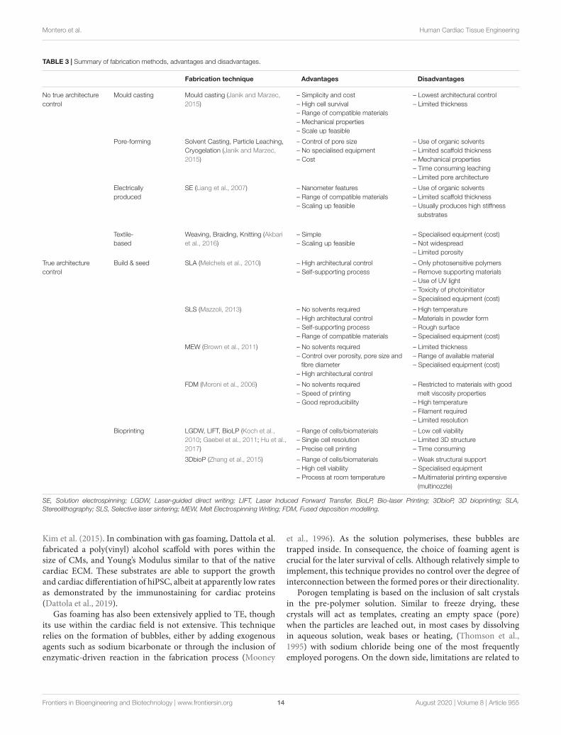
Fabrication StrategiesMaterials confer cTE with significant options, not only due tothe available range, but also through the application of differentfabrication modalities, which can deliver different properties outof the same starting material. For example, collagen can bemould-casted, (Godier-Furnémont et al., 2015) extruded, (Arañaet al., 2014) or bioprinted, (Lee et al., 2019) and the resultingproperties will vary widely, with casted and bioprinted collagenhaving a stiffness in the range of kPa, whilst the extrudedfilm will be significantly stiffer (MPa). In addition, structurewill also differ. 3D printing and bioprinting have no doubtrevolutionised our capacity to engineer cardiac tissues, however,other technologies can provide relevant features. The followingparagraphs outline some of the most employed strategies,classified depending on their capacity to produce a controlledarchitecture (Table 3).
Techniques With No True Architecture ControlMould castingProbably the simplest and most widely employed fabricationmode, requires the generation of a mould of the desiredshape, and is the fabrication technique of choice in manybiomedical-based laboratories, where other methods could notbe implemented due to lack of expertise or specific equipment.It does not provide much control over the resulting architecture,but can be combined with others, like porogen leaching, to addspecifically selected features to the resulting tissue. The firstreports on engineered cardiac tissues by Thomas Eschenhagenand coworkers in the late 1990s were developed by mould-castinga mixture of chick embryonic CMs embedded in a collagensolution. This was allowed to gel between two Velcro-coatedglass tubes. The resulting tissues, later termed engineered hearttissues (EHTs), could be cultured in vitro and maintained overseveral days, responded to electrical stimulation, displayed apositive Frank-Starling relationship, were sensitive to levels ofextracellular calcium, and could be modified with viral vectors(Eschenhagen et al., 1997; Zimmermann et al., 2000).
Since then, the technology has evolved enormously. It wasexpanded to neonatal rat cells, (Zimmermann et al., 2002) andrefined, with the possibility of fabricating more complex andthicker EHTs, which functioned synchronously (Zimmermannet al., 2006). These stacked EHTs also showed promise as
a therapy when transplanted in a rat model of infarction.EHTs have been employed by the groups of Eschenhagen andZimmermann, either fibrin- or collagen type I-based, to studythe effect of chronic stretch on CM hyperthrophy, (Fink et al.,2000) as a disease model of hypertrophic cardiomyopathy, (Stöhret al., 2013) or to analyze the effect of electrical stimulation (Hirtet al., 2014). EHT technology has greatly benefited from theimplementation of hPSC-derived cells, as the findings, modelsand application have gained greater impact, be it as a drugtesting platform (Eder et al., 2016), an in vitro tool to studycardiac stimulation, (Godier-Furnémont et al., 2015) or as apotential myocardial regenerative therapeutic (Weinberger et al.,2016; Tiburcy et al., 2017). The Bursac group has also employedmould casting of rat and human (hiPSC-derived) cells to engineercardiac tissues and explore different strategies to mature themin vitro (Jackman et al., 2016). They applied this strategy tofabricate tissues up to human scale, though thickness was limitedby nutrient and oxygen diffusion. When assayed in a rat modelof disease, their cardiopatches retained integrity after 3 weeks,showing extensive vascularisation by host-derived vessels andmaintaining electrical activity, although they did not functionallyintegrate with the endogenous myocardium (Shadrin et al., 2017).The team of Milica Radisic employed PDMS moulds to formengineered cardiac tissues around a surgical suture, composedof hPSC-derived cardiac cells, collagen type I and 10% Matrigel(Nunes et al., 2013). By employing electrical stimulation, throughcarbon rods immersed in the culture medium, they were ableto drive the hPSC-CMs towards a more mature phenotype andfunctionality. This system was later applied also to drug testing(Feric et al., 2019). Finally, one of the most advanced piecesof evidence for hPSC-CM maturation in vitro was providedby the group of Gordana Vunjak-Novakovic, as explained inthe previous section (Ronaldson-Bouchard et al., 2018). Theyemployed a 3:1 mixture of hPSC-CMs and dermal fibroblastsembedded in a fibrin matrix, cast into wells where flexible PDMSposts were also included. These conferred pre-tension on thegenerated engineered myocardium, thus adding physiological-like auxotonic stimulation (Mannhardt et al., 2019). A set ofcustom-made carbon electrodes delivered electrical stimulation.The resulting bioartifical myocardium displayed adult-like geneexpression profiles, ultrastructural features such as M-bandsand T-tubules, as well as oxidative metabolism and maturefunctionality. In summary, although mould casting cannotgenerate fine features, it is one of the most widely applied andhighly evolved methods to obtain human mature cardiac tissue,and the one closest to translation.
Macro-to-micro pore-forming strategiesCryogelation, based on freeze drying, is a common preservationstrategy that is also applied to TE. The pre-crosslinked materialis subjected to freeze-thawing cycles, which induces ice crystalformation. These crystals act as porogens, to be removedwhen pressure is decreased and the solvent sublimated. Ingeneral, pores are well inter-connected, as demonstrated earlyon by O’Brien and colleagues for collagen-glycosaminoglycansscaffolds (O’Brien et al., 2004). In addition, varying conditionsmake it possible to modulate material properties, as shown by
Frontiers in Bioengineering and Biotechnology | www.frontiersin.org 13 August 2020 | Volume 8 | Article 955
fbioe-08-00955 August 9, 2020 Time: 12:3 # 14
Montero et al. Human Cardiac Tissue Engineering
TABLE 3 | Summary of fabrication methods, advantages and disadvantages.
Fabrication technique Advantages Disadvantages
No true architecturecontrol
Mould casting Mould casting (Janik and Marzec,2015)
– Simplicity and cost– High cell survival– Range of compatible materials– Mechanical properties– Scale up feasible
– Lowest architectural control– Limited thickness
Pore-forming Solvent Casting, Particle Leaching,Cryogelation (Janik and Marzec,2015)
– Control of pore size– No specialised equipment– Cost
– Use of organic solvents– Limited scaffold thickness– Mechanical properties– Time consuming leaching– Limited pore architecture
Electricallyproduced
SE (Liang et al., 2007) – Nanometer features– Range of compatible materials– Scaling up feasible
– Use of organic solvents– Limited scaffold thickness– Usually produces high stiffness
substrates
Textile-based
Weaving, Braiding, Knitting (Akbariet al., 2016)
– Simple– Scaling up feasible
– Specialised equipment (cost)– Not widespread– Limited porosity
True architecturecontrol
Build & seed SLA (Melchels et al., 2010) – High architectural control– Self-supporting process
– Only photosensitive polymers– Remove supporting materials– Use of UV light– Toxicity of photoinitiator– Specialised equipment (cost)
SLS (Mazzoli, 2013) – No solvents required– High architectural control– Self-supporting process– Range of compatible materials
– High temperature– Materials in powder form– Rough surface– Specialised equipment (cost)
MEW (Brown et al., 2011) – No solvents required– Control over porosity, pore size and
fibre diameter– High architectural control
– Limited thickness– Range of available material– Specialised equipment (cost)
FDM (Moroni et al., 2006) – No solvents required– Speed of printing– Good reproducibility
– Restricted to materials with goodmelt viscosity properties
– High temperature– Filament required– Limited resolution
Bioprinting LGDW, LIFT, BioLP (Koch et al.,2010; Gaebel et al., 2011; Hu et al.,2017)
– Range of cells/biomaterials– Single cell resolution– Precise cell printing
– Low cell viability– Limited 3D structure– Time consuming
3DbioP (Zhang et al., 2015) – Range of cells/biomaterials– High cell viability– Process at room temperature
– Weak structural support– Specialised equipment– Multimaterial printing expensive
(multinozzle)
SE, Solution electrospinning; LGDW, Laser-guided direct writing; LIFT, Laser Induced Forward Transfer, BioLP, Bio-laser Printing; 3DbioP, 3D bioprinting; SLA,Stereolithography; SLS, Selective laser sintering; MEW, Melt Electrospinning Writing; FDM, Fused deposition modelling.
Kim et al. (2015). In combination with gas foaming, Dattola et al.fabricated a poly(vinyl) alcohol scaffold with pores within thesize of CMs, and Young’s Modulus similar to that of the nativecardiac ECM. These substrates are able to support the growthand cardiac differentiation of hiPSC, albeit at apparently low ratesas demonstrated by the immunostaining for cardiac proteins(Dattola et al., 2019).
Gas foaming has also been extensively applied to TE, thoughits use within the cardiac field is not extensive. This techniquerelies on the formation of bubbles, either by adding exogenousagents such as sodium bicarbonate or through the inclusion ofenzymatic-driven reaction in the fabrication process (Mooney
et al., 1996). As the solution polymerises, these bubbles aretrapped inside. In consequence, the choice of foaming agent iscrucial for the later survival of cells. Although relatively simple toimplement, this technique provides no control over the degree ofinterconnection between the formed pores or their directionality.
Porogen templating is based on the inclusion of salt crystalsin the pre-polymer solution. Similar to freeze drying, thesecrystals will act as templates, creating an empty space (pore)when the particles are leached out, in most cases by dissolvingin aqueous solution, weak bases or heating, (Thomson et al.,1995) with sodium chloride being one of the most frequentlyemployed porogens. On the down side, limitations are related to
Frontiers in Bioengineering and Biotechnology | www.frontiersin.org 14 August 2020 | Volume 8 | Article 955

fbioe-08-00955 August 9, 2020 Time: 12:3 # 15
Montero et al. Human Cardiac Tissue Engineering
low architectural control and suboptimal processing of porogens,which might compromise biocompatibility as well as mechanicalproperties. Salt leaching has not been as widely employed in cTEas in other areas. Ganji et al. used table salt as a simple yet efficientway to generate defined pores in polyurethane-based scaffoldswhere gold nanotubes and nanowires were incorporated, thuscombining 3-dimensionality with the additional benefit ofelectrical conductivity (Ganji et al., 2016). Biocompatibility testedwith H9c2 rat cardiomyoblasts showed a positive effect oncell growth, though no hPSC-derived cells were tested. Otherexamples have employed porogen templating for the fabricationof porous polysaccharide-based vascular scaffolds and engineeredheart valves (Lavergne et al., 2012; Masoumi et al., 2014).
Thermally induced phase separation is based on the useof temperature to induce the de-mixing of a homogeneouspolymer solution. The controlled change in temperature promptsthe formation of a polymer-rich and a polymer-poor phase,which can be used to obtain an interconnected porous structure(Nam and Park, 1999). Vozzi et al. synthesised the elastomerpolyesterurethane, and used thermal phase separation to createa porous and biocompatible structure, which they functionalisedwith fibronectin by NHS-EDC chemistry. Although materialproperties were off the cardiac optimal range (in the orderof > 0.25 MPa), neonatal rat cardiac cells attached and grewon the scaffolds, with specific modulation of gene expression.However, no use of human cells was reported (Vozzi et al., 2018).
Electrically producedSolution electrospinning (SE), on the other hand, was one ofthe first and most employed fabrication modalities in the field.In this technique, a polymer solution is subjected to a relativelyhigh electric field while it travels towards a collector plate.This electric field causes the polymeric jet to whip, producingvery thin fibres of even sub-micron size, that can be collectedrandomly or in an aligned fashion. For a review see Sensini andCristofolini (2018). In general, most SE applications produce lowthickness mats of (semi-)randomly arranged fibres of differentmaterials, though use of appliances such as a rotating mandrelcan increase alignment (Han et al., 2016). Although relying onthe use of potentially toxic solvents, SE has the advantage of beingopen to a wide range of materials, both natural and synthetic(Kitsara et al., 2017).
The concurrent use of SE with hPSC-derived cardiac cellshas not been widely explored but some remarkable examplescan be found. The group of Onnik Agbulut tested differentcrosslinking times of a clinical-grade collagen electrospunscaffold for the most suitable conditions for hiPSC-CM culture.The resulting mats had fibres of 0.6-2.2 µm, with pores inthe range of 2-3 µm. After no deleterious effects were foundafter mat implantation in animals, the mats were seeded withhiPSC-CMs (106 per scaffold), and produced a significantbenefit when transplanted in a dilated cardiomyopathy model.Importantly, scaffolds were compliant enough to allow thegeneration of macroscopic contractions by the hiPSC-CMs(Joanne et al., 2016). Sireesha et al. used a combination of theelastomer poly[1,8-octanediol-co-(citric acid)-co-(sebacic acid)]and fibrinogen to produce scaffolds with a sub-micron fibre
diameter, whose mechanical properties laid on the upper endof cardiac elasticity (hundreds of kPa) and which showed goodbiocompatibility with human CMs. However, no further testsin animal models were performed (Sireesha et al., 2015). Khanand co-workers electrospun polylactide-co-glycolide (PLGA) as50 µm-thick aligned nanofibrous scaffolds, seeded with hiPSC-CM, and compared the outcome versus conventional tissueculture plastic surfaces. Results showed the scaffolds were ableto align the CMs, as well as inducing changes at the level offunctionality (Ca transients) and gene expression, though thehigh stiffness of the mats (1-2 orders of magnitude above thecardiac tissue) could have a negative impact (Khan et al., 2015).On the other side of the coin, Han et al. assayed the capacityof aligned electrospun mats made of PCL coated with matrigelto induce hiPSC-CM maturation, showing a limited effect at thefunctional level, but with some differences in gene expression(Han et al., 2016).
Aside from employing different materials or combinations ofnatural and synthetic polymers, SE offers increased possibilitiesby modifying the properties of the resulting engineeredtissue by combination with nanoparticles or other fabricationtechnologies. For example, the group of Tal Dvir built on hisprevious work on the electrospinning of albumin (Fleischer et al.,2014) to increase the anisotropy of the fabricated engineeredtissue. To do this, they employed a double strategy: they used laserpatterning to create micro-holes and unidirectional grooves, inorder to increase mass transport and cell alignment respectively,and they stacked several layers of patterned mats, inspiredby the anisotropy found across the ventricular wall. In thisfashion, they also modulated the mechanical properties of thelayers, getting closer to human myocardial values. They did not,however, test the stiffness of the resulting stacked construct, norpopulate it with human cells, using rat neonatal cardiac cellsinstead (Fleischer et al., 2017). The group of Ali Khademhosseiniused poly(glycerol sebacate):gelatin (PG) solution where gelatin-methacryloyl (GelMA)-coated carbon nanotubes had beenincorporated to electrospun aligned nanofibrous mats withimproved electrical properties, though at the cost of increasedstiffness (Kharaziha et al., 2014). This is similar to their previouswork with GelMA-embedded carbon nanotubes, (Shin et al.,2013) with a significant effect on gene expression, structure andfunctionality, but again no human cells were employed. Walkeret al. employed bio-ionic liquids to modulate the electricalproperties and adhesion strength of electrospun GelMA scaffolds,assaying their in vitro and in vivo regenerative potential, thoughno tissue was constructed (Walker et al., 2019). Again, mechanicalproperties were affected. Recently, the group of Felix Engelused SE to generate fibres combining the conductive polymerpolyaniline (PANi) with collagen/hyaluronic acid, producingmats of suitable mechanical and electrical properties. Scaffoldssupported the attachment of hiPSC-CMs, which displayedtypical striations and contractions. Substrates incorporatingPANi induced a faster beating rate on hiPSC-CMs, though thiswas not further explored (Roshanbinfar et al., 2020). All in all, SEis a very versatile fabrication technique, and this is also increasedby the possibility of combining it with other materials andtechnologies. However, it faces limitations related to the thickness
Frontiers in Bioengineering and Biotechnology | www.frontiersin.org 15 August 2020 | Volume 8 | Article 955

fbioe-08-00955 August 9, 2020 Time: 12:3 # 16
Montero et al. Human Cardiac Tissue Engineering
and dimensions of the final construct, as well as long-rangealignment capacity.
Textile-based fabricationHistorically speaking, weaving is one of the oldest fabricationtechniques and many (bio)materials can be processed byknitting, weaving or braiding, (Akbari et al., 2016) giving riseto new architectures and importantly, mechanical propertiesunattainable for individual fibres. Curiously, these have notbeen mainstream in the cTE field, with few studies combiningthese fabrication methods with hPSC-derived cells. Thoughtheir work cannot fully be categorised as textile-based, thelab of Kevin K. Parker employed pull spinning to generatenanofibrous scaffolds composed of PCL/Gelatin over anellipsoidal mandrel, mimicking the shape of an idealisedventricle. After additional coating with the ECM proteinfibronectin, they seeded either neonatal cardiac rat cells orhiPSC-CMs, generating scale-models of the heart with strikingfunctional properties (Macqueen et al., 2018).
Techniques With True Architecture ControlAdditive manufacturing, also known as 3D printing, is one ofthe technological revolutions of this century. It is based on thebuilding of a volumetric object from a computer aided design(CAD), often in a layer-by-layer basis. Broadly speaking, we candistinguish between bioprinting, where cells are also printed,from the build & seed strategies, where the scaffold is first printedand the cells incorporated in a subsequent step, either as astandalone element or embedded in a hydrogel matrix. In anycase, the degree of control over the resulting structure is ordersof magnitude above what is achievable with the above-mentionedtechnologies, with the exception of filament dimensions, with SEbeing able to go into sub-micron diameter, albeit at the expenseof thickness and architecture.
3D Build and SeedIn this category we have gathered those additive manufacturingtechnologies that do not allow the concurrent printing ofmaterials and cells, be it because they employ high temperature,damaging lasers, toxic solvents or for other reasons. Thissection includes selective laser sintering (SLS), consisting ofiteratively spreading layers of powdered materials and fusingit together to achieve the programmed shape, (Duan et al.,2010) stereolithography, where a bath with photosensitiveresin is selectively cured layer by layer with a UV laser orsimilar power source, (Gauvin et al., 2012) or fused depositionmodelling (FDM), by which the printed polymer is meltedand deposited in layers in order to acquire the desired 3Darchitecture (Moroni et al., 2006). All these technologies havein common the need to first generate the architecture andin a second step add the biologicals, with the only exceptionof laser-curable materials in SLS. Although these technologiesare in increased demand for the building of prosthetics,personalised solid implants or educational/surgery planningmodels, (Giannopoulos et al., 2016) their application to cTE isnot widespread, especially in light of the increasing importanceof bioprinting techniques (next section).
A special note however must be dedicated to MeltElectrowriting (MEW), a highly specialised additivemanufacturing technology (Brown et al., 2011). It works onthe same basis as SE, where a high voltage is applied betweena syringe tip and a grounded collector. In this case however,the polymer is not dissolved, but melted. The superior stabilityof the jet allows a highly controlled deposition of fibres, whichsolidify in-flight or shortly upon collection. These, althoughbigger in diameter than those from SE, are at least an order ofmagnitude below what is usually attainable with 3D printing orbioprinting (tens of µm in diameter). The polymer of choicemust be melted within the capacities of the printer, whichlimits the range of the available polymers. Another significantlimitation is the achievable 3-dimensionality, which is usuallybelow the mm-range. The groups of Paul Dalton and DietmarHutmacher have spearheaded its use (Bas et al., 2015, 2017;Hochleitner et al., 2018) and have developed methods toincrease the thickness of the MEW scaffolds to almost onecm, but again these are far from mainstream (Wunner et al.,2018). The groups of Jos Malda and Joost Sluijter have beenthe first ones to apply MEW to cTE. In their first approach,they MEW-printed the hydroxyl-functionalised polyester,(poly(hydroxymethylglycolide-co-ε-caprolactone) in orthogonalpatterns, and seeded them with human cardiac progenitor cells(CPC) embedded in a collagen type I hydrogel. CPCs survivedwell, with the scaffolds producing a significant alignment(Castilho et al., 2017). In a further optimisation of the system,Castilho et al. fabricated hexagonally patterned MEW scaffoldswith superior compliance, and seeded them with hiPSC-CMs.Their results showed the superior biaxial mechanical propertiesof the hexagonal designs, and their significant, positive effect oncell alignment and gene expression. Moreover, these scaffoldscould be transplanted in a large animal (pig) after passing througha catheter-like tube, paving the way for their future percutaneoustransplantation (Castilho et al., 2018). Finally, an auxetic patch,that is, featuring a negative Poisson ratio, was recently fabricatedwith MEW, and the conductive polymer polypyrrole in situpolymerised on it, with electroconductive properties close tothose of the human myocardium (Olvera et al., 2020).
BioprintingThe capacity to print human tissues on-demand has roused theexpectations of scientists, clinicians and patients alike, with arange of modalities available. Laser-assisted bioprinting (LAB) forexample, features several methodologies. Laser-induced forwardtransfer of material/ink (LIFT)(Koch et al., 2010) allows printingwith a single-cell resolution at high densities (up to 108 cells/ml)(Gaebel et al., 2011). However, it requires selected materialsto gel relatively fast, which in practice restricts the range ofavailable materials, and can be technically challenging. 2 photonpolymerisation is a type of LAB with high spatial resolution (aslow as 70 nm) without the need for support structures (Hu et al.,2017). Vaithilingam et al. employed this technique to fabricateelectroactive 3D architectures by incorporation of multi-walledcarbon nanotubes (CNTs). The mechanical properties werehowever highly deviated from what has been described forhuman cardiac tissue (GPa range). hiPSC-CMs were able to
Frontiers in Bioengineering and Biotechnology | www.frontiersin.org 16 August 2020 | Volume 8 | Article 955

fbioe-08-00955 August 9, 2020 Time: 12:3 # 17
Montero et al. Human Cardiac Tissue Engineering
survive and, after the application of electrical stimulation, asignificant effect on sarcomere length was found (Vaithilingamet al., 2019). There are other printing technologies based onLAB, such as Biological Laser Printing and Laser Guided DirectWriting, (Odde and Renn, 2000; Barron et al., 2004) but noreports on these include hiPSC-derived cardiac cells.
Extrusion-based bioprinting is probably the best-knownbioprinting method: a bioink containing both cells and thebiomaterial is extruded through a nozzle onto a motorisedcollector. The coordination between the gelation of the bioinkand its deposition permits the generation of thick tissues.A range of nozzles, including pneumatic, screw- or plunger-based, as well as inks, is available. However, the materialproperties of the latter are fundamental, as shear stress generatedupon printing highly impacts cell viability (Jungst et al.,2016). Some strategies, like in situ crosslinking of the bioinkimmediately prior to its deposition, (Ouyang et al., 2017)are being developed to overcome these and other limitations.Extrusion-based bioprinting has been extensively researchedin the cTE field. Jang et al. employed a multi-nozzle printerto generate tissues with 2 decellularised cardiac ECM-basedbioinks reinforced with a thermoplastic (PCL) backbone. Oneof the bioinks contained human CPCs (c-kit-positive), and theother mesenchymal stem cells (MSC) and pro-angiogenic factors(VEGF). The application of this patch in a rat model of diseasedemonstrated a rapid vascularisation as well as a significantbenefit at the functional and histological levels. However, nonew contractile tissue was formed (Jang et al., 2017). Zhang andcoworkers employed a more advanced strategy, where they usedcoaxial printing to pattern human umbilical vein endothelialcells (HUVECs) in a ink containing alginate, GelMA and theUV-curable photoinitiator Irgacure. The mixture was quicklycrosslinked by CaCl2 from the outer sheath, and further gelledby exposure to UV light (for alginate and GelMA respectively)(Zhang Y. S. et al., 2016). HUVECs migrated to the outer borderof the printed structures, forming 3D vessel-like structures overa period of 15 days, after which neonatal rat CMs were added tocomplete the tissue. Izadifar et al. explored varying the electricalproperties of the generated tissues by incorporating alginate-coated carbon nanotubes (CNT) to methacrylated collagen. Thesystem displayed as expected enhanced electrical properties, oncemore at the expense of becoming stiffer, though within acceptablevalues, and was able to support the growth of human coronaryartery endothelial cells. No CMs were employed (Izadifar et al.,2018). Maiullari and coworkers applied a different version ofextrusion printing termed microfluidic printing, to generatevascularised engineered tissues combining HUVECs and hiPSC-CMs (Maiullari et al., 2018). The bioartificial myocardiumwas fabricated with a bioink combining alginate for a rapidcrosslinking, and polyethylene glycol (PEG) monoacrylate-fibrinogen, for UV-curing and cell adhesion. These tissues weretested in vivo, were the presence of vascular cells was able topromote integration with the recipient vascular system. Zhuet al. formulated a bioink with capacity to electrically influenceneonatal rat CMs by incorporating gold nanorods in GelMA.Thick constructs were generated, although cell viability was below80% in most cases. This is extremely relevant if we take into
account that CMs are non-proliferative cells, so no re-growth islikely to occur (Zhu et al., 2017).
Freeform embedding of suspended hydrogels (FRESH) is oneof most exciting 3D bioprinting methods (Hinton et al., 2015).Here, the biomaterial ink or bioink of choice is printed withinthe support of another hydrogel serving as a temporary support.Noor and co-workers developed a personalised hydrogel ink fromhuman decellularised omental tissue, which could be printed intocomplex structures using FRESH. They used it in combinationwith hiPSC which they differentiated within the matrix intobeating, perfused cardiac mini-tissues (Noor et al., 2019). Lastly,the Feinberg group used FRESH to accurately (20 µm resolution)3D print different components of the human heart with collagen,including a model of the left ventricle on the scale of theneonatal organ, incorporating hESC-CMs (Lee et al., 2019). Theseengineered tissues displayed synchronised functional activity.
Finally, some the applications build tissues without the useof biomaterials. This includes the additive manufacturing withspheroids, as demonstrated by Arai et al., where they combinedcommercially available hPSC-CMs with HUVECs and dermalfibroblasts to generate the spheroids, organised in a tubular 3Dstructure with a bioprinter (Arai et al., 2018). However, theauthors used a needle array to provide initial transient support fortheir structures, which in the end played the role of a scaffoldingmaterial. Recently, Ayan et al. reported a novel strategy, termedaspiration-assisted bioprinting, relying on the aspiration of 3Dspheroids and their precise deposition in 3D. This could also becombined with FRESH, but no use of hiPSC-derived cardiac cellswas reported (Ayan et al., 2020).
CLINICAL TRANSLATION OFhPSC-BASED cTE STRATEGIES
Survival post-cardiac ischemia has raised with advances inthe cardiology and cardiovascular surgery areas, as well asthrough the development of new drugs in the last decades. Thisincludes the application of reperfusion by percutaneous coronaryintervention, anticoagulants and antithrombotics, amongstothers. Though cTE has potential to address different cardiacconditions, its main activity has been directed towards the loss ofviable myocardium by supplying new one, engineered in the lab.
In stark contrast to the short time elapsing between thefirst reports on regenerative medicine with adult stem cellsand their first in-human application (reviewed in Banerjeeet al., 2018) cTE with hPSCs has scarcely reached the clinicalarena. Be it because both scientists and clinicians have learntfrom past experiences or because translation presents additionalcomplications, (Desgres and Menasché, 2019; Ghaemi et al.,2019; Ke and Murphy, 2019) clinical application is yet tobecome a reality. The technology faces challenges in thearea of regulation, where reprogrammed cells under GMP-conditions are not widely derived, in logistics, with the size ofengineered tissues in most instances requiring open chest surgery,and in economic terms. However, progress is being made.Tiburcy et al., created an engineered human myocardium withadvanced structural and functional maturation by combining
Frontiers in Bioengineering and Biotechnology | www.frontiersin.org 17 August 2020 | Volume 8 | Article 955

fbioe-08-00955 August 9, 2020 Time: 12:3 # 18
Montero et al. Human Cardiac Tissue Engineering
hPSC-derived cells with collagen type I (Tiburcy et al., 2017).Their work, standing on the shoulders of decade-long progress,(Zimmermann et al., 2002, 2006) demonstrated the buildingof human-scale relevant tissues (3.5 × 3.4 cm, containing40 million CMs) under defined conditions and their pre-clinical safety assessment in rats, though no large animaltesting was performed. The group of Prof Bursac applieddynamic culture conditions to improve the maturation of apatch formed from hiPSC-derived cardiac cells in a fibrinmatrix. Although they achieved human adult-like electricalproperties (e.g., conduction velocity) and formed large graftswhen transplanted into rodents, the use of matrigel in theforming mixture precludes further translation (Shadrin et al.,2017; Jackman et al., 2018). In addition, myocytes were mostlyrandomly arrayed, which added to the low thickness of thegrafts precludes any efficient and substantial force generation,and therefore makes providing contractile support to diseasedhearts difficult.
Menasche et al. performed the first clinical case of anhPSC-based patch in 2015, transplanting a 20 cm2 fibrin-based tissue. This patch contained 4 million hESC-derived Isl+SSEA1+ CPCs and was implanted epicardially in a 68-year-old patient with advanced heart failure. Concomitant to theimplantation of the patch, a coronary artery bypass was alsoperformed. After 3 months, the patient had better symptoms,and there was improved evidence in the echocardiogramtoo (form akinetic to moderately hypokinetic). Moreover, noadverse effect was observed (Menasché et al., 2015). This waspart of the first clinical trial carried out with six patientssuffering from advanced IHD (NCT02057900), called ESCORT.The aim of the trial was to demonstrate the safety ofthe patch at 1 year. All patients improved symptomatically,with four showing an increased systolic motion. One diedearly in the post-operative period from treatment-unrelatedcomorbidities and another after in 22 months due to heart failure(Menasché et al., 2018).
Japan has made huge efforts to spearhead research in thehiPSC field, and has one of the most streamlined regulatoryframeworks (see Cyranoski, 2019 for a comment). It has recentlystarted the first clinical trial of hiPSC-CM transplant in ischemicheart dysfunction. The study will deliver 100 million hiPSC-CMsin the form of a cell sheet of 4 × 5 × 0.1 cm (Kawamura et al.,2017). The aim of the study is to evaluate efficacy and safety ofthe treatment in a total of 10 patients (Cyranoski, 2018).
Overall, clinical applications of hPSC-based cardiacengineered tissues, though scarce, are starting to emerge.However, the scientific and logistic/economic concernsmentioned above need addressing further before any ofthese technologies reach the clinical arena.
CONCLUSION AND OUTLOOK
The capacity to fabricate human myocardium in the laboratoryis no longer restricted to science fiction. Recent breakthroughssuch as cell reprogramming and 3D (bio)printing have shiftedthe question from ‘Can we do it?’ to ‘How do we want to do it?’.
However, lessons from its predecessor in the area of regenerativemedicine should teach us the value of caution. Crucial questionsremain, some of which include the following:
-What are the most suitable components for building anengineered myocardium? Of course, this absolutely depends onthe application/pathology we are targeting. If the objective isto reconstruct the post-disease myocardium, especially in caseswhen muscular mass has been loss (as IHD), this will requirenew CMs, alongside new vessels and support cells. Althoughnot supplying new CMs might still be beneficial, it will notregenerate the tissue to pre-disease levels. So far, only hPSCscan produce the required cells in sufficiently large amounts,and whether it is better to use fully differentiated cells orprogenitors is still an open question. Regarding the ECM-mimics,the current state can seem confusing. All reported (bio)materialformulations seem to provide a benefit (Table 2). It is plausiblethat the most important factor is for the material to recapitulatethe mechanical environment of cardiac tissue long enough fordelivered cells to build their own structure. Also, althoughcollagen type I has repeatedly been positioned as being themain component of the cardiac ECM, this does not accountfor all its mechanical capacities, nor it is as abundant as thelevels supplemented in engineered tissues. Probably, cost andtranslation capacity will have much to say here. Lastly, regardingthe fabrication mode, though 3D bioprinting has captivatedboth scientists and the wider public, its actual ability to delivertissues able to withstand the mechanical forces in play in thehuman myocardium is far from optimal in most cases. Fibrereinforcement might be a solution to this, but remains to befully tested in a human scale. In addition, recent technicaldevelopments such as 4D printing or cyborganics will certainlywiden the scope of possibilities at hand (Tamay et al., 2019;Orive et al., 2020).
– How is translation going to happen? The hurdles aresignificant, (Desgres and Menasché, 2019; Ghaemi et al., 2019)not only due to the use of genetically modified cells (hiPSCs),exogenous materials and complex equipment, but also becausein order to attain sufficient functional capacity, it is likely thatsome lengthy period of in vitro electromechanical maturationmust be implemented. Also, no standard equipment currentlyexists to apply this to a human-scale tissue. In this area, it iscrucial that biomedical researchers work alongside lawmakers.Cell therapy mostly relied on an autologous application of anon-cultured material, which smoothened its approval. Tissueengineering applications based on hiPSC products are highlypromising but are impeded by stringent regulations. Japan hasspearheaded hiPSC translation as a national flagship (Azumaand Yamanaka, 2016). Although some consider this too loose,it is clear that unless scientists provide a clear direction and getthemselves involved in depth in the regulatory process, advanceswill be limited.
– Will costs be sensible? hiPSC culture and differentiationare expensive. Additive manufacturing is expensive. GMP-grade materials are expensive. hiPSC line derivation costsbetween €4000-8000 depending on the provider, multi-nozzle3D bioprinters are usually budgeted above €30000, and somemedical-grade materials as human collagen type I are prohibitive
Frontiers in Bioengineering and Biotechnology | www.frontiersin.org 18 August 2020 | Volume 8 | Article 955

fbioe-08-00955 August 9, 2020 Time: 12:3 # 19
Montero et al. Human Cardiac Tissue Engineering
(> €200 per 100 µg). Highly trained personnel costs needto be added on top of this, as well as the cost of hiPSCexpansion and differentiation. The cost of a heart transplant inthe United States is above $150000 (including 120-day medicalcare), with Left Ventricular Assist Devices (LVADs), employedfor patients with end stage heart failure as a bridge to transplant(and now trialled as destination therapy) are estimated to costeven more (see Miller et al., 2013 for a concise review). Ifeconomic issues are not addressed from day one, we run therisk of ending up with a highly effective therapy only thatvery few people can pay for. Large-scale production of hiPSC-derived cells has been demonstrated and will no doubt helpdecrease costs (Abecasis et al., 2017; Halloin et al., 2019; Buikemaet al., 2020). Strategies to induce hiPSC-CM division wouldsignificantly improve the economics.
– Will the new therapy be safe? hPSCs have been marredby concerns related to persistence of pluripotent cells afterdifferentiation and the subsequent risk of teratoma formation.But also, the new tissue must conform to the host’s electricalactivity. Again, lessons from Cell Therapy call for caution(Menasché et al., 2008). Advanced genomics and other analysistools not available 10 years ago will significantly enhance thesafety analysis and help researchers and clinicians along the way(Wang Z. et al., 2020).
– Are the aims achievable? The objective of cTE is to generatenew human myocardium whose properties and functionality aresimilar to its natural counterpart, but that might not be achievablewithin a sensible timeframe. Human cardiac maturation advancessignificantly after birth but continues throughout the firstdecades of life. Current advanced methods for maturation,like those based on electromechanical stimulation, do notachieve anything even close to adult properties. However,it is plausible that only a minimal maturity is needed inorder to avoid lethal arrhythmias, as hPSC-CMs are reportedto undergo maturation after transplantation in the heart(Kadota et al., 2017).
– What is the state of non-therapeutic applications? Thoughthe aim of cTE within regenerative medicine is directedtowards treatment, bioartificial human myocardium is becomingincreasingly relevant with respect to other applications suchas disease modelling and drug testing (Vunjak-Novakovicet al., 2014; Sharma et al., 2020), where the scale of thefabricated tissues is not an issue. In drug testing, the CiPAinitiative has already pointed to the relevance of hiPSC-CMs, (Millard et al., 2018) especially given cardiac sideeffects are one of the main reasons for drug withdrawal,and there is no primary source of human CMs available
(Laverty et al., 2011). Current efforts have already proven thecapacity of cTE-based systems to better recapitulate physiologicalhuman drug response [reviewed in Kadota et al. (2017)and Millard et al. (2018), or to generate chamber-specificmicrotissues (Zhao et al., 2019)]. Translation towards industrialand clinical testing is envisioned to be more straightforwardand immediate.
As can be seen, the challenges ahead are not minor. However,humankind has never been closer to achieving success intissue fabrication. Whole functional heart generation in thelaboratory, though frequently dreamt of, is still not achievable,as severe physiological and technical roadblocks remain inplace. Challenges include ensuring adequate vascularisation,electromechanical coupling over long distances, atrioventriculardelay, autonomous nervous system innervation or designingmaterials both biocompatible but capable of supporting thestrong mechanics of a whole heart, amongst others. At thisextraordinary moment, it is crucial for scientists, clinicians, andlay people to stay focused and deliver.
AUTHOR CONTRIBUTIONS
PM, MF-I, SM, and MP revised the literature, wrote the initialdraft of the manuscript and figures. DP and JG revised thetext and generated the tables. CS, GO, and FP supervised thedevelopment and revised the text. MM wrote the final revisedversion. All authors contributed to the article and approved thesubmitted version.
FUNDING
This work was supported by funds from the ISCIII RedTERCEL RETIC RD16/0011/0005, PI 19/01350, ERANET II(Nanoreheart) and Gobierno de Navarra Departamento deSalud GNa8/2019, co-funded by FEDER funds, MINECO(Program RETOS Cardiomesh RTC-2016-4911-1), Gobierno deNavarra 0011-1383-2019-000006 and 0011-1383-2018-000011,and European Union’s H2020 Program under grant agreementNo. 874827 (BRAV∃).
ACKNOWLEDGMENTS
The authors acknowledge Medical Arts and BioRender(registered user) for the building of the figures in the manuscript.
REFERENCESAbecasis, B., Aguiar, T., Arnault, É, Costa, R., Gomes-Alves, P., Aspegren, A., et al.
(2017). Expansion of 3D human induced pluripotent stem cell aggregates inbioreactors: bioprocess intensification and scaling-up approaches. J. Biotechnol.246, 81–93. doi: 10.1016/j.jbiotec.2017.01.004
Adler, C. P., and Costabel, U. (1975). Cell number in human heart in atrophy,hypertrophy, and under the influence of cytostatics. Recent Adv. Stud. Card.Struct. Metab. 6, 343–355.
Akbari, M., Tamayol, A., Bagherifard, S., Serex, L., Mostafalu, P., Faramarzi, N.,et al. (2016). Textile technologies and tissue engineering: a path toward organweaving. Adv. Healthc. Mater. 5, 751–766. doi: 10.1002/adhm.201500517
Amdursky, N., Mazo, M. M., Thomas, M. R., Humphrey, E. J., Puetzer, J. L., St-Pierre, J. P., et al. (2018). Elastic serum-albumin based hydrogels: mechanismof formation and application in cardiac tissue engineering. J. Mater. Chem. B 6,5604–5612. doi: 10.1039/c8tb01014e
Andreu, I., Luque, T., Sancho, A., Pelacho, B., Iglesias-García, O., Melo, E., et al.(2014). Heterogeneous micromechanical properties of the extracellular matrix
Frontiers in Bioengineering and Biotechnology | www.frontiersin.org 19 August 2020 | Volume 8 | Article 955

fbioe-08-00955 August 9, 2020 Time: 12:3 # 20
Montero et al. Human Cardiac Tissue Engineering
in healthy and infarcted hearts. Acta Biomater. 10, 3235–3242. doi: 10.1016/j.actbio.2014.03.034
Anversa, P., Loud, A. V., Giacomelli, F., and Wiener, J. (1978). Absolutemorphometric study of myocardial hypertrophy in experimental hypertension.II. Ultrastructure of myocytes and interstitium. Lab. Investig. 38, 597–609.
Arai, K., Murata, D., Verissimo, A. R., Mukae, Y., Itoh, M., Nakamura, A.,et al. (2018). Fabrication of scaffold-free tubular cardiac constructs usinga Bio-3D printer. PLoS ONE 13:e0209162. doi: 10.1371/journal.pone.0209162
Araña, M., Gavira, J. J., Peña, E., González, A., Abizanda, G., Cilla, M., et al. (2014).Epicardial delivery of collagen patches with adipose-derived stem cells in ratand minipig models of chronic myocardial infarction. Biomaterials 35, 143–151.doi: 10.1016/j.biomaterials.2013.09.083
Ayan, B., Heo, D. N., Zhang, Z., Dey, M., Povilianskas, A., Drapaca, C., et al. (2020).Aspiration-assisted bioprinting for precise positioning of biologics. Sci. Adv.6:eaaw5111. doi: 10.1126/sciadv.aaw5111
Azuma, K., and Yamanaka, S. (2016). Recent policies that support clinicalapplication of induced pluripotent stem cell-based regenerative therapies.Regen. Ther. 4, 36–47. doi: 10.1016/j.reth.2016.01.009
Banerjee,. I., Fuseler, J. W., Price, R. L., Borg, T. K., and Baudino, T. A. (2007).Determination of cell types and numbers during cardiac development in theneonatal and adult rat and mouse. Am. J. Physiol. – Hear Circ. Physiol. 293,1883–1891
Banerjee, M. N., Bolli, R., and Hare, J. M. (2018). Clinical studies of cell therapy incardiovascular medicine recent developments and future directions. Circ. Res.123, 266–287. doi: 10.1161/circresaha.118.311217
Bargehr, J., Ong, L. P., Colzani, M., Davaapil, H., Hofsteen, P., Bhandari, S., et al.(2019). Epicardial cells derived from human embryonic stem cells augmentcardiomyocyte-driven heart regeneration. Nat. Biotechnol. 37, 895–906. doi:10.1038/s41587-019-0197-9
Barkauskas, C. E., Noble, P. W., Brigid, L. M., Barkauskas, C. E., Cronce, M. J.,Rackley, C. R., et al. (2013). Type 2 alveolar cells are stem cells in adult lung.J. Clin. Invest. 123, 3025–3036.
Barreiro, D. L., Martín-Moldes, Z., Yeo, J., Shen, S., Hawker, M. J., Martin-Martinez, F. J., et al. (2019). Conductive silk-based composites using biobasedcarbon materials. Adv. Mater. 31:e1904720.
Barron, J., Wu, P., Ladouceur, H. D., and Ringeisen, B. R. (2004). Biologicallaser printing: a novel technique for creating heterogeneous 3-dimensional cellpatterns. Biomed. Microdevices 6, 139–147. doi: 10.1023/b:bmmd.0000031751.67267.9f
Bas, O., D’Angella, D., Baldwin, J. G., Castro, N. J., Wunner, F. M., Saidy,N. T., et al. (2017). An integrated design, material, and fabricationplatform for engineering biomechanically and biologically functional softtissues. ACS Appl. Mater. Interfaces. 9, 29430–29437. doi: 10.1021/acsami.7b08617
Bas, O., De-Juan-Pardo, E. M., Chhaya, M. P., Wunner, F. M., Jeon, J. E., Klein,T. J., et al. (2015). Enhancing structural integrity of hydrogels by using highlyorganised melt electrospun fibre constructs. Eur. Polym. J. 72, 451–463. doi:10.1016/j.eurpolymj.2015.07.034
Baudino, T. A., Carver, W., Giles, W., and Borg, T. K. (2006). Cardiac fibroblasts:friend or foe? Am. J. Physiol. – Hear Circ. Physiol. 291, H1015–H1026.
Bedada, F. B., Chan, S. S. K., Metzger, S. K., Zhang, L., Zhang, J., Garry, D. J., et al.(2014). Acquisition of a quantitative, stoichiometrically conserved ratiometricmarker of maturation status in stem cell-derived cardiac myocytes. Stem CellRep. 3, 594–605. doi: 10.1016/j.stemcr.2014.07.012
Behfar, A., Crespo-Diaz, R., Terzic, A., and Gersh, B. J. (2014). Cell therapy forcardiac repair-lessons from clinical trials. Nat. Rev. Cardiol. 11, 232–246. doi:10.1038/nrcardio.2014.9
Beltrami, A. P., Barlucchi, L., Torella, D., Baker, M., Limana, F., Chimenti, S.,et al. (2003). Adult cardiac stem cells are multipotent and support myocardialregeneration. Cell 114, 763–776. doi: 10.1016/s0092-8674(03)00687-1
Belviso, I., Romano, V., Sacco, A. M., Ricci, G., Massai, D., Cammarota, M., et al.(2020). Decellularized human dermal matrix as a biological scaffold for cardiacrepair and regeneration. Front. Bioeng. Biotechnol. 8:229. doi: 10.3389/fbioe.2020.00229
Bergmann, O., Zdunek, S., Felker, A., Salehpour, M., Alkass, K., Bernard, S., et al.(2015). Dynamics of cell generation and turnover in the human heart. Cell 161,1566–1575. doi: 10.1016/j.cell.2015.05.026
Bertlein, S., Brown, G., Lim, K. S., Jungst, T., Boeck, T., Blunk, T., et al. (2017).Thiol-ene clickable gelatin: a platform bioink for multiple 3D biofabricationtechnologies. Adv. Mater. 29:1703404.
Bhana, B., Iyer, R. K., Chen, W. L. K., Zhao, R., Sider, K. L., Likhitpanichkul, M.,et al. (2010). Influence of substrate stiffness on the phenotype of heart cells.Biotechnol. Bioeng. 105, 1148–1160.
Birket, M. J., Ribeiro, M. C., Verkerk, A. O., Ward, D., Leitoguinho, A. R.,Den Hartogh, S. C., et al. (2015). Expansion and patterning of cardiovascularprogenitors derived from human pluripotent stem cells. Nat. Biotechnol. 33,970–979. doi: 10.1038/nbt.3271
Blin, G., Nury, D., Stefanovic, S., Neri, T., Guillevic, O., Brinon, B., et al. (2010).A purified population of multipotent cardiovascular progenitors derived fromprimate pluripotent stem cells engrafts in postmyocardial infarcted nonhumanprimates. J. Clin. Invest. 120, 1125–1139. doi: 10.1172/jci40120
Bondue, A., Lapouge, G., Paulissen, C., Semeraro, C., Iacovino, M., Kyba, M.,et al. (2008). Mesp1 acts as a master regulator of multipotent cardiovascularprogenitor specification. Cell Stem Cell 3, 69–84. doi: 10.1016/j.stem.2008.06.009
Brand, T. (2003). Heart development: molecular insights into cardiac specificationand early morphogenesis. Dev. Biol. 258, 1–19. doi: 10.1016/s0012-1606(03)00112-x
Bressan, M., Liu, G., and Mikawa, T. (2013). Early mesodermal cues assign aviancardiac pacemaker fate potential in a tertiary heart field. Science(80-) 340,744–748. doi: 10.1126/science.1232877
Brown, T. D., Dalton, P. D., and Hutmacher, D. W. (2011). Direct writing by way ofmelt electrospinning. Adv. Mater. 23, 5651–5657. doi: 10.1002/adma.201103482
Buckberg, G., Nanda, N., Nguyen, C., and Kocica, M. (2018). What is the heart?Anatomy, function, pathophysiology, and misconceptions. J. Cardiovasc Dev.Dis. 5:33. doi: 10.3390/jcdd5020033
Buckberg, G. D. (2002). Basic science review: the helix and the heart. J. Thorac.Cardiovasc. Surg. 124, 863–883. doi: 10.1067/mtc.2002.122439
Buckberg, G. D., Hoffman, J. I. E., Cecil Coghlan, H., and Nanda, N. C. (2015a).Ventricular structure-function relations in health and disease: part I. Thenormal heart. Eur. J. Cardio-thoracic Surg. 47, 587–601. doi: 10.1093/ejcts/ezu278
Buckberg, G. D., Hoffman, J. I. E., Coghlan, H. C., and Nanda, N. C. (2015b).Ventricular structure-function relations in health and disease: part II. Clinicalconsiderations. Eur. J. Cardio-thoracic. Surg. 47, 778–787. doi: 10.1093/ejcts/ezu279
Buckingham, M., Meilhac, S., and Zaffran, S. (2005). Building the mammalianheart from two sources of myocardial cells. Nat. Rev. Genet. 6, 826–835. doi:10.1038/nrg1710
Buikema, J. W., Lee, S., Goodyer, W. R., Maas, R. G., Chirikian, O., Li, G., et al.(2020). Wnt activation and reduced cell-cell contact synergistically inducemassive expansion of functional human iPSC-derived cardiomyocytes. CellStem Cell 27, 50.e5–63.e5.
Burnett, W., Finnigan-Bunick, A., Yoon, K., and Rosenbloom, J. (1982). Analysisof elastin gene expression in the developing chick aorta using cloned elastincDNA. J. Biol. Chem. 257, 1569–1572.
Burridge, P. W., Anderson, D., Priddle, H., Barbadillo Muñoz, M. D., Chamberlain,S., Allegrucci, C., et al. (2007). Improved human embryonic stem cell embryoidbody homogeneity and cardiomyocyte differentiation from a novel V-96 plateaggregation system highlights interline variability. Stem Cells 25, 929–938. doi:10.1634/stemcells.2006-0598
Burridge, P. W., Matsa, E., Shukla, P., Lin, Z. C., Churko, J. M., Ebert, A. D., et al.(2014). Chemically defned generation of human cardiomyocytes. Nat. Methods11, 855–860. doi: 10.1038/nmeth.2999
Burridge, P. W., Sharma, A., and Wu, J. C. (2015). Genetic and epigeneticregulation of human cardiac reprogramming and differentiation in regenerativemedicine. Annu. Rev. Genet. 49, 461–484. doi: 10.1146/annurev-genet-112414-054911
Camelliti, P., Borg, T. K., and Kohl, P. (2005). Structural and functionalcharacterisation of cardiac fibroblasts. Cardiovasc. Res. 65, 40–51. doi: 10.1016/j.cardiores.2004.08.020
Camelliti, P., Green, C. R., LeGrice, I., and Kohl, P. (2004). Fibroblast network inrabbit sinoatrial node: structural and functional identification of homogeneousand heterogeneous cell coupling. Circ. Res. 94, 828–835. doi: 10.1161/01.res.0000122382.19400.14
Frontiers in Bioengineering and Biotechnology | www.frontiersin.org 20 August 2020 | Volume 8 | Article 955

fbioe-08-00955 August 9, 2020 Time: 12:3 # 21
Montero et al. Human Cardiac Tissue Engineering
Castilho, M., Feyen, D., Flandes-Iparraguirre, M., Hochleitner, G., Groll, J.,Doevendans, P. A. F., et al. (2017). Melt electrospinning writing of poly-hydroxymethylglycolide-co-ε-caprolactone-based scaffolds for cardiac tissueengineering. Adv. Healthc. Mater. 6:1700311.
Castilho, M., van Mil, A., Maher, M., Metz, C., Hochleitner, G., Groll, J., et al.(2018). Melt electrowriting allows tailored microstructural and mechanicaldesign of scaffolds to advance functional human myocardial tissue formation.Adv. Funct. Mater. 28:1803151. doi: 10.1002/adfm.201803151
Chan, S. S. K., Shi, X., Toyama, A., Arpke, R. W., Dandapat, A., Iacovino, M.,et al. (2013). Mesp1 patterns mesoderm into cardiac, hematopoietic, or skeletalmyogenic progenitors in a context-dependent manner. Cell Stem Cell 12,587–601. doi: 10.1016/j.stem.2013.03.004
Chan, Y. C., Ting, S., Lee, Y. K., Ng, K. M., Zhang, J., Chen, Z., et al. (2013).Electrical stimulation promotes maturation of cardiomyocytes derived fromhuman embryonic stem cells. J. Cardiovasc. Transl. Res. 6, 989–999. doi: 10.1007/s12265-013-9510-z
Chen, Q. Z., Bismarck, A., Hansen, U., Junaid, S., Tran, M. Q., Harding, S. E., et al.(2008). Characterisation of a soft elastomer poly(glycerol sebacate) designed tomatch the mechanical properties of myocardial tissue. Biomaterials 29, 47–57.doi: 10.1016/j.biomaterials.2007.09.010
Christoffels, V. M., Burch, J. B. E., and Moorman, A. F. M. (2004). Architecturalplan for the heart: early patterning and delineation of the chambers and thenodes. Trends Cardiovasc. Med. 14, 301–307. doi: 10.1016/j.tcm.2004.09.002
Chung, C. Y., Bien, H., and Entcheva, E. (2007). The role of cardiac tissue alignmentin modulating electrical function. J. Cardiovasc. Electrophysiol. 18, 1323–1329.doi: 10.1111/j.1540-8167.2007.00959.x
Crowder, S. W., Leonardo, V., Whittaker, T., Papathanasiou, P., and Stevens, M. M.(2016). Material cues as potent regulators of epigenetics and stem cell function.Cell Stem Cell 18, 39–52. doi: 10.1016/j.stem.2015.12.012
Cui, X., Soliman, B. G., Alcala-Orozco, C. R., Li, J., Vis, M. A. M., Santos, M.,et al. (2020). Rapid photocrosslinking of silk hydrogels with high cell densityand enhanced shape fidelity. Adv. Healthc. Mater. 9:e1901667.
Cyranoski, D. (2018). “Reprogrammed” stem cells approved to mend human heartsfor the first time. Nature 557, 619–620. doi: 10.1038/d41586-018-05278-8
Cyranoski, D. (2019). The potent effects of Japan’s stem-cell policies. Nature 573,482–485. doi: 10.1038/d41586-019-02847-3
Dattola, E., Parrotta, E. I., Scalise, S., Perozziello, G., Limongi, T., Candeloro,P., et al. (2019). Development of 3D PVA scaffolds for cardiac tissueengineering and cell screening applications. RSC Adv. 9, 4246–4257. doi: 10.1039/c8ra08187e
Davidson, J. M., Smith, K., Shibahara, S., Tolstoshev, P., and Crystal, R. G. (1982).Regulation of elastin synthesis in developing sheep nuchal ligament by elastinmRNA levels. J. Biol. Chem. 257, 747–754.
Desgres, M., and Menasché, P. (2019). Clinical translation of pluripotent stemcell therapies: challenges and considerations. Cell Stem Cell 25, 594–606. doi:10.1016/j.stem.2019.10.001
Doetsch, F., Caille, I., Lim, D. A., Garcı , J. M., and Alvarez-buylla, A. (1999).Subventricular zone astrocytes are neural stem cells in the adult mammalianbrain. Cell 97, 703–716. doi: 10.1016/s0092-8674(00)80783-7
Domian, I. J., Yu, H., and Mittal, N. (2017). On materials for cardiac tissueengineering. Adv. Healthc. Mater. 6:1600768.
Du, D. T. M., Hellen, N., Kane, C., and Terracciano, C. M. N. (2015).Action potential morphology of human induced pluripotent stem cell-derivedcardiomyocytes does not predict cardiac chamber specificity and is dependenton cell density. Biophys. J. 108, 1–4. doi: 10.1016/j.bpj.2014.11.008
Duan, B., Wang, M., Zhou, W. Y., Cheung, W. L., Li, Z. Y., and Lu, W. W.(2010). Three-dimensional nanocomposite scaffolds fabricated via selectivelaser sintering for bone tissue engineering. Acta Biomater. 6, 4495–4505. doi:10.1016/j.actbio.2010.06.024
Dubick, M. A., Rucker, R. B., Cross, C. E., and Last, J. A. (1981). Elastin metabolismin rodent lung. BBA – Gen. Subj. 672, 303–306. doi: 10.1016/0304-4165(81)90297-x
Eder, A., Vollert, I., Hansen, A., and Eschenhagen, T. (2016). Human engineeredheart tissue as a model system for drug testing. Adv. Drug Deliv. Rev. 96,214–224. doi: 10.1016/j.addr.2015.05.010
Eghbali, M., Czaja, M. J., Zeydel, M., Weiner, F. R., Zern, M. A., Seifter, S., et al.(1988). Collagen chain mRNAs in isolated heart cells from young and adult rats.J. Mol. Cell. Cardiol. 20, 267–276. doi: 10.1016/s0022-2828(88)80059-2
Eghbali, M., and Weber, K. T. (1990). Collagen and the myocardium: fibrillarstructure, biosynthesis and degradation in relation to hypertrophy and itsregression. Mol. Cell. Biochem. 96, 1–14.
Elliott, D. A., Braam, S. R., Koutsis, K., Ng, E. S., Jenny, R., Lagerqvist, E. L., et al.(2011). NKX2-5eGFP/w hESCs for isolation of human cardiac progenitors andcardiomyocytes. Nat. Methods 8, 1037–1040. doi: 10.1038/nmeth.1740
Engler, A. J., Carag-Krieger, C., Johnson, C. P., Raab, M., Tang, H. Y., Speicher,D. W., et al. (2008). Embryonic cardiomyocytes beat best on a matrix with heart-like elasticity: scar-like rigidity inhibits beating. J. Cell Sci. 121, 3794–3802.doi: 10.1242/jcs.029678
Eschenhagen, T., Bolli, R., Braun, T., Field, L. J., Fleischmann, B. K., Frisén, J., et al.(2017). Cardiomyocyte regeneration: a consensus statement. Circulation 136,680–686. doi: 10.1161/circulationaha.117.029343
Eschenhagen, T., Fink, C., Remmers, U., Scholz, H., Wattchow, J., Weil, J., et al.(1997). Three-dimensional reconstitution of embryonic cardiomyocytes in acollagen matrix: a new heart muscle model system. FASEB J. 11, 683–694.doi: 10.1096/fasebj.11.8.9240969
Fan, D., Takawale, A., Lee, J., and Kassiri, Z. (2012). Cardiac fibroblasts, fibrosisand extracellular matrix remodeling in heart disease. Fibrogenes Tissue Repair.5, 1–13.
Feaster, T. K., Cadar, A. G., Wang, L., Williams, C. H., Chun, Y. W., Hempel,J. E., et al. (2015). Matrigel mattress: a method for the generation of singlecontracting human-induced pluripotent stem cell-derived cardiomyocytes. CircRes. 117, 995–1000. doi: 10.1161/circresaha.115.307580
Feric, N. T., Pallotta, I., Singh, R., Bogdanowicz, D. R., Gustilo, M. M., Chaudhary,K. W., et al. (2019). Engineered cardiac tissues generated in the biowire II: aplatform for human-based drug discovery. Toxicol. Sci. 172, 89–97. doi: 10.1093/toxsci/kfz168
Fink, C., Ergün, S., Kralisch, D., Remmers, U., Weil, J., and Eschenhagen, T. (2000).Chronic stretch of engineered heart tissue induces hypertrophy and functionalimprovement. FASEB J. 14, 669–679. doi: 10.1096/fasebj.14.5.669
Fleischer, S., Shapira, A., Feiner, R., and Dvir, T. (2017). Modular assembly of thickmultifunctional cardiac patches. Proc. Natl. Acad. Sci. U.S.A. 114, 1898–1903.doi: 10.1073/pnas.1615728114
Fleischer, S., Shapira, A., Regev, O., Nseir, N., Zussman, E., and Dvir, T. (2014).Albumin fiber scaffolds for engineering functional cardiac tissues. Biotechnol.Bioeng. 111, 1246–1257. doi: 10.1002/bit.25185
Fomovsky, G. M., Thomopoulos, S., and Holmes, J. W. (2010). Contribution ofextracellular matrix to the mechanical properties of the heart. J. Mol. CellCardiol. 48, 490–496. doi: 10.1016/j.yjmcc.2009.08.003
Forrest, L., and Jackson, D. (1971). Intermolecular cross-linking of collagen inhuman and guinea pig scar tissue. Biochim. Biophys. Acta Protein Struct. 229,681–689. doi: 10.1016/0005-2795(71)90284-4
Frangogiannis, N. G. (2019). The extracellular matrix in ischemic and nonischemicheart failure. Circ Res. 125, 117–146. doi: 10.1161/circresaha.119.311148
Gaebel, R., Ma, N., Liu, J., Guan, J., Koch, L., Klopsch, C., et al. (2011).Patterning human stem cells and endothelial cells with laser printing for cardiacregeneration. Biomaterials 32, 9218–9230. doi: 10.1016/j.biomaterials.2011.08.071
Ganji, Y., Li, Q., Quabius, E. S., Böttner, M., Selhuber-Unkel, C., and Kasra,M. (2016). Cardiomyocyte behavior on biodegradable polyurethane/goldnanocomposite scaffolds under electrical stimulation. Mater. Sci. Eng. C 59,10–18. doi: 10.1016/j.msec.2015.09.074
Garreta, E., de Oñate, L., Fernández-Santos, M. E., Oria, R., Tarantino, C., Climent,A. M., et al. (2016). Myocardial commitment from human pluripotent stemcells: rapid production of human heart grafts. Biomaterials 1, 64–78. doi: 10.1016/j.biomaterials.2016.04.003
Gaudesius, G., Miragoli, M., Thomas, S. P., and Rohr, S. (2003). Coupling of cardiacelectrical activity over extended distances by fibroblasts of cardiac origin. CircRes. 93, 421–428. doi: 10.1161/01.res.0000089258.40661.0c
Gauvin, R., Chen, Y. C., Lee, J. W., Soman, P., Zorlutuna, P., Nichol, J. W., et al.(2012). Microfabrication of complex porous tissue engineering scaffolds using3D projection stereolithography. Biomaterials 33, 3824–3834. doi: 10.1016/j.biomaterials.2012.01.048
Ghaemi, R. V., Siang, L. C., and Yadav, V. G. (2019). Improving the rate oftranslation of tissue engineering products. Adv. Healthc. Mater. 8:e1900538.
Giacomelli, E., Meraviglia, V., Campostrini, G., Cochrane, A., Cao, X., vanHelden, R. W. J., et al. (2020). Human-iPSC-derived cardiac stromal cells
Frontiers in Bioengineering and Biotechnology | www.frontiersin.org 21 August 2020 | Volume 8 | Article 955
fbioe-08-00955 August 9, 2020 Time: 12:3 # 22
Montero et al. Human Cardiac Tissue Engineering
enhance maturation in 3D cardiac microtissues and reveal non-cardiomyocytecontributions to heart disease. Cell Stem Cell 26, 862.e11–879.e11.
Giannopoulos, A. A., Mitsouras, D., Yoo, S. J., Liu, P. P., Chatzizisis, Y. S., andRybicki, F. J. (2016). Applications of 3D printing in cardiovascular diseases.Nat.Rev. Cardiol. 13, 701–718.
Gilbert, S. H., Benson, A. P., Li, P., and Holden, A. V. (2007). Regional localisationof left ventricular sheet structure: integration with current models of cardiacfibre, sheet and band structure. Eur. J. Cardio-Thoracic. Surg. 32, 231–249.doi: 10.1016/j.ejcts.2007.03.032
Godier-Furnémont, A. F. G., Tiburcy, M., Wagner, E., Dewenter, M., Lämmle,S., El-Armouche, A., et al. (2015). Physiologic force-frequency response inengineered heart muscle by electromechanical stimulation. Biomaterials 1,82–91. doi: 10.1016/j.biomaterials.2015.03.055
Goldfracht, I., Efraim, Y., Shinnawi, R., Kovalev, E., Huber, I., Gepstein, A., et al.(2019). Engineered heart tissue models from hiPSC-derived cardiomyocytesand cardiac ECM for disease modeling and drug testing applications. ActaBiomater. 92, 145–159. doi: 10.1016/j.actbio.2019.05.016
Günthel, M., Barnett, P., and Christoffels, V. M. (2018). Development,proliferation, and growth of the mammalian heart. Mol. Ther. 26, 1599–1609.doi: 10.1016/j.ymthe.2018.05.022
Guo, Y., and Pu, W. T. (2020). Cardiomyocyte maturation: new phase indevelopment. Circ. Res. 126, 1086–1106. doi: 10.1161/circresaha.119.315862
Gyöngyösi, M., Wojakowski, W., Navarese, E. P., and Moye, L. (2016). Meta-analyses of human cell-based cardiac regeneration therapies: controversies inmeta-analyses results on cardiac cell-based regenerative studies. Circ. Res. 118,1254–1263. doi: 10.1161/circresaha.115.307347
Halloin, C., Coffee, M., Manstein, F., and Zweigerdt, R. (2019). Production ofcardiomyocytes from human pluripotent stem cells by bioreactor technologies.Methods Mol. Biol. 1994, 55–70. doi: 10.1007/978-1-4939-9477-9_5
Han, J., Wu, Q., Xia, Y., Wagner, M. B., and Xu, C. (2016). Cell alignmentinduced by anisotropic electrospun fibrous scaffolds alone has limited effect oncardiomyocyte maturation. Stem Cell Res. 16, 740–750. doi: 10.1016/j.scr.2016.04.014
Happe, C. L., and Engler, A. J. (2016). Mechanical forces reshape differentiationcues that guide cardiomyogenesis. Circ. Res. 118, 296–310. doi: 10.1161/circresaha.115.305139
Hasenfuss, G., Mulieri, L. A., Blanchard, E. M., Holubarsch, C., Leavitt, B. J.,Ittleman, F., et al. (1991). Energetics of isometric force development in controland volume-overload human myocardium, Comparison with animal species.Circ Res. 68, 836–846. doi: 10.1161/01.res.68.3.836
Hattori, F., Chen, H., Yamashita, H., Tohyama, S., Satoh, Y.-S., Yuasa, S., et al.(2010). Nongenetic method for purifying stem cell-derived cardiomyocytes.Nat. Methods 7, 61–66. doi: 10.1038/nmeth.1403
Herget, G. W., Neuburger, M., Plagwitz, R., and Adler, C. P. (1997). DNAcontent, ploidy level and number of nuclei in the human heart after myocardialinfarction. Cardiovasc. Res. 36, 45–51. doi: 10.1016/s0008-6363(97)00140-5
Hernández, D., Millard, R., Sivakumaran, P., Wong, R. C. B., Crombie,D. E., Hewitt, A. W., et al. (2016). Electrical stimulation promotes cardiacdifferentiation of human induced pluripotent stem cells. Stem Cells Int. 2016,1718041.
Herron, T. J., Da Rocha, A. M., Campbell, K. F., Ponce-Balbuena, D., Willis, B. C.,Guerrero-Serna, G., et al. (2016). Extracellular matrix-mediated maturationof human pluripotent stem cell-derived cardiac monolayer structure andelectrophysiological function. Circ. Arrhythmia Electrophysiol. 9, 1–12.
Hildreth, V., Webb, S., Bradshaw, L., Brown, N. A., Anderson, R. H., andHenderson, D. J. (2008). Cells migrating from the neural crest contribute tothe innervation of the venous pole of the heart. J. Anat. 212, 1–11.
Hinton, T. J., Jallerat, Q., Palchesko, R. N., Park, J. H., Grodzicki, M. S., Shue, H. J.,et al. (2015). Three-dimensional printing of complex biological structures byfreeform reversible embedding of suspended hydrogels. Sci. Adv. 1:e1500758.doi: 10.1126/sciadv.1500758
Hirt, M. N., Boeddinghaus, J., Mitchell, A., Schaaf, S., Börnchen, C., Müller, C., et al.(2014). Functional improvement and maturation of rat and human engineeredheart tissue by chronic electrical stimulation. J. Mol. Cell Cardiol. 74, 151–161.doi: 10.1016/j.yjmcc.2014.05.009
Hochleitner, G., Fürsattel, E., Giesa, R., Groll, J., Schmidt, H.-W., and Dalton,P. D. (2018). Melt electrowriting of thermoplastic elastomers. Macromol. RapidCommun. 39:e1800055.
Hodgkinson, C. P., Bareja, A., Gomez, J. A., and Dzau, V. J. (2016). Emergingconcepts in paracrine mechanisms in regenerative cardiovascular medicine andbiology. Circ Res. 118, 95–107. doi: 10.1161/circresaha.115.305373
Holland, C., Numata, K., Rnjak-Kovacina, J., and Seib, F. P. (2019). The biomedicaluse of silk: past, present, future. Adv. Healthc. Mater. 8:e1800465.
Holopainen, T., Räsänen, M., Anisimov, A., Tuomainen, T., Zheng, W., Tvorogov,D., et al. (2015). Endothelial Bmx tyrosine kinase activity is essential formyocardial hypertrophy and remodeling. Proc. Natl. Acad. Sci. U.S.A. 112,13063–13068. doi: 10.1073/pnas.1517810112
Hu, Q., Sun, X. Z., Parmenter, C. D. J., Fay, M. W., Smith, E. F., Rance, G. A., et al.(2017). Additive manufacture of complex 3D Au-containing nanocompositesby simultaneous two-photon polymerisation and photoreduction. Sci. Rep. 7,1–9.
Hulsmans, M., Clauss, S., Xiao, L., Aguirre, A. D., King, K. R., Hanley, A., et al.(2017). Macrophages facilitate electrical conduction in the heart. Cell 169,510.e20–522.e20.
Ivashchenko, C. Y., Pipes, G. C., Lozinskaya, I. M., Lin, Z., Xiaoping, X., Needle,S., et al. (2013). Human-induced pluripotent stem cell-derived cardiomyocytesexhibit temporal changes in phenotype. Am. J. Physiol. – Hear Circ. Physiol. 305,H913–H922.
Iyer, R. K., Chiu, L. L. Y., and Radisic, M. (2009). Microfabricated poly(ethyleneglycol) templates enable rapid screening of triculture conditions for cardiactissue engineering. J. Biomed. Mater. Res. – Part A 89, 616–631. doi: 10.1002/jbm.a.32014
Izadifar, M., Chapman, D., Babyn, P., Chen, X., and Kelly, M. E. (2018). UV-assisted3D bioprinting of nanoreinforced hybrid cardiac patch for myocardial tissueengineering. Tissue Eng. Part C Methods 24, 74–88. doi: 10.1089/ten.tec.2017.0346
Jackman, C. P., Carlson, A. L., and Bursac, N. (2016). Dynamic culture yieldsengineered myocardium with near-adult functional output. Biomaterials 1,66–79. doi: 10.1016/j.biomaterials.2016.09.024
Jackman, C. P., Ganapathi, A. M., Asfour, H., Qian, Y., Allen, B. W., Li, Y.,et al. (2018). Engineered cardiac tissue patch maintains structural and electricalproperties after epicardial implantation. Biomaterials 1, 48–58. doi: 10.1016/j.biomaterials.2018.01.002
Jacot, J. G., Martin, J. C., and Hunt, D. L. (2010). Mechanobiology of cardiomyocytedevelopment. J. Biomech. 43, 93–98. doi: 10.1016/j.jbiomech.2009.09.014
Jacot, J. G., McCulloch, A. D., and Omens, J. H. (2008). Substrate stiffness affectsthe functional maturation of neonatal rat ventricular myocytes. Biophys J. 1,3479–3487. doi: 10.1529/biophysj.107.124545
James, D., Nam, H., Seandel, M., Nolan, D., Janovitz, T., Tomishima, M., et al.(2010). Expansion and maintenance of human embryonic stem cell–derivedendothelial cells by TGFb inhibition is Id1 dependent. Nat. Biotechnol. 28,161–167.
Jang, J., Park, H. J., Kim, S. W., Kim, H., Park, J. Y., Na, S. J., et al. (2017). 3D printedcomplex tissue construct using stem cell-laden decellularized extracellularmatrix bioinks for cardiac repair. Biomaterials 112, 264–274. doi: 10.1016/j.biomaterials.2016.10.026
Janik, H., and Marzec, M. (2015). A review: fabrication of porous polyurethanescaffolds. Mater. Sci. Eng. C 48, 586–591. doi: 10.1016/j.msec.2014.12.037
Jiang, Y., Park, P., Hong, S. M., and Ban, K. (2018). Maturation of cardiomyocytesderived from human pluripotent stem cells: current strategies and limitations.Mol. Cells 41, 613–621.
Joanne, P., Kitsara, M., Boitard, S. E., Naemetalla, H., Vanneaux, V., Pernot,M., et al. (2016). Nanofibrous clinical-grade collagen scaffolds seeded withhuman cardiomyocytes induces cardiac remodeling in dilated cardiomyopathy.Biomaterials 80, 157–168. doi: 10.1016/j.biomaterials.2015.11.035
Jong, F. D., Virágh, S., and Moorman, A. F. M. (1997). Cardiac development: amorphologically integrated molecular approach. Compar. Toxicol. 7, 131–146.doi: 10.1017/s1047951100009379
Jungst, T., Smolan, W., Schacht, K., Scheibel, T., and Groll, J. (2016). Strategiesand molecular design criteria for 3D printable hydrogels. Chem. Rev. 116,1496–1539. doi: 10.1021/acs.chemrev.5b00303
Kadota, S., Minami, I., Morone, N., Heuser, J. E., Agladze, K., and Nakatsuji, N.(2013). Development of a reentrant arrhythmia model in human pluripotentstem cell-derived cardiac cell sheets. Eur. Heart J. 34, 1147–1156. doi: 10.1093/eurheartj/ehs418
Frontiers in Bioengineering and Biotechnology | www.frontiersin.org 22 August 2020 | Volume 8 | Article 955

fbioe-08-00955 August 9, 2020 Time: 12:3 # 23
Montero et al. Human Cardiac Tissue Engineering
Kadota, S., Pabon, L., Reinecke, H., and Murry, C. E. (2017). In vivo maturationof human induced pluripotent stem cell-derived cardiomyocytes in neonataland adult rat hearts. Stem Cell Rep. 8, 278–289. doi: 10.1016/j.stemcr.2016.10.009
Kapnisi, M., Mansfield, C., Marijon, C., Guex, A. G., Perbellini, F., Bardi,I., et al. (2018). Auxetic cardiac patches with tunable mechanical andconductive properties toward treating myocardial infarction. Adv. Funct Mater.28:1800618. doi: 10.1002/adfm.201800618
Karbassi, E., Fenix, A., Marchiano, S., Muraoka, N., Nakamura, K., Yang, X., et al.(2020). Cardiomyocyte maturation: advances in knowledge and implicationsfor regenerative medicine. Nat. Rev. Cardiol. 17, 341–359. doi: 10.1038/s41569-019-0331-x
Kattman, S. J., Huber, T. L., and Keller, G. M. M. (2006). Multipotent Flk-1+cardiovascular progenitor cells give rise to the cardiomyocyte, endothelial, andvascular smooth muscle lineages. Dev. Cell 11, 723–732. doi: 10.1016/j.devcel.2006.10.002
Kattman, S. J., Witty, A. D., Gagliardi, M., Dubois, N. C., Niapour, M., Hotta, A.,et al. (2011). Stage-specific optimization of activin/nodal and BMP signalingpromotes cardiac differentiation of mouse and human pluripotent stem celllines. Cell Stem Cell 8, 228–240. doi: 10.1016/j.stem.2010.12.008
Katz, T. C., Singh, M. K., Degenhardt, K., Rivera-Feliciano, J., Johnson, R. L.,Epstein, J. A., et al. (2012). Distinct compartments of the proepicardial organgive rise to coronary vascular endothelial cells. Dev. Cell 22, 639–650. doi:10.1016/j.devcel.2012.01.012
Kawamura, M., Miyagawa, S., Fukushima, S., Saito, A., Miki, K., Funakoshi, S., et al.(2017). Enhanced therapeutic effects of human iPS cell derived-cardiomyocyteby combined cell-sheets with omental flap technique in porcine ischemiccardiomyopathy model. Sci. Rep. 7:8824.
Ke, D., and Murphy, S. V. (2019). Current challenges of bioprinted tissues towardsclinical translation. Tissue Eng. Part B Rev. 25, 1–13. doi: 10.1089/ten.teb.2018.0132
Kehat, I., Kenyagin-Karsenti, D., Snir, M., Segev, H., Amit, M., Gepstein, A.,et al. (2001). Human embryonic stem cells can differentiate into myocyteswith structural and functional properties of cardiomyocytes. J. Clin. Invest. 108,407–414. doi: 10.1172/jci200112131
Keyte, A. L., Alonzo-Johnsen, M., and Hutson, M. R. (2014). Evolutionary anddevelopmental origins of the cardiac neural crest: building a divided outflowtract. Birth Defects Res. Part C – Embryo Today Rev. 102, 309–323. doi: 10.1002/bdrc.21076
Khan, M., Xu, Y., Hua, S., Johnson, J., Belevych, A., Janssen, P. M. L., et al.(2015). Evaluation of changes in morphology and function of human inducedpluripotent stem cell derived cardiomyocytes (hiPSC-CMs) cultured on analigned-nanofiber cardiac patch. PLoS ONE 10:e0126338. doi: 10.1371/journal.pone.0126338
Kharaziha, M., Nikkhah, M., Shin, S. R., Annabi, N., Masoumi, N., Gaharwar,A. K., et al. (2013). PGS:Gelatin nanofibrous scaffolds with tunable mechanicaland structural properties for engineering cardiac tissues. Biomaterials 34, 6355–6366. doi: 10.1016/j.biomaterials.2013.04.045
Kharaziha, M., Shin, S. R., Nikkhah, M., Topkaya, S. N., Masoumi, N., Annabi,N., et al. (2014). Tough and flexible CNT-polymeric hybrid scaffolds forengineering cardiac constructs. Biomaterials 35, 7346–7354. doi: 10.1016/j.biomaterials.2014.05.014
Kielty, C. M., Shuttleworth, M. J., Sherratt, and Adrian, C. (2002). Elastic fibres.J. Cell Sci. 115, 2817–2828.
Kim, J., Wende, A. R., Sena, S., Theobald, H. A., Soto, J., Sloan, C., et al. (2008).Insulin-like growth factor I receptor signaling is required for exercise-inducedcardiac hypertrophy. Mol. Endocrinol. 22, 2532–2543.
Kim, T. H., An, D. B., Oh, S. H., Kang, M. K., Song, H. H., and Lee, J. H. (2015).Creating stiffness gradient polyvinyl alcohol hydrogel using a simple gradualfreezing-thawing method to investigate stem cell differentiation behaviors.Biomaterials 40, 51–60. doi: 10.1016/j.biomaterials.2014.11.017
Kitsara, M., Agbulut, O., Kontziampasis, D., Chen, Y., and Menasché, P. (2017).Fibers for hearts: a critical review on electrospinning for cardiac tissueengineering. Acta Biomater. 48, 20–40. doi: 10.1016/j.actbio.2016.11.014
Klabunde, R. (2012). “Cellular structure and function,” in CardiovascularPhysiology Concepts, 2nd Edn, ed. C. Taylor (Philadelphia, PA: LippincottWilliams & Wilkins), 41–58.
Koch, L., Kuhn, S., Sorg, H., Gruene, M., Schlie, S., Gaebel, R., et al. (2010). Laserprinting of skin cells and human stem cells. Tissue Eng. Part C Methods 16,847–854. doi: 10.1089/ten.tec.2009.0397
Kohl, P. (2004). Cardiac cellular heterogeneity and remodelling. Cardiovasc. Res.64, 195–197. doi: 10.1016/j.cardiores.2004.08.011
Koshy, S. T., Desai, R. M., Joly, P., Li, J., Bagrodia, R. K., Lewin, S. A., et al. (2016).Click-crosslinked injectable gelatin hydrogels. Adv. Healthc. Mater. 5, 541–547.doi: 10.1002/adhm.201500757
Krieg, M., Arboleda-Estudillo, Y., Puech, P. H., Käfer, J., Graner, F., Müller, D. J.,et al. (2008). Tensile forces govern germ-layer organization in zebrafish. Nat.Cell Biol. 10, 429–436. doi: 10.1038/ncb1705
Kumar, A., Placone, J. K., and Engler, A. J. (2017). Understanding the extracellularforces that determine cell fate and maintenance. Development 144, 4261–4270.doi: 10.1242/dev.158469
Laflamme, M. A., Chen, K. Y., Naumova, A. V., Muskheli, V., Fugate, J. A., Dupras,S. K., et al. (2007). Cardiomyocytes derived from human embryonic stem cellsin pro-survival factors enhance function of infarcted rat hearts. Nat. Biotechnol.25, 1015–1024. doi: 10.1038/nbt1327
Lavergne, M., Derkaoui, M., Delmau, C., Letourneur, D., Uzan, G., and LeVisage, C. (2012). Porous polysaccharide-based scaffolds for human endothelialprogenitor cells. Macromol. Biosci. 12, 901–910. doi: 10.1002/mabi.201100431
Laverty, H. G., Benson, C., Cartwright, E. J., Cross, M. J., Garland, C., Hammond,T., et al. (2011). How can we improve our understanding of cardiovascularsafety liabilities to develop safer medicines? Br. J. Pharmacol. 163, 675–693.doi: 10.1111/j.1476-5381.2011.01255.x
Lee, A., Hudson, A. R., Shiwarski, D. J., Tashman, J. W., Hinton, T. J., Yerneni, S.,et al. (2019). 3D bioprinting of collagen to rebuild components of the humanheart. Science (80-) 365, 482–487. doi: 10.1126/science.aav9051
LeGrice, I. J., Takayama, Y., and Covell, J. W. (1995). Transverse shear alongmyocardial cleavage planes provides a mechanism for normal systolic wallthickening. Circ. Res. 77, 182–193. doi: 10.1161/01.res.77.1.182
Leucker, T. M., and Jones, S. P. (2014). Endothelial dysfunction as a nexus forendothelial cell-cardiomyocyte miscommunication. Front. Physiol. 5:328. doi:10.3389/fphys.2014.00328
Levenberg, S., Golub, J. S., Amit, M., Itskovitz-Eldor, J., and Langer, R. (2002).Endothelial cells derived from human embryonic stem cells. Proc. Natl. Acad.Sci. U.S.A. 99, 4391–4396.
Lian, X., Hsiao, C., Wilson, G., Zhu, K., Hazeltine, L. B., Azarin, S. M., et al. (2012).Robust cardiomyocyte differentiation from human pluripotent stem cells viatemporal modulation of canonical Wnt signaling. Proc. Natl. Acad. Sci. U.S.A.109, E1848–E1857.
Liang, D., Hsiao, B. S., and Chu, B. (2007). Functional electrospun nanofibrousscaffolds for biomedical applications. Adv. Drug Deliv. Rev. 59, 1392–1412.doi: 10.1016/j.addr.2007.04.021
Liaw, N. Y., and Zimmermann, W. H. (2016). Mechanical stimulation in theengineering of heart muscle. Adv. Drug Deliv. Rev. 96, 156–160. doi: 10.1016/j.addr.2015.09.001
Liu, H., Wang, Y., Cui, K., Guo, Y., Zhang, X., and Qin, J. (2019). Advances inhydrogels in organoids and organs-on-a-chip. Adv Mater. 31:e1902042.
Louch, W. E., Koivumäki, J. T., and Tavi, P. (2015). Calcium signalling indeveloping cardiomyocytes: implications for model systems and disease.J. Physiol. 593, 1047–1063. doi: 10.1113/jphysiol.2014.274712
Lux, M., Andrée, B., Horvath, T., Nosko, A., Manikowski, D., Hilfiker-Kleiner,D., et al. (2016). In vitro maturation of large-scale cardiac patches based on aperfusable starter matrix by cyclic mechanical stimulation. Acta Biomater. 30,177–187. doi: 10.1016/j.actbio.2015.11.006
MacIver, D. H., Partridge, J. B., Agger, P., Stephenson, R. S., Boukens, B. J. D.,Omann, C., et al. (2018a). The end of the unique myocardial band: partII. Clinical and functional considerations. Eur. J. Cardio-thoracic Surg. 53,120–128. doi: 10.1093/ejcts/ezx335
MacIver, D. H., Stephenson, R. S., Jensen, B., Agger, P., Sánchez-Quintana, D.,Jarvis, J. C., et al. (2018b). The end of the unique myocardial band: part I.Anatomical considerations. Eur. J. Cardio-Thoracic. Surg. 53, 112–119. doi:10.1093/ejcts/ezx290
Macqueen, L. A., Sheehy, S. P., Chantre, C. O., Zimmerman, J. F., Pasqualini, F. S.,Liu, X., et al. (2018). A tissue-engineered scale model of the heart ventricle. Nat.Biomed. Eng. 2, 930–941. doi: 10.1038/s41551-018-0271-5
Frontiers in Bioengineering and Biotechnology | www.frontiersin.org 23 August 2020 | Volume 8 | Article 955

fbioe-08-00955 August 9, 2020 Time: 12:3 # 24
Montero et al. Human Cardiac Tissue Engineering
Maiullari, F., Costantini, M., Milan, M., Pace, V., Chirivì, M., Maiullari, S., et al.(2018). A multi-cellular 3D bioprinting approach for vascularized heart tissueengineering based on HUVECs and iPSC-derived cardiomyocytes. Sci. Rep. 8,1–15.
Majkut, S., Idema, T., Swift, J., Krieger, C., Liu, A., and Discher, D. E. (2013). Heart-specific stiffening in early embryos parallels matrix and myosin expression tooptimize beating. Curr. Biol. 23, 2434–2439. doi: 10.1016/j.cub.2013.10.057
Mannhardt, I., Warncke, C., Trieu, H. K., Müller, J., and Eschenhagen, T. (2019).Piezo-bending actuators for isometric or auxotonic contraction analysis ofengineered heart tissue. J. Tissue Eng. Regen Med. 13, 3–11. doi: 10.1002/term.2755
Marvin, M. J., Di Rocco, G., Gardiner, A., Bush, S. M., and Lassar, A. B. (2001).Inhibition of Wnt activity induces heart formation from posterior mesoderm.Genes Dev. 15, 316–327. doi: 10.1101/gad.855501
Masoumi, N., Annabi, N., Assmann, A., Larson, B. L., Hjortnaes, J., Alemdar,N., et al. (2014). Tri-layered elastomeric scaffolds for engineering heartvalve leaflets. Biomaterials 35, 7774–7785. doi: 10.1016/j.biomaterials.2014.04.039
Massai, D., Cerino, G., Gallo, D., Pennella, F., Deriu, M. A., Rodriguez, A., et al.(2013). Bioreactors as engineering support to treat cardiac muscle and vasculardisease. J. Healthc. Eng. 4, 329–370. doi: 10.1260/2040-2295.4.3.329
Mastikhina, O., Moon, B. U., Williams, K., Hatkar, R., Gustafson, D., Mourad,O., et al. (2020). Human cardiac fibrosis-on-a-chip model recapitulates diseasehallmarks and can serve as a platform for drug testing. Biomaterials 233:119741.doi: 10.1016/j.biomaterials.2019.119741
Mawad, D., Mansfield, C., Lauto, A., Perbellini, F., Nelson, G. W., Tonkin, J., et al.(2016). A conducting polymer with enhanced electronic stability applied incardiac models. Sci. Adv. 2:e1601007. doi: 10.1126/sciadv.1601007
Mazzoli, A. (2013). Selective laser sintering in biomedical engineering. Med. Biol.Eng. Comput. 51, 245–256. doi: 10.1007/s11517-012-1001-x
McKenna, W. J., Maron, B. J., and Thiene, G. (2017). Classification, epidemiology,and global burden of cardiomyopathies. Circ. Res. 121, 722–730. doi: 10.1161/circresaha.117.309711
Meilhac, S. M., and Buckingham, M. E. (2018). The deployment of cell lineagesthat form the mammalian heart. Nat. Rev. Cardiol. 15, 705–724. doi: 10.1038/s41569-018-0086-9
Meilhac, S. M., Lescroart, F., Blanpain, C. D., and Buckingham, M. E. (2014).Cardiac cell lineages that form the heart. Cold Spring. Harb. Perspect. Med.4:a013888. doi: 10.1101/cshperspect.a013888
Melchels, F. P. W., Feijen, J., and Grijpma, D. W. (2010). A review onstereolithography and its applications in biomedical engineering. Biomaterials31, 6121–6130. doi: 10.1016/j.biomaterials.2010.04.050
Menasché, P. (2018). Cell therapy trials for heart regeneration – Lessons learnedand future directions. Nat. Rev. Cardiol. 15, 659–671. doi: 10.1038/s41569-018-0013-0
Menasché, P., Alfieri, O., Janssens, S., McKenna, W., Reichenspurner, H.,Trinquart, L., et al. (2008). The myoblast autologous grafting in ischemiccardiomyopathy (MAGIC) trial: first randomized placebo-controlled studyof myoblast transplantation. Circulation 117, 1189–1200. doi: 10.1161/circulationaha.107.734103
Menasché, P., Vanneaux, V., Hagège, A., Bel, A., Cholley, B., Cacciapuoti, I., et al.(2015). Human embryonic stem cell-derived cardiac progenitors for severeheart failure treatment: first clinical case report. Eur. Heart J. 36, 2011–2017.doi: 10.1093/eurheartj/ehv189
Menasché, P., Vanneaux, V., Hagège, A., Bel, A., Cholley, B., Parouchev, A., et al.(2018). Transplantation of human embryonic stem cell–derived cardiovascularprogenitors for severe ischemic left ventricular dysfunction. J. Am. Coll Cardiol.71, 429–438. doi: 10.1016/j.jacc.2017.11.047
Millard, D., Dang, Q., Shi, H., Zhang, X., Strock, C., Kraushaar, U., et al.(2018). Cross-site reliability of human induced pluripotent stem cell-derivedcardiomyocyte based safety assays using microelectrode arrays: results froma blinded cipa pilot study. Toxicol. Sci. 164, 550–562. doi: 10.1093/toxsci/kfy110
Miller, L. W., Guglin, M., and Rogers, J. (2013). Cost of ventricular assistdevices can we afford the progress? Circulation 127, 743–748. doi: 10.1161/circulationaha.112.139824
Miyagawa, S., Domae, K., Kainuma, S., Matsuura, R., Yoshioka, D., Hata, H., et al.(2018). Long-term outcome of a dilated cardiomyopathy patient after mitral
valve surgery combined with tissue-engineered myoblast sheets—report of acase. Surg. Case Rep. 4, 1–5.
Mjaatvedt, C. H., Nakaoka, T., Moreno-Rodriguez, R., Norris, R. A., Kern, M. J.,Eisenberg, C. A., et al. (2001). The outflow tract of the heart is recruited from anovel heart-forming field. Dev. Biol. 238, 97–109. doi: 10.1006/dbio.2001.0409
Mohamed, T. M. A., Stone, N. R., Berry, E. C., Radzinsky, E., Huang, Y., Pratt,K., et al. (2017). Chemical enhancement of in vitro and in vivo direct cardiacreprogramming. Circulation 135, 978–995. doi: 10.1161/circulationaha.116.024692
Mollova, M., Bersell, K., Walsh, S., Savla, J., Das, L. T., Park, S. Y., et al. (2013).Cardiomyocyte proliferation contributes to heart growth in young humans.Proc. Natl. Acad. Sci. U.S.A. 110, 1446–1451. doi: 10.1073/pnas.1214608110
Mommersteeg, M. T. M., Domínguez, J. N., Wiese, C., Norden, J., De Gier-DeVries, C., Burch, J. B. E., et al. (2010). The sinus venosus progenitors separateand diversify from the first and second heart fields early in development.Cardiovasc. Res. 87, 92–101. doi: 10.1093/cvr/cvq033
Mooney, D. J., Baldwin, D. F., Suh, N. P., Vacanti, J. P., and Langer, R. (1996).Novel approach to fabricate porous sponges of poly(D,L-lactic-co-glycolic acid)without the use of organic solvents. Biomaterials 17, 1417–1422. doi: 10.1016/0142-9612(96)87284-x
Moretti, A., Bellin, M., Welling, A., Jung, C. B., Lam, J. T., Bott-Flügel, L.,et al. (2010). Patient-specific induced pluripotent stem-cell models for long-QTsyndrome. New. Engl. J. Med. 363, 1397–1409.
Moroni, L., De Wijn, J. R., and Van Blitterswijk, C. A. (2006). 3D fiber-depositedscaffolds for tissue engineering: influence of pores geometry and architectureon dynamic mechanical properties. Biomaterials 27, 974–985. doi: 10.1016/j.biomaterials.2005.07.023
Mummery, C., Ward-van Oostwaard, D., Doevendans, P., Spijker, R., Van denBrink, S., Hassink, R., et al. (2003). Differentiation of human embryonic stemcells to cardiomyocytes: role of coculture with visceral endoderm-like cells.Circulation 107, 2733–2740. doi: 10.1161/01.cir.0000068356.38592.68
Myers, B., Dubick, M. A., Keen, C. L., and Rucker, R. B. (1985). Elastin synthesisduring perinatal lung development in the rat. Exp. Lung Res. 8, 227–241. doi:10.3109/01902148509087806
Nakamura, M., and Sadoshima, J. (2018). Mechanisms of physiological andpathological cardiac hypertrophy. Nat. Rev. Cardiol. 15, 387–407. doi: 10.1038/s41569-018-0007-y
Nam, Y. S., and Park, T. G. (1999). Porous biodegradable polymeric scaffoldsprepared by thermally induced phase separation. J. Biomed. Mater. Res. 47,8–17. doi: 10.1002/(sici)1097-4636(199910)47:1<8::aid-jbm2>3.0.co;2-l
Neejy, J. R. (1974). Relationship between carbohydrate and lipid metabolism andthe energy balance of heart muscle. Annu. Rev. Physiol. 36, 413–459. doi: 10.1146/annurev.ph.36.030174.002213
Nguyen-Truong, M., and Wang, Z. (2018). Biomechanical properties andmechanobiology of cardiac ECM. Adv. Exp. Med. Biol. 1098, 1–19. doi: 10.1007/978-3-319-97421-7_1
Noor, N., Shapira, A., Edri, R., Gal, I., Wertheim, L., and Dvir, T. (2019). 3DPrinting of personalized thick and perfusable cardiac patches and hearts. Adv.Sci. 6:1900344. doi: 10.1002/advs.201900344
Nowbar, A. N., Gitto, M., Howard, J. P., Francis, D. P., and Al-Lamee, R. (2019).Mortality from ischemic heart disease: analysis of data from the world healthorganization and coronary artery disease risk factors from NCD risk factorcollaboration. Circ. Cardiovasc. Qual. Outcomes 12, 1–11.
Nunes, S. S., Miklas, J. W., Liu, J., Aschar-Sobbi, R., Xiao, Y., Zhang, B., et al. (2013).Biowire: a platform for maturation of human pluripotent stem cell-derivedcardiomyocytes. Nat. Methods 10, 781–787.
O’Brien, F. J., Harley, B. A., Yannas, I. V., and Gibson, L. (2004). Influenceof freezing rate on pore structure in freeze-dried collagen-GAG scaffolds.Biomaterials 25, 1077–1086. doi: 10.1016/s0142-9612(03)00630-6
Odde, D. J., and Renn, M. J. (2000). Laser-guided direct writing of living cells.Biotechnol. Bioeng. 67, 312–318. doi: 10.1002/(sici)1097-0290(20000205)67:3<312::aid-bit7>3.0.co;2-f
Olvera, D., Molina, M., Hendy, G., and Monaghan, M. (2020). Electroconductivemelt electrowritten patches matching the mechanical anisotropy of humanmyocardium . Adv. Funct. Mater. 1909880.
Orive, G., Taebnia, N., and Dolatshahi-Pirouz, A. (2020). A new era for cyborgscience is emerging: the promise of cyborganic beings. Adv. Healthc. Mater.9:e1901023.
Frontiers in Bioengineering and Biotechnology | www.frontiersin.org 24 August 2020 | Volume 8 | Article 955

fbioe-08-00955 August 9, 2020 Time: 12:3 # 25
Montero et al. Human Cardiac Tissue Engineering
Orive, G., Tam, S. K., Pedraz, J. L., and Hallé, J. P. (2006). Biocompatibility ofalginate-poly-l-lysine microcapsules for cell therapy. Biomaterials 27, 3691–3700. doi: 10.1016/j.biomaterials.2006.02.048
Orlova, V. V., Van Den Hil, F. E., Petrus-Reurer, S., Drabsch, Y., Ten Dijke, P.,and Mummery, C. L. (2014). Generation, expansion and functional analysis ofendothelial cells and pericytes derived from human pluripotent stem cells. Nat.Protoc. 9, 1514–1531. doi: 10.1038/nprot.2014.102
Ott, H. C., Matthiesen, T. S., Goh, S. K., Black, L. D., Kren, S. M., Netoff, T. I., et al.(2008). Perfusion-decellularized matrix: using nature’s platform to engineer abioartificial heart. Nat. Med. 14, 213–221. doi: 10.1038/nm1684
Ouyang, L., Highley, C. B., Sun, W., and Burdick, J. A. (2017). A generalizablestrategy for the 3D bioprinting of hydrogels from nonviscous photo-crosslinkable inks. Adv. Mater. 29:1604983.
Pagano, F., Picchio, V., Chimenti, I., Sordano, A., De Falco, E., Peruzzi, M., et al.(2019). On the road to regeneration: “tools” and “routes” towards efficientcardiac cell therapy for ischemic cardiomyopathy. Curr. Cardiol. Rep. 21:133.
Paige, S. L., Osugi, T., Afanasiev, O. K., Pabon, L., Reinecke, H., and Murry,C. E. (2010). Endogenous wnt/β-Catenin signaling is required for cardiacdifferentiation in human embryonic stem cells. PLoS ONE 5:e11134. doi: 10.1371/journal.pone.0011134
Palpant, N. J., Pabon, L., Friedman, C. E., Roberts, M., Hadland, B., Zaunbrecher,R. J., et al. (2017). Generating high-purity cardiac and endothelial derivativesfrom patterned mesoderm using human pluripotent stem cells. Nat. Protoc. 12,15–31. doi: 10.1038/nprot.2016.153
Parks, W. C., Secrist, H., Wu, L. C., and Mecham, R. P. (1988). Developmentalregulation of tropoelastin isoforms. J. Biol. Chem. 263, 4416–4423.
Parmley, W. W., Chuck, L., Kivowitz, C., Matloff, J. M., and Swan, H. J. C. (1973).In vitro length-tension relations of human ventricular aneurysms: relation ofstiffness to mechanical disadvantage. Am. J. Cardiol. 32, 889–894. doi: 10.1016/s0002-9149(73)80153-5
Parsa, H., Ronaldson, K., and Vunjak-Novakovic, G. (2016). Bioengineeringmethods for myocardial regeneration. Adv. Drug Deliv. Rev. 96, 195–202. doi:10.1016/j.addr.2015.06.012
Patsch, C., Challet-Meylan, L., Thoma, E. C., Urich, E., Heckel, T., O’Sullivan, J. F.,et al. (2015). Generation of vascular endothelial and smooth muscle cells fromhuman pluripotent stem cells. Nat. Cell Biol. 17, 994–1003.
Peña, B., Laughter, M., Jett, S., Rowland, T. J., Taylor, M. R. G., Mestroni, L., et al.(2018). Injectable hydrogels for cardiac tissue engineering. Macromol. Biosci.18, 1–22.
Pérez-Pomares, J. M., Phelps, A., Sedmerova, M., Carmona, R., González-Iriarte,M., Muoz-Chápuli, R., et al. (2002). Experimental studies on the spatiotemporalexpression of WT1 and RALDH2 in the embryonic avian heart: a model forthe regulation of myocardial and valvuloseptal development by epicardiallyderived cells (EPDCs). Dev. Biol. 247, 307–326. doi: 10.1006/dbio.2002.0706
Pinto, A. R., Ilinykh, A., Ivey, M. J., Kuwabara, J. T., D’antoni, M. L., Debuque, R.,et al. (2016). Revisiting cardiac cellular composition. Circ. Res. 118, 400–409.doi: 10.1161/circresaha.115.307778
Pollock, J., Baule, V. J., Rich, C. B., Ginsburg, C. D., Curtiss, S. W., and Foster, J. A.(1990). Chick tropoelastin isoforms. From the gene to the extracellular matrix.J. Biol. Chem. 265, 3697–3702.
Poveda, F., Gil, D., Martí, E., Andaluz, A., Ballester, M., and Carreras, F. (2013).Helical structure of the cardiac ventricular anatomy assessed by diffusiontensor magnetic resonance imaging with multiresolution tractography.Rev. Española Cardiol. (English Ed.) 66, 782–790. doi: 10.1016/j.rec.2013.04.021
Prabhu, P. K. (2019). Is presumed consent an ethically acceptable way ofobtaining organs for transplant? J. Intensive Care Soc. 20, 92–97. doi: 10.1177/1751143718777171
Prakash, Y. S., Cody, M. J., Housmans, P. R., Hannon, J. D., and Sieck, G. C. (1999).Comparison of cross-bridge cycling kinetics in neonatal vs. adult rat ventricularmuscle. J. Muscle Res. Cell Motil. 20, 717–723.
Protze, S. I., Liu, J., Nussinovitch, U., Ohana, L., Backx, P. H., Gepstein, L., et al.(2017). Sinoatrial node cardiomyocytes derived from human pluripotent cellsfunction as a biological pacemaker. Nat. Biotechnol. 35, 56–68. doi: 10.1038/nbt.3745
Radisic, M., Park, H., Shing, H., Consi, T., Schoen, F. J., Langer, R., et al. (2004a).Functional assembly of engineered myocardium by electrical stimulation of
cardiac myocytes cultured on scaffolds. Proc. Natl. Acad. Sci. U.S.A. 101,18129–18134. doi: 10.1073/pnas.0407817101
Radisic, M., Yang, L., Boublik, J., Cohen, R. J., Langer, R., Freed, L. E., et al.(2004b). Medium perfusion enables engineering of compact and contractilecardiac tissue. Am. J. Physiol. – Hear Circ. Physiol. 286, 507–516.
Richards, D. J., Tan, Y., Coyle, R., Li, Y., Xu, R., Yeung, N., et al. (2016). Nanowiresand electrical stimulation synergistically improve functions of hiPSC cardiacspheroids. Nano Lett. 16, 4670–4678. doi: 10.1021/acs.nanolett.6b02093
Ritz-Timme, S., Laumeier, I., and Collins, M. J. (2003). Aspartic acid racemization:evidence for marked longevity of elastin in human skin. Br. J. Dermatol. 149,951–959. doi: 10.1111/j.1365-2133.2003.05618.x
Roberts, D. E., Hersh, L. T., and Scher, A. M. (1979). Influence of cardiac fiberorientation on wavefront voltage, conduction velocity, and tissue resistivity inthe dog. Circ Res. 44, 701–712. doi: 10.1161/01.res.44.5.701
Roberts, W. C., Siegel, R. J., and McManus, B. M. (1987). Idiopathic dilatedcardiomyopathy: analysis of 152 necropsy patients. Am. J. Cardiol. 60, 1340–1355. doi: 10.1016/0002-9149(87)90618-7
Rock, J. R., Onaitis, M. W., Rawlins, E. L., Lu, Y., Clark, C. P., Xue, Y., et al. (2009).Basal cells as stem cells of the mouse trachea and human airway epithelium.Proc. Natl. Acad. Sci. U.S.A. 106, 12771–12775. doi: 10.1073/pnas.0906850106
Ronaldson-Bouchard, K., Ma, S. P., Yeager, K., Chen, T., Song, L. J., Sirabella,D., et al. (2018). Advanced maturation of human cardiac tissue grown frompluripotent stem cells. Nature 1, 239–243. doi: 10.1038/s41586-018-0016-3
Roshanbinfar, K., Vogt, L., Ruther, F., Roether, J. A., Boccaccini, A. R., and Engel,F. B. (2020). Nanofibrous composite with tailorable electrical and mechanicalproperties for cardiac tissue engineering. Adv. Funct. Mater. 30:1908612.
Ruan, J. L., Tulloch, N. L., Razumova, M. V., Saiget, M., Muskheli, V., Pabon, L.,et al. (2016). Mechanical stress conditioning and electrical stimulation promotecontractility and force maturation of induced pluripotent stem cell-derivedhuman cardiac tissue. Circulation 134, 1557–1567. doi: 10.1161/circulationaha.114.014998
Ruiz-Villalba, A., Hoppler, S., and van den Hoff, M. J. B. (2016). Wnt signalingin the heart fields: variations on a common theme. Dev. Dyn. 245, 294–306.doi: 10.1002/dvdy.24372
Sartiani, L., Bettiol, E., Stillitano, F., Mugelli, A., Cerbai, E., and Jaconi, M. E.(2007). Developmental changes in cardiomyocytes differentiated from humanembryonic stem cells: a molecular and electrophysiological approach. StemCells25, 1136–1144. doi: 10.1634/stemcells.2006-0466
Sato, S., Ashraf, M., Millard, R. W., Fujiwara, H., and Schwartz, A. (1983).Connective tissue changes in early ischemia of porcine myocardium: anultrastructural study. J. Mol. Cell Cardiol. 15, 261–275. doi: 10.1016/0022-2828(83)90281-x
Sauer, H., Rahimi, G., Hescheler, J., and Wartenberg, M. (1999). Effects of electricalfields on cardiomyocyte differentiation of embryonic stem cells. J. Cell Biochem.75, 710–723. doi: 10.1002/(sici)1097-4644(19991215)75:4<710::aid-jcb16>3.0.co;2-z
Schächinger, V., Erbs, S., Elsässer, A., Haberbosch, W., Hambrecht, R.,Hölschermann, H., et al. (2006). Intracoronary bone marrow-derivedprogenitor cells in acute myocardial infarction. N. Engl. J. Med. 355, 1210–1221.
Scollan, D. F., Holmes, A., Winslow, R., and Forder, J. (1998). Histologicalvalidation of myocardial microstructure obtained from diffusion tensormagnetic resonance imaging. Am. J. Physiol. – Hear Circ Physiol. 275, 2308–2318.
Sensini, A., and Cristofolini, L. (2018). Biofabrication of electrospun scaffolds forthe regeneration of tendons and ligaments. Materials (Basel) 11, 1–43.
Sephel, G. C., Buckley, A., and Davidson, J. M. (1987). Developmental initiation ofelastin gene expression by human fetal skin fibroblasts. J. Invest. Dermatol. 88,732–735. doi: 10.1111/1523-1747.ep12470403
Serena, E., Figallo, E., Tandon, N., Cannizzaro, C., Gerecht, S., Elvassore, N.,et al. (2009). Electrical stimulation of human embryonic stem cells: cardiacdifferentiation and the generation of reactive oxygen species. Exp. Cell Res. 315,3611–3619. doi: 10.1016/j.yexcr.2009.08.015
Serra, M., Correia, C., Malpique, R., Brito, C., Jensen, J., Bjorquist, P., et al. (2011).Microencapsulation technology: a powerful tool for integrating expansion andcryopreservation of human embryonic stem cells. PLoS One 6:e23212. doi:10.1371/journal.pone.0023212
Sessions, A. O., and Engler, A. J. (2016). Mechanical regulation of cardiac aging inmodel systems. Circ Res. 118, 1553–1562. doi: 10.1161/circresaha.116.307472
Frontiers in Bioengineering and Biotechnology | www.frontiersin.org 25 August 2020 | Volume 8 | Article 955

fbioe-08-00955 August 9, 2020 Time: 12:3 # 26
Montero et al. Human Cardiac Tissue Engineering
Shadrin, I. Y., Allen, B. W., Qian, Y., Jackman, C. P., Carlson, A. L., Juhas, M. E.,et al. (2017). Cardiopatch platform enables maturation and scale-up of humanpluripotent stem cell-derived engineered heart tissues. Nat. Commun. 8:1825.
Sharma, A., Sances, S., Workman, M. J., and Svendsen, C. N. (2020). Multi-lineagehuman iPSC-derived platforms for disease modeling and drug discovery. CellStem Cell 26, 309–329. doi: 10.1016/j.stem.2020.02.011
Shin, S. R., Jung, S. M., Zalabany, M., Kim, K., Zorlutuna, P., Kim, S. B., et al.(2013). Carbon-nanotube-embedded hydrogel sheets for engineering cardiacconstructs and bioactuators. ACS Nano 7, 2369–2380. doi: 10.1021/nn305559j
Sireesha, M., Jagadeesh Babu, V., and Ramakrishna, S. (2015). Biocompatible andbiodegradable elastomer/fibrinogen composite electrospun scaffolds for cardiactissue regeneration. RSC Adv. 5, 103308–103314. doi: 10.1039/c5ra20322h
Sizarov, A., Lamers, W. H., Mohun, T. J., Brown, N. A., Anderson, R. H., andMoorman, A. F. M. (2012). Three-dimensional and molecular analysis of thearterial pole of the developing human heart. J. Anat. 220, 336–349. doi: 10.1111/j.1469-7580.2012.01474.x
Solaro, R. J. (2007). Mechanisms of the frank-starling law of the heart: the beat goeson. Biophys. J. 93, 4095–4096. doi: 10.1529/biophysj.107.117200
Sommer, G., Haspinger, D. C., Andrä, M., Sacherer, M., Viertler, C., Regitnig, P.,et al. (2015a). Quantification of shear deformations and corresponding stressesin the biaxially tested human myocardium. Ann. Biomed. Eng. 43, 2334–2348.doi: 10.1007/s10439-015-1281-z
Sommer, G., Schriefl, A. J., Andrä, M., Sacherer, M., Viertler, C., Wolinski, H., et al.(2015b). Biomechanical properties and microstructure of human ventricularmyocardium. Acta Biomater. 24, 172–192. doi: 10.1016/j.actbio.2015.06.031
Spach, M. S., and Heidlage, J. F. (1995). The stochastic nature of cardiacpropagation at a microscopic level. Electrical description of myocardialarchitecture and its application to conduction. Circ Res. 76, 366–380. doi: 10.1161/01.res.76.3.366
Stehlik, J., Edwards, L. B., Kucheryavaya, A. Y., Benden, C., Christie, J. D., Dobbels,F., et al. (2011). The registry of the international society for heart and lungtransplantation: twenty-eighth adult heart transplant report-2011. J. Hear LungTransplant. 30, 1078–1094. doi: 10.1016/j.healun.2011.08.003
Stöhr, A., Friedrich, F. W., Flenner, F., Geertz, B., Eder, A., Schaaf, S., et al. (2013).Contractile abnormalities and altered drug response in engineered heart tissuefrom Mybpc3-targeted knock-in mice. J. Mol. Cell Cardiol. 63, 189–198. doi:10.1016/j.yjmcc.2013.07.011
Stoppel, W. L., Kaplan, D. L., and Black, L. D. (2016). Electrical and mechanicalstimulation of cardiac cells and tissue constructs. Adv. Drug Deliv. Rev. 96,135–155. doi: 10.1016/j.addr.2015.07.009
Sussman, M. A., McCulloch, A., and Borg, T. K. (2002). Dance band on theTitanic: biomechanical signaling in cardiac hypertrophy. Circ. Res. 91, 888–898.doi: 10.1161/01.res.0000041680.43270.f8
Taccardi, B., Punske, B. B., Macchi, E., MacLeod, R. S., and Ershler, P. R.(2008). Epicardial and intramural excitation during ventricular pacing: effectof myocardial structure. Am. J. Physiol. – Hear Circ Physiol. 294, 1753–1766.
Takahashi, K., and Yamanaka, S. (2006). Induction of pluripotent stem cells frommouse embryonic and adult fibroblast cultures by defined factors. Cell 126,663–676. doi: 10.1016/j.cell.2006.07.024
Tamay, D. G., Usal, T. D., Alagoz, A. S., Yucel, D., Hasirci, N., and Hasirci, V.(2019). 3D and 4D printing of polymers for tissue engineering applications.Front. Bioeng. Biotechnol. 7:164. doi: 10.3389/fbioe.2019.00164
Tan, S. H., and Ye, L. (2018). Maturation of pluripotent stemcell-derivedcardiomyocytes: a critical step for drug development and cell therapy.J. Cardiovasc. Transl. Res. 11, 375–392. doi: 10.1007/s12265-018-9801-5
Tandon, N., Cannizzaro, C., Chao, P. P.-H. G., Maidhof, R., Marsano, A.,Au, H. T. H., et al. (2009). Electrical stimulation systems for cardiac tissueengineering. Nat. Protoc. 4, 155–173.
Tandon, N., Marsano, A., Cannizzaro, C., Voldman, J., and Vunjak-Novakovic,G. (2008). Design of electrical stimulation bioreactors for cardiac tissueengineering. Conf. Proc. IEEE Eng. Med. Biol. Soc. 2008, 3594–3597.
Thomson, J. A., Itskovitz-eldor, J., Shapiro, S. S., Michelle, A., Swiergiel, J. J.,Marshall, V. S., et al. (1998). Embryonic stem cell lines derived fromhuman blastocysts. Science (80-) 282, 1145–1147. doi: 10.1126/science.282.5391.1145
Thomson, R. C., Yaszemski, M. J., Powers, J. M., and Mikos, A. G. (1995).Fabrication of biodegradable polymer scaffolds to engineer trabecular bone.J. Biomater. Sci. Polym. Ed. 7, 23–38. doi: 10.1163/156856295x00805
Tiburcy, M., Hudson, J. E., Balfanz, P., Schlick, S., Meyer, T., Liao, M. L. C., et al.(2017). Defined engineered human myocardium with advanced maturation forapplications in heart failure modeling and repair. Circulation 9, 1832–1847.doi: 10.1161/circulationaha.116.024145
Tohyama, S., Hattori, F., Sano, M., Hishiki, T., Nagahata, Y., Matsuura, T., et al.(2013). Distinct metabolic flow enables large-scale purification of mouse andhuman pluripotent stem cell-derived cardiomyocytes. Cell Stem Cell 12, 127–137. doi: 10.1016/j.stem.2012.09.013
Townsend, N., Wilson, L., Bhatnagar, P., Wickramasinghe, K., Rayner, M., andNichols, M. (2016). Cardiovascular disease in Europe: epidemiological update2016. Eur. Heart J. 37, 3232–3245. doi: 10.1093/eurheartj/ehw334
Tran, T. H., Wang, X., Browne, C., Zhang, Y., Schinke, M., Izumo, S., et al.(2009). Wnt3a-induced mesoderm formation and cardiomyogenesis in humanembryonic stem cells. Stem Cells 27, 1869–1878. doi: 10.1002/stem.95
Ueno, S., Weidinger, G., Osugi, T., Kohn, A. D., Golob, J. L., Pabon, L., et al.(2007). Biphasic role for Wnt/β-catenin signaling in cardiac specification inzebrafish and embryonic stem cells. Proc. Natl. Acad. Sci. U.S.A. 104, 9685–9690.doi: 10.1073/pnas.0702859104
Ulmer, B. M., Stoehr, A., Schulze, M. L., Patel, S., Gucek, M., Mannhardt, I.,et al. (2018). Contractile work contributes to maturation of energy metabolismin hiPSC-derived cardiomyocytes. Stem Cell Rep. 10, 834–847. doi: 10.1016/j.stemcr.2018.01.039
Uosaki, H., Fukushima, H., Takeuchi, A., Matsuoka, S., Nakatsuji, N., Yamanaka, S.,et al. (2011). Efficient and scalable purification of cardiomyocytes from humanembryonic and induced pluripotent stem cells by VCAM1 surface expression.PLoS ONE 6:e23657. doi: 10.1371/journal.pone.0023657
Vaithilingam, J., Sanjuan-Alberte, P., Campora, S., Rance, G. A., Jiang, L., Thorpe,J., et al. (2019). Multifunctional bioinstructive 3D architectures to modulatecellular behavior. Adv. Funct. Mater. 29:1902016.
Valiente-Alandi, I., Potter, S. J., Salvador, A. M., Schafer, A. E., Schips, T., Carrillo-Salinas, F., et al. (2018). Inhibiting fibronectin attenuates fibrosis and improvescardiac function in a model of heart failure. Circulation 138, 1236–1252. doi:10.1161/circulationaha.118.034609
Vallier, L., Mendjan, S., Brown, S., Ching, Z., Teo, A., Smithers, L. E., et al.(2009). Activin/Nodal signalling maintains pluripotency by controlling Nanogexpression. Development 136, 1339–1349. doi: 10.1242/dev.033951
Valls-Margarit, M., Iglesias-García, O., Di Guglielmo, C., Sarlabous, L., Tadevosyan,K., Paoli, R., et al. (2019). Engineered macroscale cardiac constructs elicithuman myocardial tissue-like functionality. Stem Cell Rep. 9, 207–220. doi:10.1016/j.stemcr.2019.05.024
Vincent, S. D., and Buckingham, M. E. (2010). How to make a heart. The originand regulation of cardiac progenitor cells. Curr. Top. Dev. Biol. 90, 1–41.
Vozzi, F., Logrand, F., Cabiati, M., Cicione, C., Boffito, M., Carmagnola, I.,et al. (2018). Biomimetic engineering of the cardiac tissue through processing,functionalization, and biological characterization of polyester urethanes.Biomed. Mater. 13:055006. doi: 10.1088/1748-605x/aaca5b
Vreeker, A., Van Stuijvenberg, L., Hund, T. J., Mohler, P. J., Nikkels, P. G. J., andVan Veen, T. A. B. (2014). Assembly of the cardiac intercalated disk duringpreand postnatal development of the human heart. PLoS ONE 9:e94722. doi:10.1371/journal.pone.0094722
Vunjak-Novakovic, G., Eschenhagen, T., and Mummery, C. (2014). Myocardialtissue engineering: in vitro models. Cold Spring Harb. Perspect. Med. 4, 1–16.
Walker, B. W., Lara, R. P., Yu, C. H., Sani, E. S., Kimball, W., Joyce, S., et al.(2019). Engineering a naturally-derived adhesive and conductive cardiopatch.Biomaterials 1, 89–101. doi: 10.1016/j.biomaterials.2019.03.015
Walker, I. F., Garbe, F., Wright, J., Newell, I., Athiraman, N., Khan, N., et al.(2018). The Economic Costs of Cardiovascular Disease, Diabetes Mellitus, andAssociated Complications in South Asia: A Systematic Review. Available onlineat: http://eprints.whiterose.ac.uk/118785/ (accessed June 16, 2020).
Wang, Y., Yao, F., Wang, L., Li, Z., Ren, Z., Li, D., et al. (2020). Single-cell analysisof murine fibroblasts identifies neonatal to adult switching that regulatescardiomyocyte maturation. Nat. Commun. 11:2585.
Wang, Z., Gagliardi, M., Mohamadi, R. M., Ahmed, S. U., Labib, M., Zhang, L.,et al. (2020). Ultrasensitive and rapid quantification of rare tumorigenic stemcells in hPSC-derived cardiomyocyte populations. Sci. Adv. 6, 1–12.
Watson, S. A., Duff, J., Bardi, I., Zabielska, M., Atanur, S. S., Jabbour, R. J.,et al. (2019). Biomimetic electromechanical stimulation to maintain adultmyocardial slices in vitro. Nat. Commun. 10:2168.
Frontiers in Bioengineering and Biotechnology | www.frontiersin.org 26 August 2020 | Volume 8 | Article 955

fbioe-08-00955 August 9, 2020 Time: 12:3 # 27
Montero et al. Human Cardiac Tissue Engineering
Weber, K. T. (1989). Cardiac interstitium in health and disease: the fibrillar collagennetwork. J. Am. Coll. Cardiol. 13, 1637–1652. doi: 10.1016/0735-1097(89)90360-4
Weber, K. T., Pick, R., Janicki, J. S., Gadodia, G., and Lakier, J. B. (1988). Inadequatecollagen tethers in dilated cardiopathy. Am. Heart J. 116, 1641–1646. doi: 10.1016/0002-8703(88)90763-6
Weeke-Klimp, A., Bax, N. A. M., Bellu, A. R., Winter, E. M., Vrolijk, J.,Plantinga, J., et al. (2010). Epicardium-derived cells enhance proliferation,cellular maturation and alignment of cardiomyocytes. J. Mol. Cell Cardiol. 49,606–616. doi: 10.1016/j.yjmcc.2010.07.007
Weinberger, F., Breckwoldt, K., Pecha, S., Kelly, A., Geertz, B., Starbatty, J., et al.(2016). Cardiac repair in Guinea pigs with human engineered heart tissuefrom induced pluripotent stem cells. Sci. Transl. Med. 8:363ra148. doi: 10.1126/scitranslmed.aaf8781
White, M. P., Rufaihah, A. J., Liu, L., Ghebremariam, Y. T., Ivey, K. N., Cooke,J. P., et al. (2013). Limited gene expression variation in human embryonic stemcell and induced pluripotent stem cell-derived endothelial cells. Stem Cells 31,92–103. doi: 10.1002/stem.1267
Wickline, S. A., Verdonk, Wong, A. K., Shepard, R. K., and Miller, J. G.(1992). Structural remodeling of human myocardial tissue after infarction,quantification with ultrasonic backscatter. Circulation 85, 259–268. doi: 10.1161/01.cir.85.1.259
Witty, A. D., Mihic, A., Tam, R. Y., Fisher, S. A., Mikryukov, A., Shoichet, M. S.,et al. (2014). Generation of the epicardial lineage from human pluripotent stemcells. Nat. Biotechnol. 32, 1026–1035.
Wollert, K. C., Meyer, G. P., Lotz, J., Ringes-Lichtenberg, S., Lippolt, P.,and Breidenbach, C. (2004). Randomised controlled trial of intracoroanryautologous bone marrow cell transfer after myocardial infarction. Lancet 2004,141–148. doi: 10.1016/s0140-6736(04)16626-9
Woodall, M. C., Woodall, B. P., Gao, E., Yuan, A., and Koch, W. J. (2016). Cardiacfibroblast GRK2 deletion enhances contractility and remodeling followingischemia/reperfusion injury. Circ. Res. 119, 1116–1127. doi: 10.1161/circresaha.116.309538
Woodruff, M. A., and Hutmacher, D. W. (2010). The return of a forgottenpolymer – Polycaprolactone in the 21st century. Prog. Polym. Sci. 35, 1217–1256. doi: 10.1016/j.progpolymsci.2010.04.002
World Health Organization [WHO] (2018). Global Health Estimates 2016: Deathsby Cause, Age, Sex, by Country and by Region, 2000–2016. Genoa: World HealthOrganization [WHO].
Wunner, F. M., Wille, M. L., Noonan, T. G., Bas, O., Dalton, P. D., De-Juan-Pardo,E. M., et al. (2018). Melt electrospinning writing of highly ordered large volumescaffold architectures. Adv. Mater. 30, 1–6.
Xu, C., Dai, G., and Hong, Y. (2019). Recent advances in high-strength and elastichydrogels for 3D printing in biomedical applications. Acta Biomater. 95, 50–59.doi: 10.1016/j.actbio.2019.05.032
Yamauchi, K., Sumi, T., Minami, I., Otsuji, T. G., Kawase, E., Nakatsuji, N., et al.(2010). Cardiomyocytes develop from anterior primitive streak cells induced byβ-catenin activation and the blockage of BMP signaling in hESCs. Genes Cells15, 1216–1227. doi: 10.1111/j.1365-2443.2010.01455.x
Yang, L., Soonpaa, M. H., Adler, Roepke, T. K., Kattman, S. J., Kennedy, M.,et al. (2008). Human cardiovascular progenitor cells develop from a KDR+embryonic-stem-cell-derived population. Nature 453, 524–528. doi: 10.1038/nature06894
Yang, X., Pabon, L., and Murry, C. E. (2014). Engineering adolescence: maturationof human pluripotent stem cell-derived cardiomyocytes. Circ. Res 114, 511–523.doi: 10.1161/circresaha.114.300558
Yap, L., Wang, J. W., Moreno-Moral, A., Chong, L. Y., Sun, Y., Harmston, N.,et al. (2019). In vivo generation of post-infarct human cardiac muscle bylaminin-promoted cardiovascular progenitors. Cell Rep. 26, 3231.e9–3245.e9.
Young, J. L., and Engler, A. J. (2011). Hydrogels with time-dependent materialproperties enhance cardiomyocyte differentiation in vitro. Biomaterials 32,1002–1009. doi: 10.1016/j.biomaterials.2010.10.020
Yu, J., Vodyanik, M. A., Smuga-Otto, K., Antosiewicz-Bourget, J., Frane, J. L., Tian,S., et al. (2007). Induced pluripotent stem cell lines derived from human somaticcells. Science (80-) 318, 1917–1920.
Yu, P., Pan, G., Yu, J., and Thomson, J. A. (2011). FGF2 sustains NANOGand switches the outcome of BMP4-induced human embryonic stem celldifferentiation. Cell Stem Cell 8, 326–334. doi: 10.1016/j.stem.2011.01.001
Yuasa, S., Itabashi, Y., Koshimizu, U., Tanaka, T., Sugimura, K., Kinoshita,M., et al. (2005). Transient inhibition of BMP signaling by Noggin induces
cardiomyocyte differentiation of mouse embryonic stem cells. Nat. Biotechnol.23, 607–611. doi: 10.1038/nbt1093
Yue, K., Trujillo-de Santiago, G., Alvarez, M. M., Tamayol, A., Annabi, N., andKhademhosseini, A. (2015). Synthesis, properties, and biomedical applicationsof gelatin methacryloyl (GelMA) hydrogels. Biomaterials 73, 254–271.
Zhang, D., Shadrin, I. Y., Lam, J., Xian, H. Q., Snodgrass, H. R., and Bursac, N.(2013). Tissue-engineered cardiac patch for advanced functional maturation ofhuman ESC-derived cardiomyocytes. Biomaterials 34, 5813–5820. doi: 10.1016/j.biomaterials.2013.04.026
Zhang, H., Tian, L., Shen, M., Tu, C., Wu, H., Gu, M., et al. (2019). Generationof quiescent cardiac fibroblasts from human induced pluripotent stem cellsfor in vitro modeling of cardiac fibrosis. Circ. Res. 125, 552–566. doi: 10.1161/circresaha.119.315491
Zhang, J., Tao, R., Campbell, K. F., Carvalho, J. L., Ruiz, E. C., Kim, G. C., et al.(2019). Functional cardiac fibroblasts derived from human pluripotent stemcells via second heart field progenitors. Nat. Commun. 10:2238.
Zhang, J., Wilson, G. F., Soerens, A. G., Koonce, C. H., Yu, J., Palecek, S. P., et al.(2009). Functional cardiomyocytes derived from human induced pluripotentstem cells. Circ. Res. 104, 30–41.
Zhang, J., Zhu, W., Radisic, M., and Vunjak-Novakovic, G. (2018). Can we engineera human cardiac patch for therapy? Circ. Res. 123, 244–265. doi: 10.1161/circresaha.118.311213
Zhang, L. G., Fisher, J., and Leong, K. (2015). 3D Bioprinting and Nanotechnologyin Tissue Engineering and RegenerativeMedicine, 1st Edn. Cambridge: AcademicPress, 60–68.
Zhang, P., Li, J., Tan, Z., Wang, C., Liu, T., Chen, L., et al. (2008). Short-term BMP-4treatment initiates mesoderm induction in human embryonic stem cells. Blood111, 1933–1941. doi: 10.1182/blood-2007-02-074120
Zhang, Y., Cao, N., Huang, Y., Spencer, C. I., Fu, J. D., Yu, C., et al. (2016).Expandable cardiovascular progenitor cells reprogrammed from fibroblasts.Cell Stem Cell 18, 368–381. doi: 10.1016/j.stem.2016.02.001
Zhang, Y. S., Arneri, A., Bersini, S., Shin, S. R., Zhu, K., Goli-Malekabadi, Z., et al.(2016). Bioprinting 3D microfibrous scaffolds for engineering endothelializedmyocardium and heart-on-a-chip. Biomaterials 110, 45–59. doi: 10.1016/j.biomaterials.2016.09.003
Zhao, Y., Rafatian, N., Feric, N. T., Cox, B. J., Aschar-Sobbi, R., Wang, E. Y.,et al. (2019). A platform for generation of chamber-specific cardiac tissues anddisease modeling. Cell 176, 913.e18–927.e18.
Zhou, P., and Pu, W. T. (2016). Recounting cardiac cellular composition. Circ. Res.118, 368–370. doi: 10.1161/circresaha.116.308139
Zhu, K., Shin, S. R., van Kempen, T., Li, Y. C., Ponraj, V., Nasajpour, A., et al.(2017). Gold nanocomposite bioink for printing 3D cardiac constructs. Adv.Funct. Mater. 27:1605352. doi: 10.1002/adfm.201605352
Zimmermann, W. H., Fink, C., Kralisch, D., Remmers, U., Weil, J., andEschenhagen, T. (2000). Three-dimensional engineered heart tissue fromneonatal rat cardiac myocytes. Biotechnol. Bioeng. 68, 106–114. doi: 10.1002/(sici)1097-0290(20000405)68:1<106::aid-bit13>3.0.co;2-3
Zimmermann, W. H., Melnychenko, I., Wasmeier, G., Didié, M., Naito, H.,Nixdorff, U., et al. (2006). Engineered heart tissue grafts improve systolic anddiastolic function in infarcted rat hearts. Nat. Med. 12, 452–458. doi: 10.1038/nm1394
Zimmermann, W. H., Schneiderbanger, K., Schubert, P., Didié, M., Münzel, F.,Heubach, J. F., et al. (2002). Tissue engineering of a differentiated cardiacmuscle construct. Circ. Res. 90, 223–230. doi: 10.1161/hh0202.103644
Zócalo, Y., Guevara, E., Bia, D., Giacche, E., Pessana, F., Peidro, R., et al. (2008).A reduction in the magnitude and velocity of left ventricular torsion maybe associated with increased left ventricular efficiency: evaluation by speckle-tracking echocardiography. Rev. española Cardiol. 61, 705–713.
Conflict of Interest: The authors declare that the research was conducted in theabsence of any commercial or financial relationships that could be construed as apotential conflict of interest.
Copyright © 2020 Montero, Flandes-Iparraguirre, Musquiz, Pérez Araluce, Plano,Sanmartín, Orive, Gavira, Prosper and Mazo. This is an open-access articledistributed under the terms of the Creative Commons Attribution License (CC BY).The use, distribution or reproduction in other forums is permitted, provided theoriginal author(s) and the copyright owner(s) are credited and that the originalpublication in this journal is cited, in accordance with accepted academic practice. Nouse, distribution or reproduction is permitted which does not comply with these terms.
Frontiers in Bioengineering and Biotechnology | www.frontiersin.org 27 August 2020 | Volume 8 | Article 955
Related Documents


