Dr.Omneya Elmakhzangy Special Fetal Care Unit Ain Shams University 3D DOPPLER ULTRASOUND IN OBSTETRICS

Welcome message from author
This document is posted to help you gain knowledge. Please leave a comment to let me know what you think about it! Share it to your friends and learn new things together.
Transcript

Dr.Omneya ElmakhzangySpecial Fetal Care UnitAin Shams University
3D DOPPLER ULTRASOUNDIN OBSTETRICS
A HISTORICAL BACKGROUND In the past decade the use of 3D
ultrasonography moved from an advertising toy highlighting the baby's face to a new powerful tool in perinatal diagnosis.
WHY DO WE USE 3D ACQUISITION RATHER THAN 2D?
The use of color and power Doppler in the early 1990's has improved perinatal diagnosis of complex C.V.S malformations over the grey scale ultrasound.
The draw back in using 2D color or power Doppler is that they generally allow the visualization of vessels running in a straight course or lying on the same 2D plane.

In most cases the examiner has to mentally reconstruct a spatial image of the vessels examined.
In recent years 3D Doppler has helped in the reconstruction of the vessels of interest and thus improves the understanding of the spatial appearance of the Vascular tree.
The images acquired were close to X-ray or MR angiography.
3D POWER OR 3D COLOR DOPPLER WHAT TO CHOSE?
3D power Doppler offers a more sensitive detection of the blood flow regardless to its direction giving a unified color mapping .
The draw back in Power Doppler compared to color Doppler is that it's more susceptible to interference and noise in addition to being unable to detect different directions of flow within the region of interest.

3D Power Doppler

3D COLOR DOPPLER

TECHNICAL BACKGROUND
Two main aspects have to be taken in consideration when acquiring a volume image :
1- Volume Data Acquisition.
2-Image rendering .

VOLUME DATA ACQUISITION
There are two ways to achieve :
1- Static 3D mode which is a series of still images.
2- A 4D mode which can be either by a real time 3D scanning or an offline 4D which is one of the recent advents in the software that allows spatial and temporal image correlation known as "STIC".
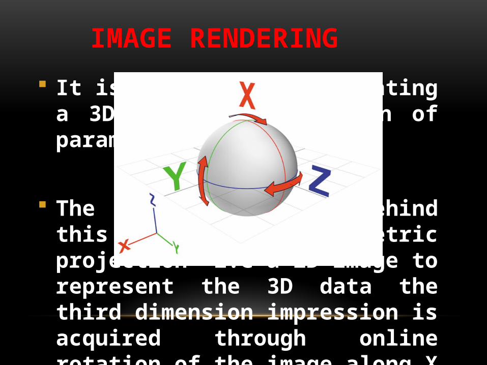
IMAGE RENDERING
It is the process of creating a 3D visual presentation of parameters of interest.
The main principle behind this is "planar geometric projection" i.e a 2D image to represent the 3D data the third dimension impression is acquired through online rotation of the image along X , Y and Z axis

The exam can show the vessel of interest alone "Inversion mode" or along with the gray scale image in what's called the "Glass body rendering mode".

ARE WE LOOKING AT A VESSEL OR AT A SPECIFIC ORGAN
VASCULARITY?
If a specific vessel is targeted we simply apply 3D power or Color Doppler on the vessel of interest but if an organ or a structure as a whole is targeted we use a software technology known as VOCAL (virtual organ computer aided analysis).

OBTAINING THE IMAGE STEP BY STEP
1-Choose the 3D/4D probe to use for your scan
2- From the submenu of the 3D/4D probe choose the application most appropriate according to your case e.g gestational age 1st, 2nd and 3rd trimsteric scanning.
3- Perform a usual real time 2D scan to visualize and locate your organ or region of interest .


4- Press on the power or color Doppler button to acquire a blood flow mapping.
5- Adjust the box that's going show up on your screen to include your area of interest only.
6- Press on 3D button and keep your hands steady to acquire the volume.
7- Once volume is acquired save it on your machine to process it immediately or later but make sure to save it as a 3D volume not a 2 D image.

8- Choose to perform volume analysis from the submenu.
9- Press on the VOCAL button which will allow you to manually trace the volume at a specified angles of rotation chosen by the operator.


10- Your ultrasound machine will give the option of accepting you ROI " region of interest" , once accepted you can then choose the volume histogram which gives you a numerical value of the vascular indices.


QUANTIFYING THE BLOOD FLOW

1- VI (Vascularization index): Vascularization index is the ratio of the number of color voxels (volumetric pixel) to the total number of voxels in the sampled tissue, thus it represents the percentage of vascularized tissue

2- FI (flow index) : Flow index is the average colour value of all colour voxels and it describes the mean velocity of flow in the sampled tissue.

3- VFI (vascularization flow index) : is the average colour value of all colour and grey voxels and describes both: the vascularization and the blood flow.

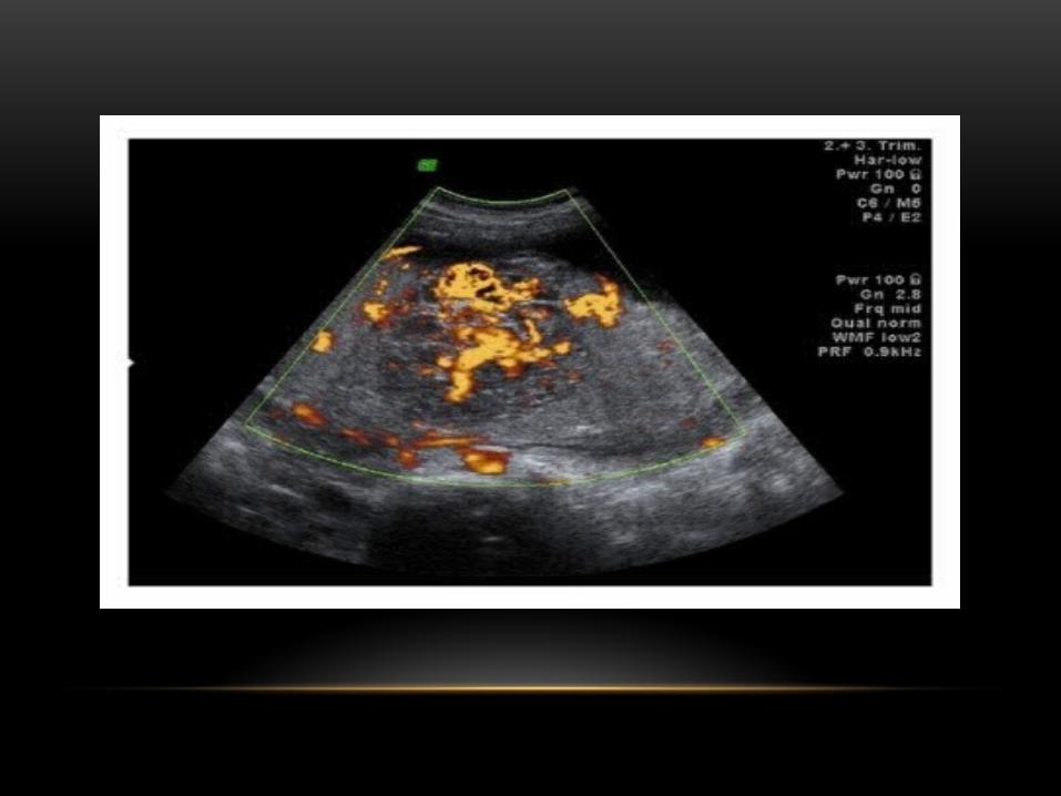
CLINICAL APPLICATIONS OF 3D POWER DOPPLER IN OBSTETRICS
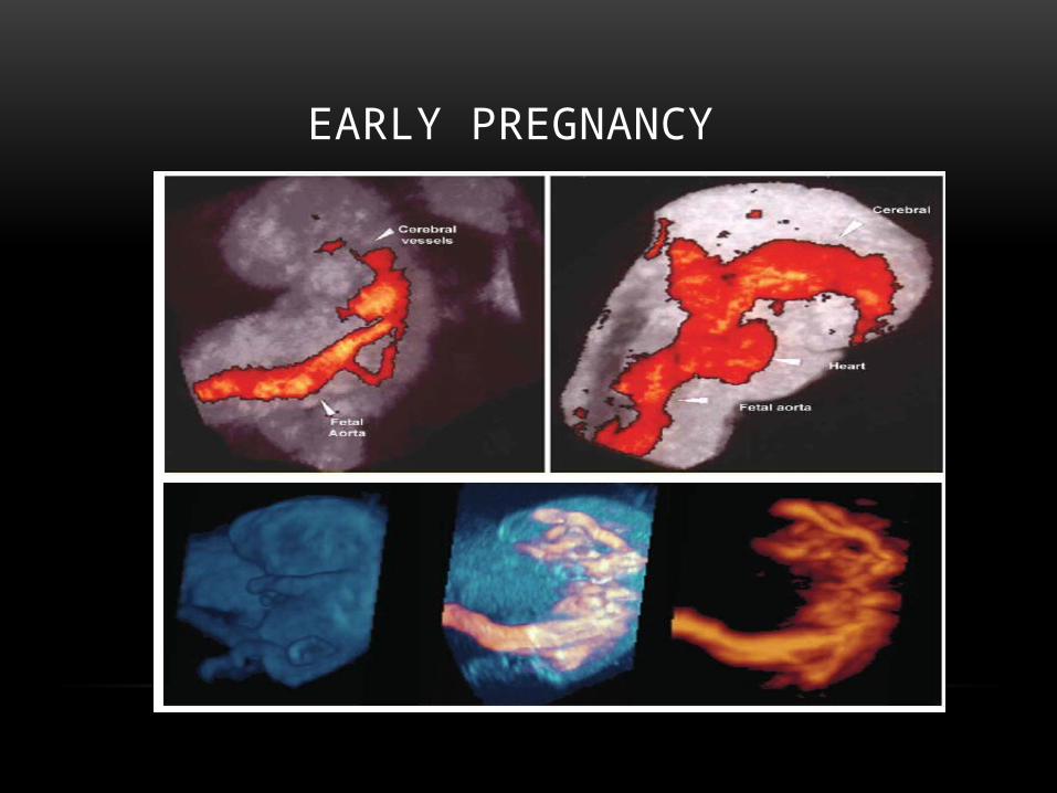
EARLY PREGNANCY

Umbilical cord : true and false knots, single umbilical artery and normal and abnormal insertion in placenta.

Placental anomalies as placenta previa , placenta accreta or vasa previa.

Placental vascularity assessment for IUGR and PIH disorders.


FETAL INTRA ABDOMINAL VESSELS

Currently the main field of interest seems to be the absence of ductus venosus with different possibilities of umbilical vein connection, in this condition the role of 3D power Doppler is to demonstrate the spatial course of the aberrant vessel .

Abnormal site of intra-abdominal vessels in gastroschisis
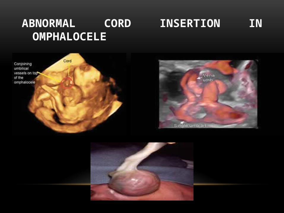
ABNORMAL CORD INSERTION IN OMPHALOCELE

Abnormal umbilical vein size (varix or ectasia)

Another interesting condition is the abnormal course of vessels in isomerism i.e the interruption of inferior vena cava with Azygous continuity

Visualization of renal vessels increase the accuracy of diagnosis of renal malformation as renal vascular tree is easily demonstrated

Fetal cardiac circulation

Extended fetal echocardiography can be acquiring and documenting different cross sectional planes in the heart.
In the last decade the advent in 3D and 4D (real time 3D) has allowed the operator to automatically reconstruct the cardiac circulatory system and its connection spatially instead of doing it mentally as in the past.
A new software technique known as STIC (spatiotemporal image correlation) has allowed this automatic reconstruction.


Fetal cerebral circulation

Many vascular malformation in the fetal cerebral circulation can demonstrated by the use of 3D power Doppler reconstruction

NORMAL BRAIN VASCULARITY
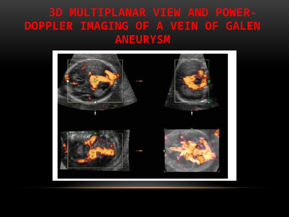
3D MULTIPLANAR VIEW AND POWER-DOPPLER IMAGING OF A VEIN OF GALEN
ANEURYSM

INTRACRANIAL TUMORS
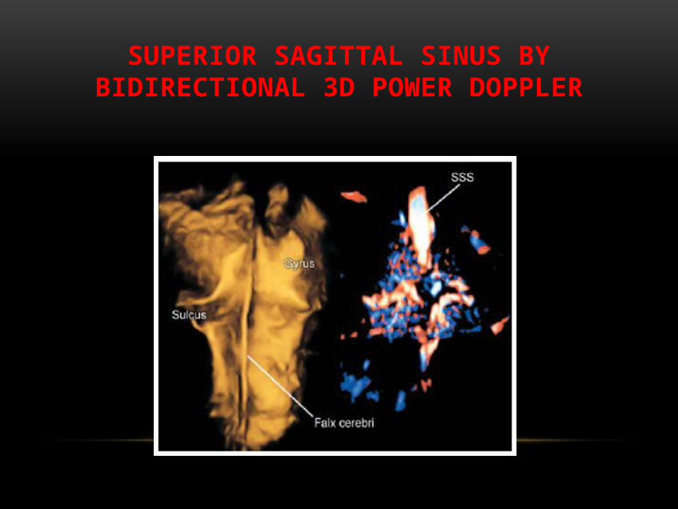
SUPERIOR SAGITTAL SINUS BY BIDIRECTIONAL 3D POWER DOPPLER

Corpus Callosal agensis

A-V MALFORMATIONS IN CEREBRAL CIRCULATION
The 3D Power Angio reconstruction demonstrates connection feeder and draining vessels (arrows) of the vein of Galen ectasia and sinuses of brain (ts – transverses sinus, sps – superior petrosal sinus)


CIRCLE OF WILLIS IN NORMAL AND HOLOPROSENCEPHALY

Related Documents