1 AIUM Practice Parameter for the Performance of a Transcranial Doppler Ultrasound Examination for Adults and Children Parameter developed in conjunction with the American College of Radiology (ACR), the Society for Pediatric Radiology (SPR), and the Society of Radiologists in Ultrasound (SRU). The American Institute of Ultrasound in Medicine (AIUM) is a multidisciplinary association dedicated to advancing the safe and effective use of ultrasound in medicine through professional and public education, research, development of parameters, and accreditation. To promote this mission, the AIUM is pleased to publish, in conjunction with the American College of Radiology (ACR), the Society for Pediatric Radiology (SPR), and the Society of Radiologists in Ultrasound (SRU), this AIUM Practice Parameter for the Performance of a Transcranial Doppler Ultrasound Examination for Adults and Children. We are indebted to the many volunteers who contributed their time, knowledge, and energy to bringing this document to completion. The AIUM represents the entire range of clinical and basic science interests in medical diagnostic ultrasound, and, with hundreds of volunteers, the AIUM has promoted the safe and effective use of ultrasound in clinical medicine for more than 65 years. This document and others like it will continue to advance this mission. © 2017 American Institute of Ultrasound in Medicine 14750 Sweitzer Ln, Suite 100 Laurel, MD 20707-5906 USA www.aium.org

Welcome message from author
This document is posted to help you gain knowledge. Please leave a comment to let me know what you think about it! Share it to your friends and learn new things together.
Transcript

1
AIUM Practice Parameter for the Performance of a
Transcranial Doppler Ultrasound Examination for
Adults and Children Parameter developed in conjunction with the American College of Radiology (ACR), the Society
for Pediatric Radiology (SPR), and the Society of Radiologists in Ultrasound (SRU).
The American Institute of Ultrasound in Medicine (AIUM) is a multidisciplinary association
dedicated to advancing the safe and effective use of ultrasound in medicine through
professional and public education, research, development of parameters, and accreditation.
To promote this mission, the AIUM is pleased to publish, in conjunction with the American
College of Radiology (ACR), the Society for Pediatric Radiology (SPR), and the Society of
Radiologists in Ultrasound (SRU), this AIUM Practice Parameter for the Performance of a
Transcranial Doppler Ultrasound Examination for Adults and Children. We are indebted to the
many volunteers who contributed their time, knowledge, and energy to bringing this document to
completion.
The AIUM represents the entire range of clinical and basic science interests in medical
diagnostic ultrasound, and, with hundreds of volunteers, the AIUM has promoted the safe and
effective use of ultrasound in clinical medicine for more than 65 years. This document and
others like it will continue to advance this mission.
© 2017 American Institute of Ultrasound in Medicine 14750 Sweitzer Ln, Suite 100 Laurel, MD 20707-5906 USA
www.aium.org

2
Practice parameters of the AIUM are intended to provide the medical ultrasound community with
parameters for the performance and recording of high-quality ultrasound examinations. The
parameters reflect what the AIUM considers the minimum criteria for a complete examination in
each area but are not intended to establish a legal standard of care. AIUM-accredited practices
are expected to generally follow the parameters with recognition that deviations from these
parameters will be needed in some cases, depending on patient needs and available
equipment. Practices are encouraged to go beyond the parameters to provide additional service
and information as needed.
I. Introduction
The clinical aspects contained in specific sections of this parameter (Introduction, Indications,
Specifications of the Examination, and Equipment Specifications) were developed
collaboratively by the American Institute of Ultrasound in Medicine (AIUM), the American
College of Radiology (ACR), the Society for Pediatric Radiology (SPR), and the Society of
Radiologists in Ultrasound (SRU). Recommendations for Qualifications and Responsibilities of
Personnel, Written Request for the Examination, Documentation, and Quality Control and
Improvement, Safety, Infection Control, and Patient Education vary among the organizations
and are addressed by each separately.
Transcranial Doppler (TCD) ultrasound is a noninvasive technique that assesses blood flow
within the circle of Willis and the vertebrobasilar system.
II. Indications
A. Indications for a TCD ultrasound examination of children and adults include but are not limited to: 1. Evaluation of sickle cell disease to determine stroke risk.1–3
2. Detection and follow-up of stenosis or occlusion in a major intracranial artery in the
circle of Willis or vertebrobasilar system, including monitoring and potentiation of
thrombolytic therapy for acute stroke patients.3–5
© 2017 American Institute of Ultrasound in Medicine 14750 Sweitzer Ln, Suite 100 Laurel, MD 20707-5906 USA
www.aium.org

3
3. Detection of cerebral vasculopathy.3,6
4. Detection and monitoring of vasospasm in patients with spontaneous or traumatic
subarachnoid hemorrhage.7,8
5. Evaluation of collateral pathways of intracranial blood flow, including after
intervention.9–11
6. Detection of circulating cerebral microemboli or high-intensity transient signals
(HITS)5
7. Detection of right-to-left shunts.12,13
8. Assessment of cerebral vasomotor reactivity (VMR).13,14
9. As an adjunct to the clinical diagnosis of brain death.15,16
10. Intraoperative and periprocedural monitoring to detect cerebral thrombosis,
embolization, hypoperfusion, and hyperperfusion.17,18
11. Assessment of arteriovenous malformations, before and after treatment.6,19
12. Detection and follow-up of intracranial aneurysms.20
13. Evaluation of positional vertigo.21
B. Additional applications in children include but are not limited to: 1. Assessment of intracranial pressure and hydrocephalus.22,23
2. Assessment of hypoxic-ischemic encephalopathy.6,24
3. Assessment of dural venous sinus patency.6,25
III. Qualifications and Responsibilities of Personnel
See www.aium.org for AIUM Official Statements, including Standards and Guidelines for the
Accreditation of Ultrasound Practices and relevant Physician Training Guidelines.26
IV. Written Request for the Examination
The written or electronic request for an ultrasound examination should provide sufficient
information to allow for the appropriate performance and interpretation of the examination.
© 2017 American Institute of Ultrasound in Medicine 14750 Sweitzer Ln, Suite 100 Laurel, MD 20707-5906 USA
www.aium.org
4
The request for the examination must be originated by a physician or another appropriately
licensed health care provider or under the physician’s or provider’s direction. The accompanying
clinical information should be provided by a physician or another appropriate health care
provider familiar with the patient’s clinical situation and should be consistent with relevant legal
and local health care facility requirements.
V. Specifications of the Examination
Cerebral blood flow velocities and the resistive indices are variable and affected by age, the
arterial carbon dioxide (CO2) level, and cerebral and systemic perfusion. They are influenced by
body temperature, the state of patient arousal, mechanical ventilation and suctioning, the
presence of systemic shunts, cardiac disease, and/or anemia. It is important to perform the
examination when the patient is awake, quiet, and calm. Generally speaking, examinations
should not be performed if the patient has been sedated or anesthetized earlier the same day.
However, these considerations are not relevant when studies are done for determination of
brain death or to detect brain perfusion abnormalities intraoperatively or postoperatively.
A. Infants Before Fontanelle Closure: Depending on the size of the child, sector, curvilinear, or linear transducers with grayscale and
Doppler frequencies from approximately 5 to 15 MHz should be used.27 The highest frequency
transducer that permits adequate cerebrovascular interrogation is recommended. Duplex
ultrasound is preferred over nonimaging Doppler methods in children for more precise
localization and insonation of the targeted vessels.28,29 Duplex imaging may be more difficult in
adults, especially the elderly, in whom the acoustic window is often small.
In infants, an open fontanelle provides an acoustic window to the intracranial circulation. The
distal internal carotid vessels and the branches of the circle of Willis can be interrogated through
the anterior fontanelle in the coronal and sagittal planes (although the middle cerebral artery
[MCA] may be better interrogated via a transtemporal approach; see below).3 For basic
assessment of global cerebral arterial flow and spectral waveform analysis, interrogation of the
pericallosal branch of the anterior cerebral artery (ACA) on sagittal imaging via the anterior
fontanelle is the simplest, most reliable approach. The superior sagittal sinus can be evaluated
© 2017 American Institute of Ultrasound in Medicine 14750 Sweitzer Ln, Suite 100 Laurel, MD 20707-5906 USA
www.aium.org

5
through an open sagittal suture. Imaging of the posterior circulation can be performed via the
foramen magnum or via the posterolateral fontanelle located just posterior to the mastoid
process.30,31
When assessing for elevated intracranial pressure, interrogation of the pericallosal branch of the
ACA can be performed both before and after gentle compression of the anterior fontanelle.32,33
Care should be taken to minimize the degree and duration of compression.
B. Adults and Children After Fontanelle Closure
Transcranial spectral Doppler sonography, power M-mode Doppler sonography, or transcranial
color-coded duplex sonography (TCCS) should be performed with the patient supine. If velocity
reference standards have been previously acquired with nonimaging TCD methods (and thus
not angle corrected), velocity measurements with imaging methods (TCCS) should not be angle
corrected to allow comparison with reference values.28,34 It should be noted that velocities
obtained with duplex imaging equipment may be lower than those obtained with nonduplex
imaging equipment. Therefore, stroke risk thresholds determined with imaging equipment may
need to be lowered.27,35–37 However, if validated reference values for angle-corrected TCCS
velocities exist in an ultrasound laboratory, and a sufficient length of a vessel is visualized
during TCCS to allow angle correction, then angle-corrected velocities can be obtained.38
In adults, TCD studies require the use of lower-frequency transducers to adequately penetrate
the calvarium to produce useful grayscale images and obtain Doppler signals. A 2- to 3-MHz
transducer or multifrequency transducer with 2- to 3-MHz spectral Doppler capability is
commonly required. For children or small adults, adequate imaging may be possible at higher
transducer frequencies.20
Representative views and velocities should be obtained of the distal internal carotid arteries
(ICAs); the ACAs, MCAs, and posterior cerebral arteries (PCAs) in the circle of Willis; and the
vertebrobasilar system. Any abnormalities should be further evaluated and documented. Both
the left and right sides of the brain should be interrogated unless the examination is performed
to follow up a known abnormality of a specific vessel.
© 2017 American Institute of Ultrasound in Medicine 14750 Sweitzer Ln, Suite 100 Laurel, MD 20707-5906 USA
www.aium.org
6
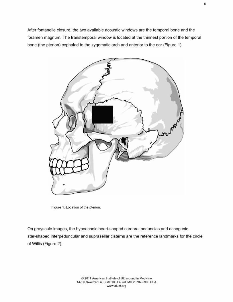
After fontanelle closure, the two available acoustic windows are the temporal bone and the
foramen magnum. The transtemporal window is located at the thinnest portion of the temporal
bone (the pterion) cephalad to the zygomatic arch and anterior to the ear (Figure 1).
Figure 1. Location of the pterion.
On grayscale images, the hypoechoic heart-shaped cerebral peduncles and echogenic
star-shaped interpeduncular and suprasellar cisterns are the reference landmarks for the circle
of Willis (Figure 2).
© 2017 American Institute of Ultrasound in Medicine 14750 Sweitzer Ln, Suite 100 Laurel, MD 20707-5906 USA
www.aium.org

7
Figure 2. Transtemporal grayscale image showing the cerebral peduncles (P) with the
echogenic interpeduncular and suprasellar basilar cistern (*) located immediately anteriorly.
Anterior and lateral to the cistern is the MCA, which should be insonated by using color and
spectral Doppler imaging (Figure 3).
© 2017 American Institute of Ultrasound in Medicine 14750 Sweitzer Ln, Suite 100 Laurel, MD 20707-5906 USA
www.aium.org

8
Figure 3. Transtemporal color Doppler image of the circle
of Willis. * indicates cerebral peduncle.
The MCA should be interrogated from its most superficial point below the calvarium to the
bifurcation of the A1 segment of the ACA and the M1 segment of the MCA.28,29 Normally, flow in
the MCA is directed toward the transducer. The ACA should be interrogated distal to the
bifurcation. Flow in the ACA should be away from the transducer (Figure 3). The PCA courses
around the heart-shaped cerebral peduncles with flow directed toward the transducer in the P1
segment and directed away from the transducer in the more distal P2 segment.39,40 Tracing the
PCAs medially to the top of the basilar artery with its normally bidirectional flow can be used to
verify correct positioning of the Doppler sample volume within the posterior cerebral arteries.
The foramen magnum can be used to study the vertebral and basilar arteries. An optimal
window is often obtained with the patient turned to one side with the neck flexed so that the chin
touches the chest. The transducer is placed over the upper neck at the base of the skull and
angled cephalad through the foramen magnum toward the nose.30,40
© 2017 American Institute of Ultrasound in Medicine 14750 Sweitzer Ln, Suite 100 Laurel, MD 20707-5906 USA
www.aium.org

9
On TCCS, the vertebral arteries have a V-shaped configuration as they extend superiorly to
form the basilar artery. The reference landmark is the hypoechoic medulla (Figure 4). Flow in
the vertebral and basilar arteries is directed away from the transducer and should be
interrogated up to the distal end of the basilar artery.
Figure 4. Color Doppler image of the paired vertebral (V)
and basilar (B) arteries with a spectral tracing obtained
from the right vertebral artery. * indicates medulla.
In patients with suspected carotid artery stenosis or occlusion, a transorbital examination of the
ophthalmic arteries and carotid siphons can be performed.10,41 A transorbital window permits
visualization of the ophthalmic artery and the carotid siphon. The transducer is placed so that it
rests lightly on the closed superior eyelid.20 The study must be performed at reduced power
settings with a mechanical index not to exceed 0.23 to prevent ocular injury.42 Angle correction
is not performed.
In children with sickle cell disease, spectral Doppler waveform analysis should include the
time-averaged maximum mean velocity as defined by the Stroke Prevention Trial in Sickle Cell
© 2017 American Institute of Ultrasound in Medicine 14750 Sweitzer Ln, Suite 100 Laurel, MD 20707-5906 USA
www.aium.org
10
Anemia criteria.43–46 Velocity measurements are obtained at 2-mm intervals along the entire
course of the MCA and PCA and at 2 depths from the ACA and distal ICA. Velocity can be
measured with either an automatic tracing method or by manual placement of cursors.
Angle-corrected TCCS velocities have typically not been used for pediatric sickle cell
evaluations. Both imaging and nonimaging techniques are routinely used, with most pediatric
radiology departments preferring the imaging technique and other departments using a
nonimaging technique. To date, there is no evidence that TCD measurement is beneficial in
individuals with sickle cell disease who are older than 16 years.1,47
Patients with subarachnoid hemorrhage may develop vasospasm, with increased arterial
velocities developing by day 3 after the onset of the hemorrhage and peaking between days 6
and 12.15 Parameters used to measure vasospasm include peak systolic velocity, mean flow
velocity (MFV), pulsatility index, and resistive index. Threshold values depend on which vessels
are insonated and which measures are performed. Since hyperemia, autoregulation,
hypertension, and hypervolemia can also result in increased flow velocities, a submandibular
approach can be used to sample the distal internal carotid artery in the neck to calculate mean
flow velocity ratios between the middle cerebral and internal carotid arteries, the so-called
hemispheric or Lindegaard index.48,49 A Lindegaard ratio or index (MFVMCA/MFVICA) of 3 to 6 is
indicative of mild to moderate vasospasm, and a ratio of greater than 6 is indicative of severe
vasospasm.49 Angle correction is not performed.
Nonimaging TCD monitoring is useful for the assessment of cerebral VMR. VMR is the
physiologic mechanism that maintains constant cerebral flow across a wide range of blood
pressure fluctuations through regulation of the vasomotor tone of the distal cerebral
arterioles.13,14 Under pathologic conditions (eg, traumatic and nontraumatic brain injury, stroke,
and arterial occlusion), VMR may be impaired. VMR is measured with a TCD challenge test,
most commonly the CO2 inhalation test or the breath-holding index (BHI). Continuous TCD
tracings of the MFV from the MCA (or PCA), heart rate, respiratory rate, and expiratory pCO2
are recorded during several minutes of baseline measurements, after inhalation of 5% CO2 and
air for 2 minutes, and for several minutes after inhalation. VMR is calculated as the percent rise
in the MCA MFV per 1-mm Hg pCO2 increase from baseline. Normal VMR is defined as a rise in
the MCA MFV of greater than 2%/mm Hg pCO2 50. Similarly, the BHI is calculated as the percent
rise in the MCA (or PCA) MFV recorded immediately at the end of the breath-holding period
© 2017 American Institute of Ultrasound in Medicine 14750 Sweitzer Ln, Suite 100 Laurel, MD 20707-5906 USA
www.aium.org
11
(usually 30 seconds or less) from the MFV at baseline per seconds of breath holding.51 A BHI of
0.69 or higher is considered normal.52
Cerebral embolism accounts for up to 70% of all ischemic strokes.18,53 Cerebral microemboli can
be diagnosed by nonimaging TCD monitoring through the detection of HITS and are defined by
the following criteria:
1. HITS usually lasting less than 300 milliseconds.
2. Doppler amplitude exceeding background the Doppler frequency spectrum signal
by at least 3 dB.
3. A unidirectional signal within the Doppler velocity spectrum.
4. A characteristic “moaning” or “chirping” sound.54
The most common sources of HITS include artery-to-artery embolization from the proximal
carotid, vertebral, and intracranial arteries; the aortic arch; or the heart (related to atrial
fibrillation, right-to-left cardiac shunts [particularly from a patent foramen ovale], prosthetic heart
valves, and after cardiac surgery). Bilateral or unilateral monitoring of a targeted intracranial
vessel is recorded for a minimum of 30 minutes. Most TCD systems are equipped with
automated HITS detection software that counts the number of microemboli and measures the
microembolic signal intensity.55 However, both visual and auditory inspection and confirmation
of each detected HITS are required by the rater/interpreter for a reliable diagnosis.
For detection of right-to-left shunts, TCD monitoring is performed during the intravenous
injection of agitated saline or a contrast medium and patient performance of a Valsalva
maneuver to enhance flow across the shunt. The degree of shunting is quantitatively assessed
by the number of detected HITS.56
VI. Documentation
Adequate documentation is essential for high-quality patient care. There should be a permanent
record of the ultrasound examination and its interpretation. Images of all appropriate areas, both
normal and abnormal, should be recorded. Variations from normal size should be accompanied
© 2017 American Institute of Ultrasound in Medicine 14750 Sweitzer Ln, Suite 100 Laurel, MD 20707-5906 USA
www.aium.org

12
by measurements. Images should be labeled with the patient identification, facility identification,
examination date, and side (right or left) of the anatomic site imaged. An official interpretation
(final report) of the ultrasound findings should be included in the patient’s medical record.
Retention of the ultrasound examination should be consistent both with clinical needs and with
relevant legal and local health care facility requirements.
Reporting should be in accordance with the AIUM Practice Parameter for Documentation of an
Ultrasound Examination.
VII. Equipment Specifications
Transcranial Doppler imaging should be performed with a real-time imaging scanner with
Doppler capability using ultrasound frequencies that can penetrate the temporal bone and
foramen magnum or a nonimaging Doppler instrument (TCD or power M-mode Doppler). Color
or spectral Doppler imaging should be used to locate the intracranial vessels in all cases. The
color gain settings should be maximized so that a well-defined vessel is displayed. The Doppler
settings should be maximized so that a well-defined vessel is displayed. The Doppler setting
should be adjusted to obtain the highest velocity in all cases. The Doppler power output should
be as low as reasonably achievable.
VIII. Quality Control and Improvement, Safety, Infection Control, and Patient Education Policies and procedures related to quality control, patient education, infection control, and safety
should be developed and implemented in accordance with the AIUM Standards and Guidelines
for the Accreditation of Ultrasound Practices.
Equipment performance monitoring should be in accordance with the AIUM Standards and
Guidelines for the Accreditation of Ultrasound Practices.
© 2017 American Institute of Ultrasound in Medicine 14750 Sweitzer Ln, Suite 100 Laurel, MD 20707-5906 USA
www.aium.org

13
IX. ALARA Principle
The potential benefits and risks of each examination should be considered. The ALARA (as low
as reasonably achievable) principle should be observed when adjusting controls that affect the
acoustic output and by considering transducer dwell times. Further details on ALARA may be
found in the AIUM publication Medical Ultrasound Safety, Third Edition.
Acknowledgments
This parameter was revised by the American Institute of Ultrasound in Medicine (AIUM) in
collaboration with the American College of Radiology (ACR), the Society for Pediatric Radiology
(SPR), and the Society of Radiologists in Ultrasound (SRU) according to the process described
in the AIUM Clinical Standards Committee Manual.
Collaborative Committee: Members represent their societies in the initial and final revision of this
parameter.
ACR
Harriet J. Paltiel, MD, Chair
Dorothy I. Bulas, MD, FACR
Brian D. Coley, MD, FACR
Monica S. Epelman, MD
AIUM
Mary E. McCarville, MD
Tatjana Rundek, MD, PhD
SPR
Harris L. Cohen, MD, FACR
Erica L. Riedesel, MD
Cicero T. Silva, MD
© 2017 American Institute of Ultrasound in Medicine 14750 Sweitzer Ln, Suite 100 Laurel, MD 20707-5906 USA
www.aium.org
14
SRU
Leslie M. Scoutt, MD, FACR
AIUM Clinical Standards Committee
Joseph Wax, MD, FAIUM, Chair
John Pellerito, MD, FACR, FAIUM, FSRU, Vice Chair
Sandra Allison, MD
Bryann Bromley, MD, FAIUM
Anil Chauhan, MD
Stamatia Destounis, MD, FACR,
Eitan Dickman, MD, RDMS, FACEP
Joan Mastrobattista, MD, FAIUM
Marsha Neumyer, BS, RVT, FSVU, FAIUM, FSDMS
Tatjana Rundek, MD, PhD
Khaled Sakhel, MD
James Shwayder, MD, JD, FAIUM
Ants Toi, MD, FRCP, FAIUM
Isabelle Wilkins, MD, FAIUM
Original copyright 2007; revised 2012, 2017
Renamed 2015
References 1. DeBaun MR, Kirkham FJ. Central nervous system complications and management in
sickle cell disease. Blood 2016; 127:829–838.
2. Brousse V, Kossorotoff M, de Montalembert M. How I manage cerebral vasculopathy in
children with sickle cell disease. Br J Haematol 2015; 170:615–625.
3. Verlhac S. Transcranial Doppler in children. Pediatr Radiol. 2011; 41(suppl 1):S153–S165.
© 2017 American Institute of Ultrasound in Medicine 14750 Sweitzer Ln, Suite 100 Laurel, MD 20707-5906 USA
www.aium.org

15
4. Saqqur M, Tsivgoulis G, Nicoli F, et al. The role of sonolysis and sonothrombolysis in
acute ischemic stroke: a systematic review and meta-analysis of randomized controlled
trials and case-control studies. J Neuroimaging 2014; 24:209–220.
5. Purkayastha S, Sorond F. Transcranial Doppler ultrasound: technique and application.
Semin Neurol 2012; 32:411–420.
6. Soetaert AM, Lowe LH, Formen C. Pediatric cranial Doppler sonography in children:
non-sickle cell applications. Curr Probl Diagn Radiol 2009; 38:218–227.
7. Kalanuria A, Nyquist PA, Armonda RA, Razumovsky A. Use of Transcranial Doppler
(TCD) ultrasound in the neurocritical care unit. Neurosurg Clin N Am 2013; 24:441–456.
8. Marshall SA, Nyquist P, Ziai WC. The role of transcranial Doppler ultrasonography in the
diagnosis and management of vasospasm after aneurysmal subarachnoid hemorrhage.
Neurosurg Clin N Am 2010; 21:291–303.
9. Guan J, Zhang S, Zhou Q, Li C, Lu Z. Usefulness of transcranial Doppler ultrasound in
evaluating cervical-cranial collateral circulations. Interv Neurol 2013; 2:8–18.
10. Baumgartner RW. Intracranial stenoses and occlusions, and circle of willis collaterals.
Front Neurol Neurosci 2006; 21:117–126.
11. von Budingen HC, Staudacher T, von Budingen HJ. Ultrasound diagnostics of the
vertebrobasilar system. Front Neurol Neurosci 2006; 21:57–69.
12. Mojadidi MK, Roberts SC, Winoker JS, et al. Accuracy of transcranial Doppler for the
diagnosis of intracardiac right-to-left shunt: a bivariate meta-analysis of prospective
studies. JACC Cardiovasc Imaging 2014; 7:236–250.
13. Tsivgoulis G, Alexandrov AV, Sloan MA. Advances in transcranial Doppler
ultrasonography. Curr Neurol Neurosci Rep 2009; 9:46–54.
14. Wolf ME. Functional TCD: regulation of cerebral hemodynamics—cerebral autoregulation,
vasomotor reactivity, and neurovascular coupling. Front Neurol Neurosci 2015; 36:40–56.
© 2017 American Institute of Ultrasound in Medicine 14750 Sweitzer Ln, Suite 100 Laurel, MD 20707-5906 USA
www.aium.org

16
15. Rasulo FA, De Peri E, Lavinio A. Transcranial Doppler ultrasonography in intensive care.
Eur J Anaesthesiol Suppl 2008; 42:167–173.
16. Monteiro LM, Bollen CW, van Huffelen AC, Ackerstaff RG, Jansen NJ, van Vught AJ.
Transcranial Doppler ultrasonography to confirm brain death: a meta-analysis. Intensive
Care Med 2006; 32:1937–1944.
17. Alexandrov AV, Sloan MA, Tegeler CH, et al. Practice standards for transcranial Doppler
(TCD) ultrasound, part II: clinical indications and expected outcomes. J Neuroimaging
2012; 22:215–224.
18. Sloan MA, Alexandrov AV, Tegeler CH, et al. Assessment: transcranial Doppler
ultrasonography: report of the Therapeutics and Technology Assessment Subcommittee of
the American Academy of Neurology. Neurology 2004; 62:1468–1481.
19. Fu B, Zhao JZ, Yu LB. The application of ultrasound in the management of cerebral
arteriovenous malformation. Neurosci Bull 2008; 24:387–394.
20. Kirsch JD, Mathur M, Johnson MH, Gowthaman G, Scoutt LM. Advances in transcranial
Doppler US: imaging ahead. Radiographics 2013; 33:E1–E14.
21. Machaly SA, Senna MK, Sadek AG. Vertigo is associated with advanced degenerative
changes in patients with cervical spondylosis. Clin Rheumatol 2011; 30:1527–1534.
22. de Oliveira RS, Machado HR. Transcranial color-coded Doppler ultrasonography for
evaluation of children with hydrocephalus. Neurosurg Focus 2003; 15:ECP3.
23. Taylor GA. Sonographic assessment of posthemorrhagic ventricular dilatation. Radiol Clin
North Am 2001; 39:541–551.
24. Cassia GS, Faingold R, Bernard C, Sant’Anna GM. Neonatal hypoxic-ischemic injury:
sonography and dynamic color Doppler sonography perfusion of the brain and abdomen
with pathologic correlation. AJR Am J Roentgenol 2012; 199:W743–W752.
25. Stolz EP. Role of ultrasound in diagnosis and management of cerebral vein and sinus
thrombosis. Front Neurol Neurosci 2008; 23:112–121.
© 2017 American Institute of Ultrasound in Medicine 14750 Sweitzer Ln, Suite 100 Laurel, MD 20707-5906 USA
www.aium.org
17
26. American Institute of Ultrasound in Medicine. AIUM physician training guidelines.
American Institute of Ultrasound in Medicine website.
http://www.aium.org/resources/ptGuidelines.aspx.
27. Bulas D. Transcranial Doppler: applications in neonates and children. Ultrasound Clin
2009; 4:533–551.
28. McCarville MB. Comparison of duplex and nonduplex transcranial Doppler
ultrasonography. Ultrasound Q 2008; 24:167–171.
29. Bulas D. Screening children for sickle cell vasculopathy: guidelines for transcranial
Doppler evaluation. Pediatr Radiol 2005; 35:235–241.
30. Brennan CM, Taylor GA. Sonographic imaging of the posterior fossa utilizing the foramen
magnum. Pediatr Radiol 2010; 40:1411–1416.
31. Buckley KM, Taylor GA, Estroff JA, Barnewolt CE, Share JC, Paltiel HJ. Use of the
mastoid fontanelle for improved sonographic visualization of the neonatal midbrain and
posterior fossa. AJR Am J Roentgenol 1997; 168:1021–1025.
32. Taylor GA, Madsen JR. Neonatal hydrocephalus: hemodynamic response to fontanelle
compression: correlation with intracranial pressure and need for shunt placement.
Radiology 1996; 201:685–689.
33. Taylor GA, Phillips MD, Ichord RN, Carson BS, Gates JA, James CS. Intracranial
compliance in infants: evaluation with Doppler US. Radiology 1994; 191:787–791.
34. Neish AS, Blews DE, Simms CA, Merritt RK, Spinks AJ. Screening for stroke in sickle cell
anemia: comparison of transcranial Doppler imaging and nonimaging US techniques.
Radiology 2002; 222:709–714.
35. Krejza J, Rudzinski W, Pawlak MA, et al. Angle-corrected imaging transcranial Doppler
sonography versus imaging and nonimaging transcranial doppler sonography in children
with sickle cell disease. AJNR Am J Neuroradiol 2007; 28:1613–1618.
© 2017 American Institute of Ultrasound in Medicine 14750 Sweitzer Ln, Suite 100 Laurel, MD 20707-5906 USA
www.aium.org

18
36. McCarville MB, Li C, Xiong X, Wang W. Comparison of transcranial Doppler sonography
with and without imaging in the evaluation of children with sickle cell anemia. AJR Am J
Roentgenol 2004; 183:1117–1122.
37. Jones AM, Seibert JJ, Nichols FT, et al. Comparison of transcranial color Doppler imaging
(TCDI) and transcranial Doppler (TCD) in children with sickle-cell anemia. Pediatr Radiol
2001; 31:461–469.
38. Nedelmann M, Stolz E, Gerriets T, et al. Consensus recommendations for transcranial
color-coded duplex sonography for the assessment of intracranial arteries in clinical trials
on acute stroke. Stroke 2009; 40:3238–3244.
39. Krejza J, Mariak Z, Melhem ER, Bert RJ. A guide to the identification of major cerebral
arteries with transcranial color Doppler sonography. AJR Am J Roentgenol 2000;
174:1297–1303.
40. Lupetin AR, Davis DA, Beckman I, Dash N. Transcranial Doppler sonography, part 1:
principles, technique, and normal appearances. Radiographics 1995; 15:179–191.
41. You Y, Hao Q, Leung T, et al. Detection of the siphon internal carotid artery stenosis:
transcranial Doppler versus digital subtraction angiography. J Neuroimaging 2010;
20:234–239.
42. Phillips R, Harris G. Information for manufacturers seeking marketing clearance of
diagnostic ultrasound systems and transducers. US Food and Drug Administration
website; 2008.
www.fda.gov/downloads/MedicalDevices/DeviceRegulationGuidance/GuidanceDocuments
/UCM070911.pdf. Accessed November 6, 2015.
43. Ware RE, Davis BR, Schultz WH, et al. Hydroxycarbamide versus chronic transfusion for
maintenance of transcranial Doppler flow velocities in children with sickle cell
anaemia-TCD with transfusions changing to hydroxyurea (TWiTCH): a multicentre,
open-label, phase 3, non-inferiority trial. Lancet 2016; 387:661–670.
44. Adams RJ, Brambilla D. Discontinuing prophylactic transfusions used to prevent stroke in
sickle cell disease. N Engl J Med 2005; 353:2769–2778.
© 2017 American Institute of Ultrasound in Medicine 14750 Sweitzer Ln, Suite 100 Laurel, MD 20707-5906 USA
www.aium.org
http://www.fda.gov/downloads/MedicalDevices/DeviceRegulationGuidance/GuidanceDocuments/UCM070911.pdf

19
45. Adams RJ. TCD in sickle cell disease: an important and useful test. Pediatr Radiol 2005;
35:229–234.
46. Adams R, McKie V, Nichols F, et al. The use of transcranial ultrasonography to predict
stroke in sickle cell disease. N Engl J Med 1992; 326:605–610.
47. Valadi N, Silva GS, Bowman LS, et al. Transcranial Doppler ultrasonography in adults with
sickle cell disease. Neurology 2006; 67:572–574.
48. Lindegaard KF, Nornes H, Bakke SJ, Sorteberg W, Nakstad P. Cerebral vasospasm
diagnosis by means of angiography and blood velocity measurements. Acta Neurochir
1989; 100:12–24.
49. Alexandrov AV, Sloan MA, Wong LK, et al. Practice standards for transcranial Doppler
ultrasound, part I: test performance. J Neuroimaging 2007; 17:11–18.
50. Marshall RS, Rundek T, Sproule DM, Fitzsimmons BF, Schwartz S, Lazar RM. Monitoring
of cerebral vasodilatory capacity with transcranial Doppler carbon dioxide inhalation in
patients with severe carotid artery disease. Stroke 2003; 34:945–949.
51. Markus HS, Harrison MJ. Estimation of cerebrovascular reactivity using transcranial
Doppler, including the use of breath-holding as the vasodilatory stimulus. Stroke 1992;
23:668–673.
52. Vernieri F, Pasqualetti P, Passarelli F, Rossini PM, Silvestrini M. Outcome of carotid artery
occlusion is predicted by cerebrovascular reactivity. Stroke 1999; 30:593–598.
53. Babikian VL, Feldmann E, Wechsler LR, et al. Transcranial Doppler ultrasonography: year
2000 update. J Neuroimaging 2000; 10:101–115.
54. Ringelstein EB, Droste DW, Babikian VL, et al. Consensus on microembolus detection by
TCD: International Consensus Group on Microembolus Detection. Stroke 1998;
29:725–729.
55. Markus HS, MacKinnon A. Asymptomatic embolization detected by Doppler ultrasound
predicts stroke risk in symptomatic carotid artery stenosis. Stroke 2005; 36:971–975.
© 2017 American Institute of Ultrasound in Medicine 14750 Sweitzer Ln, Suite 100 Laurel, MD 20707-5906 USA
www.aium.org

20
56. Homma S, Messe SR, Rundek T, et al. Patent foramen ovale. Nat Rev Dis Primers 2016;
2:15086.
© 2017 American Institute of Ultrasound in Medicine 14750 Sweitzer Ln, Suite 100 Laurel, MD 20707-5906 USA
www.aium.org
Related Documents