Histology of female reproductive sys tem By Dr. Mudassar Ali Roomi (MBBS, M. Phil)

Welcome message from author
This document is posted to help you gain knowledge. Please leave a comment to let me know what you think about it! Share it to your friends and learn new things together.
Transcript
7/31/2019 2nd Lecture on the Female Reproductive Histology by Dr. Roomi
http://slidepdf.com/reader/full/2nd-lecture-on-the-female-reproductive-histology-by-dr-roomi 1/20
Histologyof
female reproductive systemBy
Dr. Mudassar Ali Roomi (MBBS, M. Phil)
7/31/2019 2nd Lecture on the Female Reproductive Histology by Dr. Roomi
http://slidepdf.com/reader/full/2nd-lecture-on-the-female-reproductive-histology-by-dr-roomi 2/20
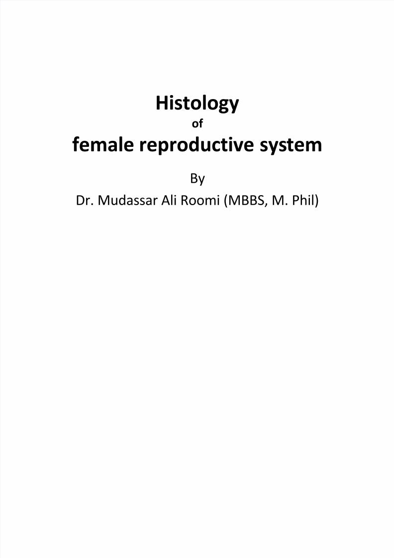
Corpus Luteum-overview
• Formed from corpushemrrhagicus
• It is highly vascularizedtemporary endocrinegland
• It is composed of granulosa lutein cells andtheca lutein cells
• Formation is dependenton LH.
7/31/2019 2nd Lecture on the Female Reproductive Histology by Dr. Roomi
http://slidepdf.com/reader/full/2nd-lecture-on-the-female-reproductive-histology-by-dr-roomi 3/20
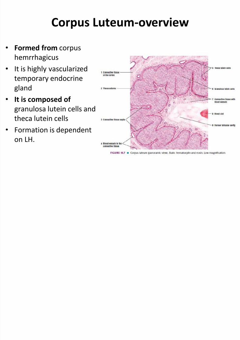
Granulosa lutein cells• Large (30um diameter) Pale
staining cells• Derived from the cells of
membrana granulosa•
Each cells has gotabundance of SER, RER,mitochondria, well-developed Golgi apparatusand lipid droplets.
• Function :1. Formation of progestrone2. Conversion of androgens to
estrogens
7/31/2019 2nd Lecture on the Female Reproductive Histology by Dr. Roomi
http://slidepdf.com/reader/full/2nd-lecture-on-the-female-reproductive-histology-by-dr-roomi 4/20
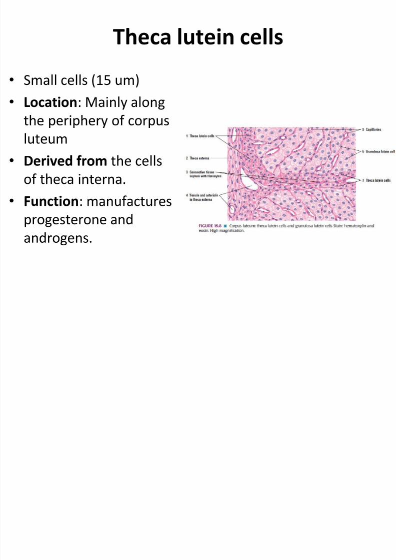
Theca lutein cells
• Small cells (15 um)• Location : Mainly along
the periphery of corpus
luteum• Derived from the cells
of theca interna.•
Function : manufacturesprogesterone andandrogens.
7/31/2019 2nd Lecture on the Female Reproductive Histology by Dr. Roomi
http://slidepdf.com/reader/full/2nd-lecture-on-the-female-reproductive-histology-by-dr-roomi 5/20
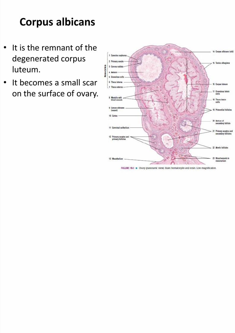
Corpus albicans
• It is the remnant of thedegenerated corpusluteum.
• It becomes a small scaron the surface of ovary.
7/31/2019 2nd Lecture on the Female Reproductive Histology by Dr. Roomi
http://slidepdf.com/reader/full/2nd-lecture-on-the-female-reproductive-histology-by-dr-roomi 6/20
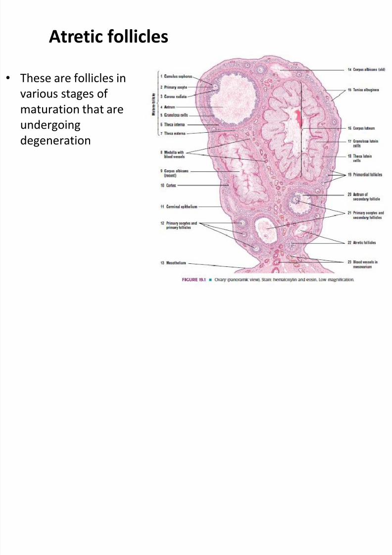
Atretic follicles
• These are follicles invarious stages of maturation that areundergoingdegeneration
7/31/2019 2nd Lecture on the Female Reproductive Histology by Dr. Roomi
http://slidepdf.com/reader/full/2nd-lecture-on-the-female-reproductive-histology-by-dr-roomi 7/20
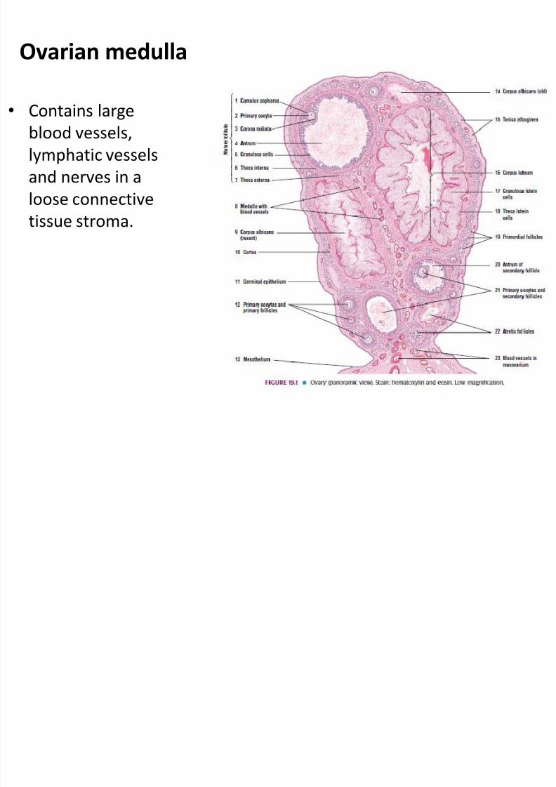
Ovarian medulla
• Contains largeblood vessels,lymphatic vesselsand nerves in aloose connectivetissue stroma.
7/31/2019 2nd Lecture on the Female Reproductive Histology by Dr. Roomi
http://slidepdf.com/reader/full/2nd-lecture-on-the-female-reproductive-histology-by-dr-roomi 8/20
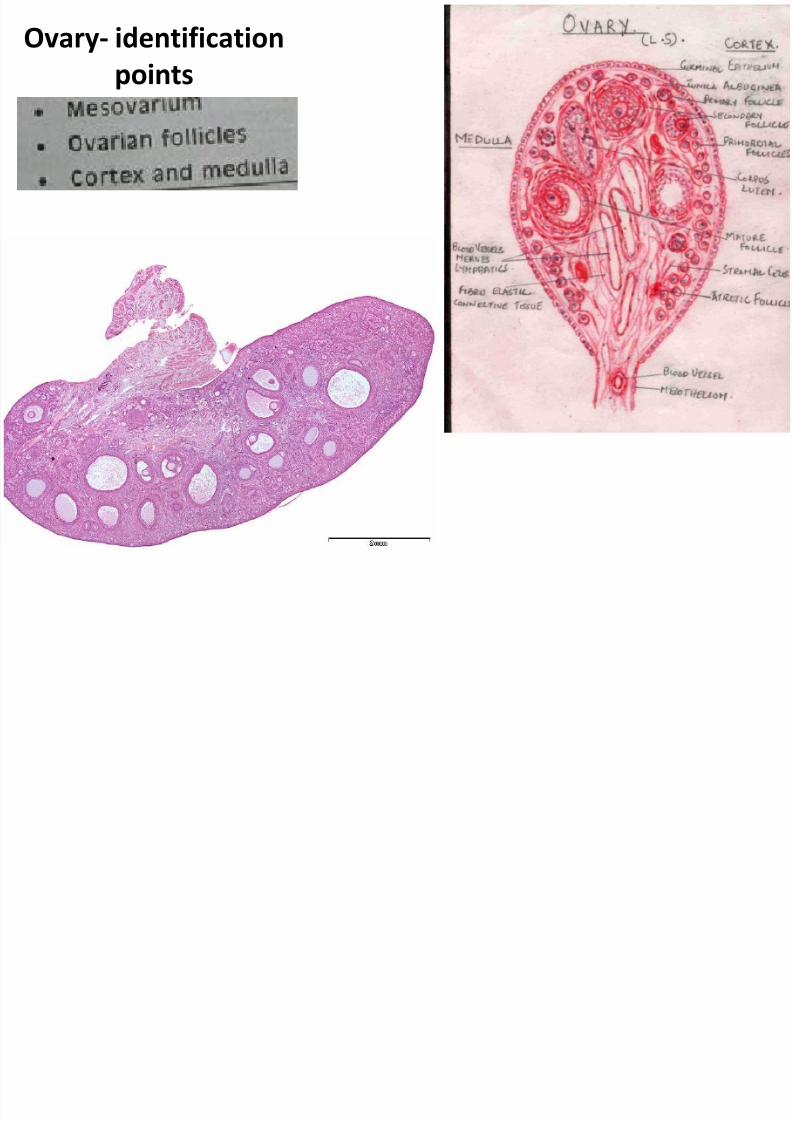
Ovary- identificationpoints
7/31/2019 2nd Lecture on the Female Reproductive Histology by Dr. Roomi
http://slidepdf.com/reader/full/2nd-lecture-on-the-female-reproductive-histology-by-dr-roomi 9/20
7/31/2019 2nd Lecture on the Female Reproductive Histology by Dr. Roomi
http://slidepdf.com/reader/full/2nd-lecture-on-the-female-reproductive-histology-by-dr-roomi 10/20
7/31/2019 2nd Lecture on the Female Reproductive Histology by Dr. Roomi
http://slidepdf.com/reader/full/2nd-lecture-on-the-female-reproductive-histology-by-dr-roomi 11/20
7/31/2019 2nd Lecture on the Female Reproductive Histology by Dr. Roomi
http://slidepdf.com/reader/full/2nd-lecture-on-the-female-reproductive-histology-by-dr-roomi 12/20
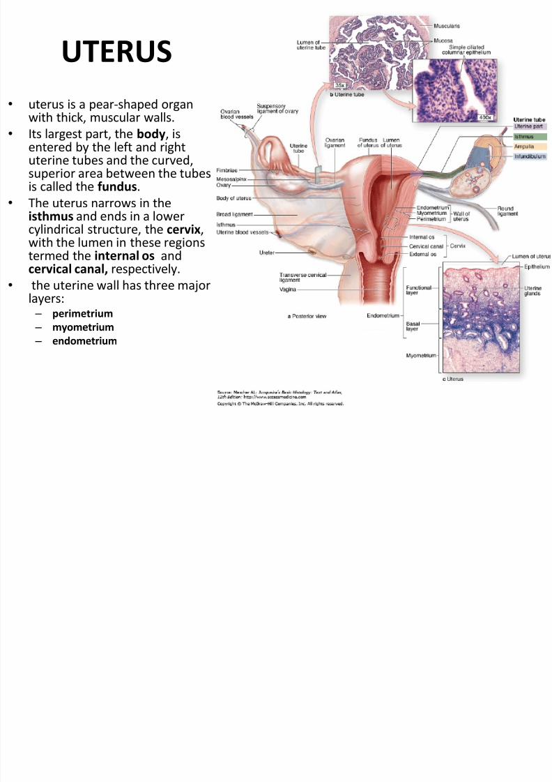
UTERUS• uterus is a pear-shaped organ
with thick, muscular walls.• Its largest part, the body , is
entered by the left and rightuterine tubes and the curved,superior area between the tubesis called the fundus .
• The uterus narrows in theisthmus and ends in a lowercylindrical structure, the cervix ,with the lumen in these regionstermed the internal os andcervical canal, respectively.
•
the uterine wall has three majorlayers: – perimetrium – myometrium – endometrium
7/31/2019 2nd Lecture on the Female Reproductive Histology by Dr. Roomi
http://slidepdf.com/reader/full/2nd-lecture-on-the-female-reproductive-histology-by-dr-roomi 13/20
7/31/2019 2nd Lecture on the Female Reproductive Histology by Dr. Roomi
http://slidepdf.com/reader/full/2nd-lecture-on-the-female-reproductive-histology-by-dr-roomi 14/20
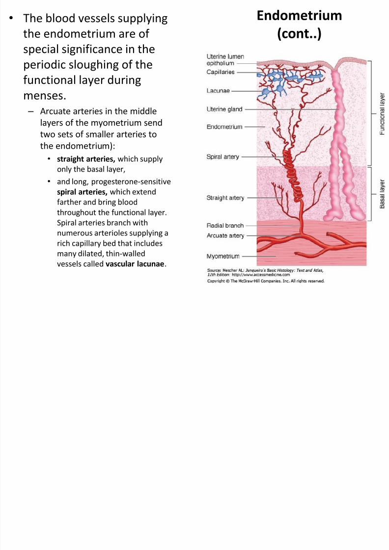
Endometrium(cont..)
• The blood vessels supplyingthe endometrium are of special significance in the
periodic sloughing of thefunctional layer duringmenses.
– Arcuate arteries in the middlelayers of the myometrium send
two sets of smaller arteries tothe endometrium):• straight arteries, which supply
only the basal layer,• and long, progesterone-sensitive
spiral arteries, which extend
farther and bring bloodthroughout the functional layer.Spiral arteries branch withnumerous arterioles supplying arich capillary bed that includesmany dilated, thin-walledvessels called vascular lacunae .
7/31/2019 2nd Lecture on the Female Reproductive Histology by Dr. Roomi
http://slidepdf.com/reader/full/2nd-lecture-on-the-female-reproductive-histology-by-dr-roomi 15/20
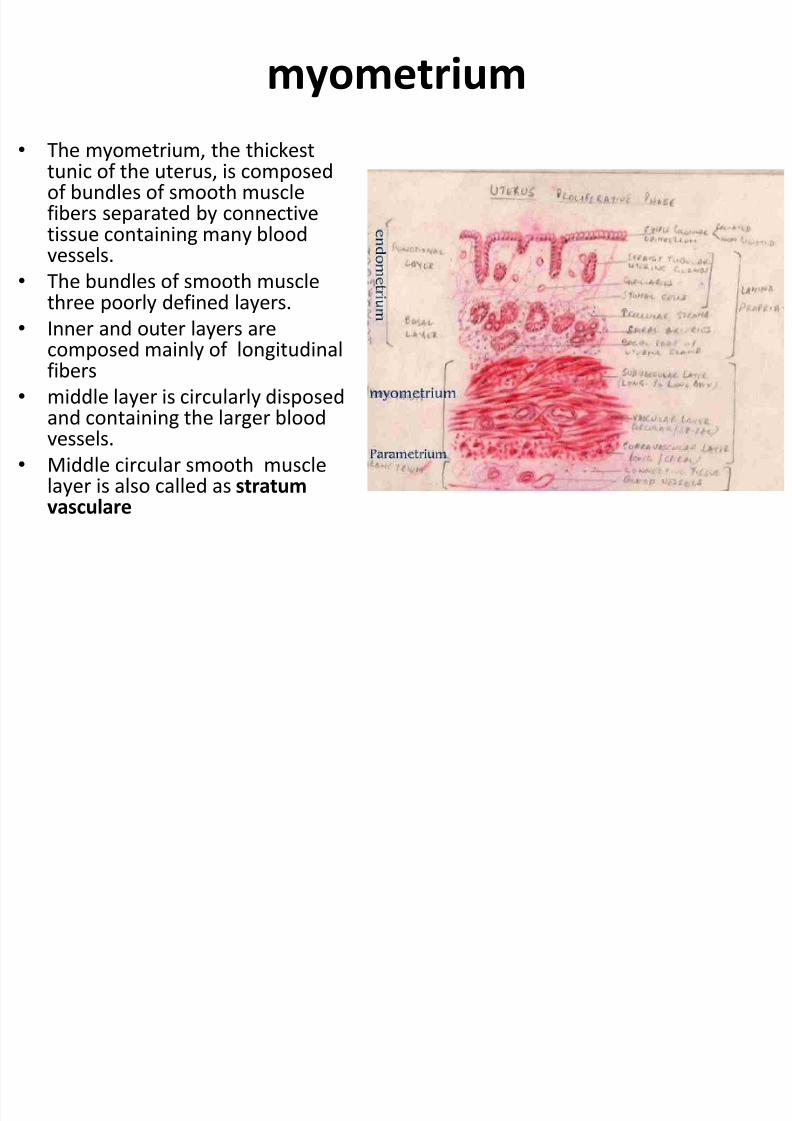
myometrium • The myometrium, the thickest
tunic of the uterus, is composedof bundles of smooth musclefibers separated by connectivetissue containing many bloodvessels.
• The bundles of smooth musclethree poorly defined layers.
• Inner and outer layers arecomposed mainly of longitudinalfibers
• middle layer is circularly disposedand containing the larger bloodvessels.
• Middle circular smooth musclelayer is also called as stratumvasculare
7/31/2019 2nd Lecture on the Female Reproductive Histology by Dr. Roomi
http://slidepdf.com/reader/full/2nd-lecture-on-the-female-reproductive-histology-by-dr-roomi 16/20
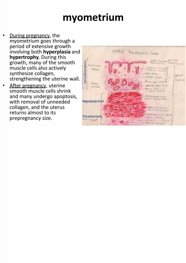
myometrium • During pregnancy, the
myometrium goes through aperiod of extensive growthinvolving both hyperplasia andhypertrophy . During thisgrowth, many of the smoothmuscle cells also activelysynthesize collagen,strengthening the uterine wall.
• After pregnancy, uterinesmooth muscle cells shrink
and many undergo apoptosis,with removal of unneededcollagen, and the uterusreturns almost to itsprepregnancy size.
7/31/2019 2nd Lecture on the Female Reproductive Histology by Dr. Roomi
http://slidepdf.com/reader/full/2nd-lecture-on-the-female-reproductive-histology-by-dr-roomi 17/20
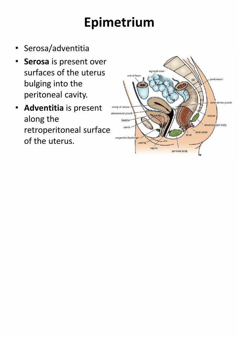
Epimetrium
• Serosa/adventitia• Serosa is present over
surfaces of the uterus
bulging into theperitoneal cavity.
• Adventitia is presentalong theretroperitoneal surfacesof the uterus.
7/31/2019 2nd Lecture on the Female Reproductive Histology by Dr. Roomi
http://slidepdf.com/reader/full/2nd-lecture-on-the-female-reproductive-histology-by-dr-roomi 18/20
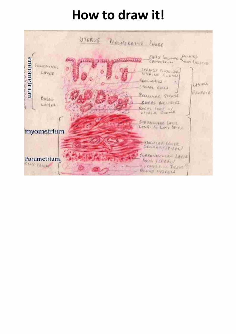
How to draw it!
7/31/2019 2nd Lecture on the Female Reproductive Histology by Dr. Roomi
http://slidepdf.com/reader/full/2nd-lecture-on-the-female-reproductive-histology-by-dr-roomi 19/20
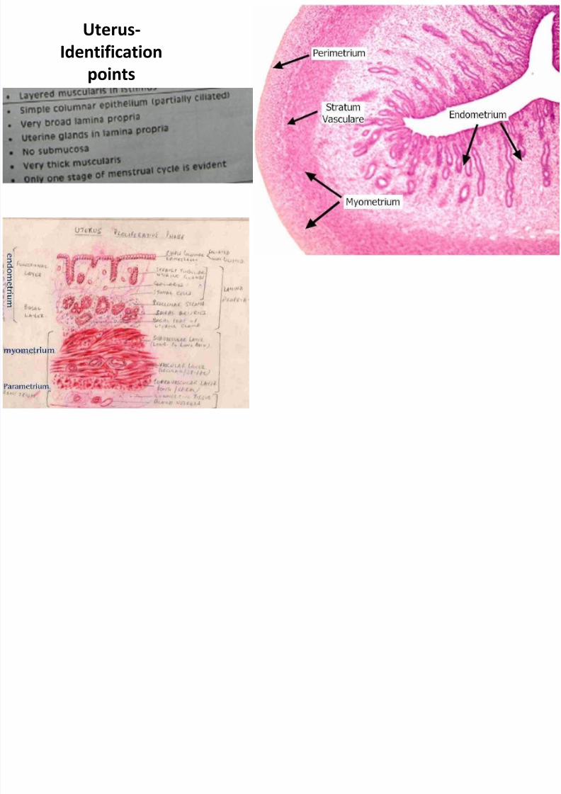
Uterus-Identification
points
7/31/2019 2nd Lecture on the Female Reproductive Histology by Dr. Roomi
http://slidepdf.com/reader/full/2nd-lecture-on-the-female-reproductive-histology-by-dr-roomi 20/20
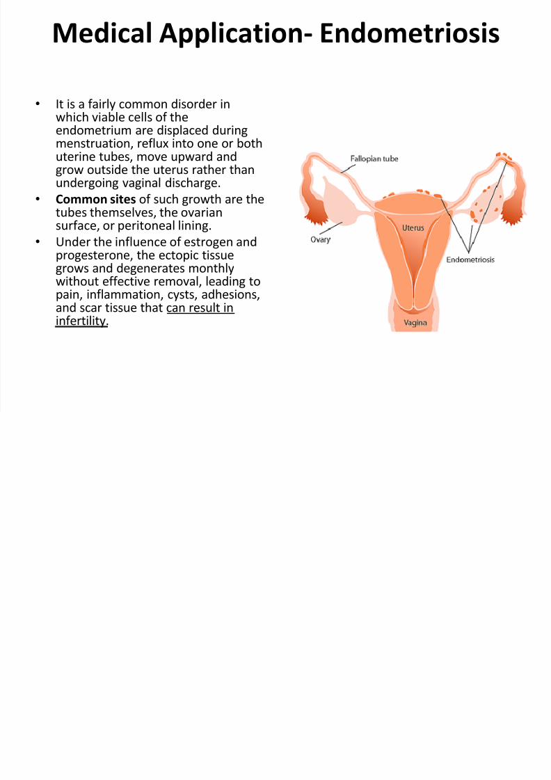
Medical Application- Endometriosis
• It is a fairly common disorder inwhich viable cells of theendometrium are displaced duringmenstruation, reflux into one or bothuterine tubes, move upward andgrow outside the uterus rather thanundergoing vaginal discharge.
• Common sites of such growth are thetubes themselves, the ovariansurface, or peritoneal lining.
• Under the influence of estrogen andprogesterone, the ectopic tissuegrows and degenerates monthlywithout effective removal, leading topain, inflammation, cysts, adhesions,and scar tissue that can result ininfertility.
Related Documents