HISTOLOG Y OF GIT BY DR. MUDASSAR ALI ROOMI (MBBS, M. Phil.)

Welcome message from author
This document is posted to help you gain knowledge. Please leave a comment to let me know what you think about it! Share it to your friends and learn new things together.
Transcript
8/2/2019 1st Lec on GIT Histology by Dr Roomi
http://slidepdf.com/reader/full/1st-lec-on-git-histology-by-dr-roomi 1/19
HISTOLOGY OF
GIT
BY
DR. MUDASSAR ALI ROOMI (MBBS, M. Phil.)

8/2/2019 1st Lec on GIT Histology by Dr Roomi
http://slidepdf.com/reader/full/1st-lec-on-git-histology-by-dr-roomi 2/19
TWO MAIN COMPONENTS OF DIGESTIVE SYSTEM:
1. The digestive tract
(alimentary canal)
2. Major glands
associated with the
tract (salivary glands,
pancreas, liver
alongwith biliary
passages)

8/2/2019 1st Lec on GIT Histology by Dr Roomi
http://slidepdf.com/reader/full/1st-lec-on-git-histology-by-dr-roomi 3/19
Digestive tract
Anatomically divided into
two parts:
1. The oral cavity.
2. The tubular digestive
tract which includes
esophagus, stomach,
small intestine and
large intestine.

8/2/2019 1st Lec on GIT Histology by Dr Roomi
http://slidepdf.com/reader/full/1st-lec-on-git-histology-by-dr-roomi 4/19
Oral cavity
It is irregular space
bounded by the lips,
cheeks and palate.
It contains tongue,
gums and teeth.
8/2/2019 1st Lec on GIT Histology by Dr Roomi
http://slidepdf.com/reader/full/1st-lec-on-git-histology-by-dr-roomi 5/19
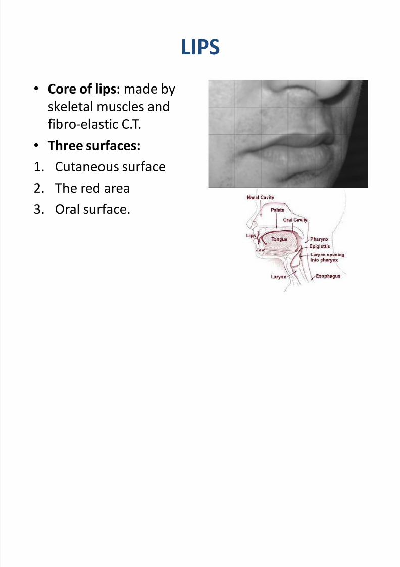
LIPS
Core of lips: made by
skeletal muscles and
fibro-elastic C.T.
Three surfaces:
1. Cutaneous surface
2. The red area
3. Oral surface.

8/2/2019 1st Lec on GIT Histology by Dr Roomi
http://slidepdf.com/reader/full/1st-lec-on-git-histology-by-dr-roomi 6/19
Histology of Lips
8/2/2019 1st Lec on GIT Histology by Dr Roomi
http://slidepdf.com/reader/full/1st-lec-on-git-histology-by-dr-roomi 7/19
Histology of Lips
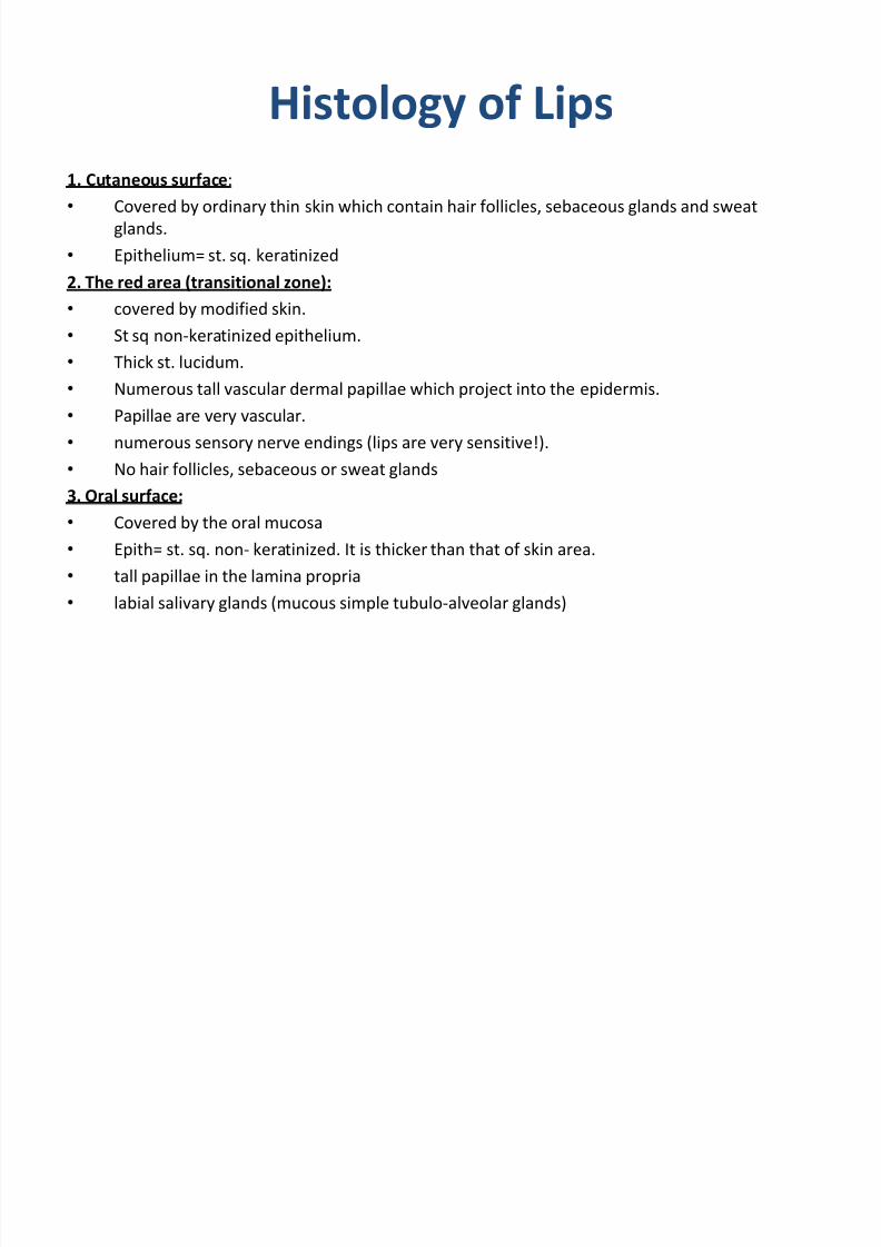
1. Cutaneous surface:
Covered by ordinary thin skin which contain hair follicles, sebaceous glands and sweat
glands.
Epithelium= st. sq. keratinized
2. The red area (transitional zone):
covered by modified skin. St sq non-keratinized epithelium.
Thick st. lucidum.
Numerous tall vascular dermal papillae which project into the epidermis.
Papillae are very vascular.
numerous sensory nerve endings (lips are very sensitive!).
No hair follicles, sebaceous or sweat glands
3. Oral surface:
Covered by the oral mucosa
Epith= st. sq. non- keratinized. It is thicker than that of skin area.
tall papillae in the lamina propria
labial salivary glands (mucous simple tubulo-alveolar glands)

8/2/2019 1st Lec on GIT Histology by Dr Roomi
http://slidepdf.com/reader/full/1st-lec-on-git-histology-by-dr-roomi 8/19
TONGUE
It is a mass of sk. Musclescovered by the mucousmembrane.
Muscles: running in all the 3
dimensions.
Epithelium:
1. On ventral (lower) surface:
st. sq. non-keratinized
2. On dorsal (upper) surface: partially keratized,otherwise it is mainly st.sq. non- keratinized maily.

8/2/2019 1st Lec on GIT Histology by Dr Roomi
http://slidepdf.com/reader/full/1st-lec-on-git-histology-by-dr-roomi 9/19
TONGUE
SURFACES OF TONGUE:
1. Dorsal surface: roughdue to presence of
lingual papillae,irregular, divided intoanterior (2/3) andposterior (1/3) by V-shaped sulcusterminalis.
2. Ventral surface
8/2/2019 1st Lec on GIT Histology by Dr Roomi
http://slidepdf.com/reader/full/1st-lec-on-git-histology-by-dr-roomi 10/19
LINGUAL PAPILLAE
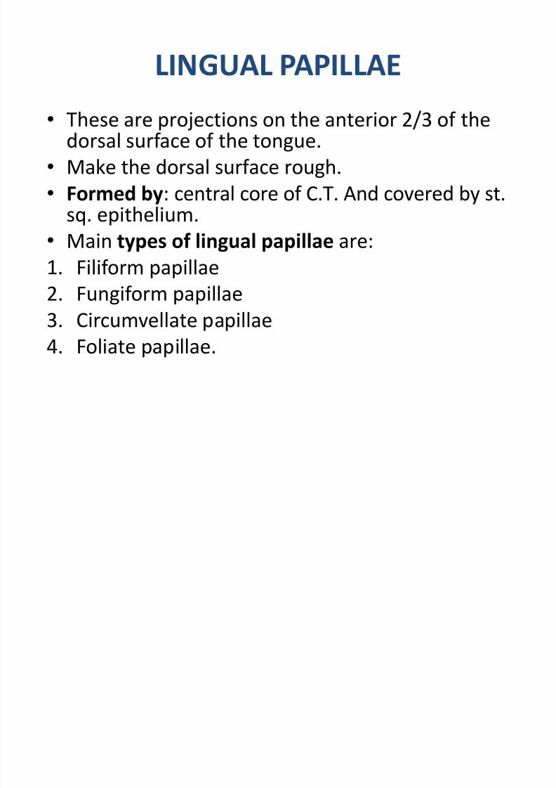
These are projections on the anterior 2/3 of thedorsal surface of the tongue.
Make the dorsal surface rough.
Formed by: central core of C.T. And covered by st.sq. epithelium.
Main types of lingual papillae are:
1. Filiform papillae
2. Fungiform papillae3. Circumvellate papillae
4. Foliate papillae.

8/2/2019 1st Lec on GIT Histology by Dr Roomi
http://slidepdf.com/reader/full/1st-lec-on-git-histology-by-dr-roomi 11/19
LINGUAL PAPILLAE
8/2/2019 1st Lec on GIT Histology by Dr Roomi
http://slidepdf.com/reader/full/1st-lec-on-git-histology-by-dr-roomi 12/19
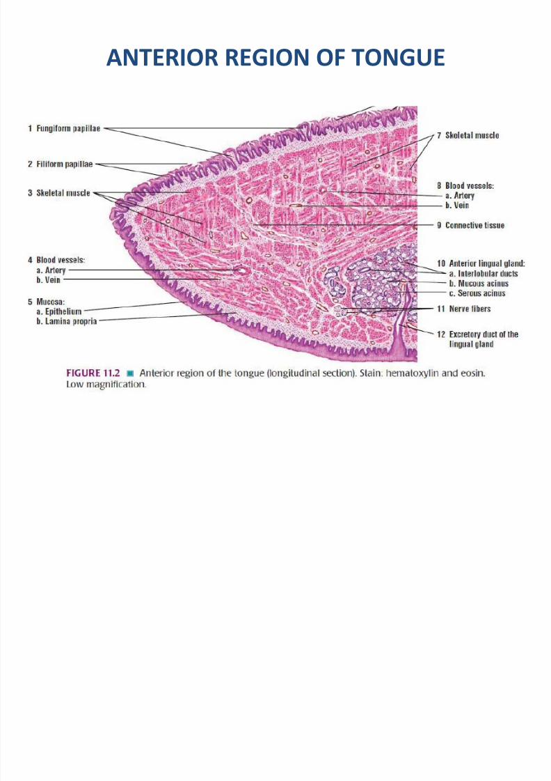
ANTERIOR REGION OF TONGUE
8/2/2019 1st Lec on GIT Histology by Dr Roomi
http://slidepdf.com/reader/full/1st-lec-on-git-histology-by-dr-roomi 13/19
LINGUAL PAPILLAE
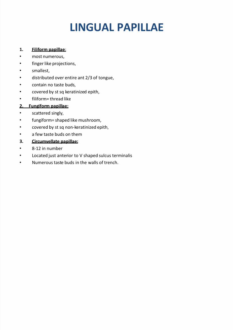
1. Filiform papillae:
most numerous,
finger like projections,
smallest,
distributed over entire ant 2/3 of tongue,
contain no taste buds,
covered by st sq keratinized epith,
filiform= thread like
2. Fungiform papillae:
scattered singly,
fungiform= shaped like mushroom, covered by st sq non-keratinized epith,
a few taste buds on them
3. Circumvellate papillae:
8-12 in number
Located just anterior to V shaped sulcus terminalis
Numerous taste buds in the walls of trench.

8/2/2019 1st Lec on GIT Histology by Dr Roomi
http://slidepdf.com/reader/full/1st-lec-on-git-histology-by-dr-roomi 14/19
LINGUAL PAPILLAE

8/2/2019 1st Lec on GIT Histology by Dr Roomi
http://slidepdf.com/reader/full/1st-lec-on-git-histology-by-dr-roomi 15/19
ANTERIOR REGION OF TONGUE

8/2/2019 1st Lec on GIT Histology by Dr Roomi
http://slidepdf.com/reader/full/1st-lec-on-git-histology-by-dr-roomi 16/19
GLANDS OF TONGUE
1. Anterior lingual glands:
pair,
mixed seromucous,
near the tip of tongue,
duct open on the ventral surface.2. Glands of von Ebner:
purely serous,
located in the region of circumvellate papillae,
ducts open in the trenches of circumvellate papillae,
3. Mucous glands of the root: Numerous, small, purely mucous.
Located near the posterior region of tongue.
Ducts open in the crypts of the lingual tonsils.
8/2/2019 1st Lec on GIT Histology by Dr Roomi
http://slidepdf.com/reader/full/1st-lec-on-git-histology-by-dr-roomi 17/19
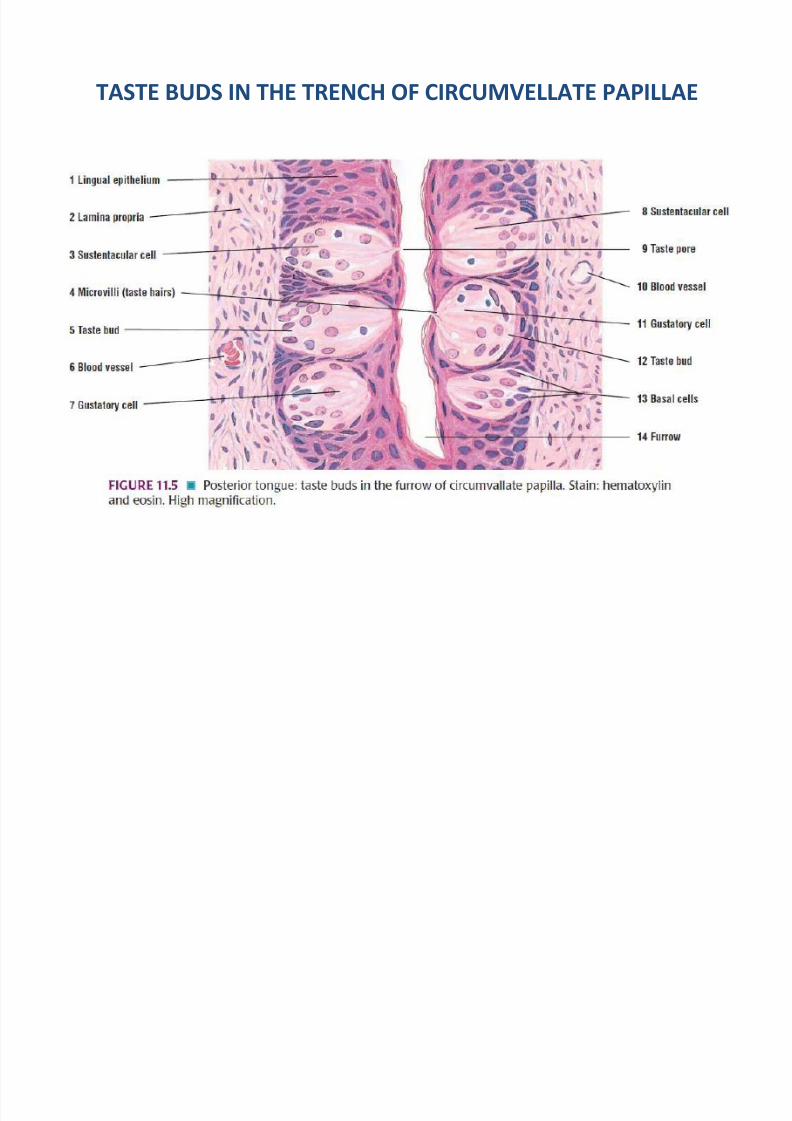
TASTE BUDS IN THE TRENCH OF CIRCUMVELLATE PAPILLAE

8/2/2019 1st Lec on GIT Histology by Dr Roomi
http://slidepdf.com/reader/full/1st-lec-on-git-histology-by-dr-roomi 18/19
TASTE BUDS
Receptors for tastesensation.
Location: dorsal surfaceof tongue, soft palate,
epiglottis. on H/E stain, taste bud
appears as oval, palestaining body (30-50 umwide).
50-90 cells One taste pore (gustatory
pore).

8/2/2019 1st Lec on GIT Histology by Dr Roomi
http://slidepdf.com/reader/full/1st-lec-on-git-histology-by-dr-roomi 19/19
TASTE BUDS
TYPES OF CELLS in the tastebuds:
1. Neuroepithelial cells:
Tall columnar cells
Basal portion of cellscontain synaptic vesicles.
2. Sustancular cells:
Function is to support
3. Basal cells:
Stem cells
Related Documents