Welcome message from author
This document is posted to help you gain knowledge. Please leave a comment to let me know what you think about it! Share it to your friends and learn new things together.
Transcript
What we’ll talk about…
• Hormones that regulate structural changes in the menstrual cycle
• Follicular development in the ovary
• Changes in the endometrium during the menstrual cycle

The menstrual cycle comprises functional and structural changes in the ovary and endometrium.
0 2 4 6 8 10 12 14 16 18 20 22 24 26 28
Menses Proliferative Secretory
Follicular Luteal
Ovarian Cycle
Endometrial Cycle
Days
Ovulation
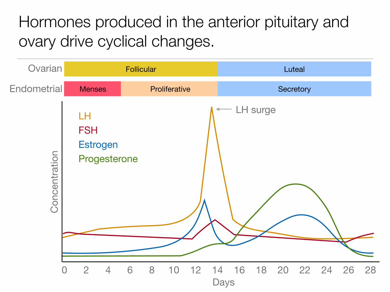
Hormones produced in the anterior pituitary and ovary drive cyclical changes.
0 2 4 6 8 10 12 14 16 18 20 22 24 26 28Days
Follicular Luteal
Menses Proliferative Secretory
Ovarian
Endometrial
LH
EstrogenFSH
Progesterone
LH surge
Conc
entra
tion

A portal system delivers releasing hormones from the hypothalamus to the anterior pituitary.
Hypothalamus
Anterior Pituitary
Portal Vein

Ovary
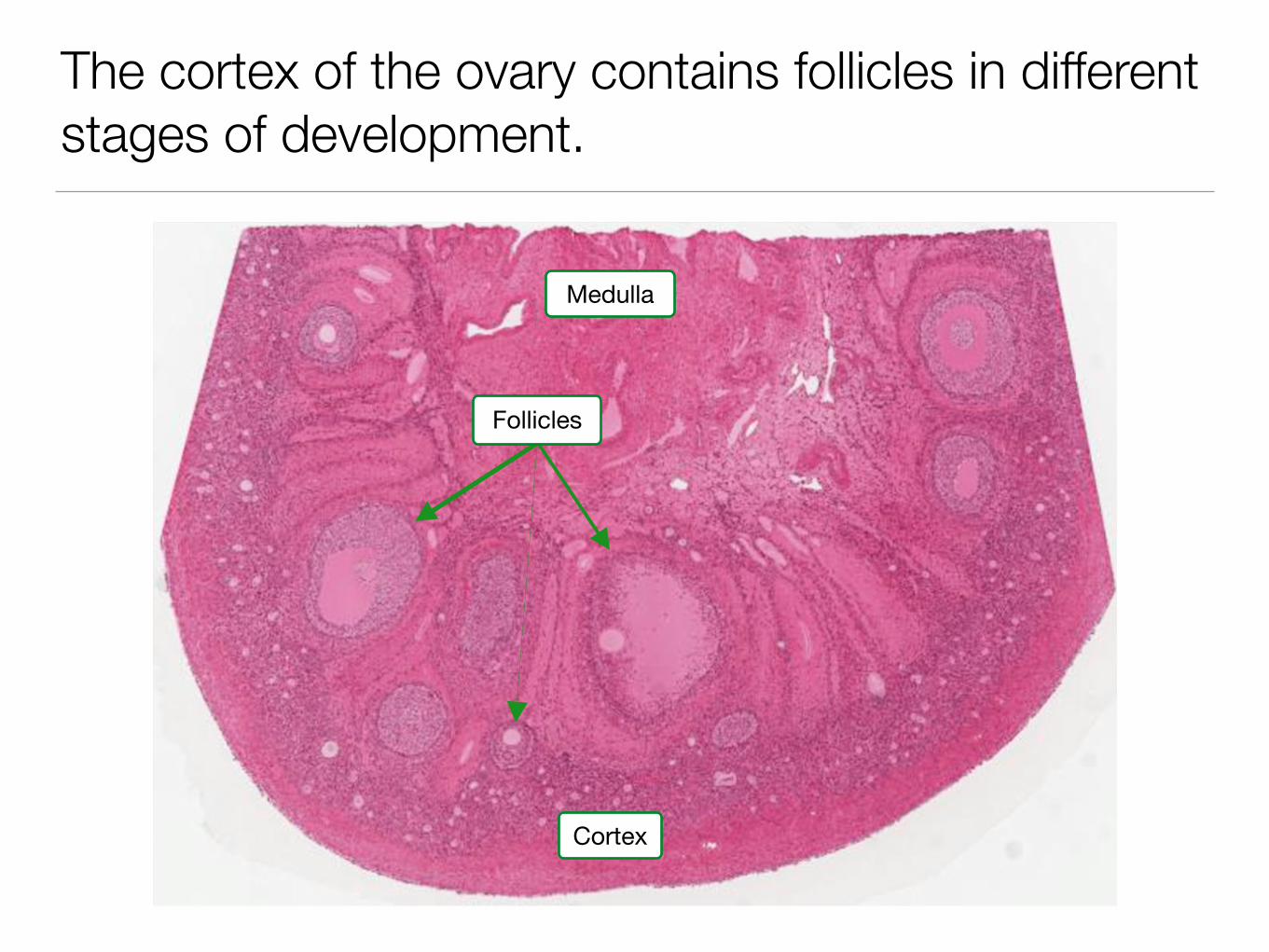
The cortex of the ovary contains follicles in different stages of development.
Medulla
Cortex
Follicles

Stages of follicle development in the ovary
• Primordial
• Primary
• Secondary
• Graffian

Primordial follicles contain an oocyte and a layer of squamous granulosa cells.
OocyteGranulosa Cell

Primary follicles contain a larger oocyte and cuboidal granulosa cells.
OocyteGranulosa Cells
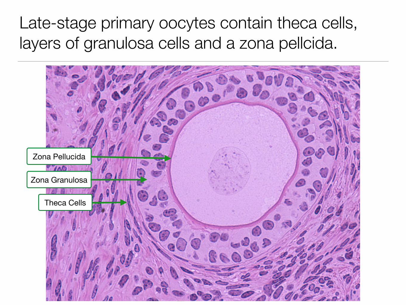
Late-stage primary oocytes contain theca cells, layers of granulosa cells and a zona pellcida.
Zona Pellucida
Zona Granulosa
Theca Cells
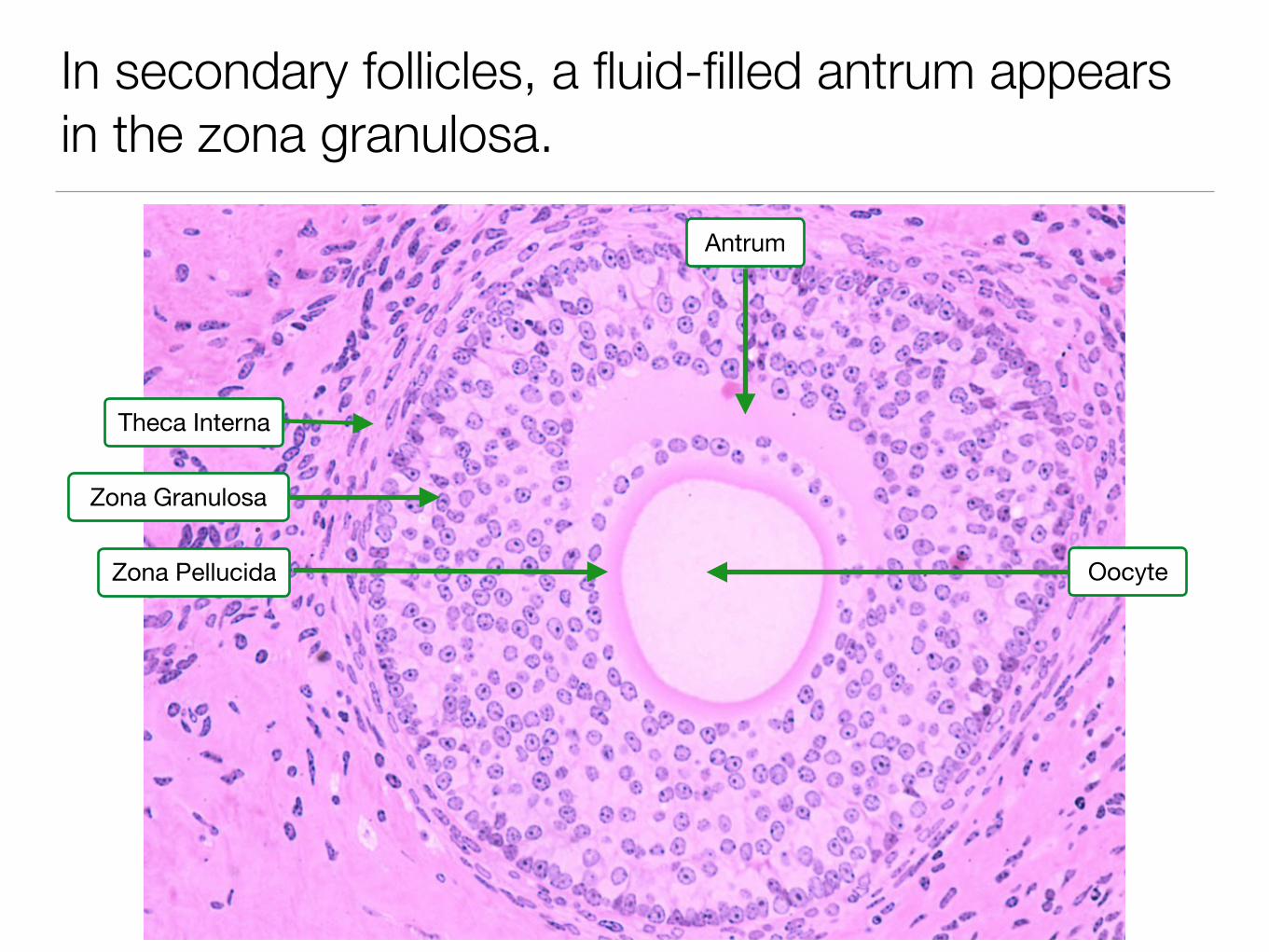
In secondary follicles, a fluid-filled antrum appears in the zona granulosa.
Antrum
Theca Interna
Zona Granulosa
Zona Pellucida Oocyte

Cells in the theca internal produce androgens which cell in the granulosa convert to estrogen.
Zona Granulosa FSH Receptor
Theca InternaLH Receptor
Androstenedione
Estrogen
Lipid Droplet

Graafian follicles contain an eccentrically localized oocyte and large antrum.
Antrum
Zona Granulosa
Oocyte
Zona Pellucida
Corona Radiata
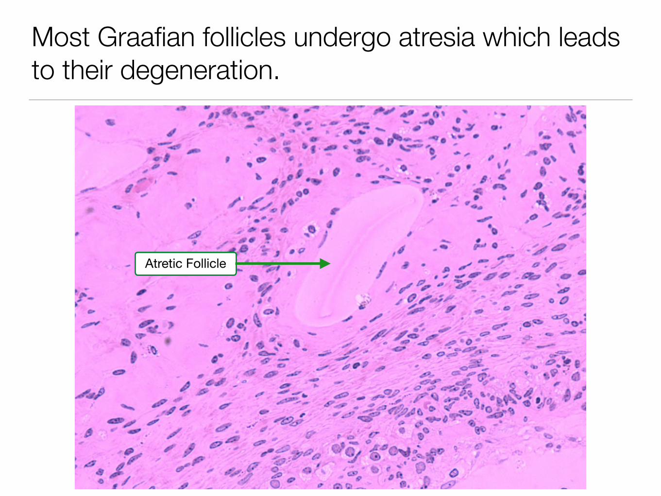
Most Graafian follicles undergo atresia which leads to their degeneration.
Atretic Follicle
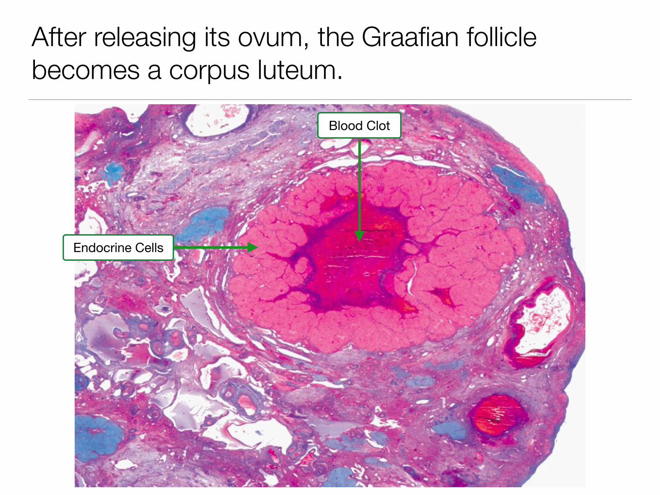
After releasing its ovum, the Graafian follicle becomes a corpus luteum.
Blood Clot
Endocrine Cells

The theca lutein and granulosa lutein cells produce progesterone and estrogen.
Granulosa LuteinTheca Lutein
Blood Clot
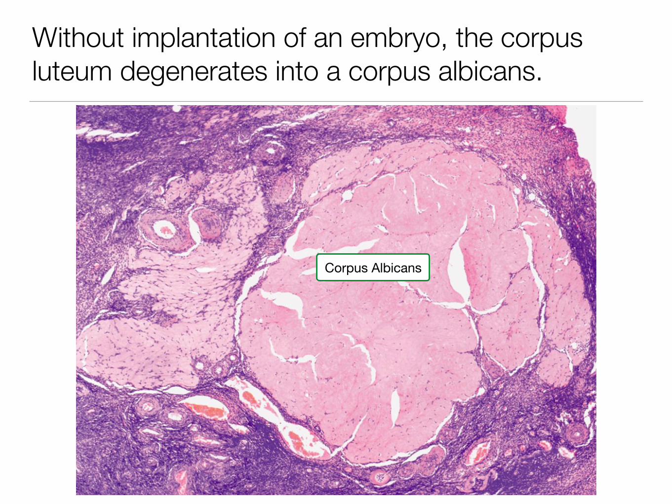
Without implantation of an embryo, the corpus luteum degenerates into a corpus albicans.
Corpus Albicans

The oviduct consists of an inner mucosa and outer layer of smooth muscle.
Smooth Muscle
Mucosa
Components of the female reproductive tract
• Oviduct
• Uterus
• Cervix
• Vagina
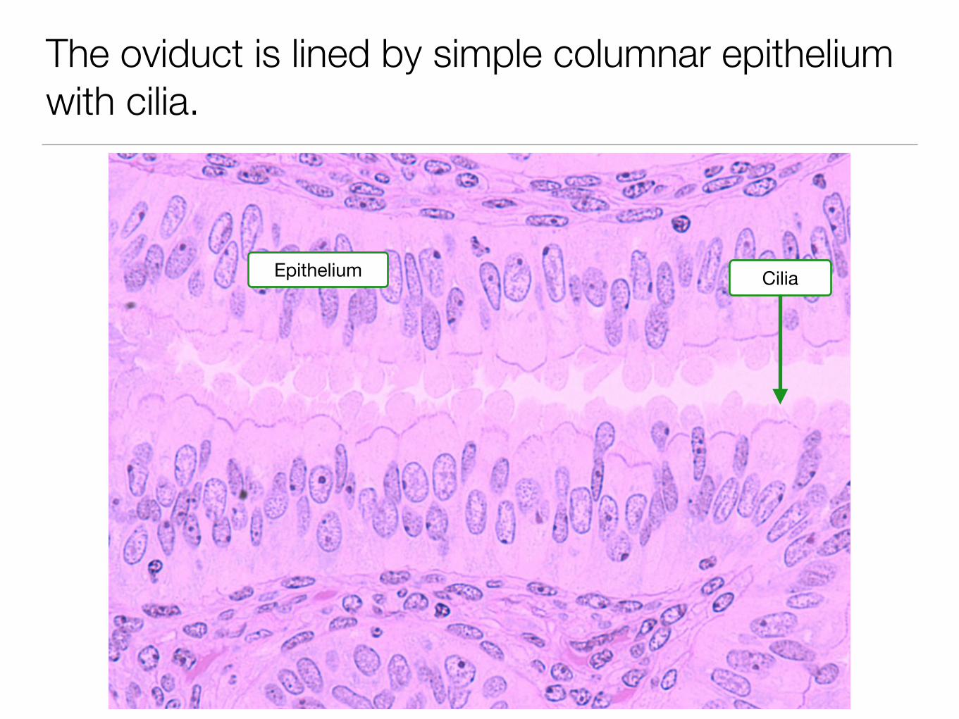
The oviduct is lined by simple columnar epithelium with cilia.
CiliaEpithelium
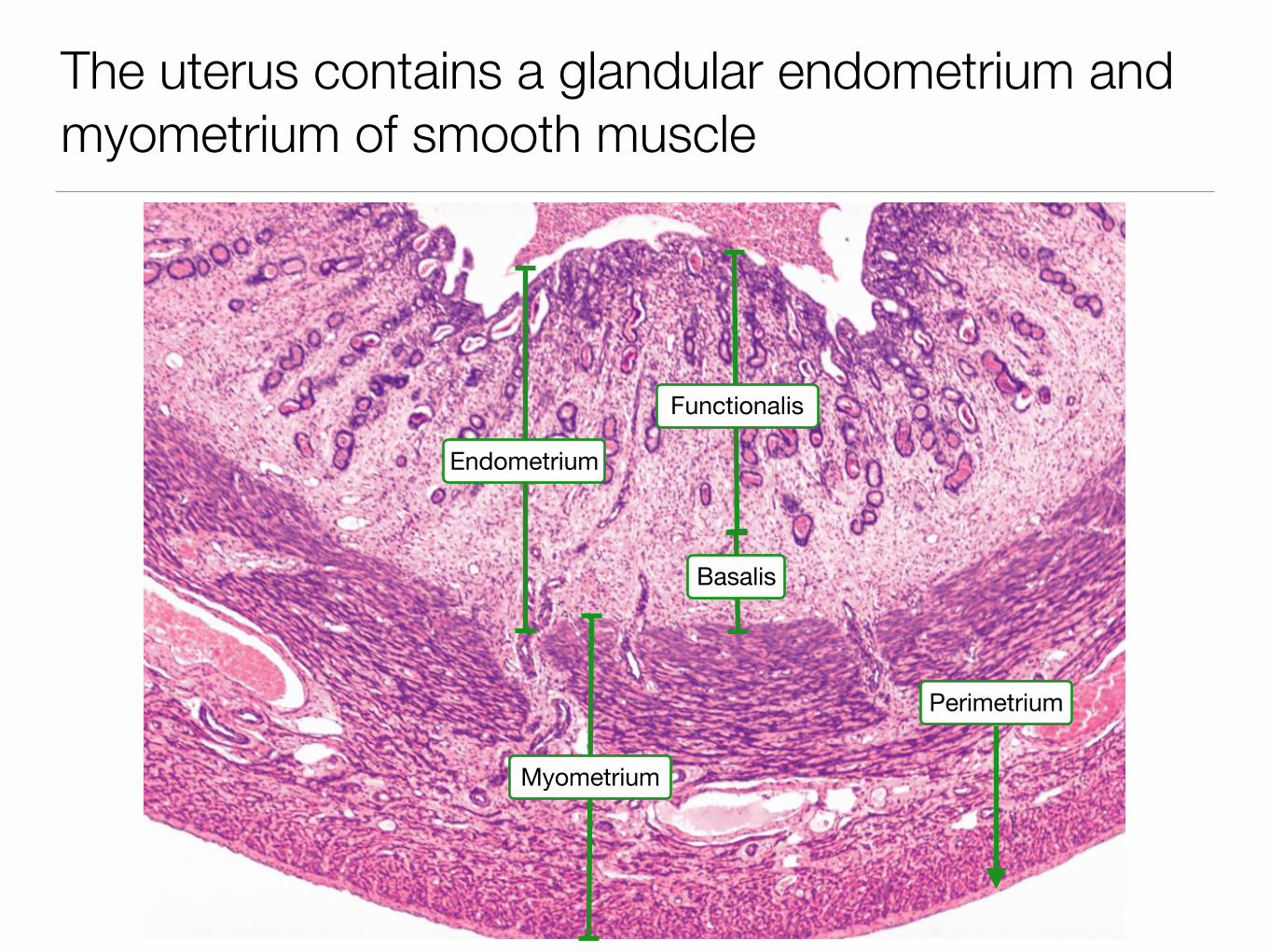
The uterus contains a glandular endometrium and myometrium of smooth muscle
Myometrium
Endometrium
Basalis
Functionalis
Perimetrium
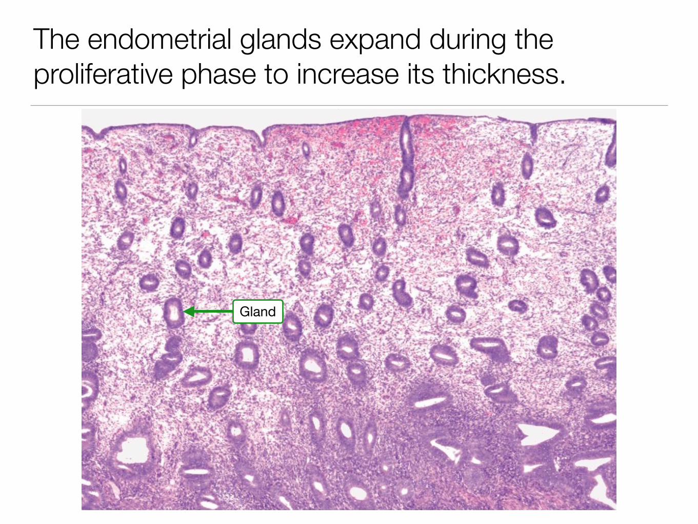
The endometrial glands expand during the proliferative phase to increase its thickness.
Gland

The late proliferative phase see larger and more numerous glands.
Gland
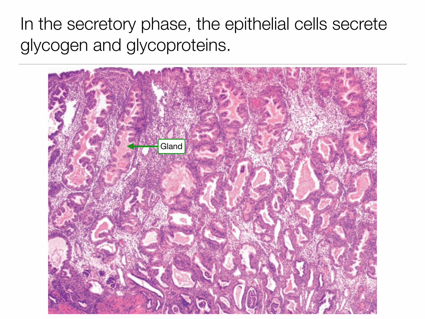
In the secretory phase, the epithelial cells secrete glycogen and glycoproteins.
Gland
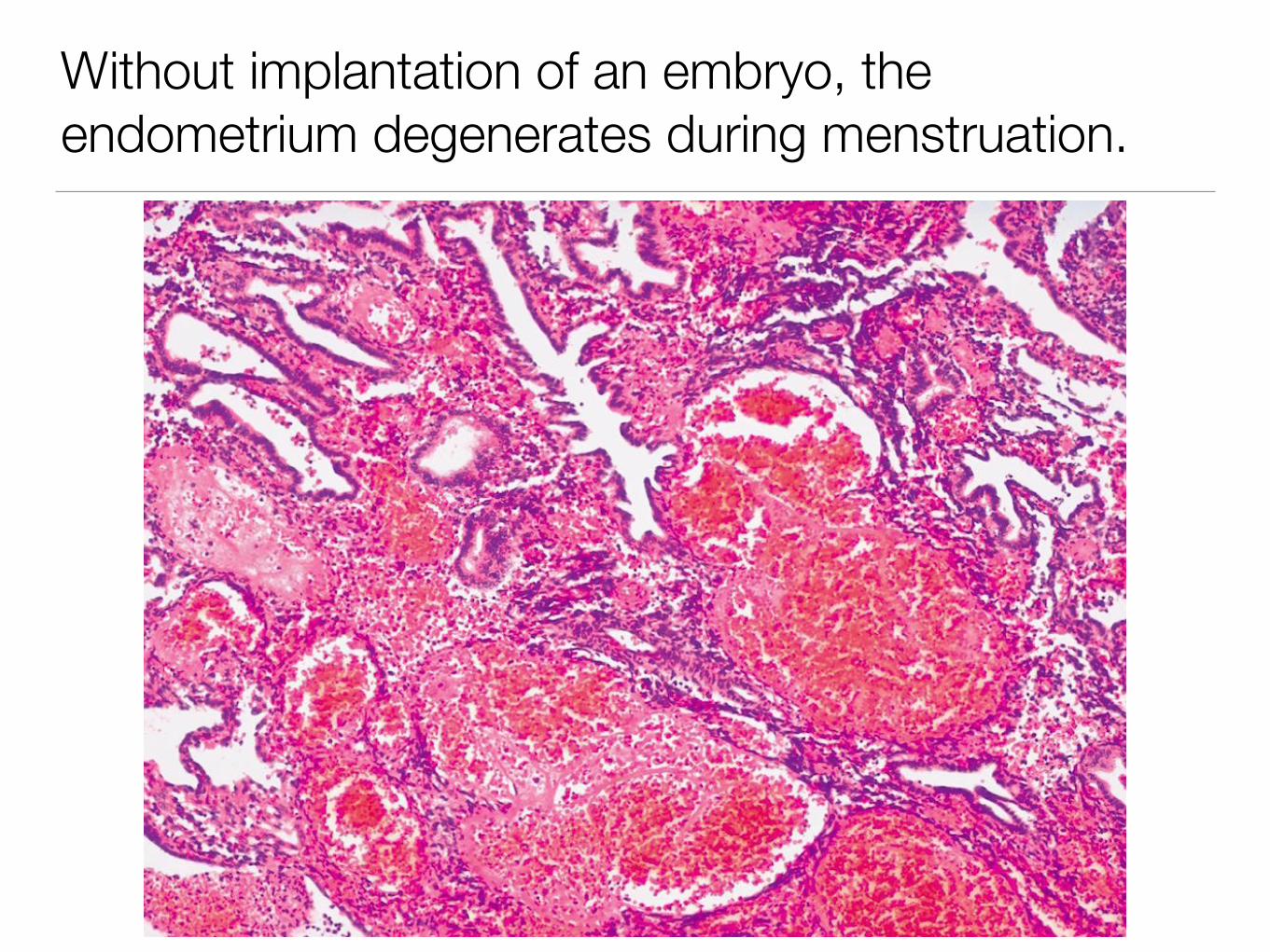
Without implantation of an embryo, the endometrium degenerates during menstruation.

The epithelium of the cervix transitions from simple columnar to stratified squamous.
Upper Cervix: Simple Columnar
Lower Cervix: Stratified Squamous
Transition
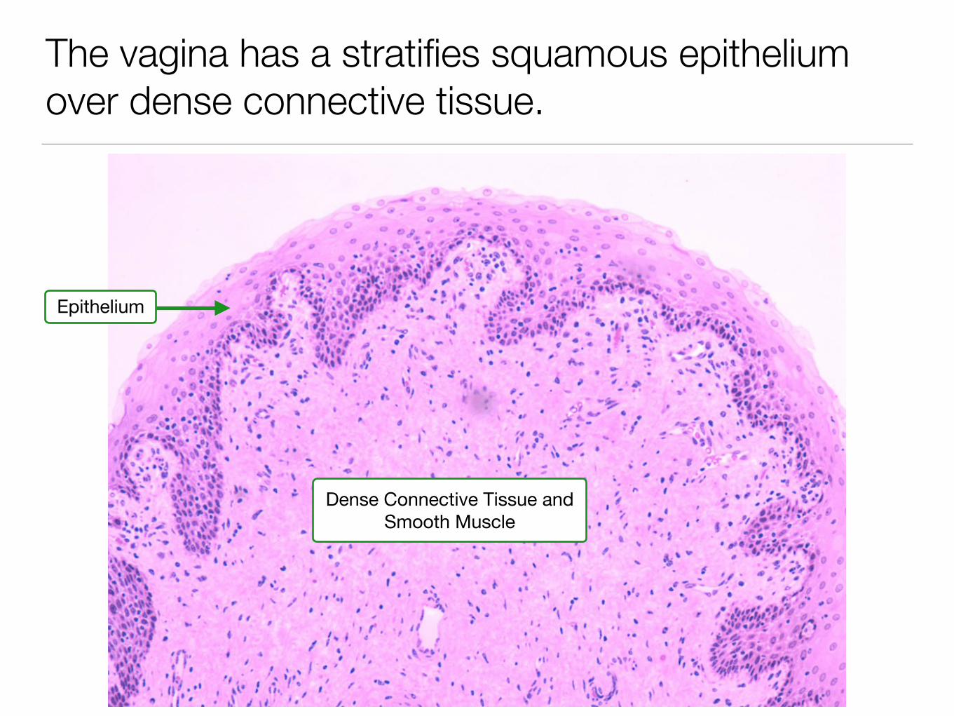
The vagina has a stratifies squamous epithelium over dense connective tissue.
Epithelium
Dense Connective Tissue and Smooth Muscle
Take home messages…
• Gonadotrophs in the anterior pituitary secrete FSH and LH in response to GnRH.
• FSH stimulates granulosa cells and LH stimulates theca interna cells and granulosa lutein cells.
• Follicles develop through well defined stages
• The endometrium cycles through proliferative, secretory and menstrual phases.
Related Documents