BioMed Central Page 1 of 11 (page number not for citation purposes) Radiation Oncology Open Access Research [(18)F]Fluoroethyltyrosine- positron emission tomography-guided radiotherapy for high-grade glioma Damien C Weber* 1 , Thomas Zilli 1 , Franz Buchegger 2 , Nathalie Casanova 1 , Guy Haller 3 , Michel Rouzaud 1 , Philippe Nouet 1 , Giovanna Dipasquale 1 , Osman Ratib 2 , Habib Zaidi 2 , Hansjorg Vees 1 and Raymond Miralbell 1 Address: 1 Department of Radiation Oncology, Geneva University Hospital, CH-12011 Geneva 14, Switzerland, 2 Department of Nuclear Medicine, Geneva University Hospital, CH-12011 Geneva 14, Switzerland and 3 Unit of Clinical Epidemiology and Statistics, Geneva University Hospital, CH-12011 Geneva 14, Switzerland Email: Damien C Weber* - [email protected]; Thomas Zilli - [email protected]; Franz Buchegger - [email protected]; Nathalie Casanova - [email protected]; Guy Haller - [email protected]; Michel Rouzaud - [email protected]; Philippe Nouet - [email protected]; Giovanna Dipasquale - [email protected]; Osman Ratib - [email protected]; Habib Zaidi - [email protected]; Hansjorg Vees - [email protected]; Raymond Miralbell - [email protected] * Corresponding author Abstract Background: To compare morphological gross tumor volumes (GTVs), defined as pre- and postoperative gadolinium enhancement on T 1 -weighted magnetic resonance imaging to biological tumor volumes (BTVs), defined by the uptake of 18 F fluoroethyltyrosine (FET) for the radiotherapy planning of high-grade glioma, using a dedicated positron emission tomography (PET)-CT scanner equipped with three triangulation lasers for patient positioning. Methods: Nineteen patients with malignant glioma were included into a prospective protocol using FET PET-CT for radiotherapy planning. To be eligible, patients had to present with residual disease after surgery. Planning was performed using the clinical target volume (CTV = GTV ∪ BTV) and planning target volume (PTV = CTV + 20 mm). First, the interrater reliability for BTV delineation was assessed among three observers. Second, the BTV and GTV were quantified and compared. Finally, the geometrical relationships between GTV and BTV were assessed. Results: Interrater agreement for BTV delineation was excellent (intraclass correlation coefficient 0.9). Although, BTVs and GTVs were not significantly different (p = 0.9), CTVs (mean 57.8 ± 30.4 cm 3 ) were significantly larger than BTVs (mean 42.1 ± 24.4 cm 3 ; p < 0.01) or GTVs (mean 38.7 ± 25.7 cm 3 ; p < 0.01). In 13 (68%) and 6 (32%) of 19 patients, FET uptake extended ≥ 10 and 20 mm from the margin of the gadolinium enhancement. Conclusion: Using FET, the interrater reliability had excellent agreement for BTV delineation. With FET PET-CT planning, the size and geometrical location of GTVs and BTVs differed in a majority of patients. Published: 24 December 2008 Radiation Oncology 2008, 3:44 doi:10.1186/1748-717X-3-44 Received: 13 November 2008 Accepted: 24 December 2008 This article is available from: http://www.ro-journal.com/content/3/1/44 © 2008 Weber et al; licensee BioMed Central Ltd. This is an Open Access article distributed under the terms of the Creative Commons Attribution License (http://creativecommons.org/licenses/by/2.0 ), which permits unrestricted use, distribution, and reproduction in any medium, provided the original work is properly cited.

Welcome message from author
This document is posted to help you gain knowledge. Please leave a comment to let me know what you think about it! Share it to your friends and learn new things together.
Transcript
BioMed CentralRadiation Oncology
ss
Open AcceResearch[(18)F]Fluoroethyltyrosine- positron emission tomography-guided radiotherapy for high-grade gliomaDamien C Weber*1, Thomas Zilli1, Franz Buchegger2, Nathalie Casanova1, Guy Haller3, Michel Rouzaud1, Philippe Nouet1, Giovanna Dipasquale1, Osman Ratib2, Habib Zaidi2, Hansjorg Vees1 and Raymond Miralbell1Address: 1Department of Radiation Oncology, Geneva University Hospital, CH-12011 Geneva 14, Switzerland, 2Department of Nuclear Medicine, Geneva University Hospital, CH-12011 Geneva 14, Switzerland and 3Unit of Clinical Epidemiology and Statistics, Geneva University Hospital, CH-12011 Geneva 14, Switzerland
Email: Damien C Weber* - [email protected]; Thomas Zilli - [email protected]; Franz Buchegger - [email protected]; Nathalie Casanova - [email protected]; Guy Haller - [email protected]; Michel Rouzaud - [email protected]; Philippe Nouet - [email protected]; Giovanna Dipasquale - [email protected]; Osman Ratib - [email protected]; Habib Zaidi - [email protected]; Hansjorg Vees - [email protected]; Raymond Miralbell - [email protected]
* Corresponding author
AbstractBackground: To compare morphological gross tumor volumes (GTVs), defined as pre- andpostoperative gadolinium enhancement on T1-weighted magnetic resonance imaging to biologicaltumor volumes (BTVs), defined by the uptake of 18F fluoroethyltyrosine (FET) for the radiotherapyplanning of high-grade glioma, using a dedicated positron emission tomography (PET)-CT scannerequipped with three triangulation lasers for patient positioning.
Methods: Nineteen patients with malignant glioma were included into a prospective protocolusing FET PET-CT for radiotherapy planning. To be eligible, patients had to present with residualdisease after surgery. Planning was performed using the clinical target volume (CTV = GTV ∪ BTV)and planning target volume (PTV = CTV + 20 mm). First, the interrater reliability for BTVdelineation was assessed among three observers. Second, the BTV and GTV were quantified andcompared. Finally, the geometrical relationships between GTV and BTV were assessed.
Results: Interrater agreement for BTV delineation was excellent (intraclass correlation coefficient0.9). Although, BTVs and GTVs were not significantly different (p = 0.9), CTVs (mean 57.8 ± 30.4cm3) were significantly larger than BTVs (mean 42.1 ± 24.4 cm3; p < 0.01) or GTVs (mean 38.7 ±25.7 cm3; p < 0.01). In 13 (68%) and 6 (32%) of 19 patients, FET uptake extended ≥ 10 and 20 mmfrom the margin of the gadolinium enhancement.
Conclusion: Using FET, the interrater reliability had excellent agreement for BTV delineation.With FET PET-CT planning, the size and geometrical location of GTVs and BTVs differed in amajority of patients.
Published: 24 December 2008
Radiation Oncology 2008, 3:44 doi:10.1186/1748-717X-3-44
Received: 13 November 2008Accepted: 24 December 2008
This article is available from: http://www.ro-journal.com/content/3/1/44
© 2008 Weber et al; licensee BioMed Central Ltd. This is an Open Access article distributed under the terms of the Creative Commons Attribution License (http://creativecommons.org/licenses/by/2.0), which permits unrestricted use, distribution, and reproduction in any medium, provided the original work is properly cited.
Page 1 of 11(page number not for citation purposes)
Radiation Oncology 2008, 3:44 http://www.ro-journal.com/content/3/1/44
BackgroundPositron emission tomotherapy (PET) is used in neuro-oncology practice essentially for diagnosis[1,2], prognosisevaluation[3], staging procedures[4] and monitoring thetumor response after treatment[5]. It can also be used forplanning purposes, as to combine the biological and mor-phological information to guide radiation dose delivery.As such, biologically image-guided radiation therapy(RT), coupled to the current anatomical imaging technol-ogy, will deliver optimally radiation, with a high-degree ofgeometrical precision and biological conformity.
High-precision radiation therapy necessitates howeverprecise anatomical and biological target delineation.[(18)F]fluoroethyltyrosine (FET) has been shown to havea high sensitivity (>90%) and specificity (>80%) for gli-oma[6]. In vitro and in vivo experiments have demon-strated that FET enters the cell by specific amino acidstransports, but is not incorporated into proteins [7-9]. Thedelineation of the glial tumor extent is easier with radiola-belled amino acids than with 18F-fluorodeoxyglucose(FDG)[10], as a result of the high glucose metabolism inthe cerebral cortex of the latter tracer and is thus therational for the integration of FET in glioma volume delin-eation for RT planning. Although inter-observer variabil-ity has been assessed for tumor definition with otheramino-acids, no such analysis has been taken in gliomadelineation with FET. If the tumour delineation with FETproves to be unreliable, the consequential treatment plansmay be inappropriate. As such, the inter-observer variabil-ity of FET during the planning process must be thoroughlyevaluated.
Defining biological target volumes (BTVs) can result insubstantial changes of target volumes for the planning ofRT, as the size and location of FET is defined by metabolicactivity rather than by the morphologic process of gliomagrowth, defined by magnetic resonance imaging(MRI)[11,12]. This may consequently lead to larger radio-therapy fields that will irradiate a larger volume of brainand possible increase of acute or late adverse events. It istherefore of paramount importance to determine whetherFET can be used to delineate glioma for radiation therapyand how this method compares to more traditional meth-ods, such as conventional gross tumour volumes (GTVs)delineation using MRI.
The purpose of this study was 1) to assess the interratervariability of high-grade glioma delineation using FET; 2)to quantify the BTVs and GTVs and to assess their volu-metric and geometric relationships and 3) to assess thetreatment characteristics after FET PET RT planning.
MethodsPatientsThe study population comprised 19 patients (10 females,9 males), referred to Geneva University Hospital, whowere prospectively entered into a protocol assessing thevalue of postoperative FET-PET imaging for the RT plan-ning of high-grade glioma. The inclusion criteria for thetrial were: 1) the diagnosis of high-grade glioma; 2) resid-ual tumor on MRI performed ≤ 24 hours postoperatively;3) Karnofsky performance status ≥ 70; 4) age between 18years and 70 years; and 5) written informed consent. Thepatient's and tumor's characteristics are detailed in Table1. Patients undergoing stereotactic biopsy were eligible.Patients presenting de novo or recurrent high-grade gliomawere eligible for this study. No previous RT to the brain ormeninges interfering with the protocol treatment planwas however allowed for the latter patients. Postoperativetreatment consisted of RT, using a linac with multileaf col-limation (Varian 2100 CD, Palo Alto, CA), and concomi-tant temozolomide, followed by adjuvant temozolomidefor all patients[13]. This study was approved by the insti-tutional ethical committee. All subjects gave writteninformed consent for their participation in the study.
PET-CT scanPatients underwent subtotal resection or stereotacticbiopsy (Table 1) and brain FET PET/CT imaging postoper-atively (mean, 8.3 days) (Biograph 16; Siemens MedicalSolutions, Erlangen, Germany) using listmode PET dataacquisition at the Department of Nuclear Medicinebetween July 2006 and December 2007. One accruedpatient presented with a heterogeneous brainstem mass inT1-weighted MRI images, with a subtle rim of peripheralenhancement after gadolinium enhancement, which wasconsidered a grade IV glioma (Table 1). FET was preparedat the cyclotron unit of the University Hospital of Zürich.Patients were placed in scanning position and CT imagingwas performed (120 kVp, 90 mAs, 16 × 1.5 collimation, apitch of 0.8 and a 0.5 second rotation) with an individu-alized immobilization plastic mask. Patients were injectedintravenously with 200 MBq of FET after a 4–6-h fastingperiod. The PET data acquisition was started immediatelyafter tracer injection[14] and was collected in list-modeformat to allow flexible choice of frames. The dynamicstudies (3 × 10 minutes) corresponding to 1 bed position,were covering the head up to the second cervical vertebralbody. Following Fourier rebinning and model-based scat-ter correction, PET images were reconstructed using two-dimensional iterative normalized attenuation-weightedordered subsets expectation maximization[15]. The CT-based attenuation correction map was used to reconstructthe emission data. The default parameters used wereordered subsets expectation maximization iterative recon-struction with four iterations and eight subsets followed
Page 2 of 11(page number not for citation purposes)
Radiation Oncology 2008, 3:44 http://www.ro-journal.com/content/3/1/44
by a post-processing Gaussian filter (kernel full-with half-maximal height, 5 mm).
A set of three triangulation lasers (central and laterals)identical to those used on the linear accelerators wereused for patient accurate positioning. Two-mm thick CTimages were acquired for planning purposes.
Magnetic resonance imaging/CT fusionPatient's diagnostic 1.5 Tesla MRI (Gyroscan Intera,Philips, Cleveland, OH) studies (axial T1-weighted withgadolinium enhancement) were transferred through thehospital picture archiving communication system (PACS)to the virtual simulation workstation (AcQSim® System,Philips Medical System, Cleveland OH) and were fusedwith the CT performed during the metabolic imaging. Thehead was not immobilized during the preoperative MRIexamination. Acquisition was done using a standard headcoil from the second cervical vertebral body upwards.Patients' CT and MRI were automatically fused accordingto the bony and non-bony anatomy (orbital cavity, clivus,nasal cavity, mastoid air cells, and optic nerve). The datafrom the postoperative MRI, performed within 24 hourson the same MRI unit, was not fused with the planning CTbut these data (T1- [with gadolinium] and T2- weightedsequence) were used mainly to assess the extend of resec-tion and any residual disease was comprehensivelyincluded during the GTV delineation for any residual dis-ease.
Biological and morphological gross tumor volume delineationFirst, BTVs, as conceptualized by Ling et al [16], were inde-pendently contoured by 3 experienced radiation oncolo-gists (D.C.W, HV and T.Z), one with nuclear medicinetraining (H.V), using the Leonardo® platform (SiemensMedical Solutions/CTI, Knoxville, TN). All brain CTimages were interpreted by an experienced diagnosticradiologist. The PET, CT, and fused PET/CT images weredisplayed for review in axial, coronal, and sagital planes.All studies were interpreted and reviewed with knowledgeof the patient's clinical history and results of previousimaging studies. The biopsied tumor, or residual tumor,defined by FET uptake was delineated manually. Maxi-mum standardized uptake values (SUVmax) were calcu-lated for ROIs of focal hyperactivity by dividing theobserved activity per gram in attenuation corrected PETwith the injected activity per gram body weight[17]. Athreshold value of 40% of SUVmax was considered for thetumor margin in all patients, as FET tumor/brain uptakeratio may be inappropriate in high-grade gliomapatients[14]. This value was determined previously in aset of high-grade glioma patients in a delineation compar-ative study, as the best thresholding value discriminatingoptimally the tumoral and background (grey matter in theopposite hemisphere) maximum SUV[18].
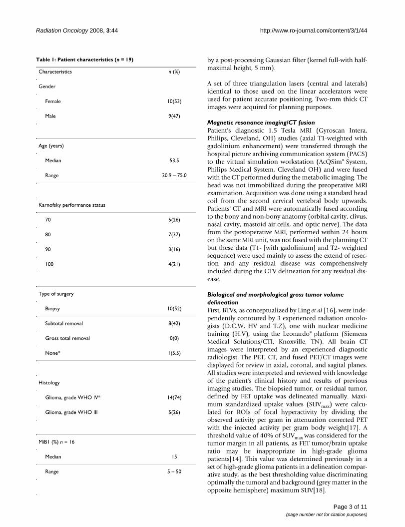
Table 1: Patient characteristics (n = 19)
Characteristics n (%)
Gender
Female 10(53)
Male 9(47)
Age (years)
Median 53.5
Range 20.9 – 75.0
Karnofsky performance status
70 5(26)
80 7(37)
90 3(16)
100 4(21)
Type of surgery
Biopsy 10(52)
Subtotal removal 8(42)
Gross total removal 0(0)
None* 1(5.5)
Histology
Glioma, grade WHO IV* 14(74)
Glioma, grade WHO III 5(26)
MiB1 (%) n = 16
Median 15
Range 5 – 50
Page 3 of 11(page number not for citation purposes)
Radiation Oncology 2008, 3:44 http://www.ro-journal.com/content/3/1/44
In the delineation process, the same windowing was used.Each physician (DCW, HV and TZ) manually delineatedthe BTVs. Within the Eclipse treatment planning station(TPS), composite, common and differential BTVs weregenerated using a Boolean algorithm. Common BTV (COM-
MONBTV) were defined as the intersection of all observers'BTVs (COMMONBTV = BTVDCW ∩ BTVHV ∩ BTVTZ). Finally,differential BTVs were defined as: DIFFBTV = [BTVDCW ∪BTVHV ∪ BTVTZ] - [BTVDCW ∩ BTVHV ∩ BTVTZ]. Observer'sBTVs, COMMONBTVs and DIFFBTVs were transferred to theAcQSim® workstation using the PACS for planning pur-poses. A BTV-interrater agreement was assessed by intrac-lass correlation coefficient (ICC) computations[19].
Second, gross tumor volume (GTV) was defined as theresidual macroscopic tumor after surgery or biopsy andthe preoperative GTV (gadolinium ring contrast enhance-ment). GTVs were defined by one radiation oncologist(D.C.W) in the AcQSim® virtual simulation workstation.
Comparative assessment of the metabolic- and morphologic tumor volumesThe GTV data was also transferred from the AcQSim®
workstation to the Eclipse® (Varian Medical Systems, PaloAlto, CA) TPS, for volume analysis and volumetric com-parison. The selected BTV for comparison was defined byone radiation oncologist (D.C.W) for consistency. Clini-cal target volume (CTV) was defined as the union of theGTV and BTV (CTV = GTV ∪ BTV). Noteworthy, CTVdefined the volume of microscopic spread but was notdefined as per the ICRU formalism in this prospective pro-tocol. The common volume between the GTVs and BTVs
was also assessed (COMMONCTV = GTV ∩ BTV). Additionallythe differential CTV (DIFFCTV = [GTV ∪ BTV] - [GTV ∩BTV]) was computed. The Boolean operator in theEclipse® TPS was used for volume measurements and vol-ume mismatch analysis. In case of BTV/GTV mismatch,the differential margins of these two volumes were meas-ured on axial slices.
RT planningFor treatment planning, the MD Anderson Cancer Centertarget policy was used[20]. Planning was performed onthe CTVs. The planning target volume (PTV) included theCTV plus an anisotropic margin of 20 mm, not includinghowever comprehensively the T2-weighted sequencehyperintense signal seen on the postoperative MRI.
Treatment characteristics with FET PET planningAs to determine the impact of FET PET-guided RT plan-ning, the treatment characteristics of the study patientswere retrospectively assessed. The treatment characteris-tics of 19 other matched high-grade glioma patients(tumor location, GTV) were also analyzed. The differenceof all study and matched patient's GTV were less than10%, except for a patient with a brainstem glioblastoma.For this case and his matched counterpart, GTVs were 4.5and 2.2 cm3, respectively. Excluding this latter patient, themedian percentage-difference between the study andmatched patients was 0.7% (range, -7.3 – 8.4).
Statistical analysisWe performed all analyses using the Statistical Package forSocial Sciences (SPSS, Ver. 15.1, SPSS Inc, Chicago, IL).For descriptive analyses of patients' characteristics andvolumes sizes we used percents and mean score. The GTV,BTV and CTV delineation methods were compared usingthe Wilcoxon signed-rank test as numerical data were notnormally distributed. The field size comparisons of theFET PET-guided- and non-FET PET-guided RT were per-formed using the Man Whitney U test. Statistical analysesused to test the interrater reliability of the biologicaltumor volume delineation by the three observers were theintraclass correlation coefficient and analysis of variancewith the expectation to uphold the null hypothesis[19]. Atwo-sided random effect model was used. A p value of lessthan 0.05 was considered to indicate statistical signifi-cance.
ResultsAbnormal FET uptake was observed in all patients.Median SUVmax at 0 – 10, 10 – 20, 20 – 30 minutes were3.05 (range, 0.51 – 4.52), 3.64 (range, 1.6 – 6.31) and3.77 (range, 1.91 – 7.22), respectively. Fig. 1 details theBTV contoured by each observer. Mean BTVs for observer1, 2 and 3 were 35.8 ± 21.7, 39.1 ± 23.6 and 36.3 ± 21.8cm3, respectively. The interrater agreement was excellent
Glioma
De novo presentation (primary) 17(89)
Secondary 2(11)
Localisation
Frontal 10(52)
Temporal 4(21)
Parietal 3(16)
Thalamus 1(5.5)
Brainstem* 1(5.5)
*Heterogeneous brainstem mass in T1-weighted MRI images, with a subtle rim of peripheral enhancement after gadolinium enhancement, considered a grade IV glioma in one patient
Table 1: Patient characteristics (n = 19) (Continued)
Page 4 of 11(page number not for citation purposes)

Radiation Oncology 2008, 3:44 http://www.ro-journal.com/content/3/1/44
(ICC = 0.9) and volumetric difference between observer'sBTV delineation did not reach statistical significance (p =0.99). The mean COMMONBTV was 32.0 ± 20.1 cm3. The DIFF-BTV ranged from 0.3 to 19.0 cm3 (mean, 8.0 ± 5.3).
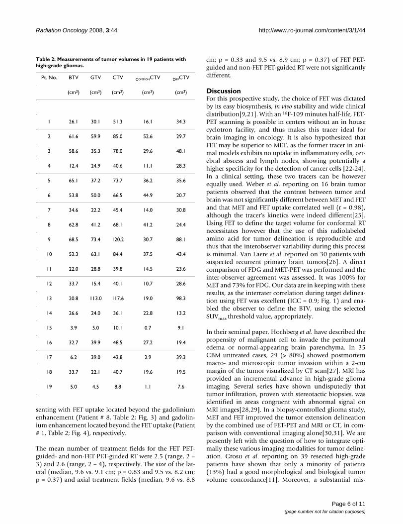
The results of volumetric measurements of GTV, BTV andCTV are presented in Table 2. The BTVs were usuallylarger, but not significantly so (p = 0.9) than their mor-phologic counterpart: mean BTV and GTV were 35.8 ±21.7 and 38.4 ± 25.7 cm3, respectively (Table 2).
Unsurprisingly, the CTV, with which the patients wereplanned, were significantly larger than the GTV (p < 0.01)or the BTV (p < 0.01). For the whole group, the mean CTVwas 57.8 ± 30.4 cm3and the mean COMMONCTV was 22.8 ±15.1 cm3 (Table 2). The DIFFCTV ranged from to 7.6 to 98.3cm3 (mean 33.8 ± 23.6; Table 2). The mean ratio (COM-
MONCTV)/(CTV) was 37.3% and ranged from 6.8% to67.5%, indicating a mismatch in a substantial number of
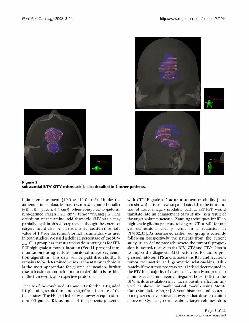
patients. FET uptake was detected up to 34.8 mm beyondgadolinium enhancement (mean, 15.1 ± 8.1 mm; range,4.6 – 34.8) in 1 patient. The mean BTV located outside theGTV was 18.3 ± 12.4 cm3 and ranged from 3.2 to 45.5.Thus, the percentage of BTV not included in the GTVranged from 3.9% to 155.2% (mean, 62.6%), bearing inmind that occasionally the BTV was larger than the GTV.In 13 (68%) and 6 (32%) of 19 patients, FET uptakeextended = 10 and 20 mm from the margin of the gado-linium enhancement. Likewise, gadolinium enhancementwas detected up to 35.9 mm beyond FET uptake (mean,13.4 ± 9.6 mm; range, 0.0 – 35.9) in 1 patient. The meanGTV located outside the BTV was 15.0 ± 22.3 cm3andranged from 0.0 to 93.8. In 12 (63%) and 4 (21%) of 19patients, gadolinium enhancement extended = 10 and 20mm from the margin of the FET uptake. The target vol-umes are presented in Fig 2, with a relevant case present-ing a good BTV-GTV matching (Patient # 14, Table 2; Fig.2). Target volumes of 2 other patients are detailed, pre-
Biological tumor volume measurements by three observers for each high-grade glioma case (1 through 19)Figure 1Biological tumor volume measurements by three observers for each high-grade glioma case (1 through 19).
Page 5 of 11(page number not for citation purposes)
Radiation Oncology 2008, 3:44 http://www.ro-journal.com/content/3/1/44
senting with FET uptake located beyond the gadoliniumenhancement (Patient # 8, Table 2; Fig. 3) and gadolin-ium enhancement located beyond the FET uptake (Patient# 1, Table 2; Fig. 4), respectively.
The mean number of treatment fields for the FET PET-guided- and non-FET PET-guided RT were 2.5 (range, 2 –3) and 2.6 (range, 2 – 4), respectively. The size of the lat-eral (median, 9.6 vs. 9.1 cm; p = 0.83 and 9.5 vs. 8.2 cm;p = 0.37) and axial treatment fields (median, 9.6 vs. 8.8
cm; p = 0.33 and 9.5 vs. 8.9 cm; p = 0.37) of FET PET-guided and non-FET PET-guided RT were not significantlydifferent.
DiscussionFor this prospective study, the choice of FET was dictatedby its easy biosynthesis, in vivo stability and wide clinicaldistribution[9,21]. With an 18F-109 minutes half-life, FET-PET scanning is possible in centers without an in housecyclotron facility, and thus makes this tracer ideal forbrain imaging in oncology. It is also hypothesized thatFET may be superior to MET, as the former tracer in ani-mal models exhibits no uptake in inflammatory cells, cer-ebral abscess and lymph nodes, showing potentially ahigher specificity for the detection of cancer cells [22-24].In a clinical setting, these two tracers can be howeverequally used. Weber et al. reporting on 16 brain tumorpatients observed that the contrast between tumor andbrain was not significantly different between MET and FETand that MET and FET uptake correlated well (r = 0.98),although the tracer's kinetics were indeed different[25].Using FET to define the target volume for conformal RTnecessitates however that the use of this radiolabeledamino acid for tumor delineation is reproducible andthus that the interobserver variability during this processis minimal. Van Laere et al. reported on 30 patients withsuspected recurrent primary brain tumors[26]. A directcomparison of FDG and MET-PET was performed and theinter-observer agreement was assessed. It was 100% forMET and 73% for FDG. Our data are in keeping with theseresults, as the interrater correlation during target delinea-tion using FET was excellent (ICC = 0.9; Fig. 1) and ena-bled the observer to define the BTV, using the selectedSUVmax threshold value, appropriately.
In their seminal paper, Hochberg et al. have described thepropensity of malignant cell to invade the peritumoraledema or normal-appearing brain parenchyma. In 35GBM untreated cases, 29 (> 80%) showed postmortemmacro- and microscopic tumor invasion within a 2-cmmargin of the tumor visualized by CT scan[27]. MRI hasprovided an incremental advance in high-grade gliomaimaging. Several series have shown undisputedly thattumor infiltration, proven with stereotactic biopsies, wasidentified in areas congruent with abnormal signal onMRI images[28,29]. In a biopsy-controlled glioma study,MET and FET improved the tumor extension delineationby the combined use of FET-PET and MRI or CT, in com-parison with conventional imaging alone[30,31]. We arepresently left with the question of how to integrate opti-mally these various imaging modalities for tumor deline-ation. Grosu et al. reporting on 39 resected high-gradepatients have shown that only a minority of patients(13%) had a good morphological and biological tumorvolume concordance[11]. Moreover, a substantial mis-
Table 2: Measurements of tumor volumes in 19 patients with high-grade gliomas.
Pt. No. BTV GTV CTV COMMONCTV DIFFCTV
(cm3) (cm3) (cm3) (cm3) (cm3)
1 26.1 30.1 51.3 16.1 34.3
2 61.6 59.9 85.0 52.6 29.7
3 58.6 35.3 78.0 29.6 48.1
4 12.4 24.9 40.6 11.1 28.3
5 65.1 37.2 73.7 36.2 35.6
6 53.8 50.0 66.5 44.9 20.7
7 34.6 22.2 45.4 14.0 30.8
8 62.8 41.2 68.1 41.2 24.4
9 68.5 73.4 120.2 30.7 88.1
10 52.3 63.1 84.4 37.5 43.4
11 22.0 28.8 39.8 14.5 23.6
12 33.7 15.4 40.1 10.7 28.6
13 20.8 113.0 117.6 19.0 98.3
14 26.6 24.0 36.1 22.8 13.2
15 3.9 5.0 10.1 0.7 9.1
16 32.7 39.9 48.5 27.2 19.4
17 6.2 39.0 42.8 2.9 39.3
18 33.7 22.1 40.7 19.6 19.5
19 5.0 4.5 8.8 1.1 7.6
Page 6 of 11(page number not for citation purposes)

Radiation Oncology 2008, 3:44 http://www.ro-journal.com/content/3/1/44
match between these two volumes was observed: onepatient out of two had MET uptake extension beyond thehyperintensity signal on T2-weigted MRI. Likewise, gado-linium extension was observed outside the MET uptake ina majority (69%) of patients. This morphological and bio-logical non-conformity has been observed in otherseries[12] and is in line with our results (Table 2; Fig. 3and 4). Consequentially to these published results, theCTV was prospectively defined as the union of both BTVand GTV in this protocol. According to the ICRU defini-tion, CTV should include all region of possible micro-scopic spread. Using a biologic paradigm, we believe thatthis region may be best defined by the summation of themorphological and biological data and not by a generi-
cally-defined 3D margin. In our series, the region of FETuptake beyond 20 mm of the Gadolinium enhancementin one third of patients is however remarkable. In short,this observation suggests that in a substantial number ofpatients the current RT margins may not be appropriate.This aforementioned consideration should be howevervalidated in the follow-up of this study. Plan is to fuse thePD-volumes with the target volumes (i.e. BTV, GTV andCTV).
In our study, the BTVs were usually larger than their mor-phologic counterpart (Table 2). This observation is in linewith the German data, which showed that the MET PETvolumes were also larger than the ones defined by gado-
Biological (BTV, blue) and morphological gross tumour (GTV, red) volume defining the clinical target volume in 19 patients with high-grade gliomaFigure 2Biological (BTV, blue) and morphological gross tumour (GTV, red) volume defining the clinical target volume in 19 patients with high-grade glioma. Note the common volume between the tumour volumes (yellow chicken wire). Good BTV-GTV matching is shown in 1 patient.
Page 7 of 11(page number not for citation purposes)
Radiation Oncology 2008, 3:44 http://www.ro-journal.com/content/3/1/44
linium enhancement (19.0 vs. 11.0 cm3). Unlike theaforementioned data, Mahasittiwat et al. reported smallerMET PET- (mean, 6.4 cm3), when compared to gadolin-ium-defined (mean, 92.1 cm3), tumor volumes[12]. Thedefinition of the amino acid threshold SUV value maypartially explain this discrepancy, although the extent ofsurgery could also be a factor. A delineation-thresholdvalue of 1.7 for the tumor/normal tissue index was usedin both studies. We used a defined percentage of the SUV-
max. Our group has investigated various strategies for FET-PET high-grade tumor delineation (Vees H, personal com-munication) using various functional image segmenta-tion algorithms. This data will be published shortly. Itremains to be determined which segmentation techniqueis the most appropriate for glioma delineation, furtherresearch using amino acid for tumor definition is justifiedin the framework of prospective protocols.
The use of the combined BTV and GTV for the FET-guidedRT planning resulted in a non-significant increase of thefields' sizes. The FET-guided RT was however equitoxic tonon-FET-guided RT, as none of the patients presented
with CTCAE grade > 2 acute treatment morbidity (datanot shown). It is somewhat paradoxical that the introduc-tion of newer imagery modality, such as FET-PET, wouldtranslate into an enlargement of field size, as a result ofthe target volume increase. Planning techniques for RT inhigh-grade glioma patients, relying on CT or MRI for tar-get delineation, usually result in a reduction inPTV[32,33]. As mentioned earlier, our group is currentlyfollowing prospectively the patients from the currentstudy, as to define precisely where the tumoral progres-sion is located, relative to the BTV, GTV and CTVs. Plan isto import the diagnostic MRI performed for tumor pro-gression into our TPS and to assess the BTV and recurrenttumor volumetric and geometric relationships. Ulti-mately, if the tumor progression is indeed documented inthe BTV in a majority of cases, it may be advantageous toadminister a simultaneous integrated boost (SIB) to theBTV, as dose escalation may have a possible effect on sur-vival as shown in mathematical models using MonteCarlo simulations[34,35]. Several historical and contem-porary series have shown however that dose escalationabove 60 Gy, using non-metabolic target volumes, does
substantial BTV-GTV mismatch is also detailed in 2 other patientsFigure 3substantial BTV-GTV mismatch is also detailed in 2 other patients.
Page 8 of 11(page number not for citation purposes)

Radiation Oncology 2008, 3:44 http://www.ro-journal.com/content/3/1/44
not result in improved survival but causes, more oftenthan not, more adverse events [36-39]. Moreover, the fail-ure pattern analysis of high-grade glioma treated withhigh-dose (> 80 Gy) radiation indicates generally a pre-dominant local pattern, suggesting that the morphologi-cal-defined tumor volumes are indeed inappropriate[40].Other series have reported a significant increase in out-field failures after high dose RT[41,42]. It is howeverunclear if this differential failure pattern results from dis-similar failure definitions or parameters related to RTtechniques or surgery. Boosting the radiation dose to alimited volume containing [34,35] tumor cells, not iden-tified by non-metabolic imaging, may be highly desirable,using amino acids. The SIB paradigm has been success-fully applied in a small Japanese series, delivering 68 Gyhypofractionated RT to the GTV, defined as the area ofintensive MET uptake[5].
ConclusionUsing a threshold value of 40% of FET SUVmax for BTVdelineation, the interrater delineation was excellent. FETPET- and MRI-defined tumor volumes differed substan-
tially. In our series, only a minority (5%) of patients hadgood BTV and GTV concordance. The RT planning forhigh-grade glioma, based on a biologic paradigm, hasshown a non significant treatment field increase, whencompared to conventionally (i.e. GTV based on MRIenhancement) planned RT.
AbbreviationsPET: Positron emission tomotherapy; RT: radiotherapy;FET: [(18)F]fluoroethyltyrosine; FDG: 18F-fluorodeoxyglu-cose; BTV: biological target volume; MRI: magnetic reso-nance imaging; GTV: gross tumour volume; PTV: planningtarget volume. CTV: Clinical tumor volume; COMMONCTV:Common clinical tumor volume; DIFFCTV: Differential clin-ical tumor volume; WHO: World Health Organisation.
Competing interestsThe authors declare that they have no competing interests.
Authors' contributionsDCW was responsible for the primary concept and thedesign of the study; DCW, TZ, NC and HJV performed the
substantial BTV-GTV mismatch is also detailed in 2 other patientsFigure 4substantial BTV-GTV mismatch is also detailed in 2 other patients.
Page 9 of 11(page number not for citation purposes)
Radiation Oncology 2008, 3:44 http://www.ro-journal.com/content/3/1/44
data capture and analysis. DCW drafted the manuscript;GH performed the statistical analysis; DCW and TZreviewed patient data; FB, HZ, RO and RM revised themanuscript.
AcknowledgementsThis work was supported in part by the Swiss National Science Foundation under grant No. 3152A0-102143 and the foundation Cellex International.
References1. Tsuyuguchi N, Sunada I, Iwai Y, Yamanaka K, Tanaka K, Takami T,
Otsuka Y, Sakamoto S, Ohata K, Goto T, Hara M: Methionine pos-itron emission tomography of recurrent metastatic braintumor and radiation necrosis after stereotactic radiosur-gery: is a differential diagnosis possible? J Neurosurg 2003,98:1056-1064.
2. Mehrkens JH, Popperl G, Rachinger W, Herms J, Seelos K, Tatsch K,Tonn JC, Kreth FW: The positive predictive value of O-(2-[(18)F]fluoroethyl)-L: -tyrosine (FET) PET in the diagnosisof a glioma recurrence after multimodal treatment. J Neu-rooncol 2008, 88(1):27-35.
3. Floeth FW, Pauleit D, Sabel M, Stoffels G, Reifenberger G, Riemen-schneider MJ, Jansen P, Coenen HH, Steiger H-J, Langen K-J: Prog-nostic Value of O-(2-18F-Fluoroethyl)-L-Tyrosine PET andMRI in Low-Grade Glioma. 2007:519-527.
4. Mohile NA, DeAngelis LM, Abrey LE: The utility of body FDGPET in staging primary central nervous system lymphoma.Neuro Oncol 2008, 10(2):223-228.
5. Miwa K, Matsuo M, Shinoda J, Oka N, Kato T, Okumura A, Ueda T,Yokoyama K, Yamada J, Yano H, Yoshimura S, Iwama T: Simultane-ous integrated boost technique by helical tomotherapy forthe treatment of glioblastoma multiforme with (11)C-methionine PET: report of three cases. J Neurooncol87(3):333-339.
6. Pauleit D, Floeth F, Hamacher K, Riemenschneider MJ, ReifenbergerG, Muller H-W, Zilles K, Coenen HH, Langen K-J: O-(2-[18F]fluor-oethyl)-L-tyrosine PET combined with MRI improves thediagnostic assessment of cerebral gliomas. 2005:678-687.
7. Langen KJ, Jarosch M, Muhlensiepen H, Hamacher K, Broer S, JansenP, Zilles K, Coenen HH: Comparison of fluorotyrosines andmethionine uptake in F98 rat gliomas. Nuclear medicine and biol-ogy 2003, 30:501-508.
8. Heiss P, Mayer S, Herz M, Wester HJ, Schwaiger M, Senekowitsch-Schmidtke R: Investigation of transport mechanism anduptake kinetics of O-(2-[18F]fluoroethyl)-L-tyrosine in vitroand in vivo. J Nucl Med 1999, 40:1367-1373.
9. Wester HJ, Herz M, Weber W, Heiss P, Senekowitsch-Schmidtke R,Schwaiger M, Stocklin G: Synthesis and radiopharmacology ofO-(2-[18F]fluoroethyl)-L-tyrosine for tumor imaging. J NuclMed 1999, 40:205-212.
10. Pirotte B, Goldman S, Massager N, David P, Wikler D, VandesteeneA, Salmon I, Brotchi J, Levivier M: Comparison of 18F-FDG and11C-methionine for PET-guided stereotactic brain biopsy ofgliomas. J Nucl Med 2004, 45:1293-1298.
11. Grosu AL, Weber WA, Riedel E, Jeremic B, Nieder C, Franz M,Gumprecht H, Jaeger R, Schwaiger M, Molls M: L-(methyl-11C)methionine positron emission tomography for target deline-ation in resected high-grade gliomas before radiotherapy. IntJ Radiat Oncol Biol Phys 2005, 63:64-74.
12. Mahasittiwat P, Mizoe J-e, Hasegawa A, Ishikawa H, Yoshikawa K,Mizuno H, Yanagi T, Takagi R, Pattaranutaporn P, Tsujii H: l-[methyl-11C] Methionine positron emission tomography for targetdelineation in malignant gliomas: impact on results of car-bon ion radiotherapy. Int J Radiat Oncol Biol Phys 70(2):515-522.
13. Stupp R, Mason WP, Bent MJ van den, Weller M, Fisher B, TaphoornMJ, Belanger K, Brandes AA, Marosi C, Bogdahn U, Curschmann J,Janzer RC, Ludwin SK, Gorlia T, Allgeier A, Lacombe D, CairncrossJG, Eisenhauer E, Mirimanoff RO: Radiotherapy plus concomitantand adjuvant temozolomide for glioblastoma. N Engl J Med2005, 352:987-996.
14. Weckesser M, Langen KJ, Rickert CH, Kloska S, Straeter R,Hamacher K, Kurlemann G, Wassmann H, Coenen HH, Schober O:O-(2-[18F]fluorethyl)-L-tyrosine PET in the clinical evalua-
tion of primary brain tumours. European journal of nuclear medi-cine and molecular imaging 2005, 32:422-429.
15. Michel C, Sibomana M, Boi A, Bernard X, Lonneux M, Defrise M: Pre-serving Poisson characteristics of PET data with weightedOSEM reconstruction. Proc IEEE Nuclear Science Symp. and MedicalImaging Conf 1998.
16. Ling CC, Humm J, Larson S, Amols H, Fuks Z, Leibel S, Koutcher JA:Towards multidimensional radiotherapy (MD-CRT): biolog-ical imaging and biological conformality. International Journal ofRadiation Oncology*Biology*Physics 2000, 47:551-560.
17. Thie JA: Understanding the standardized uptake value, itsmethods, and implications for usage. J Nucl Med 2004,45:1431-1434.
18. Vees H, Senthamizhchelvan S, Miralbell R, Weber DC, Ratib O, ZaidiH: Assessment of various strategies for (18)F-FET PET-guided delineation of target volumes in high-grade gliomapatients. Eur J Nucl Med Mol Imaging 2008. Epub ahead of print
19. Morton AP, Dobson AJ: Assessing agreement. Med J Aust 1989,150:384-387.
20. Chang EL, Akyurek S, Avalos T, Rebueno N, Spicer C, Garcia J,Famiglietti R, Allen PK, Chao KS, Mahajan A, Woo SY, Maor MH:Evaluation of peritumoral edema in the delineation of radio-therapy clinical target volumes for glioblastoma. Int J RadiatOncol Biol Phys 2007, 68:144-150.
21. Hamacher K, Coenen HH: Efficient routine production of the18F-labelled amino acid O-2-18F fluoroethyl-L-tyrosine. ApplRadiat Isot 2002, 57:853-856.
22. Rau FC, Weber WA, Wester HJ, Herz M, Becker I, Kruger A,Schwaiger M, Senekowitsch-Schmidtke R: O-(2-[(18)F]Fluoroe-thyl)- L-tyrosine (FET): a tracer for differentiation of tumourfrom inflammation in murine lymph nodes. European journal ofnuclear medicine and molecular imaging 2002, 29:1039-1046.
23. Kaim AH, Weber B, Kurrer MO, Westera G, Schweitzer A,Gottschalk J, von Schulthess GK, Buck A: (18)F-FDG and (18)F-FET uptake in experimental soft tissue infection. Europeanjournal of nuclear medicine and molecular imaging 2002, 29:648-654.
24. Salber D, Stoffels G, Pauleit D, Oros-Peusquens AM, Shah NJ, KlauthP, Hamacher K, Coenen HH, Langen KJ: Differential uptake of O-(2-18F-fluoroethyl)-L-tyrosine, L-3H-methionine, and 3H-deoxyglucose in brain abscesses. J Nucl Med 2007,48:2056-2062.
25. Weber WA, Wester HJ, Grosu AL, Herz M, Dzewas B, Feldmann HJ,Molls M, Stocklin G, Schwaiger M: O-(2-[18F]fluoroethyl)-L-tyro-sine and L-[methyl-11C]methionine uptake in braintumours: initial results of a comparative study. European jour-nal of nuclear medicine 2000, 27:542-549.
26. Van Laere K, Ceyssens S, Van Calenbergh F, de Groot T, Menten J,Flamen P, Bormans G, Mortelmans L: Direct comparison of 18F-FDG and 11C-methionine PET in suspected recurrence ofglioma: sensitivity, inter-observer variability and prognosticvalue. European journal of nuclear medicine and molecular imaging2005, 32:39-51.
27. Hochberg FH, Pruitt A: Assumptions in the radiotherapy ofglioblastoma. Neurology 1980, 30:907-911.
28. Watanabe M, Tanaka R, Takeda N: Magnetic resonance imagingand histopathology of cerebral gliomas. Neuroradiology 1992,34:463-469.
29. Kelly PJ, Daumas-Duport C, Kispert DB, Kall BA, Scheithauer BW,Illig JJ: Imaging-based stereotaxic serial biopsies in untreatedintracranial glial neoplasms. J Neurosurg 1987, 66:865-874.
30. Langen KJ, Floeth FW, Stoffels G, Hamacher K, Coenen HH, PauleitD: [Improved diagnostics of cerebral gliomas using FETPET]. Zeitschrift fur medizinische Physik 2007, 17:237-241.
31. Mosskin M, von Holst H, Bergstrom M, Collins VP, Eriksson L, John-strom P, Noren G: Positron emission tomography with 11C-methionine and computed tomography of intracranialtumours compared with histopathologic examination ofmultiple biopsies. Acta Radiol 1987, 28:673-681.
32. Weber DC, Wang H, Albrecht S, Ozsahin M, Tkachuk E, Rouzaud M,Nouet P, Dipasquale G: Open Low-field Magnetic ResonanceImaging for Target Definition, Dose Calculations and Set-upVerification during Three-dimensional CRT for Glioblast-oma Multiforme. Clinical oncology (Royal College of Radiologists (GreatBritain)) 2008, 20:157-167.
33. Crosby TD, Melcher AA, Wetherall S, Brockway S, Burnet NG: Acomparison of two planning techniques for radiotherapy of
Page 10 of 11(page number not for citation purposes)
http://www.ncbi.nlm.nih.gov/entrez/query.fcgi?cmd=Retrieve&db=PubMed&dopt=Abstract&list_uids=9935078
http://www.ncbi.nlm.nih.gov/entrez/query.fcgi?cmd=Retrieve&db=PubMed&dopt=Abstract&list_uids=9935078
http://www.ncbi.nlm.nih.gov/entrez/query.fcgi?cmd=Retrieve&db=PubMed&dopt=Abstract&list_uids=2716662
http://www.ncbi.nlm.nih.gov/entrez/query.fcgi?cmd=Retrieve&db=PubMed&dopt=Abstract&list_uids=6252514
http://www.ncbi.nlm.nih.gov/entrez/query.fcgi?cmd=Retrieve&db=PubMed&dopt=Abstract&list_uids=6252514
http://www.ncbi.nlm.nih.gov/entrez/query.fcgi?cmd=Retrieve&db=PubMed&dopt=Abstract&list_uids=1436452
http://www.ncbi.nlm.nih.gov/entrez/query.fcgi?cmd=Retrieve&db=PubMed&dopt=Abstract&list_uids=1436452
http://www.ncbi.nlm.nih.gov/entrez/query.fcgi?cmd=Retrieve&db=PubMed&dopt=Abstract&list_uids=3033172
http://www.ncbi.nlm.nih.gov/entrez/query.fcgi?cmd=Retrieve&db=PubMed&dopt=Abstract&list_uids=3033172
http://www.ncbi.nlm.nih.gov/entrez/query.fcgi?cmd=Retrieve&db=PubMed&dopt=Abstract&list_uids=2962599
http://www.ncbi.nlm.nih.gov/entrez/query.fcgi?cmd=Retrieve&db=PubMed&dopt=Abstract&list_uids=2962599
http://www.ncbi.nlm.nih.gov/entrez/query.fcgi?cmd=Retrieve&db=PubMed&dopt=Abstract&list_uids=2962599

Radiation Oncology 2008, 3:44 http://www.ro-journal.com/content/3/1/44
Publish with BioMed Central and every scientist can read your work free of charge
"BioMed Central will be the most significant development for disseminating the results of biomedical research in our lifetime."
Sir Paul Nurse, Cancer Research UK
Your research papers will be:
available free of charge to the entire biomedical community
peer reviewed and published immediately upon acceptance
cited in PubMed and archived on PubMed Central
yours — you keep the copyright
Submit your manuscript here:http://www.biomedcentral.com/info/publishing_adv.asp
BioMedcentral
high grade astrocytomas. Clinical oncology (Royal College of Radiol-ogists (Great Britain)) 1998, 10:392-398.
34. Kirkby NF, Jefferies SJ, Jena R, Burnet NG: A mathematical modelof the treatment and survival of patients with high-gradebrain tumours. Journal of Theoretical Biology 2007, 245:112-124.
35. Burnet NG, Jena R, Jefferies SJ, Stenning SP, Kirkby NF: Mathemat-ical modelling of survival of glioblastoma patients suggests arole for radiotherapy dose escalation and predicts pooreroutcome after delay to start treatment. Clinical oncology (RoyalCollege of Radiologists (Great Britain)) 2006, 18:93-103.
36. Werner-Wasik M, Scott CB, Nelson DF, Gaspar LE, Murray KJ, Fis-chbach JA, Nelson JS, Weinstein AS, Curran WJ Jr: Final report ofa phase I/II trial of hyperfractionated and accelerated hyper-fractionated radiation therapy with carmustine for adultswith supratentorial malignant gliomas. Radiation TherapyOncology Group Study 83-02. Cancer 1996, 77:1535-1543.
37. Graf R, Hildebrandt B, Tilly W, Sreenivasa G, Ullrich R, Felix R, WustP, Maier-Hauff K: Dose-escalated conformal radiotherapy ofglioblastomas – results of a retrospective comparison apply-ing radiation doses of 60 and 70 Gy. Onkologie 2005, 28:325-330.
38. Coughlin C, Scott C, Langer C, Coia L, Curran W, Rubin P: Phase II,two-arm RTOG trial (94-11) of bischloroethyl-nitrosoureaplus accelerated hyperfractionated radiotherapy (64.0 or70.4 Gy) based on tumor volume (> 20 or < or = 20 cm(2),respectively) in the treatment of newly-diagnosed radiosur-gery-ineligible glioblastoma multiforme patients. Int J RadiatOncol Biol Phys 2000, 48:1351-1358.
39. Nieder C, Nestle U, Ketter R, Kolles H, Gentner SJ, Steudel WI,Schnabel K: Hyperfractionated and accelerated-hyperfrac-tionated radiotherapy for glioblastoma multiforme. Radiationoncology investigations 1999, 7:36-41.
40. Chan JL, Lee SW, Fraass BA, Normolle DP, Greenberg HS, Junck LR,Gebarski SS, Sandler HM: Survival and failure patterns of high-grade gliomas after three-dimensional conformal radiother-apy. J Clin Oncol 2002, 20:1635-1642.
41. Nakagawa K, Aoki Y, Fujimaki T, Tago M, Terahara A, Karasawa K,Sakata K, Sasaki Y, Matsutani M, Akanuma A: High-dose conformalradiotherapy influenced the pattern of failure but did notimprove survival in glioblastoma multiforme. Int J Radiat OncolBiol Phys 1998, 40:1141-1149.
42. Fitzek MM, Thornton AF, Rabinov JD, Lev MH, Pardo FS, MunzenriderJE, Okunieff P, Bussiere M, Braun I, Hochberg FH, Hedley-Whyte ET,Liebsch NJ, Harsh GRt: Accelerated fractionated proton/pho-ton irradiation to 90 cobalt gray equivalent for glioblastomamultiforme: results of a phase II prospective trial. J Neurosurg1999, 91:251-260.
Page 11 of 11(page number not for citation purposes)
http://www.ncbi.nlm.nih.gov/entrez/query.fcgi?cmd=Retrieve&db=PubMed&dopt=Abstract&list_uids=9890542
http://www.ncbi.nlm.nih.gov/entrez/query.fcgi?cmd=Retrieve&db=PubMed&dopt=Abstract&list_uids=8608540
http://www.ncbi.nlm.nih.gov/entrez/query.fcgi?cmd=Retrieve&db=PubMed&dopt=Abstract&list_uids=8608540
http://www.ncbi.nlm.nih.gov/entrez/query.fcgi?cmd=Retrieve&db=PubMed&dopt=Abstract&list_uids=8608540
http://www.ncbi.nlm.nih.gov/entrez/query.fcgi?cmd=Retrieve&db=PubMed&dopt=Abstract&list_uids=9539570
http://www.ncbi.nlm.nih.gov/entrez/query.fcgi?cmd=Retrieve&db=PubMed&dopt=Abstract&list_uids=9539570
Related Documents


![[18F]Fluorodeoxyglucose Positron Emission Tomography in Nonseminomatous Germ Cell Tumors After Chemotherapy: The German Multicenter Positron Emission Tomography Study Group](https://static.cupdf.com/doc/110x72/6337621bd102fae1b60756e7/18ffluorodeoxyglucose-positron-emission-tomography-in-nonseminomatous-germ-cell.jpg)


![· Web view[18F]-Fluorodeoxyglucose positron emission tomography in children with neurofibromatosis type 1 and plexiform neurofibromas: correlation with malignant transformation.J](https://static.cupdf.com/doc/110x72/5b1c5e287f8b9a37258fdaa9/-web-view18f-fluorodeoxyglucose-positron-emission-tomography-in-children-with.jpg)
