RESEARCH ARTICLE Surrogate measurements of an effect of CTL infusion remain a crucial component of these studies. Compelling results have been obtained regarding sustained T-cell persis- tence and trafficking using “gene marking” in which adop- tively transferred CTLs were serially detected by polymerase chain reaction (PCR) amplification in the peripheral blood and tumor biopsies of treated patients. 3,8,9 In recent studies, PCR amplification was also used to detect the persistence and localization of CAR-modified T lymphocytes. 6,10 In addition to describing the presence of T cells, the functional status of infused T cells has been demonstrated by interferon-γ ELISPOT assay and major histocompatibility complex multi- mers used to quantify the frequency of CTL precursors. 1–3,11 A limitation of repetitive measurements of T-cell number and function based on longitudinal sampling of blood and tumor is that it does not allow assessment of the dynamic and spatial-temporal distribution of adoptively transferred CTLs. This limitation can be overcome by the application of noninvasive, accurate, and sensitive whole-body imag- ing technologies allowing repetitive measurement in vivo of these cells. This could be achieved by repetitive positron A DOPTIVE TRANSFER OF ANTIGEN-SPECIFIC CYTOTOXIC T LYMPHOCYTES (CTLs) has been suc- cessfully used to treat patients with melanoma, 1,2 Hodgkin lymphoma 3 and nasopharyngeal carcinoma. 4,5 Moreover, infusion of gene-modified T cells expressing chimeric anti- gen receptors (CARs) or transgenic α β T-cell receptor chains represents an innovative approach to expand the application of T-cell therapies to other hematologic malignancies and solid tumors. 6,7 Repetitive Noninvasive Monitoring of HSV1-tk-Expressing T Cells Intravenously Infused into Nonhuman Primates Using Positron Emission Tomography and Computed Tomography with 18 F-FEAU Gianpietro Dotti*, Mei Tian*, Barbara Savoldo*, Amer Najjar, Laurence J.N. Cooper, James Jackson, Amanda Smith, Osama Mawlawi, Rajesh Uthamanthil, Agatha Borne, David Brammer, Vincenzo Paolillo, Mian Alauddin, Carlos Gonzalez, David Steiner, William K. Decker, Frank Marini, Steven Kornblau, Catherine M. Bollard, Elizabeth J. Shpall, and Juri G. Gelovani Abstract Adoptive transfer of antigen-specific cytotoxic T lymphocytes (CTLs) has been successfully used to treat patients with different types of cancer. However, the long-term spatial-temporal dynamics of the distribution of systemically infused CTLs remains largely unknown. Noninvasive imaging of adoptively transferred CTLs using molecular-genetic reporter imaging with positron emission tomography and computed tomography (PET-CT) represents an innovative approach to understanding the long-term migratory patterns and therapeutic potential of adoptively transferred T cells. Here we report the application of repetitive PET-CT imaging with [ 18 F]fluoro-5-ethyl-1-beta-D-arabinofuranosyluracil ( 18 F-FEAU) in two nonhuman primates demonstrating that autologous polyclonal macaque T lymphocytes activated and transduced with a retroviral vector encoding for the sr39 mutant herpes simplex virus 1 thymidine kinase ( sr39HSV1-tk) reporter gene can be detected after intravenous infusion in discrete lymphoid organs and in sites of inflammation. This study represents a proof of principle and supports the application of 18 F-FEAU PET-CT imaging for monitoring the distribution of intravenously administered sr39HSV1-tk gene–transduced CTLs in humans. *Authors who contributed equally to this work. From the Departments of Experimental Diagnostic Imaging, Nuclear Medicine, Imaging Physics, Veterinary Medicine and Surgery, Stem Cell Transplantation and Cellular Therapy, and Pediatrics, The University of Texas M.D. Anderson Cancer Center, and the Center for Cell and Gene Therapy, Baylor College of Medicine, Houston, TX. Address reprint requests to: Juri G. Gelovani, Md, PhD, Department of Experimental Diagnostic Imaging, The University of Texas M.D. Anderson Cancer Center, 1515 Holcombe Boulevard, Houston, TX 77030; e-mail: [email protected]. DOI 10.2310/7290.2009.00022 © 2009 BC Decker Inc 230 Molecular Imaging, Vol 8, No 4 (July–August 2009): pp 230–237 MI_2009_00022_230-237.indd 230 MI_2009_00022_230-237.indd 230 8/7/09 11:08:47 AM 8/7/09 11:08:47 AM

Welcome message from author
This document is posted to help you gain knowledge. Please leave a comment to let me know what you think about it! Share it to your friends and learn new things together.
Transcript
ARTICLE RESEARCH ARTICLE
Surrogate measurements of an effect of CTL infusion remain a crucial component of these studies. Compelling results have been obtained regarding sustained T-cell persis-tence and traffi cking using “gene marking” in which adop-tively transferred CTLs were serially detected by polymerase chain reaction (PCR) amplifi cation in the peripheral blood and tumor biopsies of treated patients. 3,8,9 In recent studies, PCR amplifi cation was also used to detect the persistence and localization of CAR-modifi ed T lymphocytes. 6,10 In addition to describing the presence of T cells, the functional status of infused T cells has been demonstrated by interferon-γ ELISPOT assay and major histocompatibility complex multi-mers used to quantify the frequency of CTL precursors. 1–3,11
A limitation of repetitive measurements of T-cell number and function based on longitudinal sampling of blood and tumor is that it does not allow assessment of the dynamic and spatial-temporal distribution of adoptively transferred CTLs. This limitation can be overcome by the application of noninvasive, accurate, and sensitive whole-body imag-ing technologies allowing repetitive measurement in vivo of these cells. This could be achieved by repetitive positron
ADOPTIVE TRANSFER OF ANTIGEN-SPECIFIC CYTOTOXIC T LYMPHOCYTES (CTLs) has been suc-
cessfully used to treat patients with melanoma, 1,2 Hodgkin lymphoma 3 and nasopharyngeal carcinoma. 4,5 Moreover, infusion of gene-modifi ed T cells expressing chimeric anti-gen receptors (CARs) or transgenic α β T-cell receptor chains represents an innovative approach to expand the application of T-cell therapies to other hematologic malignancies and solid tumors. 6,7
Repetitive Noninvasive Monitoring of HSV1-tk-Expressing T Cells Intravenously Infused into Nonhuman Primates Using Positron Emission Tomography and Computed Tomography with 18 F-FEAU Gianpietro Dotti*, Mei Tian*, Barbara Savoldo*, Amer Najjar, Laurence J.N. Cooper, James Jackson, Amanda Smith, Osama Mawlawi, Rajesh Uthamanthil, Agatha Borne, David Brammer, Vincenzo Paolillo, Mian Alauddin, Carlos Gonzalez, David Steiner, William K. Decker, Frank Marini, Steven Kornblau, Catherine M. Bollard, Elizabeth J. Shpall, and Juri G. Gelovani
Abstract
Adoptive transfer of antigen-specifi c cytotoxic T lymphocytes (CTLs) has been successfully used to treat patients with different types of cancer. However, the long-term spatial-temporal dynamics of the distribution of systemically infused CTLs remains largely unknown. Noninvasive imaging of adoptively transferred CTLs using molecular-genetic reporter imaging with positron emission tomography and computed tomography (PET-CT) represents an innovative approach to understanding the long-term migratory patterns and therapeutic potential of adoptively transferred T cells. Here we report the application of repetitive PET-CT imaging with [18F]fl uoro-5-ethyl-1-beta-D-arabinofuranosyluracil (18F-FEAU) in two nonhuman primates demonstrating that autologous polyclonal macaque T lymphocytes activated and transduced with a retroviral vector encoding for the sr39 mutant herpes simplex virus 1 thymidine kinase ( sr39HSV1-tk ) reporter gene can be detected after intravenous infusion in discrete lymphoid organs and in sites of infl ammation. This study represents a proof of principle and supports the application of 18 F-FEAU PET-CT imaging for monitoring the distribution of intravenously administered sr39HSV1-tk gene–transduced CTLs in humans.
*Authors who contributed equally to this work.
From the Departments of Experimental Diagnostic Imaging, Nuclear Medicine, Imaging Physics, Veterinary Medicine and Surgery, Stem Cell Transplantation and Cellular Therapy, and Pediatrics, The University of Texas M.D. Anderson Cancer Center, and the Center for Cell and Gene Therapy, Baylor College of Medicine, Houston, TX.
Address reprint requests to: Juri G. Gelovani, Md, PhD, Department of Experimental Diagnostic Imaging, The University of Texas M.D. Anderson Cancer Center, 1515 Holcombe Boulevard, Houston, TX 77030; e-mail: [email protected].
DOI 10.2310/7290.2009.00022
© 2009 BC Decker Inc
230 Molecular Imaging, Vol 8, No 4 (July–August 2009): pp 230–237
MI_2009_00022_230-237.indd 230MI_2009_00022_230-237.indd 230 8/7/09 11:08:47 AM8/7/09 11:08:47 AM

T-Lymphocyte Imaging 231
Carlsbad, CA] 45%, Click’s medium [Irvine Scientifi c, Santa Ana, CA] 45%, supplemented with 10% fetal bovine serum [FBS] and 2 mmol/L l -glutamine [GIBCO-BRL]). By day 20 of culture, more than 1 × 10 9 cells LNGFR + cells were obtained. Transduction effi ciency of the T cells was evalu-ated by immunophenotyping for LNGFR expression. Cells were stained with PE-conjugated anti-LNGFR and CD3-allophycocyanin (APC) or CD4- and CD8-APC monoclo-nal antibodies (all from Becton-Dickinson, Mountain View, CA). Cells were analyzed by a FACScan (Becton Dickinson) equipped with the set of fi lters for triple-label fl uorescence quantifi cation and sorting.
In Vitro Radiotracer Uptake Study
An aliquot of sr39HSV1-tk + T cells was used to measure the uptake of the radiotracer in vitro as previously described. 12 Briefl y, untransduced (control) and sr39HSV1-tk gene–transduced T cells (3 × 10 6 ) were centrifuged at 500 rpm for 2 minutes, resuspended in 1.5 mL of RPMI medium con-taining 10% FBS plus 3 H-FEAU (0.1 µCi/mL) (Moravek, Brea, CA), and incubated for 120 minutes at 37°C in 10 cm 2 dishes. The cells were then transferred to 1.5 mL tubes and centrifuged at 500 rpm for 2 minutes. Supernatant aliquots (100 µL) were transferred to preweighed scintillation vials, the rest of the supernatant was removed by aspiration, and the cell pellet was snap-frozen on dry ice. The frozen pel-lets were transferred into the preweighed scintillation vials, weighed, and thoroughly disaggregated by vortexing in 0.5 mL of Soluene-350 (Perkin-Elmer, Boston, MA) until fully dissolved and clear. Thereafter, 4 mL of Insta-Fluor Plus scintillation reagent (Perkin-Elmer, Shelton, CT) was added to each vial, and radioactivity concentrations in samples were measured using a Packard Tri-Carb 3100TR scintilla-tion counter (Perkin-Elmer, Shelton, CT). Radioactivity con-centration ratios in cell pellet versus medium (dpm/g cells)/(dpm/g medium) were determined (C/M ratio). Each assay was performed in triplicate. The C/M ratio values were plotted versus time of radiotracer accumulation, and a linear regres-sion fi t was performed to calculate the rate of the radiotracer accumulation in cells over time ( Ki , mL/g/min, or min –1 ). The rate of the radiotracer accumulation in cells refl ects the level of sr39HSV1-tk gene expression.
Animal Anesthesia and sr39HSV1-tk + T-Cell Administration
Two healthy adult rhesus macaques (one male and one female with body weights of 13 kg and 8.2 kg, respectively) were used in this study. The macaques were housed separately in an
emission tomography and computed tomography (PET-CT) imaging with 2�-fl uoro-2�-deoxy-1-β-D-arabinofuranosyl-5-iodouracil (FIAU), 9-(4-[ 18 F]fl uoro-3-(hydroxymethyl)butyl)guanine ( 18 F-FHBG), or 2�-[ 18 F]fl uoro-5-ethyl-1-beta-D-arabinofuranosyluracil ( 18 F-FEAU) of T cells genetically labeled to express the herpes simplex virus 1 thymidine kinase ( HSV1-tk ) reporter gene. Using this approach, HSV1-tk + CTLs have been effi ciently detected in small animals after the infusion of [ 131 I]FIAU or [ 124 I]FIAU by serial images obtained by scintigraphy or PET, respectively. 12,13 Recently, Yaghoubi and colleagues reported the results of studies involving only a single patient with grade IV glioblastoma multiforme (a case report) and demonstrated that 18 F-FHBG PET can detect HSV1-tk + T lymphocytes infused intracrani-ally into the site of tumor resection. 14
In this article, we report the results of repetitive 18 F-FEAU PET-CT imaging in two nonhuman primates, which demonstrated that activated autologous T lymphocytes trans-duced with a retroviral vector encoding for sr39HSV1-tk can be detected after intravenous infusion in discrete lymphoid organs and in the sites of infl ammation. The results of this preclinical study in nonhuman primates support the applica-tion of this technology to noninvasive repetitive imaging of spatial-temporal dynamics of biodistribution of intravenously administered adoptively transferred CTLs in human patients.
Materials and Methods
Retroviral Construct
A retroviral construct was generated encoding for mutant sr39HSV1-tk and a truncated form of low-affi nity nerve growth factor receptor (LNGFR). Retroviral supernatant was obtained from a stable PG13 retroviral packaging cell line. 15
Activation, Transduction, and Expansionof Macaque T Lymphocytes
Peripheral blood was obtained from each of the two rhesus macaques (30 mL per animal, obtained in two blood draws) according to Institutional Animal Care and Use Committee (IACUC)-approved procedures. The peripheral blood mono-nuclear cells (PBMCs) were isolated and cryopreserved. The cells were then thawed, activated, and transduced as previously described. 6,16,17 Seven days after transduction, the NGFR + cells were selected using anti-LNGFR antibody-coated paramag-netic microbeads (Miltenyi, Auburn, CA) 18 and expanded over 20 days, using γ-irradiated human PBMCs, Epstein-Barr virus (EBV) immortalized lymphoblastoid cell lines, and interleukin-2 (100 U/mL) 16 (Proleukin, Chiron, Emeryville, CA) in complete medium (RPMI 1640 [GIBCO-BRL,
MI_2009_00022_230-237.indd 231MI_2009_00022_230-237.indd 231 8/5/09 1:22:56 AM8/5/09 1:22:56 AM
232 Dotti et al
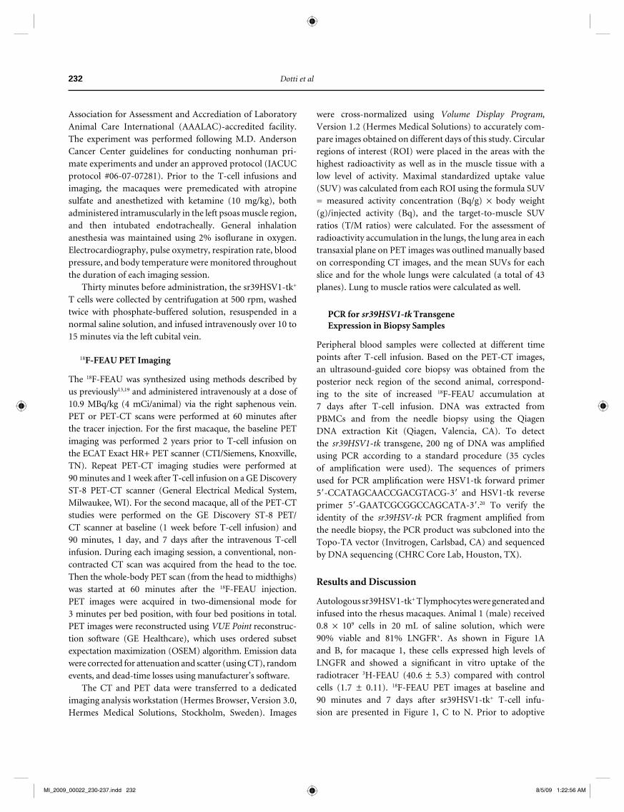
were cross-normalized using Volume Display Program , Version 1.2 (Hermes Medical Solutions) to accurately com-pare images obtained on different days of this study. Circular regions of interest (ROI) were placed in the areas with the highest radioactivity as well as in the muscle tissue with a low level of activity. Maximal standardized uptake value (SUV) was calculated from each ROI using the formula SUV = measured activity concentration (Bq/g) × body weight(g)/injected activity (Bq), and the target-to-muscle SUV ratios (T/M ratios) were calculated. For the assessment of radioactivity accumulation in the lungs, the lung area in each transaxial plane on PET images was outlined manually based on corresponding CT images, and the mean SUVs for each slice and for the whole lungs were calculated (a total of 43 planes). Lung to muscle ratios were calculated as well.
PCR for sr39HSV1-tk TransgeneExpression in Biopsy Samples
Peripheral blood samples were collected at different time points after T-cell infusion. Based on the PET-CT images, an ultrasound-guided core biopsy was obtained from the posterior neck region of the second animal, correspond-ing to the site of increased 18 F-FEAU accumulation at7 days after T-cell infusion. DNA was extracted from PBMCs and from the needle biopsy using the Qiagen DNA extraction Kit (Qiagen, Valencia, CA). To detect the sr39HSV1-tk transgene, 200 ng of DNA was amplifi ed using PCR according to a standard procedure (35 cycles of amplifi cation were used). The sequences of primers used for PCR amplifi cation were HSV1-tk forward primer 5�-CCATAGCAACCGACGTACG-3� and HSV1-tk reverse primer 5�-GAATCGCGGCCAGCATA-3�. 20 To verify the identity of the sr39HSV-tk PCR fragment amplifi ed from the needle biopsy, the PCR product was subcloned into the Topo-TA vector (Invitrogen, Carlsbad, CA) and sequenced by DNA sequencing (CHRC Core Lab, Houston, TX).
Results and Discussion
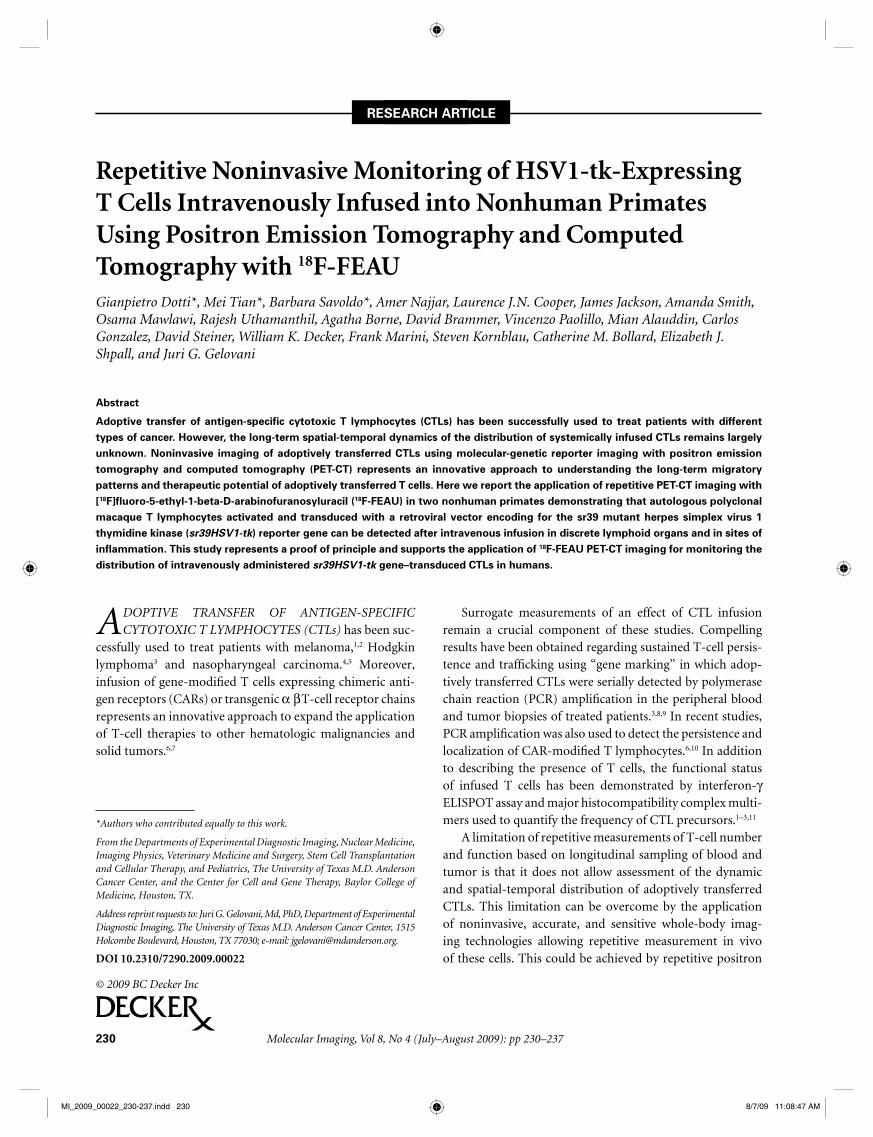
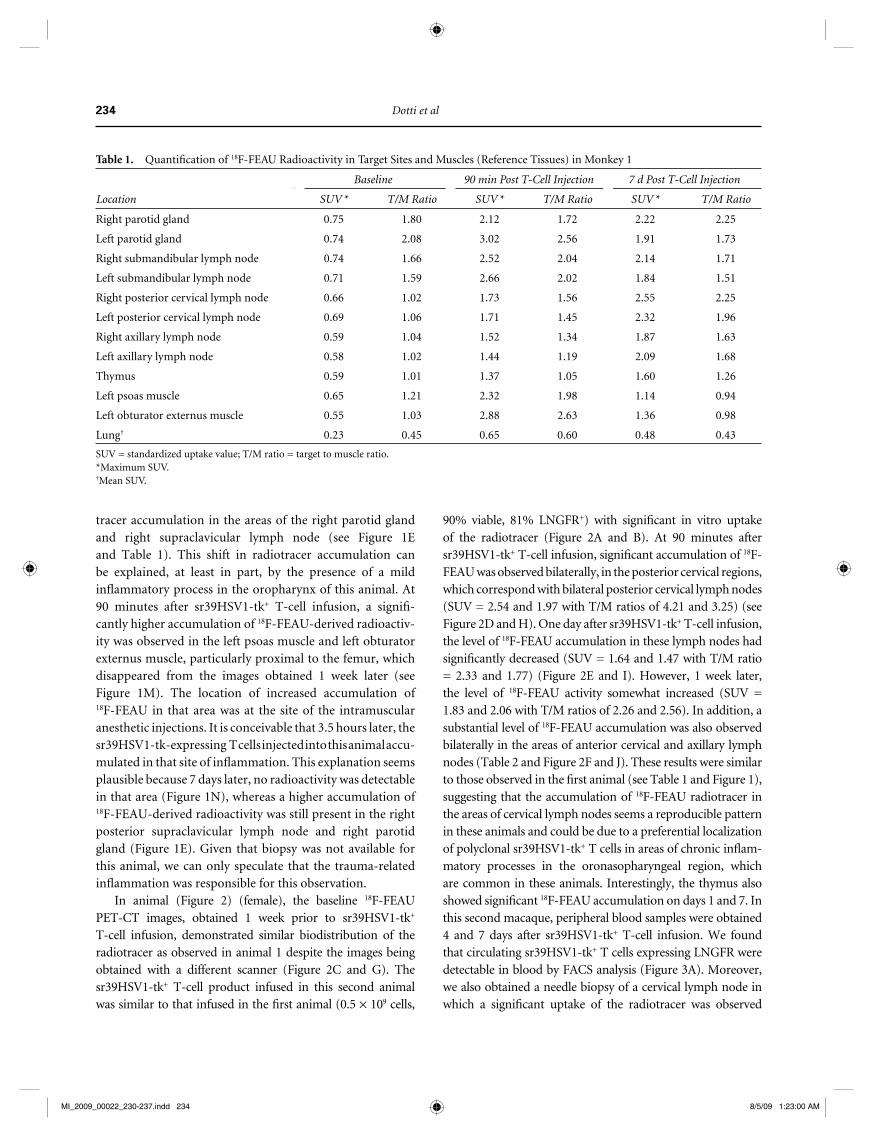
Autologous sr39HSV1-tk + T lymphocytes were generated and infused into the rhesus macaques. Animal 1 (male) received0.8 × 10 9 cells in 20 mL of saline solution, which were 90% viable and 81% LNGFR + . As shown in Figure 1 A and B , for macaque 1, these cells expressed high levels of LNGFR and showed a signifi cant in vitro uptake of the radiotracer 3 H-FEAU (40.6 ± 5.3) compared with control cells (1.7 ± 0.11). 18 F-FEAU PET images at baseline and90 minutes and 7 days after sr39HSV1-tk + T-cell infu-sion are presented in Figure 1 , C to N . Prior to adoptive
Association for Assessment and Accrediation of Laboratory Animal Care International (AAALAC)-accredited facility. The experiment was performed following M.D. Anderson Cancer Center guidelines for conducting nonhuman pri-mate experiments and under an approved protocol (IACUC protocol #06-07-07281). Prior to the T-cell infusions and imaging, the macaques were premedicated with atropine sulfate and anesthetized with ketamine (10 mg/kg), both administered intramuscularly in the left psoas muscle region, and then intubated endotracheally. General inhalation anesthesia was maintained using 2% isofl urane in oxygen. Electrocardiography, pulse oxymetry, respiration rate, blood pressure, and body temperature were monitored throughout the duration of each imaging session.
Thirty minutes before administration, the sr39HSV1-tk + T cells were collected by centrifugation at 500 rpm, washed twice with phosphate-buffered solution, resuspended in a normal saline solution, and infused intravenously over 10 to 15 minutes via the left cubital vein.
18 F-FEAU PET Imaging
The 18 F-FEAU was synthesized using methods described by us previously 13,19 and administered intravenously at a dose of 10.9 MBq/kg (4 mCi/animal) via the right saphenous vein. PET or PET-CT scans were performed at 60 minutes after the tracer injection. For the fi rst macaque, the baseline PET imaging was performed 2 years prior to T-cell infusion on the ECAT Exact HR+ PET scanner (CTI/Siemens, Knoxville, TN). Repeat PET-CT imaging studies were performed at90 minutes and 1 week after T-cell infusion on a GE Discovery ST-8 PET-CT scanner (General Electrical Medical System, Milwaukee, WI). For the second macaque, all of the PET-CT studies were performed on the GE Discovery ST-8 PET/CT scanner at baseline (1 week before T-cell infusion) and90 minutes, 1 day, and 7 days after the intravenous T-cell infusion. During each imaging session, a conventional, non-contracted CT scan was acquired from the head to the toe. Then the whole-body PET scan (from the head to midthighs) was started at 60 minutes after the 18 F-FEAU injection. PET images were acquired in two-dimensional mode for3 minutes per bed position, with four bed positions in total. PET images were reconstructed using VUE Point reconstruc-tion software (GE Healthcare), which uses ordered subset expectation maximization (OSEM) algorithm. Emission data were corrected for attenuation and scatter (using CT), random events, and dead-time losses using manufacturer’s software.
The CT and PET data were transferred to a dedicated imaging analysis workstation (Hermes Browser, Version 3.0, Hermes Medical Solutions, Stockholm, Sweden). Images
MI_2009_00022_230-237.indd 232MI_2009_00022_230-237.indd 232 8/5/09 1:22:56 AM8/5/09 1:22:56 AM

T-Lymphocyte Imaging 233
after sr39HSV1-tk + T-cell infusion, PET imaging demon-strated intensive accumulation of 18 F-FEAU-derived radioac-tivity in the areas of the left parotid gland and in the cervical lymph nodes (see Figure 1D ). PET images obtained 7 days after sr39HSV1-tk + T-cell infusion revealed higher levels of
immunotherapy, the 18 F-FEAU-derived radioactivity on PET imaging was predominantly located in the liver, small intes-tine, kidneys, and urinary bladder, which is consistent with the normal routes of 18 F-FEAU excretion through the hepa-tobiliary and renal systems (see Figure 1C ). Ninety minutes
C
I
F
L
J
D
G
M
K
E
H
N
Baseline 90 min postT cell infusion
7 days postT cell infusion
NGFR
CD
3
0.7%
38%
81%
Control
104
103
102
101
100
104103102101100
104
103
102
101
100
104103102101100
104
103
102
101
100
104103102101100
Aftertransduction
Afterselection
A
B
Untransduced
Cel
l/Med
ium
upt
ake
ratio
50.0
40.0
30.0
20.0
10.0
0.0
HSV1-tk-Transduced
Figure 1. Repetitive PET-CT images of 18F-FEAU distribution in the rhesus macaque 1 before and after injection of sr39HSV1-tk+ autolo-gous T lymphocytes. A illustrates the percentage of LNGFR+ T lymphocytes, as assessed by FACS analysis using a monoclonal antibody detecting NGFR (Becton-Dickinson), 7 days after retroviral transduction and at the time of infusion after selection using LNGFR micro-beads. B illustrates the uptake of [3H]-FEAU by sr39HSV1-tk+ T lymphocytes. Each assay was performed in triplicate. Sr39HSV1-tk+ T cells exhibited a cell to medium uptake ratio of 40.6, 24.5× higher than the control. Columns represent means; bars indicate ± standard deviation. C to N illustrate the whole-body PET scan 60 minutes after the tracer injection. PET images were reconstructed, analyzed, and normalized as previously described.12 C, D, and E represent maximum-intensity three-dimensional projections of whole-body PET images collected before T-cell infusion (C) and 90 minutes (D) and 7 days (E) after T-cell infusion. F, G, H, I, J, and K demonstrate the differences in 18F-FEAU accumulation in the parotid glands and cervical lymph nodes between baseline (F, I), 90 minutes (G, J ), and 7 days (H, K ) after T-cell administration. L, M, and N represent coronal PET or PET-CT images through the area of target lesions in the left thigh visible at 90 minutes after T-cell administration and 60 minutes after 18F-FEAU injection (M), which is absent in the baseline image (L) and 7 days later (N).
MI_2009_00022_230-237.indd 233MI_2009_00022_230-237.indd 233 8/5/09 1:22:56 AM8/5/09 1:22:56 AM
234 Dotti et al
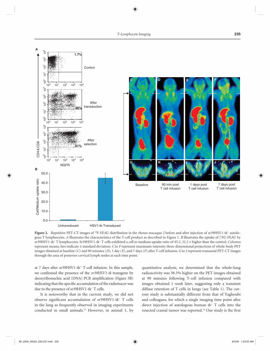
90% viable, 81% LNGFR + ) with signifi cant in vitro uptake of the radiotracer ( Figure 2 A and B ). At 90 minutes after sr39HSV1-tk + T-cell infusion, signifi cant accumulation of 18 F-FEAU was observed bilaterally, in the posterior cervical regions, which correspond with bilateral posterior cervical lymph nodes (SUV = 2.54 and 1.97 with T/M ratios of 4.21 and 3.25) (see Figure 2 D and H ). One day after sr39HSV1-tk + T-cell infusion, the level of 18 F-FEAU accumulation in these lymph nodes had signifi cantly decreased (SUV = 1.64 and 1.47 with T/M ratio = 2.33 and 1.77) ( Figure 2 E and I ). However, 1 week later, the level of 18 F-FEAU activity somewhat increased (SUV = 1.83 and 2.06 with T/M ratios of 2.26 and 2.56). In addition, a substantial level of 18 F-FEAU accumulation was also observed bilaterally in the areas of anterior cervical and axillary lymph nodes (Table 2 and Figure 2 F and J ). These results were similar to those observed in the fi rst animal (see Table 1 and Figure 1 ), suggesting that the accumulation of 18 F-FEAU radiotracer in the areas of cervical lymph nodes seems a reproducible pattern in these animals and could be due to a preferential localization of polyclonal sr39HSV1-tk + T cells in areas of chronic infl am-matory processes in the oronasopharyngeal region, which are common in these animals. Interestingly, the thymus also showed signifi cant 18 F-FEAU accumulation on days 1 and 7. In this second macaque, peripheral blood samples were obtained 4 and 7 days after sr39HSV1-tk + T-cell infusion. We found that circulating sr39HSV1-tk + T cells expressing LNGFR were detectable in blood by FACS analysis ( Figure 3A ). Moreover, we also obtained a needle biopsy of a cervical lymph node in which a signifi cant uptake of the radiotracer was observed
tracer accumulation in the areas of the right parotid glandand right supraclavicular lymph node (see Figure 1E and Table 1). This shift in radiotracer accumulation can be explained, at least in part, by the presence of a mild infl ammatory process in the oropharynx of this animal. At90 minutes after sr39HSV1-tk + T-cell infusion, a signifi -cantly higher accumulation of 18 F-FEAU-derived radioactiv-ity was observed in the left psoas muscle and left obturator externus muscle, particularly proximal to the femur, which disappeared from the images obtained 1 week later (see Figure 1M ). The location of increased accumulation of 18 F-FEAU in that area was at the site of the intramuscular anesthetic injections. It is conceivable that 3.5 hours later, the sr39HSV1-tk-expressing T cells injected into this animal accu-mulated in that site of infl ammation. This explanation seems plausible because 7 days later, no radioactivity was detectable in that area ( Figure 1N ), whereas a higher accumulation of 18 F-FEAU-derived radioactivity was still present in the right posterior supraclavicular lymph node and right parotid gland ( Figure 1E ). Given that biopsy was not available for this animal, we can only speculate that the trauma-related infl ammation was responsible for this observation.
In animal (Figure 2) (female), the baseline 18 F-FEAU PET-CT images, obtained 1 week prior to sr39HSV1-tk + T-cell infusion, demonstrated similar biodistribution of the radiotracer as observed in animal 1 despite the images being obtained with a different scanner ( Figure 2 C and G ). The sr39HSV1-tk + T-cell product infused in this second animal was similar to that infused in the fi rst animal (0.5 × 10 9 cells,
1. Table Quantifi cation of 18F-FEAU Radioactivity in Target Sites and Muscles (Reference Tissues) in Monkey 1
Location
Baseline 90 min Post T-Cell Injection 7 d Post T-Cell Injection
SUV * T/M Ratio SUV * T/M Ratio SUV * T/M Ratio
Right parotid gland 0.75 1.80 2.12 1.72 2.22 2.25
Left parotid gland 0.74 2.08 3.02 2.56 1.91 1.73
Right submandibular lymph node 0.74 1.66 2.52 2.04 2.14 1.71
Left submandibular lymph node 0.71 1.59 2.66 2.02 1.84 1.51
Right posterior cervical lymph node 0.66 1.02 1.73 1.56 2.55 2.25
Left posterior cervical lymph node 0.69 1.06 1.71 1.45 2.32 1.96
Right axillary lymph node 0.59 1.04 1.52 1.34 1.87 1.63
Left axillary lymph node 0.58 1.02 1.44 1.19 2.09 1.68
Thymus 0.59 1.01 1.37 1.05 1.60 1.26
Left psoas muscle 0.65 1.21 2.32 1.98 1.14 0.94
Left obturator externus muscle 0.55 1.03 2.88 2.63 1.36 0.98
Lung† 0.23 0.45 0.65 0.60 0.48 0.43
SUV = standardized uptake value; T/M ratio = target to muscle ratio.*Maximum SUV.†Mean SUV.
MI_2009_00022_230-237.indd 234MI_2009_00022_230-237.indd 234 8/5/09 1:23:00 AM8/5/09 1:23:00 AM
T-Lymphocyte Imaging 235
quantitative analysis, we determined that the whole-lung radioactivity was 38.5% higher on the PET images obtained at 90 minutes following T-cell infusion compared with images obtained 1 week later, suggesting only a transient diffuse retention of T cells in lungs (see Table 1). The cur-rent study is substantially different from that of Yaghoubi and colleagues, for which a single imaging time point after direct injection of autologous human tk+ T cells into the resected cranial tumor was reported. 14 Our study is the fi rst
at 7 days after sr39HSV1-tk + T-cell infusion. In this sample, we confi rmed the presence of the sr39HSV1-tk transgene by deoxyribonucleic acid (DNA) PCR amplifi cation ( Figure 3B ) indicating that the specifi c accumulation of the radiotracer was due to the presence of sr39HSV1-tk + T cells.
It is noteworthy that in the current study, we did not observe signifi cant accumulation of sr39HSV1-tk + T cells in the lung as frequently observed in imaging experiments conducted in small animals. 12 However, in animal 1, by
NGFR
CD
4&
CD
8
1.7%
48%
81%
Control
104
103
102
101
100
104103102101100
104
103
102
101
100
104103102101100
104
103
102
101
100
104103102101100
Aftertransduction
Afterselection
A
B
Untransduced
Cel
l/Med
ium
upt
ake
ratio
50.0
40.0
30.0
20.0
10.0
0.0
HSV1-tk-Transduced
Baseline 90 min postT cell infusion
1 days postT cell infusion
7 days postT cell infusion
C ED F
G IH J
Figure 2. Repetitive PET-CT images of 18F-FEAU distribution in the rhesus macaque 2 before and after injection of sr39HSV1-tk+ autolo-gous T lymphocytes. A illustrates the characteristics of the T-cell product as described in Figure 1. B illustrates the uptake of [3H]-FEAU by sr39HSV1-tk+ T lymphocytes. Sr39HSV1-tk+ T cells exhibited a cell to medium uptake ratio of 45.2, 32.2 × higher than the control. Columns represent means; bars indicate ± standard deviation. C to F represent maximum-intensity three-dimensional projections of whole-body PET images obtained at baseline (C) and 90 minutes (D), 1 day (E), and 7 days (F) after T-cell infusion. G to J represent transaxial PET-CT images through the area of posterior cervical lymph nodes at each time point.
MI_2009_00022_230-237.indd 235MI_2009_00022_230-237.indd 235 8/5/09 1:23:00 AM8/5/09 1:23:00 AM

236 Dotti et al
Table 2. Quantifi cation of 18F-FEAU Radioactivity in Target Sites and Muscles (Reference Tissues) in Monkey 2
Location
Baseline90 min Post T-Cell
Injection1 d Post T-Cell
Injection7 d Post T-Cell
Injection
SUV * T/M Ratio SUV * T/M Ratio SUV * T/M Ratio SUV * T/M Ratio
Right parotid gland 1.12 1.51 1.04 1.81 1.36 1.80 2.13 2.08
Left parotid gland 1.15 1.56 1.04 1.81 1.30 1.72 2.75 2.61
Right submandibular lymph node 1.05 1.48 0.80 1.27 1.53 2.25 1.62 1.96
Left submandibular lymph node 1.11 1.66 0.70 1.16 1.21 1.69 2.01 2.48
Right posterior cervical lymph node 1.55 2.24 2.54 4.21 1.64 2.33 1.83 2.26
Left posterior cervical lymph node 1.55 2.36 1.97 3.25 1.47 1.77 2.06 2.56
Right axillary lymph node 1.41 1.63 1.01 1.41 1.58 1.95 2.17 2.14
Left axillary lymph node 1.35 1.50 1.00 1.39 1.43 1.81 2.11 2.31
Thymus 1.10 1.17 0.93 1.19 1.49 1.63 1.41 1.42
Lung† 0.24 0.30 0.21 0.32 0.24 0.28 0.23 0.30
SUV = standardized uptake value; T/M ratio = target to muscle ratio.*Maximum SUV.†Mean SUV.
β actin (312 bp)
HSV1-tk (643 bp)
500 -400 -300 -
600 -700 -800 -
100
bp la
dder
Wat
er c
ontr
ol
HS
Vtk
1 T
cel
ls
PB
MC
pre
PB
MC
D4
PB
MC
D7
Lym
pho
node
D7B
---
---
NT
T c
ells
NGFR
CD
4&
CD
8
1%
8.2%
7.8%
16%
15%
19%
104
103
102
101
100
104103102101100
104
103
102
101
100
104103102101100
104
103
102
101
100
104103102101100
A pre T-cell infusion
Post day 4
Post day 7
Figure 3. Detection of sr39HSV1-tk + T lymphocytes in peripheral blood and in a posterior neck lymph node biopsy. A illustrates the detec-tion of circulating NGFR + T cells as assessed by phenotypic analysis in blood samples collected before and after T-cell infusion. B illustrates the detection of sr39HSV-tk + expression by polymerase chain reaction in peripheral blood mononuclear cells (PBMCs) preinfusion, 4 days postinfusion, and 7 days postinfusion and in a posterior neck lymph node biopsy sample obtained 7 days after T-cell infusion. Ex vivo expanded T nontransduced cells and ex vivo expanded sr39HSV1-tk + T cells were also used as negative and positive control, respectively.
MI_2009_00022_230-237.indd 236MI_2009_00022_230-237.indd 236 8/5/09 1:23:03 AM8/5/09 1:23:03 AM

T-Lymphocyte Imaging 237
8. Heslop HE, Ng CY, Li C, et al. Long-term restoration of immunity against Epstein-Barr virus infection by adoptive transfer of gene-modifi ed virus-specifi c T lymphocytes. Nat Med 1996; 2: 551– 5 .
9. Rosenberg SA, Aebersold P, Cornetta K, et al. Gene transfer into humans—immunotherapy of patients with advanced melanoma, using tumor-infi ltrating lymphocytes modifi ed by retroviral gene transduction. N Engl J Med 1990; 323: 570– 8 .
10. Till BG, Jensen MC, Wang J, et al. Adoptive immunotherapy for indolent non-Hodgkin lymphoma and mantle cell lym-phoma using genetically modifi ed autologous CD20-specifi c T cells. Blood 2008; 112: 2261– 71 .
11. Savoldo B, Goss JA, Hammer MM, et al. Treatment of solid organ transplant recipients with autologous Epstein Barr virus-specifi c cytotoxic T lymphocytes (CTLs). Blood 2006; 108: 2942– 9 .
12. Koehne G, Doubrovin M, Doubrovina E, et al. Serial in vivo imaging of the targeted migration of human HSV- TK-transduced antigen-specifi c lymphocytes. Nat Biotechnol 2003; 21: 405– 13 .
13. Soghomonyan S, Hajitou A, Rangel R, et al. Molecular PET imaging of HSV1-tk reporter gene expression using [18F]FEAU. Nat Protoc 2007; 2: 416– 23 .
14. Yaghoubi SS, Jensen MC, Satyamurthy N, et al. Noninvasive detection of therapeutic cytolytic T cells with 18F-FHBG PET in a patient with glioma. Nat Clin Pract Oncol 2009; 6: 53– 8 .
15. Kornblau SM, Aycox PG, Stephens C, et al. Control of graft-versus-host disease with maintenance of the graft-versus-leukemia effect in a murine allogeneic transplant model using retrovirally transduced murine suicidal lymphocytes. Exp Hematol 2007; 35: 842– 53 .
16. Berger C, Jensen MC, Lansdorp PM, et al. Adoptive transfer of effector CD8+ T cells derived from central memory cells establishes persistent T cell memory in primates. J Clin Invest 2008; 118: 294– 305 .
17. Vera J, Savoldo B, Vigouroux S, et al. T lymphocytes redi-rected against the kappa light chain of human immunoglob-ulin effi ciently kill mature B lymphocyte-derived malignant cells. Blood 2006; 108: 3890– 7 .
18. Bonini C, Grez M, Traversari C, et al. Safety of retrovi-ral gene marking with a truncated NGF receptor. Nat Med 2003; 9: 367– 9 .
19. Alauddin MM, Shahinian A, Park R, et al. In vivo evaluation of 2�-deoxy-2�-[18F]fl uoro-5-iodo-1-beta-D-arabinofurano-syluracil ([18F]FIAU) and 2�-deoxy-2�-[18F]fl uoro-5-ethyl-1-beta-D-arabinofuranosyluracil ([18F]FEAU) as markers for suicide gene expression. Eur J Nucl Med Mol Imaging 2007; 34: 822– 9 .
20. Traversari C, Marktel S, Magnani Z, et al. The potential immunogenicity of the TK suicide gene does not prevent full clinical benefi t associated with the use of TK-transduced donor lymphocytes in HSCT for hematologic malignancies. Blood 2007; 109: 4708– 15 .
21. Najjar AM, Nishii R, Maxwell DS, et al. Molecular-genetic PET imaging using an HSV1-tk mutant reporter gene with enhanced specifi city to acycloguanosine nucleoside analogs. J Nucl Med 2009; 50: 409– 16 .
to demonstrate the feasibility of repetitive PET-CT imaging of spatial-temporal dynamics of distribution and traffi ck-ing of sr39HSV1-tk reporter gene–expressing T cells in non human primates after intravenous administration.
We conclude that PET imaging with 18 F-FEAU may be used in future clinical trials to evaluate the biodistribu-tion of adoptively transferred CTLs modifi ed to express the sr39HSV1-tk reporter gene or its variants. 21 This technology will ultimately allow for long-term monitoring of traffi cking patterns and persistence of the T cells in patients and facili-tate critical improvements in T-cell therapy for cancer.
Acknowledgments
We thank Dana Toomey, Julie Basham, Deborah Petit, Alfredo Santiago, and Jennifer Miller for their excellent tech-nical support and Nancy Swanston for excellent coordina-tion of this study. Financial disclosure of authors: This work was supported by the Dana Foundation Award “Clinical Concepts in Neuro-Immuno Imaging” (to J.G.), the New Research Program Development Fund of the Department of Experimental Diagnostic Imaging (to J.G. and M.A.), The University of Texas M.D. Anderson Cancer Center, and a Baylor College of Medicine-M.D. Anderson Multi-Institutional Research Grant (to G.D., C.M.B., E.J.S.).
Financial disclosure of reviewers: None reported.
References
1. Dudley ME, Yang JC, Sherry R, et al. Adoptive cell therapy for patients with metastatic melanoma: evaluation of intensive myeloablative chemoradiation preparative regimens. J Clin Oncol 2008; 26: 5233– 9 .
2. Yee C, Thompson JA, Byrd D, et al. Adoptive T cell therapy using antigen-specifi c CD8+ T cell clones for the treatment of patients with metastatic melanoma: in vivo persistence, migration, and antitumor effect of transferred T cells. Proc Natl Acad Sci U S A 2002; 99: 16168– 73 .
3. Bollard CM, Aguilar L, Straathof KC, et al. Cytotoxic T lym-phocyte therapy for Epstein-Barr virus+ Hodgkin’s disease. J Exp Med 2004; 200: 1623– 33 .
4. Straathof KC, Bollard CM, Popat U, et al. Treatment of nasopharyngeal carcinoma with Epstein-Barr virus–specifi c T lymphocytes. Blood 2005; 105: 1898– 904 .
5. Comoli P, Pedrazzoli P, Maccario R, et al. Cell therapy of stage IV nasopharyngeal carcinoma with autologous Epstein-Barr virus-targeted cytotoxic T lymphocytes. J Clin Oncol 2005; 23: 8942– 9 .
6. Pule MA, Savoldo B, Myers GD, et al. Virus-specifi c T cells engineered to coexpress tumor-specifi c receptors: persistence and antitumor activity in individuals with neuroblastoma. Nat Med 2008; 14: 1264– 70 .
7. Morgan RA, Dudley ME, Wunderlich JR, et al. Cancer regres-sion in patients after transfer of genetically engineered lym-phocytes. Science 2006; 314: 126– 9 .
MI_2009_00022_230-237.indd 237MI_2009_00022_230-237.indd 237 8/5/09 1:23:05 AM8/5/09 1:23:05 AM

Related Documents