1 Title: Expression of GDNF transgene in astrocytes improves cognitive deficits in aged rats Authors: M Pertusa a,1 , S García-Matas a,1 , H Mammeri b,1 , A Adell c , T Rodrigo d , J Mallet b , R Cristòfol a , C Sarkis b , C Sanfeliu* a Departament de Farmacologia i Toxicologia, Institut d’Investigacions Biomèdiques de Barcelona, CSIC-IDIBAPS, 08036 Barcelona, Spain. b Laboratoire de Génétique Moléculaire de la Neurotransmission et des Processus Neurodégénératifs, CNRS, Hopital de la Salpetrière, 75013 Paris, France. c Departament de Neuroquímica, Institut d’Investigacions Biomèdiques de Barcelona, CSIC-IDIBAPS, 08036 Barcelona, Spain. d Unitat d’Experimentació Animal de Psicologia, Universitat de Barcelona, 08035 Barcelona, Spain. 1 These authors contributed equally to this work *Corresponding author: Coral Sanfeliu Departament de Farmacologia i Toxicologia Institut d’Investigacions Biomèdiques de Barcelona (IIBB), CSIC -IDIBAPS Rosselló 161 08036 Barcelona phone: +34 933638338 fax: +34 933638301

Welcome message from author
This document is posted to help you gain knowledge. Please leave a comment to let me know what you think about it! Share it to your friends and learn new things together.
Transcript

1
Title:
Expression of GDNF transgene in astrocytes improves cognitive deficits in aged rats
Authors:
M Pertusaa,1, S García-Matasa,1, H Mammerib,1, A Adellc, T Rodrigod, J Malletb, R
Cristòfola, C Sarkisb, C Sanfeliu*
aDepartament de Farmacologia i Toxicologia, Institut d’Investigacions Biomèdiques de
Barcelona, CSIC-IDIBAPS, 08036 Barcelona, Spain.
bLaboratoire de Génétique Moléculaire de la Neurotransmission et des Processus
Neurodégénératifs, CNRS, Hopital de la Salpetrière, 75013 Paris, France.
cDepartament de Neuroquímica, Institut d’Investigacions Biomèdiques de Barcelona,
CSIC-IDIBAPS, 08036 Barcelona, Spain.
dUnitat d’Experimentació Animal de Psicologia, Universitat de Barcelona, 08035
Barcelona, Spain.
1These authors contributed equally to this work
*Corresponding author:
Coral Sanfeliu
Departament de Farmacologia i Toxicologia
Institut d’Investigacions Biomèdiques de Barcelona (IIBB), CSIC-IDIBAPS
Rosselló 161
08036 Barcelona
phone: +34 933638338
fax: +34 933638301

2
Abstract
Glial cell line-derived neurotrophic factor (GDNF) was assayed for its neurotrophic effects
against the neuronal atrophy that causes cognitive deficits in old age. Aged Fisher 344 rats
with impairment in the Morris water maze received intrahippocampal injections at the
dorsal CA1 area of either a lentiviral vector encoding human GDNF or the same vector
encoding human green fluorescent protein as a control. Recombinant lentiviral vectors
constructed with human cytomegalovirus promotor and pseudotyped with lyssavirus
Mokola glycoprotein specifically transduced the astrocytes in vivo. Astrocyte-secreted
GDNF enhanced neuron function as shown by local increases in synthesis of the
neurotransmitters acetylcholine, dopamine and serotonin. This neurotrophic effect led to
cognitive improvement of the rats as early as two weeks after gene transduction. Spatial
learning and memory testing showed a significant gain in cognitive abilities due to GDNF
exposure, whereas control-transduced rats kept their performance at the chance level. These
results confirm the broad spectrum of the neurotrophic action of GDNF and open new gene
therapy possibilities for reducing age-related neurodegeneration.
Key words
Glial cell line-derived neurotrophic factor (GDNF); lentiviral vector; aging; learning and
memory; gene therapy; acetylcholine; dopamine; serotonin; rat.

3
1. Introduction
Glial cell line-derived neurotrophic factor (GDNF) is a member of the transforming growth
factor superfamily. It was first isolated from culture medium of rat B49 glioblastoma
cells in a search of trophic factors for midbrain dopaminergic neurons [73,74]. GDNF has
remarkable regenerative and restorative effects upon nigrostriatal dopaminergic neurons
[16,46,51]. Soon after its discovery, it was found that GDNF is a potent neurotrophic factor
active on a broad spectrum of neuronal types and has neuroprotective effects in several
experimental paradigms of nerve cell injury. For instance, GDNF reduces neuronal damage
in axotomized neurons [56,96,121,126], in experimental brain trauma [7] and ischemia
[23,64,87,122], and in several excitotoxic [78,92,100,124] and neurotoxic injuries
modeling neurodegenerative diseases [6,50,119]. Therapeutic GDNF administration has
raised expectations that it could be a preventive or palliative treatment for Parkinson’s
disease [for review see: 35,38], amyotrophic lateral sclerosis [for review see: 12,82] and
Huntington’s disease [for review see: 3].
GDNF is expressed in both neurons and astrocytes [73,77,92,108]. It signals
through the two-component receptor complex GFR-/Ret [118], also expressed in both
cell types [92]. An increase in astrocyte GDNF synthesis and protein expression is believed
to play an active role in neuron survival and plasticity after ischemia [88,117] and
excitotoxic damage [60,77]. Accordingly, experimental strategies of GDNF delivery by
astrocytes have shown to be neuroprotective in vivo for motor neurons [97,128] and
dopaminergic neurons [29,31,34].
Endogenous GDNF plays an important role in cognitive abilities as demonstrated by
the impaired spatial learning performance of mice that are heterozygous for a targeted
deletion of the GDNF gene [49]. Spatial learning and memory is associated with intact
hippocampus function and it has long been known that memory deficits in old age are
similar to those produced by bilateral hippocampal lesions [18,52]. Different categories of
agents, such as neurotrophins [40,41,79], antioxidant agents [72,94] and cholinergic drugs
[13,58], have been assayed in aged rodents in a search for effective and harmless treatments
that reverse the memory loss of normal aging and ameliorate memory loss in pathological

4
aging caused by Alzheimer’s disease. The potential of GDNF against age-related cognitive
deterioration has not been fully explored. In a previous study, spatial learning and memory
improved in aged rats after i.c.v. administration of GDNF but no brain regional correlates
were analyzed [99]. We studied the restorative effects of GDNF on spatial learning and
memory after local delivery into the hippocampus. For this purpose, the astrocytes of the
dorsal CA1 hippocampal area of cognitively impaired aged Fisher 344 rats were transduced
in vivo with a lentiviral vector expressing the human gene for GDNF. The efficacy of
transgene delivery, either GDNF or the human green fluorescent protein (GFP) used as a
control, and the behavioral and neurotransmitter changes shown by the aged rats were
determined throughout the study. Brain regional GDNF gene expression allowed the
evaluation of the involvement of the hippocampal CA1 function in maintaining spatial
learning and memory through aging. Vector specificity for the GDNF gene transfer to
astrocytes enabled the study of the neuroprotective potential of these glial cells in
neurodegenerative processes.
2. Methods
2.1. Animal groups
Thirty male 7-month old Fisher 344 rats retired from breeding were purchased from
Charles River (Lyon, France) and kept in the animal house of the University of Barcelona
until they were old. They were maintained in standard conditions of temperature and
humidity, with two animals per cage, a 12:12 h light-dark cycle, and food and water ad
libitum. Only animals showing a healthy general status at 22 months of age were included
in the study. A group of 10 young male rats of the same origin were used to test the viral
stock. A further group of 10 6-month old male Fisher 344 rats were included in the study
for comparative neurotransmitter determinations. All handling and experimental procedures
were approved by the Animal Ethics and Health Committees of the University of
Barcelona.

5
2.1. Lentiviral vectors
A recombinant lentiviral vector encoding human GDNF was constructed for the specific
transduction of astrocytes in vivo. The combination of the lyssavirus Mokola glycoprotein
(Mokola G) pseudotype with the human cytomegalovirus promoter (CMV) allowed
efficient transgene expression under these experimental conditions. A vector encoding GFP
instead of GDNF was used as a control vector. Vectors were constructed and produced as
previously described [10,91]. Briefly, the plasmids Flap-CMV-GDNF-WPRE and Flap-
CMV-GFP-WPRE were derived from the lentiviral vector genome. These plasmids contain
the backbone of the lentiviral genome including the central Flap sequence [127]. The
GDNF and GFP transgenes were under control of the CMV promoter. The woodchuck
hepatitis virus post-transcriptional regulatory element (WPRE) was added 3 to the GDNF
or GFP gene to increase transgene expression [129]. Lentiviral vector stocks were produced
by transient co-transfection of 293T cells with the vector genome plasmid, a packaging
plasmid and a plasmid coding for the envelope that included the Mokola G gene. The cell
supernatant for each transgene vector was harvested 48 h post-transfection, treated with
DNase, filtered through 0.45 µm pore-sized filters, and subjected to ultra-centrifugation at
64,000 x g for 90 min. The virus pellet was resuspended in phosphate-buffered saline
(PBS) and its physical titer was quantified by ELISA of the p24 capsid protein. Viral stocks
were kept at -80ºC until use. At that moment, stocks were diluted with PBS to 30 ng/l of
p24 protein.
2.3. Behavioral testing
The behavioral test for spatial learning and memory was performed in a Morris water maze
[89]. The apparatus was modeled as described elsewhere [22]. Briefly, it measured 1.60 m
in diameter and water was made opaque by the addition of 100 mL/L of latex. Water
temperature was maintained at 22±1ºC. Black curtains surrounded the pool. Inside the
black enclosure four landmarks hanging from a false black ceiling defined the position of
the hidden platform: A, a 40-W fixed light; B, a beach ball with horizontal color drawings;

6
C, a vertical white structure with three truncated cones of decreasing size; and D, three
silver bottles bound by their neck. Landmark objects were hung at 90º distance. The
platform has a diameter of 0.11 m and was placed between landmarks A and B, 1 cm below
the surface and 0.38 m from the side. The landmarks and the platform were semi-randomly
rotated with respect to the room (90º, 180º, 270º, 360º) in a daily-equated way in order to
ensure that the rats used these landmarks, rather than any inadvertently remaining static
room cue, to locate the platform. A closed-circuit video camera recorded rat movements
that were analyzed by computer. A pre-training test with the platform made visible, located
1 cm above the water surface, consisted of 8 trials during 2 days. The rat was given 90 s to
find the platform, and then allowed to stay on for 30 s. When the rat did not reach the
platform, it was picked up, placed on it and left there for 30 s. Rats that failed to find the
visible platform were discarded from the study. For the acquisition test, the procedure was
the same, but animals had to rely on the landmarks to find the hidden platform. Animals
were given 4 escape trials per day for 12 days, a total of 48 trials. Latencies to reach the
escape platform were recorded for each trial and averaged for the 4 daily trials. On the last
day, a 60 s probe trial was performed with the platform removed. To analyze the rat’s
behavior, the surface of the pool was divided into four quadrants and the time that the rat
spent in the quadrant where the platform was previously was calculated. Impaired aged rats
were defined as those spending a time not different from chance performance (15 s out of
60 s) in the right quadrant during the spatial probe trial. Two weeks later, the impaired
animals were divided in two groups that were treated for hippocampal astrocyte expression
of GFP and GDNF, respectively (see surgical procedures below). Test assays were initiated
two weeks after surgery, when rats were nearly 24 months old. Animals were given 4
escape trials per day and on the fourth day a probe trial was performed. This procedure
continued, with a probe trial every four days until 48 escape trials had been conducted.
Behavioral results for rats that did not survive until the end of the study were not included.
2.3. Surgical procedures
A total of 23 aged rats underwent surgery: 7 rats of the GFP group, 8 rats of the GDNF

7
group and 8 unimpaired rats. Animals were anesthetized with 10 mg/kg xylacine (Rompun
2%, Bayer) and 80 mg/kg ketamine (Ketolar 50 mg/ml, Pfizer) i.m., and placed in a
stereotactic apparatus. Bilateral infusions were performed into the CA1 area of the
hippocampus at the coordinates: anterior-posterior –3.8 mm, medio-lateral +/-2 mm and
dorso-ventral –2.4 mm, from bregma and dural surface. The right coordinates to reach were
previously checked in 3 animals discarded from the study. One l of the vector solution
was delivered to the application point with a 25-gauge stainless steal cannula (Small Parts
Inc., Miami, FL) connected to a Hamilton syringe through a Teflon tube. The syringe was
attached to a micro-infusion pump (Bioanalytical systems Inc., West Lafayette, IN). The
cannula was left in position for 5 min after delivery to prevent the solution from surging
back.
2.4. Brain dissection and histology
After completion of the behavioral studies, animals were decapitated under light anesthesia
with 40 mg/kg ketamine. Brains were quickly removed, immersed in cold saline solution
and placed in a Kopf brain blocker. The cerebrum was cut into three blocks and then
bisected sagittaly at the midline. A block containing the half dorsal hippocampal area of
selected animals from the GFP group was fixed by immersion in 10% phosphate buffered
formaldehyde, cryoprotected with 10% sucrose and frozen on a stainless steel plate over
dry ice. Slices 16 m thin were double-stained with the antibodies against GFP 1:1000
(polyclonal, Abcam, Cambridge, UK) and GFAP 1:400 (monoclonal, clone GA5, Sigma,
St. Louis, MO), as previously described [10]. Secondary antibodies used were Alexa Fluor
488 1:1000 and Alexa Fluor 546 1:1000, respectively (Molecular Probes, Eugene, OR).
Nuclei were counterstained with bisbenzimide (Sigma). For all other animals, both
hemispheres were further dissected on a chilled glass plate, according to the procedure
described in detail elsewhere [8]. Tissue samples for 6 hippocampal areas (dorsal and
ventral CA1, dorsal and ventral CA2/3, dorsal and ventral dentate gyrus), 5 cerebral cortical
areas (cingulate, frontal, temporal, entorhinal, parietal), septum and caudate [98], were
weighed and frozen at –80ºC until analysis.

8
2.6. GDNF determination
The level of GDNF expression of the viral stock under the injection conditions used was
tested in a group of 10 young rats of the same colony used for the experiment. A
microinjection of the GDNF vector was performed in the dorsal CA1 area of the right
hippocampus as described above. GFP vector was injected on the left side of half the
animals and the other half were untreated. Rats were killed a week later, and the
hippocampus dissected as described above. Regional tissue levels of GDNF were
determined by ELISA with the GDNF Emax ImmunoAssay System kit (Promega), following
the manufacturer’s instructions. Long-term stability of this viral stock was previously
checked and expression was stable for one year (unpublished data).
2.5. Choline acetyl transferase and monoamine neurotransmitter determinations
Choline acetyl transferase (ChAT) activity in the tissue samples was measured by the
formation of 14[C]acetylcholine from 14[C]acetylcoenzyme-A (60 mCi/mmol; New England
Nuclear, PerkinElmer, Wellesley, MA) and choline (Sigma). For this purpose, homogenates
from left cerebral hemisphere regions were processed following the procedure described
elsewhere [5].
The monoamine neurotransmitters, dopamine and serotonin, the acid metabolites of
dopamine 3,4-dihydroxyphenylacetic acid (DOPAC) and homovanillic acid (HVA) and the
acid metabolite of serotonin 5-hydroxiindoleacetic (5-HIAA) were analyzed in the right
hemisphere tissue samples of GFP, GDNF and young rat groups. Tissue samples were
homogenized in a cold solution of 0.4 M perchloric acid, 0.01 % EDTA, 0.1 % sodium
bisulfite and 0.01 % cystine and centrifuged at 12000 x g for 30min. The concentrations of
neurotransmitters and metabolites in supernatants were determined by HPLC using a 3-µm
octadecylsilica (ODS) column (7.5 cm x 0.46 cm; Beckman) and detected
amperometrically with a Hewlett-Packard 1049 detector set at oxidation potentials of 0.6 V

9
(serotonin and 5-HIAA) and 0.7 V (dopamine, DOPAC and HVA). The mobile phase for 5-
HT consisted of 0.15 M NaH2PO4, 1.8 mM octyl sodium sulfate, 0.2 mM EDTA (pH 2.8
adjusted with phosphoric acid) and 30 % methanol. The mobile phase for dopamine
consisted of 0.15 M NaH2PO4, 0.46 mM octyl sodium sulfate, 0.5 mM EDTA (pH 2.8
adjusted with phosphoric acid), and 18 % methanol. Both mobile phases were pumped at
0.7 mL/min. Quantitative analyses were performed by the external standard method.
2.7. Statistics
Results are expressed as mean ± SEM. Statistical evaluation was performed with ANOVA
procedures, followed by Bonferroni’s or Newman-Keules’s multi-range test for comparison
of the mean group values.
3. Results
3.1. Astrocyte transduction with GFP and GDNF
The injection of lentiviral particles aimed at the dorsal CA1 hippocampal area yielded a
wide GFP expression mainly in the stratum radiatum and also in the oriens layer of this
area. No expression was detected beyond the hippocampus. Transduced proteins were
selectively expressed in astrocytes as demonstrated by overlapping of GFP and GFAP
immunostaining whereas no GFP expression was detected in the neurons of the pyramidal
molecular layer (GFP transduced rats, Fig. 1a-g). In a parallel study, we have confirmed the
astrocyte specific expression of the GFP reporter gene with the presently used vector in
several animal species (mouse, rat and non-human primate; Brizard et al., unpublished
observations).
The ability of the GDNF-engineered lentiviral vector to drive a detectable production of
GDNF was previously confirmed in a separate group of animals. GDNF levels determined
by ELISA were higher in the dorsal CA1 transduced with GDNF than in the GFP-
transduced or untreated CA1 area of the contralateral side (Table 1). A lesser GDNF over-
expression was detected in the ipsilateral adjacent area CA2/3.

10
3.2. Improved performance of GDNF injected rats in the Morris water maze
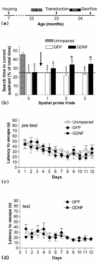
Timing of the behavioral studies is indicated in Fig. 2a. The aged animals were clearly
classified as non-impaired or learning-impaired, according to the results of the pre-test
spatial probe trial (Fig. 2b, probe trial 0). Non-impaired rats spent 27.8 ± 3.6 s (n=8) and
impaired rats spent 15.5 ± 4.3 s (n=15) out of the 60 s in the pool quadrant that previously
contained the platform (p<0.0001 and p=0.6746 as against to chance value of 15 s,
respectively). During prior training, latencies to find the hidden escape platform decreased
steadily without significant group differences (two-way ANOVA, effect of day factor:
F(11,211)=4.915, p<0.0001) (Fig 2c).
In the spatial probe trials after surgery, the GDNF group showed steady learning
improvement that was not present in the GFP group (Fig. 2b, probe trials 1-3). The latter
rats kept their values of searching time in the correct quadrant at the chance level. By the
end of the test-training period, GDNF rats showed values close to the unimpaired control
group. The performance differences between GFP- and GDNF-injected rats were
significant (ANOVA, F(1,44)=9.869, p=0.0030). No differences of latency to find the
hidden platform during the 12 training days were detected between the two rat groups (Fig.
3d).
The swim speed was not affected by either the surgery or the transgene expression.
Pre-surgery values measured in the pre-test probe trial were 21.58 ± 1.24 cm/s for the non-
impaired rats, 21.61 ± 0.69 cm/s for the GFP group and 21.83 ± 0.81 cm/s for the GDNF
group. Final swim speed measured in the last test probe trial was 22.73 ± 0.69 cm/s and
21.89 ± 1.07 cm/s for the GFP and the GDNF groups, respectively.
3.3. Increase of neurotransmitter levels in the hippocampus after GDNF transduction
Tissue concentration of ChAT, indicative of acetylcholine levels, in selected brain regions
of young and aged impaired rats is shown in Fig. 3a (hippocampus areas) and Table 2 (all
brain areas assayed). Two-way ANOVA indicated an effect of the cerebral brain region
(F(12,180)=14.50, p<0.0001) and of the rat group (F(2,180)=7.56, p=0.0007). By paired

11
ANOVA testing, GDNF rat group was significantly different from GFP and from young rat
groups (not shown). In GDNF transduced rats, an increase was detected in the whole CA1
area, as against GFP rats; whereas the ventral part increased also over young rat values.
Cingulate cortex ChAT increased in GDNF rats over the other two groups. There was a
decrease in the ventral CA2/3 area of GDNF rats, as against the group of young rats.
Regional brain concentration of dopamine is shown in Fig. 3b (hippocampus areas)
and Table 3 (all brain areas assayed) and its metabolites DOPAC and HVA are shown in
Table 3. Two-way ANOVA for brain region and rat group indicated an effect of the brain
region factor for the concentration of dopamine and its metabolites (not shown), whereas
the rat group factor was significant for both metabolites DOPAC (F(2,321)=12.98,
p<0.0001) and HVA (F(2,319)=71.59, p<0.0001). The group effect was significant for
dopamine levels when the brain regions were restricted to the hippocampus
(F(2,108)=15.81, p<0.0001). In this area, the increase of dopamine shown by the GDNF
group was significant in the dorsal CA1 area and in the whole dentate gyrus, as against the
group of young rats. A decrease of HVA in both aged GFP and GDNF groups was
observed in the dorsal CA1 and CA2/3, cerebral cortical areas and septum. In the caudate
region of both aged rat groups there was a decrease in both metabolites, DOPAC and HVA.
Regional brain concentration of serotonin is shown in Fig. 3c (hippocampus areas)
and Table 4 (all brain areas assayed) and its metabolite 5-HIAA is shown in Table 4. Two-
way ANOVA showed an effect of brain region (not shown) and rat group for serotonin and
5-HIAA (F(2,319)=25.45, p<0.0001 and F(2,319)=13.91, p<0.0001, respectively). The
GDNF group increased serotonin levels in the whole dorsal hippocampus and in the ventral
dentate gyrus more than the GFP group. Both groups of aged rats showed a hippocampal
increase of 5-HIAA in the dorsal dentate gyrus and the ventral CA1 area. Serotonin also
increased in cingulate and temporal cortical areas and in the caudate of control GFP aged
rats, as against to young rats.
4. Discussion
The over-expression of GDNF in hippocampal astrocytes induced a recovery of spatial

12
cognitive abilities in aged impaired rats as demonstrated by an enhancement of memory
retention of the platform location in the test probe trials. This better response in the Morris
water maze cannot be attributed to a mere improvement of age-related motor impairment
by GDNF. Several studies have shown that GDNF enhances motor function in aged rats
and non-human primates, which has been related to dopaminergic induction and
regeneration of the nigrostriatal pathway [15,33,53,57,69]. In the present study, GDNF was
over-expressed only in a restricted area of the hippocampus and no changes were induced
in the swimming speed. In addition, we found no differences in the latency to escape
between the rats expressing GDNF and control rats transduced with GFP during the test
assays, where an increase in swimming ability would help the rats to find the platform in a
shorter time. Latency to escape to the platform is routinely recorded as an additional spatial
learning and memory measure, but its outcome can be affected by external factors. This is
not the case with probe trial performance, which is accepted as the most accurate
measurement because it requires memory of the precise location of the platform [19,42].
Accordingly, in previous studies in our behavioral facilities, the probe trial was established
as a reliable result for learning acquisition and retention under our experimental conditions
[103].
The present results confirm the previous report of a significant recovery of cognitively
impaired two-year old Fisher 344 rats in the Morris water maze two weeks after an i.c.v.
injection of GDNF [99]. The degree of spatial learning recovery after a GDNF transgene
expression in dorsal CA1 astrocytes was similar to that obtained by earlier authors. Spatial
learning and memory require the dorsal hippocampus to function [25,102], with the CA1
area being particularly crucial for spatial discrimination tasks [90,120]. In old age, CA1
pyramidal neurons suffer a loss of functional synaptic contacts from the Schaffer collaterals
and an alteration in Ca2+ regulation, both changes leading to plasticity and cognition deficits
[59,105,116]. The absence of a generalized loss of cells and synapses in the hippocampus
and the whole brain in normal aging [61,85] facilitates amelioration of cognitive decline by
neurotrophic action. Both GFR1 and Ret receptor molecules are highly expressed in the
hippocampal neurons of the pyramidal layer of the Ammon’s horn [20,107,111]. Therefore,
GDNF secreted by CA1 transduced astrocytes was able to exert its trophic action on local
neurons. Ectopic fiber sprouting is unlikely because GDNF secreted by transduced

13
astrocytes is not or very poorly transported by neurons to distal areas [34] (Mammeri H. et
al., unpublished observations). Nevertheless, a histological study should be performed
when a long-lasting GDNF gene expression in hippocampus is planned to fully discard the
possibility of unwanted effects in projection areas.
Neurochemical changes in cognitively impaired aged rats have been extensively
studied in a search for the causes underlying frequent memory loss in older individuals.
Atrophy of acetylcholine containing neurons is considered a hallmark of aging and
dementia [115], but this cholinergic dysfunction occurs within a wider context of
neurotransmitter system changes [30,114]. Cholinergic and monoaminergic
neurotransmitter systems are involved in the spatial learning and memory processes in the
hippocampus [32,37,84,104,109]. Hippocampus is a target cholinergic area and no local
decrease of ChAT activity was reported in aged rats, confirming our results, whereas ChAT
decreases have been found in the cholinergic cell body containing areas of the basal
forebrain and striatum in cognitively impaired rats [43]. Hippocampus receives its
dopaminergic innervation from the midbrain and is integrated in the mesolimbic system.
Many studies have demonstrated an age-related decline in dopaminergic neurotransmission,
mainly in the nigrostriatal system [43,71,86]. In addition, decreased density of dopamine
receptors D1 to D5 have been reported in several brain areas, including the hippocampus of
aged rat and human [4,55,63]. We found a widespread decrease of dopamine metabolite
HVA in aged rats, indicative of a lower turnover rate. The effect was higher in the caudate,
in which both metabolites, DOPAC and HVA, decreased. The role of the serotonergic
system in aging impairment has raised controversy. Several authors have reported
unchanged or enhanced serotonin levels and turnover in aged rats [43,71], while others
reported decreases [86]. Confirming the former we found a trend to higher serotonin and 5-
HIAA concentrations in several brain regions of the aged rats. As regards serotonin
receptors, no age-induced or cognitive-related changes have been reported in 5HT1A
receptor [14,70].
GDNF over-expression in CA1 astrocytes induced a local neurotrophic effect.
ChAT activity, indicative of acetylcholine levels, rose in hippocampus. In a study of motor
activity enhancement, two weeks of i.c.v. infusion of GDNF induced an increase in ChAT
activity in the septum, hippocampus, striatum and cortex of aged rats [69]. This study

14
reported a similar enhancement of ChAT by GDNF or neural growth factor (NGF). The
trophic effects of NGF on aged cholinergic neurons have been extensively studied
[106,112]. NGF binds with high affinity to the TrkA receptor, which is mainly located on
cholinergic neurons in the basal forebrain nuclei and striatum [62]. A partial recovery of the
cognitive abilities in the Morris water maze, similar to that we obtained with GDNF, was
reported initially [39,40]. In subsequent studies, an almost complete reversal was obtained
either by a higher dosage through i.c.v. chronic infusion [41] or by NGF-secreting cell
implants at the two main cholinergic forebrain cell groups, nucleus basalis magnocellularis
and medial septum/diagonal band of Broca (MS/DB) [79]. Although hippocampus function
resulted significantly enhanced by astrocyte-secreted GDNF, we did not obtain complete
recovery of spatial learning. This could be due to a partial inability of locally expressed
GDNF to recover the function of the circuitry entering the hippocampus from cholinergic
cell bodies located in the MS/DB. A retrograde signaling of GDNF to these cell bodies [27]
may not be enough when functional terminals are already reduced. On the other hand, the
complexity of the reciprocal cholinergic connections between CA1 and MS/DB [47,95]
may modulate the functional response. In this regard, ventral CA1 that receives cholinergic
afferents from a broader MS/DB area showed a slightly higher ChAT enhancement than the
greater GDNF-expressing dorsal CA1 area.
In addition to cholinergic enhancement, GDNF secreted by transduced astrocytes
enhanced dopaminergic and serotonergic neurotransmission over control aged rats
transduced with GFP. Dopamine and serotonin levels increased without the concentration
of their metabolites rising, thus demonstrating a significant neurotransmitter synthesis
increase. High variability of dopamine level led to a lack of statistical significance for its
increase in the dorsal CA1 area, even though GDNF dopamine value nearly doubled the
GFP group value. Given the reduction of dopamine receptor function in the hippocampus
discussed above, the increased neurotransmitter levels would help to restore dopaminergic
function. On the other hand, the tissue dopamine levels obtained barely could account for
an increase of the risk of oxidative damage by dopamine auto-oxidation [21]. However, the
presence of hydroxylated adducts in hippocampus should be analyzed whenever a longer
therapy is planned. While GDNF increased serotonin levels less than dopamine it similarly
enhanced specific synaptic transmission, as long as there was no loss of serotonin receptors

15
due to aging. As discussed above for the cholinergic neurotransmission, GDNF
enhancement of the dopaminergic mesencephalic afferents [44] and the serotonergic
afferents from the median raphe [83] may not be enough to fully recover the hippocampus
functionality. To notice that studies in Parkinson’s disease models have demonstrated that
GDNF induces a higher functional recovery when acting in the striatum terminals than in
the substantia nigra dopaminergic bodies [26,65]. Therefore, an earlier and sustained
therapy of GDNF in the hippocampus would probably result in a better strategy for spatial
memory recovery than its delivery to other targets. Nonetheless, testing the GDNF effects
on other related brain areas cannot be discarded.
GDNF showed a broad neurotrophic effect and probably also enhanced other
neurotransmitters involved in learning and memory such as glutamate and GABA.
GABAergic interneurons, present in the stratum pyramidale and stratum oriens express
GFR-1 [107], lose function with aging [113]. Decrease of NMDA receptor density in the
aged CA1 region correlates with spatial learning decline [76]. GDNF has shown trophic
effects on GABAergic neurons [45] and neuroprotective effects against NMDA-mediated
excitotoxicity [92]. Therefore GDNF may directly enhance these systems or/and counteract
the alterations of the interactions between the different brain neurotransmitter systems that
contribute to aging deterioration [30,110].
The amelioration of senile neurons or damaged dysfunctional neurons in future
human therapies may need a continuous supply of trophic factor [93]. Chronic infusion of
GDNF through a catheter implanted in the brain has been assayed in Parkinson’s patients
for periods of up to several years [75], but may be accompanied by both practical and
safety problems. Gene delivery ex vivo or in vivo allows selective local diffusion of the
growth factor without undesirable side effects. Strategies in various experimental animal
models of neural diseases include the implant of either astrocytes [29,31,34], neural
progenitors [2,7,9,66] or fibroblasts [100,101] engineered to secrete GDNF. However, the
advances in the design of viral vectors convert in vivo gene therapy into an attractive
strategy for long-term delivery of trophic factors to the nervous system. Neuroprotective
effects in experimental models of neurodegenerative diseases have been obtained by direct
in vivo transfer of GDNF gene with recombinant adenoviral [1,11,24,31,68], adeno-
associated [36,65,81,123] and lentiviral vectors [17,48,54,67,124]. Replication-defective

16
lentiviral vectors are safe and can sustain a strong expression of transgenes for long periods
[91]. They can efficiently transduce neurons and glial cells and cause minimal
inflammatory and immunological responses [80,125]. The combination of viral particle
pseudotyping and gene promoter facilitates targeting of specific cell types [10,28]. We
constructed a lentiviral vector pseudotyped with Mokola G protein and its gene expression
driven by the CMV promoter. This vector showed selectivity for transducing the astrocytes
in the hippocampal area of injection.
In conclusion, a short chronic delivery of GDNF in the dorsal CA1 hippocampal
astrocytes enhanced local cholinergic, dopaminergic and serotonergic transmission, leading
to improved spatial learning and memory performance in cognitively impaired aged rats.
Astrocytes proved to be an appropriated cell type for over-secreting GDNF in gene therapy
strategies. Deficiencies were not totally reverted; suggesting that GDNF paracrine action in
the CA1 area could be too restricted to obtain complete recovery of the complex behavioral
learning task. Finally, lentiviral vectors showed high efficacy, making feasible longer
chronic treatment which would probably have a better outcome. Therefore, the present
results demonstrated the therapeutic value of lentiviral vectors expressing GDNF transgene
in rat age-related cognitive decline. Further studies to explore the therapeutic possibilities
of GDNF in human aging and Alzheimer’s disease memory loss, are required.
Acknowledgements
This research was supported by grants FIS 03/0467 and Red CIEN V-2003-REDC10F-C
from the Spanish Fondo de Investigaciones Sanitarias of the Ministry of Health and DURSI
2005/SGR/00826 of the Generalitat de Catalunya. M. Pertusa and S. García-Matas
acknowledge a pre-doctoral fellowship from IDIBAPS and the Generalitat de Catalunya,
respectively. We are grateful to Dr H. Almirall and J.M. Marimón for their valuable advice
and collaboration in the behavioral studies and to L. Campa and A. Parull for their skilful
technical assistance. Confocal microscopic images were obtained by Dr M. Calvo of the
SCT of the University of Barcelona.

17
References
[1] Acsadi G, Anguelov RA, Yang H, Toth G, Thomas R, Jani A, Wang Y, Ianakova E,
Mohammad S, Lewis RA, Shy ME. Increased survival and function of SOD1 mice after
glial cell-derived neurotrophic factor gene therapy. Human Gen Ther 2002; 13(9):1047-59.
[2] Akerud P, Canals JM, Snyder EY, Arenas E. Neuroprotection through delivery of glial
cell line-derived neurotrophic factor by neural stem cells in a mouse model of Parkinson’s
disease. J Neurosci 2001; 21(20):8108-18.
[3] Alberch J, Pérez-Navarro E, Canals JM. Neurotrophic factors in Huntington’s disease.
Prog Brain Res 2004; 146:195-229.
[4] Amenta F, Mignini F, Ricci A, Sabbatini M, Tomassoni D, Tayebati SK. Age-related
changes of dopamine receptors in the rat hippocampus: a light microscope autoradiography
study. Mech Ageing Dev 2001; 122(16):2071-83
[5] Araujo DM, Lapchak PA, Meaney MJ, Collier B, Quirion R. Effects of aging on
nicotinic and muscarinic autoreceptor function in the rat brain: relationship to presynaptic
cholinergic markers and binding sites. J Neurosci 1990; 10(9):3069-78.
[6] Arenas E, Trupp M, Akerud P, Ibañez CF. GDNF prevents degeneration and promotes
the phenotype of brain noradrenergic neurons in vivo. Neuron 1995; 15(6):1465-73.
[7] Bakshi A, Shimizu S, Keck CA, Cho S, Lebold DG, Morales D, Arenas E, Snyder EY,
Watson DJ, McIntosh TK. Neural progenitor cells engineered to secrete GDNF show
enhanced survival, neuronal differentiation and improve cognitive function following
traumatic brain injury. Eur J Neurosci 2006; 23(8):2119-34.
[8] Baxter MG, Frick KM, Price DL, Breckler SJ, Markowska AL, Gorman LK.
Presynaptic markers of cholinergic function in the rat brain: relationship with age and

18
cognitive status. Neuroscience 1999; 89(3):771-80.
[9] Behrstock S, Ebert A, McHugh J, Vosberg S, Moore J, Schneider B, Capowski E, Hei
D, Kordower J, Aebischer P, Svendsen CN. Human neural progenitors deliver glial cell
line-derived neurotrophic factor to parkinsonian rodents and aged primates. Gene Ther
2006; 13(5):379-88.
[10] Bemelmans AP, Bonnel S, Houhou L, Dufour N, Nandrot E, Helmlinger D, Sarkis C,
Abitbol M, Mallet J. Retinal cell type expression specificity of HIV-1-derived gene transfer
vectors upon subretinal injection in the adult rat: influence of pseudotyping and promoter. J
Gene Med 2005; 7(10):1367-74.
[11] Bilang-Bleuel A, Revah F, Colin P, Locquet I, Robert JJ, Mallet J, Horellou P.
Intrastriatal injection of an adenoviral vector expressing glial-cell-line-derived neurotrophic
factor prevents dopaminergic neuron degeneration and behavioral impairment in a rat
model of Parkinson disease. Proc Natl Acad Sci USA 1997; 94(16):8818-23.
[12] Bohn MC. Motoneurons crave glial cell line-derived neurotrophic factor. Exp Neurol
2004; 190(2):263-75.
[13] Bontempi B, Whelan KT, Risbrough VB, Lloyd GK, Menzaghi F. Cognitive
enhancing properties and tolerability of cholinergic agents in mice: a comparative study of
nicotine, donepezil, and SIB-1553A, a subtype-selective ligand for nicotinic acetylcholine
receptors. Neuropsychopharmacology 2003; 28(7):1235-46.
[14] Borg J, Andree B, Lundberg J, Halldin C, Farde L. Search for correlations between
serotonin 5-HT(1A) receptor expression and cognitive functions-a strategy in translational
psychopharmacology. Psychopharmacology (Berl) 2006; 185(3):389-94.

19
[15] Bowenkamp KE, Lapchak PA, Hoffer BJ, Bickford PC. Glial-cell derived
neurotrophic factor reverses motor impairment in 16-17 month old rats. Neurosci Lett
1996; 211(2):81-4.
[16] Bowenkamp KE, Lapchak PA, Hoffer BJ, Miller, PJ, Bickford PC.
Intracerebroventricular glial cell line-derived neurotrophic factor improves motor function
and supports nigrostriatal dopamine neurons in bilaterally 6-hydroxydopamine lesioned
rats. Exp Neurol 1997; 145(1):104-17.
[17] Brizard M, Carcenac C, Bemelmans AP, Feuerstein C, Mallet J, Savasta M. Functional
reinnervation from remaining DA terminals induced by GDNF lentivirus in a rat model of
early Parkinson's disease. Neurobiol Dis 2006; 21(1):90-101.
[18] Broadbent NJ, Squire LR, Clark RE. Spatial memory, recognition memory, and the
hippocampus. Proc Acad Sci USA 2004; 101(40):1415-14520.
[19] Bucci DJ, Chiba AA, Gallagher M. Spatial learning in male and female Long-Evans
rats. Behav Neurosci 1995; 109(1):180-183.
[20] Burazin TC, Gundlach AL. Brain Localization of GDNF/neurturin receptor (c-ret,
GFRalpha-1 and alpha-2) mRNAs in postnatal rat brain: differential regional and temporal
expression in hippocampus, cortex and cerebellum. Mol Brain Res 1999; 73(1-2):151-71.
[21] Camp DM, Loeffler DA, LeWitt PA. L-DOPA does not enhance hydroxyl radical
formation in the nigrostriatal dopamine system of rats with a unilateral 6-hydroxydopamine
lesion. J Neurochem 2000; 74(3):1229-40.
[22] Chamizo VD, Manteiga RD, Rodrigo T, Mackintosh NJ. Competition between
landmarks in spatial learning: The role of proximity to the goal. Behav Processes 2006;
71(1):59-65.

20
[23] Cheng H, Huang SS, Lin SM, Lin MJ, Chu YC, Chih CL, Tsai MJ, Lin HC, Huang
WC, Tsai SK. The neuroprotective effect of glial cell line-derived neurotrophic factor in
fibrin glue against chronic focal cerebral ischemia in conscious rats. Brain Res 2005;
1033(1):28-33.
[24] Choi-Lundberg DL, Lin Q, Schallert T, Crippens D, Davidson BL, Chang YN, Chiang
YL, Qian J, Bardwaj L, Bohn MC. Behavioral and cellular protection of rat dopaminergic
neurons by an adenoviral vector encoding glial cell line-derived neurotrophic factor. Exp
Neurol 1998; 154(2):261-75.
[25] Compton DM. Behavior strategy learning in rat: effect of lesions of the dorsal striatum
or dorsal hippocampus. Behav Processes 2004; 67(3):335-42.
[26] Connor B, Kozlowski DA, Schallert T, Tillerson JL, Davidson BL, Bohn MC.
Differential effects of glial cell line-derived neurotrophic factor (GDNF) in the striatum and
substantia nigra of the aged Parkinsonian rat. Gene Ther 1999; 6(12):1936-51.
[27] Coulpier M, Ibáñez CF. Retrograde propagation of GDNF-mediated signals in
sympathetic neurons. Mol Cell Neurosci 2004; 27(2):132-9.
[28] Cronin J, Zhang XY, Reiser J. Altering the tropism of lentiviral vectors through
pseudotyping. Curr Gene Ther 2005; 5(4):387-98.
[29] Cunningham LA, Su C. Astrocyte delivery of glial cell line-derived neurotrophic
factor in a mouse model of Parkinson’s disease. Exp Neurol 2002; 174(2):230-242.
[30] Decker MW, McGaugh JL. The role of interactions between the cholinergic system
and other neuromodulatory systems in learning and memory. Synapse 1991; 7(2):151-68.
[31] Do Thi NA, Saillour P, Ferrero L, Dedieu JF, Mallet J, Paunio T. Delivery of GDNF
by an E1,E3/E4 deleted adenoviral vector and driven by a GFAP promoter prevents

21
dopaminergic neuron degeneration in a rat model of Parkinson's disease. Gene Ther 2004;
11(9):746-56.
[32] El-Ghundi M, Fletcher PJ, Drago J, Sibley DR, O'Dowd BF, George SR. Spatial
learning deficit in dopamine D(1) receptor knockout mice. Eur J Pharmacol 1999;
383(2):95-106.
[33] Emerich DF, Plone M, Francis J, Frydel BR, Winn SR, Lindner MD. Alleviation of
behavioral deficits in aged rodents following implantation of encapsulated GDNF-
producing fibroblasts. Brain Res 1996; 736(1-2):99-110.
[34] Ericson C, Georgievska B, Lundberg C. Ex vivo gene delivery of GDNF using
primary astrocytes transduced with a lentiviral vector provides neuroprotection in a rat
model of Parkinson's disease. Eur J Neurosci 2005; 22(11):2755-64.
[35] Eslamboli A. Assessment of GDNF in primate models of Parkinson’s disease:
comparison with human studies. Rev Neurosci 2005; 16(4):303-10.
[36] Eslamboli A, Georgievska B, Ridley RM, Baker HF, Muzyczka N, Burger C, Mandel
RJ, Annett L, Kirik D. Continuous low-level glial cell line-derived neurotrophic factor
delivery using recombinant adeno-associated viral vectors provides neuroprotection and
induces behavioral recovery in a primate model of Parkinson's disease. J Neurosci 2005;
25(4):769-77.
[37] Everitt BJ, Robbins TW. Central cholinergic systems and cognition. Annu Rev
Psychol 1997; 48:649-84.
[38] Fernández-Espejo. Pathogenesis of Parkinson’s disease: prospects of neuroprotective
and restorative therapies. Mol Neurobiol 2004; 29(1):15-30.
22
[39] Fischer W, Wictorin K, Bjorklund A, Williams LR, Varon S, Gage FH. Amelioration
of cholinergic neuron atrophy and spatial memory impairment in aged rats by nerve growth
factor. Nature 1987; 329(6134):65-8.
[40] Fischer W, Björklund A, Chen K, Gage FH. NGF improves spatial memory in aged
rodents as a function of age. J Neurosci 1991; 11(7):1889-906.
[41] Fischer W, Sirevaag A, Wiegand SJ, Lindsay RM, Björklund A. Reversal of memory
impairment in aged rats by nerve growth factor and neurotrophins 3 and 4/5 but not by
brain-derived neurotrophic factor. Proc Natl Acad Sci USA 1994; 91(18):8607-11.
[42] Frick KM, Baxter MG, Markowska AL, Olton DS, Price DL. Age-related spatial
reference and working memory deficits assessed in the water maze. Neurobiol Aging 1995;
16(2):149-60.
[43] Gallagher M, Burwell RD, Kodsi MH, McKinney M, Southerland S, Vella-Rountree
L, Lewis MH. Markers for biogenic amines in the aged brain: Relationship to decline in
spatial learning ability. Neurobiol Aging 1990; 11(5):507-14.
[44] Gasbarri A, Sulli A, Packard MG. The dopaminergic mesencephalic projections to the
hippocampal formation in the rat. Prog Neuropsychopharmacol Biol Psychiat 1997; 21:1-
22.
[45] García-Martínez JM, Pérez-Navarro E, Gavaldà N, Alberch J. Glial cell line-derived
neurotrophic factor promotes the arborization of cultured striatal neurons through the
p42/p44 mitogen-activated protein kinase pathway. J Neurosci Res 2006; 83(1):68-79.
[46] Gash DM, Zhang Z, Ovadia A, Cass WA, Yi A, Simmerman L, Russell D, Martin D,
Lapchak PA, Collins F, Hoffer BJ, Gerhardt GA. Functional recovery in parkinsonian
monkeys treated with GDNF. Nature 1996; 380(6571):252-5.

23
[47] Gaykema RPA, van der Kuil J, Hersh LB, Luiten PGM. Patterns of direct projections
from the hippocampus to the medial septum-diagonal band complex: anterograde tracing
with Phaseolus Vulgaris leucoagglutinin combined with immunohistochemistry of choline
acetyltransferase. Neuroscience 1991; 43(2-3):349-60.
[48] Georgievska B, Kirik D, Rosenblad C, Lundberg C, Bjorklund A. Neuroprotection in
the rat Parkinson model by intrastriatal GDNF gene transfer using a lentiviral vector.
Neuroreport 2002; 13(1):75-82.
[49] Gerlai R, McNamara A, Choi-Lundberg DL, Armanini M, Ross J, Powell-Braxton L,
Phillips HS. Impaired water maze learning performance without altered dopaminergic
function in mice heterozygous for the GDNF mutation. Eur J Neurosci 2001; 14(7):1153-
63.
[50] Ghribi O, Herman MM, Pramoonjago P, Spaulding NK, Savori J. GDNF regulates the
A-induced endoplasmic reticulum stress response in rabbit hippocampus by inhibiting the
activation of gad 153 and the JNK and ERK kinases. Neurobiol Dis 2004; 16(2):417-27.
[51] Gill SS, Patel NK, Hotton GR, O'Sullivan K, McCarter R, Bunnage M, Brooks DJ,
Svendsen CN, Heywood P. Direct brain infusion of glial cell line-derived neurotrophic
factor in Parkinson disease. Nat Med 2003; 9(5):589-95.
[52] Good M. Spatial memory and hippocampal function: Where are we now? Psicológica
2002; 23:109-138.
[53] Grondin R, Cass WA, Zhang Z, Stanford JA, Gash DM, Gerhardt GA. Glial cell line-
derived neurotrophic factor increases stimulus-evoked dopamine release and motor speed in
aged rhesus monkeys. J Neurosci 2003; 23(5):1974-80.

24
[54] Guillot S, Azzouz M, Deglon N, Zurn A, Aebischer P. Local GDNF expression
mediated by lentiviral vector protects facial nerve motoneurons but not spinal motoneurons
in SOD1(G93A) transgenic mice. Neurobiol Dis 2004; 16(1):139-49.
[55] Hemby SE, Trojanowski JQ, Ginsberg SD. Neuron-specific age-related decreases in
dopamine receptor subtype mRNAs. J Comp Neurol. 2003; 456(2):176-83.
[56] Henderson CE, Phillips HS, Pollock RA, Davies AM, Lemeulle C, Armanini M,
Simmons L, Moffet B, Vandlen RA, Simpson LC. GDNF: a potent survival factor for
motoneurons present in peripheral nerve and muscle. Science 1994; 266(5187):1062-4.
[57] Herbert MA, Gerhardt GA. Behavioral and neurochemical effects of intranigral
administration of glial cell line-derived neurotrophic factor on aged Fischer 344 rats. J
Pharmacol Exp Ther 1997; 282(2):760-8.
[58] Hernández CM, Parikh V, Hohnadel EJ, Davis LW, Middlemore ML, Warsi SP,
Waller JL, Terry AV Jr. Comparison of galantamine and donepezil for effects on nerve
growth factor, cholinergic markers, and memory performance in aged rats. J Pharmacol
Exp Ther 2006; 316(2):679-84.
[59] Himeda T, Mizuno K, Kato H, Araki T. Effects of age on immunohistochemical
changes in the mouse hippocampus. Mech Ageing Develop 2005; 126 (6-7):673-7.
[60] Ho A, Gore AC, Weickert CS, Blum M. Glutamate regulation of GDNF gene
expression in the striatum and primary striatal astrocytes. Neuroreport 1995; 6(10):1454-8.
[61] Hof PR, Morrison JH. The aging brain: Morphomolecular senescence of cortical
circuits. Trends Neurosci 2004; 27(10):607-13.

25
[62] Holtzman DM, Kilbridge J, Li Y, Cunningham ET Jr, Lenn NJ, Clary DO, Reichardt
LF, Mobley WC. TrkA expression in the CNS: evidence for the existence of several novel
NGF-responsive CNS neurons. J Neurosci 1995; 15(2):1567-76.
[63] Kaasinen V, Vilkman H, Hietala J, Nagren K, Helenius H, Olsson H, Farde L, Rinne J.
Age-related dopamine D2/D3 receptor loss in extrastriatal regions of the human brain.
Neurobiol Aging 2000; 21(5):683-8.
[64] Kilic U, Kilic E, Dietz GP, Bahr M. Intravenous TAT-GDNF is protective after focal
cerebral ischemia in mice. Stroke 2003; 34(5):1304-10.
[65] Kirik D, Rosenblad C, Bjorklund A, Mandel RJ. Long-term rAAV-mediated gene
transfer of GDNF in the rat Parkinson's model: intrastriatal but not intranigral transduction
promotes functional regeneration in the lesioned nigrostriatal system. J Neurosci 2000;
20(12):4686-700.
[66] Klein SM, Behrstock S, McHugh J, Hoffmann K, Wallace K, Suzuki M, Aebischer P,
Svendsen CN. GDNF delivery using human neural progenitor cells in a rat model of ALS.
Hum Gene Ther 2005; 16(4):509-21.
[67] Kordower JH, Emborg ME, Bloch J, Ma SY, Chu Y, Leventhal L, McBride J, Chen
EY, Palfi S, Roitberg BZ, Brown WD, Holden JE, Pyzalski R, Taylor MD, Carvey P, Ling
Z, Trono D, Hantraye P, Deglon N, Aebischer P. Neurodegeneration prevented by lentiviral
vector delivery of GDNF in primate models of Parkinson's disease. Science 2000;
290(5492):767-73.
[68] Lapchak PA, Araujo DM, Hilt DC, Sheng J, Jiao S. Adenoviral vector-mediated
GDNF gene therapy in a rodent lesion model of late stage Parkinson's disease. Brain Res
1997; 777(1-2):153-60.

26
[69] Lapchak PA, Miller PJ, Jiao S. Glial cell line-derived neurotrophic factor induces the
dopaminergic and cholinergic phenotype and increases locomotor activity in aged Fischer
344 rats. Neuroscience 1997; 77(3):745-52.
[70] Lazaris A, Bertrand F, Lazarus C, Galani R, Stemmelin J, Poirier R, Kelche C, Cassel
JC. Baseline and 8-OH-DPAT-induced release of acetylcholine in the hippocampus of aged
rats with different levels of cognitive dysfunction. Brain Res 2003; 967(1-2):181-90.
[71] Lee JM, Ross ER, Gower A, Paris JM, Martensson R, Lorens SA. Spatial learning
deficits in the aged rat: Neuroanatomical and neurochemical correlates. Brain Res Bull
1994; 33(5):489-500.
[72] Levin ED, Christopher NC, Lateef S, Elamir BM, Patel M, Liang LP, Crapo JD.
Extracellular superoxide dismutase overexpression protects against aging-induced cognitive
impairment in mice. Behav Genet 2002; 32(2):119-25.
[73] Lin LF, Doherty DH, Lile JD, Bektesh S, Collins F. GDNF: a glial cell line-derived
neurotrophic factor for midbrain dopaminergic neurons. Science 1993; 260(5111):1130-2.
[74] Lin LF, Zhang TJ, Collins F, Armes LG. Purification and initial characterization of rat
B4 glial cell line-derived neurotrophic factor. J Neurochem 1994; 63(2):758-68.
[75] Love S, Plaha P, Patel NK, Hotton GR, Brooks DJ, Gill SS. Glial cell line-derived
neurotrophic factor induces neuronal sprouting in human brain. Nature Med 2005;
11(7):703-4.
[76] Magnusson KR. Aging of glutamate receptors: correlations between binding and
spatial memory performance in mice. Mech Ageing Dev 1998; 104(3):227-48.
27
[77] Marco S, Canudas AM, Canals JM, Gavaldà N, Pérez-Navarro E, Alberch J.
Excitatory amino acids differentially regulate the expression of GDNF, neurturin, and their
receptors in the adult rat striatum. Exp Neurol 2002; 174(2):243-52.
[78] Martin D, Miller G, Rosendahl M, Russell DA. Potent inhibitory effects of glial
derived neurotrophic factor against kainic acid mediated seizures in the rat. Brain Res
1995; 683(2):172-8.
[79] Martínez-Serrano A, Fischer W, Söderström S, Ebendal T, Björklund A. Long-term
functional recovery from age-induced spatial memory impairments by nerve growth factor
gene transfer to the rat basal forebrain. Proc. Natl. Acad Sci USA 1996; 93(13):6355-60.
[80] Mazarakis ND, Azzouz M, Rohll JB, Ellard FM, Wilkes FJ, Olsen AL, Carter EE,
Barber RD, Baban DF, Kingsman SM, Kingsman AJ, O'Malley K, Mitrophanous KA.
Rabies virus glycoprotein pseudotyping of lentiviral vectors enables retrograde axonal
transport and access to the nervous system after peripheral delivery. Hum Mol Genet 2001;
10(19):2109-21.
[81] McBride JL, During MJ, Wuu J, Chen EY, Leurgans SE, Kordower JH. Structural and
functional neuroprotection in a rat model of Huntington's disease by viral gene transfer of
GDNF. Exp Neurol 2003; 181(2):213-23.
[82] McGeer EG, McGeer PL. Pharmacological approaches to the treatment of
amyotrophic lateral sclerosis. BioDrugs 2005; 19(1):31-7.
[83] McKenna JT, Vertes RP. Collateral projections from the median raphe nucleus to the
median septum and hippocampus. Brain Res Bull 2001; 54(6):619-30.
[84] McNamara RK, Skelton RW. The neuropharmacological and neurochemical basis of
place learning in the Morris water maze. Brain Res Rev 1993; 18(1):33-49.

28
[85] Miller DB, O'Callaghan JP. Aging, stress and the hippocampus. Ageing Res Rev 2005;
4(2):123-40.
[86] Mínguez JM, Aldegunde M, Paz-Valiñas L, Recio J, Sánchez-Barceló E. Selective
changes in the contents of noradreline, dopamine and serotonin in rat brain areas during
aging. J Neural Transm 1999; 106(11-12):1089-98.
[87] Miyazaki H, Okuma Y, Fuji Y, Nagashima K, Nombra Y. Glial cell line-derived
neurotrophic factor protects against delayed neuronal death after transient forebrain
ischemia in rats. Neuroscience 1999; 89(3):643-7.
[88] Miyazaki H, Nagashima K, Okuma Y, Nomura Y. Expression of glial cell line-
derived neurotrophic factor induced by transient forebrain ischemia in rats. Brain Res.
2001; 922(2):165-72.
[89] Morris RGM. Spatial localization does not require the presence of local cues. Learn
Motiv 1981; 12: 239-60.
[90] Naghdi N, Majlessi N, Bozorgmehr T. The effects of anisomycin (a protein synthesis
inhibitor) on spatial learning and memory in CA1 region of rats hippocampus. Behav Brain
Res 2003; 139(1-2):69-73.
[91] Naldini L, Blömer U, Gage FH, Trono D, Verma IM. Efficient transfer, integration,
and sustained long-term expression of the transgene in adult rat brains injected with a
lentiviral vector. Proc Natl Acad Sci USA 1996; 93(21):11382-8.
[92] Nicole O, Ali C, Docagne F, Plawinski L, MacKenzie ET, Vivien D, Buisson A.
Neuroprotection mediated by glial cell line-derived neurotrophic factor: involvement of a
reduction of NMDA-induced calcium influx by the mitogen-activated protein kinase
pathway. J Neurosci 2001; 21(9):3024-33.
29
[93] Niewiadomska G, Komorowski S, Baksalerska-Pazera M. Amelioration of cholinergic
neurons dysfunction in aged rats depends on the continuous supply of NGF. Neurobiol
Aging 2002; 23(4):601-13.
[94] Nishiyama N, Moriguchi T, Saito H. Beneficial effects of aged garlic extract on
learning and memory impairment in the senescence-accelerated mouse. Exp Gerontol 1997;
32(1/2):149-60.
[95] Nyakas C, Luiten PG, Spencer DG, Traber J. Detailed projection patterns of septal and
diagonal band efferents to the hippocampus in the rat with emphasis on innervation of CA1
and dentate gyrus. Brain Res Bull. 1987; 18(4):533-45.
[96] Oppenheim RW, Houenou LJ, Johnson JE, Lin LF, Li L, Lo AC, Newsome AL,
Prevette DM, Wang S. Developing motor neurons rescued from programmed and axotomy-
induced cell death by GDNF. Nature 1995; 373(6512):334-6.
[97] Parsadanian A, Pan Y, Li W, Myckatyn TM, Brakefield D. Astrocyte-derived
transgene GDNF promotes complete and long-term survival of adult facial motoneurons
following avulsion and differentially regulates the expression of transcription factors of
AP-1 and ATF/CREB families. Exp Neurol 2006; 200(1):26-37.
[98] Paxinos G, Watson C. The Rat Brain in Stereotaxis Coordinates. 2nd ed. Sydney:
Academic Press; 1986.
[99] Pelleymounter MA, Cullen MJ, Baker MB, Healy D. Glial cell-line derived
neurotrophic factor (GDNF) improves spatial learning in aged Fisher 344 rats.
Psychobiology 1999; 27(3): 397-401.
[100] Pérez-Navarro E, Arenas E, Reiriz J, Calvo N, Alberch J. Glial cell line-derived
neurotrophic factor protects striatal calbindin-immunoreactive neurons from excitotoxic
damage. Neuroscience 1996; 75(2):345-52.

30
[101] Pérez-Navarro E, Arenas E, Marco S, Alberch J. Intrastriatal grafting of a GDNF-
producing cell line protects striatonigral neurons from quinolinic acid excitotoxicity in vivo.
Eur J Neurosci 1999; 11(1):241-9.
[102] Porthuizen HH, Zhang WN, Jongen-Relo AL, Feldon J, Yee BK. Dissociation of
function between the dorsal and the ventral hippocampus in spatial learning abilities of the
rat: a within-subject, within-task comparison of reference and working spatial memory. Eur
J Neurosci 2004; 19(3):705-12.
[103] Prados J, Trobalon JB. Locating an invisible goal in a water maze requires at least
two landmarks. Psychobiology 1998; 26(1):42–8.
[104] Richter-Levin G, Segal M. Serotonin, aging and cognitive functions of the
hippocampus. Rev Neurosci 1996; 7(2):103-13.
[105] Rosenzweig ES, Barnes CA. Impact of aging on hippocampal function: plasticity,
network dynamics, and cognition. Prog Neurobiol 2003; 69(3):143-79.
[106] Rylett RJ, Williams LR Role of neurotrophins in cholinergic-neurone function in the
adult and aged CNS. Trends Neurosci 1994; 17(11):486-90.
[107] Sarabi A, Hoffer BJ, Olson L, Morales M. GFR-1 is expressed in parvalbumin
GABAergic neurons in the hippocampus. Brain Res 2000; 877:262-70.
[108] Schaar DG, Sieber BA, Dreyfus CF, Black IB. Regional and cell-specific expression
of GDNF in rat brain. Exp Neurol 1993; 124(2):368-71.
[109] Seamans JK, Floresco SB, Phillips AG. D1 receptor modulation of hippocampal-
prefontal cortical circuits integrating spatial memory with executive functions in the rat. J
Neurosci 1998; 18(4):1613-21.

31
[110] Segovia G, Porras A, Del Arco A, Mora F. Glutamatergic neurotransmission in
aging: A critical perspective. Mech Ageing Dev 2001; 122(1):1-29.
[111] Serra MP, Quartu M, Mascia F, Manca A, Boi M, Pisu MG, Lai ML, Del Fiacco
M.Ret, GFRalpha-1, GFRalpha-2 and GFRalpha-3 receptors in the human hippocampus
and fascia dentata. Int J Dev Neurosci 2005; 23(5):425-38.
[112] Sofroniew MV, Howe CL, Mobley WC. Nerve growth factor signaling,
neuroprotection, and neural repair. Annu Rev Neurosci 2001; 24:1217-81.
[113] Stanley DP, Shetty AK. Aging in the rat hippocampus is associated with widespread
reductions in the number of glutamate decarboxylase-67 positive interneurons but not
interneuron degeneration. J Neurochem. 2004; 89(1):204-16.
[114] Stemmelin J, Lazarus C, Cassel S, Kelche C, Cassel JC. Immunohistological and
neurochemical correlates of learning deficits in aged rats. Neuroscience 2000; 96(2):275-
89.
[115] Terry AV Jr, Buccafusco JJ. The cholinergic hypothesis of age and Alzheimer's
disease-related cognitive deficits: recent challenges and their implications for novel drug
development. J Pharmacol Exp Ther 2003; 306(3):821-7.
[116] Thibault O, Hadley R, Landfield PW. Elevated postsynaptic [Ca2+]i and L-type
calcium channel activity in aged hippocampal neurons: relationship to impaired synaptic
plasticity. J Neurosci 2001; 21(24):9744-56.
[117] Tokumine J, Kakinohana O, Cizkova D, Smith DW, Marsala M. Changes in spinal
GDNF, BDNF, and NT-3 expression after transient spinal cord ischemia in the rat. J
Neurosci Res 2003; 74(4):552-61.

32
[118] Treanor JJ, Goodman L, de Sauvage F, Stone DM, Poulsen KT, Beck CD, Gray C,
Armanini MP, Pollock RA, Hefti F, Phillips HS, Goddard A, Moore MW, Buj-Bello A,
Davies AM, Asai N, Takahashi M, Vandlen R, Henderson CE, Rosenthal A.
Characterization of a multicomponent receptor for GDNF. Nature 1996; 82(6586):80-3.
[119] Ugarte SD, Lin E, Klann E, Zigmond MJ, Perez RG. Effects of GDNF on 6-OHDA-
induced death in a dopaminergic cell line: modulation by inhibitors of PI3 kinase and
MEK. J Neurosci Res 2003; 73(1):105-12.
[120] Volpe BT, Davies HP, Towle A, Dunlap WP. Loss of hippocampal CA1 pyramidal
neurons correlates with memory impairment in rats with ischemic or neurotoxin lesions.
Behav Neurosci 1992; 106(3):457-64.
[121] Williams LR, Inouye G, Cummins V, Pelleymounter MA. Glial cell line-derived
neurotrophic factor sustains axotomized basal forebrain cholinergic neurons in vivo: Dose-
response comparison to nerve growth factor and brain-derived neurotrophic factor. J
Pharmacol Exp Ther 1996; 277(2):1140-51.
[122] Wang Y, Lin SZ, Chiou AL, Williams LR, Hoffer BJ. Glial cell line-derived
neurotrophic factor protects against ischemia-induced injury in the cerebral cortex. J
Neurosci. 1997; 17(11):4341-8.
[123] Wang LJ, Lu YY, Muramatsu S, Ikeguchi K, Fujimoto K, Okada T, Mizukami H,
Matsushita T, Hanazono Y, Kume A, Nagatsu T, Ozawa K, Nakano I. Neuroprotective
effects of glial cell line-derived neurotrophic factor mediated by an adeno-associated virus
vector in a transgenic animal model of amyotrophic lateral sclerosis. Gene Ther 2002;
9(6):381-9.
[124] Wong LF, Ralph GS, Walmsley LE, Bienemann AS, Parham S, Kingsman SM, Uney
JB, Mazarakis ND. Lentiviral-mediated delivery of Bcl-2 or GDNF protects against
excitotoxicity in the rat hippocampus. Mol Ther 2005; 11(1):89-95.

33
[125] Wong LF, Goodhead L, Prat C, Mitrophanous KA, Kingsman SM, Mazarakis ND.
Lentivirus-mediated gene transfer to the central nervous system: therapeutic and research
applications. Human Gene Ther 2006; 17(1):1-9.
[126] Yan Q, Matheson C, Lopez OT. In vivo neurotrophic effects of GDNF on neonatal
and adult facial motor neurons. Nature 1995; 373(6512): 341-4.
[127] Zennou V, Petit C, Guetard D, Nerhbass U, Montagnier L, Charneau P. HIV-1
genome nuclear import is mediated by a central DNA flap. Cell 2000; 101(2):173-85.
[128] Zhao Z, Alam S, Oppenheim RW, Prevette DM, Evenson A, Parsadanian A.
Overexpression of glial cell line-derived neurotrophic factor in the CNS rescues
motoneurons from programmed cell death and promotes their long-term survival following
axotomy. Exp Neurol 2004; 190(2):356-72.
[129] Zufferey R, Donello JE, Trono D, Hope TJ. Woodchuck hepatitis virus
posttranscriptional regulatory element enhances expression of transgenes delivered by
retroviral vectors. J Virol 1999; 73(4):2886-92.

34
Figure legends
Fig. 1. Transgene expression of GFP driven by lentiviral vectors in the brain of aged rats.
Confocal micrographs of a representative transduced CA1 region (a-d) show the
immunostaining for GFP (green), for GFAP (red), the nuclei stained with bisbenzimide
(blue) and triple staining, respectively. Asterisk indicates the pyramidal layer, with the
oriens layer on the right and the stratum radiatum on the left of the images. Note that no
GFP was detected in the neuron cell bodies of the pyramidal layer. At higher magnification,
the cells secreting GFP (e) can be identified as GFAP immunostained astrocytes (f),
showing in these cells double labeling (g) (see arrows).
Fig. 2. Behavioral testing in the Morris water maze. (a) Experimental design scheduled for
the aged rats, with pre-test and test indicating the behavioral studies performed before and
after the surgical injection of the viral vectors, respectively. (b) Spatial probe trial results
distinguished impaired and unimpaired rats in the pre-test (trial 0) and showed the steadily
better performance of GDNF rats in the test (trials 1-3). Dotted line indicates chance
performance. Arrow indicates surgery. Values are the mean ± SEM, n=6-8, *p<0.05,
**p<0.01 and ***p<0.001, as compared with unimpaired rats; and #p<0.05, as compared to
the GFP group; ANOVA followed by Newman-Keules’ test. (c) The latencies to escape
during the pre-test training did not differ between GFP and GDNF rat groups. Values are
the mean ± SEM, n=6-7. (d) The latencies to escape during the test training did not differ,
either, between GFP and GDNF rat groups, Values are the mean ± SEM, n=6-7.
Fig. 3. Hippocampal regional concentration of neurotransmitters. (a) Choline acetyl
transferase (ChAT) activity, indicative of acetylcholine levels, and (b) dopamine and (c)
serotonin tissue levels. Young, 8-month old rats; GFP, 24-month old cognitively impaired rats
transduced with human green fluorescent protein in the dorsal CA1 area; GDNF, 24-month old
cognitively impaired rats transduced with human glial cell line derived neurotrophic factor in the
dorsal CA1. Numeric values are detailed in Tables 2, 3 and 4 respectively. Values are the
mean ± SEM, n=6-8, *p<0.05, **p<0.01 and ***p<0.001, as compared with unimpaired

35
rats; and #p<0.05, ###p<0.001 as compared to the GFP group; ANOVA followed by
Newman-Keules’ test.

Tabla 1. Levels of GDNF after lentiviral transduction with hGDNF gene
GDNF (pg/mg tissue)
GDNF(n=7)
GFP(n=3)
Naïve(n=3)
Hippocampusdorsal CA1 288.1 ± 54.6
a,b 5.6 ± 3.6 9.8 ± 3.9
CA2/3 173.3 ± 48.9a,c 4.1 ± 1.8 6.5 ± 0.8
Dentategyrus 52.5 ± 20.1 4.0 ± 1.1 6.3 ± 1.8
ventralCA1 96.2 ± 44.7 7.0 ± 3.0 7.9 ± 1.2
CA2/3 58.5 ± 23.5 2.8 ± 0.7 4.9 ± 2.2Dentategyrus 15.8 ± 4.0 5.5 ± 1.3 3.3 ± 1.1
Experimental groups: GDNF, right hippocampus of young rats transduced with human glial
cell line derived neurotrophic factor in the dorsal CA1 area; GFP, left hippocampus
transduced with the control vector expressing human green fluorescent protein in the dorsal
CA1; naïve, untransduced left hippocampus. Results are the mean ± SEM, n = 7, 3 and 3, for
GDNF, GFP and naïve group, respectively. Statistics: (a) significance of GDNF group as
compared to GFP or naïve group, (b) significance of dorsal CA1 as compared to all the other
regions, (c) significance of dorsal CA2/3 as compared to ventral dentate gyrus; ANOVA
followed by Newman-Keules’ test at the significance level of p<0.05.

Table 2Brain regional activity of choline acetyl transferase.
ChAT (14C-ACh nmol/mg protein/h)
Young GFP GDNF
Hippocampusdorsal CA1 3.97 ± 0.26 4.28 ± 0.23 4.97 ± 0.23
a
CA2/3 4.42 ± 0.20 4.90 ± 0.50 5.21 ± 0.38
Dentategyrus 4.32 ± 0.35 3.99 ± 0.26 5.02 ± 0.21
ventral CA1 3.69 ± 0.23 3.61 ± 0.32 5.18 ± 0.17a,b
CA2/3 5.05 ± 0.29 4.54 ± 0.12 4.23 ± 0.19a
Dentategyrus 4.07 ± 0.28 4.50 ± 0.52 4.82 ± 0.31
Cortex Cingulate 3.60 ± 0.25 4.22 ± 0.39 5.83 ± 0.58a,b
Frontal 3.36 ± 0.30 3.26 ± 0.14 4.06 ± 0.46
Parietal 3.00 ± 0.27 3.46 ± 0.28 4.09 ± 0.81
Temporal 3.03 ± 0.24 3.20 ± 0.39 3.24 ± 0.45
Entorhinal 3.64 ± 0.24 3.31 ± 0.08 3.38 ± 0.17
Septum 6.76 ± 0.76 5.10 ± 0.12 5.47 ± 0.38
Caudate 1.82 ± 0.32 3.22 ± 0.69 2.33 ± 0.27
Experimental groups: Young, 8-month old rats; GFP, 24-month old cognitively impaired rats
transduced with human green fluorescent protein in the dorsal hippocampus CA1; GDNF, 24-
month old cognitively impaired rats transduced with human glial cell line derived neurotrophic
factor in the dorsal hippocampus CA1. Values are the mean ± SEM of 6-9 animals. Statistics:
(a) significance of GFP or GDNF groups as compared to young rats and (b) significance of
GDNF as compared to GFP group; ANOVA followed by Newman-Keules’ test at the
significance level of p<0.05.

Table 3Brain regional concentration of dopamine and its metabolites DOPAC and HVA.
Dopamine (fmol/mg tissue) DOPAC (fmol/mg tissue) HVA (fmol/mg tissue)
Young GFP GDNF Young GFP GDNF Young GFP GDNF
Hippocampusdorsal CA1 41 ± 6 74 ± 12 136 ± 44
a 264 ± 87 180 ± 31 293 ± 79 287 ± 43 114 ± 35a
122 ± 28a
CA2/3 61 ± 12 76 ± 12 111 ± 27 260 ± 73 192 ± 53 229 ± 52 211 ± 16 117 ± 26a
123 ± 24a
Dentategyrus 50 ± 11 119 ± 35 217 ± 70
a 249 ± 91 237 ± 37 428 ± 115 257 ± 65 144 ± 29 173 ± 30
ventral CA1 45 ± 11 67 ± 8 102 ± 37 157 ± 24 179 ± 24 214 ± 60 201 ± 26 91 ± 12 159 ± 61
CA2/3 52 ± 9 75 ± 25 91 ± 15 264 ± 61 184 ± 75 228 ± 43 254 ± 37 251 ± 103 119 ± 22Dentategyrus 43 ± 10 150 ± 64 221 ± 58
a 255 ± 80 313 ± 122 370 ± 74 259 ± 40 152 ± 41 196 ± 74
Cortex Cingulate 193 ± 46 279 ± 58 156 ± 33 533 ± 95 510 ± 128 364 ± 122 633 ± 47 338 ± 86a
233 ± 68a
Frontal 139 ± 58 132 ± 17 126 ± 40 354 ± 73 233 ± 43 203 ± 46 554 ± 98 164 ± 25a
115 ± 12a
Parietal 62 ± 9 93 ± 12 114 ± 21a 390 ± 113 311 ± 52 223 ± 49 776 ± 183 226 ± 27
a163 ± 40
a
Temporal 701 ± 175 607 ± 149 633 ± 143 672 ± 146 501 ± 143 491 ± 47 590 ± 142 185 ± 39a
172 ± 18a
Entorhinal 299 ± 90 290 ± 64 436 ± 106 503 ± 89 323 ± 85 444 ± 47 455 ± 95 154 ± 31a
184 ± 12a
Septum 6702 ± 2156 7085 ± 8081 ± 2165 9474 ± 2485 7844 ± 2300 9907 ± 2728 4437 ± 933 2476 ± 728a
2483 ± 562a

1803
Caudate 22289 ±3104
19901 ±2339
19964 ± 3732 21965 ±1829
10012 ±2553
a11653 ± 1707
a 10185 ± 850 3006 ± 613a 3040 ±
429a
Experimental groups: Young, 8-month old untransduced rats; GFP, 24-month old cognitively impaired rats transduced with human green fluorescent protein
in the dorsal hippocampus CA1; GDNF, 24-month old cognitively impaired rats transduced with human glial cell line derived neurotrophic factor in the dorsal
hippocampus CA1. Values are the mean ± SEM of 6-9 animals. Statistics: (a) significance of GFP or GDNF groups as compared to young rats; ANOVA
followed by Newman-Keules’ test at the significance level of p<0.05.
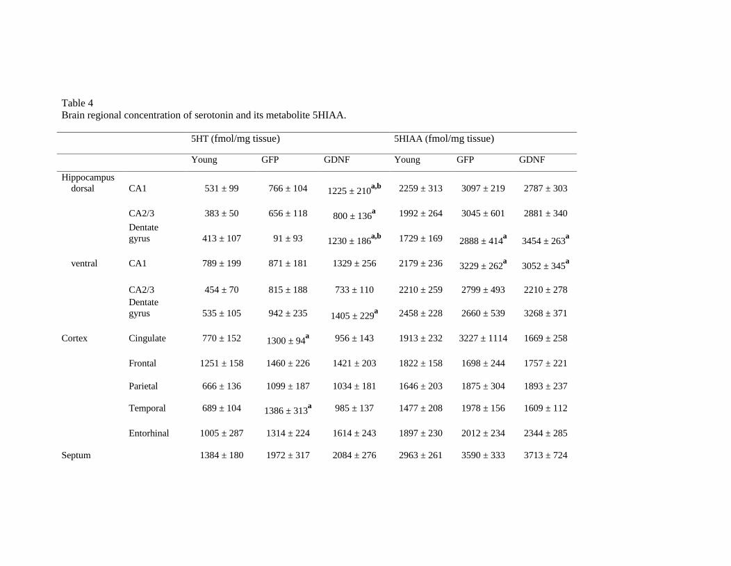
Table 4Brain regional concentration of serotonin and its metabolite 5HIAA.
5HT (fmol/mg tissue) 5HIAA (fmol/mg tissue)
Young GFP GDNF Young GFP GDNF
Hippocampusdorsal CA1 531 ± 99 766 ± 104 1225 ± 210
a,b 2259 ± 313 3097 ± 219 2787 ± 303
CA2/3 383 ± 50 656 ± 118 800 ± 136a 1992 ± 264 3045 ± 601 2881 ± 340
Dentategyrus 413 ± 107 91 ± 93 1230 ± 186
a,b 1729 ± 169 2888 ± 414a
3454 ± 263a
ventral CA1 789 ± 199 871 ± 181 1329 ± 256 2179 ± 236 3229 ± 262a
3052 ± 345a
CA2/3 454 ± 70 815 ± 188 733 ± 110 2210 ± 259 2799 ± 493 2210 ± 278Dentategyrus 535 ± 105 942 ± 235 1405 ± 229
a 2458 ± 228 2660 ± 539 3268 ± 371
Cortex Cingulate 770 ± 152 1300 ± 94a 956 ± 143 1913 ± 232 3227 ± 1114 1669 ± 258
Frontal 1251 ± 158 1460 ± 226 1421 ± 203 1822 ± 158 1698 ± 244 1757 ± 221
Parietal 666 ± 136 1099 ± 187 1034 ± 181 1646 ± 203 1875 ± 304 1893 ± 237
Temporal 689 ± 104 1386 ± 313a 985 ± 137 1477 ± 208 1978 ± 156 1609 ± 112
Entorhinal 1005 ± 287 1314 ± 224 1614 ± 243 1897 ± 230 2012 ± 234 2344 ± 285
Septum 1384 ± 180 1972 ± 317 2084 ± 276 2963 ± 261 3590 ± 333 3713 ± 724

Caudate 736 ± 99 1352 ± 215a 1131 ± 152 2038 ± 153 1991 ± 240 2354 ± 300
Experimental groups: Young, 8-month old rats; GFP, 24-month old cognitively impaired rats transduced with human green fluorescent protein in the dorsal
hippocampus CA1; GDNF, 24-month old cognitively impaired rats transduced with human glial cell line derived neurotrophic factor in the dorsal
hippocampus CA1. Values are the mean ± SEM of 6-9 animals. Statistics: (a) significance of GFP or GDNF groups as compared to young rats and (b)
significance of GDNF as compared to GFP group; ANOVA followed by Newman-Keules’s test at the significance level of p<0.05.


Related Documents