09/22/2009 Biochem: Carbohydrates II Carbohydrates II Andy Howard Introductory Biochemistry, Fall 2009 22 September 2009

09/22/2009 Biochem: Carbohydrates II Carbohydrates II Andy Howard Introductory Biochemistry, Fall 2009 22 September 2009.
Dec 13, 2015
Welcome message from author
This document is posted to help you gain knowledge. Please leave a comment to let me know what you think about it! Share it to your friends and learn new things together.
Transcript

09/22/2009Biochem: Carbohydrates II
Carbohydrates II
Andy HowardIntroductory Biochemistry,
Fall 2009 22 September 2009

09/22/2009
Biochem: Carbohydrates II p. 2 of 58
Sugars and polysaccharides
Sugars are vital as energy sources, and they also serve as building blocks for lipid-carbohydrate and protein-carbohydrate complexes
09/22/2009
Biochem: Carbohydrates II p. 3 of 58
Plans for Today Sugar Concepts
Monosaccharides Cyclization Reducing and nonreducing sugars
Sugar Derivatives
Oligosaccharides Glycosides
Polysaccharides Starch & glycogen
Cellulose and chitin
Glycoconjugates Proteoglycans Peptidoglycans Glycoproteins
09/22/2009
Biochem: Carbohydrates II p. 4 of 58
Sugar nomenclature All sugars with m ≤ 7 have specific names apart from their enantiomeric(L or D) designation,e.g. D-glucose, L-ribose.
The only 7-carbon sugar that routinely gets involved in metabolism is sedoheptulose, so we won’t try to articulate the names of the others
09/22/2009
Biochem: Carbohydrates II p. 5 of 58
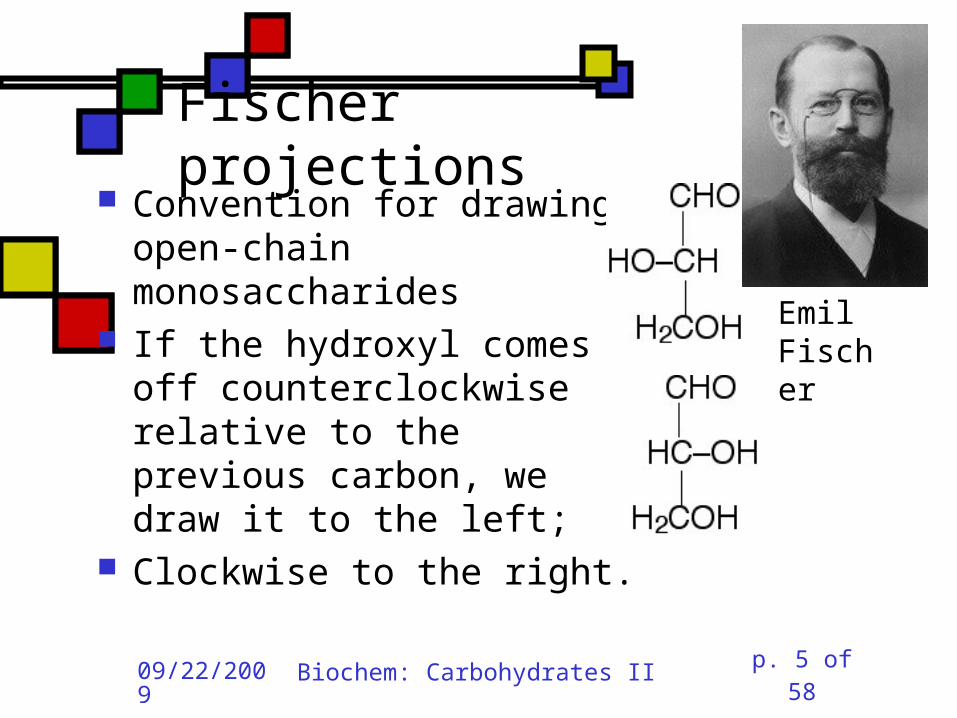
Fischer projections
Convention for drawing open-chain monosaccharides
If the hydroxyl comes off counterclockwise relative to the previous carbon, we draw it to the left;
Clockwise to the right.
Emil Fischer
09/22/2009
Biochem: Carbohydrates II p. 6 of 58
Cyclic sugars Sugars with at least four carbons can readily interconvert between the open-chain forms we have drawn and five-membered(furanose) or six-membered (pyranose) ring forms in which the carbonyl oxygen becomes part of the ring
There are no C=O bonds in the ring forms
09/22/2009
Biochem: Carbohydrates II p. 7 of 58
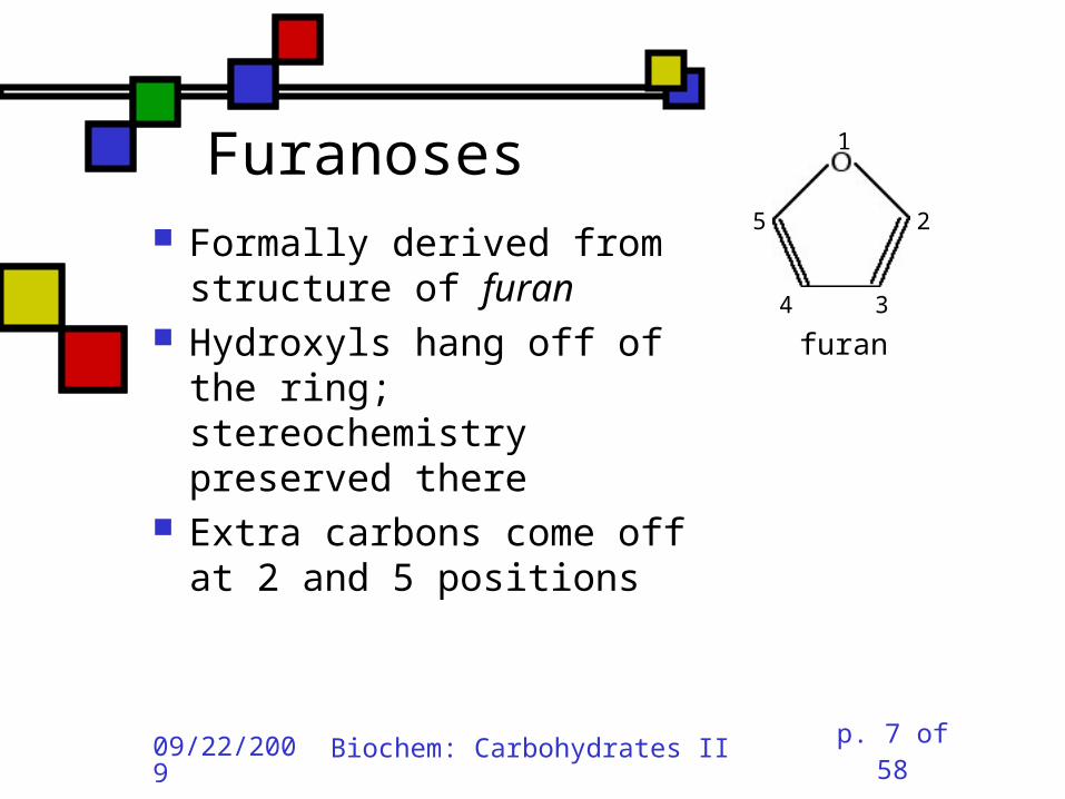
Furanoses Formally derived from structure of furan
Hydroxyls hang off of the ring; stereochemistry preserved there
Extra carbons come off at 2 and 5 positions
3
2
1
4
5
furan

09/22/2009
Biochem: Carbohydrates II p. 8 of 58
Pyranoses Formally derived from structure of pyran
Hydroxyls hang off of the ring; stereochemistry preserved there
Extra carbons come off at 2 and 6 positions
3
2
4
5
1
6
pyran

09/22/2009
Biochem: Carbohydrates II p. 9 of 58
How do we cyclize a sugar?
Formation of an internal hemiacetal or hemiketal (see a few slides from here) by conversion of the carbonyl oxygen to a ring oxygen
Not a net oxidation or reduction;in fact it’s a true isomerization.
The molecular formula for the cyclized form is the same as the open chain form
09/22/2009
Biochem: Carbohydrates II p. 10 of 58
Family tree of aldoses Simplest: D-, L- glyceraldehyde (C3) Add —CHOH: D,L-threose, erythrose (C4) Add —CHOH:D,L- lyxose, xylose, arabinose, ribose (C5)
Add —CHOH:D,L-talose, galactose, idose, gulose,mannose, glucose, altrose, allose (C6)
09/22/2009
Biochem: Carbohydrates II p. 11 of 58
Family tree of ketoses Simplest: dihydroxyacetone (C3) Add —CHOH: D,L-erythrulose (C4) Add —CHOH:D,L- ribulose, xylulose (C5)
Add —CHOH:D,L-sorbose, tagatose, fructose, psicose (C6)

09/22/2009
Biochem: Carbohydrates II p. 12 of 58
Haworth projections
…provide a way of keeping track the chiral centers in a cyclic sugar, as the Fischer projections enable for straight-chain sugars
Sir Walter Haworth

09/22/2009
Biochem: Carbohydrates II p. 13 of 58
The anomeric carbon In any cyclic sugar (monosaccharide, or single unit of an oligosaccharide, or polysaccharide) there is one carbon that has covalent bonds to two different oxygen atoms
We describe this carbon as the anomeric carbon
C
O
O
09/22/2009
Biochem: Carbohydrates II p. 14 of 58
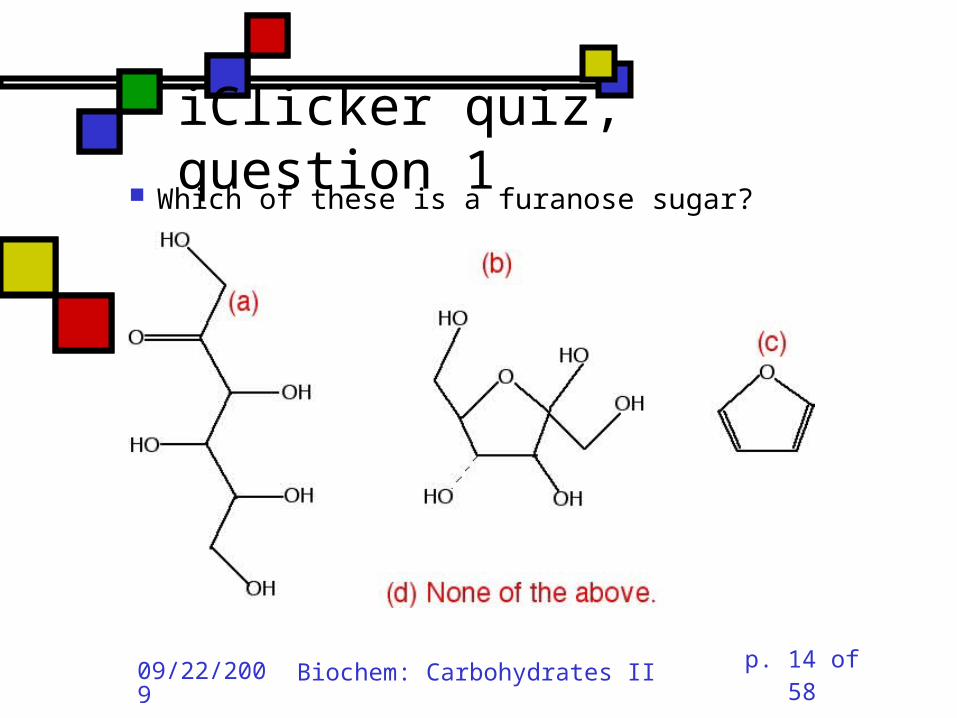
iClicker quiz, question 1
Which of these is a furanose sugar?

09/22/2009
Biochem: Carbohydrates II p. 15 of 58
iClicker quiz, question 2 Which carbon is the anomeric carbon in this sugar?
(a) 1 (b) 2 (c) 5 (d) 6 (e) none of these.

09/22/2009
Biochem: Carbohydrates II p. 16 of 58
iClicker, question 3 How many 7-carbon D-ketoses are there?
(a) none. (b) 4 (c) 8 (d) 16 (e) 32
09/22/2009
Biochem: Carbohydrates II p. 17 of 58
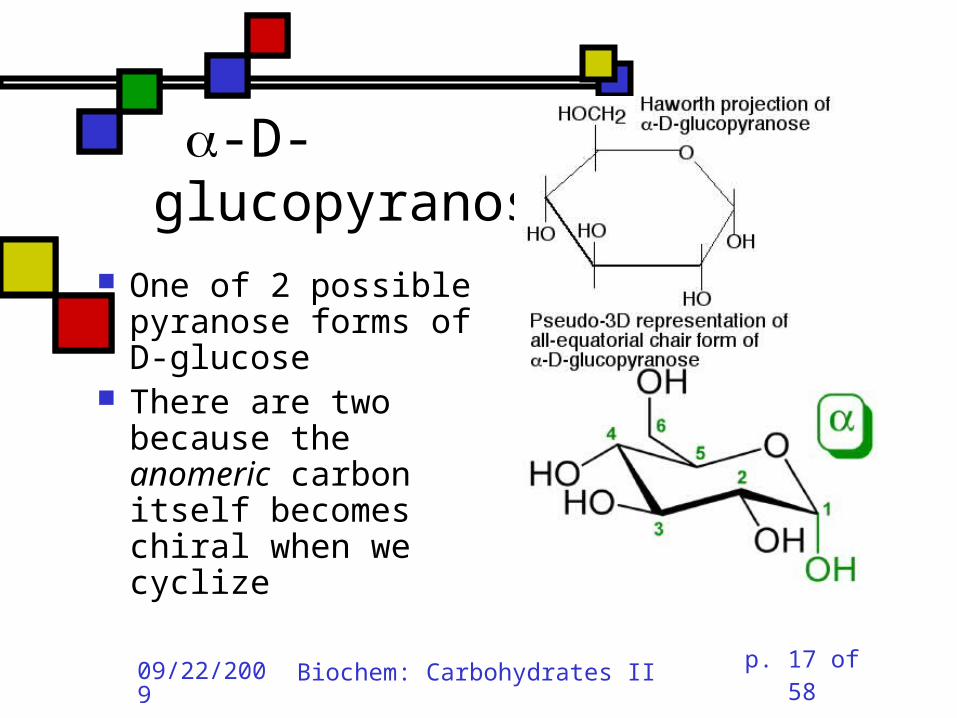
-D-glucopyranose
One of 2 possible pyranose forms of D-glucose
There are two because the anomeric carbon itself becomes chiral when we cyclize

09/22/2009
Biochem: Carbohydrates II p. 18 of 58
-D-glucopyranose
Differs from -D-gluco-pyranose only at anomeric carbon
09/22/2009
Biochem: Carbohydrates II p. 19 of 58
Count carefully!
It’s tempting to think that hexoses are pyranoses and pentoses are furanoses;
But that’s not always true The ring always contains an oxygen, so even a pentose can form a pyranose
In solution: pyranose, furanose, open-chain forms are all present
Percentages depend on the sugar
09/22/2009
Biochem: Carbohydrates II p. 20 of 58
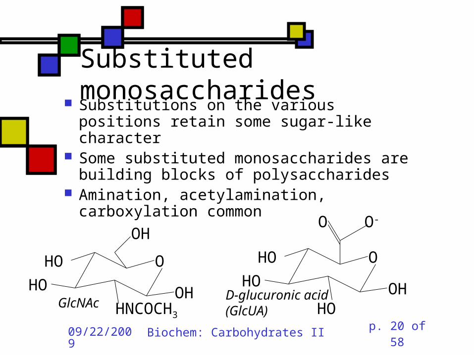
Substituted monosaccharides
Substitutions on the various positions retain some sugar-like character
Some substituted monosaccharides are building blocks of polysaccharides
Amination, acetylamination, carboxylation common
O
OH
HO
HO
HNCOCH3
OH
OHO
HO
HOOH
O O-
GlcNAcD-glucuronic acid(GlcUA)

09/22/2009
Biochem: Carbohydrates II p. 21 of 58
Sugar acids (fig. 7.10) Gluconic acid: glucose carboxylated @ 1 position In equilibrium with lactone form
Glucuronic acid:glucose carboxylated @ 6 position
Glucaric acid:glucose carboxylated @ 1 and 6 positions
Iduronic acid: idose carboxylated @ 6
D--gluconolactone
1
2
5
3
4
6

09/22/2009
Biochem: Carbohydrates II p. 22 of 58
Sugar alcohols (fig.7.11)
Mild reduction of sugars convert aldehyde moiety to alcohol
Generates an additional asymmetric center in ketoses except dihyroxyacetone
These remain in open-chain forms Smallest: glycerol Sorbitol, myo-inositol, ribitol are important

09/22/2009
Biochem: Carbohydrates II p. 23 of 58
Sugar esters (fig. 7.13) Phosphate esters of sugars are significant metabolic intermediates
5’ position on ribose is phosphorylated in nucleotides
Glucose 6-phosphate
09/22/2009
Biochem: Carbohydrates II p. 24 of 58
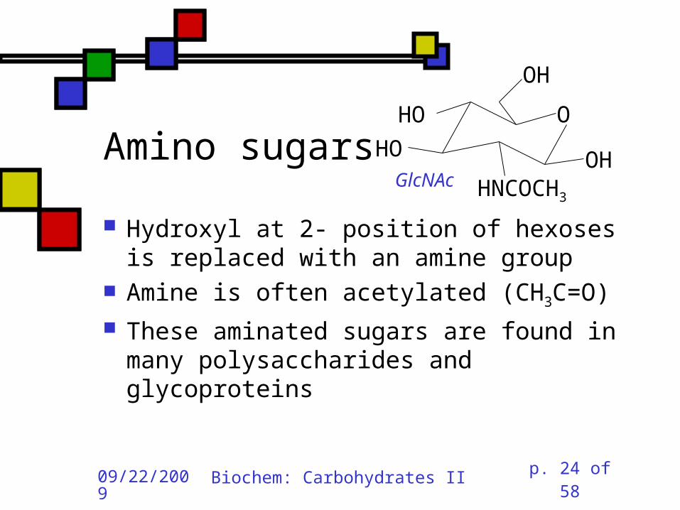
Amino sugars
Hydroxyl at 2- position of hexoses is replaced with an amine group
Amine is often acetylated (CH3C=O)
These aminated sugars are found in many polysaccharides and glycoproteins
O
OH
HO
HO
HNCOCH3
OHGlcNAc

09/22/2009
Biochem: Carbohydrates II p. 25 of 58
Hemiacetals and hemiketals
Hemiacetals and hemiketals are compounds that have an –OH and an –OR group on the same carbon
Cyclic monosaccharides are hemiacetals & hemiketals

09/22/2009
Biochem: Carbohydrates II p. 26 of 58
Acetals and ketals Acetals and ketals have two —OR groups on a single carbon
Acetals and ketals are found in glycosidic bonds

09/22/2009
Biochem: Carbohydrates II p. 27 of 58
Oligosaccharides and other glycosides A glycoside is any compound in which the hydroxyl group of the anomeric carbon is replaced via condensation with an alcohol, an amine, or a thiol
All oligosaccharides are glycosides, but so are a lot of monomeric sugar derivatives, like nucleosides
09/22/2009
Biochem: Carbohydrates II p. 28 of 58
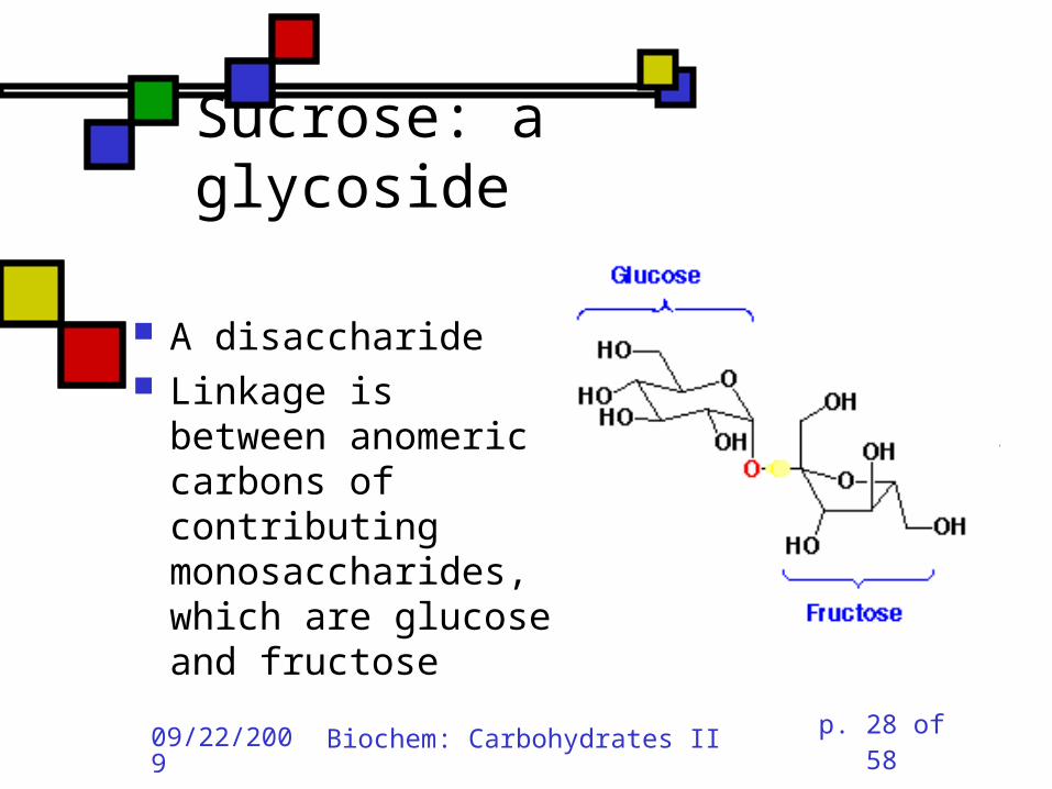
Sucrose: a glycoside
A disaccharide Linkage is between anomeric carbons of contributing monosaccharides, which are glucose and fructose

09/22/2009
Biochem: Carbohydrates II p. 29 of 58
Other disaccharides
Maltose glc-glc with -glycosidic bond from left-hand glc
Produced in brewing, malted milk, etc. Cellobiose
-glc-glc Breakdown product from cellulose
Lactose: -gal-glc Milk sugar Lactose intolerance caused by absence of enzyme capable of hydrolyzing this glycoside

09/22/2009
Biochem: Carbohydrates II p. 30 of 58
Reducing sugars
Sugars that can undergo ring-opening to form the open-chain aldehyde compounds that can be oxidized to carboxylic acids
We describe those as reducing sugars because they can reduce metal ions or amino acids in the presence of base
Benedict’s test:2Cu2+ + RCH=O + 5OH- Cu2O + RCOO- + 3H2O
Cuprous oxide is red and insoluble

09/22/2009
Biochem: Carbohydrates II p. 31 of 58
Ketoses are reducing sugars In presence of base a ketose can spontaneously rearrange to an aldose via an enediol intermediate, and then the aldose can be oxidized.
09/22/2009
Biochem: Carbohydrates II p. 32 of 58
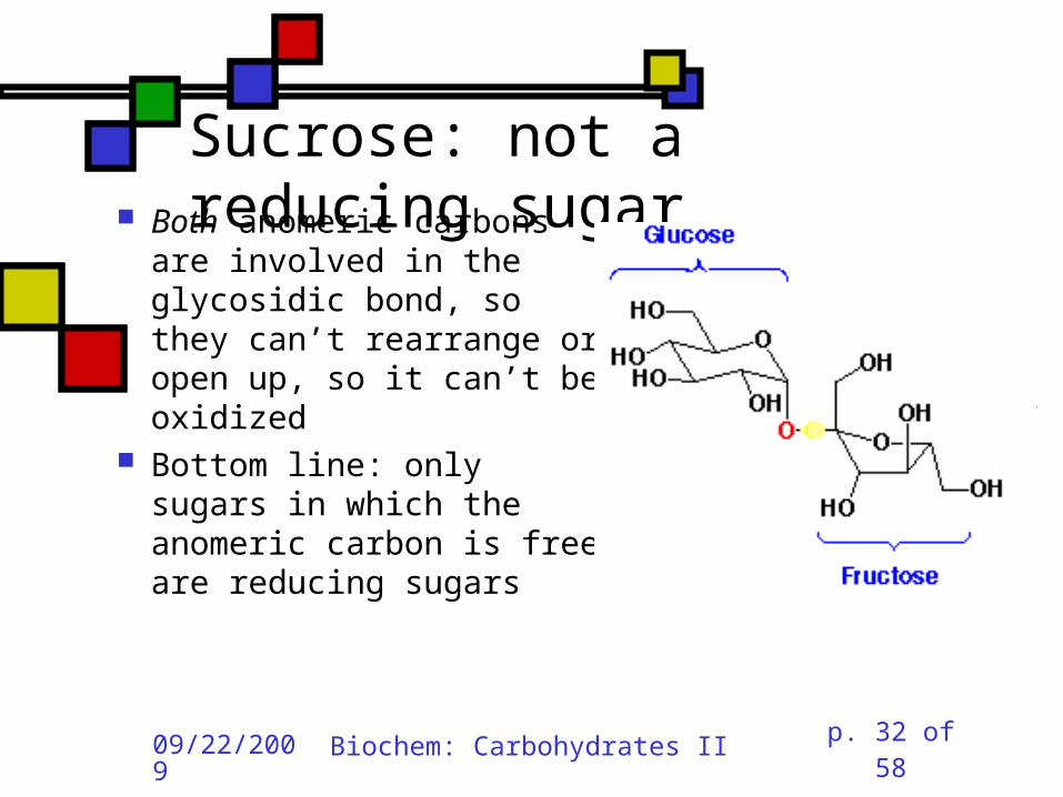
Sucrose: not a reducing sugar Both anomeric carbons
are involved in the glycosidic bond, so they can’t rearrange or open up, so it can’t be oxidized
Bottom line: only sugars in which the anomeric carbon is free are reducing sugars

09/22/2009
Biochem: Carbohydrates II p. 33 of 58
Reducing & nonreducing ends Typically, oligo and polysaccharides have a reducing end and a nonreducing end
Non-reducing end is the sugar moiety whose anomeric carbon is involved in the glycosidic bond
Reducing end is sugar whose anomeric carbon is free to open up and oxidize
Enzymatic lengthening and degradation of polysaccharides occurs at nonreducing end or ends
09/22/2009
Biochem: Carbohydrates II p. 34 of 58
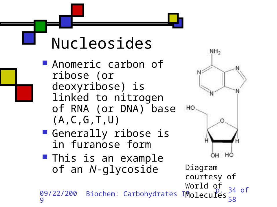
Nucleosides Anomeric carbon of ribose (or deoxyribose) is linked to nitrogen of RNA (or DNA) base (A,C,G,T,U)
Generally ribose is in furanose form
This is an example of an N-glycoside
Diagram courtesy of World of Molecules

09/22/2009
Biochem: Carbohydrates II p. 35 of 58
Polysaccharides
Homoglycans: all building blocks same
Heteroglycans: more than one kind of building block
No equivalent of genetic code for carbohydrates, so long ones will be heterogeneous in length and branching, and maybe even in monomer identity

09/22/2009
Biochem: Carbohydrates II p. 36 of 58
Categories of polysaccharides Storage homoglycans (all Glc)
Starch: amylose ((14)Glc) , amylopectin
Glycogen Structural homoglycans
Cellulose ((14)Glc) Chitin ((14)GlcNAc)
Heteroglycans Glycosaminoglycans (disacch.units) Hyaluronic acid (GlcUA,GlcNAc)((1 3,4))
09/22/2009
Biochem: Carbohydrates II p. 37 of 58
Storage polysaccharides
Available sources of glucose for energy and carbon
Long-chain polymers of glucose Starch (amylose and amylopectin):in plants, it’s stored in 3-100 µm granules
Glycogen Branches found in all but amylose

09/22/2009
Biochem: Carbohydrates II p. 38 of 58
Amylose Unbranched, -14 linkages Typically 100-1000 residues Not soluble but can form hydrated micelles and may be helical
Amylases hydrolyze -14 linkages
Diagram courtesyLangara College

09/22/2009
Biochem: Carbohydrates II p. 39 of 58
Amylopectin Mostly -14 linkages; 4% -16
Each sidechain has 15-25 glucose moieties
-16 linkages broken down by debranching enzymes
300-6000 total glucose units per amylopectin molecule
One reducing end, many nonreducing ends
09/22/2009
Biochem: Carbohydrates II p. 40 of 58
Glycogen Principal storage form of glucose in human liver; some in muscle
Branched (-14 + a few -16) More branches (~10%) Larger than starch: 50000 glucose One reducing end, many nonreducing ends
Broken down to G-1-P units Built up fromG-6-P G-1-P UDP-Glucose units

09/22/2009
Biochem: Carbohydrates II p. 41 of 58
Glycogen structure

09/22/2009
Biochem: Carbohydrates II p. 42 of 58
Structural polysaccharides I
Insoluble compounds designed to provide strength and rigidity
Cellulose: glucose -14 linkages Rigid, flat structure: each glucose is upside down relative to its nearest neighbors (fig.7.27)
300-15000 glucose units Found in plant cell walls Resistant to most glucosidases Cellulases found in termites,ruminant gut bacteria
Chitin: GlcNAc -14 linkages:exoskeletons, cell walls (fig. 7.26)

09/22/2009
Biochem: Carbohydrates II p. 43 of 58
Structural polysaccharides II
Alginates: poly(-D-mannuronate),poly(-L-guluronate), linked 14 Cellulose-like structure when free Complexed to metal ions:3-fold helix (“egg-carton”)
Agarose: alternating D-gal, 3,6-anhydro-L-gal, with 6-methyl-D-gal side chains Forms gels that hold huge amounts of H2O Can be processed to use in the lab for gel exclusion chromatography
Glycosaminoglycans: see next section
09/22/2009
Biochem: Carbohydrates II p. 44 of 58
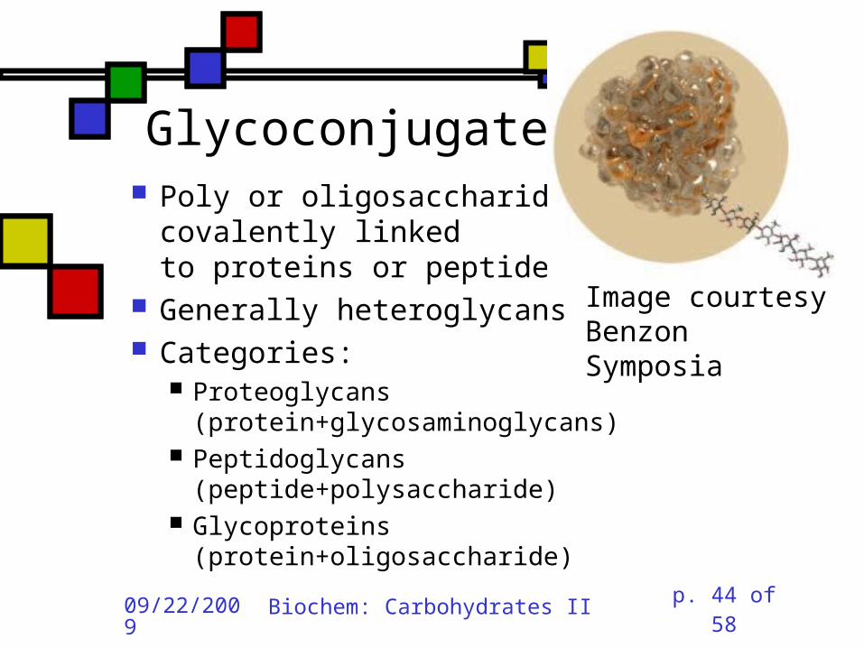
Glycoconjugates Poly or oligosaccharidescovalently linkedto proteins or peptides
Generally heteroglycans Categories:
Proteoglycans (protein+glycosaminoglycans)
Peptidoglycans (peptide+polysaccharide)
Glycoproteins (protein+oligosaccharide)
Image courtesy Benzon Symposia
09/22/2009
Biochem: Carbohydrates II p. 45 of 58
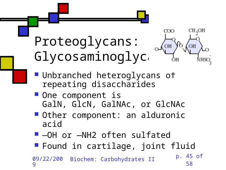
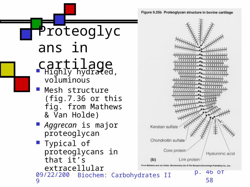
Proteoglycans: Glycosaminoglycans Unbranched heteroglycans of repeating disaccharides
One component isGalN, GlcN, GalNAc, or GlcNAc
Other component: an alduronic acid
—OH or —NH2 often sulfated Found in cartilage, joint fluid
09/22/2009
Biochem: Carbohydrates II p. 46 of 58
Proteoglycans in cartilage Highly hydrated, voluminous
Mesh structure (fig.7.36 or this fig. from Mathews & Van Holde)
Aggrecan is major proteoglycan
Typical of proteoglycans in that it’s extracellular

09/22/2009
Biochem: Carbohydrates II p. 47 of 58
Peptidoglycans(G&G fig. 7.29) Polysaccharides linked to small
proteins Featured in bacterial cell walls:alternating GlcNAc + MurNAclinked with -(14) linkages
Lysozyme hydrolyzes these polysaccharides
Peptide is species-specific:often contains D-amino acids

09/22/2009
Biochem: Carbohydrates II p. 48 of 58
Peptidoglycans in bacteria
Gram-negative: thin peptidoglycan layer separates two phospholipid bilayer membranes
Gram-positive: only one bilayer, with thicker peptidoglycan cell wall outside it
Gram stain binds to thick wall, not thin layer
Fig. 7.30 shows multidimensionality of these walls
09/22/2009
Biochem: Carbohydrates II p. 49 of 58
Peptide component(G&G fig. 7.29)
Sugars are crosslinked with entities containing(L-ala)-(isoglutamate)-(L-Lys)-(D-ala)
Gram-neg: L-Lys crosslinks via D-ala
Gram-pos: L-lys crosslinks via pentaglycine followed by D-ala

09/22/2009
Biochem: Carbohydrates II p. 50 of 58
Gram-negative bacteria:the periplasmic space(G&G fig. 7.30b, 7.31)
Periplasmic space: space inside cell membrane but inside just-described peptidoglycan layer (note error in fig. legend!)
Peptidoglycan is attached to outer membrane via 57-residue hydrophobic proteins
Outer membrane has a set of lipopolysaccharides attached to it; these sway outward from the membrane
09/22/2009
Biochem: Carbohydrates II p. 51 of 58
Gram-negative membranes and periplasmic space
QuickTime™ and aTIFF (Uncompressed) decompressor
are needed to see this picture.
Figure courtesy Kenyon College microbiology Wiki
09/22/2009
Biochem: Carbohydrates II p. 52 of 58
Glycoproteins 1-30 carbohydrate moieties per protein
Proteins can be enzymes, hormones, structural proteins, transport proteins
Microheterogeneity:same protein, different sugar combinations
Eight sugars common in eukaryotes PTM glycosylation much more common in eukaryotes than prokaryotes

09/22/2009
Biochem: Carbohydrates II p. 53 of 58
Diversity in glycoproteins Variety of sugar monomers or glycosidic linkages Linkages always at C-1 on one sugar but can be C-2,3,4,6 on the other one
Up to 4 branches But:not all the specific glycosyltransferases you would need to get all this diversity exist in any one organism
09/22/2009
Biochem: Carbohydrates II p. 54 of 58
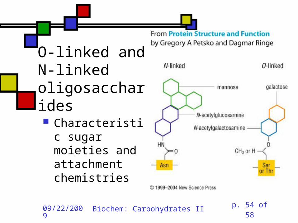
O-linked and N-linked oligosaccharides Characteristic sugar moieties and attachment chemistries
09/22/2009
Biochem: Carbohydrates II p. 55 of 58
O-linked oligosaccharides(fig. fig 7.32a, 7.33 in G&G) GalNAc to ser or thr;often with Gal or Sialic acid on GalNAc
5-hydroxylysines on collagen are joined to D-Gal
Some proteoglycans joined viaGal-Gal-Xyl-ser
Single GlcNAc on ser or thr
09/22/2009
Biochem: Carbohydrates II p. 56 of 58
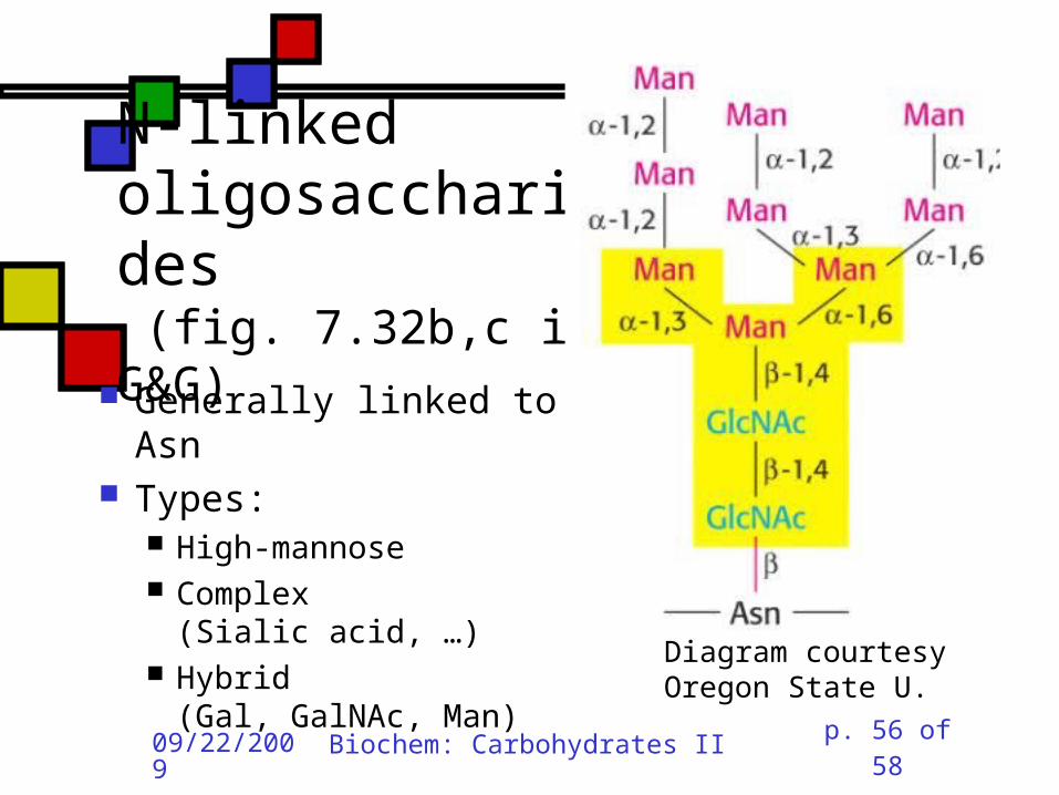
N-linked oligosaccharides (fig. 7.32b,c in G&G) Generally linked to Asn
Types: High-mannose Complex(Sialic acid, …)
Hybrid(Gal, GalNAc, Man)
Diagram courtesy Oregon State U.

09/22/2009
Biochem: Carbohydrates II p. 57 of 58
iClicker question 4
Suppose you isolate a polysaccharide with 5000 glucose units, and 3% of the linkages are 1,6 crosslinks. This is:
(a) amylose (b) amylopectin (c) glycogen (d) chitin (e) none of the above.

09/22/2009
Biochem: Carbohydrates II p. 58 of 58
iClicker question 5 Suppose you isolate an enzyme that breaks down -1,4-glycosidic linkages between GlcNAc units. This would act upon:
(a) glycogen (b) cellulose (c) chitin (d) all of the above (e) none of the above.
Related Documents


![Biochem [Gluconeogenesis]](https://static.cupdf.com/doc/110x72/577c82b31a28abe054b1e4af/biochem-gluconeogenesis.jpg)