~ 0 ~
Thiocaffeine derivatives as inhibitors of monoamine oxidase
Hermanus Perold Booysen
B. Pharm
Dissertation submitted in partial fulfillment of the requirements for the degree
Magister Scientiae in Pharmaceutical Chemistry at the North-West University,
Potchefstroom Campus
Supervisor: Prof. J.P. Petzer
Co-supervisor: Prof. J.J. Bergh
Potchefstroom
2011

~ 1 ~
Abstract
Parkinson’s disease (PD) is a neurodegenerative disorder which is characterized by selective
loss of dopaminergic neurons in the substantia nigra pars compacta (SNpc) of the brain and
reduced striatal dopamine (DA). Neuropathologically, PD is characterized by the presence of
intraneuronal inclusions called Lewy Bodies (LBs). While the pathogenesis of PD is unknown, it
is thought that monoamine oxidase (MAO) may play an important role in the neurodegenerative
process. In the basal ganglia DA is oxidized by MAO, a process which is associated with the
formation of toxic metabolic by-products. For each mole of DA oxidized by MAO, one mole of
hydrogen peroxide and dopaldehyde are formed. Both these products are potentially neurotoxic
if not quickly cleared. Inhibitors of MAO reduce the MAO-catalyzed metabolism of DA and as a
result, reduce the formation of these toxic by-products. MAO inhibitors are therefore considered
useful as a treatment strategy to slow the progression of PD since they may exert a
neuroprotective effect in the brain. Since MAO is the principal enzyme for the catabolism of DA
in the brain, inhibitors of MAO may conserve the dopamine supply in the brain and therefore
exert a symptomatic benefit in PD. MAO inhibitors are frequently combined with L-dopa, the
metabolic precursor of DA, in the therapy of PD. MAO inhibitors have been shown to enhance
the levels of DA derived from L-dopa, and therefore enhance the therapeutic efficacy of L-dopa.
MAO exists as two isoforms, MAO-A and MAO-B. These enzymes are products of distinct
genes and exhibit differing substrate and inhibitor specificities. Both isoforms are present in the
brain and utilize DA as substrate. In the brain, the MAO-B isoform exhibits higher activity and
density than MAO-A and is therefore considered to play a more important role in DA metabolism
than MAO-A. Also MAO-B activity in the brain increases with age while MAO-A activity remains
unchanged. In the aged PD brain MAO-B is therefore thought to be the main MAO isozyme
responsible for DA catabolism and inhibitors of this enzyme are considered to be useful in the
treatment of PD. As mentioned above, MAO-B inhibitors may conserve dopamine in the PD
brain and offer a symptomatic effect. MAO-B inhibitors may also protect against further
degeneration by reducing potential toxic by-products associated with the oxidative metabolism
of DA.

~ 2 ~
While irreversible inhibitors of MAO-B have been used clinically in the treatment of PD,
irreversible inhibition may be associated with certain disadvantages. For example, after
terminating treatment with an irreversible MAO inhibitor, recovery of enzyme activity may
require several weeks, since the turnover rate for the biosynthesis of MAO in the human brain
may be as much as 40 days. In contrast, for reversible inhibitors, following withdrawal of the
drug, enzyme activity is recovered quickly upon elimination of the drug from the tissues. This
study focuses on the design of new MAO inhibitors that are selective for the MAO-B isoform and
which act reversibly with the enzyme.
In this study caffeine served as lead compound for the design of new MAO inhibitors. Although
caffeine is a weak MAO-B inhibitor, substitution at the C-8 position with a variety of substituents
has been shown to enhance the MAO-B inhibition potency of caffeine to a large degree. In a
previous study it was shown that substitution at C-8 of caffeine with alkyloxy substituents
yielded particularly potent MAO-B inhibitors with IC50 values in the nM range. Based on these
promising results, the present study will investigate the possibility that alkylthio substituents at
C-8 of caffeine may similarly enhance the MAO-B inhibition potency of caffeine. For this
purpose, a series of twelve aryl- and alkylthiocaffeine analogues (4a-l) were synthesized and
evaluated as potential inhibitors of recombinant human MAO-A and –B. This study was
therefore an exploratory study to discover new caffeine derived MAO inhibitors.
Chemistry: The C-8-substituted alkyl- and arylthiocaffeine analogues (4a-l) were synthesized by
reacting 8-chlorocaffeine with the appropriate alkyl- and arylthiol derivatives in the presence of a
base. The structures and purities of the target inhibitors were verified by NMR, MS and HPLC
analysis.
MAO inhibition studies: Among the thiocaffeine inhibitors, 8-[4-bromobenzene-
methanethiol]caffeine (4e) was the most potent MAO-B inhibitor, with an IC50 value of 0.16 µM.
This inhibitor also exhibited a high degree of selectivity towards MAO-B. The results indicated
that extending the length of the C-8 chain of the 8-thiocaffeine analogues yielded MAO-B
inhibitors with enhanced inhibition potency. It was also shown that substitution on the phenyl
ring of the C-8 substituent with halogens (Cl, Br and F) enhances the MAO-B inhibition
potencies. Another potent MAO-B inhibitor was a phenoxyethyl substituted homologue, 8-(2-
phenoxyethanethiol)caffeine (4h), with an IC50 value of 0.332 µM.
~ 3 ~
Time-dependency and mode of inhibition: This study demonstrates that one selected inhibitor,
compound 4e, does not reduce the catalytic rates of MAO-A and –B in a time dependent
manner. This result shows that the inhibition of MAO-A and –B is reversible. For the inhibition
of MAO-A and –B by compound 4e, sets of Lineweaver–Burke plots were constructed. The
results showed that the Lineweaver-Burke plots intersected on the y-axis which indicates that
this inhibitor is a competitive inhibitor of both MAO-A and –B and is further proof of the
reversible interaction of 4e with the MAO enzymes.
Future recommendations: Based on the promising MAO-B inhibition potencies of some of the
thiocaffeine derivatives, this study recommends that further studies be carried out to optimize
the MAO inhibition activities of these compounds. This study specifically recommends that
phenylethyl and phenoxyethyl substituted thiocaffeine derivatives, which contain halogens on
the phenyl ring, be synthesized and evaluated as MAO inhibitors. Such structures may be
particularly potent MAO-B inhibitors.
Conclusions: From the results of this study it may be concluded that thiocaffeine derivatives are
promising inhibitors of MAO-A and –B. These compounds are competitive and reversible
inhibitors of MAO.
~ 4 ~
Opsomming
Parkinson se siekte is ʼn neurodegeneratiewe siekte wat gekenmerk word deur die verlies van
dopaminergiese neurone in die substantia nigra pars compacta van die brein, met die gevolglike
verlies van dopamien (DA) in die striatum. Parkinson se siekte word gekarakteriseer deur
intraneuronale komplekse naamlik “Lewy liggame” (LBs). Alhoewel die patogenese steeds
onbekend is, speel die ensiem, monoamienoksidase (MAO) moontlik 'n rol in die
neurodegeneratiewe proses. In die basale ganglia van die brein word DA geoksideer deur
MAO. Hierdie proses word geassosieer met die vorming van toksiese metaboliese
neweprodukte. Vir elke mol DA wat deur MAO geoksideer word, word daar een mol
waterstofperoksied en dopaldehied gevorm. Albei hierdie neweprodukte is neurotoksies indien
dit nie opgeruim word nie. MAO-inhibeerders verlaag die katalitiese afbraak van DA asook die
vorming van hierdie neurotoksiese produkte. Om hierdie rede word MAO-inhibeerders gebruik
om die verloop van die siekte te vertraag. Hierdie inhibeerders besit ook 'n moontlike
neurobeskermde rol in die brein. MAO is hoofsaaklik verantwoordelik vir die afbraak van DA in
die brein en daarom kan MAO-inhibeerders die konsentrasie DA in die brein verhoog. Dié
verbindings kan dus as simptomatiese behandeling vir Parkinson se siekte aangewend word.
MAO-inhibeerders word in kombinasie met L-dopa aan pasiënte toegedien. L-dopa is 'n
metaboliese voorloper van DA en word meestal gebruik vir die behandeling van Parkinson se
siekte. Daar is bewys dat MAO-inhibeerders DA konsentrasies in die brein kan verhoog. Om
dié rede kan MAO inhibeerders dus die terapeutiese effek van L-dopa verbeter.
MAO kom voor as twee verskillende ensieme, MAO-A en MAO-B. Hierdie ensieme is produkte
van verskillende gene en het verskillende substraat- en inhibeerderselektiwiteite. Beide
ensieme kom in die brein voor en gebruik DA as substraat. Die MAO-B ensiem vertoon hoër
aktiwiteit en digtheid in die brein as die MAO-A ensiem. MAO-B speel dus 'n groter rol in die
metabolisme van DA in die brein as MAO-A. Die MAO-B aktiwiteit verhoog ook met ouderdom
in vergelyking met MAO-A aktiwiteit wat dieselfde bly. MAO-B is dus ’n belangrike ensiem vir
die afbraak van DA in bejaarde pasiënte, en MAO-B-inhibeerders word gevolglik gebruik vir die
behandeling van Parkinson se siekte. Soos reeds genoem, verhoog MAO-B-inhibeerders DA
~ 5 ~
konsentrasies in die brein en bied sodoende simptomatiese verligting. Inhibeerders van hierdie
ensiem kan ook verdere degenerasie verhoed deur die verlaging van die vorming van toksiese
neweprodukte.
Alhoewel onomkeerbare MAO-B-inhibeerders vir die behandeling van Parkinson se siekte
gebruik word, hou onomkeerbare inhibeerders sekere nadele in. Dit neem ongeveer 40 dae na
behandeling met onomkeerbare inhibeerders, vir MAO-ensiemaktiwiteit om weer na normaal te
herstel. Na behandeling met omkeerbare MAO-inhibeerders herstel ensiemaktiwiteit binne ure
nadat die inhibeerder uit die weefsel opgeruim is. Hierdie studie fokus dus op die ontwikkeling
van selektiewe omkeerbare MAO-B-inhibeerders.
In hierdie studie dien kafeïen as leidraadverbinding. Alhoewel kafeïen 'n swak MAO-B-
inhibeerder is, lei substitusie op die C-8 posisie van die kafeïenring tot verhoogde MAO-B-
inhiberingspotensie van kafeïen. 'n Vorige studie het getoon dat substitusie met
alkieloksiesubstituente op C-8 van kafeïen, verbindings lewer wat potente MAO-B-inhibeerders
is met IC50 waardes in die nM-gebied. Op grond van hierdie resultate word daar in die huidige
studie die moontlikheid ondersoek dat alkieltiosubstituente op C-8 van kafeïen ook kan lei tot ‘n
verhoging van die MAO-B-inhibisiepotensie van kafeïen. Vir hierdie doel is 'n reeks van twaalf
ariel- en alkieltiokafeïenanaloë (4a-l) gesintetiseer en geëvalueer as moontlike inhibeerders van
rekombinante menslike MAO-A en -B. Hierdie studie is 'n verkennende studie met die doel om
nuwe kafeïen-afgeleide MAO-remmers te ontdek.
Chemie: Die alkiel- en arieltiokafeïen analoë (4a-l) is gesintetiseer deur 8-chlorokafeïen met die
toepaslike alkiel- en arieltiolderivate in die teenwoordigheid van 'n basis te reageer. Die
strukture en suiwerhede van die teikeninhibeerders is deur KMR, MS en HPLC analise
geverifieer.
~ 6 ~
MAO-inhibisiestudies: Van al die tiokafeïeninhibeerders, is 8-[4-bromobenseen-
metaantiol]kafeïen (4e) die potentste met 'n IC50-waarde van 0.16 μM. Hierdie inhibeerder besit
ook 'n hoë mate van selektiwiteit vir MAO-B. Die resultate dui aan dat die verlenging van die C-
8 syketting van die 8-tiokafeïenanaloog lei tot verbeterde MAO-B-inhibisie. Substitusie met
halogene (Cl, Br en F), op die fenielring van die C-8 substituent verhoog ook die MAO-B-
inhibisiepotensie. Nog 'n potente MAO-B-inhibeerder is die fenoksietielanaloog, 8-(2-
fenoksietaan-tiol)kafeïen (4h), met 'n IC50 waarde van 0.332 μM.
Tydsafhanklikheid en meganisme van inhibisie: Hierdie studie toon dat een geselekteerde
inhibeerder (4e), nie die katalitiese tempo van MAO-A en -B op 'n tydsafhanklike wyse verlaag
nie. Hierdie resultaat toon dus dat die inhibisie van MAO-A en B omkeerbaar is. Vir verbinding
4e is stelle Lineweaver-Burke-grafieke opgestel vir die inhibisie van MAO-A en -B. Die resultate
toon dat die Lineweaver-Burke-grafieke op een punt op die y-as sny wat daarop dui dat hierdie
inhibeerder 'n kompeterende inhibeerder van MAO-A en -B is. Hierdie resultaat is 'n verdere
bewys dat 4e omkeerbare interaksies met MAO ondergaan.
Aanbevelings: Op grond van die belowende MAO-B-inhibisiepotensies van sommige van die
tiokafeïenanaloë, beveel hierdie studie aan dat verdere studies uitgevoer word om hierdie
verbindings se MAO-inhibisieaktiwiteite te optimaliseer. Hierdie studie beveel spesifiek aan dat
fenieletiel-en fenoksietielgesubstitueerde tiokafeïenanaloë, wat halogene op die fenielring bevat,
gesintetiseer en geëvalueer word as MAO-inhibeerders. Sulke strukture kan moontlik potente
MAO-B-inhibeerders wees.
Gevolgtrekkings: Uit hierdie studie kan afgelei word dat tiokafeïenanaloë belowende MAO-A en
MAO-B inhibeerders is. Hierdie analoë is ook kompeterende en omkeerbare inhibeerders van
MAO.
~ 7 ~
Table of contents
Abstract ……………………………………………………………………………………………..……1
Opsomming ………………………………………………………………………………………..…….4
Table of contents………………………………………………………………………………….…….7
Abbreviations…………………………………………………………………………………….…….11
Chapter 1 - Introduction..........................................................................................................14
1.1 Parkinson’s disease ..................................................................................................14
1.2 Monoamine oxidase ..................................................................................................16
1.3 Rationale of this study ...............................................................................................17
1.4 Objectives of this study .............................................................................................21
Chapter 2 - Literature study ...................................................................................................22
2.1 Parkinson’s disease ..................................................................................................22
2.1.1 General background ..........................................................................................22
2.1.2 Symptomatic treatment ......................................................................................26
2.1.3 Drugs for neuroprotection ..................................................................................31
2.1.4 Mechanisms of neurodegeneration ....................................................................37
2.2 The monoamine oxidases .........................................................................................42
2.2.1 General background and tissue distribution of MAO ..........................................42
2.2.2 Biological function of MAO-B .............................................................................44

~ 8 ~
2.2.3 Biological function of MAO-A .............................................................................45
2.2.4 The role of MAO-B in PD ...................................................................................48
2.2.5 The potential role of MAO-A in PD .....................................................................50
2.2.6 Irreversible inhibitors of MAO-B .........................................................................50
2.2.7 Reversible inhibitors of MAO-B ..........................................................................52
2.2.8 Inhibitors of MAO-A............................................................................................53
2.2.9 Mechanism of action of MAO-B..........................................................................55
2.2.10 Three-dimensional structure of MAO-B ..............................................................60
2.2.11 Three-dimensional structure of MAO-A ..............................................................65
2.2.12 Animal models of PD .........................................................................................67
2.2.14 Rotenone ...........................................................................................................71
2.2.15 Paraquat ............................................................................................................72
2.2.16 Copper-containing amine oxidases ....................................................................73
2.2.17 Enzyme kinetics .................................................................................................76
2.2.18 Conclusion .........................................................................................................79
Chapter 3 - Synthesis .............................................................................................................80
3.1 Introduction ...............................................................................................................80
3.2 General synthetic approach for the synthesis of 8-thiocaffeine analogues (4a–l) and
8-chlorocaffeine. .......................................................................................................81
3.3 Detailed synthetic methods for the synthesis of 8-thiocaffeine analogues (4a–l) and 8-
chlorocaffeine. ...........................................................................................................82

~ 9 ~
3.4 Chemicals and instrumentation .................................................................................84
3.5 Physical characterization...........................................................................................85
3.6 Results ......................................................................................................................85
3.6.1 The physical data for the 8-thiocaffeine derivatives .............................................85
3.6.2 Interpretation of the NMR spectra .......................................................................88
3.6.3 Interpretation of the mass spectra .......................................................................91
3.7 Conclusion ................................................................................................................92
Chapter 4 - Enzymology .........................................................................................................93
4.1 Introduction ...............................................................................................................93
4.2 Chemicals and instrumentation .................................................................................94
4.3 Biological evaluation to determine the IC50 values .....................................................94
4.3.1 Introduction .........................................................................................................94
4.3.2 Method................................................................................................................94
4.3.3 Results – Sigmoidal curves obtained for the IC50 determinations ........................96
4.3.4 Results – Table with IC50 values .........................................................................97
4.3.5 Comparison of the MAO inhibition properties of the 8-thiocaffeines with those of
the 8-benzyloxycaffeines. ................................................................................. 102
4.4 Time-dependent studies .......................................................................................... 105
4.4.1 Introduction ....................................................................................................... 105
4.4.2 Method.............................................................................................................. 106
~ 10 ~
4.4.3 Results.............................................................................................................. 108
4.5 Mode of inhibition - Construction of Lineweaver-Burk plots .................................... 108
4.5.1 Introduction ....................................................................................................... 108
4.5.2 Method.............................................................................................................. 109
4.5.3 Results – Lineweaver-Burk plost ....................................................................... 111
4.6. Molecular modelling ................................................................................................ 111
4.6.1 Background ...................................................................................................... 111
4.6.2 Method.............................................................................................................. 112
4.6.3 Results and discussion ..................................................................................... 112
4.7 Conclusion .............................................................................................................. 113
Chapter 5 - Summary ............................................................................................................ 115
Bibliography .......................................................................................................................... 120
Addendum……………………………………………………………………………………………..136
• NMR spectra………………………………………………………………………………….137
• HPLC chromatograms………………………………………………………………………149
• Mass spectra……………………………………………………………………………...….155
• Concept article…………………………………………………………………………..…..161
Acknowledgements…………………………………………………………………………….…..201
~ 11 ~
Abbreviations
5-HT - Serotonin
6-OHDA - 6-Hydroxydopamine
AD - Alzheimer’s disease
ADH - Aldehyde dehydrogenase
AOs - Amine oxidases
ATP - Adenosine-5'-triphosphate
BDNF - Brain-derived neurotrophic factor
CNS - Central nervous system
COMT - Catechol-O-methyl-transferase
COX - Cyclooxygenase
CSC - (E)-8-(3-Chlrorostyryl)caffeine
DA - Dopamine
DDC - DOPA decarboxylase
DMDPO - Dimethyldecylphosphine oxide
FAD - Flavine adenine dinucleotide
GAPDH - Glyceraldehyde-3-phosphate dehydrogenase
GDNF - Glial-derived neurotrophic factor
GPO - Glutathione peroxidase
GSH - Glutathione
~ 12 ~
HNE - 4-Hydroxy-2-nonenal
JNK - c-Jun N-terminal
LBs - Lewy Bodies
LDL - Low-density lipoprotein
LOX - Lipoxygenase
MAO-A - Monoamine oxidase A
MAO-B - Monoamine oxidase B
MPDP+ - 1-Methyl-4-phenyl-2,3-dihydropyridium
MPP+ - 1-Methyl-4-phenylpyridinium
MPPP - 1-Methyl-4-phenyl-4-propionpiperidine
MPTP - 1-Methyl-4-phenyl-1,2,3,6-tetrahydropyridine
NA - Nor-adrenaline
NET - Norepinephrine transporters
NGF - Nerve growth factor
NSAID - Nonsteroidal anti-inflammatory drug
PD - Parkison’s disease
PGE2 - Prostaglandin E2
PNS - Peripheral nervous system
ROS - Reactive oxygen species
SET - Single electron transfer
SNpc - Substantia nigra pars compacta
~ 13 ~
SSAO - Semicarbazide-sensitive amine oxidase
TH - Tyrosine hydroxylase
TNFα - Tumor necrosis factor-α
TPQ - Topa-quinone
UCH-L1 - Ubiquitin carboxyl-terminal hydrolase L1
~ 14 ~
Chapter 1 Introduction
1.1 Parkinson’s disease
In the early 1800s James Parkinson discovered an unrecognized disorder by studying six
patients. Jean Martin Charcot, the father of neurology, proposed that the syndrome should be
called maladie de Parkinson (Parkinson’s disease) (Lees et al., 2009). Parkinson’s disease
(PD) is a sporadic, neurodegenerative disorder characterized by selective loss of dopaminergic
neurons in the substantia nigra pars compacta (SNpc) of the brain and reduced striatal
dopamine (DA). A prominent neuropathological feature of PD is the presence of intraneuronal
inclusions called Lewy Bodies (LBs) (Przedborski, 2004). An abnormal and aggregated form of
the presynaptic protein α-synuclein is the main component of these LBs (Lees et al., 2009).
The clinical manifestations normally encountered with this disease are motor dysfunctions
(Lees, 2005). The incidence of this disease rises steeply with age and the disease has a high
mortality rate (Lees et al., 2009).
The pathogenesis may occur by at least 3 interrelated mechanisms (Figure 1.1) (Dauer &
Przedborski, 2003). The first mechanism proposes that misfolded proteins within the
nigrostriatal neurons may aggregate and lead to neurotoxicity by deforming the cell, by
interfering with intracellular trafficking and by sequestering proteins that are important for the
survival of the neuron (Cumming et al., 1999; Warrick et al., 1999; Cumming et al., 2001; Auluck
et al., 2002). The second mechanism proposes that mitochondrial dysfunction within
nigrostriatal neurons may lead to the generation of reactive oxygen species (ROS) which in turn
leads to neuronal death. The parkinsonian nigrostriatal neuron appears to be a particularly
fertile environment for the formation of ROS, since it is reported to contain elevated levels of
iron, which is required for the conversion of hydrogen peroxide to the highly reactive and toxic
hydroxyl radical. The presence of ROS within the nigrostriatal neuron may in turn lead to the
misfolding of proteins. The third mechanism proposes that DA oxidation by monoamine oxidase

~ 15 ~
(MAO) within the basal ganglia may lead to the formation of toxic products and
neurodegeneration (Fernandez & Chen, 2007). For each mole of DA oxidized by MAO, one
mole of hydrogen peroxide and dopaldehyde are formed. Both these products are potentially
toxic if not quickly cleared. The levels of both aldehyde dehydrogenase (ADH), which
metabolises dopaldehyde, and glutathione peroxidise, which metabolises hydrogen peroxide,
are reported to be reduced in the basal ganglia of the parkinsonian brain (Yacoubain &
Standeart, 2009). MAO therefore plays an important role in the neurodegenerative processes
associated with PD and inhibitors of this enzyme have become important drugs for the
treatment of this disease.
Figure 1.1 Illustration of mechanisms that are implicated in the pathogenesis of PD (Dauer &
Przedborski, 2003).
Since MAO inhibitors block DA metabolism and reduce the formation of the toxic by-products,
they are considered useful as a treatment strategy to slow the progression of the disease
(Burke, 2003). This approach is termed neuroprotection. In the next section MAO will be
discussed in more detail. It will be showed that MAO exists as two isoforms in human tissues
and that inhibitors of the MAO’s are considered useful for the treatment of depression and PD.
~ 16 ~
Since MAO inhibitors reduce the catabolism of dopamine, they are frequently combined with the
dopamime precursor, L-dopa, in the therapy of PD.
1.2 Monoamine oxidase
Monoamine oxidase (MAO) A and B are flavin adenine dinucleotide (FAD) containing enzymes
which are tightly anchored to the mitochondrial outer membrane (Binda et al., 2001). Although
MAO-A and –B are encoded by separate genes, they share approximately 70% amino acid
sequence identity (Shih et al., 1999). MAO-A preferentially utilizes serotonin and
norepinephrine as substrates and is irreversibly inhibited by clorgyline while MAO-B
preferentially utilizes benzylamine as substrate and is irreversibly inhibited by (R)-deprenyl.
Both isoforms catalyze the oxidative deamination of DA (Youdim et al., 2006). Due to their roles
in the metabolism of neurotransmitter amines, inhibitors of MAO-A and –B have been used in
the treatment of neurological disorders. MAO-A inhibitors are used to treat depressive illness
(Youdim et al., 2006) while MAO-B inhibitors are useful in the treatment of PD (Fernandez &
Chen, 2007). The antidepressant effect of MAO-A inhibitors are dependent on the inhibition of
the catabolism of serotonin, norepinephrine and DA in the brain which leads to increased levels
of these neurotransmitters. MAO-A inhibitors are particularly effective in the treatment of
depression in elderly patients (Youdim et al., 2006). Inhibitors of MAO-B are employed in the
treatment of neurodegenerative disorders such as PD. MAO-B appears to be the major DA
metabolizing enzyme in the basal ganglia, and inhibitors of this enzyme may conserve the
depleted DA stores in the PD brain. This may lead to enhanced dopaminergic
neurotransmission and consequently symptomatic relief of PD (Collins et al., 1970). As a
consequence, MAO-B inhibitors are employed as adjuvants to L-dopa in the symptomatic
treatment of PD (Fernandez & Chen, 2007). MAO-B inhibitors also may exert a neuroprotective
effect by reducing the formation of potentially toxic side-products associated with the
metabolism of monoamines. These include H2O2 and aldehydes that may be neurotoxic if not
rapidly metabolized to inactive compounds (Youdim & Bakhle, 2006). Since MAO-B activity as
well as density increases in most brain regions with age, MAO-B inhibition may be especially
relevant as a treatment strategy in the aged parkinsonian brain (Nicotra et al., 2004).
~ 17 ~
Based on these observations, this research project will be directed towards the design of new
reversible inhibitors of MAO, particularly MAO-B. These inhibitors may find application as both
a symptomatic treatment strategy of PD as well as a potential neuroprotective strategy.
1.3 Rationale of this study
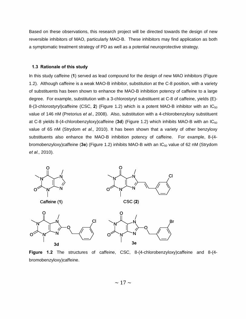
In this study caffeine (1) served as lead compound for the design of new MAO inhibitors (Figure
1.2). Although caffeine is a weak MAO-B inhibitor, substitution at the C-8 position, with a variety
of substituents has been shown to enhance the MAO-B inhibition potency of caffeine to a large
degree. For example, substitution with a 3-chlorostyryl substituent at C-8 of caffeine, yields (E)-
8-(3-chlorostyryl)caffeine (CSC, 2) (Figure 1.2) which is a potent MAO-B inhibitor with an IC50
value of 146 nM (Pretorius et al., 2008). Also, substitution with a 4-chlorobenzyloxy substituent
at C-8 yields 8-(4-chlorobenzyloxy)caffeine (3d) (Figure 1.2) which inhibits MAO-B with an IC50
value of 65 nM (Strydom et al., 2010). It has been shown that a variety of other benzyloxy
substituents also enhance the MAO-B inhibition potency of caffeine. For example, 8-(4-
bromobenzyloxy)caffeine (3e) (Figure 1.2) inhibits MAO-B with an IC50 value of 62 nM (Strydom
et al., 2010).
N
N
N
N
O
O
N
N
N
N
O
O
Cl
N
N
N
N
O
OO
ClN
N
N
N
O
O
OBr
Caffeine (1) CSC (2)
3d 3e
N
N
N
N
O
O
N
N
N
N
O
O
Cl
N
N
N
N
O
OO
ClN
N
N
N
O
O
OBr
Caffeine (1) CSC (2)
Figure 1.2 The structures of caffeine, CSC, 8-(4-chlorobenzyloxy)caffeine and 8-(4-
bromobenzyloxy)caffeine.
~ 18 ~
In the current study, twelve 8-thiocaffeine analogues (4a–l) will be synthesized and evaluated as
inhibitors of human MAO-A and –B. These thiocaffeine derivatives bear close structural
resemblance to the 8-oxycaffeine derivatives that were previously shown to be potent MAO
inhibitors (Strydom et al., 2010) and may therefore have similar biological properties (Table 1.1).
This study will determine if C-8 substitution of caffeine with a variety of thiol containing
substituents will enhance the MAO-B inhibition activity of caffeine to a similar degree than
substitution with an aryl- or alkyloxy substituent (Strydom et al., 2010). Since the 8-oxycaffeines
are also reported to be MAO-A inhibitors, the thiocaffeines that will be examined in this study
will also be evaluated as inhibitors of MAO-A (Strydom et al., 2010).
The structures of the compounds that will be examined in this study are shown in table 1.2.
Since this study is an exploratory study, to evaluate the possibility that thiocaffeine derivatives
may act as MAO inhibitors, a variety of side chains were selected for substitution at C-8 of the
caffeine ring. All side chains will be attached via a thioether linkage at C-8 of the caffeine ring.
The side chains selected include phenyl (4a), benzyl (4b) and phenylethyl (4c) substituents.
The benzyl substituted thiocaffeines will be further expanded, with the substitution of chlorine
(4d), bromine (4e), fluorine (4f) and methoxy (4g) on the benzyloxy ring. Also included will be a
phenoxyethyl (4h) substituent and the saturated cyclohexyl and cyclopentyl rings (4i and 4j). Finaly, two of the thiocaffeines to be examined here, will also contain a naphthalenyl ring (4k)
and an aliphatic side chain (4l).
This study will therefore explore the possibility that 8-thiocaffeine analogues may act as MAO-A
and –B inhibitors. Secondly, the effect of the presence of a thioether functional group at C-8 of
the caffeine ring on MAO-A and –B inhibition activity, will be evaluated. For this purpose the
MAO-A and –B inhibition potencies of the 8-thiocaffeine analogues will be compared to that of
the previously studied 8-oxycaffeine analogues (3a–h) (Table 1.1). Thirdly this study will
examine the effect that a variety of subsituents on C-8 of the caffeine will have on the MAO-A
and –B inhibition potencies of 8-thiocaffeine. The major potential outcomes of this study may
be:
1. Identification of new potent reversible 8-thiocaffeine derived MAO-A and –B inhibitors.
~ 19 ~
N
N
N
NO
O
O R
2. The proposal of additional promising 8-thiocaffeine analogues that may be investigated
in future studies.
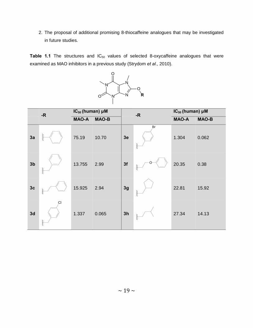
Table 1.1 The structures and IC50 values of selected 8-oxycaffeine analogues that were
examined as MAO inhibitors in a previous study (Strydom et al., 2010).
-R
IC50 (human) μM -R
IC50 (human) μM
MAO-A MAO-B MAO-A MAO-B
3a
75.19 10.70 3e
Br
1.304 0.062
3b
13.755 2.99 3f O
20.35 0.38
3c
15.925 2.94 3g
22.81 15.92
3d
Cl
1.337 0.065 3h
27.34 14.13

~ 20 ~
Table 1.2 The structures of the 8-thiocaffeine analogues that will be examined in the current
study.
-R -R
4a
4g
O
4b
4h O
4c
4i
4d
Cl
4j
4e
Br
4k
4f
F
4l
N
N
N
N
O
O
S
R
~ 21 ~
1.4 Objectives of this study
Based on the discussion above the objectives of this study are summarized below:
• Twelve 8-thiocaffeine analogues (4a–l) will be synthesized. The starting materials for
these syntheses will be 8-chlorocaffeine and a corresponding mercaptan. All the
mercaptans required for this study are commercially available. 8-Chlorocaffeine will be
synthesized from caffeine and Cl2 gas.
• The 8-thiocaffeine analogues will be evaluated as inhibitors of MAO-A and –B. For this
purpose the recombinant human enzymes will be used. The inhibition potencies will be
expressed as the IC50 values (concentration of the inhibitor that produces 50%
inhibition). A fluorometric assay will be used to measure the enzyme activities. Certain
MAO substrates are oxidized to fluorescent products. For example, kynuramine (which
is a substrate for both MAO-A and –B) is oxidized to 4-hydroxyquinoline (4-HQ). 4-HQ
concentrations may be measured with a fluorescence spectrophotometer at an excitation
wavelength of 310 nm and an emission wavelength of 400 nm. Fluorescence decreases
as the 4-HQ production is decreased by a MAO inhibitor.
• The time-dependency of inhibition of both MAO-A and –B by selected 8-thiocaffeine
analogues will be evaluated. This will be done in order to determine if the inhibitor
interacts reversibly or irreversibly with the MAO isozymes. Reversible inhibitors are
more desirable than irreversible enzyme inhibitors.
• If the inhibition is found to be reversible, a set of Lineweaver-Burk plots will be generated
for selected inhibitors in order to determine if the inhibition mode of the test compound is
competitive.
~ 22 ~
Chapter 2 Literature study
2.1 General background of Parkinson’s disease
2.1.1 General background
2.1.1.1 Neurochemical and neuropathological features
PD is primarily the result of the death of dopaminergic neurons in the substantia nigra pars
compacta (SNpc) of the brain. This loss of SNpc neurons leads to striatal DA deficiency which
is the cause of all major symptoms of PD. DA replacement therapy, through oral administration
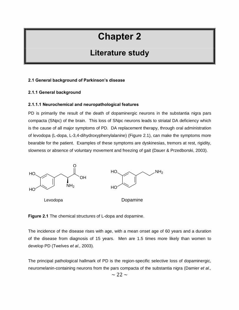
of levodopa (L-dopa, L-3,4-dihydroxyphenylalanine) (Figure 2.1), can make the symptoms more
bearable for the patient. Examples of these symptoms are dyskinesias, tremors at rest, rigidity,
slowness or absence of voluntary movement and freezing of gait (Dauer & Przedborski, 2003).
OH
O
NH2
HO
HO
NH2HO
HO
Levodopa Dopamine
Figure 2.1 The chemical structures of L-dopa and dopamine.
The incidence of the disease rises with age, with a mean onset age of 60 years and a duration
of the disease from diagnosis of 15 years. Men are 1.5 times more likely than women to
develop PD (Twelves et al., 2003).
The principal pathological hallmark of PD is the region-specific selective loss of dopaminergic,
neuromelanin-containing neurons from the pars compacta of the substantia nigra (Damier et al.,
~ 23 ~
1999). These neurons exhibit the presence of intraneuronal proteinacious cytoplasmic
inclusions termed ‘Lewy Bodies’ (LBs). Terminal loss in the striatum appears to be more distinct
than SNpc dopaminergic cell body loss, indicating that the primary target of the degenerative
process is the striatal dopaminergic nerve terminals (Bernheimer et al., 1973).
Neurodegeneration and the formation of LBs are also found in noradrenergic, serotonergic and
cholinergic systems. Even before the onset of PD symptoms, there may already be damage to
other neurochemical systems. This is the reason why some patients develop depression
months or years before the onset of PD motor symptoms (Dauer & Przedborski, 2003). A prior
hypothesis has also been proposed for the pathogenesis of PD. It is suggested that α-synuclein
misfolds or aggregates in one brain region, and triggers other α-synuclein proteins to misfold or
aggregate in interconnected neuronal groups. These misfolded proteins are then deposited in
the dopaminergic neurons (Hardy, 2005).
2.1.1.2 Aetiology
The cause of sporadic PD is unknown and the environmental toxin hypothesis was dominant for
most of the 20th century, because of the discovery of toxin-induced Parkinsonism. The
discovery of PD genes has renewed the interest in inherited PD. Both factors may play a role in
the aetiology of PD (Dauer & Przedborski, 2003).
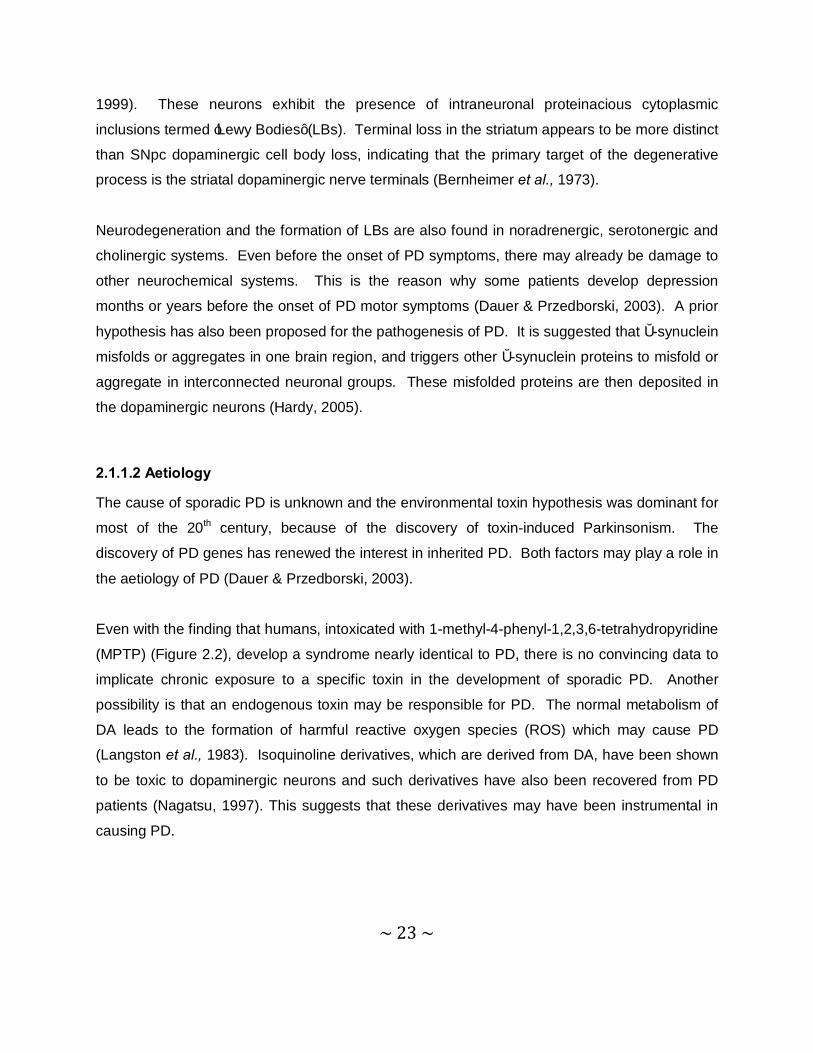
Even with the finding that humans, intoxicated with 1-methyl-4-phenyl-1,2,3,6-tetrahydropyridine
(MPTP) (Figure 2.2), develop a syndrome nearly identical to PD, there is no convincing data to
implicate chronic exposure to a specific toxin in the development of sporadic PD. Another
possibility is that an endogenous toxin may be responsible for PD. The normal metabolism of
DA leads to the formation of harmful reactive oxygen species (ROS) which may cause PD
(Langston et al., 1983). Isoquinoline derivatives, which are derived from DA, have been shown
to be toxic to dopaminergic neurons and such derivatives have also been recovered from PD
patients (Nagatsu, 1997). This suggests that these derivatives may have been instrumental in
causing PD.
~ 24 ~
N
Figure 2.2 The chemical structure of the neurotoxin MPTP.
One of the causes of PD is thought to be gene mutation, especially those leading to mutations
of the protein α-synuclein. LBs contain the α-synuclein protein, which is essential for the normal
function of the nigrostriatal system. Overexpression of human α-synuclein in nerve cells can
lead to an age-dependent loss of dopaminergic neurons (Dauer & Przedborski, 2003). Parkin is
another gene of which mutations may lead to PD. This mutated parkin gene was reported in a
case of autosomal juvenile Parkinsonism (Kitada et al., 1998).
2.1.1.3 Pathogenesis
Although PD is a sporadic disease (Taylor et al., 2005; Dauer & Przedborski, 2003), and its
origin is still unknown, a number of environmental causes have been identified. Ageing is
thought to be a major risk factor since PD is more prevalent at an advanced age (Taylor et al.,
2005). An interesting phenomenon is that non-smokers are twice as likely to develop PD
compared to smokers. This has been shown in women, who are not using hormonal
replacement therapy as well as in men. A low intake of caffeine has also been correlated to the
development of PD (Ascherio et al., 2003). Some reports have also shown that there is a
relationship between PD and head injuries, rural living, obesity, minimum exercise and exposure
to pesticides or herbicides (Elbaz & Tranchant, 2007). There is further a link between L-dopa
responsive parkinsonism and seven genetic mutations that can cause this disease. These
mutations are in the proteins, parkin, PINK-1, DJ-1, ATP13A2, α-synuclein, LRRK-2 and GABA.
Parkin mutations are the second most common cause of genetic PD (Healy et al., 2008;
Williams et al., 2005).
Dauer & Przedborski (2003) suggested two hypotheses for the pathogenesis of PD. The first
proposes that misfolding and aggregation of proteins are instrumental in the death of SNpc
dopaminergic neurons and the second proposes that mitochondrial dysfunction, with the
consequent oxidative stress and the formation of toxic oxidized DA species, may play a key role
in the development of PD. α-Synuclein, or genetically mutated α-synuclein misfolds or
~ 25 ~
aggregates as a result of oxidative damage. This protein may induce cell death by different
mechanisms such as deforming the cell or interfering with intracellular trafficking in neurons.
Pathogenic mutations may directly induce abnormal protein conformations or may damage the
cell’s cellular machinery, which detect and degrade any misfolded proteins (Dauer &
Przedborski, 2003).
A prominent neuropathological feature of PD is intraneuronal inclusions, LBs, in the nigral
dopaminergic neurons. LBs are composed of a variety of proteins, such as α-synuclein, parkin,
ubiquitin and neurofilaments. They are spherical, eosinophilic, cytoplasmic aggregates
(Przedborski, 2004). As already mentioned, an abnormal and aggregated form of α-synuclein is
the main component of LBs (Scherfler et al., 2006). Oxidative modified α-synuclein exhibits a
greater propensity to aggregate in vitro than unmodified α-synuclein (Giasson et al., 2000).
Controversy exists about whether LBs promote toxicity or protect the cell from the harmful
effects of misfolded proteins (Dauer & Przedborski, 2003).
Over the past few decades a large amount of data has been obtained from clinical studies and
in vitro and in vivo experimental models of PD. Available data suggests that the mechanism of
neuronal death in PD begins with a healthy dopaminergic neuron being affected by an
etiological factor, for example, mutant α-synuclein. This neuron will eventually be degenerated
as a result of deleterious factors, such as free radicals, mitochondrial dysfunction, excitotoxicity,
neuroinflammation and apoptosis that will eventually lead to its death (Lees et al., 2009).
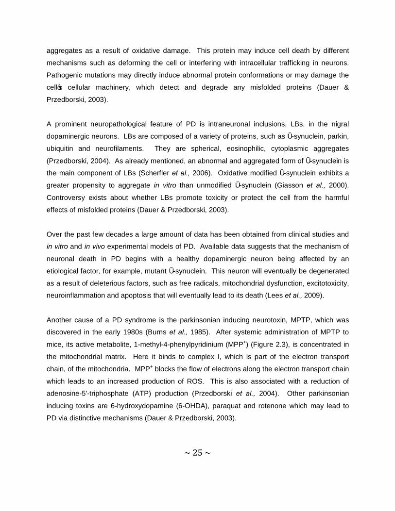
Another cause of a PD syndrome is the parkinsonian inducing neurotoxin, MPTP, which was
discovered in the early 1980s (Burns et al., 1985). After systemic administration of MPTP to
mice, its active metabolite, 1-methyl-4-phenylpyridinium (MPP+) (Figure 2.3), is concentrated in
the mitochondrial matrix. Here it binds to complex I, which is part of the electron transport
chain, of the mitochondria. MPP+ blocks the flow of electrons along the electron transport chain
which leads to an increased production of ROS. This is also associated with a reduction of
adenosine-5'-triphosphate (ATP) production (Przedborski et al., 2004). Other parkinsonian
inducing toxins are 6-hydroxydopamine (6-OHDA), paraquat and rotenone which may lead to
PD via distinctive mechanisms (Dauer & Przedborski, 2003).
~ 26 ~
NH3C
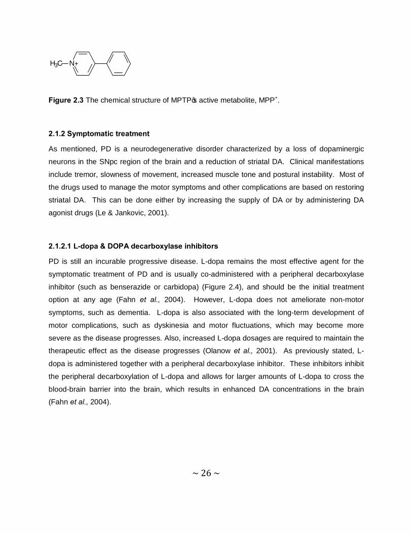
Figure 2.3 The chemical structure of MPTP’s active metabolite, MPP+.
2.1.2 Symptomatic treatment
As mentioned, PD is a neurodegenerative disorder characterized by a loss of dopaminergic
neurons in the SNpc region of the brain and a reduction of striatal DA. Clinical manifestations
include tremor, slowness of movement, increased muscle tone and postural instability. Most of
the drugs used to manage the motor symptoms and other complications are based on restoring
striatal DA. This can be done either by increasing the supply of DA or by administering DA
agonist drugs (Le & Jankovic, 2001).
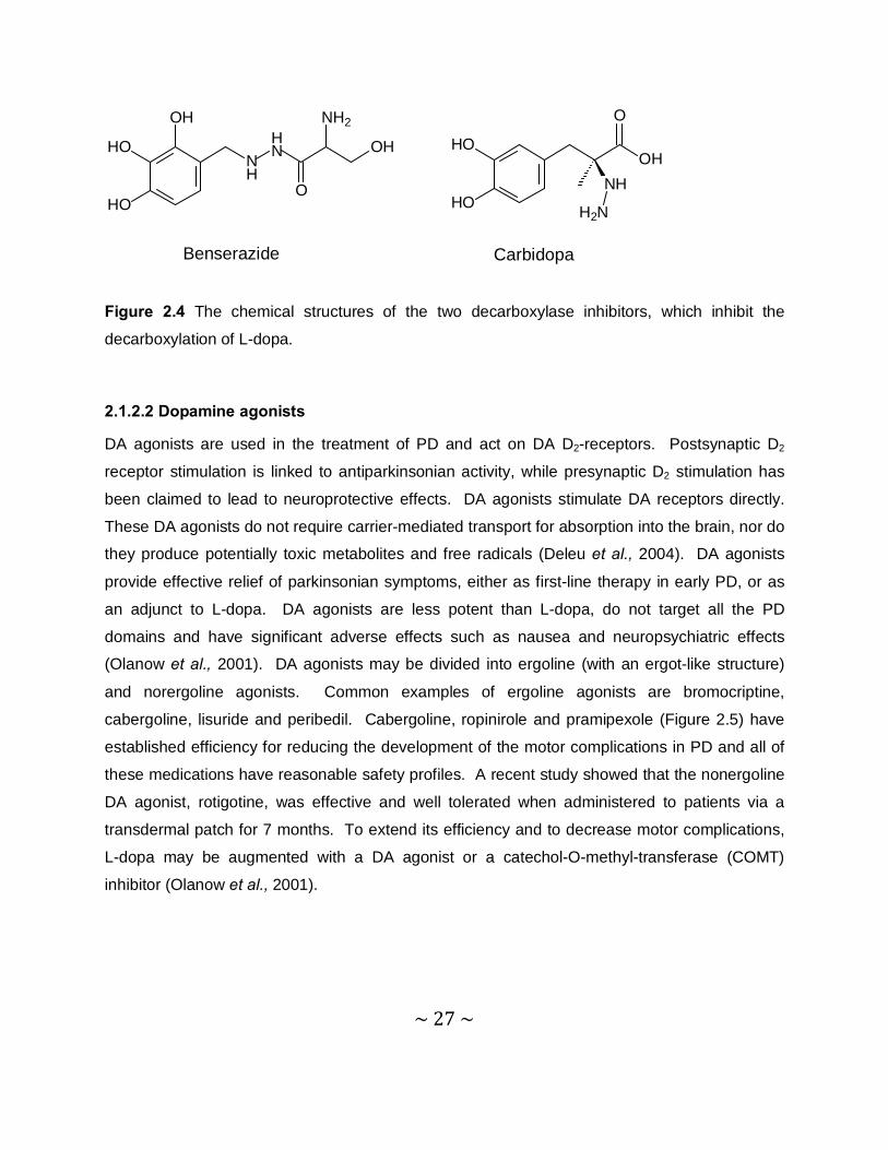
2.1.2.1 L-dopa & DOPA decarboxylase inhibitors
PD is still an incurable progressive disease. L-dopa remains the most effective agent for the
symptomatic treatment of PD and is usually co-administered with a peripheral decarboxylase
inhibitor (such as benserazide or carbidopa) (Figure 2.4), and should be the initial treatment
option at any age (Fahn et al., 2004). However, L-dopa does not ameliorate non-motor
symptoms, such as dementia. L-dopa is also associated with the long-term development of
motor complications, such as dyskinesia and motor fluctuations, which may become more
severe as the disease progresses. Also, increased L-dopa dosages are required to maintain the
therapeutic effect as the disease progresses (Olanow et al., 2001). As previously stated, L-
dopa is administered together with a peripheral decarboxylase inhibitor. These inhibitors inhibit
the peripheral decarboxylation of L-dopa and allows for larger amounts of L-dopa to cross the
blood-brain barrier into the brain, which results in enhanced DA concentrations in the brain
(Fahn et al., 2004).
~ 27 ~
NH
HN OH
O
NH2OH
HO
HO
OHHO
HONH
O
H2N
Benserazide Carbidopa
Figure 2.4 The chemical structures of the two decarboxylase inhibitors, which inhibit the
decarboxylation of L-dopa.
2.1.2.2 Dopamine agonists
DA agonists are used in the treatment of PD and act on DA D2-receptors. Postsynaptic D2
receptor stimulation is linked to antiparkinsonian activity, while presynaptic D2 stimulation has
been claimed to lead to neuroprotective effects. DA agonists stimulate DA receptors directly.
These DA agonists do not require carrier-mediated transport for absorption into the brain, nor do
they produce potentially toxic metabolites and free radicals (Deleu et al., 2004). DA agonists
provide effective relief of parkinsonian symptoms, either as first-line therapy in early PD, or as
an adjunct to L-dopa. DA agonists are less potent than L-dopa, do not target all the PD
domains and have significant adverse effects such as nausea and neuropsychiatric effects
(Olanow et al., 2001). DA agonists may be divided into ergoline (with an ergot-like structure)
and norergoline agonists. Common examples of ergoline agonists are bromocriptine,
cabergoline, lisuride and peribedil. Cabergoline, ropinirole and pramipexole (Figure 2.5) have
established efficiency for reducing the development of the motor complications in PD and all of
these medications have reasonable safety profiles. A recent study showed that the nonergoline
DA agonist, rotigotine, was effective and well tolerated when administered to patients via a
transdermal patch for 7 months. To extend its efficiency and to decrease motor complications,
L-dopa may be augmented with a DA agonist or a catechol-O-methyl-transferase (COMT)
inhibitor (Olanow et al., 2001).
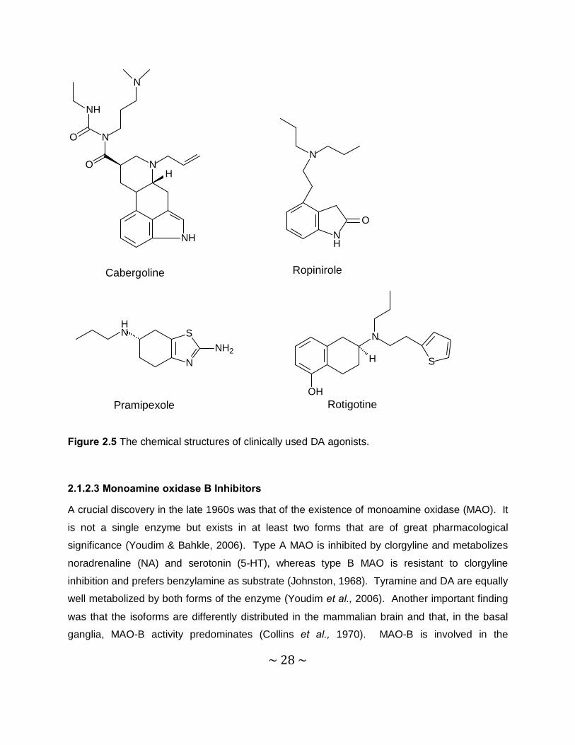
~ 28 ~
NH
NO
N
N
NH
O
H
NH
O
N
Cabergoline Ropinirole
S
NNH2
HN N
OH
H S
Pramipexole Rotigotine
Figure 2.5 The chemical structures of clinically used DA agonists.
2.1.2.3 Monoamine oxidase B Inhibitors
A crucial discovery in the late 1960s was that of the existence of monoamine oxidase (MAO). It
is not a single enzyme but exists in at least two forms that are of great pharmacological
significance (Youdim & Bahkle, 2006). Type A MAO is inhibited by clorgyline and metabolizes
noradrenaline (NA) and serotonin (5-HT), whereas type B MAO is resistant to clorgyline
inhibition and prefers benzylamine as substrate (Johnston, 1968). Tyramine and DA are equally
well metabolized by both forms of the enzyme (Youdim et al., 2006). Another important finding
was that the isoforms are differently distributed in the mammalian brain and that, in the basal
ganglia, MAO-B activity predominates (Collins et al., 1970). MAO-B is involved in the
~ 29 ~
metabolism of DA to ultimately yield 3,4-dihydroxyphenylacetic acid and homovanillic acid.
MAO-B also deaminates β-phenylethylamine, an endogenous amine that stimulates DA release
and inhibits neuronal DA uptake (Saura et al., 1990). The development of selective MAO-B
inhibitors has made it possible to block only the B isoform of the enzyme, for which DA is the
preferred substrate. By inactivating this enzyme, selective MAO-B inhibitors increase the
concentrations of both endogenous DA and DA produced from exogenously administered L-
dopa (Yamada & Yasuhara, 2004). Progressive deterioration of the dopaminergic neurons in
the SNpc results in a depletion of DA along the nigrostriatal pathway. The primary rationale for
using selective MAO-B inhibition in PD is that it enhances striatal dopaminergic activity by
inhibiting the metabolism of DA, thereby improving PD motor symptoms (Samii et al., 2004).
2.1.2.4 Anti-cholinergic drugs
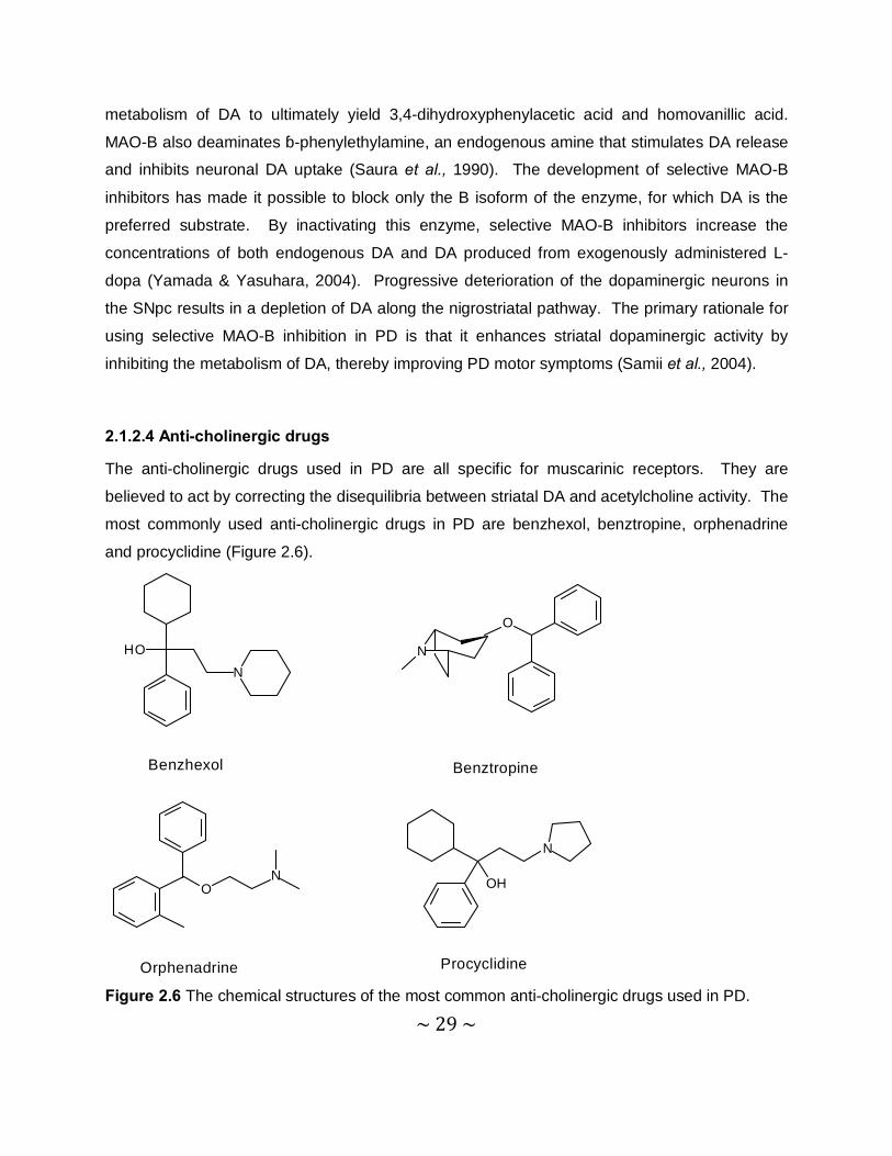
The anti-cholinergic drugs used in PD are all specific for muscarinic receptors. They are
believed to act by correcting the disequilibria between striatal DA and acetylcholine activity. The
most commonly used anti-cholinergic drugs in PD are benzhexol, benztropine, orphenadrine
and procyclidine (Figure 2.6).
ON
N
OH
Orphenadrine Procyclidine
HO
N
O
N
Benzhexol Benztropine
Figure 2.6 The chemical structures of the most common anti-cholinergic drugs used in PD.
~ 30 ~
An important factor, limiting the use of these drugs, is the occurrence of anti-cholinergic adverse
effects such as impaired neuropsychiatric and cognitive function. Anti-cholinergic drugs have to
be used with the utmost caution in these patient groups. These drugs offer mild symptomatic
control in PD when used as monotherapy or in combination with other agents. They have been
used particularly in tremor-predominant PD, although it is unknown whether their effect on
tremor is greater than that of other motor outcome measures (Deleu et al., 2004).
2.1.2.5 Adenosine A2a receptor antagonists
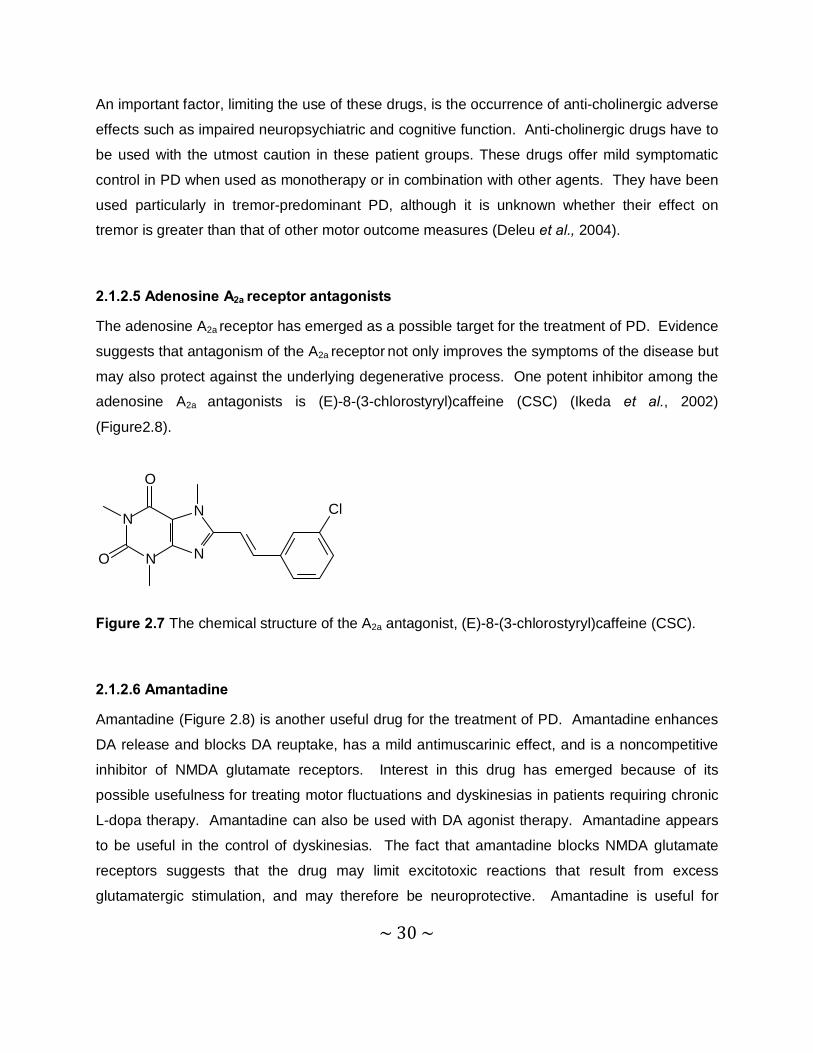
The adenosine A2a receptor has emerged as a possible target for the treatment of PD. Evidence
suggests that antagonism of the A2a receptor not only improves the symptoms of the disease but
may also protect against the underlying degenerative process. One potent inhibitor among the
adenosine A2a antagonists is (E)-8-(3-chlorostyryl)caffeine (CSC) (Ikeda et al., 2002)
(Figure2.8).
N
N
N
N
O
O
Cl
Figure 2.7 The chemical structure of the A2a antagonist, (E)-8-(3-chlorostyryl)caffeine (CSC).
2.1.2.6 Amantadine
Amantadine (Figure 2.8) is another useful drug for the treatment of PD. Amantadine enhances
DA release and blocks DA reuptake, has a mild antimuscarinic effect, and is a noncompetitive
inhibitor of NMDA glutamate receptors. Interest in this drug has emerged because of its
possible usefulness for treating motor fluctuations and dyskinesias in patients requiring chronic
L-dopa therapy. Amantadine can also be used with DA agonist therapy. Amantadine appears
to be useful in the control of dyskinesias. The fact that amantadine blocks NMDA glutamate
receptors suggests that the drug may limit excitotoxic reactions that result from excess
glutamatergic stimulation, and may therefore be neuroprotective. Amantadine is useful for
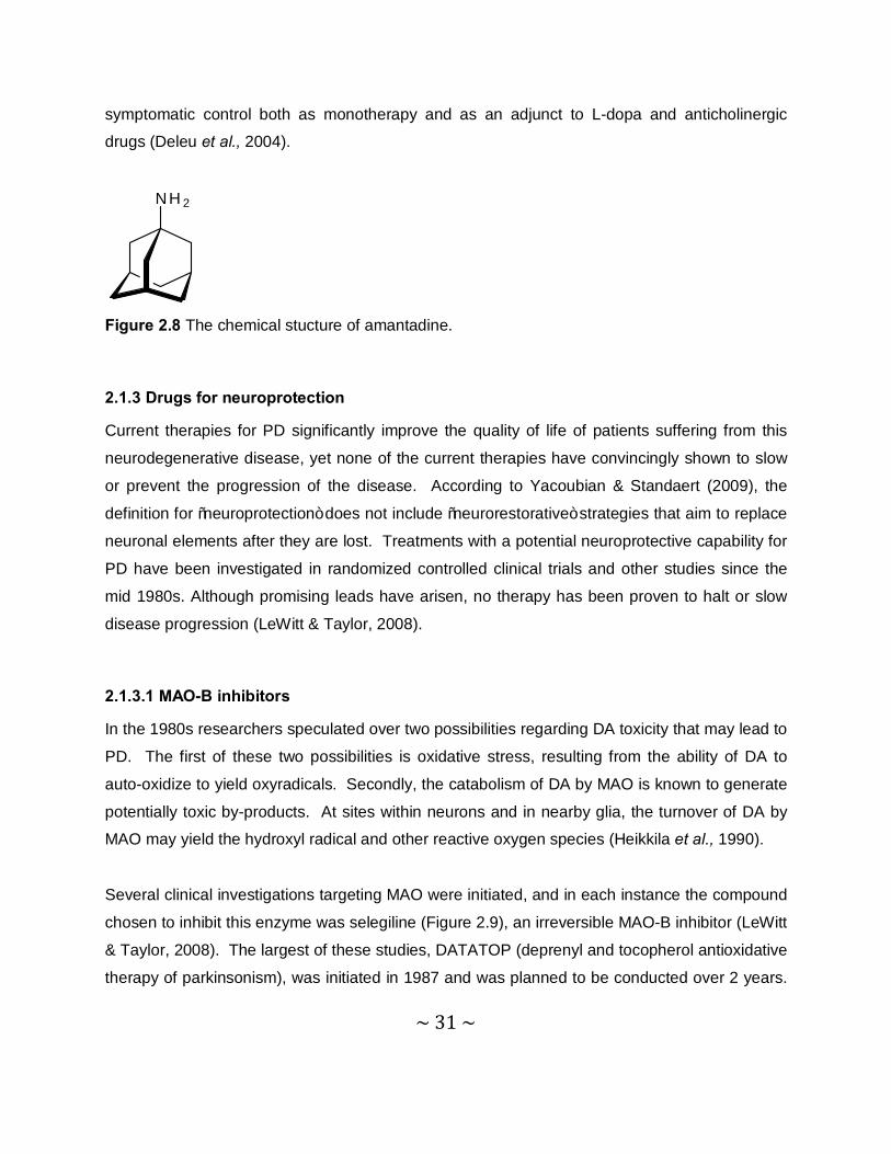
~ 31 ~
symptomatic control both as monotherapy and as an adjunct to L-dopa and anticholinergic
drugs (Deleu et al., 2004).
NH 2
Figure 2.8 The chemical stucture of amantadine.
2.1.3 Drugs for neuroprotection
Current therapies for PD significantly improve the quality of life of patients suffering from this
neurodegenerative disease, yet none of the current therapies have convincingly shown to slow
or prevent the progression of the disease. According to Yacoubian & Standaert (2009), the
definition for “neuroprotection” does not include “neurorestorative” strategies that aim to replace
neuronal elements after they are lost. Treatments with a potential neuroprotective capability for
PD have been investigated in randomized controlled clinical trials and other studies since the
mid 1980s. Although promising leads have arisen, no therapy has been proven to halt or slow
disease progression (LeWitt & Taylor, 2008).
2.1.3.1 MAO-B inhibitors
In the 1980s researchers speculated over two possibilities regarding DA toxicity that may lead to
PD. The first of these two possibilities is oxidative stress, resulting from the ability of DA to
auto-oxidize to yield oxyradicals. Secondly, the catabolism of DA by MAO is known to generate
potentially toxic by-products. At sites within neurons and in nearby glia, the turnover of DA by
MAO may yield the hydroxyl radical and other reactive oxygen species (Heikkila et al., 1990).
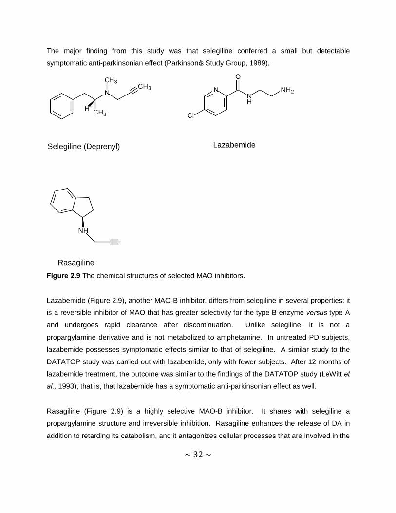
Several clinical investigations targeting MAO were initiated, and in each instance the compound
chosen to inhibit this enzyme was selegiline (Figure 2.9), an irreversible MAO-B inhibitor (LeWitt
& Taylor, 2008). The largest of these studies, DATATOP (deprenyl and tocopherol antioxidative
therapy of parkinsonism), was initiated in 1987 and was planned to be conducted over 2 years.
~ 32 ~
The major finding from this study was that selegiline conferred a small but detectable
symptomatic anti-parkinsonian effect (Parkinson’s Study Group, 1989).
NNH
NH2
Cl
O
N
CH3
CH3
H
CH3
NH
Selegiline (Deprenyl) Lazabemide
Rasagiline Figure 2.9 The chemical structures of selected MAO inhibitors.
Lazabemide (Figure 2.9), another MAO-B inhibitor, differs from selegiline in several properties: it
is a reversible inhibitor of MAO that has greater selectivity for the type B enzyme versus type A
and undergoes rapid clearance after discontinuation. Unlike selegiline, it is not a
propargylamine derivative and is not metabolized to amphetamine. In untreated PD subjects,
lazabemide possesses symptomatic effects similar to that of selegiline. A similar study to the
DATATOP study was carried out with lazabemide, only with fewer subjects. After 12 months of
lazabemide treatment, the outcome was similar to the findings of the DATATOP study (LeWitt et
al., 1993), that is, that lazabemide has a symptomatic anti-parkinsonian effect as well.
Rasagiline (Figure 2.9) is a highly selective MAO-B inhibitor. It shares with selegiline a
propargylamine structure and irreversible inhibition. Rasagiline enhances the release of DA in
addition to retarding its catabolism, and it antagonizes cellular processes that are involved in the
~ 33 ~
cascade of events leading to apoptosis. A clinical study was also carried out with rasagiline, the
TEMPO trial. The results of the TEMPO trial were in favour of disease-modifying action (Akao
et al., 2001). The effectiveness of selegiline, lazabemide and rasagiline as disease-modifying
agents provides a focus on their shared property of MAO-B inhibition. Additional potentially
protective pharmacological properties of propargylamine compounds, that are unrelated to
MAO-B inhibition, however, have also been shown in laboratory models of neurodegeneration
and apoptosis studies (Mandel et al., 2003).
2.1.3.2 Dopaminergic drugs
The DA agonists all act on DA D2-like receptors. Postsynaptic D2 receptor stimulation is linked
to an antiparkinsonian activity and presynaptic D2 stimulation has been claimed to have
neuroprotective effects. Unlike L-dopa, DA agonists stimulate DA receptors, directly. Other
theoretical advantages of the DA agonists are that they do not require carrier-mediated
transport for absorption into the brain, nor do they produce potentially toxic metabolites and free
radicals (Deleu et al., 2004). DA receptor agonists have been hypothesized to be potentially
neuroprotective by acting at D2 autoreceptors found on dopaminergic SN terminals to suppress
DA release and thus reduce oxidative stress. Certain agonists, such as pramipexole, may also
act as direct antioxidants (Olanow et al., 1998). Although developed for their symptomatic
actions in PD, several drugs may also have neuroprotective actions against oxidative stress and
may protect dopaminergic neurons against various experimental toxins, including
methamphetamine, 3-acetylpyridine, 6-OHDA and MPTP (Ferger et al., 2000). Furthermore,
studies investigating a stereoisomer of pramipexole, that is inactive at DA receptors, have
shown that it also exerts neuroprotective properties. In mice, the dopaminergic agonist,
ropinirole, also enhances mechanisms against oxidative stress and exerts a protective action
against 6-OHDA-induced loss of nigrostriatal dopaminergic projections (Tanaka et al., 2001).
2.1.3.3 Antioxidant therapy
Although several compounds with antioxidant properties have been considered for clinical
investigation, only α-tocopherol has undergone evaluation. α-Tocopherol, a chain breaking
antioxidant that enters into lipid-soluble cellular regions such as biological membranes, acts by
quenching oxyradical species. There is no evidence for deficiency of α-tocopherol in PD, and
~ 34 ~
severe deficiency states do not lead to parkinsonism. Experimental evidence suggest that there
is no evidence for a disease-modifying effect (Parkinson’s Study Group, 1993).
2.1.3.4 Mitochondrial energy enhancement drugs
One of the few systematic markers for PD is altered mitochondrial function. Mitochondria of the
SN, platelets and skeletal muscle in PD possess reduced activity of the first step of the
mitochondrial electron transport chain, complex I. Coenzyme Q10 is an essential cofactor
serving as an electron acceptor for mitochondrial complex I. It is also a potent antioxidant in
lipid membranes and mitochondria. Creatine serves as a precursor for the conversion to the
energy intermediate, phosphocreatine, which in mitochondria transfers phosphoryl groups for
ATP synthesis. The effect of increasing creatine intake is an enhancement of phosphocreatine
formation. Ultimately the result is the reduction in oxidative stress through the opening of the
mitochondrial transition pore. Creatine-treated subjects as well as the coenzyme Q10 treated
subjects, tended to require less increase of dopaminergic therapy dose over time (Shults et al.,
1999).
2.1.3.5 Anti-inflammatory drugs
The role of inflammation in PD has become more recognized recently. Activation of microglia,
increased cytokine production and increased complement protein levels, have been
demonstrated in PD. As a means to slow disease progression, anti-inflammatory agents,
including nonsteroidal anti-inflammatory drugs (NSAIDs) and minocycline, have been pursued
as potential disease-modifying treatments for PD. Several studies in culture and in animal
models have shown that certain NSAIDs, such as aspirin, have neuroprotective qualities
(Tansey et al., 2007; Esposito et al., 2007). An example of an alternative approach to targeting
neuroinflammation may be the use of statins (3-hydroxy-3-mythylglutaryl-coenzyme A reductase
inhibitors). In addition to lowering cholesterol, these drugs have anti-inflammatory effects,
including the reduction of tumor necrosis factor-α (TNFα), nitric oxide and superoxide production
by microglia. Simvastatin has been shown to reduce DA loss in MPTP animal models. Recent
epidemiological studies showed that statin use, particularly simvastatin, is associated with
reduced PD incidence (Selley, 2005).
~ 35 ~
2.1.3.6 Anti-apoptotic drugs
Apoptosis is a mechanism that participates in neural development and plays a role in some
forms of neural injury. Activation of these cell death pathways most likely represents end-stage
processes in PD neurodegeneration. Therefore, inhibitors of these cell death pathways have
been proposed as potential neuroprotective agents regardless of the initial causes for
neurodegeration in PD (Yacoubian & Standaert, 2009). Several lines of evidence have pointed
to the activation of apoptosis as a possible mechanism for neurodegeneration in PD. On this
basis, the search of anti-apoptotic interventions led to proposals for the study of three different
compounds and how they interact with pro-apoptotic mechanisms (Waldmeier et al., 2006).
The propargylamine, TCH346, is an anti-apoptotic compound that inhibits the glycolytic enzyme,
glyceraldehyde-3-phosphate dehydrogenase (GAPDH), which can initiate apoptosis (Yacoubian
& Standaert, 2009). TCH346 was developed because of its shared structural similarities with
selegiline. TCH346 does not inhibit MAO-B, however, and unlike selegiline, it is not
metabolized to amphetamine metabolites. In rhesus monkeys, exposed to MPTP, near-
complete protection against the development of motor impairment was achieved. Unfortunately
it did not reveal any evidence for a neuroprotective effect in clinical trials (Olanow et al., 2006).
CEP-1347, an inhibitor of mixed lineage kinases, that can activate the c-Jun N-terminal (JNK)
pathway, that is involved in cell death, is another anti-apoptotic agent that showed promise in
preclinical studies (Maroney et al., 1998).
Minocycline (Figure 2.10) has been extensively studied because of its promise in treating
neurodegenerative diseases. In rodent models of parkinsonism induced by 6-OHDA and
MPTP, pre-treatment with minocycline improved survival of dopaminergic SN neurons.
Minocycline inhibits the activation of microglia, which is a prominent feature in the brain of PD
patients and in experimental neurotoxin models. Although these properties seem to be in favour
of minocycline providing a possible neuroprotective effect in PD, preclinical results have not
supported this possibility (Wu et al., 2002).
~ 36 ~
OO
O
H2N
OH OH OH
NHH
N
OH
Figure 2.10 The chemical structure of the anti-apoptotic drug, minocycline.
2.1.3.7 Anti-glutamatergic drugs
Because glutamate can act as an excitotoxin, contributing to neural damage, one rationale for
PD neuroprotection has been to block glutamate neurotransmission in the SN. Riluzole (Figure
2.11) demonstrates limited but definite effectiveness in slowing the deterioration of amyotrophic lateral sclerosis and has been FDA-approved for this use. Riluzole acts by blocking the
presynaptic release of glutamate. Unlike other compounds, that are potent glutamate blockers
and that can cause significant CNS toxicity, riluzole is well tolerated (Rascol et al., 2002).
N
SH2N
OCF3
Figure 2.11 The chemical structure of an anti-glutamatergic drug, riluzole
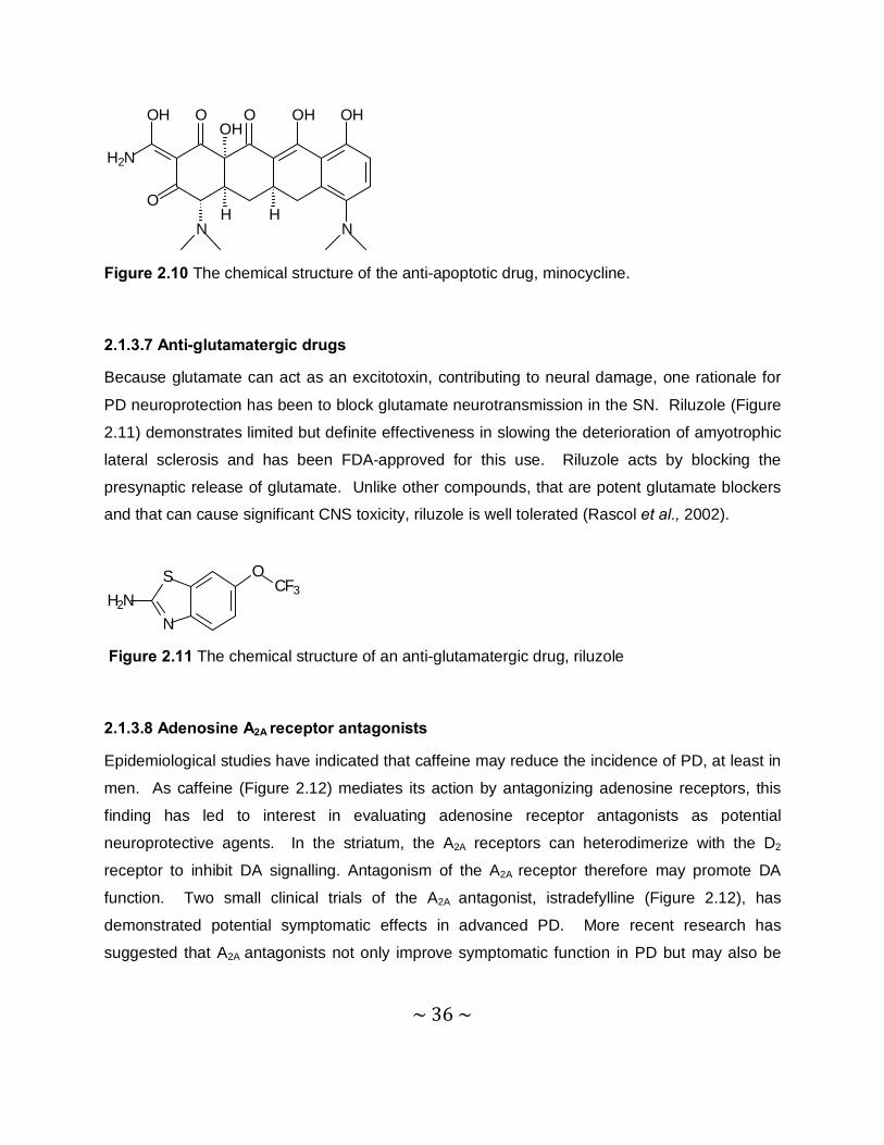
2.1.3.8 Adenosine A2A receptor antagonists
Epidemiological studies have indicated that caffeine may reduce the incidence of PD, at least in
men. As caffeine (Figure 2.12) mediates its action by antagonizing adenosine receptors, this
finding has led to interest in evaluating adenosine receptor antagonists as potential
neuroprotective agents. In the striatum, the A2A receptors can heterodimerize with the D2
receptor to inhibit DA signalling. Antagonism of the A2A receptor therefore may promote DA
function. Two small clinical trials of the A2A antagonist, istradefylline (Figure 2.12), has
demonstrated potential symptomatic effects in advanced PD. More recent research has
suggested that A2A antagonists not only improve symptomatic function in PD but may also be
~ 37 ~
neuroprotective (Yacoubian & Standaert, 2009). Caffeine and istradefylline are both
neuroprotective in the MPTP animal model of PD (Ikeda et al., 2002).
Caffeine Istradefylline
N
N N
N
CH3
H3CCH3
O
O
N
N
H3C
CH3
N
N
O
H3C
O
CH3
CH3O
O
Figure 2.12 The chemical structures of selected adenosine receptor antagonists.
2.1.4 Mechanisms of neurodegeneration
Several mechanisms have been implicated in PD pathogenesis. No one mechanism appears to
be primary in all cases of PD, and these pathogenic mechanisms likely act synergistically
through complex interactions to promote neurodegeneration (Yacoubian & Standaert, 2009).
2.1.4.1 Oxidative stress and mitochondrial dysfunction
Oxidative stress results from an overabundance of reactive free radicals secondary to either an
overproduction of reactive species or a failure of cell buffering mechanisms that normally limit
their accumulation. DA metabolism promotes oxidative stress through the production of
quinones, peroxides, and other ROS. Mitochondrial dysfunction is another source for the
production of ROS, which can further damage mitochondria. For example, Complex I inhibitors,
such as MPP+ and rotenone, cause a parkinsonian syndrome in animals. Increased iron levels,
seen in the SN of PD patients, also promote free radical damage, particularly in the presence of
neuromelanin. Several different strategies have been proposed to limit oxidative stress in PD.
These strategies include inhibitors of MAO, a key enzyme involved in DA catabolism, and
enhancers of mitochondrial electron transport, such as coenzyme Q10. Other strategies include
compounds that can directly quench free radicals, such as vitamin E, and molecules that can
~ 38 ~
promote endogenous mechanisms that buffer free radicals, such as selenium (Hastings &
Lewis, 1996).
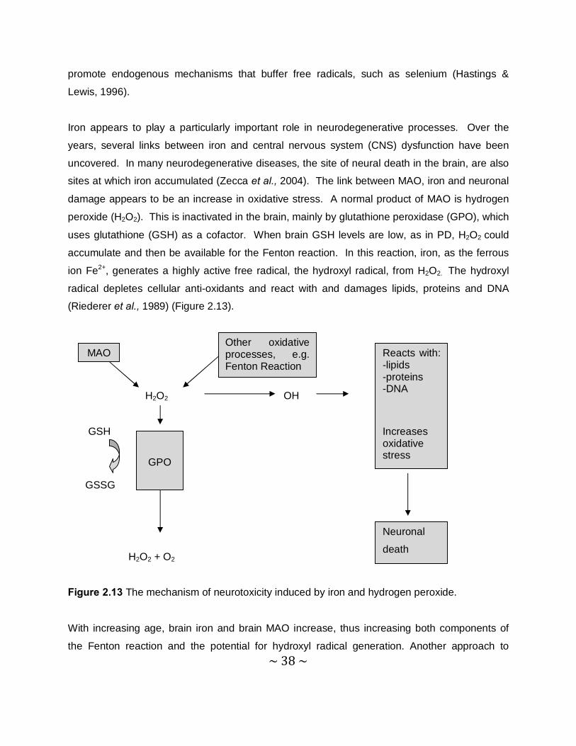
Iron appears to play a particularly important role in neurodegenerative processes. Over the
years, several links between iron and central nervous system (CNS) dysfunction have been
uncovered. In many neurodegenerative diseases, the site of neural death in the brain, are also
sites at which iron accumulated (Zecca et al., 2004). The link between MAO, iron and neuronal
damage appears to be an increase in oxidative stress. A normal product of MAO is hydrogen
peroxide (H2O2). This is inactivated in the brain, mainly by glutathione peroxidase (GPO), which
uses glutathione (GSH) as a cofactor. When brain GSH levels are low, as in PD, H2O2 could
accumulate and then be available for the Fenton reaction. In this reaction, iron, as the ferrous
ion Fe2+, generates a highly active free radical, the hydroxyl radical, from H2O2. The hydroxyl
radical depletes cellular anti-oxidants and react with and damages lipids, proteins and DNA
(Riederer et al., 1989) (Figure 2.13).
H2O2 OH
GSH
GSSG
H2O2 + O2
Figure 2.13 The mechanism of neurotoxicity induced by iron and hydrogen peroxide.
With increasing age, brain iron and brain MAO increase, thus increasing both components of
the Fenton reaction and the potential for hydroxyl radical generation. Another approach to
MAO Other oxidative processes, e.g. Fenton Reaction
GPO
Reacts with: -lipids -proteins -DNA
Increases oxidative stress
Neuronal
death

~ 39 ~
protect against the degenerative processes in PD is to remove the Fe2+ ions. Thus, the
intraventricular injection of a well-known iron chelator, deferal, protects against lesions of
nigrostriatal DA neurons induced by 6-hydroxydopamine (6-OHDA) or MPTP (Youdim & Bakhle,
2006).
2.1.4.2 Protein aggregation and misfolding
Protein aggregation and misfolding have emerged as important mechanisms in many
neurodegenerative disorders, including PD. In PD, the primary aggregating protein is α-synuclein, whose link to PD was first identified through rare families with autosomal dominant
PD caused by mutations in this protein. While mutations in α-synuclein are found in a small
number of inherited PD cases, α-synuclein is the major component of LBs and Lewy neurites
found in sporadic PD (Athanassiadou et al., 1999; Spillantini et al., 1997). Recent studies,
implicating parkin and ubiquitin carboxyl-terminal hydrolase L1 (UCH-L1) in genetic forms of PD,
reinforce the connection between protein aggregation and PD pathogenesis. Parkin is an E3
ubiquitin ligase involved in targeting misfolded proteins for degradation. Mutations of parkin
found in genetic forms of PD, disrupt its E3 ubiquitin ligase activity (Kitada et al., 1998).
Overproduction or impaired clearance of α-synuclein results in aggregation and may be a
central mechanism for PD. Therefore, therapeutic strategies to prevent protein aggregation or
to enhance the clearance of misfolded proteins are the subject of intensive study. Inhibitors of
α-synuclein aggregation could serve as potential neuroprotective therapies, although a clearer
understanding of the toxicity form of α-synuclein is important (Yacoubian & Standaert’s, 2009).
2.1.4.3 Neuroinflammation
Neuroinflammation is likely to contribute to neuronal dysfunction and eventual death of
vulnerable neuronal populations. While acute inflammation in the CNS is often accompanied by
secretion of microglial-derived neuroprotective factors, which promote repair, chronic
neuroinflammation is more likely to increase susceptibility of vulnerable neurons to toxic injury,
because it can induce oxidative stress. The two mechanisms by which neuroinflammation
induces oxidative stress, are via the production of high levels of ROS by activated glia, such as
microglia and astrocytes, and via arachidonic acid signalling, through the activation of
cyclooxygenase (COX) and lipoxygenase (LOX) pathways. Prostaglandin E2 (PGE2), produced
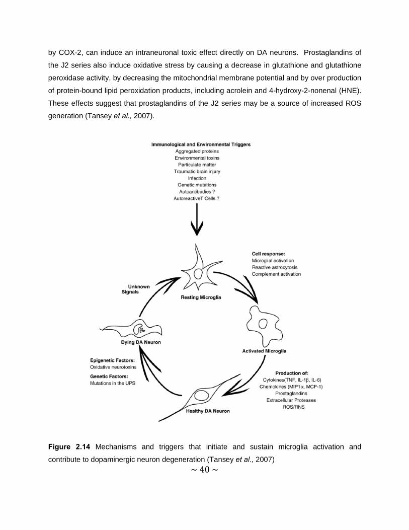
~ 40 ~
by COX-2, can induce an intraneuronal toxic effect directly on DA neurons. Prostaglandins of
the J2 series also induce oxidative stress by causing a decrease in glutathione and glutathione
peroxidase activity, by decreasing the mitochondrial membrane potential and by over production
of protein-bound lipid peroxidation products, including acrolein and 4-hydroxy-2-nonenal (HNE).
These effects suggest that prostaglandins of the J2 series may be a source of increased ROS
generation (Tansey et al., 2007).
Figure 2.14 Mechanisms and triggers that initiate and sustain microglia activation and
contribute to dopaminergic neuron degeneration (Tansey et al., 2007)
~ 41 ~
2.1.4.4 Excitotoxicity
Excitotoxicity has been implicated as a pathogenic mechanism in several neurodegenerative
disorders, including PD. Glutamate is the primary excitatory transmitter in the mammalian
central nervous system and a primary driver of the excitotoxicity process. Dopaminergic
neurons in the SN have high levels of glutamate receptors and receive glutamatergic
innervations from the subthalamic nucleus and cortex. Excessive NMDA receptor activation by
glutamate could increase intracellular calcium levels that then activate cell death pathways.
Calcium influx produced by excessive glutamate receptor activation can also promote
peroxynitrite production through the activation of nitric oxide synthase. NMDA receptor
antagonists protect against dopaminergic cell loss in MPTP models (Yacoubian & Standaert,
2009).
2.1.4.5 Apoptosis
Apoptosis is a mechanism that has been demonstrated to participate in neural development and
to play a role in some forms of neural injury. There has been controversy as to whether
apoptosis is directly involved in PD. Several pathological studies have revealed signs of both
apoptotic and autophagic cell death in the SN of PD brains, although the extent is limited
because of the slow process of cell death which underlies PD. Alterations in cell death
pathways are unlikely to be the primary cause of PD, but both apoptotic and autophagic cell
death pathways are hypothesized to become activated in PD through oxidative stress, protein
aggregation, excitotoxicity or inflammatory processes. Activation of these cell death pathways
most likely represents end-stage processes in PD neurodegeneration (Tatton et al., 2003).
2.1.4.6 Loss of trophic factors
The loss of neurotrophic factors has been implicated as a potential contributor to cell death
observed in PD. The neurotrophic factors, brain-derived neurotrophic factor (BDNF), glial-
derived neurotrophic factor (GDNF) and nerve growth factor (NGF) have all been demonstrated
to be reduced in the nigra in PD. As a result, treatment with growth factors have been proposed
as a potential neuroprotective therapy in PD. Indeed, the potent ability of these agents to
stimulate growth of dopaminergic neurons suggest that they may be useful neuroprotective
~ 42 ~
treatments, even if deficiency of the factor is not the primary cause of the disease process
(Howells et al., 2000).
2.2 The monoamine oxidases
2.2.1 General background and tissue distribution of MAO
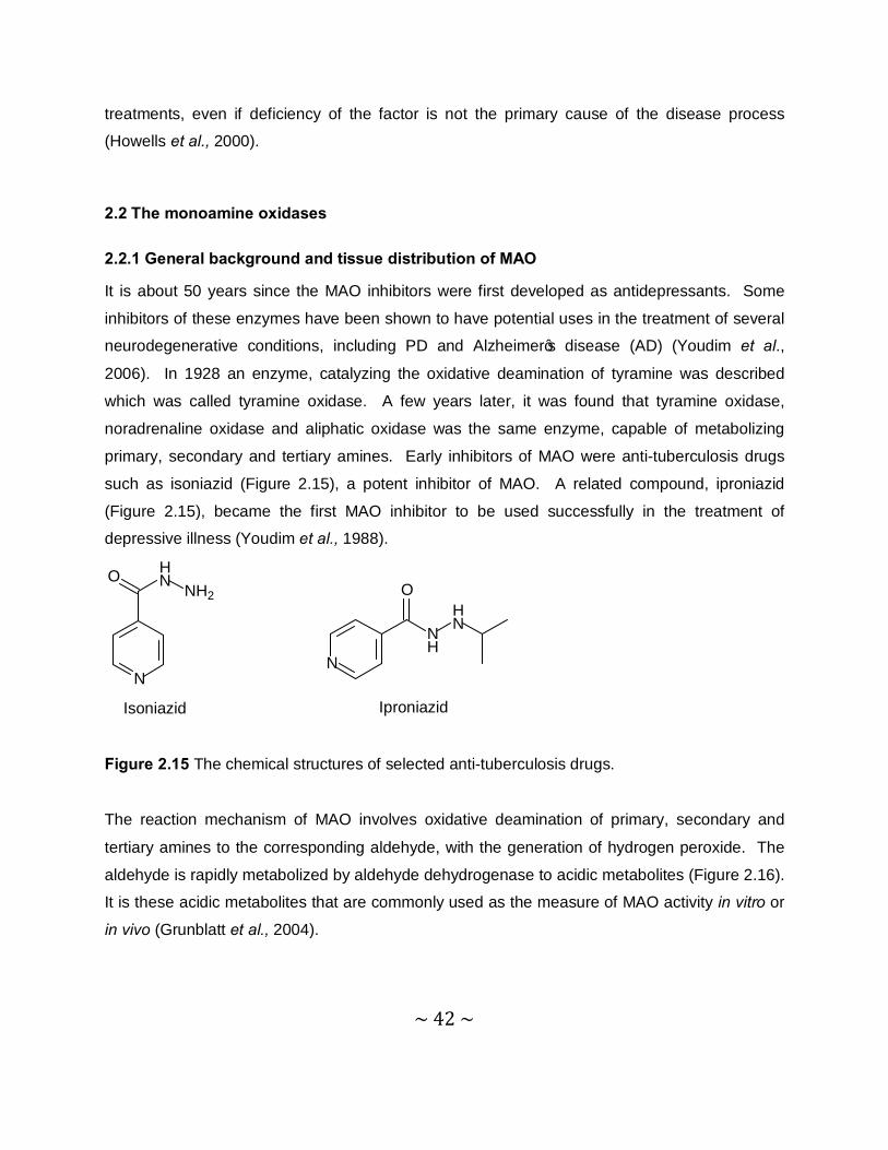
It is about 50 years since the MAO inhibitors were first developed as antidepressants. Some
inhibitors of these enzymes have been shown to have potential uses in the treatment of several
neurodegenerative conditions, including PD and Alzheimer’s disease (AD) (Youdim et al.,
2006). In 1928 an enzyme, catalyzing the oxidative deamination of tyramine was described
which was called tyramine oxidase. A few years later, it was found that tyramine oxidase,
noradrenaline oxidase and aliphatic oxidase was the same enzyme, capable of metabolizing
primary, secondary and tertiary amines. Early inhibitors of MAO were anti-tuberculosis drugs
such as isoniazid (Figure 2.15), a potent inhibitor of MAO. A related compound, iproniazid
(Figure 2.15), became the first MAO inhibitor to be used successfully in the treatment of
depressive illness (Youdim et al., 1988).
Isoniazid Iproniazid
N
HN NH2
O
N
NH
HN
O
Figure 2.15 The chemical structures of selected anti-tuberculosis drugs.
The reaction mechanism of MAO involves oxidative deamination of primary, secondary and
tertiary amines to the corresponding aldehyde, with the generation of hydrogen peroxide. The
aldehyde is rapidly metabolized by aldehyde dehydrogenase to acidic metabolites (Figure 2.16).
It is these acidic metabolites that are commonly used as the measure of MAO activity in vitro or
in vivo (Grunblatt et al., 2004).

~ 43 ~
ADH
H2O2 O2 + H+
FAD FADH2
RCH2NR1R2 RCHO + NHR1R2
RCOOH Figure 2.16 Reaction pathway of monoamine metabolism by oxidative deamination by
mitochondrial MAO.
Gene profiling of post-mortem samples of SN from PD patients disclosed a deficiency in
aldehyde dehydrogenase (ADH) that could allow a build-up of neurotoxic aldehydes derived
from DA by MAO (Grunblatt et al., 2004). A crucial finding in the late 1960s was that MAO was
not a single enzyme but could exist in at least two forms (type A and type B) that had different
pH optima and sensitivity to heat inactivation. Type A MAO was inhibited by clorgyline and
metabolized NA and 5-HT, where type B MAO was inhibited by selegiline and metabolizes
benzylamine. Tyramine and DA were equally well metabolized by both forms of the enzyme
(Johnston, 1968).
MAO-A and -B are tightly associated with the mitochondrial outer membrane, although a small
proportion of each enzyme is associated with the microsomal fraction. During development,
MAO-A appears before MAO-B, with the level of the latter increasing dramatically in the brain
after birth. MAO is present in most mammalian tissues, but the proportion of the two iso-
enzymes varies from tissue to tissue. Microvessels in the blood-brain barrier are rich in MAO-B.
In the human brain, there are regional differences in MAO activity: the basal ganglia (striatum)
and hypothalamus show the highest activities, whereas low levels of activity are observed in the
cerebellum and neocortex. The two iso-enzymes are not evenly distributed in the human brain,
and the main form in the basal ganglia is MAO-B. Both MAO-A and -B can contribute to DA
metabolism in the human brain (Youdim et al., 2006).
MAO

~ 44 ~
2.2.2 Biological function of MAO-B
MAO in peripheral tissues, such as the intestine, liver, lungs and placenta, seems to protect the
body by oxidizing amines from the blood or by preventing their entry into the circulation. MAO-B
in the microvessels of the blood-brain barrier presumably has a similar protective function,
acting as a metabolic barrier. It has been suggested that in the peripheral nervous system
(PNS) and CNS, intraneuronal MAO-A and -B protect neurons from exogenous amines,
terminate the actions of amine neurotransmitters and regulate the contents of intracellular amine
stores.
Hydrogen peroxide formed during the MAO catalytic reaction might have important metabolic
and signalling functions in the brain, and the aldehydes derived from 5-HT and NA deamination
might be involved in the regulation of sleep. At higher concentrations, the products, ammonia
and hydrogen peroxide, are toxic. The aldehyde derived from DA is known to be cytotoxic. This
is important in PD because, as mentioned, the levels of ADH in the SN are greatly reduced.
Such aldehydes might also form adducts with amine groups to yield toxic compounds such as
tetrahydropapaveroline that have been associated with parkinsonism and with alcohol-related
abnormalities (Shin et al., 2004).
Figure 2.17 DA synthesis and its metabolism by MAO-A and MAO-B (Youdim et al., 2006).
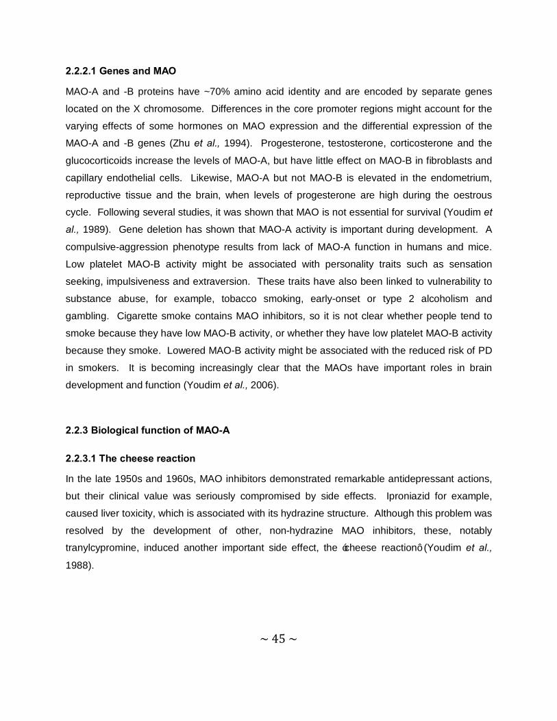
~ 45 ~
2.2.2.1 Genes and MAO
MAO-A and -B proteins have ~70% amino acid identity and are encoded by separate genes
located on the X chromosome. Differences in the core promoter regions might account for the
varying effects of some hormones on MAO expression and the differential expression of the
MAO-A and -B genes (Zhu et al., 1994). Progesterone, testosterone, corticosterone and the
glucocorticoids increase the levels of MAO-A, but have little effect on MAO-B in fibroblasts and
capillary endothelial cells. Likewise, MAO-A but not MAO-B is elevated in the endometrium,
reproductive tissue and the brain, when levels of progesterone are high during the oestrous
cycle. Following several studies, it was shown that MAO is not essential for survival (Youdim et
al., 1989). Gene deletion has shown that MAO-A activity is important during development. A
compulsive-aggression phenotype results from lack of MAO-A function in humans and mice.
Low platelet MAO-B activity might be associated with personality traits such as sensation
seeking, impulsiveness and extraversion. These traits have also been linked to vulnerability to
substance abuse, for example, tobacco smoking, early-onset or type 2 alcoholism and
gambling. Cigarette smoke contains MAO inhibitors, so it is not clear whether people tend to
smoke because they have low MAO-B activity, or whether they have low platelet MAO-B activity
because they smoke. Lowered MAO-B activity might be associated with the reduced risk of PD
in smokers. It is becoming increasingly clear that the MAOs have important roles in brain
development and function (Youdim et al., 2006).
2.2.3 Biological function of MAO-A
2.2.3.1 The cheese reaction
In the late 1950s and 1960s, MAO inhibitors demonstrated remarkable antidepressant actions,
but their clinical value was seriously compromised by side effects. Iproniazid for example,
caused liver toxicity, which is associated with its hydrazine structure. Although this problem was
resolved by the development of other, non-hydrazine MAO inhibitors, these, notably
tranylcypromine, induced another important side effect, the ‘cheese reaction’ (Youdim et al.,
1988).
~ 46 ~
Figure 2.18 The mechanism of potentiation of cardiovascular effects of tyramine: the cheese
reaction (Youdim & Bakhle, 2006).
The cheese reaction (Figure 2.18) is induced by tyramine and other indirectly acting
sympathomimetic amines present in food and fermented drinks, such as beer and wine. Under
normal circumstances, such dietary amines are extensively metabolized by MAO in the gut wall
and in the liver and they are thus prevented from entering the systemic circulation. In the
presence of a MAO inhibitor, this protective system is inactivated and tyramine or other
monoamines present in ingested food are not metabolized and enter the circulation. From here
they have access to the brain, and induce a significant release of NA from peripheral adrenergic
neurons. The consequence of this release is a severe hypertensive response which, in some
cases, can be fatal. These serious side effects stimulated a search for antidepressants that are
not MAO inhibitors, and led to their eventual replacement by the neurotransmitter uptake
inhibitors, the tricyclic antidepressants, and more recently the 5-HT selective re-uptake inhibitors
(SSRI) such as fluoxetine (Finberg et al., 1981).

~ 47 ~
CF3
OHN
Figure 2.19 The chemical structure of the SSRI Fluoxetine.
Selective, irreversible MAO-B inhibitors have no such effects, because the intestine contains
little MAO-B and tyramine is effectively metabolized by intestinal MAO-A. The development of
reversible MAO-A inhibitors, such as moclobemide, also avoided this problem because
reversible inhibitors can sufficiently block MAO-A in the CNS to obtain an antidepressant effect,
while dietary tyramine is able to displace the inhibitor from peripheral MAO-A, allowing for its
metabolism. A recent developed brain-selective MAO-A/B inhibitor, ladostigil, does not cause
the cheese reaction (Youdim et al., 2006).
Moclobemide Ladostigil
NH
N
OO
Cl
ON
O
NH
Figure 2.20 The chemical structures of selected MAO-A inhibitors.
2.2.3.2 MAO-A in depression
MAO inhibitors have been used for decades in the treatment of depression. The antidepressant
properties of MAO inhibitors result from selective MAO-A inhibition in the CNS, which leads to
increased brain levels of DA, NA and 5-HT (Zisook, 1985). When one isoform of MAO is fully
inhibited, the other isoform metabolize DA adequately. Thus, with selective inhibition of MAO-A
or -B, the level of DA will not change drastically in the human striatum (Riederer & Youdim,
1986). The two monoamines implicated in depressive illness are NA and 5-HT, both substrates
~ 48 ~
for MAO-A. The antidepressant effects of MAO-A inhibition with the earlier, non-selective,
irreversible inhibitors had been already established. The major disadvantage was the incidence
of the cheese reaction with those early inhibitors. Because the selective reversible inhibitors did
not provoke this reaction, moclobemide was assessed as an antidepressant and found to be
effective in improving vigilance, psychomotor speed and long-term memory (Youdim & Bakhle,
2006).
In the treatment of therapy-resistant depression, MAO-inhibitors provide an important option and
a combination of reversible MAO-A and reversible MAO-B inhibitors may be worth considering.
However, combination of MAO-inhibitors with uptake inhibitors, such as the tricyclic
antidepressants and SSRI`s, should be avoided as such combinations may provoke the ‘5-HT
syndrome’, a serious adverse reaction. Moclobemide shows a useful antidepressant action and
is well tolerated with standard PD therapies. Furthermore, in PD patients who are not
depressed, moclobemide does not significantly influence cognitive measures of mood (Youdim
& Weinstock, 2004).
2.2.4 The role of MAO-B in PD
2.2.4.1 Metabolism of DA
Tyrosine passes through the blood-brain barrier and is hydroxylated by tyrosine hydroxylase
(TH) to L-dopa, which is then decarboxylated by DOPA decarboxylase (DDC) to DA within the
neuron. DA is taken up into synaptic vesicles or is metabolized by MAO-A at neuronal
mitochondria. After release from the terminal, extracellular DA is cleared by uptake into
astrocytes and glia which contain both MAO-A and –B (Figure 2.21). Selective inhibition of one
MAO isoform allows the other to metabolize DA effectively and does not alter the steady state
levels of striatal DA. On the other hand, non-selective inhibition of both isoforms induces highly
significant increases in striatal DA and in other regions (Youdim & Bakhle, 2006).
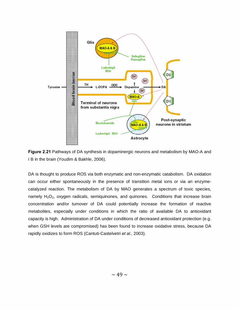
~ 49 ~
Figure 2.21 Pathways of DA synthesis in dopaminergic neurons and metabolism by MAO-A and
–B in the brain (Youdim & Bakhle, 2006).
DA is thought to produce ROS via both enzymatic and non-enzymatic catabolism. DA oxidation
can occur either spontaneously in the presence of transition metal ions or via an enzyme-
catalyzed reaction. The metabolism of DA by MAO generates a spectrum of toxic species,
namely H2O2, oxygen radicals, semiquinones, and quinones. Conditions that increase brain
concentration and/or turnover of DA could potentially increase the formation of reactive
metabolites, especially under conditions in which the ratio of available DA to antioxidant
capacity is high. Administration of DA under conditions of decreased antioxidant protection (e.g.
when GSH levels are compromised) has been found to increase oxidative stress, because DA
rapidly oxidizes to form ROS (Cantuti-Castelvetri et al., 2003).

~ 50 ~
2.2.5 The potential role of MAO-A in PD
In rodents, MAO-A is present in the extraneuronal compartment and within the dopaminergic
terminals, were it is involved in the metabolism of both intraneuronal and released DA,
respectively. The intraneuronal enzyme ensures a low level of the neurotransmitter
monoamines within the neuron (Youdim et al., 1988). Little attention had been paid to MAO-A
inhibition as a practical means of controlling DA levels in the brain, even though it had clearly
been established that DA is as well metabolized by MAO-A, as by MAO-B, and that the striatum
contains MAO-A (Green et al., 1977). It is therefore plausible that mixed MAO-A/B inhibitors
may be more effective in enhancing central DA levels and may present a more effective
therapeutic strategy than selective MAO inhibitors (Youdim & Bakhle, 2006).
2.2.6 Irreversible inhibitors of MAO-B
2.2.6.1 (R)-Deprenyl
Deprenyl (selegiline) irreversibly inhibits MAO-B and its (-)-isomer is a more potent inhibitor,
than its (+)-enantiomer. The neuroprotective action of selegiline is also multi-fold in nature.
There are at least six accepted mechanisms by which selegiline could prevent
neurodegeneration:
1. It may decrease the free radical formation from the normal metabolism of the biogenic
amines, mainly DA, by inhibition of MAO-B in the CNS. Further reaction between
endogenous amines and aldehydes formed by MAO-B, can also play a role in
neurodegeneration.
2. The inhibitor may increase the free radical scavenging capacity of the brain.
3. Due to MAO-B inhibition, selegiline may prevent the activation of the environmental pre-
toxins.
4. Oral administration of selegiline inhibits the oxidation of low-density lipoprotein (LDL)
isolated from healthy men and post-menopausal women.
5. Selegiline exhibits protective effects against neuronal apoptosis in cell culture.
6. Due to the uptake inhibitory properties at the nerve endings, selegiline may reduce
neuronal damage (Riederer et al., 2004).
~ 51 ~
Studies have shown that treatment with selegiline slows the progression of PD (Riederer et al.,
2004). In other studies, statistically significant effects of selegiline on memory and attention was
shown, that seem to be due to an improved function of the monoaminergic systems involved in
the process of neuronal degeneration (Riederer et al., 2004). Selegiline at low doses inhibits
the oxidative deamination of DA, phenylethylamine and benzylamine, but not of NA or 5-HT. At
higher doses the selectivity is lost. Selegiline is free from the cheese reaction and suitable for
use as an adjunct in PD patients already treated with L-dopa, since the basal ganglia from
human brain contains MAO-B. A preliminary analysis of the clinical response and survival of
these patients indicated that those receiving selegiline plus L-dopa had a better survival rate
than those treated with L-dopa alone. These results suggested that selegiline may slow the rate
of degeneration of dopaminergic neurons (Youdim & Bakhle, 2006).
2.2.6.2 Rasagiline
Rasagiline is a propargylamine-containing compound that possesses a selective irreversible
inhibitory effect on MAO-B. It has structural similarity to selegiline, but unlike the latter, is not a
sympathomimetic drug and is not metabolized to amphetamine. In vivo studies demonstrated
that rasagiline is up to 10 times more active than selegiline as a MAO-B inhibitor. Its
neuroprotective activity has been examined in several in vitro, in vivo and cell culture studies. It
was found to also prevent the cell death caused by apoptosis (Finberg et al., 1999).
For a MAO inhibitor to be effective in PD, it must raise the levels of DA at its receptor sites in the
striatum. Using microdialysis techniques in rat striatum, chronic treatment with rasagiline and
selegiline was shown to increase, by a similar extent, DA levels in the microdialysate. In
primates, which have a larger proportion of MAO-B in the brain than rodents, extracellular DA
levels in the striatum were studied, following local infusion of L-dopa via a microdialysis probe.
Rasagiline, administered systemically, enhanced DA levels in microdialysate following L-dopa
treatment. This MAO-B inhibitor appears to have the appropriate activities to alleviate the
symptoms of PD (Finberg et al., 1998). As for neuroprotection, rasagiline possesses activity
superior to that of selegiline in neuronal cultures. The neuroprotective activity of rasagiline is
dependent of its stereochemistry, however it is independent of stereochemistry, since the S-
enantiomer of rasagiline, TVP1022 exhibits similar activity. Studies also claim that the clinical

~ 52 ~
benefits of rasagiline are not entirely symptomatic in nature and that rasagiline may have an
added neuroprotective effect (Mandell et al., 2005).
2.2.7 Reversible inhibitors of MAO-B
2.2.7.1 Lazabemide
Lazabemide is a short-acting, reversible, highly selective inhibitor of MAO-B, which unlike
deprenyl, is not metabolized to pharmacologically active metabolites. Previously, lazabemide
was found to be safe and well tolerated at dosages of up to 400 mg per day during a 6-week
study of patients with early-untreated PD. At dosages raging from 25 to 200 mg per day,
lazabemide was well tolerated and delayed the need for L-dopa in early, otherwise untreated
PD. The antioxidant activity of lazabemide was significantly higher than that of either vitamin E
or the MAO-B inhibitor, selegiline. The ability of lazabemide to inhibit oxidative damage may be
attributed to physico-chemical interactions with the membrane lipid bilayer, as determined by
small angle X-ray diffraction methods (Mason et al., 2000).
Lazabemide differs from selegiline in several properties: it is a reversible inhibitor of MAO that
has greater selectivity for the type B enzyme versus type A and undergoes rapid clearance after
discontinuation. Lazabemide is not a propargylamine derivative and is not metabolized to
amphetamine (Parkinson’s Study Group, 1994). Some studies showed that the symptomatic
effects of lazabemide are similar to those of selegiline. It is reported that lazabemide reduces
the need for L-dopa treatment by 51% (Parkinson’s Study Group, 1996).
2.2.7.2 Safinamide
Safinamide is an α-aminoamide derivative that may combine MAO-B inhibition with DA reuptake
inhibition. Treatment with safinamide is associated with improvements in measures of cognitive
function, strategic target detection and auditory number sequencing. Safinamide may represent
an alternative to currently available therapies as an adjunct to L-dopa or DA agonists in patients
with PD (Fernandez & Chen, 2007).
~ 53 ~
2.2.8 Inhibitors of MAO-A
The studies with selegiline and rasagiline on MAO-B provided encouragement for others to
continue with the development of selective, but reversible, inhibitors of MAO-A, lacking the
cheese reaction. In rodents, MAO-A is present in the extraneuronal compartment and within the
dopaminergic terminals, where it is involved in the metabolism of both intraneuronal and
released DA, respectively (Youdim et al., 1988). MAO-A inhibition as a practical means of
controlling DA levels in brain, received little attention even though it had been clearly
established that DA is as well metabolized by MAO-A, as by MAO-B, and that the striatum
contains MAO-A. This was partly because MAO-A inhibition was known to induce the cheese
reaction and partly because there was little evidence of the effect of MAO-A inhibition on DA
levels in humans. In brains, obtained at autopsy from patients after treatment with either
selegiline or clorgyline, the increase in DA was not as marked as the increase in
phenylethylamine, NA and 5-HT (Youdim et al., 1972). Clorgyline does not alter DA levels in
the rat brain. The results of laboratory experiments indicated that when one isoform of MAO
was fully inhibited, the other isoform would metabolize DA adequately. Thus, with selective
inhibition of MAO-A or -B, the levels of DA will not change drastically in the human striatum
(Youdim & Bakhle, 2006).
The risk of producing the cheese reaction was abolished by the advent of the reversible MAO-A
inhibitors such as moclobemide. This is because reversibility allows competition, so that
ingested tyramine is able to displace the inhibitor from the enzyme and can be metabolized in
the normal way, in the gut and liver. It had also become possible to show dynamic changes in
striatal DA by microdialysis studies in rodents and these showed a clear increase of DA release
after moclobemide, clorgyline or rasagiline treatment (Haefely et al., 1992). Although selective
inhibition of MAO-A or -B did not affect the steady state levels of DA in the brain, such inhibition
did affect its release. Such action could explain the anti-symptomatic effects of these drugs in
PD patients. Clinical studies of moclobemide in PD were carried out as an addition to therapy
with L-dopa and dopaminergic agonists. Moclobemide has a mild symptomatic benefit, mostly
on motor functions. This inhibitor is also safe and effective in patients treated with L-dopa and a
peripheral decarboxylase inhibitor (Youdim & Weinstock, 2004).

~ 54 ~
Microdialysis studies showed that moclobemide can be displaced from its binding site on MAO-
A by DA and dis-inhibition of the enzyme would result (Colzi et al., 1993). This would allow DA
to be metabolized and decrease the amounts available for binding to receptors. Clinical
evaluation of other reversible selective MAO-A inhibitors, such as brofaromine and befloxatone,
which have greater affinity for MAO-A, are underway (Davidson, 2003).
ONH
Br
O
H
H2N
H
OF3C
N
OO
O
OH
Brofaromine Befloxatone
Tranylcypromine
Figure 2.22 The chemical structures of selected MAO-A inhibitors.
MAO inhibitors have a range of potential therapeutic uses. As previously discussed, the first
MAO inhibitory antidepressant to be discovered was iproniazid. This compound was initially
developed for the treatment of tuberculosis. Although it proved to be ineffective, it was
observed to have ‘psychoenergizing’ effects in patients and was also shown to inhibit MAO.
This was followed by the development of other hydrazine derived MAO inhibitors, such as
phenelzine, as antidepressants. Reports of liver toxicity, hypertensive crises, haemorrhage, and
in some cases, death, resulted in the withdrawal of most hydrazine type MAO inhibitors (Youdim
et al., 2006).

~ 55 ~
N
NH
HN
OHN NH2
Iproniazid Phenelzine
Figure 2.23 The chemical structures of selected antidepressant MAO-A inhibitors.
2.2.9 Mechanism of action of MAO-B
2.2.9.1 General background
Monoamine oxidase A and B are mitochondrial bound flavoproteins that catalyze the oxidative
deamination of neurotransmitters and biogenic amines. A number of mechanism-based
inhibitors have been developed for clinical use as antidepressants and as neuroprotective
drugs. The binding of substrates or inhibitors to MAO-B involves an initial negotiation of a
protein loop, occurring near the surface of the membrane and two hydrophobic cavities, an
‘entrance’ cavity and an ‘active site’ cavity. These two cavities can either be separate or in a
fused state, depending on the conformation of the Ile-199 side chain, which appears to function
as a gate. The amine functional groups of the bound substrate approaches the re face of the
bent and ‘puckered’ covalent FAD through an ‘aromatic cage’ formed by two tyrosine residues
that are perpendicular to the plane of the flavin ring. No amino acid residues that may function
as acids or bases are found near the catalytic site (Edmondson et al., 2004).
A breakthrough to our understanding of the molecular properties of MAO-A and -B was the
cloning and sequencing of the respective genes for these two enzymes, which convincingly
demonstrated that MAO-A and -B are two separate enzymes that share many similar properties
such as the same type of covalent flavin, an 8α-S-cysteinyl FAD and a 70% sequence identity
(Bach et al., 1988).
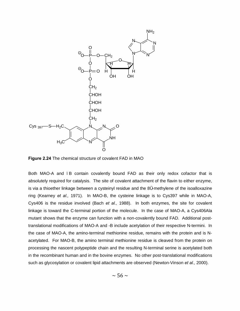
~ 56 ~
N
N
N
N
NH2
OH
OH
H
OH
CH2
H
OP
O
O
O
P OO
O
CH2
CHOH
CHOH
CHOH
CH2
N
N
N
NH
O
O
H2C
H3C
SCys 397
H
Figure 2.24 The chemical structure of covalent FAD in MAO
Both MAO-A and –B contain covalently bound FAD as their only redox cofactor that is
absolutely required for catalysis. The site of covalent attachment of the flavin to either enzyme,
is via a thioether linkage between a cysteinyl residue and the 8α-methylene of the isoalloxazine
ring (Kearney et al., 1971). In MAO-B, the cysteine linkage is to Cys397 while in MAO-A,
Cys406 is the residue involved (Bach et al., 1988). In both enzymes, the site for covalent
linkage is toward the C-terminal portion of the molecule. In the case of MAO-A, a Cys406Ala
mutant shows that the enzyme can function with a non-covalently bound FAD. Additional post-
translational modifications of MAO-A and -B include acetylation of their respective N-termini. In
the case of MAO-A, the amino-terminal methionine residue, remains with the protein and is N-
acetylated. For MAO-B, the amino terminal methionine residue is cleaved from the protein on
processing the nascent polypeptide chain and the resulting N-terminal serine is acetylated both
in the recombinant human and in the bovine enzymes. No other post-translational modifications
such as glycosylation or covalent lipid attachments are observed (Newton-Vinson et al., 2000).
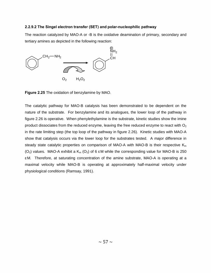
~ 57 ~
2.2.9.2 The Singel electron transfer (SET) and polar-nucleophilic pathway
The reaction catalyzed by MAO-A or -B is the oxidative deamination of primary, secondary and
tertiary amines as depicted in the following reaction:
CH2 NH2 CH
NH2
O2 H2O2
Figure 2.25 The oxidation of benzylamine by MAO.
The catalytic pathway for MAO-B catalysis has been demonstrated to be dependent on the
nature of the substrate. For benzylamine and its analogues, the lower loop of the pathway in
figure 2.26 is operative. When phenylethylamine is the substrate, kinetic studies show the imine
product dissociates from the reduced enzyme, leaving the free reduced enzyme to react with O2
in the rate limiting step (the top loop of the pathway in figure 2.26). Kinetic studies with MAO-A
show that catalysis occurs via the lower loop for the substrates tested. A major difference in
steady state catalytic properties on comparison of MAO-A with MAO-B is their respective Km
(O2) values. MAO-A exhibit a Km (O2) of 6 μM while the corresponding value for MAO-B is 250
μM. Therefore, at saturating concentration of the amine substrate, MAO-A is operating at a
maximal velocity while MAO-B is operating at approximately half-maximal velocity under
physiological conditions (Ramsay, 1991).
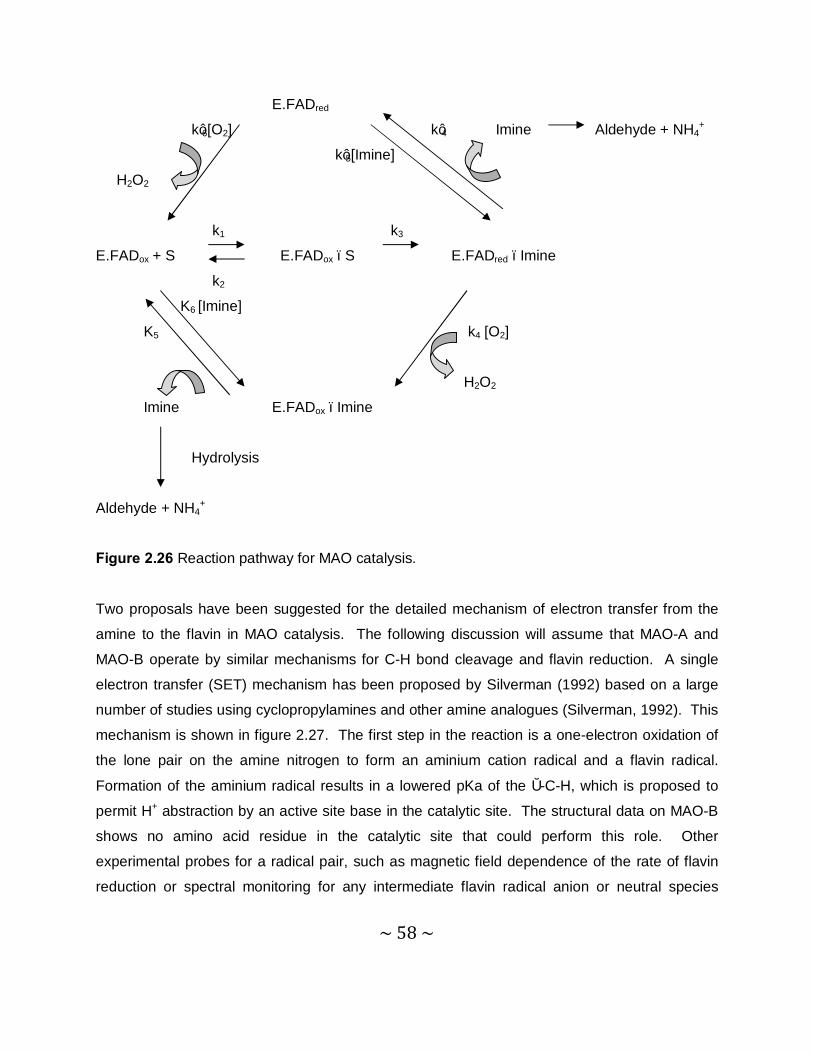
~ 58 ~
E.FADred
k’6[O2] k’4 Imine Aldehyde + NH4+
k’5[Imine]
H2O2
k1 k3
E.FADox + S E.FADox –S E.FADred –Imine
k2
K6 [Imine]
K5 k4 [O2]
H2O2
Imine E.FADox –Imine
Hydrolysis
Aldehyde + NH4+
Figure 2.26 Reaction pathway for MAO catalysis.
Two proposals have been suggested for the detailed mechanism of electron transfer from the
amine to the flavin in MAO catalysis. The following discussion will assume that MAO-A and
MAO-B operate by similar mechanisms for C-H bond cleavage and flavin reduction. A single
electron transfer (SET) mechanism has been proposed by Silverman (1992) based on a large
number of studies using cyclopropylamines and other amine analogues (Silverman, 1992). This
mechanism is shown in figure 2.27. The first step in the reaction is a one-electron oxidation of
the lone pair on the amine nitrogen to form an aminium cation radical and a flavin radical.
Formation of the aminium radical results in a lowered pKa of the α-C-H, which is proposed to
permit H+ abstraction by an active site base in the catalytic site. The structural data on MAO-B
shows no amino acid residue in the catalytic site that could perform this role. Other
experimental probes for a radical pair, such as magnetic field dependence of the rate of flavin
reduction or spectral monitoring for any intermediate flavin radical anion or neutral species
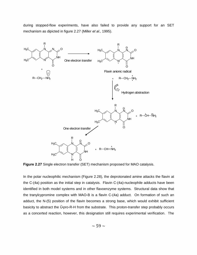
~ 59 ~
during stopped-flow experiments, have also failed to provide any support for an SET
mechanism as dipicted in figure 2.27 (Miller et al., 1995).
N
NNH
NH3C
H3C
O
O
R
One electron transfer
N
NNH
NH3C
H3C
O
O
R
Flavin anionic radical
+ R CH2 NH2
+
R CH2 NH2
Hydrogen abstraction
N
NNH
NH3C
H3C
O
O
R
One electron transfer
N
NNH
NH3C
H3C
O
O
R
H
R CH NH2+
R CH NH2+
Figure 2.27 Single electron transfer (SET) mechanism proposed for MAO catalysis.
In the polar nucleophilic mechanism (Figure 2.28), the deprotonated amine attacks the flavin at
the C-(4a) position as the initial step in catalysis. Flavin C-(4a)-nucleophile adducts have been
identified in both model systems and in other flavoenzyme systems. Structural data show that
the tranylcypromine complex with MAO-B is a flavin C-(4a) adduct. On formation of such an
adduct, the N-(5) position of the flavin becomes a strong base, which would exhibit sufficient
basicity to abstract the α-pro-R-H from the substrate. This proton-transfer step probably occurs
as a concerted reaction, however, this designation still requires experimental verification. The
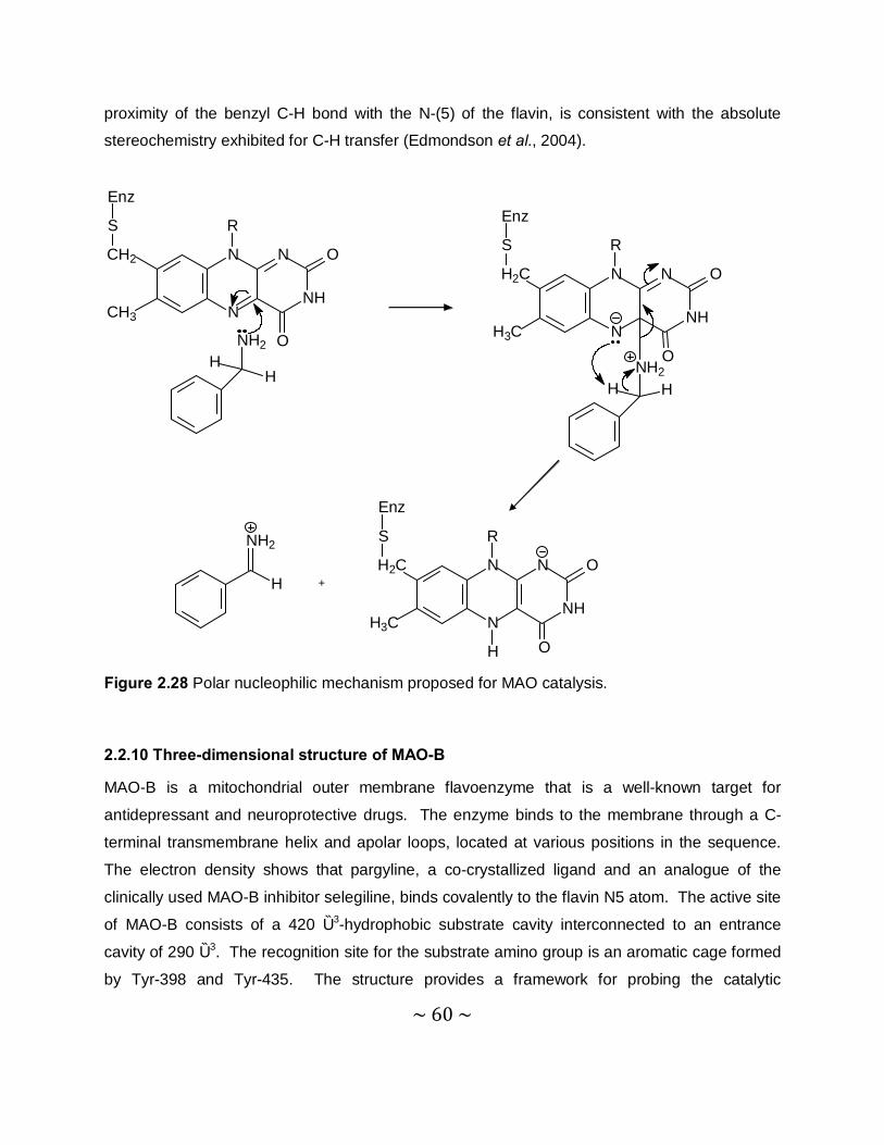
~ 60 ~
proximity of the benzyl C-H bond with the N-(5) of the flavin, is consistent with the absolute
stereochemistry exhibited for C-H transfer (Edmondson et al., 2004).
N
NNH
N O
O
CH2
CH3
RS
Enz
NH2
HH
N
NNH
N O
O
H2C
H3C
R
NH2HH
S
Enz
N
NNH
N O
O
H2C
H3C
RS
Enz
H
H
NH2
+
Figure 2.28 Polar nucleophilic mechanism proposed for MAO catalysis.
2.2.10 Three-dimensional structure of MAO-B
MAO-B is a mitochondrial outer membrane flavoenzyme that is a well-known target for
antidepressant and neuroprotective drugs. The enzyme binds to the membrane through a C-
terminal transmembrane helix and apolar loops, located at various positions in the sequence.
The electron density shows that pargyline, a co-crystallized ligand and an analogue of the
clinically used MAO-B inhibitor selegiline, binds covalently to the flavin N5 atom. The active site
of MAO-B consists of a 420 Ǻ3-hydrophobic substrate cavity interconnected to an entrance
cavity of 290 Ǻ3. The recognition site for the substrate amino group is an aromatic cage formed
by Tyr-398 and Tyr-435. The structure provides a framework for probing the catalytic
~ 61 ~
mechanism, understanding the differences between the MAO-A and -B isoforms and designing
specific inhibitors. The human MAO-B structure was solved by the single isomorphous
replacement method combined with multicrystal 12-fold averaging. These results reveal the
architecture of the catalytic centre and of sites on the protein that are important for its binding to
the outer membrane of the mitochondrion (Binda et al., 2001).
The 520 amino acids of MAO-B fold into a compact structure that exhibit a topology initially
found in p-hydroxybensoate hydroxylase and then observed in several other flavoproteins. The
crystal structure shows that the enzyme is dimeric, which is unlikely to be a crystal packing
artifact because the dimer is present in the two crystal forms (orthorhombic and triclinic)
employed in the structure determination. The monomer-monomer interactions are indeed
extensive (Binda et al., 2001), with each monomer consisting of a globular domain anchored to
the membrane through a C-terminal helix. The MAO-B active site consists of two cavities, the
substrate cavity in front of the flavin and the entrance cavity located underneath the protein
surface and closed by the loop formed by residues 99-112 (Binda et al., 2007). MAO-B is tightly
bound to the outer mitochondrial membrane, as evidenced by the need for digestion of
phospholipids for its efficient detergent extraction. The protein region responsible for membrane
attachment is formed by the C-terminal amino acids, 461-520. Analysis of the MAO-B amino
acid sequence, predicts that residues 489-515 form a transmembrane helix, 27 amino acids
long, which is well within the range of values observed for transmembrane helices in membrane
proteins of known three-dimensional structures (Binda et al., 2001).
The crystal structure reveals that the C-terminal residues form an extended polypeptide chain,
that traverses the monomer surface. This extended chain is followed by a α-helix that initiates
at Val-489, forming the predicted transmembrane helical segment. The helix of each monomer
protrudes from the basal face of the dimer, with each helical axis approximately parallel to the
molecular two-fold axis. This observation suggests that the dimer binds to the membrane with
its two-fold axis perpendicular to the membrane plane and the C-terminal helices inserted in the
lipid bilayer. In addition to the C-terminal helical segment, the structure shows that other protein
regions may be involved in membrane binding (Ulmschneider & Sansom, 2001).
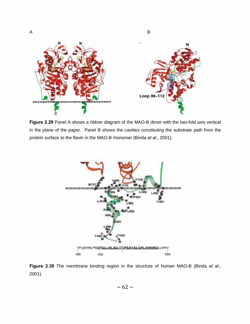
~ 62 ~
A B
Figure 2.29 Panel A shows a ribbon diagram of the MAO-B dimer with the two-fold axis vertical
in the plane of the paper. Panel B shows the cavities constituting the substrate path from the
protein surface to the flavin in the MAO-B monomer (Binda et al., 2001).
Figure 2.30 The membrane binding region in the structure of human MAO-B (Binda et al.,
2001).
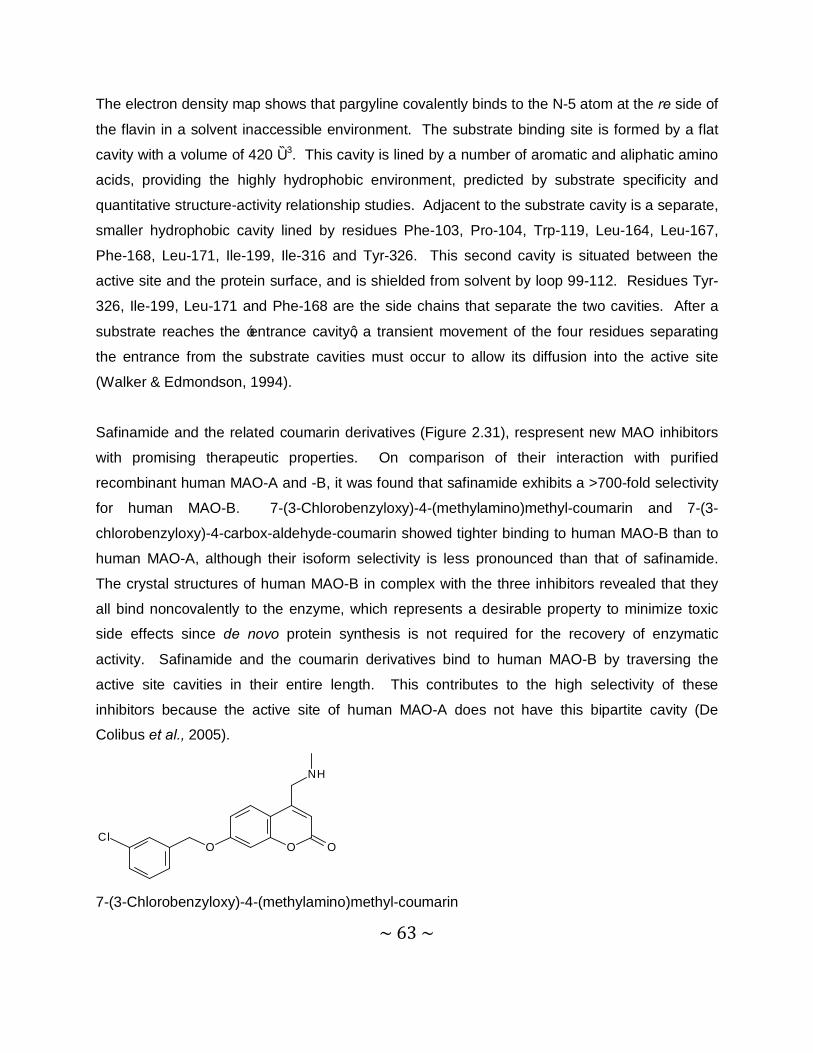
~ 63 ~
The electron density map shows that pargyline covalently binds to the N-5 atom at the re side of
the flavin in a solvent inaccessible environment. The substrate binding site is formed by a flat
cavity with a volume of 420 Ǻ3. This cavity is lined by a number of aromatic and aliphatic amino
acids, providing the highly hydrophobic environment, predicted by substrate specificity and
quantitative structure-activity relationship studies. Adjacent to the substrate cavity is a separate,
smaller hydrophobic cavity lined by residues Phe-103, Pro-104, Trp-119, Leu-164, Leu-167,
Phe-168, Leu-171, Ile-199, Ile-316 and Tyr-326. This second cavity is situated between the
active site and the protein surface, and is shielded from solvent by loop 99-112. Residues Tyr-
326, Ile-199, Leu-171 and Phe-168 are the side chains that separate the two cavities. After a
substrate reaches the ‘entrance cavity’, a transient movement of the four residues separating
the entrance from the substrate cavities must occur to allow its diffusion into the active site
(Walker & Edmondson, 1994).
Safinamide and the related coumarin derivatives (Figure 2.31), respresent new MAO inhibitors
with promising therapeutic properties. On comparison of their interaction with purified
recombinant human MAO-A and -B, it was found that safinamide exhibits a >700-fold selectivity
for human MAO-B. 7-(3-Chlorobenzyloxy)-4-(methylamino)methyl-coumarin and 7-(3-
chlorobenzyloxy)-4-carbox-aldehyde-coumarin showed tighter binding to human MAO-B than to
human MAO-A, although their isoform selectivity is less pronounced than that of safinamide.
The crystal structures of human MAO-B in complex with the three inhibitors revealed that they
all bind noncovalently to the enzyme, which represents a desirable property to minimize toxic
side effects since de novo protein synthesis is not required for the recovery of enzymatic
activity. Safinamide and the coumarin derivatives bind to human MAO-B by traversing the
active site cavities in their entire length. This contributes to the high selectivity of these
inhibitors because the active site of human MAO-A does not have this bipartite cavity (De
Colibus et al., 2005).
O O OCl
NH
7-(3-Chlorobenzyloxy)-4-(methylamino)methyl-coumarin
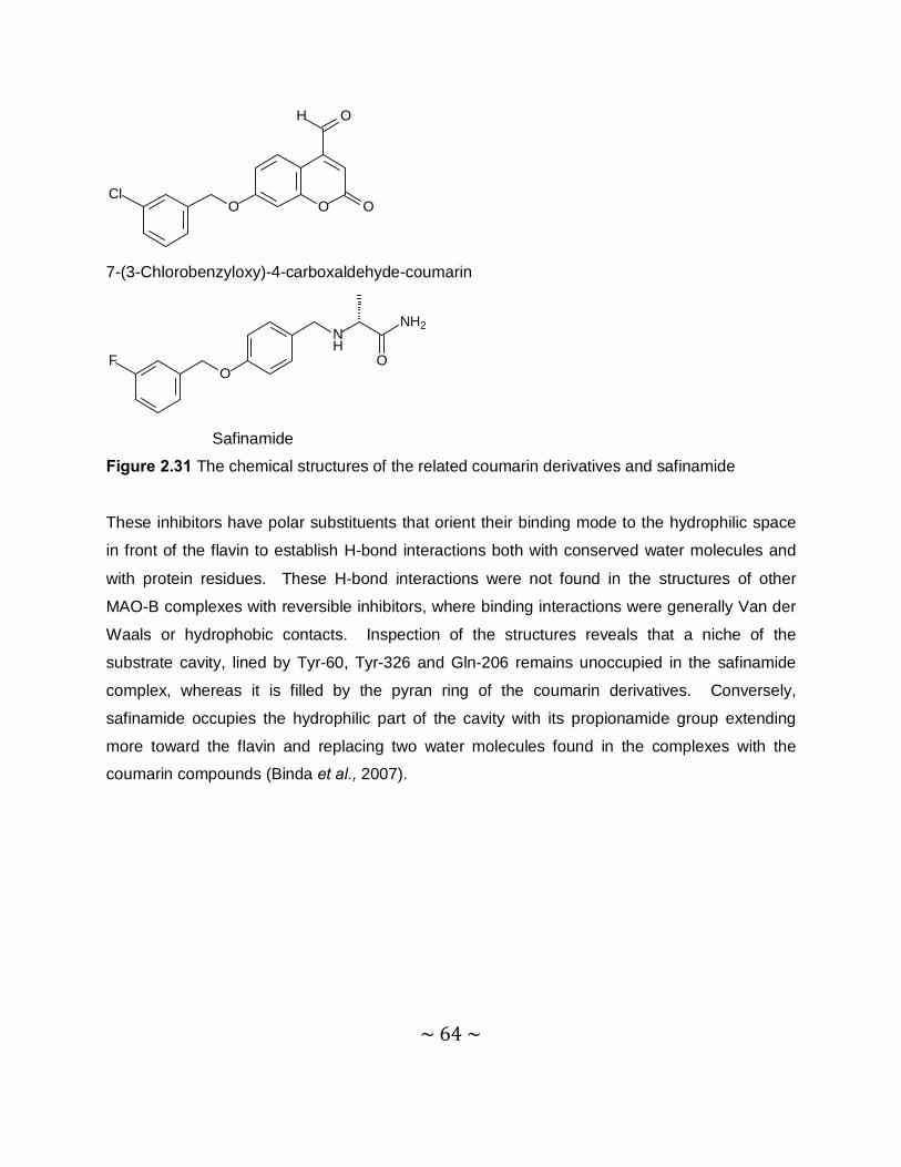
~ 64 ~
O O OCl
OH
7-(3-Chlorobenzyloxy)-4-carboxaldehyde-coumarin
O
NH
NH2
F O
Safinamide
Figure 2.31 The chemical structures of the related coumarin derivatives and safinamide
These inhibitors have polar substituents that orient their binding mode to the hydrophilic space
in front of the flavin to establish H-bond interactions both with conserved water molecules and
with protein residues. These H-bond interactions were not found in the structures of other
MAO-B complexes with reversible inhibitors, where binding interactions were generally Van der
Waals or hydrophobic contacts. Inspection of the structures reveals that a niche of the
substrate cavity, lined by Tyr-60, Tyr-326 and Gln-206 remains unoccupied in the safinamide
complex, whereas it is filled by the pyran ring of the coumarin derivatives. Conversely,
safinamide occupies the hydrophilic part of the cavity with its propionamide group extending
more toward the flavin and replacing two water molecules found in the complexes with the
coumarin compounds (Binda et al., 2007).
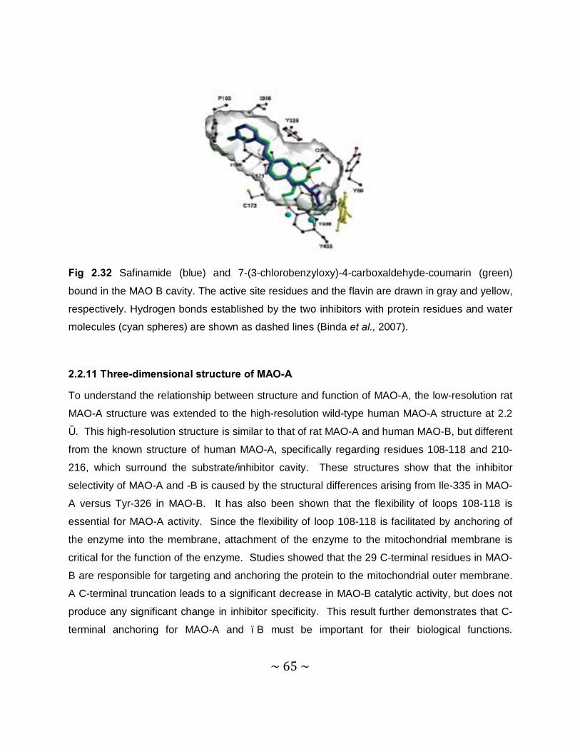
~ 65 ~
Fig 2.32 Safinamide (blue) and 7-(3-chlorobenzyloxy)-4-carboxaldehyde-coumarin (green)
bound in the MAO B cavity. The active site residues and the flavin are drawn in gray and yellow,
respectively. Hydrogen bonds established by the two inhibitors with protein residues and water
molecules (cyan spheres) are shown as dashed lines (Binda et al., 2007).
2.2.11 Three-dimensional structure of MAO-A
To understand the relationship between structure and function of MAO-A, the low-resolution rat
MAO-A structure was extended to the high-resolution wild-type human MAO-A structure at 2.2
Ǻ. This high-resolution structure is similar to that of rat MAO-A and human MAO-B, but different
from the known structure of human MAO-A, specifically regarding residues 108-118 and 210-
216, which surround the substrate/inhibitor cavity. These structures show that the inhibitor
selectivity of MAO-A and -B is caused by the structural differences arising from Ile-335 in MAO-
A versus Tyr-326 in MAO-B. It has also been shown that the flexibility of loops 108-118 is
essential for MAO-A activity. Since the flexibility of loop 108-118 is facilitated by anchoring of
the enzyme into the membrane, attachment of the enzyme to the mitochondrial membrane is
critical for the function of the enzyme. Studies showed that the 29 C-terminal residues in MAO-
B are responsible for targeting and anchoring the protein to the mitochondrial outer membrane.
A C-terminal truncation leads to a significant decrease in MAO-B catalytic activity, but does not
produce any significant change in inhibitor specificity. This result further demonstrates that C-
terminal anchoring for MAO-A and –B must be important for their biological functions.
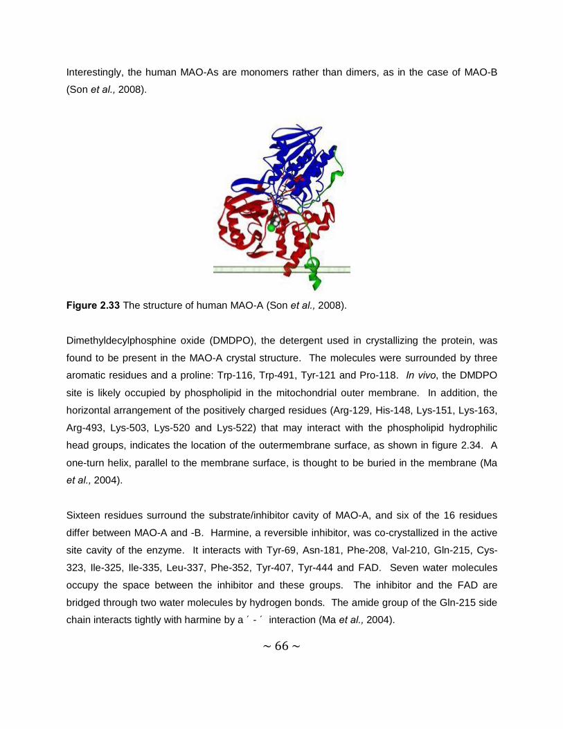
~ 66 ~
Interestingly, the human MAO-As are monomers rather than dimers, as in the case of MAO-B
(Son et al., 2008).
Figure 2.33 The structure of human MAO-A (Son et al., 2008).
Dimethyldecylphosphine oxide (DMDPO), the detergent used in crystallizing the protein, was
found to be present in the MAO-A crystal structure. The molecules were surrounded by three
aromatic residues and a proline: Trp-116, Trp-491, Tyr-121 and Pro-118. In vivo, the DMDPO
site is likely occupied by phospholipid in the mitochondrial outer membrane. In addition, the
horizontal arrangement of the positively charged residues (Arg-129, His-148, Lys-151, Lys-163,
Arg-493, Lys-503, Lys-520 and Lys-522) that may interact with the phospholipid hydrophilic
head groups, indicates the location of the outermembrane surface, as shown in figure 2.34. A
one-turn helix, parallel to the membrane surface, is thought to be buried in the membrane (Ma
et al., 2004).
Sixteen residues surround the substrate/inhibitor cavity of MAO-A, and six of the 16 residues
differ between MAO-A and -B. Harmine, a reversible inhibitor, was co-crystallized in the active
site cavity of the enzyme. It interacts with Tyr-69, Asn-181, Phe-208, Val-210, Gln-215, Cys-
323, Ile-325, Ile-335, Leu-337, Phe-352, Tyr-407, Tyr-444 and FAD. Seven water molecules
occupy the space between the inhibitor and these groups. The inhibitor and the FAD are
bridged through two water molecules by hydrogen bonds. The amide group of the Gln-215 side
chain interacts tightly with harmine by a π- π interaction (Ma et al., 2004).
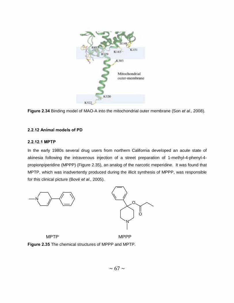
~ 67 ~
Figure 2.34 Binding model of MAO-A into the mitochondrial outer membrane (Son et al., 2008).
2.2.12 Animal models of PD
2.2.12.1 MPTP
In the early 1980s several drug users from northern California developed an acute state of
akinesia following the intravenous injection of a street preparation of 1-methyl-4-phenyl-4-
propionpiperidine (MPPP) (Figure 2.35), an analog of the narcotic meperidine. It was found that
MPTP, which was inadvertently produced during the illicit synthesis of MPPP, was responsible
for this clinical picture (Bové et al., 2005).
N
N
O
O
MPTP MPPP Figure 2.35 The chemical structures of MPPP and MPTP.
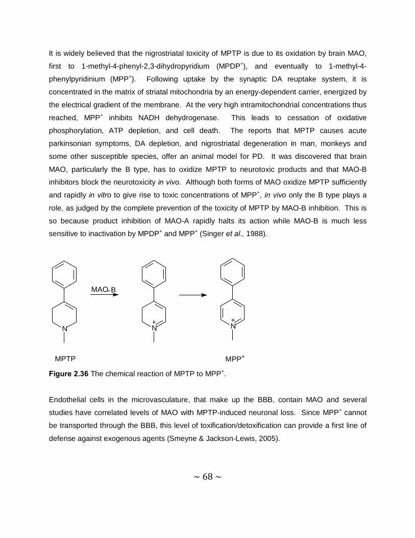
~ 68 ~
It is widely believed that the nigrostriatal toxicity of MPTP is due to its oxidation by brain MAO,
first to 1-methyl-4-phenyl-2,3-dihydropyridium (MPDP+), and eventually to 1-methyl-4-
phenylpyridinium (MPP+). Following uptake by the synaptic DA reuptake system, it is
concentrated in the matrix of striatal mitochondria by an energy-dependent carrier, energized by
the electrical gradient of the membrane. At the very high intramitochondrial concentrations thus
reached, MPP+ inhibits NADH dehydrogenase. This leads to cessation of oxidative
phosphorylation, ATP depletion, and cell death. The reports that MPTP causes acute
parkinsonian symptoms, DA depletion, and nigrostriatal degeneration in man, monkeys and
some other susceptible species, offer an animal model for PD. It was discovered that brain
MAO, particularly the B type, has to oxidize MPTP to neurotoxic products and that MAO-B
inhibitors block the neurotoxicity in vivo. Although both forms of MAO oxidize MPTP sufficiently
and rapidly in vitro to give rise to toxic concentrations of MPP+, in vivo only the B type plays a
role, as judged by the complete prevention of the toxicity of MPTP by MAO-B inhibition. This is
so because product inhibition of MAO-A rapidly halts its action while MAO-B is much less
sensitive to inactivation by MPDP+ and MPP+ (Singer et al., 1988).
N N
MAO B
N
MPTP MPP+
Figure 2.36 The chemical reaction of MPTP to MPP+.
Endothelial cells in the microvasculature, that make up the BBB, contain MAO and several
studies have correlated levels of MAO with MPTP-induced neuronal loss. Since MPP+ cannot
be transported through the BBB, this level of toxification/detoxification can provide a first line of
defense against exogenous agents (Smeyne & Jackson-Lewis, 2005).
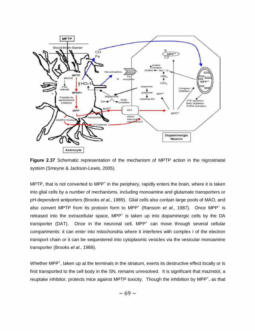
~ 69 ~
Figure 2.37 Schematic representation of the mechanism of MPTP action in the nigrostriatal
system (Smeyne & Jackson-Lewis, 2005).
MPTP, that is not converted to MPP+ in the periphery, rapidly enters the brain, where it is taken
into glial cells by a number of mechanisms, including monoamine and glutamate transporters or
pH-dependent antiporters (Brooks et al., 1989). Glial cells also contain large pools of MAO, and
also convert MPTP from its protoxin form to MPP+ (Ransom et al., 1987). Once MPP+ is
released into the extracellular space, MPP+ is taken up into dopaminergic cells by the DA
transporter (DAT). Once in the neuronal cell, MPP+ can move through several cellular
compartments: it can enter into mitochondria where it interferes with complex I of the electron
transport chain or it can be sequestered into cytoplasmic vesicles via the vesicular monoamine
transporter (Brooks et al., 1989).
Whether MPP+, taken up at the terminals in the striatum, exerts its destructive effect locally or is
first transported to the cell body in the SN, remains unresolved. It is significant that mazindol, a
reuptake inhibitor, protects mice against MPTP toxicity. Though the inhibition by MPP+, as that
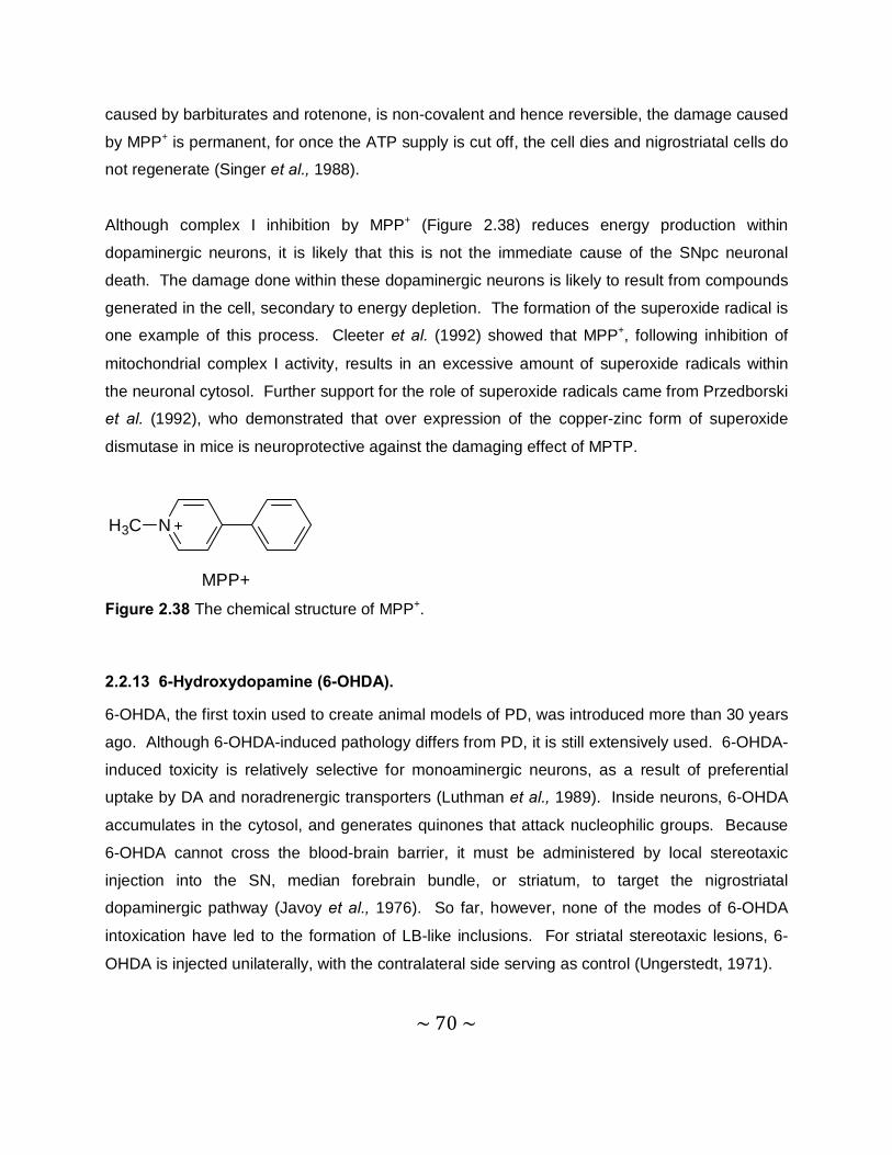
~ 70 ~
caused by barbiturates and rotenone, is non-covalent and hence reversible, the damage caused
by MPP+ is permanent, for once the ATP supply is cut off, the cell dies and nigrostriatal cells do
not regenerate (Singer et al., 1988).
Although complex I inhibition by MPP+ (Figure 2.38) reduces energy production within
dopaminergic neurons, it is likely that this is not the immediate cause of the SNpc neuronal
death. The damage done within these dopaminergic neurons is likely to result from compounds
generated in the cell, secondary to energy depletion. The formation of the superoxide radical is
one example of this process. Cleeter et al. (1992) showed that MPP+, following inhibition of
mitochondrial complex I activity, results in an excessive amount of superoxide radicals within
the neuronal cytosol. Further support for the role of superoxide radicals came from Przedborski
et al. (1992), who demonstrated that over expression of the copper-zinc form of superoxide
dismutase in mice is neuroprotective against the damaging effect of MPTP.
NH3C
MPP+ Figure 2.38 The chemical structure of MPP+.
2.2.13 6-Hydroxydopamine (6-OHDA).
6-OHDA, the first toxin used to create animal models of PD, was introduced more than 30 years
ago. Although 6-OHDA-induced pathology differs from PD, it is still extensively used. 6-OHDA-
induced toxicity is relatively selective for monoaminergic neurons, as a result of preferential
uptake by DA and noradrenergic transporters (Luthman et al., 1989). Inside neurons, 6-OHDA
accumulates in the cytosol, and generates quinones that attack nucleophilic groups. Because
6-OHDA cannot cross the blood-brain barrier, it must be administered by local stereotaxic
injection into the SN, median forebrain bundle, or striatum, to target the nigrostriatal
dopaminergic pathway (Javoy et al., 1976). So far, however, none of the modes of 6-OHDA
intoxication have led to the formation of LB-like inclusions. For striatal stereotaxic lesions, 6-
OHDA is injected unilaterally, with the contralateral side serving as control (Ungerstedt, 1971).

~ 71 ~
6-OHDA shares some structural similarities with DA and NA, exhibiting a high affinity for several
catecholaminergic plasma membrane transporters such as the DAT and norepinephrine
transporters (NET). With respect to its mode of action, it is well accepted that 6-OHDA destroys
catecholaminergic structures by a combined effect of ROS and quinones. This view stems
primarily from the demonstration that 6-OHDA, once dissolved in an aerobic and alkaline milieu,
readily oxidizes, yielding H2O2 and para-quinone (Bové et al., 2005).
HO
O NH2
OH2O2
HO OH
NH2HO
O2
Figure 2.39 The oxidation of 6-OHDA.
Like other parkinsonian neurotoxins to be discussed here, 6-OHDA can be administered by
systemic injection. However, contrary to MPTP, rotenone or paraquat, this route of
administration will not produce the desired nigrostriatal lesion. Instead, this route of 6-OHDA
administration will lead to damage of the peripheral nervous system. After 6-OHDA injections
into the substantia nigra or the medial forebrain bundle, dopaminergic neurons start to die within
the first 24 hours and show a nonapoptotic morphology. Maximal reduction of striatal DA levels
are reached within 3-4 days after lesion, and in most studies, residual striatal DA content is less
than 20% of controls (Faull & Laverty, 1969). In contrast, unilateral injections cause a typical
asymmetric circling motor behaviour whose magnitude in rodents depends on the degree of the
nigrostriatal lesion. This specific behavioural abnormality is most prominent after administration
of drugs that stimulate dopaminergic receptors, such as apomorphine. Quantification of this
turning behaviour has been used extensively to assess the antiparkinsonian potency of new
drugs, transplantation, and gene therapies and to study the motor fluctuations after the chronic
treatment with L-dopa (Bové et al., 2002).
2.2.14 Rotenone
Among the animal models of PD, rotenone represents one of the most recently used
approaches. Rotenone is a member of the rotenoids, a family of natural cytotoxic compounds
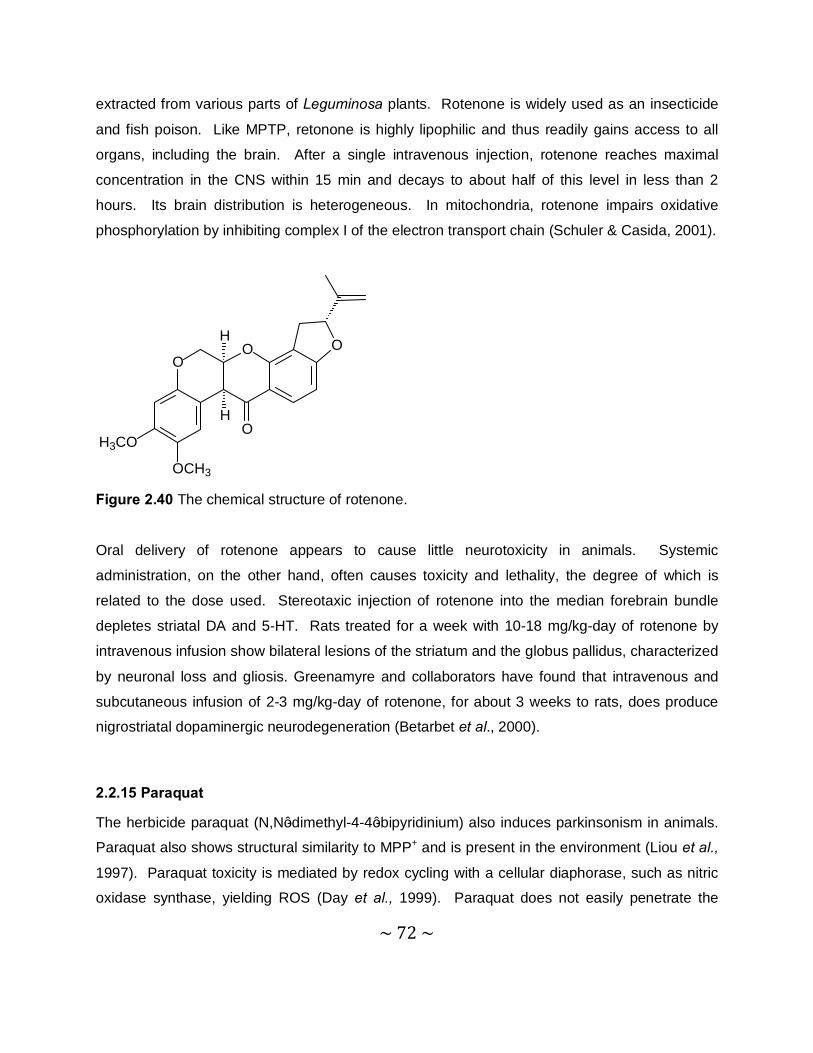
~ 72 ~
extracted from various parts of Leguminosa plants. Rotenone is widely used as an insecticide
and fish poison. Like MPTP, retonone is highly lipophilic and thus readily gains access to all
organs, including the brain. After a single intravenous injection, rotenone reaches maximal
concentration in the CNS within 15 min and decays to about half of this level in less than 2
hours. Its brain distribution is heterogeneous. In mitochondria, rotenone impairs oxidative
phosphorylation by inhibiting complex I of the electron transport chain (Schuler & Casida, 2001).
OO O
O
H
H
H3CO
OCH3 Figure 2.40 The chemical structure of rotenone.
Oral delivery of rotenone appears to cause little neurotoxicity in animals. Systemic
administration, on the other hand, often causes toxicity and lethality, the degree of which is
related to the dose used. Stereotaxic injection of rotenone into the median forebrain bundle
depletes striatal DA and 5-HT. Rats treated for a week with 10-18 mg/kg-day of rotenone by
intravenous infusion show bilateral lesions of the striatum and the globus pallidus, characterized
by neuronal loss and gliosis. Greenamyre and collaborators have found that intravenous and
subcutaneous infusion of 2-3 mg/kg-day of rotenone, for about 3 weeks to rats, does produce
nigrostriatal dopaminergic neurodegeneration (Betarbet et al., 2000).
2.2.15 Paraquat
The herbicide paraquat (N,N’-dimethyl-4-4’-bipyridinium) also induces parkinsonism in animals.
Paraquat also shows structural similarity to MPP+ and is present in the environment (Liou et al.,
1997). Paraquat toxicity is mediated by redox cycling with a cellular diaphorase, such as nitric
oxidase synthase, yielding ROS (Day et al., 1999). Paraquat does not easily penetrate the
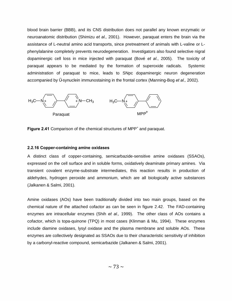
~ 73 ~
blood brain barrier (BBB), and its CNS distribution does not parallel any known enzymatic or
neuroanatomic distribution (Shimizu et al., 2001). However, paraquat enters the brain via the
assistance of L-neutral amino acid transports, since pretreatment of animals with L-valine or L-
phenylalanine completely prevents neurodegeneration. Investigators also found selective nigral
dopaminergic cell loss in mice injected with paraquat (Bové et al., 2005). The toxicity of
paraquat appears to be mediated by the formation of superoxide radicals. Systemic
administration of paraquat to mice, leads to SNpc dopaminergic neuron degeneration
accompanied by α-synuclein immunostaining in the frontal cortex (Manning-Bog et al., 2002).
Paraquat MPP
N NH3C CH3 NH3C
Figure 2.41 Comparison of the chemical structures of MPP+ and paraquat.
2.2.16 Copper-containing amine oxidases
A distinct class of copper-containing, semicarbazide-sensitive amine oxidases (SSAOs),
expressed on the cell surface and in soluble forms, oxidatively deaminate primary amines. Via
transient covalent enzyme-substrate intermediates, this reaction results in production of
aldehydes, hydrogen peroxide and ammonium, which are all biologically active substances
(Jalkanen & Salmi, 2001).
Amine oxidases (AOs) have been traditionally divided into two main groups, based on the
chemical nature of the attached cofactor as can be seen in figure 2.42. The FAD-containing
enzymes are intracellular enzymes (Shih et al., 1999). The other class of AOs contains a
cofactor, which is topa-quinone (TPQ) in most cases (Klinman & Mu, 1994). These enzymes
include diamine oxidases, lysyl oxidase and the plasma membrane and soluble AOs. These
enzymes are collectively designated as SSAOs due to their characteristic sensitivity of inhibition
by a carbonyl-reactive compound, semicarbazide (Jalkanen & Salmi, 2001).
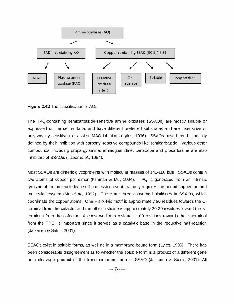
~ 74 ~
Amine oxidases (AO)
FAD – containing AO Copper containing SSAO (EC 1,4,3,6)
MAO Plasma amine oxidase (PAO)
Diamine oxidase (DAO)
Cell-surface
Soluble Lysyloxidase
Figure 2.42 The classification of AOs.
The TPQ-containing semicarbazide-sensitive amine oxidases (SSAOs) are mostly soluble or
expressed on the cell surface, and have different preferred substrates and are insensitive or
only weakly sensitive to classical MAO inhibitors (Lyles, 1996). SSAOs have been historically
defined by their inhibition with carbonyl-reactive compounds like semicarbazide. Various other
compounds, including propargylamine, aminoguanidine, carbidopa and procarbazine are also
inhibitors of SSAO’s (Tabor et al., 1954).
Most SSAOs are dimeric glycoproteins with molecular masses of 140-180 kDa. SSAOs contain
two atoms of copper per dimer (Klinman & Mu, 1994). TPQ is generated from an intrinsic
tyrosine of the molecule by a self-processing event that only requires the bound copper ion and
molecular oxygen (Mu et al., 1992). There are three conserved histidines in SSAOs, which
coordinate the copper atoms. One His-X-His motif is approximately 50 residues towards the C-
terminal from the cofactor and the other histidine is approximately 20-30 residues toward the N-
terminus from the cofactor. A conserved Asp residue, ~100 residues towards the N-terminal
from the TPQ, is important since it serves as a catalytic base in the reductive half-reaction
(Jalkanen & Salmi, 2001).
SSAOs exist in soluble forms, as well as in a membrane-bound form (Lyles, 1996). There has
been considerable disagreement as to whether the soluble form is a product of a different gene
or a cleavage product of the transmembrane form of SSAO (Jalkanen & Salmi, 2001). All
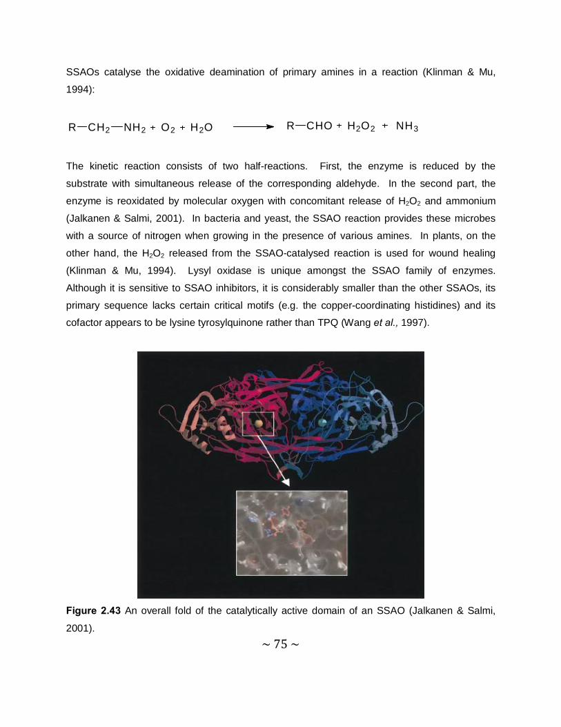
~ 75 ~
SSAOs catalyse the oxidative deamination of primary amines in a reaction (Klinman & Mu,
1994):
R CH2 NH2 O2 H2O R CHO H2O2 NH3
The kinetic reaction consists of two half-reactions. First, the enzyme is reduced by the
substrate with simultaneous release of the corresponding aldehyde. In the second part, the
enzyme is reoxidated by molecular oxygen with concomitant release of H2O2 and ammonium
(Jalkanen & Salmi, 2001). In bacteria and yeast, the SSAO reaction provides these microbes
with a source of nitrogen when growing in the presence of various amines. In plants, on the
other hand, the H2O2 released from the SSAO-catalysed reaction is used for wound healing
(Klinman & Mu, 1994). Lysyl oxidase is unique amongst the SSAO family of enzymes.
Although it is sensitive to SSAO inhibitors, it is considerably smaller than the other SSAOs, its
primary sequence lacks certain critical motifs (e.g. the copper-coordinating histidines) and its
cofactor appears to be lysine tyrosylquinone rather than TPQ (Wang et al., 1997).
Figure 2.43 An overall fold of the catalytically active domain of an SSAO (Jalkanen & Salmi,
2001).

~ 76 ~
2.2.17 Enzyme kinetics
Enzyme kinetics play a role in the determination of inhibitor potency. The Ki value is the
inhibitor constant. This constant expresses the inhibitor’s potency. The lower the value of Ki,
the higher the inhibitor’s potency while the higher the value of Ki, the lower the inhibitor’s
potency. A general equation for a chemical reaction between a substrate and enzyme can be
illustrated as:
S + E SE
Where S is the substrate and E the enzyme, Vi is the velocity of the forward reaction. If the
substrate is in excess compared to the enzyme, Vi will be directly proportionate to the enzyme
concentration (Vi α [E]). When the substrate concentration increases, Vi will increase until Vmax
is reached where a further increase in substrate will have no effect on Vi. The enzyme is thus
saturated. Vi is dependant on how quickly the substrate is released from the enzyme for
another reaction. The Michaelis-Menten equation illustrates the relationship between initial
reaction velocity Vi and substrate concentration [S], as shown graphically below:
Figure 2.44 Effect of substrate concentration on the initial velocity of an enzyme-catalyzed
reaction.
Vi
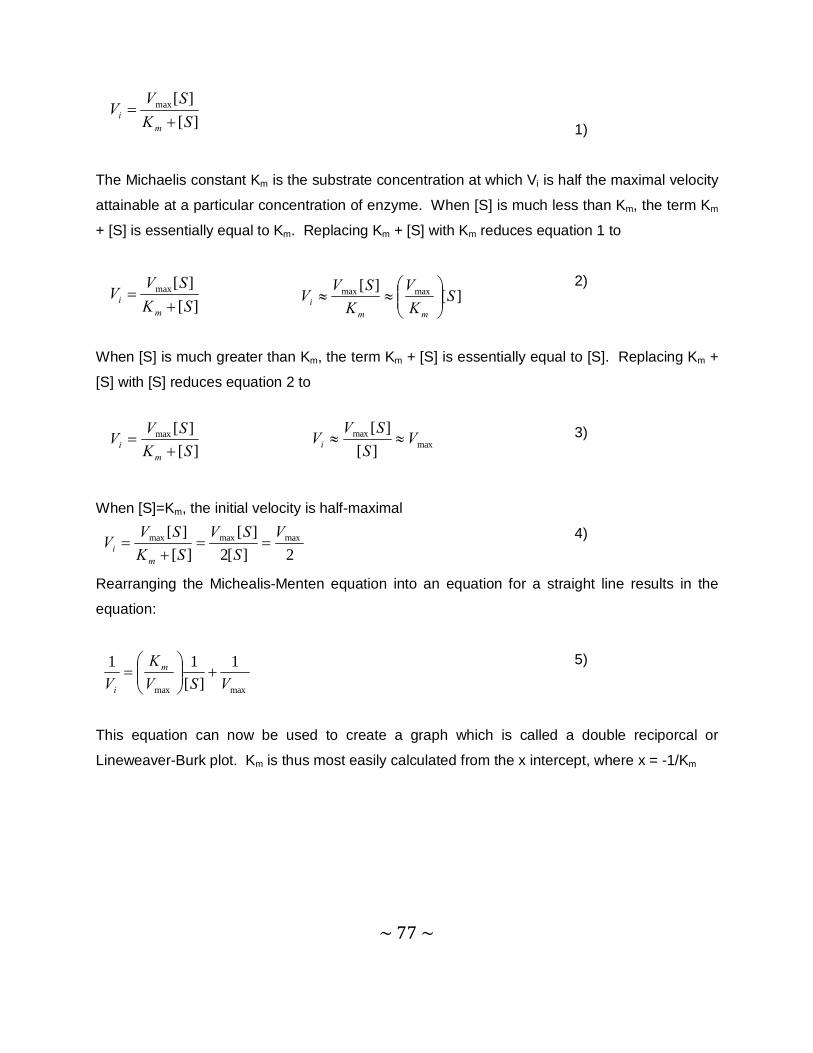
~ 77 ~
][][max
SKSV
Vm
i +=
][][max
SKSV
Vm
i += ][
][ maxmax SK
VK
SVV
mmi
≈≈
][][max
SKSVV
mi +
= maxmax
][][ V
SSVVi ≈≈
2][2][
][][ maxmaxmax V
SSV
SKSVV
mi ==
+=
maxmax
1][
11VSV
KV
m
i
+
=
1)
The Michaelis constant Km is the substrate concentration at which Vi is half the maximal velocity
attainable at a particular concentration of enzyme. When [S] is much less than Km, the term Km
+ [S] is essentially equal to Km. Replacing Km + [S] with Km reduces equation 1 to
2)
When [S] is much greater than Km, the term Km + [S] is essentially equal to [S]. Replacing Km +
[S] with [S] reduces equation 2 to
3)
When [S]=Km, the initial velocity is half-maximal
4)
Rearranging the Michealis-Menten equation into an equation for a straight line results in the
equation:
5)
This equation can now be used to create a graph which is called a double reciporcal or
Lineweaver-Burk plot. Km is thus most easily calculated from the x intercept, where x = -1/Km
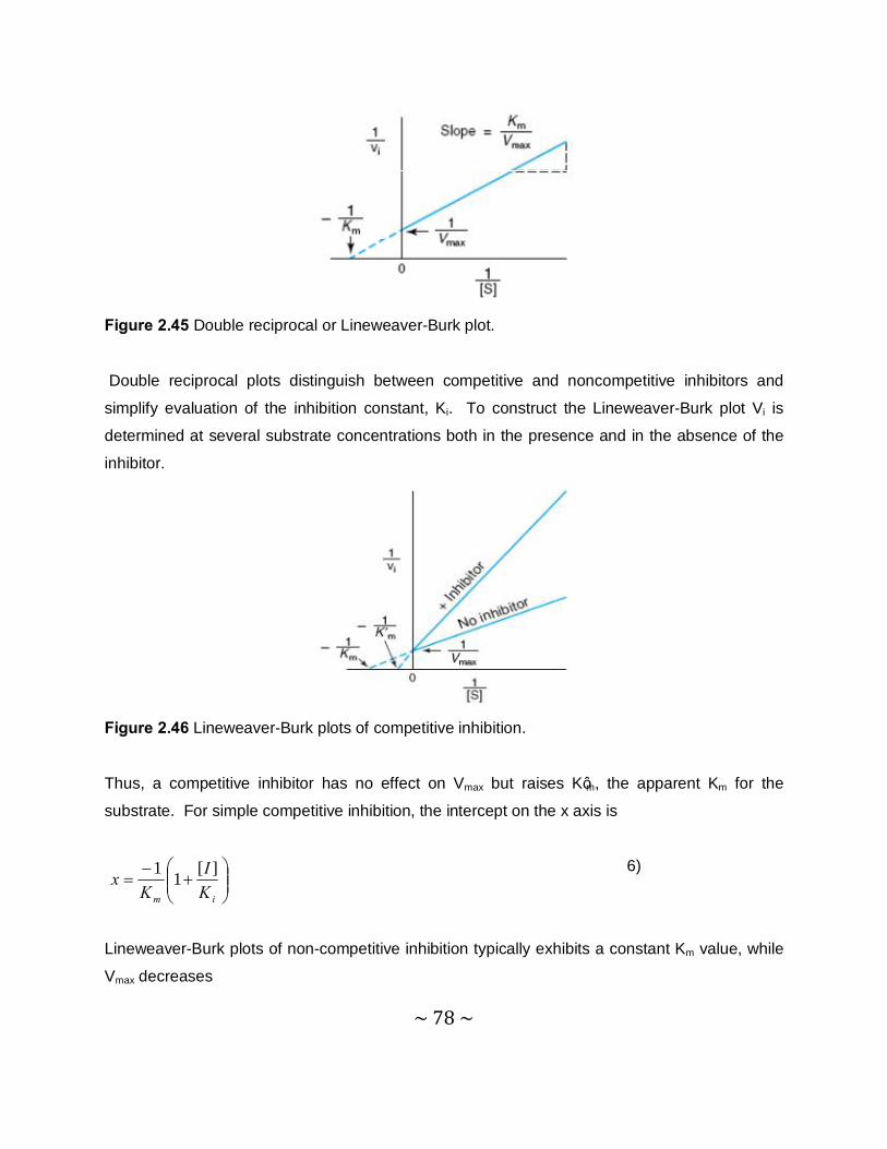
~ 78 ~
+
−=
im KI
Kx ][11
Figure 2.45 Double reciprocal or Lineweaver-Burk plot.
Double reciprocal plots distinguish between competitive and noncompetitive inhibitors and
simplify evaluation of the inhibition constant, Ki. To construct the Lineweaver-Burk plot Vi is
determined at several substrate concentrations both in the presence and in the absence of the
inhibitor.
Figure 2.46 Lineweaver-Burk plots of competitive inhibition.
Thus, a competitive inhibitor has no effect on Vmax but raises K’m, the apparent Km for the
substrate. For simple competitive inhibition, the intercept on the x axis is
6)
Lineweaver-Burk plots of non-competitive inhibition typically exhibits a constant Km value, while
Vmax decreases
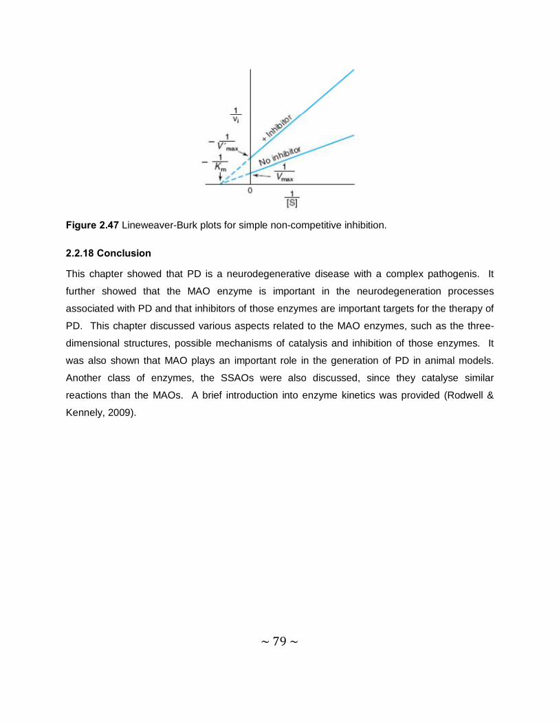
~ 79 ~
Figure 2.47 Lineweaver-Burk plots for simple non-competitive inhibition.
2.2.18 Conclusion
This chapter showed that PD is a neurodegenerative disease with a complex pathogenis. It
further showed that the MAO enzyme is important in the neurodegeneration processes
associated with PD and that inhibitors of those enzymes are important targets for the therapy of
PD. This chapter discussed various aspects related to the MAO enzymes, such as the three-
dimensional structures, possible mechanisms of catalysis and inhibition of those enzymes. It
was also shown that MAO plays an important role in the generation of PD in animal models.
Another class of enzymes, the SSAOs were also discussed, since they catalyse similar
reactions than the MAOs. A brief introduction into enzyme kinetics was provided (Rodwell &
Kennely, 2009).
~ 80 ~
Chapter 3 Synthesis
3.1 Introduction
As mentioned in the introduction chapter, the current study aims to synthesize twelve 8-
thiocaffeine analogues (4a–l) and evaluate them as inhibitors of MAO-A and –B. Although
caffeine is a weak MAO-B inhibitor, substitution at C-8 with a variety of side chains have been
shown to enhance the MAO-B inhibition potency of caffeine by several orders of a magnitude.
The (E)-8-(3-chlorostyryl) substituent of CSC, for example, is an illustration of a substituent that
enhances the MAO-B inhibition potency of caffeine to a large extent (Ikeda et al., 2002). It has
also been found that substitution of caffeine at C-8, with a benzyloxy side-chain, dramatically
enhances the MAO-B inhibition potency of caffeine (Strydom et al., 2010). In this study,
caffeine will be substituted with a variety of substituents at C-8 of caffeine via a thioether
linkage. One of the aims of this study is therefore to compare the MAO-B inhibition potencies of
the 8-thiocaffeine analogues with the previously studied oxycaffeine analogues. Since the 8-
substituted oxycaffeine are also reported to be MAO-A inhibitors, the 8-thiocaffeine analogues
that will be synthesized here will also be evaluated as human MAO-A inhibitors. Table 3.1
illustrates the 8-thiocaffeine analogues that will be synthesized in this study.
~ 81 ~
N
N
N
N
O
OS
R
Table 3.1. The structures of the 8-thiocaffeine analogues that will be examined in the current
study.
3.2 General synthetic approach for the synthesis of 8-thiocaffeine analogues (4a–l) and 8-chlorocaffeine.
8-Thiocaffeine analogues: A general synthetic route used for the preparation of 8-thiocaffeine
analogues, makes use of 8-chlorocaffeine (A) as starting material (Figure 3.1) (Long, 1947). 8-
Chlorocaffeine is reacted with an appropriate mercaptan (B), which is commercially available, to
yield the desired thioether (C). This reaction occurs in the presence of sodium hydroxide and a
water–ethanol mixture serves as the solvent (Long, 1947).
N
N
N
N
O
O
S
R
N
N
N
N
O
O
Cl + R SHi
A
B
C Figure 3.1 Reaction scheme for the synthesis of 8-thiocaffeine analogues. Key: (i) NaOH,
water–ethanol.
8-Chlorocaffeine: The method for the synthesis of 8-chlorocaffeine is essentially that of Fischer
and Reese (1883). A mixture of dry caffeine (D) and chloroform is prepared in a flask fitted with
Compound R Compound R
4a C6H5 4g -CH2-(4-OCH3-C6H4)
4b -CH2-C6H5 4h -(CH2)2-O-C6H5
4c -(CH2)2-C6H5 4i -C6H11
4d -CH2-(4-Cl-C6H4) 4j -C5H9
4e -CH2-(4-Br-C6H4) 4k -2-Naphthalenyl
4f -CH2-(4-F-C6H4) 4l -(CH2)2-CH(CH3)2
~ 82 ~
an inlet tube and a reflux condenser and heated to reflux until the caffeine is dissolved. In a
separate flask, hydrochloric acid is carefully added to potassium permanganate to produce
chlorine gas (Cl2). The Cl2 gas is subsequently passed through the inlet tube and reacts with the
dissolved caffeine at room temperature. After the solvent is removed by distillation in vacuo, 8-
chlorocaffeine (A) is obtained in high yields.
N
N
N
N
O
O
N
N
N
N
O
O
ClCl2i
AD Figure 3.2 Reaction scheme for the synthesisis of 8-chlorocaffeine. Key: (i) CHCl3, room
temperature.
3.3 Detailed synthetic methods for the synthesis of 8-thiocaffeine analogues (4a–l) and 8-chlorocaffeine.
8-Thiocaffeine analogues (4a-l): Sodium hydroxide (0.12 g, 3 mmol) was dissolved in 3.5 ml
water at room temperature. Ethanol (3.5 ml) was added to the mixture. The reaction was
cooled in an ice-bath for 5 minutes and the appropriate thiol (3 mmol) (Table 3.2) was
subsequently added to the reaction. 8-Chlorocaffeine (0.701 g, 3 mmol) was added rapidly in a
single portion to the mixture to yield a white suspension. The reaction was heated under reflux
at the temperature that corresponds to the boiling point of the thiol reagent. These boiling
points were obtained from literature and are given in table 3.2. The reaction mixture was stirred
and heated for 60 minutes.
Thin layer chromatography was used to determine whether the thiol and the 8-chlorocaffeine
reacted completely in the respective reactions. If the reaction was not complete after 60
minutes of heating, the solution was reheated to the same temperature and refluxed for another
60 minutes. The product was collected by filtration and allowed to dry at room temperature.
The product was recrystalized from 30 ml ethanol and again allowed to dry at room temperature
~ 83 ~
overnight. The product was weighed and the percentage yield and the melting point were
determined.
Table 3.2 The boiling points of the thiol starting materials that were used for the synthesis of the
8-thiocaffeine analogues 4a–l. The melting points of the 8-thiocaffeine analogues 4a–l that
were recorded are also given.
Thiol starting material Boiling Point (˚C) Product Melting Point (˚C)
Phenyl mercaptan 169 4a 149
Benzyl mercaptan 75-77 4b 149
Phenylethyl mercaptan 217-218 4c 95
4-Chlorobenzyl mercaptan 75 4d 169
4-Bromobenzyl mercaptan 265 4e 166
4-Fluorobenzyl mercaptan 72-74 4f 175
4-Methoxybenzyl mercaptan 90-95 4g 159
2-Phenoxyethanethiolin 201 4h 114
Cyclohexanethiol 132 4i 135
Cyclopentanethiol 157-158 4j 133
2-Naphthalenethiol 286 4k 175
3-Methyl-1-butanethiol 120 4l 79
8-Chlorocaffeine: As shown in figure 3.3 the hydrochloric acid (160 ml) (1) was slowly added to
26 g (0.17 mol) potassium permanganate (2) at room temperature (Figure 3.3). The chlorine
gas (Cl2), which is the product from this reaction, was then passed through three consecutive
flasks (3, 4 and 5). The first flask (3) contained 200 ml water, the second flask (4) contained
100 ml of sulphuric acid and the third flask (5) was left empty. The water was used to remove
any residual HCl from the generated Cl2 gas. The sulphuric acid acted to dry the Cl2 gas while
the empty flask served as safety trap between the Cl2 generating source and the reaction. The
Cl2 was then passed through a solution of caffeine (5 g, 0.03 mol) (6) in 40 ml chloroform at 85
˚C. After several minutes, a white precipitate, the hydrochloric acid salt of chlorocaffeine formed
in (6). At this point the reaction mixture took on a light yellow appearance. The chloroform
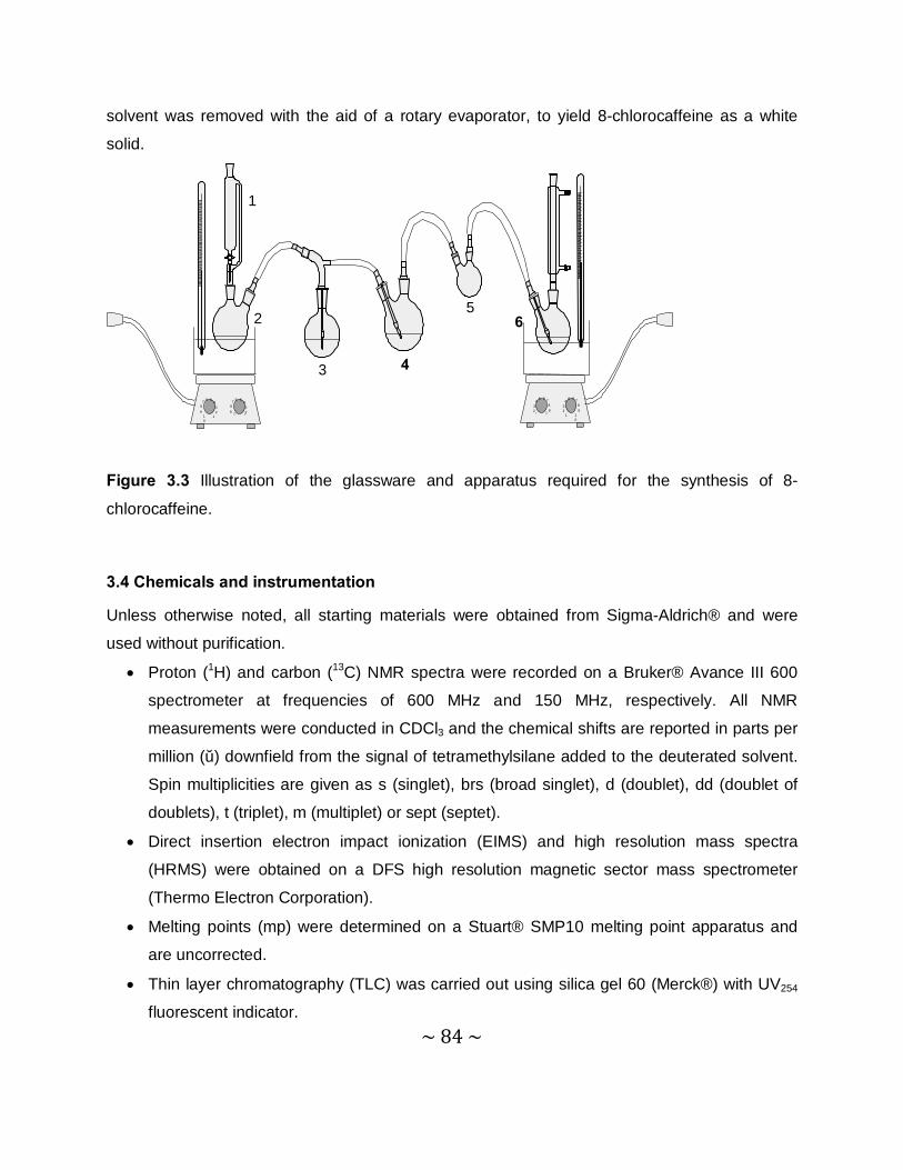
~ 84 ~
solvent was removed with the aid of a rotary evaporator, to yield 8-chlorocaffeine as a white
solid.
123
45 6 7
89
1 10
234
5 6 789
1 1123
4567
89
110
23
4567
89 11
4
6
3
2
1
5
Figure 3.3 Illustration of the glassware and apparatus required for the synthesis of 8-
chlorocaffeine.
3.4 Chemicals and instrumentation
Unless otherwise noted, all starting materials were obtained from Sigma-Aldrich® and were
used without purification.
• Proton (1H) and carbon (13C) NMR spectra were recorded on a Bruker® Avance III 600
spectrometer at frequencies of 600 MHz and 150 MHz, respectively. All NMR
measurements were conducted in CDCl3 and the chemical shifts are reported in parts per
million (δ) downfield from the signal of tetramethylsilane added to the deuterated solvent.
Spin multiplicities are given as s (singlet), brs (broad singlet), d (doublet), dd (doublet of
doublets), t (triplet), m (multiplet) or sept (septet).
• Direct insertion electron impact ionization (EIMS) and high resolution mass spectra
(HRMS) were obtained on a DFS high resolution magnetic sector mass spectrometer
(Thermo Electron Corporation).
• Melting points (mp) were determined on a Stuart® SMP10 melting point apparatus and
are uncorrected.
• Thin layer chromatography (TLC) was carried out using silica gel 60 (Merck®) with UV254
fluorescent indicator.
~ 85 ~
• To determine the purity of the synthesized compounds, HPLC analyses were conducted
with an Agilent® 1100 HPLC system equipped with a quaternary pump and an Agilent®
1100 series diode array detector. HPLC grade acetonitrile (Merck®) and Milli-Q water
(Millipore®) was used for the chromatography.
3.5 Physical characterization
The structures of the 8-thiocaffeine analogues were verified by 1H-NMR and 13C-NMR, as well
as by high resolution mass spectrometry. The purities of the target compounds were verified by
HPLC analysis. For the purpose of the HPLC analysis, strong eluting conditions (up to 85%
acetonitrile) were employed and the eluent was monitored at 210 nm, a wavelength where most
organic compounds absorb UV light. It is therefore likely that impurities, if present, will elute and
be detected under these conditions. The HPLC conditions and chromatograms obtained, are
given in the addendum.
3.6 Results
3.6.1 The physical data for the 8-thiocaffeine derivatives
8-(Phenylsulfanyl)caffeine (4a) The title compound was prepared from phenyl mercaptan in a yield of 64.3%: mp 149 °C
(ethanol). 1H NMR (Bruker Avance III 600, CDCl3) δ 3.37 (s, 3H), 3.53 (s, 3H), 3.90 (s, 3H), 7.32
(m, 5H); 13C NMR (Bruker Avance III 600, CDCl3) δ 28.0, 29.9, 33.1, 109.6, 128.2, 129.6, 130.5,
130.9, 146.4, 148.0, 151.4, 154.9; EI-HRMS m/z: calcd for C14H15N4O2S (MH+), 303.0916, found
303.0912; Purity (HPLC): 98%.
8-(Benzylsulfanyl)caffeine (4b) The title compound was prepared from benzyl mercaptan in a yield of 59.0%: mp 149 °C
(ethanol). 1H NMR (Bruker Avance III 600, CDCl3) δ 3.35 (s, 3H), 3.57 (s, 3H), 3.69 (s, 3H), 4.42
(s, 2H), 7.27 (m, 3H), 7.31 (m, 2H); 13C NMR (Bruker Avance III 600, CDCl3) δ 27.8, 29.7, 32.2,
37.4, 108.7, 127.9, 128.7, 128.9, 136.6, 148.3, 150.0, 151.5, 154.6; EI-HRMS m/z: calcd for
C15H17O2N4S (MH+), 317.1072, found 317.1073; Purity (HPLC): 99%.
~ 86 ~
8-[(2-Phenylethyl)sulfanyl]caffeine (4c) The title compound was prepared from phenylethyl mercaptan in a yield of 22.9%: mp 95 °C
(ethanol). 1H NMR (Bruker Avance III 600, CDCl3) δ 3.04 (t, 2H, J = 7.9 Hz), 3.36 (s, 3H), 3.49
(t, 2H, J = 7.9 Hz), 3.55 (s, 3H), 3.78 (s, 3H), 7.21 (m, 3H), 7.29 (m, 2H); 13C NMR (Bruker
Avance III 600, CDCl3) δ 27.8, 29.7, 32.1, 33.8, 36.0, 108.5, 126.7, 128.5, 139.3, 148.5, 150.9,
151.5, 154.5; EI-HRMS m/z: calcd for C16H19N4O2S (MH+), 331.1229, found 331.1229; Purity
(HPLC): 99%.
8-{[(4-Chlorophenyl)methyl]sulfanyl}caffeine (4d)
The title compound was prepared from 4-chlorobenzyl-methanethiol in a yield of 85.6%: mp 169
°C (ethanol). 1H NMR (Bruker Avance III 600, CDCl3) δ 3.35 (s, 3H), 3.55 (s, 3H), 3.72 (s, 3H),
4.39 (s, 2H), 7.26 (m, 4H); 13C NMR (Bruker Avance III 600, CDCl3) δ 27.8, 29.7, 32.2, 36.4,
108.7, 128.8, 130.3, 133.7, 135.2, 148.3, 149.7, 151.4, 154.5; EI-HRMS m/z: calcd for
C15H16ClN4O2S (MH+), 351.0682, found 351.0679; Purity (HPLC): 97%.
8-{[(4-Bromophenyl)methyl]sulfanyl}caffeine (4e) The title compound was prepared from 4-bromobenzyl mercaptan in a yield of 82.0%: mp 166
°C (ethanol). 1H NMR (Bruker Avance III 600, CDCl3) δ 3.35 (s, 3H), 3.55 (s, 3H), 3.72 (s, 3H),
4.38 (s, 2H), 7.22 (d, 2H, J = 8.3 Hz), 7.39 (d, 2H, J = 8.3 Hz); 13C NMR (Bruker Avance III 600,
CDCl3) δ 27.8, 29.7, 32.2, 36.4, 108.2, 121.8, 130.6, 131.8, 135.8, 148.3, 149.7, 151.4, 154.5;
EI-HRMS m/z: calcd for C15H16BrN4O2S (MH+), 395.0177, found 395.0178; Purity (HPLC): 98%.
8-{[(4-Fluorophenyl)methyl]sulfanyl}caffeine (4f) The title compound was prepared from 4-fluorobenzyl mercaptan in a yield of 75.3%: mp 175 °C
(ethanol). 1H NMR (Bruker Avance III 600, CDCl3) δ 3.35 (s, 3H), 3.55 (s, 3H), 3.72 (s, 3H), 4.40
(s, 2H), 6.96 (t, 2H, J = 8.4 Hz), 7.30 (q, 2H, J = 5.3 Hz); 13C NMR (Bruker Avance III 600,
CDCl3) δ 27.8, 29.7, 32.1, 36.4, 108.7, 115.5, 130.6, 132.4, 148.3, 149.8, 151.4, 154.5, 161.4,
163.1; EI-HRMS m/z: calcd for C15H16FN4O2S (MH+), 335.0976, found 335.0972; Purity (HPLC):
95%.
8-{[(4-Methoxyphenyl)methyl]sulfanyl}caffeine (4g) The title compound was prepared from 4-methoxybenzyl mercaptan in a yield of 94.3%: mp 159
°C (ethanol). 1H NMR (Bruker Avance III 600, CDCl3) δ 3.36 (s, 3H), 3.58 (s, 3H), 3.71 (s, 3H),
~ 87 ~
3.76 (s, 3H), 4.39 (s, 2H), 6.81 (d, 2H, J = 8.7 Hz ), 7.23 (t, 2H, J = 8.7 Hz); 13C NMR (Bruker
Avance III 600, CDCl3) δ 27.9, 29.7, 32.2, 37.0, 55.3, 108.6, 114.1, 128.4, 130.2, 148.4, 150.3,
151.5, 154.6, 159.2; EI-HRMS m/z: calcd for C15H19N4O3S (MH+), 347.1176, found 347.1171;
Purity (HPLC): 94%.
8-[(2-Phenoxyethyl)sulfanyl]caffeine (4h) The title compound was prepared from 2-phenoxyethanethiol in in a yield of 43.9%: mp 114 °C
(ethanol). 1H NMR (Bruker Avance III 600, CDCl3) δ 3.37 (s, 3H), 3.53 (s, 3H), 3.63 (t, 2H, J =
6.2 Hz), 3.83 (s, 3H), 4.30 (t, 2H, J = 6.4 Hz), 6.91 (d, 2H, J = 8.3 Hz), 6.94 (t, 1H, J = 7.2 Hz),
7.25 (t, 2H, J = 8.3 Hz); 13C NMR (Bruker Avance III 600, CDCl3) δ 27.8, 29.7, 31.5, 32.2, 66.3,
108.7, 114.5, 121.3, 129.5, 148.4, 150.3, 151.5, 154.5, 158.1; EI-HRMS m/z: calcd for
C16H19N4O3S (MH+), 347.1176, found 347.1173; Purity (HPLC): 95%.
8-(Cyclohexylsulfanyl)caffeine (4i) The title compound was prepared from cyclohexanethiol in a yield of 37.2%: mp 133 °C
(ethanol). 1H NMR (Bruker Avance III 600, CDCl3) δ 1.28 (m, 1H), 1.39 (m, 2H), 1.47 (m, 2H),
1.58 (m, 1H), 1.73 (m, 2H), 2.03 (m, 2H), 3.34 (s, 3H), 3.52 (s, 3H), 3.71 (m, 1H), 3.82 (s,3H); 13C NMR (Bruker Avance III 600, CDCl3) δ 25.4, 25.8, 27.8, 29.7, 32.3, 33.4, 47.2, 108.4, 148.5,
150.3, 151.5, 154.6; EI-HRMS m/z: calcd for C14H21N4O2S (MH+), 309.1385, found 309.1385;
Purity (HPLC): 99%.
8-(Cyclopentylsulfanyl)caffeine (4j) The title compound was prepared from cyclopentanethiol in a yield of 60.9%: mp 135 °C
(ethanol). 1H NMR (Bruker Avance III 600, CDCl3) δ 1.63 (m, 4H), 1.76 (m, 2H), 2.15 (m, 2H),
3.34 (s, 3H), 3.51 (s, 3H), 3.80 (s, 3H), 3.99 (pent, 1H, J = 7.2 Hz); 13C NMR (Bruker Avance III
600, CDCl3) δ 24.6, 27.8, 29.7, 32.2, 33.8, 46.4, 108.3, 148.5, 151.3, 151.5, 154.6; EI-HRMS
m/z: calcd for C13H19N4O2S (MH+), 295.1229, found 295.1233; Purity (HPLC): 95%.
8-(Naphthalen-2-ylsulfanyl)caffeine (4k) The title compound was prepared from 2-naphthalenethiol in a yield of 87.7%: mp 175 °C
(ethanol). 1H NMR (Bruker Avance III 600, CDCl3) δ 3.37 (s, 3H), 3.53 (s, 3H), 3.91 (s, 3H), 7.36
(dd, 1H, J = 1.6, 9.4 Hz), 7.48 (m, 2H), 7.73 (m, 1H), 7.78 (m, 2H), 7.84 (s, 1H); 13C NMR
(Bruker Avance III 600, CDCl3) δ 27.9, 29.8, 33.1, 109.5, 126.9, 127.0 127.5, 127.8, 129.4,
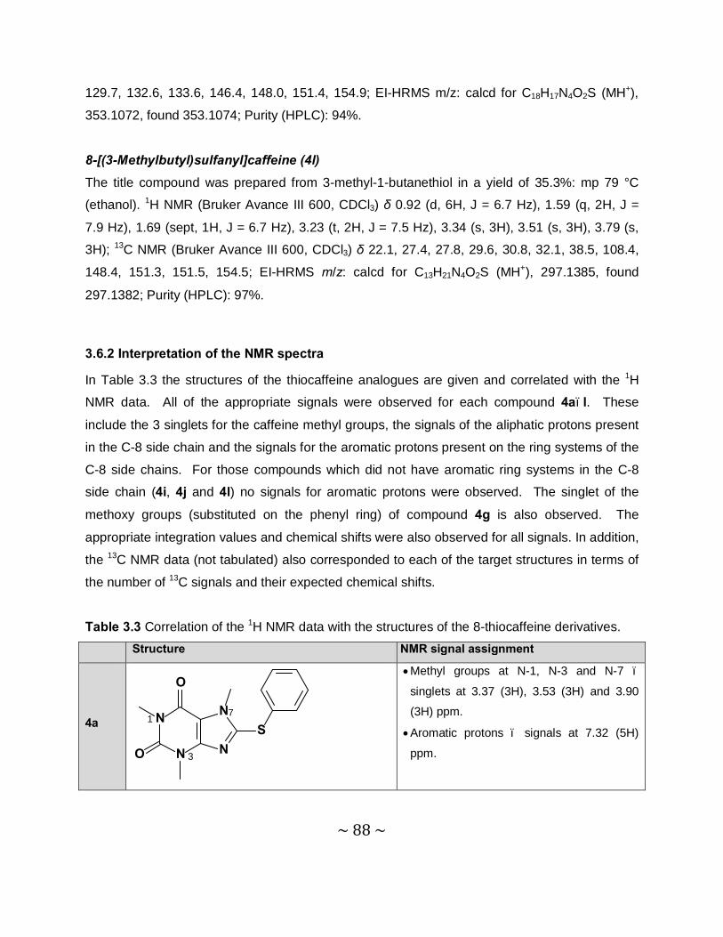
~ 88 ~
129.7, 132.6, 133.6, 146.4, 148.0, 151.4, 154.9; EI-HRMS m/z: calcd for C18H17N4O2S (MH+),
353.1072, found 353.1074; Purity (HPLC): 94%.
8-[(3-Methylbutyl)sulfanyl]caffeine (4l) The title compound was prepared from 3-methyl-1-butanethiol in a yield of 35.3%: mp 79 °C
(ethanol). 1H NMR (Bruker Avance III 600, CDCl3) δ 0.92 (d, 6H, J = 6.7 Hz), 1.59 (q, 2H, J =
7.9 Hz), 1.69 (sept, 1H, J = 6.7 Hz), 3.23 (t, 2H, J = 7.5 Hz), 3.34 (s, 3H), 3.51 (s, 3H), 3.79 (s,
3H); 13C NMR (Bruker Avance III 600, CDCl3) δ 22.1, 27.4, 27.8, 29.6, 30.8, 32.1, 38.5, 108.4,
148.4, 151.3, 151.5, 154.5; EI-HRMS m/z: calcd for C13H21N4O2S (MH+), 297.1385, found
297.1382; Purity (HPLC): 97%.
3.6.2 Interpretation of the NMR spectra
In Table 3.3 the structures of the thiocaffeine analogues are given and correlated with the 1H
NMR data. All of the appropriate signals were observed for each compound 4a–l. These
include the 3 singlets for the caffeine methyl groups, the signals of the aliphatic protons present
in the C-8 side chain and the signals for the aromatic protons present on the ring systems of the
C-8 side chains. For those compounds which did not have aromatic ring systems in the C-8
side chain (4i, 4j and 4l) no signals for aromatic protons were observed. The singlet of the
methoxy groups (substituted on the phenyl ring) of compound 4g is also observed. The
appropriate integration values and chemical shifts were also observed for all signals. In addition,
the 13C NMR data (not tabulated) also corresponded to each of the target structures in terms of
the number of 13C signals and their expected chemical shifts.
Table 3.3 Correlation of the 1H NMR data with the structures of the 8-thiocaffeine derivatives. Structure H NMR signal assignment
4a N
N
N
N
O
O
S7
1
3
• Methyl groups at N-1, N-3 and N-7 –
singlets at 3.37 (3H), 3.53 (3H) and 3.90
(3H) ppm.
• Aromatic protons – signals at 7.32 (5H)
ppm.
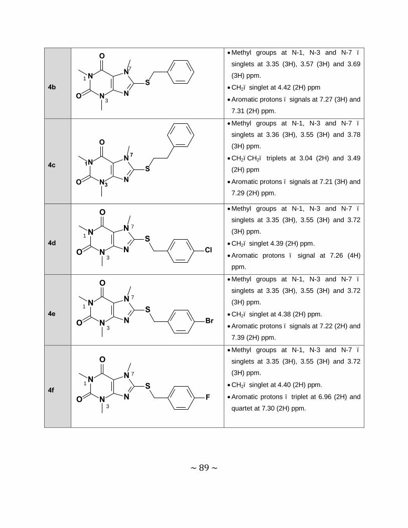
~ 89 ~
4b
N
N
N
N
O
O
S
7
1
3
• Methyl groups at N-1, N-3 and N-7 –
singlets at 3.35 (3H), 3.57 (3H) and 3.69
(3H) ppm.
• CH2– singlet at 4.42 (2H) ppm
• Aromatic protons – signals at 7.27 (3H) and
7.31 (2H) ppm.
4c N
N
N
NS
O
O
1
3
7
• Methyl groups at N-1, N-3 and N-7 –
singlets at 3.36 (3H), 3.55 (3H) and 3.78
(3H) ppm.
• CH2–CH2– triplets at 3.04 (2H) and 3.49
(2H) ppm
• Aromatic protons – signals at 7.21 (3H) and
7.29 (2H) ppm.
4d
N
N
N
NS
O
O Cl
1
3
7
• Methyl groups at N-1, N-3 and N-7 –
singlets at 3.35 (3H), 3.55 (3H) and 3.72
(3H) ppm.
• CH2– singlet 4.39 (2H) ppm.
• Aromatic protons – signal at 7.26 (4H)
ppm.
4e
N
N
N
NS
O
O Br
1
3
7
• Methyl groups at N-1, N-3 and N-7 –
singlets at 3.35 (3H), 3.55 (3H) and 3.72
(3H) ppm.
• CH2– singlet at 4.38 (2H) ppm.
• Aromatic protons – signals at 7.22 (2H) and
7.39 (2H) ppm.
4f
N
N
N
NS
O
O F
1
3
7
• Methyl groups at N-1, N-3 and N-7 –
singlets at 3.35 (3H), 3.55 (3H) and 3.72
(3H) ppm.
• CH2– singlet at 4.40 (2H) ppm.
• Aromatic protons – triplet at 6.96 (2H) and
quartet at 7.30 (2H) ppm.
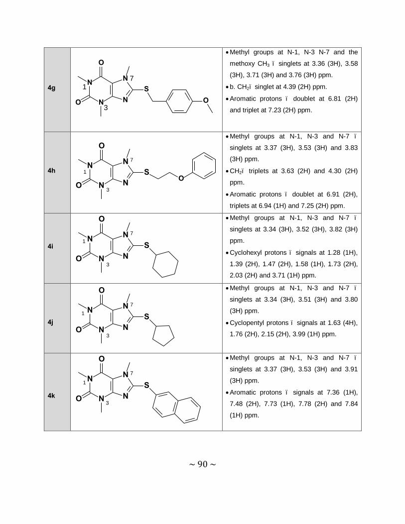
~ 90 ~
4g 1
3
7N
N
N
NS
O
O O
• Methyl groups at N-1, N-3 N-7 and the
methoxy CH3 – singlets at 3.36 (3H), 3.58
(3H), 3.71 (3H) and 3.76 (3H) ppm.
• b. CH2– singlet at 4.39 (2H) ppm.
• Aromatic protons – doublet at 6.81 (2H)
and triplet at 7.23 (2H) ppm.
4h N
N
N
NS
O
OO
1
3
7
• Methyl groups at N-1, N-3 and N-7 –
singlets at 3.37 (3H), 3.53 (3H) and 3.83
(3H) ppm.
• CH2– triplets at 3.63 (2H) and 4.30 (2H)
ppm.
• Aromatic protons – doublet at 6.91 (2H),
triplets at 6.94 (1H) and 7.25 (2H) ppm.
4i N
N
N
NS
O
O
1
3
7
• Methyl groups at N-1, N-3 and N-7 –
singlets at 3.34 (3H), 3.52 (3H), 3.82 (3H)
ppm.
• Cyclohexyl protons – signals at 1.28 (1H),
1.39 (2H), 1.47 (2H), 1.58 (1H), 1.73 (2H),
2.03 (2H) and 3.71 (1H) ppm.
4j
N
N
N
NS
O
O
1
3
7
• Methyl groups at N-1, N-3 and N-7 –
singlets at 3.34 (3H), 3.51 (3H) and 3.80
(3H) ppm.
• Cyclopentyl protons – signals at 1.63 (4H),
1.76 (2H), 2.15 (2H), 3.99 (1H) ppm.
4k
N
N
N
NS
O
O
1
3
7
• Methyl groups at N-1, N-3 and N-7 –
singlets at 3.37 (3H), 3.53 (3H) and 3.91
(3H) ppm.
• Aromatic protons – signals at 7.36 (1H),
7.48 (2H), 7.73 (1H), 7.78 (2H) and 7.84
(1H) ppm.

~ 91 ~
4l
N
N
N
NS
O
O
1
3
7
11
• Methyl groups at N-1, N-3 and N-7 –
singlets at 3.34 (3H), 3.51 (3H) and 3.79
(3H) ppm.
• Aliphatic side chain – doublet at 0.92 (6H),
quartet at 1.59 (2H), septet at 1.69 (1H)
and triplet at 3.23 (2H) ppm.
3.6.3 Interpretation of the mass spectra
As shown in table 3.4, the high resolution masses that were obtained for each of the 8-
tiocaffeine analogues very closely corresponded to the calculated values. This is further
confirmation of the structures of these compounds.
Table 3.4 Correlation of the calculated exact masses with the experimentally obtained masses
of the 8-thiocaffeine derivatives. All masses are given as MH+.
appm = (found – calcd.)/calcd. X 1 000 000. In general a ppm difference smaller than 5 is considered to
be in good agreement.
Mass Spectrometry
Compound R Calcd. Found Formula ppma
4a -C6H5 303.0916 303.0912 C14H15N4O2S 1.3
4b -CH2-C6H5 317.1072 317.1073 C15H17O2N4S 0.3
4c -(CH2)2-C6H5 331.1229 331.1229 C16H19N4O2S 0
4d -CH2-(4-Cl-C6H4) 351.0682 351.0679 C15H16ClN4O2S 0.9
4e -CH2-(4-Br-C6H4) 395.0177 395.0178 C15H16BrN4O2S 0.3
4f -CH2-(4-F-C6H4) 335.0978 335.0972 C15H16FN4O2S 1.8
4g -CH2-(4-OCH3-C6H4) 347.1178 347.1171 C15H19N4O3S 2.0
4h -(CH2)2-O-C6H5 347.1178 347.1173 C16H19N4O3S 1.4
4i -C6H11 309.1385 309.1385 C14H21N4O2S 0
4j -C5H9 295.1229 295.1233 C13H19N4O2S 1.4
4k -2-Naphthalenyl 353.1072 353.1074 C18H17N4O2S 0.6
4l -(CH2)2-CH(CH3)2 297.1385 297.1382 C13H21N4O2S 1.0
N
N
N
N
O
O
SR
~ 92 ~
3.7 Conclusion
This chapter described the successful synthesis of the target 8-thiocaffeine derivatives (4a-l). All the structures were confirmed by NMR and MS and the purities were established by HPLC
analysis. Both the 1H NMR and 13C NMR spectra corresponded with the proposed structures
and the expected exact masses were also recorded for each compound. In addition, HPLC
analysis revealed a single peak for each compound analysed.
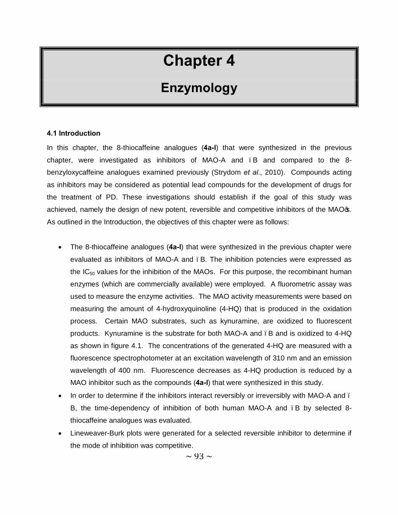
~ 93 ~
Chapter 4 Enzymology
4.1 Introduction
In this chapter, the 8-thiocaffeine analogues (4a-l) that were synthesized in the previous
chapter, were investigated as inhibitors of MAO-A and –B and compared to the 8-
benzyloxycaffeine analogues examined previously (Strydom et al., 2010). Compounds acting
as inhibitors may be considered as potential lead compounds for the development of drugs for
the treatment of PD. These investigations should establish if the goal of this study was
achieved, namely the design of new potent, reversible and competitive inhibitors of the MAO’s.
As outlined in the Introduction, the objectives of this chapter were as follows:
• The 8-thiocaffeine analogues (4a-l) that were synthesized in the previous chapter were
evaluated as inhibitors of MAO-A and –B. The inhibition potencies were expressed as
the IC50 values for the inhibition of the MAOs. For this purpose, the recombinant human
enzymes (which are commercially available) were employed. A fluorometric assay was
used to measure the enzyme activities. The MAO activity measurements were based on
measuring the amount of 4-hydroxyquinoline (4-HQ) that is produced in the oxidation
process. Certain MAO substrates, such as kynuramine, are oxidized to fluorescent
products. Kynuramine is the substrate for both MAO-A and –B and is oxidized to 4-HQ
as shown in figure 4.1. The concentrations of the generated 4-HQ are measured with a
fluorescence spectrophotometer at an excitation wavelength of 310 nm and an emission
wavelength of 400 nm. Fluorescence decreases as 4-HQ production is reduced by a
MAO inhibitor such as the compounds (4a-l) that were synthesized in this study.
• In order to determine if the inhibitors interact reversibly or irreversibly with MAO-A and –
B, the time-dependency of inhibition of both human MAO-A and –B by selected 8-
thiocaffeine analogues was evaluated.
• Lineweaver-Burk plots were generated for a selected reversible inhibitor to determine if
the mode of inhibition was competitive.
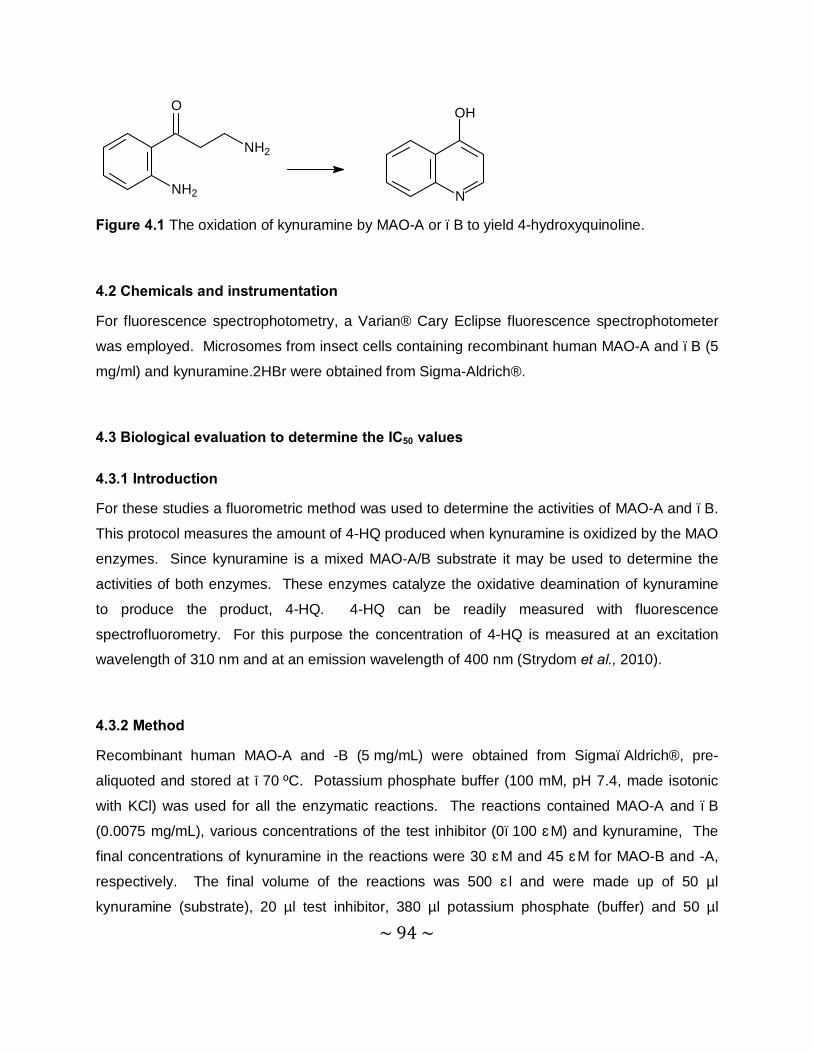
~ 94 ~
NH2
NH2
O
N
OH
Figure 4.1 The oxidation of kynuramine by MAO-A or –B to yield 4-hydroxyquinoline.
4.2 Chemicals and instrumentation
For fluorescence spectrophotometry, a Varian® Cary Eclipse fluorescence spectrophotometer
was employed. Microsomes from insect cells containing recombinant human MAO-A and –B (5
mg/ml) and kynuramine.2HBr were obtained from Sigma-Aldrich®.
4.3 Biological evaluation to determine the IC50 values
4.3.1 Introduction
For these studies a fluorometric method was used to determine the activities of MAO-A and –B.
This protocol measures the amount of 4-HQ produced when kynuramine is oxidized by the MAO
enzymes. Since kynuramine is a mixed MAO-A/B substrate it may be used to determine the
activities of both enzymes. These enzymes catalyze the oxidative deamination of kynuramine
to produce the product, 4-HQ. 4-HQ can be readily measured with fluorescence
spectrofluorometry. For this purpose the concentration of 4-HQ is measured at an excitation
wavelength of 310 nm and at an emission wavelength of 400 nm (Strydom et al., 2010).
4.3.2 Method
Recombinant human MAO-A and -B (5 mg/mL) were obtained from Sigma–Aldrich®, pre-
aliquoted and stored at −70 ºC. Potassium phosphate buffer (100 mM, pH 7.4, made isotonic
with KCl) was used for all the enzymatic reactions. The reactions contained MAO-A and –B
(0.0075 mg/mL), various concentrations of the test inhibitor (0–100 μM) and kynuramine, The
final concentrations of kynuramine in the reactions were 30 μM and 45 μM for MAO-B and -A,
respectively. The final volume of the reactions was 500 μl and were made up of 50 µl
kynuramine (substrate), 20 µl test inhibitor, 380 µl potassium phosphate (buffer) and 50 µl
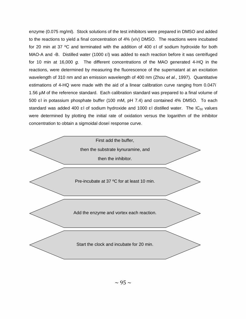
~ 95 ~
enzyme (0.075 mg/ml). Stock solutions of the test inhibitors were prepared in DMSO and added
to the reactions to yield a final concentration of 4% (v/v) DMSO. The reactions were incubated
for 20 min at 37 ºC and terminated with the addition of 400 μl of sodium hydroxide for both
MAO-A and -B. Distilled water (1000 μl) was added to each reaction before it was centrifuged
for 10 min at 16,000 g. The different concentrations of the MAO generated 4-HQ in the
reactions, were determined by measuring the fluorescence of the supernatant at an excitation
wavelength of 310 nm and an emission wavelength of 400 nm (Zhou et al., 1997). Quantitative
estimations of 4-HQ were made with the aid of a linear calibration curve ranging from 0.047–
1.56 µM of the reference standard. Each calibration standard was prepared to a final volume of
500 μl in potassium phosphate buffer (100 mM, pH 7.4) and contained 4% DMSO. To each
standard was added 400 μl of sodium hydroxide and 1000 μl distilled water. The IC50 values
were determined by plotting the initial rate of oxidation versus the logarithm of the inhibitor
concentration to obtain a sigmoidal dose–response curve.
Start the clock and incubate for 20 min.
Add the enzyme and vortex each reaction.
Pre-incubate at 37 ºC for at least 10 min.
First add the buffer,
then the substrate kynuramine, and
then the inhibitor.
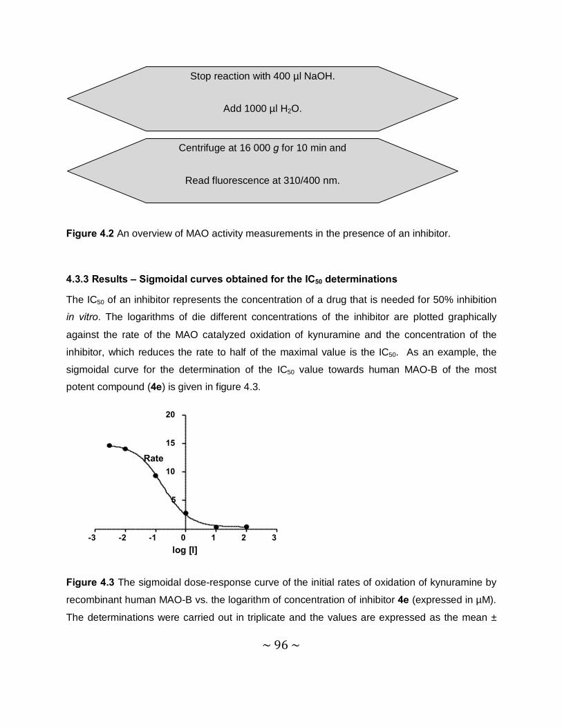
~ 96 ~
Figure 4.2 An overview of MAO activity measurements in the presence of an inhibitor.
4.3.3 Results – Sigmoidal curves obtained for the IC50 determinations
The IC50 of an inhibitor represents the concentration of a drug that is needed for 50% inhibition
in vitro. The logarithms of die different concentrations of the inhibitor are plotted graphically
against the rate of the MAO catalyzed oxidation of kynuramine and the concentration of the
inhibitor, which reduces the rate to half of the maximal value is the IC50. As an example, the
sigmoidal curve for the determination of the IC50 value towards human MAO-B of the most
potent compound (4e) is given in figure 4.3.
Figure 4.3 The sigmoidal dose-response curve of the initial rates of oxidation of kynuramine by
recombinant human MAO-B vs. the logarithm of concentration of inhibitor 4e (expressed in µM).
The determinations were carried out in triplicate and the values are expressed as the mean ±
-3 -2 -1 0 1 2 3
5
10
15
20
log [I]
Rate
Centrifuge at 16 000 g for 10 min and
Read fluorescence at 310/400 nm.
Stop reaction with 400 µl NaOH.
Add 1000 µl H2O.
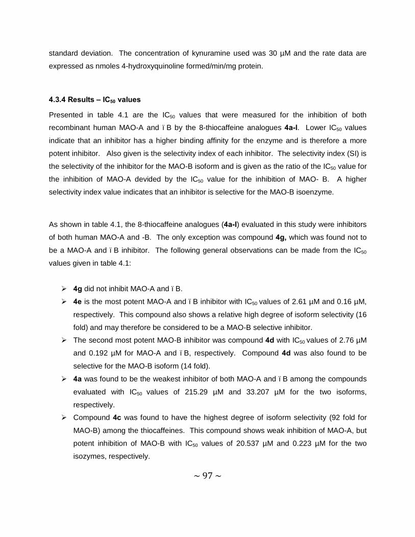
~ 97 ~
standard deviation. The concentration of kynuramine used was 30 µM and the rate data are
expressed as nmoles 4-hydroxyquinoline formed/min/mg protein.
4.3.4 Results – IC50 values
Presented in table 4.1 are the IC50 values that were measured for the inhibition of both
recombinant human MAO-A and –B by the 8-thiocaffeine analogues 4a-l. Lower IC50 values
indicate that an inhibitor has a higher binding affinity for the enzyme and is therefore a more
potent inhibitor. Also given is the selectivity index of each inhibitor. The selectivity index (SI) is
the selectivity of the inhibitor for the MAO-B isoform and is given as the ratio of the IC50 value for
the inhibition of MAO-A devided by the IC50 value for the inhibition of MAO- B. A higher
selectivity index value indicates that an inhibitor is selective for the MAO-B isoenzyme.
As shown in table 4.1, the 8-thiocaffeine analogues (4a-l) evaluated in this study were inhibitors
of both human MAO-A and -B. The only exception was compound 4g, which was found not to
be a MAO-A and –B inhibitor. The following general observations can be made from the IC50
values given in table 4.1:
Ø 4g did not inhibit MAO-A and –B.
Ø 4e is the most potent MAO-A and –B inhibitor with IC50 values of 2.61 µM and 0.16 µM,
respectively. This compound also shows a relative high degree of isoform selectivity (16
fold) and may therefore be considered to be a MAO-B selective inhibitor.
Ø The second most potent MAO-B inhibitor was compound 4d with IC50 values of 2.76 µM
and 0.192 µM for MAO-A and –B, respectively. Compound 4d was also found to be
selective for the MAO-B isoform (14 fold).
Ø 4a was found to be the weakest inhibitor of both MAO-A and –B among the compounds
evaluated with IC50 values of 215.29 µM and 33.207 µM for the two isoforms,
respectively.
Ø Compound 4c was found to have the highest degree of isoform selectivity (92 fold for
MAO-B) among the thiocaffeines. This compound shows weak inhibition of MAO-A, but
potent inhibition of MAO-B with IC50 values of 20.537 µM and 0.223 µM for the two
isozymes, respectively.
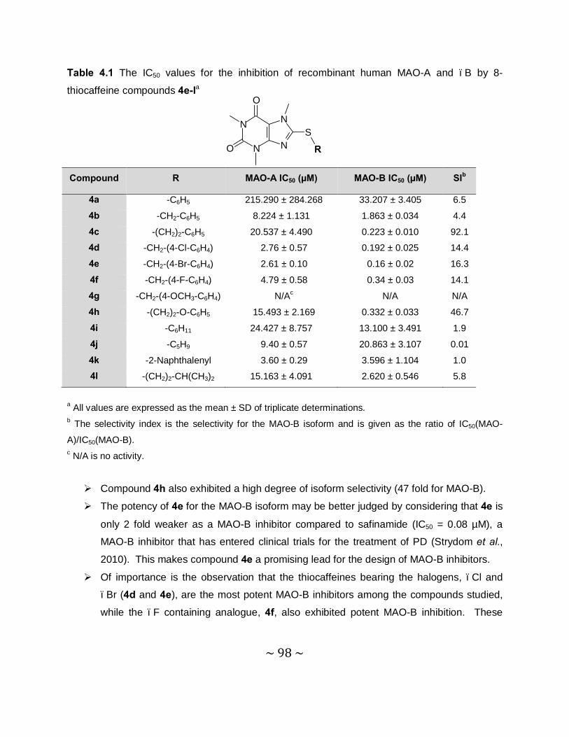
~ 98 ~
Table 4.1 The IC50 values for the inhibition of recombinant human MAO-A and –B by 8-
thiocaffeine compounds 4e-la
a All values are expressed as the mean ± SD of triplicate determinations. b The selectivity index is the selectivity for the MAO-B isoform and is given as the ratio of IC50(MAO-
A)/IC50(MAO-B). c N/A is no activity.
Ø Compound 4h also exhibited a high degree of isoform selectivity (47 fold for MAO-B). Ø The potency of 4e for the MAO-B isoform may be better judged by considering that 4e is
only 2 fold weaker as a MAO-B inhibitor compared to safinamide (IC50 = 0.08 µM), a
MAO-B inhibitor that has entered clinical trials for the treatment of PD (Strydom et al.,
2010). This makes compound 4e a promising lead for the design of MAO-B inhibitors. Ø Of importance is the observation that the thiocaffeines bearing the halogens, –Cl and
–Br (4d and 4e), are the most potent MAO-B inhibitors among the compounds studied,
while the –F containing analogue, 4f, also exhibited potent MAO-B inhibition. These
Compound R MAO-A IC50 (µM) MAO-B IC50 (µM) SIb
4a -C6H5 215.290 ± 284.268 33.207 ± 3.405 6.5
4b -CH2-C6H5 8.224 ± 1.131 1.863 ± 0.034 4.4
4c -(CH2)2-C6H5 20.537 ± 4.490 0.223 ± 0.010 92.1
4d -CH2-(4-Cl-C6H4) 2.76 ± 0.57 0.192 ± 0.025 14.4
4e -CH2-(4-Br-C6H4) 2.61 ± 0.10 0.16 ± 0.02 16.3
4f -CH2-(4-F-C6H4) 4.79 ± 0.58 0.34 ± 0.03 14.1
4g -CH2-(4-OCH3-C6H4) N/Ac N/A N/A
4h -(CH2)2-O-C6H5 15.493 ± 2.169 0.332 ± 0.033 46.7
4i -C6H11 24.427 ± 8.757 13.100 ± 3.491 1.9
4j -C5H9 9.40 ± 0.57 20.863 ± 3.107 0.01
4k -2-Naphthalenyl 3.60 ± 0.29 3.596 ± 1.104 1.0
4l -(CH2)2-CH(CH3)2 15.163 ± 4.091 2.620 ± 0.546 5.8
N
N
N
N
O
O
S
R
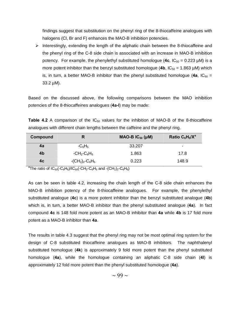
~ 99 ~
findings suggest that substitution on the phenyl ring of the 8-thiocaffeine analogues with
halogens (Cl, Br and F) enhances the MAO-B inhibition potencies. Ø Interestingly, extending the length of the aliphatic chain between the 8-thiocaffeine and
the phenyl ring of the C-8 side chain is associated with an increase in MAO-B inhibition
potency. For example, the phenylethyl substituted homologue (4c, IC50 = 0.223 µM) is a
more potent inhibitor than the benzyl substituted homologue (4b, IC50 = 1.863 µM) which
is, in turn, a better MAO-B inhibitor than the phenyl substituted homologue (4a, IC50 =
33.2 µM).
Based on the discussed above, the following comparisons between the MAO inhibition
potencies of the 8-thiocaffeines analogues (4a-l) may be made:
Table 4.2 A comparison of the IC50 values for the inhibition of MAO-B of the 8-thiocaffeine
analogues with different chain lengths between the caffeine and the phenyl ring.
aThe ratio of IC50(-C6H5)/IC50(-CH2-C6H5 and -(CH2)2-C6H5)
As can be seen in table 4.2, increasing the chain length of the C-8 side chain enhances the
MAO-B inhibition potency of the 8-thiocaffeine analogues. For example, the phenylethyl
substituted analogue (4c) is a more potent inhibitor than the benzyl substituted analogue (4b)
which is, in turn, a better MAO-B inhibitor than the phenyl substituted analogue (4a). In fact
compound 4c is 148 fold more potent as an MAO-B inhibitor than 4a while 4b is 17 fold more
potent as a MAO-B inhibitor than 4a.
The results in table 4.3 suggest that the phenyl ring may not be most optimal ring system for the
design of C-8 substituted thiocaffeine analogues as MAO-B inhibitors. The naphthalenyl
substituted homologue (4k) is approximately 9 fold more potent than the phenyl substituted
homologue (4a), while the homologue containing an aliphatic C-8 side chain (4l) is
approximately 12 fold more potent than the phenyl substituted homologue (4a).
Compound R MAO-B IC50 (µM) Ratio C6H5/Xa
4a -C6H5 33.207 -
4b -CH2-C6H5 1.863 17.8
4c -(CH2)2-C6H5 0.223 148.9
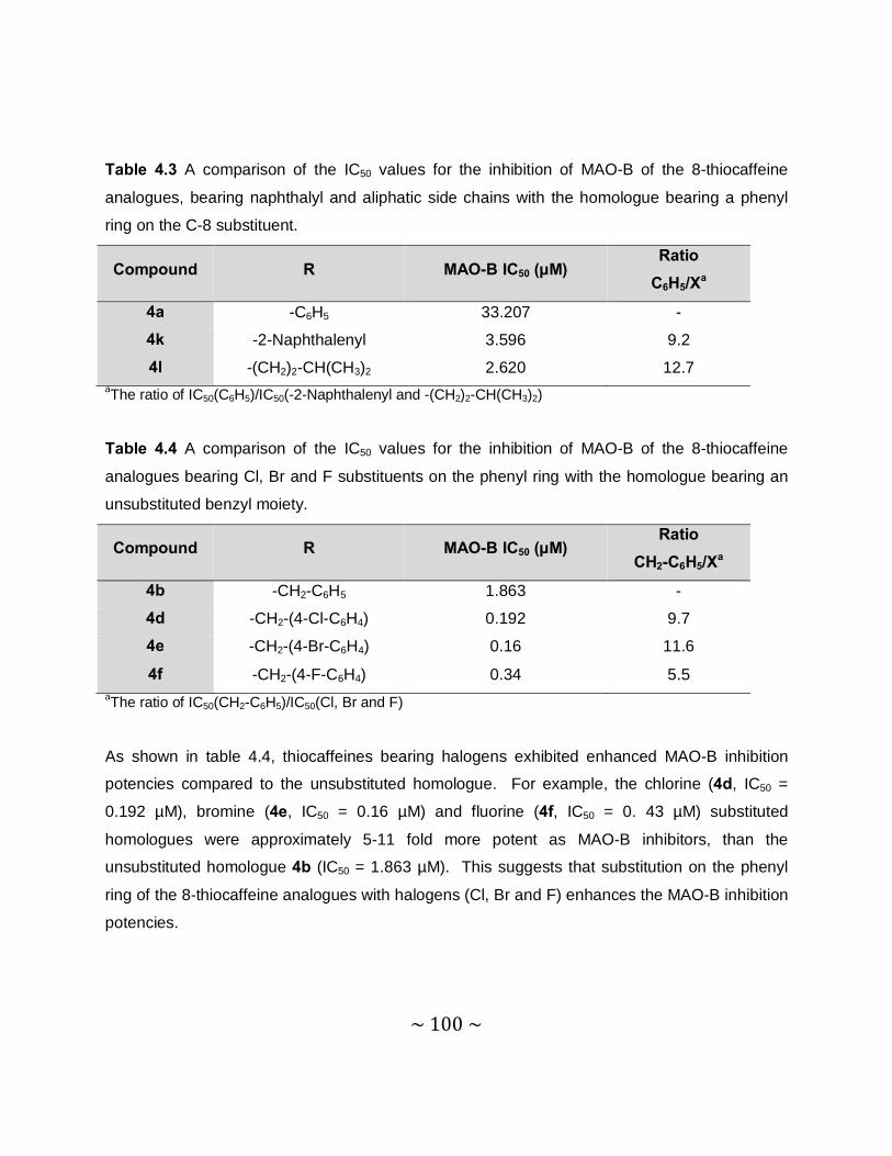
~ 100 ~
Table 4.3 A comparison of the IC50 values for the inhibition of MAO-B of the 8-thiocaffeine
analogues, bearing naphthalyl and aliphatic side chains with the homologue bearing a phenyl
ring on the C-8 substituent.
aThe ratio of IC50(C6H5)/IC50(-2-Naphthalenyl and -(CH2)2-CH(CH3)2)
Table 4.4 A comparison of the IC50 values for the inhibition of MAO-B of the 8-thiocaffeine
analogues bearing Cl, Br and F substituents on the phenyl ring with the homologue bearing an
unsubstituted benzyl moiety.
aThe ratio of IC50(CH2-C6H5)/IC50(Cl, Br and F)
As shown in table 4.4, thiocaffeines bearing halogens exhibited enhanced MAO-B inhibition
potencies compared to the unsubstituted homologue. For example, the chlorine (4d, IC50 =
0.192 µM), bromine (4e, IC50 = 0.16 µM) and fluorine (4f, IC50 = 0. 43 µM) substituted
homologues were approximately 5-11 fold more potent as MAO-B inhibitors, than the
unsubstituted homologue 4b (IC50 = 1.863 µM). This suggests that substitution on the phenyl
ring of the 8-thiocaffeine analogues with halogens (Cl, Br and F) enhances the MAO-B inhibition
potencies.
Compound R MAO-B IC50 (µM) Ratio
C6H5/Xa
4a -C6H5 33.207 -
4k -2-Naphthalenyl 3.596 9.2
4l -(CH2)2-CH(CH3)2 2.620 12.7
Compound R MAO-B IC50 (µM) Ratio
CH2-C6H5/Xa
4b -CH2-C6H5 1.863 -
4d -CH2-(4-Cl-C6H4) 0.192 9.7
4e -CH2-(4-Br-C6H4) 0.16 11.6
4f -CH2-(4-F-C6H4) 0.34 5.5
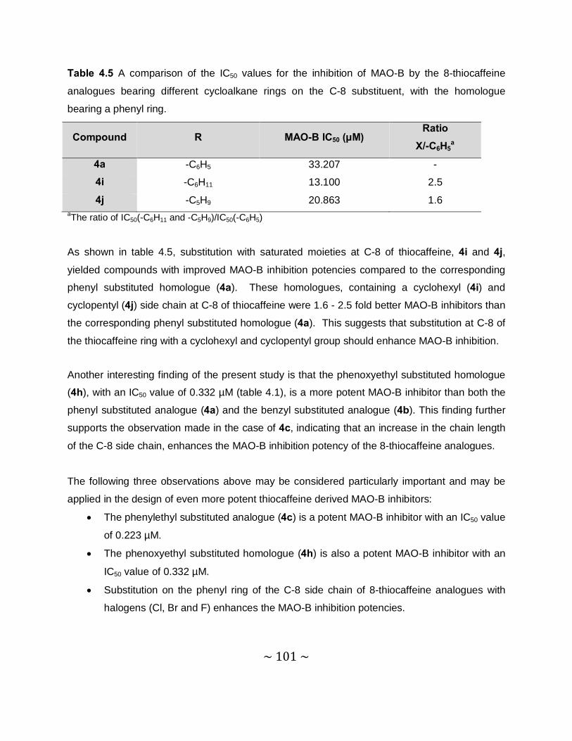
~ 101 ~
Table 4.5 A comparison of the IC50 values for the inhibition of MAO-B by the 8-thiocaffeine
analogues bearing different cycloalkane rings on the C-8 substituent, with the homologue
bearing a phenyl ring.
aThe ratio of IC50(-C6H11 and -C5H9)/IC50(-C6H5)
As shown in table 4.5, substitution with saturated moieties at C-8 of thiocaffeine, 4i and 4j, yielded compounds with improved MAO-B inhibition potencies compared to the corresponding
phenyl substituted homologue (4a). These homologues, containing a cyclohexyl (4i) and
cyclopentyl (4j) side chain at C-8 of thiocaffeine were 1.6 - 2.5 fold better MAO-B inhibitors than
the corresponding phenyl substituted homologue (4a). This suggests that substitution at C-8 of
the thiocaffeine ring with a cyclohexyl and cyclopentyl group should enhance MAO-B inhibition.
Another interesting finding of the present study is that the phenoxyethyl substituted homologue
(4h), with an IC50 value of 0.332 µM (table 4.1), is a more potent MAO-B inhibitor than both the
phenyl substituted analogue (4a) and the benzyl substituted analogue (4b). This finding further
supports the observation made in the case of 4c, indicating that an increase in the chain length
of the C-8 side chain, enhances the MAO-B inhibition potency of the 8-thiocaffeine analogues.
The following three observations above may be considered particularly important and may be
applied in the design of even more potent thiocaffeine derived MAO-B inhibitors:
• The phenylethyl substituted analogue (4c) is a potent MAO-B inhibitor with an IC50 value
of 0.223 µM.
• The phenoxyethyl substituted homologue (4h) is also a potent MAO-B inhibitor with an
IC50 value of 0.332 µM.
• Substitution on the phenyl ring of the C-8 side chain of 8-thiocaffeine analogues with
halogens (Cl, Br and F) enhances the MAO-B inhibition potencies.
Compound R MAO-B IC50 (µM) Ratio
X/-C6H5a
4a -C6H5 33.207 -
4i -C6H11 13.100 2.5
4j -C5H9 20.863 1.6
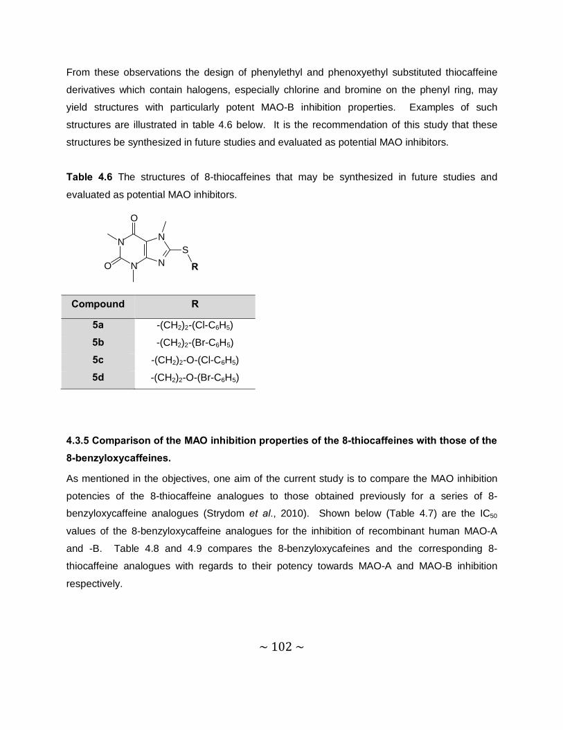
~ 102 ~
From these observations the design of phenylethyl and phenoxyethyl substituted thiocaffeine
derivatives which contain halogens, especially chlorine and bromine on the phenyl ring, may
yield structures with particularly potent MAO-B inhibition properties. Examples of such
structures are illustrated in table 4.6 below. It is the recommendation of this study that these
structures be synthesized in future studies and evaluated as potential MAO inhibitors.
Table 4.6 The structures of 8-thiocaffeines that may be synthesized in future studies and
evaluated as potential MAO inhibitors.
4.3.5 Comparison of the MAO inhibition properties of the 8-thiocaffeines with those of the 8-benzyloxycaffeines.
As mentioned in the objectives, one aim of the current study is to compare the MAO inhibition
potencies of the 8-thiocaffeine analogues to those obtained previously for a series of 8-
benzyloxycaffeine analogues (Strydom et al., 2010). Shown below (Table 4.7) are the IC50
values of the 8-benzyloxycaffeine analogues for the inhibition of recombinant human MAO-A
and -B. Table 4.8 and 4.9 compares the 8-benzyloxycafeines and the corresponding 8-
thiocaffeine analogues with regards to their potency towards MAO-A and MAO-B inhibition
respectively.
Compound R
5a -(CH2)2-(Cl-C6H5)
5b -(CH2)2-(Br-C6H5)
5c -(CH2)2-O-(Cl-C6H5)
5d -(CH2)2-O-(Br-C6H5)
N
N
N
N
O
O
S
R

~ 103 ~
Table 4.7 The IC50 values for the inhibition of recombinant human MAO-A and –B by 8-
benzyloxycaffeine analogues (Strydom et al., 2010).
aThe selectivity index is the selectivity for the MAO-B isoform and is given as the
ratio of IC50(MAO-A)/IC50(MAO-B).
Table 4.8 A comparison of the IC50 values for the inhibition of MAO-A by the 8-thiocaffeines
with the IC50 values of the 8-benzyloxycaffeines. The homologues compared with each other
bears similar side chains at C-8 of the caffeine ring.
Compound R MAO-A IC50 (µM) MAO-B IC50 (µM) SIa
3a C6H5 75.19 10.705 7.0
3b -CH2-C6H9 13.755 2.99 4.6
3c -(CH2)2-C6H5 15.925 2.943 5.4
3d -CH2-(4-Cl-C6H4) 1.337 0.065 20.6
3e -CH2-(4-Br-C6H4) 1.304 0.062 21.0
3f -(CH2)2-O-C6H5 20.35 0.383 53.1
3g -C5H9 22.81 15.915 1.4
3h -(CH2)2-CH(CH3)2 27.34 14.13 1.9
Compared Compounds
Human MAO-A IC50 (µM)
8-benzyloxycaffeine 8-thiocaffeine
3a with 4a 75.19 215.290
3b with 4b 13.755 8.224
3c with 4c 15.925 20.537
3d with 4d 1.337 2.76
3e with 4e 1.304 2.61
3f with 4h 20.35 15.493
3g with 4j 22.81 9.40
3h with 4l 27.34 15.163
N
N
N
N
O
O
OR
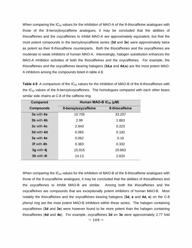
~ 104 ~
When comparing the IC50 values for the inhibition of MAO-A of the 8-thiocaffeine analogues with
those of the 8-benzyloxycaffeine analogues, it may be concluded that the abilities of
thiocaffeines and the oxycaffeines to inhibit MAO-A are approximately equivalent, but that the
most potent compounds in the benzyloxycaffeine series (3d and 3e) were approximately twice
as potent as their 8-thiocaffeine counterparts. Both the thiocaffeines and the oxycaffeines are
moderate to weak inhibitors of human MAO-A. Interestingly, halogen substitution enhances the
MAO-A inhibition activities of both the thiocaffeines and the oxycaffeines. For example, the
thiocaffeines and the oxycaffeines bearing halogens (3d,e and 4d,e) are the most potent MAO-
A inhibitors among the compounds listed in table 4.8.
Table 4.9 A comparison of the IC50 values for the inhibition of MAO-B of the 8-thiocaffeines with
the IC50 values of the 8-benzyloxycaffeines. The homologues compared with each other bears
similar side chains at C-8 of the caffeine ring.
When comparing the IC50 values for the inhibition of MAO-B of the 8-thiocaffeine analogues with
those of the 8-oxycaffeine analogues, it may be concluded that the abilities of thiocaffeines and
the oxycaffeines to inhibit MAO-B are similar. Among both the thiocaffeines and the
oxycaffeines are compounds that are exceptionally potent inhibitors of human MAO-B. Most
notably the thiocaffeines and the oxycaffeines bearing halogens (3d, e and 4d, e) on the C-8
phenyl ring are the most potent MAO-B inhibitors within these series. The halogen containing
oxycaffeines (3d and 3e) were however found to be more potent than the halogen containing
thiocaffeines (4d and 4e). For example, oxycaffeines 3d en 3e were approximately 2.77 fold
Compared Compounds
Human MAO-B IC50 (µM)
8-benzyloxycaffeine 8-thiocaffeine
3a with 4a 10.705 33.207
3b with 4b 2.99 1.863
3c with 4c 2.943 0.223
3d with 4d 0.065 0.192
3e with 4e 0.062 0.16
3f with 4h 0.383 0.332
3g with 4j 15.915 20.863
3h with 4l 14.13 2.620
~ 105 ~
more potent than the corresponding thiocaffeienes 4d en 4e. On the other hand the thiocaffeine
analogue bearing a phenylethyl side chain at C-8 (4c, IC50 = 0.223 µM) was significantly more
potent than the oxycaffeine analogues bearing a phenylethyl side chain at C-8 (3c, IC50 = 2.943
µM). Similarly, the thiocaffeine analogue bearing an aliphatic side chain at C-8 (4l, IC50 = 2.620
µM) was significantly more potent than the oxycaffeine bearing a phenylethyl side chain at C-8
(3h, IC50 = 14.13 µM). In general it may be concluded that while the most potent MAO-B
inhibitors among the compounds listed in table 4.9 were oxycaffeines (3d and 3e), there were
also potent inhibitors among the thiocaffeine analogues. Similar to the oxycaffeines, the
thiocaffeins may also be considered as promising lead compounds for the design of potent
MAO-B inhibitors. With the appropriate substitution pattern and C-8 side chain, the MAO-B
inhibition potencies of the thiocaffeines may approach those of the more potent oxycaffeines.
4.4 Time-dependent studies
4.4.1 Introduction
Also known as reversibility studies, time-dependent studies evaluate whether an inhibitor binds
covalently (irreversible) or non-covalently (reversible) to an enzyme. For the time-dependent
studies, an assay was used in which the enzyme activity measurements are based on the
extent to which kynuramine is oxidized to 4-HQ by the MAO isoforms (Novaroli et al., 2005)
(Figure 4.4). The inhibitor and the enzyme (MAO-A or –B) were combined and incubated for
different periods of time (0, 15, 30, 60 minutes). The substrate (kynuramine) was subsequently
added and the reaction mixtures were incubated for a further 15 min. The MAO-catalysed 4-HQ
formation was measured fluorometrically at excitation and emission wavelengths of 310 nm and
400 nm, respectively. An inhibitor is reversible if the amount of 4-HQ generated by MAO over
the 4 time periods remains unchanged.
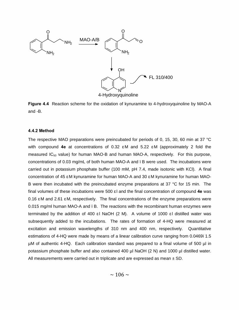
~ 106 ~
NH2
NH2
O
O
NH2
O
N
OH
MAO-A/B
FL 310/400
4-Hydroxyquinoline Figure 4.4 Reaction scheme for the oxidation of kynuramine to 4-hydroxyquinoline by MAO-A
and -B.
4.4.2 Method
The respective MAO preparations were preincubated for periods of 0, 15, 30, 60 min at 37 °C
with compound 4e at concentrations of 0.32 μM and 5.22 μM (approximately 2 fold the
measured IC50 value) for human MAO-B and human MAO-A, respectively. For this purpose,
concentrations of 0.03 mg/mL of both human MAO-A and –B were used. The incubations were
carried out in potassium phosphate buffer (100 mM, pH 7.4, made isotonic with KCl). A final
concentration of 45 μM kynuramine for human MAO-A and 30 μM kynuramine for human MAO-
B were then incubated with the preincubated enzyme preparations at 37 °C for 15 min. The
final volumes of these incubations were 500 μl and the final concentration of compound 4e was
0.16 μM and 2.61 μM, respectively. The final concentrations of the enzyme preparations were
0.015 mg/ml human MAO-A and –B. The reactions with the recombinant human enzymes were
terminated by the addition of 400 μl NaOH (2 M). A volume of 1000 μl distilled water was
subsequently added to the incubations. The rates of formation of 4-HQ were measured at
excitation and emission wavelengths of 310 nm and 400 nm, respectively. Quantitative
estimations of 4-HQ were made by means of a linear calibration curve ranging from 0.0469–1.5
µM of authentic 4-HQ. Each calibration standard was prepared to a final volume of 500 µl in
potassium phosphate buffer and also contained 400 µl NaOH (2 N) and 1000 µl distilled water.
All measurements were carried out in triplicate and are expressed as mean ± SD.
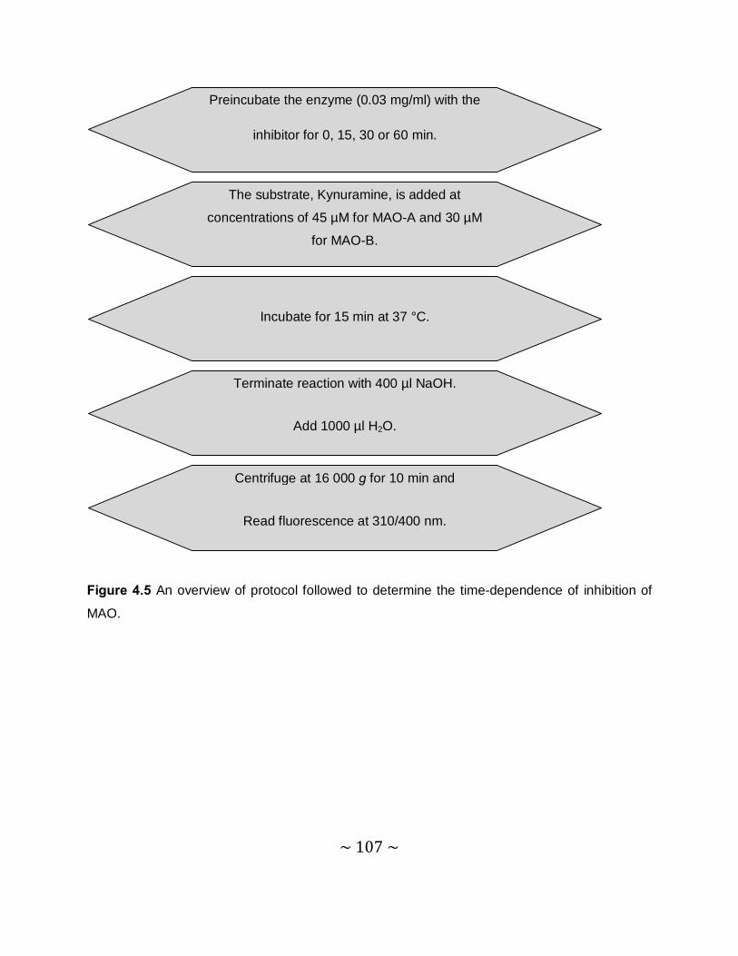
~ 107 ~
Figure 4.5 An overview of protocol followed to determine the time-dependence of inhibition of
MAO.
Centrifuge at 16 000 g for 10 min and
Read fluorescence at 310/400 nm.
Terminate reaction with 400 µl NaOH.
Add 1000 µl H2O.
Incubate for 15 min at 37 °C.
The substrate, Kynuramine, is added at
concentrations of 45 µM for MAO-A and 30 µM
for MAO-B.
Preincubate the enzyme (0.03 mg/ml) with the
inhibitor for 0, 15, 30 or 60 min.
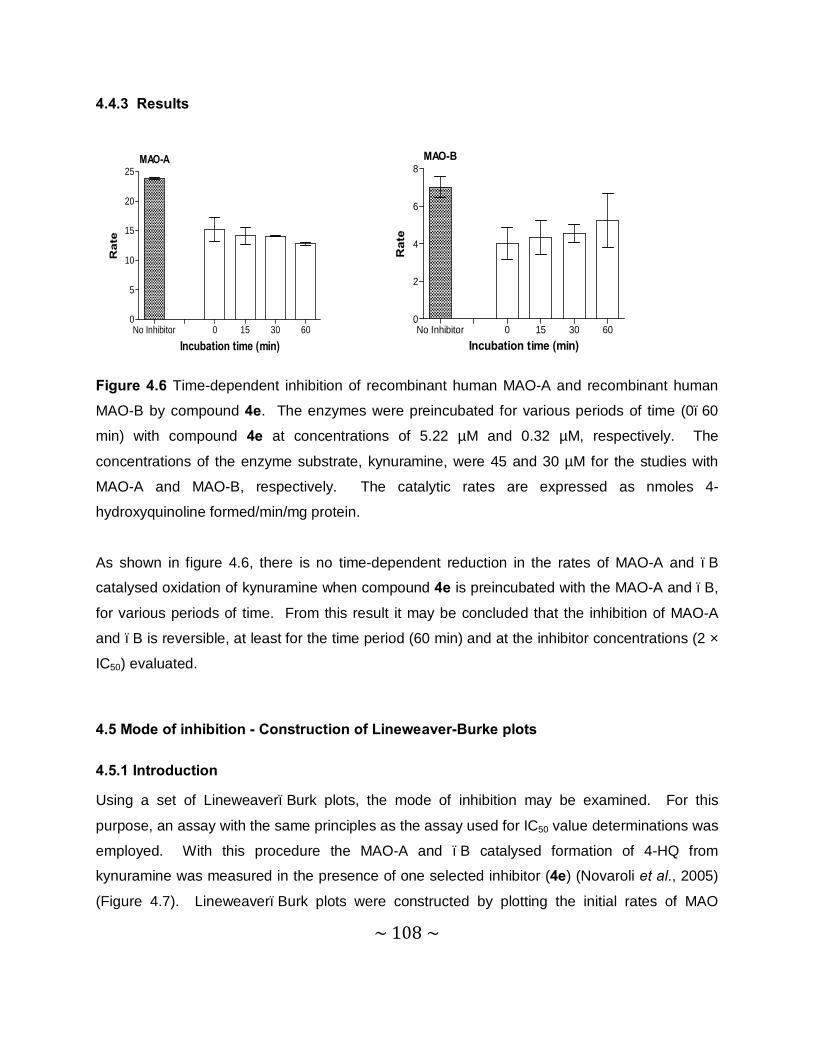
~ 108 ~
4.4.3 Results
Figure 4.6 Time-dependent inhibition of recombinant human MAO-A and recombinant human
MAO-B by compound 4e. The enzymes were preincubated for various periods of time (0–60
min) with compound 4e at concentrations of 5.22 µM and 0.32 µM, respectively. The
concentrations of the enzyme substrate, kynuramine, were 45 and 30 µM for the studies with
MAO-A and MAO-B, respectively. The catalytic rates are expressed as nmoles 4-
hydroxyquinoline formed/min/mg protein.
As shown in figure 4.6, there is no time-dependent reduction in the rates of MAO-A and –B
catalysed oxidation of kynuramine when compound 4e is preincubated with the MAO-A and –B,
for various periods of time. From this result it may be concluded that the inhibition of MAO-A
and –B is reversible, at least for the time period (60 min) and at the inhibitor concentrations (2 ×
IC50) evaluated.
4.5 Mode of inhibition - Construction of Lineweaver-Burke plots
4.5.1 Introduction
Using a set of Lineweaver–Burk plots, the mode of inhibition may be examined. For this
purpose, an assay with the same principles as the assay used for IC50 value determinations was
employed. With this procedure the MAO-A and –B catalysed formation of 4-HQ from
kynuramine was measured in the presence of one selected inhibitor (4e) (Novaroli et al., 2005)
(Figure 4.7). Lineweaver–Burk plots were constructed by plotting the initial rates of MAO
No Inhibitor 0 15 30 600
2
4
6
8
Incubation time (min)
Rat
e
MAO-B
No Inhibitor 0 15 30 600
5
10
15
20
25
Incubation time (min)
Rat
e
MAO-A
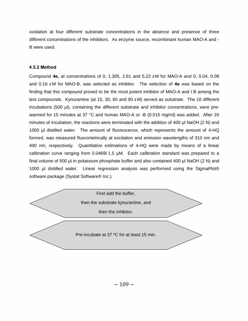
~ 109 ~
oxidation at four different substrate concentrations in the absence and presence of three
different concentrations of the inhibitors. As enzyme source, recombinant human MAO-A and -
B were used.
4.5.2 Method
Compound 4e, at concentrations of 0, 1.305, 2.61 and 5.22 μM for MAO-A and 0, 0.04, 0.08
and 0.16 μM for MAO-B, was selected as inhibitor. The selection of 4e was based on the
finding that this compound proved to be the most potent inhibitor of MAO-A and –B among the
test compounds. Kynuramine (at 15, 30, 60 and 90 μM) served as substrate. The 16 different
incubations (500 µl), containing the different substrate and inhibitor concentrations, were pre-
warmed for 15 minutes at 37 °C and human MAO-A or -B (0.015 mg/ml) was added. After 20
minutes of incubation, the reactions were terminated with the addition of 400 µl NaOH (2 N) and
1000 µl distilled water. The amount of fluorescence, which represents the amount of 4-HQ
formed, was measured fluorometrically at excitation and emission wavelengths of 310 nm and
400 nm, respectively. Quantitative estimations of 4-HQ were made by means of a linear
calibration curve ranging from 0.0469–1.5 µM. Each calibration standard was prepared to a
final volume of 500 µl in potassium phosphate buffer and also contained 400 µl NaOH (2 N) and
1000 µl distilled water. Linear regression analysis was performed using the SigmaPlot®
software package (Systat Software® Inc.).
Pre-incubate at 37 ºC for at least 15 min.
First add the buffer,
then the substrate kynuramine, and
then the inhibitor.
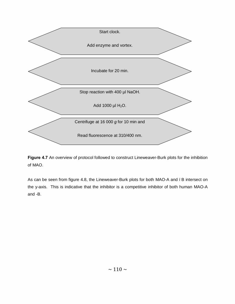
~ 110 ~
Figure 4.7 An overview of protocol followed to construct Lineweaver-Burk plots for the inhibition
of MAO. As can be seen from figure 4.8, the Lineweaver-Burk plots for both MAO-A and –B intersect on
the y-axis. This is indicative that the inhibitor is a competitive inhibitor of both human MAO-A
and -B.
Centrifuge at 16 000 g for 10 min and
Read fluorescence at 310/400 nm.
Stop reaction with 400 µl NaOH.
Add 1000 µl H2O.
Incubate for 20 min.
Start clock.
Add enzyme and vortex.

~ 111 ~
4.5.3 Results – Lineweaver-Burk plots
Panel A Panel B
Figure 4.8 Lineweaver-Burk plots of the recombinant human MAO-A and -B catalyzed
oxidation of kynuramine in the absence (filled squares) and presence of various concentrations
of compound 4e. For the studies with MAO-A the concentrations of compound 4e were: 1.305
µM (open squares), 2.61 µM (filled circles) and 5.22 µM (open circles). For the studies with
MAO-B the concentrations of compound 4e were: 0.04 µM (open squares), 0.08 µM (filled
circles) and 0.16 µM (open circles). The rates (V) are expressed as nmol product
formed/min/mg protein.
4.6. Molecular modeling
4.6.1 Background
The development and discovery of new drugs for therapeutic application can be done by
designing ligands for a specific receptor target. This is done by way of structural and
computational chemistry. Molecular docking is the process of computationally fitting a ligand to
a receptor or enzyme.
Docking studies are not only employed to identify new drugs, but can be used to examine the
interaction between the active site of an enzyme and the docked inhibitor. Due to an increasing
availability of high resolution structural data on enzymes and other protein receptors, docking
studies are becoming an integral part of drug discovery (Knegtel et al., 1997). In combination
-0.02 0.00 0.02 0.04 0.060.00
0.04
0.08
0.12
0.16
1/[S]
1/V
MAO-A
-0.02 0.00 0.02 0.04 0.060.0
0.1
0.2
0.3
0.4
0.5
1/[S]
1/V
MAO-B
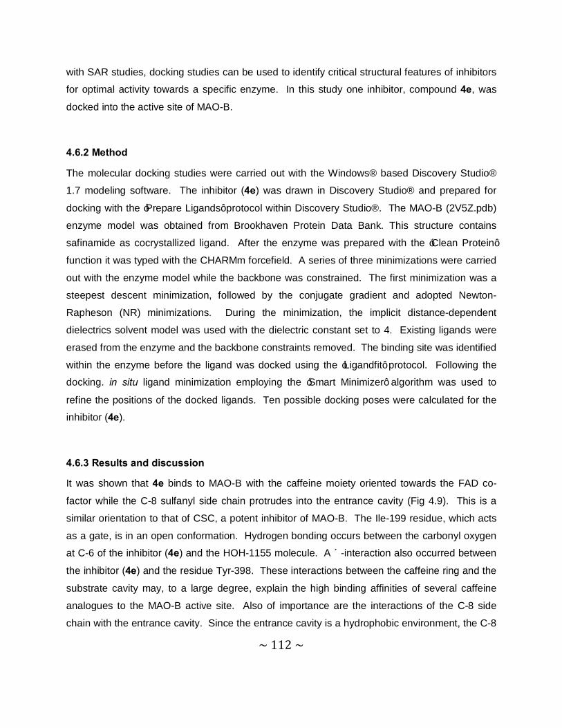
~ 112 ~
with SAR studies, docking studies can be used to identify critical structural features of inhibitors
for optimal activity towards a specific enzyme. In this study one inhibitor, compound 4e, was
docked into the active site of MAO-B.
4.6.2 Method
The molecular docking studies were carried out with the Windows® based Discovery Studio®
1.7 modeling software. The inhibitor (4e) was drawn in Discovery Studio® and prepared for
docking with the ‘Prepare Ligands’ protocol within Discovery Studio®. The MAO-B (2V5Z.pdb)
enzyme model was obtained from Brookhaven Protein Data Bank. This structure contains
safinamide as cocrystallized ligand. After the enzyme was prepared with the ‘Clean Protein’
function it was typed with the CHARMm forcefield. A series of three minimizations were carried
out with the enzyme model while the backbone was constrained. The first minimization was a
steepest descent minimization, followed by the conjugate gradient and adopted Newton-
Rapheson (NR) minimizations. During the minimization, the implicit distance-dependent
dielectrics solvent model was used with the dielectric constant set to 4. Existing ligands were
erased from the enzyme and the backbone constraints removed. The binding site was identified
within the enzyme before the ligand was docked using the ‘Ligandfit’ protocol. Following the
docking. in situ ligand minimization employing the ‘Smart Minimizer’ algorithm was used to
refine the positions of the docked ligands. Ten possible docking poses were calculated for the
inhibitor (4e).
4.6.3 Results and discussion
It was shown that 4e binds to MAO-B with the caffeine moiety oriented towards the FAD co-
factor while the C-8 sulfanyl side chain protrudes into the entrance cavity (Fig 4.9). This is a
similar orientation to that of CSC, a potent inhibitor of MAO-B. The Ile-199 residue, which acts
as a gate, is in an open conformation. Hydrogen bonding occurs between the carbonyl oxygen
at C-6 of the inhibitor (4e) and the HOH-1155 molecule. A π-interaction also occurred between
the inhibitor (4e) and the residue Tyr-398. These interactions between the caffeine ring and the
substrate cavity may, to a large degree, explain the high binding affinities of several caffeine
analogues to the MAO-B active site. Also of importance are the interactions of the C-8 side
chain with the entrance cavity. Since the entrance cavity is a hydrophobic environment, the C-8
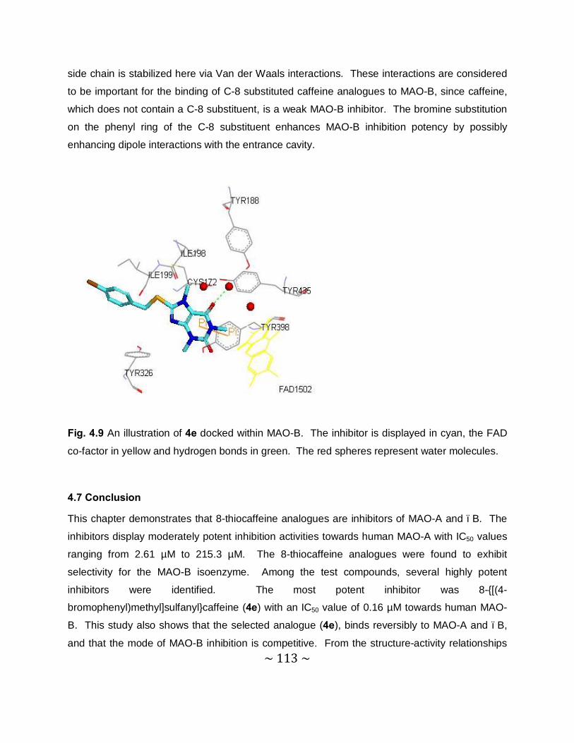
~ 113 ~
side chain is stabilized here via Van der Waals interactions. These interactions are considered
to be important for the binding of C-8 substituted caffeine analogues to MAO-B, since caffeine,
which does not contain a C-8 substituent, is a weak MAO-B inhibitor. The bromine substitution
on the phenyl ring of the C-8 substituent enhances MAO-B inhibition potency by possibly
enhancing dipole interactions with the entrance cavity.
Fig. 4.9 An illustration of 4e docked within MAO-B. The inhibitor is displayed in cyan, the FAD
co-factor in yellow and hydrogen bonds in green. The red spheres represent water molecules.
4.7 Conclusion
This chapter demonstrates that 8-thiocaffeine analogues are inhibitors of MAO-A and –B. The
inhibitors display moderately potent inhibition activities towards human MAO-A with IC50 values
ranging from 2.61 µM to 215.3 µM. The 8-thiocaffeine analogues were found to exhibit
selectivity for the MAO-B isoenzyme. Among the test compounds, several highly potent
inhibitors were identified. The most potent inhibitor was 8-{[(4-
bromophenyl)methyl]sulfanyl}caffeine (4e) with an IC50 value of 0.16 µM towards human MAO-
B. This study also shows that the selected analogue (4e), binds reversibly to MAO-A and –B,
and that the mode of MAO-B inhibition is competitive. From the structure-activity relationships
~ 114 ~
the following three observations may be considered particularly valuable for the design of
thiocaffeine derived MAO-B inhibitors:
• The phenylethyl substituent at C-8 yields compounds with potent MAO-B inhibition
activity.
• The phenoxyethyl substituent at C-8 yields compounds with potent MAO-B inhibition
activity.
• Substitution on the phenyl ring of the C-8 side chain of 8-thiocaffeine analogues with
halogens (Cl, Br and F) enhances the MAO-B inhibition potencies.
When comparing the inhibition potencies of the 8-thiocaffeine derivatives with those of the 8-
benzyloxycaffeine analogues it may be concluded that, while the most potent MAO-B inhibitors
are among the oxycaffeines, the thiocaffeines also represents a promising candidate class of
compounds for the design of potent MAO-B inhibitors.
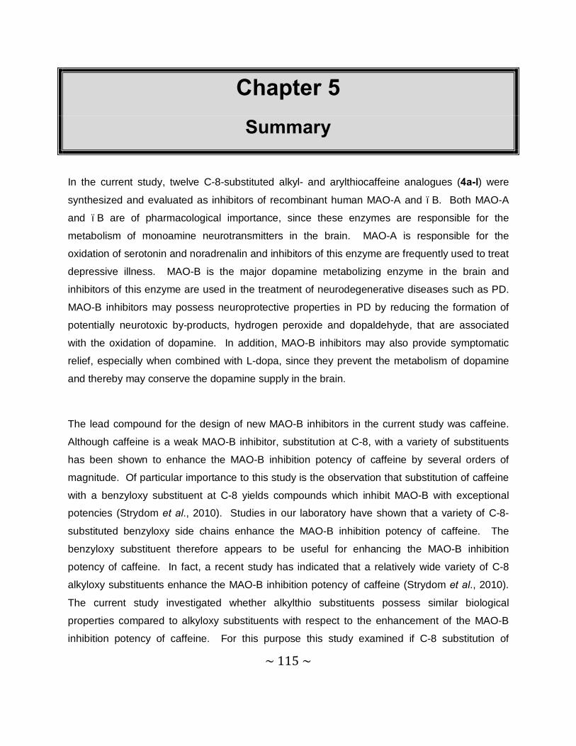
~ 115 ~
Chapter 5 Summary
In the current study, twelve C-8-substituted alkyl- and arylthiocaffeine analogues (4a-l) were
synthesized and evaluated as inhibitors of recombinant human MAO-A and –B. Both MAO-A
and –B are of pharmacological importance, since these enzymes are responsible for the
metabolism of monoamine neurotransmitters in the brain. MAO-A is responsible for the
oxidation of serotonin and noradrenalin and inhibitors of this enzyme are frequently used to treat
depressive illness. MAO-B is the major dopamine metabolizing enzyme in the brain and
inhibitors of this enzyme are used in the treatment of neurodegenerative diseases such as PD.
MAO-B inhibitors may possess neuroprotective properties in PD by reducing the formation of
potentially neurotoxic by-products, hydrogen peroxide and dopaldehyde, that are associated
with the oxidation of dopamine. In addition, MAO-B inhibitors may also provide symptomatic
relief, especially when combined with L-dopa, since they prevent the metabolism of dopamine
and thereby may conserve the dopamine supply in the brain.
The lead compound for the design of new MAO-B inhibitors in the current study was caffeine.
Although caffeine is a weak MAO-B inhibitor, substitution at C-8, with a variety of substituents
has been shown to enhance the MAO-B inhibition potency of caffeine by several orders of
magnitude. Of particular importance to this study is the observation that substitution of caffeine
with a benzyloxy substituent at C-8 yields compounds which inhibit MAO-B with exceptional
potencies (Strydom et al., 2010). Studies in our laboratory have shown that a variety of C-8-
substituted benzyloxy side chains enhance the MAO-B inhibition potency of caffeine. The
benzyloxy substituent therefore appears to be useful for enhancing the MAO-B inhibition
potency of caffeine. In fact, a recent study has indicated that a relatively wide variety of C-8
alkyloxy substituents enhance the MAO-B inhibition potency of caffeine (Strydom et al., 2010).
The current study investigated whether alkylthio substituents possess similar biological
properties compared to alkyloxy substituents with respect to the enhancement of the MAO-B
inhibition potency of caffeine. For this purpose this study examined if C-8 substitution of
~ 116 ~
caffeine with a variety of alkylthio substituents also enhances caffeine`s MAO-B inhibition
potency to a similar degree than that observed with the alkyloxy substituents (Strydom et al.,
2010). Twelve C-8-substituted alkyl- and arylthiocaffeine analogues (4a-l) were synthesized
and evaluated as inhibitors of recombinant human MAO-A and –B. The MAO-A and –B
inhibition potencies of the 8-thiocaffeine analogues were subsequently compared to that of the
previously studied 8-alkyl- and aryloxycaffeine analogues.
Chemistry: Twelve C-8-substituted alkyl- and arylthiocaffeine analogues (4a-l) were successfully
synthesized by reacting 8-chlorocaffeine with the appropriate alkyl- and arylthiol derivatives in
the presence of water, sodium hydroxide and ethanol. All of the thiol starting materials were
commercially available. The structures of the target inhibitors were verified by NMR and MS
analysis. Both the 1H NMR and 13C NMR spectra corresponded with the proposed structures
and the expected exact masses were also recorded for each compound. HPLC analysis
revealed a single peak for almost all the compounds analyzed, which indicates a high degree of
purity for each compound.
MAO inhibition studies: The 8-thiocaffeine analogues were evaluated as inhibitors of
recombinant human MAO-A and –B. The recombinant human enzymes were commercially
Compound R Compound R
4a C6H5 4g -CH2-(4-OCH3-C6H4)
4b -CH2-C6H5 4h -(CH2)2-O-C6H5
4c -(CH2)2-C6H5 4i -C6H11
4d -CH2-(4-Cl-C6H4) 4j -C5H9
4e -CH2-(4-Br-C6H4) 4k -2-Naphthalenyl
4f -CH2-(4-F-C6H4) 4l -(CH2)2-CH(CH3)2
N
N
N
N
O
O
S
R
~ 117 ~
available. A fluorometric assay was employed to measure the inhibition potencies of the test
compounds, and the activities were expressed as the IC50 values. The MAO activity
measurements were based on measuring the amount of 4-HQ that is produced when the MAO-
A/B mixed substrate, kynyramine, is oxidized by the MAO enzymes. Since 4-HQ is fluorescent,
the concentrations of the generated 4-HQ were measured with a fluorescence
spectrophotometer at an excitation wavelength of 310 nm and an emission wavelength of 400
nm.
IC50 values: The results showed that, while the 8-thiocaffeine analogues also inhibited MAO-A,
they were MAO-B selective inhibitors. The following observations are noteworthy:
• Among all of the thiocaffeine inhibitors, 4e was the most potent MAO-B inhibitor with an
IC50 value of 0.16 µM. This inhibitor also exhibited a high degree of selectivity towards
MAO-B with a SI value of 16.3.
• Compound 4c exhibited the highest degree of selectivity for MAO-B with a SI value of
92.1.
• Extending the length of the C-8 chain of the 8-thiocaffeine analogues resulted in an
increase in MAO-B inhibition potency.
• Substitution on the phenyl ring of the C-8 substituent of the 8-thiocaffeine analogues with
halogens (Cl, Br and F) enhances the MAO-B inhibition potencies.
• Replacing the C-8 phenyl ring of compound 4a with saturated cyclopentyl and cyclohexyl
rings, as well as a naphthalenyl ring and an aliphatic side chain yielded improved MAO-
B inhibition potencies compared to 4a.
• The phenoxyethyl substituted homologue (4h) was also found to be a potent MAO-B
inhibitor with an IC50 value of 0.332 µM. These findings provide further evidence that
increasing the chain length of the C-8 side chain, with oxygen in the chain like in this
case, enhances the MAO-B inhibition potency of the 8-thiocaffeine analogues.
~ 118 ~
Time-dependency and mode of inhibition: The time-dependency of inhibition of both MAO-A and
–B by a selected 8-thiocaffeine analogue was evaluated. The results showed that compound
4e, when preincubated with the MAO-A and –B enzymes, respectively, did not reduce the
catalytic rates of MAO-A and –B in a time dependent manner. From this result it was concluded
that the inhibition of MAO-A and –B is reversible. For the inhibition of MAO-A and –B by
compound 4e, sets of Lineweaver–Burk plots were constructed in order to determine the mode
of inhibition. The results showed that the Lineweaver-Burk plots intersected on the y-axis which
indicates that this inhibitor is a competitive inhibitor of both MAO-A and -B.
Future recommendations: Based on the high MAO-B inhibition potencies of some of the
thiocaffeine derivatives, this study recommends that further studies be carried out to optimize
the MAO inhibition activities of these compounds. The following observations may guide the
design of novel thiocaffiene derivatives with improved MAO-B inhibition potencies:
• the phenylethyl substituted analogue (4c) was an exceptionally potent MAO-B inhibitor.
• the phenoxyethyl substituted homologue (4h) was also found to be a very potent MAO-
B inhibitor.
• substitution on the phenyl ring of the C-8 side chain of the 8-thiocaffeine analogues with
halogens (Cl, Br and F) enhances the MAO-B inhibition potencies.
This study therefore recommends that phenylethyl and phenoxyethyl substituted thiocaffeine
derivatives which contain halogens on the phenyl ring be synthesized and evaluated as MAO
inhibitors. Such structures may be particularly potent MAO-B inhibitors. Examples of such
structures are given in table 5.1.
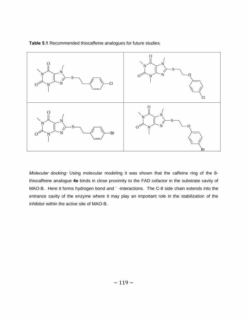
~ 119 ~
Table 5.1 Recommended thiocaffeine analogues for future studies.
Molecular docking: Using molecular modeling it was shown that the caffeine ring of the 8-
thiocaffeine analogue 4e binds in close proximity to the FAD cofactor in the substrate cavity of
MAO-B. Here it forms hydrogen bond and π-interactions. The C-8 side chain extends into the
entrance cavity of the enzyme where it may play an important role in the stabilization of the
inhibitor within the active site of MAO-B.
N
N
N
N
O
O
SCl
N
N
N
N
O
O
SO
Cl
N
N
N
N
O
O
SBr
N
N
N
N
O
O
SO
Br
~ 120 ~
Bibliography
Akao, Y., Youdim, M.B., Davis, B.A., Naoi, M., & Rabey, J.M. (2001). Rasagiline mesylate,
a new MAO-B inhibitor for the treatment of Parkinson’s disease: a double-blind study as
adjunctive therapy to L-dopa. Journal of neurochemistry, 78:727-735.
Ascherio, A., Chen, H., Schwarzschild, M.A., Zhang, S.M., Colditz, G.A., & Speizer, F.E.
(2003). Caffeine, postmenopausal estrogen, and risk of Parkinson's disease. Neurology,
60:790-795.
Athanassiadou, A., Voutsinas, G., Psiouri, L., Leroy, E., Polymeropoulos, M.H., Ilias, A., &
Maniatis, G.M. (1999). Genetic analysis of families with Parkinson’s disease that carry the
Ala53thr mutation in the gene encoding alpha-synuclein. American journal of human
genetics, 65:555-558.
Auluck, P.K., Chan, H.Y., Trojanowski, J.Q., Lee, V.M.Y., & Bonini, N.M. (2002).
Chaperone suppression of alpha-synuclein toxicity in a Drosophila model for Parkinson's
disease. Science, 295:865-868.
Bach, A.W., Lan, N.C., Johnson, D.L., Abell, C.W., Bembenek, M.E., Kwan, S.W., Seeburg,
P.H., & Shih, J.C. (1988) cDNA cloning of human liver monoamine oxidase A and B:
molecular basis of differences in enzymatic properties. Proceedings of the national
academy of sciences of the United States of America, 85:4934–4938.
Bernheimer, H., Birkmayer, W., Hornykiewicz, O., Jellinger, K., & Seitelberger, F. (1973).
Brain dopamine and the syndromes of Parkinson and Huntington. Clinical, morphological
and neurochemical correlations. Journal of neurology science, 20:415-455.
Betarbet, R., Sherer, T.B., MacKenzie, G., Garcia-Osuna, M., Panov, A.V. & Greenamyre,
J.T. (2000). Chronic systemic pesticide exposure reproduces features of Parkinson's
disease. Nature Neuroscience, 3:1301–1306.
~ 121 ~
Binda, C., Newton-Vinson, P., Hubálek F., Edmondson, D.E., & Mattevi, A. (2001).
Structure of human monoamine oxidase B, a drug target for the treatment of neurological
disorders. Nature structural biology, 9:22-26.
Binda, C., Wang, J., Pisani, L., Caccia, C., Carotti, A., Salvati, P., Edmondson, D.E., &
Mattevi, A. (2007). Structures of human monoamine oxidase B complexes with selective
noncovalent inhibitors: Safinamide and coumarin analogs. Journal of medical chemistry,
50:5848-5852.
Bové, J., Marin, C., Bonastre, M., & Tolosa, E. (2002). Adenosine A2a antagonism reverses
levodopa-induced motor alterations in hemiparkinsonian rats. Synapse, 46:251-257.
Bové, J., Prou, D., Perier, C., & Przedborski, S. (2005). Toxin-induced models of
Parkinson’s disease. Journal of the American society for experimental neurotherapeutics,
2:484-494.
Brooks, W.J., Jarvis, M.F., & Wagner, G.C. (1989). Astrocytes as a primary locus for the
conversion of MPTP into MPP+. Journal of neural transmission, 76:1-12.
Burke, WJ. (2003). 3,4-Dihydroxyphenylacetaldehyde: a potential target for neuroprotective
therapy in Parkinson's disease. Current drug targets for CNS neurology disorders, 2:143-
148.
Burns, R.S., LeWitt, P., Ebert, M.H., Pakkenberg, H., & Kopin, I.J. (1985). The clinical
syndrome of striatal dopamine deficiency; parkinsonism induced by MPTP. New England
journal of medicine, 312:1418-1421.
Cantuti-Castelvetri, I., Shukkitt-Hale, B., & Joseph, J.A. (2003). Dopamine neurotoxicity:
age-dependent behavioral and histological effects. Neurobiology of aging, 24:697-706.
Cleeter, M.W., Cooper, J.M., & Schapira, A.H. (1992). Irreversible inhibition of
mitochondrial complex I by 1-methyl-4-phenylpyridinium: evidence for free radical
involvement. Journal of neurochemistry, 58:786-789.
~ 122 ~
Collins, G.G.S., Sandler, M., Williams, E.D., & Youdim, M.B.H. (1970). Multiple forms of
human brain mitochondrial monoamine oxidase. Nature, 225:817-820.
Colzi, A., D’agostini, F., Cesura, A.M., Borroni, E., & Da Prada, M. (1993). MAO-A
inhibitors and dopamine metabolism in rat caudatus: evidence that an increased cytosolic
level of dopamine displaces reversible MAO-A inhibitors in vivo. Journal of pharmacology
and experimental therapeutics, 265:103-111.
Cumming, P., Munk, O.L., & Doudet, D. (2001). Loss of metabolites from monkey striatum
during PET with FDOPA. Synapse, 41:212−218.
Cumming, R.G., Thomas, M., Szonyi, G., Salkeld, G., O’Neill, L., Westbury, C., & Frampton,
G. (1999). Home visits by an occupational therapist for assessment and modification of
environmental hazards: a randomized trial of falls prevention. Journal of the American
geriatrics society, 47:1397–1402.
Damier, P., Hirsch, E.C., Agid, Y., & Graybiel, A.M. (1999). The substantia nigra of the
human brain, II. Patterns of loss of dopamine-containing neurons in Parkinson’s disease.
Brain, 122:1437-1448.
Dauer, W., & Przedborski, S. (2003). Parkinson’s Disease: mechanisms and models.
Neuron, 39:889-909.
Davidson, J.R. (2003). Pharmacotherapy of social phobia. Acta psychiatrica Scandinavica,
417:65-71.
Day, B.J., Patel, M., Calavetta, L., Chang, L.Y., & Stamler, J.S. (1999). A mechanism of
paraquat toxicity involving nitric oxide synthase. Proceedings of the national academy of
sciences of the United States of America, 96:12760-12765.
De Colibus, L., Li, M., Binda, C., Lustig, A., Edmondson, D.E., & Mattevi, A. (2005). Three-
dimensional structure of human monoamine oxidase A: relation to the structures of rat MAO-
~ 123 ~
A and human MAO-B. Proceedings of the national academy of sciences of the United
States of America, 102:12684-12689.
Deleu, D., Northway, M.G., & Hanssens, Y. (2004). Clinical pharmacokinetic and
pharmacodynamic properties of drugs used in the treatment of Parkinson’s disease. Clinical
pharmacokinetics, 41:261-309.
Edmondson, D.E., Mattevi, A., Binda, C., Li, M., & Hubálek, F. (2004). Structure and
mechanism of monoamine oxidase. Current medicinal chemistry, 11:1983-1993.
Elbaz, A., &Tranchant, C. (2007). Epidemiologic studies of environmental exposures in
Parkinson’s disease. Journal of neurology science, 262:37-44.
Esposito, E., Di Matteo, A., Benigno, M., Pierucci, M., Crescimanno, G., & Di Giovanni, G.
(2007). Non-steroidal anti-inflammatory drugs in Parkinson’s disease. Experimental
neurology, 205:295-312.
Fahn, S., Oakes, D., Shoulson, I., Kieburtz, K., Rudolph, A., Lang, A., Olanow, C.W.,
Tanner, C., & Marek, K. (2004). L-dopa and the progression of Parkinson’s disease. New
England journal of medicine, 351:2498-2508.
Faull, R.L., & Laverty, R. (1969). Changes in dopamine levels in the corpus striatum
following lesions in the substantia nigra. Experimental neurology, 23:332-340.
Ferger, B., Teismann, P., & Mierau, J. (2000). The dopamine agonist pramipexole
scavenges hydroxyl free radicals induced by striatal application of 6-hydroxydopamine in
rats: an in vivo microdialysis study. Brain research, 883:216-223.
Fernandez, H.H., & Chen, J.J. (2007). Monoamine oxidase-B inhibition in the treatment of
Parkinson’s disease. Pharmacotherapy, 27:174S-185S.
~ 124 ~
Finberg, J.P., Lamensdorf, I., Weinstock, M., Schwartz, M., & Youdim, M.B. (1999).
Pharmacology of rasagiline (N-propargyl-1R-aminoindan). Advanced neurology, 80:495-
499.
Finberg, J.P., Tenne, M., & Youdim, M.B. (1981). Tyramine antagonistic properties of AGN
1135, an irreversible inhibitor of monoamine oxidase type B. British journal for
pharmacology, 73:65-74.
Finberg, J.P., Wang, J., Bankiewicz, K., Harvey-White, J., Kopin, I.J., & Goldsteen, D.S.
(1998). Increased striatal dopamine production from L-dopa following selective inhibition of
monoamine oxidase B by R(+)-N-propargyl-1-aminoindan (rasagiline) in the monkey.
Journal of neural transmission, 52:279-285.
Fischer, E., & Reese, L. (1883). 8-Chlorocaffeine. Liebigs annalen der chemie, 221:336.
Giasson, B.I., Duda, J.E., Murray, I.V., Chen, Q., Souza, J.M., Hurtig, H.I., Ischiropoulos, H.,
Trojanowski, J.Q., & Lee, V.M. (2000). Oxidative damage linked to neurodegeneration by
selective alpha-synuclein nitration in synucleinopathy lesions. Science, 290:985-989.
Green, A.R., Mitchell, B.D., Tordoff, A.F., & Youdim, M.B. (1977). Evidence for dopamine
deamination by both type A and type B monoamine oxidase in rat brain in vivo and for the
degree if inhibition of enzyme necessary for increased functional activity of dopamine and 5-
hydroxytryptamine. British journal of pharmacology, 60:343-349.
Grunblatt, E., Mandel, S., Jacob-Hirsch, J., Zeligson, S., Amariglo, N., Rechavi, G., Li, J.,
Ravid, R., Roggendorf, W., Riederer, P., & Youdim, M.B. (2004). Gene expression profiling
of parkinsonian substantia nigra pars compacta; alterations in ubiquitin-proteasome, heat
shock protein, iron and oxidative stress regulated proteins, cell adhesion/cellular matrix and
vesicle trafficking genes. Journal of neural transmission, 111:1543-1573.
Haefely, W., Burkard, W.P., Cesura, A.M., Kettler, R., Lorez, H.P., Martin, J.R., Richards,
J.G., Scherschlicht, R., & Da Prada, M. (1992). Biochemistry and pharmacology of
moclobemide, a prototype RIMA. Psychopharmacology, 106:S6-S14.
~ 125 ~
Hardy, J. (2005). Expression of normal sequence pathogenic proteins for
neurodegenerative disease contributes to disease risk: ‘permissive templating’ as a general
mechanism underlying neurodegeneration. Biochemical society transactions, 33:578-581.
Hastings, T.G., & Lewis, D.A. (1996). Reactive dopamine metabolites and neurotoxicity:
implications for Parkinson’s disease. Advances in experimental medicine and biology,
387:97-106.
Healy, D.G., Falchi, M., O'Sullivan, S.S., Bonifati, V., Durr, A., Bressman, S., Brice, A.,
Aasly, J., Zabetian, C.P., Goldwurm, S., Ferreira, J.J., Tolosa, E., Kay, D.M., Klein, C.,
Williams, D.R., Marras, C., Lang, A.E., Wszolek, Z.K., Berciano, J., Schapira, A.H., Lynch,
T., Bhatia, K.P., Gasser, T., Lees, A.J., & Wood NW. (2008). Phenotype, genotype and
worldwide genetic penetrance of LRRK-2-associated Parkinson’s disease: a case-control
study. Lancet neurology, 7:583-590.
Heikkila, R.E., Terleckyj, I., & Sieber, B.A. (1990). Monoamine oxidase and the
bioactivation of MPTP and related neurotoxins: relevance to DATATOP. Journal of neural
transmission, 32:217-227.
Howells, D.W., Porritt, M.J., Wong, J.Y., Batchelor, P.E., Kalnins, R., Hughes, A.J., &
Donnan, G.A. (2000). Reduced BDNF mRNA expression in the Parkinson’s disease
substantia nigra. Experimental neurology, 166:127-135.
Hubálek, F., Binda, C., Khalil, A., Li, M., Mattevi, A., Castagnoli, N., & Edmondson D.E.
(2005). Demonstration of isoleucine 199 as a structural determinant for the selective
inhibition of human monoamine oxidase B by specific reversible inhibitors. The journal of
biological chemistry, 280:15761-15766.
Ikeda, K., Kurokawa, M., Aoyama, S., & Kuwana, Y. (2002). Neuroprotection by adenosine
A2A receptor blockade in experimental models of Parkinson’s disease. Journal of
neurochemistry, 80:262-270.
~ 126 ~
Jalkanen, S., & Salmi, M. (2001). Cell surface monoamine oxidase: enzymes in search of a
function. The EMBO journal, 20:3893-3901.
Javoy, F., Sotelo, C., Herbert, A., & Agid, Y. (1976). Specificity of dopaminergic neuronal
degeneration induced by intracerebral injection of 6-hydroxydopamine in the nigrostriatal
dopamine system. Brain research, 102:210-215.
Johnston, P. (1968). Some observations upon a new inhibitor of monoamine oxidase in
brain tissue. Biochemical pharmacology, 17:1285-1297.
Kearney, E.B., Salach, J.I., Walker, W.H., Seng, R.L., Kenney, W., Zeszotek, E., & Singer,
T.P. (1971). Structure of the covalently bound flavin of monoamine oxidase. Biochemical
and biophysical research communications, 42:490-496.
Kitada, T., Asakawa, S., Hattori, N., Matsumine, H., Yamamura, Y., Minoshima, S., Yokochi,
M., Mizuno, Y., & Shimizu, N. (1998). Mutations in the parkin gene cause autosomal
recessive juvenile parkinsonism. Nature, 392:605-608.
Klinman, J.P., & Mu, D. (1994). Quinoenzymes in biology. Proceedings of the national
academy of sciences of the United States of America, 63:299-344.
Knetgel, R.M.A., Kuntz, I.D., & Oshiro, C.M. (1997). Molecular docking to ensembles of
protein structures. Journal of molecular biology, 266:424-440.
Langston, J.W., Ballard, P., & Irwin, I. (1983). Chronic pakinsonsm in humans due to a
product of meperidine-analog synthesis. Science, 219:979-980.
Le, W.D., & Jankovic, J. (2001). Are dopamine receptor agonists neuroprotective in
Parkinson’s disease? Drugs and aging, 18:389-396.
Lees, A. (2005). Alternatives to L-dopa in the initial treatment of early Parkinson’s disease.
Drugs and aging, 22:731-740.
~ 127 ~
Lees, A.J., Hardy, J., & Revesz, T. (2009). Parkinson’s disease. Lancet, 373:2055-2066.
LeWitt, P.A., & Taylor, D.C. (2008). Protection against parkinson’s disease progression:
clinical experience. Neurotherapeutics, 5:210-225.
LeWitt, P.A., Segel, S.A., Mistura, K.L., & Schork, K.L. (1993). Symptomatic anti-
parkinsonism effects of monoamine oxidase-B inhibition: comparison of selegiline and
lazabemide. Clinical neuropharmacology, 16:332-337.
Liou, H.H., Tsai, M.C., Chen, C.J., Jeng, J.S., Chang, Y.C., Chen, S.Y., & Chen, R.C.
(1997). Environmental risk factors and Parkinson’s disease: a case-control study in Taiwan.
Neurology, 48:1583-1588.
Long, L.M. (1947). 8-R-Thio- and 8-R-sulfonylcaffeine derivatives. Journal of the American
chemical society, 69:2939-2940.
Luthman, J., Fredriksson, A., Sundstrom, E., Jonsson, G., & Archer, T. (1989). Selective
lesion of central dopamine or noradrenaline neuron systems in the neonatal rat: motor
behavior and monoamine alterations at adult stage. Behavioural brain research, 33:267-
277.
Lyles, G.A. (1996). Mammalian plasma and tissue-bound semicarbazide-sensitive amine
oxidases: biochemical, pharmacological and toxicological aspects. Journal of biochemistry
and cellular biology, 28:259-274.
Ma, J., Yoshimura, M., Yamashita, E., Nakagawa, A., Ito, A., & Tsukihara, T. (2004).
Structure of rat monoamine oxidase A and its specific recognitions for substrates and
inhibitors. Journal of molecular biology, 338: 103-114.
Mandel, S., Grünblatt, E., Riederer, P., Gerlach, M., Levites, Y., & Youdim, M.B.H. (2003).
Neuroprotective strategies in Parkinson’s disease: an update on progress. Central nervous
system drugs, 17:729-762.
~ 128 ~
Mandell, S., Weinreb, O., Amit, T., & Youdim, M.B. (2005). Mechanism of neuroprotective
action of the anti-parkinson drug rasagiline and its derivatives. Brain research reviews,
48:379-387.
Manning-Bog, A.B., McCormack, A.L., Li, J., Uversky, V.N., Fink, A.L., & Di Monte, D.A.
(2002). The herbicide paraquat causes upregulation and aggregation of alpha-synuclein in
mice: paraquat and alpha-synuclein. Journal of biological chemistry, 277:1641-1644.
Maroney, A.C., Glicksman, M.A., Basma, A.N., Walton, K.M., Knight, E. Jr., Murphy, C.A.,
Bartlett, B.A., Finn, J.P., Angeles, T., Matsuda, Y., Neff, N.T., & Dionne, C.A. (1998). Motor
neuron apoptosis is inflicted by CEP-1347 (KT7515), a novel inhibitor of the JNK signalling
pathway. Journal of neuroscience, 18:104-111.
Mason, R.P., Olmstead, E.G., & Jacob, R.F. (2000). Antioxidant activity of the monoamine
oxidase B inhibitor lazabemide. Biochemical pharmacology, 60:709-716.
Miller, J.R., Edmondson, D.E., & Grissom, C.B. (1995). Mechanistic probes of monoamine
oxidase B catalysis: Rapid-scan stopped flow and magnetic field independence of the
reductive half-reaction. Journal of the American chemical society, 117:7830-7831.
Mu, D., Janes, S.M., Smith, A.J., Brown, D.E., Dooley, D.M., & Klinman, J.P. (1992).
Tyrosine codon corresponds to topa quinine at the active site of copper amine oxidases.
Journal of biological chemistry, 267:7979-7982.
Nagatsu, T. (1997). Isoquinoline neurotoxins in the brain and Parkinson’s disease.
Neuroscience research, 29:99-111.
Newton Vinson, P., Hubalek, F., & Edmondson, D.E. (2000) High-level expression of
human liver monoamine oxidase B in Pichia pastoris. Protein expression and purification,
20:334-345.
Nicotra, A., Pierucci, F., Parvez, H., & Senatori, O. (2004). Monoamine oxidase expression
during development and aging. Neurotoxicology, 25:155-165.
~ 129 ~
Novaroli, L., Reist, M., Favre, E., Carotti, A., Catto, M., & Carrupt, P.A. (2005). Human
recombinant monoamine oxidase B as reliable and efficient enzyme source for inhibitor
screening. Bioorganic & medicinal chemistry, 13:6212–6217.
Olanow, C.W., Jenner, P., & Brooks, D. (1998). Dopamine agonist and neuroprotection in
Parkinson’s disease. Annals of neurology, 44:S167-S174.
Olanow, C.W., Anthony, A., Schapira, LeWitt, P.A., Kieburtz, K., Sauer, D., Olivieri,
G., Pohlmann, H., & Hubble, J. (2006). TCH346 as a neuroprotective drug in Parkinson’s
disease: a double-blind, randomised, controlled trial. Lancet neurology, 5:1013-1020.
Olanow, C.W., Watts, R.L., & Koller, W.C. (2001). An algorithm (decision tree) for the
management of Parkinson’s disease: treatment guidelines. Neurology, 56 (Suppl 5):S1-88.
Parkinson’s study group. (1989). DATATOP: a multicenter controlled clinical trial in early
Parkinson’s disease. Archives of neurology, 46:1052-1060.
Parkinson’s study group. (1993). Effects of tocopherol and deprenyl on the progression of
disability in early Parkinson’s disease. New English journal of medicine, 328:176-183.
Parkinson’s study group. (1994). A controlled trial of lazabemide in L-dopa-treated
Parkinson’s disease. Archives of neurology, 51:342-347.
Parkinson’s study group. (1996). Effect of lazabemide on the progression of disability in
early Parkinson’s disease. Annals of neurology, 40:99-107.
Pretorius, J., Malan, S.F., Castagnoli, N., Jr., Bergh, J.J., & Petzer, J.P. (2008). Dual
inhibition of monoamine oxidase B and antagonism of the adenosine A(2A) receptor by
(E,E)-8-(4-phenylbutadien-1-yl)caffeine analogues. Bioorganic & medical chemistry,
16:8676-8684.
~ 130 ~
Przedborksi, S. (2004). Pathogenesis of nigral cell death in Parkinson’s disease.
Parkinson’s and related disorders, 11:S3-S7.
Przedborski, S., Kostic, V., Jaskson-Lewis, V., Naini, A.B., Simonetti,S., Fahn, S., Carlson,
E., Epstein, C.J., & Cadet, J.L. (1992). Transgenic mice with increased Cu/Zn-superoxide
dismutase activity are resistant to N-methyl-4-phenyl-1,2,3,6-tetrahydropyridine-induced
neurotoxicity. Journal of neuroscience, 12:1658-1667.
Przedborski, S., Tieu, K., Perier, C., & Vila, M. (2004). MPTP as a mitochondrial neurotoxic
model of Parkinson’s disease. Journal of bioenergetics and biomembranes, 36:375-379.
Ramsay, R.R. (1991). Kinetic mechanism of monoamine oxidase A. Biochemistry,
30:4624-4629.
Ransom, B.R., Kunis, D.M., Irwin, I., & Langston, J.W. (1987). Astrocytes convert the
parkinsonism inducing neurotoxin, MPTP, to its active metabolite. Neuroscience letters,
75:323-328.
Rascol, O., Olanow, C.W., Brooks, D., Koch, G., Truffinet, P., & Bejuit, R. (2002). A 2-year
multicenter placebo-controlled, double blind parallel group study of the effect of rulizole in
Parkinson’s disease. Movement disorders, 17:39.
Riederer, P., & Youdim, M.B. (1986). Monoamine oxidase acitivity and monoamine
metabolism in brains of parkinsonian patients treated with selegiline. Journal of
neurochemistry, 46:1359-1365.
Riederer, P., Danielczyk, W., & Grünblatt, E. (2004). Monoamine oxidase-B inhibition in
Alzheimer’s disease. Neurotoxicology, 25:271-277.
Riederer, P., Sofic, E., Rausch, W.D., Schmidt, B., Reynolds, G.P., Jellinger, K., & Youdim,
M.B. (1989). Transition metals, ferritin, glutathione, and ascorbic acid in parkinsonian
brains. Journal of neurochemistry, 52:515-520.
~ 131 ~
Rodwell, V.W., & Kennely, P.J. Haper’s Illustrated Biochemistry. 28th Edition, Chapter 8:60-
71.
Samii, A., Nutt, J.G., & Ransom, B.R. (2004). Parkinson’s disease. Lancet, 363:1783-
1793.
Saura, M.J., Kettler, R., Da Prada, M., & Richards, J.C. (1990). Molecular neuroanatomy of
MAO-A and –B. Journal of neural transmission, 32:49-53.
Scherfler, C., Schocke, M.F., Seppi, K., Esterhammer, R., Brenneis, C., Jaschke, W.,
Wenning, G.K., & Poewe, W. (2006). Voxel-wise analysis of diffusion weighted imaging
reveals disruption of the olfactory tract in Parkinson’s disease. Brain, 129:538-542.
Schuler, F., & Casida, J.E. (2001). Functional coupling of PSST and ND1 subunits in
NADH: ubiquinone oxidoreductase established by photoaffinity labeling. Biochimica et
biophysica acta, 1506:79-87.
Selley, M.L. (2005). Simvastatin prevents 1-methyl-4-phenyl-1,2,3,6-tetrahydropyridine-
induced striatal dopamine depletion and protein tyrosine nitration in mice. Brain research,
1037:1-6.
Shih, J.C., Chen, K., & Ridd, M.J. (1999). Monoamine oxidase: from genes to behavior.
Annual review of neuroscience, 22:197-217.
Shimizu, K., Ohtaki, K., Matsubara, K., Aoyama, K., Uezono, T., Saito, O., Suno, M.,
Ogawa, K., Hayase, N., Kimura, K., & Shiono, H. (2001). Carrier-mediated processes in
blood-brain barrier penetration and neural uptake of paraquat. Brain research, 906:135-142.
Shin, M.H., Jang, J.H., & Surh, Y.J. (2004). Potential roles of NF-κB and ERK1/2 in
cytoprotection against oxidative cell death induced by tetrahydropapaveroline. Free radical
biology and medicine, 36:1185–1194.
~ 132 ~
Shults, C.W., Haas, R.H., & Beal, M.F. (1999). A possible role of coenzyme Q10 in the
etiology and treatment of Parkinson’s disease. Biofactors, 9:267-272.
Silverman, R.B. (1992) The organic chemistry of enzyme-catalyzed reactions. Advances in
electron transfer chemistry, 2:177-180.
Singer, T.P., Ramsay, R.R., McKeown, K., Trévor, A., & Castagnoli, N. (1988). Mechanism
of the neurotoxicity of 1-methyl-4-phenylpyridinium (MPP+), the toxic bioactivation product of
1-methyl-4-phenyl-1,2,3,6-tetrahydropyridine (MPTP). Toxicology, 49:17-23.
Smeyne, R.J., & Jackson-Lewis, V. (2005). The MPTP model of Parkinson’s disease.
Molecular brain research, 134:57-66.
Son, S., Ma, J., Kondou, Y., Yoshimura, M., Yamashita, E., & Tsukihara, T. (2008).
Structure of human monoamine oxidase A at 2.2-Å resolution: The control of opening the
entry for substrates/inhibitors. Proceedings of the national academy of sciences of the
United States of America, 105:5739-5744.
Spillantini, M.G., Schmidt, M.L., Lee, V.M., Trojanowski, R., & Goedert, M. (1997). Alpha-
synuclein in Lewy bodies. Nature, 388:839-840.
Strydom, B., Malan, S.F., Castagnoli, N., Jr., Bergh, J.J., & Petzer, J.P. (2010). Inhibition of
monoamine oxidase by 8-benzyloxycaffeine analogues. Bioorganic & medicinal chemistry,
18:1018-1028.
Tabor, C.W., Tabor, H., & Rosenthal, S.M. (1954). Purification of amine oxidase from beef
plasma. Journal of biological chemistry, 208:645-661.
Tanaka, K., Miyazaki, I., Fujita, N., Haque, M.E., Asanuma, M., & Ogawa, N. (2001).
Molecular mechanism in activation of glutathione system by ropinirole, a selective dopamine
D2 agonist. Neurochemistry research, 26:31-36.
~ 133 ~
Tansey, M.G., McCoy, M.K., & Frank-Cannon, T.C. (2007). Neuroinflammatory
mechanisms in Parkinson’s disease: potential environmental triggers, pathways, and targets
for early therapeutic intervention. Experimental neurology, 208:1-25.
Tatton, W.G., Chalmers-Redman, R., Brown, D., & Tatton, N. (2003). Apoptosis in
Parkinson’s disease: signals for neuronal degradation. Annals of neurology, 53:S61-S70.
Taylor, K.S., Counsell, C.E., Gordon, J.C., & Harris, C.E. (2005). Screening for
undiagnosed parkinsonism among older people in general practice. Age and aging, 34:501-
504.
Twelves, D., Perkins, K.S., & Counsell, C. (2003). Systematic review of incidence studies
of Parkinson’s disease. Movement Disorders, 18:19-31.
Ulmschneider, M.B., & Sansom, M.S. (2001). Amino acid distributions in integral
membrane protein structures. Biochimica et biophysica acta, 2:1512-1515.
Ungerstedt, U. (1971). Stereotaxic mapping of the monoamine pathways in the rat brain.
Acta physiologica Scandinavica, 367:1-48.
Waldmeier, P, Bozyczko-Coyne, D., Williams, M., & Vaught, J.L. (2006). Recent clinical
failures in Parkinson’s disease with apoptosis inhibitors underline the need for a paradigm
shift in drug discovery for neurodegenerative diseases. Biochemical pharmacology,
72:1197-1206.
Walker, M.C., & Edmondson, D.E. (1994). Structure-activity relationships in the oxidation of
benzylamine analogs by bovine liver mitochondrial monoamine oxidase B. Biochemistry,
33:7088–7098.
Wang, S.X., Nakamura, N., Mure, M., Klinman, J.P., & Sanders-Loehr, J. (1997).
Characterization of the native lysine tyrosylquinone cofactor in lysyl oxidase by Raman
spectroscopy. Journal of biological chemistry, 272:28841-28844.
~ 134 ~
Warrick, J.M., Chan, H.Y., Gray-Board, G.L., Chai, Y., Paulson, H.L., & Bonini, N.M. (1999).
Suppression of polyglutamine-mediated neurodegeneration in Drosophila by the molecular
chaperone HSP70. Nature genetics, 23:425-428.
Williams, D.R., Hadded, A., al-Din, A.S., Wreikat, A.L., & Lees, A.J. (2005). Kufor Rakeb
disease: autosomal recessive L-dopa responsive Parkinsonism with pyramidal
degeneration, supranuclear gaze palsy and dementia. Movement disorders, 20:1264-1271.
Wu, D.C., Jackson-Lewis, V., Vila, M., Tieu, K., Teismann, P., Vadseth, C., Choi, D.K.,
Ischiropoulos, H., & Przedborski, S. (2002). Blockade of microglial activation is
neuroprotective in the 1-methyl-4-phenyl-1,2,3,6-tetrahydropyridine mouse model of
Parkinson’s disease. Journal of neuroscience, 22:1763-1771.
Yacoubian, T.A., & Standaert, D.G. (2009). Targets for neuroprotection in Parkinson’s
disease. Biochimica et biophysica acta, 1792:676-687.
Yamada, M., & Yasuhara, H. (2004). Clinical pharmacology of MAO inhibitors: safety and
future. Neurotoxicology, 25:215-21.
Yamada, M., & Yasuhara, H. (2004). Clinical pharmacology of MAO inhibitors: Safety and
future. Neurotoxicology, 25:215-221.
Youdim, M.B.H., & Weinstock, M. (2004). Therapeutic applications of selective and non-
selective inhibitors of monoamine oxidase A and B that do not cause significant tyramine
potentiation. Neurotoxicology, 25:243-250.
Youdim, M.B.H., Banerjee, D.K., Kelner, K., Offutt, L., & Pollard, H.B. (1989). Steroid
regulation of monoamine oxidase activity in the adrenal medulla. Federation of American
societies for experimental biology journal, 3:1753–1759
Youdim, M.B.H., Finberg, J.P., & Tipton, K.F. (1988). Monoamine oxidase. Advances in
experimental pharmacology, 2:119-192.
~ 135 ~
Youdim, M.B.H., & Bakhle, Y.S. (2006). Monoamine oxidase: isoforms and inhibitors in
Parkinson’s disease and depressive illness. British journal of pharmacology, 147:S287-
S296.
Youdim, M.B.H., Collins, G.G.S., Sandler, M., Bevan-Jones, A.B., Pare, C.M., & Nicholson,
W.J. (1972). Human brain monoamine oxidase, multiple forms and selective inhibitors.
Nature, 236:225-228.
Youdim, M.B.H., Edmondson, D., & Tipton, K.F. (2006). The therapeutic potential of
monoamine oxidase inhibitors. Neuroscience, 7:295-309.
Zecca, L., Youdim, M.B., Riederer, P Connor, J.R., & Crichton, R.R. (2004). Iron, brain
ageing and neurodegenerative disorders. Nature reviews neuroscience, 5:863-873.
Zhou, M., Diwu, Z., Panchuk-Voloshina, N., & Haugland, R.P. (1997). A stable
nonfluorescent derivative of resorufin for the fluorometric determination of trace hydrogen
peroxide applications in detecting the activity of phagocyte NADPH oxidase and other
oxidases. Analytical biochemistry, 253:162-168.
Zhu, Q., Chen, K., & Shih, J. (1994). Bidirectional promoter of human monoamine oxidase
A (MAO A) controlled by transcription factor Sp1. Journal of neuroscience, 14:7393–7403.
Zisook, S.E. (1985). Clinical overview of monoamine oxidase inhibitors. Psychosomatics,
26:240–251.
~ 136 ~
Addendum
List of 1H and 13C spectrums:
• 8-(Phenylsulfanyl)caffeine ............................................................................................137
• 8-(Benzylsulfanyl)caffeine .............................................................................................138
• 8-[(2-Phenylethyl)sulfanyl]caffeine ................................................................................139
• 8-{[(4-Chlorophenyl)methyl]sulfanyl}caffeine ................................................................140
• 8-{[(4-Bromophenyl)methyl]sulfanyl}caffeine ................................................................141
• 8-{[(4-Fluorophenyl)methyl]sulfanyl}caffeine ................................................................142
• 8-{[(4-Methoxyphenyl)methyl]sulfanyl}caffeine .............................................................143
• 8-[(2-Phenoxyethyl)sulfanyl]caffeine .............................................................................144
• 8-(Cyclohexylsulfanyl)caffeine ......................................................................................145
• 8-(Cyclopentylsulfanyl)caffeine .....................................................................................146
• 8-(Naphthalen-2-ylsulfanyl)caffeine ..............................................................................147
• 8-[(3-Methylbutyl)sulfanyl]caffeine.................................................................................148
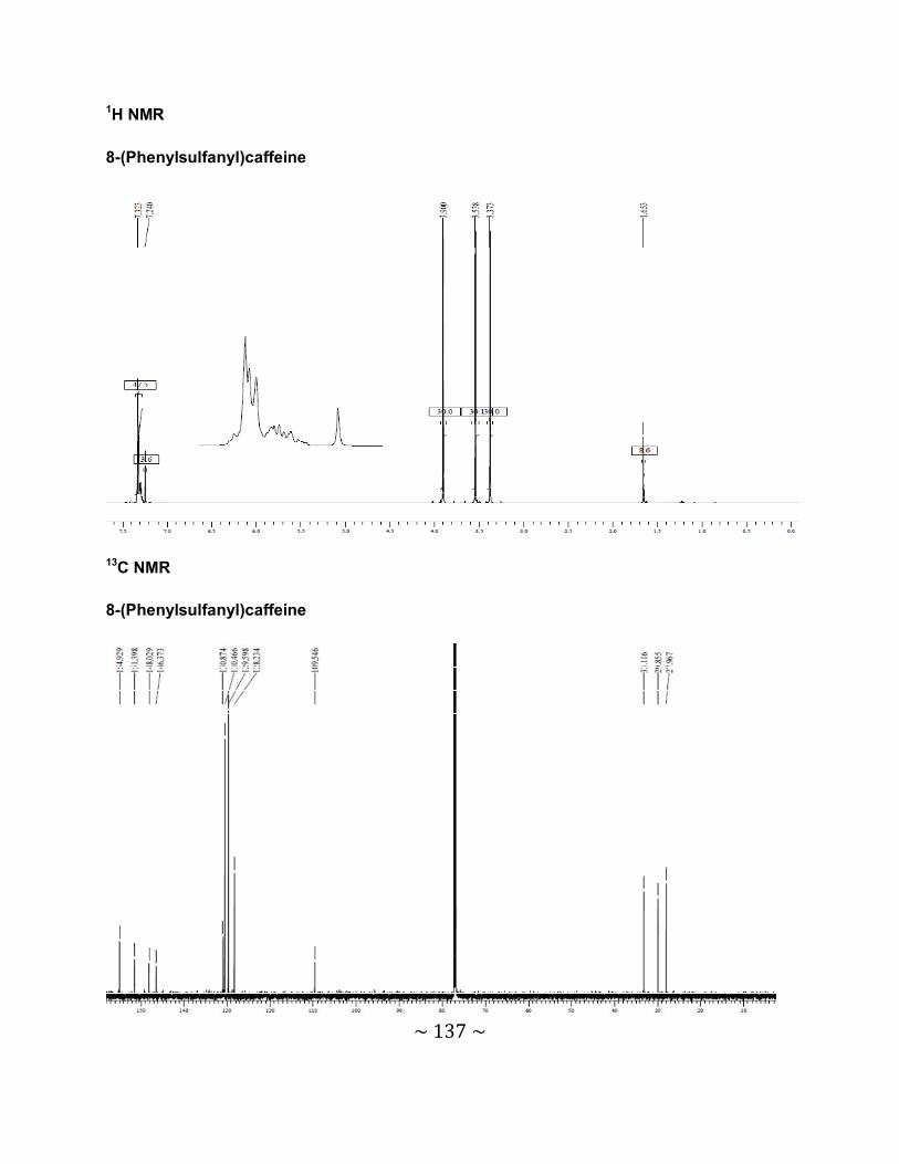
~ 137 ~
1H NMR
8-(Phenylsulfanyl)caffeine
13C NMR
8-(Phenylsulfanyl)caffeine
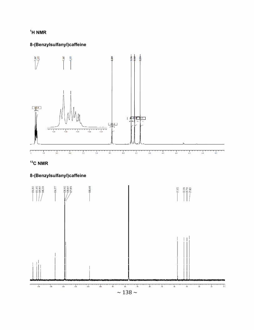
~ 138 ~
1H NMR
8-(Benzylsulfanyl)caffeine
13C NMR
8-(Benzylsulfanyl)caffeine
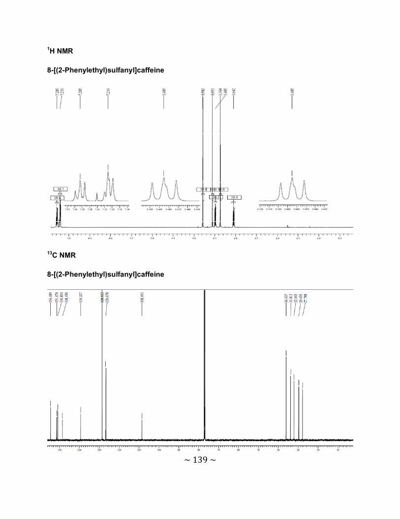
~ 139 ~
1H NMR
8-[(2-Phenylethyl)sulfanyl]caffeine
13C NMR
8-[(2-Phenylethyl)sulfanyl]caffeine
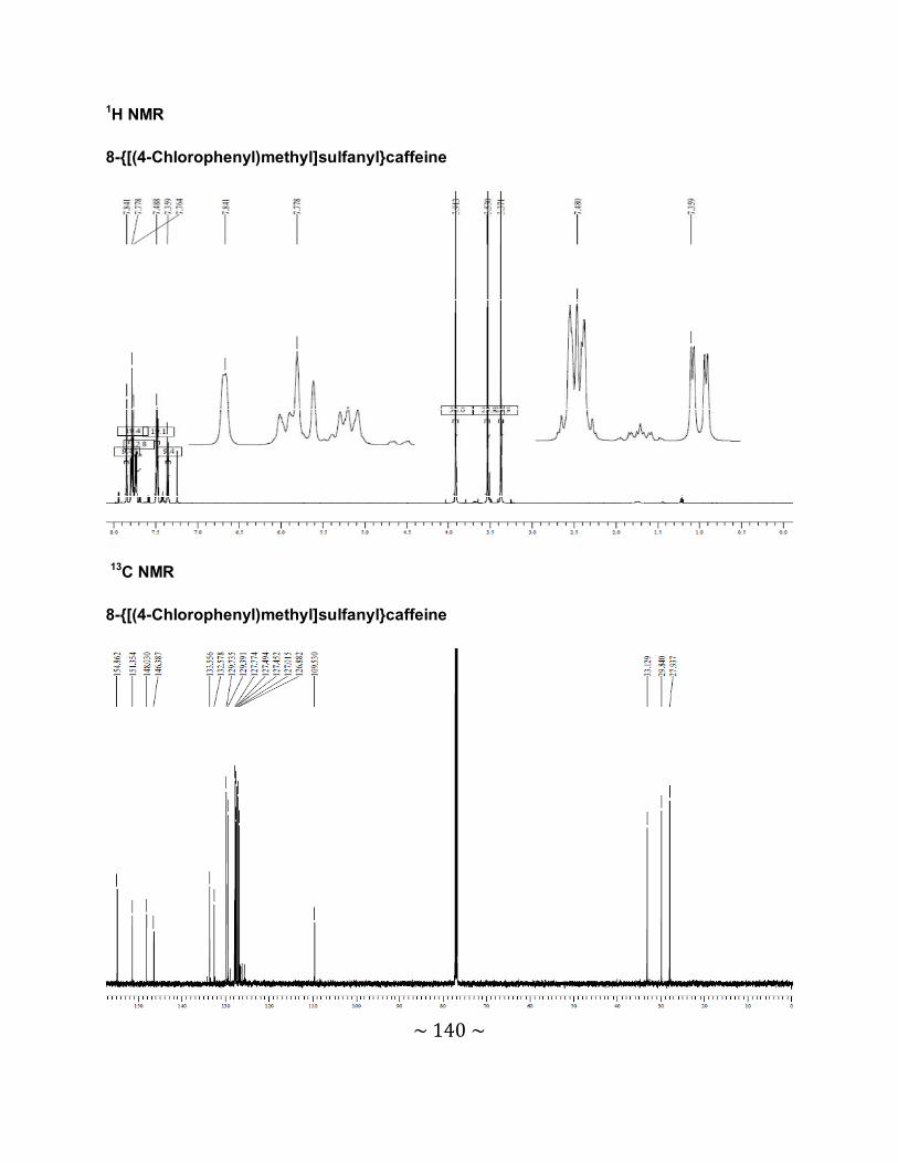
~ 140 ~
1H NMR
8-{[(4-Chlorophenyl)methyl]sulfanyl}caffeine
13C NMR
8-{[(4-Chlorophenyl)methyl]sulfanyl}caffeine

~ 141 ~
1H NMR
8-{[(4-Bromophenyl)methyl]sulfanyl}caffeine
13C NMR
8-{[(4-Bromophenyl)methyl]sulfanyl}caffeine
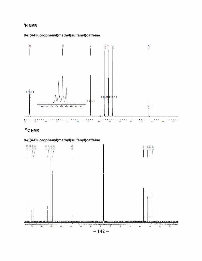
~ 142 ~
1H NMR
8-{[(4-Fluorophenyl)methyl]sulfanyl}caffeine
13C NMR
8-{[(4-Fluorophenyl)methyl]sulfanyl}caffeine
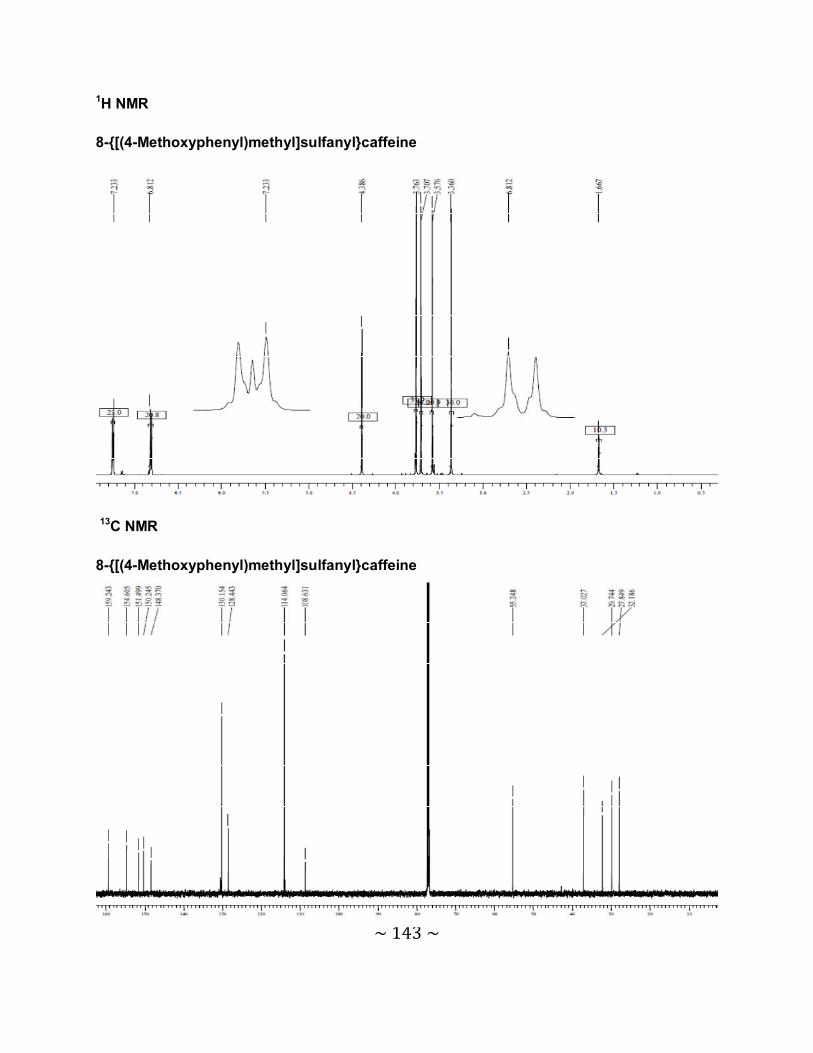
~ 143 ~
1H NMR
8-{[(4-Methoxyphenyl)methyl]sulfanyl}caffeine
13C NMR
8-{[(4-Methoxyphenyl)methyl]sulfanyl}caffeine

~ 144 ~
1H NMR
8-[(2-Phenoxyethyl)sulfanyl]caffeine
13C NMR
8-[(2-Phenoxyethyl)sulfanyl]caffeine
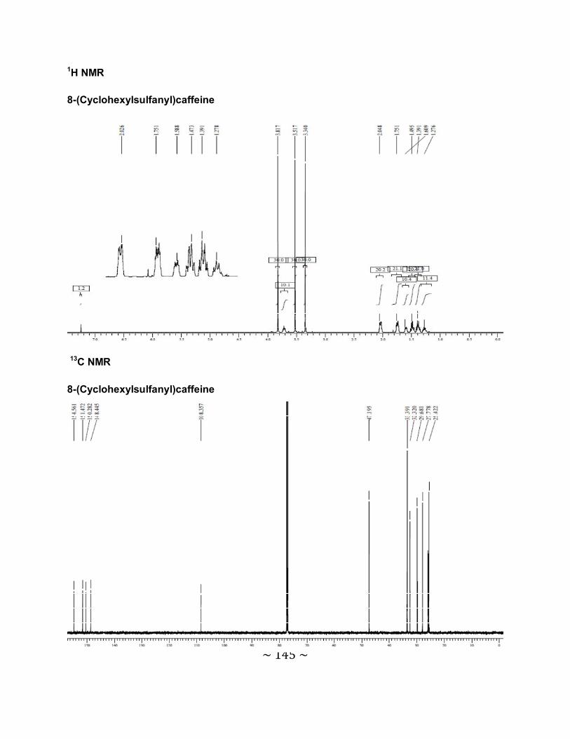
~ 145 ~
1H NMR
8-(Cyclohexylsulfanyl)caffeine
13C NMR
8-(Cyclohexylsulfanyl)caffeine
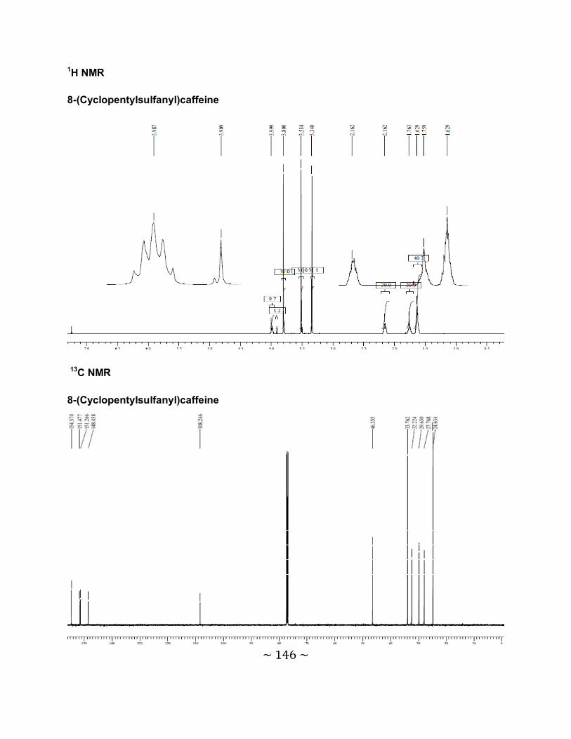
~ 146 ~
1H NMR
8-(Cyclopentylsulfanyl)caffeine
13C NMR
8-(Cyclopentylsulfanyl)caffeine
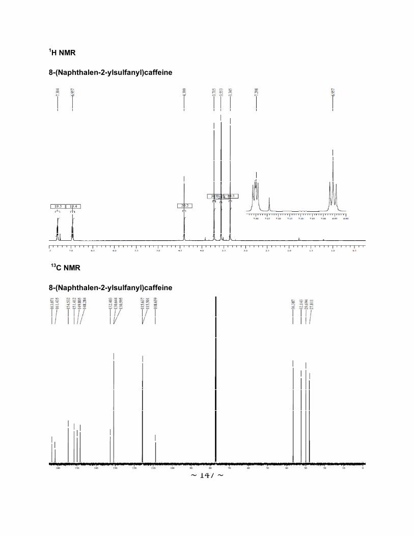
~ 147 ~
1H NMR
8-(Naphthalen-2-ylsulfanyl)caffeine
13C NMR
8-(Naphthalen-2-ylsulfanyl)caffeine
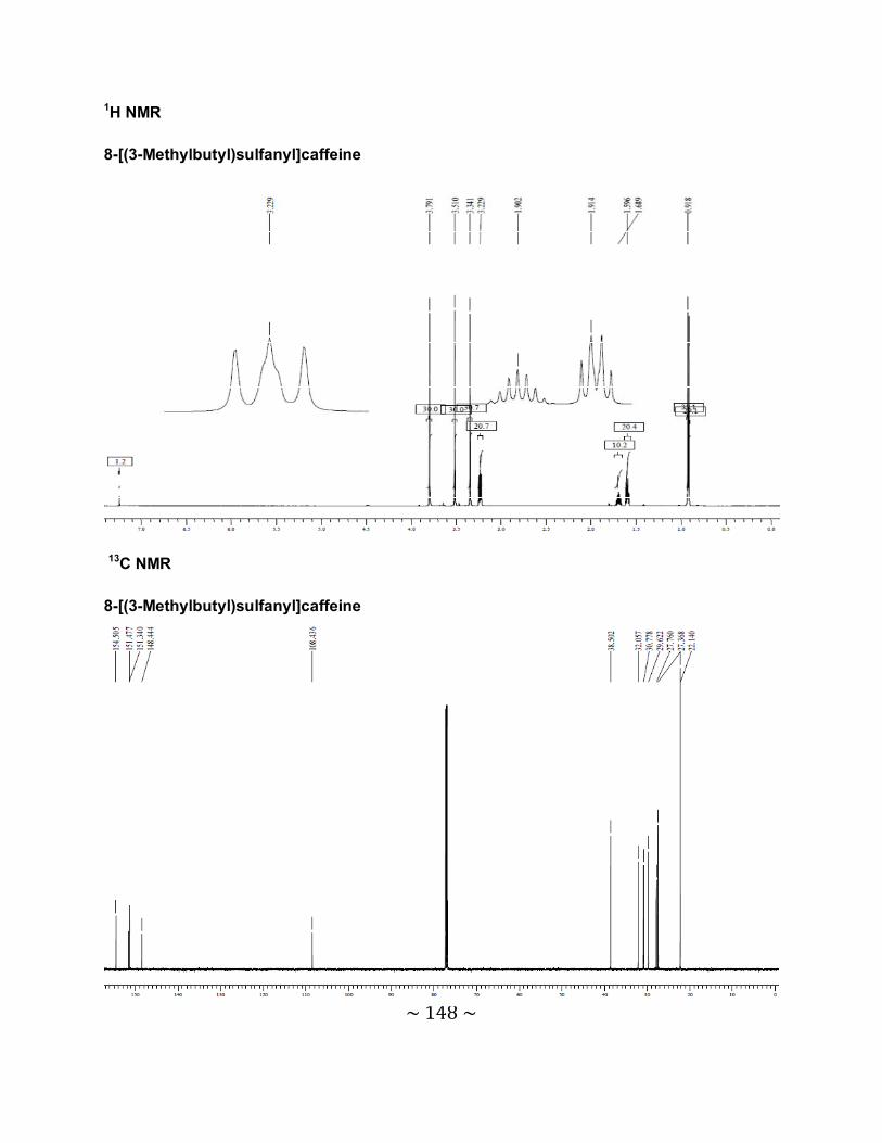
~ 148 ~
1H NMR
8-[(3-Methylbutyl)sulfanyl]caffeine
13C NMR
8-[(3-Methylbutyl)sulfanyl]caffeine

~ 149 ~
HPLC chromatograms 8-(Phenylsulfanyl)caffeine
8-(Benzylsulfanyl)caffeine
min0 2 4 6 8 10 12 14
mAU
0
250
500
750
1000
1250
1500
1750
2000
DAD1 A, Sig=210,4 Ref=off (PETZER\31MAR021.D)
6.0
66
min0 2 4 6 8 10 12 14
mAU
0
250
500
750
1000
1250
1500
1750
2000
DAD1 A, Sig=210,4 Ref=off (PETZER\31MAR012.D)
6.6
16
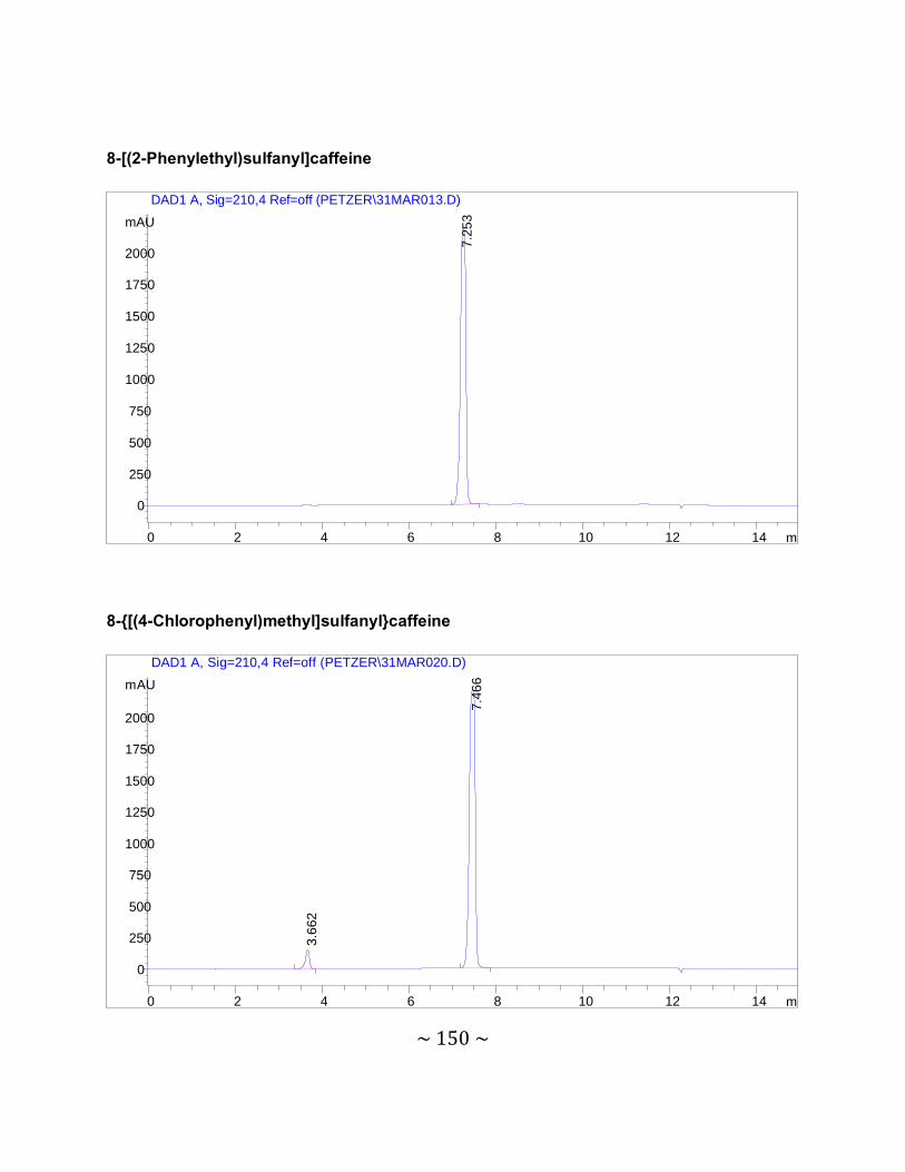
~ 150 ~
8-[(2-Phenylethyl)sulfanyl]caffeine
8-{[(4-Chlorophenyl)methyl]sulfanyl}caffeine
min0 2 4 6 8 10 12 14
mAU
0
250
500
750
1000
1250
1500
1750
2000
DAD1 A, Sig=210,4 Ref=off (PETZER\31MAR013.D)
7.2
53
min0 2 4 6 8 10 12 14
mAU
0
250
500
750
1000
1250
1500
1750
2000
DAD1 A, Sig=210,4 Ref=off (PETZER\31MAR020.D)
7.4
66
3.6
62
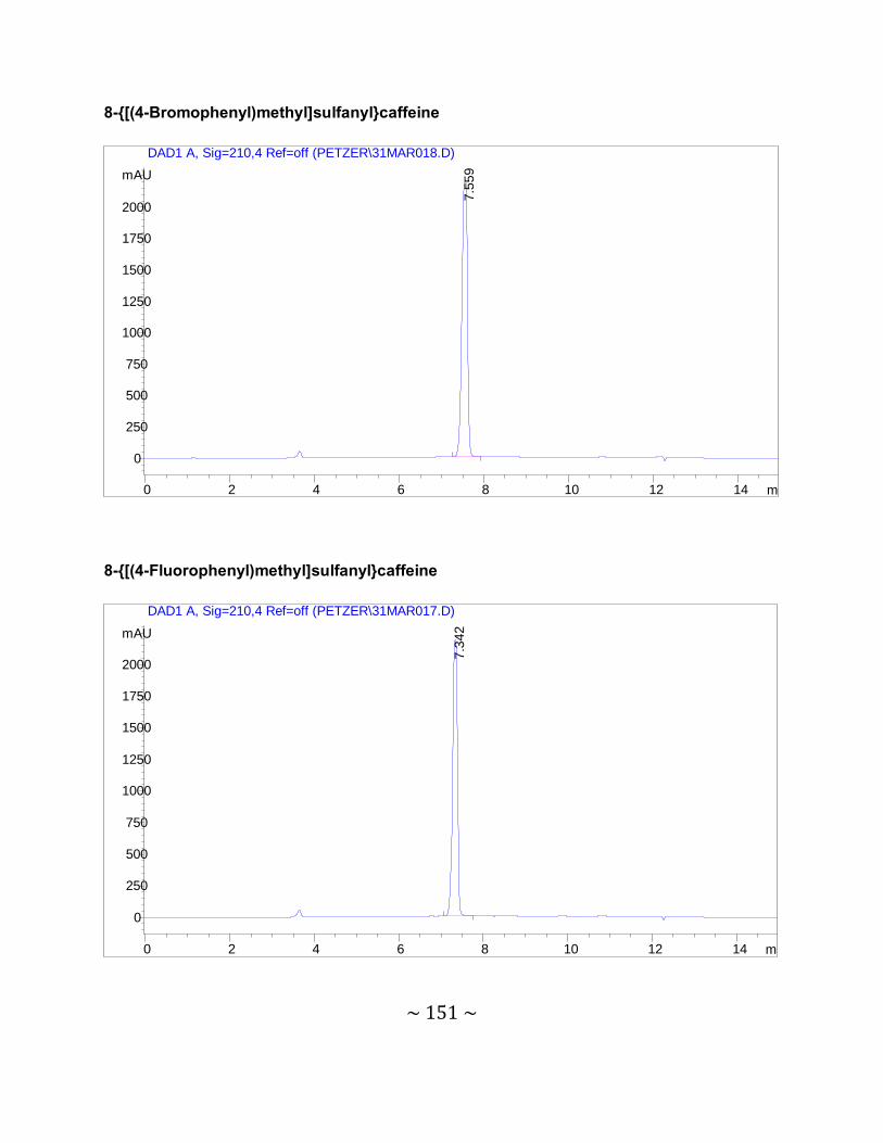
~ 151 ~
8-{[(4-Bromophenyl)methyl]sulfanyl}caffeine
8-{[(4-Fluorophenyl)methyl]sulfanyl}caffeine
min0 2 4 6 8 10 12 14
mAU
0
250
500
750
1000
1250
1500
1750
2000
DAD1 A, Sig=210,4 Ref=off (PETZER\31MAR018.D)
7.5
59
min0 2 4 6 8 10 12 14
mAU
0
250
500
750
1000
1250
1500
1750
2000
DAD1 A, Sig=210,4 Ref=off (PETZER\31MAR017.D)
7.3
42

~ 152 ~
8-{[(4-Methoxyphenyl)methyl]sulfanyl}caffeine
8-[(2-Phenoxyethyl)sulfanyl]caffeine
min0 2 4 6 8 10 12 14
mAU
0
250
500
750
1000
1250
1500
1750
2000
DAD1 A, Sig=210,4 Ref=off (PETZER\31MAR023.D)
6.5
20
9.8
28
min0 2 4 6 8 10 12 14
mAU
0
250
500
750
1000
1250
1500
1750
2000
DAD1 A, Sig=210,4 Ref=off (PETZER\31MAR022.D)
6.8
62
7.7
70
11.
807
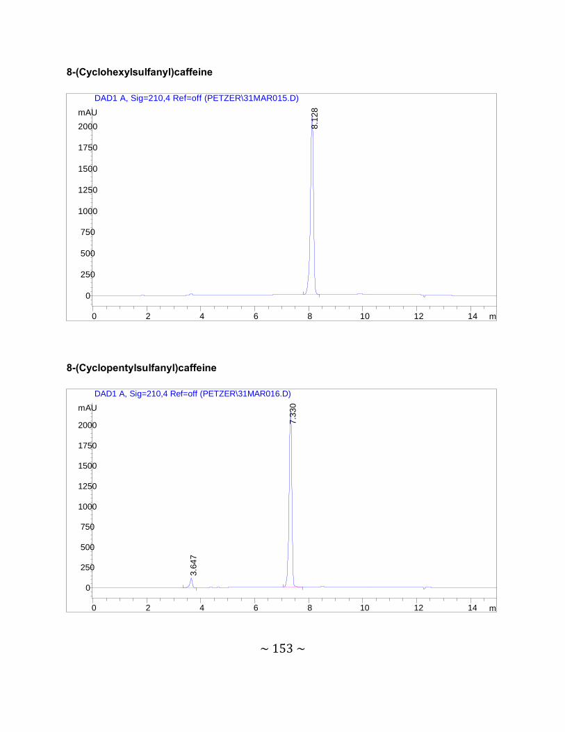
~ 153 ~
8-(Cyclohexylsulfanyl)caffeine
8-(Cyclopentylsulfanyl)caffeine
min0 2 4 6 8 10 12 14
mAU
0
250
500
750
1000
1250
1500
1750
2000
DAD1 A, Sig=210,4 Ref=off (PETZER\31MAR015.D)
8.1
28
min0 2 4 6 8 10 12 14
mAU
0
250
500
750
1000
1250
1500
1750
2000
DAD1 A, Sig=210,4 Ref=off (PETZER\31MAR016.D)
7.3
30
3.6
47
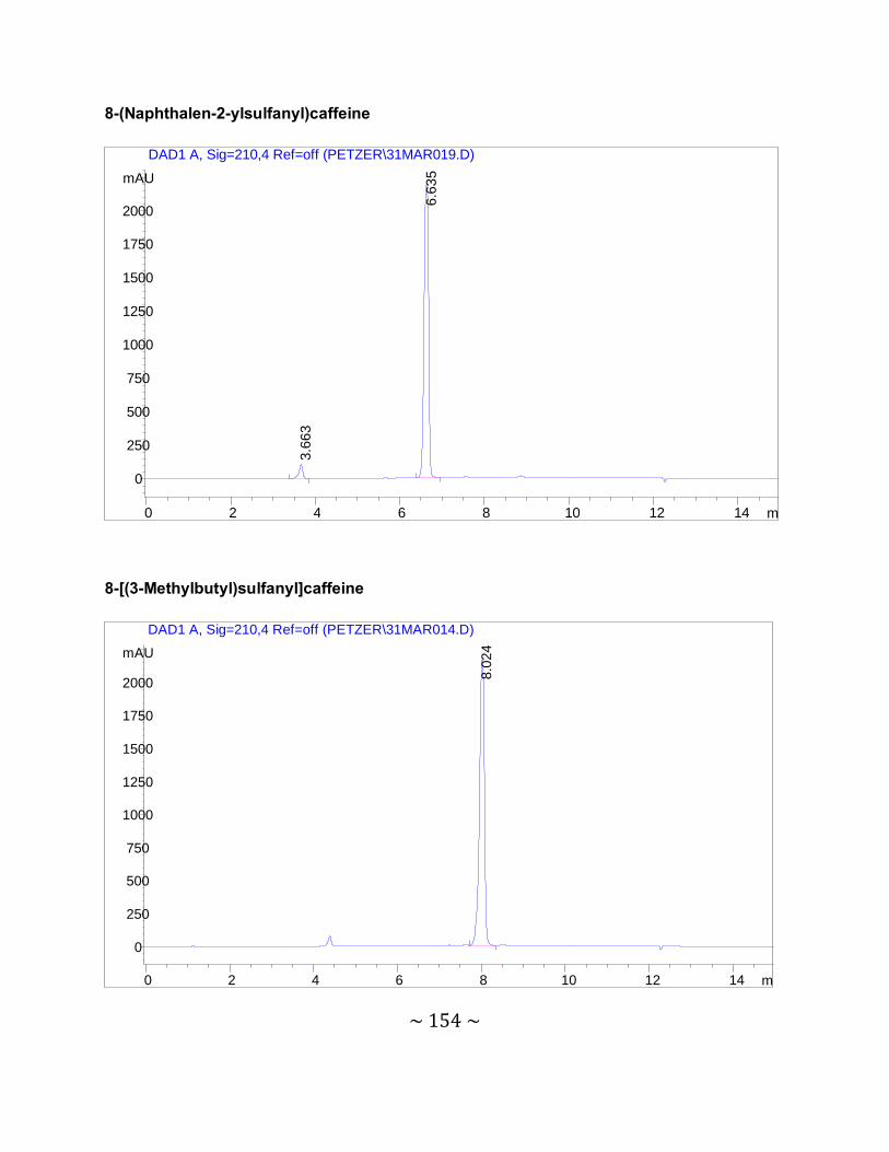
~ 154 ~
8-(Naphthalen-2-ylsulfanyl)caffeine
8-[(3-Methylbutyl)sulfanyl]caffeine
min0 2 4 6 8 10 12 14
mAU
0
250
500
750
1000
1250
1500
1750
2000
DAD1 A, Sig=210,4 Ref=off (PETZER\31MAR019.D)
6.6
35
3.6
63
min0 2 4 6 8 10 12 14
mAU
0
250
500
750
1000
1250
1500
1750
2000
DAD1 A, Sig=210,4 Ref=off (PETZER\31MAR014.D)
8.0
24
~ 155 ~
MS spectra 8-(Phenylsulfanyl)caffeine
8-(Benzylsulfanyl)caffeine
~ 156 ~
8-[(2-Phenylethyl)sulfanyl]caffeine
8-{[(4-Chlorophenyl)methyl]sulfanyl}caffeine
~ 157 ~
8-{[(4-Bromophenyl)methyl]sulfanyl}caffeine
8-{[(4-Fluorophenyl)methyl]sulfanyl}caffeine
~ 158 ~
8-{[(4-Methoxyphenyl)methyl]sulfanyl}caffeine
8-[(2-Phenoxyethyl)sulfanyl]caffeine
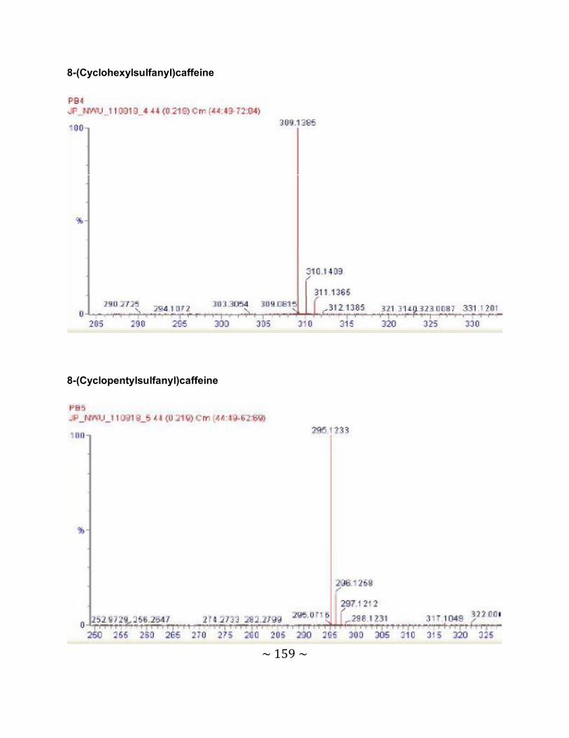
~ 159 ~
8-(Cyclohexylsulfanyl)caffeine
8-(Cyclopentylsulfanyl)caffeine
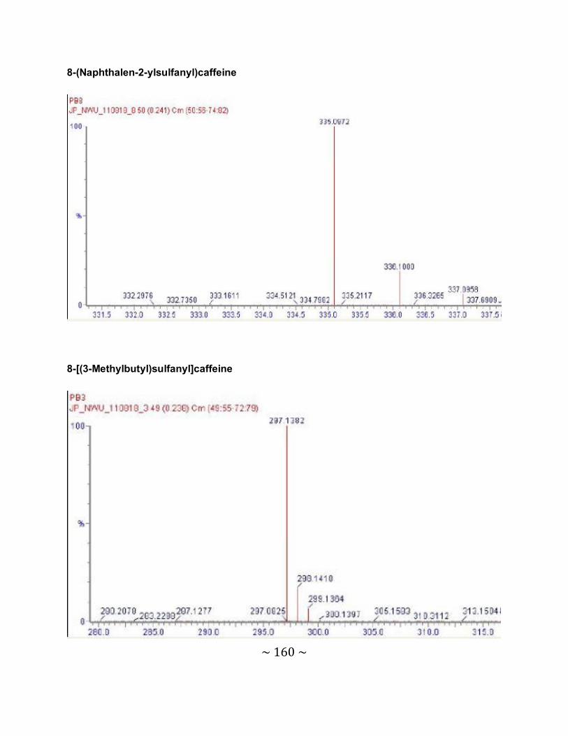
~ 160 ~
8-(Naphthalen-2-ylsulfanyl)caffeine
8-[(3-Methylbutyl)sulfanyl]caffeine
~ 161 ~
Concept article The research conducted in this project was included as part of a paper published in Bioorganic
& Medicinal Chemistry. See article below.
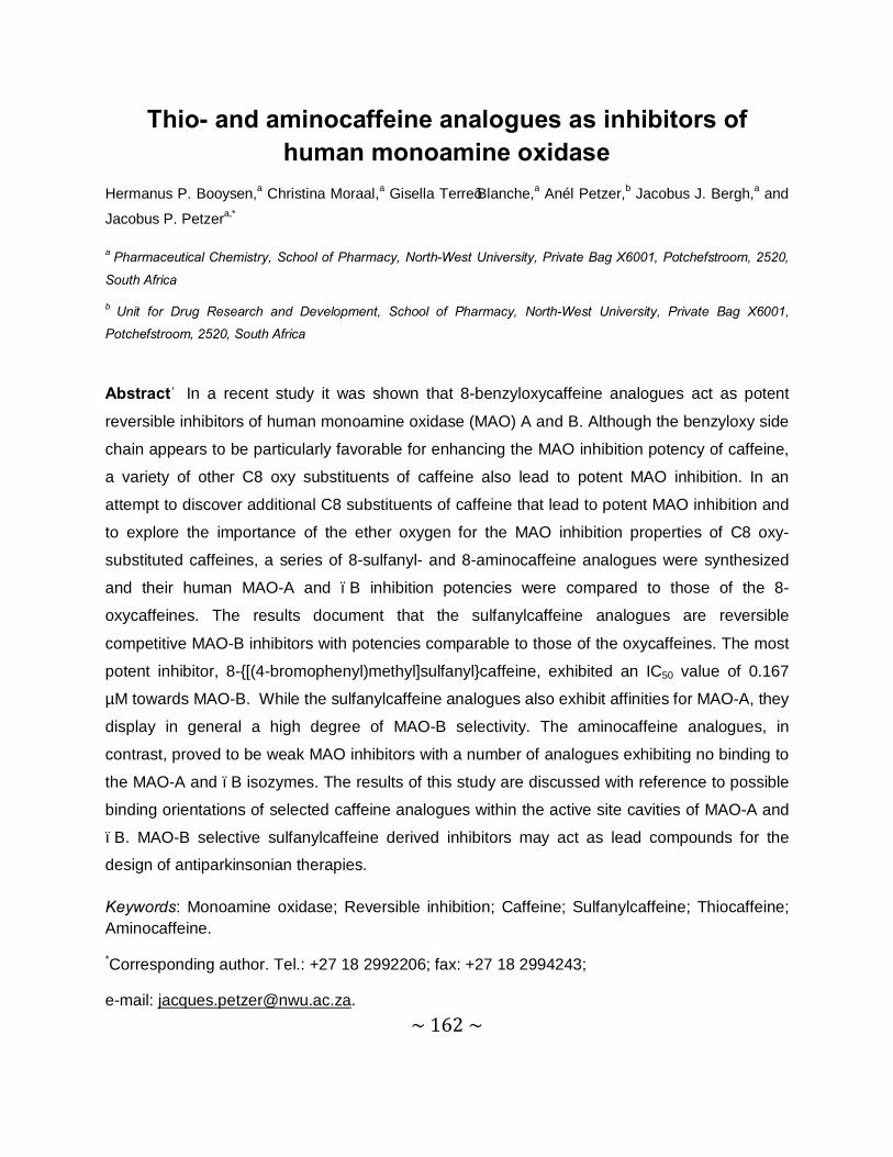
~ 162 ~
Thio- and aminocaffeine analogues as inhibitors of human monoamine oxidase
Hermanus P. Booysen,a Christina Moraal,a Gisella Terre’Blanche,a Anél Petzer,b Jacobus J. Bergh,a and
Jacobus P. Petzera,*
a Pharmaceutical Chemistry, School of Pharmacy, North-West University, Private Bag X6001, Potchefstroom, 2520,
South Africa
b Unit for Drug Research and Development, School of Pharmacy, North-West University, Private Bag X6001,
Potchefstroom, 2520, South Africa
Abstract―In a recent study it was shown that 8-benzyloxycaffeine analogues act as potent
reversible inhibitors of human monoamine oxidase (MAO) A and B. Although the benzyloxy side
chain appears to be particularly favorable for enhancing the MAO inhibition potency of caffeine,
a variety of other C8 oxy substituents of caffeine also lead to potent MAO inhibition. In an
attempt to discover additional C8 substituents of caffeine that lead to potent MAO inhibition and
to explore the importance of the ether oxygen for the MAO inhibition properties of C8 oxy-
substituted caffeines, a series of 8-sulfanyl- and 8-aminocaffeine analogues were synthesized
and their human MAO-A and –B inhibition potencies were compared to those of the 8-
oxycaffeines. The results document that the sulfanylcaffeine analogues are reversible
competitive MAO-B inhibitors with potencies comparable to those of the oxycaffeines. The most
potent inhibitor, 8-{[(4-bromophenyl)methyl]sulfanyl}caffeine, exhibited an IC50 value of 0.167
µM towards MAO-B. While the sulfanylcaffeine analogues also exhibit affinities for MAO-A, they
display in general a high degree of MAO-B selectivity. The aminocaffeine analogues, in
contrast, proved to be weak MAO inhibitors with a number of analogues exhibiting no binding to
the MAO-A and –B isozymes. The results of this study are discussed with reference to possible
binding orientations of selected caffeine analogues within the active site cavities of MAO-A and
–B. MAO-B selective sulfanylcaffeine derived inhibitors may act as lead compounds for the
design of antiparkinsonian therapies.
Keywords: Monoamine oxidase; Reversible inhibition; Caffeine; Sulfanylcaffeine; Thiocaffeine; Aminocaffeine.
*Corresponding author. Tel.: +27 18 2992206; fax: +27 18 2994243;
e-mail: [email protected].
~ 163 ~
1. Introduction
The monoamine oxidases (MAO) A and B are mitochondrial bound flavin adenine dinucleotide
(FAD) enzymes which catalyze the α-carbon oxidation of a variety of aminyl substrates.1 Human
MAO-A and –B consist of 529 and 520 amino acids, respectively, and the FAD is covalently
bound to a cysteinyl residue in both enzymes (Cys-406 and Cys-397 in MAO-A and –B,
respectively). While MAO-A and –B are products of separate genes they share approximately
70% amino acid sequence identity.2 The X-ray crystallographic structures of MAO-A and –B
indicate that the amino acid residues comprising the active sites and their relative geometries
are similar with only 6 of the 16 active site amino acid residues differing between the 2
enzymes.3,4 In spite of these similarities, MAO-A and –B have different substrate and inhibitor
specificities. Most notably, MAO-A metabolizes the neurotransmitters, serotonin and
norepinephrine, as well as the dietary amine, tyramine. MAO-B is well known to metabolize
extraneous amines such as benzylamine and phenylethylamine. Dopamine is considered to be
a substrate for both isozymes.5
Since MAO-A and –B are both involved in the degradation of neurotransmitter amines, inhibitors
of these enzymes are employed as drugs in the treatment of several disorders.5 For example,
MAO-A inhibitors block the central oxidation of serotonin by MAO-A and are used as
antidepressants. MAO-B inhibitors reduce the MAO-B catalyzed oxidative metabolism of
dopamine in the brain and are used in the treatment of Parkinson’s disease. Of importance is
the observation that MAO-B activity and density increase in most brain regions including the
basal ganglia with age while MAO-A activity remains unchanged.6,7 In the aged parkinsonian
brain MAO-B is therefore thought to be the principal MAO isozyme responsible for dopamine
catabolism. MAO-B inhibitors may conserve dopamine in the basal ganglia and offer a
symptomatic benefit in the treatment of Parkinson’s disease.8–10 MAO-B inhibitors are frequently
combined with levodopa therapy since inhibitors of this enzyme have been shown to enhance
the elevation of dopamine levels derived from levodopa.11 MAO-B inhibitors may permit a
reduction of the dose of levodopa required for a therapeutic effect and therefore the occurrence
of levodopa associated side effects.12 MAO may also play an important role in the
neurodegenerative processes associated with Parkinson’s disease. The oxidation of dopamine
by MAO stoichiometrically yields potentially toxic metabolic by-products.13 For each mole of
~ 164 ~
dopamine oxidized by MAO, one mole of hydrogen peroxide (which may lead to oxidative
damage) and dopaldehyde (which may react with exocyclic amino groups of nucleosides and N-
terminal and lysine ε-amino groups of proteins) are formed.13 Inhibitors of MAO reduce the
MAO-catalyzed metabolism of DA and as a result reduce the formation of these toxic by-
products. MAO inhibitors are therefore considered as a potential treatment strategy to slow the
progression of Parkinson’s disease since they may exert neuroprotective effects in the brain.13
Based on the therapeutic value of MAO inhibitors the current study aims to discover new
reversible inhibitors of the MAO enzymes, particularly the B isozyme. For this purpose caffeine
(1) serve as lead compound (Fig. 1). Although caffeine is a weak MAO-B inhibitor (Ki = 3.6 mM),
substitution at the C8 position with a variety of substituents has been shown to enhance the
MAO-B inhibition potency of caffeine to a large degree.14 In previous studies it was shown that
substitution at C8 of caffeine with alkyloxy substituents (2) yielded particularly potent MAO-B
inhibitors with a number of compounds exhibiting IC50 values in the nM range.15,16 Interestingly
these oxycaffeines are also MAO-A inhibitors, a property that may be attributed to the relatively
large degree of rotational freedom of the C8 side chain at the carbon-oxygen ether bond. It has
been suggested that structures with a relatively larger degree of conformational freedom may be
better suited for binding to MAO-A than relatively rigid structures.15 Based on these promising
results the present study investigates the possibility that alkylsulfanyl and alkylamino
substituents at C8 of caffeine may similarly enhance the MAO-A and –B inhibition potency of
caffeine. For this purpose, a series of twelve aryl- and alkylsulfanylcaffeine analogues (3a–l) and ten aryl- and alkylaminocaffeine analogues (4a–h, 5a–b) were synthesized and evaluated
as potential inhibitors of recombinant human MAO-A and –B (Fig. 2 and Tables 1–3).
2. Results
2.1. Chemistry
The aryl- and alkylsulfanylcaffeine analogues (3a–l) were synthesized by reacting 8-
chlorocaffeine (6) with an appropriate thiol reagent (7) in the presence of NaOH and employing
a mixture of ethanol and water as reaction solvent (Scheme 1).17 The aryl- and
~ 165 ~
alkylaminocaffeine analogues (4a–h) were similarly prepared by reaction of an appropriate
amine reagent (8) with 8-chlorocaffeine but with the exception that the addition of base and
additional solvent were not required (Scheme 2).18 Methylation of 4c and 4e at the C8 amine to
yield aminomethyl caffeine analogues 5a and 5b, respectively, were carried out in DMSO using
CH3I as alkylating agent and KOH as base. The structures of all the target compounds were
verified by 1H NMR, 13C NMR and mass spectrometry. The purities of the compounds were
estimated by HPLC analysis.
2.2. Inhibition of MAO-A and –B
The MAO inhibition potencies of the sulfanylcaffeine (3a–l) and aminocaffeine analogues (4a–h,
5a–b) were examined by employing the recombinant human MAO-A and –B enzymes as
enzyme sources.19 The mixed MAO-A/B substrate, kynuramine, was used as substrate for the
inhibition studies of both MAO-A and –B. Kynuramine displays similar Km values towards the
two enzymes with values of 16.1 µM and 22.7 µM for MAO-A and –B, respectively.15 The MAO-
catalyzed oxidation of kynuramine yields 4-hydroxyquinoline, a fluorescent compound which is
readily measured in basic solutions at excitation and emission wavelengths of 310 nm and 400
nm, respectively. Neither the substrate nor the test inhibitors fluoresce under these conditions,
or quench the fluorescence of 4-hydroxyquinoline. The inhibition potencies of the
sulfanylcaffeine and aminocaffeine analogues are expressed as the IC50 values, which were
determined from sigmoidal dose-response curves constructed in triplicate from 6 different
inhibitor concentrations spanning at least 3 orders of magnitude.
2.2.1. MAO inhibition by sulfanylcaffeine analogues (3a–l)
The MAO inhibition potencies of the sulfanylcaffeine analogues (3a–l) are presented in table 1.
As shown by the selectivity index (SI) values, the sulfanylcaffeine analogues are selective
inhibitors of MAO-B. The only exceptions are 3k which displays slight selectivity for the MAO-A
isozyme and 3l which is essentially nonselective. 8-(Phenylsulfanyl)caffeine (3a) which displays
slight selectivity for MAO-B, was found to be a relatively weak inhibitor of both MAO-A and –B.
Extension of the C8 side chain by one methylene unit to yield the benzylsulfanyl homologue 3b
~ 166 ~
(IC50 = 1.86 µM) enhances the MAO-B inhibition potency 17-fold compared to 3a (IC50 = 33.2
µM). A further increase in the length of the C8 side chain yields even more potent MAO-B
inhibitors. For example, compounds 3c and 3d, the phenylethylsulfanyl and
phenoxyethylsulfanyl homologues exhibited IC50 values of 0.223 µM and 0.332 µM,
respectively. While the extension of the C8 side chain of 3a (IC50 = 56.4 µM) by one methylene
unit to yield 3b (IC50 = 8.22 µM) also results in improved MAO-A inhibition, further increasing the
length of the C8 side chain does not result in a further enhancement of inhibition activity. For
example, 3c (IC50 = 20.5 µM) and 3d (IC50 = 15.5 µM) are weaker MAO-A inhibitors than the
benzylsulfanyl homologue 3b (IC50 = 8.22 µM).
Interestingly, halogen substitution of the phenyl ring of the C8 side chain is also associated with
an increase in MAO-B inhibition potency. The benzylsulfanyl substituted caffeine homologues
containing chlorine (3e; IC50 = 0.192 µM), bromine (3f; IC50 = 0.167 µM) and fluorine (3g; IC50 =
0.348 µM) on the benzyl phenyl ring were found to be 5–11-fold more potent than the
corresponding unsubstituted homologue 3b (IC50 = 1.86 µM). Methoxy substitution of the phenyl
ring of 8-(benzylsulfanyl)caffeine, in contrast, is associated with a loss of both MAO-A and –B
inhibition activity. Halogen substitution of 8-(benzylsulfanyl)caffeine (3b) also enhances MAO-A
inhibition potency, although by a lesser degree compared to MAO-B. The homologues
containing chlorine (3e; IC50 = 2.77 µM), bromine (3f; IC50 =2.62 µM) and fluorine (3g; IC50 =
4.80 µM) on the benzyl phenyl ring are 1.7–3-fold more potent than the corresponding
unsubstituted homologue 3b (IC50 = 8.22 µM).
The results document that compound 3i, the sulfanylcaffeine analogue containing a branched
alkyl side chain at C8, is also a relatively good MAO-B inhibitor with an IC50 value of 2.62 µM. In
fact, 3i is approximately 12-fold more potent as an MAO-B inhibitor than was 8-
(phenylsulfanyl)caffeine (3a) which contains a C8 phenylsulfanyl side chain. This result
demonstrates that a ring system in the C8 side chain is not an absolute requirement for MAO-B
inhibition by sulfanylcaffeine analogues. In accordance with this view, compound 3i also proved
to be more potent as a MAO-B inhibitor than the sulfanylcaffeine analogues containing
cyclohexyl (3j), cyclopentyl (3k) and napthalenyl (3l) C8 side chains. Interestingly the
~ 167 ~
sulfanylcaffeine analogues containing cyclohexyl (3j), cyclopentyl (3k) and napthalenyl (3l) C8
side chains were more potent inhibitors of both MAO-A and –B than the phenyl substituted
analogue 3a. This result suggests that, with the appropriate structural modification, these
moieties may be suitable for the future design of sulfanylcaffeine derived MAO inhibitors. An
example of such a structural modification would be the extension of the length of the C8 side
chain. Based on the observations that the naphthalenyl substituted sulfanylcaffeine analogue 3l is 15- and 9-fold more potent as a MAO-A and –B inhibitor, respectively, than the phenyl
substituted sulfanylcaffeine analogue 3a and that 3l is a nonselective inhibitor, the naphthalenyl
moiety may be particularly suited for the design of sulfanylcaffeine derived mixed MAO-A/B
inhibitors.
2.2.2. MAO inhibition by aminocaffeine analogues (4a–h, 5a–b)
The MAO inhibition potencies of the aminocaffeine analogues 4a–h are presented in tables 2
and 3. The data show that these compounds were relatively weak inhibitors of both MAO-A and
–B with IC50 values ranging from 5.78–45.2 µM and 9.60–24.4 µM for the inhibition of MAO-A
and –B, respectively. In fact, several of the compounds exhibited no binding to the MAO-A and
–B isozymes. Even homologues containing extended C8 side chains such as 4d (-NH-(CH2)3-
C6H5) and 4e (-NH-(CH2)4-C6H5) did not exhibit potent MAO inhibition. Similarly, homologue 4g
which contains a halogen on the phenyl ring of the C8 side chain was found to be a relatively
weak MAO inhibitor. These results demonstrate that, in contrast to the sulfanylcaffeine
analogues, extension of the length of the C8 side chain and halogen substitution do not lead to
potent MAO-B inhibition by the aminocaffeine analogues. Compared to the sulfanylcaffeine
analogues, the aminocaffeine analogues are therefore weak MAO-B inhibitors. For example,
sulfanylcaffeine analogue 3c (IC50 = 0.223 µM) is 78-fold more potent as an MAO-B inhibitor
than its corresponding aminocaffeine homologue 4c (IC50 = 17.6 µM).
To investigate the possibility of enhancing the MAO inhibition potencies of the aminocaffeines,
selected analogues, 4c and 4e, were methylated at the C8 amine to yield compounds 5a and
5b, respectively. While methylation improved the MAO-B inhibition potency of 4e by
approximately 3-fold, MAO-A inhibition activity was slightly reduced. Methylation of 4c did not
~ 168 ~
result in a significant improvement of MAO-B inhibition potency and led to a reduction in MAO-A
inhibition. Compared to the sulfanylcaffeine analogues, the aminocaffeine analogues are also
weak MAO-B inhibitors. For example, sulfanylcaffeine analogue 3c (IC50 = 0.223 µM) is 75-fold
more potent than its aminomethylcaffeine homologue 5a (IC50 = 16.8 µM). It is noteworthy that
the most potent MAO-B inhibitor among the aminocaffeine analogues was the C8 methylated
derivative 5b with an IC50 value of 2.97 µM. It can therefore be concluded that while methylation
of the C8 amine of aminocaffeine analogues may result in enhanced MAO-B inhibition potency,
the resulting compounds remain relatively weak inhibitors compared to the sulfanylcaffeine
analogues.
2.3. Reversibility of MAO-A and –B inhibition
Based on the nature of the interactions with the MAO enzymes, inhibitors may be classified as
reversible or irreversible. Irreversible inhibitors normally form covalent interactions with the
enzymes while reversible inhibitors bind via intermolecular interactions. While irreversible
inhibitors of MAO have been clinically used for many years, this mode of inhibition may be
associated with certain shortcomings.20 These include a slow and variable recovery of enzyme
activity following withdrawal of the irreversible inhibitor.21 The turnover rate for the biosynthesis
of MAO-B in the human brain may be as much as 40 days.22 In contrast, enzyme activity is
regained relatively quickly following withdrawal of a reversible inhibitor, once the inhibitor is
cleared from the tissues. Based on these observations, the reversibility of MAO-A and –B
inhibition by the sulfanylcaffeine and aminocaffeine analogues was examined. For this purpose
the time-dependency of the inhibition of MAO-A and –B, respectively, by sulfanylcaffeine
analogue 3f was measured. The time-dependency of the inhibition of MAO-A and –B by
aminocaffiene analogues 4g and 5b, respectively, was also examined. While irreversible
inhibitors would lead to a time-dependent reduction of enzyme activity, the degree of enzyme
inhibition in the presence of a reversible MAO inhibitor remains unchanged irrespective of the
time for which the inhibitor is incubated with the enzyme.23
The test inhibitors, at concentrations of approximately 2-fold their measured IC50 values for the
inhibition of the respective MAO enzymes, were preincubated with recombinant human MAO-A
~ 169 ~
or –B for time periods of 0, 15, 30 and 60 min. Following these preincubations the residual MAO
catalytic activities were measured after the addition of the substrate, kynuramine. The results of
these reversibility studies are presented in figures 3 and 4. The graphs show that when 3f and
4g are preincubated with MAO-A and 3f and 5b are preincubated with MAO-B there are no
time-dependent reductions of MAO-A and –B catalytic activities. Even after a period of 60 min
the test compounds do not reduce the MAO catalytic rates. These results suggest that the test
caffeines are not time-dependent inhibitors of MAO-A and –B and interact reversibly, at least for
the time period (0–60 min) and at the inhibitor concentrations (2 × IC50) evaluated.
This study also examined the possibility that 3f may act as a competitive inhibitor of human
MAO-A and –B. For this purpose, sets of Lineweaver–Burk plots were constructed for the
inhibition of these enzymes by 3f. The initial catalytic rates of MAO-A or –B were measured in
the absence and presence of three different concentrations of 3f. These measurements were
carried out using four different concentrations of the substrate, kynuramine (15–90 µM). The
Lineweaver–Burk plots obtained from these experiments are shown in figure 5. The graphs
show that the Lineweaver–Burk plots constructed for the inhibition of MAO-A and –B are linear
and intersect at the y-axis. This indicates that the inhibition of the MAO enzymes by 3f is
competitive. This result is further support that 3f is a reversible MAO inhibitor.
2.4. Molecular modeling
The results of the MAO inhibition studies shows that among the sulfanylcaffeine analogues
evaluated here, several compounds act as potent reversible inhibitors of MAO-B with IC50
values in the nM range. Interestingly, extension of the length of the C8 side chain leads to
enhanced MAO-B inhibition potency. While the sulfanylcaffeine analogues are also MAO-A
inhibitors, they display, for the most part, selectivity for the MAO-B isozyme. In contrast to the
sulfanylcaffeine analogues, the aminocaffeine analogues were found to be weak MAO-B
inhibitors with many analogues exhibiting no binding to either MAO-A or B. To provide additional
insight, the predicted binding modes of selected analogues (3a–c and 4c) in the active site
cavities of MAO-A and –B were examined using molecular docking.
~ 170 ~
The docking studies were carried out using the LigandFit application of the Discovery Studio
modeling software (Accelrys) according to a previously reported protocol.23 As enzyme models,
the three-dimensional structures of human MAO-A cocrystallized with harmine (PDB entry:
2Z5X)3 and human MAO-B cocrystallized with safinamide (PDB entry: 2V5Z)4 were selected.
The enzyme models were prepared by calculating the protonation states of the ionizable
residues and adding the hydrogen atoms accordingly. After the valences of the FAD cofactor
and cocrystallized ligands were corrected, hydrogen atoms were added and the models were
subjected to an energy minimization cascade while the protein backbone was constrained. For
the purpose of the docking, only the crystal waters which are reported to be conserved and non-
displaceable were retained (see Experimental).3,4
The best ranked docking solution for the binding of the selected analogues (3a–c and 4c) to
MAO-B shows one prevailing orientation for all of the inhibitors. As shown by the binding
orientation of 3c, the caffeine ring binds within the substrate cavity of MAO-B, in close proximity
to the FAD cofactor (Fig. 6). This places the carbonyl oxygen at C2 of the caffeine ring 3.4 Å
from the flavin N5 and the carbonyl oxygen at C6 within hydrogen bond distance to the phenolic
hydrogen of Tyr-435. The caffeine ring also forms a potential π–π interaction with the aromatic
ring of Tyr-398. The region defined by the flavin isoalloxazine ring, Tyr-398 and Tyr-435 is the
only polar space of the MAO-B active site and is also the site where amine catalysis occurs.24 In
the MAO-B model selected for these studies, the side chain of Ile-199 is rotated into an
alternative conformation to allow for the fusion of the substrate and entrance cavities.25 This
rotation of the Ile-199 side chain from the active site cavity is essential for relatively large
inhibitors, such as safinamide and C8 substituted caffeine derivatives, to be able to bind to
MAO-B. As a result the phenylethyl C8 side chain of 3c is allowed to extend into the
hydrophobic entrance cavity where it may be stabilized via Van der Waals interactions. As
expected, the relatively shorter phenyl (3a) and benzyl (3b) C8 side chains of sulfanylcaffeine
homologues 3a and 3b do not extend as deep into the MAO-B entrance cavity as the side chain
of 3c, and may therefore undergo interactions with the entrance cavity to a lesser extent
compared to 3c (Fig. 7). Despite the similar binding orientations of 3a and 3b within the MAO-B
active site, 3b was found to be a 17-fold more potent inhibitor. Since 3b protrudes only slightly
~ 171 ~
deeper (by ~1.2 Å) into the entrance cavity compared to 3a, this result suggest that a relatively
small enhancement of the space occupied by an inhibitor in the entrance cavity leads to a large
increase in binding affinity. Another possible explanation may be that the larger degree of
conformational freedom afforded by the longer C8 side chain of 3b may facilitate improved
interaction with the entrance cavity. The view that interaction with the entrance cavity is
essential for high affinity inhibitor binding is supported by the observation that caffeine is a weak
MAO-B inhibitor.14 Lacking a C8 side chain, caffeine is expected to bind only within the
substrate cavity and is unable to interact with the entrance cavity. The lower MAO-B inhibition
potencies of 3a and 3b compared to compound 3c may thus be explained by weaker interaction
with the entrance cavity.
Interestingly the aminocaffeine analogue 4c adopts a similar binding mode to that described
above for 3c and as a result forms similar interactions with the MAO-B active site (Fig. 8). The
only additional interaction that may occur between 4c and MAO-B is a potential hydrogen bond
between the C8 amine and the phenolic group of Tyr-326. The results of the MAO inhibition
studies however documents that the aminocaffeine analogues are weak MAO-B inhibitors with
4c being 78-fold weaker than the corresponding sulfanylcaffeine homologue 3c. The docking
studies therefore suggest that differing binding orientations cannot account for the apparent loss
of MAO-B inhibition activity of the aminocaffeine analogues.
The predicted binding orientation of 3c within the active site of MAO-A is similar to the binding
orientation observed in MAO-B with the caffeine ring bound in close proximity to the FAD
cofactor and the C8 side chain extending towards the entrance of the active site (Fig. 9).
Interestingly, the caffeine ring is rotated by ~180 ºC compared to the binding orientation adopted
in the MAO-B active site. This dissimilarity in binding orientations in the MAO-A and –B active
sites has also been observed in docking studies with 8-benzyloxycaffeine analogues.15 As a
result of the flipped orientation of the caffeine ring, the C2 carbonyl oxygen is within hydrogen
bond distance of the phenolic hydrogen of Tyr-444 and two active site waters. Also, the caffeine
ring of 3c binds more distant from Tyr-407 (~4.3 Å) in MAO-A than from the corresponding
residue, Tyr-398 (~3.6 Å), in MAO-B. For this reason, a π–π interaction similar to that observed
~ 172 ~
between the caffeine ring and Tyr-398 in MAO-B, is not observed between 3c and the MAO-A
active site. This may represent a possible reason for the finding that sulfanylcaffeine analogues
are in general more potent MAO-B inhibitors than MAO-A inhibitors. Also noteworthy is the
observation that, in the MAO-A active site, the C8 side chain of 3c is bent at the CH2-S thioether
bond from the plane of the caffeine ring while in the MAO-B active site, the side chain of 3c
exhibits a modest deviation from the plane of the caffeine ring (Fig. 10). The differing binding
orientations adopted by C8 substituted caffeine derivatives in MAO-A and –B may, for the most
part, be attributed to steric hindrance caused by the aromatic moieties of Phe-208 in MAO-A
and Tyr-326 in MAO-B. As illustrated in figure 11A, the binding orientation and position of 3c in
the MAO-B active site cannot be reproduced in MAO-A because this would result in structural
overlap with the phenyl ring of Phe-208. In MAO-B, the amino acid residue that occupies the
same position as Phe-208 in MAO-A is Ile-199. In MAO-B the side chain of Ile-199 may rotate
out of the active site cavity to allow for the observed binding pose of 3c.25 Similarly, the binding
orientation and position of 3c in the MAO-A active site cannot be reproduced in MAO-B because
of structural overlap with of Tyr-326 (Fig. 11B). In MAO-A, the amino acid residue that occupies
the analogous position as Tyr-398 in MAO-B is Ile-335. The relatively smaller side chain of Ile-
335 compared to the aromatic ring of Tyr-398, does not sterically prevent the observed binding
orientation of 3c in the MAO-A active site.3
3. Discussion
Based on previous reports that oxycaffeine analogues are MAO inhibitors,15,16 the present study
investigated the possibility that C8 substituted sulfanylcaffeine and aminocaffeine analogues
may also act as inhibitors of human MAO-A and –B. The results demonstrated that several of
the sulfanylcaffeine analogues act as potent MAO-B inhibitors and that the inhibition is
reversible. For example, the bromine substituted sulfanylcaffeine analogue 3f was the most
potent MAO-B inhibitor with an IC50 value of 0.167 µM. The relatively high MAO-B inhibition
potencies of the sulfanylcaffeine analogues may be evaluated by comparison of the IC50 value
of 3f (IC50 = 0.167 µM.) with the reversible inhibitor safinamide, which binds to MAO-B with an
IC50 value of 0.08 µM.4 While the sulfanylcaffeine analogues are also MAO-A inhibitors, they are
for the most part selective for the MAO-B isoform. Modeling studies predict that the
sulfanylcaffeine analogues adopt dissimilar binding modes in the MAO-A and –B active site
~ 173 ~
cavities, respectively. Compared to the predicted orientation in MAO-B, the caffeine ring is
flipped by approximately 180 º in the MAO-A active site which results in differing interactions of
the caffeine ring with the polar regions of the MAO-A and –B substrate cavities. The alternative
binding orientation of the caffeine ring in MAO-A may be less optimal for the formation of
stabilizing polar interactions compared to the binding orientation adopted in MAO-B and may
explain, at least in part, the lower binding affinities of the sulfanylcaffeine analogues to MAO-
A.15 Interestingly, modeling studies suggest that the C8 side chain of the sulfanylcaffeine
analogue 3c is bent to a high degree from the plane of the caffeine ring at the CH2-S thioether
bond while in the MAO-B active site, the C8 side chain displays only a modest deviation from
the plane of the caffeine moiety. The ability of the C8 side chain to adopt a bent orientation may
be an important requirement for the inhibition of MAO-A. Rigid C8 substituted caffeine
analogues such as (E)-8-(3-chlorostyryl)caffeine (CSC) (Fig. 12) are not MAO-A inhibitors while
displaying high affinity binding to MAO-B.26 The observation that CSC does not bind to MAO-A
may be explained by its low degree of flexibility and inability to adopt a bent orientation similar to
that observed for 3c in the MAO-A active site.
The notion that the C8 side chains of the caffeine analogues are important structural features for
MAO-A and –B inhibition is supported by the observation that caffeine is a weak MAO
inhibitor.14 Modeling shows that, in the MAO-B active site, the C8 side chains of the
sulfanylcaffeine analogues may extend into the hydrophobic entrance cavity where they are
stabilized by Van der Waals interactions. Since extension of the C8 chain length results in
enhanced MAO-B inhibition potency it may be concluded that longer C8 side chains form more
productive interactions with the MAO-B entrance cavity, which thus leads to more potent
enzyme inhibition. Halogen substitution on the phenyl ring of the C8 side chain also leads to a
significant enhancement of MAO-B inhibition. This result may be explained by the possibility that
halogen substitution may further improve Van der Waals and dipole interactions between the
MAO-B entrance cavity and the C8 side chain. While it is not clear why methoxy substitution of
8-(benzylsulfanyl)caffeine leads to a loss of both MAO-A and –B inhibition potency, this result is
in accordance with the findings of a previous study which showed that MAO-B inhibition
potencies of a series of benzyloxycaffeine analogues correlate with the electronegativity of
substituents on the phenyl ring of the C8 side chain and that electron-withdrawing groups
enhance MAO-B inhibition potency.15 Interestingly, this study shows that C8 side chains that do
~ 174 ~
not contain phenyl rings are also suitable for MAO inhibition. Examples of sulfanylcaffeine
analogues containing such side chains are the 3-methylbutyl (3i), cyclohexyl (3j), cyclopentyl
(3k) and napthalenyl (3l) substituted homologues.
One of the most significant findings of this study is that the aminocaffeine analogues are weak
MAO inhibitors with most homologues displaying no inhibition. The predicted binding orientation
and interactions of aminocaffeine analogue 4c in the MAO-B active site is similar to the
orientation of sulfanylcaffeine analogue 3c. In fact 4c displays an additional hydrogen bond
interaction with Tyr-326. In spite of these predictions 4c is approximately 78-fold weaker as a
MAO-B inhibitor compared to 3c. Even methylation of the C8 amines to yield tertiary amines
does not produce inhibitors with similar potencies to those of the sulfanylcaffeine analogues.
While the reasons for this behavior is not clear, differing ionization states of the sulfanylcaffeine
and aminocaffeine analogues do not explain the difference in binding affinities to the MAO
enzymes, since both the sulfanylcaffeine and aminocaffeine analogues are expected to be
uncharged in the buffer used for the inhibition studies (pH 7.4). Also, it is unlikely that
aminocaffeines are excluded from entering the access channel leading to the active site cavity
since aminyl substrates are thought to be deprotonated prior to entering the MAO active sites.24
In conclusion, the sulfanylcaffeine analogues exhibit similar MAO-B inhibition potencies to those
of the previously reported oxycaffeine analogues with various homologues from both series
exhibiting IC50 values in the nM range.15,16 The attachment of substituents at C8 of caffeine via a
thioether linkage therefore enhances MAO-B inhibition activity to a similar extent compared to
attachment via an oxyether. In contrast, C8 substituted aminocaffeines are not suitable for MAO
inhibition. Based on the potent MAO-B inhibition properties of the sulfanylcaffeine analogues,
they may be considered as lead compounds for the development of reversible MAO-B inhibitors.
~ 175 ~
4. Experimental section
4.1. Chemicals and instrumentation
Unless otherwise noted, all starting materials were obtained from Sigma-Aldrich and were used
without purification. Proton (1H) and carbon (13C) NMR spectra were recorded on a Bruker
Avance III 600 spectrometer at frequencies of 600 MHz and 150 MHz, respectively. All NMR
measurements were conducted in CDCl3 and DMSO-d6 and the chemical shifts are reported in
parts per million (δ) downfield from the signal of tetramethylsilane added to the deuterated
solvent. Spin multiplicities are given as s (singlet), d (doublet), dd (doublet of doublets), t
(triplet), q (quartet), qn (quintet), sept (septet) or m (multiplet). High resolution mass spectra
(HRMS) were obtained on a Waters Synapt G2 instrument in electrospray ionization (ESI)
mode. The HRMS spectrum of 4a was recorded on a DFS high resolution magnetic sector mass
spectrometer (Thermo Electron Corporation) in atmospheric pressure chemical ionization
(APCI) mode. Melting points (mp) were measured with a Stuart SMP10 melting point apparatus
and are uncorrected. The purity of the synthesized compounds were determined via HPLC
analyses which were conducted with an Agilent 1100 HPLC system equipped with a quaternary
gradient pump and an Agilent 1100 series diode array detector (see Supplementary Material).
HPLC grade acetonitrile (Merck) and Milli-Q water (Millipore) were used for the chromatography.
For fluorescence spectrophotometry, a Varian Cary Eclipse fluorescence spectrophotometer
was employed. Microsomes from insect cells containing recombinant human MAO-A and –B (5
mg/mL) and kynuramine.2HBr were obtained from Sigma-Aldrich.
4.2. Synthesis of C8-substituted thiocaffeine analogues (3a–l)
A solution of NaOH (4 mmol) in 3.5 mL water and 7 mL ethanol was cooled in an ice bath and
the appropriate thiol (4 mmol) was added. The reaction mixture was stirred and 8-chlorocaffeine
(4 mmol) was added in a single portion to yield a suspension. The reaction was heated under
reflux for 60 min and then cooled on ice. The white precipitate was collected by filtration and
washed with 30 mL ethanol. The product was recrystallized from 30 mL ethanol at room
temperature and the crystals were washed with 30 mL ethanol.17
~ 176 ~
4.2.1. 8-(Phenylsulfanyl)caffeine (3a)
The title compound was prepared from thiophenol in a yield of 65.1%: mp 149 °C (ethanol). 1H
NMR (Bruker Avance III 600, CDCl3) δ 3.37 (s, 3H), 3.54 (s, 3H), 3.90 (s, 3H), 7.32 (m, 5H); 13C
NMR (Bruker Avance III 600, CDCl3) δ 28.0, 29.9, 33.1, 109.5, 128.2, 129.6, 130.5, 130.9,
146.4, 148.0, 151.4, 154.9; ESI-HRMS m/z: calcd for C14H15N4O2S (MH+), 303.0916, found
303.0912; Purity (HPLC): 98%.
4.2.2. 8-(Benzylsulfanyl)caffeine (3b)
The title compound was prepared from benzyl mercaptan in a yield of 59%: mp 149 °C
(ethanol). 1H NMR (Bruker Avance III 600, CDCl3) δ 3.35 (s, 3H), 3.57 (s, 3H), 3.69 (s, 3H), 4.42
(s, 2H), 7.27 (m, 3H), 7.31 (m, 2H); 13C NMR (Bruker Avance III 600, CDCl3) δ 27.8, 29.7, 32.2,
37.4, 108.7, 127.9, 128.7, 128.9, 136.6, 148.3, 150.0, 151.5, 154.6; ESI-HRMS m/z: calcd for
C15H17O2N4S, 317.1072 (MH+), found 317.1073; Purity (HPLC): 99%.
4.2.3. 8-[(2-Phenylethyl)sulfanyl]caffeine (3c)
The title compound was prepared from phenylethyl mercaptan in a yield of 22.9%: mp 95 °C
(ethanol). 1H NMR (Bruker Avance III 600, CDCl3) δ 3.04 (t, 2H, J = 7.9 Hz), 3.36 (s, 3H), 3.49
(t, 2H, J – 7.9 Hz), 3.55 (s, 3H), 3.78 (s, 3H), 7.21 (m, 3H), 7.29 (m, 2H); 13C NMR (Bruker
Avance III 600, CDCl3) δ 27.8, 29.7, 32.1, 33.8, 36.0, 108.5, 126.7, 128.5, 139.3, 148.5, 150.9,
151.5, 154.5; ESI-HRMS m/z: calcd for C16H19N4O2S, 331.1229 (MH+), found 331.1229; Purity
(HPLC): 99%.
4.2.4. 8-[(2-Phenoxyethyl)sulfanyl]caffeine (3d)
The title compound was prepared from 2-phenoxyethanethiol in a yield of 43.9%: mp 114 °C
(ethanol). 1H NMR (Bruker Avance III 600, CDCl3) δ 3.37 (s, 3H), 3.53 (s, 3H), 3.63 (t, 2H, J =
6.4 Hz), 3.83 (s, 3H), 4.30 (t, 2H, J = 6.4 Hz), 6.91 (d, 2H, J = 8.3 Hz), 6.94 (t, 1H, J = 7.2 Hz),
7.25 (m, 2H); 13C NMR (Bruker Avance III 600, CDCl3) δ 27.8, 29.7, 31.5, 32.2, 66.3, 108.7,
~ 177 ~
114.5, 121.3, 129.5, 148.4, 150.3, 151.5, 154.5, 158.1; ESI-HRMS m/z: calcd for C16H19N4O3S
(MH+), 347.1176, found 347.1173; Purity (HPLC): 95%.
4.2.5. 8-{[(4-Chlorophenyl)methyl]sulfanyl}caffeine (3e)
The title compound was prepared from 4-chlorobenzyl mercaptan in a yield of 85.6%: mp 169
°C (ethanol). 1H NMR (Bruker Avance III 600, CDCl3) δ 3.36 (s, 3H), 3.56 (s, 3H), 3.73 (s, 3H),
4.40 (s, 2H), 7.26 (m, 4H); 13C NMR (Bruker Avance III 600, CDCl3) δ 27.9, 29.7, 32.2, 36.4,
108.8, 128.8, 130.3, 133.8, 135.2, 148.3, 149.7, 151.5, 154.6; ESI-HRMS m/z: calcd for
C15H16ClN4O2S (MH+), 351.0682, found 351.0679; Purity (HPLC): 97%.
4.2.6. 8-{[(4-Bromophenyl)methyl]sulfanyl}caffeine (3f)
The title compound was prepared from 4-bromobenzyl mercaptan in a yield of 82.0%: mp 166
°C (ethanol). 1H NMR (Bruker Avance III 600, CDCl3) δ 3.35 (s, 3H), 3.55 (s, 3H), 3.72 (s, 3H),
4.38 (s, 2H), 7.21 (d, 2H, J = 8.3 Hz), 7.40 (d, 2H, J = 8.3 Hz); 13C NMR (Bruker Avance III 600,
CDCl3) δ 27.8, 29.7, 32.2, 36.4, 108.7, 121.8, 130.6, 131.8, 135.8, 148.3, 149.6, 151.4, 154.5;
ESI-HRMS m/z: calcd for C15H16BrN4O2S (MH+), 395.0177, found 395.0178; Purity (HPLC):
98%.
4.2.7. 8-{[(4-Fluorophenyl)methyl]sulfanyl}caffeine (3g)
The title compound was prepared from 4-fluorobenzyl mercaptan in a yield of 71.6%: mp 175 °C
(ethanol). 1H NMR (Bruker Avance III 600, CDCl3) δ 3.35 (s, 3H), 3.55 (s, 3H), 3.72 (s, 3H), 4.40
(s, 2H), 6.96 (t, 2H, J = 8.3 Hz), 7.30 (q, 2H, J = 5.3 Hz); 13C NMR (Bruker Avance III 600,
CDCl3) δ 27.8, 29.7, 32.1, 36.4, 108.7, 115.6 (d), 130.6 (d), 132.4 (d), 148.3, 149.8, 151.4,
154.5, 161.4, 163.1; ESI-HRMS m/z: calcd for C15H16FN4O2S (MH+), 335.0976, found 335.0972;
Purity (HPLC): 95%.
~ 178 ~
4.2.8. 8-{[(4-Methoxyphenyl)methyl]sulfanyl}caffeine (3h)
The title compound was prepared from 4-methoxybenzyl mercaptan in a yield of 90.5%: mp 159
°C (ethanol). 1H NMR (Bruker Avance III 600, CDCl3) δ 3.36 (s, 3H), 3.58 (s, 3H), 3.71 (s, 3H),
3.76 (s, 3H), 4.39 (s, 2H), 6.80 (d, 2H, J = 8.7 Hz), 7.24 (d, 2H, J = 8.7 Hz); 13C NMR (Bruker
Avance III 600, CDCl3) δ 27.8, 29.7, 32.1, 37.0, 55.3, 108.6, 114.1, 128.4, 130.2, 148.4, 150.2,
151.5, 154.6, 159.2; ESI-HRMS m/z: calcd for C16H19N4O3S (MH+), 347.1176, found 347.1171;
Purity (HPLC): 94%.
4.2.9. 8-[(3-Methylbutyl)sulfanyl]caffeine (3i)
The title compound was prepared from 3-methyl-1-butanethiol in a yield of 35.3%: mp 79 °C
(ethanol). 1H NMR (Bruker Avance III 600, CDCl3) δ 0.91 (d, 6H, J = 6.8 Hz), 1.59 (q, 2H, J =
7.9 Hz), 1.69 (sept, 1H, J = 6.8 Hz), 3.23 (t, 2H, J = 7.5 Hz), 3.34 (s, 3H), 3.51 (s, 3H), 3.79 (s,
3H); 13C NMR (Bruker Avance III 600, CDCl3) δ 22.1, 27.4, 27.8, 29.6, 30.8, 32.1, 38.5, 108.4,
148.4, 151.3, 151.5, 154.5; ESI-HRMS m/z: calcd for C13H21N4O2S (MH+), 297.1385, found
297.1382; Purity (HPLC): 97%.
4.2.10. 8-(Cyclohexylsulfanyl)caffeine (3j)
The title compound was prepared from cyclohexanethiol in a yield of 37.2%: mp 133 °C
(ethanol). 1H NMR (Bruker Avance III 600, CDCl3) δ 1.28 (m, 1H), 1.38 (m, 2H), 1.48 (m, 2H),
1.58 (m, 1H), 1.74 (m, 2H), 2.03 (m, 2H), 3.34 (s, 3H), 3.52 (s, 3H), 3.71 (m, 1H), 3.82 (s, 3H); 13C NMR (Bruker Avance III 600, CDCl3) δ 25.4, 25.8, 27.8, 29.7, 32.3, 33.4, 47.2, 108.4, 148.4,
150.3, 151.5, 154.6; ESI-HRMS m/z: calcd for C14H21N4O2S (MH+), 309.1385, found 309.1385;
Purity (HPLC): 99%.
4.2.11. 8-(Cyclopentylsulfanyl)caffeine (3k)
The title compound was prepared from cyclopentanethiol in a yield of 60.9%: mp 135 °C
(ethanol). 1H NMR (Bruker Avance III 600, CDCl3) δ 1.63 (m, 4H), 1.76 (m, 2H), 2.15 (m, 2H),
~ 179 ~
3.34 (s, 3H), 3.51 (s, 3H), 3.80 (s, 3H), 3.99 (qn, 1H); 13C NMR (Bruker Avance III 600, CDCl3) δ
24.6, 27.8, 29.7, 32.2, 33.8, 46.4, 108.2, 148.5, 151.3, 151.5, 154.6; ESI-HRMS m/z: calcd for
C13H19N4O2S (MH+), 295.1229, found 295.1233; Purity (HPLC): 95%.
4.2.12. 8-(Naphthalen-2-ylsulfanyl)caffeine (3l)
The title compound was prepared from 2-naphthalenethiol in a yield of 87.7%: mp 175 °C
(ethanol). 1H NMR (Bruker Avance III 600, CDCl3) δ 3.37 (s, 3H), 3.53 (s, 3H), 3.91 (s, 3H), 7.35
(dd, 1H, J = 1.9, 8.3 Hz), 7.48 (m, 2H), 7.73 (m, 1H), 7.78 (m, 2H), 7.84 (s, 1H); 13C NMR
(Bruker Avance III 600, CDCl3) δ 27.9, 29.8, 33.1, 109.5, 126.9, 127.0, 127.4, 127.5, 127.8,
127.9, 129.4, 129.7, 132.6, 133.6, 146.4, 148.0, 151.4, 154.9; ESI-HRMS m/z: calcd for
C18H17N4O2S (MH+), 353.1072, found 353.1074; Purity (HPLC): 94%.
4.3. Synthesis of C8-substituted aminocaffeine analogues (4a–h)
A mixture of 8-chlorocaffeine (2 mmol) and the appropriate amine (10 mmol) was heated under
reflux (175–180 ºC) for 3 hours. The reaction was cooled to room temperature and treated with
50 mL acetic acid (5%). The resulting suspension was stirred for 15 min at room temperature
and the precipitate was collected by filtration. The product was dried at 60 ºC and recrystallized
twice from ethanol (30 mL) at 0 ºC.18
4.3.1. 8-(Phenylamino)caffeine (4a)
The title compound was prepared from aniline and 8-chlorocaffeine in a yield of 24.2%: mp 164–
265 °C (ethanol). 1H NMR (Bruker Avance III 600, DMSO-d6) δ 3.15 (s, 3H), 3.35 (s, 3H), 3.74
(s, 3H), 6.96 (t, 1H, J = 7.5 Hz), 7.29 (t, 2H, J = 7.5 Hz), 7.67 (d, 2H, J = 8.3 Hz), 9.07 (s, 1H); 13H NMR (Bruker Avance III 600, DMSO-d6) δ 27.3, 29.4, 30.5, 102.0, 118.1, 121.7, 128.7,
140.0, 147.2, 149.3, 150.9, 153.3; APCI-HRMS m/z: calcd for C14H15N5O2 (M+), 285.1226, found
285.1230; Purity (HPLC): 98%.
~ 180 ~
4.3.2. 8-(Benzylamino)caffeine (4b)
The title compound was prepared from benzylamine and 8-chlorocaffeine in a yield of 74.0%:
mp 230 °C (ethanol). 1H NMR (Bruker Avance III 600, DMSO-d6) δ 3.14 (s, 3H), 3.35 (s, 3H),
3.58 (s, 3H), 4.53 (d, 2H, J = 5.6 Hz), 7.23 (t, 1H, J = 7.5 Hz), 7.32 (t, 2H, J = 7.5 Hz), 7.36 (d,
2H, J = 7.5 Hz), 7.56 (t, 1H, J = 5.6 Hz); 13H NMR (Bruker Avance III 600, DMSO-d6) δ 27.1,
29.2, 29.8, 45.7, 102.0, 126.9, 127.4, 128.3, 139.6, 148.2, 150.9, 152.9, 154.0; ESI-HRMS m/z:
calcd for C15H18N5O2 (MH+), 300.1460, found 300.1459; Purity (HPLC): 99%.
4.3.3. 8-[(2-Phenylethyl)amino]caffeine (4c)
The title compound was prepared from 2-phenylethylamine and 8-chlorocaffeine in a yield of
68.3%: mp 221 °C (ethanol). 1H NMR (Bruker Avance III 600, DMSO-d6) δ 2.88 (t, 2H, J = 7.2
Hz), 3.14 (s, 3H), 3.32 (s, 3H), 3.49 (m, 2H), 3.51 (s, 3H), 7.11 (t, 1H, J = 5.3 Hz), 7.19 (t, 1H, J
= 7.2 Hz), 7.22 (d, 2H, J = 7.5 Hz), 7.29 (t, 2H, J = 7.5 Hz); 13H NMR (Bruker Avance III 600,
DMSO-d6) δ 27.1, 29.2, 29.7, 35.3, 44.1, 101.8, 126.1, 128.3, 128.7, 139.4, 148.3, 150.9, 152.9,
153.9; ESI-HRMS m/z: calcd for C16H20N5O2 (MH+), 314.1617, found 314.1621; Purity (HPLC):
99%.
4.3.4. 8-[(3-Phenylpropyl)amino]caffeine (4d)
The title compound was prepared from 3-phenylpropylamine and 8-chlorocaffeine in a yield of
76.8%: mp 204–205 °C (ethanol). 1H NMR (Bruker Avance III 600, DMSO-d6) δ 1.88 (qn, 2H,
7.5 Hz), 2.64 (t, 2H, J = 7.5 Hz), 3.13 (s, 3H), 3.30 (s, 3H), 3. 32 (m, 2H), 3.52 (s, 3H), 6.98 (t,
1H, J = 5.3 Hz), 7.16 (t, 1H, J = 7.2 Hz), 7.22 (d, 2H, J = 7.5 Hz), 7.26 (t, 2H, J = 7.5 Hz); 13H
NMR (Bruker Avance III 600, DMSO-d6) δ 27.1, 29.2, 29.7, 30.8, 32.3, 42.0, 101.8, 125.7,
128.2, 128.3, 141.7, 148.3, 150.9, 152.8, 154.1; ESI-HRMS m/z: calcd for C17H22N5O2 (MH+),
328.1773, found 328.1774; Purity (HPLC): 99%.
~ 181 ~
4.3.5. 8-[(4-Phenylbutyl)amino]caffeine (4e)
The title compound was prepared from 4-phenylbutylamine and 8-chlorocaffeine in a yield of
63.0%: mp 179–180 °C (ethanol). 1H NMR (Bruker Avance III 600, DMSO-d6) δ 1.59 (m, 4H),
2.60 (t, 2H, J = 7.2 Hz), 3.13 (s, 3H), 3.30 (s, 3H), 3.32 (m, 2H), 3.51 (s, 3H), 6.94 (t, 1H, J = 5.6
Hz), 7.14 (t, 1H, J = 7.2 Hz), 7.18 (d, 2H, J = 7.2 Hz), 7.25 (t, 2H, J = 7.2 Hz); 13C NMR (Bruker
Avance III 600, DMSO-d6) δ 27.1, 28.2, 28.8, 29.2, 29.7, 34.8, 42.2, 101.7, 125.6, 128.2, 128.3,
142.1, 148.3, 150.9, 152.8, 154.1; ESI-HRMS m/z: calcd for C18H24N5O2 (MH+), 342.1930, found
342.1929; Purity (HPLC): 99%.
4.3.6. 8-{[2-(Pyridin-2-yl)ethyl]amino}caffeine (4f)
The title compound was prepared from 2-(2-pyridyl)ethylamine and 8-chlorocaffeine in a yield of
20.4%: mp 196–197 °C (ethanol). 1H NMR (Bruker Avance III 600, DMSO-d6) δ 3.03 (t, 2H, J =
7.2 Hz), 3.13 (s, 3H), 3.31 (s, 3H), 3.50 (s, 3H), 3.65 (q, 2H, 6.7 Hz), 7.10 (t, 1H, J = 5.6 Hz),
7.20 (t, 1H, J = 5.6 Hz), 7.26 (d, 1H, J = 7.5 Hz), 7.69 (t, 1H, J = 7.5 Hz), 8.49 (d, 1H, J = 4.1
Hz); 13C NMR (Bruker Avance III 600, DMSO-d6) δ 27.1, 29.2, 29.7, 37.5, 42.4, 101.8, 121.5,
123.2, 136.4, 148.3, 149.0, 150.9, 152.8, 153.9, 159.1; ESI-HRMS m/z: calcd for C15H19N6O2
(MH+), 315.1569, found 315.1570; Purity (HPLC): 98%.
4.3.7. 8-{[2-(3-Chlorophenyl)ethyl]amino}caffeine (4g)
The title compound was prepared from 2-(3-chlorophenyl)ethanamine and 8-chlorocaffeine in a
yield of 48.5%: mp 111–113 °C (ethanol). 1H NMR (Bruker Avance III 600, DMSO-d6) δ 2.88 (t,
2H, J = 7.2 Hz), 3.13 (s, 3H), 3.32 (s, 3H), 3.50 (s, 3H), 3.52 (m, 2H), 7.10 (t, 1H, J = 5.6 Hz),
7.18 (d, 1H, J = 7.5 Hz), 7.24 (d, 1H, J = 8.3 Hz), 7.29 (d, 1H, J = 7.5 Hz), 7.31 (s, 1H); 13C NMR
(Bruker Avance III 600, DMSO-d6) δ 27.1, 29.2, 29.7, 34.8, 43.7, 101.8, 126.1, 127.5, 128.6,
130.1, 132.9, 142.0, 148.3, 150.9, 152.8, 153.8; ESI-HRMS m/z: calcd for C16H19N5O2Cl (MH+),
348.1227, found 348.1225; Purity (HPLC): 98%.
~ 182 ~
4.3.8. 8-(Cyclopentylamino)caffeine (4h)
The title compound was prepared from cyclopentylamine and 8-chlorocaffeine in a yield of
38.6%: mp 217–218 °C (ethanol). 1H NMR (Bruker Avance III 600, DMSO-d6) δ 1.52 (m, 4H),
1.68 (m, 2H), 1,93 (m, 2H), 3.13 (s, 3H), 3.31 (s, 3H), 3.53 (s, 3H), 4.10 (m, 1H), 6.76 (d, 1H, J =
7.2 Hz); 13H NMR (Bruker Avance III 600, DMSO-d6) δ 23.4, 27.1, 29.2, 29.8, 32.4, 54.2, 101.7,
148.3, 150.9, 152.8, 153.8; ESI-HRMS m/z: calcd for C13H20N5O2 (MH+), 278.1617, found
278.1612; Purity (HPLC): 96%.
4.4. Methylation of the C8-substituted aminocaffeine analogues (5a–b)
Potassium hydroxide (0.05 g) was powderized and suspended in 5 mL DMSO. The resulting
mixture was stirred for 30 min at room temperature and the aminocaffeine analogue (3 mmol)
dissolved in DMSO (5 mL) was added. The reaction was heated to 40 ºC (in order for the
aminocaffeine analogue to remain in solution) and iodomethane (0.8 mmol) was added. Stirring
of the reaction was continued and another portion of iodomethane (0.8 mmol) was added every
20 min until silica gel TLC (petroleum ether/ ethyl actetate 30:70) indicated completion of the
reaction. The pH of the reaction was also continually measured, and when acidic (pH paper),
another portion of potassium hydroxide (0.05 g) was added. Upon completion, the reaction was
cooled to room temperature and water (250 mL) was added. The resulting solution was
incubated for several days at 4 ºC and the formed crystals were collected by filtration.
4.4.1. 8-[Methyl(2-phenylethyl)amino]caffeine (5a)
The title compound was prepared from 8-[(2-phenylethyl)amino]caffeine (4c) and iodomethane
in a yield of 57.7%: mp 103 °C (ethanol). 1H NMR (Bruker Avance III 600, DMSO-d6) δ 2.88 (t,
2H, J = 7.5 Hz), 2.98 (s, 3H), 3.16 (s, 3H), 3.33 (s, 3H), 3.47 (t, 2H, J = 7.5 Hz), 3.59 (s, 3H),
7.18 (m, 1H), 7.25 (m, 4H); 13C NMR (Bruker Avance III 600, DMSO-d6) δ 27.3, 29.3, 32.5, 33.0,
38.6, 54.5, 103.8, 126.1, 128.3, 128.8, 139.0, 147.1, 150.9, 153.5, 156.6; ESI-HRMS m/z: calcd
for C17H22N5O2 (MH+), 328.1774, found 328.1734; Purity (HPLC): 99%.
~ 183 ~
4.4.2. 8-[Methyl(4-phenylbutyl)amino]caffeine (5b)
The title compound was prepared from 8-[(4-phenylbutyl)amino]caffeine (4e) and iodomethane
in a yield of 49.9%: mp 114 °C (ethanol). 1H NMR (Bruker Avance III 600, DMSO-d6) δ 1.57 (m,
4H), 2.57 (t, 2H, J = 7.15 Hz), 2.90 (s, 3H), 3.16 (s, 3H), 3.26 (t, 2H, J = 7.15 Hz), 3.32 (s, 3H),
3.65 (s, 3H), 7.15 (m, 3H), 7.24 (t, 2H, J = 7.91 Hz); 13C NMR (Bruker Avance III 600, DMSO-d6)
δ 26.3, 27.3, 27.9, 29.3, 32.6, 34.7, 38.4, 52.5, 103.8, 125.7, 128.2, 128.2, 142.0, 147.1, 150.9,
153.5, 156.9; ESI-HRMS m/z: calcd for C19H26N5O2 (MH+), 356.2087, found 356.2088; Purity
(HPLC): 98%.
4.5. IC50 determinations for the inhibition of human MAO
Microsomal preparations form insect cells containing recombinant human MAO-A and –B (5
mg/mL) served as enzyme sources and all enzymatic reactions were conducted in potassium
phosphate buffer (100 mM, pH 7.4, made isotonic with KCl) to a final volume of 500 µL.19 The
reactions contained the MAO-A/B mixed substrate, kynuramine, at concentrations of 45 µM and
30 µM for the incubations with MAO-A and –B, respectively, various concentrations of the test
inhibitor (0–100 µM) and the MAO enzymes (0.0075 mg/mL). The enzyme activities employed
for the IC50 value determinations were 24–28 nmoles 4-hydroxyquinoline formed/min/mg protein
for MAO-A and 6–8 nmoles 4-hydroxyquinoline formed/min/mg protein for MAO-B. Stock
solutions of the test inhibitors were prepared in DMSO and added to the reactions to yield a final
concentration of 4% (v/v) DMSO. The enzyme reactions were incubated at 37 °C for 20 minutes
and then terminated with the addition of 400 µL NaOH (2 N) and 1000 µL distilled water. After
centrifugation at 16,000 g for 10 min, the fluorescence of the MAO generated 4-
hydroxyquinoline in the supernatant fractions were measured (λex = 310 nm, λem = 400 nm). To
determine the concentrations of 4-hydroxyquinoline, a linear calibration curve was constructed
from solutions of 4-hydroxyquinoline (0.047–1.50 µM) in potassium phosphate buffer. The
calibration standards were prepared to a volume of 500 µL and contained 4% DMSO, 400 µL
NaOH (2 N) and 1000 µL distilled water. The initial rate of MAO catalysis was plotted versus the
logarithm of the inhibitor concentration to obtain a sigmoidal dose–response curve. Each curve
was constructed from 6 different inhibitor concentrations spanning at least 3 orders of a
magnitude. These data were fitted to the one site competition model incorporated into the
~ 184 ~
GraphPad Prism software and the IC50 values were determined in triplicate and are expressed
as mean ± standard deviation (SD).
4.6. Time-dependent inhibition studies
The reversibility of MAO inhibition was examined by determining the time-dependence of
inhibition of three selected inhibitors, 3f, 4g and 5b. The selected inhibitors were preincubated
in potassium phosphate buffer (100 mM, pH 7.4, made isotonic with KCl) with MAO-A and –B
(0.015–0.03 mg/mL) for periods of 0, 15, 30, 60 min at 37 ºC. The concentrations of the
inhibitors used were two-fold their measured IC50 values for the inhibition of the respective MAO
enzymes and were: 5.22 µM (3f) and 11.56 µM (4g) for the studies with MAO-A, and 0.32 µM
(3f) and 5.94 µM (5b) for the studies with MAO-B. To compensate for a potential time-
dependent loss of enzyme activity, the enzyme preincubations were firstly incubated at 37 ºC
and the inhibitors were subsequently added at different time points. These time points were
selected as to ensure that all enzyme preparations were preheated at 37 ºC for exactly 60 min,
irrespective of the time period (0–60 min) for which the enzyme preparations were preincubated
in the presence of the test inhibitor. These reactions were diluted 2-fold by the addition of
kynuramine at concentrations of 45 µM and 30 µM for the incubations with MAO-A and –B,
respectively, and the resulting reactions (500 µL final volume) were incubated at 37 °C for a
further 15 minutes. The final enzyme concentration in these reactions was 0.0075–0.015 mg/mL
and the concentrations of the selected inhibitors were approximately equal to their IC50 values
for the inhibition of the respective isozymes. The reactions were terminated with the addition of
400 µL NaOH (2 N) and 1000 µL distilled water and the rates of MAO catalyzed generation of 4-
hydroxyquinoline were measured and calculated as described above. All measurements were
carried out in triplicate and are expressed as mean ± SD.23
4.7. Construction of Lineweaver-Burk plots
A set consisting of four Lineweaver–Burk plots were constructed for the inhibition of MAO-A and
–B by the selected inhibitor 3f. One plot was constructed in the absence of inhibitor while three
plots were constructed in the presence of three different concentrations of 3f each. These
~ 185 ~
concentrations were 1.31–5.22 µM and 0.04–0.16 µM for the inhibition studies with MAO-A and
–B, respectively. Four different kynuramine concentrations (15–90 µM) were employed for each
plot and the concentrations of recombinant human MAO-A and –B used were 0.015 mg/mL. The
initial MAO catalytic rates were measured as described above. Linear regression analysis was
performed using GraphPad Prism.23
4.8. Molecular modeling studies
The modeling studies were carried out with the Windows based Discovery Studio 1.7 molecular
modeling software (Accelrys).23,27 For this purpose the crystallographic structures of MAO-A co-
crystallized with harmine (PDB code: 2Z5X)3 and MAO-B co-crystallized with safinamide (PDB
code: 2V5Z)4 were obtained from the Brookhaven Protein Data Bank (www.rcsb.org/pdb). The
protonation states of the ionizable amino acids residues were calculated at pH 7.4 and
hydrogen atoms were added to the receptor models. The valences of the FAD cofactors
(oxidized state) and co-crystallized ligands were corrected and hydrogen atoms were added
according to the appropriate protonation states at pH 7.4. The structures were typed
automatically with the Momany and Rone CHARMm forcefield, the backbone of the protein was
constrained and the structures were subjected to a three step energy minimization. The first
step was a steepest descent minimization which was followed by conjugate gradient
minimization. For both protocols the termination criteria was set to a maximum of 2500 steps or
a minimum value of 0.1 for the root mean square of the energy gradient. The final step was an
adopted basis Newton-Rapheson minimization and the termination criteria was set to a
maximum of 5000 steps or a minimum value for the root mean square of the energy gradient of
0.01. For these minimization steps the implicit generalized Born solvation model with simple
switching was employed with the dielectric constant set to 4. For both the MAO-A and –B
models, the crystal water molecules were removed with the exception of 3 active site waters in
each model. The X-ray crystallographic structures of MAO-B shows that three active site water
molecules (HOH 1155, 1170 and 1351; A-chain) are conserved, all located in the vicinity of the
FAD cofactor.4 In the MAO-A model, the crystal waters HOH 710, 718 and 739 which occupies
the analogous positions in the MAO-A active site compared to those cited above for MAO-B,
were retained. The co-crystallized ligands and the backbone constraints were subsequently
removed from the models and the binding sites were identified by a flood-filling algorithm. The
~ 186 ~
structures of 3a–c and 4c were constructed within Discovery Studio, their hydrogen atoms were
added according to the appropriate protonation states at pH 7.4. The geometries of the ligands
were briefly optimized in Discovery Studio using a fast Dreiding-like forcefield (1000 iterations)
and the atom potential types and partial charges were assigned with the Momany and Rone
CHARMm forcefield. Docking of the ligands was carried out with the LigandFit application of
Discovery Studio and the docking solutions were refined using the Smart Minimizer algorithm.
The parameters for the docking runs were set to their default values, ten possible binding
solutions were computed for each docked ligand and the best-ranked binding conformation of
each ligand was determined according to the DockScore values. The illustrations were prepared
in PyMOL.28 It is interesting to note that among the 10 best ranked binding orientations, there
were orientations which exhibited a reversed binding mode with the caffeine ring directed
towards the entrance of the MAO-A and –B active sites while the C8 sulfanyl and amino side
chains project towards the FAD cofactor. Based on the low DockScore values these orientations
were however deemed unlikely.
Acknowledgements
The NMR spectra were recorded by André Joubert of the SASOL Centre for Chemistry, North-
West University while the MS spectra were recorded by the Mass Spectrometry Service,
University of the Witwatersrand and the Mass Spectrometry Unit, Stellenbosch University. This
work was supported by grants from the National Research Foundation and the Medical
Research Council, South Africa.
References
1. Binda, C.; Newton-Vinson, P.; Hubálek, F.; Edmondson, D. E.; Mattevi, A. Nat. Struct. Biol. 2002,
9, 22.
2. Shih, J. C.; Chen, K.; Ridd, M. J.; Annu. Rev. Neurosci. 1999, 22, 197.
3. Son, S. –Y.; Ma, J.; Kondou, Y.; Yoshimura, M.; Yamashita, E.; Tsukihara, T. Proc. Natl. Acad.
Sci. U.S.A. 2008, 105, 5739.
4. Binda, C.; Wang, J.; Pisani, L.; Caccia, C.; Carotti, A.; Salvati, P.; Edmondson, D. E.; Mattevi, A.
J. Med. Chem. 2007, 50, 5848.
~ 187 ~
5. Youdim, M. B. H.; Edmondson, D.; Tipton, K. F. Nat. Rev. Neurosci. 2006, 7, 295.
6. Nicotra, A.; Pierucci, F.; Parvez, H.; Senatori, O. Neurotoxicology. 2004, 25, 155.
7. Fowler, J. S.; Volkow, N. D.; Wang, G. J.; Logan, J.; Pappas, N.; Shea, C.; MacGregor, R.
Neurobiol. Aging. 1997, 18, 431.
8. Youdim, M. B. H.; Collins, G. G. S.; Sandler, M.; Bevan-Jones, A. B.; Pare, C. M.; Nicholson, W.
J. Nature. 1972, 236, 225.
9. Collins, G. G. S.; Sandler, M.; Williams, E. D.; Youdim, M. B. H. Nature. 1970, 225, 817.
10. Di Monte, D. A.; DeLanney, L. E.; Irwin, I.; Royland, J. E.; Chan, P.; Jakowec, M. W.; Langston, J.
W. Brain. Res. 1996, 738, 53.
11. Finberg, J. P.; Wang, J.; Bankiewich, K.; Harvey-White, J; Kopin, I. J.; Goldstein, D. S. J. Neural
Transm. Suppl. 1998, 52, 279.
12. Fernandez, H. H.; Chen, J. J. Pharmacotherapy. 2007, 27, 174S.
13. Youdim, M. B. H.; Bakhle, Y. S. Br. J. Pharmacol. 2006, 147, S287.
14. Van der Walt, E. M.; Milczek, E. M.; Malan, S. F.; Edmondson, D. E.; Castagnoli, N., Jr.; Bergh, J.
J.; Petzer, J. P. Bioorg. Med. Chem. Lett. 2009, 19, 2509.
15. Strydom, B.; Malan, S. F.; Castagnoli, N.; Bergh, J. J.; Petzer, J. P. Bioorg. Med. Chem. 2010,
18, 1018.
16. Strydom, B.; Bergh, J. J.; Petzer, J. P. Eur. J. Med. Chem. 2011, 46, 3474.
17. Long, L. M. J. Am. Chem. Soc. 1947, 69, 2939.
18. Cramer, L. Chem. Ber. 1894, 27, 3089.
19. Novaroli, L.; Reist, M.; Favre, E.; Carotti, A.; Catto, M.; Carrupt, P. A. Bioorg. Med. Chem. 2005,
13, 6212.
20. Prins, L. H. A.; Petzer, J. P.; Malan, S. F. Eur. J. Med. Chem. 2010, 45, 4458.
21. Tipton, K.F.; Boyce, S.; O’Sullivan, J.; Davey, G. P.; Healy, J. Curr. Med. Chem. 2004, 11, 1965.
22. Fowler, J. S.; Volkow, N. D.; Logan, J.; Wang, G.; MacGregor, R. R.; Schlyer, D.; Wolf, A. P.;
Pappas, N.; Alexoff, D.; Shea, C.; Dorflinger, E.; Kruchowy, L.; Yoo, K.; Fazzini, E.; Patlak, C.
Synaps. 1994, 18, 86.
23. Manley-King, C. I.; Bergh, J. J.; Petzer, J. P. Bioorg. Med. Chem. 2011, 19, 261.
24. Binda, C.; Mattevi, A.; Edmondson, D. E.; J. Biol. Chem. 2002, 277, 23973.
25. Hubálek, F.; Binda, C.; Khalil, A.; Li, M.; Mattevi, A.; Castagnoli, N., Jr.; Edmondson, D. E. J. Biol.
Chem. 2005, 280, 15761.
26. Vlok, N.; Malan, S. F.; Castagnoli, N., Jr.; Bergh, J. J.; Petzer, J. P. Bioorg. Med. Chem. 2006, 14,
3512.
27. Accelrys Discovery Studio 1.7, Accelrys Software Inc., San Diego, CA, USA. 2006,
http://www.accelrys.com.
~ 188 ~
28. DeLano, W. L. The PyMOL Molecular Graphics System. DeLano Scientific, San Carlos, USA,
2002.
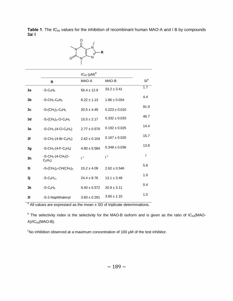
~ 189 ~
Table 1. The IC50 values for the inhibition of recombinant human MAO-A and –B by compounds 3a–l
N
N
N
N
O
O
R
IC50 (µM)a
R MAO-A MAO-B SIb
3a -S-C6H5 56.4 ± 12.9 33.2 ± 3.41 1.7
3b -S-CH2-C6H5 8.22 ± 1.13 1.86 ± 0.034 4.4
3c -S-(CH2)2-C6H5 20.5 ± 4.49 0.223 ± 0.010 91.9
3d -S-(CH2)2-O-C6H5 15.5 ± 2.17 0.332 ± 0.033 46.7
3e -S-CH2-(4-Cl-C6H4) 2.77 ± 0.570 0.192 ± 0.025 14.4
3f -S-CH2-(4-Br-C6H4) 2.62 ± 0.104 0.167 ± 0.020 15.7
3g -S-CH2-(4-F-C6H4) 4.80 ± 0.584 0.348 ± 0.036 13.8
3h -S-CH2-(4-CH3O-C6H4) –c –c –
3i -S-(CH2)2-CH(CH3)2 15.2 ± 4.09 2.62 ± 0.546 5.8
3j -S-C6H11 24.4 ± 8.76 13.1 ± 3.49 1.9
3k -S-C5H9 9.40 ± 0.572 20.9 ± 3.11 0.4
3l -S-2-Naphthalenyl 3.60 ± 0.291 3.60 ± 1.10 1.0
a All values are expressed as the mean ± SD of triplicate determinations.
b The selectivity index is the selectivity for the MAO-B isoform and is given as the ratio of IC50(MAO-
A)/IC50(MAO-B).
cNo inhibition observed at a maximum concentration of 100 µM of the test inhibitor.
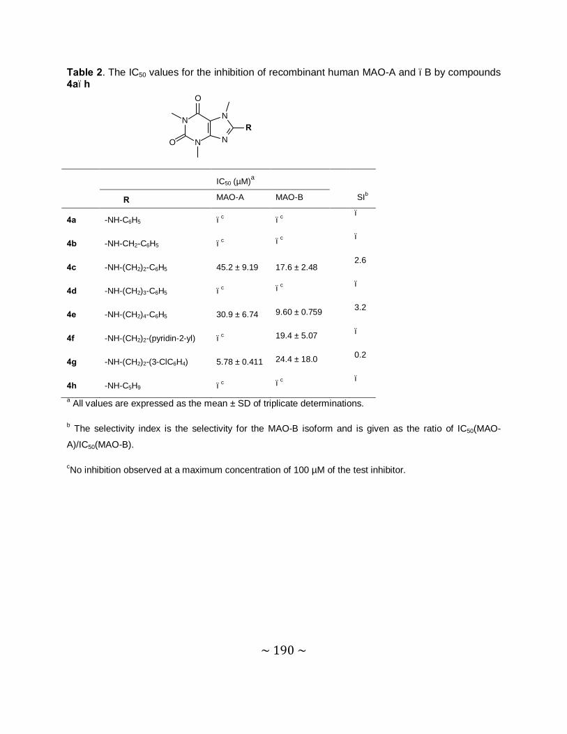
~ 190 ~
Table 2. The IC50 values for the inhibition of recombinant human MAO-A and –B by compounds 4a–h
N
N
N
N
O
O
R
IC50 (µM)a
R MAO-A MAO-B SIb
4a -NH-C6H5 –c –c –
4b -NH-CH2-C6H5 –c –c –
4c -NH-(CH2)2-C6H5 45.2 ± 9.19 17.6 ± 2.48 2.6
4d -NH-(CH2)3-C6H5 –c –c –
4e -NH-(CH2)4-C6H5 30.9 ± 6.74 9.60 ± 0.759 3.2
4f -NH-(CH2)2-(pyridin-2-yl) –c 19.4 ± 5.07 –
4g -NH-(CH2)2-(3-ClC6H4) 5.78 ± 0.411 24.4 ± 18.0 0.2
4h -NH-C5H9 –c –c –
a All values are expressed as the mean ± SD of triplicate determinations.
b The selectivity index is the selectivity for the MAO-B isoform and is given as the ratio of IC50(MAO-
A)/IC50(MAO-B).
cNo inhibition observed at a maximum concentration of 100 µM of the test inhibitor.
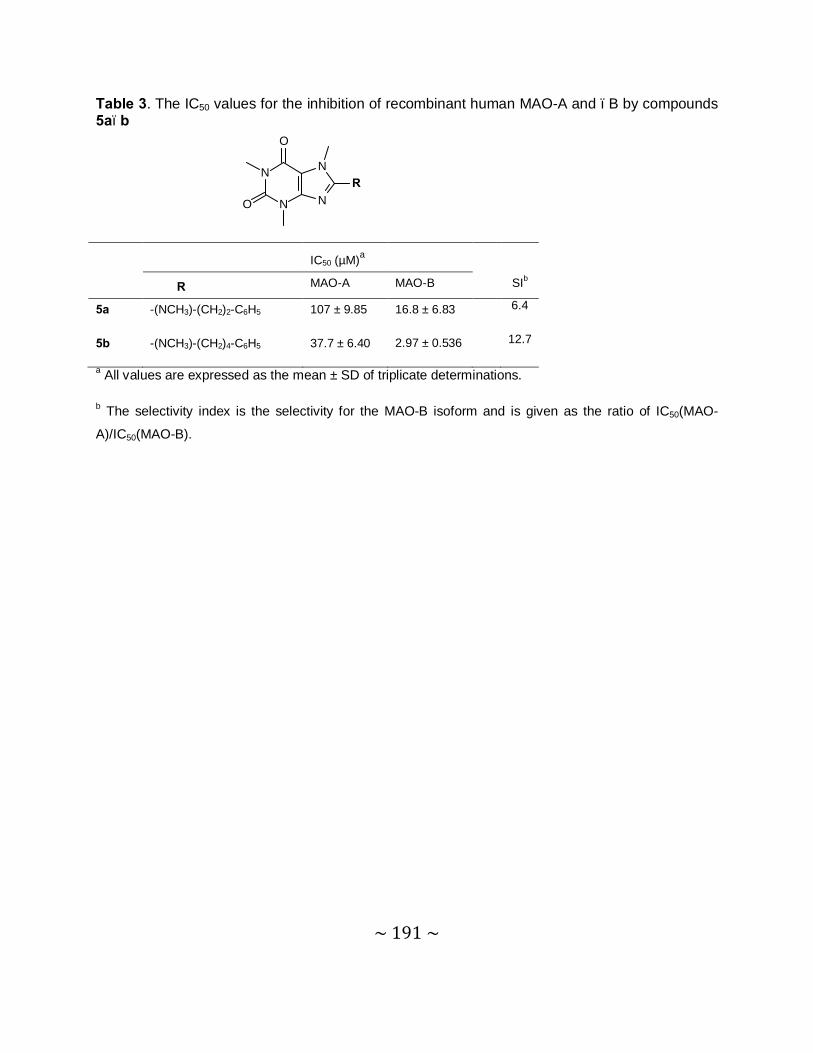
~ 191 ~
Table 3. The IC50 values for the inhibition of recombinant human MAO-A and –B by compounds 5a–b
N
N
N
N
O
O
R
IC50 (µM)a
R MAO-A MAO-B SIb
5a -(NCH3)-(CH2)2-C6H5 107 ± 9.85 16.8 ± 6.83 6.4
5b -(NCH3)-(CH2)4-C6H5 37.7 ± 6.40 2.97 ± 0.536 12.7
a All values are expressed as the mean ± SD of triplicate determinations.
b The selectivity index is the selectivity for the MAO-B isoform and is given as the ratio of IC50(MAO-
A)/IC50(MAO-B).
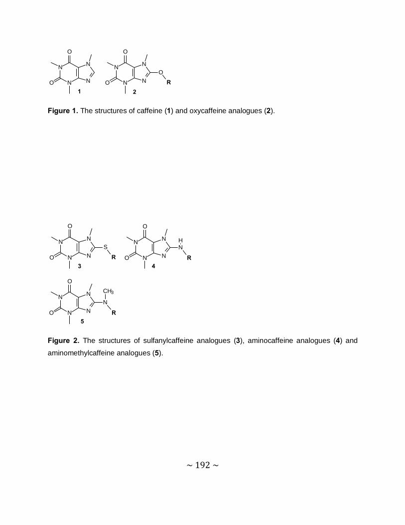
~ 192 ~
N
N
N
N
O
O1 2
N
N
N
N
O
O
O
R
Figure 1. The structures of caffeine (1) and oxycaffeine analogues (2).
3 4
5
N
N
N
N
O
O
N
R
HN
N
N
N
O
O
S
R
N
N
N
N
O
O
N
R
CH3
Figure 2. The structures of sulfanylcaffeine analogues (3), aminocaffeine analogues (4) and
aminomethylcaffeine analogues (5).
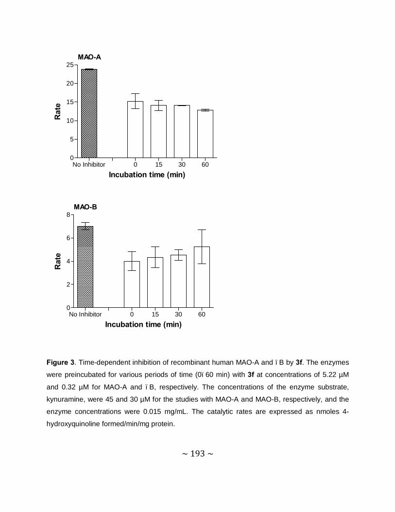
~ 193 ~
Figure 3. Time-dependent inhibition of recombinant human MAO-A and –B by 3f. The enzymes
were preincubated for various periods of time (0–60 min) with 3f at concentrations of 5.22 µM
and 0.32 µM for MAO-A and –B, respectively. The concentrations of the enzyme substrate,
kynuramine, were 45 and 30 µM for the studies with MAO-A and MAO-B, respectively, and the
enzyme concentrations were 0.015 mg/mL. The catalytic rates are expressed as nmoles 4-
hydroxyquinoline formed/min/mg protein.
No Inhibitor 0 15 30 600
5
10
15
20
25
Incubation time (min)
Rat
eMAO-A
No Inhibitor 0 15 30 600
2
4
6
8
Incubation time (min)
Rat
e
MAO-B
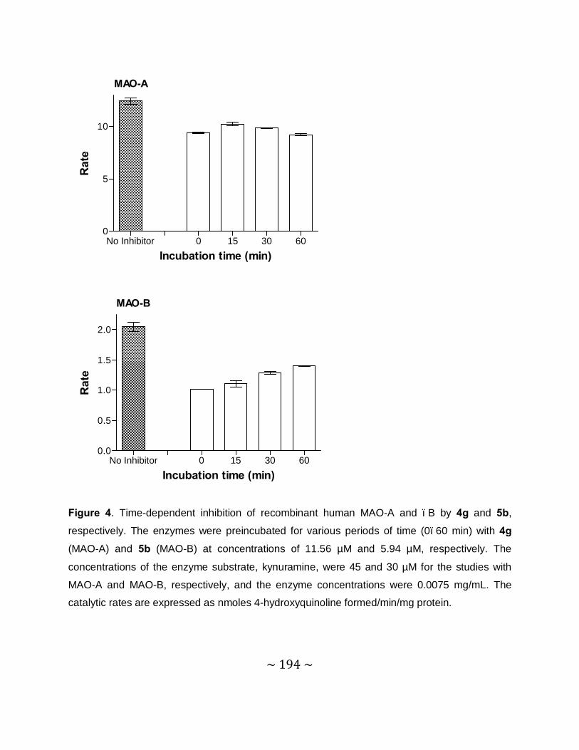
~ 194 ~
Figure 4. Time-dependent inhibition of recombinant human MAO-A and –B by 4g and 5b,
respectively. The enzymes were preincubated for various periods of time (0–60 min) with 4g
(MAO-A) and 5b (MAO-B) at concentrations of 11.56 µM and 5.94 µM, respectively. The
concentrations of the enzyme substrate, kynuramine, were 45 and 30 µM for the studies with
MAO-A and MAO-B, respectively, and the enzyme concentrations were 0.0075 mg/mL. The
catalytic rates are expressed as nmoles 4-hydroxyquinoline formed/min/mg protein.
No Inhibitor 0 15 30 600
5
10
Incubation time (min)
Rat
eMAO-A
No Inhibitor 0 15 30 600.0
0.5
1.0
1.5
2.0
Incubation time (min)
Rat
e
MAO-B
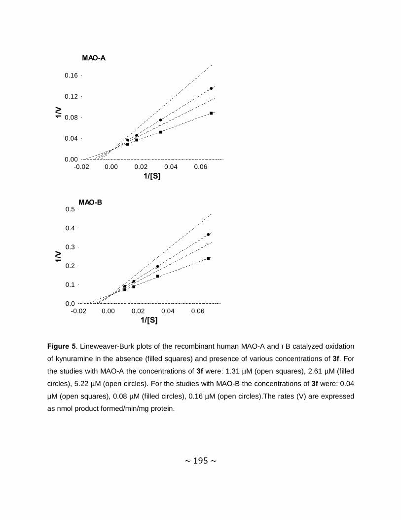
~ 195 ~
Figure 5. Lineweaver-Burk plots of the recombinant human MAO-A and –B catalyzed oxidation
of kynuramine in the absence (filled squares) and presence of various concentrations of 3f. For
the studies with MAO-A the concentrations of 3f were: 1.31 µM (open squares), 2.61 µM (filled
circles), 5.22 µM (open circles). For the studies with MAO-B the concentrations of 3f were: 0.04
µM (open squares), 0.08 µM (filled circles), 0.16 µM (open circles).The rates (V) are expressed
as nmol product formed/min/mg protein.
-0.02 0.00 0.02 0.04 0.060.00
0.04
0.08
0.12
0.16
1/[S]
1/V
MAO-A
-0.02 0.00 0.02 0.04 0.060.0
0.1
0.2
0.3
0.4
0.5
1/[S]
1/V
MAO-B
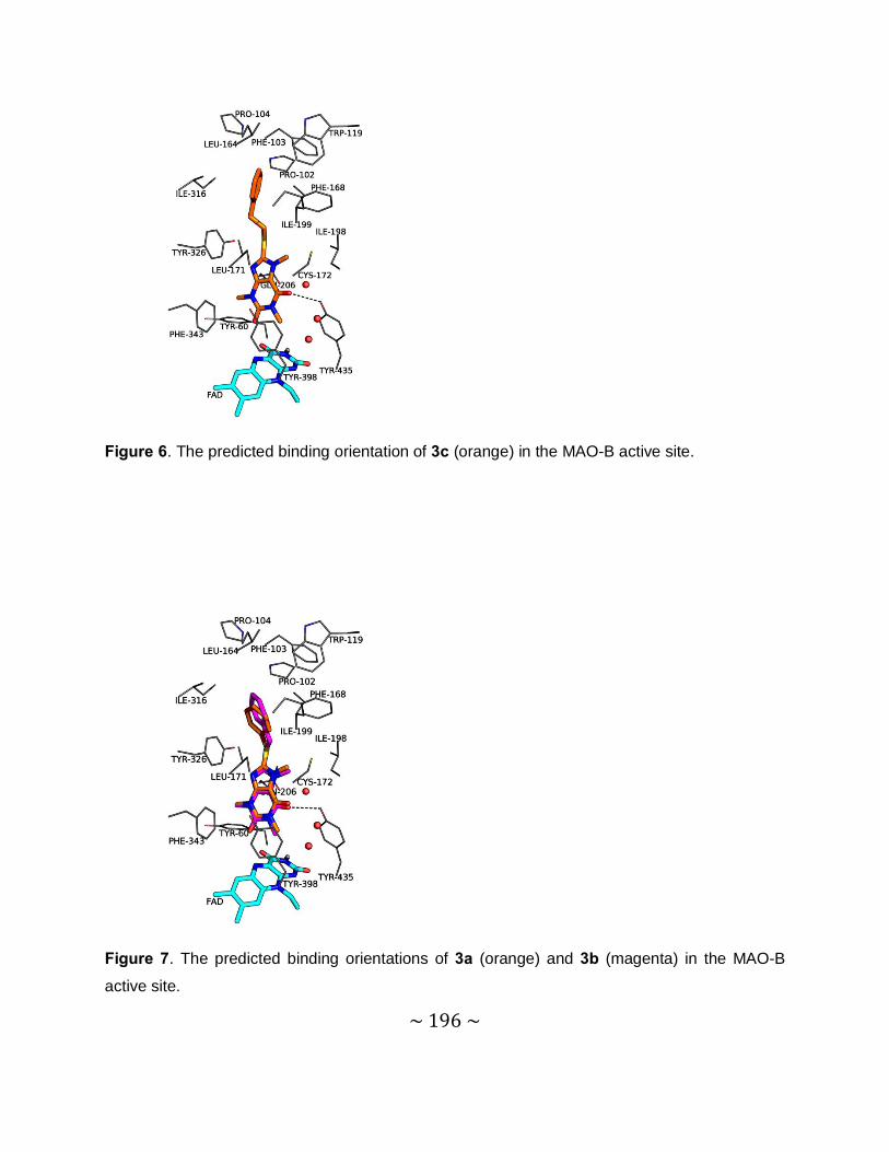
~ 196 ~
Figure 6. The predicted binding orientation of 3c (orange) in the MAO-B active site.
Figure 7. The predicted binding orientations of 3a (orange) and 3b (magenta) in the MAO-B
active site.

~ 197 ~
Figure 8. The predicted binding orientation of 4c (green) in the MAO-B active site.
Figure 9. The predicted binding orientation of 3c (magenta) in the MAO-A active site.
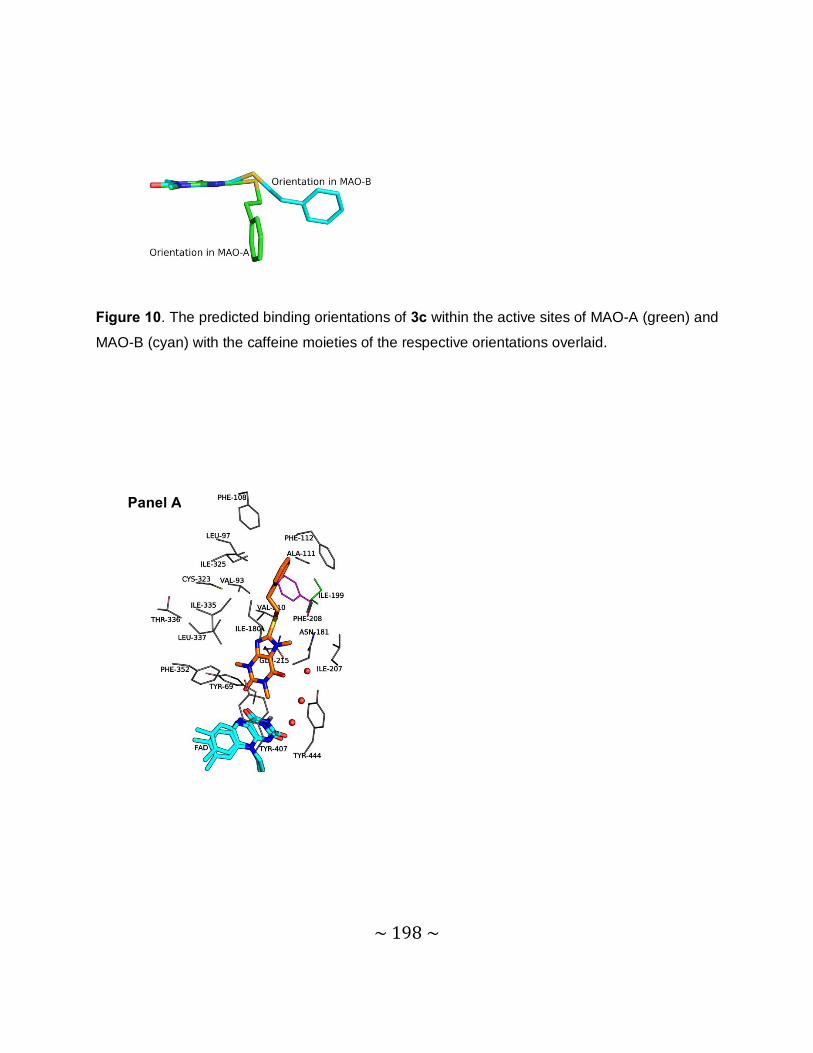
~ 198 ~
Figure 10. The predicted binding orientations of 3c within the active sites of MAO-A (green) and
MAO-B (cyan) with the caffeine moieties of the respective orientations overlaid.
Panel A
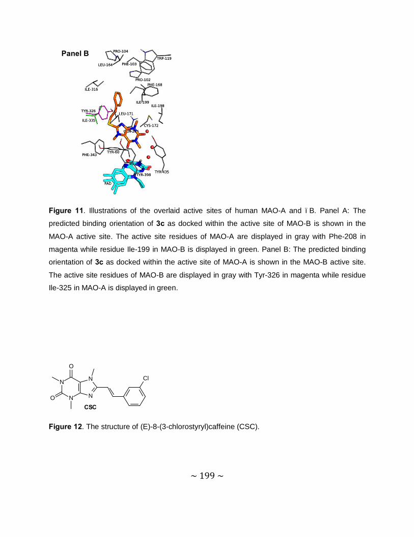
~ 199 ~
Figure 11. Illustrations of the overlaid active sites of human MAO-A and –B. Panel A: The
predicted binding orientation of 3c as docked within the active site of MAO-B is shown in the
MAO-A active site. The active site residues of MAO-A are displayed in gray with Phe-208 in
magenta while residue Ile-199 in MAO-B is displayed in green. Panel B: The predicted binding
orientation of 3c as docked within the active site of MAO-A is shown in the MAO-B active site.
The active site residues of MAO-B are displayed in gray with Tyr-326 in magenta while residue
Ile-325 in MAO-A is displayed in green.
N
N
N
N
O
O
Cl
CSC
Figure 12. The structure of (E)-8-(3-chlorostyryl)caffeine (CSC).
Panel B
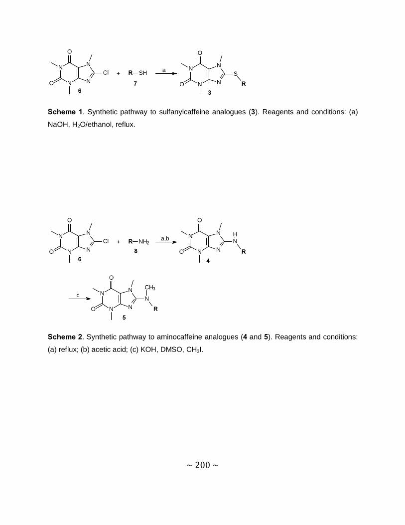
~ 200 ~
N
N
N
N
O
O
Cl
67
+ R SH a
3
N
N
N
N
O
O
S
R
Scheme 1. Synthetic pathway to sulfanylcaffeine analogues (3). Reagents and conditions: (a)
NaOH, H2O/ethanol, reflux.
8
N
N
N
N
O
O
Cl
6
+ R NH2a,b
c
4
N
N
N
N
O
O
N
R
H
5
N
N
N
N
O
O
N
R
CH3
Scheme 2. Synthetic pathway to aminocaffeine analogues (4 and 5). Reagents and conditions:
(a) reflux; (b) acetic acid; (c) KOH, DMSO, CH3I.

~ 201 ~
Acknowledgements
• To my Heavenly Father, thank you for all the blessings and unconditional love.
• Prof. J.P. Petzer, thank you for all your knowledge and wisdom.
• Prof. J.J. Bergh, thank you for all your advice and guidance.
• Mari and Walter, thank you for all your love and support during all the good and difficult
times.
• To all my friends, thank you for all the good times and fond memories.