The Purine-Utilizing Bacterium Clostridium acidurici 9a:A Genome-Guided Metabolic ReconsiderationKatrin Hartwich, Anja Poehlein, Rolf Daniel*
Department of Genomic and Applied Microbiology, and Gottingen Genomics Laboratory, Institute of Microbiology and Genetics, Georg-August University Gottingen,
Gottingen, Germany
Abstract
Clostridium acidurici is an anaerobic, homoacetogenic bacterium, which is able to use purines such as uric acid as solecarbon, nitrogen, and energy source. Together with the two other known purinolytic clostridia C. cylindrosporum and C.purinilyticum, C. acidurici serves as a model organism for investigation of purine fermentation. Here, we present the firstcomplete sequence and analysis of a genome derived from a purinolytic Clostridium. The genome of C. acidurici 9a consistsof one chromosome (3,105,335 bp) and one small circular plasmid (2,913 bp). The lack of candidate genes encoding glycinereductase indicates that C. acidurici 9a uses the energetically less favorable glycine-serine-pyruvate pathway for glycinedegradation. In accordance with the specialized lifestyle and the corresponding narrow substrate spectrum of C. acidurici 9a,the number of genes involved in carbohydrate transport and metabolism is significantly lower than in other clostridia suchas C. acetobutylicum, C. saccharolyticum, and C. beijerinckii. The only amino acid that can be degraded by C. acidurici isglycine but growth on glycine only occurs in the presence of a fermentable purine. Nevertheless, the addition of glycineresulted in increased transcription levels of genes encoding enzymes involved in the glycine-serine-pyruvate pathway suchas serine hydroxymethyltransferase and acetate kinase, whereas the transcription levels of formate dehydrogenase-encoding genes decreased. Sugars could not be utilized by C. acidurici but the full genetic repertoire for glycolysis wasdetected. In addition, genes encoding enzymes that mediate resistance against several antimicrobials and metals wereidentified. High resistance of C. acidurici towards bacitracin, acriflavine and azaleucine was experimentally confirmed.
Citation: Hartwich K, Poehlein A, Daniel R (2012) The Purine-Utilizing Bacterium Clostridium acidurici 9a: A Genome-Guided Metabolic Reconsideration. PLoSONE 7(12): e51662. doi:10.1371/journal.pone.0051662
Editor: Paul Jaak Janssen, Belgian Nuclear Research Centre SCK/CEN, Belgium
Received August 20, 2012; Accepted November 6, 2012; Published December 11, 2012
Copyright: � 2012 Hartwich et al. This is an open-access article distributed under the terms of the Creative Commons Attribution License, which permitsunrestricted use, distribution, and reproduction in any medium, provided the original author and source are credited.
Funding: This work was supported by the Bundesministerium fur Bildung und Forschung (BMBF) and the Niedersachsisches Ministerium fur Wissenschaft undKultur. The funders had no role in study design, data collection and analysis, decision to publish, or preparation of the manuscript.
Competing Interests: The authors have declared that no competing interests exist.
* E-mail: [email protected]
Introduction
Clostridia represent one of the largest and most heterogeneous
classes within the bacteria [1]. The genus Clostridium belongs to the
phylum Firmicutes and its members share a Gram-positive cell
wall and an anaerobic lifestyle. Special metabolic traits have been
found in clostridia such as the Stickland reaction in C. sporogenes [2]
or C. sticklandii [3] and different routes for fermentation of purines,
which are dependent on the availability of certain trace elements
[4,5]. C. acidurici, C. cylindrosporum and C. purinilyticum are the
described representatives of purinolytic clostridia, which are able
to use purines as sole carbon, nitrogen, and energy source [5–7].
C. acidurici was discovered in 1909 by Liebert and originally named
‘‘Bacillus acidi-urici’’ [7]. The strain was isolated from garden soil in
a medium containing uric acid and K2HPO4 dissolved in tap
water. Liebert observed an active motile, spore-forming, and rod-
shaped bacterium that was able to convert uric acid to ammonia,
carbon dioxide, and acetate. In the late 1930s, Barker started to
work on uric acid-fermenting anaerobes and rediscovered and
reclassified this organism as Clostridium acidi-urici. The type strain
9a was defined in 1942 [6]. Interestingly, it could be isolated from
nearly all soil samples but also from avian feces indicating that this
organism plays a role in decomposition of uric acid, which is the
main nitrogenous end product of avians [7]. Most purines can be
fermented by C. acidurici but sugars, amino acids or complex
nitrogenous compounds such as tryptone are not degraded.
Nevertheless, it is able to synthesize all amino acids de novo. For
purinolytic clostridia, two pathways forming acetate from purines
are known. (i) The glycine-serine-pyruvate pathway in which
glycine is converted to serine and then to acetate via pyruvate and
acetyl phosphate by the activities of the glycine cleavage complex
and formate dehydrogenase [8]. (ii) The energetically favorable
glycine reductase pathway [4] in which formate dehydrogenase
and the glycine cleavage complex are still involved. In this
pathway, acetate is synthesized directly by the reduction of glycine
via the selenium-dependent enzyme glycine reductase. The use of
the glycine reductase pathway has been postulated for all three
purinolytic clostridia, although reductase activity has not been
detected in C. acidurici [4]. In this study, a genome-based approach
was employed to solve such inconsistencies. Although the
fascinating metabolism of purinolytic clostridia was subject of
many investigations [4,9–11], genome-wide studies of these
organisms have not been published. Here we present the first
completely sequenced and annotated genome of the purinolytic C.
acidurici type strain 9a. Based on a comparative genome analysis,
we performed a genome-guided physiological analysis of C.
acidurici 9a and provide a general overview of the metabolic
capabilities of this organism.
PLOS ONE | www.plosone.org 1 December 2012 | Volume 7 | Issue 12 | e51662

Materials and Methods
Strains and Growth ConditionsThe type strain Clostridium acidurici 9a (DSM 604) was obtained
from the DSMZ (German Collection of Microorganisms and Cell
Cultures, Braunschweig, Germany). Cultivation was performed in
liquid uric acid medium (pH 7.3) containing 12 mM uric acid,
12 mM KOH, 4 mM K2HPO4, 1 g/l yeast extract, 0.14 mM
MgSO4 x 7H2O, 6.3 mM FeSO4 x 7H2O, 29 mM CaCl2 x 2H2O,
0.1 mM MnSO4 x H2O, 0.1 mM Na2SeO3 x 5H2O, 0.1 mM
Na2WO4 x 2H2O, 0.1 mM Na2MoO4 x 2H2O, 4.4 mM resazurin,
20 mM KHCO3, and 29.2 mM thioglycolic acid [10]. Cells were
grown at 37uC under anaerobic conditions according to the
method of Rabinowitz [12]. For growth tests with substrate
variations, the medium was altered by lowering the uric acid
concentration to 10 mM, 5 mM, 3 mM or 0 mM, and/or the
addition of 100 mM glycine. Growth tests were performed in
10 ml medium, which was inoculated to an optical density (OD) of
0.1 using an overnight-grown culture of C. acidurici 9a. OD was
determined spectrophotometrically at 600 nm (OD600 nm) with
a WPA CO8000 Biowave cell density meter (Biochrom Ltd,
Cambridge, UK). All tests were carried out in triplicate.
Genome Sequencing and FinishingGenome sequencing of C. acidiurici 9a was done using 454
Titanium pyrosequencing technology as recommended by the
manufacturer (Roche, Penzberg, Germany). Raw sequences were
assembled into contigs using the Newbler assembly tool v2.3 from
Roche. Gap closure and all manual editing was done using the
software Gap4 (v 4.11) of the Staden package [13]. The contig
order was determined by employing vectorette PCR [14] and
multiplex PCR [15] approaches. Remaining gaps were closed by
PCR-based approaches and Sanger sequencing [16] of the
resulting PCR products using Big Dye 3.0 chemistry and an
ABI3730XL capillary sequencer (Applied Biosystems, Life Tech-
nologies GmbH, Darmstadt, Germany).
Gene Prediction and AnnotationInitial gene prediction was done using the YACOP tool [17]. All
predicted genes were manually curated based on GC frame plot
analysis, presence of ribosome-binding sites, and comparison to
known protein-encoding sequences employing the Sanger Artemis
tool [18]. Functional annotation was initially done with the ERGO
software tool [19] (Integrated Genomics, Chicago, USA) and
manually corrected by comparison to the Swiss-Prot and TrEMBL
databases [20], the analysis of functional domains with InterPro-
Scan [21] and the use of the IMG/ER (Integrated Microbial
Genomes/Expert Review) system [22].
Sequence Analysis and Comparative GenomicsDeduced gene products were classified into functional categories
performing a BLAST search against the COG database [23].
Comparative analyses of different Firmicutes was done as de-
scribed previously [24] using a bidirectional BLAST algorithm
combined with a global sequence alignment based on the
Needleman-Wunsch algorithm [25]. ORFs were defined as
orthologs at a similarity higher than 30% and a BLAST e-value
lower than 10e-21. Visualizations of chromosome, plasmid and
other DNA sequences were done by using DNAPlotter [26]. To
identify metabolic pathways the pathway tools software from the
BioCyc database collection [27] was employed. Reconstruction of
pathways was manually curated. Alien genes and genomic islands
were detected employing COLOMBO [28] and IslandViewer
[29]. The 16S rRNA gene sequence-based calculations of
phylogenetic affiliations were done employing the ARB software
package [30].
Semi-quantitative and End-point Reverse TranscriptasePCR (RT-PCR)
To analyze the transcription level of different genes total RNA
was isolated from C. acidurici 9a cells with the QIAGEN RNeasy
Mini kit (QIAGEN, Hilden, Germany) according to the manual of
the manufacturer. Cells were grown in 20 to 30 ml medium with
different uric acid concentrations and in the absence or presence of
glycine. Initially, cells were sampled after 1 h of incubation at
37uC. Subsequently, samples were taken every 2 h until cells
reached stationary growth phase (usually, after 9 h). After RNA
isolation, remaining DNA was digested with the AmbionHTURBO DNA-freeTM DNase (Ambion, Life Technologies GmbH,
Darmstadt, Germany) according to the protocol of the manufac-
turer. The DNA digestion was evaluated by PCR with RNA as
template and oligonucleotides specific for the constitutively
expressed RNA polymerase gene (subunit A, rpoA). A standard
PCR reaction was set up using the BIO-X-ACTTM short DNA
polymerase (0.04 units/ml), OptiBuffer and MgCl2 according to
the manufacturer’s guidelines (all Bioline, Luckenwalde, Ger-
many). The PCR reactions were initiated at 98uC (2 min),
followed by 30 cycles of 96uC (20 s), 60uC (20 s), 68uC (2 min),
and ended with incubation at 72uC for 10 min. Genomic DNA of
C. acidurici 9a served as positive control and nuclease-free water as
negative control. RNA was quantified using a NanoDrop ND-
1000 spectrophotometer (Peqlab Biotechnologie GmbH, Erlan-
gen, Germany). Reverse transcription of the mRNA to cDNA was
done employing the RevertAidTM H Minus First Strand cDNA
synthesis kit (Fermentas, St. Leon-Rot, Germany) according to the
instructions of the manufacturer. In each reaction, 5 mg of DNA-
free RNA was used as starting material. Semi-quantitative PCR
was carried out as described above, but the number of cycles was
reduced to 20. The used oligonucleotides and the corresponding
genes are listed in Table S1. To verify operon structures end-point
RT-PCR was performed according to Passalacqua et al. [31].
Plasmid Copy Number DeterminationThe plasmid copy number (PCN) was determined by the
method of Skulj [32] with modifications. Cells of C. acidiurici 9a
were harvested after 5 h exponential growth, frozen using liquid
nitrogen, and stored at 280uC until use. After thawing, whole
genomic DNA, including chromosomal and plasmid DNA, was
isolated with the MasterPureTM Gram positive DNA purification
kit (Epicentre Biotechnologies, Madison, USA) as recommended
by the manufacturer. Oligonucleotides were deduced within the
genes encoding the chromosomal DNA replication protein A
(dnaA) and the plasmid replication protein B (repB) (Table S1).
Serial dilutions were performed with the purified DNA as follows:
undiluted, 1:5, 1:25, 1:50, 1:125 and 1:250. Real-time PCR
reactions (15 ml mixtures) containing Absolute Blue SYBR Green
ROX mix (Fisher Scientific GmbH, Schwerte, Germany) were
prepared as recommended by the manufacturer. Each reaction
was performed in quadruplicate. Reactions were run on a Bio-Rad
iQ5 real-time PCR detection system using the iQ5 optical system
software version 2.1 (Bio-Rad Laboratories GmbH, Munchen,
Germany) for analysis. Cycling conditions were: 15 min at 95uC,
followed by 40 cycles at 95uC (15 s) for denaturation, at 60uC(30 s) for annealing and at 72uC (30 s) for extension. A dissociation
(melting) step at 95uC for 30 s was added, followed by 60uC (30 s)
and 80 cycles ranging from 60 to 95uC (10 s) for melting.
Threshold cycle (Ct) values were automatically generated by the
iQ5 optical system software. The average Ct and standard
Genome-Guided Analysis of Clostridium acidurici 9a
PLOS ONE | www.plosone.org 2 December 2012 | Volume 7 | Issue 12 | e51662

deviation were calculated. Dilutions with standard deviation values
above 0.3 were not used to determine amplification efficiency and
copy number. Both were calculated according to Skulj [32]. For
chromosome and plasmid different amplification efficiencies and
Ct values were considered using the method of Pfaffl [33].
Calculations were carried out for each sample and dilution and the
average and standard deviation were determined.
Antimicrobial Resistance TestsStock solutions of antibiotics and antibacterial agents were
prepared according to the guidelines of the manufacturers
(AppliChem, Darmstadt, Germany and Sigma-Aldrich, Stein-
heim, Germany). We used the above-described standard liquid
uric acid medium mixed with different concentrations of the
respective antimicrobial agent [34]. The following 4 different
concentrations of each tested drug were chosen and added to
10 ml of liquid uric acid medium: Ampicillin (20, 50, 100, 150 mg/
ml), kanamycin (20, 50, 100, 150 mg/ml), chloramphenicol (5, 10,
30, 50 mg/ml), erythromycin (5, 10, 50, 100 mg/ml), vancomycin
(5–40 mg/ml), spectinomycin (20, 50, 100, 200 mg/ml), gentami-
cin (10, 20, 50, 100 mg/ml), tetracycline (5, 10, 20, 30 mg/ml),
bacitracin (10, 30, 50, 100 mg/ml), acriflavin (50, 150, 250,
350 mg/ml), 4-azaleucin (30, 50, 100, 200 mg/ml), thiamphenicol
(2, 5, 15, 30 mg/ml), and clarithromycin (2, 5, 10, 20 mg/ml). The
OD600 nm was measured after 6 h and 24 h and compared to
growth in uric acid medium without antibiotics. Tests were carried
out in triplicate.
AvailabilityThe complete genome sequence of Clostridium acidurici 9a has
been deposited in GenBank under accession numbers CP003326
(chromosome) and CP003327 (plasmid).
Results and Discussion
General Genome FeaturesThe genome of C. acidurici 9a consists of one chromosome with
a size of 3,105,335 bp and a circular plasmid of 2,913 bp (Fig. 1).
The G+C content of C. acidurici 9a (29.9%) is in the typical range
(26–39%) known from other clostridia. Six rRNA clusters plus two
additional single 5S rRNA genes and 81 tRNAs, including those
responsible for selenocysteine synthesis, were identified in the
genome of C. acidurici 9a (Fig. 1). The genome harbors 2979
predicted open reading frames (ORFs) of which 2878 are putative
protein-encoding genes and 17 putative pseudogenes. A 16S
rRNA gene-based phylogenetic classification (Fig. 2) confirmed the
findings of Collins et al. [35] that C. purinilyticum was the closest
relative of C. acidurici 9a. C. cylindrosporum, the third known
purinolytic Clostridium, showed only a distant relationship to C.
acidurici 9a. Thus, despite strong phenotypical similarities, a signif-
icant genotypic difference between C. cylindrosporum and C. acidurici
9a is indicated [34]. As an endospore-forming bacterium, C.
acidurici 9a is equipped with a set of sporulation genes including
universal transcription initiation factor spo0A (Curi_c13920),
spoIIAA-spoIIAB-sigF operon (Curi_c10290-10310) (Fig. S1),
spoIIGA-sigE operon (Curi_c13080-Curi_c13090) (Fig. S1), and
the three sigma factors sigG (Curi_c13100), sigK (Curi_c13480) and
sigH (Curi_c22810). As found in most other clostridia [36], the
genome of C. acidurici 9a does not harbor genes encoding the
phosphorelay components Spo0F and Spo0B. Recently, Steiner
et al. discovered five orphan kinases in C. acetobutylicum that are
able to overtake their function by interacting directly with Spo0A
and controlling its phosphorylation [36]. We identified nine genes
encoding putative orphan kinases in C. acidurici 9a of which three
(Curi_c11130, Curi_c26240, Curi_c26310) show high similarities
to the orphan kinases of C. acetobutylicum. C. acidurici 9a is motile
and possesses two large cluster regions containing flagellar
biosynthesis genes (Curi_c15800-16070 and Curi_c02130-
02580). As described for Clostridium ljungdahlii [37], a cluster of
chemotaxis genes (Curi_c15760-15790) is located directly in the
neighborhood of the flagella gene clusters.
COG classification of protein-encoding genes revealed that
many C. acidurici 9a genes are related to transcription (8.3%),
amino acid transport and metabolism (8.4%), and signal trans-
duction mechanisms (8.1%) (Fig. 1). A comparison of COG
categories with 15 other Clostridiaceae (Table S2) showed lower
representation of genes coding for carbohydrate transport and
metabolism in C. acidurici 9a. Only 3.1% of all putative genes code
for proteins of this category, whereas other clostridia show an
average of 7.4%. This difference might be due to the narrow
substrate spectrum of C. acidurici 9a.
Alien Genes and Genomic IslandsApproximately 91 kb (2.9%) of the genome represent putative
alien genes. Clusters of alien genes were located in 4 regions
(Fig. 1). Most genes in C. acidurici 9a potentially derived from other
bacteria are related to ABC transport systems such as Cu-
ri_c04900-04930 and Curi_c04500-04520, which encode a poten-
tial ferrichrome ABC-transporter and a transporter with unknown
function, respectively. In region 1, harboring the ferrichrome
ABC-transporter genes, putative alien genes mediating vancomy-
cin resistance (Curi_c05040-05080) were detected. In addition,
alien genes present within regions 1, 2, 3, and 4 code for
acetyltransferases (Curi_c05130, Curi_c05320) and transcriptional
regulators (Curi_c04890, Curi_c05120) or two-component signal
transduction systems (Curi_c04810-04820, Curi_c05350-05360,
Curi_c05440-05450). Since the genus Clostridium is heterogeneous
and C. acidurici 9a employs a specialized way of life, we performed
a bidirectional blast comparing the genome sequence of C. acidurici
9a to nine genome sequences of other clostridia and related
bacteria to identify other traits or unique genes (Fig. 1). The
organisms were divided into three subgroups based on the
substrate spectra: (i) pathogens/proteolytic (C. difficile ATCC
9689, C. botulinum A ATCC 25763, C. tetani E88), (ii) solvent
producer/saccharolytic/proteolytic (C. beijerinckii DSM 791, C.
acetobutylicum ATCC 824, C. ljungdahlii DSM 13528), and (iii)
utilizers of special substrates (C. sticklandii DSM 519, A. oremlandii
OhILAs, C. kluyveri DSM 555). Four genomic regions of C. acidurici
9a showed low or no similarities to the other clostridia (Fig. 1, I–
IV). These regions contained genes encoding S layer domain-
containing proteins (I), uncharacterized acetyltransferases and
transcriptional regulators (II) or hypothetical proteins (III and IV).
The genome of C. acidurici 9a possesses 21 genes encoding
transposases (Fig. 1) and genetic mobile elements, but some of
these were identified as pseudogenes. No complete phages could
be identified in the genome sequence but genes coding for remains
of phages such as integrase family proteins (Curi_c05580,
Curi_c24810, and Curi_c24830) and the small subunit of phage
terminase (Curi_c14970) are present. In summary, the incorpo-
ration of foreign DNA into the genome by horizontal gene transfer
is indicated.
Purine BreakdownPurines such as uric acid, xanthine, hypoxanthine and guanine
serve as sole carbon, nitrogen and energy source of C. acidurici 9a,
but adenine is not utilized [7], although putative genes coding for
adenine deaminase (Curi_c04950, Curi_c09560, Curi_c29030)
were detected in the genome. Products during growth on purines
Genome-Guided Analysis of Clostridium acidurici 9a
PLOS ONE | www.plosone.org 3 December 2012 | Volume 7 | Issue 12 | e51662

are acetate, CO2 and NH3 (Fig. 3). The first step in purine
degradation is the conversion of the purine to xanthine. These
reactions are mainly catalyzed by xanthine dehydrogenase. The
xanthine dehydrogenase in C. acidurici 9a consists of three subunits,
which bind molybdenum (XdhA), FAD (XdhB), or iron-sulfur
(XdhC). We found one cluster of the corresponding genes in the
genome (Curi_c23900-23920) and one additional single xdhC gene
(Curi_c28980) that clusters together with genes coding for
selenium-dependent molybdenum hydroxylase proteins (Cu-
ri_c28990-29010). A second cluster harbors an additional single
xdhA gene (Curi_c19420) together with genes encoding the delta
(Curi_c19430) and gamma subunit (Curi_c19440) of purine
hydroxylase, which performs hydroxylation of hypoxanthine and
purine to xanthine [38]. With guanine as substrate, guanine
deaminase instead of xanthine dehydrogenase catalyzes the
conversion to xanthine (Curi_c00270, Curi_c19450) [39]. When
xanthine serves directly as substrate, 4-ureido-5-imidazole car-
boxylic acid is formed by the hydrolysis of the covalent bond
between atoms 1 and 6 in the pyrimidine ring employing the
enzyme xanthine amidohydrolase [40]. Although three putative
genes coding for amidohydrolases (Curi_c02650, Curi_c26230,
Curi_c27500) were identified in the genome, a specific one for
xanthine could not be detected. Subsequently, 4-ureido-5-imidaz-
ole carboxylic acid is converted to formiminoglycine. The involved
enzymes are characterized in C. cylindrosporum and partially in C.
acidurici [40,41]. However, the sequences of the corresponding
genes are not published. Thus, we were not able to detect
candidate genes encoding these enzymes in the genome of C.
acidurici 9a. In addition, no gene encoding glycine formimino-
transferse that converts formiminoglycine to glycine involving
tetrahydrofolate (THF) was detected [40].
Figure 1. Circular maps of C. acidurici 9a chromosome and plasmid. Rings from the outside to the inside on the chromosome: 1 and 2,leading and lagging strand open reading frames (ORFs) colored according to COG categories; 3, rRNA cluster (pink), tRNAs (turquoise) andtransposases (black); 4, predicted alien genes (purple); and 5–13, orthologous ORFs predicted with the Needleman-Wunsch algorithm. Organisms areroughly grouped into special substrates utilizers (green tones), solvent producers (red tones) and pathogens (blue tones). The shade of thecorresponding color represents the value of the algorithm. Darker colors indicate higher values: dark green/red/blue, e-90/100; light green/red/blue,e-50/70: and grey, e-20/30, indicating no ortholog in the respective organism. Reference organisms from the outside to the inside: Clostridiumsticklandii DSM 519, Clostridium kluyveri DSM 555, Alkaliphilus oremlandii OhILAs, Clostridium ljungdahlii DSM 13528, Clostridium acetobutylicum ATCC824, Clostridium beijerinckii DSM 791, Clostridium difficile ATCC 9689, Clostridium botulinum A ATCC 25763 and Clostridium tetani E88. 14, G+C contentwithin the chromosome below the average (magenta) and above the average (olive). Predicted genomic islands (arabic numerals) and unique regions(roman numerals) are separated by black lines. Rings from the outside to the inside on the plasmid: 1, open reading frames colored according to COGcategories; and 2, G+C content below the average (magenta) and above the average (olive).doi:10.1371/journal.pone.0051662.g001
Genome-Guided Analysis of Clostridium acidurici 9a
PLOS ONE | www.plosone.org 4 December 2012 | Volume 7 | Issue 12 | e51662

Utilization of GlycineGlycine as intermediate of purine degradation. The role
of glycine as intermediate in purine fermentation and its
degradation to acetate was studied in anaerobic acetogens and
especially in the purinolytic bacteria C. acidurici, C. cylindrosporum
and C. purinilyticum [4–6,10]. Two pathways from glycine to acetate
are known. The first one discovered was the glycine-serine-
pyruvate pathway [42] (Fig. 3). Glycine is oxidized (decarboxy-
lated) and the resulting methylene-THF condenses with another
glycine to serine via serine hydroxymethyltransferase (GlyA,
Curi_c17240). Serine is deaminated to pyruvate via L-serine
dehydratase (SdhA/SdhB, Curi_c17690-17700), and pyruvate:-
ferredoxin oxidoreductase (Por, Curi_c20330, Curi_c21330)
catalyzes the further conversion to acetyl-coenzyme A and CO2.
A phosphotransacetylase (Curi_c12550) reversibly transfers the
acetyl unit from CoA to phosphate and acetate is built from acetyl
phosphate by the action of acetate kinase (Curi_c16390). The
remaining CO2 can be partly reconverted to methylene-THF. An
important element of this pathway is the glycine-cleavage system
(termed also as glycine decarboxylase/synthase). As glycine
decarboxylase, this multienzyme complex is responsible for the
oxidation of glycine to methylene-THF, NAD(P)H, CO2 and
NH3. The reverse glycine-forming reaction is also possible by the
glycine synthase activity of the complex [9]. C. acidurici 9a possesses
all genes necessary for encoding this enzyme complex, which
includes genes encoding an aminomethyltransferase (gcvT, Cu-
ri_c00740), a heat-stable hydrogen carrier protein (gcvH, Cu-
Figure 2. Taxonomic affiliation of C. acidurici 9a. The 16S rRNA gene sequences of type strains were used for construction of the neighbor-joining tree. GenBank accession numbers are given in parentheses. Numbers at nodes describe the bootstrap values in percent from 1,000 replicates.Bootstrap values .50% are shown. Black circles display reproducible nodes calculated with the maximum-likelyhood method. Length of the barrepresents 0.01 substitutions per nucleotide position. C. acidurici 9a is marked in red. Tree calculation was done using the ARB software package [30].The archaeal strains Methanosarcina acetivorans DSM 2834 (M59137), Methanosarcina mazei DSM 2053 (AJ012095) and Methanosarcina barkeri DSM800 (AJ012094) were used as outgroups to define the tree root (not shown).doi:10.1371/journal.pone.0051662.g002
Genome-Guided Analysis of Clostridium acidurici 9a
PLOS ONE | www.plosone.org 5 December 2012 | Volume 7 | Issue 12 | e51662
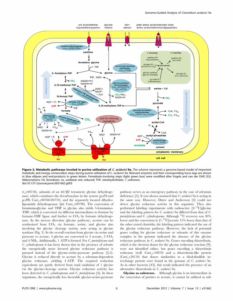
ri_c00750), subunits of an a2/b2 tetrameric glycine dehydroge-
nase, which constitutes the decarboxylase in the system (gcvPA and
gcvPB, Curi_c00760-00770), and the separately located dihydro-
lipoamide dehydrogenase (lpd, Curi_c09790). The conversion of
formiminoglycine and THF to glycine also yields 5-formimino-
THF, which is converted via different intermediates to formate by
formate-THF ligase and further to CO2 by formate dehydroge-
nase. In the inverse direction (glycine pathway), acetate can be
synthesized from CO2 via formate, serine, and glycine also
involving the glycine cleavage system, now acting as glycine
synthase (Fig. 3). In the overall reaction from glycine via serine and
pyruvate to acetate, 4 glycine are converted to 3 acetate, 2 CO2
and 4 NH3. Additionally, 1 ATP is formed. For C. purinilyticum and
C. cylindrosporum it has been shown that in the presence of selenite
the energetically more favored glycine reductase pathway is
pursued instead of the glycine-serine-pyruvate pathway [4,5].
Glycine is reduced directly to acetate by a selenium-dependent
glycine reductase, yielding 4 ATP. The required reduction
equivalents are partly derived from total oxidation of 1 glycine
via the glycine-cleavage system. Glycine reductase activity has
been detected in C. cylindrosporum and C. purinilyticum [4]. In these
organisms, the energetically less favorable glycine-serine-pyruvate
pathway serves as an emergency pathway in the case of selenium
deficiency [5]. It was always assumed that C. acidurici 9a is acting in
the same way. However, Durre and Andreesen [4] could not
detect glycine reductase activity in this organism. They also
performed labeling experiments with radioactive [2-14C]glycine
and the labeling pattern for C. acidurici 9a differed from that of C.
purinilyticum and C. cylindrosporum. Although 14C recovery was 30%
lower and the conversion to [1-14C]acetate 15% lower than that of
the other tested clostridia, the labeling pattern indicated the use of
the glycine reductase pathway. However, the lack of potential
genes coding for glycine reductase or subunits of this enzyme
complex in the genome indicated the absence of the glycine
reductase pathway in C. acidurici 9a. Genes encoding thioredoxin,
which is the electron donor for the glycine reductase reaction [9],
were not identified either, but genes encoding a thioredoxin
reductase (trxB, Curi_c10870) and a thioredoxin-like protein
(Curi_c20110) that shares similarities to a thiol:disulfide in-
terchange protein were found in the genome of C. acidurici 9a.
As in other bacteria [43], this result indicated the presence of an
alternative thioredoxin in C. acidurici 9a.
Glycine as substrate. Although glycine is an intermediate in
the conversion of purines to acetate, it cannot be utilized as sole
Figure 3. Metabolic pathways involed in purine utilization of C. acidurici 9a. The scheme represents a genome-based model of importantmetabolic and energy conservation steps during purine utilization of C. acidurici 9a. Relevant enzymes and their corresponding locus tags are shownin blue ellipses and end-products in green letters. Ferredoxin-involving steps (light green box) were modified after Vogels and van der Drift [53].Abbreviations: Fd, ferredoxin; ox, oxidized; red, reduced; THF, tetrahydrofolate; ?, unknown.doi:10.1371/journal.pone.0051662.g003
Genome-Guided Analysis of Clostridium acidurici 9a
PLOS ONE | www.plosone.org 6 December 2012 | Volume 7 | Issue 12 | e51662

substrate by C. acidurici 9a [6]. Degradation and growth are only
possible in the presence of a purine. The import of glycine into the
cell might be facilitated by an ABC transport system. Three
putative genes were identified, encoding a glycine/betaine ABC
transporter (Curi_c10180-10200). To further analyze the glycine
utilization of C. acidurici 9a, growth tests were performed in media
containing glycine and different concentrations of uric acid. Tests
were implemented with cells not adapted to glycine compared to
adapted cells. Media containing glycine without uric acid served as
negative control. As shown in Fig. 4A, non-adapted cells showed
a longer lag phase than adapted cells when shifted from uric acid
medium to glycine-containing medium. This was not surprising, as
high concentrations of glycine exhibit an inhibitory effect on
growth of many bacteria by disturbing the biosynthesis of
peptidoglycan [9]. Adapted cells showed no extended lag phase
and the maximal cell densities of 0.72, 0.57, 0.37, and 0.26 in
glycine-containing medium with 12 mM, 10 mM, 5 mM, or
3 mM uric acid, respectively, were identical to those in medium
containing solely uric acid. Growth of C. acidurici 9a depended only
on the concentration of uric acid, since the addition of glycine
showed neither an increase nor a decrease in optical density. To
examine the influence of glycine on the conversion of glycine to
acetate the transcription levels of genes involved in this process
were determined. The selected genes code for the beta-subunit of
the decarboxylase of the glycine cleavage system (gcvPB, Cu-
ri_c00770), serine hydroxymethyltransferase (glyA, Curi_c17240),
pyruvate:ferredoxin oxidoreductase (por, Curi_c20330, Cu-
ri_c21330), acetate kinase (ackA, Curi_c16390), NAD+-dependent
formate dehydrogenase (fdhA/fdhB, Curi_c16670/Curi_c16640)
and formate dehydrogenases H (fdhF1/fdhF2, Curi_c29370-
29380). Adapted cells were grown in media containing glycine
and altering concentrations of uric acid and samples were taken in
two hours intervals. In general, semi-quantitative RT-PCR
revealed lower transcription levels of all tested genes when the
uric acid concentration was decreased (Fig. 4B). The addition of
glycine resulted in increased transcription levels of gcvPB, glyA, ackA
and por, whereas the transcription of formate dehydrogenase genes
slightly decreases. Although no transcription start analysis was
performed, a potential glycine riboswitch with two aptamers
(Curi_c00731) has been identified directly upstream of the genes
encoding the glycine cleavage system. Thus, the increased gene
expression might be due to the turn-on of the riboswitch [44]. The
metabolism is forced to the direction of synthesizing C1 units and
thereby increasing the transcription of glyA, por and ackA. We
assume that in the presence of external glycine cells no longer need
to synthesize it from purines and thus, less 5-formimino-THF is
build. Accordingly, the formation of formate is reduced. This
explains the decrease in transcription levels of genes encoding
formate dehydrogenase. A second interesting aspect is the different
transcription of por2 and fdhA. In contrast to their orthologs, these
two genes are not or only slightly transcribed. Nevertheless, no
obvious sequence variations, which could explain the inactivity of
the genes, were identified in their promoter regions. We assume
these genes are not active under the conditions tested.
Utilization of Sugars and MacromoleculesSugars such as glucose were already tested as substrates in early
studies by Barker and Beck [7]. Growth was analyzed in medium
containing solely glucose as carbon source or glucose-containing
medium supplemented with purines. Similar to the growth tests
with glycine, growth was only observed after addition of an
utilizable purine. In contrast to the studies with glycine, no
decomposition of glucose could be detected [7]. In addition to
glucose, we tested other hexoses and pentoses such as fructose,
galactose, sucrose, ribose, and ribulose in media supplemented
with different nitrogen sources such as ammonium chloride,
ammonium sulfate, and ammonium nitrate, but growth was never
observed (data not shown). This might be partly due to the fact
that C. acidurici 9a does not possess a functional phosphotransferase
system. We identified a putative EIIC component (Curi_c00230)
and phosphocarrier protein Hpr (Curi_c20230) of a phosphotrans-
ferase system, but genes coding for unspecific enzyme I and other
components of the substrate specific enzyme II are missing.
Interestingly, all necessary genes coding for glycolysis/gluconeo-
genesis were identified in the genome of C. acidurici 9a, including
genes encoding key enzymes such as phosphofructokinase
(Curi_c20210) and pyruvate kinase (Curi_c20200). RT-PCR-
based gene expression studies revealed that these genes are
constitutively transcribed indicating that they have a metabolic
function independent of sugar degradation. At least some of these
enzymes are required for gluconeogenesis. Pyruvate kinase and
phosphofructokinase may also be involved in purine or secondary
metabolite metabolism. Additionally, putative genes involved in
solvent production such as the genes encoded by the sol operon of
C. acetobutylicum [45] were not identified in the genome sequence of
C. acidurici 9a.
Except for a gene encoding a putative amylopullulanase (apu,
Curi_c06010), putative genes coding for potential exoenzymes
involved in degradation of macromolecules such as starch, proteins
or lipids were not present. This again emphasizes the substrate
specialization of C. acidurici 9a.
Energy ConservationGenes encoding two energy conservation steps during growth
on purines were detected in the genome sequence. One is initiated
by the hydrolysis of methenyl-THF to 10-formyl-THF followed by
forming ATP and formate from ADP and 10-formyl-THF [46].
The second major energy-yielding step is the generation of acetate
and ATP in the acetate kinase reaction (Curi_c16390). As
described for other clostridia [37,47,48], C. acidurici 9a may also
perform energy conservation by electron-transport phosphoryla-
tion employing the Rnf-complex (Curi_c09970-10020). This
enzyme complex catalyzes the flow of electrons from reduced
ferredoxin to NAD+ coupled to ion-translocation across the
membrane. ATP is generated employing the F1F0-type ATPase
(Curi_c01980-02060), which contains amino acid motifs typical for
Na+-dependent F1F0 ATPases [49]. Recently, an ancient way of
combining carbon dioxide fixation with the generation and
utilization of a sodium ion gradient for ATP synthesis was
described for A. woodii [50]. The Rnf-complex was shown to
couple the electron transfer from reduced ferredoxin to NAD+
with electrogenic Na+ transport. The whole system plays an
important role during growth on H2 and CO2 employing the
Wood-Ljungdahl pathway [48]. For C. acidurici 9a and the other
purinolytic clostridia, the glycine pathway was postulated and the
necessary genes were identified in the genome of C. acidurici 9a.
The glycine pathway shares the first steps with the Wood-
Ljungdahl pathway [51]. The initial reduction of CO2 to formate,
is catalyzed by the enzyme formate dehydrogenase. Most formate
dehydrogenases are only able to function in the direction from
formate to CO2, thus, the enzyme of acetogenic bacteria is rather
unique being able to convert CO2 to formate [9]. As mentioned
above, four genes encoding formate dehydrogenases were
identified in the genome of C. acidurici 9a (Fig. 5). Two of them
(fdhB and fdhF2) are coding for selenoproteins. The genes fdhF1
and fdhF2 encode formate dehydrogenase H. They are clustered
together with three genes encoding proteins that show similarity to
a NAD(P)+-dependent iron-only hydrogenase (hydABC, Cu-
Genome-Guided Analysis of Clostridium acidurici 9a
PLOS ONE | www.plosone.org 7 December 2012 | Volume 7 | Issue 12 | e51662

Figure 4. Growth on uric acid and glycine (A) and corresponding gene expression at different time points (B). (A) Growth of cells wasmeasured over 12 h in medium containing different concentrations of uric acid (UA) with (dashed line) or without (solid line) glycine (Gly). Non-adapted cells were shifted directly from uric acid medium to uric acid with glycine whereas adapted cells were grown three times in 12 mM uric acidmedium with 100 mM glycine prior to transfer and measurement. (B) Dependent on the addition of glycine a semi-quantitative analysis oftranscription levels of genes involved in the conversion of glycine to acetate was performed. Shown are the results for adapted cells grown in 3 mMand 12 mM uric acid medium or in the same media supplemented with 100 mM glycine. Samples were taken after 1 h, 3 h, 5 h, 7 h and 9 h. DNA astemplate in the RT-PCR reaction served as positive control (P) and water as negative control (N). Abbreviations: gcvPB, glycine dehydrogenase betasubunit (Curi_c00770); glyA, serine hydroxymethyltransferase (Curi_c17240); ackA, acetate kinase (Curi_c16390); por1/por2, pyruvate:ferredoxinoxidoreductase (Curi_c20330/Curi_c21330); fdhA/fdhB, formate dehydrogenase subunits A; (Curi_c16670, Curi_c16640); fdhF1/fdhF2, formatedehydrogenase H (Curi_c29370/Curi_c29380); rpoA, DNA-directed RNA polymerase alpha subunit (Curi_c22350, reference).doi:10.1371/journal.pone.0051662.g004
Genome-Guided Analysis of Clostridium acidurici 9a
PLOS ONE | www.plosone.org 8 December 2012 | Volume 7 | Issue 12 | e51662

ri_c29390-29410) that bears resemblance to the trimeric iron
hydrogenase of Thermotoga maritima [52] and the HydABC subunits
of the multimeric iron hydrogenase of A. woodii [50]. Accordingly,
we assume a similar electron bifurcating mechanism. The formate
dehydrogenase accessory protein-encoding gene fdhD (Cu-
ri_c29330) was identified downstream of this cluster. Two
additional genes encoding putative hydrogenase HydA homo-
logues were detected separated from the hydABC cluster. One
(hydA1, Curi_c05020) is located in alien gene region 1 (Fig. 1) and
the other one (hydA2, Curi_c05750) directly within a cluster of
genes coding for proteins involved in cobalamin biosynthesis
(Curi_c05740-05800). Interestingly, C. acidurici 9a was assumed to
possess no or only low levels of hydrogenase activity [53]. The lack
of H2 production has always been peculiar because C. acidurici 9a
produces ferredoxin, which is mainly found in hydrogen-evolving
species. The other two genes fdhA and fdhB encode catalytic
subunits for the NAD+-dependent formate dehydrogenases. They
are separated from each other by the genes moeA and mobB
(Curi_c16650-16660), which are involved in molybdopterin bio-
synthesis (Fig. 5).
In the second step of the glycine pathway, formate-THF ligase
(Curi_c03020) catalyzes the formation of formyl-THF from
formate and THF under the consumption of one ATP. After this
step the pathway of C. acidurici 9a diverts from that of most
acetogens such as A. woodii. Via the conversion of several THF
intermediates and the involvement of the glycine synthase system,
glycine is formed and acetate is synthesized via serine and
pyruvate [51]. In contrast to acetogens employing the Wood-
Ljungdahl pathway, carbon monoxide dehydrogenase activity
could not be measured in purinolytic acetogens [11]. Accordingly,
only the putative gene encoding the subunit CooS of carbon
monoxide dehydrogenase (Curi_c06440) has been detected in the
genome of C. acidurici 9a.
Intermediary MetabolismLike most other anaerobes, C. acidurici 9a lacks an entire
tricarboxylic acid (TCA) cycle but is able to pursue at least parts
for biosynthetic purposes. A (Re)-citrate synthase in C. acidurici was
already partially purified and described by Gottschalk in 1968
[54]. The coding gene (Curi_c11840) was identified in a cluster
harboring also a gene encoding cis-aconitase (Curi_c11850), which
converts citrate to cis-aconitate and isocitrate. The gene encoding
isocitrate dehydrogenase (Curi_c28890) is located elsewhere and
putatively encodes a NADP+-dependent enzyme. Glutamate can
be synthesized directly from a-ketoglutarate by glutamate de-
hydrogenase (Curi_c02830) or by glutamate synthase via gluta-
mine and glutamine synthetase (Curi_c11830). Fumarate is
synthesized via aspartate employing aspartate aminotransferase
(Curi_c15360, Curi_c23220), and argininosuccinate synthase and
lyase (Curi_c0688-06890). Several clostridia produce high levels of
corrins [55]. 5-Aminolevulinic acid is the general precursor for
biosynthesis of corrins and other tetrapyrroles. Two pathways for
the synthesis of 5-aminolevulinic acid are known. In C. acidurici 9a
the whole set of genes for synthesis of 5-aminolevulinic acid from
glutamate (C5 pathway) is present. The genes encoding the key
enzymes glutamate-1-semialdehyde aminotransferase (hemL, Cu-
ri_c24630) and glutamyl-tRNA reductase (hemA, Curi_c24690),
are clustered together with hemB, hemC and hemD, which are
required to synthesize uroporphyrinogen-III from 5-aminolevu-
linic acid and corrins such as coenzyme B12 (Curi_c24680-24950).
This gene cluster is interrupted by a cluster of genes encoding
phage integrase family proteins and hypothetical proteins
(Curi_c24760-24880). Genes for biosynthesis of heme were absent
in the genome of C. acidurici 9a, indicating an inability to form
cytochromes.
Plasmid CharacterizationThe small circular plasmid of C. acidurici 9a (2,916 bp) harbors
only three predicted ORFs (Fig. 1) of which one encodes a RepB
family replication initiation protein (Curi_3p00010). The other
two putative genes encode proteins of unknown function
(Curi_c3p00020-00030). These so-called cryptic plasmids with
no known or obvious function occur often within bacteria,
especially in staphylococci [56]. Two examples for cryptic
clostridial plasmids are pSMBb and pCB101 of C. acetobutylicum
DSM 1731 and C. butyricum NCIB 7423, respectively [57]. The
number of plasmid copies per chromosome (PCN) of C. acidurici 9a
was estimated by quantitative real-time PCR in cells harvested in
exponential phase. For this purpose, the occurrence on DNA level
of a unique gene on the plasmid was used for comparison to
a unique gene on the chromosome. We chose the genes for the
plasmid replication initiation protein (Curi_3p00010) and the
chromosomal replication protein DnaA (Curi_c00010). The
observed PCN value of 961 for the C. acidurici 9a plasmid
indicates a low-copy plasmid. Gene expression studies of both
plasmid-derived genes coding for hypothetical proteins showed
expression of both during exponential growth (Fig. S2). This
indicated an important but so far unknown function of both
hypothetical proteins. In addition, the plasmid remains stable even
after more than 30 transfers.
Figure 5. Organization and localization of the gene cluster encoding formate dehydrogenases. Cluster I, the genes fdhA and fdhB(Curi_c16640, Curi_c16670) encoding two subunits of NAD-dependent formate dehydrogenase are separated by two molybdopterin biosynthesisprotein-encoding genes (Curi_c16650-16660); cluster II, genes fdhF1 and fdhF2 (Curi_c29370-29380) encoding formate dehydrogenase H clustertogether with the genes hydA, hydB and hydC (Curi_c29390-29410) encoding three subunits of an iron-only hydrogenase. Downstream of this cluster,the formate dehydrogenase accessory protein encoding gene fdhD (Curi_c29330) is located.doi:10.1371/journal.pone.0051662.g005
Genome-Guided Analysis of Clostridium acidurici 9a
PLOS ONE | www.plosone.org 9 December 2012 | Volume 7 | Issue 12 | e51662

Antimicrobial ResistanceThe C. acidurici 9a genome contains several genes similar to
known genes mediating resistance against antimicrobials (Table
S3). Responses to some antibiotics such as kanamycin and
tetracycline were already tested by Schiefer-Ullrich et al. [34].
Since the reported results are partly inconsistent with the genomic
findings, especially regarding resistance against bacitracin, a series
of resistance tests was performed. C. acidurici 9a reacted sensitive
towards kanamycin and tetracycline (Table S4). These findings are
supported by the lack of corresponding resistance-encoding genes
in the genome sequence. We also observed sensitivity against
gentamicin, which belongs to the same group of antibiotics as
kanamycin. Interestingly, these antibiotics are known to be most
active against Gram-negative bacteria and are usually inactive
against strict anaerobes, as transport of the aminoglycoside into
cells is facilitated through an oxygen-consuming process [58].
Similar to Schiefer-Ullrich et al. [34] a slight resistance to the
glycopeptide vancomycin, which inhibits peptidoglycan cross-
linking, has been observed. In enterococci, six types of vancomycin
resistances have been identified [59]. All function similar but
possess a partly different structure. Two gene clusters encoding
vancomycin resistance enzymes were identified in C. acidurici 9a
(Table S3). One harbors the two-component signal transduction
system VanR-VanS (Curi_c20130-20140 and genes encoding the
D-alanyl-D-alanine carboxypeptidase VanY (Curi_c20120), which
prevents vancomycin-binding to the peptidoglycan precursors. In
the same region additional genes coding for VanY (Curi_c20120)
and a VanZ-like protein (Curi_c20080) were identified. The
second cluster contains genes (Curi_c05040-05080) coding for the
vancomycin B/G-type resistance protein VanW, the D-alanine-D-
serine ligase VanG, the D-alanyl-D-alanine carboxypeptidase
VanXY and the serine-type alanine racemase VanT. A gene
encoding a second putative VanR is also present, but the
corresponding kinase VanS is missing. Taking the findings in
Entercoccus faecalis [60] into account we postulate a G-type
vancomycin resistance for C. acidurici 9a. Additionally, G-type
resistance systems are known to mediate only moderate or rather
weak resistance against vancomycin in enterococci (MIC = 16 mg/
ml) [60]. Similar results were observed for C. acidurici 9a (MIC
approximately 20 mg/ml, Table S4). However, in contrast to E.
faecalis, the transcriptional regulation unit plus the additional
VanY are separated from the resistance-mediating genes in C.
acidurici 9a. Furthermore, G-type resistant E. faecalis possesses
upstream of vanR a second gene encoding a vancomycin
transcriptional regulator called VanU. This regulatory protein
was absent in the C. acidurici 9a genome. Other tested antibiotics
were ampicillin, bacitracin, chloramphenicol and erythromycin.
Schiefer-Ullrich et al. observed sensitivity against these antibiotics
[34]. In our tests, growth was observed in all cases (Table S4) and
especially bacitracin showed almost no effect on growth of C.
acidurici 9a, indicating a high resistance against this antibiotic. In
accordance, C. acidurici 9a possesses a bacitracin ABC export
system (Curi_c27110-27120) and an additional bacitracin re-
sistance protein (Curi_c18810). Finally, putative genes encoding
resistance proteins against acriflavine (Curi_c03050-03080, Cu-
ri_c29460-29480) and azaleucine (Curi_c06570, Curi_c25350-
25360) were detected in the C. acidurici 9a genome. Susceptibility
tests revealed that C. acidurici 9a possesses high tolerance against
both compounds as no growth inhibition was visible at the highest
tested concentrations.
Additional to resistance genes against antibiotics, we also
identified various genes that encode proteins similar to those
involved in metal resistance in other organisms (Table S5). Besides
chromate transport protein-encoding genes (Curi_c01850-01860,
Curi_c04760) and one putative gene encoding an aluminium
resistance protein (Curi_c15240), genes coding for a complete
copper homeostasis system (Curi_c01370-01390) were identified.
The copper homeostasis system-encoding genes are organized in
an operon (cop operon, Fig. 6A, Fig. S1). It consists of a copper-
translocating P-type ATPase (CopA, Curi_c01370), a copper
chaperone (CopZ, Curi_c01380) and a copper-sensing transcrip-
tional repressor (CsoR, Curi_c01390). In Enterococcus faecalis and
Lactococcus lactis, the expression of the cop operon is modulated by
the CopY-type transcriptional repressors CopY or CopR. B. subtilis
and clostridia such as C. acidurici 9a employ CsoR-type transcrip-
tional repressors. They are tetrameric and each homodimer binds
two Cu+ forming a bridge between the subunits. This construct
binds to a 30 bp-region upstream of the cop operon overlapping its
promoter. Binding of additional Cu+ is thought to cause
a conformational change of CsoR that leads to dissociation from
the DNA [61]. In all shown organisms (Fig. 6) the transcriptional
repressor-encoding gene is located upstream of the remaining cop
operon genes whereas in C. acidurici 9a it is located downstream at
the end of the operon. In B. subtilis, csoR is transcribed from its own
promoter, as well as copZA [62]. This is different in C. acidurici 9a as
a promoter search suggests a promoter region upstream of copA but
not in front of copZ or csoR (Fig. S3).
We also identified two putative genes involved in cation
detoxification. One encodes an ortholog of the P-type ATPase
CadA (Curi_c06400), which mediates Cd, Co and Zn resistance. A
putative gene encoding the corresponding transcriptional regula-
tor CadC was not found. The other one encodes the Zn/H(+)-K(+)
antiporter CzcD (Curi_c26810). First experiments indicated
tolerance against copper and zinc (data not shown).
ConclusionsIn this study, we present the first complete genome sequence of
a purinolytic Clostridium. The annotation of C. acidurici 9a enabled
a reconstruction of its energy metabolism. Since the genome
revealed the absence of genes encoding glycine reductase, its
metabolism has to be reconsidered. Alternatively, we postulate the
employment of the glycine-serine-pyruvate pathway for which all
genes have been identified in the genome. Furthermore, our
results show that additional glycine in the medium leads to a shift
of expression of genes involved in acetate formation. We could
confirm that in the presence of glycine expression levels of genes
encoding the glycine cleavage system and conversion of glycine to
acetate via serine and pyruvate increase. In contrast, genes
encoding formate dehydrogenase are less transcribed indicating
a reduced usage of the formate-CO2 branch.
Analysis of the genome sequence indicated the presence of
resistance mechanisms against several antimicrobials such as
bacitracin and acriflavine or copper and zinc, respectively. The
cryptic plasmid pCuri3 of C. acidurici 9a is a low-copy plasmid. It
merely encodes a replication protein and two hypothetical proteins
with unknown function. Nevertheless, the hypothetical genes seem
to mediate important functions, as they are constitutively
transcribed and the plasmid is very stable. C. acidurici 9a possesses
the full genetic equipment for glycolysis including the key enzymes,
although it is not able to grow on hexoses and pentoses. This
inability might be partly due to the absence of a functional
phosphotransferase system. In the future, it would be interesting to
test the ability to restore growth on sugars of C. acidurici 9a by
transfer of genes encoding a complete phosphotransferase system.
Possible candidates would be the fructose-specific or glucose-
specific phosphotransferase systems of C. acetobutylicum. For this
purpose, the establishment of a gene transfer method for C. acidurici
9a would be necessary. A recently developed system for
Genome-Guided Analysis of Clostridium acidurici 9a
PLOS ONE | www.plosone.org 10 December 2012 | Volume 7 | Issue 12 | e51662

constructing customized stable replicative shuttle plasmids, the
pMTL80000 modular plasmids, could provide an effective way to
genetically access C. acidurici 9a, as it was successfully employed for
several other clostridia [63]. Alternatively, the cryptic plasmid
pCuri3 could be modified to serve as shuttle vector.
Supporting Information
Figure S1 Operon structure verification with end-pointRT-PCR.(PDF)
Figure S2 Semi-quantitative transcription analysis ofplasmid-related genes.(PDF)
Figure S3 Schematic depiction of the copAZ-csoR locusand its estimated promoter region.(PDF)
Table S1 Oligonucleotides used for RT-PCR and Real-time qPCR.(PDF)
Table S2 COG categories of the predicted genesencoded by the C. acidurici 9a genome and genomes ofother selected clostridia.
(PDF)
Table S3 Genes encoding antibiotic resistance proteinsof C. acidurici 9a.
(PDF)
Table S4 Antibiogram of C. acidurici 9a.
(PDF)
Table S5 Genes encoding metal resistance proteins ofC. acidurici 9a.
(PDF)
Acknowledgments
The authors would like to thank Peter Durre (University of Ulm) and his
department for guidance in anaerobic cultivation, support, and critical
reading of the manuscript. We would also like to thank Heiko Liesegang
(University of Gottingen) for critical reading of the manuscript, Gerhard
Gottschalk (University of Gottingen) for guidance and support, and Andrea
Thurmer, Stefanie Offschanka and Frauke-Dorothee Meyer (University of
Gottingen) for preparing the 454 sequencing library and data assembly.
Author Contributions
Conceived and designed the experiments: RD. Performed the experiments:
KH. Analyzed the data: KH AP. Wrote the paper: KH RD.
References
1. Gupta RS, Gao B (2009) Phylogenomic analyses of clostridia and identification
of novel protein signatures that are specific to the genus Clostridium sensu stricto
(cluster I). Int J Syst Evol Microbiol 59: 285–294.
2. Stickland LH (1934) Studies in the metabolism of the strict anaerobes (genus
Clostridium): The chemical reactions by which Cl. sporogenes obtains its energy.
Biochem J 28: 1746–1759.
3. Barker HA (1981) Amino acid degradation by anaerobic bacteria. Annu Rev
Biochem 50: 23–40.
Figure 6. Organization of the cop operon in several bacteria. Organization of the copAZ-csoR locus of C. acidurici 9a (C. acid) compared to thecopper homeostatic systems of C. phytofermentans (C. phyt), C. beijerinckii (C. bei), C. ljungdahlii (C. ljun), C. kluyveri (C. kluy), Alkaliphilus oremlandii (A.orem), Bacillus subtilis (B. subt), Lactococcus lactis (L. lact) and Enterococcus faecalis (E. faec.). Genes encoding the copper P-type ATPases copA andcopB are shown in blue and light blue, respectively. Genes coding for the copper chaperone copZ are green and the different types of transcriptionalrepressors are depicted in red tones. Neighboring genes are shown in grey.doi:10.1371/journal.pone.0051662.g006
Genome-Guided Analysis of Clostridium acidurici 9a
PLOS ONE | www.plosone.org 11 December 2012 | Volume 7 | Issue 12 | e51662

4. Durre P, Andreesen JR (1983) Purine and glycine metabolism by purinolytic
clostridia. J Bacteriol 154: 192–199.5. Durre P, Andreesen JR (1982) Selenium-dependent growth and glycine
fermentation by Clostridium purinolyticum. J Gen Microbiol 128: 1457–1466.
6. Barker HA, Beck JV (1941) The fermentative decomposition of purines byClostridium acidi-urici and Clostridium cylindrosporum. J Biol Chem 141: 3–27.
7. Barker HA, Beck JV (1942) Clostridium acidi-uridi and Clostridium cylindrosporum,organisms fermenting uric acid and some other purines. J Bacteriol 43: 291–304.
8. Waber LJ, Wood HG (1979) Mechanism of acetate synthesis from CO2 by
Clostridium acidiurici. J Bacteriol 140: 468–478.9. Andreesen JR (1994) Glycine metabolism in anaerobes. Antonie Van
Leeuwenhoek 66: 223–237.10. Durre P, Andersch W, Andreesen JR (1981) Isolation and characterization of an
adenine-utilizing, anaerobic sporeformer, Clostridium purinolyticum sp. nov.Int J Syst Bacteriol. 184–194.
11. Durre P, Andreesen JR (1982) Pathway of carbon dioxide reduction to acetate
without a net energy requirement in Clostridium purinolyticum. FEMS MicrobiolLett 15: 51–56.
12. Rabinowitz JC (1963) Intermedites in purine breakdown. In: SP C, NO K,editors. Methods in Enzymology. New York: Academic Press. 703–713.
13. Staden R, Beal KF, Bonfield JK (2000) The Staden package, 1998. Methods
Mol Biol 132: 115–130.14. Hui EK, Wang PC, Lo SJ (1998) Strategies for cloning unknown cellular
flanking DNA sequences from foreign integrants. Cell Mol Life Sci 54: 1403–1411.
15. Tettelin H, Radune D, Kasif S, Khouri H, Salzberg SL (1999) Optimizedmultiplex PCR: efficiently closing a whole-genome shotgun sequencing project.
Genomics 62: 500–507.
16. Sanger F, Nicklen S, Coulson AR (1992) DNA sequencing with chain-terminating inhibitors. 1977. Biotechnology 24: 104–108.
17. Tech M, Merkl R (2003) YACOP: Enhanced gene prediction obtained bya combination of existing methods. In Silico Biol 3: 441–451.
18. Carver TJ, Rutherford KM, Berriman M, Rajandream MA, Barrell BG, et al.
(2005) ACT: the Artemis Comparison Tool. Bioinformatics 21: 3422–3423.19. Overbeek R, Larsen N, Walunas T, DSouza M, Pusch G, et al. (2003) The
ERGO genome analysis and discovery system. Nucleic Acids Res 31: 164–171.20. ExPASy: SIB Bioinformatics Resource Portal. http://expasy.org/.
21. Zdobnov EM, Apweiler R (2001) InterProScan–an integration platform for thesignature-recognition methods in InterPro. Bioinformatics 17: 847–848.
22. Markowitz VM, Mavromatis K, Ivanova NN, Chen IM, Chu K, et al. (2009)
IMG ER: a system for microbial genome annotation expert review and curation.Bioinformatics 25: 2271–2278.
23. Tatusov RL, Koonin EV, Lipman DJ (1997) A genomic perspective on proteinfamilies. Science 278: 631–637.
24. Schmeisser C, Liesegang H, Krysciak D, Bakkou N, Le Quere A, et al. (2009)
Rhizobium sp. strain NGR234 possesses a remarkable number of secretionsystems. Appl Environ Microbiol 75: 4035–4045.
25. Kalhoefer D, Thole S, Voget S, Lehmann R, Liesegang H, et al. (2011)Comparative genome analysis and genome-guided physiological analysis of
Roseobacter litoralis. BMC Genomics 12: 324.26. Carver T, Thomson N, Bleasby A, Berriman M, Parkhill J (2009) DNAPlotter:
circular and linear interactive genome visualization. Bioinformatics 25: 119–120.
27. BioCyc Database Collection. Available: http://biocyc.org/.28. Waack S, Keller O, Asper R, Brodag T, Damm C, et al. (2006) Score-based
prediction of genomic islands in prokaryotic genomes using hidden Markovmodels. BMC Bioinformatics 7: 142.
29. Langille MG, Brinkman FS (2009) IslandViewer: an integrated interface for
computational identification and visualization of genomic islands. Bioinformatics25: 664–665.
30. Ludwig W, Strunk O, Westram R, Richter L, Meier H, et al. (2004) ARB:a software environment for sequence data. Nucleic Acids Res 32: 1363–1371.
31. Passalacqua KD, Varadarajan A, Ondov BD, Okou DT, Zwick ME, et al.
(2009) Structure and complexity of a bacterial transcriptome. J Bacteriol 191:3203–3211.
32. Skulj M, Okrslar V, Jalen S, Jevsevar S, Slanc P, et al. (2008) Improveddetermination of plasmid copy number using quantitative real-time PCR for
monitoring fermentation processes. Microb Cell Fact 7: 6.33. Pfaffl MW (2001) A new mathematical model for relative quantification in real-
time RT-PCR. Nucleic Acids Res 29: e45.
34. Schiefer-Ullrich H, Wagner R, Durre P, Andreesen JR (1984) Comparativestudies on physiology and taxonomy of obligately purinolytic clostridia. Arch
Microbiol 138: 345–353.35. Collins MD, Lawson PA, Willems A, Cordoba JJ, Fernandez-Garayzabal J, et al.
(1994) The phylogeny of the genus Clostridium: proposal of five new genera and
eleven new species combinations. Int J Syst Bacteriol 44: 812–826.
36. Steiner E, Dago AE, Young DI, Heap JT, Minton NP, et al. (2011) Multipleorphan histidine kinases interact directly with Spo0A to control the initiation of
endospore formation in Clostridium acetobutylicum. Mol Microbiol 80: 641–
654.
37. Kopke M, Held C, Hujer S, Liesegang H, Wiezer A, et al. (2010) Clostridium
ljungdahlii represents a microbial production platform based on syngas. Proc NatlAcad Sci U S A 107: 13087–13092.
38. Self WT, Stadtman TC (2000) Selenium-dependent metabolism of purines: A
selenium-dependent purine hydroxylase and xanthine dehydrogenase werepurified from Clostridium purinolyticum and characterized. Proc Natl Acad Sci U S A
97: 7208–7213.
39. Rakosky J, Beck JV (1955) Guanine degradation by Clostridium acidiurici. I.Evidence for the presence of guanase. J Bacteriol 69: 563–565.
40. Rabinowitz JC, Pricer WE (1956) Purine fermentation by Clostridium cylindros-
porum. IV. 4-Ureido-5-imidazolecarboxylic acid. J Biol Chem 218: 189–199.
41. Pricer WE, Rabinowitz JC (1956) Purine fermentation by Clostridium cylindros-
porum. V. Formiminoglycine. J Biol Chem 222: 537–554.
42. Sagers RD, Gunsalus IC (1961) Intermediatry metabolism of Diplococcus
glycinophilus. I. Glycine cleavage and one-carbon interconversions. J Bacteriol
81: 541–549.
43. Prinz WA, Aslund F, Holmgren A, Beckwith J (1997) The Role of the
Thioredoxin and Glutaredoxin Pathways in Reducing Protein Disulfide Bondsin the Escherichia coliCytoplasm. Journal of Biological Chemistry 272: 15661–
15667.
44. Mandal M, Lee M, Barrick JE, Weinberg Z, Emilsson GM, et al. (2004) Aglycine-dependent riboswitch that uses cooperative binding to control gene
expression. Science 306: 275–279.
45. Nolling J, Breton G, Omelchenko MV, Makarova KS, Zeng Q, et al. (2001)Genome sequence and comparative analysis of the solvent-producing bacterium
Clostridium acetobutylicum. J Bacteriol 183: 4823–4838.
46. Rabinowitz J (1959) Fermentative metabolism. Ann Rev Microbiol 13: 441–464.
47. Fonknechten N, Chaussonnerie S, Tricot S, Lajus A, Andreesen JR, et al. (2010)
Clostridium sticklandii, a specialist in amino acid degradation:revisiting itsmetabolism through its genome sequence. BMC Genomics 11: 555.
48. Schmidt S, Biegel E, Muller V (2009) The ins and outs of Na(+) bioenergetics in
Acetobacterium woodii. Biochim Biophys Acta 1787: 691–696.
49. Rahlfs S, Muller V (1999) Sequence of subunit a of the Na(+)-translocating
F1F0-ATPase of Acetobacterium woodii: proposal for residues involved in Na+binding. FEBS Lett 453: 35–40.
50. Poehlein A, Schmidt S, Kaster AK, Goenrich M, Vollmers J, et al. (2012) An
Ancient Pathway Combining Carbon Dioxide Fixation with the Generation andUtilization of a Sodium Ion Gradient for ATP Synthesis. PLoS One 7: e33439.
51. Andreesen JR (1994) Acetate via glycine: a different form of acetogenesis. In:
Drake HL, editor. Acetogenesis. New York: Chapman and Hall. 568–629.
52. Schut GJ, Adams MW (2009) The iron-hydrogenase of Thermotoga maritima
utilizes ferredoxin and NADH synergistically: a new perspective on anaerobichydrogen production. J Bacteriol 191: 4451–4457.
53. Vogels GD, Van der Drift C (1976) Degradation of purines and pyrimidines by
microorganisms. Bacteriol Rev 40: 403–468.
54. Gottschalk G (1968) The stereospecificity of the citrate synthase in sulfate-
reducing and photosynthetic bacteria. Eur J Biochem 5: 346–351.
55. Friedmann HC, Cagen LM (1970) Microbial biosynthesis of B12-likecompounds. Annu Rev Microbiol 24: 159–208.
56. Dyke KGH, Curnock SP (1989) The nucleotide sequence of a small cryptic
plasmid found in Staphylococcus aureus and its relationship to other plasmids.FEMS Microbiology Letters 58: 209–215.
57. Brehm JK, Pennock A, Bullman HM, Young M, Oultram JD, et al. (1992)Physical characterization of the replication origin of the cryptic plasmid pCB101
isolated from Clostridium butyricum NCIB 7423. Plasmid 28: 1–13.
58. Shakil S, Khan R, Zarrilli R, Khan AU (2008) Aminoglycosides versus bacteria–a description of the action, resistance mechanism, and nosocomial battleground.
J Biomed Sci 15: 5–14.
59. Courvalin P (2006) Vancomycin resistance in gram-positive cocci. Clin InfectDis 42 Suppl 1: S25–34.
60. McKessar SJ, Berry AM, Bell JM, Turnidge JD, Paton JC (2000) Geneticcharacterization of vanG, a novel vancomycin resistance locus of Enterococcus
faecalis. Antimicrob Agents Chemother 44: 3224–3228.
61. Liu T, Ramesh A, Ma Z, Ward SK, Zhang L, et al. (2007) CsoR is a novelMycobacterium tuberculosis copper-sensing transcriptional regulator. Nat Chem Biol
3: 60–68.
62. Smaldone GT, Helmann JD (2007) CsoR regulates the copper efflux operoncopZA in Bacillus subtilis. Microbiology 153: 4123–4128.
63. Heap JT, Pennington OJ, Cartman ST, Minton NP (2009) A modular system forClostridium shuttle plasmids. J Microbiol Methods 78: 79–85.
Genome-Guided Analysis of Clostridium acidurici 9a
PLOS ONE | www.plosone.org 12 December 2012 | Volume 7 | Issue 12 | e51662