OSTRACODA (ISO15)
The ontogeny of appendages of Heterocypris salina(Brady, 1868) Ostracoda (Crustacea)
Nerdin Kubanc Æ Oya Ozulug Æ Cuneyt Kubanc
� Springer Science+Business Media B.V. 2007
Abstract The post-embryonic development of
the appendages of the Cyprididae ostracod
Heterocypris salina (Brady, 1868) are described
in detail and compared with those of other
podocope species documented in previous studies.
Generally, the appearence of limbs during onotg-
eny of H. salina is similar to that of other species,
but small differences in limb morphologies were
identified between H. salina and other Cyprididae
species, including other Heterocypris species.
Some features appear either earlier or later in
the development of H. salina compared with other
species, even species of the same genus. These
features may be useful characters for phylogenetic
analyses at the genus and family levels.
Keywords Ontogeny � Appendages �Ostracoda � Cyprididae
Introduction
Ostracods grow by moulting (ecdysis) and in the
Podocopida there are usually seven or eight
moult stages, or instars, between the egg and
adult stages. The adult stage is usually termed ‘A’,
with the previous instars designated as ‘A-1’ (one
stage before the adult stage), ‘A-2’ (two stages
before the adult stage) etc. The first instar (A-8)
of the Cypridoidea exhibits three pairs of append-
ages, namely the antennules, antennae and man-
dibles, and a poorly calcified carapace. A properly
calcified carapace appears at the A-7 stage, and
each successive moult adds soft-part and carapace
features, the larger instars showing an increasing
likeness to the adult of the species. Only the last
stage (the adult) is fully formed and sexually
mature (Athersuch et al., 1989).
The majority of studies of the ontogenetic
development of podocopid ostracod appendages
and carapaces have concerned the superfamily
Cypridoidea, e.g., various instars of Cyclocypris
ovum (as Cypris ovum), Cypridopsis vidua (as
Cypris vidua) and Dolerocypris fasciata (as Cypris
fasciata) by Claus (1865, 1868); Heterocypris in-
congruens (as Cyprinotus incongruens) by Schrei-
ber (1922); Cypridopsis vidua by Kesling (1951);
Herpetocypris agilis, Heterocypris incongruens,
Cypridopsis vidua and Cypria ophthalmica by
Fox (1964); undetermined species of the genera
Stenocypris, Potamocypris and Cypridopsis by
Ghetti (1970); some features of the latter stages
of Pseudocandona serbani by Broodbakker &
Danielopol (1982); Heterocypris bogotensis by
Roessler (1983); the antenna of the last two instars
Guest editors: R. Matzke-Karasz, K. Martens& M. SchudackOstracodology – Linking Bio- and Geosciences
N. Kubanc (&) � O. Ozulug � C. KubancFaculty of Science, Department of Biology, IstanbulUniversity, 34118 Vezneciler, Istanbul, Turkeye-mail: [email protected]
123
Hydrobiologia (2007) 585:255–272
DOI 10.1007/s10750-007-0642-5
of Sclerocypris species by Martens (1987); Eucypris
virens by Smith & Martens (2000); the carapace
shape of Eucypris virens by Baltanas et al. (2000);
carapace shape and ornamentation of most stages
of Potamocypris humilis by Horne & Smith (2004);
and the development of the antennule of
H. incongruens by Smith & Tsukagoshi (2005).
Additionally, the ontogenetic development of
species belonging to the superfamily Cytheroidea,
e.g. Limnocythere inopinata by Scheerer-Oster-
meyer (1940), Cyprideis torosa (as C. litoralis) by
Weygoldt (1960), Loxoconcha japonica by Smith
& Kamiya (2003), Uncinocythere occidentalis by
Hart et al., 1985 and Smith & Kamiya (2005); the
superfamily Bairdioidea, e.g. Neonesidea oligo-
dentata by Smith & Kamiya (2002); the super-
family Terrestricytheroidea, e.g. Terrestricythere
elisabethae by Horne et al. (2004); and the
superfamily Darwinuloidea e.g. Darwinula
stevensoni by Scheerer-Ostermeyer, 1940 have
also been studied.
Heterocypris salina prefers both small and
slightly salty coastal and inland waters. Addition-
ally, it also occurs in pure freshwater habitats,
including springs. Only female populations are
known; males have never been found. This
species can be successfully cultured in pure fresh
water in the laboratory (Meisch, 2000).
This is the first detailed ontogenetic study of
the appendages of this species. As most work on
the ontogeny of ostracods have concerned the
Cyprididae, this provides and opportunity to
document the differences in development of
species within the same family. In particular, it
aims to identify possible developmental variations
within the Cyprididae that could be used for
future phylogenetic analyses of the family.
Materials and methods
Heterocypris salina was collected alive from
Buyukcekmece Lake (41�06¢16¢¢ N, 28�31¢53¢¢ E)
Istanbul. Live adults were kept in 24-well tissue
culture plates (17 mm diameter, 20 mm deep) and
were fed periodically (once in 3 days) with a
mixture of Chroococcus (Cyanobacteria) and
Nitzschia (Diatoms). During the study, the water
temperature was constant at 23�C. The appendages
were dissected and mounted in lactophe-
nol + orange G on glass slides. The appendages
were drawn with the aid of a camera lucida.
Terminology
The species was identified according to Meisch
(2000). Chaetotaxy of limbs follows the model
proposed by Broodbakker and Danielopol (1982),
revised for the antennae by Martens (1987) and
Smith & Martens (2000).
Results
The following section does not offer a full
description of the chaetotaxy of each instar as
this can be determined from the illustrations.
Instar A-8 (Fig. 1)
Carapace length 160–170 lm; maximum height
120 lm. Antennule consisting of four podomeres,
first podomere long and rectangular. Second
podomere with one apical seta; third podomere
with two long apical setae; last podomere with
two long, one medium length setae and an
aesthetasc (ya). Antenna consists of a protopo-
dite, endopodite and exopodite. Protopodite
consists of two podomeres. Exopodite is located
on the apical outer side of a small base with two
very short and one long seta. Endopodite is made
up of three podomeres; first podomere with one
long seta and anlage of aesthetasc Y; second
podomere with one short seta and one large
dorso-apical claw (G3) and Anlage of G1; termi-
nal podomere with two large claws (GM and g),
an aesthetasc Y3 and a medium length seta.
Mandibular palp is a very simple structure. First
podomere with one seta; second podomere with
one seta and a strong, curved apical claw.
Instar A-7 (Fig. 1)
Length of carapace 190–210 lm, maximum height
150 lm. Antennule similar to A-8. Antenna, one
seta appear on the protopodite; Exopodite similar
to instar A-8 as first endopodial podomere.
Second podomere of endopodite with claw G3
256 Hydrobiologia (2007) 585:255–272
123
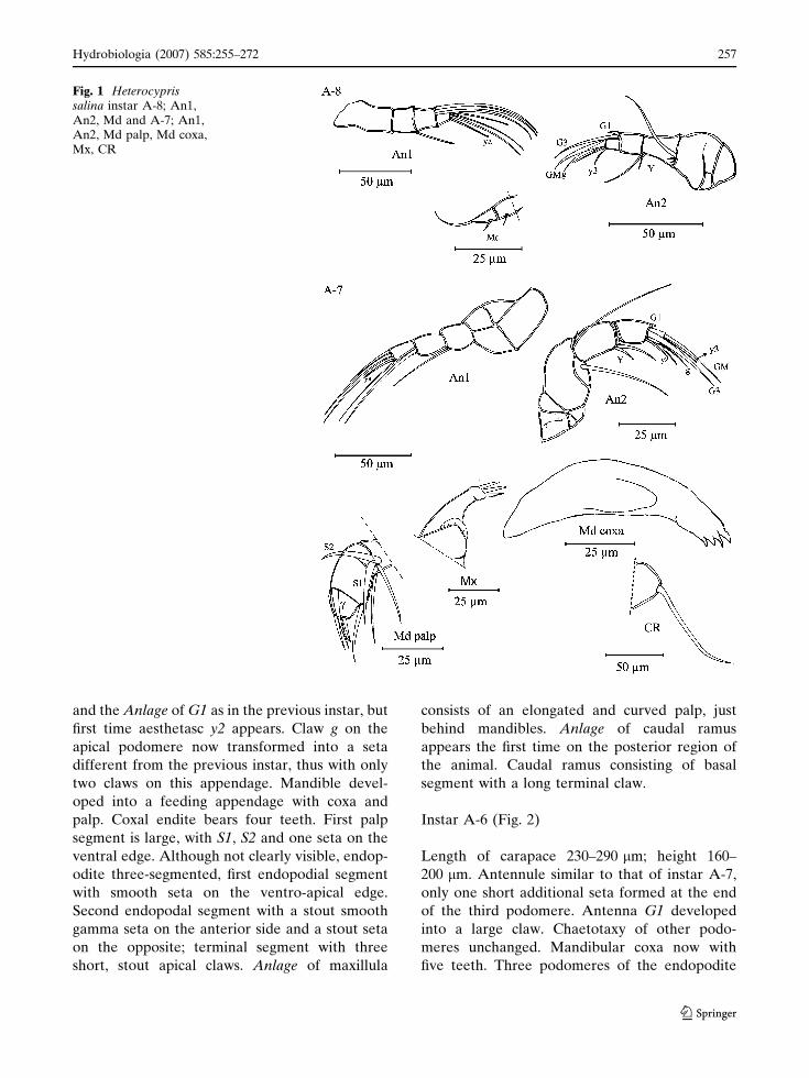
and the Anlage of G1 as in the previous instar, but
first time aesthetasc y2 appears. Claw g on the
apical podomere now transformed into a seta
different from the previous instar, thus with only
two claws on this appendage. Mandible devel-
oped into a feeding appendage with coxa and
palp. Coxal endite bears four teeth. First palp
segment is large, with S1, S2 and one seta on the
ventral edge. Although not clearly visible, endop-
odite three-segmented, first endopodial segment
with smooth seta on the ventro-apical edge.
Second endopodal segment with a stout smooth
gamma seta on the anterior side and a stout seta
on the opposite; terminal segment with three
short, stout apical claws. Anlage of maxillula
consists of an elongated and curved palp, just
behind mandibles. Anlage of caudal ramus
appears the first time on the posterior region of
the animal. Caudal ramus consisting of basal
segment with a long terminal claw.
Instar A-6 (Fig. 2)
Length of carapace 230–290 lm; height 160–
200 lm. Antennule similar to that of instar A-7,
only one short additional seta formed at the end
of the third podomere. Antenna G1 developed
into a large claw. Chaetotaxy of other podo-
meres unchanged. Mandibular coxa now with
five teeth. Three podomeres of the endopodite
Fig. 1 Heterocyprissalina instar A-8; An1,An2, Md and A-7; An1,An2, Md palp, Md coxa,Mx, CR
Hydrobiologia (2007) 585:255–272 257
123
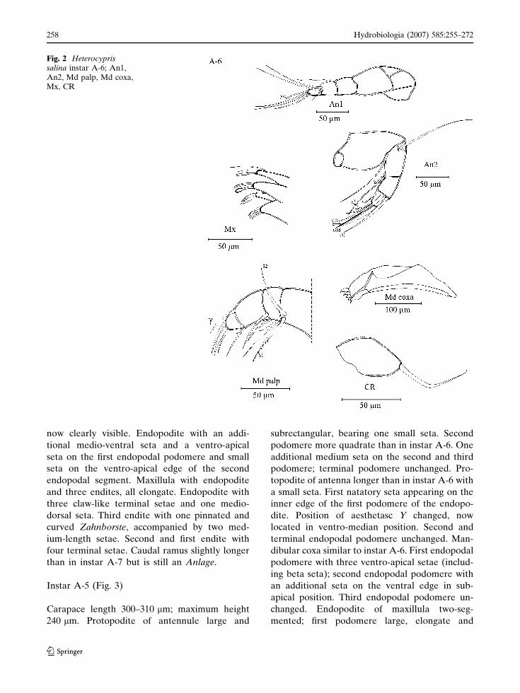
now clearly visible. Endopodite with an addi-
tional medio-ventral seta and a ventro-apical
seta on the first endopodal podomere and small
seta on the ventro-apical edge of the second
endopodal segment. Maxillula with endopodite
and three endites, all elongate. Endopodite with
three claw-like terminal setae and one medio-
dorsal seta. Third endite with one pinnated and
curved Zahnborste, accompanied by two med-
ium-length setae. Second and first endite with
four terminal setae. Caudal ramus slightly longer
than in instar A-7 but is still an Anlage.
Instar A-5 (Fig. 3)
Carapace length 300–310 lm; maximum height
240 lm. Protopodite of antennule large and
subrectangular, bearing one small seta. Second
podomere more quadrate than in instar A-6. One
additional medium seta on the second and third
podomere; terminal podomere unchanged. Pro-
topodite of antenna longer than in instar A-6 with
a small seta. First natatory seta appearing on the
inner edge of the first podomere of the endopo-
dite. Position of aesthetasc Y changed, now
located in ventro-median position. Second and
terminal endopodal podomere unchanged. Man-
dibular coxa similar to instar A-6. First endopodal
podomere with three ventro-apical setae (includ-
ing beta seta); second endopodal podomere with
an additional seta on the ventral edge in sub-
apical position. Third endopodal podomere un-
changed. Endopodite of maxillula two-seg-
mented; first podomere large, elongate and
Fig. 2 Heterocyprissalina instar A-6; An1,An2, Md palp, Md coxa,Mx, CR
258 Hydrobiologia (2007) 585:255–272
123
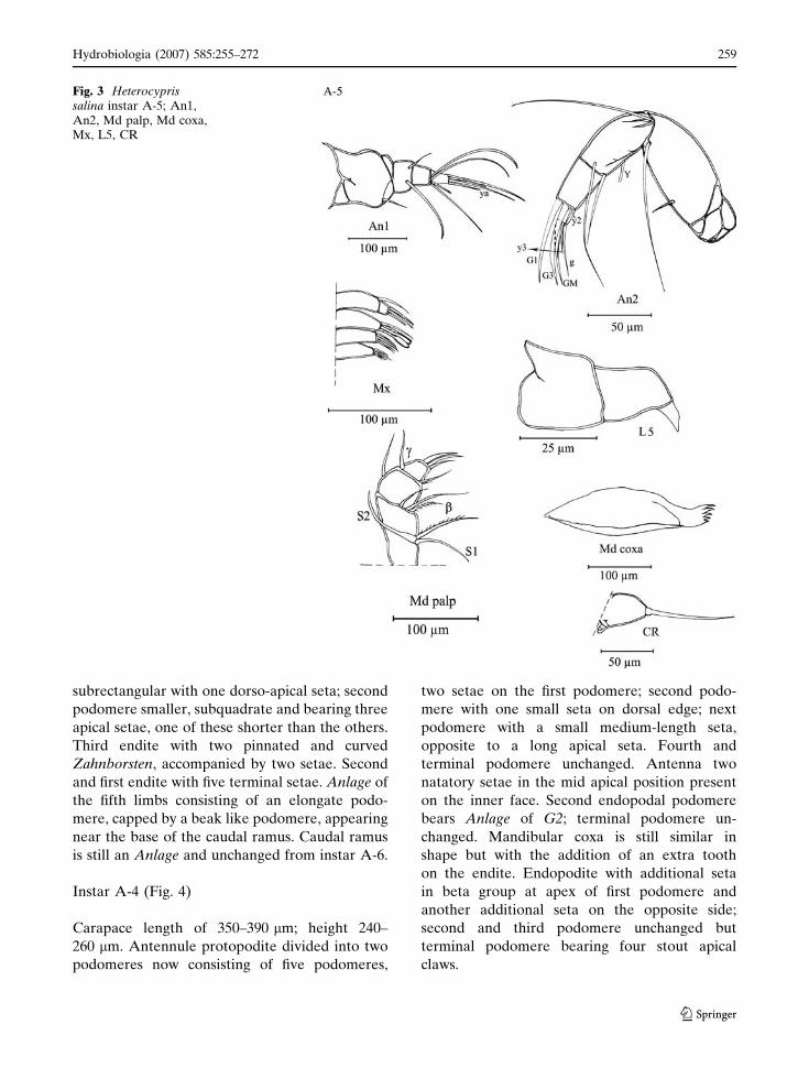
subrectangular with one dorso-apical seta; second
podomere smaller, subquadrate and bearing three
apical setae, one of these shorter than the others.
Third endite with two pinnated and curved
Zahnborsten, accompanied by two setae. Second
and first endite with five terminal setae. Anlage of
the fifth limbs consisting of an elongate podo-
mere, capped by a beak like podomere, appearing
near the base of the caudal ramus. Caudal ramus
is still an Anlage and unchanged from instar A-6.
Instar A-4 (Fig. 4)
Carapace length of 350–390 lm; height 240–
260 lm. Antennule protopodite divided into two
podomeres now consisting of five podomeres,
two setae on the first podomere; second podo-
mere with one small seta on dorsal edge; next
podomere with a small medium-length seta,
opposite to a long apical seta. Fourth and
terminal podomere unchanged. Antenna two
natatory setae in the mid apical position present
on the inner face. Second endopodal podomere
bears Anlage of G2; terminal podomere un-
changed. Mandibular coxa is still similar in
shape but with the addition of an extra tooth
on the endite. Endopodite with additional seta
in beta group at apex of first podomere and
another additional seta on the opposite side;
second and third podomere unchanged but
terminal podomere bearing four stout apical
claws.
Fig. 3 Heterocyprissalina instar A-5; An1,An2, Md palp, Md coxa,Mx, L5, CR
Hydrobiologia (2007) 585:255–272 259
123
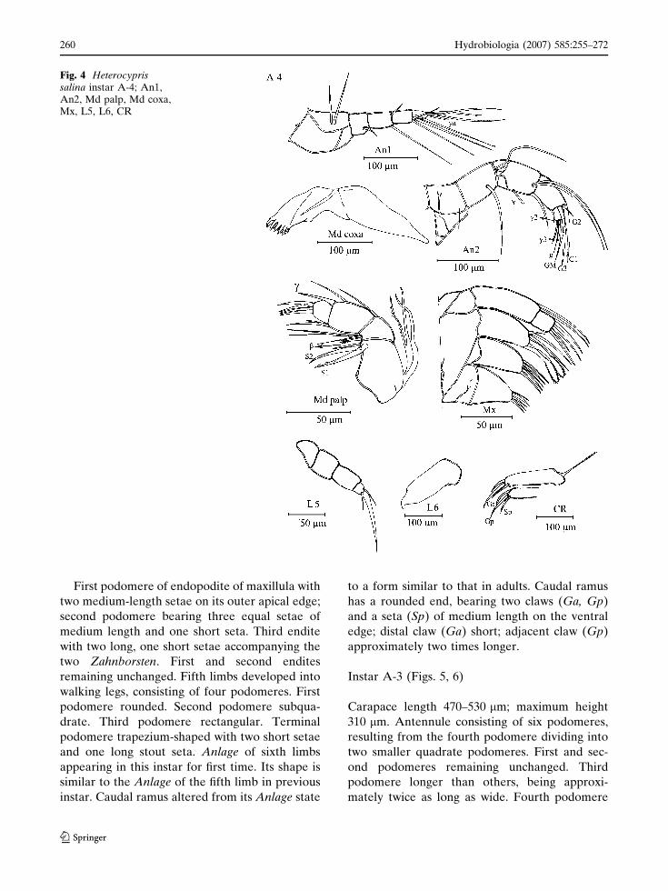
First podomere of endopodite of maxillula with
two medium-length setae on its outer apical edge;
second podomere bearing three equal setae of
medium length and one short seta. Third endite
with two long, one short setae accompanying the
two Zahnborsten. First and second endites
remaining unchanged. Fifth limbs developed into
walking legs, consisting of four podomeres. First
podomere rounded. Second podomere subqua-
drate. Third podomere rectangular. Terminal
podomere trapezium-shaped with two short setae
and one long stout seta. Anlage of sixth limbs
appearing in this instar for first time. Its shape is
similar to the Anlage of the fifth limb in previous
instar. Caudal ramus altered from its Anlage state
to a form similar to that in adults. Caudal ramus
has a rounded end, bearing two claws (Ga, Gp)
and a seta (Sp) of medium length on the ventral
edge; distal claw (Ga) short; adjacent claw (Gp)
approximately two times longer.
Instar A-3 (Figs. 5, 6)
Carapace length 470–530 lm; maximum height
310 lm. Antennule consisting of six podomeres,
resulting from the fourth podomere dividing into
two smaller quadrate podomeres. First and sec-
ond podomeres remaining unchanged. Third
podomere longer than others, being approxi-
mately twice as long as wide. Fourth podomere
Fig. 4 Heterocyprissalina instar A-4; An1,An2, Md palp, Md coxa,Mx, L5, L6, CR
260 Hydrobiologia (2007) 585:255–272
123
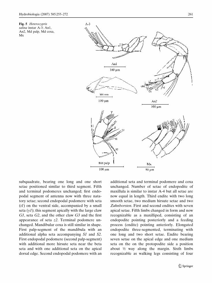
subquadrate, bearing one long and one short
setae positioned similar to third segment. Fifth
and terminal podomeres unchanged; first endo-
podal segment of antenna now with three nata-
tory setae; second endopodal podomere with seta
(t1) on the ventral side, accompanied by a small
seta (y1); this segment apically with the large claw
G1, seta G2, and the other claw G3 and the first
appearance of seta z2. Terminal podomere un-
changed. Mandibular coxa is still similar in shape.
First palp-segment of the mandibula with an
additional alpha seta accompanying S1 and S2.
First endopodal podomere (second palp-segment)
with additional more hirsute seta near the beta
seta and with one additional seta on the apical
dorsal edge. Second endopodal podomere with an
additional seta and terminal podomere and coxa
unchanged. Number of setae of endopodite of
maxillula is similar to instar A-4 but all setae are
now equal in length. Third endite with two long
smooth setae, two medium hirsute setae and two
Zahnborsten. First and second endites with seven
apical setae. Fifth limbs changed in form and now
recognizable as a maxilliped, consisting of an
endopodite pointing posteriorly and a feeding
process (endite) pointing anteriorly. Elongated
endopodite three-segmented, terminating with
one long and two short setae. Endite bearing
seven setae on the apical edge and one medium
seta on the on the protopodite side a position
about ½ way along the margin. Sixth limbs
recognizable as walking legs consisting of four
Fig. 5 Heterocyprissalina instar A-3; An1,An2, Md palp, Md coxa,Mx
Hydrobiologia (2007) 585:255–272 261
123
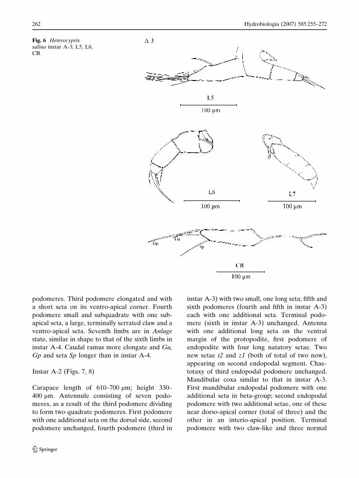
podomeres. Third podomere elongated and with
a short seta on its ventro-apical corner. Fourth
podomere small and subquadrate with one sub-
apical seta, a large, terminally serrated claw and a
ventro-apical seta. Seventh limbs are in Anlage
state, similar in shape to that of the sixth limbs in
instar A-4. Caudal ramus more elongate and Ga,
Gp and seta Sp longer than in instar A-4.
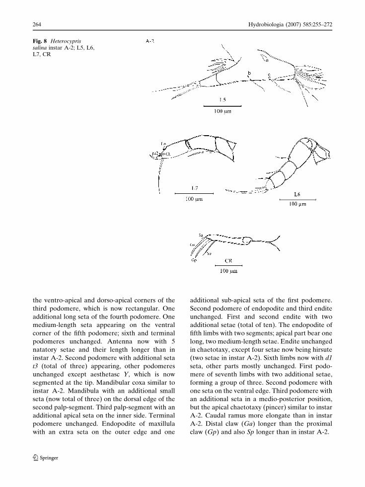
Instar A-2 (Figs. 7, 8)
Carapace length of 610–700 lm; height 330–
400 lm. Antennule consisting of seven podo-
meres, as a result of the third podomere dividing
to form two quadrate podomeres. First podomere
with one additional seta on the dorsal side, second
podomere unchanged, fourth podomere (third in
instar A-3) with two small, one long seta; fifth and
sixth podomeres (fourth and fifth in instar A-3)
each with one additional seta. Terminal podo-
mere (sixth in instar A-3) unchanged. Antenna
with one additional long seta on the ventral
margin of the protopodite, first podomere of
endopodite with four long natatory setae. Two
new setae t2 and z1 (both of total of two now),
appearing on second endopodal segment. Chae-
totaxy of third endopodal podomere unchanged.
Mandibular coxa similar to that in instar A-3.
First mandibular endopodal podomere with one
additional seta in beta-group; second endopodal
podomere with two additional setae, one of these
near dorso-apical corner (total of three) and the
other in an interio-apical position. Terminal
podomere with two claw-like and three normal
Fig. 6 Heterocyprissalina instar A-3; L5, L6,CR
262 Hydrobiologia (2007) 585:255–272
123

setae. Maxillula endopodite with two additional
short setae on top of the first podomere; second
podomere with additional one shorth seta on its
outer apical edge. Third endite with an additional
seta on the inner edge, close to the two, stout
pinnated Zahnborsten and additional long seta on
the inner side. First and second endites now with
approximately eight terminal setae. Endopodite
of fifth limbs now two segmented. Protopodite
with two small setae a and one long seta b; endite
with four additional long setae, forming a group
of eleven. Proximal to this group one isolated seta
(d). Sixth limbs consisting of five podomeres
(third podomere in instar A-3 divided into two
segments). Second and third podomere with two
additional setae on the ventral apical corner.
Fourth podomere with a small seta on the ventral
apical corner; terminal podomere unchanged.
Seventh limbs developed into a cleaning limb
consisting of three elongate podomeres. First
podomere with a seta on the apical region.
Terminal podomere capped in a fully developed
cleaning pincer as seen in the adults. Caudal
ramus bearing two equal claws (Ga and Gp, one
short seta Sa and long seta Sp).
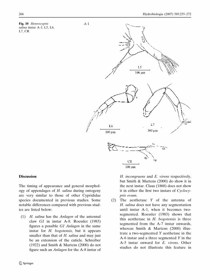
Instar A-1 (Figs. 9, 10)
Carapace length 800–890 lm; maximum height
520 lm. Antennule with two additional setae on
Fig. 7 Heterocyprissalina instar A-2; An1,An2, Md palp, Md coxa,Mx
Hydrobiologia (2007) 585:255–272 263
123
the ventro-apical and dorso-apical corners of the
third podomere, which is now rectangular. One
additional long seta of the fourth podomere. One
medium-length seta appearing on the ventral
corner of the fifth podomere; sixth and terminal
podomeres unchanged. Antenna now with 5
natatory setae and their length longer than in
instar A-2. Second podomere with additional seta
t3 (total of three) appearing, other podomeres
unchanged except aesthetasc Y, which is now
segmented at the tip. Mandibular coxa similar to
instar A-2. Mandibula with an additional small
seta (now total of three) on the dorsal edge of the
second palp-segment. Third palp-segment with an
additional apical seta on the inner side. Terminal
podomere unchanged. Endopodite of maxillula
with an extra seta on the outer edge and one
additional sub-apical seta of the first podomere.
Second podomere of endopodite and third endite
unchanged. First and second endite with two
additional setae (total of ten). The endopodite of
fifth limbs with two segments; apical part bear one
long, two medium-length setae. Endite unchanged
in chaetotaxy, except four setae now being hirsute
(two setae in instar A-2). Sixth limbs now with d1
seta, other parts mostly unchanged. First podo-
mere of seventh limbs with two additional setae,
forming a group of three. Second podomere with
one seta on the ventral edge. Third podomere with
an additional seta in a medio-posterior position,
but the apical chaetotaxy (pincer) similar to instar
A-2. Caudal ramus more elongate than in instar
A-2. Distal claw (Ga) longer than the proximal
claw (Gp) and also Sp longer than in instar A-2.
Fig. 8 Heterocyprissalina instar A-2; L5, L6,L7, CR
264 Hydrobiologia (2007) 585:255–272
123
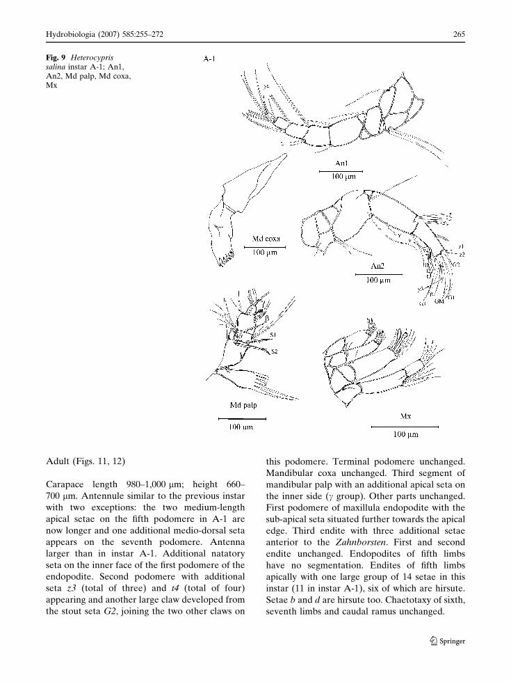
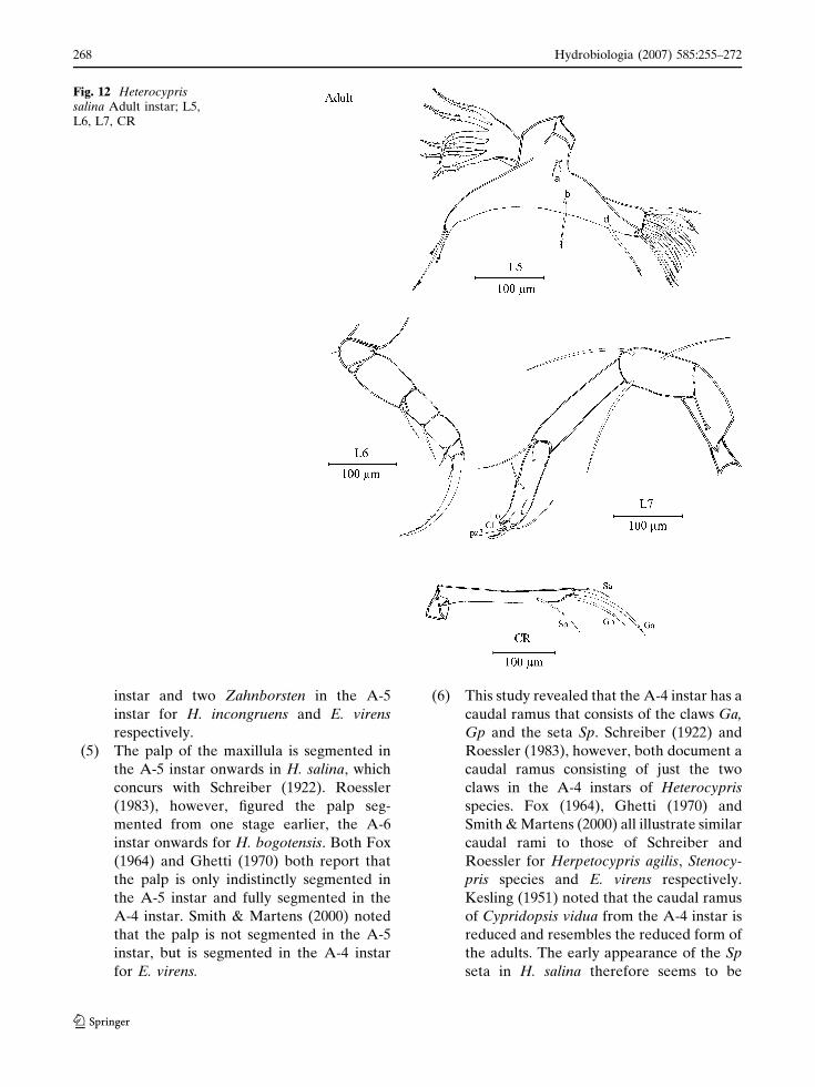
Adult (Figs. 11, 12)
Carapace length 980–1,000 lm; height 660–
700 lm. Antennule similar to the previous instar
with two exceptions: the two medium-length
apical setae on the fifth podomere in A-1 are
now longer and one additional medio-dorsal seta
appears on the seventh podomere. Antenna
larger than in instar A-1. Additional natatory
seta on the inner face of the first podomere of the
endopodite. Second podomere with additional
seta z3 (total of three) and t4 (total of four)
appearing and another large claw developed from
the stout seta G2, joining the two other claws on
this podomere. Terminal podomere unchanged.
Mandibular coxa unchanged. Third segment of
mandibular palp with an additional apical seta on
the inner side (c group). Other parts unchanged.
First podomere of maxillula endopodite with the
sub-apical seta situated further towards the apical
edge. Third endite with three additional setae
anterior to the Zahnborsten. First and second
endite unchanged. Endopodites of fifth limbs
have no segmentation. Endites of fifth limbs
apically with one large group of 14 setae in this
instar (11 in instar A-1), six of which are hirsute.
Setae b and d are hirsute too. Chaetotaxy of sixth,
seventh limbs and caudal ramus unchanged.
Fig. 9 Heterocyprissalina instar A-1; An1,An2, Md palp, Md coxa,Mx
Hydrobiologia (2007) 585:255–272 265
123
Discussion
The timing of appearance and general morphol-
ogy of appendages of H. salina during ontogeny
are very similar to those of other Cyprididae
species documented in previous studies. Some
notable differences compared with previous stud-
ies are listed below:
(1) H. salina has the Anlagen of the antennal
claw G1 in instar A-8. Roessler (1983)
figures a possible G1 Anlagen in the same
instar for H. bogotensis, but it appears
smaller than that of H. salina and may just
be an extension of the cuticle. Schreiber
(1922) and Smith & Martens (2000) do not
figure such an Anlagen for the A-8 instar of
H. incongruens and E. virens respectively,
but Smith & Martens (2000) do show it in
the next instar. Claus (1868) does not show
it in either the first two instars of Cyclocy-
pris ovum.
(2) The aesthetasc Y of the antenna of
H. salina does not have any segmentation
until instar A-1, when it becomes two-
segmented. Roessler (1983) shows that
this aesthetasc in H. bogotensis is three
segmented from the A-7 instar onwards,
whereas Smith & Martens (2000) illus-
trate a two-segmented Y aesthetasc in the
A-6 instar and a three segmented Y in the
A-5 instar onward for E. virens. Other
studies do not illustrate this feature in
Fig. 10 Heterocyprissalina instar A-1; L5, L6,L7, CR
266 Hydrobiologia (2007) 585:255–272
123

sufficient detail to accurately determine
segmentation, but both Schreiber (1922)
and Kesling (1951) figure it with at least
two segments from the A-6 instar
onwards for H. incongruens and C. vidua
respectively.
(3) The mandibular palp of H. salina has four
podomeres in the A-7 instar onwards,
which concurs with both the studies of
Claus (1868), Schreiber (1922), Kesling
(1952), Fox (1964) and Roessler (1983).
Smith & Martens (2000) show only three
mandibular podomeres in the A-7 and A-6
instars of E. virens, but in both these instars
there are setae that protrude from where
the extra podomere boundary should be,
indicating that it maybe just weakly ex-
pressed rather than missing altogether.
(4) In H. salina a single Zahnborsten appears
on the third endite of the maxillula in instar
A-6 for the first time, and the second
Zahnborsten appears in the A-5 instar.
Most other studies don’t show the maxillula
in detail, but both Schreiber (1922) and
Smith & Martens (2000) show the third
endite with one claw-like seta in the A-6
Fig. 11 Heterocyprissalina Adult instar; An1,An2, Md palp, Md coxa,Mx
Hydrobiologia (2007) 585:255–272 267
123
instar and two Zahnborsten in the A-5
instar for H. incongruens and E. virens
respectively.
(5) The palp of the maxillula is segmented in
the A-5 instar onwards in H. salina, which
concurs with Schreiber (1922). Roessler
(1983), however, figured the palp seg-
mented from one stage earlier, the A-6
instar onwards for H. bogotensis. Both Fox
(1964) and Ghetti (1970) both report that
the palp is only indistinctly segmented in
the A-5 instar and fully segmented in the
A-4 instar. Smith & Martens (2000) noted
that the palp is not segmented in the A-5
instar, but is segmented in the A-4 instar
for E. virens.
(6) This study revealed that the A-4 instar has a
caudal ramus that consists of the claws Ga,
Gp and the seta Sp. Schreiber (1922) and
Roessler (1983), however, both document a
caudal ramus consisting of just the two
claws in the A-4 instars of Heterocypris
species. Fox (1964), Ghetti (1970) and
Smith & Martens (2000) all illustrate similar
caudal rami to those of Schreiber and
Roessler for Herpetocypris agilis, Stenocy-
pris species and E. virens respectively.
Kesling (1951) noted that the caudal ramus
of Cypridopsis vidua from the A-4 instar is
reduced and resembles the reduced form of
the adults. The early appearance of the Sp
seta in H. salina therefore seems to be
Fig. 12 Heterocyprissalina Adult instar; L5,L6, L7, CR
268 Hydrobiologia (2007) 585:255–272
123
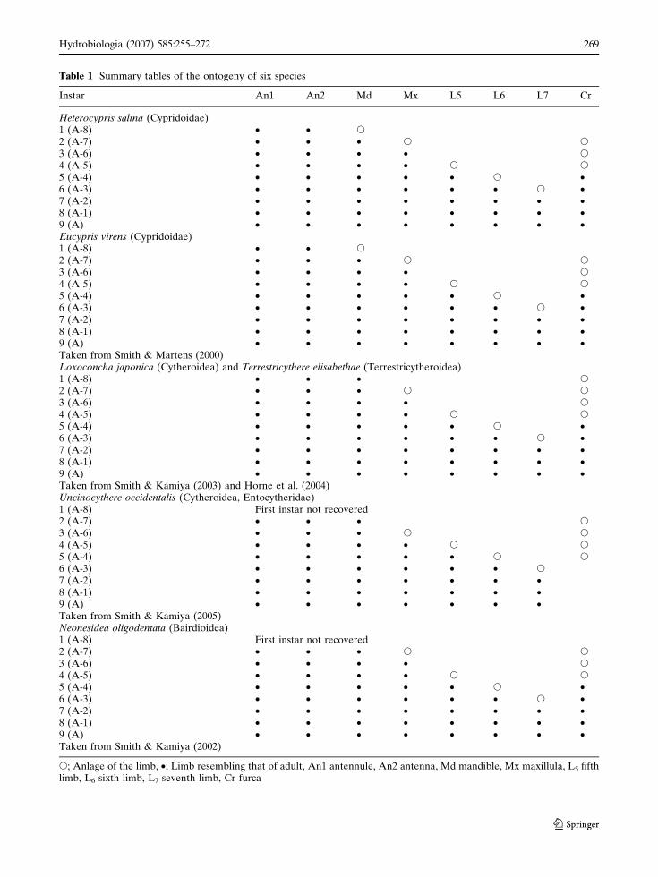
Table 1 Summary tables of the ontogeny of six species
Instar An1 An2 Md Mx L5 L6 L7 Cr
Heterocypris salina (Cypridoidae)1 (A-8) • • s
2 (A-7) • • • s s
3 (A-6) • • • • s
4 (A-5) • • • • s s
5 (A-4) • • • • • s •6 (A-3) • • • • • • s •7 (A-2) • • • • • • • •8 (A-1) • • • • • • • •9 (A) • • • • • • • •Eucypris virens (Cypridoidae)1 (A-8) • • s
2 (A-7) • • • s s
3 (A-6) • • • • s
4 (A-5) • • • • s s
5 (A-4) • • • • • s •6 (A-3) • • • • • • s •7 (A-2) • • • • • • • •8 (A-1) • • • • • • • •9 (A) • • • • • • • •Taken from Smith & Martens (2000)Loxoconcha japonica (Cytheroidea) and Terrestricythere elisabethae (Terrestricytheroidea)1 (A-8) • • • s
2 (A-7) • • • s s
3 (A-6) • • • • s
4 (A-5) • • • • s s
5 (A-4) • • • • • s •6 (A-3) • • • • • • s •7 (A-2) • • • • • • • •8 (A-1) • • • • • • • •9 (A) • • • • • • • •Taken from Smith & Kamiya (2003) and Horne et al. (2004)Uncinocythere occidentalis (Cytheroidea, Entocytheridae)1 (A-8) First instar not recovered2 (A-7) • • • s
3 (A-6) • • • s s
4 (A-5) • • • • s s
5 (A-4) • • • • • s s
6 (A-3) • • • • • • s
7 (A-2) • • • • • • •8 (A-1) • • • • • • •9 (A) • • • • • • •Taken from Smith & Kamiya (2005)Neonesidea oligodentata (Bairdioidea)1 (A-8) First instar not recovered2 (A-7) • • • s s
3 (A-6) • • • • s
4 (A-5) • • • • s s
5 (A-4) • • • • • s •6 (A-3) • • • • • • s •7 (A-2) • • • • • • • •8 (A-1) • • • • • • • •9 (A) • • • • • • • •Taken from Smith & Kamiya (2002)
s; Anlage of the limb, •; Limb resembling that of adult, An1 antennule, An2 antenna, Md mandible, Mx maxillula, L5 fifthlimb, L6 sixth limb, L7 seventh limb, Cr furca
Hydrobiologia (2007) 585:255–272 269
123
restricted to this species. From the A-3
instar onwards the caudal rami of all species
are similar, with the exception of C. vidua.
(7) Smith & Martens (2000) mentions an
antenna with the Gm claw in the A-3 instar
of E. virens, whereas this is missing in the
antenna of H. salina. Likewise, no c setae
on the fifth limb and d2 seta on the sixth
limb were observed for H. salina, both of
which are present in E. virens. These are
differences that are observable in adult
specimens, but it is noteworthy that the
missing features in the adults of H. salina
are missing in all juvenile stages as well,
rather than just missing in the adult stage
alone.
(8) The z3 seta of the antenna of H. salina first
appears in the adult stage, one stage later
than that of H. bogotensis, Sclerocypris
species, E. virens and Pseudocandona ser-
bani (Roessler, 1983; Broodbakker & Da-
nielopol, 1982; Martens, 1987; Smith &
Martens, 2000).
(9) The endopodite (= palp) of the fifth limb of
H. salina has three segments in instar A-3,
and two segments in both the A-2 and A-1
instars. Fox (1964) reported that the palp
was segmented in the A-3 instar, weakly
segmented in the A-2 instar and with no
segmentation in the A-1 instar for the
cyprid species he studied. Schreiber
(1922), Kesling (1951), Ghetti (1970),
Roessler (1983) and Smith & Martens
(2000) all reported that the A-3 instar had
a three-segmented palp, but this segmen-
tation was missing from the A-2 instar
onwards. All studies reported a palp with
no segmentation in the adult.
(10) This study documented an antennule with
just four podomeres in the A-8 and A-7
instars of H. salina, in contrast to five
documented by Claus (1868), Schreiber
(1922), Kesling (1951), Ghetti (1970),
Smith & Martens (2000) and Smith &
Tsukagoshi (2005). From the A-6 instar
onwards the development is similar.
Some of these features mentioned above
appear either earlier or later in the development
of H. salina compared with other species, even
species in the same genus. For instance, the G1
claw of the antennule, the Sp seta of the caudal
ramus, and the Zahnborsten of the maxillula all
appear earlier in H. salina compared with other
species. Loss of segmentation in the fifth limb
palp, appearance of segmentation of the antennal
Y aesthetasc, and the appearance of the z3 seta on
the antenna however, all occur in later instars of
H. salina compared with other species. This shows
that even within a family, closely related species
can show unexpected variations in development.
The early appearance of the Sp seta on the caudal
ramus in the A-4 instar, and the four-segmented
antennule in the first two instars of H. salina are
particularly noteworthy. Both of these features
vary in the juveniles compared with those of other
species, but the adult forms are similar to other
species. Such a divergence in development in just
one or two early instars, rather than all subse-
quent instars, is puzzling. The late appearance of
the antennal z3 seta in H. salina is also unex-
pected, as other Cyprididae (e.g., H. bogotensis
and E. virens) and Candonidae (e.g., Pseudocan-
dona serbani) species show a similar z3 develop-
ment to each other (Broodbakker & Danielopol,
1982; Roessler, 1983; Smith & Martens, 2000).
This would indicate that the late appearance of
this feature in H. salina is an apomorphy.
Within the genus, H. salina varies from the two
other Heterocypris species previously studied,
H. incongruens and H. bogotensis, in the follow-
ing: the four-segmented antennules of the first two
instars (five segmented in the other two species),
the segmentation of the fifth limb palps in the A-2
and A-1 instars (not segmented in the other two
species) and the appearance of the Sp seta of the
caudal ramus in the A-4 instar (first appears in the
A-3 instar in the other two species). (Other
features are not clearly illustrated in all three
studies to allow a comparison.) This suggests that
H. incongruens and H. bogotensis are more closely
related to each other than to H. salina. As some
features mentioned above are present in very
early instars (e.g., A-8 and A-7 instars for the
antennule segmentation) they suggest that diver-
gence of the lineage that led to H. salina occurred
from an early point in the history of the genus.
270 Hydrobiologia (2007) 585:255–272
123

If the variations in ontogeny of these features
can be verified in other species/genera, then they
maybe useful characters to use in phylogenetic
analyses at the genus and family levels.
H. salina (Cypridoidae) has nine instars from
the egg to the adult stage. This is the same
number of instars as e.g., E. virens (Cypridoidea)
(Smith & Martens 2000), L. japonica (Cythero-
idea) (Smith & Kamiya, 2003) and T. elisabethae
(Terrestricytheroidea) (Horne et al., 2004), but
one more than N. oligodentata (Smith & Kamiya,
2002) and U. occidentalis (Entocytheridae)
(Smith & Kamiya, 2005), which have only eight
instar stages. Smith & Kamiya (2005) noted that
this is probably due to the first instar in N.
oligodentata and U. occidentalis moulting within
the egg. Both the Terrestricytheroidea and Cyt-
heroidea have the Anlagen of the caudal ramus in
the A-8 instar, but it is missing in the A-8 instar
of the Cypridoidea. From instar A-7 onwards the
general appearance of the limbs of H. salina are
similar to those of species from the superfamilies
Cypridoidea, Cytheroidea, Bairdioidea and Ter-
restricytheroidea (Table 1), with the exception of
the maxillula of the Entocytheridae (a family of
the Cytheroidea), which first appears in instar
A-6, one stage later than other groups.
Acknowledgements We are very grateful to Dr. RenateMatzke-Karasz an an anonymous reviewer for commentson earlier versions of this manuscript and are also gratefulto Ferda Percin and Huseyin Akıncı for their helps indrawings.
References
Athersuch, J., D. J. Horne & J. E. Whittaker, 1989. Marineand Brackish Water Ostracods (Superfamilies Cyp-ridacea and Cytheracea). In Kermack, D. M. & R. S.K. Barnes (eds), Synopses of the British Fauna (NewSeries), 43. Brill, Leiden.
Baltanas, A., M. Otero, L. Arqueros, G. Rossetti & V.Rossi, 2000. Ontogenetic changes in the carapaceshape of non-marine ostracods Eucypris virens(Jurine). Hydrobiologia 419: 65–72.
Broodbakker, N. W. & D. L. Danielopol, 1982. Thechaetotaxy of Cypridacea (Crustacea, Ostracoda)limbs: proposals for a descriptive model. Bijdragentot de Dierkunde, 52: 103–120.
Claus, C., 1865. Zur naheren Kenntnis der Jugendformenvon Cypris ovum. Zeitschrift fur wissenschaftlicheZoologie 15: 391–398.
Claus, C., 1868. Beitrage zur Kenntnis der Ostracoden. 1:Entwicklungsgeschichte von Cypris. Schriften derGesellschaft zur Beforderung der Gesammten Natur-wissenschaften zu Marburg, 9: 151–166.
Fox, H. M., 1964. On the larval stages of Cyprids and onSiphlocandona (Crustacea. Ostracoda). Proceedingsof the Zoological Society of London 142: 165–176.
Ghetti, P. F., 1970. The taxonomic significance of ostracodlarval stages: with examples from the Burundi rice-fields. Bollettino di Zoologia 37: 103–119.
Hart, C. W. Jr., L. C. Hayek, J. Clark & W. H. Clark, 1985.The Life History and Ecology of the entocytheridostracod Uncinocythere occidentalis (Kozloff andWhitmann) in Idaho. Smithsonian Contributions toZoology 419: 1–22.
Horne, D. J. & R. J. Smith, 2004. First British record ofPotamocypris humilis (Sars, 1924), a freshwaterostracod with a disjunct distribution in northernEurope and southern Africa. Bollettino della SocietaPaleontologica Italiana 43: 297–306.
Horne, D. J., R. J. Smith, J. E. Whittaker & J. W. Murray,2004. The first British record and a new species of thesuperfamily Terrestricytheroidea (Crustacea, Ostra-coda): morphology, ontogeny, lifestyle and phylogeny.Zoological Journal of the Linnean Society 142: 253–288.
Kesling, R. V., 1951. The morphology of ostracod moltstages. Illinois Biological Monographs 21, Urbana.
Martens, K., 1987. Homology and functional morphologyof the sexual dimorphism in the antenna of Sclerocy-pris Sars, 1924 (Crustacea, Ostracoda, Megalocyprid-inae). Bijdragen tot de Dierkunde 57: 183–190.
Meisch, C., 2000. Freshwater Ostracoda of Western andCentral Europe. In Schwoerbel, J. & P. Zwick (eds),Sußwasserfauna von Mitteleuropa 8/3. SpektrumAkademischer Verlag, Heidelberg-Berlin.
Roessler, E. W., 1983. Estudios taxonomicos, ontogenet-icos, ecologicos y etologicos sobre los ostracodos deagua dulce en Colombia. IV. Desarrollo postembrio-nario de Heterocyrpis bogotensis Roessler (Ostracoda,Podocopa, Cyprididae). Caldasia, 13: 755–776.
Scheerer-Ostermeyer, E., 1940. Beitrag zur Ent-wicklungsgeschichte der Sußwasserostrakoden. Zoo-logische Jahrbucher, Abteilung fur Anatomie undOntogenie der Tiere, 66: 349–370.
Schreiber, E., 1922. Beitrage zur Kenntnis der Morphol-ogie, Entwicklung und Lebensweise der Sußwasser-Ostracoden. Zoologische Jahrbucher, Abteilung furAnatomie und Ontogenie der Tiere 43: 485–539.
Smith, R. J. & K. Martens, 2000. The Ontogeny of cyprididOstracoda Eucypris virens (Jurine, 1820) (Crustacea,Ostracoda). Hydrobiologia 419: 31–63.
Smith, R. J. & T. Kamiya, 2002. The Ontogeny ofNeonesidea oligodentata (Bairdioidea, Ostracoda,Crustacea). Hydrobiologia 489: 245–275.
Hydrobiologia (2007) 585:255–272 271
123
Smith, R. J. & T. Kamiya, 2003. The Ontogeny ofLoxoconcha japonica Ishizaki, 1968 (Cytheroidea,Ostracoda, Crustacea). Hydrobiologia 490: 31–52.
Smith, R. J. & T. Kamiya, 2005. The Ontogeny of theentocytherid Ostracod Uncinocythere occidentalis(Kozloff & Whitman, 1954) Hart, 1962 (Crustacea).Hydrobiologia 538: 217–229.
Smith, R. J. & A. Tsukagoshi, 2005. The chaetotaxy,ontogeny and musculature of antennule of podocopid
ostracods (Crustacea). Journal of Zoology, London265: 157–177.
Weygoldt, P., 1960. Embryologische Untersuchungen anOstrakoden: Die Entwicklung von Cyprideis litoralis(G. S. Brady) (Ostracoda, Podocopa, Cytheridae).Zoologische Jahrbucher, Abteilung fur Anatomie undOntogenie der Tiere, 78: 367–426.
272 Hydrobiologia (2007) 585:255–272
123