Dr. Maha ELBeltagyAssistant Professor of Anatomy
Faculty of Medicine
The University of Jordan
2018
Neuroanatomy
Dr Maha ELbeltagy

Dr Maha ELbeltagy
Dr Maha ELbeltagy
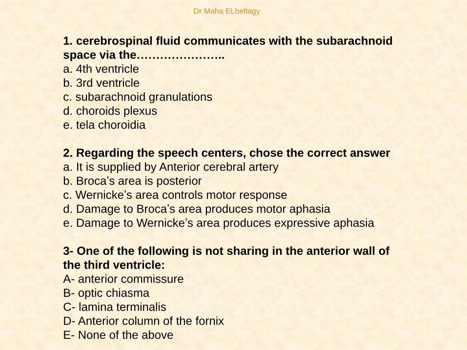
1. cerebrospinal fluid communicates with the subarachnoid
space via the…………………..
a. 4th ventricle
b. 3rd ventricle
c. subarachnoid granulations
d. choroids plexus
e. tela choroidia
2. Regarding the speech centers, chose the correct answer
a. It is supplied by Anterior cerebral artery
b. Broca’s area is posterior
c. Wernicke’s area controls motor response
d. Damage to Broca’s area produces motor aphasia
e. Damage to Wernicke’s area produces expressive aphasia
3- One of the following is not sharing in the anterior wall of
the third ventricle:
A- anterior commissure
B- optic chiasma
C- lamina terminalis
D- Anterior column of the fornix
E- None of the above
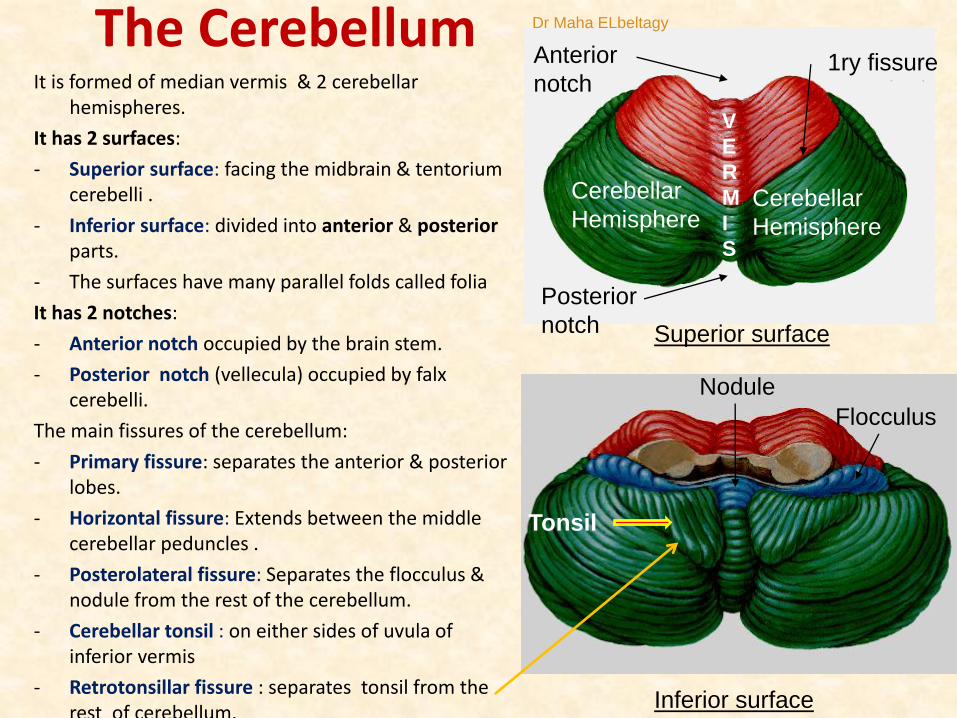
The CerebellumIt is formed of median vermis & 2 cerebellar
hemispheres.
It has 2 surfaces:
- Superior surface: facing the midbrain & tentoriumcerebelli .
- Inferior surface: divided into anterior & posteriorparts.
- The surfaces have many parallel folds called folia
It has 2 notches:
- Anterior notch occupied by the brain stem.
- Posterior notch (vellecula) occupied by falxcerebelli.
The main fissures of the cerebellum:
- Primary fissure: separates the anterior & posterior lobes.
- Horizontal fissure: Extends between the middle cerebellar peduncles .
- Posterolateral fissure: Separates the flocculus & nodule from the rest of the cerebellum.
- Cerebellar tonsil : on either sides of uvula of inferior vermis
- Retrotonsillar fissure : separates tonsil from the rest of cerebellum.
Superior surface
Inferior surface
1ry fissureAnterior
notch
Posterior
notch
Cerebellar
Hemisphere
V
E
R
M
I
S
Flocculus
Nodule
Cerebellar
Hemisphere
Tonsil
Dr Maha ELbeltagy

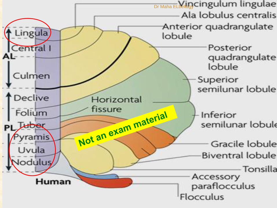
Anterior Lobe
Posterior Lobe
1ry FissureLobes of the cerebellum: (Horizontal division)
- Anterior lobe: in front of the primary fissure.
- Posterior lobe: behind the primary fissure.
- Flocculo-nodular lobe: Consists of the flocculus & nodule .
hemispheres
Paravermis
Dr Maha ELbeltagy
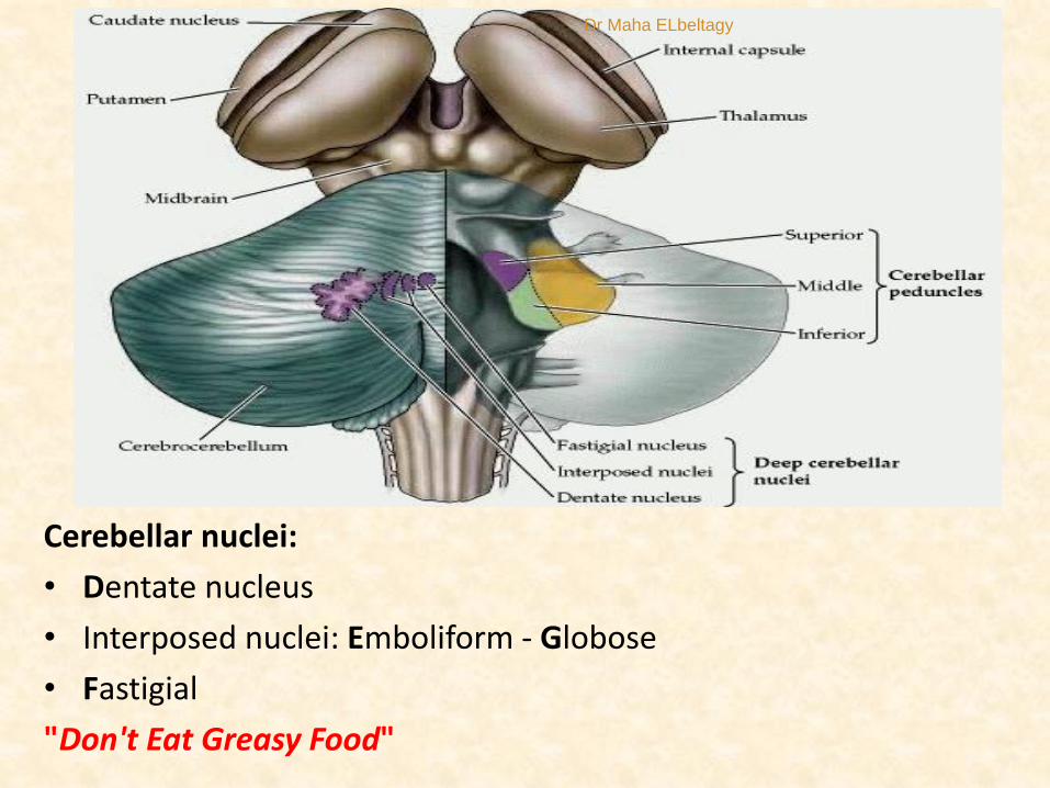
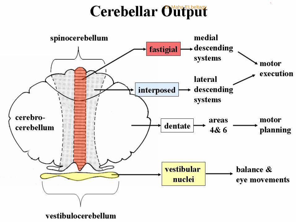
Cerebellar nuclei:
• Dentate nucleus
• Interposed nuclei: Emboliform - Globose
• Fastigial
"Don't Eat Greasy Food"
Dr Maha ELbeltagy

Arbor vitaeIn latin “ tree of life” it is the white matter of
cerebellum.
•It is so called because of the tree like
appearance.
•It brings sensory and motor sensation to
and from cerebellum
Vertical subdivisions of the
cerebellum
1- vermis (central part on superior and
inferior surfaces) represents head, neck,
trunk, shoulders and hips). Projects to
Fastigeal N
2- Paravemis (lateral to vermis) represents
muscles of upper and lower limbs
Projects to Globose and Emboliform N
3- Rest of cerebellar hemispheres
Project to Dentate N
Arbor vitae
Fourth ventricle
Dr Maha ELbeltagy
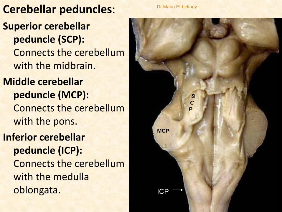
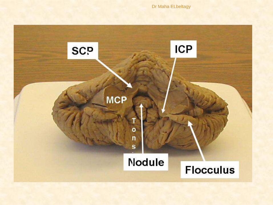
Cerebellar peduncles:
Superior cerebellar peduncle (SCP):Connects the cerebellum with the midbrain.
Middle cerebellar peduncle (MCP):Connects the cerebellum with the pons.
Inferior cerebellar peduncle (ICP):Connects the cerebellum with the medulla oblongata.
MCP
ICP
Dr Maha ELbeltagy
Dr Maha ELbeltagy
Dr Maha ELbeltagy
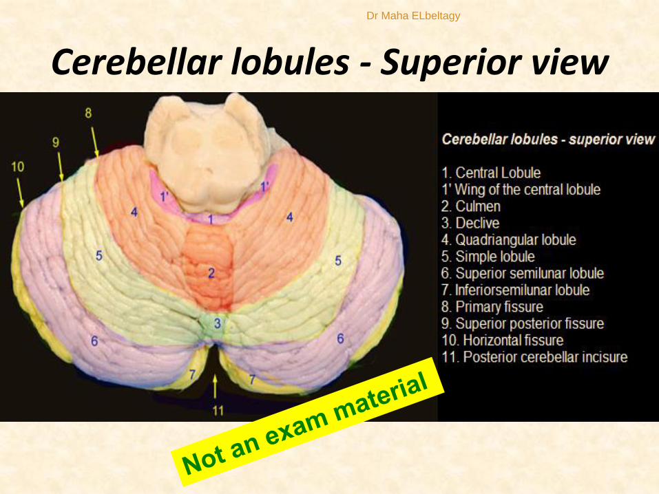
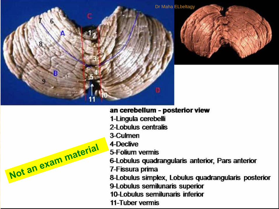
Cerebellar lobules - Superior view
Dr Maha ELbeltagy
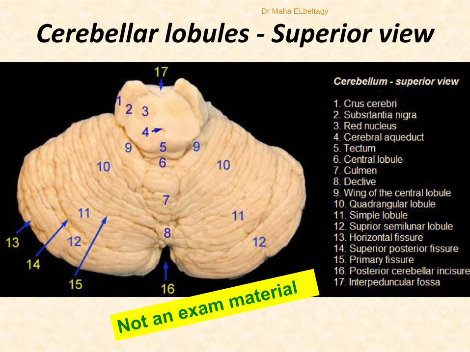
Cerebellar lobules - Superior viewDr Maha ELbeltagy

Cerebellar Lobules (inferior view)Dr Maha ELbeltagy
Dr Maha ELbeltagy

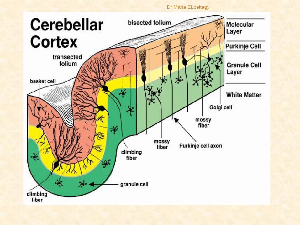
1- Cerebellar Cortex
Outer Molecular Layer (stellate and basket cells)
Middle Purkinje Cell Layer (inhibitory to all other cells)
Inner Granular Layer Include 2G cells (granule and golgi)Granule cells are the only (excitatory to all other cells).
2- Corpus Medullare (Medullary Center)
3- Deep Cerebellar Nuclei
Structure of the cerebellum
12
Dr Maha ELbeltagy
Dr Maha ELbeltagy

White matter of the
cerebellum•Consists of three types of nerve
fibres in the white matter
A. Mossy fibres (afferent)
They end in the granular layer first
then purkinje layer (indirect
activation of purkinje).
B. Climbing fibres (afferent)
They end directly in purkinje (direct
activation) or molecular layer
(olivocerebellar tracts mainly)
Dr Maha ELbeltagy
C- Axons of purkinje cells (efferent)
The only axons to leave cerebellar cortex
to end in deep cerebellar nuclei (inhibitory).
These fibers then projects to brain stem
nuclei, thalamus and cerebral cortex.
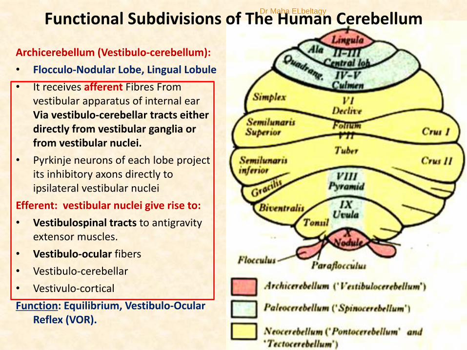
Functional Subdivisions of The Human Cerebellum
Archicerebellum (Vestibulo-cerebellum):
• Flocculo-Nodular Lobe, Lingual Lobule
• It receives afferent Fibres From vestibular apparatus of internal ear Via vestibulo-cerebellar tracts either directly from vestibular ganglia or from vestibular nuclei.
• Pyrkinje neurons of each lobe project its inhibitory axons directly to ipsilateral vestibular nuclei
Efferent: vestibular nuclei give rise to:
• Vestibulospinal tracts to antigravity extensor muscles.
• Vestibulo-ocular fibers
• Vestibulo-cerebellar
• Vestivulo-cortical
Function: Equilibrium, Vestibulo-Ocular Reflex (VOR).
Dr Maha ELbeltagy

Paleocerebellum (Spino-cerebelllum):
1- Anterior lobe+ midline vermis (fastigeal N)2- surrounding paravermis + globose & emboliform nuclei.
1- Vermal zone of the spino-
cerebellumPurkinje neurons of each hemivermis projects
inhibitory axons to ipsilateral fastigeal nuclei.
Afferent : venteral and dorsal spinocerebellar,
olivo-cerebellar and cuneocerellar tracts.
Projects to fastigeal N
Fastigeal N gives bilateral excitatory fibers to the
medial mtotor system that controls axial and
porximal limb muscles through:
Efferent:
Fasigeo-Vestibulo-spinal (ipsilateral and
contralateral vestibular nuclei)
Fastigeo- Reticulo-spinal (Ipsilateral and
contralateral RF)
Anterior cortico-spinal (ipsilateral and
contralateral VL nucleus of thalamus which project
to trunk part of area 4.
(cerebello-fastigeo-thalamo-cortico-spinal)
Function: Regulate muscle tone
of axial and proximal limb
muscles
Dr Maha ELbeltagy
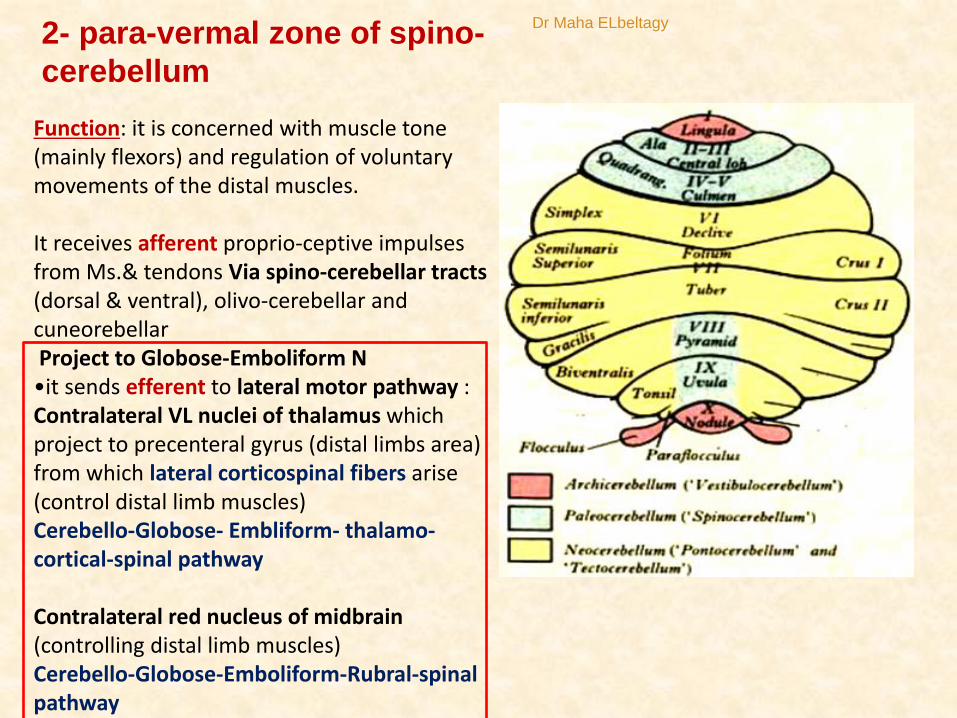
Function: it is concerned with muscle tone (mainly flexors) and regulation of voluntary movements of the distal muscles.
It receives afferent proprio-ceptive impulses from Ms.& tendons Via spino-cerebellar tracts (dorsal & ventral), olivo-cerebellar and cuneorebellarProject to Globose-Emboliform N•it sends efferent to lateral motor pathway :Contralateral VL nuclei of thalamus which project to precenteral gyrus (distal limbs area) from which lateral corticospinal fibers arise (control distal limb muscles)Cerebello-Globose- Embliform- thalamo-cortical-spinal pathway
Contralateral red nucleus of midbrain (controlling distal limb muscles)Cerebello-Globose-Emboliform-Rubral-spinal pathway
2- para-vermal zone of spino-
cerebellum
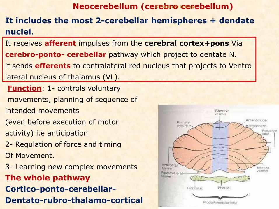
Neocerebellum (cerebro cerebellum)
It includes the most 2-cerebellar hemispheres + dendate
nuclei.
It receives afferent impulses from the cerebral cortex+pons Via
cerebro-ponto- cerebellar pathway which project to dentate N.
it sends efferents to contralateral red nucleus that projects to Ventro
lateral nucleus of thalamus (VL).
Function: 1- controls voluntary
movements, planning of sequence of
intended movements
(even before execution of motor
activity) i.e anticipation
2- Regulation of force and timing
Of Movement.
3- Learning new complex movements
The whole pathway
Cortico-ponto-cerebellar-
Dentato-rubro-thalamo-cortical
Dr Maha ELbeltagy
Dr Maha ELbeltagy

Fibres entering and leaving through cerebellar peduncles
Superior cerebellar peduncle (major efferent)
Fibres entering the cerebellum
Ventral spino-cerebellar tract
Trigimino-cerebellar from Mesencephalic nucleus
Tecto-cerebellar fibres
Fibres leaving the cerebellum
Cerebello-rubral fibres
(Globose-Emboliform-rubral)
Cerebello-thalamic fibres
(Dentato-thalamo-cortical)
Cerebello-reticular fibres
(Fastigeal nucleus)
Dr Maha ELbeltagy

Middle cerebellar peduncle (afferent
Pontocerebellar fibres(cortico-ponto-cerebellum) to dentate
nucleus)
Inferior cerebellar peduncle (afferent)Fibres entering cerebellum
(restiform body)
Posterior spino cerebellar tract
Cuneo-cerebellar tract
Olivo-cerebellar fibres
Reticulo-cerebellar
Vestibulo-cerebellar fibres
Trigemino-cerebellar fibres
Anterior external arcuate fibers
Fibres Leaving the cerebellum
(juxta-restiform body)
Cerebello-olivary fibres
Cerebello (Fastigio)-vestibular fibres
Cerebello (Fastigio)- reticular fibres
Dr Maha ELbeltagy

Blood Supply of the CerebellumIt is supplied by 3
cerebellar arteries
• Superior cerebellar artery: from the basilar artery
• Anterior inferior cerebellar artery: from the basilar artery
• Posterior inferior cerebellar artery: from the vertebral artery
Dr Maha ELbeltagy
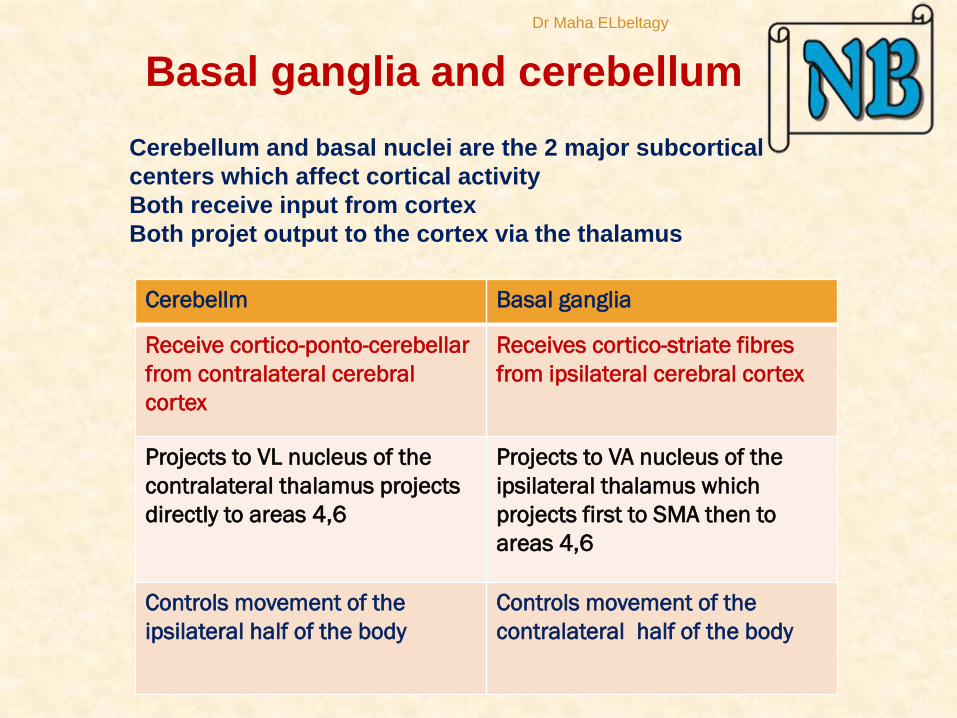
Cerebellm Basal ganglia
Receive cortico-ponto-cerebellar
from contralateral cerebral
cortex
Receives cortico-striate fibres
from ipsilateral cerebral cortex
Projects to VL nucleus of the
contralateral thalamus projects
directly to areas 4,6
Projects to VA nucleus of the
ipsilateral thalamus which
projects first to SMA then to
areas 4,6
Controls movement of the
ipsilateral half of the body
Controls movement of the
contralateral half of the body
Cerebellum and basal nuclei are the 2 major subcortical
centers which affect cortical activity
Both receive input from cortex
Both projet output to the cortex via the thalamus
Basal ganglia and cerebellum
Dr Maha ELbeltagy
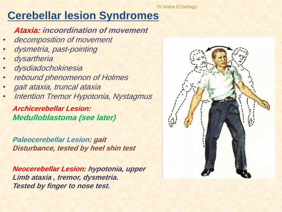
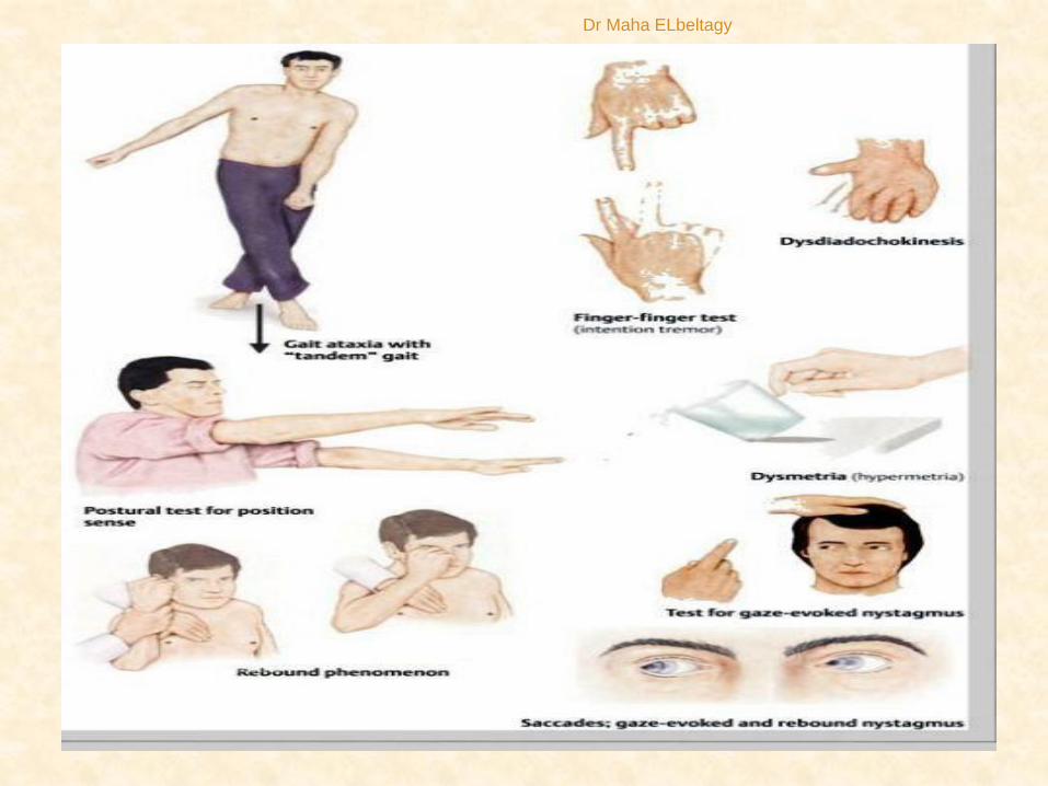
Cerebellar lesion Syndromes
Ataxia: incoordination of movement• decomposition of movement• dysmetria, past-pointing• dysartheria• dysdiadochokinesia• rebound phenomenon of Holmes• gait ataxia, truncal ataxia• Intention Tremor Hypotonia, Nystagmus
Archicerebellar Lesion:
Medulloblastoma (see later)
Paleocerebellar Lesion: gaitDisturbance, tested by heel shin test
Neocerebellar Lesion: hypotonia, upper Limb ataxia , tremor, dysmetria. Tested by finger to nose test.
Dr Maha ELbeltagy
Dr Maha ELbeltagy

The child in this picture:
- would not try to stand
unsupported•would not let go of the bed rail if
she was stood on the floor.
Cerebellar tumors on vermis•Truncal Ataxia
•Frequent Falling
Cerebellar Medulloblastoma
Dr Maha ELbeltagy
THANK YOU
Dr Maha ELbeltagy