Systemic Inflammation, Nutritional Status and TumorImmune Microenvironment Determine Outcome ofResected Non-Small Cell Lung CancerMarco Alifano1,2*, Audrey Mansuet-Lupo2,3,4,5, Filippo Lococo6, Nicolas Roche2,7, Antonio Bobbio1,
Emelyne Canny1, Olivier Schussler1, Herve Dermine6, Jean-Francois Regnard1,2, Barbara Burroni3,
Jeremy Goc2,4,5, Jerome Biton2,4,5, Hanane Ouakrim2,4,5, Isabelle Cremer2,4,5, Marie-Caroline
Dieu-Nosjean2,4,5, Diane Damotte2,3,4,5
1 Deparment of Thoracic Surgery, Paris Centre University Hospitals, AP-HP, Paris, France, 2 University Paris Descartes; Paris, France, 3 Deparments of Pathology, Paris
Centre University Hospitals, AP-HP, Paris, France, 4 INSERM U1138, Cancer and Immune Escape, Cordeliers Research Center, Paris, France, 5 University Pierre and Marie
Curie, UMRS U1138, Paris, France, 6 Unit of thoracic Surgery, IRCCS-Arcispedale Santa Maria Nuova, Reggio Emilia, Italy, 7 Departments of Chest Disease, Paris Centre
University Hospitals, AP-HP, Paris, France
Abstract
Background: Hypothesizing that nutritional status, systemic inflammation and tumoral immune microenvironment play arole as determinants of lung cancer evolution, the purpose of this study was to assess their respective impact on long-termsurvival in resected non-small cell lung cancers (NSCLC).
Methods and Findings: Clinical, pathological and laboratory data of 303 patients surgically treated for NSCLC wereretrospectively analyzed. C-reactive protein (CRP) and prealbumin levels were recorded, and tumoral infiltration by CD8+lymphocytes and mature dendritic cells was assessed. We observed that factors related to nutritional status, systemicinflammation and tumoral immune microenvironment were correlated; significant correlations were also found betweenthese factors and other relevant clinical-pathological parameters. With respect to outcome, at univariate analysis we foundstatistically significant associations between survival and the following variables: Karnofsky index, American Society ofAnesthesiologists (ASA) class, CRP levels, prealbumin concentrations, extent of resection, pathologic stage, pT and pNparameters, presence of vascular emboli, and tumoral infiltration by either CD8+ lymphocytes or mature dendritic cells and,among adenocarcinoma type, tumor grade (all p,0.05). In multivariate analysis, prealbumin levels (Relative Risk (RR): 0.34[0.16–0.73], p = 0.0056), CD8+ cell count in tumor tissue (RR = 0.37 [0.16–0.83], p = 0.0162), and disease stage (RR 1.73 [1.03–2.89]; 2.99[1.07–8.37], p = 0.0374- stage I vs II vs III-IV) were independent prognostic markers. When taken together,parameters related to systemic inflammation, nutrition and tumoral immune microenvironment allowed robust prognosticdiscrimination; indeed patients with undetectable CRP, high (.285 mg/L) prealbumin levels and high (.96/mm2) CD8+ cellcount had a 5-year survival rate of 80% [60.9–91.1] as compared to 18% [7.9–35.6] in patients with an opposite pattern ofvalues. When stages I-II were considered alone, the prognostic significance of these factors was even more pronounced.
Conclusions: Our data show that nutrition, systemic inflammation and tumoral immune contexture are prognosticdeterminants that, taken together, may predict outcome.
Citation: Alifano M, Mansuet-Lupo A, Lococo F, Roche N, Bobbio A, et al. (2014) Systemic Inflammation, Nutritional Status and Tumor Immune MicroenvironmentDetermine Outcome of Resected Non-Small Cell Lung Cancer. PLoS ONE 9(9): e106914. doi:10.1371/journal.pone.0106914
Editor: Prasad S. Adusumilli, Memorial Sloan-Kettering Cancer Center, United States of America
Received May 16, 2014; Accepted August 2, 2014; Published September 19, 2014
Copyright: � 2014 Alifano et al. This is an open-access article distributed under the terms of the Creative Commons Attribution License, which permitsunrestricted use, distribution, and reproduction in any medium, provided the original author and source are credited.
Data Availability: The authors confirm that all data underlying the findings are fully available without restriction. All relevant data are within the paper and itsSupporting Information files.
Funding: This work was supported by 1. Institut National de la Sante et de la Recherche Medicale (INSERM), Universite Paris-Descartes, Universite Pierre et MarieCurie, Labex Immunooncology (11LAXE62_9UMRS872 FRIDMAN). Authors who received the funding: DD, IC, MCDN www.sorbonne-paris-cite.fr. 2. Fondation ARCpour la Recherche sur le Cancer (SL220110603483). Author who received the funding: IC www.fondation-arc.org. The funders had no role in study design, datacollection and analysis, decision to publish, or preparation of the manuscript.
Competing Interests: The authors have declared that no competing interests exist.
* Email: [email protected]
Introduction
Lung carcinoma is a leading cause of cancer-related death
worldwide [1–2]. Despite progresses in chemotherapy and
biologically-targeted therapy and refinement in multimodal
therapeutic combinations, long-term outcome remains poor, with
the exception of stage IA disease, stressing the need for research to
better understand the biology of the disease and factors
conditioning long-term survival and risk of relapse [3–6].
The interactions between systemic inflammation and tumoral
immune microenvironment are increasingly investigated in cancer
patients [7–9]. Pro-inflammatory cytokines and associated growth
PLOS ONE | www.plosone.org 1 September 2014 | Volume 9 | Issue 9 | e106914

factors are involved in carcinogenesis through their effects on
tumor cell growth, survival, proliferation and migration [10]. It
has been shown that slight elevations of inflammatory markers are
associated with an increased risk of non-small cell lung carcinoma
(NSCLC) occurrence [11–12], and serum C-reactive protein
(CRP) has been identified as a prognostic factor in both advanced
and resectable NSCLC [13,14]. The tumoral immune microen-
vironment has been also shown to be an important determinant of
long-term outcome in primary and metastatic tumors [9]:
particularly in NSCLC, high levels of mature dendritic cells
(mDC) and of CD8+ lymphocytes have been both identified as
robust prognostic factors [15–17].
Lung cancer is frequently associated with chronic bronchitis and
chronic obstructive pulmonary disease (COPD) [1]. COPD is also
associated with systemic inflammation and shares several risk
factors and pathophysiological mechanisms with NSCLC, includ-
ing airways inflammation, protease/antiprotease imbalance, oxi-
dative stress and abnormal repair mechanisms [18]. In COPD,
markers of systemic inflammation are related to survival and
nutritional status [18]. Nutritional status is also a determinant of
survival in lung cancer patients, as illustrated by the association
between prognosis and low albumin levels [14,19] or low body
mass index (BMI) [19]. Low pre-albumin levels have also been
found to be associated to early recurrence and poorer short-term
outcome in resected NSCLC [14,20].
Nowadays, no study have been performed to investigate the
respective impact of systemic inflammation, nutritional status, and
immune microenvironment on the survival of patients with
resected NSCLC and to assess the interactions between these
factors and the outcome. The present study was designed to
address these issues in a large series of patients with resected
NSCLC.
Patients and Methods
Clinical and pathological data of 303 consecutive patients who
underwent major lung resection for NSCLC at Hotel-Dieu
University Hospital in Paris between June 2001 and December
2002 were retrospectively analysed. The research was conducted
according to recommendations outlined in the Helsinki declara-
tion. IRB approval was obtained (Comite de Protection des
Personnes [CPP] Ile de France II, nu 2008-133 and 2012 06-12).
IRB dispensed from obtaining informed consent, because of the
retrospective, non-interventional character of the study and the
high number of deceased patients when the study was performed.
Patient records were anonymized and de-identified prior to
analysis.
Inclusion/Exclusion CriteriaPatients were not included if fever. 38uC, purulent sputum,
antibiotic treatment, or lung atelectasis were present in the four
weeks before surgery. A standard staging protocol was adopted.
Patients who undergone neoadjuvant treatment (chemotherapy
and or radiotherapy) were not included in the present analysis
(confounder factor) in order to limit the heterogeneity of the
population. Similarly, we have not included in the present study
those patients surgically treated with not-anatomical resections
(‘‘wedge resections’’). Therefore, the surgical procedures consisted
of anatomical lung resection (lobectomy or pneumonectomy) with
radical nodal dissection in all cases. Finally, adjuvant radiotherapy
or chemotherapy was proposed on an individual basis following
evidence-based discussions under the care of referring physicians.
Collected dataPatient’s characteristics, treatment procedures, and short-term
outcomes were prospectively collected using a standardized case
report form [21]. In particular, serum CRP, albumin and pre-
albumin levels were measured in laboratories by nephelometry (as
a part of routine pre-surgical laboratory examinations during the
study period). Medians were used as cut-off values for statistical
analysis. Age, sex, BMI, tobacco consumption, lung function,
Karnofsky index were also recorded. Collected pathologic data
included histologic type, tumor stage (7th TNM edition [22]) and
presence of vascular or lymphatic tumoral emboli. Data on long-
term survival were obtained retrospectively through direct
telephonic contact with the patient or family. When no clinical
follow-up was available, information on vital status was obtained
through the municipality of birth of the patient.
Pathological reviewA centralized pathological review of the samples was performed
by two expert pathologists. All adenocarcinoma cases have been
reclassified independently by two expert pathologists (DD and
AML) according to the IASLC/ATS/ERS classification [23]
based on predominant architectural pattern. Therefore, adeno-
carcinomas were graded into three prognostic groups as previously
described [24] and the tumour stages were modified in accordance
to the 7th edition of the TNM classification [22].
The intra-tumoral density of CD8+ T cells and mature
dendritics cells have been analysed in all specimen using
immunohistochemistry staining. We have selected the paraffin-
embedded tumor block containing the highest density of immune
cells and performed immunostaining with the following antibodies
CD3 (A0452, Dako cytomation), DC-Lamp (1010E1.01, Dendri-
tics), CD8+ (SP16, Springbioscience) and epithelial antibody
(AE1/AE3, Dako cytomation). In order to better identify mature
DCs and CD8 T cells within CD3+ T lymphocytes rich area, and
tumoral epithelial nests we performed double double labeling
CD3/DC-Lamp and CD8/AE1/AE3. Cells were enumerated in
the whole section (original magnification 6100), with Calopix
software (Tribvn) and results were expressed as an absolute
number of positive cells/mm2, as previously described [17].
Quantification was reviewed by two independent observers (JG,
MCDN).
CD8+ T cells were found both in stromal area and tumor cell
nest. All associations were studied with both values and we found
significant associations with both CD8+ T cells location. Because
of the potential difficulties in reporting and understanding
overlapping data, we decided to report in the paper only the
results of immune cells infiltrating tumor cells nests.
Data analysisData processing and analysis were performed with the statistical
Software SEM (SILEX Developpment, Mireffleurs, France).
Results are expressed as percentage, mean +/2SD for normally
distributed and median [interquartile range] for non-normally
distributed quantitative variables.
The first step of analyses was the assessment of factors associated
with nutritional status (prealbumin level), systemic inflammation
(CRP) and tumor immune microenvironment (CD8+ cells and
mDC in tumor tissue). Correlations were assessed by the
Spearman rank test for continuous variables. Mann-Whitney
and Kruskal-Wallis tests were used to perform group comparisons
as appropriate. For CD8+ and DC-LAMP+ densities, the
‘‘minimum p-value approach’’ was used to determine the best
separation of Kaplan-Meier curves referring to the outcome, with
Outcome of Resected NSCLC
PLOS ONE | www.plosone.org 2 September 2014 | Volume 9 | Issue 9 | e106914

Table 1. Clinical, surgical and pathological characteristics and parameters related to nutrition, systemic inflammation and tumoralimmune microenvironment in the whole population.
n (%) or mean ± SD or median [interquartile range]
Men 244 (80.5%)
Age 62 yrs [53–69 yrs]
Smoking history
Past or present smoking 264 (87.1%)
Smoking cessation at least 2 months before surgery 170 (56.1%)
Cumulative smoking: Pack/Year index 40 [30–50]
Comorbid illnesses
Alcohol abuse 70 (23.1%)
Diabetes mellitus 35 (11.6%)
Ischaemic heart disease 41 (13.5%)
Stroke 16 (5.3%)
Lower limb atheroma 60 (19.8%)
Respiratory status
Chronic bronchitis 181 (59.7%)
FEV1 (% predicted) 80.8618.7
FEV1/FVC (%) 70.9611.0
ASA I-II/III/IV 13 (4.3%)/187 (61.7%)/100 (33.0%)/3 (1.0%)
Karnofsky 100%/90%/#80% 97 (32.0%)/110 (36.3%)/96 (31.7%)
Surgical procedures
Lobectomy/bilobectomy 235 (77.6%)
Pneumonectomy 68 (22.4%)
Histological type
Squamous cell carcinoma 118 (38.9%)
Adenocarcinoma 137 (45.2%)
Large-cell carcinoma 41 (13.5%)
Others* 7 (2.32%)
Pathological stage
IA 60 (19.8%)
IB 54 (17.8%)
IIA 47 (15.5%)
IIB 38 (12.5%)
IIIA 88 (29.0%)
IIIB 9 (3.0%)
IV 7 (2.3%)
Vascular emboli 134 (44.2%)
Lymphatic emboli 75 (24.8)%
Body mass index 24.2+4.4 Kg/m2
Usual body weight 70 (62–81) Kg
Current body weight 69 (60–80) Kg
CRP 3 (3–17) mg/L
Albumin 45 (40–49) g/L
Prealbumin 285 (220–346) mg/L
Nutritional risk index 107 (99–113)
CD8+ T lymphocytes density 96/mm2 (39.7–210.3)
Mature dendritic cells density 1.42/mm2 (0.57–3.34)
*these including sarcomatoid carcinomas and adenosquamous carcinomas.doi:10.1371/journal.pone.0106914.t001
Outcome of Resected NSCLC
PLOS ONE | www.plosone.org 3 September 2014 | Volume 9 | Issue 9 | e106914
the following cut-offs: CD8: 96 cells/mm2 and DC-Lamp: 1.42
cells/mm2.
Multivariate analyses (i.e., multilinear regression, including
factors significantly associated at univariate analysis) were used
to identify factors independently associated with the biomarkers of
interest.
Survival analyses were then carried out by Kaplan-Meier
method and univariate comparisons were performed using log-
rank tests. Risk factors associated with outcomes in univariate
analysis with a p-value ,0.05 were entered into a multivariate
step-by-step Cox model analysis, to identify independent predic-
tors of survival. A p-value ,0.05 was considered significant.
Results
Three-hundred and three patients treated by lobectomy/
bilobectomy or pneumonectomy for NSCLC in the study period
were analysed. Table 1 summarizes the main preoperative
characteristics.
Factors associated with nutritional statusPrealbumin levels were significantly correlated with other
parameters related to nutritional status (body weight, BMI,
albumin levels, nutritional risk index), systemic inflammation
(inverse relationship with CRP levels) and with several histological
features, such as histological type (lower in squamous cell
carcinoma), pT parameter, and presence of vascular emboli.
Prealbumin levels also correlated positively to mDC density. At
multivariate analysis, prealbumin levels were found to be
independently associated with CRP levels only (Table 2).
Factors associated with systemic inflammationIn univariate analysis, CRP levels were correlated to smoking
status, extent of resection (higher in pneumonectomy patients), p-
stage and pT-parameter, and inversely correlated with albumin
levels and nutritional risk index. CRP levels were also inversely
correlated with prealbumin levels and mDC density, and these last
two associations could be characterized as independent by
multilinear regression (Table 2).
Table 2. Associations between the biomarkers related to systemic inflammation, nutrition and tumoral immunemicroenvironment with clinical and pathological variables.
Prealbumin CRP CD8 mDC
Age 0.835 NS 0.0062 0.0061
Sex NS NS NS 0.0011
Usual body weight 0.0231 NS 0.0825 0.0055
Actual body weight 0.0001 NS NS 0.0374
BMI 0.0002 NS NS 0.0294
Smoking status NS 0.0055 NS 0.0087
Pack/year NS NS 0.0725 0.0813
Alcohol abuse NS NS NS NS
Chronic bronchitis NS NS NS 0.0621
COPD NS NS NS 0.0272
Atheroma NS NS NS NS
Diabetes 0.0333 NS NS 0.0581
Angor NS NS NS 0.0991
Stroke NS 0.0432 NS 0.0432
Karnofsky NS NS NS NS
ASA 0.0153 0.063 NS 0.0004
Extent resection NS 0.0003 0.0081 0.0947
Pathologic stage NS 0.0006 NS NS
pT 0.0011 #0.0001 NS 0.0551
pN NS NS NS NS
Histological type 0.0497 0.0572 0.0414 0.0008
Vascular emboli 0.0192 0.0773 NS NS
Lymphatic emboli NS NS NS NS
Albumin #0.0001 #0.0001 NS ,0.0001
Nutritional risk index #0.0001 #0.0001 NS 0.0123
Prealbumin NA #0.0001 NS 0.0003
CRP #0.0001 NA NS #0.0001
CD8 NS NS NA #0.0001
mDC 0.0003 #0.0001 0.0001 NA
P values (univariate analyses) are displayed when ,0.1. Significant p values are shown in bold. Independent associations with biomarkers (identified using multivariateanalysis) are underlined. Corresponding p-values on multivariate analysis are shown in footnote.doi:10.1371/journal.pone.0106914.t002
Outcome of Resected NSCLC
PLOS ONE | www.plosone.org 4 September 2014 | Volume 9 | Issue 9 | e106914
Factors associated with tumor immunemicroenvironment
The density of CD8+ cells and mature DC is heterogenous
among lung carcinoma (Figure 1). The density of CD8+ infiltrat-
ing T lymphocytes was found to be correlated with histologic type
(lower in squamous cell carcinoma) and extent of resection (lower
in pneumonectomy patients) at univariate analysis, as well as with
age and density of infiltrating mDC; the association with these last
two factors remained significant at multivariate analysis. More-
over, the density of intra-tumoral mDC infiltrating cells was
correlated with the main clinical, pathological and laboratory
parameters, including age (inverse correlation), sex (higher in
women), smoking status (lower in smokers), COPD status (lower in
COPD patients), ASA score (lower in higher classes), histology
(lower in squamous cell carcinoma), nutritional parameters (body
weight, BMI, prealbumin and albumin levels, nutritional risk
index), CRP levels (negative correlation), as well as with CD8+ T
cells density. Multilinear regression revealed independent associ-
ations with presence of COPD, histological type, CRP levels, and
CD8+ T cells density (Table 2).
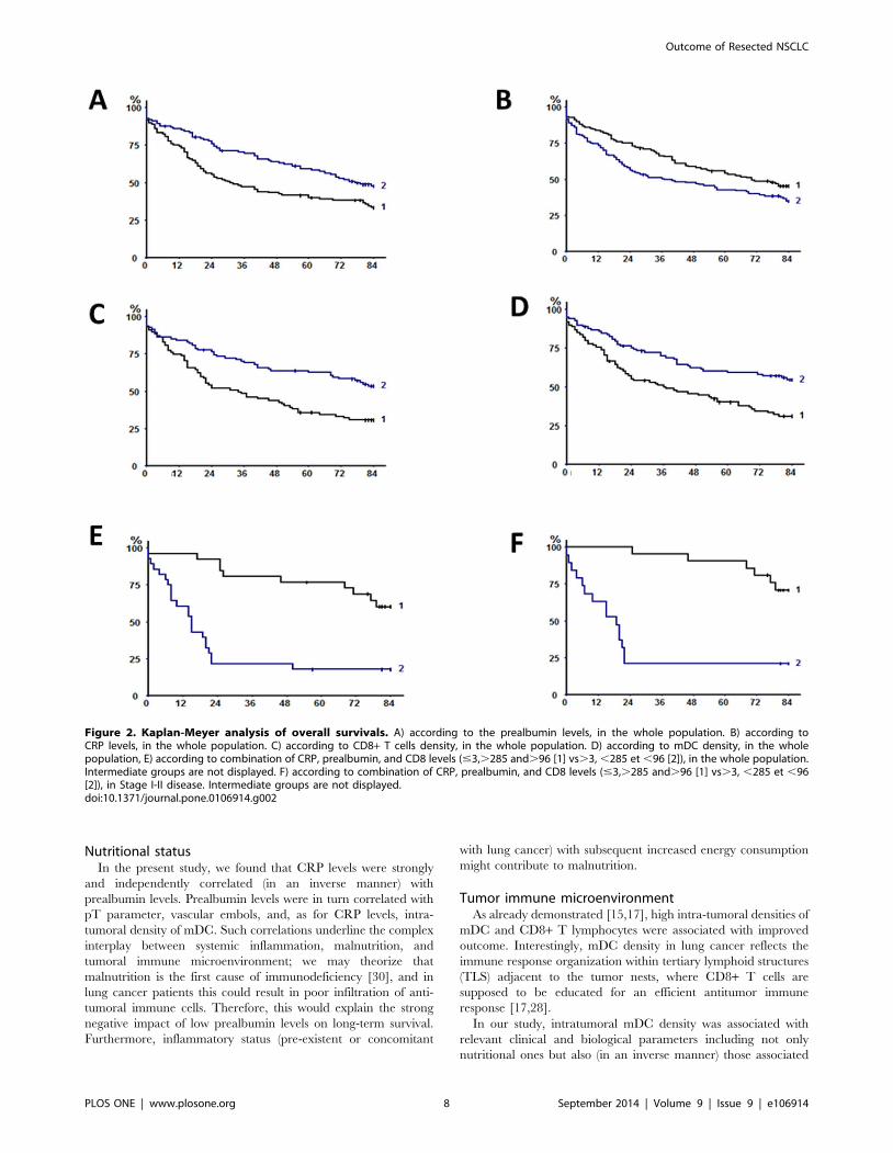
Long-term survival and prognostic factorsFor the whole population, median survival was 52 months; 3, 5,
and 7-year overall survivals (OS) were 56.1% (95% C.I: 51.0–
62.3), 47.0% (41.3–52.7), and 38.6% (33.0–44.4), respectively.
Clinical and pathological factors associated with long-term survival
in univariate analysis are shown in Table 3. Of note, chronic
bronchitis, Karnofsky index, ASA class, extent of resection, p-
stage, pT, and pN parameters as well as the presence of vascular
emboli negatively affected survival. IASLC/ATS/ERS classifica-
tion based on histological grade for adenocarcinoma [23] was
found to be an independent prognostic factor. Intermediate grade
- lepidic, tubular and papillary predominant adenocarcinoma- had
a better prognosis than high grade group - solid predominant,
micropapillary predominant and mucinous adenocarcinoma-
(p = 0.0521).
Table 4 summarizes univariate associations between survival
and biomarkers of nutritional status, systemic inflammation and
tumoral immune microenvironment. In particular, prealbumin
levels may predict the long-term outcome (p = 0.0087; 5-year
survival rates of 39.9% and 58.3% in patients with levels below
and superior to median value, respectively, Figure 2A). In
agreement with our previous report [14], systemic inflammation
also influenced the long-term survival, with undetectable (#3 mg/
L) CRP levels being associated to more favorable outcome
(p = 0.0372; 5-year survival rate of 54.1% vs 42.6%, Figure 2B).
Similarly, the tumoral immune microenvironment was proven to
be a strong predictor of long-term outcome; indeed, high densities
(superior to median value) of either CD8+ T lymphocytes or mDC
were found to be related with a significantly better prognosis (5-
year survival rates of 35.7% and 63.3% and of 40.2% and 59.2%
in patients with low and high density of CD8+ T cells, and of
mDC, respectively, p = 0.0312 and p = 0.0015, Figures 2C-D).
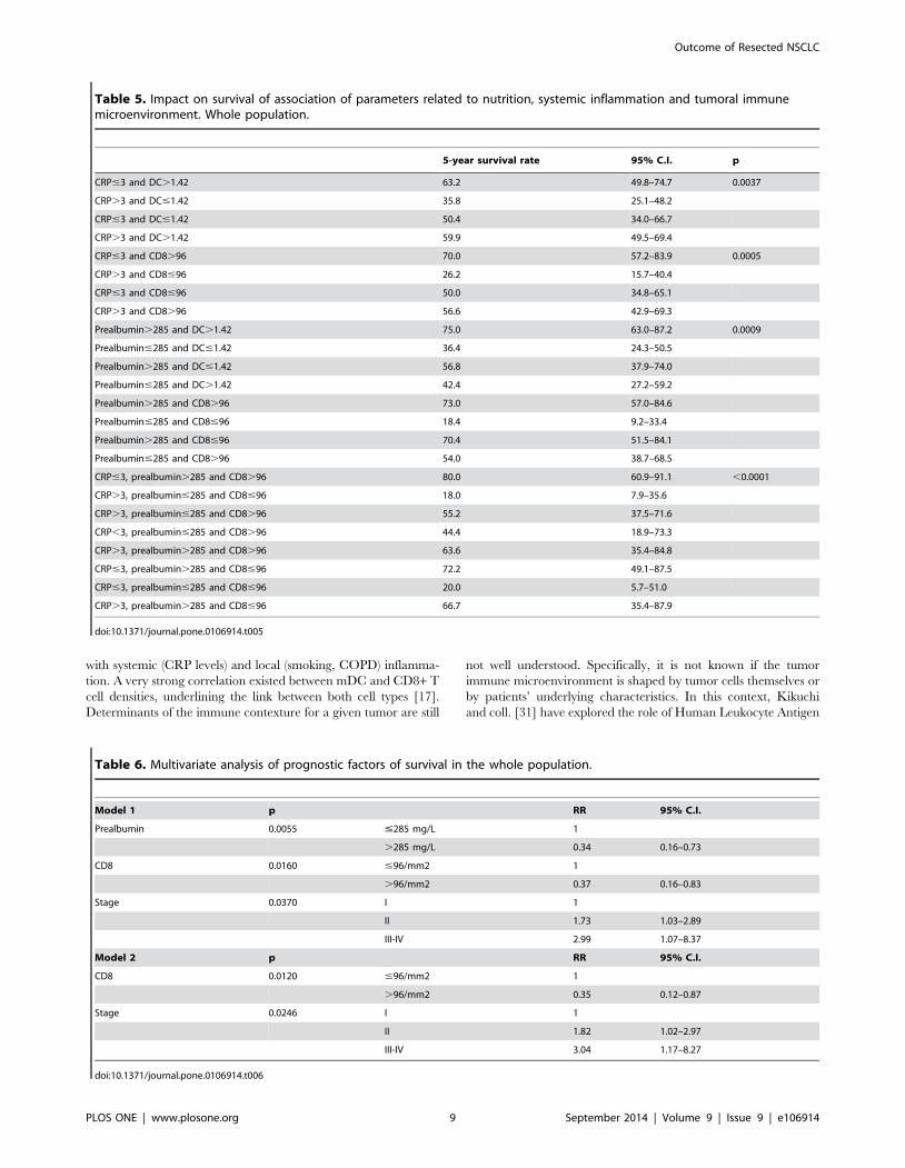
We defined subgroups of patients based on biomarkers levels of
systemic inflammation, nutritional status and tumoral immune
microenvironment. The resulting categorization was highly
discriminant in terms of survival (p,0.0001): patients with the
following ‘‘pattern’’ (CRP#3 mg/L, prealbumin.285 mg/L
and.96/mm2 CD8+ cell density) had a better 5-year survival
rate (80% versus 18%, respectively; figure 2E) when compared
with those patients with an opposite pattern (CRP levels.3 mg/L,
prealbumin level#285 mg/L and CD8+ cell density #96/mm2).
Patients in the other subgroups showed intermediate prognosis
(Table 5).
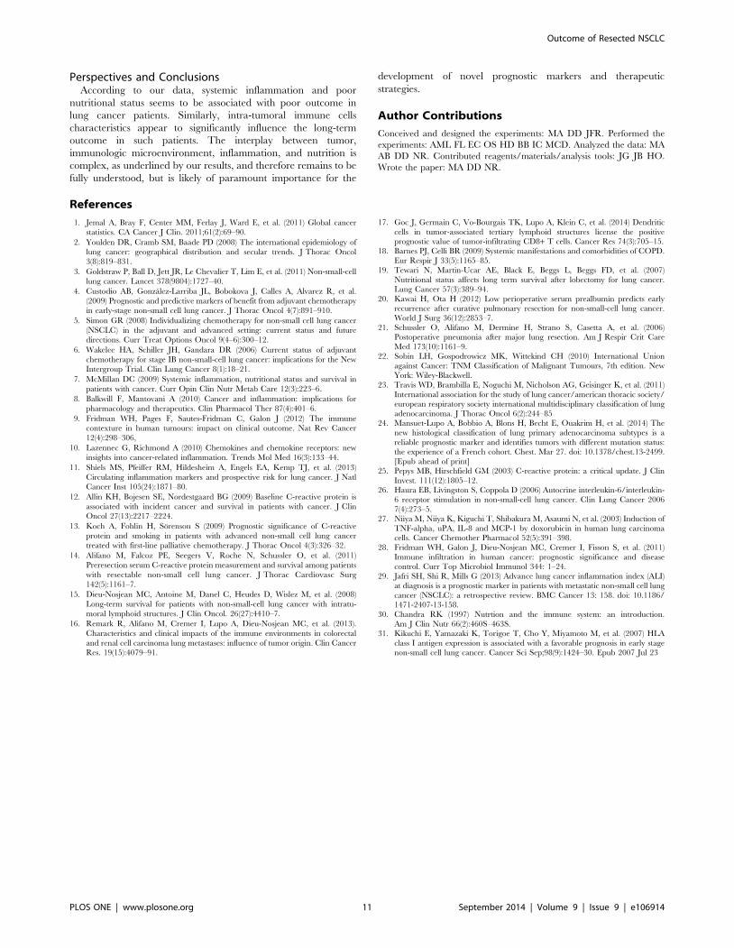
Cox models were then built. Since significant correlations were
found between CRP and prealbumin levels on the one hand, and
CD8+ T cells and mDC densities on the other hand, these
variables were mutually exclusive. Thus, the first model included
chronic bronchitis, Karnowski PS, ASA class, extent of resection,
stage of disease, presence of vascular emboli, prealbumin levels
and CD8+ T cells density. Prealbumin levels, CD8+ T cells
density, and disease stage were finally identified as independent
prognostic markers (Table 6). The second model, in which CRP
Figure 1. CD8+ and DC-Lamp+ cell densities are heterogeneous in non-small cell carcinoma. Magnification x 100 A) High density of DC-Lamp+ cells (red), these cells are located in CD3+ T-cell rich area (blue). B) Low density of DC-Lamp+ cells. C) High density of CD8+ T cells (red) amongpan-cytokeratins+ tumor nests (blue). D) Low density of CD8+ T cells.doi:10.1371/journal.pone.0106914.g001
Outcome of Resected NSCLC
PLOS ONE | www.plosone.org 5 September 2014 | Volume 9 | Issue 9 | e106914
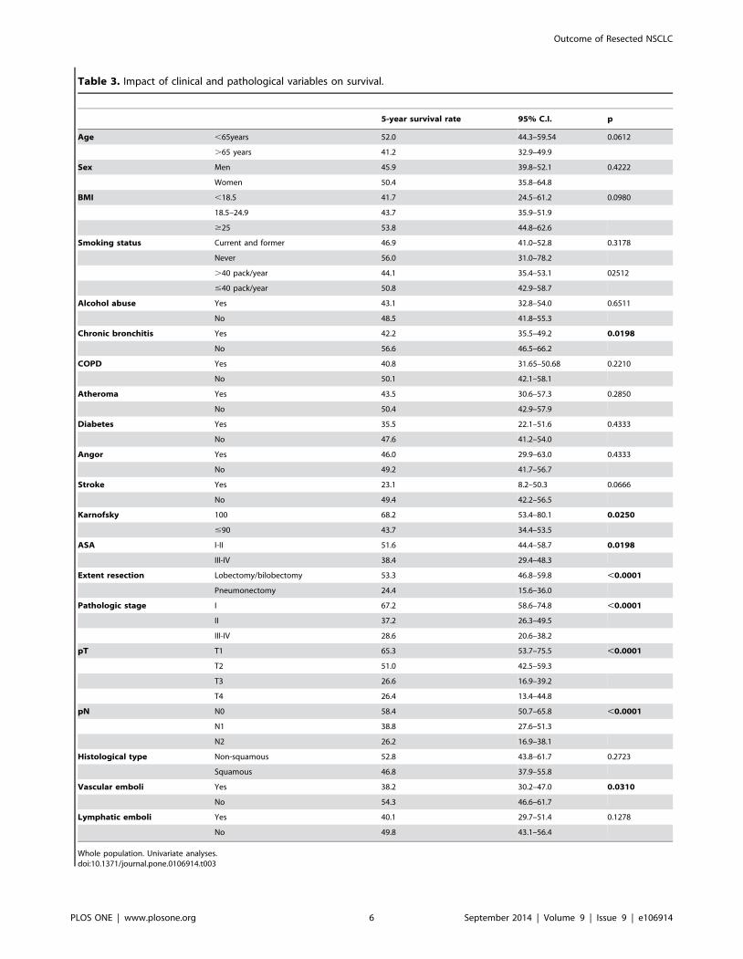
Table 3. Impact of clinical and pathological variables on survival.
5-year survival rate 95% C.I. p
Age ,65years 52.0 44.3–59.54 0.0612
.65 years 41.2 32.9–49.9
Sex Men 45.9 39.8–52.1 0.4222
Women 50.4 35.8–64.8
BMI ,18.5 41.7 24.5–61.2 0.0980
18.5–24.9 43.7 35.9–51.9
$25 53.8 44.8–62.6
Smoking status Current and former 46.9 41.0–52.8 0.3178
Never 56.0 31.0–78.2
.40 pack/year 44.1 35.4–53.1 02512
#40 pack/year 50.8 42.9–58.7
Alcohol abuse Yes 43.1 32.8–54.0 0.6511
No 48.5 41.8–55.3
Chronic bronchitis Yes 42.2 35.5–49.2 0.0198
No 56.6 46.5–66.2
COPD Yes 40.8 31.65–50.68 0.2210
No 50.1 42.1–58.1
Atheroma Yes 43.5 30.6–57.3 0.2850
No 50.4 42.9–57.9
Diabetes Yes 35.5 22.1–51.6 0.4333
No 47.6 41.2–54.0
Angor Yes 46.0 29.9–63.0 0.4333
No 49.2 41.7–56.7
Stroke Yes 23.1 8.2–50.3 0.0666
No 49.4 42.2–56.5
Karnofsky 100 68.2 53.4–80.1 0.0250
#90 43.7 34.4–53.5
ASA I-II 51.6 44.4–58.7 0.0198
III-IV 38.4 29.4–48.3
Extent resection Lobectomy/bilobectomy 53.3 46.8–59.8 ,0.0001
Pneumonectomy 24.4 15.6–36.0
Pathologic stage I 67.2 58.6–74.8 ,0.0001
II 37.2 26.3–49.5
III-IV 28.6 20.6–38.2
pT T1 65.3 53.7–75.5 ,0.0001
T2 51.0 42.5–59.3
T3 26.6 16.9–39.2
T4 26.4 13.4–44.8
pN N0 58.4 50.7–65.8 ,0.0001
N1 38.8 27.6–51.3
N2 26.2 16.9–38.1
Histological type Non-squamous 52.8 43.8–61.7 0.2723
Squamous 46.8 37.9–55.8
Vascular emboli Yes 38.2 30.2–47.0 0.0310
No 54.3 46.6–61.7
Lymphatic emboli Yes 40.1 29.7–51.4 0.1278
No 49.8 43.1–56.4
Whole population. Univariate analyses.doi:10.1371/journal.pone.0106914.t003
Outcome of Resected NSCLC
PLOS ONE | www.plosone.org 6 September 2014 | Volume 9 | Issue 9 | e106914

levels replaced prealbumin levels, identified stage and CD8+ T
cells density as independent prognostic factors.
When stage I-II disease were considered alone, the prognostic
impact of factors related to poor nutrition, systemic inflammation
and tumoral immune cell infiltration was even more remarkable
(Table 7); in detail, the prealbumin and CRP levels, as well as
intra-tumoral CD8+ T cells and mDC density strongly predicted
the long-term outcome (p = 0.0008, p = 0.0049, p = 0.0004, and
p = 0.0050, respectively). Taken together, their prognostic value
was very high, allowing a powerful separation of patients groups:
5-year survival rate was 86.4% in patients with CRP#3 mg/L,
prealbumin levels.285 mg/L, and CD8+ T cells density.96/
mm2 versus 21.1% in patients with CRP.3 mg/L, prealbumin
levels#285 mg/L, and CD8+ T cells density #96/mm2 (Fig-
ure 2F). As for the whole population, two Cox models were
generated for stage I-II disease, one including prealbumin levels
and CD8+ T cells density together with the other significant
clinical and pathological variables, and the other including CRP
levels instead of prealbumine levels. In both models (Table 8),
CD8+ T cells density and either prealbumin or CRP levels
(depending on which of these two variables was entered in the
statistical model) could be identified as independent prognostic
factors.
Discussion
In this large cohort of consecutive patients it was found that, in
resected NSCLC patients, biomarkers related to systemic inflam-
mation, nutritional status, and tumoral immune microenviron-
ment are interrelated and these represent major determinants of
long-term outcome that, when taken into account together, allow
highly robust discrimination of groups of patients with different
prognosis.
Role of systemic inflammationCRP is secreted by hepatocytes following stimulation by
circulating pro-inflammatory cytokines, in particular IL-1, TNF-
a, and mainly IL-6 [25]. Experimental studies [26–28] have
suggested that NSCLC cells are able to release IL-6 and TNF-a.
In spite of this, the exact role of systemic inflammation and tumor
burden in determining progression and outcome is still contro-
versial. This relationship is even more questionable in ‘‘pre-clinical
disease’’: a study on a large cohort showed that increased CRP
levels in cancer-free subjects were associated with a higher risk of
lung cancer occurrence [12]. This finding has been recently
confirmed by a nested case-control study [11]: among 77
evaluated inflammatory biomarkers, 11 were found to be
associated with an increased risk of developing lung cancer, even
after adjustment for smoking. Among these 11 markers, CRP was
the most robust predictor of lung cancer risk. [11]. Moreover,
increased baseline CRP levels were associated with early death
after diagnosis of any cancer in patients without metastatic disease
at diagnosis [12]. These findings strongly suggest a possible role of
pre-existing systemic inflammation in determining the occurrence
and prognosis of lung cancer. In our population, CRP levels were
an independent prognostic factor in stage I-II disease only. Higher
CRP levels were associated to higher pT parameter and (albeit
non-significant) higher occurrence of vascular emboli, an impor-
tant determinant of cancer progression and spread. Similarly,
systemic inflammation has been reported to be an independent
negative prognostic marker in patients with advanced non-small
cell lung cancer [29].
Table 4. Impact of parameters related to nutrition, systemic inflammation and tumoral immune microenvironment on survival.
5-year survival rate 95% C.I. p
BMI ,18.5 41.7 24.5–61.2 0.0980
18.5–24.9 43.7 35.9–51.9
$25 53.8 44.8–62.6
Usual body weight #70 45.4 37.1–54.0 0.2810
.70 51.0 42.9–59.0
Actual body weight #69 41.9 34.2–50.0 0.0564
.69 52.8 44.5–61.2
Albumin .45 g/L 54.5 44.9–63.8 0.0420
#45 g/L 44.3 36.9–52.0
Buzby’s risk index #83.5 28.6 11.7–54.6 0.2610
83.6–97.5 54.5 37.2–70.8
.97.5 51.0 42.8–59.1
Prealbumin .285 mg/L 58.3 49.0–67.1 0.0087
#285 mg/L 39.9 31.7–48.6
CRP .3 mg/L 42.6 34.8–50.8 0.0370
#3 mg/L 54.1 45.6–62.3
CD8 #96/mm2 35.7 26.4–46.2 0.0028
.96/mm2 63.3 53.1–72.4
mDC #1.42/mm2 40.2 31.0–50.2 0.0015
.1.42/mm2 59.2 50.3–69.6
Whole population. Univariate analysis.doi:10.1371/journal.pone.0106914.t004
Outcome of Resected NSCLC
PLOS ONE | www.plosone.org 7 September 2014 | Volume 9 | Issue 9 | e106914
Nutritional statusIn the present study, we found that CRP levels were strongly
and independently correlated (in an inverse manner) with
prealbumin levels. Prealbumin levels were in turn correlated with
pT parameter, vascular embols, and, as for CRP levels, intra-
tumoral density of mDC. Such correlations underline the complex
interplay between systemic inflammation, malnutrition, and
tumoral immune microenvironment; we may theorize that
malnutrition is the first cause of immunodeficiency [30], and in
lung cancer patients this could result in poor infiltration of anti-
tumoral immune cells. Therefore, this would explain the strong
negative impact of low prealbumin levels on long-term survival.
Furthermore, inflammatory status (pre-existent or concomitant
with lung cancer) with subsequent increased energy consumption
might contribute to malnutrition.
Tumor immune microenvironmentAs already demonstrated [15,17], high intra-tumoral densities of
mDC and CD8+ T lymphocytes were associated with improved
outcome. Interestingly, mDC density in lung cancer reflects the
immune response organization within tertiary lymphoid structures
(TLS) adjacent to the tumor nests, where CD8+ T cells are
supposed to be educated for an efficient antitumor immune
response [17,28].
In our study, intratumoral mDC density was associated with
relevant clinical and biological parameters including not only
nutritional ones but also (in an inverse manner) those associated
Figure 2. Kaplan-Meyer analysis of overall survivals. A) according to the prealbumin levels, in the whole population. B) according toCRP levels, in the whole population. C) according to CD8+ T cells density, in the whole population. D) according to mDC density, in the wholepopulation, E) according to combination of CRP, prealbumin, and CD8 levels (#3,.285 and.96 [1] vs.3, ,285 et ,96 [2]), in the whole population.Intermediate groups are not displayed. F) according to combination of CRP, prealbumin, and CD8 levels (#3,.285 and.96 [1] vs.3, ,285 et ,96[2]), in Stage I-II disease. Intermediate groups are not displayed.doi:10.1371/journal.pone.0106914.g002
Outcome of Resected NSCLC
PLOS ONE | www.plosone.org 8 September 2014 | Volume 9 | Issue 9 | e106914
with systemic (CRP levels) and local (smoking, COPD) inflamma-
tion. A very strong correlation existed between mDC and CD8+ T
cell densities, underlining the link between both cell types [17].
Determinants of the immune contexture for a given tumor are still
not well understood. Specifically, it is not known if the tumor
immune microenvironment is shaped by tumor cells themselves or
by patients’ underlying characteristics. In this context, Kikuchi
and coll. [31] have explored the role of Human Leukocyte Antigen
Table 5. Impact on survival of association of parameters related to nutrition, systemic inflammation and tumoral immunemicroenvironment. Whole population.
5-year survival rate 95% C.I. p
CRP#3 and DC.1.42 63.2 49.8–74.7 0.0037
CRP.3 and DC#1.42 35.8 25.1–48.2
CRP#3 and DC#1.42 50.4 34.0–66.7
CRP.3 and DC.1.42 59.9 49.5–69.4
CRP#3 and CD8.96 70.0 57.2–83.9 0.0005
CRP.3 and CD8#96 26.2 15.7–40.4
CRP#3 and CD8#96 50.0 34.8–65.1
CRP.3 and CD8.96 56.6 42.9–69.3
Prealbumin.285 and DC.1.42 75.0 63.0–87.2 0.0009
Prealbumin#285 and DC#1.42 36.4 24.3–50.5
Prealbumin.285 and DC#1.42 56.8 37.9–74.0
Prealbumin#285 and DC.1.42 42.4 27.2–59.2
Prealbumin.285 and CD8.96 73.0 57.0–84.6
Prealbumin#285 and CD8#96 18.4 9.2–33.4
Prealbumin.285 and CD8#96 70.4 51.5–84.1
Prealbumin#285 and CD8.96 54.0 38.7–68.5
CRP#3, prealbumin.285 and CD8.96 80.0 60.9–91.1 ,0.0001
CRP.3, prealbumin#285 and CD8#96 18.0 7.9–35.6
CRP.3, prealbumin#285 and CD8.96 55.2 37.5–71.6
CRP,3, prealbumin#285 and CD8.96 44.4 18.9–73.3
CRP.3, prealbumin.285 and CD8.96 63.6 35.4–84.8
CRP#3, prealbumin.285 and CD8#96 72.2 49.1–87.5
CRP#3, prealbumin#285 and CD8#96 20.0 5.7–51.0
CRP.3, prealbumin.285 and CD8#96 66.7 35.4–87.9
doi:10.1371/journal.pone.0106914.t005
Table 6. Multivariate analysis of prognostic factors of survival in the whole population.
Model 1 p RR 95% C.I.
Prealbumin 0.0055 #285 mg/L 1
.285 mg/L 0.34 0.16–0.73
CD8 0.0160 #96/mm2 1
.96/mm2 0.37 0.16–0.83
Stage 0.0370 I 1
II 1.73 1.03–2.89
III-IV 2.99 1.07–8.37
Model 2 p RR 95% C.I.
CD8 0.0120 #96/mm2 1
.96/mm2 0.35 0.12–0.87
Stage 0.0246 I 1
II 1.82 1.02–2.97
III-IV 3.04 1.17–8.27
doi:10.1371/journal.pone.0106914.t006
Outcome of Resected NSCLC
PLOS ONE | www.plosone.org 9 September 2014 | Volume 9 | Issue 9 | e106914

(HLA) class I that displays a repertoire of endogenously processed
peptides to CD8 (+) T lymphocytes. The authors observed that the
down-regulation of HLA class I expression in NSCLC is a marker
of poor prognosis, and this may play a critical role in immune
surveillance of patients with NSCLC.
Interestingly we have observed for the first time to our
knowledge, that tumor immune microenvironment is linked to
nutritional status and systemic inflammation as reflected by the
correlation between mDC density and several conditions and
clinical features (such as stroke, COPD, usual body weight, CRP
and prealbumin levels, etc.). Overall, our results suggest that
preexisting systemic inflammation/poor nutritional status could
impact the intra-tumoral immune contexture and the patient
survival.
Role of biomarkers as predictors of survivalWhen associating mDC or CD8+ T cell densities with either
CRP or prealbumin levels, we could identify subgroups of patients
with significantly different long-term outcomes.
The best discrimination was achieved when taking into account
simultaneously biomarkers related to inflammation with nutrition-
al status and intra-tumoral immune infiltration. With this model,
the differences in survival were remarkable when comparing, in
the whole population as in stage I-II disease, patients with high
CD8+ T cells density, low CRP levels and high prealbumin levels
to those with low CD8+ T cells density, high CRP levels and low
prealbumin levels. Interestingly, groups with intermediate biolog-
ical characteristics had intermediate long-term outcomes.
Limitations of the studyOur study suffers from the common bias of investigations on
surgical registries as well as the retrospective nature and a certain
degree of heterogeneity of the sample (uncontrolled cohort of
multiple stages and therapies). Missing data led us to exclude some
potentially eligible patients, however this limitation concerned
only a minority of patients, and is therefore unlikely to have
affected the results. Such limitations should be kept firmly in mind
by the readers when considering the clinical implications deriving
from the present analysis.
Table 7. Impact of parameters related to nutrition, systemic inflammation and tumoral immune microenvironment on survival inpatients with stage I-II disease.
5-year survival rate 95% C.I. p
Prealbumin.285 mg/L 70.0 59.0–79.1 0.0008
Prealbumin#285 mg/L 44.4 33.9–55.3
CRP#3 mg/L 64.8 54.9–73.6 0.0049
CRP.3 mg/L 48.4 38.1–58.7
CD8.96/mm2 71.6 59.9–81.0 0.0004
CD8#96/mm2 42.4 30.6–55.1
mDC.1.42/mm2 68.9 56.8–78.9 0.0050
mDC#1.42/mm2 48.6 36.7–60.5
CRP#3, prealbumin.285 and CD8.96 86.4 66.7–95.2 0.0063
CRP.3, prealbumin #285 and CD8#96 21.1 8.5–43.3
CRP#3, prealbumin.285 and DC.1.42 85.3 67.7–94.1 0.0004
CRP.3, prealbumin#285 and DC#1.42 44.4 27.6–63.0
Univariate analysis.doi:10.1371/journal.pone.0106914.t007
Table 8. Multivariate analysis of prognostic factors of survival in the stage I-II disease.
Model 1 p RR 95% C.I.
Prealbumin 0.0210 #285 mg/L 1
.285 mg/L 0.41 0.23–0.73
CD8 0.0162 #96/mm2 1
.96/mm2 0.41 0.23–0.71
Model 2 p RR 95% C.I.
CRP 0.0240 #3 1
.3 1.78 1.08–2.93
CD8 0.0041 #96/mm2 1
.96/mm2 0.49 0.30–0.80
Stage 0.0377 I 1
II 1.87 1.12–3.12
doi:10.1371/journal.pone.0106914.t008
Outcome of Resected NSCLC
PLOS ONE | www.plosone.org 10 September 2014 | Volume 9 | Issue 9 | e106914
Perspectives and ConclusionsAccording to our data, systemic inflammation and poor
nutritional status seems to be associated with poor outcome in
lung cancer patients. Similarly, intra-tumoral immune cells
characteristics appear to significantly influence the long-term
outcome in such patients. The interplay between tumor,
immunologic microenvironment, inflammation, and nutrition is
complex, as underlined by our results, and therefore remains to be
fully understood, but is likely of paramount importance for the
development of novel prognostic markers and therapeutic
strategies.
Author Contributions
Conceived and designed the experiments: MA DD JFR. Performed the
experiments: AML FL EC OS HD BB IC MCD. Analyzed the data: MA
AB DD NR. Contributed reagents/materials/analysis tools: JG JB HO.
Wrote the paper: MA DD NR.
References
1. Jemal A, Bray F, Center MM, Ferlay J, Ward E, et al. (2011) Global cancer
statistics. CA Cancer J Clin. 2011;61(2):69–90.
2. Youlden DR, Cramb SM, Baade PD (2008) The international epidemiology of
lung cancer: geographical distribution and secular trends. J Thorac Oncol
3(8):819–831.
3. Goldstraw P, Ball D, Jett JR, Le Chevalier T, Lim E, et al. (2011) Non-small-cell
lung cancer. Lancet 378(9804):1727–40.
4. Custodio AB, Gonzalez-Larriba JL, Bobokova J, Calles A, Alvarez R, et al.
(2009) Prognostic and predictive markers of benefit from adjuvant chemotherapy
in early-stage non-small cell lung cancer. J Thorac Oncol 4(7):891–910.
5. Simon GR (2008) Individualizing chemotherapy for non-small cell lung cancer
(NSCLC) in the adjuvant and advanced setting: current status and future
directions. Curr Treat Options Oncol 9(4–6):300–12.
6. Wakelee HA, Schiller JH, Gandara DR (2006) Current status of adjuvant
chemotherapy for stage IB non-small-cell lung cancer: implications for the New
Intergroup Trial. Clin Lung Cancer 8(1):18–21.
7. McMillan DC (2009) Systemic inflammation, nutritional status and survival in
patients with cancer. Curr Opin Clin Nutr Metab Care 12(3):223–6.
8. Balkwill F, Mantovani A (2010) Cancer and inflammation: implications for
pharmacology and therapeutics. Clin Pharmacol Ther 87(4):401–6.
9. Fridman WH, Pages F, Sautes-Fridman C, Galon J (2012) The immune
contexture in human tumours: impact on clinical outcome. Nat Rev Cancer
12(4):298–306,
10. Lazennec G, Richmond A (2010) Chemokines and chemokine receptors: new
insights into cancer-related inflammation. Trends Mol Med 16(3):133–44.
11. Shiels MS, Pfeiffer RM, Hildesheim A, Engels EA, Kemp TJ, et al. (2013)
Circulating inflammation markers and prospective risk for lung cancer. J Natl
Cancer Inst 105(24):1871–80.
12. Allin KH, Bojesen SE, Nordestgaard BG (2009) Baseline C-reactive protein is
associated with incident cancer and survival in patients with cancer. J Clin
Oncol 27(13):2217–2224.
13. Koch A, Fohlin H, Sorenson S (2009) Prognostic significance of C-reactive
protein and smoking in patients with advanced non-small cell lung cancer
treated with first-line palliative chemotherapy. J Thorac Oncol 4(3):326–32.
14. Alifano M, Falcoz PE, Seegers V, Roche N, Schussler O, et al. (2011)
Preresection serum C-reactive protein measurement and survival among patients
with resectable non-small cell lung cancer. J Thorac Cardiovasc Surg
142(5):1161–7.
15. Dieu-Nosjean MC, Antoine M, Danel C, Heudes D, Wislez M, et al. (2008)
Long-term survival for patients with non-small-cell lung cancer with intratu-
moral lymphoid structures. J Clin Oncol. 26(27):4410–7.
16. Remark R, Alifano M, Cremer I, Lupo A, Dieu-Nosjean MC, et al. (2013).
Characteristics and clinical impacts of the immune environments in colorectal
and renal cell carcinoma lung metastases: influence of tumor origin. Clin Cancer
Res. 19(15):4079–91.
17. Goc J, Germain C, Vo-Bourgais TK, Lupo A, Klein C, et al. (2014) Dendritic
cells in tumor-associated tertiary lymphoid structures license the positiveprognostic value of tumor-infiltrating CD8+ T cells. Cancer Res 74(3):705–15.
18. Barnes PJ, Celli BR (2009) Systemic manifestations and comorbidities of COPD.Eur Respir J 33(5):1165–85.
19. Tewari N, Martin-Ucar AE, Black E, Beggs L, Beggs FD, et al. (2007)
Nutritional status affects long term survival after lobectomy for lung cancer.Lung Cancer 57(3):389–94.
20. Kawai H, Ota H (2012) Low perioperative serum prealbumin predicts earlyrecurrence after curative pulmonary resection for non-small-cell lung cancer.
World J Surg 36(12):2853–7.
21. Schussler O, Alifano M, Dermine H, Strano S, Casetta A, et al. (2006)Postoperative pneumonia after major lung resection. Am J Respir Crit Care
Med 173(10):1161–9.22. Sobin LH, Gospodrowicz MK, Wittekind CH (2010) International Union
against Cancer: TNM Classification of Malignant Tumours, 7th edition. NewYork: Wiley-Blackwell.
23. Travis WD, Brambilla E, Noguchi M, Nicholson AG, Geisinger K, et al. (2011)
International association for the study of lung cancer/american thoracic society/european respiratory society international multidisciplinary classification of lung
adenocarcinoma. J Thorac Oncol 6(2):244–8524. Mansuet-Lupo A, Bobbio A, Blons H, Becht E, Ouakrim H, et al. (2014) The
new histological classification of lung primary adenocarcinoma subtypes is a
reliable prognostic marker and identifies tumors with different mutation status:the experience of a French cohort. Chest. Mar 27. doi: 10.1378/chest.13-2499.
[Epub ahead of print]25. Pepys MB, Hirschfield GM (2003) C-reactive protein: a critical update. J Clin
Invest. 111(12):1805–12.26. Haura EB, Livingston S, Coppola D (2006) Autocrine interleukin-6/interleukin-
6 receptor stimulation in non-small-cell lung cancer. Clin Lung Cancer 2006
7(4):273–5.27. Niiya M, Niiya K, Kiguchi T, Shibakura M, Asaumi N, et al. (2003) Induction of
TNF-alpha, uPA, IL-8 and MCP-1 by doxorubicin in human lung carcinomacells. Cancer Chemother Pharmacol 52(5):391–398.
28. Fridman WH, Galon J, Dieu-Nosjean MC, Cremer I, Fisson S, et al. (2011)
Immune infiltration in human cancer: prognostic significance and diseasecontrol. Curr Top Microbiol Immunol 344: 1–24.
29. Jafri SH, Shi R, Mills G (2013) Advance lung cancer inflammation index (ALI)at diagnosis is a prognostic marker in patients with metastatic non-small cell lung
cancer (NSCLC): a retrospective review. BMC Cancer 13: 158. doi: 10.1186/1471-2407-13-158.
30. Chandra RK (1997) Nutrtion and the immune system: an introduction.
Am J Clin Nutr 66(2):460S–463S.31. Kikuchi E, Yamazaki K, Torigoe T, Cho Y, Miyamoto M, et al. (2007) HLA
class I antigen expression is associated with a favorable prognosis in early stagenon-small cell lung cancer. Cancer Sci Sep;98(9):1424–30. Epub 2007 Jul 23
Outcome of Resected NSCLC
PLOS ONE | www.plosone.org 11 September 2014 | Volume 9 | Issue 9 | e106914