“fnhum-07-00764” — 2013/11/8 — 20:29 — page 1 — #1
ORIGINAL RESEARCH ARTICLEpublished: xx November 2013
doi: 10.3389/fnhum.2013.00764
Situating emotional experienceChristine D. Wilson-Mendenhall 1*, Lisa Feldman Barrett1† and Lawrence W. Barsalou 2 †
1 Department of Psychology, Northeastern University, Boston, MA, USA2 Department of Psychology, Emory University, Atlanta, GA, USA
Edited by:
Martin Klasen, Rheinisch-WestfälischeTechnische Hochschule AachenUniversity, Germany
Reviewed by:
Ruben Gur, University of PennsylvaniaSchool of Medicine, USACindy Hagan, University ofCambridge, UK
*Correspondence:
Christine D. Wilson-Mendenhall,Department of Psychology,Northeastern University, 125Nightingale Hall, Boston, MA 02115,USAe-mail: [email protected]
†Lisa Feldman Barrett and LawrenceW. Barsalou have joint seniorauthorship.
Psychological construction approaches to emotion suggest that emotional experienceis situated and dynamic. Fear, for example, is typically studied in a physical dangercontext (e.g., threatening snake), but in the real world, it often occurs in social contexts,especially those involving social evaluation (e.g., public speaking). Understanding situatedemotional experience is critical because adaptive responding is guided by situationalcontext (e.g., inferring the intention of another in a social evaluation situation vs. monitoringthe environment in a physical danger situation). In an fMRI study, we assessed situatedemotional experience using a newly developed paradigm in which participants vividlyimagine different scenarios from a first-person perspective, in this case scenarios involvingeither social evaluation or physical danger. We hypothesized that distributed neural patternswould underlie immersion in social evaluation and physical danger situations, with sharedactivity patterns across both situations in multiple sensory modalities and in circuitryinvolved in integrating salient sensory information, and with unique activity patterns for eachsituation type in coordinated large-scale networks that reflect situated responding. Morespecifically, we predicted that networks underlying the social inference and mentalizinginvolved in responding to a social threat (in regions that make up the “default mode”network) would be reliably more active during social evaluation situations. In contrast,networks underlying the visuospatial attention and action planning involved in responding toa physical threat would be reliably more active during physical danger situations.The resultssupported these hypotheses. In line with emerging psychological construction approaches,the findings suggest that coordinated brain networks offer a systematic way to interpret thedistributed patterns that underlie the diverse situational contexts characterizing emotionallife.
Keywords: emotion, situated cognition, affective neuroscience, affect, cognitive neuroscience
INTRODUCTIONDarwin’s The Expression of the Emotions in Man and Animals isoften used to motivate emotion research that focuses on identi-fying the biological signatures for five or so emotion categories(Ekman, 2009; Hess and Thibault, 2009). Interestingly, though,the evolution paradigm shift initiated by Darwin and other scien-tists heavily emphasized variability: species are biopopulations inwhich individuals within a population are unique and in whichindividual variation within a species is meaningfully tied to varia-tion in the environment (and they are not physical types definedby essential features; Barrett, 2013). In other words, an individ-ual organism is best understood by the situational context inwhich it operates. It is not a great leap, then, to hypothesizethat “situatedness” is also a basic principle by which the humanmind operates, during emotions and during many other mentalphenomena (Barrett, 2013).
Situated approaches to the mind typically view the brain asa coordinated system designed to use information captured dur-ing prior situations (and stored in memory) to flexibly interpretand infer what is happening in the current situation – dynam-ically shaping moment-to-moment responding in the form ofperceiving, coordinating action, regulating the body, and orga-nizing thoughts (Glenberg, 1997; Barsalou, 2003, 2009; Aydede
and Robbins, 2009; Mesquita et al., 2010; Barrett, 2013). “Cog-nitive” research domains (e.g., episodic and semantic memory,visual object recognition, language comprehension) are increas-ingly adopting a situated view of the mind (for empirical reviews,see Zwaan and Radvansky, 1998; Barsalou, 2003; Bar, 2004; Yehand Barsalou, 2006; Mesquita et al., 2010). In contrast, emo-tion research largely remains entrenched in a “stimulus-response”reflexive approach to brain function, which typically views thebrain as reacting to the demands of the environment, often ina simple, stereotyped way (cf. Raichle, 2010). Traditional “basic”emotion views often assume that an event (i.e., a stimulus) triggersone of several stereotyped responses in the brain and body that canbe classified as either fear, disgust, anger, sadness, happiness, etc.(for a review of basic emotion models, see Tracy and Randles,2011). Decades of research have revealed substantial variabilityin the neural, physiological, and behavioral patterns associatedwith these emotion categories (cf. Barrett, 2006; Lindquist et al.,2012). Whereas basic emotion approaches now focus on try-ing to identify primitive “core” (and often narrowly defined)instances of these emotions, alternative theoretical approachesto emotion, such as psychological construction, propose tak-ing a situated approach to explaining the variability that existsin the experiences people refer to using words like fear, disgust,
Frontiers in Human Neuroscience www.frontiersin.org November 2013 | Volume 7 | Article 764 | 1
001
002
003
004
005
006
007
008
009
010
011
012
013
014
015
016
017
018
019
020
021
022
023
024
025
026
027
028
029
030
031
032
033
034
035
036
037
038
039
040
041
042
043
044
045
046
047
048
049
050
051
052
053
054
055
056
057
058
059
060
061
062
063
064
065
066
067
068
069
070
071
072
073
074
075
076
077
078
079
080
081
082
083
084
085
086
087
088
089
090
091
092
093
094
095
096
097
098
099
100
101
102
103
104
105
106
107
108
109
110
111
112
113
114
“fnhum-07-00764” — 2013/11/8 — 20:29 — page 2 — #2
Wilson-Mendenhall et al. Situating emotional experience
anger, sadness, happiness (and using many other emotion terms;Barrett, 2009b, 2013).
In the psychological construction view that we have developed,emotions are not fundamentally different from other kinds ofbrain states (Barrett, 2009a, 2012; Wilson-Mendenhall et al., 2011).During emotional experiences and during other kinds of experi-ences, the brain is using prior experience to dynamically interpretongoing neural activity, which guides an individual’s respondingin the situation. We refer to this process, which often occurs with-out awareness (i.e., it is a fundamental process for making senseof one’s relation to the world at any given moment), as situatedconceptualization. The term situated takes on a broad meaningin our view, referring to the distributed neural activity across themodal systems of the brain involved in constructing situations, notjust to perception of the external environment or to what mightbe considered the background. More specifically, situated neuralactivity reflects the dynamic actions that individuals engage in,and the events, internal bodily sensations, and mentalizing thatthey experience, as well as the perceptions of the external environ-mental setting and the physical entities and individuals it contains(Wilson-Mendenhall et al., 2011).
Emotions, like other classes of mental experiences, operatein this situation-specific way because rich, cross-modal knowl-edge is critical for interpreting, inferring, and responding whensimilar situations occur in the future. On this view, situationalknowledge develops for emotion categories like fear, anger, etc.,as it does for other abstract categories of experiences (e.g., sit-uations that involve the abstract categories gossip, modesty, orambition). Experiences categorized as fear, for example, can occurwhen delivering a speech to a respected audience or when losingcontrol while driving a car. A situated, psychological construc-tion perspective suggests that it is more adaptive to responddifferently in these situations, guided by knowledge of the situ-ation, than to respond in a stereotyped way. Whereas respondingin the social speech situation involves inferring what audiencemembers are thinking, responding in the physical car situationinvolves rapid action and attention to the environment. Stereo-typed responding in the form of preparing the body to flee or fightdoes not address the immediate threat present in either of thesesituations. A psychological construction approach highlights theimportance of studying the situations commonly categorized asemotions like fear or anger, not because these situations merelydescribe emotions, but because emotions would not exist withoutthem.
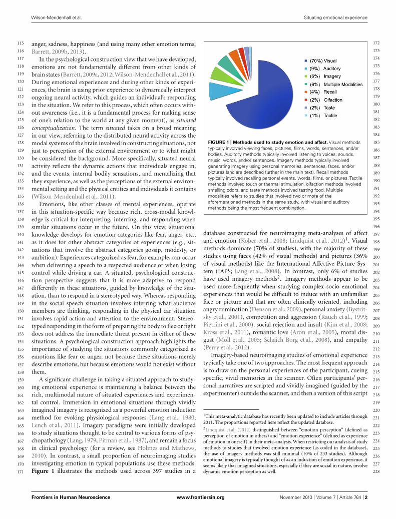
A significant challenge in taking a situated approach to study-ing emotional experience is maintaining a balance between therich, multimodal nature of situated experiences and experimen-tal control. Immersion in emotional situations through vividlyimagined imagery is recognized as a powerful emotion inductionmethod for evoking physiological responses (Lang et al., 1980;Lench et al., 2011). Imagery paradigms were initially developedto study situations thought to be central to various forms of psy-chopathology (Lang, 1979; Pitman et al., 1987), and remain a focusin clinical psychology (for a review, see Holmes and Mathews,2010). In contrast, a small proportion of neuroimaging studiesinvestigating emotion in typical populations use these methods.Figure 1 illustrates the methods used across 397 studies in a
FIGURE 1 | Methods used to study emotion and affect. Visual methodstypically involved viewing faces, pictures, films, words, sentences, and/orbodies. Auditory methods typically involved listening to voices, sounds,music, words, and/or sentences. Imagery methods typically involvedgenerating imagery using personal memories, sentences, faces, and/orpictures (and are described further in the main text). Recall methodstypically involved recalling personal events, words, films, or pictures. Tactilemethods involved touch or thermal stimulation, olfaction methods involvedsmelling odors, and taste methods involved tasting food. Multiplemodalities refers to studies that involved two or more of theaforementioned methods in the same study, with visual and auditorymethods being the most frequent combination.
database constructed for neuroimaging meta-analyses of affectand emotion (Kober et al., 2008; Lindquist et al., 2012)1. Visualmethods dominate (70% of studies), with the majority of thesestudies using faces (42% of visual methods) and pictures (36%of visual methods) like the International Affective Picture Sys-tem (IAPS; Lang et al., 2008). In contrast, only 6% of studieshave used imagery methods2. Imagery methods appear to beused more frequently when studying complex socio-emotionalexperiences that would be difficult to induce with an unfamiliarface or picture and that are often clinically oriented, includingangry rumination (Denson et al., 2009), personal anxiety (Bystrit-sky et al., 2001), competition and aggression (Rauch et al., 1999;Pietrini et al., 2000), social rejection and insult (Kim et al., 2008;Kross et al., 2011), romantic love (Aron et al., 2005), moral dis-gust (Moll et al., 2005; Schaich Borg et al., 2008), and empathy(Perry et al., 2012).
Imagery-based neuroimaging studies of emotional experiencetypically take one of two approaches. The most frequent approachis to draw on the personal experiences of the participant, cueingspecific, vivid memories in the scanner. Often participants’ per-sonal narratives are scripted and vividly imagined (guided by theexperimenter) outside the scanner, and then a version of this script
1This meta-analytic database has recently been updated to include articles through2011. The proportions reported here reflect the updated database.2Lindquist et al. (2012) distinguished between “emotion perception” (defined asperception of emotion in others) and “emotion experience” (defined as experienceof emotion in oneself) in their meta-analysis. When restricting our analysis of studymethods to studies that involved emotion experience (as coded in the database),the use of imagery methods was still minimal (10% of 233 studies). Althoughemotional imagery is typically thought of as an induction of emotion experience, itseems likely that imagined situations, especially if they are social in nature, involvedynamic emotion perception as well.
Frontiers in Human Neuroscience www.frontiersin.org November 2013 | Volume 7 | Article 764 | 2
115
116
117
118
119
120
121
122
123
124
125
126
127
128
129
130
131
132
133
134
135
136
137
138
139
140
141
142
143
144
145
146
147
148
149
150
151
152
153
154
155
156
157
158
159
160
161
162
163
164
165
166
167
168
169
170
171
172
173
174
175
176
177
178
179
180
181
182
183
184
185
186
187
188
189
190
191
192
193
194
195
196
197
198
199
200
201
202
203
204
205
206
207
208
209
210
211
212
213
214
215
216
217
218
219
220
221
222
223
224
225
226
227
228
“fnhum-07-00764” — 2013/11/8 — 20:29 — page 3 — #3
Wilson-Mendenhall et al. Situating emotional experience
is used to induce these memory-based emotional experiences dur-ing neuroimaging (e.g., Bystritsky et al., 2001; Marci et al., 2007;Gillihan et al., 2010). Less often, a specific visual stimulus is potentenough to easily evoke personal, emotional imagery in the scan-ner (e.g., face of a romantic partner; Aron et al., 2005; Kross et al.,2011). The second approach is to present standard prompts (e.g.,a sentence) that participants use to generate imagery underly-ing emotional experiences (e.g., Colibazzi et al., 2010; Costa et al.,2010). A key strength of the first approach is that emotional expe-riences are tightly tied to situated, real-life memories, whereas akey strength of the second approach is the experimental controlafforded by presenting the same prompts to all participants. Inboth cases, though, the situational context of the emotional expe-riences is typically lost, either because the situational details arespecific to the individual (and thus lost in group-level analyses)or because standard prompts are not designed to cultivate and/orsystematically manipulate the situational context of the emotionalexperience.
Building on the strengths of existing imagery-basedapproaches, we developed a neuroimaging procedure that wouldallow us to examine participants’ immersion in rich, situatedemotional experiences while maximizing experimental controland rigor. In our paradigm, participants first received training
outside the scanner on how to immerse themselves in richlydetailed, full paragraph-long versions of emotional scenarios froma first-person perspective. The scenarios reflected two ecologi-cally important situation types in which emotional experiencesare often grounded: social evaluation and physical danger. Everyscenario was constructed using written templates to induce asocial evaluation emotional experience or a physical danger emo-tional experience (see Table 1 for examples). Participants listenedto audio recordings of the scenarios, which facilitated immer-sion by allowing participants to close their eyes. In the scanner,participants were prompted with shorter, core (audio) versionsof the scenarios in the scanner, so that a statistically powerfulneuroimaging design could be implemented.
We hypothesized that immersion across both social evaluationand physical danger situations would be characterized by dis-tributed neural patterns across multiple sensory modalities andacross regions involved in detecting and integrating salient sensoryinformation. Much previous research has demonstrated neuraloverlap between sensorimotor perception/action and sensorimo-tor imagery (for a review, see Kosslyn et al., 2001). If our scenarioimmersion method induces richly situated emotional experiences,then the vivid mental imagery generated should be grounded inbrain regions underlying sensory perception and action. Perhaps
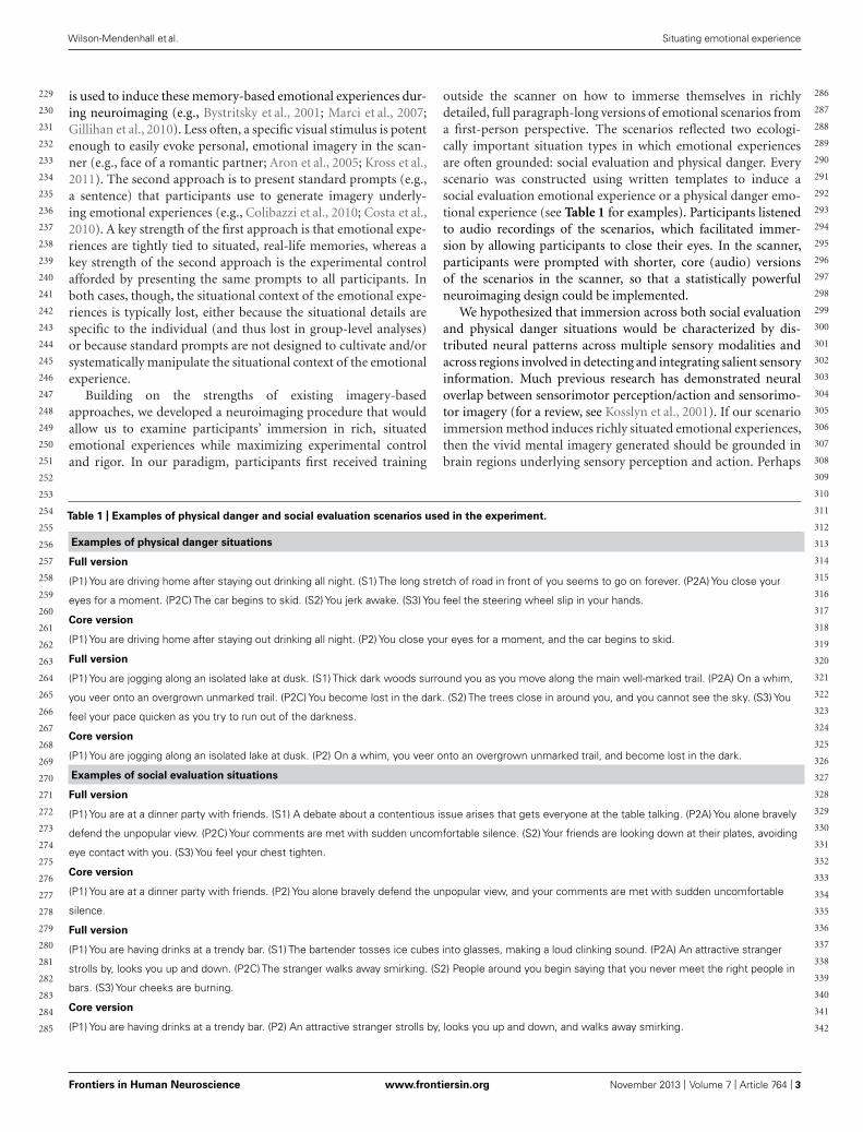
Table 1 | Examples of physical danger and social evaluation scenarios used in the experiment.
Examples of physical danger situations
Full version
(P1) You are driving home after staying out drinking all night. (S1) The long stretch of road in front of you seems to go on forever. (P2A) You close your
eyes for a moment. (P2C) The car begins to skid. (S2) You jerk awake. (S3) You feel the steering wheel slip in your hands.
Core version
(P1) You are driving home after staying out drinking all night. (P2) You close your eyes for a moment, and the car begins to skid.
Full version
(P1) You are jogging along an isolated lake at dusk. (S1) Thick dark woods surround you as you move along the main well-marked trail. (P2A) On a whim,
you veer onto an overgrown unmarked trail. (P2C) You become lost in the dark. (S2) The trees close in around you, and you cannot see the sky. (S3) You
feel your pace quicken as you try to run out of the darkness.
Core version
(P1) You are jogging along an isolated lake at dusk. (P2) On a whim, you veer onto an overgrown unmarked trail, and become lost in the dark.
Examples of social evaluation situations
Full version
(P1) You are at a dinner party with friends. (S1) A debate about a contentious issue arises that gets everyone at the table talking. (P2A) You alone bravely
defend the unpopular view. (P2C) Your comments are met with sudden uncomfortable silence. (S2) Your friends are looking down at their plates, avoiding
eye contact with you. (S3) You feel your chest tighten.
Core version
(P1) You are at a dinner party with friends. (P2) You alone bravely defend the unpopular view, and your comments are met with sudden uncomfortable
silence.
Full version
(P1) You are having drinks at a trendy bar. (S1) The bartender tosses ice cubes into glasses, making a loud clinking sound. (P2A) An attractive stranger
strolls by, looks you up and down. (P2C) The stranger walks away smirking. (S2) People around you begin saying that you never meet the right people in
bars. (S3) Your cheeks are burning.
Core version
(P1) You are having drinks at a trendy bar. (P2) An attractive stranger strolls by, looks you up and down, and walks away smirking.
Frontiers in Human Neuroscience www.frontiersin.org November 2013 | Volume 7 | Article 764 | 3
229
230
231
232
233
234
235
236
237
238
239
240
241
242
243
244
245
246
247
248
249
250
251
252
253
254
255
256
257
258
259
260
261
262
263
264
265
266
267
268
269
270
271
272
273
274
275
276
277
278
279
280
281
282
283
284
285
286
287
288
289
290
291
292
293
294
295
296
297
298
299
300
301
302
303
304
305
306
307
308
309
310
311
312
313
314
315
316
317
318
319
320
321
322
323
324
325
326
327
328
329
330
331
332
333
334
335
336
337
338
339
340
341
342
“fnhum-07-00764” — 2013/11/8 — 20:29 — page 4 — #4
Wilson-Mendenhall et al. Situating emotional experience
surprisingly, studies using imagery paradigms to investigate emo-tional experiences do not typically examine sensorimotor activity,because the goal is often to isolate a category of experience (e.g.,anger, disgust) or other “emotion” components. In contrast, ourapproach is designed to examine the distributed neural patternsthat underlie emotional experiences.
Our second, primary hypothesis was motivated by a situ-ated approach to studying the varieties of emotional experience.We hypothesized that unique activity patterns for each situa-tion type would occur in coordinated large-scale networks thatreflect situated responding. Whereas networks underlying thesocial inference and mentalizing involved in responding to a socialthreat (in regions that make up the“default mode”network) wouldbe reliably more active during social evaluation situations (forreviews of default mode network functions, see Buckner et al.,2008; Barrett and Satpute, 2013)3, networks underlying the visu-ospatial attention and action planning involved in responding toa physical threat would be reliably more active during physicaldanger situations (for reviews of attention networks, see Chunet al., 2011; Petersen and Posner, 2012; Posner, 2012). Theselarge-scale, distributed networks largely consist of heteromodalregions that engage in the multimodal integration necessary forcoordinated interpretation and responding (Sepulcre et al., 2012;Spreng et al., 2013)
As a further test of our second hypothesis, we examined whetherparticipants’ trial-by-trial ratings of immersion during the train-ing session correlated with neural activity, across social evaluationscenarios and across physical danger scenarios. If emotional expe-rience is situated, then feeling immersed in a situation should berealized by neural circuitry that underlies engaging in the spe-cific situation. Whereas immersion in social evaluation situationsshould occur when affect is grounded in mentalizing about others,immersion in physical danger situations should occur when affectis grounded in taking action in the environment.
MATERIALS AND METHODSPARTICIPANTSTwenty right-handed, native-English speakers from the Emorycommunity, ranging in age from 20 to 33 (10 female), participatedin the experiment. Six additional participants were dropped due toproblems with audio equipment (three participants) or excessivehead motion in the scanner. Participants had no history of psy-chiatric illness and were not currently taking any psychotropicmedication. They received $100 in compensation, along withanatomical images of their brain.
MATERIALSA full and core form of each scenario was constructed, the latterbeing a subset of the former (see Table 1). The full form served to
3There is substantial evidence that default mode network (DMN) regions are activeduring tasks that involve social inference and mentalizing (for reviews, see Barrettand Satpute, 2013; Buckner and Carroll, 2007; Van Overwalle and Baetens, 2009)and that the DMN is disrupted in disorders involving social deficits (for reviews, seeMenon, 2011; Whitfield-Gabrieli and Ford, 2012). Recent work has directly demon-strated that neural activity during social/mentalizing tasks occurs in the DMN as it isdefined using resting state analyses (e.g., Andrews-Hanna et al., 2010) and that rest-ing state connectivity in the DMN predicts individual differences in social processing(e.g., Yang et al., 2012).
provide a rich, detailed, and affectively compelling scenario. Thecore form served to minimize presentation time in the scanner,so that the number of necessary trials could be completed in thetime available. Each full and core scenario described an emotionalsituation from a first-person perspective, such that the participantcould immerse him- or herself in it. As described shortly, partic-ipants practiced enriching the core form of the scenario duringthe training sessions using details from the full form, so that theywould be prepared to immerse in the rich situational detail of thefull forms during the scanning session when they received the coreforms.
Both situation types were designed so the threat described couldbe experienced as any number of high arousal, negative emotionslike fear or anger (and participants’ ratings of the ease of experi-encing negative emotions in the two situation types validated thisapproach; see Wilson-Mendenhall et al., 2011 for details). In socialevaluation situations, another person put the immersed partici-pant in a socially threatening situation that involved damage tohis or her social reputation/ego. In physical danger situations,the immersed participant put him- or herself in a physicallythreatening situation that involved impending or actual bodilyharm.
Templates were used to systematically construct different sce-narios in each situation type (social evaluation and physicaldanger). Table 1 provides examples of the social evaluation andphysical danger scenarios. Each template for the full scenariosspecified a sequence of six sentences: three primary sentences (Pi)also used in the related core scenario, and three secondary sen-tences (Si) not used in the core scenario that provided additionalrelevant detail. The two sentences in each core scenario were cre-ated using P1 as the first sentence and a conjunction of P2A andP2C as the second sentence.
For the social evaluation scenarios, the template specified thefollowing six sentences in order: P1 described a setting and activityperformed by the immersed participant in the setting, along withrelevant personal attributes; S1 provided auditory detail aboutthe setting; P2A described an action (A) of the immersed par-ticipant; P2C described the consequence (C) of that action; S2
described another person’s action in response to the consequence;S3 described the participant’s resulting internal bodily experi-ence. The templates for the physical danger scenarios were similar,except that S1 provided visual detail about the setting (instead ofauditory), S2 described the participant’s action in response to theconsequence (instead of another person’s action), and S3 describedthe participant’s resulting external somatosensory experience (onthe body surface).
A broad range of real-world situations served as the content ofthe experimental situations. The physical danger scenarios weredrawn from situations that involved vehicles, pedestrians, water,eating, wildlife, fire, power tools, and theft. The social evaluationscenarios were drawn from situations that involved friends, family,neighbors, love, work, classes, public events, and service.
During the training sessions and the critical scan session,30 social evaluation scenarios and 30 physical danger scenarioswere presented. An additional three scenarios of each type wereincluded in the training sessions so participants could practice thescanner task prior to the scan session.
Frontiers in Human Neuroscience www.frontiersin.org November 2013 | Volume 7 | Article 764 | 4
343
344
345
346
347
348
349
350
351
352
353
354
355
356
357
358
359
360
361
362
363
364
365
366
367
368
369
370
371
372
373
374
375
376
377
378
379
380
381
382
383
384
385
386
387
388
389
390
391
392
393
394
395
396
397
398
399
400
401
402
403
404
405
406
407
408
409
410
411
412
413
414
415
416
417
418
419
420
421
422
423
424
425
426
427
428
429
430
431
432
433
434
435
436
437
438
439
440
441
442
443
444
445
446
447
448
449
450
451
452
453
454
455
456
“fnhum-07-00764” — 2013/11/8 — 20:29 — page 5 — #5
Wilson-Mendenhall et al. Situating emotional experience
IMAGING DESIGNThe event-related neuroimaging design involved two criticalevents: (1) immersing in an emotional scenario (either a socialevaluation or physical danger scenario) and (2) experiencing theimmersed state in one of four ways upon hearing an auditorycategorization cue (as emotional: fearful or angry, or as anotheractive state: planning or observing). We will refer to the firstevent as “immersion” and the second event as “categorization.”Because all neural patterns described here reflect activity duringthe first immersion event, we focus on this element of the design(for the categorization results and related methodological details,please see Wilson-Mendenhall et al., 2011). This design afforded aunique opportunity to examine the situations in which emotionsemerge before the emotional state was explicitly categorized. Aswill be described later, the participant could not predict whichcategorization cue would follow the scenario, so the immersionperiod reflects situated activity that is not tied to a specific emotioncategory.
In order to separate neural activity during the immersion eventsfrom neural activity during the categorization events, we imple-mented a catch trial design (Ollinger et al., 2001a,b). Participantsreceived 240 complete trials that each contained a social evalua-tion scenario or a physical danger scenario followed immediatelyby one of the four categorization cues. Participants also received120 partial “catch” trials containing only a scenario (with no sub-sequent categorization cue), which enabled separation of the firstscenario immersion event from the second categorization event.The partial trials constituted 33% of the total trials, a proportionin the recommended range for an effective catch trial design. Eachof the 30 social evaluation scenarios and the 30 physical dangerscenarios was followed once by each categorization cue, for a totalof 240 complete trials (60 scenarios followed by 4 categorizations).Each of the 60 scenarios also occurred twice as a partial trial, for atotal of 120 catch trials.
During each of 10 fMRI runs, participants received 24 completetrials and 12 partial trials. The complete and partial trials wereintermixed with no-sound baseline periods that ranged from 0 to12 s in increments of 3 s (average 4.5 s) in a pseudo-random orderoptimized by optseq2 (Greve, 2002). On a given trial, participantscould not predict whether a complete or partial trial was coming,a necessary condition for an effective catch trial design (Ollingeret al., 2001a,b). Participants also could not predict the type ofsituation or the categorization cue they would hear. Across trialsin a run, social evaluation and physical danger situations eachoccurred 18 times, and each of the 4 categorization cues (anger,fear, observe, plan) occurred 6 times, equally often with socialevaluation and physical danger scenarios. A given scenario wasnever repeated within a run.
PROCEDUREThe experiment contained two training sessions and an fMRI scansession. The first training session occurred 24–48 h before the sec-ond training session, followed immediately by the scan. During thetraining sessions, participants were encouraged to immerse them-selves in all scenarios from a first-person perspective, to imaginethe scenario in as much vivid detail as possible, and to constructmental imagery as if the scenario events were actually happening
to them. The relation of the full to the core scenarios was alsodescribed, and participants were encouraged to reinstate the fullscenario whenever they heard a core scenario.
During the first training session, participants listened over com-puter headphones to the full versions of the 66 scenarios thatthey would later receive on the practice trials and in the criticalscan 24–48 h later, with the social evaluation and physical dangerscenarios randomly intermixed. After hearing each full scenario,participants provided three judgments about familiarity and priorexperiences, prompted by questions and response scales on thescreen. After taking a break, participants listened to the 66 coreversions of the scenarios, again over computer headphones andrandomly intermixed. While listening to each core scenario, par-ticipants were instructed to reinstate the full version that theylistened to earlier, immersing themselves fully into the respectivescenario as it became enriched and developed from memory. Afterhearing each core scenario over the headphones, participants ratedthe vividness of the imagery that they experienced while immersedin the scenario. This task encouraged the participants to developrich imagery upon hearing the core version. A detailed account ofthe first training session can be found in Wilson-Mendenhall et al.(2011).
During the second training session directly before the scan,participants first listened to the 66 full scenarios to be used in thepractice and critical scans, and rated how much they were able toimmerse themselves in each scenario, again hearing the scenariosover computer headphones and in a random order. After listeningto each full scenario, the computer script presented the question,“How much did you experience ‘being there’ in the situation?” Par-ticipants responded on the computer keyboard, using a 1–7 scale,where one meant not experiencing being there in the situation atall, four meant experiencing being there a moderate amount, andseven meant experiencing being there very much, as if it was actu-ally happening to them. The full scenarios were presented againat this point to ensure that participants were reacquainted withall the details before hearing the core versions later in the scan-ner. This first phase of the second training session lasted about anhour.
Participants were then instructed on the task that they wouldperform in the scanner and performed a run of practice trials. Dur-ing the practice and during the scans, audio events were presentedand responses collected using E-prime software (Schneider et al.,2002). On each complete trial, participants were told to immersein the core version of a scenario as they listened to it, and thatthey would receive one of four words (anger, fear, observe, plan)afterward. The participant’s task was to judge how easy it was toexperience what the word described in the context of the situa-tion. The core scenario was presented auditorily at the onset of a9 s period, lasting no more than 8 s. The word was then presentedauditorily at the onset of a 3 s period, and participants respondedas soon as ready. To make their judgments, participants pressedone of three buttons on a button box for not easy, somewhateasy, and very easy. During the practice trials, participants usedan E-Prime button box to practice making responses. In the scan-ner, participants used a Current Designs fiber optic button boxdesigned for high magnetic field environments. Participants werealso told that there would be partial trials containing scenarios
Frontiers in Human Neuroscience www.frontiersin.org November 2013 | Volume 7 | Article 764 | 5
457
458
459
460
461
462
463
464
465
466
467
468
469
470
471
472
473
474
475
476
477
478
479
480
481
482
483
484
485
486
487
488
489
490
491
492
493
494
495
496
497
498
499
500
501
502
503
504
505
506
507
508
509
510
511
512
513
514
515
516
517
518
519
520
521
522
523
524
525
526
527
528
529
530
531
532
533
534
535
536
537
538
539
540
541
542
543
544
545
546
547
548
549
550
551
552
553
554
555
556
557
558
559
560
561
562
563
564
565
566
567
568
569
570
“fnhum-07-00764” — 2013/11/8 — 20:29 — page 6 — #6
Wilson-Mendenhall et al. Situating emotional experience
and no word cues, and that they were not to respond on thesetrials.
At the beginning of the practice trials, participants heard thesame short instruction that they would hear before every run in thescanner: “Please close your eyes. Listen to each scenario and expe-rience being there vividly. If a word follows, rate how easy it wasto have that experience in the situation.” Participants performed apractice run equal in length to the runs that they would performin the scanner. Following the practice run, the experimenter andthe participant walked 5 min across campus to the scanner. Oncesettled safely and comfortably in the scanner, an initial anatomicalscan was performed, followed by the 10 critical functional runs,and finally a second anatomical scan. Prior to beginning each func-tional run, participants heard the same short instruction from thepractice run over noise-muffling headphones. Participants took ashort break between each of the 8 min 3 s runs. Total time in thescanner was a little over 1.5 h.
IMAGE ACQUISITIONThe neuroimaging data were collected in the Biomedical ImagingTechnology Center at Emory University on a research-dedicated3T Siemens Trio scanner. In each functional run, 163 T2∗-weightedecho planar image volumes depicting BOLD contrast were col-lected using a Siemens 12-channel head coil and parallel imagingwith an iPAT acceleration factor of 2. Each volume was col-lected using a scan sequence that had the following parameters:56 contiguous 2 mm slices in the axial plane, interleaved sliceacquisition, TR = 3000 ms, TE = 30 ms, flip angle = 90◦,bandwidth = 2442 Hz/Px, FOV = 220 mm, matrix = 64,voxel size = 3.44 mm × 3.44 mm × 2 mm. This scanningsequence was selected after testing a variety of sequences for sus-ceptibility artifacts in orbitofrontal cortex, amygdala, and thetemporal poles. We selected this sequence not only because itminimized susceptibility artifacts by using thin slices and par-allel imaging, but also because using 3.44 mm in the X–Ydimensions yielded a voxel volume large enough to produce asatisfactory temporal signal-to-noise ratio. In each of the twoanatomical runs, 176 T1-weighted volumes were collected usinga high resolution MPRAGE scan sequence that had the followingparameters: 192 contiguous slices in the sagittal plane, single-shot acquisition, TR = 2300 ms, TE = 4 ms, flip angle = 8◦,FOV = 256 mm, matrix = 256, bandwidth = 130 Hz/Px, voxelsize = 1 mm × 1 mm × 1 mm.
IMAGE PREPROCESSING AND ANALYSISImage preprocessing and statistical analysis were conducted inAFNI (Cox, 1996). The first anatomical scan was registered tothe second, and the average of the two scans computed to cre-ate a single high-quality anatomical scan. Initial preprocessingof the functional data included slice time correction and motioncorrection in which all volumes were registered spatially to a vol-ume within the last functional run. A volume in the last run wasselected as the registration base because it was collected closest intime to the second anatomical scan, which facilitated later align-ment of the functional and anatomical data. The functional datawere then smoothed using an isotropic 6 mm full-width half-maximum Gaussian kernel. Voxels outside the brain were removed
from further analysis at this point, as were high-variability low-intensity voxels likely to be shifting in and out of the brain due tominor head motion. Finally, the signal intensities in each volumewere divided by the mean signal value for the respective run andmultiplied by 100 to produce percent signal change from the runmean. All later analyses were performed on these percent signalchange data.
The averaged anatomical scan was corrected for non-uniformity in image intensity, skull-stripped, and then alignedwith the functional data. The resulting aligned anatomical datasetwas warped to Talairach space using an automated procedureemploying the TT_N27 template (also known as the Colin brain,an averaged dataset from one person scanned 27 times).
Regression analyses were performed on each individual’s pre-processed functional data using a canonical, fixed-shape Gammafunction to model the hemodynamic response. In the first regres-sion analysis, betas were estimated using the event onsets for10 conditions: 2 situation immersion conditions (social, physi-cal) and 8 categorization conditions that resulted from crossingthe situation with the categorization cue (social-anger, physical-anger, social-fear, physical-fear, social-observe, physical-observe,social-plan, physical-plan). Again, we only present results for thetwo situation immersion conditions here (see Wilson-Mendenhallet al., 2011 for the categorization results). The two situationimmersion conditions were modeled by creating regressors thatincluded scenario immersion events from both the complete tri-als and the partial trials. Including scenario immersion eventsfrom both trial types in one regressor made it possible to mathe-matically separate the situation immersion conditions from thesubsequent categorization conditions (Ollinger et al., 2001a,b).Because scenario immersion events were 9 s in duration, theGamma function was convolved with a boxcar function forthe entire duration to model the situation immersion condi-tions. Six regressors obtained from volume registration duringpreprocessing were also included to remove any residual sig-nal changes correlated with movement (translation in the X, Y,and Z planes; rotation around the X, Y, and Z axes). Scan-ner drift was removed by finding the best-fitting polynomialfunction correlated with time in the preprocessed time coursedata.
At the group level, the betas resulting from the each individual’sregression analysis were then entered into a second-level, random-effects ANOVA. Two key analyses were computed at this level ofanalysis using a voxel-wise threshold of p < 0.005 in conjunctionwith the 41-voxel extent threshold determined by AFNI ClustSimto produce an overall corrected threshold of p < 0.05. In the firstanalysis (that assessed our first hypothesis), we extracted clustersthat were more active during immersion in social evaluation situ-ations than in the no-sound baseline and clusters that were moreactive during immersion in physical danger situations than in theno-sound baseline (using the voxel-wise and extent thresholdsspecified above). We then entered the results of these two con-trasts (social evaluation > baseline; physical danger > baseline)into a conjunction analysis to determine clusters shared by the twosituation types (i.e., overlapping regions of activity). In the sec-ond analysis (that assessed our second hypothesis), we computeda standard contrast to directly compare immersion during social
Frontiers in Human Neuroscience www.frontiersin.org November 2013 | Volume 7 | Article 764 | 6
571
572
573
574
575
576
577
578
579
580
581
582
583
584
585
586
587
588
589
590
591
592
593
594
595
596
597
598
599
600
601
602
603
604
605
606
607
608
609
610
611
612
613
614
615
616
617
618
619
620
621
622
623
624
625
626
627
628
629
630
631
632
633
634
635
636
637
638
639
640
641
642
643
644
645
646
647
648
649
650
651
652
653
654
655
656
657
658
659
660
661
662
663
664
665
666
667
668
669
670
671
672
673
674
675
676
677
678
679
680
681
682
683
684

“fnhum-07-00764” — 2013/11/8 — 20:29 — page 7 — #7
Wilson-Mendenhall et al. Situating emotional experience
evaluation situations to immersion during physical danger situa-tions using t tests (social evaluation > physical danger; physicaldanger > social evaluation).
A second individual-level regression was computed to examinethe relationship between neural activity and the scenario immer-sion ratings collected during the training session just prior to thescan session, providing an additional test of our second hypothesis.This regression model paralleled the first regression model with thefollowing exceptions. In this regression analysis, each participant’s“being there” ratings were specified trial-by-trial for each scenarioin the social evaluation immersion condition and in the physi-cal danger immersion condition. For the two situation immersionconditions (social evaluation and physical danger), both the onsettimes and ratings were then entered into the regression using theamplitude modulation option in AFNI. This option specified tworegressors for each situation immersion condition, which wereused to detect: (1) voxels in which activity was correlated with theratings (also known as a parametric regressor); (2) voxels in whichactivity was constant for the condition and was not correlated withthe ratings.
At the group level, each participant’s betas produced from thefirst parametric regressor for each situation immersion condition(i.e., indicating the strength of the correlation between neuralactivity and “being there” immersion ratings) were next enteredinto a second-level analysis. In this analysis, the critical statis-tic for each condition was a t test indicating if the mean acrossindividuals differed significantly from zero (zero indicating nocorrelation between neural activity and the ratings). In these anal-yses, a slightly smaller cluster size of 15 contiguous voxels was usedin conjunction with the voxel-wise threshold of p < 0.005.
In summary, this analysis is examining whether scenarios ratedas easier to immerse in during the training are associated withgreater neural activity in any region of the brain (the individual-level analysis), and whether this relationship between immersionratings and neural activity is consistent across participants (group-level analysis). We computed this analysis separately for socialevaluation and for physical danger situation types to test ourhypothesis. This analysis is not examining between-subject indi-vidual differences in immersion (i.e., whether participants whogenerally experience greater immersion across all scenarios alsoshow greater neural activity in specific regions), which is a differentquestion that is not of interest here.
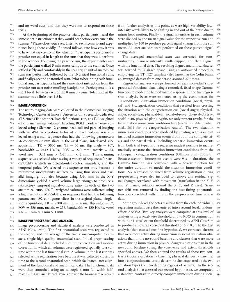
RESULTSCOMMON NEURAL ACTIVITY DURING IMMERSION ACROSSSITUATIONSOur first hypothesis was that neural activity during both situationswould be reliably greater than baseline across multiple sensorymodalities and across regions involved in detecting and integratingsalient sensory information (see Table 2 for the baseline contrasts).As shown in Figure 2A, neural activity was reliably greater thanbaseline in bilateral primary somatomotor and visual cortex, aswell as premotor cortex, SMA, and extrastriate visual cortex, sug-gesting that participants easily immersed in the situations. Theself-reported rating data from the training session confirmed thatparticipants found the social evaluation and physical danger sit-uations relatively easy to immerse in (see Figure 2B), with no
significant differences in “being there” ratings between situationtypes [repeated measures t test; t(19) = 1.64, p > 0.05]. Becauseparticipants listened to the scenarios with their eyes closed andbecause participants did not make responses while immersing inthe scenarios, it is significant that these sensorimotor regions weresignificantly more active than the no-sound baseline. As wouldbe expected with an auditory, language-based immersion pro-cedure, we observed activity in bilateral auditory cortex and insuperior temporal and inferior frontal regions associated with lan-guage processing, with more extensive activity in the left frontalregions.
Consistent with the hypothesis that immersion would alsogenerally involve selection, encoding, and integration of salientsensory and other information, we observed activity in bilateralhippocampus and in right amygdala (see Figure 2C). Extensiveevidence implicates the hippocampus in mnemonic functions(Squire and Zola-Morgan, 1991; Tulving, 2002; Squire, 2004),especially the integration and binding of the multimodal infor-mation involved in constructing (and reconstructing) situatedmemories (Addis and McAndrews, 2006; Kroes and Fernandez,2012). More recent evidence establishes a central role for thisstructure in simulating future, imagined situations (Addis et al.,2007; Hassabis et al., 2007; Schacter et al., 2007, 2012), which issimilar in nature to our immersion paradigm, and which requiressimilar integration and binding of concepts established in mem-ory (from prior experience). The amygdala plays a central rolein emotional experiences by efficiently integrating multisensoryinformation to direct attention and guide encoding (Costafredaet al., 2008; Bliss-Moreau et al., 2011; Klasen et al., 2012; Lindquistet al., 2012), especially during situations that involve threat(Adolphs, 2008; Miskovic and Schmidt, 2012). As we will see,no differences emerged in the amygdala or in the hippocam-pus during the social evaluation and physical danger situations,suggesting these structures played a similar role in both types ofexperiences.
UNIQUE NEURAL PATTERNS EMERGE FOR SOCIAL EVALUATION ANDPHYSICAL DANGER SITUATIONSOur second hypothesis was that networks underlying the socialinference and mentalizing involved in responding to a socialthreat would be reliably more active during social evaluationsituations, whereas networks underlying visuospatial attentionand action planning involved in responding to a physical threatwould be reliably more active during physical danger situa-tions. As Table 3, together with Figures 3–5, illustrate, theneural patterns that emerged when we compared social evalua-tion situations to physical danger situations are consistent withthese predictions. Figure 3 shows these results on represen-tative 2D slices, with regions showing reliably greater activityduring social evaluation in orange, and regions showing reli-ably greater activity during physical danger in green. Figures 4and 5 display these maps projected onto the surface of thebrain4, and directly compare the maps from this study with
4It is important to note that each individual’s data were not analyzed on the surface.We are using a standardized (Talairach) surface space for illustration of the groupresults in comparison to the resting state network maps from a large sample thathave been made freely available (Yeo et al., 2011).
Frontiers in Human Neuroscience www.frontiersin.org November 2013 | Volume 7 | Article 764 | 7
685
686
687
688
689
690
691
692
693
694
695
696
697
698
699
700
701
702
703
704
705
706
707
708
709
710
711
712
713
714
715
716
717
718
719
720
721
722
723
724
725
726
727
728
729
730
731
732
733
734
735
736
737
738
739
740
741
742
743
744
745
746
747
748
749
750
751
752
753
754
755
756
757
758
759
760
761
762
763
764
765
766
767
768
769
770
771
772
773
774
775
776
777
778
779
780
781
782
783
784
785
786
787
788
789
790
791
792
793
794
795
796
797
798

“fnhum-07-00764” — 2013/11/8 — 20:29 — page 8 — #8
Wilson-Mendenhall et al. Situating emotional experience
Table 2 | Social evaluation > baseline and physical danger > baseline contrasts.
Cluster Brain region Brodmann area(s) Mean t Spatial extent Peak
x y z
Social evaluation > baseline
1 R temp pole/STG/STS 38, 21, 22, 41, 42 4.73 1868 46 −16 −7
R angular g 39
R ITG/fusiform g 37, 19
R mid/sup occipital g 19
2 L temp pole/STG/STS 38, 21, 22, 41, 42 4.83 1780 −45 9 −15
L angular g 39
L mid/sup occipital g 19
3 L and R calarine/lingual g 17, 18, 19 4.21 1532 14 −57 14
L and R posterior cingulate 31
L and R parahippocampal g 35, 36
L and R hippocampus/amygdala
4 L premotor/precentral g 6, 4 4.36 921 −37 −6 50
L postcentral g 2, 3
L lateral PFC/Ant insula 44, 45, 46, 9
5 L and R SMA/precentral g 6, 4 4.60 596 −4 7 48
6 R premotor/precentral g 6, 4 4.52 501 50 −11 51
R postcentral g 2, 3
7 mPFC/mOFC 10, 11 4.45 115 −1 34 −8
8 R lateral PFC 45/46 4.09 77 52 19 22
9 L fusiform g 37 4.00 58 −36 −38 −11
Phyiscal danger > baseline
1 L and R SMA/premotor 6 4.37 6887 −5 7 47
L and R precentral g 4
L and R postcentral g 2, 3
L and R mid cingulate 24, 31
L lateral PFC/Ant insula 44, 45, 46, 9
L and R temp Pole/STG/STS 38, 21, 22, 41, 42
L and R MTG 37
L ITG/fusiform 37
L and R parahippocampal g 35, 36
L and R hippocampus/amygdala
L and R mid/sup occipital g 19
L and R calcarine/lingual g 17, 18, 19
L inferior parietal 40, 7
2 L and R thalamus 4.22 85 −9 −21 2
3 R lateral PFC 45/46 3.93 68 55 19 26
Spatial extent is the number of 23.67 mm3 functional voxels. L is left and R is right, Ant is anterior, Mid is middle, Sup is superior, m is medial, and g is gyrus. PFCis prefrontal cortex and OFC is orbitofrontal cortex. STG is superior temporal gyrus, STS superior temporal sulcus, MTG is middle temporal gyrus, and ITG is inferiortemporal gyrus. SMA is supplementary motor area.
Frontiers in Human Neuroscience www.frontiersin.org November 2013 | Volume 7 | Article 764 | 8
799
800
801
802
803
804
805
806
807
808
809
810
811
812
813
814
815
816
817
818
819
820
821
822
823
824
825
826
827
828
829
830
831
832
833
834
835
836
837
838
839
840
841
842
843
844
845
846
847
848
849
850
851
852
853
854
855
856
857
858
859
860
861
862
863
864
865
866
867
868
869
870
871
872
873
874
875
876
877
878
879
880
881
882
883
884
885
886
887
888
889
890
891
892
893
894
895
896
897
898
899
900
901
902
903
904
905
906
907
908
909
910
911
912
“fnhum-07-00764” — 2013/11/8 — 20:29 — page 9 — #9
Wilson-Mendenhall et al. Situating emotional experience
FIGURE 2 | (A) shared neural activity during social evaluation and physicaldanger situations in sensorimotor cortex (revealed by the conjunction analysisin which each situation was compared to the “no sound” baseline) (B) self-
reported immersion ratings from the training session (error bars depict SEMacross participant condition means) (C) shared neural activity revealed by theconjunction analysis in the amygdala and hippocampus.
the large-scale networks that have been defined using restingstate connectivity techniques across large samples (Yeo et al.,2011).
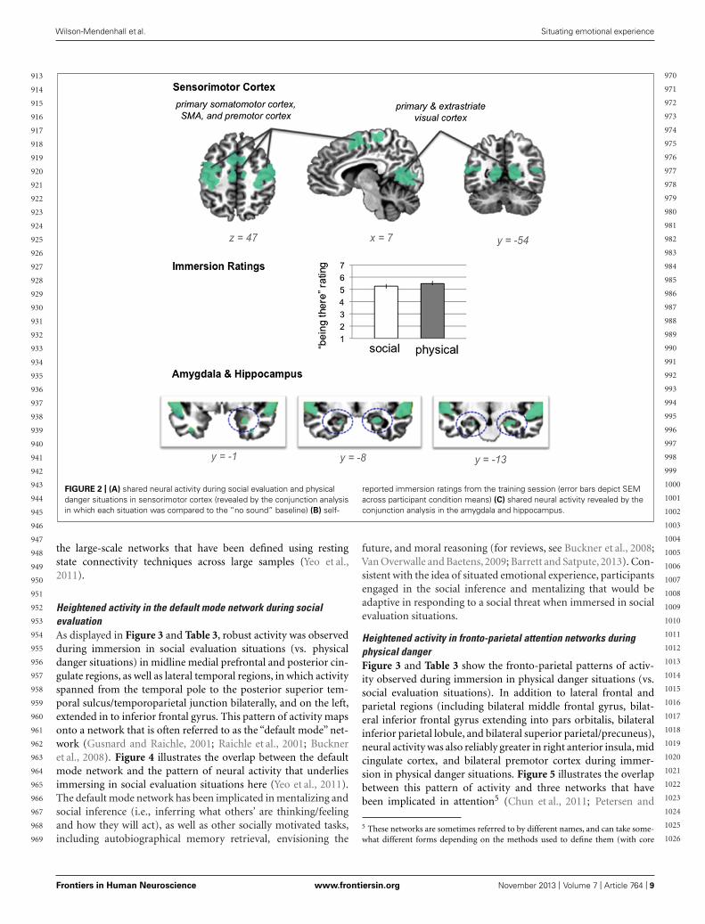
Heightened activity in the default mode network during socialevaluationAs displayed in Figure 3 and Table 3, robust activity was observedduring immersion in social evaluation situations (vs. physicaldanger situations) in midline medial prefrontal and posterior cin-gulate regions, as well as lateral temporal regions, in which activityspanned from the temporal pole to the posterior superior tem-poral sulcus/temporoparietal junction bilaterally, and on the left,extended in to inferior frontal gyrus. This pattern of activity mapsonto a network that is often referred to as the “default mode” net-work (Gusnard and Raichle, 2001; Raichle et al., 2001; Buckneret al., 2008). Figure 4 illustrates the overlap between the defaultmode network and the pattern of neural activity that underliesimmersing in social evaluation situations here (Yeo et al., 2011).The default mode network has been implicated in mentalizing andsocial inference (i.e., inferring what others’ are thinking/feelingand how they will act), as well as other socially motivated tasks,including autobiographical memory retrieval, envisioning the
future, and moral reasoning (for reviews, see Buckner et al., 2008;Van Overwalle and Baetens, 2009; Barrett and Satpute, 2013). Con-sistent with the idea of situated emotional experience, participantsengaged in the social inference and mentalizing that would beadaptive in responding to a social threat when immersed in socialevaluation situations.
Heightened activity in fronto-parietal attention networks duringphysical dangerFigure 3 and Table 3 show the fronto-parietal patterns of activ-ity observed during immersion in physical danger situations (vs.social evaluation situations). In addition to lateral frontal andparietal regions (including bilateral middle frontal gyrus, bilat-eral inferior frontal gyrus extending into pars orbitalis, bilateralinferior parietal lobule, and bilateral superior parietal/precuneus),neural activity was also reliably greater in right anterior insula, midcingulate cortex, and bilateral premotor cortex during immer-sion in physical danger situations. Figure 5 illustrates the overlapbetween this pattern of activity and three networks that havebeen implicated in attention5 (Chun et al., 2011; Petersen and
5 These networks are sometimes referred to by different names, and can take some-what different forms depending on the methods used to define them (with core
Frontiers in Human Neuroscience www.frontiersin.org November 2013 | Volume 7 | Article 764 | 9
913
914
915
916
917
918
919
920
921
922
923
924
925
926
927
928
929
930
931
932
933
934
935
936
937
938
939
940
941
942
943
944
945
946
947
948
949
950
951
952
953
954
955
956
957
958
959
960
961
962
963
964
965
966
967
968
969
970
971
972
973
974
975
976
977
978
979
980
981
982
983
984
985
986
987
988
989
990
991
992
993
994
995
996
997
998
999
1000
1001
1002
1003
1004
1005
1006
1007
1008
1009
1010
1011
1012
1013
1014
1015
1016
1017
1018
1019
1020
1021
1022
1023
1024
1025
1026

“fnhum-07-00764” — 2013/11/8 — 20:29 — page 10 — #10
Wilson-Mendenhall et al. Situating emotional experience
Table 3 | Brain regions that emerged in the social evaluation vs. physical danger contrast.
Cluster Brain region Brodmann area(s) Mean t Spatial extent Peak
x y z
Social evaluation > physical danger
1 L STG/STS/post insula/angular g/temp pole/OFC/IFG 41, 42, 22, 21, 39, 38, 47, 45 5.13 2059 −58 −17 −1
2 R STG/STS/post insula/temp pole 41, 42, 22, 21, 38 4.77 1668 51 9 −20
3 mPFC/mOFC/SMA 10, 11, 9, 8, 6 4.63 1136 4 51 31
4 Post cingulate/precuneus 31, 7 4.73 498 −7 −53 34
5 R STG/STS/angular g 22, 39 3.97 112 40 −49 22
6 L cuneus 18 3.67 57 −7 −95 23
Physical danger > social evaluation
1 L inf/sup parietal/precuneus 40, 7 4.23 992 −59 −33 38
2 Mid cing/L premotor/L MFG 24, 6 4.20 715 4 6 31
3 L MTG/fusiform g/parahippocampal g 37, 20, 35 4.37 478 −49 −54 0
4 Mid cing 31, 23 4.35 321 −13 −26 37
5 L MFG 46, 9, 10 4.14 266 −37 38 16
6 R MFG/Ant insula/OFC 10 3.99 212 37 44 6
7 R inf parietal 40 4.14 199 59 −37 35
8 R premotor 6 4.16 173 15 2 59
9 R MFG 9 3.95 104 31 30 38
10 R precuneus 7 3.94 74 7 −56 53
11 L OFC 11 3.82 49 −29 44 −5
12 R restrosplenial 29 3.78 42 12 −44 12
Spatial extent is the number of 23.67 mm3 functional voxels. L is left and R is right. Post is posterior, Ant is anterior, Inf is inferior, Sup is superior, m is medial, andg is gyrus. PFC is prefrontal cortex, OFC is orbitofrontal cortex, Cing is cingulate, and MFG is middle frontal gyrus. STG is superior temporal gyrus, STS is superiortemporal sulcus, and MTG is middle temporal gyrus. SMA is supplementary motor area.
FIGURE 3 | Social evaluation vs. physical danger contrast, with regions reliably more active during social evaluation in orange and regions reliably
more active during physical danger in green.
Frontiers in Human Neuroscience www.frontiersin.org November 2013 | Volume 7 | Article 764 | 10
1027
1028
1029
1030
1031
1032
1033
1034
1035
1036
1037
1038
1039
1040
1041
1042
1043
1044
1045
1046
1047
1048
1049
1050
1051
1052
1053
1054
1055
1056
1057
1058
1059
1060
1061
1062
1063
1064
1065
1066
1067
1068
1069
1070
1071
1072
1073
1074
1075
1076
1077
1078
1079
1080
1081
1082
1083
1084
1085
1086
1087
1088
1089
1090
1091
1092
1093
1094
1095
1096
1097
1098
1099
1100
1101
1102
1103
1104
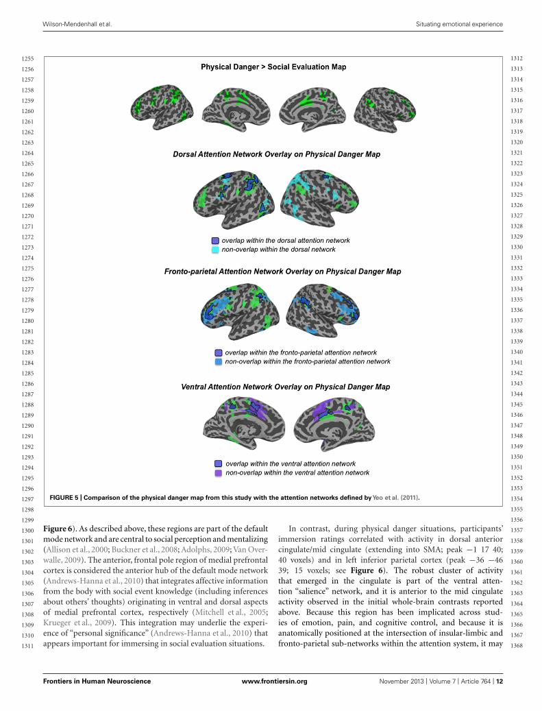
1105
1106
1107
1108
1109
1110
1111
1112
1113
1114
1115
1116
1117
1118
1119
1120
1121
1122
1123
1124
1125
1126
1127
1128
1129
1130
1131
1132
1133
1134
1135
1136
1137
1138
1139
1140

“fnhum-07-00764” — 2013/11/8 — 20:29 — page 11 — #11
Wilson-Mendenhall et al. Situating emotional experience
FIGURE 4 | Comparison of the social evaluation map from this study with the default mode network defined byYeo et al. (2011).
Posner, 2012; Posner, 2012). The most significant overlap wasobserved in the lateral fronto-parietal executive network and thedorsal attention network. These networks are thought to allocateattentional resources to prioritize specific sensory inputs (what isoften referred to as“orienting”to the external environment) and toguide flexible shifts in behavior (Dosenbach et al., 2007; Petersenand Posner, 2012). The operations they carry out are critical formaintaining a vigilant state (Tang et al., 2012), which is importantduring threat. Less overlap was evident in the ventral attention net-work that is thought to interrupt top-down operations throughbottom-up “salience” detection (Corbetta et al., 2008), althoughrobust activity was observed in the mid cingulate regions shownin Figure 5 that support the action monitoring that occurs, espe-cially, in situations involving physical pain (Morecraft and VanHoesen, 1992; Vogt, 2005). Taken together, this pattern of resultssuggests, strikingly, that immersion in the physical danger situa-tions (from a first-person perspective with eyes closed) engagedattention networks that are studied almost exclusively using
nodes remaining the same). Because the network maps we present here are takenfrom Yeo et al. (2011), we use their terminology. They note (and thus so do we) thatthe ventral attention network, especially, is similar to what has been described as thesalience network (Seeley et al., 2007) and the cingulo-opercular network (Dosenbachet al., 2007).
external visual cues. Consistent with the idea of situated emotionalexperience, participants engaged in the monitoring of the environ-ment and preparation for flexible action that would be adaptivein action to a physical threat when immersed in physical dangersituations.
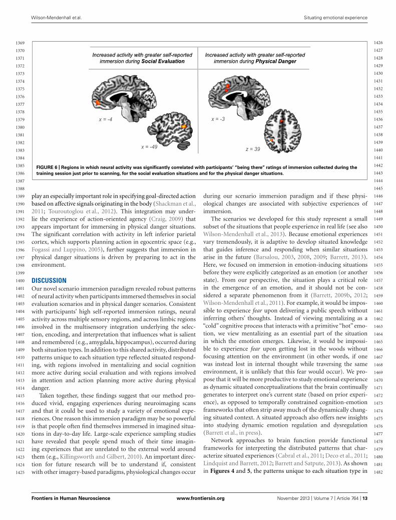
Immersion ratings correlate with activity in different regions duringsocial evaluation vs. physical danger situationsTo provide another test of our second hypothesis, we examinedwhether self-reported immersion ratings of “being there” in thesituation (from the training session) were associated with brainactivity during the two situation types. If emotional experience issituated, then feeling immersed in a situation should be realized byneural circuitry that underlies engaging in the specific situation.Whereas immersion in social evaluation situations should occurwhen affect is grounded in mentalizing about others, immersion inphysical danger situations should occur when affect is grounded intaking action in the environment. The results displayed in Figure 6support this prediction.
During social evaluation situations, participants’ immersionratings correlated with activity in anterior medial prefrontal cortex(frontal pole area; peak voxel −6 51 0; 23 voxels) and in superiortemporal gyrus/sulcus (peak voxel −47 −49 14; 24 voxels; see
Frontiers in Human Neuroscience www.frontiersin.org November 2013 | Volume 7 | Article 764 | 11
1141
1142
1143
1144
1145
1146
1147
1148
1149
1150
1151
1152
1153
1154
1155
1156
1157
1158
1159
1160
1161
1162
1163
1164
1165
1166
1167
1168
1169
1170
1171
1172
1173
1174
1175
1176
1177
1178
1179
1180
1181
1182
1183
1184
1185
1186
1187
1188
1189
1190
1191
1192
1193
1194
1195
1196
1197
1198
1199
1200
1201
1202
1203
1204
1205
1206
1207
1208
1209
1210
1211
1212
1213
1214
1215
1216
1217
1218
1219
1220
1221
1222
1223
1224
1225
1226
1227
1228
1229
1230
1231
1232
1233
1234
1235
1236
1237
1238
1239
1240
1241
1242
1243
1244
1245
1246
1247
1248
1249
1250
1251
1252
1253
1254
“fnhum-07-00764” — 2013/11/8 — 20:29 — page 12 — #12
Wilson-Mendenhall et al. Situating emotional experience
FIGURE 5 | Comparison of the physical danger map from this study with the attention networks defined byYeo et al. (2011).
Figure 6). As described above, these regions are part of the defaultmode network and are central to social perception and mentalizing(Allison et al., 2000; Buckner et al., 2008; Adolphs, 2009; Van Over-walle, 2009). The anterior, frontal pole region of medial prefrontalcortex is considered the anterior hub of the default mode network(Andrews-Hanna et al., 2010) that integrates affective informationfrom the body with social event knowledge (including inferencesabout others’ thoughts) originating in ventral and dorsal aspectsof medial prefrontal cortex, respectively (Mitchell et al., 2005;Krueger et al., 2009). This integration may underlie the experi-ence of “personal significance” (Andrews-Hanna et al., 2010) thatappears important for immersing in social evaluation situations.
In contrast, during physical danger situations, participants’immersion ratings correlated with activity in dorsal anteriorcingulate/mid cingulate (extending into SMA; peak −1 17 40;40 voxels) and in left inferior parietal cortex (peak −36 −4639; 15 voxels; see Figure 6). The robust cluster of activitythat emerged in the cingulate is part of the ventral atten-tion “salience” network, and it is anterior to the mid cingulateactivity observed in the initial whole-brain contrasts reportedabove. Because this region has been implicated across stud-ies of emotion, pain, and cognitive control, and because it isanatomically positioned at the intersection of insular-limbic andfronto-parietal sub-networks within the attention system, it may
Frontiers in Human Neuroscience www.frontiersin.org November 2013 | Volume 7 | Article 764 | 12
1255
1256
1257
1258
1259
1260
1261
1262
1263
1264
1265
1266
1267
1268
1269
1270
1271
1272
1273
1274
1275
1276
1277
1278
1279
1280
1281
1282
1283
1284
1285
1286
1287
1288
1289
1290
1291
1292
1293
1294
1295
1296
1297
1298
1299
1300
1301
1302
1303
1304
1305
1306
1307
1308
1309
1310
1311
1312
1313
1314
1315
1316
1317
1318
1319
1320
1321
1322
1323
1324
1325
1326
1327
1328
1329
1330
1331
1332
1333
1334
1335
1336
1337
1338
1339
1340
1341
1342
1343
1344
1345
1346
1347
1348
1349
1350
1351
1352
1353
1354
1355
1356
1357
1358
1359
1360
1361
1362
1363
1364
1365
1366
1367
1368
“fnhum-07-00764” — 2013/11/8 — 20:29 — page 13 — #13
Wilson-Mendenhall et al. Situating emotional experience
FIGURE 6 | Regions in which neural activity was significantly correlated with participants’ “being there” ratings of immersion collected during the
training session just prior to scanning, for the social evaluation situations and for the physical danger situations.
play an especially important role in specifying goal-directed actionbased on affective signals originating in the body (Shackman et al.,2011; Touroutoglou et al., 2012). This integration may under-lie the experience of action-oriented agency (Craig, 2009) thatappears important for immersing in physical danger situations.The significant correlation with activity in left inferior parietalcortex, which supports planning action in egocentric space (e.g.,Fogassi and Luppino, 2005), further suggests that immersion inphysical danger situations is driven by preparing to act in theenvironment.
DISCUSSIONOur novel scenario immersion paradigm revealed robust patternsof neural activity when participants immersed themselves in socialevaluation scenarios and in physical danger scenarios. Consistentwith participants’ high self-reported immersion ratings, neuralactivity across multiple sensory regions, and across limbic regionsinvolved in the multisensory integration underlying the selec-tion, encoding, and interpretation that influences what is salientand remembered (e.g., amygdala, hippocampus), occurred duringboth situation types. In addition to this shared activity, distributedpatterns unique to each situation type reflected situated respond-ing, with regions involved in mentalizing and social cognitionmore active during social evaluation and with regions involvedin attention and action planning more active during physicaldanger.
Taken together, these findings suggest that our method pro-duced vivid, engaging experiences during neuroimaging scansand that it could be used to study a variety of emotional expe-riences. One reason this immersion paradigm may be so powerfulis that people often find themselves immersed in imagined situa-tions in day-to-day life. Large-scale experience sampling studieshave revealed that people spend much of their time imagin-ing experiences that are unrelated to the external world aroundthem (e.g., Killingsworth and Gilbert, 2010). An important direc-tion for future research will be to understand if, consistentwith other imagery-based paradigms, physiological changes occur
during our scenario immersion paradigm and if these physi-ological changes are associated with subjective experiences ofimmersion.
The scenarios we developed for this study represent a smallsubset of the situations that people experience in real life (see alsoWilson-Mendenhall et al., 2013). Because emotional experiencesvary tremendously, it is adaptive to develop situated knowledgethat guides inference and responding when similar situationsarise in the future (Barsalou, 2003, 2008, 2009; Barrett, 2013).Here, we focused on immersion in emotion-inducing situationsbefore they were explicitly categorized as an emotion (or anotherstate). From our perspective, the situation plays a critical rolein the emergence of an emotion, and it should not be con-sidered a separate phenomenon from it (Barrett, 2009b, 2012;Wilson-Mendenhall et al., 2011). For example, it would be impos-sible to experience fear upon delivering a public speech withoutinferring others’ thoughts. Instead of viewing mentalizing as a“cold” cognitive process that interacts with a primitive “hot” emo-tion, we view mentalizing as an essential part of the situationin which the emotion emerges. Likewise, it would be impossi-ble to experience fear upon getting lost in the woods withoutfocusing attention on the environment (in other words, if onewas instead lost in internal thought while traversing the sameenvironment, it is unlikely that this fear would occur). We pro-pose that it will be more productive to study emotional experienceas dynamic situated conceptualizations that the brain continuallygenerates to interpret one’s current state (based on prior experi-ence), as opposed to temporally constrained cognition-emotionframeworks that often strip away much of the dynamically chang-ing situated context. A situated approach also offers new insightsinto studying dynamic emotion regulation and dysregulation(Barrett et al., in press).
Network approaches to brain function provide functionalframeworks for interpreting the distributed patterns that char-acterize situated experiences (Cabral et al., 2011; Deco et al., 2011;Lindquist and Barrett, 2012; Barrett and Satpute, 2013). As shownin Figures 4 and 5, the patterns unique to each situation type in
Frontiers in Human Neuroscience www.frontiersin.org November 2013 | Volume 7 | Article 764 | 13
1369
1370
1371
1372
1373
1374
1375
1376
1377
1378
1379
1380
1381
1382
1383
1384
1385
1386
1387
1388
1389
1390
1391
1392
1393
1394
1395
1396
1397
1398
1399
1400
1401
1402
1403
1404
1405
1406
1407
1408
1409
1410
1411
1412
1413
1414
1415
1416
1417
1418
1419
1420
1421
1422
1423
1424
1425
1426
1427
1428
1429
1430
1431
1432
1433
1434
1435
1436
1437
1438
1439
1440
1441
1442
1443
1444
1445
1446
1447
1448
1449
1450
1451
1452
1453
1454
1455
1456
1457
1458
1459
1460
1461
1462
1463
1464
1465
1466
1467
1468
1469
1470
1471
1472
1473
1474
1475
1476
1477
1478
1479
1480
1481
1482
“fnhum-07-00764” — 2013/11/8 — 20:29 — page 14 — #14
Wilson-Mendenhall et al. Situating emotional experience
this study can be differentiated by the anatomically constrainedresting state networks6 identified in previous work (Raichle et al.,2001; Fox et al., 2005; Vincent et al., 2006; Dosenbach et al., 2007;Fair et al., 2007; Seeley et al., 2007; Yeo et al., 2011; Tourouto-glou et al., 2012). Whereas the neural patterns underlying socialthreat situations primarily map onto the default mode networkthat supports social inference and mentalizing, the neural pat-terns underlying physical threat situations primarily map ontoattention networks underlying monitoring of the environmentand action planning. The neural pattern unique to each situ-ation type reflects adaptive, situated responding. Furthermore,regions traditionally associated with emotion diverged in linewith these networks (e.g., ventromedial prefrontal cortex as partof the default mode network; lateral orbitofrontal cortex andcingulate regions as part of the attention networks). Interest-ingly, these regions appear to be central to immersion in eachtype of situation, with the anterior medial prefrontal cortex(which is often considered part of ventromedial prefrontal cor-tex) associated with immersion during social evaluation situationsand dorsal anterior cingulate associated with immersion dur-ing physical danger situations. These results suggest, strikingly,that the brain realizes immersion differently depending on thesituation.
Resting state networks provide a starting point for examininghow networks underlie situated experiences, but recent evidencesuggests that coordination between regions in these networksdynamically changes during different psychological states (e.g.,van Marle et al., 2010; Raz et al., 2012; Wang et al., 2012). Inthis study, for example, the neural patterns underlying physicaldanger experiences recruited various aspects of several differentattention networks. Attention is primarily studied using simplevisual detection tasks that examine external stimuli vs. internalgoal dichotomies. Recent reviews emphasize the need for researchthat examines how attention systems operate during experiencesguided by memory (e.g., Hutchinson and Turk-Browne, 2012),which arguably constitute much of our experience. Because infe-rior parietal cortex and cingulate regions figured prominently inthe pattern observed across the attention networks in this study,this particular configuration may reflect the attention operationsinvolved in coordinating bodily actions in space. It is also impor-tant to consider that these patterns reflect relative differencesbetween the social and physical threat situations. As we showedinitially, the situation types also share patterns of activity thatcontribute to the overall pattern of situated activity. In our view,it is useful to think about situated neural activity as dynamicallychanging patterns that are distributed across structurally and func-tionally distinct networks (see also Barrett and Satpute, 2013).Even within a structurally defined network, different distributedpatterns of neural activity may reflect unique functional motifsthat underlie different experiences and behaviors (Sporns andKotter, 2004).
In closing, a psychological construction approach to studyingsituated emotion motivates different questions than traditional
6The term “resting state” is often misinterpreted to mean the resting brain. It shouldnot be assumed that the brain is actually “at rest” during these scans, but simply thatthere is no externally orienting task.
approaches to studying emotion. It invites shifting research agen-das from defining five or so emotion categories to studying therich situations that characterize emotional experiences.
ACKNOWLEDGMENTSPreparation of this manuscript was supported by an NIH Direc-tor’s Pioneer Award DPI OD003312 to Lisa Feldman Barrett atNortheastern University with a sub-contract to Lawrence Barsa-lou at Emory University. We thank A. Satpute and K. Lindquist forthe meta-analysis codes indicating study methods/tasks.
REFERENCESAddis, D. R., and McAndrews, M. P. (2006). Prefrontal and hippocampal contribu-
tions to the generation and binding of semantic associations during successfulencoding. Neuroimage 33, 1194–1206. doi: 10.1016/j.neuroimage.2006.07.039
Addis, D. R., Wong, A. T., and Schacter, D. L. (2007). Remembering thepast and imagining the future: common and distinct neural substrates dur-ing event construction and elaboration. Neuropsychologia 45, 1363–1377. doi:10.1016/j.neuropsychologia.2006.10.016
Adolphs, R. (2008). Fear, faces, and the human amygdala. Curr. Opin. Neurobiol. 18,166–172. doi: 10.1016/j.conb.2008.06.006
Adolphs, R. (2009). The social brain: neural basis of social knowledge. Annu. Rev.Psychol. 60, 693–716. doi: 10.1146/annurev.psych.60.110707.163514
Allison, T., Puce, A., and McCarthy, G. (2000). Social perception from visualcues: role of the STS region. Trends Cogn. Sci. 4, 267–278. doi: 10.1016/S1364-6613(00)01501-1
Andrews-Hanna, J. R., Reidler, J. S., Sepulcre, J., Poulin, R., and Buckner, R. L.(2010). Functional-anatomic fractionation of the brain’s default network. Neuron65, 550–562. doi: 10.1016/j.neuron.2010.02.005
Aron, A., Fisher, H., Mashek, D. J., Strong, G., Li, H., and Brown, L. L. (2005).Reward, motivation, and emotion systems associated with early-stage intenseromantic love. J. Neurophysiol. 94, 327–337. doi: 10.1152/jn.00838.2004
Aydede, H., and Robbins, P. (eds). (2009). The Cambridge Handbook of SituatedCognition. New York, NY: Cambridge University Press.
Bar, M. (2004). Visual objects in context. Nat. Rev. Neurosci. 5, 617–629. doi:10.1038/nrn1476
Barrett, L. F. (2006). Emotions as natural kinds? Perspect. Psychol. Sci. 1, 28–58. doi:10.1111/j.1745-6916.2006.00003.x
Barrett, L. F. (2009a). The future of psychology: connecting mind to brain. Perspect.Psychol. Sci. 4, 326–339. doi: 10.1111/j.1745-6924.2009.01134.x
Barrett, L. F. (2009b). Variety is the spice of life: a psychological constructionapproach to understanding variability in emotion. Cogn. Emot. 23, 1284–1306.doi: 10.1080/02699930902985894
Barrett, L. F. (2012). Emotions are real. Emotion 12, 413–429. doi: 10.1037/a0027555Barrett, L. F. (2013). Psychological construction: a Darwinian approach to the
science of emotion. Emot. Rev. 5, 379–389.Barrett, L. F., and Satpute, A. B. (2013). Large-scale brain networks in affective and
social neuroscience: towards an integrative functional architecture of the brain.Curr. Opin. Neurobiol. 23, 361–372 doi: 10.1016/j.conb.2012.12.012
Barrett, L. F., Wilson-Mendenhall, C. D., and Barsalou, L. (in press). “A psychologicalconstruction account of emotion regulation and dysregulation: the role of situatedconceptualizations,” The Handbook of Emotion Regulation, 2nd Edn, ed. J. J. Gross(New York: Guilford).
Barsalou, L. W. (2003). Situated simulation in the human conceptual system. Lang.Cogn. Process 18, 513–562. doi: 10.1080/01690960344000026
Barsalou, L. W. (2008). “Situating concepts” in Cambridge Handbook of SituatedCognition, eds P. Robbins and M. Aydede (New York: Cambridge University Press),236–263.
Barsalou, L. W. (2009). Simulation, situated conceptualization, and prediction. Phi-los. Trans. R. Soc. Lond. B Biol. Sci. 364, 1281–1289. doi: 10.1098/rstb.2008.0319
Bliss-Moreau, E., Bauman, M. D., and Amaral, D. G. (2011). Neonatal amygdalalesions result in globally blunted affect in adult rhesus macaques. Behav. Neurosci.125, 848–858. doi: 10.1037/a0025757
Buckner, R. L., Andrews-Hanna, J. R., and Schacter, D. L. (2008). The brain’s defaultnetwork: anatomy, function, and relevance to disease. Ann. N. Y. Acad. Sci. 1124,1–38. doi: 10.1196/annals.1440.011
Frontiers in Human Neuroscience www.frontiersin.org November 2013 | Volume 7 | Article 764 | 14
1483
1484
1485
1486
1487
1488
1489
1490
1491
1492
1493
1494
1495
1496
1497
1498
1499
1500
1501
1502
1503
1504
1505
1506
1507
1508
1509
1510
1511
1512
1513
1514
1515
1516
1517
1518
1519
1520
1521
1522
1523
1524
1525
1526
1527
1528
1529
1530
1531
1532
1533
1534
1535
1536
1537
1538
1539
1540
1541
1542
1543
1544
1545
1546
1547
1548
1549
1550
1551
1552
1553
1554
1555
1556
1557
1558
1559
1560
1561
1562
1563
1564
1565
1566
1567
1568
1569
1570
1571
1572
1573
1574
1575
1576
1577
1578
1579
1580
1581
1582
1583
1584
1585
1586
1587
1588
1589
1590
1591
1592
1593
1594
1595
1596

“fnhum-07-00764” — 2013/11/8 — 20:29 — page 15 — #15
Wilson-Mendenhall et al. Situating emotional experience
Buckner, R. L., and Carroll, D. C. (2007). Self-projection and the brain. Trends Cogn.Sci. 11, 49–57. doi: 10.1016/j.tics.2006.11.004
Bystritsky, A., Pontillo, D., Powers, M., Sabb, F. W., Craske, M. G., and Bookheimer,S. Y. (2001). Functional MRI changes during panic anticipation and imageryexposure. Neuroreport 12, 3953–3957. doi: 10.1097/00001756-200112210-00020
Cabral, J., Hugues, E., Sporns, O., and Deco, G. (2011). Role of local networkoscillations in resting-state functional connectivity. Neuroimage 57, 130–139. doi:10.1016/j.neuroimage.2011.04.010
Chun, M. M., Golomb, J. D., and Turk-Browne, N. B. (2011). A taxonomyof external and internal attention. Annu. Rev. Psychol. 62, 73–101. doi:10.1146/annurev.psych.093008.100427
Colibazzi, T., Posner, J., Wang, Z., Gorman, D., Gerber, A., Yu, S., et al. (2010).Neural systems subserving valence and arousal during the experience of inducedemotions. Emotion 10, 377–389. doi: 10.1037/a0018484
Corbetta, M., Patel, G., and Shulman, G. L. (2008). The reorienting system of thehuman brain: from environment to theory of mind. Neuron 58, 306–324. doi:10.1016/j.neuron.2008.04.017
Costa, V. D., Lang, P. J., Sabatinelli, D., Versace, F., and Bradley, M. M. (2010).Emotional imagery: assessing pleasure and arousal in the brain’s reward circuitry.Hum. Brain Mapp. 31, 1446–1457. doi: 10.1002/hbm.20948
Costafreda, S. G., Brammer, M. J., David, A. S., and Fu, C. H. (2008). Pre-dictors of amygdala activation during the processing of emotional stimuli: ameta-analysis of 385 PET and fMRI studies. Brain Res. Rev. 58, 57–70. doi:10.1016/j.brainresrev.2007.10.012
Cox, R. W. (1996). AFNI: software for analysis and visualization of functionalmagnetic resonance neuroimages. Comput. Biomed. Res. 29, 162–173. doi:10.1006/cbmr.1996.0014
Craig, A. D. (2009). How do you feel – now? The anterior insula and humanawareness. Nat. Rev. Neurosci. 10, 59–70. doi: 10.1038/nrn2555
Deco, G., Jirsa, V. K., and McIntosh, A. R. (2011). Emerging concepts for thedynamical organization of resting-state activity in the brain. Nat. Rev. Neurosci.12, 43–56. doi: 10.1038/nrn2961
Denson, T. F., Pedersen, W. C., Ronquillo, J., and Nandy, A. S. (2009). The angrybrain: neural correlates of anger, angry rumination, and aggressive personality. J.Cogn. Neurosci. 21, 734–744. doi: 10.1162/jocn.2009.21051
Dosenbach, N. U., Fair, D. A., Miezin, F. M., Cohen, A. L., Wenger, K. K.,Dosenbach, R. A., et al. (2007). Distinct brain networks for adaptive and sta-ble task control in humans. Proc. Natl. Acad. Sci. U.S.A. 104, 11073–11078. doi:10.1073/pnas.0704320104
Ekman, P. (2009). Darwin’s contributions to our understanding of emotionalexpressions. Philos. Trans. R. Soc. Lond. B Biol. Sci. 364, 3449–3451. doi:10.1098/rstb.2009.0189
Fair, D. A., Dosenbach, N. U., Church, J. A., Cohen, A. L., Brahmbhatt, S., Miezin,F. M., et al. (2007). Development of distinct control networks through segre-gation and integration. Proc. Natl. Acad. Sci. U.S.A. 104, 13507–13512. doi:10.1073/pnas.0705843104
Fogassi, L., and Luppino, G. (2005). Motor functions of the parietal lobe. Curr.Opin. Neurobiol. 15, 626–631. doi: 10.1016/j.conb.2005.10.015
Fox, M. D., Snyder, A. Z., Vincent, J. L., Corbetta, M., Van Essen, D. C., andRaichle, M. E. (2005). The human brain is intrinsically organized into dynamic,anticorrelated functional networks. Proc. Natl. Acad. Sci. U.S.A. 102, 9673–9678.doi: 10.1073/pnas.0504136102
Gillihan, S. J., Rao, H., Wang, J., Detre, J. A., Breland, J., Sankoorikal, G. M.,et al. (2010). Serotonin transporter genotype modulates amygdala activity duringmood regulation. Soc. Cogn. Affect. Neurosci. 5, 1–10. doi: 10.1093/scan/nsp035
Glenberg, A. M. (1997). What memory is for. Behav. Brain Sci. 20, 1–19; discussion19–55.
Greve, D. N. (2002). Optseq2. [Computer software]. Available at:http://surfer.nmr.mgh.harvard.edu/optseq/
Gusnard, D. A., and Raichle, M. E. (2001). Searching for a baseline: functionalimaging and the resting human brain. Nat. Rev. Neurosci. 2, 685–694. doi:10.1038/35094500
Hassabis, D., Kumaran, D., Vann, S. D., and Maguire, E. A. (2007). Patients withhippocampal amnesia cannot imagine new experiences. Proc. Natl. Acad. Sci.U.S.A. 104, 1726–1731. doi: 10.1073/pnas.0610561104
Hess, U., and Thibault, P. (2009). Darwin and emotion expression. Am. Psychol. 64,120–128. doi: 10.1037/a0013386
Holmes, E. A., and Mathews, A. (2010). Mental imagery in emotion and emotionaldisorders. Clin. Psychol. Rev. 30, 349–362. doi: 10.1016/j.cpr.2010.01.001
Hutchinson, J. B., and Turk-Browne, N. B. (2012). Memory-guided attention:control from multiple memory systems. Trends Cogn. Sci. 16, 576–579. doi:10.1016/j.tics.2012.10.003
Killingsworth, M. A., and Gilbert, D. T. (2010). A wandering mind is an unhappymind. Science 330, 932. doi: 10.1126/science.1192439
Kim, J. W., Choi, E. A., Kim, J. J., Jeong, B. S., Kim, S. E., and Ki, S. W. (2008).The role of amygdala during auditory verbal imagery of derogatory appraisals byothers. Neurosci. Lett. 446, 1–6. doi: 10.1016/j.neulet.2008.09.019
Klasen, M., Chen, Y. H., and Mathiak, K. (2012). Multisensory emotions: perception,combination and underlying neural processes. Rev. Neurosci. 23, 381–392. doi:10.1515/revneuro-2012-0040
Kober, H., Barrett, L. F., Joseph, J., Bliss-Moreau, E., Lindquist, K., and Wager,T. D. (2008). Functional grouping and cortical-subcortical interactions in emo-tion: a meta-analysis of neuroimaging studies. Neuroimage 42, 998–1031. doi:10.1016/j.neuroimage.2008.03.059
Kosslyn, S. M., Ganis, G., and Thompson, W. L. (2001). Neural foundations ofimagery. Nat. Rev. Neurosci. 2, 635–642. doi: 10.1038/35090055
Kroes, M. C., and Fernandez, G. (2012). Dynamic neural systems enableadaptive, flexible memories. Neurosci. Biobehav. Rev. 36, 1646–1666. doi:10.1016/j.neubiorev.2012.02.014
Kross, E., Berman, M. G., Mischel, W., Smith, E. E., and Wager, T. D. (2011). Socialrejection shares somatosensory representations with physical pain. Proc. Natl.Acad. Sci. U.S.A. 108, 6270–6275. doi: 10.1073/pnas.1102693108
Krueger, F., Barbey, A. K., and Grafman, J. (2009). The medial prefrontal cor-tex mediates social event knowledge. Trends Cogn. Sci. 13, 103–109. doi:10.1016/j.tics.2008.12.005
Lang, P. J. (1979). Presidential address, 1978. A bio-informational theoryof emotional imagery. Psychophysiology 16, 495–512. doi: 10.1111/j.1469-8986.1979.tb01511.x
Lang, P. J., Bradley, M. M., and Curthbert, B. N. (2008). International Affective PictureSystem (IAPS): Affective Ratings of Pictures and Instruction Manual. Gainsville, FL:University of Florida.
Lang, P. J., Kozak, M. J., Miller, G. A., Levin, D. N., and McLean, A. Jr. (1980). Emo-tional imagery: conceptual structure and pattern of somato-visceral response.Psychophysiology 17, 179–192. doi: 10.1111/j.1469-8986.1980.tb00133.x
Lench, H. C., Flores, S. A., and Bench, S. W. (2011). Discrete emotions predictchanges in cognition, judgment, experience, behavior, and physiology: a meta-analysis of experimental emotion elicitations. Psychol. Bull. 137, 834–855. doi:10.1037/a0024244
Lindquist, K. A., and Barrett, L. F. (2012). A functional architecture of the humanbrain: emerging insights from the science of emotion. Trends Cogn. Sci. 16,533–540. doi: 10.1016/j.tics.2012.09.005
Lindquist, K. A., Wager, T. D., Kober, H., Bliss-Moreau, E., and Barrett, L. F. (2012).The brain basis of emotion: a meta-analytic review. Behav. Brain Sci. 35, 121–143.doi: 10.1017/S0140525X11000446
Marci, C. D., Glick, D. M., Loh, R., and Dougherty, D. D. (2007). Autonomic andprefrontal cortex responses to autobiographical recall of emotions. Cogn. Affect.Behav. Neurosci. 7, 243–250. doi: 10.3758/CABN.7.3.243
Menon, V. (2011). Large-scale brain networks and psychopathology: aunifying triple network model. Trends Cogn. Sci. 15, 483–506. doi:10.1016/j.tics.2011.08.003
Mesquita, B., Barrett, L. F., and Smith, E. R., (eds). (2010). The Mind in Context.New York: Guilford.
Miskovic, V., and Schmidt, L. A. (2012). Social fearfulness in the human brain.Neurosci. Biobehav. Rev. 36, 459–478. doi: 10.1016/j.neubiorev.2011.08.002
Mitchell, J. P., Banaji, M. R., and MacRae, C. N. (2005). The link between socialcognition and self-referential thought in the medial prefrontal cortex. J. Cogn.Neurosci. 17, 1306–1315. doi: 10.1162/0898929055002418
Moll, J., de Oliveira-Souza, R., Moll, F. T., Ignacio, F. A., Bramati, I. E., Caparelli-Daquer, E. M., et al. (2005). The moral affiliations of disgust: a functionalMRI study. Cogn. Behav. Neurol. 18, 68–78. doi: 10.1097/01.wnn.0000152236.46475.a7
Morecraft, R. J., and Van Hoesen, G. W. (1992). Cingulate input to the primary andsupplementary motor cortices in the rhesus monkey: evidence for somatotopyin areas 24c and 23c. J. Comp. Neurol. 322, 471–489. doi: 10.1002/cne.903220403
Frontiers in Human Neuroscience www.frontiersin.org November 2013 | Volume 7 | Article 764 | 15
1597
1598
1599
1600
1601
1602
1603
1604
1605
1606
1607
1608
1609
1610
1611
1612
1613
1614
1615
1616
1617
1618
1619
1620
1621
1622
1623
1624
1625
1626
1627
1628
1629
1630
1631
1632
1633
1634
1635
1636
1637
1638
1639
1640
1641
1642
1643
1644
1645
1646
1647
1648
1649
1650
1651
1652
1653
1654
1655
1656
1657
1658
1659
1660
1661
1662
1663
1664
1665
1666
1667
1668
1669
1670
1671
1672
1673
1674
1675
1676
1677
1678
1679
1680
1681
1682
1683
1684
1685
1686
1687
1688
1689
1690
1691
1692
1693
1694
1695
1696
1697
1698
1699
1700
1701
1702
1703
1704
1705
1706
1707
1708
1709
1710
“fnhum-07-00764” — 2013/11/8 — 20:29 — page 16 — #16
Wilson-Mendenhall et al. Situating emotional experience
Ollinger, J. M., Corbetta, M., and Shulman, G. L. (2001a). Separating processeswithin a trial in event-related functional MRI. Neuroimage 13, 218–229. doi:10.1006/nimg.2000.0711
Ollinger, J. M., Shulman, G. L., and Corbetta, M. (2001b). Separating processeswithin a trial in event-related functional MRI. Neuroimage 13, 210–217. doi:10.1006/nimg.2000.0710
Perry, D., Hendler, T., and Shamay-Tsoory, S. G. (2012). Can we share the joy ofothers? Empathic neural responses to distress vs joy. Soc. Cogn. Affect. Neurosci.7, 909–916. doi: 10.1093/scan/nsr073
Petersen, S. E., and Posner, M. I. (2012). The attention system of the human brain: 20years after. Annu. Rev. Neurosci. 35, 73–89. doi: 10.1146/annurev-neuro-062111-150525
Pietrini, P., Guazzelli, M., Basso, G., Jaffe, K., and Grafman, J. (2000). Neu-ral correlates of imaginal aggressive behavior assessed by positron emissiontomography in healthy subjects. Am. J. Psychiatry 157, 1772–1781. doi:10.1176/appi.ajp.157.11.1772
Pitman, R. K., Orr, S. P., Forgue, D. F., de Jong, J. B., and Claiborn,J. M. (1987). Psychophysiologic assessment of posttraumatic stress disorderimagery in Vietnam combat veterans. Arch. Gen. Psychiatry 44, 970–975. doi:10.1001/archpsyc.1987.01800230050009
Posner, M. I. (2012). Imaging attention networks. Neuroimage 61, 450–456. doi:10.1016/j.neuroimage.2011.12.040
Raichle, M. E. (2010). Two views of brain function. Trends Cogn. Sci. 14, 180–190.doi: 10.1016/j.tics.2010.01.008
Raichle, M. E., MacLeod, A. M., Snyder, A. Z., Powers, W. J., Gusnard, D. A., andShulman, G. L. (2001). A default mode of brain function. Proc. Natl. Acad. Sci.U.S.A. 98, 676–682. doi: 10.1073/pnas.98.2.676
Rauch, S. L., Shin, L. M., Dougherty, D. D., Alpert, N. M., Orr, S. P., Lasko, M., et al.(1999). Neural activation during sexual and competitive arousal in healthy men.Psychiatry Res. 91, 1–10. doi: 10.1016/S0925-4927(99)00020-7
Raz, G., Winetraub, Y., Jacob, Y., Kinreich, S., Maron-Katz, A., Shaham, G.,et al. (2012). Portraying emotions at their unfolding: a multilayered approachfor probing dynamics of neural networks. Neuroimage 60, 1448–1461. doi:10.1016/j.neuroimage.2011.12.084
Schacter, D. L., Addis, D. R., and Buckner, R. L. (2007). Remembering the past toimagine the future: the prospective brain. Nat. Rev. Neurosci. 8, 657–661. doi:10.1038/nrn2213
Schacter, D. L., Addis, D. R., Hassabis, D., Martin, V. C., Spreng, R. N., and Szpunar,K. K. (2012). The future of memory: remembering, imagining, and the brain.Neuron 76, 677–694. doi: 10.1016/j.neuron.2012.11.001
Schaich Borg, J., Lieberman, D., and Kiehl, K. A. (2008). Infection, incest, andiniquity: investigating the neural correlates of disgust and morality. J. Cogn.Neurosci. 20, 1529–1546. doi: 10.1162/jocn.2008.20109
Schneider, W., Eschman, A., and Zuccolotto, A. (2002). E-Prime Computer Softwareand Manual. Pittsburgh, PA: Psychology Software Tools Inc.
Seeley, W. W., Menon, V., Schatzberg, A. F., Keller, J., Glover, G. H., Kenna, H., et al.(2007). Dissociable intrinsic connectivity networks for salience processing andexecutive control. J. Neurosci. 27, 2349–2356. doi: 10.1523/JNEUROSCI.5587-06.2007
Sepulcre, J., Sabuncu, M. R., Yeo, T. B., Liu, H., and Johnson, K. A. (2012). Stepwiseconnectivity of the modal cortex reveals the multimodal organization of thehuman brain. J. Neurosci. 32, 10649–10661. doi: 10.1523/JNEUROSCI.0759-12.2012
Shackman, A. J., Salomons, T. V., Slagter, H. A., Fox, A. S., Winter, J. J., and Davidson,R. J. (2011). The integration of negative affect, pain and cognitive control in thecingulate cortex. Nat. Rev. Neurosci. 12, 154–167. doi: 10.1038/nrn2994
Sporns, O., and Kotter, R. (2004). Motifs in brain networks. PLoS Biol. 2:e369. doi:10.1371/journal.pbio.0020369
Spreng, R. N., Sepulcre, J., Turner, G. R., Stevens, W. D., and Schacter, D. L. (2013).Intrinsic architecture underlying the relations among the default, dorsal attention,and frontoparietal control networks of the human brain. J. Cogn. Neurosci. 25,74–86. doi: 10.1162/jocn_a_00281
Squire, L. R. (2004). Memory systems of the brain: a brief history and cur-rent perspective. Neurobiol. Learn. Mem. 82, 171–177. doi: 10.1016/j.nlm.2004.06.005
Squire, L. R., and Zola-Morgan, S. (1991). The medial temporal lobe memorysystem. Science 253, 1380–1386. doi: 10.1126/science.1896849
Tang, Y. Y., Rothbart, M. K., and Posner, M. I. (2012). Neural correlates of establish-ing, maintaining, and switching brain states. Trends Cogn. Sci. 16, 330–337. doi:10.1016/j.tics.2012.05.001
Touroutoglou, A., Hollenbeck, M., Dickerson, B. C., and Barrett, L. F. (2012).Dissociable large-scale networks anchored in the right anterior insula sub-serve affective experience and attention. Neuroimage 60, 1947–1958. doi:10.1016/j.neuroimage.2012.02.012
Tracy, J. L., and Randles, D. (2011). Four models of basic emotions: a review ofEkman and Cordaro, Izard, Levenson, and Panksepp and Watt. Emot. Rev. 3,397–405. doi: 10.1177/1754073911410747
Tulving, E. (2002). Episodic memory: from mind to brain. Annu. Rev. Psychol. 53,1–25. doi: 10.1146/annurev.psych.53.100901.135114
van Marle, H. J., Hermans, E. J., Qin, S., and Fernandez, G. (2010). Enhanced resting-state connectivity of amygdala in the immediate aftermath of acute psychologicalstress. Neuroimage 53, 348–354. doi: 10.1016/j.neuroimage.2010.05.070
Van Overwalle, F. (2009). Social cognition and the brain: a meta-analysis. Hum.Brain Mapp. 30, 829–858. doi: 10.1002/hbm.20547
Van Overwalle, F., and Baetens, K. (2009). Understanding others’ actions and goalsby mirror and mentalizing systems: a meta-analysis. Neuroimage 48, 564–584.doi: 10.1016/j.neuroimage.2009.06.009
Vincent, J. L., Snyder, A. Z., Fox, M. D., Shannon, B. J., Andrews, J. R., Raichle, M.E., et al. (2006). Coherent spontaneous activity identifies a hippocampal-parietalmemory network. J. Neurophysiol. 96, 3517–3531. doi: 10.1152/jn.00048.2006
Vogt, B. A. (2005). Pain and emotion interactions in subregions of the cingulategyrus. Nat. Rev. Neurosci. 6, 533–544. doi: 10.1038/nrn1704
Wang, Z., Liu, J., Zhong, N., Qin, Y., Zhou, H., and Li, K. (2012). Changes inthe brain intrinsic organization in both on-task state and post-task resting state.Neuroimage 62, 394–407. doi: 10.1016/j.neuroimage.2012.04.051
Whitfield-Gabrieli, S., and Ford, J. M. (2012). Default mode network activityand connectivity in psychopathology. Annu. Rev. Clin. Psychol. 8, 49–76. doi:10.1146/annurev-clinpsy-032511-143049
Wilson-Mendenhall, C. D., Barrett, L. F., and Barsalou, L. W. (2013). Neural evidencethat human emotions share core affective properties. Psychol Sci. 24, 947–956. doi:10.1177/0956797612464242
Wilson-Mendenhall, C. D., Barrett, L. F., Simmons, W. K., and Barsalou, L. W.(2011). Grounding emotion in situated conceptualization. Neuropsychologia 49,1105–1127. doi: 10.1016/j.neuropsychologia.2010.12.032
Yang, X. F., Bossmann, J., Schiffhauer, B., Jordan, M., and Immordino-Yang, M.H. (2012). Intrinsic default mode network connectivity predicts spontaneousverbal descriptions of autobiographical memories during social processing. Front.Psychol. 3:592. doi: 10.3389/fpsyg.2012.00592
Yeh, W., and Barsalou, L. W. (2006). The situated nature of concepts. Am. J. Psychol.119, 349–384. doi: 10.2307/20445349
Yeo, B. T., Krienen, F. M., Sepulcre, J., Sabuncu, M. R., Lashkari, D., Hollinshead,M., et al. (2011). The organization of the human cerebral cortex estimatedby intrinsic functional connectivity. J. Neurophysiol. 106, 1125–1165. doi:10.1152/jn.00338.2011
Zwaan, R. A., and Radvansky, G. A. (1998). Situation models in languagecomprehension and memory. Psychol. Bull. 123, 162–185. doi: 10.1037/0033-2909.123.2.162
Conflict of Interest Statement: The authors declare that the research was conductedin the absence of any commercial or financial relationships that could be construedas a potential conflict of interest.
Received: 02 June 2013; accepted: 24 October 2013; published online: xx November2013.Citation: Wilson-Mendenhall CD, Barrett LF and Barsalou LW (2013) Situatingemotional experience. Front. Hum. Neurosci. 7:764. doi: 10.3389/fnhum.2013.00764This article was submitted to the journal Frontiers in Human Neuroscience.Copyright © 2013 Wilson-Mendenhall, Barrett and Barsalou. This is an open-accessarticle distributed under the terms of the Creative Commons Attribution License(CC BY). The use, distribution or reproduction in other forums is permitted, pro-vided the original author(s) or licensor are credited and that the original publication inthis journal is cited, in accordance with accepted academic practice. No use, distributionor reproduction is permitted which does not comply with these terms.
Frontiers in Human Neuroscience www.frontiersin.org November 2013 | Volume 7 | Article 764 | 16
1711
1712
1713
1714
1715
1716
1717
1718
1719
1720
1721
1722
1723
1724
1725
1726
1727
1728
1729
1730
1731
1732
1733
1734
1735
1736
1737
1738
1739
1740
1741
1742
1743
1744
1745
1746
1747
1748
1749
1750
1751
1752
1753
1754
1755
1756
1757
1758
1759
1760
1761
1762
1763
1764
1765
1766
1767
1768
1769
1770
1771
1772
1773
1774
1775
1776
1777
1778
1779
1780
1781
1782
1783
1784
1785
1786
1787
1788
1789
1790
1791
1792
1793
1794
1795
1796
1797
1798
1799
1800
1801
1802
1803
1804
1805
1806
1807
1808
1809
1810
1811
1812
1813
1814
1815
1816
1817
1818
1819
1820
1821
1822
1823
1824