Z.U.M.J.Vol.19; N.6; November; 2013
-628-
Significance Of Flat Epithelial Lesions Of The ………
SIGNIFICANCE OF FLAT EPITHELIAL LESIONS OF THE URINARY BLADDER IN
EARLY DETECTION OF BLADDER CANCER: HISTOPATHOLOGICAL AND
IMMUNOHISTOCHEMICAL STUDY
Mohamed A. Ramadan, Masoud M. Omar, Mona R. Abdel-Wahed
and Shimaa A. Ahmed
Pathology Department, Faculty of Medicine, Zagazig University
ABSTRACT
Background: Urothelial carcinoma of the urinary bladder is the second most common malignancy of the
genitourinary system, after prostate cancer. The flat lesions are classified into flat lesions without cytological
atypia (Flat hyperplasia) and flat lesions with cytological atypia; reactive atypia, atypia of unknown
significance, urothelial dysplasia and urothelial carcinoma in situ). The immunohistochemical markers such
as cytokeratin 20,CD 44 and Ki 67 may be useful in differentiating CIS from reactive changes in difficult
biopsy cases.
Aim: This study was conducted to determine the role of cytokeratin 20, CD 44 and Ki 67 in the diagnosis of
dysplasia and other flat lesions of the urinary bladder.
Methods: Sixty representative cases for flat urothelial lesions of urinary bladder were examined
immunohistochemically using antibodies against CD44, cytokeratin 20 and Ki-67.
Results: CD 44 were positive in 100%of cases of reactive urothelial atypia, but flat Hyperplasia and CIS
cases were negative. However CD 44 were positive in 42.9% of atypia of unknown significance and
14.3% of Dysplasia. CK 20 were positive in (95.5%) of Dysplasia. Non of Flat Hyperplasia were positive for
CK 20. However CK 20 was positive in 88.2% of CIS, 57.1 % of atypia of unknown significance and
7.1% of reactive urothelial atypia. ki 67 index were high in 82.4% of cases of CIS, non of flat hyperplasia
expressed high ki 67 index. However ki 67 index were high in 57.1% of atypia of unknown significance,
52.4% of dysplasia and 14.3% of reactive urothelial atypia.
Conclusion. CD 44 is the most constant marker in the reactive urothelium. CK20 is the most commonly
detected marker in the dysplastic urothelium. Nevertheless, the most accurate is application of an
immunohistochemical panel composed of the three antibodies (standard CD 44, CK20 and Ki-67) together
with correlation with morphology. This is very useful for confirming the presence of dysplastic changes in
the urothelium and differentiating reactive urothelium from dysplastic urothelium (dysplasia/CIS).
Keywords: Flat epithelial lesions, CD44, cytokeratin ck20, ki67, immunohistochemistry.
INTRODUCTION ladder cancer is a significant public health
problem worldwide. It is the fourth most
common cancer in men, accounting for 6.9% of all
cancers, and tenth most common cancer in women,
accounting for 2.6% of all cancers(1)
. In Egypt, it
constitutes 30.3% of all cancers (2)
.
In 2004 World Health Organization
classification, the flat-related preneoplastic lesions
of the urinary bladder are classified as; urothelial
hyperplasia ,reactive urothelial atypia, urothelial
atypia of unknown significance(AUS(, urothelial
dysplasia and urothelial carcinoma in situ(CIS)(3)
.
Reactive atypia denotes mild nuclear abnormalities
occurring in acutely or chronically inflamed
urothelium which may be normal or slightly
thickened. Frequent mitoses may involve the lower
layers of the urothelium.The significance of
reactive atypia is to differentiate it from dysplasia,
and CIS(4)
.
Atypia of unknown significance describes
lesions with nuclear abnormalities similar to those
of reactive changes (fewer than those of dysplasia),
but out of proportion to the degree of
inflammation(5)
.
Dysplasia showed architectural and
Cytologic abnormalities falling short of the
diagnostic threshold for carcinoma in situ. The
cytologic features are restricted to intermediate and
basal cells and less atypical than in carcinoma in
situ. The thickness of dysplastic urothelium is
usually normal, but it may be increased or
decreased(6,7)
.
Urothelial dysplasia may be primary
dysplasia (rare) or secondary (more frequent)
which is associated with papillary urothelial
neoplasms and has higher rate of progression to
carcinoma than primary dysplasia(1)
.
Carcinoma in situ (CIS) of the urinary
bladder is defined by the replacement of part or all
of the normal epithelium by cells that have
microscopic and molecular features of carcinoma,
yet are confined to the epithelium(8)
.
CIS may occur as; (1) primary CIS
occurring without associated previous urothelial
tumor, (2) concomitant CIS, occurring in
conjunction with a newly diagnosed bladder tumor
B

Z.U.M.J.Vol.19; N.6; November; 2013
-629-
Significance Of Flat Epithelial Lesions Of The ………
(3) secondary CIS, diagnosed during follow-up of
a known bladder tumor, with or without a
concomitant tumor(4,9)
.
CD44 is a transmembrane glycoprotein
involved in cell-cell and cell-matrix interactions. In
the normal urothelium, CD44 staining is limited to
the basal region(10)
. The urothelium in reactive
atypia typically shows staining with CD44 in a
diffuse membranous full-thickness pattern or with
patchy basal and intermediate cell expression. This
is in contrast to absence of CD44 staining in
dysplasia and Carcinoma in situ. It is important to
note that these staining patterns are not absolute
and correlation with morphology is critical(11,12)
.
Cytokeratin20 (CK20) is the most recently
identified type I keratin protein showing a limited
pattern of expression in normal tissues and its
expression is maintained in malignant tissues(13)
.
Normal urothelium typically exhibits
CK20 staining in the umbrella cell layer, but
reactive atypia lacks extensive staining for
CK20(10)
. In AUS, there is CK20 expression in the
deeper mucosal layers(14)
. In Carcinoma in situ
there is CK20 positivity throughout the neoplastic
urothelium(12)
.
Ki-67 is absent to positive in fewer than
10% of the basal and parabasal cells of normal
urothelium, indicating a low proliferative rate(10)
.
Both Ki 67 and CK20 positivity throughout the
neoplastic urothelium with negative CD44 favors a
diagnosis of Carcinoma in situ(12)
.
MATERIAL AND METHODS This study included paraffin blocks of 150
cases of bladder lesions selected from urinary
bladder biopsy specimens diagnosed at Pathology
Department, Faculty of Medicine, Zagazig
University, Egypt, during the period from
September 2008 to September 2011. The clinical
data of the patients were obtained from medical
files. Then 60 represetative cases of flat epithelial
lesions of the urinary bladder were selected for
immunohistochemical study.
Histopathologic examination:
Consecutive 4 µm sections were prepared
and stained with hematoxylin & eosin for
histopathological examination. Pathological
diagnosis was made according to 2004 WHO/1998
ISUP classification of urothelial neoplasms(7)
.
Immunohistochemical staining:
Immunohistochemical staining was carried
out using streptoavidin-biotin immunoperoxidase
technique (Dako-Cytomation, Glostrup, Denmark).
3–5 µm thick sections cut from formalin-fixed,
paraffin-embedded blocks, were deparaffinized in
xylene and rehydrated in graded alcohol. Sections
were boiled in citrate buffer (pH 6.0) for 20 min
and then washed in phosphate buffer saline (pH
7.3). Thereafter, blocking of endogenous
peroxidase activity with 6% H2O2 in methanol
was carried out. The slides were then incubated
overnight with monoclonal antibodies; (CD 44 Std
mouse monoclonal antibody Cat. from Thermo
Scientific/Lab Vision Corporation, Fermont, USA,
clone:156-3c11. 0.09% sodium azide. Dilution
1:100), (CK 20 mouse monoclonal antibody Cat
from Thermo Scientific/Lab Vision Corporation,
Fermont, USA, clone: Ks20.8. 0.09% sodium
azide. Dilution 1:50), (Ki-67 Rabbit Polyclonal
antibody Cat from Thermo Scientific/Lab Vision
Corporation, Fermont, USA, 0.09% sodium azide.
Dilution 1:300). Incubation with a secondary
antibody and product visualization were performed
(Dako Cytomation, Glostrup, Denmark) with
diaminobenzidine substrate (Research Genetics,
Huntsville, AL) as the chromogen. The slides were
finally counterstained with Mayer’s haematoxylin
(BioGenex Laboratories, San Ramon, CA), and
washed once each with distilled water and PBS.
Tonsil specimens were used as a positive control
for CD44 and ki67.F or ck20, colon carcinoma was
used. Negative controls for CD44 and ki67 were
obtained by substitution of primary antibodies with
blocking buffer..For ck20 Negative controls were
Immunoglobulin G (IgG).
Evaluation of the results of
immunohistochemical staining:
1- Evaluation of CD 44 and CK 20
immunostaining:
Standard CD44 is membranous in
distribution. CK20 is a cytoplasmic stain .The
degree of reactivity for each antibody was graded
from 0 to 4 (0 _ negative; 1_ weak, patchy[<50%
of the cells]; 2_ moderate, patchy [<50% of the
cells]; 3_ moderate, diffuse [>50% of cells]; 4_
strong, diffuse [>50% of cells]). To designate
overexpression for CD44 and CK 20 antibodies,
we required >50% of the urothelium to be
moderately to strongly positive (3–4 positivity)(15)
.
2- Evaluation of ki 67 immunostaining: Ki-67 was regarded as positive when
nuclei stained dark brown or contained dark brown
granules. Ki-67 index was determined as the
percentage of immunostained cells per 200 cells by
5 fields of view in each section(16)
.
Immunoreactivity to Ki-67 was ‘‘low’’ if nuclear
staining <15% and ‘‘high’’ if >15%(17)
.
Statistical analysis
The results of the study were statistically
analyzed using the SPSS software (SPSS, Chicago,
IL, USA). Data were expressed as mean ±SD for
quantitative variables, as numbers and percentages.
For categorical variables, the Student t-test was
Z.U.M.J.Vol.19; N.6; November; 2013
-630-
Significance Of Flat Epithelial Lesions Of The ………
used. P-value less than 0.05 was considered
significant.
RESULTS
In the present study, 60 cases of flat
epithelial lesions of the urinary bladder were
selected and classified according to 2004
WHO/1998 ISUP classification of urothelial
neoplasms(7)
into flat Hyperplasia (1/60, 1.7%),
reactive atypia (14/60, 23.3%), atypia of
unknown significance (7/60, 11.7%), dysplasia
(21/60, 35%) and carcinoma in situ (17/60,
28.3%).
The mean age of studied cases was 64
years and ranged between 45- 77 years. There is a
significant association between age >70 and the
presence of dysplasia and CIS. The majority of the
studied cases were males. This supports the
concept that CIS maintains the same age and sex
predilection as invasive bladder cancer.
Table 1: Incidence of different selected flat lesions of the urinary bladder
Histological diagnosis No %
Flat hyperplasia 1 1.7
reactive urothelial atypia 14 23.3
atypia of unknown significance 7 11.7
Dysplasia 21 35.0
CIS 17 28.3
Total 60 100%
The majority of studied cases were dysplasia (35.0 %), followed by CIS.
Table2: Immunohistochemical reactivity pattern of CD 44 in selected flat lesions of the urinary
bladder
Flat lesions No
Negative interpretation Positive interpretation X2 P
No % No %
Flat hyperplasia 1 1 100.0 0 0.0 0.1 0.75
reactive urothelial atypia 14 0 0.0 14** 100.0 30.29 <0.001**
atypia of unknown significance 7 4 57.1 3 42.9 0.78 0.37
Dysplasia 21 18* 85.7 3 14.3 6.09 0.013*
carcinoma in situ(CIS) 17 17** 100.0 0 0.0 12.77 <0.001**
Total 60 40 66.7 20 33.3
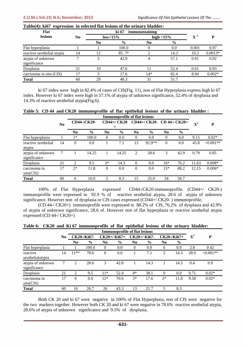
CD 44 were positive in 100%of cases of reactive urothelial atypia (Fig.2), non of flat Hyperplasia
and CIS cases were positive for CD 44(Fig.8). However CD 44 were positive in 42.9% of atypia of
unknown significance and 14.3% of Dysplasia.
Table3: Immunohistochemical reactivity pattern of cytokeratin 20 in selected flat lesions of the
urinary bladder
Flat lesions No
Negative interpretation Positive interpretation X2 P
No % No %
Flathyperplasia 1 1 100.0 0 0.0 0.1 0.75
reactive urothelial atypia 14 13** 92.9 1 7.1 23.6 < 0.001**
atypia of unknown
significance
7 3 42.9 4 57.1 0.001 0.9
Dysplasia 21 2 9.5 19* 90.5 9.2 0.002*
carcinoma in situ(CIS) 17 2 11.8 15* 88.2 5.63 0.017*
Total 60 21 35.0 39 65.0
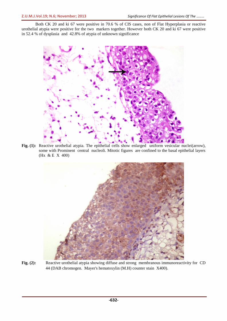
CK 20 were positive in 90.5% of cases of Dysplasia. Non of Flat Hyperplasia were positive for CK
20 .However CK 20 were positive in 88.2% of CIS(Fig. 10), 57.1 % of atypia of unknown significance
and 7.1% of reactive urothelial atypia(Fig.3) .
Z.U.M.J.Vol.19; N.6; November; 2013
-631-
Significance Of Flat Epithelial Lesions Of The ………
Table(4): ki67 expression in selected flat lesions of the urinary bladder:
Flat
lesions No
ki 67 immunostaining
X 2 P low<15% high >15%
No % No %
Flat hyperplasia 1 1 100.0 0 0.0 0.001 0.97
reactive urothelial atypia 14 12 85. 7* 2 14.3 10.2 0.0013*
atypia of unknown
significance
7 3 42.9 4 57.1 0.01 0.92
Dysplasia 21 10 47.6 11 52.4 0.01 0.93
carcinoma in situ (CIS) 17 3 17.6 14* 82.4 8.94 0.002*
Total 60 29 48.3 31 51.7
ki 67 index were high in 82.4% of cases of CIS(Fig. 11), non of Flat Hyperplasia express high ki 67
index. However ki 67 index were high in 57.1% of atypia of unknown significance, 52.4% of dysplasia and
14.3% of reactive urothelial atypia(Fig.6).
Table 5: CD 44 and CK20 immunoprofile of flat epithelial lesions of the urinary bladder :
No
Immunoprofile of flat lesions
X2 P
CD44-/CK20- CD44+/ CK20
+
CD44+/ CK20- CD 44-/ CK20+
No % No % No % No %
Flat hyperplasia 1 1* 100.0 0 0.0 0 0.0 0 0.0 9.15 0.02*
reactive urothelial
atypia
14 0 0.0 1 7.1 13 92.9** 0 0.0 45.8 <0.001**
atypia of unknown
significance
7 1 14.25 1 14.25 2 28.6 3 42.9 0.79 0.85
Dysplasia 21 2 9.5 3* 14.3 0 0.0 16* 76.2 11.63 0.008*
carcinoma in
situ(CIS)
17 2* 11.8 0 0.0 0 0.0 15* 88.2 12.15 0.006*
Total 60 6 10.0 5 8.3 15 25.0 34 56.7
100% of Flat Hyperplasia expressed CD44-/CK20-immunoprofile. (CD44+/ CK20-)
immunoprofile were expressed in 92.9 % of reactive urothelial atypia, 28.6 of atypia of unknown
significance. However non of dysplasia or CIS cases expressed (CD44+/ CK20- ) immunoprofile.
(CD 44-/ CK20+) immunoprofile were expressed in 88.2% of CIS, 76.2% of dysplasia and 42.9%
of atypia of unknown significance, 28.6 of. However non of flat hyperplasia or reactive urothelial atypia
expressed (CD 44-/ CK20+)
Table 6: CK20 and Ki 67 immunoprofile of flat epithelial lesions of the urinary bladder:
No
Immunoprofile of flat lesions
X2 P CK20-/Ki67- CK20+/ Ki67+ CK20+/ Ki67- CK20-/Ki67+
No % No % No % No %
Flat hyperplasia 1 1 100.0 0 0.0 0 0.0 0 0.0 2.8 0.42
reactive
urothelialatypia
14 11** 78.6 0 0.0 1 7.1 2 14.3 28.9 <0.001**
atypia of unknown
significance
7 2 28.6 3 42.8 1 14.3 1 14.3 0.4 0.9
Dysplasia 21 2 9.5 11* 52.4 8* 38.1 0 0.0 9.71 0.02*
carcinoma in
situ(CIS)
17 0 0.0 12* 70.6 3* 17.6 2* 11.8 9.58 0.02*
Total 60 16 26.7 26 43.3 13 21.7 5 8.3
Both CK 20 and ki 67 were negative in 100% of Flat Hyperplasia, non of CIS were negative for
the two markers together. However both CK 20 and ki 67 were negative in 78.6% reactive urothelial atypia,
28.6% of atypia of unknown significance and 9.5% of dysplasia.
Z.U.M.J.Vol.19; N.6; November; 2013
-632-
Significance Of Flat Epithelial Lesions Of The ………
Both CK 20 and ki 67 were positive in 70.6 % of CIS cases, non of Flat Hyperplasia or reactive
urothelial atypia were positive for the two markers together. However both CK 20 and ki 67 were positive
in 52.4 % of dysplasia and 42.8% of atypia of unknown significance
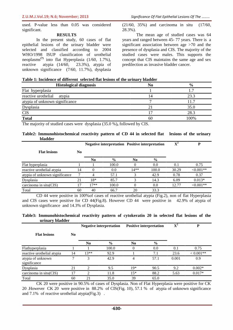
Fig. (1): Reactive urothelial atypia. The epithelial cells show enlarged uniform vesicular nuclei(arrow),
some with Prominent central nucleoli. Mitotic figures are confined to the basal epithelial layers
(Hx & E X 400)
Fig. (2): Reactive urothelial atypia showing diffuse and strong membranous immunoreactivity for CD
44 (DAB chromogen. Mayer's hematoxylin (M.H) counter stain X400).
Z.U.M.J.Vol.19; N.6; November; 2013
-633-
Significance Of Flat Epithelial Lesions Of The ………
Fig. (3): Reactive urothelial atypia showing moderate diffuse cytoplasmic immunoreactivity for CK 20
(DAB chromogen. M.H counter stain X400).
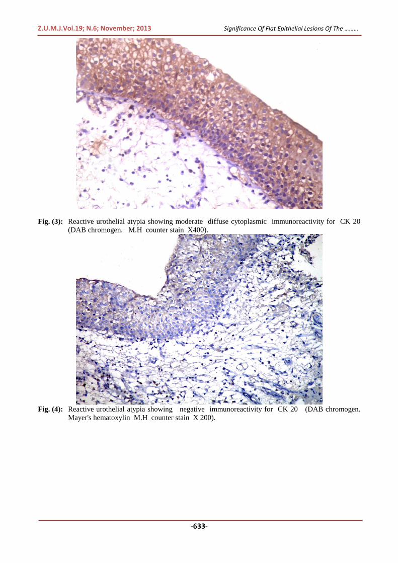
Fig. (4): Reactive urothelial atypia showing negative immunoreactivity for CK 20 (DAB chromogen.
Mayer's hematoxylin M.H counter stain X 200).

Z.U.M.J.Vol.19; N.6; November; 2013
-634-
Significance Of Flat Epithelial Lesions Of The ………
Fig. (5): Reactive urothelial atypia showing focal nuclear ki- 67 immunoreactivity (ki-67 index < 15%)
(DAB chromogen. M.H counter stain X400).
Fig. (6): Reactive urothelial atypia showing diffuse nuclear ki-67 immunoreactivity (ki- 67 index>15%)
(DAB chromogen. Mayer's hematoxylin M.H counter stain X 200).
Z.U.M.J.Vol.19; N.6; November; 2013
-635-
Significance Of Flat Epithelial Lesions Of The ………
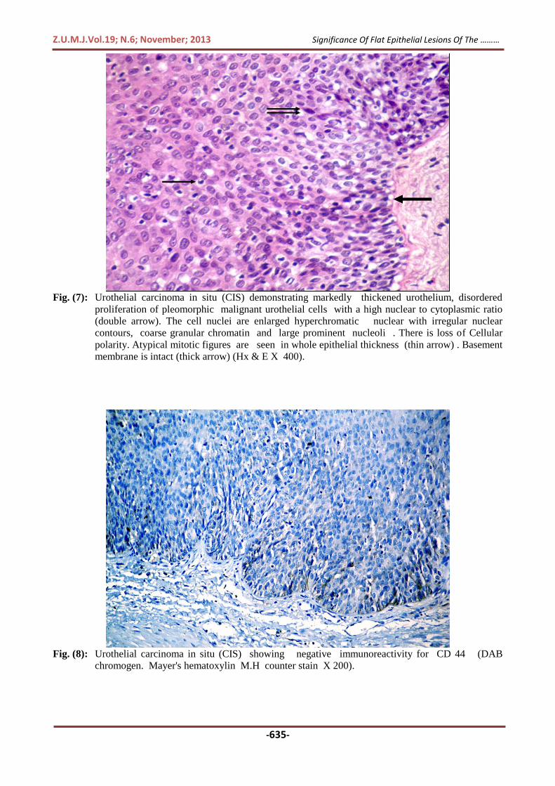
Fig. (7): Urothelial carcinoma in situ (CIS) demonstrating markedly thickened urothelium, disordered
proliferation of pleomorphic malignant urothelial cells with a high nuclear to cytoplasmic ratio
(double arrow). The cell nuclei are enlarged hyperchromatic nuclear with irregular nuclear
contours, coarse granular chromatin and large prominent nucleoli . There is loss of Cellular
polarity. Atypical mitotic figures are seen in whole epithelial thickness (thin arrow) . Basement
membrane is intact (thick arrow) (Hx & E X 400).
Fig. (8): Urothelial carcinoma in situ (CIS) showing negative immunoreactivity for CD 44 (DAB
chromogen. Mayer's hematoxylin M.H counter stain X 200).

Z.U.M.J.Vol.19; N.6; November; 2013
-636-
Significance Of Flat Epithelial Lesions Of The ………
Fig. (9): Urothelial carcinoma in situ (CIS) showing negative immunoreactivity for CK 20 (DAB
chromogen. Mayer's hematoxylin M.H counter stain X 200).
Fig. (10): Urothelial carcinoma in situ (CIS) showing moderate diffuse cytoplasmic Immunoreactivity
for CK 20 (DAB chromogen. Mayer's hematoxylin M.H counter stain X 200).
Z.U.M.J.Vol.19; N.6; November; 2013
-637-
Significance Of Flat Epithelial Lesions Of The ………
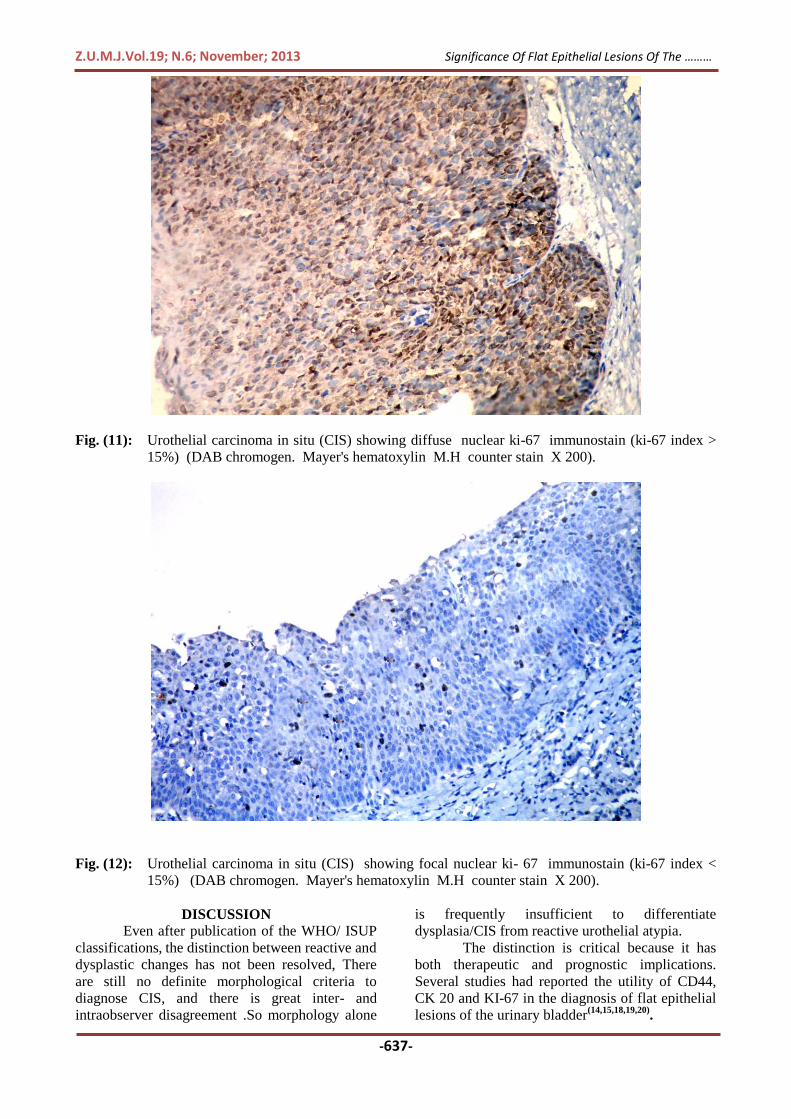
Fig. (11): Urothelial carcinoma in situ (CIS) showing diffuse nuclear ki-67 immunostain (ki-67 index >
15%) (DAB chromogen. Mayer's hematoxylin M.H counter stain X 200).
Fig. (12): Urothelial carcinoma in situ (CIS) showing focal nuclear ki- 67 immunostain (ki-67 index <
15%) (DAB chromogen. Mayer's hematoxylin M.H counter stain X 200).
DISCUSSION
Even after publication of the WHO/ ISUP
classifications, the distinction between reactive and
dysplastic changes has not been resolved, There
are still no definite morphological criteria to
diagnose CIS, and there is great inter- and
intraobserver disagreement .So morphology alone
is frequently insufficient to differentiate
dysplasia/CIS from reactive urothelial atypia.
The distinction is critical because it has
both therapeutic and prognostic implications.
Several studies had reported the utility of CD44,
CK 20 and KI-67 in the diagnosis of flat epithelial
lesions of the urinary bladder(14,15,18,19,20)
.
Z.U.M.J.Vol.19; N.6; November; 2013
-638-
Significance Of Flat Epithelial Lesions Of The ………
In our present a statistically significant
relationship was observed between age <50 and the
presence of isolated flat lesions. There is also a
significant association between age >70 and the
presence of flat lesions(dysplasia/CIS) in
association with carcinoma This goes with
previous studies by Cheng et al.(21)
, Demir et
al.(22)
, Fernando et al.(23)
, Garbar et al.(24)
and
Takenaka et al.(9)
. In the present study, the
majority of the studied cases of flat lesions
including CIS were males (88.3%). This agrees
with Jemal et al.(25)
who reported the majority of
the studied CIS cases were males. However our
cases were randomly selected, thereby an accurate
significant value regarding age and sex cannot be
given.
One case of flat hyperplasia was included
in this study, it was negative for (CD 44 and CK
20) and expressed low ki 67 index (table 2,3,4).
This immunohistochemical profile is similar to that
of normal urothelium. Similar pattern is reported
by Hodges et al.(1)
. This finding supported that flat
hyperplasia does not have a premalignant
potential
The studied 14(100%) cases of reactive
urothelial atypia were positive for CD 44. 9(64%)
out of them showed full thickness membranous
overexpression of CD 44 and 5(36%) out of them
showed patchy positivity in the urothelium (table
2).
. This is in agreement with McKenney et al.(15)
who found membranous full thickness over
expression of CD44 in (60%) of their studied
reactive urothelial atypia and patchy positivity in
the basal and intermediate cells (similar to normal
urothelium) in (40%) of them.
As regarding CK 20 in the studied 14 cases
of reactive urothelial atypia, 13(92.9%) were
negative for CK 20. There is one case that was
positive for CK 2(table 3) but this is considered as
non-specific because of benign morphology,
positivity for CD44 and low ki 67 index. This
result is close to the result of Kunju et al.(14)
who
had (96%) of RUA negative with CK20 beneath
the superficial umbrella layer and had one case
(4%) of morphological clear-cut RUA showing
focal full thickness CK20 expression that
interpreted by them as non-specific because it was
patchy with benign morphology. They stated that
abnormal CK20 staining may occur on rare
occasions; therefore correlation with morphology
is critical. They mentioned that CK20 expression
in the superficial umbrella cells of fragmented
epithelial fragments opposed to each other may
give a false impression of a full-thickness staining
pattern of UD . In a previous studies by
McKenney et al.(15)
and Mallofre et al.(19)
found
that CK20 staining only the umbrella cell layer in
(100%) of their studied reactive urothelial atypia.
Concerning Ki 67 expression in the studied
14 cases of reactive urothelial atypia, low ki 67
index was observed in 12/14(85.7%) and high ki
67 index was observed in 2/14 (14.3%), these cases
were associated with severe inflammation(table
4). The high ki 67 index in these cases were
considered non-specific after correlation with
benign morphology, and other immunostain
(positive CD44 and negative CK 20). This is in
agreement with Kunju et al.(14) who found 28%
of morphologically clear-cut RUA also had
increased expression, especially when associated
with severe inflammation. So caution must be
exercised when interpreting this antibody in an
inflamed urothelium.
Seven cases of atypia of unknown
significance were found in this study, 3/7 (42.9%)
were positive for CD44 and 4/7 (57.1%) were
negative for CD44(table 2). As regarding CK 20 in
the studied 7 cases of atypia of unknown
significance, 4/7 (57.1%) were positive for CK 20
and 3/7 (42.9%) were negative for CK 20(table 3).
Concerning Ki 67 expression in the studied 7 cases
of atypia of unknown significance low Ki 67 index
was observed in 3/7 (42.9%) and it was high in 4/7
(57.1%) (table 4) .
The three markers were evaluated together
and correlated with the morphology to reach a
diagnosis. Six cases were categorized as reactive
versus dysplastic .A total of 4/7(57.1%) were
rediagnosed as dysplasia. All the 4 cases (4/7)
were positive for CK 20, of which 2 cases (2/4)
also demonstrated high ki 67 index and were
negative for CD44; these two cases were favored
to be categorized as dysplasia. In one case (1/4) ki
67 index was high but it was positive for CD44,
the abnormal expression of both CK 20 and ki 67
made the diagnosis of dysplasia more likely. In
other case(1/4) ki 67 index was low, however
CD44 was negative, this case was also thought to
be dysplasia because of the abnormal expression of
both CK 20 and CD44 and correlation with the
morphology. Our result goes with Kunju et al.(14)
who mentioned that the greatest value of a panel
of immunostains would be the ability to resolve
cases of AUS and realized that AUS is not a
disease or a diagnostic entity but merely a
descriptive term used in diagnostically difficult
cases. On the other hand, Mallofre et al.(19) found
75% of atypia of unknown significance suggested
to be dysplastic. They based their suggestion on
strong positivity in scattered suspected dysplastic
cells through the epithelium to at least one marker.
As regarding CD 44 immunohistochemical
reactivity in dysplasia and CIS, in 21 cases of
Z.U.M.J.Vol.19; N.6; November; 2013
-639-
Significance Of Flat Epithelial Lesions Of The ………
dysplasia CD 44 produced mixed result. While
18/21 (85.7%) were negative, 3/21 (14.3%) were
positive for CD 44(table 2). . In all three cases
abnormal CK 20 expression were observed. In
Two cases(2/3) with abnormal CK 20 expression
high ki 67 index were noted .only one of them
(1/3) showed low ki 67 index. However in CIS CD
44 were negative in 17(100%) cases .Such
negativity ranged from complete absence of CD44
reactivity in 11( 64.7%) out of them to residual
CD44 membranous reactivity in the basal cell layer
in 6(35.3%). This result is closely related to that of
McKenney et al. (2001) who found, a lack of
CD44 reactivity in the neoplastic cells of all cases
of CIS but in 44% of them, an underlying residual
basal cell layer was present which showed CD44
membranous reactivity.
Concerning CK 20 immunohistochemical
reactivity in dysplasia and CIS, in dysplasia, 19/21
(90.5%) were positive for CK 20 and 2/21 (9.5%)
were negative for CK 20(table 3).. Unfortunately,
such two cases also expressed low ki 67 index but
they were negative for CD 44. In the studied 17
cases of CIS, 15/17 (88.2%) were positive for CK
20 and 2/17 (11.8%) were negative for CK
20(table 3). Fortunately, such two cases expressed
high ki 67 index and were negative for CD 44.
This agrees with McKenney et al.(15)
who showed
over expression of CK20 (cytoplasmic) in the
majority (>50%) of neoplastic cells in 81% of CIS.
Our result reflected similar findings by
Kunju et al.(14)
who demonstrated abnormal CK20
expression in 86% (43/50) of UD/ CIS group and
negative CK20 in 14%. All seven cases of CIS that
were negative with CK20, had unequivocal over-
expression of Ki-67 (>50% expression of Ki-67).
They believed that although CK20 appears to be a
fairly specific marker of urothelial dysplasia, a
small subset of morphologically clear-cut CIS can
be negative with this antibody. Increased Ki-67
expression in CIS cases negative with CK20 can
usually resolve this problem. In their study, all CIS
cases negative with CK20 showed unequivocal
over expression with Ki-67. These results are
nearly similar to that of Mallofre et al.(19)
who
found that 72 % (36/50) out of the CIS cases were
positive for CK20. Through the full thickness of
the urothelium. 86% of them (31/36) showing a
strong full-thickness positivity of >50% of atypical
cells.
As regarding ki 67 expression in dysplasia
and CIS, in dysplasia high Ki 67 index was
observed in 11/21 (52.4%) while low Ki 67 index
was observed Ki 67 in 10(47.6%) (table4). Among
Such 10 cases with low Ki 67 index, CK 20 were
positive in 8 (8/10) out of them but it were
negative in 2( 2/10) out of them . Fortunately, such
two cases were negative for CD 44. In CIS high Ki
67 index was observed in 14/17(82.4%) while low
Ki 67 index was observed in 3/17 (17.6%) (table
4).. Positive CK 20 and negative CD 44 solved the
problem in these three cases. This result is near to
the result of Mallofre et al.(19)
.
In our study, there is a statistically
significant difference between the reactive
urothelium and dysplastic urothelium (dysplasia
and CIS) as regarding their immunohistochemical
reactivity for CD 44, CK 20 and Ki 67 submitted
in the study . This is in agreement with McKenney
et al.(15)
who demonstrated utility of CD-44 in
reactive urothelium and Mallofre et al.(19)
and
Kunju et al.(14)
who found CK20 and Ki-67 to be
useful markers of UD/CIS.
Conclusion: According to our results, CD
44 is the most constant marker in the reactive
urothelium. CK20 is the most commonly detected
marker in the dysplastic urothelium. Nevertheless,
the most accurate is application of an
immunohistochemical panel composed of the three
antibodies (standard CD 44, CK20 and Ki-67)
together with correlation with morphology. This is
very useful for confirming the presence of
dysplastic changes in the urothelium and
differentiating reactive urothelium from dysplastic
urothelium (dysplasia/CIS).
REFERENCES
1. Hodges B K, Lopez-Beltran A, Davidson D D,
Montironi R and Cheng L. Urothelial dysplasia
and other flat lesions of the urinary bladder:
clinicopathologic and molecular features.J
Human Pathology (2010); 41: 155–162.
2. El-Mawla N.G., El-Bolkainy M.N. and Khaled
H.M. Bladder cancer in Africa: Update. Semin
Oncol(2001); 28(2): 178-8.
3. Eble JN, Sauter G, Epstein JI and Sesterhenn IA.
World Health Organization classification of
tumours: pathology and genetics of tumours of
the urinary system and male genital organs.
Lyon: IARC Press; (2004).
4. Cheng L., Lopez-Beltran A., MacLennan G.T.,
Montironi R. and Bostwick D.G. Neoplasms of
the urinary bladder. In: Bostwick D.G. and
Cheng L., Editors, Urologic surgical pathology .
2 nd ed. Elsevier/Mosby, Philadelphia (2008);
259–352.
5. Petersen R.O.,Sester I.A. and Davis C.J. Flat
urothelial proliferations with significant
malignant potential. In: Urologic pathology . 3 rd
ed. (2009); 214-251.
6. Montironi R., Lopez-Beltran A., Scarpelli M.,
Mazzucchelli R. and Cheng L. Morphological
classification and definition of
benign,preneoplastic and noninvasive neoplastic
lesions of the urinary bladder. Histopathology
(2008); 53: 621-633.

Z.U.M.J.Vol.19; N.6; November; 2013
-640-
Significance Of Flat Epithelial Lesions Of The ………
7. Grignon D.J. The current classification of
urothelial neoplasms. Mod Pathol (2009); 22 (2):
560–90.
8. Edge SB , Byrd DR, Compton CC, Fritz AG,
Greene FL and Trotti A. American joint
committee on cancer staging manual (7th ed.),
Springer, New York (2010).
9. Takenaka A., Yamada Y., Miyake H., Hara I.
and Fujisawa M. Clinical outcomes of Bacillus
Calmette-Guerin instillation therapy for
carcinoma in situ of urinary bladder, Int J Urol.
(2008); 15: 309–313
10. Montironi R, Mazzucchelli R, Scarpelli M,
Lopez-Beltran A and Cheng L: Morphological
diagnosis of urothelial neoplasms. J Clin Pathol
2008, 61:3-10.
11. Cheng L, Cheville JC, Neumann RM and
Bostwick DG. Flat intraepithelial lesions of the
urinary bladder. Cancer (2000); 88: 625-31
12. Williamson S R. , Montironi R , Lopez-Beltran
A, MacLennan T , Davidson D D. and Cheng
L. Diagnosis, evaluation and treatment of
carcinoma in situ of the urinary bladder: The
state of the art. Critical Reviews in
Oncology/Hematology (2010) ;76: 112–126
13. Boman H, Hedelin H and Holmang S. Four
bladder tumor markers have a disappointingly
low sensitivity for small size and low grade
recurrence. J Urol 2002;167:80–3.
14. Kunju LP, Lee CT, Montie J and Shah RB.
Utility of cytokeratin 20 and Ki-67 as markers of
urothelial dysplasia. Pathol Int (2005); 55: 248-
54.
15. McKenney JK, Desai S, Cohen C and Amin MB.
Discriminatory immunohistochemical staining of
urothelial carcinoma in situ and non-neoplastic
urothelium: an analysis of cytokeratin 20, p53,
and CD44 antigens. Am J Surg Pathol (2001);
25: 1074-8.
16. Kogiku M., Ohsawa I., Matsumoto K., Sugisaki
Y., Takahashi H and Teramoto A. Prognosis of
glioma patients by combined immunostaining for
survivin, Ki-67 and epidermal growth factor
receptor. Journal of Clinical Neuroscience
(2008) ;15: 1198–1203 .
17. Bertz S , Otto W, Denzinger S , Wieland F. ,
Burger M , Sto¨hr R , Link S , Hofsta¨dter F
and Hartmann A. Urothelial Cancer:
Combination of CK20 and Ki-67
Immunostaining Analysis Predicts Recurrence,
Progression, and Cancer-Specific Survival in pT1
Urothelial Bladder Cancer. European Urology (2
01 2); 4544 : 1-9.
18. Sun W, Zhang PL and Herrera GA. p53 protein
and Ki-67 overexpression in urothelial dysplasia
of bladder. Appl Immunohistochem Mol
Morphol2002; 10: 327–31.
19. Mallofre C, Castillo M, Morente V and Sole M.
Immunohistochemical expression of CK20, p53,
and Ki-67 as objective markers of urothelial
dysplasia. Mod Pathol(2003); 16: 187–91.
20. Retz M, Lehmann J, Amann E, Wullich B, Roder
C and Stockle M. Mucin 7 and cytokeratin 20 as
new diagnostic urinary markers for bladder
tumor.J Urol(2003);169: 86–9.
21. Cheng L, Cheville JC, Neumann RM and
Bostwick DG. Natural history of urothelial
dysplasia of the bladder. Am. J. Surg. Pathol.
(1999); 23; 443–447.
22. Demir MA, RydW, Aldenborg F and Holmang S.
Cytopathological expression of different types of
urothelial carcinoma in situ in urinary bladder
washings. BJU Int (2003);92:906–10
23. Fernando H, Thota SS, Burtt G, Waterfall N,
Husain I. Importance of red patches diagnosed in
cystoscopy for haematuria and lower urinary tract
symptoms. Postgrad Med J (2007);83:62–3.
24. Garbar C., Mascaux C. and Wespes E. Is
urinary tract cytology still useful for diagnosis of
bladder carcinomas? A large series of 592
bladder washings using a five-category
classification of different cytological diagnoses,
Cytopathology (2007); 18: 79–83.
25. Jemal A, Siegel R, Ward E, Hao Y, Xu J and
Thun MJ. Cancer statistics, 2009. CA Cancer J
Clin 2009;59:225–49.
Z.U.M.J.Vol.19; N.6; November; 2013
-641-
Significance Of Flat Epithelial Lesions Of The ………
أهمية اإلصابات الطائيةالمسطحة للمثانة البىلية فى الكشف المبكر لسرطان المثانة:دراسةهستىباثىلىخيه ومناعيه هستىكيميائيه
الملخص العربي
اإلصنابا فكثش شيعا هي الجاص البلي التاسلي، بعذ سشطاى البشستاتا. تصنالثاي ا ةالخبيث األسام األسام الطالئية البليةخلفية:
ال اصابا طالئية بلينة هسنطحة 2004في تصيف هظوة الصحة العالوية لعام سطحة السابقة للسم في الوثاة البلية الطالئية البلية الو
ة هجلننة ، الوطينن ينن، الوطيننة تعاعل: نن هننا اوطيننة ول ننةهسننطحة اصننابا طالئيننة بليننة دى اوطيننة ول ننة ت التاننخن الوونن
شنار تهنخعا الذس(نة ، األسام األلينة فني الوانا تعنالي الذس(نة . ، و - ، تسن20كينشاتيي -العنال هنا الواعينة هثني تسنيتاألوية
ي التغييشا التعاعلية في حاال فحص العيتوى هعيذ اذ 67ا - تك44د ة الصعبة في توييض األسام األلية في الواا ه
الشننار اإلصننابا فنن 67ا -تكنني 44د –، تط 20كيننشاتيي -تسننيت أ(ش ننه ننز الذساسننة لتحذ ننذ دس الهددذ : ي تشننخيص الونن
. الوسطحة األوش للوثاة البلية
كينشاتيي -: تسنيتضنذ أل(سنام الواناد ا باسنتخذامهي ا(ي التقيين الواعي تن فحصا الوسطحةحالة هوثلة لإلصابا 60 الواد األساليب:
67ا - ت كي 44د - ، تس20
األسام األلينة فني الوانا تعالينة التاخن الوون لوي تعاعليال الوطيةلا ٪ هي حاال 100في كاى إ جابي 44د -تس النتائح:
تهنخعا ٪ هني الون الشنار14.3 الالوطية هجلنة األوينة ٪ هي 42.9في كاى ا جابي 44د -تس سلبية ها رلكالذس(ة كاه
ا حالن هني إ جنابي فينلن وي 20كيشاتيي -تسيت تهخعا الذس(ة . هي الو الشار ٪ 95.5إ جابي في تكاى 20كيشاتيي -. تسيتالذس(ة
هني ٪57.1األسام األلينة فني الوانا تعالينة الذس(نة ، هني ٪88.2 فنيكناى إ جنابي 20كينشاتيي -تسنيتها رلك التاخن الوو حاال
األسام األلينة فني هني ٪82.4كناى هشتعنا فني 67ا -ت كني. هؤشنش الالوطينة التعاعلين هي حناال ٪7.1 الالوطية هجلة األوية
٪57.1 كناى هشتعنا فني 67ا -ت كنيها رلنك هؤشنش اخن الوو الت حاال ل هي حاف ا لن وي هشتعا ، الواا تعالية الذس(ة
. الالوطية التعاعلي ٪ هي14.3تهخعا الذس(ة ٪ هي الو الشار52.4 ،الالوطية هجلة األوية هي
األكثنش شنيعا فني عالهة ال 20اتيي كيش-تسيت . الالوطية التعاعلي في االكثش استوشاس عالهة ال 44د -تس :االستنتاج
20كينشاتيي -سنيت، 44 د -سنتتألف هي ثالثة أ(سنام هاناد ت هاعى . ها رلك، فئى األكثش داة تطبيق لحة الخلي التسج الطالئ
الالوطينة التعاعلينلتعش نق سيج الطالئ ال في تسج ولي التشوي. زا هعيذ (ذا لتأكيذ (د ب سبط رلك (با إل (ب ها 67 ا -كي
.األسام األلية في الواا عالية الذس(ة /هخعا الذس(ة الو الشارت الخلي التسج الطالئ هي