BRAINA JOURNAL OF NEUROLOGY
Selective imitation impairments differentiallyinteract with language processingPaola Mengotti,1,* Corrado Corradi-Dell’Acqua,1,2,* Gioia A. L. Negri,1,3 Maja Ukmar,4
Valentina Pesavento5 and Raffaella I. Rumiati1
1 Neuroscience Area, SISSA, Trieste, Italy
2 Swiss Centre for Affective Sciences, University of Geneve, Geneve, Switzerland
3 IRCCS E. Medea, Polo Friuli-Venezia Giulia, Italy
4 U.C.O. di Radiologia, Azienda Ospedaliero-Universitaria ‘Ospedali Riuniti’, Trieste, Italy
5 S.C. Medicina Riabilitativa, Azienda Ospedaliero-Universitaria ‘Ospedali Riuniti’, Trieste, Italy
*These authors contributed equally to this work.
Correspondence to: Raffaella Rumiati,
Neuroscience Area, SISSA, Via Bonomea 265,
34136 Trieste, Italy
E-mail: [email protected]
Whether motor and linguistic representations of actions share common neural structures has recently been the focus of an
animated debate in cognitive neuroscience. Group studies with brain-damaged patients reported association patterns of praxic
and linguistic deficits whereas single case studies documented double dissociations between the correct execution of gestures
and their comprehension in verbal contexts. When the relationship between language and imitation was investigated, each
ability was analysed as a unique process without distinguishing between possible subprocesses. However, recent cognitive
models can be successfully used to account for these inconsistencies in the extant literature. In the present study, in 57 patients
with left brain damage, we tested whether a deficit at imitating either meaningful or meaningless gestures differentially im-
pinges on three distinct linguistic abilities (comprehension, naming and repetition). Based on the dual-pathway models, we
predicted that praxic and linguistic performance would be associated when meaningful gestures are processed, and would
dissociate for meaningless gestures. We used partial correlations to assess the association between patients’ scores while
accounting for potential confounding effects of aspecific factors such age, education and lesion size. We found that imitation
of meaningful gestures significantly correlated with patients’ performance on naming and repetition (but not on comprehension).
This was not the case for the imitation of meaningless gestures. Moreover, voxel-based lesion-symptom mapping analysis
revealed that damage to the angular gyrus specifically affected imitation of meaningless gestures, independent of patients’
performance on linguistic tests. Instead, damage to the supramarginal gyrus affected not only imitation of meaningful gestures,
but also patients’ performance on naming and repetition. Our findings clarify the apparent conflict between associations and
dissociations patterns previously observed in neuropsychological studies, and suggest that motor experience and language can
interact when the two domains conceptually overlap.
Keywords: apraxia; aphasia; motor system; grounded cognition
Abbreviations: AAT = Aachener Aphasie Test
doi:10.1093/brain/awt194 Brain 2013: 136; 2602–2618 | 2602
Received December 5, 2012. Revised May 6, 2013. Accepted June 10, 2013
� The Author (2013). Published by Oxford University Press on behalf of the Guarantors of Brain. All rights reserved.
For Permissions, please email: [email protected]
at University of G
eneva on July 26, 2013http://brain.oxfordjournals.org/
Dow
nloaded from
IntroductionTraditionally limb apraxia is defined as a deficit in producing vol-
untary movements in absence of elementary sensorimotor or co-
ordination deficits, language comprehension deficit or severe
mental deterioration. The most popular classification of limb
apraxia closely derives from the two-step model of action control
proposed by Liepmann (1920). According to his conceptualization,
a failure to generate the mental image of the intended gesture
gives rise to ideational apraxia, which is better captured by asking
patients to use objects (Steinthal, 1871; Morlaas, 1928; Poeck,
1982; De Renzi and Lucchelli, 1988; Goldenberg and Hagmann,
1998; Rumiati et al., 2001). In contrast, a faulty ability to imple-
ment this image into the appropriate motor output corresponds to
ideomotor apraxia and it is observed, according to Liepmann’s
view later endorsed by De Renzi (1990), when patients are not
only asked to execute a gesture demonstrated by the examiner
(i.e. visuo-imitative apraxia), but also when asked to pantomime
them on verbal command [see Goldenberg, (2009) for detailed
discussions on the model]. As to the brain correlates of these
two main manifestations of apraxia, although ideational apraxia
has been primarily associated with damage to the parietal lobe
(Rumiati et al., 2001; Goldenberg and Spatt, 2009), ideomotor
apraxia has been observed after damage predominantly to the
left parietal and premotor cortices (De Renzi et al., 1980, 1983;
Goldenberg et al., 1996; Tessari et al., 2007; see Goldenberg,
2009; Rumiati et al., 2010, for recent reviews). This two-step
distinction of limb apraxia has been replaced by other accounts
as it does not fully capture all the observed dissociations and it
does not adequately explain the types of errors apraxic patients
make (Poizner et al., 1995).
As in right-handed individuals, left-brain damage frequently (al-
though not necessarily) impairs both linguistic and action abilities,
it has been suggested that apraxia and aphasia might be caused
by the disruption of a single underlying mechanism that, over
the years, has been differentially referred to as ‘asymbolia’
(Finkelnburg, 1870), deficit of ‘abstraction’ (Goldstein, 1948), of
‘conceptualization’ (Bay, 1962) or of ‘use of symbols for commu-
nication’ (Duffy et al., 1975). For instance, de Ajuriaguerra et al.
(1960) tested 415 patients and found that �90% of patients suf-
fering from apraxia (42/47 with ideomotor apraxia and 10/11
with ideational apraxia) were also aphasic. Similarly, De Renzi
et al. (1968) reported a strong correlation between a deficit in
language comprehension and ideational apraxia. These associ-
ations between apraxic and aphasic symptoms in neuropsycho-
logical patients converge with the results from neuroimaging
studies on healthy volunteers in which activations in the fronto-
parietal motor system were observed when they processed action-
related words and sentences (Hauk et al., 2004; Tettamanti et al.,
2005; Ruschemeyer et al., 2007; Postle et al., 2008; Peran et al.,
2010; Willems et al., 2010). These neuropsychological and neu-
roimaging results have, in recent years, been interpreted as evi-
dence for grounded (or embodied) theories of cognition
(Pulvermuller, 2005; Gallese and Lakoff, 2005; Barsalou, 2008).
Accordingly, access to the meaning of a verb or sentence denoting
a given action activates brain regions that are involved in the
execution of the same action. On this perspective, understanding
the meaning of verbs such as ‘grasping’ or ‘throwing’ implies the
re-enactment of the action representations that enable actual
grasping and throwing to occur, and that are thought to be
damaged in apraxic patients.
In contrast with the associations of deficits in large groups of
patients (and by neuroimaging data on healthy individuals), re-
ports of isolated patients exhibiting aphasia in the absence of
apraxia and vice versa suggest how linguistic and motor abilities
can functionally dissociate. For instance, Liepmann (quoted by de
Ajuriaguerra et al., 1960), described seven non-aphasic patients
with apraxia, six of whom had right-sided hemiplegia.
Subsequently, Kertesz et al. (1984) studied 177 cerebrovascular
patients and found that six of them with severe aphasia did not
show any apraxic deficit. Critically, Papagno et al. (1993) tested
699 patients with a shortened version of a comprehension test
(Token test, De Renzi and Faglioni, 1978) to assess language
abilities and an imitation test (De Renzi et al., 1980), and found
that 149 were aphasic but not apraxic, and 10 apraxic but not
aphasic. The double dissociations between apraxic and aphasic
deficits reported in the above studies suggest that the linguistic
ability and the praxic ability are functionally separable and that
they may each rely on a different neural network.
The striking divergence between association and dissociation
patterns was confirmed by two recent studies in which 37
(Negri et al., 2007) and 12 (Papeo et al., 2010) unilaterally
brain-damaged patients were required to perform motor and
verbal tasks on the same set of stimuli. Although at the group-
level reliable correlations between patients’ performance in motor
and linguistic tasks were documented, when the performance of
individual patients was considered, double dissociations between
object use and linguistic processing of the corresponding nouns
were observed (Negri et al., 2007; Papeo et al., 2010). There
are several possible explanations as to why conflicting results are
found in the literature: (i) the tests used to assess language and
imitation, which tend to vary considerably across studies, typically
tap either language or imitation as if they each were a single
process; (ii) patients’ averaged performance does not show the
single cases who may dissociate (i.e. the artifact of the mean,
Shallice, 1988); and (iii) the brain regions sustaining language
and imitation are contiguous and therefore a vascular lesion may
affect both abilities.
As far as imitation is concerned, only a handful of neuropsycho-
logical reports analysed at single-case level patients’ dissociating
imitative performance on meaningful and meaningless gestures
(Goldenberg and Hagmann, 1997; Peigneux et al., 2000; Bartolo
et al., 2001; Tessari et al., 2007). This evidence suggests that
imitation does not rely on a single but on multiple mechanisms
(see also Tessari and Rumiati, 2004, for evidence from healthy
individuals). Overall, three patients had more difficulty imitating
meaningful gestures than meaningless gestures, whereas 11
showed the opposite pattern. The lesions of 7 of 11 patients
with a specific deficit at imitating meaningless gestures seem to
affect the left angular gyrus (Patients LK and EN in Goldenberg
and Hagmann 1997; one patient described in Peigneux et al.,
2004; Cases 12, 13, 19 and 23 in Tessari et al., 2007), whereas
two of three patients with a specific deficit in imitating meaningful
Apraxia and aphasia Brain 2013: 136; 2602–2618 | 2603
at University of G
eneva on July 26, 2013http://brain.oxfordjournals.org/
Dow
nloaded from
gestures had lesions also involving the middle and superior tem-
poral gyri as well as the hippocampus.
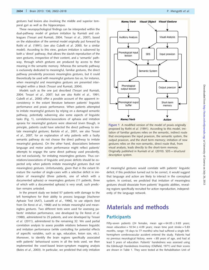
These neuropsychological findings can be interpreted within the
dual-pathway model of gesture imitation by Rumiati and col-
leagues (Tessari and Rumiati, 2004; Tessari et al., 2007), based
on the elaboration of the seminal model originally put forward by
Rothi et al. (1991); (see also Cubelli et al. 2000, for a similar
model). According to this view, gesture imitation is subserved by
both a ‘direct’ pathway, that allows the slavish reproduction of the
seen gestures, irrespective of their content, and a ‘semantic’ path-
way, through which gestures are produced by access to their
meaning in the semantic memory. Whereas the semantic pathway
is exclusively dedicated to meaningful, familiar gestures, the direct
pathway prevalently processes meaningless gestures, but it could
theoretically be used with meaningful gestures too as, for instance,
when meaningful and meaningless gestures are presented inter-
mingled within a block (Tessari and Rumiati, 2004).
Models such as the one just described (Tessari and Rumiati,
2004; Tessari et al., 2007; but see also Rothi et al., 1991;
Cubelli et al., 2000) offer a possible account of the apparent in-
consistency in the extant literature between patients’ linguistic
performance and praxic performance. When patients attempted
to imitate meaningful gestures by relying on a damaged semantic
pathway, potentially subserving also some aspects of linguistic
tasks (Fig. 1), correlations/associations of aphasia and imitation
apraxia for meaningful gestures were observed (even though, in
principle, patients could have selected the direct pathway to imi-
tate meaningful gestures; Bartolo et al., 2001, see also Tessari
et al., 2007, for an explanation of why patients with a faulty
semantic pathway do not choose the direct pathway to imitate
meaningful gestures). On the other hand, dissociations between
language and motor action performance might reflect patients’
attempts to engage the same direct pathway used prevalently,
but not exclusively, for imitating meaningless gestures. Thus cor-
relations/associations of linguistic and praxic deficits should be ex-
pected only when patients imitate meaningful gestures (but not
meaningless) gestures. Unfortunately, given that in the extant lit-
erature the number of single-cases with a selective deficit in imi-
tation of meaningful (three patients, one of which with a
documented aphasia) or meaningless gestures (11 patients, three
of which with a documented aphasia) is very small, such predic-
tion remains untested.
In the present study we tested 57 patients with damage to the
left hemisphere for their ability to process language [Aachener
Aphasie Test (AAT), Luzzatti et al., 1996], to use objects (test
from De Renzi et al., 1968) and to imitate meaningful and mean-
ingless gestures. Two different tests were used for assessing pa-
tients’ imitation performance, one developed by De Renzi et al.
(1980), administered to 25 patients, and one developed by Tessari
et al. (2011), administered to the remaining 32. We used partial
correlation analysis to assess possible relations between linguistic
and imitation performance (while controlling for potential effects
of aspecific variables, such as age, education, lesion size, etc.).
Moreover, to identify the brain regions specifically associated
with patients’ behavioural scores in all the tests used, we then
implemented the voxel-based lesion-symptom mapping analysis
(Bates et al., 2003). In particular, we predicted that only imitation
of meaningful gestures would correlate with patients’ linguistic
deficit; if this prediction turned out to be correct, it would suggest
that language and action are likely to interact in the conceptual
system. In contrast, we predicted that imitation of meaningless
gestures should dissociate from patients’ linguistic abilities, reveal-
ing regions specifically recruited for action reproduction, independ-
ently of the language network.
Materials and methods
ParticipantsFifty-seven patients (24 females, mean age = 64.05 � 9.83 years;
mean education = 10.54 � 4.09 years; mean time post stroke = 5.83
months, range: 15 days to 77 months) who had suffered a single left-
hemisphere cerebrovascular accident entered the study. Patients had
no previous neurological history, were 585 years of age, and had at
least 5 years of education. Patients’ handedness was assessed using
the Edinburgh Handedness Inventory (Oldfield, 1971) and their scores
are shown in Table 1. They were tested at the Rehabilitation Unit of
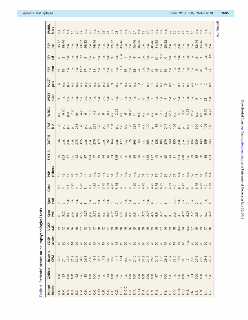
Figure 1 A modified version of the model of praxis originally
proposed by Rothi et al. (1991). According to this model, imi-
tation of familiar gestures relies on the semantic, indirect route
that encompasses the input praxicon, the semantic system, the
output praxicon, and the short term memory; imitation of new
gestures relies on the non-semantic, direct route that, from
visual analysis, leads directly to the short-term memory.
Originally published in Rumiati et al. (2010). SDS = structural
description system.
2604 | Brain 2013: 136; 2602–2618 P. Mengotti et al.
at University of G
eneva on July 26, 2013http://brain.oxfordjournals.org/
Dow
nloaded from
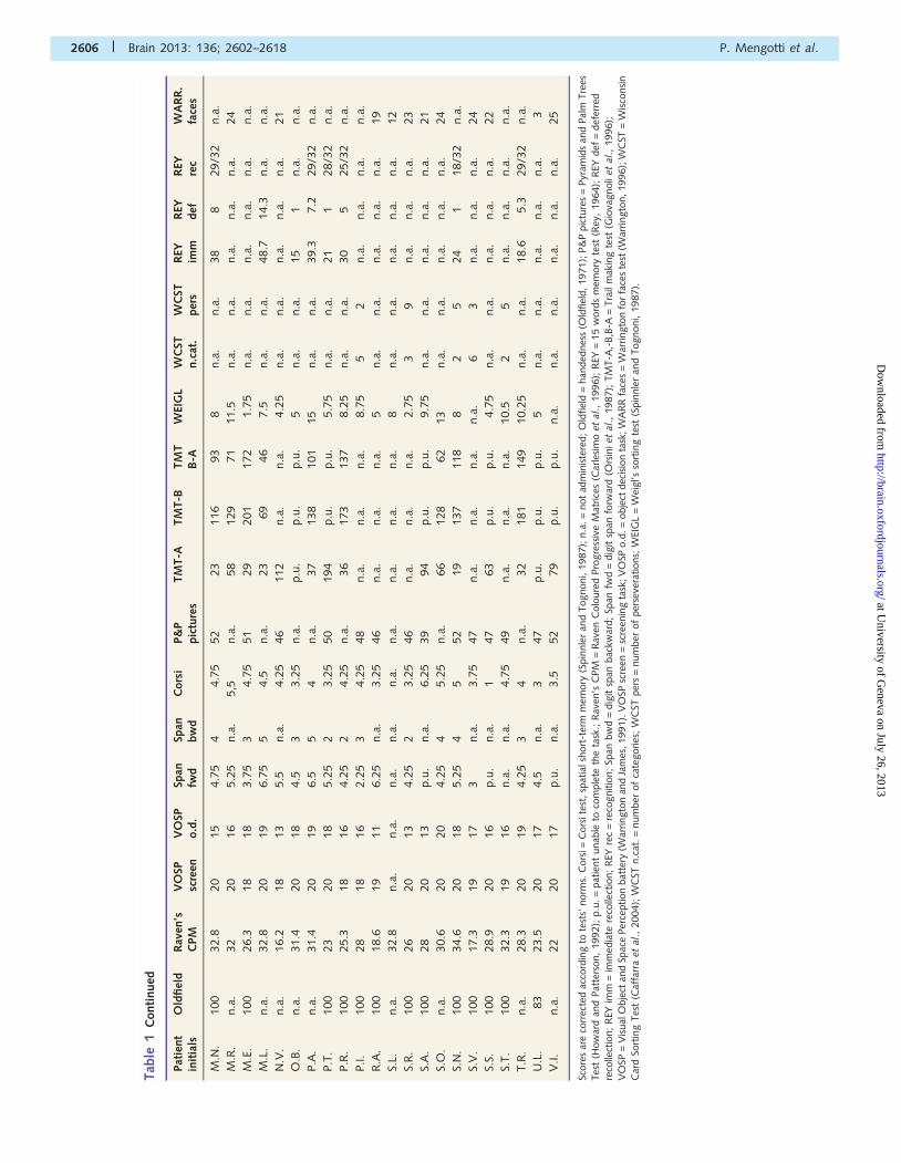
Tab
le1
Pat
ients
’sc
ore
son
neu
ropsy
cholo
gic
alte
sts
Pat
ient
init
ials
Old
fiel
dR
aven
’sC
PM
VO
SPsc
reen
VO
SPo.d
.Sp
anfw
dSp
anbw
dC
ors
iP&
Ppic
ture
sTM
T-A
TM
T-B
TM
TB
-AW
EIG
LW
CST
n.c
at.
WC
STper
sR
EYim
mR
EYdef
REY
rec
WA
RR
.fa
ces
A.N
.100
32.8
18
19
66
4.5
50
26
46
20
n.a
.5
635
626/3
2n.a
.
B.V
.83
21
20
12
3.2
53
448
203
p.u
.p.u
.6.7
5n.a
.n.a
.18
238/4
6n.a
.
B.R
.100
36.8
20
16
n.a
.n.a
.4.5
48
37
p.u
.p.u
.p.u
.p.u
.p.u
.n.a
.n.a
.n.a
.n.a
.
B.U
.100
21.2
14
16
p.u
.n.a
.2.2
546
p.u
.p.u
.p.u
.7.2
5n.a
.n.a
.n.a
.n.a
.n.a
.16
B.Z
.92
32.6
20
16
n.a
.n.a
.4
49
22
275
253
10
n.a
.n.a
.n.a
.n.a
.n.a
.24
C.A
.n.a
.34.9
20
17
5.5
45.2
5n.a
.41
116
76
4.7
5n.a
.n.a
.13.7
1.1
23/3
222
C.N
.-8
326.6
20
13
4.2
53
344
67
p.u
.p.u
.6
p.u
.p.u
.17
226/3
2n.a
.
C.C
.100
20.8
19
12
p.u
.n.a
.n.a
.47
194
p.u
.p.u
.1.5
n.a
.n.a
.n.a
.n.a
.n.a
.p.u
.
C.G
.100
19.8
19
17
3.7
52
4.2
5n.a
.47
276
229
4.5
n.a
.n.a
.31
744/4
6n.a
.
C.O
.n.a
.34.3
20
18
n.a
.n.a
.3.5
n.a
.59
179
120
5n.a
.n.a
.n.a
.n.a
.n.a
.25
C.R
.n.a
.7.2
19
12
n.a
.n.a
.3.7
5n.a
.94
p.u
.p.u
.0
p.u
.p.u
.n.a
.n.a
.n.a
.23
C.S
.83
36
20
17
n.a
.n.a
.4.7
550
10
92
82
8.5
n.a
.n.a
.n.a
.n.a
.n.a
.25
C.U
.100
30.8
20
19
6.7
55
4.7
551
63
164
101
n.a
.6
037
932/3
2n.a
.
C.Z
.n.a
.14.4
18
15
4.7
52
3.2
5n.a
.325
p.u
.p.u
.2.2
5n.a
.n.a
.n.a
.n.a
.n.a
.23
D.C
.R.
n.a
.30.1
19
18
6.5
64
52
21
177
175
n.a
.6
033.3
6.4
41/4
6n.a
.
D.C
.C.
n.a
.n.a
.20
16
n.a
.n.a
.4
n.a
.n.a
.n.a
.n.a
.6
n.a
.n.a
.15
1n.a
.n.a
.
D.B
.100
31
20
16
n.a
.n.a
.4
51
48
76
28
10.7
5n.a
.n.a
.n.a
.n.a
.n.a
.25
D.P
.100
24.5
20
15
5.5
34.2
5n.a
.24
263
239
11
n.a
.n.a
.22
445/4
624
D.O
.100
32.3
20
20
74
3.5
52
20
64
44
n.a
.6
024
440/4
6n.a
.
D.R
.100
32.3
20
19
3.7
5n.a
.4.7
551
n.a
.n.a
.n.a
.n.a
.6
1p.u
.p.u
.p.u
.19
F.R
.100
21.8
20
15
2.7
5n.a
.4
47
133
303
170
3n.a
.n.a
.n.a
.n.a
.n.a
.20
F.N
.100
26.8
20
19
4.5
44.7
552
122
p.u
.p.u
.n.a
.2
325
140/4
6n.a
.
F.U
.67
21.2
20
15
5.5
43.7
550
43
128
85
9n.a
.n.a
.45
10
31/3
2n.a
.
F.L.
n.a
.33.8
20
17
4.7
55
6.2
5n.a
.36
104
68
7.5
n.a
.n.a
.45
9.2
n.a
.n.a
.
F.S.
100
29.8
19
16
53
451
65
276
211
n.a
.4
15
15
025/3
2n.a
.
G.C
.n.a
.33.4
19
19
43
4.7
552
44
340
296
12.5
n.a
.n.a
.n.a
.n.a
.n.a
.24
G.V
.n.a
.33.5
19
18
p.u
.p.u
.4.5
51
21
155
106
n.a
.6
1n.a
.n.a
.n.a
.25
G.O
.n.a
.19.5
19
n.a
.3
n.a
.p.u
.n.a
.303
p.u
.p.u
.2
n.a
.n.a
.p.u
.p.u
.p.u
.13
G.U
.100
32
20
14
4.2
5n.a
.4.2
532
238
p.u
.p.u
.3.7
5n.a
.n.a
.n.a
.n.a
.n.a
.23
H.B
.n.a
.17
n.a
.n.a
.6.2
52
3n.a
.n.a
.n.a
.n.a
.3.7
5n.a
.n.a
.n.a
.n.a
.n.a
.18
J.N
.83
29.8
20
17
2.7
5n.a
.4
52
39
134
95
11.7
5n.a
.n.a
.n.a
.n.a
.n.a
.16
L.E.
100
27.6
20
19
2.5
23.7
548
35
339
304
n.a
.5
75
232/4
6n.a
.
L.N
.100
29.8
20
19
54
4.7
551
70
149
79
n.a
.3
520
341/4
6n.a
.
L.I.
n.a
.29.8
20
17
n.a
.n.a
.4.5
n.a
.36
189
153
9.7
523
2n.a
.n.a
.n.a
.n.a
.
L.C
.n.a
.23.3
20
16
4.7
54
5.7
5n.a
.103
243
140
6.7
5n.a
.n.a
.23
3.5
n.a
.20
(continued
)
Apraxia and aphasia Brain 2013: 136; 2602–2618 | 2605
at University of G
eneva on July 26, 2013http://brain.oxfordjournals.org/
Dow
nloaded from
Tab
le1
Conti
nued
Pat
ient
init
ials
Old
fiel
dR
aven
’sC
PM
VO
SPsc
reen
VO
SPo.d
.Sp
anfw
dSp
anbw
dC
ors
iP&
Ppic
ture
sTM
T-A
TM
T-B
TM
TB
-AW
EIG
LW
CST
n.c
at.
WC
STper
sR
EYim
mR
EYdef
REY
rec
WA
RR
.fa
ces
M.N
.100
32.8
20
15
4.7
54
4.7
552
23
116
93
8n.a
.n.a
.38
829/3
2n.a
.
M.R
.n.a
.32
20
16
5.2
5n.a
.5,5
n.a
.58
129
71
11.5
n.a
.n.a
.n.a
.n.a
.n.a
.24
M.E
.100
26.3
18
18
3.7
53
4.7
551
29
201
172
1.7
5n.a
.n.a
.n.a
.n.a
.n.a
.n.a
.
M.L
.n.a
.32.8
20
19
6.7
55
4.5
n.a
.23
69
46
7.5
n.a
.n.a
.48.7
14.3
n.a
.n.a
.
N.V
.n.a
.16.2
18
13
5.5
n.a
.4.2
546
112
n.a
.n.a
.4.2
5n.a
.n.a
.n.a
.n.a
.n.a
.21
O.B
.n.a
.31.4
20
18
4.5
33.2
5n.a
.p.u
.p.u
.p.u
.5
n.a
.n.a
.15
1n.a
.n.a
.
P.A
.n.a
.31.4
20
19
6.5
54
n.a
.37
138
101
15
n.a
.n.a
.39.3
7.2
29/3
2n.a
.
P.T
.100
23
20
18
5.2
52
3.2
550
194
p.u
.p.u
.5.7
5n.a
.n.a
.21
128/3
2n.a
.
P.R
.100
25.3
18
16
4.2
52
4.2
5n.a
.36
173
137
8.2
5n.a
.n.a
.30
525/3
2n.a
.
P.I
.100
28
18
16
2.2
53
4.2
548
n.a
.n.a
.n.a
.8.7
55
2n.a
.n.a
.n.a
.n.a
.
R.A
.100
18.6
19
11
6.2
5n.a
.3.2
546
n.a
.n.a
.n.a
.5
n.a
.n.a
.n.a
.n.a
.n.a
.19
S.L.
n.a
.32.8
n.a
.n.a
.n.a
.n.a
.n.a
.n.a
.n.a
.n.a
.n.a
.8
n.a
.n.a
.n.a
.n.a
.n.a
.12
S.R
.100
26
20
13
4.2
52
3.2
546
n.a
.n.a
.n.a
.2.7
53
9n.a
.n.a
.n.a
.23
S.A
.100
28
20
13
p.u
.n.a
.6.2
539
94
p.u
.p.u
.9.7
5n.a
.n.a
.n.a
.n.a
.n.a
.21
S.O
.n.a
.30.6
20
20
4.2
54
5.2
5n.a
.66
128
62
13
n.a
.n.a
.n.a
.n.a
.n.a
.24
S.N
.100
34.6
20
18
5.2
54
552
19
137
118
82
524
118/3
2n.a
.
S.V
.100
17.3
19
17
3n.a
.3.7
547
n.a
.n.a
.n.a
.n.a
.6
3n.a
.n.a
.n.a
.24
S.S.
100
28.9
20
16
p.u
.n.a
.1
47
63
p.u
.p.u
.4.7
5n.a
.n.a
.n.a
.n.a
.n.a
.22
S.T.
100
32.3
19
16
n.a
.n.a
.4.7
549
n.a
.n.a
.n.a
.10.5
25
n.a
.n.a
.n.a
.n.a
.
T.R
.n.a
.28.3
20
19
4.2
53
4n.a
.32
181
149
10.2
5n.a
.n.a
.18.6
5.3
29/3
2n.a
.
U.L
.83
23.5
20
17
4.5
n.a
.3
47
p.u
.p.u
.p.u
.5
n.a
.n.a
.n.a
.n.a
.n.a
.3
V.I
.n.a
.22
20
17
p.u
.n.a
.3.5
52
79
p.u
.p.u
.n.a
.n.a
.n.a
.n.a
.n.a
.n.a
.25
Score
sar
eco
rrec
ted
acco
rdin
gto
test
s’norm
s.C
ors
i=C
ors
ites
t,sp
atia
lshort
-ter
mm
emory
(Spin
nle
ran
dTognoni,
1987);
n.a
.=
not
adm
inis
tere
d;
Old
fiel
d=
han
ded
nes
s(O
ldfiel
d,
1971);
P&
Ppic
ture
s=
Pyr
amid
san
dPal
mTre
esTes
t(H
ow
ard
and
Pat
ters
on,
1992);
p.u
.=
pat
ient
unab
leto
com
ple
teth
eta
sk.;
Rav
en’s
CPM
=R
aven
Colo
ure
dPro
gre
ssiv
eM
atrice
s(C
arle
sim
oet
al.,
1996);
REY
=15
word
sm
emory
test
(Rey
,1964);
REY
def
=def
erre
dre
colle
ctio
n;
REY
imm
=im
med
iate
reco
llect
ion;
REY
rec
=re
cognitio
n;
Span
bw
d=
dig
itsp
anbac
kwar
d;
Span
fwd
=dig
itsp
anfo
rwar
d(O
rsin
iet
al.,
1987);
TM
T-A
,-B,B
-A=
Tra
ilm
akin
gte
st(G
iova
gnoli
et
al.,
1996);
VO
SP=
Vis
ual
Obje
ctan
dSp
ace
Per
ception
bat
tery
(War
ringto
nan
dJa
mes
,1991).
VO
SPsc
reen
=sc
reen
ing
task
;V
OSP
o.d
.=
obje
ctdec
isio
nta
sk;W
AR
Rfa
ces
=W
arringto
nfo
rfa
ces
test
(War
ringto
n,1996);
WC
ST=
Wis
consi
nC
ard
Sort
ing
Tes
t(C
affa
rra
et
al.,
2004);
WC
STn.c
at.=
num
ber
of
cate
gories
;W
CST
per
s=
num
ber
of
per
seve
rations;
WEI
GL
=W
eigl’s
sort
ing
test
(Spin
nle
ran
dTognoni,
1987).
2606 | Brain 2013: 136; 2602–2618 P. Mengotti et al.
at University of G
eneva on July 26, 2013http://brain.oxfordjournals.org/
Dow
nloaded from
the Ospedali Riuniti in Trieste and at the Azienda Ospedaliera-
Universitaria ‘Santa Maria della Misericordia’ in Udine. All patients (or a
relative in the case of aphasia) read and signed a written informed
consent. The study was approved by the SISSA Ethics Committee and
conducted in accordance with the Declaration of Helsinki.
Neuropsychological assessmentAll 57 patients were administered an extensive neuropsychological as-
sessment evaluating language, praxis, visuo-spatial abilities, memory
and executive functions. Scores are reported in Table 1.
The AAT (Luzzatti et al., 1996) was used to evaluate patients’ lin-
guistic abilities. The AAT provides an output in terms of presence or
absence of aphasia together with a definition of the specific type of
aphasic syndrome and related probabilities, based on the results on
different linguistic tasks (Token test, Naming, Comprehension,
Repetition and Writing). The cut-offs were determined based on the
performance of a group of 88 healthy participants (mean age: 52
years; age range: 20–85 years). Scores on these tasks are reported
in Table 2.
All patients were assessed for their ability to use objects by means of
a test developed by De Renzi et al. (1968), in which patients are asked
to use seven common objects. Maximum score on this test is 14,
which also represents the cut-off. The cut-off was determined based
on the performance of a group of 40 control participants without brain
damage. Pathological performance on this task has been interpreted as
a sign of ideational apraxia (e.g. De Renzi and Lucchelli 1988; Rumiati
et al., 2001).
They were also assessed for their ability to imitate actions using two
different tests, the first in which meaningful and meaningless gestures
are presented in separate blocks, the second in which the two action
types are presented intermingled. The first test, developed in our lab
(Tessari et al., 2011), requires patients to imitate, one after the other,
18 meaningful intransitive gestures and 18 meaningless gestures
derived from the meaningful gestures by modifying the spatial rela-
tionship between the effector and the main body axis. If a correct
response was not produced on the first trial, a second trial was
allowed. For each gesture a score of 0, 1, 2 is given according to
the performance (0 = no imitation, 1 = correct imitation in the
second trial, 2 = correct imitation in the first trial), with the cut-off
varying according to age and years of education (age between 30
and 50: cut-off meaningful4 32, cut-off meaningless4 31, cut-off
total4 63; age between 51 and 70: cut-off meaningful4 31, cut-
off meaningless4 28, cut-off total4 59; age of 71 and above: cut-
off meaningful4 25 if education 56 years and cut-off meaning-
ful4 30 if education5 7 years, cut-off meaningless4 24 if education
56 years and cut-off meaningless4 24 if education5 7 years, cut-off
total4 50 if education 56 years and cut-off total4 58 if educa-
tion5 7 years) for a total score of 72 maximum. The cut-offs were
determined based on the performance of a group of 111 healthy
participants (age range: 30–90 years). The second test, developed by
De Renzi et al. (1980), requires patients to imitate 24 gestures of
which 12 are meaningful (half distal and half proximal) and 12 are
meaningless (half distal and half proximal). Each gesture was presented
up to three times and a score from 0 to 3 was assigned depending on
patients’ performance (0 = no imitation, 1 = correct imitation in the
third trial, 2 = correct imitation in the second trial, 3 = correct imitation
in the first trial), for a maximum total score of 72. A score 553 was
considered as pathological. The cut-offs were determined based on the
performance of a group of 100 control participants without brain
damage (mean age: 52.6 years). Pathological performance on these
tasks has been interpreted as a sign of ideomotor apraxia (De Renzi
et al., 1980; Tessari et al., 2011).
Gestures were considered as incorrect if one of the following errors
was present: (i) spatial error: the gesture is recognizable but the arm or
the hand movement followed the wrong direction or axis; (ii) visuo-
semantic error: the performed gesture is visually similar and semantic-
ally related to the proper gesture; (iii) visual error: the performed ges-
ture is visually similar to the proper one, or it combines two different
gestures already presented, or it is visually similar to a gesture that was
already presented in the list; (iv) omission: the gesture is not repro-
duced; and (v) unrecognizable: the performed gesture is unrecogniz-
able. Patients imitated all the gestures with the ipsilesional left hand.
Scores on the praxic tests are reported in Table 3.
Behavioural data analysisTo ascertain the relation between praxic and linguistic performances,
we carried out partial correlation analyses aimed at assessing the
amount of shared variability between patients’ performance in two
tests while controlling for potential effects of aspecific variables, such
as age, gender, education, lesion size and illness length. Please note
that all linear effects of the confounding variables were simultaneously
removed from each of the two conditions of interest. As a measure of
linguistic proficiency, we took patients’ performance in the AAT. Thus,
for each AAT subtest, we took patients’ score as a percentage of the
maximum possible score. Likewise, as a measure of ideational apraxia,
we took participants’ score on the test from De Renzi et al. (1968) as
a percentage of the maximum possible score. Finally, for ideomotor
apraxia, we considered separately, items in which patients imitated a
meaningful and a meaningless gesture, thus for each kind of gesture
we took patients’ score as a percentage of the maximum possible
score. Furthermore, as ideomotor apraxia was investigated in 24 pa-
tients, with the test from De Renzi et al. (1980) and, in the remaining
33 patients, with the test from Tessari et al. (2011), we subtracted
from the percentage score of each patient the average score from the
test used. Statistical analysis was carried out with R.11.1 (http://cran.r-
project.org//) open source software.
Lesion analysisComputed tomography or MRI scans were available for each patient.
An experienced neuroradiologist (M.U.), blind to the aims of the study
and to the behavioural deficits of the patients, mapped the lesioned
areas of each patient onto the normalized MNI template (http://www.
bic.mni.mcgill.ca/cgi/icbm_view), with a voxel size of 1 � 1 � 1 mm3,
using MRIcro software (http://www.mricro.com; Rorden and Brett,
2000).
Neural correlates of language and praxis measures were further
investigated using voxel-based lesion-symptom mapping (Bates
et al., 2003). This technique allows analysing the relationship between
lesion data (described, for each voxel, as a variable reflecting the pu-
tative presence of a lesion) and continuous behavioural measures. To
this purpose, lesion masks were converted into ANALYZE format vol-
umes, in which 0 corresponded to undamaged tissue and 1 to the
lesioned part of the brain. The images were subsequently smoothed
with an 8-mm full-width at half-maximum kernel, thus allowing non-
integer values around the lesion borders. Each behavioural measure
(percentage scores of meaningless, meaningful, ideational apraxia,
AAT, etc.) was fed into a general linear model in which it was related
to the lesion intensity. The analysis was restricted to those voxels for
which there were at least five patients with and five patients without a
lesion (i.e. those voxels in which the sum of the smoothed lesion
Apraxia and aphasia Brain 2013: 136; 2602–2618 | 2607
at University of G
eneva on July 26, 2013http://brain.oxfordjournals.org/
Dow
nloaded from
Table 2 Summary of all patients’ performance on linguistic tests
Patient AAT
Output Token Repetition Writing Naming Comprehension
A.N. No aphasia 0 148 90 120 120
B.V. Not classified 15 134 78 109 99
B.R. Global aphasia 48 39 12 0 41
B.U. Global aphasia 36 44 9 15 40
B.Z. Wernicke’s aphasia 50 9 22 15 91
C.A. No aphasia 4 146 89 112 112
C.N. No aphasia 4 140 85 108 105
C.C. Wernicke’s aphasia 50 0 0 0 22
C.G. No aphasia 10 136 84 112 113
C.O. Global aphasia 36 39 30 17 83
C.R. Wernicke’s aphasia 50 14 0 27 49
C.S. Wernicke’s aphasia 25 102 47 61 92
C.U. Broca’s aphasia 18 142 68 91 98
C.Z. No aphasia 14 139 68 102 88
D.C.R. No aphasia 0 105 80 115 112
D.C.C. Wernicke’s aphasia 11 88 56 63 95
D.B. Wernicke’s aphasia 39 71 44 83 99
D.P. No aphasia n.a. n.a. n.a. n.a. n.a.
D.O. No aphasia n.a. 146 n.a. n.a. n.a.
D.R. Broca’s aphasia 1 94 15 69 95
F.R. Transcort. aphasia 41 95 58 34 62
F.N. No aphasia n.a. n.a. n.a. n.a. n.a.
F.U. No aphasia 3 142 88 120 117
F.L. No aphasia n.a. n.a. n.a. n.a. n.a.
F.S. Not classified 13 133 82 109 117
G.C. Not classified 5 141 86 102 113
G.V. Not classified 20 90 41 69 112
G.O. Wernicke’s aphasia 26 110 41 62 92
G.U. Wernicke’s aphasia 29 134 71 29 55
H.B. No aphasia 16 145 68 108 87
J.N. Conduction aphasia 6 134 74 109 118
L.E. Wernicke’s aphasia 47 35 58 81 79
L.N. No aphasia n.a. n.a. n.a. n.a. n.a.
L.I. Broca’s aphasia 18 9 12 0 101
L.C. No aphasia n.a. n.a. n.a. n.a. n.a.
M.N. No aphasia 4 131 84 119 118
M.R. No aphasia n.a. n.a. n.a. n.a. n.a.
M.E. Broca’s aphasia 15 123 75 110 102
M.L. No aphasia n.a. n.a. n.a. n.a. n.a.
N.V. Amnestic aphasia 20 139 37 93 90
O.B. No aphasia n.a. n.a. n.a. n.a. n.a.
P.A. No aphasia n.a. n.a. n.a. n.a. n.a.
P.T. Amnestic aphasia 20 141 57 102 89
P.R. No aphasia n.a. n.a. n.a. n.a. n.a.
P.I. Not classified 31 96 55 29 87
R.A. Transcort. aphasia 40 139 22 59 44
S.L. Global aphasia p.u. p.u. p.u. p.u. 38
S.R. Transcort. aphasia 39 147 29 67 87
S.A. Broca’s aphasia 36 101 19 29 46
S.O. No aphasia 1 140 82 118 119
S.N. No aphasia 2 149 89 114 118
S.V. Broca’s aphasia 37 97 18 10 72
S.S. Broca’s aphasia 40 94 31 43 53
(continued)
2608 | Brain 2013: 136; 2602–2618 P. Mengotti et al.
at University of G
eneva on July 26, 2013http://brain.oxfordjournals.org/
Dow
nloaded from
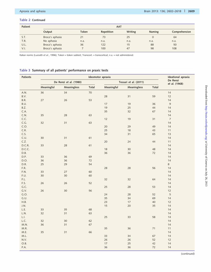
Table 2 Continued
Patient AAT
Output Token Repetition Writing Naming Comprehension
S.T. Broca’s aphasia 21 73 25 0 64
T.R. No aphasia n.a. n.a. n.a. n.a. n.a.
U.L. Broca’s aphasia 36 122 15 88 50
V.I. Broca’s aphasia 7 100 47 98 108
Italian norms (Luzzatti et al., 1996); Token = token subtest; Transcort. = transcortical; n.a. = not administered.
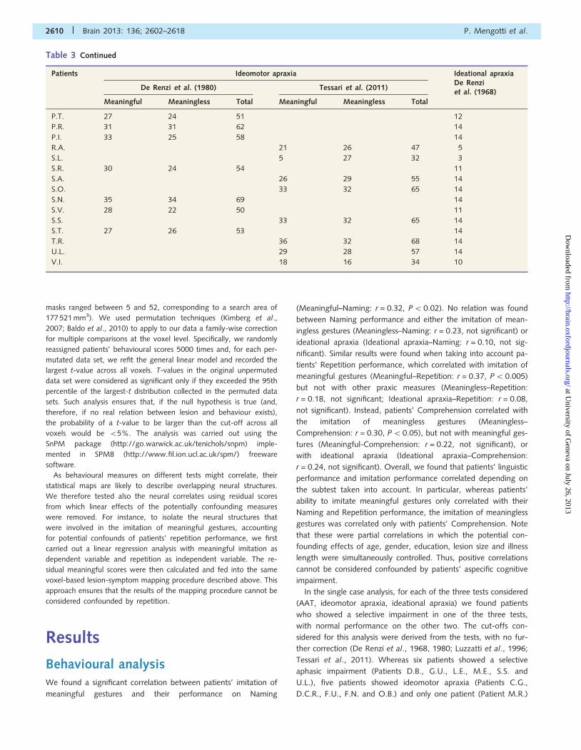
Table 3 Summary of all patients’ performance on praxic tests
Patients Ideomotor apraxia Ideational apraxiaDe Renziet al. (1968)
De Renzi et al. (1980) Tessari et al. (2011)
Meaningful Meaningless Total Meaningful Meaningless Total
A.N. 36 34 70 14
B.V. 28 31 59 12
B.R. 27 26 53 14
B.U. 17 19 36 9
B.Z. 19 25 44 14
C.A. 35 32 67 14
C.N. 35 28 63 14
C.C. 12 19 31 7
C.G. 32 31 63 14
C.O. 20 29 49 14
C.R. 25 18 43 11
C.S. 34 31 65 13
C.U. 30 31 61 14
C.Z. 20 24 44 11
D.C.R. 33 28 61 14
D.C.C. 18 30 48 14
D.B. 36 36 72 14
D.P. 33 36 69 14
D.O. 36 36 72 14
D.R. 25 29 54 8
F.R. 28 28 56 14
F.N. 33 27 60 14
F.U. 30 30 60 14
F.L. 32 32 64 14
F.S. 26 26 52 14
G.C. 25 28 53 14
G.V. 26 30 56 12
G.O. 24 28 52 5
G.U. 35 34 69 14
H.B. 23 17 40 12
J.N. 15 20 35 14
L.E. 33 35 68 14
L.N. 32 31 63 14
L.I. 25 33 58 14
L.C. 32 30 62 14
M.N. 36 31 67 14
M.R. 35 36 71 11
M.E. 35 31 66 14
M.L. 33 34 67 14
N.V. 26 26 52 12
O.B. 17 25 42 14
P.A. 36 36 72 14
(continued)
Apraxia and aphasia Brain 2013: 136; 2602–2618 | 2609
at University of G
eneva on July 26, 2013http://brain.oxfordjournals.org/
Dow
nloaded from
masks ranged between 5 and 52, corresponding to a search area of
177 521 mm3). We used permutation techniques (Kimberg et al.,
2007; Baldo et al., 2010) to apply to our data a family-wise correction
for multiple comparisons at the voxel level. Specifically, we randomly
reassigned patients’ behavioural scores 5000 times and, for each per-
mutated data set, we refit the general linear model and recorded the
largest t-value across all voxels. T-values in the original unpermuted
data set were considered as significant only if they exceeded the 95th
percentile of the largest-t distribution collected in the permuted data
sets. Such analysis ensures that, if the null hypothesis is true (and,
therefore, if no real relation between lesion and behaviour exists),
the probability of a t-value to be larger than the cut-off across all
voxels would be 55%. The analysis was carried out using the
SnPM package (http://go.warwick.ac.uk/tenichols/snpm) imple-
mented in SPM8 (http://www.fil.ion.ucl.ac.uk/spm/) freeware
software.
As behavioural measures on different tests might correlate, their
statistical maps are likely to describe overlapping neural structures.
We therefore tested also the neural correlates using residual scores
from which linear effects of the potentially confounding measures
were removed. For instance, to isolate the neural structures that
were involved in the imitation of meaningful gestures, accounting
for potential confounds of patients’ repetition performance, we first
carried out a linear regression analysis with meaningful imitation as
dependent variable and repetition as independent variable. The re-
sidual meaningful scores were then calculated and fed into the same
voxel-based lesion-symptom mapping procedure described above. This
approach ensures that the results of the mapping procedure cannot be
considered confounded by repetition.
Results
Behavioural analysisWe found a significant correlation between patients’ imitation of
meaningful gestures and their performance on Naming
(Meaningful–Naming: r = 0.32, P5 0.02). No relation was found
between Naming performance and either the imitation of mean-
ingless gestures (Meaningless–Naming: r = 0.23, not significant) or
ideational apraxia (Ideational apraxia–Naming: r = 0.10, not sig-
nificant). Similar results were found when taking into account pa-
tients’ Repetition performance, which correlated with imitation of
meaningful gestures (Meaningful–Repetition: r = 0.37, P5 0.005)
but not with other praxic measures (Meaningless–Repetition:
r = 0.18, not significant; Ideational apraxia–Repetition: r = 0.08,
not significant). Instead, patients’ Comprehension correlated with
the imitation of meaningless gestures (Meaningless–
Comprehension: r = 0.30, P50.05), but not with meaningful ges-
tures (Meaningful-Comprehension: r = 0.22, not significant), or
with ideational apraxia (Ideational apraxia–Comprehension:
r = 0.24, not significant). Overall, we found that patients’ linguistic
performance and imitation performance correlated depending on
the subtest taken into account. In particular, whereas patients’
ability to imitate meaningful gestures only correlated with their
Naming and Repetition performance, the imitation of meaningless
gestures was correlated only with patients’ Comprehension. Note
that these were partial correlations in which the potential con-
founding effects of age, gender, education, lesion size and illness
length were simultaneously controlled. Thus, positive correlations
cannot be considered confounded by patients’ aspecific cognitive
impairment.
In the single case analysis, for each of the three tests considered
(AAT, ideomotor apraxia, ideational apraxia) we found patients
who showed a selective impairment in one of the three tests,
with normal performance on the other two. The cut-offs con-
sidered for this analysis were derived from the tests, with no fur-
ther correction (De Renzi et al., 1968, 1980; Luzzatti et al., 1996;
Tessari et al., 2011). Whereas six patients showed a selective
aphasic impairment (Patients D.B., G.U., L.E., M.E., S.S. and
U.L.), five patients showed ideomotor apraxia (Patients C.G.,
D.C.R., F.U., F.N. and O.B.) and only one patient (Patient M.R.)
Table 3 Continued
Patients Ideomotor apraxia Ideational apraxiaDe Renziet al. (1968)
De Renzi et al. (1980) Tessari et al. (2011)
Meaningful Meaningless Total Meaningful Meaningless Total
P.T. 27 24 51 12
P.R. 31 31 62 14
P.I. 33 25 58 14
R.A. 21 26 47 5
S.L. 5 27 32 3
S.R. 30 24 54 11
S.A. 26 29 55 14
S.O. 33 32 65 14
S.N. 35 34 69 14
S.V. 28 22 50 11
S.S. 33 32 65 14
S.T. 27 26 53 14
T.R. 36 32 68 14
U.L. 29 28 57 14
V.I. 18 16 34 10
2610 | Brain 2013: 136; 2602–2618 P. Mengotti et al.
at University of G
eneva on July 26, 2013http://brain.oxfordjournals.org/
Dow
nloaded from
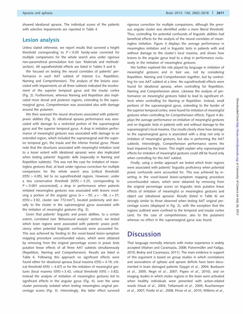
showed ideational apraxia. The individual scores of the patients
with selective impairments are reported in Table 4.
Lesion analysisUnless stated otherwise, we report results that survived a height
threshold corresponding to P50.05 family-wise corrected for
multiple comparisons for the whole search area under rigorous
non-parametrical permutation test (see ‘Materials and methods’
section). All suprathreshold effects are listed in Tables 5 and 6.
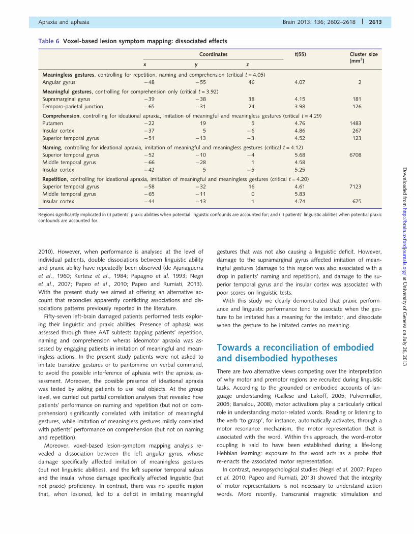
We focused on testing the neural correlates of patients’ per-
formance in each AAT subtest of interest (i.e. Repetition,
Naming and Comprehension). The analysis of the lesions asso-
ciated with impairments on all three subtests indicated the involve-
ment of the superior temporal gyrus and the insular cortex
(Fig. 2). Furthermore, whereas Naming and Repetition also impli-
cated more dorsal and posterior regions, extending to the supra-
marginal gyrus, Comprehension was associated also with damage
around the putamen.
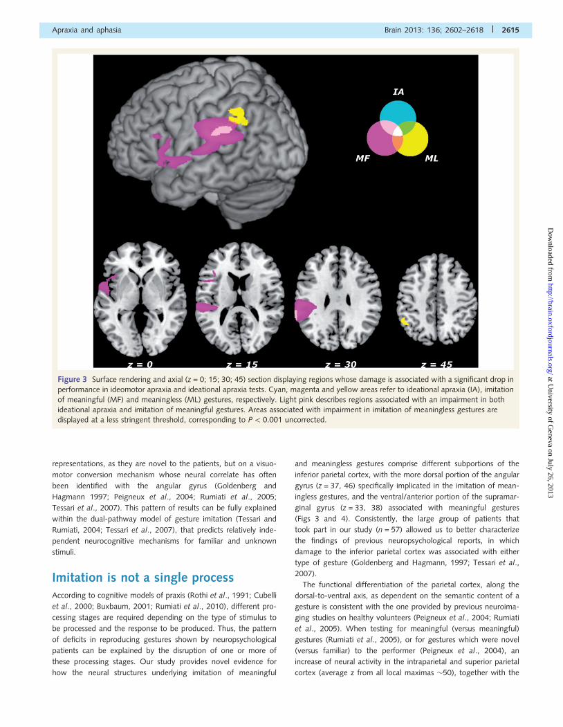
We then assessed the neural structures associated with patients’
praxic abilities (Fig. 3). Ideational apraxia performance was asso-
ciated with damage to a restricted portion of the supramarginal
gyrus and the superior temporal gyrus. A drop in imitation perfor-
mance of meaningful gestures was associated with damage to an
extended region, which included the supramarginal and the super-
ior temporal gyri, the insula and the inferior frontal gyrus. Please
note that the structures associated with meaningful imitation (and
to a lesser extent with ideational apraxia) were also implicated
when testing patients’ linguistic skills (especially in Naming and
Repetition subtests). This was not the case for imitation of mean-
ingless gestures that, at least under rigorous correction for multiple
comparisons for the whole search area [critical threshold:
t(55)44.05], led to no suprathreshold regions. However, under
a less conservative threshold [t(55)4 3.25, corresponding to
P5 0.001 uncorrected], a drop in performance when patients
imitated meaningless gestures was associated with lesions invol-
ving a portion of the angular gyrus [x = �55, y = �58, z = 37;
t(55) = 3.92, cluster size 772 mm3], located posteriorly and dor-
sally to the cluster in the supramarginal gyrus associated with
the imitation of meaningful gestures (Fig. 3).
Given that patients’ linguistic and praxic abilities, to a certain
extent, correlated (see ‘Behavioural analysis’ section), we tested
which brain regions were associated with patients’ praxic profi-
ciency when potential linguistic confounds were accounted for.
This was achieved by feeding to the voxel-based lesion-symptom
mapping procedure unconfounded values, which were obtained
by removing from the original percentage scores in praxic tests
putative linear effects of all three AAT subtests simultaneously
(Repetition, Naming and Comprehension). Results are listed in
Table 6. Following this approach no significant effects were
found either for ideational apraxia [local maxima t(55) = 4.19; crit-
ical threshold t(55)44.67] or for the imitation of meaningful ges-
tures [local maxima t(55) = 3.42; critical threshold t(55)4 3.82].
Instead the analysis of imitation of meaningless gestures led to
significant effects in the angular gyrus (Fig. 4), over the same
cluster previously isolated when testing meaningless original per-
centage scores (Fig. 3). Interestingly, this latter effect survived
rigorous correction for multiple comparisons, although the previ-
ous angular cluster was identified under a more liberal threshold.
Thus, controlling for potential confounds of linguistic abilities had
beneficial effects for the analysis of the neural correlates of mean-
ingless imitation. Figure 4 displays the average performance in
meaningless imitation and in linguistic tests in patients with and
without damage to the cluster’s local maxima, and shows how
lesions to the angular gyrus lead to a drop in performance exclu-
sively in the imitation of meaningless gestures.
We further explored the role played by language in imitation of
meaningful gestures and in tool use, not by considering
Repetition, Naming and Comprehension together, but by control-
ling for one AAT subtest at a time. No suprathreshold effects were
found for ideational apraxia, when controlling for Repetition,
Naming and Comprehension alone. Likewise the analysis of per-
formance on meaningful gestures failed to lead to significant ef-
fects when controlling for Naming or Repetition. Instead, small
portions of the supramarginal gyrus, extending to the border of
the superior temporal cortex, were found for imitation of meaningful
gestures when controlling for Comprehension effects. Figure 4 dis-
plays the average performance on imitation of meaningful gestures
and on linguistic tests in patients with and without damage to the
supramarginal’s local maxima. Our results clearly show how damage
to the supramarginal gyrus is associated with a drop not only in
imitation of meaningful gestures, but also in performing the AAT
subtests. Interestingly, Comprehension performance seems the
least impaired by the lesion. This might explain why supramarginal
effects for imitation of meaningful gestures could still be found only
when controlling for this AAT subtest.
Finally, using a similar approach we tested which brain regions
were associated with patients’ linguistic proficiency when potential
praxic confounds were accounted for. This was achieved by in-
serting in the voxel-based lesion-symptom mapping procedure
unconfounded values, which were obtained by removing from
the original percentage scores on linguistic tests putative linear
effects of imitation of meaningful or meaningless gestures and
object use (ideational apraxia). Results (listed in Table 6) are
strongly similar to those observed when testing AAT original per-
centage scores (displayed in Fig. 2), with the exception that the
regions outlined were confined to the temporal and insular cortex
(and, for the case of comprehension, also to the putamen)
whereas no effect in the supramarginal gyrus was found.
DiscussionThat language normally interacts with motor experience is widely
accepted (Mahon and Caramazza, 2008; Pulvermuller and Fadiga,
2010; Bedny and Caramazza, 2011). The main evidence in support
of this argument is based on group studies in which correlations
and associations of aphasic and apraxic deficits have been docu-
mented in brain damaged patients (Saygin et al., 2004; Buxbaum
et al., 2005; Negri et al., 2007; Papeo et al., 2010), and on
imaging studies in which motor regions in the brain were activated
when healthy individuals were presented with action-related
words (Hauk et al., 2004; Tettamanti et al., 2005; Ruschemeyer
et al., 2007; Postle et al., 2008; Peran et al., 2010; Willems et al.,
Apraxia and aphasia Brain 2013: 136; 2602–2618 | 2611
at University of G
eneva on July 26, 2013http://brain.oxfordjournals.org/
Dow
nloaded from
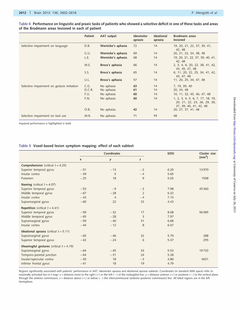
Table 4 Performance on linguistic and praxic tasks of patients who showed a selective deficit in one of these tasks and areasof the Brodmann areas lesioned in each of patient
Patient AAT output Ideomotorapraxix
Ideationalapraxia
Brodmann areaslesioned
Selective impairment on language D.B. Wernicke’s aphasia 72 14 19, 20, 21, 22, 37, 39, 41,42, 48
G.U. Wernicke’s aphasia 69 14 20, 21, 22, 34, 38, 48
L.E. Wernicke’s aphasia 68 14 19, 20, 21, 22, 37, 39, 40, 41,42, 48
M.E. Broca’s aphasia 66 14 2, 3, 4, 6, 20, 22, 38, 41, 42,43, 45, 47, 48
S.S. Broca’s aphasia 65 14 6, 11, 20, 22, 25, 34, 41, 42,43, 47, 48
U.L. Broca’s aphasia 57 14 11, 20, 25, 34, 47, 48
Selective impairment on gesture imitation C.G. No aphasia 63 14 7, 19, 39, 40D.C.R. No aphasia 61 14 20, 34, 48
F.U. No aphasia 60 14 10, 11, 32, 45, 46, 47, 48
F.N. No aphasia 60 14 1, 2, 3, 4, 5, 6, 7, 17, 18, 19,20, 21, 22, 23, 26, 29, 30,37, 39, 40, 41, 42, 48
O.B. No aphasia 42 14 20, 27, 37, 41, 48
Selective impairment on tool use M.R. No aphasia 71 11 48
Impaired performance is highlighted in bold.
Table 5 Voxel-based lesion symptom mapping: effect of each subtest
Coordinates t(55) Cluster size[mm3]
x y z
Comprehension (critical t = 4.25)
Superior temporal gyrus �51 �13 �2 6.20 13 970
Insular cortex �39 5 �4 5.65
Putamen �25 18 9 5.03 1508
Naming (critical t = 4.07)
Superior temporal gyrus �53 �9 �3 7.98 45 360
Middle temporal gyrus �67 �28 2 6.32
Insular cortex �43 4 �4 7.15
Supramarginal gyrus �60 22 32 4.43
Repetition (critical t = 4.41)
Superior temporal gyrus �59 �32 17 8.08 56 585
Middle temporal gyrus �65 �28 3 7.97
Supramarginal gyrus �59 �40 31 6.68
Insular cortex �44 �12 8 6.07
Ideational apraxia (critical t = 5.11)
Supramarginal gyrus �65 �46 32 5.79 288
Superior temporal gyrus �42 �24 6 5.47 255
Meaningful gestures (critical t = 4.19)
Supramarginal gyrus �64 �45 33 5.53 19 733
Temporo-parietal junction �64 �31 24 5.28
Insular/opercular cortex �45 18 �3 4.80 4631
Inferior frontal gyrus �41 18 19 4.79
Regions significantly associated with patients’ performance in AAT, ideomotor apraxia and ideational apraxia subtests. Coordinates (in standard MNI space) refer tomaximally activated foci in t-map: x = distance (mm) to the right ( + ) or the left (� ) of the midsagittal line; y = distance anterior ( + ) or posterior (�) to the vertical planethrough the anterior commissure; z = distance above ( + ) or below (�) the intercommissural (anterior–posterior commissure) line. All listed regions are in the left
hemisphere.
2612 | Brain 2013: 136; 2602–2618 P. Mengotti et al.
at University of G
eneva on July 26, 2013http://brain.oxfordjournals.org/
Dow
nloaded from
2010). However, when performance is analysed at the level of
individual patients, double dissociations between linguistic ability
and praxic ability have repeatedly been observed (de Ajuriaguerra
et al., 1960; Kertesz et al., 1984; Papagno et al. 1993; Negri
et al., 2007; Papeo et al., 2010; Papeo and Rumiati, 2013).
With the present study we aimed at offering an alternative ac-
count that reconciles apparently conflicting associations and dis-
sociations patterns previously reported in the literature.
Fifty-seven left-brain damaged patients performed tests explor-
ing their linguistic and praxic abilities. Presence of aphasia was
assessed through three AAT subtests tapping patients’ repetition,
naming and comprehension wheras ideomotor apraxia was as-
sessed by engaging patients in imitation of meaningful and mean-
ingless actions. In the present study patients were not asked to
imitate transitive gestures or to pantomime on verbal command,
to avoid the possible interference of aphasia with the apraxia as-
sessment. Moreover, the possible presence of ideational apraxia
was tested by asking patients to use real objects. At the group
level, we carried out partial correlation analyses that revealed how
patients’ performance on naming and repetition (but not on com-
prehension) significantly correlated with imitation of meaningful
gestures, while imitation of meaningless gestures mildly correlated
with patients’ performance on comprehension (but not on naming
and repetition).
Moreover, voxel-based lesion-symptom mapping analysis re-
vealed a dissociation between the left angular gyrus, whose
damage specifically affected imitation of meaningless gestures
(but not linguistic abilities), and the left superior temporal sulcus
and the insula, whose damage specifically affected linguistic (but
not praxic) proficiency. In contrast, there was no specific region
that, when lesioned, led to a deficit in imitating meaningful
gestures that was not also causing a linguistic deficit. However,
damage to the supramarginal gyrus affected imitation of mean-
ingful gestures (damage to this region was also associated with a
drop in patients’ naming and repetition), and damage to the su-
perior temporal gyrus and the insular cortex was associated with
poor scores on linguistic tests.
With this study we clearly demonstrated that praxic perform-
ance and linguistic performance tend to associate when the ges-
ture to be imitated has a meaning for the imitator, and dissociate
when the gesture to be imitated carries no meaning.
Towards a reconciliation of embodiedand disembodied hypothesesThere are two alternative views competing over the interpretation
of why motor and premotor regions are recruited during linguistic
tasks. According to the grounded or embodied accounts of lan-
guage understanding (Gallese and Lakoff, 2005; Pulvermuller,
2005; Barsalou, 2008), motor activations play a particularly critical
role in understanding motor-related words. Reading or listening to
the verb ‘to grasp’, for instance, automatically activates, through a
motor resonance mechanism, the motor representation that is
associated with the word. Within this approach, the word–motor
coupling is said to have been established during a life-long
Hebbian learning: exposure to the word acts as a probe that
re-enacts the associated motor representation.
In contrast, neuropsychological studies (Negri et al. 2007; Papeo
et al. 2010; Papeo and Rumiati, 2013) showed that the integrity
of motor representations is not necessary to understand action
words. More recently, transcranial magnetic stimulation and
Table 6 Voxel-based lesion symptom mapping: dissociated effects
Coordinates t(55) Cluster size[mm3]
x y z
Meaningless gestures, controlling for repetition, naming and comprehension (critical t = 4.05)
Angular gyrus �48 �55 46 4.07 2
Meaningful gestures, controlling for comprehension only (critical t = 3.92)
Supramarginal gyrus �39 �38 38 4.15 181
Temporo-parietal junction �65 �31 24 3.98 126
Comprehension, controlling for ideational apraxia, imitation of meaningful and meaningless gestures (critical t = 4.29)
Putamen �22 19 5 4.76 1483
Insular cortex �37 5 �6 4.86 267
Superior temporal gyrus �51 �13 �3 4.52 123
Naming, controlling for ideational apraxia, imitation of meaningful and meaningless gestures (critical t = 4.12)
Superior temporal gyrus �52 �10 �4 5.68 6708
Middle temporal gyrus �66 �28 1 4.58
Insular cortex �42 5 �5 5.25
Repetition, controlling for ideational apraxia, imitation of meaningful and meaningless gestures (critical t = 4.20)
Superior temporal gyrus �58 �32 16 4.61 7123
Middle temporal gyrus �65 �11 0 5.83
Insular cortex �44 �13 1 4.74 675
Regions significantly implicated in (i) patients’ praxic abilities when potential linguistic confounds are accounted for; and (ii) patients’ linguistic abilities when potential praxicconfounds are accounted for.
Apraxia and aphasia Brain 2013: 136; 2602–2618 | 2613
at University of G
eneva on July 26, 2013http://brain.oxfordjournals.org/
Dow
nloaded from
imaging studies demonstrated that the involvement of the motor
system in action word understanding is not fast and it is contin-
gent upon the task or the context in which the word is presented
(Ruschemeyer et al., 2007; Tomasino et al., 2008, 2010; Papeo
et al., 2009, 2011, 2012; Raposo et al., 2009). Using functional
MRI, Tomasino et al. (2010), for instance, showed that when
participants silently read action verbs the activations of motor
and premotor cortices were modulated by the context in which
those verbs were inserted. More specifically, motor and premotor
activations decreased in response to action verbs presented as
negative imperatives (e.g. ‘Don’t write’), in comparison with posi-
tive imperatives (‘Do write’). In a different functional MRI study
(Papeo et al., 2012), participants were asked to silently read action
and state verbs and, before words were presented, they had to
perform a mental rotation task, using either a motor or a non-
motor strategy. The type of strategy used induced a particular
cognitive context that could be transferred to the immediately
subsequent verb reading. Indeed, results showed that reading
after the motor strategy was applied led to an increase of the
activation in primary motor, premotor and somatosensory cortices
compared with reading after the non-motor strategy. The
activations of the motor systems were independent of the identity
of the verb, being action or state verb, thus suggesting a predom-
inance of the context over the semantics of the stimuli presented.
To conclude this section, there is strong evidence now suggesting
that motor resonance for linguistic stimuli can be dependent on
contextual factors and that the locus of the interaction between
language and action is not the system for action production (see
also Papeo et al., 2010; Papeo and Rumiati, 2013).
With the present study, we offer a novel interpretation of how
the action system and language system may interact. Although the
differential processing of action versus non-action verbs was ana-
lysed in other studies performed by our group (Papeo et al., 2009,
2011, 2012), in the present study we focused on how imitation of
meaningful and meaningless gestures may be influenced by dif-
ferent linguistic processes. The correlational results suggest that
imitation of familiar gestures relies on the language system,
most likely because these gestures are linked to the corresponding
lexical-semantic representations. Thus, as shown by our data,
there is an overlap between brain regions underlying linguistic
abilities and the ability to imitate meaningful gestures. In contrast,
imitation of unknown gestures cannot rely on lexical-semantic
Figure 2 Surface rendering and axial (z = �12; 0; 12; 24) sections displaying regions whose damage is associated with a significant drop
in performance in AAT subtests. Red, green and blue areas refer to Comprehension (Comp), Naming (Nam) and Repetition (Rep),
respectively. Dark yellow describes regions associated with pathological Comprehension and Naming, whereas light blue describes regions
associated with pathological Repetition and Naming. Light pink describes regions associated with an impairment in all three subtests.
2614 | Brain 2013: 136; 2602–2618 P. Mengotti et al.
at University of G
eneva on July 26, 2013http://brain.oxfordjournals.org/
Dow
nloaded from
representations, as they are novel to the patients, but on a visuo-
motor conversion mechanism whose neural correlate has often
been identified with the angular gyrus (Goldenberg and
Hagmann 1997; Peigneux et al., 2004; Rumiati et al., 2005;
Tessari et al., 2007). This pattern of results can be fully explained
within the dual-pathway model of gesture imitation (Tessari and
Rumiati, 2004; Tessari et al., 2007), that predicts relatively inde-
pendent neurocognitive mechanisms for familiar and unknown
stimuli.
Imitation is not a single processAccording to cognitive models of praxis (Rothi et al., 1991; Cubelli
et al., 2000; Buxbaum, 2001; Rumiati et al., 2010), different pro-
cessing stages are required depending on the type of stimulus to
be processed and the response to be produced. Thus, the pattern
of deficits in reproducing gestures shown by neuropsychological
patients can be explained by the disruption of one or more of
these processing stages. Our study provides novel evidence for
how the neural structures underlying imitation of meaningful
and meaningless gestures comprise different subportions of the
inferior parietal cortex, with the more dorsal portion of the angular
gyrus (z = 37, 46) specifically implicated in the imitation of mean-
ingless gestures, and the ventral/anterior portion of the supramar-
ginal gyrus (z = 33, 38) associated with meaningful gestures
(Figs 3 and 4). Consistently, the large group of patients that
took part in our study (n = 57) allowed us to better characterize
the findings of previous neuropsychological reports, in which
damage to the inferior parietal cortex was associated with either
type of gesture (Goldenberg and Hagmann, 1997; Tessari et al.,
2007).
The functional differentiation of the parietal cortex, along the
dorsal-to-ventral axis, as dependent on the semantic content of a
gesture is consistent with the one provided by previous neuroima-
ging studies on healthy volunteers (Peigneux et al., 2004; Rumiati
et al., 2005). When testing for meaningful (versus meaningful)
gestures (Rumiati et al., 2005), or for gestures which were novel
(versus familiar) to the performer (Peigneux et al., 2004), an
increase of neural activity in the intraparietal and superior parietal
cortex (average z from all local maximas �50), together with the
Figure 3 Surface rendering and axial (z = 0; 15; 30; 45) section displaying regions whose damage is associated with a significant drop in
performance in ideomotor apraxia and ideational apraxia tests. Cyan, magenta and yellow areas refer to ideational apraxia (IA), imitation
of meaningful (MF) and meaningless (ML) gestures, respectively. Light pink describes regions associated with an impairment in both
ideational apraxia and imitation of meaningful gestures. Areas associated with impairment in imitation of meaningless gestures are
displayed at a less stringent threshold, corresponding to P50.001 uncorrected.
Apraxia and aphasia Brain 2013: 136; 2602–2618 | 2615
at University of G
eneva on July 26, 2013http://brain.oxfordjournals.org/
Dow
nloaded from
precuneus, the inferior temporal gyrus and the parahippocampal
gyrus, was observed. However, when testing the imitation of
meaningful (versus meaningless), or familiar (versus novel) ges-
tures, the authors reported activations of the inferior parietal
cortex (average z� 37), as well as the middle frontal gyrus, the
left superior temporal gyrus, right parieto-occipital and occipito-
temporal junctions.
Our results clarify the previous functional findings in two ways.
First, lesion data reveal the causal contribution of the different
parietal portions towards the individuals’ ability to reproduce ges-
tures. Recent studies (Rizzolatti and Matelli, 2003; Binkofski and
Buxbaum, 2012; Mahon et al., 2013) suggested that the dorsal
pathway in the parietal cortex can be divided into two specialized
components, the dorso-dorsal stream and the ventro-dorsal
stream. The parietal cluster found in the present study is part of
the ventro-dorsal stream, thus suggesting a further specialization
of this pathway when gesture imitation is considered. Second, our
results clearly indicate that only imitation of meaningful gestures
and its neural correlates are influenced by the lexical-semantic
processes. As we have argued above, these findings are accom-
modated within the dual-pathway model of gesture imitation
(Tessari and Rumiati, 2004; Tessari et al., 2007), with the angular
gyrus being associated with the direct pathway triggered by any
gesture irrespective of its content but prevalently recruited for
meaningless gestures, and the supramarginal gyrus being recruited
by the semantic pathway, whenever the gesture to be is
reproduced by relying on the corresponding lexical-semantic
representation.
Dissociating patternsIn general, associations of aphasic and apraxic deficits are often
observed in brain-damaged patients. However, evidence that the
linguistic abilities and the ability to imitate gestures, as a key test
for ideomotor apraxia, can dissociate has also been documented
(de Ajuriaguerra et al., 1960; Kertesz et al., 1984; Papagno et al.,
1993; Negri et al., 2007; Papeo et al., 2010; Papeo and Rumiati,
2013), thus suggesting the independence of these two abilities.
Likewise dissociations between linguistic abilities and the ability to
use objects, as the key test for the presence of ideational apraxia,
have also been known since many years (Negri et al., 2007; Papeo
et al., 2010). Moreover, even though ideational apraxia is less
common than ideomotor apraxia (defined here as a prevalent def-
icit in using objects and in gesture imitation, respectively), they
have been observed to dissociate in patients (De Renzi et al.,
1968; De Renzi and Lucchelli, 1988; Rumiati et al., 2001;
Lunardelli et al., 2011) thus ruling out the account that ideational
apraxia is simply a more severe form of ideomotor apraxia (Sitting,
1931; Zangwill, 1960).
Figure 4 Surface rendering displaying regions associated with praxic effects when potential linguistic confounds are accounted for.
Yellow regions refer to effects of imitation of meaningless (ML) gestures while controlling for Repetition (Rep), Naming (Nam) and
Comprehension (Comp). Magenta regions refer to effects of imitation of meaningful (MF) gestures while controlling for Comprehension
only. Regions are displayed with the same threshold as in Fig. 2 (for magenta areas, P50.05 corrected; for yellow areas, P50.001,
uncorrected). For each region we also plotted patients’ average performance in praxic and linguistic tests against the presence/absence of
a lesion in the region’s local maxima. Dark grey bars refer to the performance of those patients in which the region is undamaged, whereas
light grey bars refer to those patients in which the region is damaged. Graph legends also report how many patients fall in each category.
Error bars refer to standard errors of the mean.
2616 | Brain 2013: 136; 2602–2618 P. Mengotti et al.
at University of G
eneva on July 26, 2013http://brain.oxfordjournals.org/
Dow
nloaded from
Our present data confirm that aphasia, ideomotor apraxia and
ideational apraxia can be observed in isolation. Dissociations are
fundamental in neuropsychological studies, as they provide strong
evidence in support of functional independence of the dissociating
abilities (Shallice, 1988). However, our findings go beyond the
inference that dissociating processes must be associated with dis-
crete neural substrates but they rather suggest that, even when
considering independent processes, these brain networks can par-
tially overlap. Thus, functional independence does not exclude the
possibility of an interaction between the systems, as showed by
the lack of brain correlates exclusively dedicated to the imitation
of meaningful gestures (controlling for the influence of language).
AcknowledgementsWe would like to thank Dr Alberta Lunardelli and Dr Alessia
Tessari for providing us with information about some patients.
FundingThis research was supported by a grant (PRIN) awarded to R.I.R.
by the Italian Ministry of Education, University and Research and
by a post-doctoral fellowship awarded to P.M. by the FSE (Fondo
Sociale Europeo) of the Friuli-Venezia Giulia Government
(S.H.A.R.M. project- Supporting Human Assets in Research and
Mobility) in collaboration with S.I.D.EM. S.p.A.
ReferencesBay E. Aphasia and non-verbal disorders of language. Brain 1962; 85:
411–26.
Baldo JV, Bunge SA, Wilson SM, Dronkers NF. Is relational reasoning
dependent on language? A voxel-based lesion symptom mapping
study. Brain Lang 2010; 113: 59–64.Barsalou LW. Grounded cognition. Annu Rev Psychol 2008; 59: 617–45.
Bartolo A, Cubelli R, Della Sala S, Drei S, Marchetti C. Double dissoci-
ation between meaningful and meaningless gesture reproduction in
apraxia. Cortex 2001; 37: 696–9.Bates E, Wilson SM, Saygin AP, Dick F, Sereno MI, Knight RT, et al.
Voxel-based lesion�symptom mapping. Nat Neurosci 2003; 6:
448–50.
Bedny M, Caramazza A. Perception, action, and word meanings in the
human brain: the case from action verbs. Ann N Y Acad Sci 2011;
1224: 81–95.Binkofski F, Buxbaum LJ. Two action systems in the human brain. Brain
Lang 2012, in press.
Buxbaum LJ. Ideomotor apraxia: a call to action. Neurocase 2001; 7:
445–58.
Buxbaum LJ, Kyle KM, Menon R. On beyond mirror neurons: internal
representations subserving imitation and recognition of skilled object-related actions in humans. Brain Res Cogn Brain Res 2005; 25:
226–39.
Caffarra P, Vezzadini G, Dieci F, Zonato F, Venneri A. Modified card
sorting test: normative data. Journal of Clinical and Experimental
Neuropsychology 2004; 26: 246–50.
Carlesimo GA, Caltagirone C, Gainotti G. The mental deterioration bat-tery: normative data, diagnostic reliability and qualitative analyses of
cognitive impairment. European Neurology 1996; 36.
Cubelli R, Marchetti C, Boscolo G, Della Sala S. Cognition in action:
Testing a model of limb apraxia. Brain Cogn 2000; 44: 144–65.
de Ajuriaguerra J, Hecaen H, Angelergues R. Les apraxies, varietes
cliniques et lateralisation lesionnelle. Rev Neurologique 1960; 102:
566–94.
De Renzi E. Apraxia. In: Boller F, Grafman J, editors. Handbook of clinical
neuropsychology. Vol. 2. Amsterdam, New York, Oxford: Elsevier;
1990. p. 245–63.De Renzi E, Faglioni P. Normative data and screening power of a shor-
tened version of the Token Test. Cortex 1978; 14: 41–9.
De Renzi E, Lucchelli F. Ideational apraxia. Brain 1988; 111: 1173–85.De Renzi E, Pieczuro A, Vignolo LA. Ideational apraxia: a quantitative
study. Neuropsychologia 1968; 6: 41–52.
De Renzi E, Motti F, Nichelli P. Imitating gestures: a quantitative
approach to ideomotor apraxia. Arch Neurol 1980; 37: 6–10.
De Renzi E, Faglioni P, Lodesani M, Vecchi A. Performance of left brain-
damaged patients on imitation of single movements and motor
sequences: frontal and parietal-injured patients compared. Cortex
1983; 19: 333–43.
Duffy RJ, Duffy JR, Pearson KL. Pantomime recognition in aphasics.
J Speech Lang Hear Res 1975; 18: 115–32.Finkelnburg FC. Sitzung der niederrheinischen gesellschaft in bonn.
Medizinische section. Berliner Klinische Wochenschrift 1870; 7:
449–50, 460–2.
Gallese V, Lakoff G. The brain’s concepts: the role of the sensory-
motor system in conceptual knowledge. Cogn Neuropsych 2005; 22:
455–79.
Giovagnoli AR, Del Pesce M, Mascheroni S, Simoncelli M, Laiacona M,
Capitani E. Trail making test: normative values from 287 normal
adult controls. The Italian Journal of Neurological Sciences 1996; 17:
305–9.Goldenberg G. Apraxia and the parietal lobes. Neuropsychologia 2009;
47: 1149–59.
Goldenberg G, Hagmann S. The meaning of meaningless gestures: a
study of visuo-imitative apraxia. Neuropsychologia 1997; 35: 333–41.
Goldenberg G, Hagmann S. Tool use and mechanical problem solving in
apraxia. Neuropsychologia 1998; 36: 581–98.
Goldenberg G, Spatt J. The neural basis of tool use. Brain 2009; 132:
1645–55.Goldenberg G, Hermsdorfer J, Spatt J. Ideomotor apraxia and cerebral
dominance for motor control. Cogn Brain Res 1996; 3: 95–100.
Goldstein K. Language and language disturbances. New York: Grune &
Stratton; 1948.
Hauk O, Johnsrude I, Pulvermuller F. Somatotopic representation of
action words in human motor and premotor cortex. Neuron 2004;
41: 301–7.
Howard D, Patterson KE. The pyramids and palm trees test. Bury St.
Edmunds, UK: Thames Valley Test Co; 1992.
Kertesz A, Ferro JM, Shewan CM. Lesion size and location in ideomotor
apraxia. Brain 1984; 107: 921–33.
Kimberg DY, Coslett HB, Schwartz MF. Power in voxel-based lesion-
symptom mapping. J Cogn Neurosci 2007; 19: 1067–80.Liepmann H. Apraxie [Apraxia]. Ergeb Gesamten Med 1920; 1: 516–43.
Lunardelli A, Negri GAL, Sverzut A, Gigli G, Rumiati RI. I know what it is,
but can’t use it! A case of ideational apraxia. Giornale italiano di
psicologia 2011; 38: 605–27.
Luzzatti C, Willmes K, De Bleser R. Aachener Aphasie Test (AAT)-
versione italiana. Firenze: Organizzazioni Speciali; 1996.
Mahon BZ, Caramazza A. A critical look at the embodied cognition hy-
pothesis and a new proposal for grounding conceptual content.
J Physiol Paris 2008; 102: 59–70.
Mahon BZ, Kumar N, Almeida J. Spatial frequency tuning reveals inter-
actions between the dorsal and ventral visual systems. J Cogn Neurosci
2013; 25: 862–71.
Morlaas J. Contribution a l’etude de l’apraxie. Paris: Legrand; 1928.
Negri GAL, Rumiati RI, Zadini A, Ukmar M, Mahon BZ, Caramazza A.
What is the role of motor simulation in action and object recognition?
Evidence from apraxia. Cogn Neuropsych 2007; 24: 795–816.Oldfield RC. The assessment and analysis of handedness: the Edinburgh
inventory. Neuropsychologia 1971; 9: 97–113.
Apraxia and aphasia Brain 2013: 136; 2602–2618 | 2617
at University of G
eneva on July 26, 2013http://brain.oxfordjournals.org/
Dow
nloaded from
Orsini A, Grossi D, Capitani E, Laiacona M, Papagno C, Vallar G. Verbaland spatial immediate memory span: normative data from 1355 adults
and 1112 children. The Italian Journal of Neurological Sciences 1987;
8: 537–48.
Papagno C, Della Sala S, Basso A. Ideomotor apraxia withoutaphasia and aphasia without apraxia: the anatomical support
for a double dissociation. J Neurol Neurosurg Psychiatry 1993; 56:
286–9.
Papeo L, Rumiati RI. Lexical and gestural symbols in left-damaged pa-tients. Cortex 2013; 49: 1668–78.
Papeo L, Corradi-Dell’Acqua CoRumiati RI. “She” is not like “I”: the tie
between language and action is in our imagination. J Cogn Neurosci2011; 23: 3939–48.
Papeo L, Vallesi A, Isaja A, Rumiati RI. Effects of TMS on different stages
of motor and non-motor verb processing in the primary motor cortex.
PLoS One 2009; 4: 1–11.Papeo L, Negri GAL, Zadini A, Rumiati RI. Action performance
and action-word understanding: evidence of double dissociations in
left-damaged patients. Cogn Neuropsych 2010; 27: 428–61.
Papeo L, Rumiati RI, Cecchetto C, Tomasino B. On-line changingof thinking about words: The effect of cognitive context
on neural responses to verb reading. J Cogn Neurosci 2012; 24:
2348–62.
Peigneux P, Van der Linden M, Andres-Benito P, Sadzot B, Franck G,Salmon E. Exploration neuropsychologique et par imagerie fonctionelle
cerebrale d’une apraxie visuo-imitative. Rev Neurol (Paris) 2000; 156:
459–72.Peigneux P, Van der Linden M, Garraux G, Laureys S, Degueldre C,
Aerts J, et al. Imaging a cognitive model of apraxia: the neural sub-
strate of gesture-specific cognitive processes. Hum Brain Mapp 2004;
21: 119–42.Peran P, Demonet JF, Cherubini A, Carbebat D, Caltagirone C,
Sabatini U. Mental representations of action: the neural correlates of
the verbal and motor components. Brain Res 2010; 1328: 89–103.
Poeck K. The two types of motor apraxia. Arch Ital Biol 1982; 120:361–9.
Poizner H, Clark M, Merians AS, Macauley B, Rothi LJG, Heilman KM.
Joint coordination deficits in limb apraxia. Brain 1995; 118: 227–42.Postle N, McMahon KL, Ashton R, Meredith M, de Zubicaray GI. Action
word meaning representations in cytoarchitectonically defined primary
and premotor cortices. Neuroimage 2008; 43: 634–44.
Pulvermuller F. Brain mechanisms linking language and action. Nat RevNeurosci 2005; 6: 576–82.
Pulvermuller F, Fadiga L. Active perception: sensorimotor circuits as a
cortical basis for language. Nat Rev Neurosci 2010; 11: 351–60.
Raposo A, Moss HE, Stamatakis EA, Tyler LK. Modulation of motor andpremotor cortices by actions, action words and action sentences.
Neuropsychologia 2009; 47: 388–96.
Rey A. L’examen clinique en psychologie [The clinical exam in psychol-ogy.]. Paris: Presses Universitaires de France; 1964.
Rizzolatti G, Matelli M. Two different streams form the dorsal visual
system: anatomy and functions. Exp Brain Res 2003; 153: 146–57.
Rorden C, Brett M. Stereotaxic display of brain lesions. Behav Neurol2000; 12: 191–200.
Rothi LJG, Ochipa C, Heilman KM. A cognitive neuropsychological
model of limb praxis. Cogn Neuropsych 1991; 8: 443–58.
Rumiati RI, Papeo L, Corradi-Dell’Acqua C. Higher-level motor processes.
Ann N Y Acad Sci 2010; 1191: 219–41.Rumiati RI, Zanini S, Vorano L, Shallice T. A form of ideational apraxia as
a delective deficit of contention scheduling. Cogn Neuropsych 2001;
18: 617–42.Rumiati RI, Weiss PH, Tessari A, Assmus A, Zilles K, Herzog H, et al.
Common and differential neural mechanisms supporting imitation of
meaningful and meaningless actions. J Cogn Neurosci 2005; 17:
1420–31.
Ruschemeyer SA, Brass M, Friederici AD. Comprehending prehending:
neural correlates of processing verbs with motor stems. J Cogn
Neurosci 2007; 19: 855–65.
Saygin AP, Wilson SM, Dronkers NF, Bates E. Action comprehension in
aphasia: linguistic and non-linguistic deficits and their lesion correlates.
Neuropsychologia 2004; 42: 1788–804.Shallice T. From neuropsychology to mental structure. Cambridge:
Cambridge University Press; 1988.Sitting O. Ueber apraxie. Berlin: Karger; 1931.
Spinnler H, Tognoni G. Standardizzazione e taratura italiana di test neu-
ropsicologici. [Italian standardization and validation neuropsychological
tests.]. Italian Journal of Neurological Sciences 1987; 6 (suppl. 8):
1–20.
Steinthal H. Abriss der Sprachwissenschaften. Berlin: Harrwitz und
Gossmann; 1871.
Tessari A, Rumiati RI. The strategic control of multiple routes in imitation
of actions. J Exp Psychol Hum Percept Perform 2004; 30: 1107–16.
Tessari A, Toraldo A, Lunardelli A, Zadini A, Rumiati RI. Prova standar-
dizzata per la diagnosi del disturbo aprassico ideomotorio selettivo per
tipo di gesto e tipo di effettore. Ricerche di Psicologia 2011; 3:
311–39.
Tessari A, Canessa N, Ukmar M, Rumiati RI. Neuropsychological evi-
dence for a strategic control of multiple routes in imitation. Brain
2007; 130: 1111–26.
Tettamanti M, Buccino G, Saccuman MC, Gallese V, Danna M, Scifo P,
et al. Listening to action-related sentences activates fronto-parietal
motor circuits. J Cogn Neurosci 2005; 17: 273–81.
Tomasino B, Weiss PH, Fink GR. To move or not to move: imperatives
modulate action-related verb processing in the motor system.
Neuroscience 2010; 169: 246–58.
Tomasino B, Fink GR, Sparing R, Dafotakis M, Weiss PH. Action verbs
and the primary motor cortex: A comparative TMS study of silent
reading, frequency judgments, and motor imagery. Neuropsychologia
2008; 46: 1915–26.
Warrington EK. The Camden memory test. Hove, UK: Psychology Press;
1996.
Warrington EK, James M. The visual object and space perception battery.
Bury St Edmunds, UK: Thames Valley Test Company; 1991.
Willems RM, Hagoort P, Casasanto D. Body specific representations of
action verbs: neural evidence from right- and left-handers. Psychol Sci
2010; 21: 67–74.
Zangwill OL. L’apraxie ideatoire. Rev Neurol 1960; 102: 595–603.
2618 | Brain 2013: 136; 2602–2618 P. Mengotti et al.
at University of G
eneva on July 26, 2013http://brain.oxfordjournals.org/
Dow
nloaded from