Secretory Structures of Croton gratissimus Burch. var.
gratissimus (Euphorbiaceae): Micromorphology and Histo-
phytochemistry
DANESHA NAIDOO
A research dissertation submitted in fulfilment of the academic requirements for the degree of
Master of Science in Biological Sciences.
School of Life Sciences
College of Agriculture, Engineering and Science University of KwaZulu-Natal
Westville South Africa
December 2018
As the candidate‘s supervisor(s) I have approved this dissertation for submission.
Signed: ________________
Professor Y. Naidoo
Supervisor
12 December 2018
Signed: ________________
Professor G. Naidoo
Co-supervisor
12 December 2018

II
PREFACE
The research contained in this dissertation was completed by the candidate while based in the
Discipline of Biological Sciences, School of Life Sciences of the College of Agriculture,
Engineering and Science, University of KwaZulu-Natal, Westville, South Africa. The National
Research Foundation (NRF) is acknowledged for financial assistance towards this research.
The contents of this work have not been submitted in any form to another university and, except
where the work of others is acknowledged in the text, the results reported are due to investigations
by the candidate.
______________________
Signed: Professor Y. Naidoo (Supervisor)
Date: 12 December 2018
______________________
Signed: Professor G. Naidoo (Co-supervisor)
Date: 12 December 2018
III
DECLARATION: PLAGIARISM
I, Danesha Naidoo, declare that:
(i) the research reported in this dissertation, except where otherwise indicated or
acknowledged, is my original work;
(ii) this dissertation has not been submitted in full or in part for any degree or examination to
any other university;
(iii) this dissertation does not contain other persons‘ data, pictures, graphs or
other information, unless specifically acknowledged as being sourced from other persons;
(iv) this dissertation does not contain other persons‘ writing, unless specifically
acknowledged as being sourced from other researchers. Where other written sources have been
quoted, then:
a) their words have been re-written but the general information attributed to them has been
referenced;
b) where their exact words have been used, their writing has been placed inside quotation
marks, and referenced;
(v) where I have used material for which publications followed, I have indicated in detail my
role in the work;
(vi) this dissertation is primarily a collection of material, prepared by myself, published as
journal articles or presented as a poster and oral presentations at conferences. In some cases,
additional material has been included;
(vii) this dissertation does not contain text, graphics or tables copied and pasted from the
Internet, unless specifically acknowledged, and the source being detailed in the dissertation and
in the References sections.
_______________________
Signed: Danesha Naidoo
Date: 12 December 2018
IV
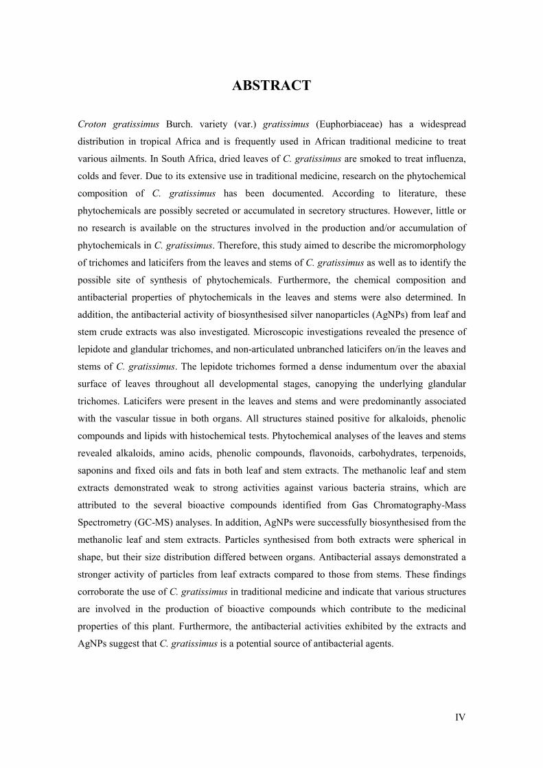
ABSTRACT
Croton gratissimus Burch. variety (var.) gratissimus (Euphorbiaceae) has a widespread
distribution in tropical Africa and is frequently used in African traditional medicine to treat
various ailments. In South Africa, dried leaves of C. gratissimus are smoked to treat influenza,
colds and fever. Due to its extensive use in traditional medicine, research on the phytochemical
composition of C. gratissimus has been documented. According to literature, these
phytochemicals are possibly secreted or accumulated in secretory structures. However, little or
no research is available on the structures involved in the production and/or accumulation of
phytochemicals in C. gratissimus. Therefore, this study aimed to describe the micromorphology
of trichomes and laticifers from the leaves and stems of C. gratissimus as well as to identify the
possible site of synthesis of phytochemicals. Furthermore, the chemical composition and
antibacterial properties of phytochemicals in the leaves and stems were also determined. In
addition, the antibacterial activity of biosynthesised silver nanoparticles (AgNPs) from leaf and
stem crude extracts was also investigated. Microscopic investigations revealed the presence of
lepidote and glandular trichomes, and non-articulated unbranched laticifers on/in the leaves and
stems of C. gratissimus. The lepidote trichomes formed a dense indumentum over the abaxial
surface of leaves throughout all developmental stages, canopying the underlying glandular
trichomes. Laticifers were present in the leaves and stems and were predominantly associated
with the vascular tissue in both organs. All structures stained positive for alkaloids, phenolic
compounds and lipids with histochemical tests. Phytochemical analyses of the leaves and stems
revealed alkaloids, amino acids, phenolic compounds, flavonoids, carbohydrates, terpenoids,
saponins and fixed oils and fats in both leaf and stem extracts. The methanolic leaf and stem
extracts demonstrated weak to strong activities against various bacteria strains, which are
attributed to the several bioactive compounds identified from Gas Chromatography-Mass
Spectrometry (GC-MS) analyses. In addition, AgNPs were successfully biosynthesised from the
methanolic leaf and stem extracts. Particles synthesised from both extracts were spherical in
shape, but their size distribution differed between organs. Antibacterial assays demonstrated a
stronger activity of particles from leaf extracts compared to those from stems. These findings
corroborate the use of C. gratissimus in traditional medicine and indicate that various structures
are involved in the production of bioactive compounds which contribute to the medicinal
properties of this plant. Furthermore, the antibacterial activities exhibited by the extracts and
AgNPs suggest that C. gratissimus is a potential source of antibacterial agents.

V
ACKNOWLEDGMENTS
Firstly, I would like to thank the National Research Foundation (NRF) for financial assistance.
I would also like to thank the following people:
Professor Yougasphree Naidoo for her guidance, support and expertise throughout this research.
Professor Gonasageran Naidoo for his expertise, guidance and constructive advice.
Professor Johnson Lin and Mr Abdullahi Jimoh for their guidance and assistance with
antibacterial assays.
The staff at the microscopy and microanalysis unit (UKZN Westville), in particular, Subashen
Naidu for his assistance with transmission electron microscopy (TEM).
Mr Vishal Bharuth for guidance and assistance with microscopy techniques. His continuous
motivation and encouragement throughout this endeavour is also deeply appreciated.
Dr C.T Sadashiva for assistance with phytochemical aspects and Thin Layer Chromatography
(TLC). His continuous motivation and support is also highly appreciated.
Mr Yegan Pillay for assistance with ultraviolet-visible (UV-VIS) spectroscopy.
Nneka Akwu for assistance with antibacterial assays. Her continuous motivation is also highly
appreciated.
To all my friends, thank you for your support, emotional assistance and everlasting humour
throughout this unforgettable journey.
A special thanks to Evashen Naidoo for his love, support and patience throughout this journey.
My sister, Terisha Naidoo, for her willingness to assist in editing and compiling my dissertation.
Finally, to my parents, Vincent and Loshni, and immediate family, for their continuous motivation
and encouragment throughout my academic journey. All my accomplishments would not have
been possible without your love and support.

VI
TABLE OF CONTENTS
PREFACE ..................................................................................................................................... II
DECLARATION: PLAGIARISM .............................................................................................. III
ABSTRACT ................................................................................................................................ IV
ACKNOWLEDGMENTS ............................................................................................................ V
TABLE OF CONTENTS ............................................................................................................ VI
LIST OF TABLES ....................................................................................................................... X
LIST OF FIGURES ..................................................................................................................... XI
ABBREVIATIONS ................................................................................................................... XV
CHAPTER 1: INTRODUCTION ................................................................................................. 1
1.1 Medicinal plants and traditional medicine .......................................................................... 1
1.2 Croton gratissimus Burch. variety (var.) gratissimus ......................................................... 2
1.3 Rationale for this study ....................................................................................................... 5
1.4 Research aims and objectives .............................................................................................. 6
1.5 References ........................................................................................................................... 7
CHAPTER 2: LITERATURE REVIEW .................................................................................... 11
2.1 Euphorbiaceae ................................................................................................................... 11
2.1.1 Taxonomy................................................................................................................... 12
2.1.2 Medicinal importance ................................................................................................. 12
2.2 The genus Croton .......................................................................................................... 13
2.2.1 Traditional uses .......................................................................................................... 13
2.2.2 Pharmacology ............................................................................................................. 14
2.3 Previous phytochemical studies of C. gratissimus var. gratissimus ................................. 15
2.3.1 Diterpenoids isolation ................................................................................................ 15
2.3.2 Antidiabetic activity ................................................................................................... 15
2.3.3 Antimalarial activity ................................................................................................... 16
2.4 Secretory tissues of plants ................................................................................................. 16
2.5 Trichomes .......................................................................................................................... 17
2.5.1 Non-glandular trichomes ............................................................................................ 17
2.5.2 Glandular trichomes ................................................................................................... 18

VII
2.5.3 Trichome variability and distribution ......................................................................... 19
2.6 Laticifers ........................................................................................................................... 20
2.6.1 Laticifer classification ................................................................................................ 21
2.6.2 Latex ........................................................................................................................... 21
2.6.3 Laticifers in Euphorbiaceae ........................................................................................ 22
2.7 Nanoparticles ..................................................................................................................... 22
2.7.1 Nanoparticle synthesis ................................................................................................ 22
2.7.2 Silver nanoparticles (AgNPs) ..................................................................................... 23
2.8 References ......................................................................................................................... 24
CHAPTER 3: MICROMORPHOLOGICAL AND HISTOCHEMICAL INVESTIGATION OF
TRICHOMES AND LATICIFERS ON/IN THE LEAVES AND STEMS OF CROTON
GRATISSIMUS BURCH. VAR. GRATISSIMUS (EUPHORBIACEAE) ................................... 33
3.1 Abstract ............................................................................................................................. 33
3.2 Introduction ....................................................................................................................... 34
3.3 Materials and methods ...................................................................................................... 35
3.3.1 Plant collection and sampling..................................................................................... 35
3.3.2 Stereomicroscopy ....................................................................................................... 35
3.3.3 Scanning electron microscopy (SEM) ........................................................................ 35
3.3.4 Sample preparation for light and transmission electron microscopy (TEM) ............. 36
3.3.5 Fluorescence microscopy ........................................................................................... 37
3.3.6 Histochemistry ........................................................................................................... 38
3.4 Results and Discussion ...................................................................................................... 39
3.4.1 Surface overview ........................................................................................................ 40
3.4.2 Lepidote trichomes ..................................................................................................... 43
3.4.3 Ultrastructure of lepidote trichomes ........................................................................... 46
3.4.4 Glandular trichomes ................................................................................................... 50
3.4.5 Laticifers..................................................................................................................... 52
3.4.6 Histochemistry and fluorescence microscopy ............................................................ 54
3.5 Conclusion ......................................................................................................................... 60
3.6 References ......................................................................................................................... 61
CHAPTER 4: PHYTOCHEMICAL AND ANTIBACTERIAL ANALYSES OF CROTON
GRATISSIMUS BURCH. VAR. GRATISSIMUS (EUPHORBIACEAE) LEAF AND STEM
EXTRACTS ................................................................................................................................ 69
4.1 Abstract ............................................................................................................................. 69
4.2 Introduction ....................................................................................................................... 70

VIII
4.3 Materials and Methods ...................................................................................................... 71
4.3.1 Plant collection and sampling..................................................................................... 71
4.3.2. Crude extracts ............................................................................................................ 71
4.3.3 Preliminary phytochemical screening ........................................................................ 71
4.3.4 Thin Layer Chromatography (TLC) ........................................................................... 73
4.3.5 Gas Chromatography-Mass Spectrometry (GC-MS) ................................................. 73
4.3.6 Preliminary antibacterial assays ................................................................................. 73
4.4 Results and Discussion ...................................................................................................... 74
4.4.1 Preliminary phytochemical screening ........................................................................ 74
4.4.2 Thin layer chromatography (TLC) ............................................................................. 77
4.4.3 Gas Chromatography-Mass Spectrometry (GC-MS) ................................................. 77
4.4.4 Preliminary antibacterial assays ................................................................................. 81
4.5 Conclusion ......................................................................................................................... 83
4.6 References ......................................................................................................................... 84
CHAPTER 5: BIOLOGICAL SYNTHESIS AND ANTIBACTERIAL ACTIVITY OF SILVER
NANOPARTICLES FROM LEAVES AND STEMS OF CROTON GRATISSIMUS BURCH.
VAR. GRATISSIMUS (EUPHORBIACEAE) ............................................................................. 93
5.1 Abstract ............................................................................................................................. 93
5.2 Introduction ....................................................................................................................... 94
5.3 Materials and Methods ...................................................................................................... 95
5.3.1 Plant collection and sampling..................................................................................... 95
5.3.2 Crude methanolic extraction ...................................................................................... 95
5.3.3 Biosynthesis of silver nanoparticles (AgNPs) ............................................................ 95
5.3.4 Ultraviolet-visible (UV-VIS) spectroscopy ................................................................ 95
5.3.5. Energy-dispersive X-ray (EDX) analysis .................................................................. 95
5.3.6 Transmission electron microscopy (TEM) and Image analysis ................................. 96
5.3.7 Fourier-transform infrared spectroscopy (FTIR)........................................................ 96
5.3.8 Preliminary antibacterial assay ................................................................................... 96
5.4 Results and Discussion ...................................................................................................... 97
5.4.1 Biosynthesis of silver nanoparticles (AgNPs) ............................................................ 97
5.4.2 Ultraviolet-visible (UV-VIS) spectroscopy ................................................................ 98
5.4.3 Energy-dispersive X-ray (EDX) analysis ................................................................... 99
5.4.4 Transmission electron microscopy (TEM) and Image analysis ............................... 100
5.4.5 Fourier-transform infrared spectroscopy (FTIR)...................................................... 102
5.4.6 Preliminary antibacterial assay ................................................................................. 103
5.5 Conclusion ....................................................................................................................... 105

IX
5.6 References ....................................................................................................................... 106
CHAPTER 6: CONCLUSIONS AND FUTURE RECOMMENDATIONS ............................ 111
6.1 Major findings ................................................................................................................. 111
6.2 Challenges ....................................................................................................................... 112
6.3 Future recommendations ................................................................................................. 112
6.4 Final conclusion .............................................................................................................. 112

X
LIST OF TABLES
CHAPTER 1
Table 1.1: Medicinal uses of Croton gratissimus. ........................................................................ 4
CHAPTER 2
Table 2.1: Traditional uses of Croton species (Salatino et al., 2007)......................................... 14
CHAPTER 4
Table 4.1: Phytochemical compounds identified in the hexane, chloroform and methanolic crude
extracts from leaves and stems of C. gratissimus var. gratissimus. ............................................ 76
Table 4.2: Gas Chromatography-Mass Spectrometry (GC-MS) analysis of methanolic leaf extract
showing major and minor compounds. ....................................................................................... 79
Table 4.3: Gas Chromatography-Mass Spectrometry (GC-MS) analysis of methanolic stem
extract showing major and minor compounds. ........................................................................... 80
Table 4.4: Antibacterial activities of leaf and stem extracts of C. gratissimus against eight
bacterial strains. ........................................................................................................................... 82
CHAPTER 5
Table 5.1: Mean percentage of elemental silver from nanoparticles synthesised from leaf and
stem extracts of C. gratissimus var. gratissimus. ...................................................................... 100
Table 5.2: Mean particle size of silver nanoparticles synthesised from leaves and stems of C.
gratissimus var. gratissimus. ..................................................................................................... 102
Table 5.3: Antibacterial activities exhibited by silver nanoparticles from leaf and stem extracts
of C. gratissimus var. gratissimus against eight bacterial strains.............................................. 104
XI
LIST OF FIGURES
CHAPTER 1
Figure 1.1: Croton gratissimus var. gratissimus occurring in the University of KwaZulu-Natal -
Westville Campus (29°49'08.1"S 30°56'38.9"E). ......................................................................... 5
CHAPTER 2
Figure 2.1: Worldwide distribution of Euphorbiaceous species (Source: Angiosperm Phylogeny
Website http://www.mobot.org/MOBOT/research/APweb/). ..................................................... 11
Figure 2.2: Possible transformational relationships between trichome types in Croton. a, simple;
b, 2-5-radiate; c, rosulate (pin-cushion); d, fasciculate; e, stellate-rotate (lateral and frontal views);
f, transition from multiradiate to dendritic; g, two-layered stellate (transitional to geminate); h,
geminate; i, dendritic. Arrows indicate directions of apparent morphological change (Webster et
al., 1996)...................................................................................................................................... 20
CHAPTER 3
Figure 3.1: Stereomicrographs showing general overview of leaves and stems. a) Adaxial surface
of emergent leaf. b) Abaxial surface of emergent leaf showing dense distribution of lepidote
trichomes on lamina, mid-vein and petiole. c) Adaxial surface of young leaf. d) Abaxial surface
of young leaf with dense indumentum of lepidote trichomes. e) Adaxial surface of mature leaf
appearing shiny, indicating the presence of a cuticle layer. f) Abaxial surface of mature leaf
showing lepidote trichomes densely distributed over the lamina and mid-vein. g) Stem covered
with lepidote trichomes. .............................................................................................................. 41
Figure 3.2: Stereomicrographs of leaf and stem surfaces. a) Glabrous lamina showing translucent
dots on adaxial surface. b) Stellate trichome along sunken mid-vein on the adaxial surface. Note
the glossy appearance of this surface which is indicative of a cuticle layer. c) Lamina of abaxial
surface densely covered with lepidote trichomes. d) Mid-vein on abaxial surface covered with
lepidote trichomes. e) Extrafloral nectaries present on the mid-vein at the base of the leaf. Note
lepidote trichomes on petiole. f) Dense indumentum of lepidote trichomes on stem. ................ 42
Figure 3.3: Scanning electron micrographs of leaves and stems. a) Adaxial surface showing
stellate trichomes along the mid-vein of leaf. Note the peeled cuticle layer on this surface. b)
Stellate trichome emerging from middle furrow (mid-vein) on adaxial surface. c) Dense
indumentum formed by lepidote trichomes on the lamina and mid-vein on the abaxial surface. d)
Lepidote trichomes fully covering stem. ST = Stellate trichome. ............................................... 43

XII
Figure 3.4: Morphology of lepidote trichomes. a) Stereomicrograph of lepidote trichome. b) SEM
of lepidote trichome showing umbo/central cell and numerous webbed radial cells. c) Lepidote
trichome with accumulated secretory substance. d) Light micrograph of lepidote trichome
showing stalk cells, subradial cells, radial cells and umbo/central cell. U = Umbo, R =
Radii/Radial cell, Sr = Subradial cell, S = Stalk, Sm = Stoma, * = Secretion. ........................... 45
Figure 3.5: Development of lepidote trichomes. a) Emergence of protodermal cells giving rise to
lepidote trichome through periclinal and anticlinal divisions. Note the periclinal divisions
initiating the development of the stalk and the anticlinal divisions of the radial cells surrounding
the central cell. b) Developing lepidote trichome. Note the increased number of stalk cells brought
about by additional periclinal divisions and the stretching of the lateral radial cells. c) Fully
developed lepidote trichome with stalk, subradial, radial and central cells. Note the elongation of
stalk cells into a prominent stalk, the developed subradial cells, the distinct central cell and the
extended radial cells. ................................................................................................................... 46
Figure 3.6: Transmission electron micrographs of lepidote trichome stalk cells. a) Section
through the stalk cells and radial cell. Large and small vacuoles surrounded by dense cytoplasm
and other organelles can be seen in the stalk cells. b) Single stalk cell containing dense cytoplasm
with numerous vacuoles, a large nucleus and a chloroplast. c) Rough endoplasmic reticulum and
vesicles at the periphery of a stalk cell wall. d) Vesicles and Golgi body present in stalk cell. e)
Thick cell wall between two adjacent stalk cells with visible plasmodesmata (white arrows).
Vacuoles, numerous mitochondria, endoplasmic reticulum and vesicles can be seen at the
periphery of these cells. Note the presence of the electron dense vesicle next to the cell wall. R =
Radial cell, S = Stalk, CW = Cell wall, Vs = Vesicle, V = Vacuole, N = Nucleus, M =
Mitochondria, RER/ER = Rough Endoplasmic Reticulum/Endoplasmic Reticulum, C =
Chloroplast, GB = Golgi body, LB = Lipid body, Adj = Adjacent cells. ................................... 49
Figure 3.7: Transmission electron micrographs of lepidote trichome radial cells. a) Radial cell
with thickened cell wall containing dense cytoplasm with vesicles, Golgi body, a lipid body and
rough endoplasmic reticulum at the periphery. b) Higher magnification of Golgi body surrounded
by dense cytoplasm. c) Vacuoles, mitochondria, lipid body and rough endoplasmic reticulum
present along the radial cell wall. d) Golgi body, rough endoplasmic reticulum, a lipid body and
numerous vesicles along the periphery of a radial cell wall. CW = Cell wall, Vs = Vesicle, V =
Vacuole, M = Mitochondria, RER/ER = Rough Endoplasmic Reticulum/Endoplasmic Reticulum,
GB = Golgi body, LB = Lipid body. ........................................................................................... 50
Figure 3.8: Micrographs showing glandular trichomes on the leaves and stems. a) Glandular
trichomes on abaxial surface of leaves beneath lepidote trichomes. b) Stem showing glandular
trichomes after removing lepidote trichomes. High magnification of single glandular trichome on

XIII
abaxial surface. Note the presence of paracytic stomata. d) Light micrograph showing unicellular
glandular trichomes of different forms canopied by several layers of lepidote trichomes. Sm =
Stoma, LT = Lepidote Trichome, GT = Glandular Trichome. .................................................... 51
Figure 3.9: Laticifer distribution in leaves and stems. a) Transverse section of leaf stained with
Toluidine-Blue showing distribution of laticifers predominantly in the vascular tissue. Note the
idioblasts at the adaxial side of the leaf. b) Transverse section of the stem stained with Toluidine-
Blue showing laticifers in the phloem and pith. c) Scanning electron micrograph of coagulated
latex within laticifer cells (associated with phloem). Druse crystals are also present in the leaf
section. d) Transverse section through stem showing latex containing laticifers in pith. Id =
Idioblast, Dr = Druse crystal. ...................................................................................................... 53
Figure 3.10: Laticifer cells showing secretory contents. a) Longitudinal section of leaf showing
latex within non-articulated laticifers. b) Light micrograph of transverse section showing laticifer
cells with latex contents. c) Freeze- fracture through laticifer cells containing coagulated latex. Lt
= Laticifer, # = Latex. ................................................................................................................. 54
Figure 3.11: Histochemical and fluorescence micrographs showing chemical compounds of
lepidote trichomes. a) Orange/brown colouration of stalk (intense), subradial, radial and central
cells (weak) suggest a positive indication for the presence of alkaloids with Wagner’s reagent. b)
Phenolics detected in stalk, subradial cells, radii and central cell with ferric chloride (brown to
black precipitate). c) Pink colouration indicated neutral lipids in stalk cells and blue colouration
of subradial, radial and central cells indicated acidic lipids with Nile blue. d) Pectin in the
subradial, radial and central cell walls was indicated by a pink colour. e) Orange staining of the
stalk and radii with Sudan III indicated the presence of cutinised walls and lipids. f) Positive
staining for lipids in the stalk, subradial and radial cells with Sudan black. g) Toluidine-Blue
revealed lignification of the subradial and central cells (blue colouration). h) Positive indication
of lignin in the subradial and central cells with phloroglucinol. i) Blue autofluorescence indicated
phenolic compounds in stalk cells. j) Yellow fluorescence with acridine orange revealed lignified
subradial and central cells. .......................................................................................................... 58
Figure 3.12: Histochemical and fluorescence micrographs showing chemical compounds of
glandular trichomes. a) Positive staining for alkaloids (brown colour) with Wagner’s reagent. b)
Glandular trichomes tested positive for phenolic compounds with ferric chloride (indicated by
brown/black precipitate). c) Pink colouration indicated neutral lipids with Nile blue. d) Lipid
droplet stained red/orange with Sudan III. e) Lignified cell walls of glandular trichome detected
with autofluorescence. ................................................................................................................. 59
Figure 3.13: Histochemical and fluorescence micrographs showing chemical compounds of
laticifer cells. a) Orange colouration a positive indication for alkaloids with Wagner’s reagent. b)
XIV
Blue colouration within laticifer cells indicated acidic lipids with Nile blue. c) Positive indication
(dark brown to black) for phenolic compounds with ferric chloride. d) Pink colouration indicated
mucilage with ruthenium red. Note the presence of druse and prismatic crystals. e) Blue staining
of laticifer cells with Toluidine-Blue indicated macromolecules with free phosphate groups. f)
Positive stain (orange colour) for lipids with Sudan III. Pr = Prismatic crystal, Dr = Druse crystal.
..................................................................................................................................................... 60
CHAPTER 4
Figure 4.1: Separation of compounds on TLC plate spotted with hexane, chloroform and
methanol extracts from leaves and stems. a) Viewed at 254 nm. b) Viewed at 365 nm. c) Viewed
after heating with ANS reagent. A = hexane leaves, B = chloroform leaves, C = methanol leaves,
D = hexane stems, E = chloroform stems, F = methanol stems. ................................................. 77
CHAPTER 5
Figure 5.1: Visual representation of the leaf and stem extracts before (a) and after (b) the 90 min
reaction time. CL = C. gratissimus var. gratissimus leaves, CS = C. gratissimus var. gratissimus
stems. ........................................................................................................................................... 98
Figure 5.2: Ultraviolet-visible spectra of silver nanoparticles synthesised from leaves and stems
of C. gratissimus var. gratissimus after the 90 min reaction time. .............................................. 99
Figure 5.3: Energy-dispersive X-ray (EDX) spectra of silver nanoparticles synthesised from leaf
(a) and stem (b) extracts of C. gratissimus var. gratissimus. .................................................... 100
Figure 5.4: Transmission electron micrograph showing silver nanoparticles synthesised from the
leaves (a) and stems (b) of C. gratissimus var. gratissimus. ..................................................... 101
Figure 5.5: Particle size distribution from leaves (a) and stems (b) of C. gratissimus var.
gratissimus. ............................................................................................................................... 101
Figure 5.6: Fourier-transform infrared spectra of silver nanoparticles synthesised from a) leaf and
b) stem extracts of C. gratissimus var. gratissimus. .................................................................. 103
XV
ABBREVIATIONS
Adj Adjacent cells
Ag/Ag+ Silver/silver ions
AgNO3 Silver nitrate
AgNPs Silver nanoparticles
ANS Anisaldehyde-sulphuric acid
C Chloroplast
CL/CS Croton leaves/stems
CW Cell wall
DPPH 2,2-diphenyl-1-picrylhydrazyl
Dr/Pr Druse/Prismatic crystals
EDX Energy dispersive X-ray
ER/RER Endoplasmic reticulum/rough endoplasmic reticulum
Fig. Figure
FTIR Fourier-transform infrared spectroscopy
GB Golgi body
GC-MS Gas chromatography-mass spectrometry
GT Glandular trichomes
HDL High density lipoproteins
HPLC High performance liquid chromatography
Id Idioblast
LB Lipid body
LDL Low density lipoproteins
Lt Laticifers
XVI
LT Lepidote trichome
M Mitochondria
MIC Minimum inhibitory concentration
MRSA Methicillin-Resistance Staphylococcus aureus
N Nucleus
OD620 Optical density at 620 nm
R Radial cell/radii
S Stalk
s.l sensu lato
s.s sensu stricto
SD Standard deviation
SEM/FEGSEM Scanning electron microscopy/Field emission gun SEM
Sm Stoma
SPR Surface plasmon resonance
Sr Subradial cell
ST Stellate trichome
TEM/HRTEM Transmission electron microscopy/High resolution TEM
TLC Thin layer chromatography
TPA 12-O-tetradecanoylphorbol-13-acetate
U Umbo
UV/UV-B Ultraviolet/ultraviolet-B
UV-VIS Ultraviolet-visible
V Vacuole
VLDL Very low density lipoproteins
Vs Vesicles

XVII
WHO World Health Organisation

1
CHAPTER 1: INTRODUCTION
1.1 Medicinal plants and traditional medicine
Plants play an important role in the survival of mankind as they provide food, medicine and other
products and services, either directly or indirectly (Terashima, 2001; Hawkins, 2008; Ahvazi et
al., 2012; Corlett, 2016). However, the expanding human population has led to an increase in
exploitation of resources, resulting in the endangerment of many species and populations. One of
the most frequent groups exploited is plants as they are collected for medicinal trade (Hawkins,
2008). Globally, the number of angiosperms being utilised for their medicinal value ranges
between 50000 – 80000. Unfortunately, many of these species are faced with the risk of extinction
due to overharvesting and habitat destruction (Chen et al., 2016). It is therefore imperative to
enforce conservation practices and sustainable use in order to preserve medicinal plant
biodiversity (Okigbo et al., 2008).
The use of plants for medicinal purposes dates back to ancient times, around 4000 – 5000 B.C.
(Hosseinzadeh et al., 2015). Medicinal plants are those that contain essential active ingredients
that are utilised for the treatment of diseases and pains (Okigbo et al., 2008). These plants are the
source of medicines that are safe, beneficial and affordable (Heamalatha et al., 2011), and they
constitute an abundance of compounds (Okigbo et al., 2008). Over 10000 compounds are
produced by plants as a defence against predators, with many of these having the potential to be
drugs (Okigbo et al., 2008). Consequently, due to the frequent use by people in underdeveloped
countries, medicinal plants form the backbone of traditional medicine around the world (Devi et
al., 2012; Singh, 2015).
Traditional medicine can be defined as “the sum total of the knowledge, skills and practices based
on the theories, beliefs and experiences indigenous to different cultures, whether explicable or
not, used in the maintenance of health as well as in the prevention, diagnosis, improvement or
treatment of physical and mental illness” (World Health Organisation (WHO), 2018).
Many people in developing countries lack access to modern drugs and therefore depend on
traditional medicine as a primary source of healthcare due to their easy accessibility and
affordability (Heamalatha et al., 2011; Hosseinzadeh et al., 2015; Masevhe et al., 2015). In Africa,
traditional healers exploit the rich plant diversity for various treatments, hence indigenous plants
are the key component in African traditional medicine (Okigbo et al., 2008; Masevhe et al., 2015).
In South Africa, the trade in traditional medicines is a huge industry, with about 27 million

2
consumers. However, the majority of the species traded for traditional medicines are harvested
from wild populations, leading to a decrease in biodiversity (Mander, 1998; Petersen et al., 2017).
The use of traditional medicine has retained its popularity due to cultural beliefs and historical
use (Masevhe et al., 2015; Singh, 2015). This knowledge of medicinal plants is passed on to each
generation through verbal exchange (Masevhe et al., 2015; Boadu and Asase, 2017). However,
the loss of biodiversity and cultural inheritance threaten the survival of this information.
Therefore, it is crucial that this knowledge be documented in order to preserve this cultural
inheritance for current and future generations to utilise (Boadu and Asase 2017). In addition,
traditional knowledge serves as a precursor for the discovery of new drugs or bioactive
compounds that can be used for treating illnesses (Farnsworth et al., 1985; Boadu and Asase,
2017). Globally, about 25% of prescription drugs contain plant-derived ingredients (Sen and
Chakraborty, 2017). According to a study by Fabricant and Farnsworth (2001), there are 122
plant-derived compounds originating from only 94 plant species that are used as modern drugs
worldwide. Of these compounds, 80% are currently used to treat the similar or same ailment as
used traditionally (Fabricant and Farnsworth, 2001; Yuan et al., 2016). As medicinal plants are
important sources of novel plant compounds and new drugs (Boadu and Asase, 2017), and with
a global approximation of 250 000 flowering plant species, there are possibly many drugs still
undiscovered (Fabricant and Farnsworth, 2001). Finally, the availability of this information on
harvested plants used for various treatments in specific regions can increase biodiversity
conservation. Large-scale harvesting of medicinal plants for commercial trade results in adverse
effects on population sizes and recovery following harvesting. Therefore, in order to achieve
conservation, the quantities harvested need to be known and documented to ensure that this
resource is maintained for future generations (Boadu and Asase, 2017).
1.2 Croton gratissimus Burch. variety (var.) gratissimus
Croton gratissimus Burch. (syn. C. zambesicus Müll. Arg.; C. microbotryus Pax., C. amabilis
Müell. Arg.) commonly known as lavender Croton or lavender fever berry, belongs to the family
Euphorbiaceae. This species comprises of two varieties, namely C. gratissimus Burch. var.
gratissimus and C. gratissimus Burch. var. subgratissimus (Curtis and Mannheimer, 2005;
Mulholland et al., 2010; Robert et al., 2010; PlantZAfrica, 2018). The former variety is the focus
of this study. Croton gratissimus is a Guineo-Congolese species with widespread distribution in
tropical, central and sub-Saharan Africa. This species grows in dry and warm areas on stony/rocky
slopes of hills throughout the north east of the continent from South Africa to the horn of Africa
(Ngadjui et al., 2002; Block et al., 2004; Mulholland et al., 2010). In South Africa, this plant is
distributed over a wide range, being indigenous to six provinces (Pudumo et al., 2018).

3
The genus Croton is of Greek origin, being a derivation of the word Kroton meaning “tick”, whilst
gratissimus, the species name, means most pleasing/pleasant, in Latin (PlantZAfrica, 2018;
Pudumo et al., 2018). The name is suitable as a lavender-like aroma is produced by the leaves
when crushed (Mulholland et al., 2010; Pudumo et al., 2018). It is a semi-deciduous shrub or tree
that can reach heights of up to 10-15 m (Block et al., 2004; Boon, 2010; Mulholland et al., 2010).
The slender shaped trees have a V-shaped crown which extends upwards with drooping foliage
and terminal branches. The leaves are simple with an alternate arrangement. The adaxial surfaces
of the leaves are shiny, dark green in colour and lack hairs. The abaxial surface appears silver
with orange-brown specks due to the presence of dense scales (Boon, 2010; Mthethwa et al.,
2014; PlantZAfrica, 2018). The trees are monoecious with terminal racemes that give rise to small
cream to golden yellow flowers. The fruit is a small three-lobed capsule (Boon, 2010;
PlantZAfrica, 2018; Pudumo et al., 2018).
Croton gratissimus is used extensively in traditional medicine to treat various illnesses, with the
whole plant having a reputation of being medicinally important (Ngadjui et al., 2002; Van Vuuren
and Viljoen, 2008). The organs of C. gratissimus are used either independently, in combination
with other parts or plants or co-administered with different species for a wide range of treatments
(Van Vuuren and Viljoen, 2008; Mulholland et al., 2010; Pudumo et al., 2018). Table 1.1 provides
a summary of the uses of C. gratissimus.

4
Table 1.1: Medicinal uses of Croton gratissimus.
Part/s used
Country Uses Preparation References
Bark South Africa As purgative for stomach and intestinal disorders
Milk infusions of bark Mulholland et al., 2010; Mthethwa et al., 2014
Unspecified uterine disorder
Powdered bark blown into the womb
Mulholland et al., 2010
Pleurisy Powdered bark rubbed into chest incisions
Mulholland et al., 2010
Nigeria Malaria Bark infusions Langat et al., 2011 Unspecified Bleeding gums,
abdominal disorders, skin inflammation, earache, chest complaints
Unspecified Van Vuuren and Viljoen, 2008; Pudumo et al., 2018
Swelling Combination of bark with root of Amaryllidaceae species applied into incisions
Van Vuuren and Viljoen, 2008
Leaves South Africa Sores associated with STI’s
Steam baths Van Vuuren and Naidoo, 2010
Influenza, colds and fever
Dried leaves smoked Mulholland et al., 2010; Langat et al., 2011
Zimbabwe/Botswana Cough Smoke from leaves, leaf decoction/tea
Mulholland et al., 2010; Langat et al., 2011
Benin/Nigeria Hypertension, urinary infection (as anti-microbial), malaria, dysentery, diarrhoea, convulsions, antidiabetic remedy
Leaf decoction Robert et al., 2010; Okokon et al., 2011; Abdalaziz et al., 2016; Kumar et al., 2017
Unspecified Restlessness and Insomnia
Paste made with ground leaves, two other Croton species and goat fat heated on coals and fumes inhaled.
Mulholland et al., 2010; Langat et al., 2011
Eye disorders, rheumatism
Unspecified Mulholland et al., 2010
Roots Zimbabwe Abdominal pains and aphrodisiac
Root infusions Mulholland et al., 2010; Mthethwa et al., 2014
Sudan Menstrual pain and constipation
Unspecified Robert et al., 2010; Kumar et al., 2017
Root and bark
Unspecified Respiratory disorders
Combination of root and bark
Van Vuuren and Viljoen, 2008

5
Figure 1.1: Croton gratissimus var. gratissimus occurring in the University of KwaZulu-Natal -
Westville Campus (29°49'08.1"S 30°56'38.9"E).
1.3 Rationale for this study
Croton gratissimus has been used extensively in African traditional medicine for a wide range of
treatments. For this reason, many phytochemical investigations have been carried out on various
parts of the plant to validate its therapeutic value (Okokon et al., 2006; Van Vuuren and Viljoen,
2008; Okokon and Nwafor, 2009; Okokon and Nwafor, 2010; Robert et al., 2010; Okokon et al.,
2011; Mthethwa et al., 2014; Kumar et al., 2017). According to Fahn (1979), these
phytochemicals are possibly synthesised or accumulated by secretory structures (Vitarelli et al.,
2015). However, limited or no research has been conducted on the micromorphology and
ultrastructure of the structures responsible for the synthesis, secretion and/or accumulation of
phytochemicals in the leaves and stems of C. gratissimus. Therefore, this study focussed on
6
identifying and describing the micromorphology and ultrastructure of the trichomes and internal
secretory structures of the leaves (adaxial and abaxial surfaces) and stems, as well as determining
the chemical composition and possible site of synthesis of the phytochemicals. In addition,
another aim of this study was to synthesise silver nanoparticles (AgNPs) from crude extracts of
leaves and stems of C. gratissimus.
1.4 Research aims and objectives
The aims and objectives for each chapter are outlined below:
Chapter 3
Aim: To determine the micromorphology and distribution of trichomes and laticifers of the leaves
and stems; and to detect the possible location of phytochemicals of C. gratissimus using various
microscopy techniques.
Objective:
To identify, describe and compare the micromorphology and distribution of trichomes on
leaves (adaxial and abaxial surfaces), at three developmental stages (emergent, young and
mature), and stems using stereomicroscopy, light microscopy and scanning electron
microscopy (SEM).
To identify and describe the laticifers within the leaves and stems using SEM and light
microscopy.
To determine the ultrastructure of lepidote trichomes using Transmission Electron
Microscopy (TEM).
Elucidate the location of chemical compounds using various histochemical tests.
Chapter 4
Aim: Determine the chemical composition of phytochemicals in the leaves and stems of C.
gratissimus and test for antibacterial activity of the methanolic extracts.
Objectives:
Determine the chemical composition of the phytochemicals in the leaves and stems by
preliminary qualitative phytochemical screening, Gas Chromatography-Mass
Spectrometry (GC-MS) analysis and Thin Layer Chromatography (TLC).
Determine the biological activity of the crude extracts from leaves and stems by
conducting antibacterial tests.

7
Chapter 5
Aim: Synthesise, characterise and test for antibacterial activity of silver nanoparticles (AgNPs)
using methanolic extracts of C. gratissimus.
Objectives:
Synthesise AgNPs from the crude extracts of C. gratissimus.
Characterise synthesised AgNPs using ultraviolet-visible (UV-VIS) spectroscopy,
energy-dispersive X-ray (EDX) analysis, TEM, and Fourier-transform infrared
spectroscopy (FTIR).
Test for biological activity of the synthesised AgNPs by conducting antibacterial tests.
1.5 References
Abdalaziz, M.N., Ali, A. and Kabbashi, A.S., 2016. In vitro antioxidant activity and
phytochemical screening of Croton zambesicus. Journal of Pharmacognosy and Phytochemistry
5, 12 – 16.
Ahvazi, M., Khalighi-Sigaroodi, F., Charkhchiyan, M.M., Mojab, F., Mozaffarian, V.A. and
Zakeri, H., 2012. Introduction of medicinal plants species with the most traditional usage in
Alamut region. Iranian Journal of Pharmaceutical Research 11, 185 – 194.
Block, S., Baccelli, C., Tinant, B., Van Meervelt, L., Rozenberg, R., Jiwan, J.L.H., Llabres, G.,
De Pauw-Gillet, M.C. and Quetin-Leclercq, J., 2004. Diterpenes from the leaves of Croton
zambesicus. Phytochemistry 65, 1165 – 1171.
Boadu, A.A. and Asase, A., 2017. Documentation of herbal medicines used for the treatment and
Management of Human Diseases by some communities in southern Ghana. Evidence-Based
Complementary and Alternative Medicine 2017, 1 – 12.
Boon, R., 2010. Pooley's Trees of Eastern South Africa: [a Complete Guide]. Flora and Fauna
Publications Trust.
Chen, S.L., Yu, H., Luo, H.M., Wu, Q., Li, C.F. and Steinmetz, A., 2016. Conservation and
sustainable use of medicinal plants: problems, progress, and prospects. Chinese medicine 11, 37.
Corlett, R.T., 2016. Plant diversity in a changing world: status, trends, and conservation needs.
Plant Diversity 38, 10 – 16.
Curtis, B., and Mannheimer, C., 2005. Tree atlas of Namibia. National Botanical Research
Institute, Windhoek.

8
Devi, S., Gupta, A.K. and Singh, M., 2012. Ethno-Medicinal use of plants belonging to families
Fabaceae and Solanaceae, Hamirpur district (HP). International Journal of Scientific and
Research Publications 2, 1 – 4.
Fabricant, D.S. and Farnsworth, N.R., 2001. The value of plants used in traditional medicine for
drug discovery. Environmental Health Perspectives 109 (Suppl 1), 69 – 75.
Fahn, A., 1979. Secretory tissues in plants, Academic Press, London, UK.
Farnsworth, N.R., Akerele, O., Bingel, A.S., Soejarto, D.D. and Guo, Z., 1985. Medicinal plants
in therapy. Bulletin of the World Health Organization 63, 965 – 981.
Hawkins, B., 2008. Plants for life: Medicinal plant conservation and botanic gardens, Botanic
Gardens Conservation International, Richmond, UK.
Heamalatha, S., Swarnalatha, S., Divya, M., Gandhi Lakshmi, R., Ganga Devi, A. and Gomathi,
E., 2011. Pharmacognostical, pharmacological, investigation on Anethum graveolens Linn: A
review. Research Journal of Pharmaceutical, Biological and Chemical Sciences 2, 564 – 574.
Hosseinzadeh, S., Jafarikukhdan, A., Hosseini, A. and Armand, R., 2015. The application of
medicinal plants in traditional and modern medicine: A review of Thymus vulgaris. International
Journal of Clinical Medicine 6, 635 – 642.
Kumar, P., Kumar, R., Rastogi, M.K., Murti, K., 2017. Exploration of Antidiabetic and
Hypolipidemic Activity of Roots of Croton zambesicus. American Journal of Pharmacology and
Toxicology 12, 1 – 6.
Langat, M.K., Crouch, N.R., Smith, P.J. and Mulholland, D.A., 2011. Cembranolides from the
Leaves of Croton gratissimus. Journal of Natural Products 74, 2349 – 2355.
Mander, M. 1998. Marketing of indigenous medicinal plants in South Africa: a case study in
KwaZulu-Natal. FAO, Rome, Italy.
Masevhe, N.A., McGaw, L.J. and Eloff, J.N., 2015. The traditional use of plants to manage
candidiasis and related infections in Venda, South Africa. Journal of Ethnopharmacology 168,
364 – 372.
Mthethwa, N.S., Oyedeji, B.A., Obi, L.C. and Aiyegoro, O.A., 2014. Anti-staphylococcal, anti-
HIV and cytotoxicity studies of four South African medicinal plants and isolation of bioactive
compounds from Cassine transvaalensis (Burtt Davy) Codd. BMC Complementary and
Alternative Medicine 14, 512.

9
Mulholland, D.A., Langat, M.K., Crouch, N.R., Coley, H.M., Mutambi, E.M. and Nuzillard, J.M.,
2010. Cembranolides from the stem bark of the southern African medicinal plant, Croton
gratissimus (Euphorbiaceae). Phytochemistry 71, 1381 – 1386.
Ngadjui, B.T., Abegaz, B.M., Keumedjio, F., Folefoc, G.N. and Kapche, G.W., 2002.
Diterpenoids from the stem bark of Croton zambesicus. Phytochemistry 60, 345 – 349.
Okigbo, R.N., Eme, U.E. and Ogbogu, S., 2008. Biodiversity and conservation of medicinal and
aromatic plants in Africa. Biotechnology and Molecular Biology Reviews 3, 127 – 134.
Okokon, J.E., Bassey, A.L. and Obot, J., 2006. Antidiabetic activity of ethanolic leaf extract of
Croton zambesicus Muell. (Thunder plant) in alloxan diabetic rats. African Journal of Traditional,
Complementary and Alternative Medicines 3, 21 – 26.
Okokon, J.E. and Nwafor, P.A., 2009. Antiplasmodial activity of root extract and fractions of
Croton zambesicus. Journal of Ethnopharmacology 121, 74 – 78.
Okokon, J.E. and Nwafor, P.A., 2010. Antiinflammatory, analgesic and antipyretic activities of
ethanolic root extract of Croton zambesicus. Pakistan Journal of Pharmaceutical Sciences 23, 385
– 392.
Okokon, J.E., Umoh, U.F., Udobang, J.A. and Etim, E.I., 2011. Antiulcerogenic activity of
ethanolic leaf extract of Croton zambesicus Muell. Arg. African Journal of Biomedical Research
14, 43 – 47.
Petersen, L., Reid, A.M., Moll, E.J. and Hockings, M.T., 2017. Perspectives of wild medicine
harvesters from Cape Town, South Africa. South African Journal of Science 113, 1 – 8.
PlantZAfrica, 2018. Croton gratissimus Burch. http://pza.sanbi.org/croton-gratissimus. Date
Accessed: 5 February 2018.
Pudumo, J., Chaudhary, S.K., Chen, W., Viljoen, A., Vermaak, I. and Veale, C.G.L., 2018.
HPTLC fingerprinting of Croton gratissimus leaf extract with Preparative HPLC-MS-isolated
marker compounds. South African Journal of Botany 114, 32 – 36.
Robert, S., Baccelli, C., Devel, P., Dogné, J.M. and Quetin-Leclercq, J., 2010. Effects of leaf
extracts from Croton zambesicus Müell. Arg. on hemostasis. Journal of Ethnopharmacology 128,
641 – 648.
Sen, S. and Chakraborty, R., 2017. Revival, modernization and integration of Indian traditional
herbal medicine in clinical practice: Importance, challenges and future. Journal of traditional and
complementary medicine 7, 234 – 244.

10
Singh, R., 2015. Medicinal plants: A review. Journal of Plant Sciences 3, 50 – 55.
Terashima, H., 2001. The relationships among plants, animals, and man in the African tropical
rain forest. African Study Monographs 27(Suppl.), 43 – 60.
Van Vuuren, S.F. and Viljoen, A.M., 2008. In vitro evidence of phyto-synergy for plant part
combinations of Croton gratissimus (Euphorbiaceae) used in African traditional healing. Journal
of Ethnopharmacology 119, 700 – 704.
Van Vuuren, S.F. and Naidoo, D., 2010. An antimicrobial investigation of plants used
traditionally in southern Africa to treat sexually transmitted infections. Journal of
Ethnopharmacology 130, 552 – 558.
Vitarelli, N.C., Riina, R., Caruzo, M.B.R., Cordeiro, I., Fuertes‐Aguilar, J. and Meira, R.M., 2015.
Foliar secretory structures in Crotoneae (Euphorbiaceae): Diversity, anatomy, and evolutionary
significance. American Journal of Botany 102, 833 – 847.
WHO, 2018. Traditional, complementary and integrative medicine.
http://www.who.int/traditional-complementary-integrative-medicine/about/en/. Date accessed:
27 May 2018.
Yuan, H., Ma, Q., Ye, L. and Piao, G., 2016. The traditional medicine and modern medicine from
natural products. Molecules 21, 559.

11
CHAPTER 2: LITERATURE REVIEW
2.1 Euphorbiaceae
Euphorbiaceae Juss. (Stevens and Davis, 2001), also known as the “Spurge family”, is one of the
largest angiosperm families with about 300 genera comprising of approximately 8000 species
(Mwine and Damme, 2011; Rahman and Akter, 2013). Members in this family are diverse in
habit, being large woody trees, shrubs, climbing lianas and even simple herbs (Mwine and
Damme, 2011; Rahman and Akter, 2013). They have a widespread distribution across the globe
(Fig. 2.1) consisting of old and new world species (Mwine and Damme, 2011). They are
predominant in the tropics, with the bulk inhabiting the Indo-Malayan realm and tropical America
(Rahman and Akter, 2013).
Figure 2.1: Worldwide distribution of Euphorbiaceous species (Source: Angiosperm Phylogeny
Website http://www.mobot.org/MOBOT/research/APweb/).
Member of this family are either monoecious or dioecious species. They possess simple leaves
with alternate arrangements. However, palmate leaves do occur in certain species. (Rahman and
Akter, 2013). Stipules are occasionally reduced to hairs, spines or glands, but may be absent in
succulent species (Rahman and Akter, 2013). Inflorescences are either spicate or cyathium, in
which flowers with reduced parts (example, the calyx and corolla) form a pseudanthium
(Richardson et al., 1987). Flowers are unisexual with radial symmetry (Rahman and Akter, 2013).
The number of stamens in staminate flowers can range from 1 to numerous, while pistillate
flowers contain a superior ovary, which eventually gives rise to a schizocarp capsule or drupe
(Richardson et al., 1987; Rahman and Akter, 2013). A characteristic feature of the family is the
presence of latex, more specifically in the species belonging to subfamilies Euphorbioideae and
12
Crotonoideae (Richardson et al., 1987; Rahman and Akter, 2013) These plants possess a milky or
colourless, acrid juice (Rizk, 1987).
2.1.1 Taxonomy
The systematics of Euphorbiaceae sensu lato (s.l.) has been very controversial due to the
complexity and heterogeneity of its members (Wurdack et al., 2004; Mwine and Damme, 2011).
Unlike other families, there is no one unique characteristic that distinguishes euphorbiaceous
species (Mwine and Damme, 2011). Alternatively, many anatomical characters, such as laticifer
type, wood anatomy, stomatal nature, trichomes, exine structure and inflorescence type are used
to group species into the family, subfamilies, tribes and genera (Mwine and Damme, 2011).
The classification of the family can be dated back to 1824, with taxonomist Adrien de Jussieu
classifying the genera of the family (Mwine and Damme, 2011). Throughout the years, several
ground-breaking contributions led to the division of Euphorbiaceae into five subfamilies, based
on the number of ovules per locule. Uniovulate subfamilies included Acalyphoideae,
Crotonoideae, Euphorbioideae, whilst Phyllanthoideae and Oldfieldoiideae were bi-ovulate.
(Wurdack et al., 2004; Mwine and Damme, 2011; Secco et al., 2012).
Eventually, lack of molecular evidence led to the division of the family into four families
including Euphorbiaceae sensu stricto (s.s) (Acalyphoideae, Crotonoideae, Euphorbioideae),
Phyllanthaceae (containing Phyllanthoideae), Picrodendraceae (containing Oldfieldoiideae) and
Putranjivaceae (Secco et al., 2012). Further molecular investigations of Euphorbiaceae s.s has
resulted in the current division of the family, which comprises the subfamilies Cheilosoideae,
Acalyphoideae, Crotonoideae and Euphorbioideae (Stevens and Davis, 2001; Wurdack et al.,
2005; Wurdack and Davis, 2009).
2.1.2 Medicinal importance
Many species in this family are poisonous (Mwine and Dame, 2011) whilst others are of economic
importance, being used for food, medicine and poisons. Many products, such as various oils,
waxes, rubbers, varnishes and paints are also derived from euphorbiaceous species (Rizk, 1987;
Schultes, 1987). Notable species include Ricinus communis L. (castor oil), Manihot esculenta
Crantz (cassava), Hevea brasiliensis Wild. ex. A. Juss. (Para rubber) and Euphorbia
antisyphylitica Zucc. (Candelilla wax) (Schultes, 1987; Wurdack et al., 2005; Mwine and
Damme, 2011; Rahman and Akter, 2013).
Euphorbiaceous species are used in the treatment of various ailments and diseases, being linked
to traditional Indian, Chinese and Yucatan herbal systems (Mwine and Damme, 2011). Examples
include, Acalypha indica L. for the treatment of ulcers, bronchitis, pneumonia and rheumatism,

13
Jatropha curcas L. for scabies, eczema, ringworm, toothache, diarrhoea and stomach aches
(Sinhababu and Banerjee, 2018) and Euphorbia tirucalli for cancer, rheumatism, tumours,
gonorrhoea and arthritis (Mwine and Damme, 2011). Ethnobotanically, species from Croton
provide an amazingly broad range of uses, indicating that this genus is one of the most interesting
in the Euphorbiaceae (Schultes, 1987).
2.2 The genus Croton
Croton L., belonging to subfamily Crotonoideae (Berry et al., 2005; Liu et al., 2013) and tribe
Crotoneae (Vitarelli et al., 2015), comprises of approximately 1300 species (Stevens and Davis,
2001; Salatino et al., 2007). The plants exist as either trees, shrubs, herbs or sometimes lianas,
with distributions in tropical and subtropical areas (Salatino et al., 2007; Liu et al., 2013).
Certain characters are used to distinguish species within this genus including petiolar glands,
unisexual flowers in condensed inflorescences, inaperturate pollen and the presence of noticeable
trichomes which are stellate or scale-like (Berry et al., 2005). The trichomes of species in Croton
are highly variable. For this reason, the indumentum is an important character in this genus
(Webster, 1993; Webster et al., 1996; de Sá-Haiad et al., 2009; Liu et al., 2013). Webster et al.
(1996) identified and described seven trichomes types within Croton i.e. stellate, fasciculate,
multiradiate/rosulate, dendritic, lepidote, papillate and glandular. Another characteristic of certain
Croton species is the presence of latex, which is a clear or coloured sap. This feature has been
linked to the medicinal properties of species as the latex contains many secondary compounds
that may possess biological or pharmacological activity (Berry et al., 2005; Salatino et al., 2007;
Lima et al., 2010).
2.2.1 Traditional uses
Croton species are known to possess a diverse range of compounds such as alkaloids, terpenoids,
flavonoids and volatile oils (Webster, 1993; Berry et al., 2005; Salatino et al., 2007). Table 2.1
illustrates species within this genus that are used in traditional medicine (Salatino et al., 2007).

14
Table 2.1: Traditional uses of Croton species (Salatino et al., 2007).
Continent Species Plant part Traditional uses
South America C. cajucara Benth. Leaves and stems Diabetes, hypercholesterolemia,
weight loss, gastrointestinal and
hepatic problems.
C. celtidifolius Baill. Bark and leaves Inflammatory diseases, leukaemia,
ulcers and rheumatism.
C. eluteria Bennett. Bark Bronchitis, diarrhoea, dysentery,
fever and antimalarial remedy.
C. lechleri L. Bark (latex) Wound healing, purgative and
tonic, homeostatic regulation and
to prevent infection from injury.
C. nepetaefolius Baill. Bark and leaves Intestinal colic, as a carminative,
stomachic and antispasmodic.
North America C. arboreous Millsp. Aerial parts Anti-inflammatory for respiratory
problems
C. californicus Müll.
Arg.
Leaves Pain reliever for rheumatism.
C. draco Cham. and
Schltdl.
Latex Cough, flu, diarrhoea, stomach
ulcers, wound healer, herpes, anti-
septic after tooth removal and oral
sores.
Africa C. macrostachys
Hochst. ex Rich.
Roots and seeds As an antidiabetic and purgative
respectively.
Asia C. oblongifolius Roxb., Leaves, flowers,
fruit, seeds, bark
and roots.
Tonic, treatment for flatworms,
abdominal cramps, as a purgative,
to treat indigestion and dysentery
respectively. Bark also used to
treat chronic hepatitis.
C. roxburghii NP
Balakr.
Various parts Treatment against snake
poisoning, for infertility, fever and
wounds.
C. tiglium L. Unspecified Laxative, tumours and cancer
sores. Oil from seeds used as a
purgative.
C. tonkinensis Gagnep Leaves Abdominal pain, to treat burns,
abscesses, impetigo, indigestion
and gastric/duodenal ulcers.
2.2.2 Pharmacology
The traditional uses of Croton species are constantly being validated by pharmacological
investigations (Lima et al., 2010; dos Santos Alves et al., 2017).
Jeeshna et al. (2011) investigated the antimicrobial properties of C. bonplandianum Baill.
Phytochemical investigations of the leaf extracts revealed the presence of various metabolites

15
including alkaloids, flavonoids, glycosides, steroids, phenols, tannins, saponins and resins. In
addition, leaf extracts exhibited antimicrobial activity.
Oil from the seeds of C. tiglum L. yielded the tumour promoter 12-O-tetradecanoylphorbol-13-
acetate (TPA). Studies showed that TPA inhibited growth, promoted apoptosis or improved
differentiation of human tumour cells for various cancer (leukaemia, lung, breast, colon, prostate
and melanoma) (Nath et al., 2013). Salatino et al. (2007) also indicated that TPA, exhibited strong
anti-HIV-1 activity.
A study by Teugwa et al. (2013) investigated the antioxidant properties of C. macrostachyus using
2,2-diphenyl-1-picrylhydrazyl (DPPH). The methanolic extract exhibited antioxidant activity
which was attributed to the flavonoids and phenols found in the fruits, leaves and roots (Maroyi,
2017).
A review by Salatino et al. (2007) on the chemistry and pharmacological activity of the crude
extracts and pure compounds of Croton species revealed various metabolites, such as
diterpenoids, volatile oils, alkaloids and phenolic substances. Croton species also display a
multitude of pharmacological activities including anti-inflammatory, antihypertensive, anti-
malarial, anti-cancer, anti-viral, antiulcer, cytotoxic, hypolipidemic, myorelaxant, antispasmodic,
antimicrobial, anti-oestrogen and hypoglycaemic effects (Salatino et al., 2007).
2.3 Previous phytochemical studies of C. gratissimus var. gratissimus
2.3.1 Diterpenoids isolation
Diterpenoids from species in the genus have been reported to possess cytotoxic, anti-tumour and
anti-HIV-1 activity (Ngadjui et al., 2002). Many studies have isolated several diterpenoid
compounds from C. gratissimus. Block et al. (2002) isolated ent-trachyloban-3β-ol, a trachyloban
diterpene from the dichloromethane leaf extract of C. zambesicus and demonstrated its
cytotoxicity on carcinoma cells of the human cervix. The dichloromethane leaf extract also
revealed two new trachylobane-type diterpenoids, one isopimarane-type diterpenoid, trans-
phytol, β-sitosterol, α-amyrin and stigmasterol (Block et al., 2004). Ngadjui et al. (2002)
identified three new clerodane diterpenoids from the stem bark extracts, whilst Mulholland et al.
(2010) revealed four cembranolides. In addition, Langat et al. (2011) isolated ten new
cembranolides from the leaf extracts.
2.3.2 Antidiabetic activity
Diabetes mellitus is a serious metabolic disease resulting in high blood glucose levels. This
deficiency of insulin related to diabetes can also cause other complications such as hyperlipidemia
as it promotes lipolysis. Although antidiabetic medication is available, diabetes and its associated
16
complications still remain a huge problem. Lately, certain medicinal plants have proved useful in
the treatment of diabetes due to their antidiabetic and antihyperlipidemic properties (Kumar et al.,
2017). Kumar et al. (2017) investigated the potential antidiabetic and hypolipidemic properties of
the roots of C. zambesicus, whilst Okokon et al. (2006) evaluated the antidiabetic activity of the
leaf extracts. The leaf and root extracts significantly reduced blood glucose levels in alloxan-
induced hyperglycaemic experimental models. The effects of the extracts were comparable to that
of the standard drugs tested in each study. These studies indicate that C. zambesicus possesses
antidiabetic activity, supporting its use in traditional medicine. This activity is probably attributed
to the alkaloids, terpenes and flavonoids in this species (Okokon et al., 2006). In addition,
experimental models treated with the root extracts also demonstrated decreased levels in serum
total cholesterol, triglycerides, low density lipoproteins (LDL) cholesterol and very low density
lipoproteins (VLDL) cholesterol, whilst high density lipoproteins (HDL) cholesterol levels were
increased thus confirming its traditional use as an antihyperlipidemic agent (Kumar et al., 2017).
2.3.3 Antimalarial activity
A study by Okokon and Nwafor (2009) determined the antiplasmodial activity of C. zambesicus
root extracts to confirm its efficacy as an antimalarial agent. Experimental models (Swiss albino
mice) were infected with Plasmodium berghei before being administered varying doses of the
ethanolic root extracts and fraction gradients of the root extracts (n-hexane, chloroform, ethyl
acetate and methanol). The root extracts demonstrated significant antiplasmodial activity which
was comparable to the positive control (standard drug). This activity may be attributed to the
alkaloids and terpenes, found in the root extracts. Thus, this study validates the ethnomedicinal
treatment of malaria using C. zambesicus.
These investigations demonstrate the medicinal properties of C. gratissimus. However, Salatino
et al. (2007) suggest that studies are needed on the structures involved in the production and
accumulation of natural metabolites.
2.4 Secretory tissues of plants
Humans have exploited the natural chemicals from plant secretions for various applications
(Fahn, 1988a; Fahn, 2000). In vascular plants, specialised secretory tissues, occurring as either
single cells or secretory structures, are responsible for producing natural chemical compounds
(Fahn, 1988a; Dickison, 2000; Fahn, 2000; Castro and Demarco, 2008). These are important as
animal attractants, food rewards and defence against predators (Fahn, 1988a; Fahn, 2000).
Secretory tissues can occur either externally or internally (Fahn, 1988b; Tissier, 2018). They are
differentiated by their structure, topography and substances they secrete. The classification of
17
these tissues is based on their secretory products (Fahn, 1988a; Demarco, 2017). Plant secretory
products include essential oils, gums, resins, latex, mineral salts and chemical compounds
(Dickison, 2000). The secretory structures may be directly involved in the synthesis and secretion
of these metabolites, such as tissues that produce mucilage, gum, oil and latex, or they may serve
as secretory vehicles for substances received from the vascular tissues (hydathodes, nectaries and
salt glands) (Fahn, 1979; Fahn, 1988a). Secretions from external secretory structures are typically
released onto the surface of the plant whilst internal secretory tissues secrete these substances into
specialised intercellular air spaces or accumulate them within the secretory cell (Fahn, 1979;
Fahn, 1988a; Fahn, 1988b). External secretory structures include trichomes/papillae and glands,
nectaries, osmophors, hydathodes and colleters. Internal secretory structures include secretory
cells/idioblasts, secretory spaces (cavities and ducts) and laticifers (Esau, 1965; Dickison, 2000).
2.5 Trichomes
Minute protuberances arising from specialised epidermal cells are known as trichomes (Marin et
al., 2008; Schilmiller et al., 2008). The term trichome is a derivation of “trichos”, the Greek word
for hair (Glas et al., 2012). These structures, which range from a few microns to centimetres in
size, occur on the surface of plant organs such as the leaves, stems and petals of most plants
(Werker, 2000; Marin et al., 2008; Glas et al., 2012). Werker (2000) defines trichomes as
“unicellular or multicellular appendages, which originate from epidermal cells only, and develop
outwards on the surface of various plant organs”.
These complex and diverse appendages vary in size, shape, cell number, origin, location, function,
secretory ability, secretion mode and secreted materials (Werker, 2000; Weryszko-Chmielewska
and Chernetskyy, 2005; Marin et al., 2008; Choi and Kim, 2013; Janošević et al., 2016). However,
trichomes within a plant group display great consistency (Esau, 1965).
Their universal occurrence and great diversity makes them important diagnostic characters in
plant taxonomy (Glas et al., 2012; Hu et al., 2012). Payne (1978) developed an illustrative and
descriptive glossary on the different morphological variations of individual trichomes and the
terminology used to characterise indumentum. However, one major criterion of trichome
classification is determining whether they are glandular (secretory) or non-glandular/simple (non-
secretory) (Glas et al., 2012; Tissier, 2012; Choi and Kim, 2013).
2.5.1 Non-glandular trichomes
Non-glandular trichomes are found on the majority of angiosperm species, with some occurrences
in gymnosperms and bryophytes (Wagner et al., 2004; Glas et al., 2012). According to Werker
(2000), these structures are unicellular or multicellular, branched or unbranched and symmetrical

18
or asymmetrical. They can also be uniseriate, biseriate or multiseriate, specifically in non-
glandular trichomes which are multicellular and unbranched. Non-glandular trichomes display
great variation in their morphology, anatomy and microstructure with these structures varying in
size, length and cell shape. The width of non-glandular trichomes may be constant or change
throughout the length of the hair, ending in a tapering or blunt tip. The diversity in their
morphology is the basis for classification of these trichomes (Werker, 2000).
The various functions of non-glandular trichomes are dependent on their morphology, location
and orientation (Werker, 2000). When non-glandular trichomes are present on leaf surfaces they
function to reduce water loss, promote gaseous exchange for photosynthesis and prevent heat
damage by reflecting solar radiation (Bhatt et al., 2010). In some instances, non-glandular
trichomes form a dense “mat” that serve as a mechanical barrier, protecting the plant from external
stresses such as herbivores and pathogens, extreme water loss and intense temperatures (Werker,
2000).
2.5.2 Glandular trichomes
Nearly 30% of vascular plants possess glandular trichomes, which are secretory structures that
consist of a stalk and a glandular head (Wagner et al., 2004; Marin et al., 2008; Huchelmann et
al., 2017). There are various types of glandular trichomes that differ by location, structure,
production mode, function and chemical composition of secreted products. These many variations
aid in the classification of these structures (Werker, 2000). Glandular trichomes are suggested to
have developed from non-glandular trichomes through the evolution of apical cells into secretory
cells (Fahn, 1988a, 1988b; Tissier, 2012). Similar to non-glandular trichomes, glandular
trichomes are unicellular or multicellular, uniseriate or multiseriate and have great diversity in
their shapes (Werker, 2000). They range from small appendages to large multicellular structures,
consisting of a distinct base, stalk and secretory head (Werker, 2000; Schilmiller et al., 2008).
However, a universal feature of glandular trichomes is their ability to synthesise, secrete or store
vast quantities of specialised metabolites, such as terpenes and flavonoids (Glas et al., 2012;
Tissier, 2012; Huchelmann et al., 2017).
Glandular trichomes function to aid in pollination and seed dispersal. These trichomes also
provide chemical protection/defence against herbivores and pathogens by deterring or poisoning
predators (Werker, 2000; Valkama et al., 2003; Choi and Kim, 2013). Phenolics and steroidal
triterpenoids that are produced by glandular trichomes on the stems of birch species aid in the
defence against mammalian herbivores (Valkama et al., 2004). Nepetalactone obtained from
Nepeta species have been shown to function as animal attractants or repellents (Kaya et al., 2007).

19
Similar to non-glandular trichomes, glandular trichomes also function to protect against water
loss, extreme temperatures and ultraviolet-B (UV-B) radiation (Werker, 2000; Valkama et al.,
2004).
The essential oils and biological activity of the secreted compounds makes them commercially
valuable with uses in the pesticide, food, pharmaceutical, fragrance and cosmetic industries
(Serrato-Valenti et al., 1997; Schilmiller et al., 2008; Baran et al, 2010; Glas et al., 2012; Choi
and Kim, 2013). For example, many sesquiterpenoid lactones are produced in the glandular
trichomes of Artemisia species. Artemisia annua produces the sesquiterpenoid lactone from which
the antimalarial drug, artemisinin, is derived (Duke and Paul, 1993). Gossypol and similar dimeric
disesquiterpenes from the trichomes of Gossypium hirsutum (cotton) are strong antifungal agents.
Lamiaceous species, Mentha piperita (mint), Ocimum basilicum (basil), Lavandula spica
(lavender), Origanum vulgare, (oregano) and Thymus vulgaris (thyme) are cultivated for their
essential oils which are produced by glandular trichomes (Glas et al., 2012).
Glandular trichomes are categorised into two major types, namely capitate and peltate, based on
their head size and stalk length (Choi and Kim, 2013). Capitate trichomes usually possess a stalk
that is more than double the size of its glandular head whilst peltate trichome comprise of a short
stalk with a large secretory head (Ascensão and Pais, 1998; Glas et al., 2012; Choi and Kim, 2013;
Huchelmann et al., 2017). In addition, peltate trichomes possess a subcuticular storage cavity
which is located between the cell wall and the cuticle of the secretory head. The secretory products
are stored in this space resulting in the “bulb-like” shape of peltate trichomes (Kaya et al., 2007;
Huang et al., 2008; Glas et al., 2012).
2.5.3 Trichome variability and distribution
Sometimes whole families or even genera may only possess one type of trichome. Examples
include the multicellular branched trichomes on Verbascum thapsus and Platanus, stinging hairs
of species in Urticaceae, the T-shaped unicellular hairs of the Malpighiaceae and scale hairs in
bromeliaceous species. Conversely, some families display a diverse range of trichome
morphologies (Dickison, 2000). This diversity in trichome morphology may also exist in a single
genus. Within Croton, five common trichomes types have been identified, namely lepidote,
stellate, fasciculate or rosulate, fasciculate-stipitate and dendritic. However, between these
trichome types, there are intermediate or transitional variations (Webster et al., 1996), as seen in
Figure 2.2.
It is also not uncommon for glandular and non-glandular trichomes (with multiple types of each)
to exist on the same individual or organ (Werker, 2000; Schilmiller et al., 2008). For example,

20
the study by dos Santos Alves et al. (2017) revealed stellate, simple and glandular trichomes on
the leaves and petioles of C. cordiifolius.
Figure 2.2: Possible transformational relationships between trichome types in Croton. a, simple;
b, 2-5-radiate; c, rosulate (pin-cushion); d, fasciculate; e, stellate-rotate (lateral and frontal views);
f, transition from multiradiate to dendritic; g, two-layered stellate (transitional to geminate); h,
geminate; i, dendritic. Arrows indicate directions of apparent morphological change (Webster et
al., 1996).
2.6 Laticifers
The term laticifer refers to an internal secretory system comprising a single specialised cell or
rows thereof that produces and accumulates latex (Fahn, 1979; Furr and Mahlberg, 1981; Pickard,
2008). Latex, is derived from the Latin word for juice (Esau, 1965). This latex, which occurs in
approximately 20,000 angiosperm species (Hagel et al., 2008; Lange, 2015), may be watery or
sticky and viscous (Dickison, 2000; Lange, 2015). Latex also varies in colour, depending on the
species, appearing white and milky (Euphorbia, Lactuca, Taraxacum, Asclepias), yellow-brown
(Cannabis), orange (Chelidonium), red and even colourless (Morus, Nerium) (Fahn, 1979; Castro
21
and Demarco, 2008). It occurs in both monocotyledonous and dicotyledonous plants of varying
habit, ranging from small herbs to shrubs, lianas and large trees (Esau, 1965; Lopes et al., 2009).
Laticifers can exist in vegetative and reproductive organs and are typically associated with
vascular tissues (phloem and xylem) but may also exist in the cortex, pith and foliar mesophyll
(Dickison, 2000, Castro and Demarco, 2008; Lange, 2015). They may occur throughout the plant
or be limited to specific tissues (Fahn, 1979; Castro and Demarco, 2008). When laticiferous tissue
is damaged, the latex is released and functions as a wound sealant (Dickison, 2000, Castro and
Demarco, 2008; Lange, 2015).
2.6.1 Laticifer classification
Laticifers can be simple, originating from a single cell, or compound, resulting from the fusion of
cells (Esau, 1965). They are classified into two major types: non-articulated and articulated (Esau,
1965; Rudall, 1987; Fahn, 1979; Dickison, 2000; Lopes et al., 2009; Lange, 2015).
Non-articulated laticifers are multinucleated elongated cells. These laticifers arise from a single
cell which elongates as the plant develops. However, cytokinesis does not occur during the
successive nuclear divisions, resulting in a multinucleate cell (Mahlberg et al., 1987; Rudall,
1987; Dickison, 2000; Hagel et al., 2008; Lopes et al., 2009; Lange, 2015). This type of laticifers
may be branched or unbranched (Dickison, 2000). The non-articulated branched laticifers develop
through the intrusive growth of the individual cell into the surrounding tissues, giving rise to
lateral branches (Dickison, 2000).
Articulated laticifers originate from interconnections with multiple cells to form a tube or vessel
(Mahlberg et al., 1987; Dickison, 2000; Hagel, 2008). The walls at the end of each cell may
continue to exist, become perforated or completely disintegrate. When complete lysis of the walls
occur, the end result is a large multinucleated structure that is similar to the non-articulated type
(Fahn, 1979). This type is categorised into non-anastomosing (unbranched) and anastomosing
(branched), with the latter formed through lateral anastomoses with adjacent laticifers (Fahn,
1979; Hagel, 2008).
2.6.2 Latex
The chemical composition of latex is extremely variable (Castro and Demarco, 2008; Lange,
2015), being reported to contain various specialised metabolites such as terpenoids, alkaloids
(Papaver somniferum), phenolics, proteins, polyisoprene hydrocarbons, starch grains
(Euphorbia), triterpenols and sterols, fatty and aromatic acids, vitamin B1 (Euphorbia), tannins
(Musa), cardiac glycosides, cannabinoids and phospholipids (Fahn, 1979; Castro and Demarco,
2008; Hagel, 2008; Lange, 2015). Latex is suggested to function in wound healing and as a

22
defence against herbivores and infectious diseases (Dickison, 2000; Hagel, 2008; Lange, 2015;
Uday et al., 2015).
Latex has been used traditionally for a wide range of applications such as wound healing, burns,
arthralgia and worm infections (Upadhyay, 2011; Uday et al., 2015). Examples include the bright
yellow latex produced by Argemone mexicana (Mexican poppy) for the treatment of boils,
dermatitis and wounds. The milky latex from the soft bark of Jatropha curcas for treating ulcers,
bleeding gums, tumours and wounds (Uday et al., 2015).
The latex constituents possess anti-carcinogenic, proteolytic, anti-proliferative, anti-
inflammatory, vasodilatory, antioxidant, antimicrobial, antiparasitic, anthelmintic, insecticidal
and wound healing properties (Upadhyay, 2011; Uday et al., 2015). For example, the opiates
morphine, codeine and thebaine are derived from the latex of Papaver somniferum (Opium
poppy) (Hagel, 2008).
2.6.3 Laticifers in Euphorbiaceae
Laticifers occur in many members of the Euphorbiaceae (Mahlberg et al., 1987; Rudall, 1987).
Their occurrence in the economically and medicinally important genera Euphorbia, Jatropha,
Hevea and Manihot have been extensively studied. However, laticiferous studies on many other
species of Euphorbiaceae are lacking (Rudall, 1987; Demarco et al., 2013). Within Croton, the
latex from laticiferous species contain specialised metabolites with possible medicinal importance
(Salatino et al., 2007; Lima et al., 2010).
2.7 Nanoparticles
Microscopic particles with sizes less than 100 nm are termed nanoparticles (Song and Kim, 2009;
Thakkar et al., 2010). These particles have a broad range of applications in various fields
including medicine, pharmaceutics, mechanics and energy science (Iravani, 2011). The use of
nanoparticles for these various applications is attributed to their chemical, physical and biological
properties (Ahmed et al., 2016a). However, these properties are dependent on the size,
morphology and distribution of the particles (Song and Kim, 2009; Gabriella et al., 2017;
Vetchinkina et al., 2018).
2.7.1 Nanoparticle synthesis
The production of nanoparticles is actively being researched and synthesised using either a “top-
down” or “bottom-up” approach. In the “top-down” method, nanoparticles are synthesised
through size reduction of suitable materials by means of physical or chemical treatments. The
“bottom-up”, or “self-assembly” approach, involves the formation of a structure from smaller
units such as atoms, molecules and smaller particles/clusters, followed by the assembly of these

23
nanostructures into the final nanoparticle. This type of synthesis is achieved using chemical or
biological processes (Thakkar et al., 2010; Mittal et al., 2013; De Matteis et al., 2018).
However, physical and chemical processes are expensive, labour-intensive and requires a high-
energy input. In addition, toxic by-products produced through these processes are harmful to the
environment and living organisms (Song and Kim, 2009; Thakkar et al., 2010; Mittal et al., 2013;
Makarov et al., 2014; Ahmed et al., 2016b; Ibrahim, 2015; Parveen et al., 2016; Khan et al., 2018).
Therefore, it is becoming increasingly important to develop environmentally friendly protocols
for synthesising nanoparticles (Song and Kim, 2009; Thakkar et al., 2010; Makarov et al., 2014).
Biological or green synthesis (Khan et al., 2018; De Matteis et al., 2018) of nanoparticles is more
advantageous over conventional procedures as it is environmentally friendly (Kuppusamy et al.,
2016), simple (Mittal et al., 2013; Ahmed et al., 2016b), less expensive and does not require toxic
chemicals (Iravani, 2011).
This method of synthesis utilises natural products such as a) microorganisms; (fungi, yeasts,
bacteria, and actinomycetes); b) plants or plant extracts; c) membranes, viruses’ DNA, and
diatoms (De Matteis et al., 2018) and d) enzymes (Song and Kim, 2009) to produce various types
of nanomaterials including, copper, titanium, magnesium, zinc, alginate, gold, silver (Logeswari
et al., 2015) nickel and platinum (Kuppusamy et al., 2016).
2.7.2 Silver nanoparticles (AgNPs)
Silver nanoparticles have received a considerable amount of attention due to their extensive use
in various fields including medical industries, optics, electronics, cosmetics, antimicrobials, drug
delivery and bio-sensing (Song and Kim, 2009; Iravani, 2011; Ibrahim, 2015; Dhand et al., 2016;
Ahmed et al., 2016a). In addition, these nanoparticles are known to possess antimicrobial,
antitumor, anti-inflammatory and antioxidant properties (Vetchinkina et al., 2018).
The use of microorganisms for the synthesis of AgNPs has been reported (Vanaja and Annadurai,
2013; Ahmed et al., 2016a; Ahmed et al., 2016b). However, the use of plants or plant extracts is
more superior over microorganisms as this method does not require the isolation, culturing and
maintenance of cell cultures. (Song and Kim, 2009; Singhal et al., 2011; Ibrahim, 2015; Ahmed
et al., 2016a). This method of biosynthesis is also much faster (Ahmed et al., 2016b) and safer for
use in human health care (Ibrahim, 2015).
Biosynthesis of AgNPs using plants usually involves the addition of plant extracts to silver nitrate
(AgNO3) solution. Biomolecules from the plant extracts induce the reduction of silver ions (Ag+)
to AgNPs, which can then be measured using ultraviolet-visible (UV-VIS) spectroscopy (Ahmed
et al., 2016a; De Matteis et al., 2018). The metabolites that are found in plants (proteins, amino

24
acids, enzymes, vitamins, terpenoids, flavonoids, tannins, alkaloids, phenolics, saponins and
polyphenols) aid in the synthesis of AgNPs (Vinmathi and Jacob, 2015; Dhand et al., 2016; Jyoti
et al., 2016; Kuppusamy et al., 2016; Gabriela et al., 2017; Raja et al., 2017; De Matteis et al.,
2018). These are responsible for the reduction and stabilisation (capping agents) of fabricated
particles (Iravani, 2011; Singhal et al., 2011; Mittal et al., 2013; Ibrahim, 2015; Vinmathi and
Jacob, 2015; Ahmed et al., 2016a; Jyoti et al., 2016; Kuppusamy et al., 2016; Gabriela et al.,
2017; Raja et al., 2017; De Matteis et al., 2018). The bioactive constituents that are attached to
the AgNPs also enhance their therapeutic properties (Sangeetha, et al., 2016).
A considerable amount of literature is available on the synthesis of AgNPs using various plant
extracts such as marigold flower, Ziziphora tenuior, Solanum tricobatum, Artemisia nilagirica,
Erythrina indica, beet root, Piper pedicellatum, mangosteen and Melia dubia (Ahmed et al.,
2016b; Raja et al., 2017).
2.8 References
Ahmed, S., Ahmad, M., Swami, B.L. and Ikram, S., 2016a. A review on plants extract mediated
synthesis of silver nanoparticles for antimicrobial applications: a green expertise. Journal of
Advanced Research 7, 17 – 28.
Ahmed, S., Ahmad, M., Swami, B.L. and Ikram, S., 2016b. Green synthesis of silver nanoparticles
using Azadirachta indica aqueous leaf extract. Journal of Radiation Research and Applied
Sciences 9, 1 – 7.
Ascensão, L. and Pais, M.S., 1998. The leaf capitate trichomes of Leonotis leonurus:
histochemistry, ultrastructure and secretion. Annals of Botany 81, 263 – 271.
Baran, P., Aktaş, K. and Özdemir, C., 2010. Structural investigation of the glandular trichomes
of endemic Salvia smyrnea L. South African Journal of Botany 76, 572 – 578.
Berry, P.E., Hipp, A.L., Wurdack, K.J., Van Ee, B. and Riina, R., 2005. Molecular phylogenetics
of the giant genus Croton and tribe Crotoneae (Euphorbiaceae sensu stricto) using ITS and trnL‐
trnF DNA sequence data. American Journal of Botany 92, 1520 – 1534.
Bhatt, A., Naidoo, Y. and Nicholas, A., 2010. An investigation of the glandular and non-glandular
foliar trichomes of Orthosiphon labiatus N.E.Br. [Lamiaceae]. New Zealand Journal of Botany
48, 153 – 161.
Block, S., Stevigny, C., De Pauw-Gillet, M.C., de Hoffmann, E., Llabres, G., Adjakidjé, V. and
Quetin-Leclercq, J., 2002. ent-Trachyloban-3β-ol, a new cytotoxic diterpene from Croton
zambesicus. Planta Medica 68, 647 – 649.

25
Block, S., Baccelli, C., Tinant, B., Van Meervelt, L., Rozenberg, R., Jiwan, J.L.H., Llabres, G.,
De Pauw-Gillet, M.C. and Quetin-Leclercq, J., 2004. Diterpenes from the leaves of Croton
zambesicus. Phytochemistry 65, 1165 – 1171.
Castro, M.D. and Demarco, D., 2008. Phenolic compounds produced by secretory structures in
plants: a brief review. Natural Product Communications 3, 1273 – 1284.
Choi, J.S. and Kim, E.S., 2013. Structural Features of Glandular and Non-glandular Trichomes
in Three Species of Mentha. Applied Microscopy, 43, 47 – 53.
De Matteis, V., Cascione, M., Toma, C.C. and Leporatti, S., 2018. Silver Nanoparticles: Synthetic
Routes, In Vitro Toxicity and Theranostic Applications for Cancer Disease. Nanomaterials 8, 1 –
23.
de Sá-Haiad, B., Serpa-Ribeiro, A.C.C., Barbosa, C.N., Pizzini, D., Leal, D.D.O., de Senna-Valle,
L. and de Santiago-Fernandes, L.D.R., 2009. Leaf structure of species from three closely related
genera from tribe Crotoneae Dumort.(Euphorbiaceae ss, Malpighiales). Plant Systematics and
Evolution 283, 179 – 202.
Demarco, D., 2017. Histochemical analysis of plant secretory structures, in: Pellicciari, C.,
Biggiogera, M. (Eds), Histochemistry of Single Molecules: Methods and Protocols, Methods in
Molecular Biology. Humana Press., New York, pp. 313 – 330.
Demarco, D., de Moraes Castro, M. and Ascensão, L., 2013. Two laticifer systems in Sapium
haematospermum—new records for Euphorbiaceae. Botany 91, 545 – 554.
Dhand, V., Soumya, L., Bharadwaj, S., Chakra, S., Bhatt, D. and Sreedhar, B., 2016. Green
synthesis of silver nanoparticles using Coffea arabica seed extract and its antibacterial activity.
Materials Science and Engineering C 58, 36 – 43.
Dickison, W.C., 2000. Integrative plant anatomy, Academic Press, California, USA.
dos Santos Alves, I.A.B., Sá, R.D., Cadena, M.B., Ximenes, R.M. and Randau, K.P., 2017.
Microscopic Characterization of Croton cordiifolius Baill. (Euphorbiaceae). Pharmacognosy
Journal 9, 361 – 366.
Duke, S.O. and Paul, R.N., 1993. Development and fine structure of the glandular trichomes of
Artemisia annua L. International Journal of Plant Sciences 154, 107 – 118.
Esau, K., 1965. Plant anatomy, 2nd ed. John Wiley & Sons, New York, USA.
Fahn, A., 1979. Secretory tissues in plants, Academic Press, London, UK.

26
Fahn, A., 1988a. Secretory tissues in vascular plants. New Phytologist 108, 229 – 257.
Fahn, A., 1988b. Secretory tissues and factors influencing their development. Phyton 28, 13 – 26.
Fahn, A., 2000. Structure and function of secretory cells. Advances in Botanical Research 31, 37
– 75.
Furr, M. and Mahlberg, P.G., 1981. Histochemical analyses of laticifers and glandular trichomes
in Cannabis sativa. Journal of Natural Products 44, 153 – 159.
Gabriela, Á.M., Gabriela, M.D.O.V., Luis, A.M., Reinaldo, P.R., Michael, H.M., Rodolfo, G.P.
and Roberto, V.B.J., 2017. Biosynthesis of Silver Nanoparticles Using Mint Leaf Extract (Mentha
piperita) and Their Antibacterial Activity. Advanced Science, Engineering and Medicine 9, 914
– 923.
Glas, J.J., Schimmel, B.C., Alba, J.M., Escobar-Bravo, R., Schuurink, R.C. and Kant, M.R., 2012.
Plant glandular trichomes as targets for breeding or engineering of resistance to herbivores.
International Journal of Molecular Sciences 13, 17077 – 17103.
Hagel, J.M., Yeung, E.C. and Facchini, P.J., 2008. Got milk? The secret life of laticifers. Trends
in Plant Science 13, 631 – 639.
Hu, G.X., Balangcod, T. and Xiang, C.L., 2012. Trichome micromorphology of the Chinese-
Himalayan genus Colquhounia (Lamiaceae), with emphasis on taxonomic implications. Biologia
67, 867 – 874.
Huang, S.S., Kirchoff, B.K. and Liao, J.P., 2008. The capitate and peltate glandular trichomes of
Lavandula pinnata L. (Lamiaceae): histochemistry, ultrastructure, and secretion. The Journal of
the Torrey Botanical Society 135, 155 – 167.
Huchelmann, A., Boutry, M. and Hachez, C., 2017. Plant glandular trichomes: natural cell
factories of high biotechnological interest. Plant Physiology 175, 6 – 22.
Ibrahim, H.M., 2015. Green synthesis and characterization of silver nanoparticles using banana
peel extract and their antimicrobial activity against representative microorganisms. Journal of
Radiation Research and Applied Sciences 8, 265 – 275.
Iravani, S., 2011. Green synthesis of metal nanoparticles using plants. Green Chemistry 13, 2638
– 2650.

27
Janošević, D., Budimir, S., Alimpić, A., Marin, P., Al Sheef, N., Giweli, A. and Duletić-Laušević,
S., 2016. Micromorphology and histochemistry of leaf trichomes of Salvia aegyptiaca
(Lamiaceae). Archives of Biological Sciences 68, 291 – 301.
Jeeshna, M.V., Paulsamy, S. and Mallikadevi, T., 2011. Phytochemical constituents and
antimicrobial studies of the exotic plant species, Croton bonplandianum Baill. Journal of Life
Sciences 3, 23 – 27.
Jyoti, K., Baunthiyal, M. and Singh, A., 2016. Characterization of silver nanoparticles synthesized
using Urtica dioica Linn. leaves and their synergistic effects with antibiotics. Journal of Radiation
Research and Applied Sciences 9, 217 – 227.
Kaya, A., Demirci, B. and Baser, K.H.C., 2007. Micromorphology of glandular trichomes of
Nepeta congesta Fisch. and Mey. var. congesta (Lamiaceae) and chemical analysis of the essential
oils. South African Journal of Botany 73, 29 – 34.
Khan, M.Z.H., Tareq, F.K., Hossen, M.A. and Roki, M.N.A.M., 2018. Green synthesis and
characterization of silver nanoparticles using Coriandrum sativum leaf extract. Journal of
Engineering Science and Technology 13, 158 – 166.
Kumar, P., Kumar, R., Rastogi, M.K., Murti, K., 2017. Exploration of Antidiabetic and
Hypolipidemic Activity of Roots of Croton zambesicus. American Journal of Pharmacology and
Toxicology 12, 1 – 6.
Kuppusamy, P., Yusoff, M.M., Maniam, G.P. and Govindan, N., 2016. Biosynthesis of metallic
nanoparticles using plant derivatives and their new avenues in pharmacological applications–An
updated report. Saudi Pharmaceutical Journal 24, 473 – 484.
Langat, M.K., Crouch, N.R., Smith, P.J. and Mulholland, D.A., 2011. Cembranolides from the
Leaves of Croton gratissimus. Journal of Natural Products 74, 2349 – 2355.
Lange, B.M., 2015. The evolution of plant secretory structures and emergence of terpenoid
chemical diversity. Annual Review of Plant Biology 66, 139 – 159.
Lima, S.G.D., Citó, A.M., Lopes, J.A., Neto, J.M., Chaves, M.H. and Silveira, E.R., 2010. Fixed
and volatile constituents of genus Croton plants: C. adenocalyx Baill-Euphorbiaceae. Revista
Latinoamericana de Química 38, 133 – 144.
Liu, H.F., Deng, Y.F. and Liao, J.P., 2013. Foliar trichomes of Croton L. (Euphorbiaceae:
Crotonoideae) from China and its taxonomic implications. Bangladesh Journal of Plant
Taxonomy 20, 85 – 94.

28
Logeswari, P., Silambarasan, S. and Abraham, J., 2015. Synthesis of silver nanoparticles using
plants extract and analysis of their antimicrobial property. Journal of Saudi Chemical Society 19,
311 – 317.
Lopes, K.L.B., Thadeo, M., Azevedo, A.A., Soares, A.A. and Meira, R.M.S.A., 2009. Articulated
laticifers in the vegetative organs of Mandevilla atroviolacea (Apocynaceae, Apocynoideae).
Botany 87, 202 – 209.
Mahlberg, P.G., Davis, D.G., Galitz, D.S. and Manners, G.D., 1987. Laticifers and the
classification of Euphorbia: the chemotaxonomy of Euphorbia esula L. Botanical Journal of the
Linnean Society 94, 165 – 180.
Makarov, V.V., Love, A.J., Sinitsyna, O.V., Makarova, S.S., Yaminsky, I.V., Taliansky, M.E.
and Kalinina, N.O., 2014. “Green” nanotechnologies: synthesis of metal nanoparticles using
plants. Acta Naturae 6, 35 – 44.
Marin, M., Budimir, S., Janošević, D., Marin, P.D., Duletić-Laušević, S.O.N.J.A. and Ljaljević-
Grbić, M., 2008. Morphology, distribution, and histochemistry of trichomes of Thymus lykae
Degen and Jav. (Lamiaceae). Archives of Biological Sciences 60, 667 – 672.
Maroyi, A., 2017. Ethnopharmacological uses, phytochemistry, and pharmacological properties
of Croton macrostachyus Hochst. ex Delile: A comprehensive review. Evidence-Based
Complementary and Alternative Medicine, 1 – 17.
Mittal, A.K., Chisti, Y. and Banerjee, U.C., 2013. Synthesis of metallic nanoparticles using plant
extracts. Biotechnology Advances 31, 346 – 356.
Mulholland, D.A., Langat, M.K., Crouch, N.R., Coley, H.M., Mutambi, E.M. and Nuzillard, J.M.,
2010. Cembranolides from the stem bark of the southern African medicinal plant, Croton
gratissimus (Euphorbiaceae). Phytochemistry 71, 1381 – 1386.
Mwine, J.T., Van Damme, P., 2011. Why do Euphorbiaceae tick as medicinal plants? A review
of Euphorbiaceae family and its medicinal features. Journal of Medicinal Plants Research 5, 652
– 662.
Nath, R., Roy, S., De, B. and Choudhury, M.D., 2013. Anticancer and antioxidant activity of
Croton: a review. International Journal of Pharmacy and Pharmaceutical Sciences 5, 63 – 70.
Ngadjui, B.T., Abegaz, B.M., Keumedjio, F., Folefoc, G.N. and Kapche, G.W., 2002.
Diterpenoids from the stem bark of Croton zambesicus. Phytochemistry 60, 345 – 349.

29
Okokon, J.E., Bassey, A.L. and Obot, J., 2006. Antidiabetic activity of ethanolic leaf extract of
Croton zambesicus Muell. (Thunder plant) in alloxan diabetic rats. African Journal of Traditional,
Complementary and Alternative Medicines 3, 21 – 26.
Okokon, J.E. and Nwafor, P.A., 2009. Antiplasmodial activity of root extract and fractions of
Croton zambesicus. Journal of Ethnopharmacology 121, 74 – 78.
Okokon, J.E. and Nwafor, P.A., 2010. Anti-inflammatory, analgesic and antipyretic activities of
ethanolic root extract of Croton zambesicus. Pakistan Journal of Pharmaceutical Sciences 23, 385
– 392.
Parveen, K., Banse, V. and Ledwani, L., 2016. Green synthesis of nanoparticles: Their advantages
and disadvantages. In AIP Conference Proceedings 1724, 020048-1–020048-7. AIP Publishing.
Payne, W.W., 1978. A glossary of plant hair terminology. Brittonia 30, 239 – 255.
Pickard, W.F., 2008. Laticifers and secretory ducts: two other tube systems in plants. New
Phytologist 177, 877 – 888.
Rahman, A.H.M.M., Akter, M., 2013. Taxonomy and medicinal uses of Euphorbiaceae (Spurge)
family of Rajshahi, Bangladesh. Research in Plant Sciences 1, 74 – 80.
Raja, S., Ramesh, V. and Thivaharan, V., 2017. Green biosynthesis of silver nanoparticles using
Calliandra haematocephala leaf extract, their antibacterial activity and hydrogen peroxide
sensing capability. Arabian Journal of Chemistry 10, 253 – 261.
Richardson, J.W., Burch, D. and Cochrane, T.S., 1987. The flora of Wisconsin, preliminary report
no. 69: Euphorbiaceae, the spurge family. Transactions of the Wisconsin Academy of Sciences,
Arts and Letters 75, 97 – 129.
Rizk, A.F.M., 1987. The chemical constituents and economic plants of the Euphorbiaceae.
Botanical Journal of the Linnean Society 94, 293 – 326.
Rudall, P.J., 1987. Laticifers in Euphorbiaceae–a conspectus. Botanical Journal of the Linnean
Society 94, 143 – 163.
Salatino, A., Salatino, M.L.F. and Negri, G., 2007. Traditional uses, chemistry and pharmacology
of Croton species (Euphorbiaceae). Journal of the Brazilian Chemical Society 18, 11 – 33.
Sangeetha, R., Niranjan, P. and Dhanalakshmi, N., 2016. Characterization of silver nanoparticles
synthesized using the extract of the leaves of Tridax procumbens. Research Journal of Medicinal
Plant 10, 159 – 166.

30
Schilmiller, A.L., Last, R.L. and Pichersky, E., 2008. Harnessing plant trichome biochemistry for
the production of useful compounds. The Plant Journal 54, 702 – 711.
Schultes, R.E., 1987. Members of Euphorbiaceae in primitive and advanced societies. Botanical
Journal of the Linnean Society 94, 79 – 95.
Secco, R.D.S., Cordeiro, I., Senna-Vale, L.D., Sales, M.F.D., Lima, L.R.D., Medeiros, D., Sá
Haiad, B.D., Oliveira, A.S.D., Caruzo, M.B.R., Carneiro-Torres, D., Bigio, N.C., 2012. An
overview of recent taxonomic studies on Euphorbiaceae sl in Brazil. Rodriguésia 63, 227 – 242.
Serrato-Valenti, G., Bisio, A., Cornara, L. and Ciarallo, G., 1997. Structural and histochemical
investigation of the glandular trichomes of Salvia aurea L. leaves, and chemical analysis of the
essential oil. Annals of Botany 79, 329 – 336.
Singhal, G., Bhavesh, R., Kasariya, K., Sharma, A.R. and Singh, R.P., 2011. Biosynthesis of
silver nanoparticles using Ocimum sanctum (Tulsi) leaf extract and screening its antimicrobial
activity. Journal of Nanoparticle Research 13, 2981 – 2988.
Sinhababu, A. and Banerjee, A., 2018. Ethno-medicinal Plants of the Family Euphorbiaceae in
Southern Bankura of West Bengal. Research & Reviews: Journal of Botany 5, 36 – 39.
Song, J.Y. and Kim, B.S., 2009. Rapid biological synthesis of silver nanoparticles using plant leaf
extracts. Bioprocess and Biosystems Engineering 32, 79 – 84.
Stevens, P.F. and Davis, H., 2001. Angiosperm phylogeny website, version 14.
http://www.mobot.org/MOBOT/research/APweb/.
Teugwa, M.C., Sonfack, D.C., Fokom, R., Penlap, B.V. and Amvam, Z.P.H., 2013. Antifungal
and antioxidant activity of crude extracts of three medicinal plants from Cameroon pharmacopeia.
Journal of Medicinal Plant Research 7, 1537 – 1542.
Thakkar, K.N., Mhatre, S.S. and Parikh, R.Y., 2010. Biological synthesis of metallic
nanoparticles. Nanomedicine: Nanotechnology, Biology and Medicine 6, 257 – 262.
Tissier, A., 2012. Glandular trichomes: what comes after expressed sequence tags? The Plant
Journal 70, 51 – 68.
Tissier, A., 2018. Plant secretory structures: more than just reaction bags. Current Opinion in
Biotechnology 49, 73 – 79.

31
Uday, P., Raghu, R.A., Bhat, R.P., Rinimol, V.R., Bindu, J., Nafeesa, Z. and Swamy, S.N., 2015.
Laticiferous plant proteases in wound care. International Journal of Pharmacy and Pharmaceutical
Sciences 7, 44 – 49.
Upadhyay, R.K., 2011. Plant latex: A natural source of pharmaceuticals and pesticides.
International Journal of Green Pharmacy 5, 169 – 180.
Valkama, E., Salminen, J.P., Koricheva, J. and Pihlaja, K., 2003. Comparative analysis of leaf
trichome structure and composition of epicuticular flavonoids in Finnish birch species. Annals of
Botany 91, 643 – 655.
Valkama, E., Salminen, J.P., Koricheva, J. and Pihlaja, K., 2004. Changes in leaf trichomes and
epicuticular flavonoids during leaf development in three birch taxa. Annals of Botany 94, 233 –
242.
Vanaja, M. and Annadurai, G., 2013. Coleus aromaticus leaf extract mediated synthesis of silver
nanoparticles and its bactericidal activity. Applied Nanoscience 3, 217 – 223.
Vetchinkina, E., Loshchinina, E., Kupryashina, M., Burov, A., Pylaev, T. and Nikitina, V., 2018.
Green synthesis of nanoparticles with extracellular and intracellular extracts of basidiomycetes.
PeerJ 6, 1 – 24.
Vinmathi, V. and Jacob, S.J.P., 2015. A green and facile approach for the synthesis of silver
nanoparticles using aqueous extract of Ailanthus excelsa leaves, evaluation of its antibacterial and
anticancer efficacy. Bulletin of Materials Science 38, 625 – 628.
Vitarelli, N.C., Riina, R., Caruzo, M.B.R., Cordeiro, I., Fuertes‐Aguilar, J. and Meira, R.M., 2015.
Foliar secretory structures in Crotoneae (Euphorbiaceae): Diversity, anatomy, and evolutionary
significance. American Journal of Botany 102, 833 – 847.
Wagner, G.J., Wang, E. and Shepherd, R.W., 2004. New approaches for studying and exploiting
an old protuberance, the plant trichome. Annals of Botany 93, 3 – 11.
Webster, G.L., 1993. A provisional synopsis of the sections of the genus Croton (Euphorbiaceae).
Taxon 42, 793 – 823.
Webster, G.L., Del-Arco-Aguilar, M.J. and Smith, B.A., 1996. Systematic distribution of foliar
trichome types in Croton (Euphorbiaceae). Botanical Journal of the Linnean Society 121, 41 –
57.
Werker, E., 2000. Trichome diversity and development. Advances in Botanical Research 31, 1 –
35.
32
Weryszko-Chmielewska, E. and Chernetskyy, M., 2005. Structure of trichomes from the surface
of leaves of some species of Kalanchoë Adans. Acta Biologica Cracoviensia Series Botanica 47,
15 – 22.
Wurdack, K.J., Hoffmann, P., Samuel, R., de Bruijn, A., van der Bank, M., Chase, M.W., 2004.
Molecular phylogenetic analysis of Phyllanthaceae (Phyllanthoideae pro parte, Euphorbiaceae
sensu lato) using plastid rbcL DNA sequences. American Journal of Botany 91, 1882 – 1900.
Wurdack, K.J., Hoffmann, P., Chase, M.W., 2005. Molecular phylogenetic analysis of uniovulate
Euphorbiaceae (Euphorbiaceae sensu stricto) using plastid rbcL and trnL‐F DNA sequences.
American Journal of Botany 92, 1397 – 1420.
Wurdack, K.J., Davis, C.C., 2009. Malpighiales phylogenetics: gaining ground on one of the most
recalcitrant clades in the angiosperm tree of life. American Journal of Botany 96, 1551 – 1570.

33
CHAPTER 3: MICROMORPHOLOGICAL AND
HISTOCHEMICAL INVESTIGATION OF TRICHOMES
AND LATICIFERS ON/IN THE LEAVES AND STEMS OF
CROTON GRATISSIMUS BURCH. VAR. GRATISSIMUS
(EUPHORBIACEAE)
3.1 Abstract
The leaves and stems of C. gratissimus possess three distinct structures, i.e. lepidote and glandular
trichomes and non-articulated unbranched laticifers. The lepidote trichomes formed a dense
indumentum on the abaxial surface of the leaves and canopied the glandular trichomes. Lepidote
trichomes are assumed to be non-glandular. However, TEM of these structures indicated high
metabolic activity within the stalk and radial cells. This suggested that lepidote trichomes play a
role in the production and/or accumulation of secretory products. Glandular trichomes were
embedded in the epidermal layer and consisted of a single cell which formed a prominent stalk
and dilated head. Laticifers were observed in the mid-vein of leaves and were predominantly
associated with the vascular tissue. In the stems, laticifers were associated with the phloem and
pith. Both trichome types and laticifers stained positive for alkaloids, phenolic compounds and
lipids with histochemical tests. Positive staining for these compounds in lepidote trichomes
suggests their involvement in the production and/or accumulation of secondary metabolites.
These results indicate that several structures are involved in the production and/or accumulation
of secondary metabolites. These metabolites may provide mechanical and chemical defence for
the plant, in addition to providing useful compounds for traditional medicine.
Keywords: Lepidote, glandular, non-articulated, secondary metabolites, indumentum.

34
3.2 Introduction
Plants are an integral component in traditional medicine (Sen and Chakraborty, 2017). Traditional
medicine is used in the prevention and treatment of various ailments and is regarded as the world’s
most ancient form of healthcare (Yuan et al., 2016). These therapeutic properties are due to
biologically active compounds, such as alkaloids, tannins, saponins, flavonoids, phenols,
glycosides, terpenoids, anthocyanins, and coumarins, produced by plants (Chikezie et al., 2015).
According to Lee and Ding (2016) and Demarco (2017), plant secretory structures are involved
in the production of these natural bioactive compounds.
Almost all plants possess tissues or organs which primarily produce and store secondary
metabolites (Lee and Ding, 2016; Demarco; 2017; Tissier, 2018). These structures comprise of
either single or multiple cells that vary in structure, topography and substance secreted (Demarco,
2017). Based on their location, they are classified into external and internal structures (Esau, 1965;
Demarco, 2017; Tissier, 2018). Trichomes/papillae and glands, nectaries, osmophors, hydathodes
and colleters are examples of external secretory structures. Secretory cells/idioblasts, cavities,
ducts and laticifers are classed as internal secretory structures (Esau, 1965; Dickison, 2000).
Within the Euphorbiaceae, trichomes and laticifers are important characteristic features used for
the taxonomic classification of the family (Mwine and Van Damme, 2011). Trichomes are minute
appendages that arise from the epidermal cells of aerial plant parts (Marin et al., 2008; Schilmiller
et al., 2008). These structures are categorised into non-glandular and glandular, based on their
secretory abilities (Wagner et al., 2004; Choi and Kim, 2013; Huchelmann et al., 2017). Non-
glandular trichomes are presumed to be non-secretory whilst glandular trichomes produce,
accumulate and/or secrete copious amounts of secondary metabolites (Wagner et al., 2004; Glas
et al., 2012; Tozin et al., 2016; Tissier, 2018).
Laticifers are internal secretory systems that comprise specialised cells (single or rows of
multiple) (Fahn, 1979; Pickard, 2008; Castelblanque et al., 2016). They are categorised into two
major types; non-articulated, which arise from a single cell, and articulated, that develop from
multiple cells (Fahn, 1979; Hagel et al., 2008). These structures are responsible for synthesising
and accumulating latex (Tan et al., 2011; Lange, 2015; Castelblanque et al., 2016) which is
released upon damage of the laticiferous tissue (Castro and Demarco, 2008; Lange, 2015). The
composition of latex is highly variable and contains various secondary metabolites (Castro and
Demarco, 2008; Hagel et al., 2008; Lima et al., 2010; Lange, 2015). Latex-bearing Croton species
have displayed medicinal potential and have been used traditionally for various treatments
(Salatino et al., 2007; Lima et al., 2010; Uday et al., 2015).

35
Croton gratissimus Burch. var. gratissimus (syn. C. zambesicus Müll. Arg.; C. microbotryus Pax.,
C. amabilis Müell. Arg.) commonly known as lavender Croton or lavender fever berry, belongs
to the family Euphorbiaceae (Mulholland et al., 2010; Robert et al., 2010; PlantZAfrica, 2018). It
is a semi-deciduous shrub or tree with widespread distribution in tropical, central and sub-Saharan
Africa (Ngadjui et al., 2002; Block et al., 2004; Mulholland et al., 2010).
This species has been used extensively in traditional medicine to treat various ailments such as
fever, uterine disorder, dysentery, pleurisy, convulsions, chest complaints, bleeding gums and
malaria (Ngadjui et al., 2002; Van Vuuren and Viljoen, 2008; Mulholland et al., 2010; Langat et
al., 2011). Due to its extensive use in traditional medicine, many phytochemical investigations
have been carried out on C. gratissimus to validate its therapeutic properties. However, research
on the location of these metabolites and the structures involved in its production are scarce.
Therefore, this study aimed to identify and describe the micromorphology of trichomes and
laticifers from the leaves and stems of C. gratissimus and to determine whether these structures
are responsible for the production and/or accumulation of metabolites.
3.3 Materials and methods
3.3.1 Plant collection and sampling
Leaves and stems of Croton gratissimus Burch. var. gratissimus were collected from the
University of KwaZulu-Natal, Westville Campus (29°49'08.1"S 30°56'38.9"E). Leaves selected
were classified into three developmental stages according to their lengths: emergent (<30 mm),
young (30 – 60 mm) and mature (>60 mm). A voucher specimen (Croton 01 – Accession No.
18224) was prepared and deposited in the Ward Herbarium located in the School of Life Sciences
at the University of KwaZulu-Natal, Westville Campus.
3.3.2 Stereomicroscopy
In order to obtain a general overview of the surfaces, fresh whole leaves (abaxial and adaxial)
were viewed using a Nikon AZ100 (Japan), stereomicroscope equipped with a Nikon DS-Fi3
camera. Images were captured at different magnifications using the NIS-Elements D 4.00 imaging
software.
3.3.3 Scanning electron microscopy (SEM)
Scanning electron microscopy was used to examine the micromorphology of the trichomes and
laticifers on/in the leaves and stems. Two methods were employed to prepare the material for
viewing, i.e. chemical fixation and freeze-drying.

36
Chemical fixation
Fresh whole leaves and stems were rinsed in a 1% JIK solution to remove excess dirt and the
abaxial surface of some leaves were stripped with cellophane tape. Thereafter, leaf and stems
segments were chemically fixed in 2.5% glutaraldehyde for 24 h at 4°C, before being subjected
to three 5 min phosphate buffer (0.1 M with a 7.2 pH) washes. This was followed by post-fixation
in 0.5% osmium tetroxide for 4 h at room temperature. Thereafter, the material underwent another
three 5 min phosphate buffer rinses and was then dehydrated in a graded series of ethanol, 30%,
50%, 70% (each twice for 5 min) and 100% (twice for 10 min). Following dehydration, samples
were critically point dried, using a Quorum K850 Critical Point Dryer. Segments were then
mounted onto aluminium stubs, which were secured with carbon conductive tape, and sputter-
coated with gold in a Quorum Q150 RES gold Sputter Coater. Samples were viewed using a Zeiss
Leo 1450 SEM (Germany) at a working distance of 18 mm. Images were captured using the
SmartSEM imaging software.
Freeze-drying
Stem and leaf segments from whole leaves were rinsed in 1% JIK solution and quenched in liquid
nitrogen. The segments were subsequently fractured on metal discs submerged in liquid nitrogen
before being freeze-dried in an Edwards EPTD3 Freeze-Dryer at -60°C (vacuum pressure 10-2
Torr) for 96 h. Freeze-dried samples were then mounted onto aluminium stubs with carbon cement
and sputter-coated with gold in a Quorum Q150 RES gold Sputter Coater. Viewing and imaging
of the samples were achieved using a Zeiss Leo SEM at a 15 mm working distance and SmartSEM
imaging software respectively.
3.3.4 Sample preparation for light and transmission electron microscopy (TEM)
Fresh leaf and stem segments were placed in buffered fixative (2.5% glutaraldehyde), for 24 h at
4°C. The material was then subjected to three 5 min washes with phosphate buffer (0.1 M with a
pH of 7.2) before being post-fixed with 0.5% osmium tetroxide for 4 h at room temperature.
Samples were subjected to another three 5 min wash with phosphate buffer following dehydration
in a graded series of acetone solutions ranging from 30%, 50%, 75% and 100% (with two 10 min
changes for each). An additional dehydration step was carried out by washing the material twice
with propylene oxide for 10 min each. Following dehydration, the samples were gradually
infiltrated with Spurr’s resin (Spurr, 1969) (resin: propylene oxide; 1:3, 1:1, 3:1), before whole
resin infiltration (100%) for 24 h. The segments were then orientated in silicon moulds with whole
resin and allowed to polymerize for 8 h at 70°C. Glass knives, used for sectioning, were prepared
using a LBK 7801A glass knife maker.
37
Light microscopy
Monitor sections (1 µm) from the resin blocks were obtained using a Leica EM UC7 Ultra
Microtome. The sections were fixed onto glass slides and stained with Toluidine-Blue. Prepared
slides were viewed using a Nikon Eclipse 80i compound and fluorescent microscope equipped
with a Nikon DS-Fi1camera. Images were captured using the NIS-Elements D 4.00 software.
Transmission electron microscopy (TEM)
A Leica EM UC7 Ultra Microtome was used to cut ultra-thin sections (100 – 130 nm) which were
picked up on copper grids and post-stained. The copper grids were placed onto drops of uranyl
acetate and allowed to stain for 10 min before being rinsed with fresh distilled water. The grids
were then placed onto drops of lead citrate enclosed in a petri dish with sodium hydroxide pellets
and stained for a further 10 min. Thereafter, grids were rinsed with fresh distilled water and dried
on filter paper. Stained sections were viewed using a JEOL 1010 TEM (Japan). Images were
captured on the iTEM software.
3.3.5 Fluorescence microscopy
Transverse sections (80 – 100 µm thick) of fresh leaves and stems were cut using an Oxford®
Vibratome Sectioning System. Sections were stained, mounted onto glass slides with distilled
water and viewed using a Nikon Eclipse 80i compound and fluorescent microscope equipped with
a Nikon Super High Pressure Mercury Lamp and a Nikon DS-Fi1camera. Images were captured
on the NIS-Elements D 4.00 software.
Acridine orange
Sections were stained with 0.01 % aqueous acridine orange for 20 min before being rinsed with
distilled water for the detection of acidic compounds, such as nucleic acids and lignin. Stained
sections were viewed under blue light. Lignified cell walls emitted a yellow-green fluorescence,
whilst non-lignified cells fluoresced red (Demarco, 2017).
Auto-fluorescence
Unstained sections were viewed with ultraviolet (UV) light to detect the presence of phenolic
compounds and lignin. Two types of fluorescence are generated at UV excitation wavelengths
between 340-360 nm. Phenolic compounds and lignin emit a blue or blue-green fluorescence.
Chlorophyll emits a red fluorescence, which indicates the presence of chloroplasts (Talamond et
al., 2015; Demarco, 2017).
38
3.3.6 Histochemistry
Fresh leaves and stems were sectioned transversely with a thickness of 80 – 100 µm using a
Vibratome. Sections were stained, mounted onto glass slides and viewed using a Nikon Eclipse
80i compound and fluorescent microscope (Japan) equipped with a Nikon DS-Fi1camera. Images
were captured on NIS-Elements D 4.00 software. The following compounds were identified and
localised using appropriate histochemical stains:
Alkaloids
Sections were stained with Wagner’s reagent for 20 min before being rinsed with distilled water.
A brown/orange colour indicated the presence of alkaloids (Furr and Mahlberg, 1981; Demarco,
2017).
Lipids
Sudan III
Sections were placed in a saturated solution of Sudan III for 15 min and were then rinsed with
70% ethanol to remove excess stain. Lipids stained red/orange (Pearse, 1985; Guo et al., 2013).
Sudan black B
Sections were flooded with Sudan black B and stained for 20 min. They were subsequently rinsed
with 70% ethanol before being washed with distilled water. Lipids stained dark blue to black
(Demarco, 2017).
Nile blue
Sections were immersed in Nile blue and stained for 5 min at 60°C. Thereafter, the sections were
washed twice in 1% acetic acid at 60°C followed by rinsing in distilled water. Acidic lipids stained
blue whilst neutral lipids stained pink (Demarco, 2017).
Phenolic compounds
Sections were placed in 10% ferric chloride and allowed to stain for 30 min. Thereafter, sections
were washed with distilled water to remove excess stain. Brown/black precipitate was a positive
indicator for the presence phenolic compounds (Demarco, 2017).
Lignin
Sections were immersed in 10% phloroglucinol for 15 min. Stained sections were mounted in
25% hydrochloric acid and viewed microscopically. Lignin stained pink/red (Demarco, 2017).

39
Mucilage and pectin
Sections were flooded with aqueous ruthenium red solution (1:5000) for 10 min. Mucilage and
pectinaceous substances stained red/pink (Johansen, 1940).
Carboxylated polysaccharides, polyuronides, macromolecules with free phosphate
groups and polyphenols
Sections were placed in 0.05% Toluidine-Blue (metachromatic stain) for 1 min before being
rinsed with distilled water. Carboxylated polysaccharides and polyuronides stained pinkish
purple, macromolecules with free phosphate groups stained purple or green-blue and polyphenols
(lignins) stained green/bright blue (O'brien et al., 1964; Zander, 2016).
3.4 Results and Discussion
The leaves and stems of Croton gratissimus possessed three types of structures, lepidote and
glandular trichomes, and non-articulated unbranched laticifers. The presence of trichomes and
laticifers in Euphorbiaceous species has been well documented (Rudall, 1994; Senakun and
Chantaranothai, 2010; Vitarelli et al., 2015; dos Santos Alves et al., 2017). In the present study,
the lepidote and glandular trichomes were present on the stems and abaxial surfaces of leaves
(Fig. 3.1, 3.2, 3.3, 3.8, 3.12). Webster et al. (1996) revealed that these are two of the seven types
of trichomes that have been identified and described in Croton. Lucena and Sales (2006) reported
similar findings and indicated that trichome type is an important character in the taxonomic
classification of Croton (dos Santos Alves et al., 2017). The lepidote trichomes formed a dense
indumentum on the stems and abaxial surface of leaves at all developmental stages. The lepidote
trichomes covered the underlying glandular trichomes (Fig. 3.1, 3.2, 3.3). The dense indumentum
may provide protection for the leaf and for the smaller, energy-consuming glandular trichomes
(Werker, 2000). The glandular trichomes were abundant on all leaf stages and on the stems (Fig.
3.8).
The adaxial surfaces also possessed non-glandular stellate trichomes along the sunken mid-vein
of the leaves (Fig. 3.1, 3.2, 3.3). Although Webster et al. (1996) identified stellate trichomes as
one of the commonly occurring trichome type in the genus, they were not studied further.
Non-articulated unbranched laticifers present in C. gratissimus were predominantly associated
with the vascular tissue of the leaves and the phloem and pith in the stems (Fig. 3.9, 3.10, 3.13).
Studies by Rudall (1989, 1994) indicated that non-articulated laticifers are common in Croton.

40
3.4.1 Surface overview
Stereomicrographs provided a general overview of the stems and leaf surfaces at the different
developmental stages (Fig. 3.1, 3.2). At low magnifications, the adaxial surface of the leaves
throughout all developmental stages appear glabrous and shiny, indicating the presence of a
cuticle layer above the epidermis (Fig. 3.1a, 3.1c, 3.1e). In vascular plants, the cuticle layer plays
an important role in preventing water loss from various organs (de Andrade et al., 2017). The
ruptured cuticle layer on the adaxial surface of a leaf in Fig. 3.3a was probably due to rupture
during the chemical preparation of the material.
The stems and abaxial surfaces of emergent, young and mature leaves were densely covered with
lepidote trichomes resulting in a silvery appearance (Fig. 3.1b, 3.1d, 3.1f, 3.1g). This phenomenon
was also recognised by Leandri (1972) who reported that many species in the genus possess a
characteristic silver indumentum with copper specks, formed by scale-like trichomes on the
abaxial surface of leaves (Berry et al., 2016). This dense indumentum protects developing leaves
from desiccation as leaves are folded inwards, exposing the abaxial surface to the environment
(Vitarelli et al., 2015). This abundance of lepidote trichomes did not decrease with maturity (Fig.
3.1b, 3.1d, 3.1f).
At higher magnifications, the lack of pubescence on the lamina of the adaxial surface was clearly
visible (Fig. 3.2a, 3.3a). Stereomicrographs revealed translucent dots on this surface, which was
not observed under SEM (Fig. 3.2a). Stellate trichomes were present on the adaxial surface of all
developmental stages, occurring along the sunken mid-vein of the leaves (Fig. 3.2b, 3.3b).
Overlapping of lepidote trichomes on leaves and stems was observed at higher magnifications
(Fig. 3.2c, 3.2d, 3.3c, 3,3d). Some of the lepidote trichomes contained an orange/brown secretion
which can clearly be seen at higher magnifications (Fig. 3.2c, 3.2d, 3.2f, 3.4c). These secretory
accumulations appeared as rust specks on the stems and abaxial surface of leaves (Fig. 3.1b, 3.1d,
3.1f, 3.1g). The trichomes also formed an indumentum over the petioles of the leaves.
Extrafloral nectaries were present on the mid-vein at the base of the leaf on the abaxial surface
(Fig. 3.2e). These structures are common in the genus and are involved in mutualistic interactions.
Their function is to provide rewards to insects that defend the plant against herbivores (Vitarelli
et al., 2015). These structures were also covered with lepidote trichomes.

41
Figure 3.1: Stereomicrographs showing general overview of leaves and stems. a) Adaxial surface
of emergent leaf. b) Abaxial surface of emergent leaf showing dense distribution of lepidote
trichomes on lamina, mid-vein and petiole. c) Adaxial surface of young leaf. d) Abaxial surface
of young leaf with dense indumentum of lepidote trichomes. e) Adaxial surface of mature leaf
appearing shiny, indicating the presence of a cuticle layer. f) Abaxial surface of mature leaf
showing lepidote trichomes densely distributed over the lamina and mid-vein. g) Stem covered
with lepidote trichomes.
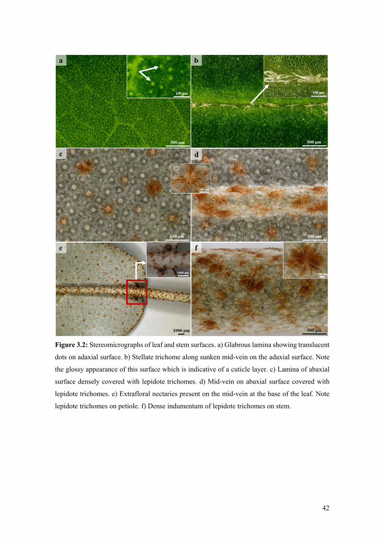
42
Figure 3.2: Stereomicrographs of leaf and stem surfaces. a) Glabrous lamina showing translucent
dots on adaxial surface. b) Stellate trichome along sunken mid-vein on the adaxial surface. Note
the glossy appearance of this surface which is indicative of a cuticle layer. c) Lamina of abaxial
surface densely covered with lepidote trichomes. d) Mid-vein on abaxial surface covered with
lepidote trichomes. e) Extrafloral nectaries present on the mid-vein at the base of the leaf. Note
lepidote trichomes on petiole. f) Dense indumentum of lepidote trichomes on stem.

43
Figure 3.3: Scanning electron micrographs of leaves and stems. a) Adaxial surface showing
stellate trichomes along the mid-vein of leaf. Note the peeled cuticle layer on this surface. b)
Stellate trichome emerging from middle furrow (mid-vein) on adaxial surface. c) Dense
indumentum formed by lepidote trichomes on the lamina and mid-vein on the abaxial surface. d)
Lepidote trichomes fully covering stem. ST = Stellate trichome.
3.4.2 Lepidote trichomes
Individual lepidote trichomes were observed at higher magnification (Fig. 3.4). According to
Webster et al. (1996), lepidote trichomes are scale-like hairs that are common in Croton species.
They are similar to the appressed-stellate trichomes. However, the radial cells of lepidote hairs
are fused, resulting in their shield-like appearance (Webster et al., 1996).
Vitarelli et al. (2016) suggest that lepidote trichomes function to increase water uptake from
atmospheric moisture as the shield-like structure provides a larger surface area for absorption. In
addition, the dense indumentum formed by lepidote trichomes may also function to protect the
plant from herbivores, pathogens, excessive water loss and increased temperatures, as reported
by Werker (2000).
Several authors (Inamdar and Gangadhara, 1977; Liu et al., 2013; Feio et al., 2018) indicated that
lepidote trichomes are non-secretory. However, TEM (Fig. 3.6, 3.7) and histochemical analyses
(Fig. 3.11) suggest that these structures may have a role in the synthesis and/or accumulation of
secretory products. de Sá-Haiad et al. (2009) demonstrated that numerous non-glandular
trichomes in several Croton species stained positive for various compounds with histochemical
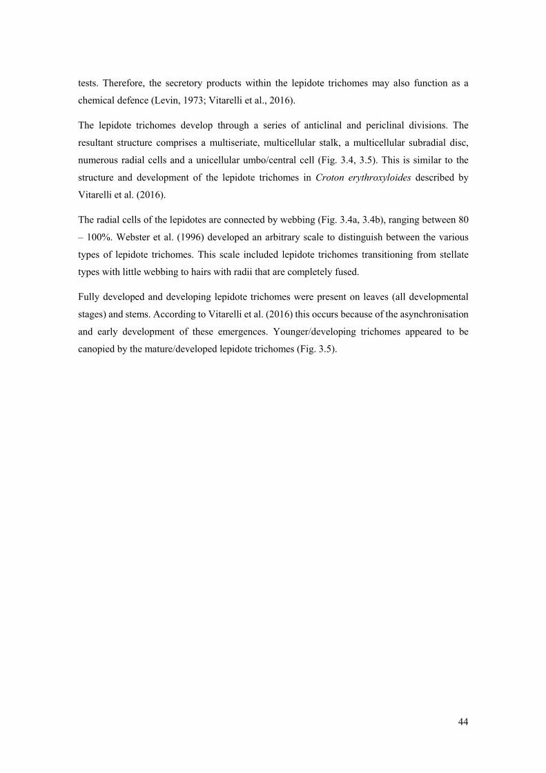
44
tests. Therefore, the secretory products within the lepidote trichomes may also function as a
chemical defence (Levin, 1973; Vitarelli et al., 2016).
The lepidote trichomes develop through a series of anticlinal and periclinal divisions. The
resultant structure comprises a multiseriate, multicellular stalk, a multicellular subradial disc,
numerous radial cells and a unicellular umbo/central cell (Fig. 3.4, 3.5). This is similar to the
structure and development of the lepidote trichomes in Croton erythroxyloides described by
Vitarelli et al. (2016).
The radial cells of the lepidotes are connected by webbing (Fig. 3.4a, 3.4b), ranging between 80
– 100%. Webster et al. (1996) developed an arbitrary scale to distinguish between the various
types of lepidote trichomes. This scale included lepidote trichomes transitioning from stellate
types with little webbing to hairs with radii that are completely fused.
Fully developed and developing lepidote trichomes were present on leaves (all developmental
stages) and stems. According to Vitarelli et al. (2016) this occurs because of the asynchronisation
and early development of these emergences. Younger/developing trichomes appeared to be
canopied by the mature/developed lepidote trichomes (Fig. 3.5).

45
Figure 3.4: Morphology of lepidote trichomes. a) Stereomicrograph of lepidote trichome. b) SEM
of lepidote trichome showing umbo/central cell and numerous webbed radial cells. c) Lepidote
trichome with accumulated secretory substance. d) Light micrograph of lepidote trichome
showing stalk cells, subradial cells, radial cells and umbo/central cell. U = Umbo, R =
Radii/Radial cell, Sr = Subradial cell, S = Stalk, Sm = Stoma, * = Secretion.

46
Figure 3.5: Development of lepidote trichomes. a) Emergence of protodermal cells giving rise to
lepidote trichome through periclinal and anticlinal divisions. Note the periclinal divisions
initiating the development of the stalk and the anticlinal divisions of the radial cells surrounding
the central cell. b) Developing lepidote trichome. Note the increased number of stalk cells brought
about by additional periclinal divisions and the stretching of the lateral radial cells. c) Fully
developed lepidote trichome with stalk, subradial, radial and central cells. Note the elongation of
stalk cells into a prominent stalk, the developed subradial cells, the distinct central cell and the
extended radial cells.
3.4.3 Ultrastructure of lepidote trichomes
Transmission electron microscopy revealed the presence of various organelles within the stalk
and radial cells of lepidote trichomes (Fig. 3.6, 3.7). Stalk cells contained numerous large and
small vacuoles which appeared to occupy the bulk of the cell (Fig. 3.7a). Vacuoles were present
in the radial cells (Fig. 3.7c). The vacuoles are reported to play a role in processing secretory
material (Machado et al., 2005; Huang et al., 2008). Large nuclei with dense nucleoplasm were
present in the stalk cells (Fig 3.6a, 3.6b). However, they were not very prominent because of the

47
surrounding dense cytoplasm (Fig 3.6a, 3.6b). The large vacuoles also constricted the space for
the nuclei and appeared appressed to these organelles. Numerous chloroplasts were also observed
in the stalk cells. Fahn (1979) states that plastids are the most common organelles involved in the
production of lipophilic substances. Werker and Fahn (1981) suggested that large amounts of
secretory substances may be produced by chloroplasts.
Stalk and radial cells contained lipid bodies, several vesicles, rough endoplasmic reticulum, Golgi
bodies and numerous mitochondria (Fig. 3.6, 3.7). However, within the radii, the cytoplasm and
the various organelles were restricted to the periphery of the cell (Fig. 3.7).
Many of the vesicles appeared translucent whilst others contained dense material (Fig. 3.6c, 3.6d,
3.6e, 3.7a, 3.7d). These vesicles in the stalk and radial cells indicate the secretion of hydrophilic
substances. Their occurrence close to the plasmalemma suggests that they undergo granulocrine
secretion (Tozin et al., 2016). The plasmalemma also appeared sinuous, indicating vesicle fusion
(Ascensão et al., 1997). Fahn (1979) indicates that granulocrine elimination of secretions occurs
in all secretory cells. Granulocrine secretion is described as the collection of secretory substances
in membrane-bound vesicles that either fuse with the plasmalemma or are eliminated by
invaginations of the plasmalemma (Fahn, 1979).
According to several authors (Ascensão and Pais 1998; Turner and Croteau 2004; Huang et al.,
2008), Golgi bodies in secretory trichomes play a role in the production of acidic and neutral
polysaccharides (Werker and Fahn, 1981). It has been suggested that endoplasmic reticulum is
also involved in the production of polysaccharides (Werker and Fahn, 1981), by producing the
protein component of the secretory product which is then transferred to the Golgi body (Ascensão
and Pais 1998). The Golgi body produces the polysaccharide component which is then transported
by the vesicles (Ascensão and Pais 1998). Huang et al. (2008) suggest that vesicles that are close
to the plasmalemma and Golgi body (Fig. 3.6d, 3.7a, 3.7d), transport the polysaccharide material
which is released through granulocrine secretion.
Stalk cells contained normal walls with visible plasmodesmata (Fig. 3.6e). However, the lateral
cell walls of the stalk appeared highly cutinised (Fig. 3.6). Ascensão and Pais (1998) suggested
that the presence of plasmodesmata enabled the symplastic transport of precursors. The cutinised
walls act as an apoplastic barrier to prevent the back-flow of secreted substances as these may be
toxic to mesophyll cells (Fahn, 1979; Ascensão et al. 1997; Werker, 2000).
Although lepidote trichomes are regarded as non-secretory, numerous organelles within the stalk
and radial cells (Fig. 3.6 and 3.7) indicate high metabolic activity (Naidoo et al, 2014). However,
much of the activity was in the stalk of the lepidote trichome. According to Fahn (1979), the

48
endoplasmic reticulum and Golgi body are involved in the secretion of hydrophilic substances.
On the other hand, various organelles, including the nucleus, mitochondria, Golgi body,
endoplasmic reticulum, plastids and ground cytoplasm may be responsible for the secretion of
lipophilic substances. All these organelles were present in the lepidote trichomes of C.
gratissimus. Therefore, these observations together with the positive staining for various
compounds by histochemical tests, suggest that they are involved in the synthesis and/or
accumulation of secondary metabolites. However, more studies are needed to confirm the
secretory process in these trichomes.

49
Figure 3.6: Transmission electron micrographs of lepidote trichome stalk cells. a) Section
through the stalk cells and radial cell. Large and small vacuoles surrounded by dense cytoplasm
and other organelles can be seen in the stalk cells. b) Single stalk cell containing dense cytoplasm
with numerous vacuoles, a large nucleus and a chloroplast. c) Rough endoplasmic reticulum and
vesicles at the periphery of a stalk cell wall. d) Vesicles and Golgi body present in stalk cell. e)
Thick cell wall between two adjacent stalk cells with visible plasmodesmata (white arrows).
Vacuoles, numerous mitochondria, endoplasmic reticulum and vesicles can be seen at the
periphery of these cells. Note the presence of the electron dense vesicle next to the cell wall. R =
Radial cell, S = Stalk, CW = Cell wall, Vs = Vesicle, V = Vacuole, N = Nucleus, M =
Mitochondria, RER/ER = Rough Endoplasmic Reticulum/Endoplasmic Reticulum, C =
Chloroplast, GB = Golgi body, LB = Lipid body, Adj = Adjacent cells.

50
Figure 3.7: Transmission electron micrographs of lepidote trichome radial cells. a) Radial cell
with thickened cell wall containing dense cytoplasm with vesicles, Golgi body, a lipid body and
rough endoplasmic reticulum at the periphery. b) Higher magnification of Golgi body surrounded
by dense cytoplasm. c) Vacuoles, mitochondria, lipid body and rough endoplasmic reticulum
present along the radial cell wall. d) Golgi body, rough endoplasmic reticulum, a lipid body and
numerous vesicles along the periphery of a radial cell wall. CW = Cell wall, Vs = Vesicle, V =
Vacuole, M = Mitochondria, RER/ER = Rough Endoplasmic Reticulum/Endoplasmic Reticulum,
GB = Golgi body, LB = Lipid body.
3.4.4 Glandular trichomes
Glandular trichomes were randomly distributed on the abaxial surfaces of emergent, young and
mature leaves and stems (Fig. 3.8). According to several authors (Wagner et al., 2004; Choi and
Kim, 2013; Glas et al., 2012; Huchelmann et al., 2017), glandular trichomes are involved in the
production, secretion and accumulation of various secondary metabolites. A study by Vitarelli et
al. (2015) also reported secretory trichomes on the abaxial surfaces of Croton species. In the
present investigation, they also occurred on the extrafloral nectaries of the leaf. According to
Webster et al. (1996), glandular trichomes exist in a limited number of Croton species and may
occur on either one or both leaf surfaces. Webster et al. (1996) described glandular trichomes as
“small embedded epidermal glands” and suggested that they contain terpenes which are
responsible for the aroma when the leaves are crushed.
These glandular trichomes were canopied under layers of lepidote trichomes (Fig. 3.8d). Light
micrographs indicate that they comprise a single cell and are embedded in the epidermal layer

51
(Fig. 3.8d). These unicellular glandular trichomes formed a prominent stalk and dilated head (Fig.
3.8d). This is consistent with the secretory trichomes of Croton species identified by Vitarelli et
al. (2015). Unicellular glandular trichomes consisted of a narrow short stalk and an expanded
distal end. The glandular trichomes existed in various forms as there is limited space to develop
beneath the dense lepidote trichomes.
Light microscopy and SEM indicated paracytic stomata on the adaxial surface of leaves (Fig.
3.4d, 3.8d). These stomata are a common character in Euphorbiaceae, occurring in many of its
species. Thakur and Patil (2014) state that paracytic stomata are primitive, whilst other types
present in the family are a derivative of this. A study by de Sá-Haiad et al. (2009) revealed that
paracytic stomata are predominant in Croton species.
Figure 3.8: Micrographs showing glandular trichomes on the leaves and stems. a) Glandular
trichomes on abaxial surface of leaves beneath lepidote trichomes. b) Stem showing glandular
trichomes after removing lepidote trichomes. High magnification of single glandular trichome on
abaxial surface. Note the presence of paracytic stomata. d) Light micrograph showing unicellular
glandular trichomes of different forms canopied by several layers of lepidote trichomes. Sm =
Stoma, LT = Lepidote Trichome, GT = Glandular Trichome.
52
3.4.5 Laticifers
A single laticifer type was observed throughout all leaf developmental stages and in stems (Fig.
3.9, 3.10). Within the Euphorbiaceae, latex and laticifer distribution are characters used to broadly
classify the family (Wurdack et al., 2005), due to their presence in several genera (dos Santos
Alves et al., 2017). The laticifers were present in the mid-vein of the leaf and were predominantly
associated with the vascular tissue, with occasional occurrences in parenchyma (Fig. 3.9a, 3.9c).
In the stems, the laticifers were predominant in the phloem and pith (Fig. 3.9b, 3.9d). This is
consistent with other work as laticifers are typically associated with the vascular tissues, more
specifically the phloem, but may also occur in the pith, cortex and foliar mesophyll (Dickison,
2000; Castro and Demarco, 2008; Lange, 2015). However, in this study, laticifers were not
observed in the foliar mesophyll.
Both non-articulated (branched and unbranched) and articulated laticifers have been reported to
occur in Euphorbiaceae (Hagel et al., 2008; Demarco et al., 2013). However, non-articulated
laticifers are more common and widespread in the family compared to the articulated type
(Demarco et al., 2013). In the present study, laticifers appeared non-articulated and unbranched
as they were composed of a single row of cells without branches (Fig. 3.10a). According to Lange
(2015), non-articulated laticifers are cells that develop from a single cell. It is suggested that they
form through apical intrusive growth (Da Cunha et al., 1998). The cell divides ceonocytically,
resulting in an elongated, multinucleated structure (Rudall, 1989; Lange, 2015). Studies by Rudall
(1994), de Sá-Haiad et al. (2009) and Feio et al. (2016) revealed the presence of non-articulated
laticifers in several Croton species.
Longitudinal and transverse monitor sections stained with Toluidine-Blue revealed latex within
laticifer cells (dark stained contents) (Fig. 3.10a, 3.10b). Fresh latex from the leaves and stems of
Croton was difficult to identify as the exudate was a clear watery sap. The latex of laticifers is
actually the protoplast of these cells. This protoplast which contains the metabolites, is housed
within a larger central vacuole (Castro and Demarco, 2008; Prado and Demarco, 2018). These
compounds may function to protect the plant against herbivores and pathogens (Prado and
Demarco, 2018).
Scanning electron micrscopy of freeze-fractured material also indicated latex within laticifer cells
(Fig. 3.10c). Coagulation of the latex within the cells was probably due to decrease of turgor
pressure within these cells during the preparation of the material. Generally, the pressure of latex
within laticifer cells is high. When there is a sudden drop in pressure, the surrounding turgid cells
compress the laticiferous cell, releasing the latex (Southorn, 1969) which polymerises when
53
exposed to air (Prado and Demarco, 2018). This coagulation of latex seals plant wounds, thus
indicating its ability to act as a physical barrier (Demarco, 2015).
Figure 3.9: Laticifer distribution in leaves and stems. a) Transverse section of leaf stained with
Toluidine-Blue showing distribution of laticifers predominantly in the vascular tissue. Note the
idioblasts at the adaxial side of the leaf. b) Transverse section of the stem stained with Toluidine-
Blue showing laticifers in the phloem and pith. c) Scanning electron micrograph of coagulated
latex within laticifer cells (associated with phloem). Druse crystals are also present in the leaf
section. d) Transverse section through stem showing latex containing laticifers in pith. Id =
Idioblast, Dr = Druse crystal.

54
Figure 3.10: Laticifer cells showing secretory contents. a) Longitudinal section of leaf showing
latex within non-articulated laticifers. b) Light micrograph of transverse section showing laticifer
cells with latex contents. c) Freeze- fracture through laticifer cells containing coagulated latex. Lt
= Laticifer, # = Latex.
3.4.6 Histochemistry and fluorescence microscopy
Histochemical and fluorescence analyses of the lepidote and glandular trichomes and laticifer
cells revealed the presence of hydrophilic and lipophilic substances. Appropriate controls were
also conducted (results not presented). The presence of these secondary metabolites indicates that
these three structures may be responsible for the production of biologically active compounds that
are used in traditional medicine (Salatino et al., 2007).
Lepidote trichomes are generally classed as non-glandular (Inamdar and Gangadhara, 1977; Liu
et al., 2013; Feio et al., 2018), and non-secretory (Wagner et al., 2004). However, these trichomes
tested positive for various compounds (Fig. 3.11).
In lepidote trichomes, subradial and central cells appeared lignified after staining with Toluidine-
Blue and phloroglucinol (Fig. 3.11g, 3.11h). A yellow fluorescence emitted by these cells after
staining with acridine orange also indicated the presence of lignified cells (Fig. 3.11j). Lignified
cells were also observed in glandular trichomes (blue autofluorescence) indicated in Fig. 3.12e.
The lateral walls of the lower stalk cells of lepidote trichomes stained orange with Sudan III,
indicating cutinised walls (Fig 3.11e). Cells that are cutinised or lignified typically act like

55
endodermal cells/Casparian strips and prevent the apoplastic flow of water, or the back-flow of
secreted substances (Fahn, 1979; Werker, 2000). The cell walls of the subradial, central and radial
cells of lepidote trichomes contained pectinaceous substances as they stained pink with ruthenium
red (Fig. 3.11d). The pectin provides support and strengthens the structure of these trichomes.
Pectin may also aid in plant defence as it induces phytoalexin accumulation which possesses
antimicrobial properties (Voragen et al., 2009).
Both trichome types and laticifers were found to possess various compounds, with alkaloids,
phenolic compounds and lipids common among all three.
Lepidote trichomes (stalk, subradial cells, radii and central cell/umbo), glandular trichomes and
laticifers stained orange/brown with Wagner’s reagent, indicating the presence of alkaloids (Fig.
3.11a, 3.12a, and 3.13a). Croton species have been reported to contain an abundance of active
alkaloids (Salatino et al., 2007). Alkaloids are common among angiosperms and are considered
to be the most active, diverse and therapeutic secretory compounds (Wink, 2015; Roy, 2017).
Their main function is to provide chemical defence against herbivores and pathogenic
microorganisms due to their toxic nature (Wink, 2015; Roy, 2017; Debnath et al., 2018). In
addition, plants containing alkaloids have been used for centuries in medicine to treat various
ailments, due to their medicinal and pharmacological properties, with many of these now isolated
and marketed as drugs (Roy, 2017; Bribi, 2018; Debnath et al., 2018).
Positive reactions for phenolic compounds were observed in the lepidote trichomes (stalk,
subradial cells, radii and central cell/umbo) and glandular trichomes, and laticifers, which all
produced a dark brown to black precipitate after staining with ferric chloride (Fig. 3.11b, 3.12b,
3.13c). Phenolics were also detected using auto-fluorescence as the stalk of lepidote trichomes
fluoresced blue under UV light. Salatino et al. (2007) state that phenolic compounds are common
among Croton species. These compounds defend the plant against pathogens, parasites and
predators (Huang et al., 2009), and may also play a role in pollination (Lin et al., 2016).
Furthermore, phenolic compounds from medicinal plants used in traditional medicine are known
to possess biological and pharmacological activities (Huang et al., 2009; Maslennikov et al.,
2014).
Lipidic compounds were detected using Sudan III, Sudan black and Nile blue. Laticifers (Fig.
3.13f) and cells comprising lepidote trichomes (Fig. 3.11e) stained orange with Sudan III,
indicating the presence of lipid components. Demarco et al. (2013) suggested that the lipid
component of latex has the ability to coagulate, demonstrating its function to seal wounds. A
positive reaction was also observed in glandular trichomes with Sudan III which stained lipid
inclusions orange-red (Fig. 3.12d). Sudan black stained lipids dark blue in the stalk, subradial,

56
radial and central cell of lepidote trichomes (Fig. 3.11f). Nile blue was used to detect acidic and
neutral lipids. Subradial, radial and central cells of lepidote trichomes (Fig. 3.11g) and laticifer
cells (Fig. 3.13b) stained blue, indicating a positive reaction for acidic lipids. The stalk cells of
lepidote trichomes (Fig. 3.11c) and glandular trichomes (Fig. 3.12c) stained pink which was a
positive indication for neutral lipids. The presence of lipids in these external structures is in
accordance with literature as lipophilic substances are usually secreted by glandular trichomes
(Valkama et al., 2003).
Alkaloids, lipids and phenolic compounds were also detected in the laticifers of C. echinocarpus
and C. urucurana (Feio et al. 2016). Feio et al. (2016) suggested that the phenolic compounds
and alkaloids present in these species are responsible for their biological activities.
Sections stained with ruthenium red revealed the presence of mucilaginous substances in laticifers
which was indicated by a pink colouration in the cells (Fig. 3.13d). According to Demarco et al.
(2013), mucilage is common in the latex of euphorbiaceous species and may play a role in wound
sealing (Fisher et al., 2009; Kuster et al., 2016). Staining with Toluidine-Blue resulted in an
intensely dark blue/purple colouration of laticifers (Fig. 3.13e), indicating that these cells contain
macromolecules with free phosphate groups.
In the present study, light and SEM micrographs (Fig. 3.9c, 3.13d) indicated druse (Dr) and
prismatic crystals (Pr) in the leaves of C. gratissimus. The organs and tissues of numerous plant
species possess calcium oxalate crystals (Franceschi and Horner, 1980). These deposits are
housed within vacuoles of specialised cells known as crystal idioblasts (Pennisi et al., 2001).
Within a crystal idioblast, there is great variation in the number, shape and size of the crystals
(Franceschi and Horner, 1980; Konyar et al., 2014). However, common shapes include the druse,
styloid, raphide, prism and crystal sand (Franceschi and Horner, 1980; Konyar et al., 2014). These
crystals have been used as taxonomic tools due to the specificity of the shape and location within
a taxon (Franceschi and Horner, 1980; Konyar et al., 2014). Solereder (1908) stated that oxalate
of lime (primitive term for calcium oxalate) is present in many forms in various genera of
Euphorbiaceae. Calcium oxalate crystals appear to have various functions. These include
removing excess calcium and oxalate to maintain ionic balance and prevent toxicity (metal
detoxification) of the plant, tissue support, and protection against foraging herbivores (Franceschi
and Horner, 1980; Nakata, 2002; Konyar et al., 2014).
In addition, secretory idioblasts were also identified at the adaxial side of leaves, as shown in Fig.
3.13a. Idioblasts are secretory cells that are noticeably different from surrounding cells, differing
by size, form, metabolism, development and content (Esau, 1965; Castro and Demarco, 2008;
Bosabalidis, 2014). These cells contain various substances such as oils, tannins, phenolic
57
compounds, mucilage, gums, resins and crystals (Esau, 1965; Dickison, 2000; Castro and
Demarco, 2008; Bosabalidis, 2014). According to Solereder (1908) internal oil or resin secreting
cells may result in the presence of transparent dots on the leaf surface. Idioblasts in Croton species
have been reported to contain lipophilic substances (de Sá-Haiad et al., 2009; Vitarelli et al.,
2015). This might suggest that the secretory idioblasts present in leaves, were probably
responsible for the translucent dots observed on this surface (Fig. 3.2a). However, more detailed
investigations need to be carried out on these cells to confirm this.
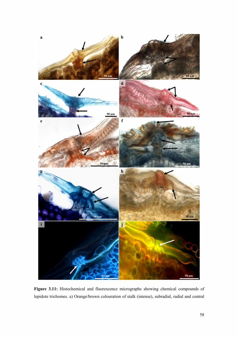
58
Figure 3.11: Histochemical and fluorescence micrographs showing chemical compounds of
lepidote trichomes. a) Orange/brown colouration of stalk (intense), subradial, radial and central

59
cells (weak) suggest a positive indication for the presence of alkaloids with Wagner’s reagent. b)
Phenolics detected in stalk, subradial cells, radii and central cell with ferric chloride (brown to
black precipitate). c) Pink colouration indicated neutral lipids in stalk cells and blue colouration
of subradial, radial and central cells indicated acidic lipids with Nile blue. d) Pectin in the
subradial, radial and central cell walls was indicated by a pink colour. e) Orange staining of the
stalk and radii with Sudan III indicated the presence of cutinised walls and lipids. f) Positive
staining for lipids in the stalk, subradial and radial cells with Sudan black. g) Toluidine-Blue
revealed lignification of the subradial and central cells (blue colouration). h) Positive indication
of lignin in the subradial and central cells with phloroglucinol. i) Blue autofluorescence indicated
phenolic compounds in stalk cells. j) Yellow fluorescence with acridine orange revealed lignified
subradial and central cells.
Figure 3.12: Histochemical and fluorescence micrographs showing chemical compounds of
glandular trichomes. a) Positive staining for alkaloids (brown colour) with Wagner’s reagent. b)
Glandular trichomes tested positive for phenolic compounds with ferric chloride (indicated by
brown/black precipitate). c) Pink colouration indicated neutral lipids with Nile blue. d) Lipid
droplet stained red/orange with Sudan III. e) Lignified cell walls of glandular trichome detected
with autofluorescence.

60
Figure 3.13: Histochemical and fluorescence micrographs showing chemical compounds of
laticifer cells. a) Orange colouration a positive indication for alkaloids with Wagner’s reagent. b)
Blue colouration within laticifer cells indicated acidic lipids with Nile blue. c) Positive indication
(dark brown to black) for phenolic compounds with ferric chloride. d) Pink colouration indicated
mucilage with ruthenium red. Note the presence of druse and prismatic crystals. e) Blue staining
of laticifer cells with Toluidine-Blue indicated macromolecules with free phosphate groups. f)
Positive stain (orange colour) for lipids with Sudan III. Pr = Prismatic crystal, Dr = Druse crystal.
3.5 Conclusion
Three types of structures, lepidote and glandular trichomes, and non-articulated, unbranched
laticifers are present on/in the leaves and stems of C. gratissimus. These structures were found to

61
contain various secondary compounds which are reported to protect the plant from herbivores and
pathogens. Furthermore, the secretory products present within trichomes and laticifers may
contribute to the medicinal properties of the plant as some of them are known to possess biological
and pharmacological activities. Although these structures were found to play a role in the
production and/or accumulation of secondary metabolites, further work is required to identify
their mode of synthesis. In addition, crystal and secretory idioblasts, extrafloral nectaries and
stellate trichomes were also identified on the leaves of C. gratissimus. However, further
investigations need to be conducted to determine whether these structures are also involved in the
production/accumulation of compounds that contribute to the biological activity of the plant.
3.6 References
Ascensao, L., Marques, N. and Pais, M.S., 1997. Peltate glandular trichomes of Leonotis leonurus
leaves: ultrastructure and histochemical characterization of secretions. International Journal of
Plant Sciences 158, 249 – 258.
Ascensão, L. and Pais, M.S., 1998. The leaf capitate trichomes of Leonotis leonurus:
histochemistry, ultrastructure and secretion. Annals of Botany 81, 263 – 271.
Berry, P.E., van Ee, B.W., Kainulainen, K. and Achtemeier, L., 2016. Croton cupreolepis
(Euphorbiaceae), a new coppery-lepidote tree species from eastern Madagascar. Systematic
Botany 41, 977 – 982.
Block, S., Baccelli, C., Tinant, B., Van Meervelt, L., Rozenberg, R., Jiwan, J.L.H., Llabres, G.,
De Pauw-Gillet, M.C. and Quetin-Leclercq, J., 2004. Diterpenes from the leaves of Croton
zambesicus. Phytochemistry 65, 1165 – 1171.
Bosabalidis, A.M., 2014. Idioblastic mucilage cells in Teucrium polium leaf. Anatomy and
histochemistry. Modern Phytomorphology 5, 49 – 52.
Bribi, N., 2018. Pharmacological activity of Alkaloids: A Review. Asian Journal of Botany 1, 1
– 6.
Castelblanque, L., Balaguer, B., Martí, C., Rodríguez, J.J., Orozco, M. and Vera, P., 2016. Novel
insights into the organization of laticifer cells: a cell comprising a unified whole system. Plant
Physiology 172, 1032 – 1044.
Castro, M.D. and Demarco, D., 2008. Phenolic compounds produced by secretory structures in
plants: a brief review. Natural Product Communications 3, 1273 – 1284.

62
Chikezie, P.C., Ibegbulem, C.O. and Mbagwu, F.N., 2015. Bioactive principles from medicinal
plants. Research Journal of Phytochemistry 9, 88 – 115.
Choi, J.S. and Kim, E.S., 2013. Structural features of glandular and non-glandular trichomes in
three species of Mentha. Applied Microscopy, 43, 47 – 53.
Da Cunha, M., Costa, C.G., Machado, R.D. and Miguens, F.C., 1998. Distribution and
differentiation of the laticifer system in Chamaesyce thymifolia (L.) Millsp. (Euphorbiaceae).
Acta Botanica Neerlandica 47, 209 – 218.
de Andrade, E.A., Folquitto, D.G., Luz, L.E.C., Paludo, K.S., Farago, P.V. and Budel, J.M., 2017.
Anatomy and histochemistry of leaves and stems of Sapium glandulosum. Revista Brasileira de
Farmacognosia 27, 282 – 289.
de Sá-Haiad, B., Serpa-Ribeiro, A.C.C., Barbosa, C.N., Pizzini, D., Leal, D.D.O., de Senna-Valle,
L. and de Santiago-Fernandes, L.D.R., 2009. Leaf structure of species from three closely related
genera from tribe Crotoneae Dumort. (Euphorbiaceae ss, Malpighiales). Plant Systematics and
Evolution 283, 179 – 202.
Debnath, B., Singh, W.S., Das, M., Goswami, S., Singh, M.K., Maiti, D. and Manna, K., 2018.
Role of plant alkaloids on human health: A review of biological activities. Materials Today
Chemistry 9, 56 – 72.
Demarco, D., de Moraes Castro, M. and Ascensão, L., 2013. Two laticifer systems in Sapium
haematospermum—new records for Euphorbiaceae. Botany 91, 545 – 554.
Demarco, D., 2015. Micromorphology and histochemistry of the laticifers from vegetative organs
of asclepiadoideae species (Apocynaceae). Acta Biológica Colombiana 20, 57 – 65.
Demarco, D., 2017. Histochemical analysis of plant secretory structures, in: Pellicciari, C.,
Biggiogera, M. (Eds), Histochemistry of Single Molecules: Methods and Protocols, Methods in
Molecular Biology. Humana Press., New York, pp. 313 – 330.
Dickison, W.C., 2000. Integrative plant anatomy. Academic Press.
dos Santos Alves, I.A.B., Sá, R.D., Cadena, M.B., Ximenes, R.M. and Randau, K.P., 2017.
Microscopic Characterization of Croton cordiifolius Baill. (Euphorbiaceae). Pharmacognosy
Journal 9, 361 – 366.
Esau, K., 1965. Plant anatomy, 2nd ed. John Wiley & Sons, New York, USA.
Fahn, A., 1979. Secretory tissues in plants, Academic Press, London, UK.

63
Feio, A.C., Riina, R. and Meira, R.M.S.A., 2016. Secretory structures in leaves and flowers of
two Dragon’s blood Croton (Euphorbiaceae): new evidence and interpretations. International
Journal of Plant Sciences 177, 511 – 522.
Feio, A.C., Meira, R.M. and Riina, R., 2018. Leaf anatomical features and their implications for
the systematics of dragon’s blood, Croton section Cyclostigma (Euphorbiaceae). Botanical
Journal of the Linnean Society 187, 614 – 632.
Fisher, J.B., Lindström, A. and Marler, T.E., 2009. Tissue responses and solution movement after
stem wounding in six Cycas species. HortScience 44, 848 – 851.
Franceschi, V.R. and Horner, H.T., 1980. Calcium oxalate crystals in plants. The Botanical
Review 46, 361 – 427.
Furr, M. and Mahlberg, P.G., 1981. Histochemical analyses of laticifers and glandular trichomes
in Cannabis sativa. Journal of Natural Products 44, 153 – 159.
Glas, J.J., Schimmel, B.C., Alba, J.M., Escobar-Bravo, R., Schuurink, R.C. and Kant, M.R., 2012.
Plant glandular trichomes as targets for breeding or engineering of resistance to herbivores.
International Journal of Molecular Sciences 13, 17077 – 17103.
Guo, J., Yuan, Y., Liu, Z. and Zhu, J., 2013. Development and structure of internal glands and
external glandular trichomes in Pogostemon cablin. Plos One 8, 1 – 13.
Hagel, J.M., Yeung, E.C. and Facchini, P.J., 2008. Got milk? The secret life of laticifers. Trends
in Plant Science 13, 631 – 639.
Huang, S.S., Kirchoff, B.K. and Liao, J.P., 2008. The capitate and peltate glandular trichomes of
Lavandula pinnata L. (Lamiaceae): histochemistry, ultrastructure, and secretion. The Journal of
the Torrey Botanical Society 135, 155 – 167.
Huang, W.Y., Cai, Y.Z. and Zhang, Y., 2009. Natural phenolic compounds from medicinal herbs
and dietary plants: potential use for cancer prevention. Nutrition and Cancer 62, 1 – 20.
Huchelmann, A., Boutry, M. and Hachez, C., 2017. Plant glandular trichomes: natural cell
factories of high biotechnological interest. Plant Physiology 175, 6 – 22.
Inamdar, J.A. and Gangadhara, M., 1977. Studies on the trichomes of some Euphorbiaceae.
Feddes Repertorium 88, 103 – 111.
Johansen, D.A., (1940). Plant Microtechnique, McGraw-Hill, London, UK.

64
Konyar, S.T., Öztürk, N. and Dane, F., 2014. Occurrence, types and distribution of calcium
oxalate crystals in leaves and stems of some species of poisonous plants. Botanical Studies 55, 1
– 9.
Kuster, V.C., da Silva, L.C., Meira, R.M.S.A. and Azevedo, A.A., 2016. Glandular trichomes and
laticifers in leaves of Ipomoea pes-caprae and I. imperati (Convolvulaceae) from coastal Restinga
formation: Structure and histochemistry. Brazilian Journal of Botany 39, 1117 – 1125.
Langat, M.K., Crouch, N.R., Smith, P.J. and Mulholland, D.A., 2011. Cembranolides from the
Leaves of Croton gratissimus. Journal of Natural Products 74, 2349 – 2355.
Lange, B.M., 2015. The evolution of plant secretory structures and emergence of terpenoid
chemical diversity. Annual Review of Plant Biology 66, 139 – 159.
Leandri, J., 1972. Contribution a l'etude des Croton malgaches a grandes feuilles argentees.
Adansonia 12, 403 – 408.
Lee, Y.L. and Ding, P., 2016. Production of essential oil in plants: Ontogeny, secretory structures
and seasonal variations. Pertanika Journal of Scholarly Research Reviews 2, 1 – 10.
Levin, D.A., 1973. The role of trichomes in plant defense. The Quarterly Review of Biology 48,
3 – 15.
Lima, S.G.D., Citó, A.M., Lopes, J.A., Neto, J.M., Chaves, M.H. and Silveira, E.R., 2010. Fixed
and volatile constituents of genus Croton plants: C. adenocalyx Baill-Euphorbiaceae. Revista
Latinoamericana de Química 38, 133 – 144.
Lin, D., Xiao, M., Zhao, J., Li, Z., Xing, B., Li, X., Kong, M., Li, L., Zhang, Q., Liu, Y. and Chen,
H., 2016. An overview of plant phenolic compounds and their importance in human nutrition and
management of type 2 diabetes. Molecules 21, 1 – 19.
Liu, H.F., Deng, Y.F. and Liao, J.P., 2013. Foliar trichomes of Croton L. (Euphorbiaceae:
Crotonoideae) from China and its taxonomic implications. Bangladesh Journal of Plant
Taxonomy 20, 85 – 94.
Lucena, M.D.F.D.A. and Sales, M.F.D., 2006. Foliar Trichome in Croton L. (Crotonoideae-
Euphorbiaceae). Rodriguésia 57, 11 – 25.
Machado, S.R., Gregório, E.A. and Guimarães, E., 2005. Ovary peltate trichomes of Zeyheria
montana (Bignoniaceae): developmental ultrastructure and secretion in relation to function.
Annals of Botany 97, 357 – 369.

65
Marin, M., Budimir, S., Janošević, D., Marin, P.D., Duletić-Laušević, S.O.N.J.A. and Ljaljević-
Grbić, M., 2008. Morphology, distribution, and histochemistry of trichomes of Thymus lykae
Degen and Jav.(Lamiaceae). Archives of Biological Sciences 60, 667 – 672.
Maslennikov, P.V., Chupakhina, G.N. and Skrypnik, L.N., 2014. The content of phenolic
compounds in medicinal plants of a botanical garden (Kaliningrad oblast). Biology Bulletin 41,
133 – 138.
Mulholland, D.A., Langat, M.K., Crouch, N.R., Coley, H.M., Mutambi, E.M. and Nuzillard, J.M.,
2010. Cembranolides from the stem bark of the southern African medicinal plant, Croton
gratissimus (Euphorbiaceae). Phytochemistry 71, 1381 – 1386.
Mwine, J.T. and Van Damme, P., 2011. Why do Euphorbiaceae tick as medicinal plants? A
review of Euphorbiaceae family and its medicinal features. Journal of Medicinal Plants Research
5, 652 – 662.
Naidoo, Y., Heneidak, S., Bhatt, A., Kasim, N. and Naidoo, G., 2014. Morphology,
histochemistry, and ultrastructure of foliar mucilage-producing trichomes of Harpagophytum
procumbens (Pedaliaceae). Turkish Journal of Botany 38, 60 – 67.
Nakata, P.A., 2002. Calcium oxalate crystal morphology. Trends in Plant Science 7, 324.
Ngadjui, B.T., Abegaz, B.M., Keumedjio, F., Folefoc, G.N. and Kapche, G.W., 2002.
Diterpenoids from the stem bark of Croton zambesicus. Phytochemistry 60, 345 – 349.
O'brien, T.P., Feder, N. and McCully, M.E., 1964. Polychromatic staining of plant cell walls by
toluidine blue O. Protoplasma 59, 368 – 373.
Pearse, A. G. E. (1985) Histochemistry: theoretical and applied. Vol. II, 4th ed. Longman.
Pennisi, S.V., McConnell, D.B., Gower, L.B., Kane, M.E. and Lucansky, T., 2001. Intracellular
calcium oxalate crystal structure in Dracaena sanderiana. New Phytologist 150, 111 – 120.
Pickard, W.F., 2008. Laticifers and secretory ducts: two other tube systems in plants. New
Phytologist 177, 877 – 888.
PlantZAfrica, 2018. Croton gratissimus Burch. http://pza.sanbi.org/croton-gratissimus. Date
Accessed: 5 February 2018.
Prado, E. and Demarco, D., 2018. Laticifers and Secretory Ducts: Similarities and Differences,
in: Ecosystem Services and Global Ecology. IntechOpen. pp. 103 – 123.

66
Robert, S., Baccelli, C., Devel, P., Dogné, J.M. and Quetin-Leclercq, J., 2010. Effects of leaf
extracts from Croton zambesicus Müell. Arg. on hemostasis. Journal of Ethnopharmacology 128,
641 – 648.
Roy, A., 2017. A review on the alkaloids an important therapeutic compound from plants.
International Journal of Plant Biotechnology 1, 1 – 9.
Rudall, P., 1989. Laticifers in vascular cambium and wood of Croton spp. (Euphorbiaceae).
International Association of Wood Anatomists Journal 10, 379 – 383.
Rudall, P., 1994. Laticifers in Crotonoideae (Euphorbiaceae): homology and evolution. Annals
of the Missouri Botanical Garden 81, 270 – 282.
Salatino, A., Salatino, M.L.F. and Negri, G., 2007. Traditional uses, chemistry and pharmacology
of Croton species (Euphorbiaceae). Journal of the Brazilian Chemical Society 18, 11 – 33.
Schilmiller, A.L., Last, R.L. and Pichersky, E., 2008. Harnessing plant trichome biochemistry for
the production of useful compounds. The Plant Journal 54, 702 – 711.
Sen, S. and Chakraborty, R., 2017. Revival, modernization and integration of Indian traditional
herbal medicine in clinical practice: Importance, challenges and future. Journal of Traditional and
Complementary Medicine 7, 234 – 244.
Senakun, C. and Chantaranothai, P., 2010. A morphological survey of foliar trichomes of Croton
L. (Euphorbiaceae) in Thailand. Thai Forest Bulletin (Botany) 38, 167 – 172.
Solereder, H., 1908. Systematic Anatomy of the Dicotyledons Vol-II, Ajay Book Service, New
Delhi, India.
Southorn, W.A., 1969. Physiology of Hevea (latex flow). Rubber Research Institute of Malaya
Journal 21, 494 – 512.
Spurr, A.R., 1969. A low-viscosity epoxy resin embedding medium for electron microscopy.
Journal of Ultrastructure Research 26, 31 – 43.
Talamond, P., Verdeil, J.L. and Conéjéro, G., 2015. Secondary metabolite localization by
autofluorescence in living plant cells. Molecules 20, 5024 – 5037.
Tan, D., Sun, X. and Zhang, J., 2011. Histochemical and immunohistochemical identification of
laticifer cells in callus cultures derived from anthers of Hevea brasiliensis. Plant Cell Reports 30,
1117 – 1124.
67
Thakur, H.A. and Patil, D.A., 2014. Foliar epidermal studies of plants in Euphorbiaceae. Taiwania
59, 59 – 70.
Tissier, A., 2018. Plant secretory structures: more than just reaction bags. Current Opinion in
Biotechnology 49, 73 – 79.
Tozin, L.R.D.S., de Melo Silva, S.C. and Rodrigues, T.M., 2016. Non-glandular trichomes in
Lamiaceae and Verbenaceae species: morphological and histochemical features indicate more
than physical protection. New Zealand Journal of Botany 54, 446 – 457.
Turner, G.W. and Croteau, R., 2004. Organization of monoterpene biosynthesis in Mentha.
Immunocytochemical localizations of geranyl diphosphate synthase, limonene-6-hydroxylase,
isopiperitenol dehydrogenase, and pulegone reductase. Plant Physiology 136, 4215 – 4227.
Uday, P., Raghu, R.A., Bhat, R.P., Rinimol, V.R., Bindu, J., Nafeesa, Z. and Swamy, S.N., 2015.
Laticiferous plant proteases in wound care. International Journal of Pharmacy and Pharmaceutical
Sciences 7, 44 – 49.
Valkama, E., Salminen, J.P., Koricheva, J. and Pihlaja, K., 2003. Comparative analysis of leaf
trichome structure and composition of epicuticular flavonoids in Finnish birch species. Annals of
Botany 91, 643 – 655.
Van Vuuren, S.F. and Viljoen, A.M., 2008. In vitro evidence of phyto-synergy for plant part
combinations of Croton gratissimus (Euphorbiaceae) used in African traditional healing. Journal
of Ethnopharmacology 119, 700 – 704.
Vitarelli, N.C., Riina, R., Caruzo, M.B.R., Cordeiro, I., Fuertes‐Aguilar, J. and Meira, R.M., 2015.
Foliar secretory structures in Crotoneae (Euphorbiaceae): Diversity, anatomy, and evolutionary
significance. American Journal of Botany 102, 833 – 847.
Vitarelli, N.C., Riina, R., Cassino, M.F. and Meira, R.M.S.A., 2016. Trichome-like emergences
in Croton of Brazilian highland rock outcrops: evidences for atmospheric water uptake.
Perspectives in Plant Ecology, Evolution and Systematics 22, 23 – 35.
Voragen, A.G., Coenen, G.J., Verhoef, R.P. and Schols, H.A., 2009. Pectin, a versatile
polysaccharide present in plant cell walls. Structural Chemistry 20, 263 – 275.
Wagner, G.J., Wang, E. and Shepherd, R.W., 2004. New approaches for studying and exploiting
an old protuberance, the plant trichome. Annals of Botany 93, 3 – 11.

68
Webster, G.L., Del-Arco-Aguilar, M.J. and Smith, B.A., 1996. Systematic distribution of foliar
trichome types in Croton (Euphorbiaceae). Botanical Journal of the Linnean Society 121, 41 –
57.
Werker, E. and Fahn, A., 1981. Secretory hairs of Inula viscosa (L.) Ait.-development,
ultrastructure, and secretion. Botanical Gazette 142, 461 – 476.
Werker, E., 2000. Trichome diversity and development. Advances in Botanical Research 31, 1 –
35.
Wink, M., 2015. Modes of action of herbal medicines and plant secondary metabolites. Medicines
2, 251 – 286.
Wurdack, K.J., Hoffmann, P. and Chase, M.W., 2005. Molecular phylogenetic analysis of
uniovulate Euphorbiaceae (Euphorbiaceae sensu stricto) using plastid rbcL and trnL‐F DNA
sequences. American Journal of Botany 92, 1397 – 1420.
Yuan, H., Ma, Q., Ye, L. and Piao, G., 2016. The traditional medicine and modern medicine from
natural products. Molecules 21, 559.
Zander, R.H., 2016. A new progressive polychrome protocol for staining bryophytes.
Phytoneuron 2016, 1 – 12.

69
CHAPTER 4: PHYTOCHEMICAL AND ANTIBACTERIAL
ANALYSES OF CROTON GRATISSIMUS BURCH. VAR.
GRATISSIMUS (EUPHORBIACEAE) LEAF AND STEM
EXTRACTS
4.1 Abstract
Croton gratissimus is commonly used in traditional medicine to treat various ailments. This study
was conducted to determine the chemical composition of the leaf and stem extracts, evaluate its
antimicrobial potential and identify compounds that are responsible for the biological activity of
this species. Preliminary phytochemical screening revealed alkaloids, amino acids, phenolic
compounds, flavonoids, carbohydrates, terpenoids, saponins and fixed oils and fats in the leaf and
stem extracts. Antibacterial assays indicated weak to strong activities of the methanolic extracts,
with stem extracts displaying stronger activity than the leaves. Several bioactive compounds were
present in the leaf and stems extracts. Many of these possess antimicrobial and antibacterial
activities. The presence of these compounds in the methanolic extracts was probably responsible
for the activity demonstrated in this study. These findings validate the use of C. gratissimus in
traditional medicine and indicate its potential as a source of antimicrobial agents.
Keywords: Terpenoids, bacterial strains, bioactive compounds, antimicrobial agents, biological
activities.

70
4.2 Introduction
Plants are known to produce a diverse range of bioactive compounds such as alkaloids, terpenoids,
saponins and phenolic compounds (Chikezie et al., 2015; Altemimi et al., 2017). These bioactive
compounds are reported to possess therapeutic properties, some of which include antidiabetic,
anticarcinogenic, anti-inflammatory, antioxidant, antimicrobial and antimalarial activity
(Chikezie et al., 2015). The use of these plants in traditional medicine has led to the discovery of
many important drugs such as morphine (Papaver somniferum L.), atropine (Atropa belladonna
L.), colchicine (Colchicum autumnale L.), quinine (Cinchona ledgeriana Moens ex. Trimen) and
reserpine (Rauvolfia serpentina (L.) Benth ex. Kurz) (Fabricant and Farnsworth, 2001). For this
reason, studies on traditional medicinal plants are important precursors in the development of
novel drugs (Heamalatha et al., 2011; Mustafa et al., 2017).
Plants used in traditional medicine to treat microbial related diseases are great sources of
antimicrobial agents (Samy and Gopalakrishnakone, 2010). Several studies have demonstrated
the antimicrobial properties of species in Croton (Fontenelle et al., 2008; Bayor et al., 2009;
Selowa et al., 2010; Fernandes et al., 2013; Obey et al., 2016; Leite et al., 2017).
The species C. gratissimus Burch. var. gratissimus belonging to the Euphorbiaceae (Robert et al.,
2010; PlantZAfrica, 2018) has a reputation of being medicinally important (Van Vuuren and
Viljoen, 2008). Several parts of this plant have been used in traditional medicine for the treatment
of various ailments (Van Vuuren and Viljoen, 2008; Mulholland et al., 2010; Robert et al., 2010;
Pudumo et al., 2018). Previous studies on the plant extracts, isolated compounds and essential
oils have revealed cytotoxic (Block et al., 2002; Block et al., 2004; Mulholland et al., 2010; Lawal
et al., 2017), antiulcerogenic (Okokon et al., 2011), antidiabetic (Okokon et al., 2006; Kumar et
al., 2017), analgesic (Okokon and Nwafor, 2010b) antipyretic (Okokon and Nwafor, 2010b),
antimicrobial (Abo et al., 1999; Van Vuuren and Viljoen, 2008; Okokon and Nwafor, 2010a; Van
Vuuren and Naidoo, 2010; Mthethwa et al., 2014; Lawal et al., 2017), antioxidant (Abdalaziz et
al., 2016), antiplasmodial (Okokon and Nwafor, 2009) and ant-HIV-1 (Mthethwa et al., 2014)
activities.
There is a growing need to develop new antimicrobials from natural sources such as medicinal
plants due to the rise in antibiotic resistance and shortage of novel antimicrobial agents (Cowan,
1999; Abdallah, 2011; Elisha et al., 2017). Antibacterial screening is a good starting point in
determining whether medicinal plants used in traditional medicine are sources of antimicrobial
agents (Samy and Gopalakrishnakone, 2010). Therefore, this study determined the chemical
composition of the leaf and stem extracts of C. gratissimus and evaluated the antibacterial activity

71
of methanolic extracts. In addition, the possible compounds responsible for antibacterial, anti-
inflammatory, antioxidant and anticancer activities were also identified.
4.3 Materials and Methods
4.3.1 Plant collection and sampling
Leaves and stems of C. gratissimus Burch. var. gratissimus were collected from the University of
KwaZulu-Natal, Westville Campus (29°49'08.1"S 30°56'38.9"E). The material was air dried for
two months before being ground to a powder for phytochemical extraction. A voucher specimen
(Croton 01 – Accession No. 18224) was prepared and deposited in the Ward Herbarium located
in the School of Life Sciences at the University of KwaZulu-Natal, Westville Campus.
4.3.2. Crude extracts
Powdered leaf and stems were placed in separate round bottom flasks and boiled through
continuous reflux with a graded series of solvents (hexane, chloroform and methanol) ranging
from non-polar to polar. Three 3 h sessions were carried out for each solvent. The extracts were
filtered and used for preliminary phytochemical screening and thin layer chromatography (TLC).
Methanolic leaf and stem extracts using high performance liquid chromatography (HPLC) grade
solvent was obtained in the same way as above for Gas Chromatography-Mass Spectrometry (GC-
MS) analysis and antibacterial assays.
4.3.3 Preliminary phytochemical screening
The hexane, chloroform and methanol crude extracts were used to carry out preliminary
phytochemical screening using standard protocols (Raaman, 2006; Benmehdi et al., 2012;
Hossain et al., 2013; Morsy, 2014). Identification of the following phytochemical compounds was
as follows:
Alkaloids
Wagner’s
Extracts were treated with Wagner’s reagent. A reddish/brown precipitate indicated the presence
of alkaloids.
Dragendorff’s
Dragendorff’s reagent was added to a few mL of each extract. A red precipitate indicated the
presence of alkaloids.

72
Carbohydrates
Molisch’s
For each extract, two drops of alcoholic α-naphthol solution were added to a test tube with one
mL of filtrate. Thereafter, 2 mL of concentrated sulphuric acid were added to the side of the test
tube. The formation of a violet ring at the junction indicated the presence of carbohydrates.
Benedict’s
One mL of each extract was treated with Benedict’s solution and heated in a water bath. A reddish
precipitate indicated the presence of reducing sugars.
Fehling’s
One mL each of Fehling’s A and B solutions was added to one mL of each extract and boiled in
a water bath. A red precipitate indicated the presence of reducing sugars.
Flavonoids
Each extract was treated with a few drops of lead acetate solution. A yellow coloured precipitate
was a positive indication for the presence of flavonoids.
Phenolic compounds
Five mL of distilled water were added to each extract. Thereafter, a few drops of neutral 5% ferric
chloride solution were added. A dark green colour indicated the presence if phenolic compounds.
Saponins
Twenty mL of distilled water were added to each extract and shaken for 15 min. The formation
of a foam layer was a positive indicator for saponins.
Fixed fats and oils
A few drops of each extract were pressed between two filter papers. Oil stains on the filter paper
was a positive indicator for fixed oils.
Terpenoids
Chloroform (2 mL) was added to 5 mL of each extract. Subsequently, 3 mL of concentrated
sulphuric acid were carefully added to form a distinct layer. A reddish brown colour at the
interface was a positive indicator for the presence of terpenoids.

73
Amino Acids
Ninhydrin reagent was added to each extract and boiled in a water bath for a few mins. A
characteristic blue-violet colour indicated the presence of amino acids or proteins.
4.3.4 Thin Layer Chromatography (TLC)
Hexane, chloroform and methanol extracts of leaves and stems were used for TLC analysis. Each
sample was pipetted (10 µL) onto a precoated silica gel 60 F254 TLC plate (E. Merck). The plate
was then placed in a mobile phase of Toluene: Ethyl acetate: Formic acid (9.5: 0.7: 0.3) and
allowed to develop. The plate was then visualised and imaged under 254 nm and 365 nm UV
light. Thereafter the plate was dipped in anisaldehyde-sulphuric acid (ANS) reagent and heated
before being imaged. The reagent was made up of 0.5 mL ANS, 10 mL Glacial acetic acid, 85
mL methanol and 5 mL concentrated sulphuric acid.
4.3.5 Gas Chromatography-Mass Spectrometry (GC-MS)
Analysis of methanolic leaf and stem extracts was carried out using GC-MS (QP-2010 SE
Shimadzu, Japan), with an Rx_5Sil MS fused silica column (Restek) in scan mode. The flow rate
of the carrier gas (helium) was 0.96 mL/min, with a total flow of 4.9 mL/min and linear velocity
of 36.7 cm/s at 3.0 mL/min purge flow. An injection volume of 1 µL was used in a splitless
injection mode. The ion source and interface temperatures were 240°C and 280°C respectively.
The initial oven temperature was set at 50°C and held for 1 min. Thereafter it was increased to
310°C at a rate of 10°C/min and held for 10 min. Total running time of GC-MS analysis was 37
min.
4.3.6 Preliminary antibacterial assays
Bacterial strains
The antibacterial properties of the methanolic leaf and stems extracts were evaluated against eight
bacterial strains. Three gram negative (Pseudomonas aeruginosa, Escherichia coli, Escherichia
coli – ATCC 25218) and five gram positive (Bacillus subtilis, Staphylococcus aureus,
Staphylococcus aureus – ATCC 29213, Methicillin-Resistance Staphylococcus aureus (MRSA)
– clinical type, Methicillin-Resistance Staphylococcus aureus (MRSA) – environmental type)
bacterial strains were used along with positive (Streptomycin – gram positive, gentamycin – gram
negative) and negative (methanol) controls.
Inoculum preparation
Bacterial strains were sub-cultured into test tubes with autoclaved (vertical type steam sterilizer
HL-340, Taiwan) nutrient broth (16 g in 1 L) (Biolab, Merck). The test tubes were then placed on
74
a suspension mixer (SM-3600, Taiwan) and incubated overnight at optimum temperatures (gram
positive = 30°C, gram negative = 37°C). Following incubation, bacterial strains were diluted with
sterile nutrient broth and measured using a spectrophotometer to attain an optical density (OD620)
between 0.08 – 0.1, equivalent to the 0.5 McFarland turbidity standard.
Preliminary antibacterial activity of leaf and stem extracts
The agar well diffusion method was used to determine antibacterial activity of the leaf and stem
extracts. Each extract was dried down and re-dissolved in methanol to attain a 1mg/mL
concentration. Autoclaved Mueller-Hilton agar (38 g in 1 L) (Biolab, Merck) was poured into
sterile petri dishes and allowed to set before being inoculated with cultured broth. Thereafter, a
sterile cork borer was used to punch 5 mm holes into the agar plates, which were then filled with
the leaf and stem extracts (~ 90 µL). The plates were then incubated at relevant temperatures for
24 h before being viewed and imaged for inhibition zones to determine potential antibacterial
activity.
4.4 Results and Discussion
4.4.1 Preliminary phytochemical screening
Within the Euphorbiaceae, several terpenoids, alkaloids, fatty acids and phenolic compounds have
been isolated from various species (Rizk, 1987). The genus Croton is represented by a diverse
range of secondary metabolites. The main constituent of the phytochemicals in Croton are
terpenoids, consisting mainly of diterpenoids. Other metabolites reported in this genus include
triterpenoids, volatile oils, alkaloids and phenolic compounds (Nath et al., 2013). In the present
study, preliminary phytochemical screening revealed alkaloids, amino acids, phenolic
compounds, flavonoids, carbohydrates, terpenoids, saponins and fixed oils and fats in the leaf and
stem crude extracts, as indicated in Table 4.1. A study by Abdalaziz et al. (2016) reported similar
compounds in the fruit and whole plant extracts of C. zambesicus.
Secondary metabolites are known to possess a wide range of biological and pharmacological
activity (Wink, 2015). Several euphorbiaceous species displayed various activities such as
antibacterial, antiviral, antifungal, anticancer and anti-inflammatory (Mwine and Van Damme,
2011).
Terpenoids are the largest class of organic compounds that exhibit various biological activities
including anticancer, antimalarial, anti-inflammatory and antimicrobial (Wang et al., 2005).
Several diterpenes have been isolated from C. gratissimus, with some of them displaying
cytotoxic activity against cancer cell lines (Block et al., 2002, 2004; Mulholland et al., 2010).

75
The presence of flavonoids, saponins and phenolic compounds in the leaf and stem extracts
indicated antimicrobial potential (Murugan et al., 2013; Azalework et al., 2017). Alkaloids are
also known to possess antimicrobial activity (Kumar et al., 2009). In addition, alkaloids isolated
from species in this genus have displayed anticancer, antioxidant and acetylcholinesterase
inhibitory activities (Salatino et al., 2007; Xu et al., 2018).
A study by Salatino et al. (2007) indicated that the medicinal properties of Croton species are
attributed to the presence of a diverse range of phytochemicals such as, diterpenoids,
triterpenoids, steroids, volatile oils (mono- and sesquiterpenoids), alkaloids, flavonoids and
phenolic compounds. Thus, the presence of these bioactive compounds support the traditional use
of C. gratissimus for various treatments.

76
Table 4.1: Phytochemical compounds identified in the hexane, chloroform and methanolic crude extracts from leaves and stems of C. gratissimus var.
gratissimus.
Compound Group Test Reaction Observed
Leaves Stems
Hexane Chloroform Methanol Hexane Chloroform Methanol
Alkaloids Wagner’s Reddish/brown precipitate
++ ++ ++ ++ ++ ++
Dragendorff’s Red precipitate ++ ++ + ++ ++ +
Phenolics Ferric Chloride
Dark green colouration
+ ++ ++ - + +
Fixed Fats and Oils
Filter Paper Oil stains on filter paper
++ - - + - -
Terpenoids Salkowski’s Reddish brown colour at interface
++ ++ + ++ ++ +
Saponins Foam Foam layer ++ ++ + + ++ +
Flavonoids Lead Acetate Yellow precipitate
- + ++ - + ++
Amino Acids Ninhydrin Blue violet colour
- - ++ - - ++
Carbohydrates Molisch’s Violet ring at junction
++ ++ + ++ ++ +
Benedict’s Reddish precipitate
++ ++ ++ + - -
Fehling’s Red precipitate - ++ - - ++ -
+/- indicates presence/absence ++ indicates intense reaction

77
4.4.2 Thin layer chromatography (TLC)
Thin layer chromatography provided a visualisation of the various classes of compounds present
in the leaf and stem extracts of C. gratissimus, as indicated in Fig. 4.1. Certain bands that were
not detected under visible light (Fig. 4.1c) were identified under UV light as seen in Fig. 4.1a and
4.1b. According to Kagan and Flythe (2014), the separation of compound groups on a TLC plate
is based on polarity. Polar compounds tend to remain closer to the spot origin while less polar
compounds travel further up the plate (Kagan and Flythe, 2014). The mobile phase employed for
the separation of compounds in this study worked best for phytochemicals extracted using non-
polar solvents (hexane and chloroform). This indicated that several classes of lipophilic
compounds were present in the leaf and stem extracts, represented by the numerous bands in Fig.
4.1. This mobile phase did not work well for hydrophilic compounds as only a few bands were
visible in the methanolic extracts of leaves and stems. It has been suggested that the number and
type of bands separated on a TLC plate is dependent on the solvent combinations of the mobile
phase (Seanego and Ndip, 2012; Kagan and Flythe, 2014).
Figure 4.1: Separation of compounds on TLC plate spotted with hexane, chloroform and
methanol extracts from leaves and stems. a) Viewed at 254 nm. b) Viewed at 365 nm. c) Viewed
after heating with ANS reagent. A = hexane leaves, B = chloroform leaves, C = methanol leaves,
D = hexane stems, E = chloroform stems, F = methanol stems.
4.4.3 Gas Chromatography-Mass Spectrometry (GC-MS)
Species from the genus Croton have been reported to contain numerous compounds with
biological activities. Some of these include antidiabetic, anticancer, antibacterial, antifungal,
antiviral, anti-inflammatory, antimalarial, acetylcholinesterase inhibitory and cytotoxic activities
(Salatino et al., 2007; Xu et al., 2018).

78
Analysis of the methanolic extracts in the current study revealed the presence of numerous
compounds in the leaves and stems. The leaf extract was shown to contain 109 compounds with
the major constituents being Isovellerdiol (8.69%), Cyclopropane, 1,1-dimethyl-2-(2-phenyl-1-
pentenylidene)- (6.38%), 9-Octadecen-1-ol, (Z)- (5.68%), Bicyclo[3.1.1]hept-2-ene,2,2'-(1,2-
ethanediyl)bis[6,6-dimethyl- (5.10%), β–Sitosterol (4.89%) and 1-Heptacosanol (4.57%). These
major compounds accounted for 35.01% of the total composition, whilst the minor constituents
and trace elements consisted of 34.33% and 30.66% respectively.
Ninety-seven compounds were detected in the methanolic stem extract. The major constituents
included l-[-]-4-Hydroxy-1-methylproline (28.02%), 9-Octadecen-1-ol, (Z)- (5.35%), β–
Sitosterol (3.92%), Cycloheptane, 4-methylene-1-methyl-2-(2-methyl-1-propen-1-yl)-1-vinyl-
(3.46%) and Bicyclo[4.3.0]nonane, 7-methylene-2,4,4-trimethyl-2-vinyl- (3.33%), which
accounted for 44.08% of the total composition. In addition to the major phytochemicals, an
unidentified compound was detected which constituted for 5.70%. Minor compounds accounted
for 26.19% and trace elements comprised 24.03%.
The analysis also revealed that five compounds were common in the leaf and stem methanolic
extracts. These included n-Heptadecanol-1, 9-Octadecen-1-ol, (Z)-, n-Nonadecanol-1,
Isovellerdiol and β–Sitosterol. Tables 4.2 and 4.3 summarise the major and minor compounds in
leaves and stems respectively.
Several constituents in the leaf and stem extracts were reported to possess biological activities.
Compounds that have been shown to possess antimicrobial activities included Phytol acetate
(Sivakumar and Dhivya, 2015; Karthikeyan et al., 2016a), n-Heptadecanol-1 (Balamurugan et al.,
2012), 9-Octadecen-1-ol, (Z)- (Gayathri and Sri, 2018), n-Nonadecanol-1 (Kuppuswamy et al.,
2013; Ogukwe et al., 2016; Arora et al., 2017), Phytol (Wagay and Rothe, 2016; Ameachi and
Chijioke, 2018; Mohiuddin et al., 2018), 1-Heptacosanol (Begum et al., 2016; Ogukwe et al.,
2016; Roy et al., 2018), Cycloisolongifolene, 8,9-dehydro-9-formyl- (Zhang et al., 2017), β–
Sitosterol (Sen et al., 2012; Bin Sayeed et al., 2016; Karthikeyan et al., 2016a), Caryophyllene
(Raman et al., 2012; Padmashree et al., 2018; Rizwana, 2018), Tributyl acetylcitrate (Hussein et
al., 2016a; Ibraheam et al., 2017) and 9-Octadecenamide, (Z)- (Selvin et al., 2009; Basa’ar and
Farooqui, 2017).

79
Table 4.2: Gas Chromatography-Mass Spectrometry (GC-MS) analysis of methanolic leaf extract showing major and minor compounds.
No. Retention time Compound name Peak area % CAS no. 1 14.766 Octanoic acid, octyl ester 1.29 2306-88-9 2 17.056 Phytol, acetate 1.24 10236-16-5 3 17.524 n-Heptadecanol-1 2.90 1454-85-9 4 19.288 9-Octadecen-1-ol, (Z)- 5.68 143-28-2 5 19.510 n-Nonadecanol-1 2.50 1454-84-8 6 19.743 Phytol 1.24 150-86-7 7 21.691 4,14-Retro-retinol 3.99 16729-22-9 8 22.059 Bicyclo[3.1.1]hept-2-ene, 2,2'-(1,2-ethanediyl)bis[6,6-dimethyl- 5.10 57988-82-6 9 23.046 Cyclopropane, 1,1-dimethyl-2-(2-phenyl-1-pentenylidene)- 6.38 0-00-0 10 23.363 2(1H)-Phenanthrenone, 3,4,4a,9,10,10a-hexahydro-1,1,4a-trimethyl- 1.17 61141-19-3 11 23.696 delta.9(11)-Methyltestosterone 1.23 1039-17-4 12 23.744 Bufa-20,22-dienolide, 14,15-epoxy-3,11-dihydroxy-, (3.beta.,5.beta.,11.alpha.,15.beta.)- 3.09 39005-15-7 13 23.817 1,4-Piperazinediethanol, .alpha.,.alpha.'-bis(phenoxymethyl)- 1.19 0-00-0 14 24.068 (7,7-Dimethyl-1-oxo-2,3,4,5,6,7-hexahydro-1H-inden-2-yl)acetic acid, ethyl ester 1.42 139571-20-3 15 24.550 Tetratetracontane 1.22 7098-22-8 16 24.596 1-Phenanthrenemethanol, 1,2,3,4,4a,9,10,10a-octahydro-1-methyl-, [1S-
(1.alpha.,4a.alpha.,10a.beta.)]- 1.09 57378-57-1
17 25.986 1-Heptacosanol 4.57 2004-39-9 18 26.125 5(S),9(S),10(S)-15,16-Epoxycleroda-3,8,13(16),14-tetraene-19,18:20,12(S)-diolactone
(swassin) 2.87 69749-03-7
19 26.501 5,8,11-Heptadecatriynoic acid, methyl ester 2.03 56554-57-5 20 27.059 Cycloisolongifolene, 8,9-dehydro-9-formyl- 1.89 59820-24-5 21 27.674 Cycloprop[e]indene-1a,2(1H)-dimethanol, 3a,4,5,6,6a,6b-hexahydro-5,5,6b-trimethyl-,
(1a.alpha.,3a.beta.,6a.beta.,6b.alpha.)-(-)- (Isovellerdiol) 8.69 37841-93-3
22 27.903 Benzenesulfonamide, 4-cyclohexyl-N-(3-pyridyl)- 2.43 0-00-0 23 29.250 .beta.-Sitosterol 4.89 83-46-5 24 30.376 B-Friedo-B':A'-neogammacer-5-en-3-ol, (3.beta.)- (beta-Simiarenol) 1.24 1615-94-7
Total 69.34
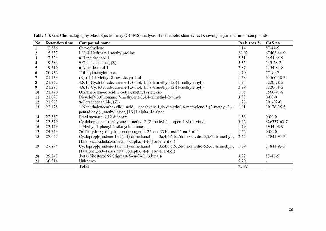
80
Table 4.3: Gas Chromatography-Mass Spectrometry (GC-MS) analysis of methanolic stem extract showing major and minor compounds.
No. Retention time Compound name Peak area % CAS no. 1 12.356 Caryophyllene 1.14 87-44-5 2 15.337 l-[-]-4-Hydroxy-1-methylproline 28.02 67463-44-9 3 17.524 n-Heptadecanol-1 2.51 1454-85-9 4 19.286 9-Octadecen-1-ol, (Z)- 5.35 143-28-2 5 19.510 n-Nonadecanol-1 2.87 1454-84-8 6 20.932 Tributyl acetylcitrate 1.70 77-90-7 7 21.138 (R)-(-)-14-Methyl-8-hexadecyn-1-ol 1.28 64566-18-3 8 21.242 4,8,13-Cyclotetradecatriene-1,3-diol, 1,5,9-trimethyl-12-(1-methylethyl)- 1.75 7220-78-2 9 21.287 4,8,13-Cyclotetradecatriene-1,3-diol, 1,5,9-trimethyl-12-(1-methylethyl)- 2.29 7220-78-2 10 21.370 Oxiraneoctanoic acid, 3-octyl-, methyl ester, cis- 1.35 2566-91-8 11 21.697 Bicyclo[4.3.0]nonane, 7-methylene-2,4,4-trimethyl-2-vinyl- 3.33 0-00-0 12 21.983 9-Octadecenamide, (Z)- 1.28 301-02-0 13 22.178 1-Naphthalenecarboxylic acid, decahydro-1,4a-dimethyl-6-methylene-5-(3-methyl-2,4-
pentadienyl)-, methyl ester, [1S-[1.alpha.,4a.alpha. 1.01 10178-35-5
14 22.567 Ethyl stearate, 9,12-diepoxy 1.56 0-00-0 15 23.370 Cycloheptane, 4-methylene-1-methyl-2-(2-methyl-1-propen-1-yl)-1-vinyl- 3.46 826337-63-7 16 23.449 1-Methyl-1-phenyl-1-silacyclobutane 1.79 3944-08-9 17 24.749 26-Dehydroxy-dihydropseudoprogenin-25-ene $$ Furost-25-en-3-ol # 1.52 0-00-0 18 27.657 Cycloprop[e]indene-1a,2(1H)-dimethanol, 3a,4,5,6,6a,6b-hexahydro-5,5,6b-trimethyl-,
(1a.alpha.,3a.beta.,6a.beta.,6b.alpha.)-(-)- (Isovellerdiol) 2.45 37841-93-3
19 27.894 Cycloprop[e]indene-1a,2(1H)-dimethanol, 3a,4,5,6,6a,6b-hexahydro-5,5,6b-trimethyl-, (1a.alpha.,3a.beta.,6a.beta.,6b.alpha.)-(-)- (Isovellerdiol)
1.69 37841-93-3
20 29.247 .beta.-Sitosterol $$ Stigmast-5-en-3-ol, (3.beta.)- 3.92 83-46-5 21 30.214 Unknown 5.70 - Total 75.97

81
Other notable activities reported for compounds that were present in C. gratissimus included anti-
inflammatory, anticancer/cancer preventive and antioxidant. Compounds that were found to be
responsible for anti-inflammatory activity are Phytol acetate (Sivakumar and Dhivya, 2015;
Karthikeyan et al., 2016a), n-Nonadecanol-1 (Anburaj et al., 2016), Phytol (Silva et al., 2014;
Wagay and Rothe, 2016; Mohiuddin et al., 2018), β–Sitosterol (Saeidnia et al., 2014; Karthikeyan
et al., 2016a; Bin Sayeed et al., 2016), B-Friedo-B':A'-neogammacer-5-en-3-ol, (3.beta.)-
(Kuroshima et al., 2005), Caryophyllene (Legault and Pichette, 2007; Mohammed et al., 2016;
Padmashree et al., 2018) and 9-Octadecenamide, (Z)- (Hussein et al., 2016a, 2016b; Hadi et al.,
2016; Basa’ar and Farooqui, 2017).
Anticancer/ cancer preventive compounds included Phytol acetate (Sivakumar and Dhivya, 2015;
Karthikeyan et al., 2016a, 2016b), n-Nonadecanol-1 (Anburaj et al., 2016), Phytol (Wagay and
Rothe, 2016; Ameachi and Chijioke, 2018; Mohiuddin et al., 2018), 1-Heptacosanol (Raman et
al., 2012), Cycloisolongifolene, 8,9-dehydro-9-formyl- (Mohiuddin et al., 2018), β–Sitosterol
(Yinusa et al., 2015; Nyigo et al., 2016; Bin Sayeed et al., 2016), Caryophyllene (Legault and
Pichette, 2007; Padmashree et al., 2018), Tributyl acetylcitrate (Hussein et al., 2016a) and (R)-(-
)-14-Methyl-8-hexadecyn-1-ol (Mohansrinivasan et al., 2015).
Compounds such as Phytol acetate (Karthikeyan et al., 2016b), Phytol (Raman et al., 2012;
Mohammed et al, 2016) Tetratetracontane (Sivakumar and Gayathri, 2015; Agarwal et al., 2017),
1-Heptacosanol (Raman et al., 2012; Begum et al., 2016; Roy et al., 2018), Cycloisolongifolene,
8,9-dehydro-9-formyl- (Zhang et al., 2017), β–Sitosterol (Yinusa et al., 2015; Saeidnia et al.,
2014; Bin Sayeed et al., 2016) and Caryophyllene (Legault and Pichette, 2007; Mohammed et al,
2016; Padmashree et al., 2018) were reported to possess antioxidant activity.
Many of the ailments treated with C. gratissimus are possibly attributed to the biological activities
of these compounds. Thus, the presence of these various bioactive compounds in the leaf and
stem extracts, further demonstrates the usefulness of C. gratissimus in traditional medicine.
4.4.4 Preliminary antibacterial assays
Biological screening of plant extracts is an important precursor in the discovery of novel
antimicrobial drugs (Sendeku et al., 2015). Antibacterial assays of C. gratissimus indicated that
the methanolic leaf extracts inhibited the growth of almost all bacterial strains except for P.
aeruginosa, which did not display any sensitivity to the extract. The leaf extract exhibited a weak
to moderate activity against both E. coli strains, MRSA (environmental type), S. aureus and B.
subtilis. Conversely, the extracts demonstrated strong activities against MRSA (clinical type) and
S. aureus (ATCC 29213). The resistance from P. aeruginosa and the weak activities of the E. coli
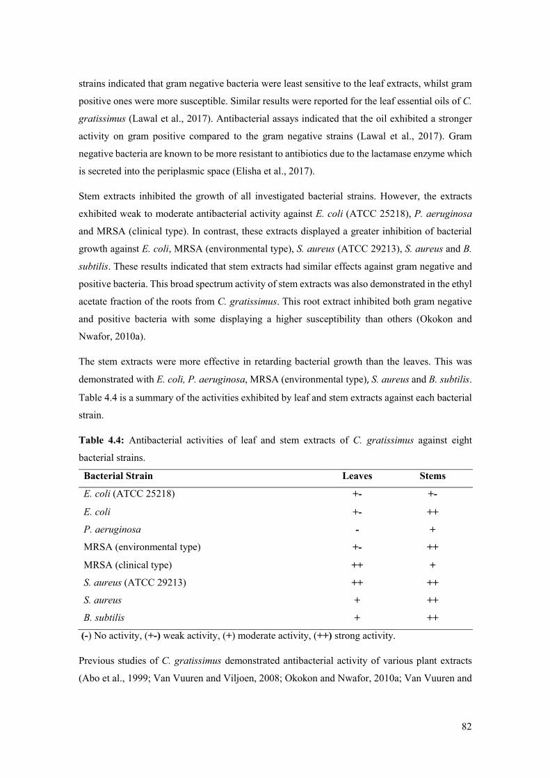
82
strains indicated that gram negative bacteria were least sensitive to the leaf extracts, whilst gram
positive ones were more susceptible. Similar results were reported for the leaf essential oils of C.
gratissimus (Lawal et al., 2017). Antibacterial assays indicated that the oil exhibited a stronger
activity on gram positive compared to the gram negative strains (Lawal et al., 2017). Gram
negative bacteria are known to be more resistant to antibiotics due to the lactamase enzyme which
is secreted into the periplasmic space (Elisha et al., 2017).
Stem extracts inhibited the growth of all investigated bacterial strains. However, the extracts
exhibited weak to moderate antibacterial activity against E. coli (ATCC 25218), P. aeruginosa
and MRSA (clinical type). In contrast, these extracts displayed a greater inhibition of bacterial
growth against E. coli, MRSA (environmental type), S. aureus (ATCC 29213), S. aureus and B.
subtilis. These results indicated that stem extracts had similar effects against gram negative and
positive bacteria. This broad spectrum activity of stem extracts was also demonstrated in the ethyl
acetate fraction of the roots from C. gratissimus. This root extract inhibited both gram negative
and positive bacteria with some displaying a higher susceptibility than others (Okokon and
Nwafor, 2010a).
The stem extracts were more effective in retarding bacterial growth than the leaves. This was
demonstrated with E. coli, P. aeruginosa, MRSA (environmental type), S. aureus and B. subtilis.
Table 4.4 is a summary of the activities exhibited by leaf and stem extracts against each bacterial
strain.
Table 4.4: Antibacterial activities of leaf and stem extracts of C. gratissimus against eight
bacterial strains.
Bacterial Strain Leaves Stems
E. coli (ATCC 25218) +- +-
E. coli +- ++
P. aeruginosa - +
MRSA (environmental type) +- ++
MRSA (clinical type) ++ +
S. aureus (ATCC 29213) ++ ++
S. aureus + ++
B. subtilis + ++
(-) No activity, (+-) weak activity, (+) moderate activity, (++) strong activity.
Previous studies of C. gratissimus demonstrated antibacterial activity of various plant extracts
(Abo et al., 1999; Van Vuuren and Viljoen, 2008; Okokon and Nwafor, 2010a; Van Vuuren and
83
Naidoo, 2010; Mthethwa et al., 2014; Lawal et al., 2017). A study by Van Vuuren and Naidoo
(2010) on the antibacterial potential of leaf oil and solvent extract of C. gratissimus indicated a
moderate to strong activity against STI-causing bacteria (Ureaplasma urealyticum and
Gardnerella vaginalis). Mthethwa et al. (2014) demonstrated that the methanolic leaf extracts
displayed good activity against S. aureus and S. epidermidis.
Abo et al. (1999) investigated the antibacterial activity of leaf and stem bark extracts. The stem
bark extracts exhibited strong activities against Proteus mirabilis, S. aureus, Bacillus megaterium
and B. subtilis. However, leaf extracts displayed little to no activity against all bacterial strains.
A study by Van Vuuren and Viljoen (2008) demonstrated antibacterial activity of the essential
oils and solvent (methanol: chloroform) extracts of leaves, roots, bark and combinations thereof.
However, the activity against bacterial strains varied between plant part and extract combinations
(Van Vuuren and Viljoen, 2008). These variations in the antibacterial activity of C. gratissimus
may be attributed to several factors including extraction methods, bacterial strains and source of
plant extracts (Mostafa et al., 2018).
The antimicrobial activities of plant extracts are likely attributed to phytochemical constituents
such as terpenoids, alkaloids and phenolic compounds. These compounds are suggested to
interfere with the enzymes and proteins of the microbial cell membrane, causing them to disrupt.
This eventually leads to the death of the microbial cell or the inhibition of enzymes involved in
the synthesis of amino acids (Mostafa et al., 2018).
The GC-MS analysis of leaf and stem extracts revealed various compounds with possible
antibacterial and antimicrobial activities. These included Phytol acetate, n-Heptadecanol-1, 9-
Octadecen-1-ol, (Z)-, n-Nonadecanol-1, Phytol, 1-Heptacosanol, Cycloisolongifolene, 8,9-
dehydro-9-formyl- (Xiang et al., 2018), β–Sitosterol (Saeidnia et al., 2014; Yinusa et al., 2015),
Caryophyllene, Tributyl acetylcitrate, 9-Octadecenamide, (Z)- (Hadi et al., 2016; Hussein et al.,
2016a, 2016b; Ibraheam et al., 2017), Tetratetracontane (Deshmukh, 2015) and Oxiraneoctanoic
acid, 3-octyl-, methyl ester, cis- (Hussein et al., 2016b). The presence of these compounds
probably contributed to the activity of the extracts against the bacterial strains.
4.5 Conclusion
The leaf and stem extracts of C. gratissimus were found to possess several phytochemical
compounds including terpenoids, carbohydrates, flavonoids, alkaloids, amino acids, phenolic
compounds, saponins and fixed fats. Antimicrobial assays of the extracts indicated weak to strong
activities against various bacterial strains. The stem extracts however exhibited stronger activity
than the leaf extracts. GC-MS analyses indicated several bioactive compounds in the leaves and

84
stems. Many of these were reported to possess antimicrobial and antibacterial properties which
probably contributed to the activity demonstrated in this study. This study confirmed the
usefulness of C. gratissimus in traditional medicine and indicated its potential as a source of
antimicrobials. However, further studies are needed to investigate the antibacterial properties of
individual compounds to determine their potency and potential as antibacterial agents. Minimum
inhibitory concentration (MIC) assays could assist in determining the antibacterial potency of the
extracts and compounds.
4.6 References
Abdalaziz, M.N., Ali, A. and Kabbashi, A.S., 2016. In vitro antioxidant activity and
phytochemical screening of Croton zambesicus. Journal of Pharmacognosy and Phytochemistry
5, 12 – 16.
Abdallah, E.M., 2011. Plants: An alternative source for antimicrobials. Journal of Applied
Pharmaceutical Science 1, 16 – 20.
Abo, K.A., Ogunleye, V.O. and Ashidi, J.S., 1999. Antimicrobial potential of Spondias mombin,
Croton zambesicus and Zygotritonia crocea. Phytotherapy Research 13, 494 – 497.
Agarwal, A., Prajapati, R., Raza, S.K. and Thakur, L.K., 2017. GC-MS analysis and antibacterial
activity of aerial parts of Quisqualis indica Plant Extracts. Indian Journal of Pharmaceutical
Education and Research 51, 329 – 336.
Altemimi, A., Lakhssassi, N., Baharlouei, A., Watson, D. and Lightfoot, D., 2017.
Phytochemicals: Extraction, isolation, and identification of bioactive compounds from plant
extracts. Plants 6, 1 – 23.
Ameachi, N.C. and Chijioke, C.L., 2018. Evaluation of bioactive compounds in
Pseudarenthemum tunicatum leaves using Gas Chromatography-Mass Spectrometry. Scientific
Papers Series Management, Economic Engineering in Agriculture and Rural Development 18, 53
– 60.
Anburaj, G., Marimuthu, M., Rajasudha, V. and Manikandan, R., 2016. Phytochemical screening
and GC-MS analysis of ethanolic extract of Tecoma stans (Family: Bignoniaceae) Yellow Bell
Flowers. International Journal of Research in Pharmacology and Pharmacotherapeutics 5, 256 –
261.
Arora, S., Kumar, G. and Meena, S., 2017. Screening and evaluation of bioactive components of
Cenchrus ciliaris L. by GC-MS analysis. International Research Journal of Pharmacy 8, 69 – 76.

85
Azalework, H.G., Sahabjada, A.J., Arshad, M. and Malik, T., 2017. Phytochemical investigation,
GC-MS profile and antimicrobial activity of a medicinal plant Ruta graveolens L. from Ethiopia.
International Journal of Pharmacy and Pharmaceutical Sciences 9, 29 – 34.
Balamurugan, K., Nishanthini, A. and Mohan, V.R., 2012. GC–MS analysis of Polycarpaea
corymbosa (L.) Lam. whole plant. Asian Pacific Journal of Tropical Biomedicine 2, S1289 -
S1292.
Basa’ar, O. and Farooqui, M., 2017. Supercritical carbon dioxide extraction and gas
chromatography–mass spectrometry analysis of Gymnema sylvestre R. Br. roots. Journal of
Liquid Chromatography and Related Technologies 40, 879 – 886.
Bayor, M.T., Gbedema, S.Y. and Annan, K., 2009. The antimicrobial activity of Croton
membranaceus, a species used in formulations for measles in Ghana. Journal of Pharmacognosy
and Phytotherapy 1, 47 – 51.
Begum, I.F., Mohankumar, R., Jeevan, M. and Ramani, K., 2016. GC–MS analysis of bioactive
molecules derived from Paracoccus pantotrophus FMR19 and the antimicrobial activity against
bacterial pathogens and MDROs. Indian Journal of Microbiology 56, 426 – 432.
Benmehdi, H., Hasnaoui, O., Benali, O. and Salhi, F., 2012. Phytochemical investigation of leaves
and fruits extracts of Chamaerops humilis L. Journal of Materials and Environmental Science 3,
320 – 337.
Bin Sayeed, M.S., Karim, S.M.R., Sharmin, T. and Morshed, M.M., 2016. Critical analysis on
characterization, systemic effect, and therapeutic potential of beta-sitosterol: a plant-derived
orphan phytosterol. Medicines 3, 1 – 25.
Block, S., Baccelli, C., Tinant, B., Van Meervelt, L., Rozenberg, R., Jiwan, J.L.H., Llabres, G.,
De Pauw-Gillet, M.C. and Quetin-Leclercq, J., 2004. Diterpenes from the leaves of Croton
zambesicus. Phytochemistry 65, 1165 – 1171.
Block, S., Stevigny, C., De Pauw-Gillet, M.C., de Hoffmann, E., Llabres, G., Adjakidjé, V. and
Quetin-Leclercq, J., 2002. ent-Trachyloban-3β-ol, a new cytotoxic diterpene from Croton
zambesicus. Planta Medica 68, 647 – 649.
Chikezie, P.C., Ibegbulem, C.O. and Mbagwu, F.N., 2015. Bioactive principles from medicinal
plants. Research Journal of Phytochemistry 9, 88 – 115.
Cowan, M.M., 1999. Plant products as antimicrobial agents. Clinical Microbiology Reviews 12,
564 – 582.

86
Deshmukh, O.S., 2015. GC-MS analysis of root acetone extracts of Spatholobus purpureus – a
high ethno-veterinary medicinal value plant. International Journal of Current Research 7, 23039
– 23042.
Elisha, I.L., Botha, F.S., McGaw, L.J. and Eloff, J.N., 2017. The antibacterial activity of extracts
of nine plant species with good activity against Escherichia coli against five other bacteria and
cytotoxicity of extracts. BMC Complementary and Alternative Medicine 17, 1 – 10.
Fabricant, D.S. and Farnsworth, N.R., 2001. The value of plants used in traditional medicine for
drug discovery. Environmental Health Perspectives 109 (Suppl 1), 69 – 75.
Fernandes, H.M.B., Leão, A.D., Oliveira-Filho, A.A., Sousa, J.P., Oliveira, T.L., Lima, E.O.,
Silva, M.S. and Tavares, J.F., 2013. Antimicrobial activity and phytochemical screening of
extracts from leaves of Croton polyandrus Spreng. International Journal of Pharmacognosy and
Phytochemical Research 5, 223 – 226.
Fontenelle, R.O.S., Morais, S.M., Brito, E.H.S., Brilhante, R.S.N., Cordeiro, R.A., Nascimento,
N.R.F., Kerntopf, M.R., Sidrim, J.J.C. and Rocha, M.F.G., 2008. Antifungal activity of essential
oils of Croton species from the Brazilian Caatinga biome. Journal of Applied Microbiology 104,
1383 – 1390.
Gayathri, A. and Sri, M.K., 2018. Study of in vitro antioxidant activity and GC-MS analysis of
seeds of Cucumis melo. World Journal of Pharmaceutical Research 7, 934 – 942.
Hadi, M.Y., Mohammed, G.J. and Hameed, I.H., 2016. Analysis of bioactive chemical
compounds of Nigella sativa using as chromatography-mass spectrometry. Journal of
Pharmacognosy and Phytotherapy 8, 8 – 24.
Heamalatha, S., Swarnalatha, S., Divya, M., Gandhi Lakshmi, R., Ganga Devi, A. and Gomathi,
E., 2011. Pharmacognostical, pharmacological, investigation on Anethum graveolens Linn: A
review. Research Journal of Pharmaceutical, Biological and Chemical Sciences 2, 564 – 574.
Hossain, M.A., AL-Raqmi, K.A.S., AL-Mijizy, Z.H., Weli, A.M. and Al-Riyami, Q., 2013. Study
of total phenol, flavonoids contents and phytochemical screening of various leaves crude extracts
of locally grown Thymus vulgaris. Asian Pacific Journal of Tropical Biomedicine 3, 705 – 710.
Hussein, H.J., Hadi, M.Y. and Hameed, I.H., 2016b. Study of chemical composition of
Foeniculum vulgare using Fourier transform infrared spectrophotometer and gas
chromatography-mass spectrometry. Journal of Pharmacognosy and Phytotherapy 8, 60 – 89.

87
Hussein, H.M., Hameed, I.H. and Ibraheem, O.A., 2016a. Antimicrobial activity and spectral
chemical analysis of methanolic leaves extract of Adiantum capillus-veneris using GC-MS and
FT-IR spectroscopy. International Journal of Pharmacognosy and Phytochemical Research 8, 369
– 385.
Ibraheam, I.A., Hussein, H.M. and Hameed, I.H., 2017. Cyclamen persicum: Methanolic extract
using Gas Chromatography-Mass Spectrometry (GC-MS) technique. International Journal of
Pharmaceutical Quality Assurance 8, 200 – 213.
Kagan, I.A. and Flythe, M.D., 2014. Thin-layer chromatographic (TLC) separations and
bioassays of plant extracts to identify antimicrobial compounds. Journal of Visualized
Experiments, 1 – 8.
Karthikeyan, K., Dhanapal, C.K. and Gopalakrishnan, G., 2016b. GC-MS analysis of petroleum
ether extract of Alysicarpus monilifer whole plant. Der Pharmacia Lettre 8, 94 – 99.
Karthikeyan, V., Baskaran, A. and Rajasekaran, C.S., 2016a. Gas Chromatography-Mass
Spectrometry (GC-MS) analysis of ethanolic extracts of Barleria acuminata Nees. International
Journal of Pharmacological Research 6, 55 – 61.
Kumar, P., Kumar, R., Rastogi, M.K., Murti, K., 2017. Exploration of antidiabetic and
hypolipidemic activity of roots of Croton zambesicus. American Journal of Pharmacology and
Toxicology 12, 1 – 6.
Kumar, P., Sharma, B. and Bakshi, N., 2009. Biological activity of alkaloids from Solanum
dulcamara L. Natural Product Research 23, 719 – 723.
Kuppuswamy, K.M., Jonnalagadda, B. and Arockiasamy, S., 2013. GC-MS analysis of
chloroform extract of Croton bonplandianum. International Journal of Pharma and Bio Sciences
4, 613 – 617.
Kuroshima, K.N., Campos-Buzzi, F., Yunes, R.A., Delle Monache, F. and Cechinel Filho, V.,
2005. Chemical composition and antinociceptive properties of Hyeronima alchorneoides leaves.
Pharmaceutical Biology 43, 573 – 578.
Lawal, O.A., Ogunwande, I.A., Osunsanmi, F.O., Opoku, A.R. and Oyedeji, A.O., 2017. Croton
gratissimus leaf essential oil composition, antibacterial, antiplatelet aggregation, and cytotoxic
activities. Journal of Herbs, Spices and Medicinal Plants 23, 77 – 87.

88
Legault, J. and Pichette, A., 2007. Potentiating effect of β‐caryophyllene on anticancer activity of
α‐humulene, isocaryophyllene and paclitaxel. Journal of Pharmacy and Pharmacology 59, 1643
– 1647.
Leite, T.R., Silva, M.A.P.D., Santos, A.C.B.D., Coutinho, H.D.M., Duarte, A.E. and Costa,
J.G.M.D., 2017. Antimicrobial, modulatory and chemical analysis of the oil of Croton limae.
Pharmaceutical Biology 55, 2015 – 2019.
Mohammed, G.J., Omran, A.M. and Hussein, H.M., 2016. Antibacterial and phytochemical
analysis of Piper nigrum using Gas Chromatography-Mass Spectrum and Fourier-Transform
Infrared Spectroscopy. International Journal of Pharmacognosy and Phytochemical Research 8,
977 – 996.
Mohansrinivasan, V., Devi, C., Deori, M., Biswas, A. and Naine, S., 2015. Exploring the
anticancer activity of grape seed extract on skin cancer cell Lines A431. Brazilian Archives of
Biology and Technology 58, 540 – 546.
Mohiuddin, Y.G., Nathar, V.N., Aziz, W.N. and Gaikwad, N.B., 2018. Investigations on
important secondary metabolites from aerial parts of Artemisia absinthium L. using GC-MS.
Journal of Pharmacognosy and Phytochemistry 7, 820 – 827.
Morsy, N., 2014. Phytochemical analysis of biologically active constituents of medicinal plants.
Main Group Chemistry 13, 7 – 21.
Mostafa, A.A., Al-Askar, A.A., Almaary, K.S., Dawoud, T.M., Sholkamy, E.N. and Bakri, M.M.,
2018. Antimicrobial activity of some plant extracts against bacterial strains causing food
poisoning diseases. Saudi Journal of Biological Sciences 25, 361 – 366.
Mthethwa, N.S., Oyedeji, B.A., Obi, L.C. and Aiyegoro, O.A., 2014. Anti-staphylococcal, anti-
HIV and cytotoxicity studies of four South African medicinal plants and isolation of bioactive
compounds from Cassine transvaalensis (Burtt Davy) Codd. BMC Complementary and
Alternative Medicine 14, 512.
Mulholland, D.A., Langat, M.K., Crouch, N.R., Coley, H.M., Mutambi, E.M. and Nuzillard, J.M.,
2010. Cembranolides from the stem bark of the southern African medicinal plant, Croton
gratissimus (Euphorbiaceae). Phytochemistry 71, 1381 – 1386.
Murugan, T., Wins, J.A. and Murugan, M., 2013. Antimicrobial activity and phytochemical
constituents of leaf extracts of Cassia auriculata. Indian Journal of Pharmaceutical Sciences 75,
122 – 125.

89
Mustafa, G., Arif, R., Atta, A., Sharif, S. and Jamil, A., 2017. Bioactive compounds from
medicinal plants and their importance in drug discovery in Pakistan. Matrix Science Pharma 1,
17 – 26.
Mwine, J.T., Van Damme, P., 2011. Why do Euphorbiaceae tick as medicinal plants? A review
of Euphorbiaceae family and its medicinal features. Journal of Medicinal Plants Research 5, 652
– 662.
Nath, R., Roy, S., De, B. and Choudhury, M.D., 2013. Anticancer and antioxidant activity of
Croton: a review. International Journal of Pharmacy and Pharmaceutical Sciences 5, 63 – 70.
Nyigo, V.A., Peter, X., Mabiki, F., Malebo, H.M., Mdegela, R.H. and Fouche, G., 2016. Isolation
and identification of euphol and β-sitosterol from the dichloromethane extracts of Synadenium
glaucescens. The Journal of Phytopharmacology 5, 100 – 104.
Obey, J.K., von Wright, A., Orjala, J., Kauhanen, J. and Tikkanen-Kaukanen, C., 2016.
Antimicrobial activity of Croton macrostachyus stem bark extracts against several human
pathogenic bacteria. Journal of Pathogens 2016, 1 – 5.
Ogukwe, C.E., Akalezi, C.O., Ezugwu, B.O. and Ojukwu, H.E., 2016. Evaluation of the anti-
tumour agents in Annona Muricata (Linn.) leaves using Column Chromatography and Gas
Chromatography - Mass Spectrometry. World Journal of Pharmaceutical Research 5, 5 – 17.
Okokon, J.E., Bassey, A.L. and Obot, J., 2006. Antidiabetic activity of ethanolic leaf extract of
Croton zambesicus Muell. (Thunder plant) in alloxan diabetic rats. African Journal of Traditional,
Complementary and Alternative Medicines 3, 21 – 26.
Okokon, J.E. and Nwafor, P.A., 2009. Antiplasmodial activity of root extract and fractions of
Croton zambesicus. Journal of Ethnopharmacology 121, 74 – 78.
Okokon, J.E. and Nwafor, P.A., 2010a. Antimicrobial activity of root extract and crude fractions
of Croton zambesicus. Pakistan Journal of Pharmaceutical Sciences 23, 114 – 118.
Okokon, J.E. and Nwafor, P.A., 2010b. Anti-inflammatory, analgesic and antipyretic activities of
ethanolic root extract of Croton zambesicus. Pakistan Journal of Pharmaceutical Sciences 23, 385
– 392.
Okokon, J.E., Umoh, U.F., Udobang, J.A. and Etim, E.I., 2011. Antiulcerogenic activity of
ethanolic leaf extract of Croton zambesicus Muell. Arg. African Journal of Biomedical Research
14, 43 – 47.

90
Padmashree, M.S., Ashwathanarayana, R. and Raja Naika, R.B., 2018. Antioxidant, cytotoxic and
nutritive properties of Roem and Schult. Ipomoea staphylina plant extracts with preliminary
phytochemical and GC-MS analysis. Asian Journal of Pharmacy and Pharmacology 4, 473 – 492.
PlantZAfrica, 2018. Croton gratissimus Burch. http://pza.sanbi.org/croton-gratissimus. Date
Accessed: 5 February 2018.
Pudumo, J., Chaudhary, S.K., Chen, W., Viljoen, A., Vermaak, I. and Veale, C.G.L., 2018.
HPTLC fingerprinting of Croton gratissimus leaf extract with preparative HPLC-MS-isolated
marker compounds. South African Journal of Botany 114, 32 – 36.
Raaman, N., 2006. Phytochemical techniques. New India Publishing Agency, New Delhi, India.
Raman, B.V., Samuel, L.A., Saradhi, M.P., Rao, B.N., Krishna, N.V., Sudhakar, M. and
Radhakrishnan, T.M., 2012. Antibacterial, antioxidant activity and GC-MS analysis of
Eupatorium odoratum. Asian Journal of Pharmaceutical and Clinical Research 5, 99 – 106.
Rizk, A.F.M., 1987. The chemical constituents and economic plants of the Euphorbiaceae.
Botanical Journal of the Linnean Society 94, 293 – 326.
Rizwana, H., 2018. In vitro antibacterial and antifungal activity of some Oils, chemical analysis
and their FTIR studies. International Journal of Agriculture and Biology 20, 1488 – 1496.
Robert, S., Baccelli, C., Devel, P., Dogné, J.M. and Quetin-Leclercq, J., 2010. Effects of leaf
extracts from Croton zambesicus Müell. Arg. on hemostasis. Journal of Ethnopharmacology 128,
641 – 648.
Roy, C.L., Naresh, S., Sunil, K.S., Suma, A., Ashika, B.D. and Sathyamurthy, B., 2018. GC-MS
and FTIR analysis on the methanolic extract of red Vitis vinifera Peel. World Journal of Pharmacy
and Pharmaceutical Sciences 7, 1110 – 1123.
Saeidnia, S., Manayi, A., Gohari, A.R. and Abdollahi, M., 2014. The story of beta-sitosterol-a
review. European Journal of Medicinal Plants 4, 590 – 609.
Salatino, A., Salatino, M.L.F. and Negri, G., 2007. Traditional uses, chemistry and pharmacology
of Croton species (Euphorbiaceae). Journal of the Brazilian Chemical Society 18, 11 – 33.
Samy, R.P. and Gopalakrishnakone, P., 2010. Therapeutic potential of plants as anti-microbials
for drug discovery. Evidence-based Complementary and Alternative Medicine 7, 283 – 294.
Seanego, C.T. and Ndip, R.N., 2012. Identification and antibacterial evaluation of bioactive
compounds from Garcinia kola (Heckel) seeds. Molecules 17, 6569 – 6584.

91
Selowa, S.C., Shai, L.J., Masoko, P., Mokgotho, M.P. and Magano, S.R., 2010. Antibacterial
activity of extracts of three Croton species collected in Mpumalanga region in South Africa.
African Journal of Traditional, Complementary and Alternative Medicines 7, 98 – 103.
Selvin, J., Shanmughapriya, S., Gandhimathi, R., Kiran, G.S., Ravji, T.R., Natarajaseenivasan,
K. and Hema, T.A., 2009. Optimization and production of novel antimicrobial agents from sponge
associated marine actinomycetes Nocardiopsis dassonvillei MAD08. Applied Microbiology and
Biotechnology 83, 435 – 445.
Sen, A., Dhavan, P., Shukla, K.K., Singh, S. and Tejovathi, G., 2012. Analysis of IR, NMR and
antimicrobial activity of β-sitosterol isolated from Momordica charantia. Science Secure Journal
of Biotechnology 1, 9 – 13.
Sendeku, W., Alefew, B., Mengiste, D., Seifu, K., Girma, S., Wondimu, E., Bekuma, G., Verma,
D. and Berhane, N., 2015. Antibacterial activity of Croton macrostachyus against some selected
pathogenic bacteria. Biotechnology International 8, 11 – 20.
Silva, R.O., Sousa, F.B.M., Damasceno, S.R., Carvalho, N.S., Silva, V.G., Oliveira, F.R.M.,
Sousa, D.P., Aragão, K.S., Barbosa, A.L., Freitas, R.M. and Medeiros, J.V.R., 2014. Phytol, a
diterpene alcohol, inhibits the inflammatory response by reducing cytokine production and
oxidative stress. Fundamental and Clinical Pharmacology 28, 455 – 464.
Sivakumar, R. and Dhivya, A., 2015. GC-MS analysis of bioactive compounds on ethyl acetate
extract of Cordia monoica Roxb. leaves. International Journal of Research and Development in
Pharmacy and Life Sciences 4, 1328 – 1333.
Sivakumar, V. and Gayathri, G., 2015. GC-MS analysis of bioactive components from ethanol
extract of Andrographis paniculata. World Journal of Pharmacy and Pharmaceutical Sciences 4,
2031 – 2039.
Van Vuuren, S.F. and Naidoo, D., 2010. An antimicrobial investigation of plants used
traditionally in southern Africa to treat sexually transmitted infections. Journal of
Ethnopharmacology 130, 552 – 558.
Van Vuuren, S.F. and Viljoen, A.M., 2008. In vitro evidence of phyto-synergy for plant part
combinations of Croton gratissimus (Euphorbiaceae) used in African traditional healing. Journal
of Ethnopharmacology 119, 700 – 704.

92
Wagay, N.A. and Rothe, S.P., 2016. Investigations on secondary metabolites of Alhagi
pseudalhagi (M. Bieb.) Desv. ex B. Keller and Shap. leaves using GC-MS. Journal of
Pharmacognosy and Phytochemistry 5, 114 – 118.
Wang, G., Tang, W. and Bidigare, R.R., 2005. Terpenoids as therapeutic drugs and
pharmaceutical agents, in: Zhang, L., Demain, A.L. (Eds), Natural products. Humana Press Inc.,
New Jersey, pp. 197 – 227.
Wink, M., 2015. Modes of action of herbal medicines and plant secondary metabolites. Medicines
2, 251 – 286.
Xiang, H., Zhang, L., Xi, L., Yang, Y., Wang, X., Lei, D., Zheng, X. and Liu, X., 2018.
Phytochemical profiles and bioactivities of essential oils extracted from seven Curcuma herbs.
Industrial Crops and Products 111, 298 – 305.
Xu, W.H., Liu, W.Y. and Liang, Q., 2018. Chemical constituents from Croton species and their
biological activities. Molecules 23, 1 – 38.
Yinusa, I., George, N.I., Ayo, R.G. and Rufai, Y., 2015. Evaluation of the pharmacological
activities of beta sitosterol isolated from the bark of Sarcocephalus latifolius (Smith Bruce).
Global Journal of Pure and Applied Chemistry and Research 3, 7 – 14.
Zhang, L., Yang, Z., Wei, J., Su, P., Pan, W., Zheng, X., Zhang, K., Lin, L., Tang, J., Fang, Y.
and Du, Z., 2017. Essential oil composition and bioactivity variation in wild-growing populations
of Curcuma phaeocaulis Valeton collected from China. Industrial Crops and Products 103, 274 –
282.

93
CHAPTER 5: BIOLOGICAL SYNTHESIS AND
ANTIBACTERIAL ACTIVITY OF SILVER
NANOPARTICLES FROM LEAVES AND STEMS OF
CROTON GRATISSIMUS BURCH. VAR. GRATISSIMUS
(EUPHORBIACEAE)
5.1 Abstract
Silver nanoparticles (AgNPs) have received considerable attention due to their strong
antimicrobial properties. This study set out to biosynthesise AgNPs from the methanolic leaf and
stem extracts of the medicinally important species Croton gratissimus. Ultraviolet-visible
spectroscopy, energy-dispersive X-ray (EDX) analysis and transmission electron microscopy
(TEM) confirmed the production of AgNPs from both extracts. Transmission electron
micrographs revealed spherical AgNPs from both leaf and stem extracts. However, the size
distribution of these particles differed between organs. Silver nanoparticles from leaf extracts
were monodispersed whilst those from stems displayed polydispersion. Antibacterial assays
indicated that AgNPs from leaf extracts were more effective in inhibiting bacterial growth than
particles from stems. These results suggest that AgNPs from leaf extracts could be potential
sources of antibacterial agents. However, optimisation in the synthesis process may improve the
potency of these silver nanoparticles.
Keywords: Peaks, wavelengths, capping agents, reducing agents, spherical.

94
5.2 Introduction
Microscopic particles with sizes ranging from 1 – 100 nm are termed nanoparticles (Song and
Kim, 2009; Thakkar et al., 2010; Bagyalakshmi and Haritha, 2017). Studies on noble metal
nanoparticles are becoming increasingly popular as they exhibit enhanced biological and
physiochemical properties compared to the bulk material of equivalent composition. (He et al.,
2001; Song and Kim, 2009; Thakkar et al., 2010; Ahmed et al., 2016c; Gharibshahi et al., 2017).
In particular, silver nanoparticles (AgNPs) are of great interest because they are known to possess
stronger antimicrobial properties than other metals (Ibrahim, 2015; Premasudha et al., 2015). For
this reason, they are regarded as potential antimicrobial agents (Ibrahim, 2015; Singh et al., 2018).
Physical and chemical processes have been employed for synthesising AgNPs. However, these
methods have proved costly, laborious, energy-consuming and harmful to environmental and
human health due to the toxic by-products produced through these processes (Thakkar et al., 2010;
Makarov et al., 2014; Ibrahim, 2015; Ahmed et al., 2016b; Dhand et al., 2016; Kuppusamy et al.,
2016; Gabriela et al., 2017; Gharibshahi et al., 2017).
Biological methods using enzymes, plants or plant extracts and microorganisms are more
advantageous over conventional processes as they are environmentally friendly, inexpensive and
do not require toxic chemicals (Song and Kim, 2009; Iravani, 2011; Parveen et al., 2016).
Conversely, the use of plants and plant extracts is more feasible than other biological methods as
it provides natural reducing and capping agents for the formation of AgNPs (Ponarulselvam et
al., 2012; Mittal et al., 2013; Gabriela et al., 2017; Khan et al., 2018). These natural capping
agents from plants are also reported to improve the therapeutic properties of the nanoparticles
(Sangeetha et al., 2016). A considerable number of plants have been used for the synthesis of
AgNPs, some of which include Azadirachta indica, Pelargonium graveolens, Aloe vera,
Capsicum annuum L, Mentha piperita, Albizia adianthifolia, Ziziphora tenuior and Jatropha
curcas (Ponarulselvam et al., 2012; Gabriela et al., 2017).
Croton gratissimus Burch. var. gratissimus (syn. C. zambesicus Müll. Arg.; C. microbotryus Pax.,
C. amabilis Müell. Arg.) belonging to the family Euphorbiaceae (Mulholland et al., 2010; Robert
et al., 2010; PlantZAfrica, 2018) is a medicinally important plant that has been used in traditional
medicine for the treatment of various ailments (Ngadjui et al., 2002; Van Vuuren and Viljoen,
2008). Previous studies on C. gratissimus revealed antimicrobial activities of extracts from
different plant organs (Abo et al., 1999; Van Vuuren and Viljoen, 2008; Okokon and Nwafor,
2010; Van Vuuren and Naidoo, 2010; Mthethwa et al., 2014; Lawal et al., 2017). Due to the
extensive use in traditional medicine and the antimicrobial properties exhibited by various plant

95
organs, this study set out to investigate the antibacterial activity of AgNPs synthesised from leaf
and stem extracts of C. gratissimus.
5.3 Materials and Methods
5.3.1 Plant collection and sampling
Leaves and stems of Croton gratissimus Burch. var. gratissimus were collected from the
University of KwaZulu-Natal, Westville Campus (29°49'08.1"S 30°56'38.9"E). The material was
air dried for two months before being ground to a powder for synthesis of AgNPs. A voucher
specimen (Croton 01 – Accession No. 18224) was prepared and deposited in the Ward Herbarium
located in the School of Life Sciences at the University of KwaZulu-Natal, Westville Campus.
5.3.2 Crude methanolic extraction
Powdered leaf and stems were placed in separate round bottom flasks with HPLC grade methanol
and boiled through continuous reflux. Two 2 h sessions were carried out for leaves and stems.
The obtained extracts were filtered and used for biological synthesis of AgNPs.
5.3.3 Biosynthesis of silver nanoparticles (AgNPs)
The synthesis of AgNPs was adapted from Khanra et al., 2016. Deionised water was used to
prepare a 1 mM silver nitrate (AgNO3) solution (Merck). Thereafter, 2 mL of the methanolic plant
extracts were added to 18 mL of AgNO3 solution (9: 1) and heated at 80°C for 90 min. This was
carried out for both leaf and stem extracts. A colour change from clear/light green to light or dark
brown indicated silver nanoparticle synthesis. These solutions were then centrifuged at 16000
rpm at 20°C for 60 min. Subsequently, the supernatant was discarded and the remaining pellet
was washed several times with distilled water to remove any impurities. Pellets were resuspended
in distilled water for silver nanoparticle characterisation using UV-VIS spectroscopy, energy-
dispersive X-ray (EDX) analysis, Transmission Electron Microscopy (TEM), and Fourier-
transform infrared spectroscopy (FTIR).
5.3.4 Ultraviolet-visible (UV-VIS) spectroscopy
The synthesis of AgNPs from each extract was measured using a SHIMADZU UV-1800 UV-VIS
Spectrophotometer (Merck, Japan) at a range of 200 – 800 nm. The 1 mM AgNO3 solution served
as the blank in the analysis.
5.3.5. Energy-dispersive X-ray (EDX) analysis
Small amounts of the aqueous AgNPs were dropped onto aluminium stubs and left to dry.
Thereafter, samples were sputter coated with gold in a Quorum Q150 RES gold Sputter Coater.
The elemental composition of the nanoparticles was determined using the Aztec Software (Oxford

96
Instruments, United Kingdom) on the Zeiss Ultra Plus field emission gun scanning electron
microscope (FEGSEM).
5.3.6 Transmission electron microscopy (TEM) and Image analysis
The size and shape of synthesised AgNPs were determined using a transmission electron
microscope (Sorescu et al., 2016). Carbon coated formvar grids were dipped into the aqueous
silver nanoparticle solution and set aside to dry. The dried grids were then viewed using a Jeol
JEM 2100 High Resolution Transmission Electron Microscope (HRTEM) at a voltage of 200 kV.
The iTEM Soft Imaging System Software (Olympus, Germany) was used to determine the size
of AgNPs from obtained images.
5.3.7 Fourier-transform infrared spectroscopy (FTIR)
The aqueous solutions of AgNPs were placed in a Perkin Elmer Spectrum 100 spectrophotometer
(USA) which allowed for the identification of functional groups that were attached to the AgNPs
(capping agents) (Sorescu et al., 2016).
5.3.8 Preliminary antibacterial assay
Bacterial Strains
The antibacterial properties of AgNPs from leaves and stems were evaluated against eight
bacterial strains. Three gram negative (Pseudomonas aeruginosa, Escherichia coli, Escherichia
coli – ATCC 25218) and five gram positive (Bacillus subtilis, Staphylococcus aureus,
Staphylococcus aureus – ATCC 29213, Methicillin-Resistant Staphylococcus aureus (MRSA) –
clinical type, Methicillin-Resistant Staphylococcus aureus (MRSA) – environmental type)
bacterial strains were used along with positive (streptomycin – gram positive, gentamycin – gram
negative) and negative (methanol) controls.
Inoculum preparation
Bacterial strains were sub-cultured into test tubes with autoclaved (vertical type steam sterilizer
HL-340, Taiwan) nutrient broth (16 g in 1 L) (Biolab, Merck). The test tubes were then placed on
a suspension mixer (SM-3600, Taiwan) and incubated overnight at optimum temperatures (gram
positive = 30°C, gram negative = 37°C). Following incubation, bacterial strains were diluted with
sterile nutrient broth and measured using a spectrophotometer to attain an optical density (OD620)
between 0.08 – 0.1, equivalent to the 0.5 McFarland turbidity standard.
97
Preliminary antibacterial activity of leaf and stem extracts
The agar well diffusion method was used to determine antibacterial activity of the AgNPs
synthesised from leaves and stems. The pellets obtained after centrifugation were re-dissolved in
distilled water to attain a 1 mg/mL concentration. Autoclaved Mueller-Hilton agar (38 g in 1 L)
(Biolab, Merck) was poured into sterile petri dishes and allowed to set before being inoculated
with cultured broth. Thereafter, a sterile cork borer was used to punch 5 mm holes into the agar
plates, which were then filled with aqueous silver nanoparticle solutions (~ 90 µL). The plates
were then incubated at relevant temperatures for 24 h before being viewed and imaged for
inhibition zones to determine potential antibacterial activity.
5.4 Results and Discussion
5.4.1 Biosynthesis of silver nanoparticles (AgNPs)
The use of plant extracts to synthesise nanoparticles is becoming increasingly popular (Mittal et
al., 2013), with AgNPs having a wide range of applications in the medical industry (Song and
Kim, 2009). In the current study, AgNPs were synthesised from the leaves and stems of Croton
gratissimus. This was indicated by the colour changes in Fig. 5.1. Leaf and stem extracts displayed
a light to dark brown colour following the 90 min reaction time. Silver nanoparticles produced
from plant extracts are known to produce a brown colour (Sorescu et al., 2016). This colour
change is suggested to be brought about by the excitation of surface plasmon vibrations of AgNPs
(Singhal et al., 2011; Khanra et al., 2016; Sorescu et al., 2016).
Metabolites present in plant extracts such as amino acids, proteins, terpenoids, flavonoids,
alkaloids, phenolic compounds and saponins, are responsible for the synthesis of AgNPs as they
provide natural reducing and capping agents for the fabricated particles (Mittal et al., 2013;
Ahmed et al., 2016a; Kuppusamy et al., 2016; Raja et al., 2017; De Matteis et al., 2018). The
visible variation in the AgNPs produced from leaf and stem extracts is probably attributed to the
different plant organs used for synthesis. Previous work suggested that extracts from different
organs of the same plant can comprise varying combinations and concentrations of reducing
agents (Mittal et al., 2013; Sigamoney et al., 2016).

98
Figure 5.1: Visual representation of the leaf and stem extracts before (a) and after (b) the 90 min
reaction time. CL = C. gratissimus var. gratissimus leaves, CS = C. gratissimus var. gratissimus
stems.
5.4.2 Ultraviolet-visible (UV-VIS) spectroscopy
The synthesis of AgNPs was analysed by UV-VIS spectroscopy. UV-VIS spectroscopy is used to
monitor the formation and stability of AgNPs (Logeswari et al., 2015; Khanra et al., 2016). Figure
5.2 shows the UV-VIS spectra of the AgNPs from the leaf and stem extracts at a range of 350 –
518 nm. Surface plasmon resonance (SPR) bands were observed at 414 nm and 421 nm in the
leaves and stems respectively. According to Mittal et al. (2013), AgNPs are usually characterised
by absorption wavelengths in the range of 400 – 450 nm. The absorption peaks observed in this
range confirmed the presence of AgNPs. The analysis also indicated that the particles are spherical
in shape as absorption bands between 400 – 450 nm indicate spherical metallic nanoparticles
(Martinez-Castanon et al., 2008; Raja et al., 2017). The AgNPs from leaf and stem extracts
displayed narrow and wide peaks respectively. It is suggested that the width of peaks is related to
the size dispersion of the particles (He et al., 2001; Martinez-Castanon et al., 2008; Ponarulselvam
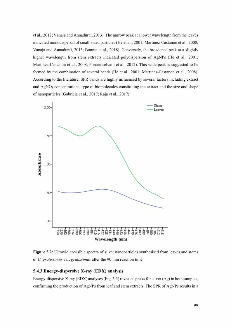
99
et al., 2012; Vanaja and Annadurai, 2013). The narrow peak at a lower wavelength from the leaves
indicated monodispersal of small-sized particles (He et al., 2001; Martinez-Castanon et al., 2008;
Vanaja and Annadurai, 2013; Bonnia et al., 2018). Conversely, the broadened peak at a slightly
higher wavelength from stem extracts indicated polydispersion of AgNPs (He et al., 2001;
Martinez-Castanon et al., 2008; Ponarulselvam et al., 2012). This wide peak is suggested to be
formed by the combination of several bands (He et al., 2001; Martinez-Castanon et al., 2008).
According to the literature, SPR bands are highly influenced by several factors including extract
and AgNO3 concentrations, type of biomolecules constituting the extract and the size and shape
of nanoparticles (Gabriela et al., 2017; Raja et al., 2017).
Figure 5.2: Ultraviolet-visible spectra of silver nanoparticles synthesised from leaves and stems
of C. gratissimus var. gratissimus after the 90 min reaction time.
5.4.3 Energy-dispersive X-ray (EDX) analysis
Energy-dispersive X-ray (EDX) analyses (Fig. 5.3) revealed peaks for silver (Ag) in both samples,
confirming the production of AgNPs from leaf and stem extracts. The SPR of AgNPs results in a

100
characteristic optical absorption at 3 keV (Ibrahim, 2015). Therefore, the peaks observed at 3 keV
for leaves and stems indicated silver (Shahverdi et al., 2007; Raja et al., 2017; Bonnia et al., 2018).
Table 5.1 represents the mean ± SD percentage of elemental silver from leaf and stem
nanoparticles. A higher percentage of elemental silver was recorded for nanoparticles from leaf
extracts compared to those from stems. However, statistical analyses revealed no significant
difference between elemental silver from leaf and stem nanoparticles.
Figure 5.3: Energy-dispersive X-ray (EDX) spectra of silver nanoparticles synthesised from leaf
(a) and stem (b) extracts of C. gratissimus var. gratissimus.
Table 5.1: Mean percentage of elemental silver from nanoparticles synthesised from leaf and
stem extracts of C. gratissimus var. gratissimus.
Plant organ Percentage of silver
Leaves 36.04 ± 17.64
Stems 23.24 ± 11.72
Mean ± standard deviation (SD), n = 3. p = 0. 354.
5.4.4 Transmission electron microscopy (TEM) and Image analysis
Transmission electron microscopy confirmed the formation and shape (spherical) of AgNPs from
the leaf and stem extracts (Fig.5. 4). However, the size distribution of the AgNPs differed between
leaves and stems. Silver nanoparticles from stem extracts varied in size. This corroborated the
results from UV-VIS spectroscopy which indicated that the particles were monodispersed in
leaves and polydispersed in stems. Figure 5.5 is an illustration of the particle size distribution
from leaves and stems. The size of AgNPs synthesised from leaves ranged from 7.19 – 24.67 nm
with a mean of 13.09 nm. The stem extracts produced AgNPs with sizes ranging from 6.24 –
40.95 nm with a mean of 13.64 nm. Table 5.2 shows the average particle size and standard
deviations from leaf and stem extracts. There was a significant difference in particle size
distribution between leaves and stems. Sigamoney et al. (2016) indicate that dissimilar plant parts
comprise varying biochemical constituents which influences the synthesis, size, shape and yield
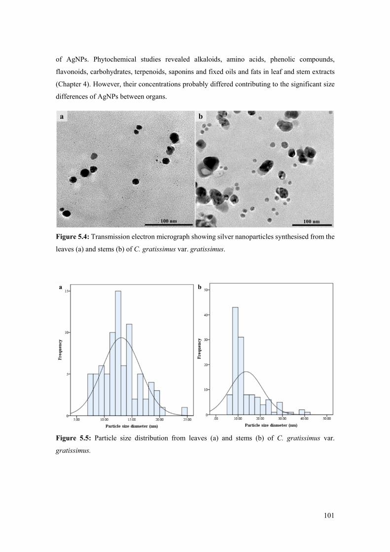
101
of AgNPs. Phytochemical studies revealed alkaloids, amino acids, phenolic compounds,
flavonoids, carbohydrates, terpenoids, saponins and fixed oils and fats in leaf and stem extracts
(Chapter 4). However, their concentrations probably differed contributing to the significant size
differences of AgNPs between organs.
Figure 5.4: Transmission electron micrograph showing silver nanoparticles synthesised from the
leaves (a) and stems (b) of C. gratissimus var. gratissimus.
Figure 5.5: Particle size distribution from leaves (a) and stems (b) of C. gratissimus var.
gratissimus.

102
Table 5.2: Mean particle size of silver nanoparticles synthesised from leaves and stems of C.
gratissimus var. gratissimus.
Plant organ Particle size (nm)
Leaves 13.09 ± 3.45
Stems 13.64 ± 7.31
Mean ± standard deviation (SD), n = 6. p = 0. 035.
5.4.5 Fourier-transform infrared spectroscopy (FTIR)
Figure 5.6 illustrates the results from FTIR spectroscopy. The FTIR spectra of AgNPs synthesised
from leaf and stem extracts displayed peaks at 3269 – 3273, 2112 – 2120, 1637 – 1639, 1375,
1217, 1016, 569 – 577 cm-1. The peaks between wave numbers of 3269 – 3273 cm-1 may
correspond to the O-H stretching of alcoholic and phenolic compounds (Koyyati, et al., 2014;
Bagyalakshmi and Haritha, 2017). Peaks at this region also indicated the C-H aldehyde stretch of
alkanes (Koyyati, et al., 2014). Peaks between 2112 – 2120 cm-1 correspond to the C≡C stretch of
alkynes (Coates, 2000). The peaks between wave numbers of 1637 – 1639 cm-1 are assigned to
the C=O stretch or bend of amide I bond of proteins (Singhal et al., 2011; Ibrahim, 2015; Ahmed
and Ikram, 2015; Ahmed et al., 2016b). The C-N stretch vibrations of aromatic amines correspond
to the peak at 1375 cm-1 (Koyyati, et al., 2014; Balashanmugam and Kalaichelvan 2015; Gabriela
et al., 2017). This peak displayed at 1375 cm-1 suggests that amino groups are involved in the
encapsulation and stabilisation of AgNPs (Balashanmugam and Kalaichelvan 2015). The peak at
1217 cm-1 displays the C-O stretch of esters or C-N stretch of amines (Raja et al., 2017). The peak
at 1016 cm-1 may be assigned to the C-O stretch of alcohol and ethers (Praba et al., 2014; Singh
et al., 2018). The peaks between 569 – 577 cm-1 may correspond to the C-Br stretch of alkyl
halides (Vinay et al., 2017).
According to literature, phytochemicals in plant extracts such as terpenoids, phenolics,
glycosides, proteins and alkaloids are involved in the formation of AgNPs (Dhand et al., 2016;
Raja et al., 2017). The results from FTIR spectroscopy indicated that AgNPs contained proteins,
phenolic and alcoholic compounds in addition to functional groups such as alkanes, alkynes,
amides, amines, ethers and alkyl halides. Similar results were reported for the AgNPs from
aqueous leaf extracts of C. zambesicus. Ahmed et al. (2016c) identified amide, carboxylic acid,
phenols, carbonyl and hydroxyl groups to be responsible for the capping and stabilisation of the
synthesised AgNPs. This suggests that the functional groups identified in this study and the
phytochemicals (Chapter 4) may act as natural reducing and capping agents in the synthesis and
stabilisation of AgNPs (Ahmed et al., 2016c; Sorescu et al., 2016; Raja et al., 2017).

103
Figure 5.6: Fourier-transform infrared spectra of silver nanoparticles synthesised from a) leaf and
b) stem extracts of C. gratissimus var. gratissimus.
5.4.6 Preliminary antibacterial assay
Silver is known to exhibit antibacterial activities against several bacterial strains (Singhal et al.,
2011). In the medical industry, silver and AgNPs are utilised for a wide range of applications such
as silver-containing topical creams and ointments used for the prevention of infection from
burns/wounds (Singhal et al., 2011). Although microorganisms have been used for the synthesis

104
of AgNPs (Vanaja and Annadurai, 2013; Ahmed et al., 2016a; Ahmed et al., 2016b), plant extracts
are reportedly safer for use in human health care (Ibrahim, 2015). Antibacterial assays of C.
gratissimus indicated that AgNPs synthesised from the leaf extracts had a stronger activity
compared to those from stems. Table 5.3 is a summary of the activities exhibited by the AgNPs
from leaf and stem extracts against the various bacterial strains.
Table 5.3: Antibacterial activities exhibited by silver nanoparticles from leaf and stem extracts
of C. gratissimus var. gratissimus against eight bacterial strains.
Bacterial Strain Leaves Stems
E. coli (ATCC 25218) + -
E. coli - -
P. aeruginosa + -
MRSA (environmental type) + +-
MRSA (clinical type) + +-
S. aureus (ATCC 29213) ++ +-
S. aureus + +-
B. subtilis + +-
(-) No activity, (+-) weak activity, (+) moderate activity, (++) strong activity.
The AgNPs from leaf extracts were effective in inhibiting growth of most bacterial strains, except
for E. coli, which did not display any sensitivity to the synthesised particles. These particles from
the leaf extracts exhibited moderate activity against E. coli (ATCC 25218), P. aeruginosa, MRSA
(environmental type), MRSA (clinical type), S. aureus and B. subtilis. The only strain to display
high susceptibility against AgNPs from leaf extracts was S. aureus (ATCC 29213). This indicated
that the AgNPs from leaf extracts had similar effects against gram negative and gram positive
bacteria. A previous study on AgNPs from the aqueous leaf extracts of C. zambesicus indicated a
minimum inhibitory concentration (MIC) at 30 mg/mL against both gram negative and positive
bacterial strains (Ahmed et al., 2016c). According to Ahmed et al. (2016c), the AgNPs caused
cell disruption which allowed penetration into the bacterial cells. The release of silver ions from
the particles ultimately results in the death of the bacteria (Dhand et al., 2016). It is suggested that
the phytochemical constituents from the leaves and the chemical groups identified in FTIR
spectroscopy contributed to the antibacterial activity of the AgNPs (Ahmed et al., 2016c).
The differences between the activities of AgNPs from leaf extracts in the study by Ahmed et al.
(2016c) and the current investigation, were probably attributed to the higher concentrations used
in antibacterial assays (Ahmed et al., 2016c). Different extraction solvents and processes may
have also played a role in the varying activities (Ahmed et al., 2016c). In addition, higher

105
concentrations of AgNO3 solution and plant extracts used for synthesis may have also contributed
to the differences in antibacterial activities (Ibrahim, 2015; Ahmed et al., 2016c).
Silver nanoparticles synthesised from stem extracts displayed little to no activity against the
bacterial strains. Escherichia coli (ATCC 25218), E. coli and P. aeruginosa were resistant to the
AgNPs from stems. These nanoparticles were also not very effective against MRSA
(environmental type), MRSA (clinical type), S. aureus (ATCC 29213), S. aureus and B. subtilis,
as indicated by the weak activities in Table 5.3. This indicated that the gram negative bacteria
used in this study were resistant to the AgNPs synthesised from stem extracts, whilst gram
positive bacteria displayed slight susceptibility. It is known that gram negative bacteria secrete a
lactamase enzyme into their periplasmic space which increases their resistance to antibiotics
(Elisha et al., 2017). In contrast, gram positive bacteria are more susceptible to attack from
antimicrobial agents as they lack an outer layer (Ahmed et al., 2016c).
According to literature, the antibacterial activities of smaller AgNPs are stronger than those that
are larger. This is due to the increased surface area of these particles which allows for greater
surface contact resulting in the death of the bacterial cell (Gabriela et al., 2017). Therefore, the
stronger antibacterial activity exhibited by AgNPs from leaf extracts was probably attributed to
the monodispersion of small particles compared to the polydispersion of those produced from
stems.
5.5 Conclusion
Silver nanoparticles were successfully synthesised from the methanolic leaf and stem extracts of
C. gratissimus. The AgNPs from both extracts were spherical in shape but their sizes differed
between the plant organs. Several groups were identified to play a role in the formation and
stabilisation of AgNPs from leaves and stems. These included proteins, phenols, alcohols,
alkanes, alkynes, amides, amines, ethers and alkyl halides. Antibacterial assays indicated that
AgNPs from leaf extracts were more effective in inhibiting bacterial growth than those from
stems. This was probably due to the monodisperal of AgNPs from leaf extracts. These results
indicate that AgNPs from leaf extracts are potential sources of antibacterial agents as they display
inhibitory properties. However, in order to consider these AgNPs as antibacterial agents, further
studies are needed to improve their potency. This can be achieved by employing higher
concentrations and combinations in the biosynthesis of AgNPs and determining their antibacterial
activities. In addition, MIC assays should be conducted to determine their potential as
antibacterial agents.

106
5.6 References
Abo, K.A., Ogunleye, V.O. and Ashidi, J.S., 1999. Antimicrobial potential of Spondias mombin,
Croton zambesicus and Zygotritonia crocea. Phytotherapy Research 13, 494 – 497.
Ahmed, S. and Ikram, S., 2015. Silver nanoparticles: one pot green synthesis using Terminalia
arjuna extract for biological application. Journal of Nanomedicine and Nanotechnology 6, 1 – 6.
Ahmed, S., Ahmad, M., Swami, B.L. and Ikram, S., 2016a. A review on plants extract mediated
synthesis of silver nanoparticles for antimicrobial applications: a green expertise. Journal of
Advanced Research 7, 17 – 28.
Ahmed, S., Ahmad, M., Swami, B.L. and Ikram, S., 2016b. Green synthesis of silver nanoparticles
using Azadirachta indica aqueous leaf extract. Journal of Radiation Research and Applied
Sciences 9, 1 – 7.
Ahmed, R.N., Sani, A., Oluwo, O.N., Sule, I.O. and Zakariyah, R.F., 2016c. Green synthesis and
antibacterial activity of silver nanoparticles from extract of leaves of Croton zambesicus. Nigerian
Journal of Microbiology 30, 3192 – 3199.
Bagyalakshmi, J. and Haritha, H., 2017. Green synthesis and characterization of silver
nanoparticles using Pterocarpus marsupium and assessment of its in vitro antidiabetic activity.
American Journal of Advanced Drug Delivery 5, 118 – 130.
Balashanmugam, P. and Kalaichelvan, P.T., 2015. Biosynthesis characterization of silver
nanoparticles using Cassia roxburghii DC. aqueous extract, and coated on cotton cloth for
effective antibacterial activity. International Journal of Nanomedicine 10 (Suppl 1), 87 – 97.
Bonnia, N.N., Fairuzi, A.A., Akhir, R.M., Yahya, S.M., Ab Rani, M.A., Ratim, S., Rahman, N.A.
and Akil, H.M., 2018. Comparison study on biosynthesis of silver nanoparticles using fresh and
hot air oven dried Imperata cylindrica leaf, in: IOP Conference Series: Materials Science and
Engineering 209. IOP Publishing, Bristol, pp. 1 – 6.
Coates, J., 2000. Interpretation of infrared spectra, a practical approach, in: Meyers, R.A. (Ed),
Encyclopaedia of analytical chemistry. John Wiley and Sons Ltd, Chichester, pp. 10815 – 10837.
De Matteis, V., Cascione, M., Toma, C.C. and Leporatti, S., 2018. Silver nanoparticles: Synthetic
routes, in vitro toxicity and theranostic applications for cancer disease. Nanomaterials 8, 1 – 23.
Dhand, V., Soumya, L., Bharadwaj, S., Chakra, S., Bhatt, D. and Sreedhar, B., 2016. Green
synthesis of silver nanoparticles using Coffea arabica seed extract and its antibacterial activity.
Materials Science and Engineering 58, 36 – 43.

107
Elisha, I.L., Botha, F.S., McGaw, L.J. and Eloff, J.N., 2017. The antibacterial activity of extracts
of nine plant species with good activity against Escherichia coli against five other bacteria and
cytotoxicity of extracts. BMC Complementary and Alternative Medicine 17, 1 – 10.
Gabriela, Á.M., Gabriela, M.D.O.V., Luis, A.M., Reinaldo, P.R., Michael, H.M., Rodolfo, G.P.
and Roberto, V.B.J., 2017. Biosynthesis of silver nanoparticles using mint leaf extract (Mentha
piperita) and their antibacterial activity. Advanced Science, Engineering and Medicine 9, 914 –
923.
Gharibshahi, L., Saion, E., Gharibshahi, E., Shaari, A.H. and Matori, K.A., 2017. Structural and
optical properties of Ag nanoparticles synthesized by thermal treatment method. Materials 10, 1
– 13.
He, S., Yao, J., Jiang, P., Shi, D., Zhang, H., Xie, S., Pang, S. and Gao, H., 2001. Formation of
silver nanoparticles and self-assembled two-dimensional ordered superlattice. Langmuir 17, 1571
– 1575.
Ibrahim, H.M., 2015. Green synthesis and characterization of silver nanoparticles using banana
peel extract and their antimicrobial activity against representative microorganisms. Journal of
Radiation Research and Applied Sciences 8, 265 – 275.
Iravani, S., 2011. Green synthesis of metal nanoparticles using plants. Green Chemistry 13, 2638
– 2650.
Khan, M.Z.H., Tareq, F.K., Hossen, M.A. and Roki, M.N.A.M., 2018. Green synthesis and
characterization of silver nanoparticles using Coriandrum sativum leaf extract. Journal of
Engineering Science and Technology 13, 158 – 166.
Khanra, K., Panja, S., Choudhuri, I., Chakraborty, A. and Bhattacharyya, N., 2016. Antimicrobial
and cytotoxicity effect of silver nanoparticle synthesized by Croton bonplandianum Baill. leaves.
Nanomedicine Journal 3, 15 – 22.
Koyyati, R., Babu Nagati, V., Nalvothula, R., Merugu, R., Kudle, K.R., Marx, P. and Padigya,
P.R.M., 2014. Antibacterial activity of silver nanoparticles synthesized using Amaranthus viridis
twig extract. International Journal of Research in Pharmaceutical Sciences 5, 32 – 39.
Kuppusamy, P., Yusoff, M.M., Maniam, G.P. and Govindan, N., 2016. Biosynthesis of metallic
nanoparticles using plant derivatives and their new avenues in pharmacological applications–An
updated report. Saudi Pharmaceutical Journal 24, 473 – 484.

108
Lawal, O.A., Ogunwande, I.A., Osunsanmi, F.O., Opoku, A.R. and Oyedeji, A.O., 2017. Croton
gratissimus leaf essential oil composition, antibacterial, antiplatelet aggregation, and cytotoxic
activities. Journal of Herbs, Spices and Medicinal Plants 23, 77 – 87.
Logeswari, P., Silambarasan, S. and Abraham, J., 2015. Synthesis of silver nanoparticles using
plants extract and analysis of their antimicrobial property. Journal of Saudi Chemical Society 19,
311 – 317.
Makarov, V.V., Love, A.J., Sinitsyna, O.V., Makarova, S.S., Yaminsky, I.V., Taliansky, M.E.
and Kalinina, N.O., 2014. “Green” nanotechnologies: synthesis of metal nanoparticles using
plants. Acta Naturae 6, 35 – 44.
Martinez-Castanon, G.A., Nino-Martinez, N., Martinez-Gutierrez, F., Martinez-Mendoza, J.R.
and Ruiz, F., 2008. Synthesis and antibacterial activity of silver nanoparticles with different sizes.
Journal of Nanoparticle Research 10, 1343 – 1348.
Mittal, A.K., Chisti, Y. and Banerjee, U.C., 2013. Synthesis of metallic nanoparticles using plant
extracts. Biotechnology Advances 31, 346 – 356.
Mthethwa, N.S., Oyedeji, B.A., Obi, L.C. and Aiyegoro, O.A., 2014. Anti-staphylococcal, anti-
HIV and cytotoxicity studies of four South African medicinal plants and isolation of bioactive
compounds from Cassine transvaalensis (Burtt Davy) Codd. BMC Complementary and
Alternative Medicine 14, 512.
Mulholland, D.A., Langat, M.K., Crouch, N.R., Coley, H.M., Mutambi, E.M. and Nuzillard, J.M.,
2010. Cembranolides from the stem bark of the southern African medicinal plant, Croton
gratissimus (Euphorbiaceae). Phytochemistry 71, 1381 – 1386.
Ngadjui, B.T., Abegaz, B.M., Keumedjio, F., Folefoc, G.N. and Kapche, G.W., 2002.
Diterpenoids from the stem bark of Croton zambesicus. Phytochemistry 60, 345 – 349.
Okokon, J.E. and Nwafor, P.A., 2010. Antimicrobial activity of root extract and crude fractions
of Croton zambesicus. Pakistan Journal of Pharmaceutical Sciences 23, 114 – 118.
Parveen, K., Banse, V. and Ledwani, L., 2016. Green synthesis of nanoparticles: Their advantages
and disadvantages, in: AIP Conference Proceedings 1724. AIP Publishing, New York, pp. 1 – 7.
PlantZAfrica, 2018. Croton gratissimus Burch. http://pza.sanbi.org/croton-gratissimus. Date
Accessed: 5 February 2018.

109
Ponarulselvam, S., Panneerselvam, C., Murugan, K., Aarthi, N., Kalimuthu, K. and Thangamani,
S., 2012. Synthesis of silver nanoparticles using leaves of Catharanthus roseus Linn. G. Don and
their antiplasmodial activities. Asian Pacific Journal of Tropical Biomedicine 2, 574 – 580.
Praba, P.S., Jeyasundari, J. and Jacob, Y.B.A., 2014. Synthesis of silver nanoparticles using Piper
betle and its antibacterial activity. European Chemical Bulletin 3, 1014 – 1016.
Premasudha, P., Venkataramana, M., Abirami, M., Vanathi, P., Krishna, K. and Rajendran, R.,
2015. Biological synthesis and characterization of silver nanoparticles using Eclipta alba leaf
extract and evaluation of its cytotoxic and antimicrobial potential. Bulletin of Materials Science
38, 965 – 973.
Raja, S., Ramesh, V. and Thivaharan, V., 2017. Green biosynthesis of silver nanoparticles using
Calliandra haematocephala leaf extract, their antibacterial activity and hydrogen peroxide
sensing capability. Arabian Journal of Chemistry 10, 253 – 261.
Robert, S., Baccelli, C., Devel, P., Dogné, J.M. and Quetin-Leclercq, J., 2010. Effects of leaf
extracts from Croton zambesicus Müell. Arg. on hemostasis. Journal of Ethnopharmacology 128,
641 – 648.
Sangeetha, R., Niranjan, P. and Dhanalakshmi, N., 2016. Characterization of silver nanoparticles
synthesized using the extract of the leaves of Tridax procumbens. Research Journal of Medicinal
Plant 10, 159 – 166.
Shahverdi, A.R., Fakhimi, A., Shahverdi, H.R. and Minaian, S., 2007. Synthesis and effect of
silver nanoparticles on the antibacterial activity of different antibiotics against Staphylococcus
aureus and Escherichia coli. Nanomedicine: Nanotechnology, Biology and Medicine 3, 168 –
171.
Sigamoney, M., Shaik, S., Govender, P. and Krishna, S.B.N., 2016. African leafy vegetables as
bio-factories for silver nanoparticles: a case study on Amaranthus dubius C Mart. Ex Thell. South
African Journal of Botany 103, 230 – 240.
Singh, P., Pandit, S., Garnæs, J., Tunjic, S., Mokkapati, V.R., Sultan, A., Thygesen, A.,
Mackevica, A., Mateiu, R.V., Daugaard, A.E. and Baun, A., 2018. Green synthesis of gold and
silver nanoparticles from Cannabis sativa (industrial hemp) and their capacity for biofilm
inhibition. International Journal of Nanomedicine 13, 3571 – 3591.

110
Singhal, G., Bhavesh, R., Kasariya, K., Sharma, A.R. and Singh, R.P., 2011. Biosynthesis of
silver nanoparticles using Ocimum sanctum (Tulsi) leaf extract and screening its antimicrobial
activity. Journal of Nanoparticle Research 13, 2981 – 2988.
Song, J.Y. and Kim, B.S., 2009. Rapid biological synthesis of silver nanoparticles using plant leaf
extracts. Bioprocess and Biosystems Engineering 32, 79 – 84.
Sorescu, A.A., Nuţă, A., Ion, R.M. and Ioana-Raluca, Ş.B., 2016. Green synthesis of silver
nanoparticles using plant extracts. The 4th International Virtual Conference on Advanced
Scientific Results, 188 – 193.
Thakkar, K.N., Mhatre, S.S. and Parikh, R.Y., 2010. Biological synthesis of metallic
nanoparticles. Nanomedicine: Nanotechnology, Biology and Medicine 6, 257 – 262.
Van Vuuren, S.F. and Viljoen, A.M., 2008. In vitro evidence of phyto-synergy for plant part
combinations of Croton gratissimus (Euphorbiaceae) used in African traditional healing. Journal
of Ethnopharmacology 119, 700 – 704.
Van Vuuren, S.F. and Naidoo, D., 2010. An antimicrobial investigation of plants used
traditionally in southern Africa to treat sexually transmitted infections. Journal of
Ethnopharmacology 130, 552 – 558.
Vanaja, M. and Annadurai, G., 2013. Coleus aromaticus leaf extract mediated synthesis of silver
nanoparticles and its bactericidal activity. Applied Nanoscience 3, 217 – 223.
Vinay, S.P., Chandrashekar, N. and Chandrappa, C.P., 2017. Silver nanoparticles: Synthesized
by leaves extract of Avocado and their antibacterial activity. International Journal of Engineering
Development and Research 5, 1608 – 1613.
111
CHAPTER 6: CONCLUSIONS AND FUTURE
RECOMMENDATIONS
Traditional medicinal plants have been the subject of numerous investigation as they are
responsible for the production of several natural bioactive compounds such as alkaloids,
terpenoids and phenolic compounds. Croton gratissimus is a medicinally important species that
has been used traditionally for the treatment of various ailments. This study aimed to identify and
describe the structures from leaves and stems of C. gratissimus in order to determine their possible
role in the production of secondary metabolites (Chapter 3). Furthermore, the study aimed to
determine the chemical composition and antibacterial activity of leaf and stem extracts (Chapter
4). In addition, this study sought out to biosynthesise silver nanoparticles (AgNPs) from the
methanolic leaf and stem extracts and determine its antibacterial properties (Chapter 5).
6.1 Major findings
Chapter 3: Microscopic investigations revealed three structures, i.e. lepidote and glandular
trichomes, and non-articulated unbranched laticifers from the leaves and stems of Croton
gratissimus. The lepidote and glandular trichomes were present on stems and the abaxial surfaces
of leaves at all developmental stages. Lepidote trichomes comprised a multiseriate, multicellular
stalk, a multicellular subradial disc, numerous radial cells and a single central cell. These
trichomes formed a dense indumentum over the leaf surface, canopying the underlying glandular
trichomes. Although this type of trichome is classed as non-glandular and non-secretory,
transmission electron microscopy (TEM) revealed several organelles, such as vacuoles, vesicles,
endoplasmic reticulum, golgi bodies and mitochondria, within stalk and radial cells suggesting
that these structures may be involved in the synthesis of secondary metabolites. Glandular
trichomes were embedded in the epidermal layer and comprised a single cell that formed a
prominent stalk and distal head. Laticifers were predominantly associated with the vascular
tissues in the leaves and stems. Histochemical analysis revealed alkaloids, phenolic compounds
and lipids to be common among all three structures. The presence of alkaloids and phenolic
compounds indicated that these structures are involved in the production of bioactive compounds
which possibly contributes to the medicinal properties of the plant.
Chapter 4: Preliminary phytochemical screening of leaf and stem extracts revealed several
compounds including terpenoids, carbohydrates, flavonoids, alkaloids, amino acids, phenolic
compounds, saponins and fixed fats. Antibacterial assays revealed weak to strong activities
against all bacterial strains investigated. However, stem extracts exhibited a stronger inhibition

112
of bacterial growth compared to leaf extracts. Several bioactive compounds identified from Gas
Chromatography-Mass Spectrometry (GC-MS) analyses are suggested to contribute to the
antibacterial activity of the extracts demonstrated in this study. This indicates that C. gratissimus
is a potential source of antibacterial agents.
Chapter 5: Silver nanoparticles were successfully synthesised from the methanolic leaf and stem
extracts of C. gratissimus. This synthesis was confirmed with ultraviolet-visible (UV-VIS)
spectroscopy as both leaf and stem extracts displayed peaks between 400 – 500 nm. Ultraviolet-
visible spectroscopy and TEM revealed spherical-shaped nanoparticles from both extracts.
However, the size distribution of these particles differed between both extracts as leaves displayed
monodispersion of particles whilst those from stems exhibited polydispersion. Phytochemical
groups that were identified to play a role in the reduction and stabilisation of AgNPs included
proteins, phenols, alcohols, alkanes, alkynes, amides, amines, ethers and alkyl halides.
Antibacterial assays revealed that AgNPs from leaf extracts displayed stronger activities than
those from stems. The activities exhibited by AgNPs from leaf extracts indicate that these particles
are potential sources of antibacterial agents.
6.2 Challenges
Poor resin infiltration of leaf and stem tissue for TEM was the only major challenge experienced
throughout this study. However, several protocols were employed to overcome these infiltration
issues.
6.3 Future recommendations
The work conducted in this study serves as a precursor for future research on C. gratissimus.
Although many structures were revealed to play a role in the production of phytochemical
compounds, further research is required to identify their mode of synthesis. In addition, further
investigations can be conducted on the crystal and secretory idioblasts, stellate trichomes and
extrafloral nectaries identified from the leaves of C. gratissimus, as these structures may
contribute to the medicinal properties of this plant. The antibacterial activity from crude extracts
provides the basis for future studies as individual compounds can be isolated to investigate their
potential as antibacterial agents. Furthermore, in order to consider AgNPs as potential
antibacterial agents, additional research is required to improve the potency of these particles.
6.4 Final conclusion
Overall, this study confirmed the traditional use of C. gratissimus and revealed three structures
that are involved in the production of some of the bioactive compounds. Furthermore, the plant
extracts contain phytochemicals that aid in the synthesis and stabilisation of AgNPs. The

113
antibacterial activity of the extracts and AgNPs indicate that C. gratissimus is a potential source
of antibacterial agents that could be utilised in the healthcare industry.