BIODIVERSITY AND BIOTECHNOLOGICAL APPLICATIONS OF ACTINOBACTERIA FROM MANGROVES OF VELLAPPALLAM
AT NAGAPATTINAM DISTRICT, TAMILNADU, INDIA
A Thesis Submitted to
Bharathidasan University
for the award of the Degree of
DOCTOR OF PHILOSOPHY
IN
MICROBIOLOGY
By
Mrs. S. DEEPA, M.Sc., M.Phil.,
(Ref. No. : 22466/Ph.D.1/Micro/FT/Oct 2011)
Under the supervision of
Dr. K. KANIMOZHI, M.Sc., M.Phil., Ph.D.,
P.G. AND RESEARCH DEPARTMENT OF BOTANY AND MICROBIOLOGY
A.V.V.M. SRI PUSHPAM COLLEGE (AUTONOMOUS)
(AFFILIATED TO BHARATHIDASAN UNIVERSITY)
POONDI – 613 503, THANJAVUR DISTRICT
TAMILNADU, INDIA.
May – 2014
A.V.V.M SRI PUSHPAM COLLEGE (AUTONOMOUS) POONDI-613 503, THANJAVUR DISTRICT
TAMIL NADU, INDIA (Affiliated to Bharathidasan University, Tiruchirappalli)
P.G. & RESEARCH DEPARTMENT OF BOTANY AND MICROBIOLOGY
Dr. K. KANIMOZHI, M.Sc., M.Phil., Ph.D., Assistant Professor and Research Advisor
CERTIFICATE
This is to certify that the thesis entitled “Biodiversity and Biotechnological
applications of Actinobacteria from Mangroves of Vellappallam at
Nagapattinam District, Tamilnadu, India.” submitted to Bharathidasan
University, Tiruchirapalli, for the award of the degree of DOCTOR OF PHILOSOPHY IN
MICROBIOLOGY embodies the result of the bonafide research work carried out by
S. DEEPA, under my guidance and supervision in the P.G. and Research Department of
Botany and Microbiology, A.V.V.M. Sri Pushpam College (Autonomous), Poondi, Thanjavur
district, Tamil Nadu, India.
I further certify that no part of this thesis has been submitted anywhere else for the
award of any degree, diploma, associateship, fellowship or other similar titles to any candidate.
Place : Poondi.
Date :
(K.KANIMOZHI)
RESEARCH ADVISER

Mrs. S .DEEPA, M.Sc., M.Phil.
Research Scholar
PG and Research Dept. of Botany and Microbiology
A.V.V.M Sri Pushpam College (Autonomous)
Poondi – 613503, Thanjavur District Tamilnadu, India.
(Affiliated to Bharathidasan Univerity, Tiruchirapalli)
DECLARATION
I do hereby declare that this work has been originally carried out by me under the
supervision of Dr. K. KANIMOZHI, Assistant Professor, Department of Botany and
Microbiology, A.V.V.M. Sri Pushpam College (Autonomous), Poondi, Thanjavur District,
Tamil Nadu, affiliated to Bharathidasan University, Tiruchirapalli – 620 024 and this work has
not been submitted elsewhere for any other degree.
Place : Poondi.
Date :
(S.DEEPA)
Research Scholar

ACKNOWLEDGEMENT
First of all my innumerable thanks to Almighty God for His blessings and
guidance at every stages of my life. All respects for God for enlighting our souls with
the essence of faith in lord and showering all His abundant blessings upon us and
enriched me with knowledge and wisdom to complete this thesis in a successful
manner.
It is a pleasure to convey my gratitude to people who rendered contribution in
assorted ways to this research.
In the first place I would like to express my deepest thanks to my Guide,
Dr.K. Kanimozhi M.Sc., M.Phil., Ph.D, Assistant professor, Department of Botany
and Microbiology, A.V.V.M. Sri Pushpam College (Autonomous), Poondi – 613 503,
Thanjavur District. Her ability to probe beneath the text is a true gift and her
insights have strengthened this study significantly. I will always be thankful for her
knowledge and deep concern on me. It has been an honour to work with her. She built
confidence in me. She showed me different ways to approach a research problem and
the need to be persistent to accomplish any goal. I am very thankful for her timely
help and valuable suggestions enthusiasm, unfailing interest throughout the period of
my research work. Her constructive ideas and encouragement made my thesis as a
profound and full-fledged one. I am very fortunate to have her guidance throughout
my work.
I express my sincere thanks to Honourable Secretary and Correspondent
Sri.K.Thulasiah Vandayar, A.V.V.M. Sri Pushpam College (Autonomous), Poondi –
613 503, for given me the golden opportunity to undergo the Ph.D., programme in the
College of excellence.
I am very grateful to Dr.R.Rajendran, Principal and Dr.U.Balasubramanian
Dean Faculty of Science, A.V.V.M. Sri Pushpam, College (Autonomous), Poondi, for
their permission to use the laboratory facilities.
I wish to acknowledge here, my carrying mentor, teacher and the tremendous
contribution of Dr.A.Panneerselvam, Doctoral committee member, Associate professor
& Head, Department of Botany and Microbiology, A.V.V.M. Sri Pushpam College
(Autonomous), Poondi – 613 503, Thanjavur District. Research co-ordinator, has to
emerge at the top of list, for him the words don’t exist describe how admirable he has
been during this whole practice. He has elevated me to a stage, where I am today
through a journey of learning, self motivation and above all honesty and dedication. I
have the comfort that he will always be there for me. I would never have made it this
far, if it weren’t the support and guidance of my dearly loved supervisor and her
endurance with her students in letting us find our path to knowledge. I owe her a
great deal.
I wish to acknowledge the support, and encouragement and my special thanks
to Dr.S. Mohammad Salique, Doctoral committee member, Associate Professor &
Vice Principal, Department of Botany, Jamal Mohamed College (Autonomous),
Trichy, for suggesting the unexplored problem, valuable guidance, constructive
criticism and kind help in phase of the work, who helped me a lot in my research
studies by providing the scientific advices.
I also wish to acknowledge the help and guidance faculty members of
Department specially, Dr. S. Jayachandran, Dr. S. Kulothungan, Dr. T. Kumar,
Dr. C. Chandran, Dr. V. Ambikapathy, Dr. P. Pandian, Miss. P. Vanathi,
Dr. S. Vasantha, Dr. V. Sathiyageetha, Dr. M. Ayyanar, Dr. G. Kanimozhi,
Dr. K. Karthikeyan, Dr. G. SenthilKumar, Dr. S. Gomathi, Dr. V. Baskar,
Dr. V. Manimegalai, Mrs. K. Karpagalashmi, Mrs. Mahadevi,
Mrs. C. Karpagasundari, Mrs. D.K. Usha, Miss. Merlyn Stephen,
Mrs. S. Jamunarani, Mr. T. Gopalakrishnan, Miss R. Elakkiya, Mrs. S. Kalavathy
and other faculty teaching and non-teaching staff members of the Department of
Botany and Microbiology, for their help in every possible ways.
I sincerely appreciate contribution of Dr. G. Chandramohan, Associate
Professor, Department of Chemistry, A.V.V.M. Sri Pushpam, College (Autonomous),
Poondi, for offering suggestions.
I extend my sincere thanks to Mr. J. Selvam, Librarian, Co-Ordinator, Dept.
of Library and Information Science of our College, for his help in a possible ways.
Iam extremely thankful to Dr. D. Dhanasekeran, Assistant Professor,
Department of Microbiology, Bharathidasan University, Tiruchirappalli his
stupendous persuasiveness, supervision and crucial contribution, which made him a
backbone of this thesis.
I appreciate the kind gestures of Dr. N. Thajuddin, Professor and Head,
Department of Microbiology, Bharathidasan University, Tiruchirappalli. I wish to
express my sincere thanks to Dr. R. Vijayakumar, Head, Department of Microbiology,
Bharathidasan University College, Perambalur .
My special thanks to Mr. Vincent Sagayaraj, Assistant Lab Technician, St.
Joseph’s College (Autonomous), Trichy-2, for his help in HPLC, UV and FT-IR
spectral analysis of compounds.
I express sincere heartfelt gratitude to Mr. K. Rajesh, Research scholar,
Department of Microbiology, Bharathidasan University, Tiruchirappalli for
constructive criticisms, valuable suggestions, crucial contribution and encouragement
in successfully carrying out this research work for the timely and valuable help during
the research period.
My sincere thanks to Mrs. S. Vijayalakshmi, Dr. R. Bharathidasan,
Ms. N. Poorani and Ms. M. Revathi Research Scholars, Department of Botany and
Microbiology, A.V.V.M. Sri Pushpam College (Autonomous), Poondi – 613 503,
Thanjavur District for their constant encouragement and vicissitude of my research
programme. I would also like to thank all of my friends who supported me in writing,
and incented me to strive towards my goal.
With deepest love and appreciation, I would like to thank my family that
their constant inspiration and guidance kept me focused and motivated. I am
grateful to my father Mr. K. Subramanian, for giving me the education I ever
dreamed. I have to express my gratitude for my mother Mrs. S.Sundarambal, in
words, whose unconditional love has been my greatest strength. They taught me the
value of hard work and importance of moral.

A special thanks to my brother Mr. S. Thennarasu, words cannot express how
grateful I am to my sister -in law Mrs. T. Geetha, for all of the sacrifices that you’ve
made on my behalf. Your prayer for me was what sustained me thus far.
I would like express appreciation to my beloved sister Mrs. E. Radhika and I
thank my uncle Mr. K. Elangovan for his support, blessings and and incented me to
strive towards my goal and my dear kutti pappus T. Shivani, T. Hasini, E. Ajeesh,
E. Anishka, E. Ashvanth and S. Kavin Yazhini.
I thank my mother in law Mrs. C.Vatchala and my father in law
Mr.R Chinnasamy, Retd.V.A.O for her support and blessings and I would like to
thank my brother Mr. N. Shakthidaran my sister in law Mrs. S. Sivasangari, my
brother in law Mr. C. Anbarasu, my sister Mrs. A. Kalpana and my lovabale kutty
S.Jaisurya and their family members for constant encouragement.
I express my sincere thanks to my husband Mr. C. Jeevarathinam who is
behind in all my success. I record my thanks for the constant love, support and
education of I ever dreamed. They are genuinely acknowledged for their
understanding, endless patience and encouragement which have made me to complete
this work as a successful one. Who spent sleepless nights with and was always my
support in the moments when there was no one to answer my queries.
I extend my heartfelt thanks to all my friends for their encouragement leading
to my success.
Finally my sincere thanks to all those who have helped me in various ways to
make this project as a full- fledged one.
S.DEEPA
CONTENTS
Page No.
1. INTRODUCTION 1 - 12
1.1. Mangrove Ecosystem 1
1.2. Mangroves in Tamilnadu 2
1.3. Actinobacteria 2
1.4. Role of Actinobacteria 3
1.5. Diversity of marine actinobacteria 4
1.6. Antimicrobial Activity 4
1.7. Antioxidant Activity 5
1.8. Anticancer compounds 6
1.9. Mechercharmycin 7
1.10. Silver Nanoparticles 8
1.10.1 Importance of Silver nanoparticles 10
1.10.2. Silver nanoparticles as an antimicrobial agent 10
1.11 Current trends in actinobacteria 11
2. REVIEW OF LITERATURE 13 – 31
2.1. Diversity of marine actinobacteria 13
2.2. Physico-chemical analysis 17
2.3. Antibacterial activity of actinobacteria 20
2.4. Molecular characterization of actinobacteria 24
2.5. Bioactive compounds of actinobacteria 26
2.6. Antioxidant activity of actinobacteria 28
2.7. Anticancer activity of actinobacteria 29
2.8. Silver nanoparticles from actinobacteria 30
3. MATERIALS AND METHODS 32 - 66
3.1. Description of sampling sites 32
3.2. Sampling schedule 33
3.3. Sample collection 34
3.4 Isolation and identification of actinobacteria 35
3 3.4.1. Purification of actinobacteria 36
3.4.2. Microscopic observation by coverslip culture technique 36
3.5. Analysis of physico-chemical characteristics of the soil 36
3.6. Statistical analysis 37
3.7. Characterization and Identification of Actinobacteria 37
3.7.1 Morphological characterization 37
3.7.2 Light Microscopy 38
3.7.3 Biochemical characterization 38
3.8. Screening of actinobacteria for antibacterial efficacy 40
3.8.1. Mass production, extraction of antibacterial compound from actinobacteria isolate
40
3.8.2. Antibacterial Assay 41
3.9. Molecular characterization of actinobacteria 41
3.9.1. Isolation of chromosomal DNA 41
3.9.2. Amplification of 16S rRNA gene in actinobacteria chromosomal DNA
42
3.9.3. Sequencing of 16S rRNA gene 42
3.9.4. Phylogenetic analysis 43
3.9.5. Restriction site analysis in 16S rRNA gene 43
3.9.6. Secondary structure prediction in 16S rRNA gene 43
3.10. Separation of bioactive compounds from actinobacteria 43
3.10.1. Thin Layer Chromatography 43
3.10.2. Preparation of Samples 44
3.10.3. Sample application 44
3.10.4. Solvent preparation 45

3.10.5. Plate development 45
3.10.6. Component detection 45
3.10.7. Determination of Rf value 46
3.10.8. Purification of bioactive compounds 46
3.10.9. Screening for antibacterial potentials of isolated bioactive compounds from actinobacteria
47
3.11. UV –Visible spectroscopic analysis of bioactive compounds 47
3.11.1. Fourier Transform - Infrared (FT – IR) analysis of bioactive compounds
47
3.12. Screening for antioxidant activity of actinobacteria 48
3.12.1. Sample preparation 48
3.12.2. Assay for 2, 2-Diphenyl-1-pycrylhydrazyl (DPPH) free radical scavenging activity
48
3.12.3. Determination of total phenolic content 49
3.13. Submerged fermentation and Mechercharmycin isolation from Thermoactinomyces vulgaris DKP01
49
3.14. Chromatographic and spectroscopic analyses 50
3.15. Screening of anticancer activity of Mechercharmycin isolated from Thermoactinomyces vulgaris DKP01
51
3.15.1. Experimental protocol 51
3.15.2. Estimation of biochemical parameters in experiment animal blood
51
3.16. Synthesis of silver nanoparticle from actinobacteria 65
3.16.1. SEM analysis of silver nanoparticles synthesized by Thermoactinomyces vulgaris DKP01
65
3.16.2. UV-Visible spectroscopic analysis of silver nanoparticles synthesized by Thermoactinomyces vulgaris DKP01
65
3.16.3. FT–IR analysis of silver nanoparticles synthesized by Thermoactinomyces vulgaris DKP01
66
3.16.4. Screening for antibacterial activity of silver nanoparticles synthesized by Thermoactinomyces vulgaris DKP01
66
4. RESULTS 67 - 127
4.1. Biodiversity of actinobacteria 67
4.1.1. Species composition 67
4.1.2. Characterization and identification of actinobacteria 69
4.1.3. Morphological characterization 69
4.1.3 Biochemical Characterization 71
4.1.4. Actinobacteria population mean density 87
4.1.5. Percentage contribution 87
4.1.6. Percentage frequency 87
4.1.7. Physico-chemical characteristics 92
4.1.8. Statistical analysis 95
4.2. Antibacterial activity of actinobacteria 96
4.2.1. Antibacterial efficacy of Thermoactinomyces vulgaris DKP01
97
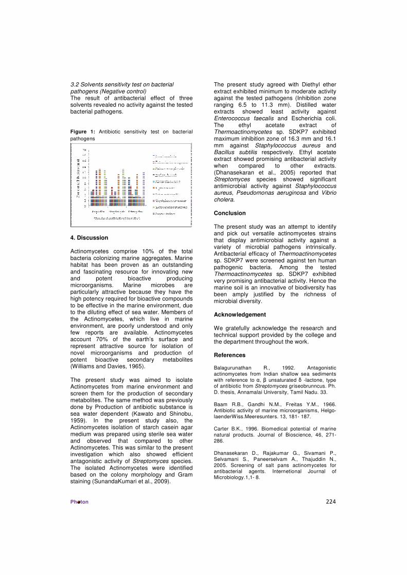
4.2.2. Antibiotic sensitivity test on bacterial pathogens (Positive control)
99
4.2.3. Solvents sensitivity test on bacterial pathogens (Negative control)
101
4.3. Molecular characterization of Thermoactinomyces vulgaris DKP01
102
4.3.1. Nucleotide sequence accession number 102
4.3.2. Evolutionary relationships 103
4.3.3. Restriction sites analysis 104
4.3.4. Secondary structure prediction 105
4.4. Separation of bioactive compounds from Thermoactinomyces vulgaris DKP01
107
4.4.1. Antibacterial activity of bioactive compounds from Thermoactinomyces vulgaris DKP01
107
4.4.2. UV – Visible spectrum of flavonoids from Thermoactinomyces vulgaris DKP01
110
4.4.3. Detection of functional groups of flavonoids from Thermoactinomyces vulgaris DKP01 by FT –IR
110
4.5. Antioxidant activity of selected Actinobacteria 111
4.5.1. Total phenolic content of actinobacteria 112
4.6. Isolation and identification of mechercharmycin from Thermoactinomyces vulgaris DKP01
112
4.6.1. Thin layer chromatographic analysis of Mechercharmycin
112
4.6.2. Ultra Violet - Visible (UV) spectroscopic analysis of mechercharmycin
113
4.6.3. FT –IR analysis of mechercharmycin 114
4.6.4. High Performance Liquid Chromatography (HPLC) analysis
116
4.6. 5. Anticancer activity of mechercharmycin 117
4.7. Biosynthesis of silver nanoparticles by Thermoactinomyces vulgaris DKP01
122
4.7.1. Ultraviolet-Visible (UV-Vis) Spectroscopic analysis of silver nanoparticles by Thermoactinomyces vulgaris DKP01
123
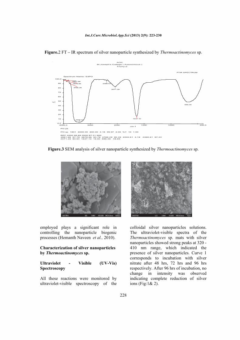
4.7.2. FT –IR analysis of silver nanoparticles synthesized by Thermoactinomyces vulgaris DKP01
123
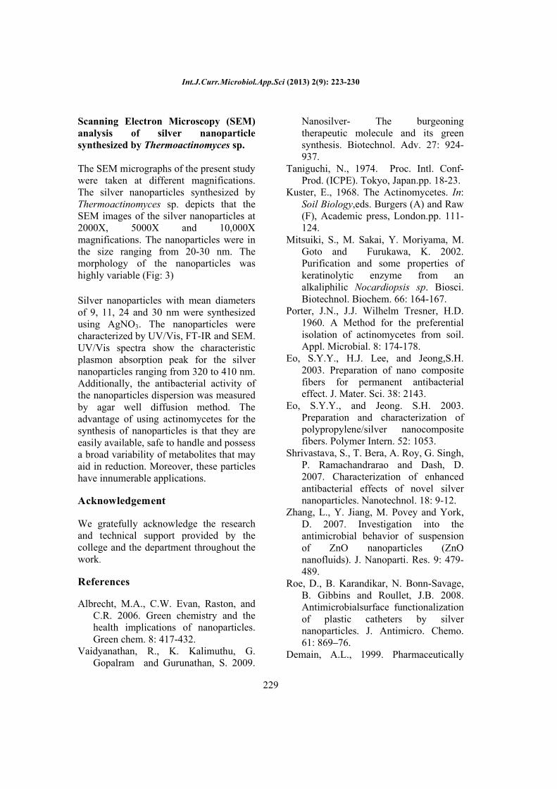
4.7.3. Scanning Electron Microscopic (SEM) analysis of silver nanoparticle synthesized by Thermoactinomyces vulgaris DKP01
125
4.7.4. Antibacterial activity of silver nanoparticle synthesized by Thermoactinomyces vulgaris DKP01
126
5. DISCUSSION 128 - 143
5.1. Actinobacteria 128
5.2. Biodiversity of marine actinobacteria 128
5.3. Physico – chemical characteristics of the soil 132
5.4. Antibacterial activity of actinobacteria 133
5.5. Molecular characterization of potential actinobacteria 135

5.6. Bioactive compounds from actinobacteria 136
5.7. Antioxidant activity of Thermoactinomyces vulgaris DKP01 138
5.8. Anticancer activity 139
5.9. Synthesis of silver nanoparticles by actinobacteria 140
6. SUMMARY AND CONCLUSION 144 - 146
REFERENCES 147 – 178

LIST OF TABLES
Table No.
Title Page No.
1. Isolates of actinobacteria from mangrove soil sample 69
2. Biochemical characterization of actinobacteria 72
3. Total number of colonies, mean density (CFU/g) and percentage contribution of Actinobacteria recorded during different seasons from Mangrove soil sample at Vellappallam, Nagapattinam District
88
4. Percentage frequency and frequency class of different species of actinobacteria recorded at Mangrove soil sample at Vellappallam, Nagapattinam District
90
5. Physico- chemical parametes of soli sample 95
6. Correlation between total actinobacteria and physico chemical parameters
96
7. Antibacterial activity of Thermoactinomyces vulgaris DKP01
97
8. Separation of bioactive compounds from Thermoactinomyces vulgaris DKP01 by TLC
107
9. Antibacterial activity of bioactive compounds from Thermoactinomyces vulgaris DKP01
108
10. Antioxidant activity of selected actinobacteria 112
11. Total phenolic content of selected actinobacteria 112
12. Effect of mechercharmycin and size of hepatocellular nodules during N, N-diethylnitrosamine (DEN) induced hepatocarcinogenesis
118
13. Body weight changes in control and experimental groups of rats
119
14. Determination of glucose and total bilirubin in the serum of control and experimental rats
120
15. Effect of Mechercharmycinon serum cholesterol and triglyceride of control and experimental rats
120
LIST OF FIGURES
Figure No.
Title Page No.
1. Map showing the sampling stations 32
2. Antibiotic sensitivity test on bacterial pathogens 99
3. Phylogenetic analysis of 16S rRNA gene in Thermoactinomyces vulgaris DKPO1 using NJ method
104
4. Restriction Site Analysis 105
5. Secondary structure prediction 106
6. UV – Visible spectrum of flavonoids fromThermoactinomyces vulgaris DKP01
110
7. FT –IR spectrum of flavonoids from Thermoactinomyces vulgaris DKP01
111
8. UV - Visible spectrum of the mechercharmycin isolated from Thermoactinomyces vulgaris DKP01
113
9. UV - Visible spectrum of the standard mechercharmycin
114
10. FT-IR spectrum of the mechercharmycin isolated from Thermoactinomyces vulgaris DKP01
115
11. FT-IR spectrum of the standard mechercharmycin 115
12. HPLC analysis of mechercharmycin isolated from Thermoactinomyces vulgaris DKP01
116
13. HPLC analysis of standard mechercharmycin 117
14. UV – Visible spectrum of silver nanoparticle synthesized
123
15. FT – IR spectrum of silver nanoparticle synthesized by Thermoactinomyces vulgaris DKP01
124
16. Antibacterial activity of silver nanoparticles synthesized using Thermoactinomyces vulgaris DKP01
126
LIST OF PLATES
Plate No.
Title Page No.
1. Aerial view of mangroves of Vellappallam 33
2. Sample collection site at Vellappallam 34
3. Isolation of Actinobacteria from Mangroves of Vellappallam
75
4. Microscopic View of Selected Actinobacteria 80
5. Antibacterial activity of Thermoactinomyces vulgaris – DKP01
98
6. Antibiotic activity test (Positive Control) 100
7. Antibiotic activity test (Negative Control) 101
8. 16S rRNA gene sequences of Thermoactinomyces vulgaris DKP01
102
9. Antibacterial activity of bioactive compounds Thermoactinomyces vulgaris – DKP01
109
10. Effect of mechercharmycin on hepatocellular nodules during DEN induced hepatocarcinogenesis
118
11. Histological observation of liver in control and experimental animals
121
12. Silver Nanoparticle synthesized using Thermoactinomyces vulgaris – DKP01
122
13. Scanning Electron Microscopic (SEM) analysis of silver nanoparticle synthesized by Thermoactinomyces vulgaris DKP01
125
14. Antibacterial activity of silver nanoparticles synthesized by Thermoactinomyces vulgaris – DKP01
127
Biodiversity and Biotechnological Applications of Actinobacteria from Mangroves of Vellappallam at Nagapattinam District, Tamil Nadu, India. 1
1. INTRODUCTION
1.1. Mangrove Ecosystem
The word "Mangrove" is considered to be a combination of the Portuguese
word "Mangue" and the English word "grove". Mangroves are salt-tolerant plants of
tropical and subtropical intertidal regions of the world. The specific regions where
these plants occur are termed as 'mangrove ecosystem'. These are highly productive but
extremely sensitive and fragile. Besides mangroves, the ecosystem also harbours other
plant and animal species.
Mangrove forests are among the world’s most productive ecosystem that
enriches coastal waters, yields commercial forest products, protect coastlines and
support coastal fisheries. However, mangroves exist under condition of high salinity,
extreme tides, strong winds, high temperature and muddy, anaerobic soils. There may
be no other group of plants with such highly developed morphological, biological,
ecological and physiological adaptations to extreme conditions.
Mangroves are woody plants that grow at the interface between land and sea in
tropical and subtropical latitudes. These plants and the associated microbes, fungi,
plants and animals, constitute the mangrove forest community or mangal (Kathiresan
and Bingham, 2001). Mangroves provide nursery habitat for commercial fish,
crustaceans and wildlife species that contribute to sustaining the survival of local fish
and shellfish populations (Brown, 1997).
Experiences have proved that the presence of mangrove ecosystems on coastline
save lives and property during natural hazards such as cyclones, storm surges and
erosion. These ecosystems are also well known for their economic importance. They
are breeding, feeding and nursery grounds for many estuarine and marine organisms.
Hence, these areas are used for captive and culture fisheries. The ecosystem has a very
large unexplored potential for natural products useful for medicinal purposes and also
for salt production, apiculture, fuel and fodder, etc.
Biodiversity and Biotechnological Applications of Actinobacteria from Mangroves of Vellappallam at Nagapattinam District, Tamil Nadu, India. 2
1.2. Mangroves in Tamilnadu
Mangroves in Tamil Nadu exist on the Cauvery delta areas. Pichavaram has a
well-developed mangrove forest dominant with Rhizophora spp and Avicennia marina.
Mangroves also occur near places like Vedaranyam, Vellappallam, Kodiakarai (Point
Calimere), Muthupet, Chatram and Tuticorin. Inspite of the fact that Pichavaram
mangrove is very small in area, it has been very well studied in all aspects of studies
like biology, chemistry and microbiology etc.
1.3. Actinobacteria
Actinobacteria are a group of prokaryotic organisms belonging to subdivision of
the Gram-positive bacteria phylum. Most of them are in subclass Actinobacteridae,
order Actinomycetales. All members of this order are characterized in part by high
G+C content (>55 mol %) in their DNA (Stackbrandt et al., 1997). They are
filamentous bacteria which produce two kinds of branching mycelium, aerial mycelium
and substrate mycelium. The aerial mycelium is important as the part of the organism
that produces spores. For this reason they have been considered as fungi, as it reflected
in their name, akitino means ray and mykes means mushroom/fungus, so actinobacteria
was called ray fungi. Actinobacteria are the most widely distributed group of
microorganisms in nature and are also well known as saprophytic soil inhabitants
(Takizawa et al., 1993).
Actinobacteria are soil organisms which have characteristics common to
bacteria and fungi and yet possess sufficient distinctive features to delimit them into a
distinct category. In the strict taxonomic sense, actinobacteria are clubbed with bacteria
in the same class of Schizomycetes but confined to the order Actinomycetales (Kumar
et al., 2005). Actinobacteria are aerobic, though they generally are low-oxygen-
utilizing bacteria. Actinobacteria indicates an organism belonging to the
Actinomycetales, a subdivision of the Prokaryotae Kingdom.
They are unicellular like bacteria, but produce a mycelium which is non-septate
(coenocytic) and more slender, like true bacteria they do not have distinct cell-wall and
their cell wall is without chitin and cellulose (commonly found in the cell wall of

Biodiversity and Biotechnological Applications of Actinobacteria from Mangroves of Vellappallam at Nagapattinam District, Tamil Nadu, India. 3
fungi). On culture media unlike slimy distinct colonies of true bacteria which grow
quickly, actinobacteria colonies grow slowly, show powdery consistency and stick
firmly to agar surface. They produce hyphae and conidia/sporangia like fungi. Certain
actinobacteria whose hyphae undergo segmentation resemble bacteria, both
morphologically and physiologically.
Actinobacteria belonging to the order of Actinomycetales are grouped under
four families viz Mycobacteriaceae, Actinomycetaceae, Streptomycetaceae and
Actinoplanaceae. Actinomycetous genera which are agriculturally and industrially
important are present in only two families of Actinomycetaceae and
Streptomycetaceae. In the order of abundance in soils, the common genera of
actinobacteria are Streptomyces (nearly 70%), Nocardia and Micromonospora,
Actinoplanes, Micromonospora and Streptosporangium are also generally encountered.
It is interesting that the world’s oceans, which cover 70% of the earth’s and
include some of the most biodiversity ecosystems on the planet, have not been widely
recognized as an important resource for novel actinobacteria. Infact, the distributions of
actinobacteria in the sea remain largely undescribed and even today, conclusive
evidence that these bacteria play an important tecological role in the marine
environment have remained elusive. Anintriguing picture of the diversity of marine
actinobacteria is beginning to emerge. Once largely considered to originate from
dormant spores that washed in from land (Goodfellow and Willams, 1983), it is now
clear that specific populations of marine adapted actinobacteria not only exist but add
significant new diversity within a broad range of actinobacterial taxa.
1.4. Role of Actinobacteria
Actinobacteria decompose all sorts of organic substances like cellulose,
polysaccharides, protein, fats, organic-acids etc. Organic residues / substances added
soil are first attacked by bacteria, fungi and later by actinobacteria, because they are
slow in activity and growth than bacteria and fungi. They decompose the more
resistant and in decomposable organic substance and produce a number of dark black
to brown pigments which contribute to the dark colour of soil humus. They are also
Biodiversity and Biotechnological Applications of Actinobacteria from Mangroves of Vellappallam at Nagapattinam District, Tamil Nadu, India. 4
responsible for subsequent further decomposition of humus (resistant material) in soil.
They are responsible for earthy odor or smell of freshly ploughed soils. Many genera,
species and strains of Streptomyces produce number of antibiotics like streptomycin,
tetramycin and aureomycin etc. One of the species of actinobacteria Streptomyces
scabies causes disease "Potato scab" in potato.
1.5. Diversity of marine actinobacteria
Marine environment is the highest reservoir of chemical and biological
diversity. As marine environmental conditions are extremely different from terrestrial
ones, it is surmised that marine actinobacteria have different characteristics from those
of terrestrial counterparts, and therefore, might produce different types of bioactive
compounds (Okami, 1984; Fenical et al., 1999 and Gesheva et al., 2005).The living
conditions to which marine actinobacteria had to adapt during evolution range from
extremely high pressure, high salinity and anaerobic conditions. It is likely that this is
reflected in the genetic and metabolic diversity of marine actinobacteria, which remains
largely unknown. Indeed, the marine environment is a virtually untapped source of
novel actinobacteria diversity (Bull et al., 2006 and Stach et al., 2003) and, therefore,
of new metabolites (Goodfellow and Hayens, 1984; Jensen et al., 2005; Fiedler et al.,
2005 and Magarvey et al., 2004).
The discovery of new bioactive compounds is a never ending process to meet
the ever lasting demand for novel drug and other biomolecules with antimicrobial and
other thereapeutic properties in order to compact plant pathogens and also to treat other
human ailments. In this, scenario, it is more important to identify never or rare
actinobacteria because they are the pivotal sources of potent molecules. Therefore, the
present research focus on marine environment has been gaining importance in recent
years.
1.6. Antimicrobial Activity
The discovery of novel antimicrobial metabolites from actinobacteria is an
important alternative to the increasing levels of drug resistance by human pathogens,
the inadequate number of effective antibiotics against diverse bacterial species and few

Biodiversity and Biotechnological Applications of Actinobacteria from Mangroves of Vellappallam at Nagapattinam District, Tamil Nadu, India. 5
new antimicrobial agents in development is probably due to relatively unfavourable
returns on asset (Song, 2008 and Yu et al., 2010).
Antimicrobial metabolites can be defined as low molecular weight organic
natural substances made by microorganisms that are active at low concentrations
against other microorganisms (Wani et al., 1971). Actinobacteria are believed to carry
out a resistance mechanism to overcome pathogenic invasion by producing secondary
metabolites (Tan and Zou, 2001).
Consistent with the tremendous diversity of actinobacteria and their ecological
roles is the outstanding chemical variety of their secondary metabolites, which often
display promising pharmaceutically or agrochemically exploitable activities when
tested in various bioassays (Strobel et al., 2004). Due to the world’s urgent need for
new antibiotics, chemotherapeutic agents and agrochemicals to scope with the growing
medicinal and environmental problems facing mankind, growing interest is taken into
the research on the chemistry of actinobacteria. Whereas between 1987 and 2000
approximately 140 new natural products were isolated from endophytic fungi (Tan and
Zou 2001), a similar number was subsequently characterized between 2000 and 2006
(Zhang et al., 2006). Many of these exhibit interesting activity profiles.
1.7. Antioxidant Activity
Reactive oxygen and nitrogen species (ROS/RNS) produced during the cellular
metabolism are essential for cell signalling, apoptosis, gene expression and ion
transportation. However, ROS can cause oxidative stress if accumulated in the body in
excess amount. The consequence of accumulation of ROS includes the damage of
DNA, RNA, proteins and lipids resulting in the inhibition of their normal functions.
The abnormal functioning of these biomolecules can enhance the risk for
cardiovascular disease, cancer, autism and other diseases (Lu et al., 2010 and Prem
anand et al., 2010). Therefore, minimizing oxidative stress will promote our physical
condition and prevent some degenerative diseases in which free radicals are involved
(Song et al., 2010).

Biodiversity and Biotechnological Applications of Actinobacteria from Mangroves of Vellappallam at Nagapattinam District, Tamil Nadu, India. 6
Antioxidants may be molecules that can neutralize free radicals by accepting or
donating electron(s) to eliminate the unpaired condition of the radical. The antioxidant
molecules may directly react with the reactive radicals and destroy them, while they
may become new free radicals which are less active, longer-lived and less dangerous
than those radicals they have neutralized.
A myriad of both natural and synthetic antioxidants has been advised for use in
the treatment of various human maladies (Cuzzocrea et al., 2001). Some synthetic
antioxidant compounds like butylated hydroxytoluene, butylated hydroxyanisole and
tertiary butylhydroquinone commonly used in processed foods. However, synthetic
antioxidants have shown potential health risks and toxicity, most notably possible
carcinogenicity. Therefore, it is of great importance to find new sources of safe and
inexpensive antioxidants of natural origin in order to use them in foods and
pharmaceutical preparations to replace synthetic antioxidants (Cuzzocrea et al., 2001;
Mundhe et al., 2011 and Lee et al., 2004).
Natural antioxidants are commonly found in medicinal plants, vegetables and
fruits. However, it has been reported that metabolites from actinobacteria can be a
potential source of novel natural antioxidants. The DPPH radical scavenging assay has
become popular in natural antioxidant studies because of its simplicity and high
sensitivity. This assay is based on the theory, that a hydrogen donor is an antioxidant.
1.8. Anticancer compounds
Cancer is a term that refers to a large group of over a hundred different diseases
that arise when defects in physiological regulation cause unrestrained proliferation of
abnormal cells (Capon et al., 2000). In most cases, these clonal cells accumulate and
multiply, forming tumors that may compress, invade and destroy normal tissue,
weakening the vital functions of the body with devastating consequences, including
loss of quality of life and mortality. Nowadays, cancer is the second cause of death in
the developed world, affecting one out of three individuals and resulting in one out of
five deaths worldwide. Diversified groups of marine actinobacteria are known to
produce different types of anticancer compounds.

Biodiversity and Biotechnological Applications of Actinobacteria from Mangroves of Vellappallam at Nagapattinam District, Tamil Nadu, India. 7
1.9. Mechercharmycin
Marine microorganisms have been recognized as apromising source for the
development of new pharmaceuticals (Blunt et al., 2004). In the course of screening for
antitumor substances from marine-derived microorganisms found the cyclic peptide-
like compound bearing four oxazoles and a thiazol, Mechercharmycin (Malet et al.,
2005).
Non ribosomal peptides (NRP) are a class of peptide secondary metabolites
these classes of natural products comprises peptides synthesized by non-ribosomal
peptide synthetases (NRPS).The antitumor activities of mechercharmycins are
produced by Thermoactinomyces sp., isolated from sea mud collected at Mecherchar.
Mechercharmycin showed cytotoxic activity against human lung adenocarcinoma A549
and Jurkatleukemia cells with IC50 values of 0.04 μM, mechercharmycin B did not
show inhibitory activity in these assays even at 1 μM, which suggests the cyclic
structure of Mechercharmycin.
Structure of Mechercharmycin
Biodiversity and Biotechnological Applications of Actinobacteria from Mangroves of Vellappallam at Nagapattinam District, Tamil Nadu, India. 8
1.10. Silver Nanoparticles
Silver nanoparticles are one of the promising products in the nanotechnology
industry. The development of consistent processes for the synthesis of silver
nanomaterials is an important aspect of current nanotechnology research. One of such
promising process is green synthesis. Silver nanoparticles can be synthesized by several
physical, chemical and biological methods. However for the past few years, various
rapid chemical methods have been replaced by green synthesis because of avoiding
toxicity of the process and increased quality.
The field of nanotechnology is one of the most active areas of research in
modern material sciences. Nanotechnology is a field that is developing day by day,
making an impact in all spheres of human life (Singh et al., 2010) and creating a
growing sense of excitement in the life sciences especially biomedical devices and
biotechnology (Prabhu et al., 2010). The use of nanoparticles is gaining imparts in the
present century, as they posses defined chemical, optical and mechanical properties
(Rai et al., 2009 and Gong et al., 2007). Metal nanoparticles are of importance due to
their potential applications in catalysis, photonics, biomedicine, antimicrobial activity
and optics (Wang et al., 2004; Biswas et al., 2004; Shipway and Willner, 2001; Nie and
Emory, 1997; Govindaraju et al., 2008 and 2009).
Nanoparticles exhibit new or improved properties based on specific
characteristics such as size, distribution and morphology. There have been impressive
developments in the field of nanotechnology in the recent past years, with numerous
methodologies developed to synthesize nanoparticles of particular shape and size
depending on specific requirements. New applications of nanoparticles and
nanomaterials are increasing rapidly.
Nanoparticles, because of their small size, have distinct properties compared to
the bulk form of the same material, thus offering many new developments in the fields
of biosensors, biomedicine, and bio nanotechnology. Nanotechnology is also being
utilized in medicine for diagnosis, therapeutic drug delivery and the development of
treatments for many diseases and disorders. Nanotechnology is an enormously

Biodiversity and Biotechnological Applications of Actinobacteria from Mangroves of Vellappallam at Nagapattinam District, Tamil Nadu, India. 9
powerful technology, which holds a huge promise for the design and development of
many types of novel products with its potential medical applications on early disease
detection, treatment and prevention.
Nanotechnology is expected to open new avenues to fight and prevent disease
using atomic scale tailoring of materials. The most promising nanomaterial with
antibacterial properties are metallic nanoparticles, which exhibit increased chemical
activity due to their large surface to volume ratios and crystallographic surface structure
(Parameswari et al., 2010). In nanotechnology, silver nanoparticles are the most
prominent one. Silver nanoparticles are nanoparticles of silver, i.e. silver particles of
between 1 nm and 100 nm in size and have attracted intensive research interest. It is
generally recognized that silver nanoparticles may attach to the cell wall, thus
disturbing cell wall permeability and cellular respiration.
Biological methods of synthesis have paved way for the “bio synthesis” of
nanoparticles and these have proven to be better methods due to slower kinetics, they
offer better manipulation and control over crystal growth and their stabilization. This
has motivated an upsurge in research on the synthesis routes that allow better control of
shape and size for various nanotechnological applications. The use of environmentally
begin materials like plant extract (Jain et al., 2009), bacteria (Saifuddin et al., 2009),
actinobacteria (Verma et al., 2010) and enzymes (Willner et al., 2007) for the synthesis
of silver nanoparticles offer numerous benefits of ecofriendliness and compatibility for
pharmaceutical and other biomedical applications.
Chemical synthesis methods lead to presence of some toxic chemical absorbed
on the surface that may have adverse effect in the medical applications. Green synthesis
provides advancement over chemical and physical method as it is cost effective,
environment friendly, easily scaled up for large scale synthesis and in this method there
is no need to use high pressure, energy, temperature and toxic chemicals (Singh et al.,
2010).
Biodiversity and Biotechnological Applications of Actinobacteria from Mangroves of Vellappallam at Nagapattinam District, Tamil Nadu, India. 10
1.10.1 Importance of Silver nanoparticles
It is for used for purification and quality management of air, biosensing,
imaging, drug delivery system. Biologically synthesized silver nanoparticles have many
applications like coatings for solar energy absorption and intercalation material for
electrical batteries, as optical receptors, as catalysts in chemical reactions, for
biolabelling, and as antimicrobials. Though silver nanoparticles are cytotoxic but they
have tremendous applications in the field of high sensitivity bimolecular detection and
diagnostics, antimicrobials and therapeutics, catalysis and micro-electronics.
It has some potential application like diagnostic biomedical optical imaging,
biological implants (like heart valves) and medical application like wound dressings,
contraceptive devices, surgical instruments and bone prostheses. Many major consumer
goods manufacturers already produed household items that utilize the antibacterial
properties of silver nanoparticles. These products include nanosilverlined refrigerators,
air conditioners and washing machines. (Chau et al., 2007; Hong et al., 2008; Martinez
Castanon et al., 2008; Wang, 2006; Zhang et al., 2008).
1.10.2 Silver nanoparticles as an antimicrobial agent
AgNP highly antimicrobial to several species of bacteria, including the common
kitchen microbe E. coli. According to the mechanism reported, silver nanoparticles
interact with the outer membrane of bacteria and arrest the respiration and some other
metabolic pathway that leads to the death of the bacteria.
New technology advances in reducing silver compound chemically to nanoscale
sized particles have enabled the integration of this valuable antimicrobial into a larger
number of materials including plastics, coatings, and foams as well as natural and
synthetic fibers. Nano-sized silver have already provides a more durable antimicrobial
protection, often for the life of the product.
Current research in inorganic nanomaterials having good antimicrobial
properties has opened a new era in pharmaceutical and medical industries. Silver is the
metal of choice as they hold the promise to kill microbes effectively. Silver
Biodiversity and Biotechnological Applications of Actinobacteria from Mangroves of Vellappallam at Nagapattinam District, Tamil Nadu, India. 11
nanoparticles have been recently known to be apromising antimicrobial agent that acts
on a broad range of target sites both extracellularly as well as intracellularly.
Silver nanoparticles shows very strong bactericidal activity against Gram
positive as well as Gram negative bacteria including multi resistant strains (Shrivastava
et al., 2007), and also it was found to be in few studies (Zeng et al., 2007 and Roe
et al., 2008). Hence there is a huge scientific progress in the study of biological
application of ZnO and Ag and other metal NP.
1.11. Current trends in actinobacteria
The role of mangrove actinobacteria compounds towards understanding of
mangrove ecological interactions is very much dependent on multidisciplinary
approach. Chemical metabolite oriented approaches may prove to be reliable tools
helping to elucidate compound’s biological properties, which may not be detectable in
any other way. The discovery of new active metabolites must be followed by adequate
biological testing, which will require the immediate availability of substantial amounts
of naturally derived material, preferentially obtained by isolation from its source and
thus increasing the reproducibility of metabolic profiles.
Biodiversity and Biotechnological Applications of Actinobacteria from Mangroves of Vellappallam at Nagapattinam District, Tamil Nadu, India. 12
Keeping these in mind and recognizing the significance of actinobacteria as a
source of novel bioactive compounds. In the present study, actinobacteria was
documented from Vellappallam mangrove forest and also to explore the antibacterial
antioxidant, anticancer activity and silver nanoparticles synthesis potential of
Thermoactinomyces vulgaris DKP01 isolate with the following objectives.
To isolate and identify the actinobacteria from the soil sample collected (Four
different seasonal variations) from Vellappallam mangrove forest Nagapattinam
District and to study the actinobacterial biodiversity and its relationship with
physico chemical properties of marine habitate.
To screen the antibacterial potentials of dominant actinobacterial isolates
against bacterial pathogens.
To identify the potential actinobacteria isolate by 16S rRNA gene sequencing
and molecular phylogetic analysis.
To separate and characterize the bioactive compounds using UV – visible
spectroscopic, FT – IR analysis for the identification of the functional groups.
To evaluate the antioxidant activity and total phenolic content of potential
Thermoactinomyces vulgaris DKP01.
To separate, characterize the mechercharmycin by TLC, UV , FT –IR, and
HPLC method find out the anticancer activity of Mechercharmycin from
Thermoactinomyces vulgaris DKP01
To synthesis, characterize the silver nanoparticles (AgNP) from
Thermoactinomyces vulgaris DKP01 by UV, FT – IR and SEM analysis and to
antibacterial efficacy of AgNPs.
Biodiversity and Biotechnological Applications of Actinobacteria from Mangroves of Vellappallam at Nagapattinam District, Tamil Nadu, India. 13
2. REVIEW OF LITERATURE
2.1. Diversity of marine actinobacteria
Actinobacteria were isolated from near shore marine sediments collected at 15
Island locations throughout the Bahamas. A total of 289 actinobacteria colonies were
observed, and all but 6 could be assigned to the suprageneric groups Actinoplanetes and
Streptomycetes. A bimodal distribution in the actinobacteria population in relation to
depth was recorded, with the maximum numbers occurring in the shallow and deep
sampling sites (Jensen et al., 1991: Kala and Chandrika 1995) used different media for
isolating and maintaining actinobacteria collected from mangrove sediments.
About 100 strains were isolated from a mangrove stand of Morib, Selangor,
Malaysia in an earlier study (Vikineswary et al., 1997). Totally 133 strains of
actinobacteria from 129 marine samples collected from various stations along the
Tuticorin coast (Patil et al., 2001). Six strains of actinobacteria were isolated from the
sediments of the Arabian Sea (Mathew and Philip, 2003).
The marine sediments were collected from Hainan Island, South China, in April
2004, for the investigation of actinobacteria diversity, ninety four marine actinobacteria
strains were isolated. About 87.5% of the isolates were Streptomyces sp., and 12.5%
Micromonospora sp. The Streptomyces isolates were classified into 13 groups, and the
Cinerogriseus group was the dominant group among the Streptomycete isolates (You
et al., 2005).
Totally 17 actinobacteria isolates were obtained from the saltpan regions of
Cuddalore and Parangipettai (Dhanasekaran et al., 2005b). Kathiresan et al., (2005)
isolated 160 strains from the sediments of mangrove, estuary, sand dune and
industrially polluted marine environment of Cuddalore. Of these, mangrove sediments
were the rich sources for actinobacteria. Sivakumar et al., (2005) isolated actinobacteria
from different stations of the Pitchavaram mangrove ecosystem using three different
media.

Biodiversity and Biotechnological Applications of Actinobacteria from Mangroves of Vellappallam at Nagapattinam District, Tamil Nadu, India. 14
Total of 173 actinobacteria were isolated from near shore marine environment
at eight different locations of Kerala, West Coast of India. Among them, 64 isolates
were morphologically distinct on the basis of spore mass colour, reverse side colour,
aerial and substrate mycelia formation and production of diffusible pigment. The
majority (47%; n=30) of these isolates were assigned to the genus Streptomyces
(Remya and Vijayakumar, 2008)
A total of 288 marine samples were collected from different locations of the
Bay of Bengal starting from Pulicat Lake to Kanyakumari, and 208 isolates of marine
actinobacteria were isolated using starch casein agar medium. The growth pattern,
mycelial coloration, production of exopolysaccharides and diffusible pigment and
abundance of Streptomyces sp. were documented. Among marine actinobacteria
Streptomyces sp. was present in (88%) large proportion (Ramesh and Mathivanan,
2009).
Totally 189 Streptomyces isolates were obtained from eight different soils of
Cuddalore, Tamil Nadu, India. Among them, only 78 isolates were morphologically
distinct. The highest diversity in the Streptomyces populations was observed
(Dhanasekaran et al., 2009). Vijayakumar et al., (2010) also studied the marine soil and
sediment samples collected from different locations of Muthupet mangrove,
Tamilnadu. A total of thirty different marine actinobacteria isolates were isolated on
starch casein agar medium. Isolated actinobacteria from Annangkoil estuarine soils of
Tamilnadu. Krishnaraj and Mathivanan (2009) reported that the total of 137 different
isolates of marine actinobacteria were isolated from deep sea sediment collected from
the Bay of Bengal.
Actinobacteria were cultivated using a variety of media and selective isolation
techniques from 20 marine samples collected from the island of Nicobar. In total, 800
actinobacteria colonies were observed and 100 (12.5%) of these, representing the range
of morphological diversity observed from each sample, were obtained in pure culture.
The majority of the strains isolated (90%) required sea water for their growth indicating
high degree of marine adaptation. The dominant actinobacteria recovered belonged to
Biodiversity and Biotechnological Applications of Actinobacteria from Mangroves of Vellappallam at Nagapattinam District, Tamil Nadu, India. 15
the genus of Streptomyces. These results support the existence of taxonomically diverse
populations of actinobacteria in the Nicobar marine environment (Karthik et al., 2010).
A total of 20 different actinobacteria were recovered from salt pan region of
Kodiakarai, Nagapattinam District using starch casein agar medium. From 20 isolated
actinobacteria, 10 were dominant in their growth. Among the 10 actinobacteria
Streptoverticillium album was highly dominant from their isolates (Gayathri et al.,
2011).
The diversity of actinobacteria in the Manakkudi mangrove ecosystem was
analysed. The diversity of actinobacteria are found maximum in the rhizosphere soil
than the non-rhizosphere soil that too mangrove associate of Achrostichum aureum
harbours maximum counts than true mangrove plants. The diversity of actinobacteria
was found maximum between the soil depth of 10-20 cm are not correlated with the
maximum level of nutrients between the soil depth of 0-10 cm. The presence of
actinobacteria in the Manakkudi mangrove ecosystem could pave the way for the
establishment of disease free mangrove seedlings in the nursery and in the field
(Ravikumar et al., 2011).
The actinobacteria were screened from the soil sample of Manora,
Thanjavur Dt. Tamil Nadu, India. Ten actinobacteria species including Actinobispora
yunnanensis, Streptomyces albus, Micromonospora echinospora, Saccharopolyspora
hirsute, Streptomycetes cyaneus, Actinomadura citrea, Saccharomonospora viridis,
Thermomonospora mesophila, Streptoverticillium album Microtetrospora fastidiosa
were isolated (Kaviyarasi et al., 2011).
Totally 107 actinobacteria isolates were obtained from 36 sediment samples
collected from two different stations such as Thondi and Karankadu of Palk Strait
region situated along the South East coast of India. The number of isolates were found
maximum in Karankadu mangrove region (62) followed by Thondi (45) sediment
samples particularly in monsoon season (Ravikumar and Suganthi, 2011).
The actinobacteria were isolated from marine sediments of different stations of
Muthupet mangrove ecosystem (10°15’-10°35’N and 79°20’–79°55’E), situated along
Biodiversity and Biotechnological Applications of Actinobacteria from Mangroves of Vellappallam at Nagapattinam District, Tamil Nadu, India. 16
the Southeast coast of India for the isolation of actinobacteria using Kuster agar
medium. The following seven isolates were characterized and identified as
Streptomyces neyagawaensis, A. aureocirculatus, A. aureocirculatus, S. spheroids,
S. albulus, S. antibioticus, S. mirabilis and S. umbrosus (Sathiyaseelan and Stella,
2011a). Seven actinobacteria isolates were obtained from the sediments collected from
the mangrove. Among the 7 isolates, 3 isolates belong to Streptomyces sp.
Sathiyaseelan and Stella (2011b) analysed the five actinobacteria were isolated from
soil collected in two different regions of Parangipettai. Morphological studies indicated
that the strains belonged to the genera Streptomyces spectabilis, Actinomadura roseale,
Streptomyces platensis, S. kavamyceticus and S. citricolor (Rajesh et al., 2011).
A total of 42 actinobacteria were isolated from mangrove sediments of
Andaman and Nicobar Islands, India (Baskaran et al., 2011). Naikpatil and Rathod
(2011) isolated 54 actinobacteria from marine environment of Karrwar, west coast of
India. Ten actinobacteria were dominant in their growth. Streptomyces sp. was highly
dominant from their isolates.
The soil samples, collected from the mangroves forest of Karwar. Fifty three
rare actinobacteria strains were chosen using selective isolation approaches, then
morphological and chemical properties of the isolates were determined. The isolates
belonged to one of the following genera such as Micromonospora, Microbispora,
Actinoplanes and Actinomadura (Sateesh et al., 2011).
Sixty actinobacteria were isolated from the soil samples collected from
Pakistan. The isolates identification falls under three genera including Actinomyces,
Streptomyces and Nocardia sp. each with the total number of 31, 17 and 12 isolates
identified respectively (Ullah et al., 2012).
Total of 116 actinobacterial colonies were recorded from 30 mangrove and
marine sediment samples of Bhitherkanikka mangrove environment east coast of
Orissa. Among them, 67 isolates were morphologically distinct on the basis of colour
of spore mass riverside colour, aerial and substrate mycelia for mat production of
diffusible pigment sporophore morphology. Forty three isolates were assigned to the
Biodiversity and Biotechnological Applications of Actinobacteria from Mangroves of Vellappallam at Nagapattinam District, Tamil Nadu, India. 17
genus Streptomyces, Saccharopolyspora (5), Nocardiopsis (5), Micromonospora (3),
Actinomadura (5), Actinobacteria (1), Actinopolyspora (5) (Rajkumar et al., 2012).
Total of thirty soil samples were collected from Konark and Western terrestrial
sea. Totally 20 species were isolated on the basis of colony characteristics on starch
casein agar. Gulve and Deshmukh (2012) isolated 107 marine actinobacteria from near
sea shore sediment samples from different sites of Konkan coast of Maharashtra
(Kalyani et al., 2012).
The actinobacteria diversity in marine sediments were studied in the coastal
areas of Gokharna and Muradeshwara of Karnataka state. Seventeen isolates were
obtained on starch-casein agar media by soil dilution technique. Morphological,
cultural and biochemical characterization indicated that the isolates belong to
Streptomyces genus of Actinobacteria (Attimarad et al., 2012).
The total of eight actinobacteria was isolated from sea shore marine
environment locations of Bigeum Island, South West coast of South Korea. Sixty eight
actinobacteria were identified at a generic level based on the colony morphology and
microscopic morphology. Identification of strains by both morphological and cultural
characteristics revealed that most (54%) of the isolates belonged to white and grey
colour series. Out of 68 isolates, 66% of isolates were assigned to the genus
Streptomyces sp. and the remaining was identified as Nocardiopsis sp. (18%),
Micromonospora sp. (11%) and Actinopolyspora sp. (5%) (Parthasarathi et al., 2012).
The actinobacteria diversity of the marine sediments from Pulicat estuary,
Muttukadu, and Ennore estuaries, Tamil Nadu. Totally 227 isolated were
morphologically distinct on the basis of spore mass colour, aerial and substrate mycelia
formation and production of diffusible pigments. The majority were assigned genus
Streptomyces (60%; 162 isolates) and Actinopolyspora (5%; 11 isolates) (Chacko Vijai
Sharma and David, 2012).
2.2. Physico-chemical analysis
The relationship between physicochemical properties of the soil and the
Streptomyces abundance was studied. There was a positive correlation between the total
Biodiversity and Biotechnological Applications of Actinobacteria from Mangroves of Vellappallam at Nagapattinam District, Tamil Nadu, India. 18
Streptomyces population and nitrogen, available phosphorus, ferrous and manganese,
while the correlation with pH and sodium was negative (Dhanasekaran et al., 2009).
The physico-chemical and biological characteristics of four soil samples and
water samples were taken from ten selected river bodies in the region, for the analysis.
Measured properties of the water samples and the corresponding results are pH (4.5 to
6.5), temperature (26.9 to 28.7 oC), electrical conductivity (18.9 to 156.4 us/cm),
turbidity (19 to 48 NTU), redox potential (-372 to +202 mV), TDS (78 to 8450 mg/l),
TOC (17.3 to 38.7 mg/l), nitrate ions (6.1 to 17.0 mg/l), sulphate ions (0.8 to 13.6
mg/l), DO (4.1 to 5.7 mg/l) (Puyate and Rim-Rukeh, 2008).
The seasonal variation of physico-chemical parameters were studied at four
different stations in Pondicherry mangroves, southeast coast of India. Atmospheric and
surface water temperatures (ºC) varied from 17.9-41.7 and 16.66-37.91 respectively.
Annual rainfall and relative humidity ranges were 1.1-808 mm and 37-100 %
respectively. Seasonal variations of different parameters investigated were as follows:
salinity (6.36-36.77 ppt), dissolved oxygen (3.45-5.49 mg/l), pH (7.11-8.52), electrical
conductivity (26.65-52 ms-1), sulphide (2.76-47.16 mg/l), soil parameters sand (63.69-
87.31 %), silt (9.89-29.32 %), clay (3.06-17.98 %) and organic matter (0.94-3.94 %)
were recorded (Satheeshkumar and Anisa, 2009).
The soil samples were collected from salt pan environment of Kodiakarai,
Vedaranyam, Nagapattinam District, Tamilnadu, India. The physico-chemical features
of the test soil were pH (7.82), electrical conductivity (0.18dsm-1), available nitrogen
(97.9 Kg/ac), available iron (4.53 ppm) and calcium (8.9 mg/kg) (Gayathri et al., 2011).
The salt pan soil samples were collected from Ribandar, Goa, India for a period
of one year i.e. September 2000 to August 2001, in order to study physico chemical
parameters. The pH was generally alkaline with a maximum of 9.03 in the monsoon.
The maximum average pore water salinity was 73.91 at 0-2 cm, 38.9 at 2-5 cm and
38.67 2 at 5-10 cm during the premonsoon season. The lowest salinity was recorded at
2 -5 cm with an average value of 10.55 during the monsoon season (Kerkar and Loka
Bharathi, 2011).
Biodiversity and Biotechnological Applications of Actinobacteria from Mangroves of Vellappallam at Nagapattinam District, Tamil Nadu, India. 19
The marine soil sample was collected from Manora, Thanjavur Dt, Tamil Nadu,
India, for the analysis of physico chemical parameters. Physically, the texture of the
soil sample was sandy loam. The physico-chemical parameters such as pH (7.56),
electrical conductivity (0.26 dsm-1), organic carbon (0.29%), organic matter (0.58%),
available nitrogen (89.6 kg/ac), available phosphorus (5.26 kg/ac), available potassium
(175 kg/ac), available zinc (0.87 ppm), available copper (0.56 ppm), available iron
(4.69 ppm), available manganese (2.45 ppm), cation exchange capacity (22.6 C. mole
proton + kg), calcium (12.4 mg/kg), magnesium (10.6 mg/kg), sodium (1.69 mg/kg)
and potassium, (0.19 mg/kg) were recorded (Kaviyarasi et al., 2011).
The traditional salt industry has been existing in Goa since 500 A.D.
Temperature measured in the hypersaline ponds was generally higher at least by 5°C in
the surface. Likewise salinity and sulphate in the hypersaline sediments were 3-4 times.
The mesohaline salterns were more alkaline especially at the surface (Kerkar and
Bharathi, 2012).
The physico-chemical parameters in the water and soil of Vedaranyam
mangroves during the year 2008- 2009 at four-seasonal intervals were performed. The
water was slightly alkaline and contained high amounts of pH. The concentration of
salinity, total, inorganic and organic phosphate, ammonia, nitrite and nitrate were fairly
stable. Other nutrients such as calcium, magnesium, chloride and bicarbonate
concentration showed remarkable variations (Ramamurthy et al., 2012).
The physico-chemical properties of soils in Badagry and Ikorodu, were studied
soil samples were taken at depths of 0-20 cm from 26 and 36 points respectively at
Badagry and Ikorodu using soil and collected in polythene bags. The soil samples were
analyzed for their texture, structure, pH, and the availability of some basic soil nutrients
such as Nitrogen, Organic Carbon, Potassium, Phosphorus, etc, in accordance with
Standard analytical procedures (Ogundele and Fatai, 2012).
The monsoonal cycle plays a crucial role in regulating microbial population
distribution in the mangrove soil. Statistical analyses revealed that organic carbon was
the most significant factor that regulated the total microbial population. Intensification
of monsoonal cycle could heavily affect microbe dominated soil biogeochemistry and

Biodiversity and Biotechnological Applications of Actinobacteria from Mangroves of Vellappallam at Nagapattinam District, Tamil Nadu, India. 20
subsequent change in the regional ecology of the Sundarban Mangrove Forest
(Das et al., 2012).
The physico-chemical parameters of salt pan and marine soil from Pillaichavadi
and Kanyakumari East coast of Tamil Nadu, India were analysed. The physico-
chemical parameters of salinity, alkalinity, total dissolved solids of sediment samples in
salt pan and marine ecosystem were 53.09%, 24.0 mg, 0.18 mg and 54.84%, 27.2 mg,
0.41mg respectively (Thamizhmani et al., 2013).
2.3. Antibacterial activity of actinobacteria
The strain was identified as morphological, biochemical, physiological and
phylogenetic characterization of Nocardiopsis sp. VITSVK5 (FJ973467). Based on the
petroleum ether extract (1000 µg/ml) obtained from the isolate showed significant
antibacterial activity against Gram negative bacteria- Escherichia coli (20 mm),
Pseudomonas aeruginosa (18 mm) and Klebsiella pneumoniae (15 mm) and Gram
positive bacteria Enterococcus faecalis (20 mm), Bacillus cereus (13 mm) and
Staphylococcus aureus (6 mm) when compared with streptomycin (25 µg/disc) (Vimal
et al., 2009).
Seventy-nine actinobacteria were isolated from soils of Kalapatthar (5545m),
Mount Everest region. Twenty seven (34.18%) of the isolates showed an antibacterial
activity against atleast one test-bacteria. Among two Gram positive and nine Gram
negative bacteria in primary screening by perpendicular streak method. Thirteen
(48.15%) showed antibacterial activity in secondary screening. The result showed that
three of the isolates K.6.3, K.14.2, and K.58.5 were highly active with an inhibition
zone of 20 mm and broad spectrum antibacterial activity including two methicillin
resistant Staphylococcus aureus (MRSA) strains (Gurung et al., 2009).
The antibacterial substances from actinobacteria were isolated from marine
environment of Lonar Lake and characterized. Out of the 24 isolates subjected to
secondary screening, 12 isolates were active against Bacillus subtilis, 13 against
Staphylococcus aureus, 7 against Escherichia coli, 3 against Proteus vulgaris and 4
against Salmonella typhi. Metabolites in the extract of broth of 48 hrs grown
Biodiversity and Biotechnological Applications of Actinobacteria from Mangroves of Vellappallam at Nagapattinam District, Tamil Nadu, India. 21
Streptomyces spp. culture proved to have antimicrobial and cytotoxic against human
lung carcinoma cell A549 (Kharat et al., 2009).
Sixty three marine actinobacteria strains were isolated from the sponge and soil
samples collected from two different stations from Arabian sea, south west coast of
India. The counts of actinobacteria were found maximum in sponges during south west
monsoon season. The antimicrobial screening showed that five Streptomyces sp.
exhibited antimicrobial activity against eye pathogens, antibiotic sensitive and resistant
bacterial pathogens (Ravikumar et al., 2010).
137 different marine actinobacteria were isolated from deep sea sediment
samples collected from the Bay of Bengal. Among them, 85 isolates were tested for
antibacterial activity against two human pathogenic bacteria, Staphylococcus aureus
(methicillin resistant) and Pseudomonas aeruginosa as well as an antibiotic sensitive
bacterial strain Bacillus pumilus. All the 85 isolates exhibited antibacterial activity.
Based on the screening results, 10 marine actinobacteria were selected and tested
against methycillin resistant S. aureus. Out of these five isolates showed good
antibacterial activity and the nine among 10 isolates were obtained from estuarine
sediments (Krishnaraj and Mathivanan, 2009).
Antibacterial substances from actinobacteria were isolated from marine
environment of Karrwar, west coast of India and characterized. Out of 28 isolates
subjected to secondary screening, 12 isolates were active against Bacillus subtilis, 15
against Staphylococcus aureus, 8 against Candida albicans, 3 against Proteus vulgaris
and 5 against Salmonella typhi. Metabolites in the extract of Streptomyces spp. culture
KR-5, proved to have antimicrobial and cytotoxic against human Breast cancer cell
(Naikpatil and Rathod, 2011).
The antimicrobial potential of 17 actinobacteria isolates were tested for
antibacterial activity against some bacterial pathogens. Preliminary test indicated that,
10 isolates showed high sensitivity against at least one of the pathogens. In secondary
screening, cell free crude extract from ACT1 isolate exhibited maximum (13±1.12 mm)
zone of inhibition against antibiotic resistant pathogen (Klebsiella sp.). The endophytic
actinobacteria isolated with persistent antibacterial activity from Karangkadu mangrove
Biodiversity and Biotechnological Applications of Actinobacteria from Mangroves of Vellappallam at Nagapattinam District, Tamil Nadu, India. 22
ecosystem could be a potential source for the exploration of novel antibacterial
metabolites for the safe treatment of bacterial diseases (Ravikumar et al., 2011).
The ethyl acetate (1:1), afforded dry extracts. The extracts were tested for
antimicrobial activity and for brine shrimp toxicity test. A total of three isolates (ACTN
1, ACTN 2 and ACTN 3) were obtained by using culture medium selective for
actinobacteria. Actinobacteria specific primers; S-C-Act-235-S-20 and S-C-Act-878-A-
19 were used to identify two isolates as Streptomyces sp and one as actinobacteria sp.
The strongest activity against Bacillus subtillis and fungus Candida albicans was
exhibited by crude extracts of Streptomyces sp. ACTN 2 and ACTN 3 (Sosovele et al.,
2012).
The antimicrobial substances in 38 strains isolated from different samples of
Pichavaram mangrove. Antibacterial activity of all the isolated actinobacteria strains
were checked by cross streak method against Gram positive bacteria, Staphylococcus sp
and Bacillus and Gram negative bacteria; E. coli, Salmonella sp, Klebsiella sp and
Proteus sp. Among the 38 isolates tested, 17 isolates were found to be antibacterial
compound producers. KMA02 showed the maximum activity against all pathogens and
it was identified as Streptomyces sp. (Sweetline et al., 2012).
Antibacterial activity of 107 marine actinobacteria isolated from near sea shore
sediment and seawater from Konkan coast of Maharashtra was studied. A total 107
actinobacteria were subjected to primary screening by perpendicular streak method
against various test microorganisms. Among them 107 actinobacteria 22, 14, 34, 14, 07,
52, 27 and 6 number of actinobacteria isolates were antagonistic against Bacillus
subtilis, Staphylococcus aureus, Proteus vulgaris, Escherichia coli, Klebsiella
aerogenes, Pseudomonas aeruginosa, Candida albicans and Aspergillus niger
respectively (Gulve and Deshmukh, 2012).
Thirty six actinobacteria isolates were screened from five soil samples using
nalidixic acid and nystatin supplemented with starch casein agar medium. Further they
were evaluated for their antimicrobial activity against a range of pathogenic resistant
bacteria including Escherichia coli (MTCC 739), Bacillus cereus (MTCC 1272),
Staphylococcus aureus (MTCC 1144), Pseudomonas aeruginosa (MTCC 1688),

Biodiversity and Biotechnological Applications of Actinobacteria from Mangroves of Vellappallam at Nagapattinam District, Tamil Nadu, India. 23
Proteus mirabilis (MTCC 1425) and Klebsiella pneumoniae (MTCC 109) adopting
agar plug method and confirmed by cross streak method (Velayudham and Murugan,
2012).
Antibacterial activity was performed for six strains K1, K2, K3, M1, M2 and
M3. All the selected strains were characterized morphologically to be under the genus
Streptomyces. Primary and secondary screenings were performed against seven human
pathogenic microorganisms such as Staphylococcus aureus ATCC 25923, Bacillus
subtilis ATCC 6633, Escherichia coli ATCC 25922, Pseudomonas aeruginosa ATCC
27853, Salmonella suis ATCC 13076, Shigella sonnei ATCC 11060 and Candida
albicans ATCC 1023. In the data, all the obtained six selected strains had shown a
positive and very promising result with little variations (Ara et al., 2012).
The actinobacteria culture was isolated from the marine environment and the
secondary metabolites from this strain were tested for antibacterial activity. All the
pathogenic strains used in the study were inhibited at 50 μl of crude extract. Among the
strains tested the E. coli (1.8 cm) showed the maximum zone of inhibition and the
Pseudomonas (0.6 cm) showed minimum zone of inhibition at 50 μl concentration
(Raja sekhar reddy and Janardhan, 2012).
The primary and secondary screening methods were used to screen
actinobacteria for antibacterial activity. The result of the screening revealed that all the
isolates were against bacterial culture. But the best strain was found to be
Streptomyces sp. as they showed broad spectrum activity with big zone of inhibition,
even though the strain Staphylococcus and Streptomyces sp. showed augmented
antibacterial activity against all the tested human bacterial pathogens. Comparatively,
when they were treated with pathogenic microorganisms. All the isolates produced
maximum and minimum zone of inhibition with its responsible broad spectrum of
bioactivity (Davin, 2013).
The antibacterial activity were used to extract of all of the 24 isolates were
active against at least to one of the test organisms. The MN38 strain showed activity
against Staphylococcus aureus (20.0±0.5 mm), Bacillus subtilis (27.0±0.2 mm), and
Escherichia coli (20.0±0.3 mm). The MN39 strain was also active against E. coli
Biodiversity and Biotechnological Applications of Actinobacteria from Mangroves of Vellappallam at Nagapattinam District, Tamil Nadu, India. 24
(23.0±0.4 mm), B. subtilis (23.0±0.2 mm) and Klebsiella pneumoniae (24±0.1 mm),
whereas, the MN3 strain showed activity against Pseudomonas aeruginosa (20.0±0.2
mm) (Mohseni et al., 2013).
2.4. Molecular characterization of actinobacteria
Actinobacteria were isolated from the Pitchavaram mangrove environment. The
16S rRNA genes of the isolated two strains were partially sequenced and they assigned
as a new species Actinopolyspora indiensis and Streptomyces kathirae to the science.
The sequence of the two new species was deposited in the Gen Bank, National Centre
for Biotechnological Information, USA under the sequence of the accession numbers
AY015427 and AY015428 (Sivakumar, 2005).
The molecular characterization of Streptoverticillium album by PCR
amplification of 16S rDNA gene was performed. The 16S rDNA genes of
Streptoverticillium album from the marine soil was partially sequenced using 16S
rDNA sequence primer. The sequence of Streptoverticillium album was deposited in
NCBI. The sequence comparisons with sequences in the EMBL database, the
phylogenetic analysis (neigbhour joining tree) revealed that the sequence of the marine
isolate is similar (98%) to the existing uncultured actinobacteria clone (Gayathri et al.,
2011).
The molecular characterization of Actinobispora yunnanensis was evaluated by
PCR amplification of 16S rDNA gene. The 16S rDNA genes of Actinobispora
yunnanensis from the marine soil was partially sequenced using 16S rDNA sequence
primer (3’TGC CAG CGG CGG TAA TAA 5’- forward primer and 5’ CCG CCG
ACG ACG TCT TTA 3’ reverse primer). The sequence comparisons with sequences in
the EMBL data base, the phylogenetic analysis (neighbour joining tree) revealed that
the sequence of the marine isolate is Actinobispora yunnanensis similar (98%) to the
existing uncultured Actinobispora yunnanensis and it has a lesser percentage of
similarity with and Actinobispora yunnanensis sp. J31 strain (Kaviyarasi et al., 2011).
The molecular taxonomy of actinobacteria was performed by 16S rRNA
sequencing and found to be Streptomyces sp VITNSJ2. PCR amplification of the
genomic DNA with actinobacteria specific forward and reverse primers resulted in
Biodiversity and Biotechnological Applications of Actinobacteria from Mangroves of Vellappallam at Nagapattinam District, Tamil Nadu, India. 25
1115 bp amplicon. The BLAST search result of the partial 16S rRNA gene sequences
of Streptomyces sp.VITNSJ2 showed 98% similarity to the isolate Streptomyces rochei
strain D164. The sequence was submitted to Genbank with accession number of
JX156416 (Jemimah Naine et al., 2012).
The phylogenetic analysis of a 16S rRNA gene sequence of strain Streptomyces
hygroscopicus BDUS 49 was done. Sequence of 16S rRNA gene of strain S.
hygroscopicus BDUS 49 (1,404 nucleotides) was obtained and submitted to the
GenBank database under an accession number GU195049. Comparison of the sequence
of strain S. hygroscopicus BDUS 49 with the corresponding sequences of representative
strains of the genus Streptomyces showed that this organism formed a distinct phyletic
line with a clade encompassed by Streptomyces hygroscopicus and S. lydicus
(Parthasarathi et al., 2012).
The molecular characterization of Pseudonocardia endophytica VUK10by16S
rDNA gene sequence analysis was carried out. The 800 bp partial 16S rDNA sequence
of the strain VUK10 submitted to the GenBank database under an accession number
JN087501. The highest 16S rRNA sequence similarity value of 98% was obtained for
the Pseudonocardia endophytica 16S rRNA (Kiranmayi Mangamuri et al., 2012).
The molecular identification of the actinobacteria was performed on the basis of
16S rDNA sequence analysis. Five isolates were attributed to Streptomyces genus,
namely: Streptomyces flavolimosus, S. spiroverticillatus, S. parvus, Streptomyces
sp.and S. violacea. The 16S rDNA sequences of strains like Streptomyces sp.
S. flavolimosus, S. spiroverticillatus, S. parvus and S. violacea have been deposited in
NCBI GenBank under the accession number of JX013966, JX013967, JX013965,
JX013968 and JX013969, respectively (Jihani et al., 2012).
The 16S rRNA gene sequencing was done for Streptomyces thermoliliacinus
and Streptomyces werreansis. The 16S rRNA fragment was amplified using universal
primers (forward) i.e. 518F (SEQ:CCAGCAGCCGCGGTAATACG) and 800R
(TACCAGGGTATCTCC). The obtained sequence was analyzed for homology using
BLAST N. 16S rRNA sequencing of isolate showed 99% similarity with Streptomyces
thermoliliacinus and isolate Streptomyces werreansis showed 100% similarity. The

Biodiversity and Biotechnological Applications of Actinobacteria from Mangroves of Vellappallam at Nagapattinam District, Tamil Nadu, India. 26
nucleotide sequences of 16S rRNA isolated from Streptomyces thermolilacinus and
S. werreansis deposited in the NCBI Gene Bank database library with accession
number of JN798175 and JN798174 (Nandini et al., 2012).
The 16S rRNA sequencing to reveal its phylogenetic relationships with
representative Streptomyces. An almost complete sequence was determined (1,488
nucleotides, GenBank accession number GQ451836). The strain was most closely
related to Streptomyces parvulus AB184326 (99.6%) and Streptomyces
malachitospinus AB249954 (99.0%) by BLAST analysis. The 16S rRNA sequence and
phenotypic characteristics, the strain was identified as a new type strain of
Streptomyces parvulus (Rahman and Uislam, 2012).
The characteristics and phylogenetic status of the actinobacteria strain
Streptomyces. 16S rRNA analysis confined the genus Streptomyces with 97% similarity
to the closely related species Streptomyces althioticus KCTC 9752. The 16S rRNA
sequence was submitted to GenBank with the accession number JN604533.1. A total of
116 actinobacterial colonies were recorded from 30 mangrove and marine sediment
samples of Bhitherkanikka mangrove environment east coast of Orissa (Radhakrishnan
et al., 2013).
2.5. Bioactive compounds of actinobacteria
The antifungal compound: 4' phenyl -1-napthyl – phenylacetamide was isolated
from marine Streptomyces sp. DBTB16. The compound was characterized by its
melting point, UV, FT-IR, 1H – NMR and Mass spectrum analysis (Dhanasekaran et
al., 2008).
The crude extracts of the bioactive compounds obtained with ethyl acetate were
screened biologically and chemically, while by chemical screening the crude culture
extracts were analyzed by TLC and UV–Vis spectrophotometer. The screening of
bioactive compounds confirmed the production of polyene substances by UV spectrum,
which resulted in absorbance peaks ranging from 225 to 245 nm and TLC analysis
yielded Rf values ranging from 0.40 to 0.78 (Selvakumar et al., 2010).

Biodiversity and Biotechnological Applications of Actinobacteria from Mangroves of Vellappallam at Nagapattinam District, Tamil Nadu, India. 27
The thin layer chromatography (TLC) of the ethyl acetate extracts using
chloroform: methanol (4:1) as solvent system was used. The spot given by the extract
of Actinomadura sp. was a circular with Rf value 0.88 and that of Micromonospora sp.
was an extended spot with Rf value 0.85. The fluorescence colours of the spots were
greenish yellow (Sateesh et al., 2011).
The potential of antibiotic production was characterized and the UV, FTIR
spectroscopy and HPLC was performed. Considering the coordinate analysis of UV and
FTIR spectroscopy pattern, the isolate G614C1 with substantial antimicrobial activity
exhibited absorption at 3411 cm-1 which is an indicator of hydroxyl groups, absorption
at 2856 and 2915 cm-1 indicating hydrocarbon and absorption at 1649 cm-1 indicating a
double bond of polygenic compound (Maleki and Mashinchian, 2011).
The bioactive compounds of Streptomyces hygroscpicus in ethyl acetate extract
was done by thin layer chromatography. The Rf value of Streptomyces hygroscpicus
was 0.40 in thin layer chromatographic separation. The UV and FT-IR spectrum of
S. hygroscopicus compound showed two absorption peaks in the region of 3500 and
1730 cm.-1.This peaks indicates that the compound had carbonyl and alkenes (C=C)
group. The absence of carboxylic acid (COOH) and ester (COOR), alkynes was
confirmed by the lack of bands in the region of 1670-1674 and 1700-750 cm-1
respectively (Parthasarathi et al., 2012).
The lead compound was isolated by bioactive guided extraction and purified by
silica gel column chromatography. Structural elucidation of the lead compound was
carried out by using UV, FT-IR, 1H NMR, 13C NMR, DEPT and HR-MS spectral data.
The purity of compound was checked by thin layer chromatography with Rf value of
0.43 (chloroform–methanol, 8:2) and single band obtained was visualized by iodine
reagent and sulphuric acid. The spectral data (UV, FT-IR, 1H NMR, 13C NMR, DEPT,
and HR-MS) obtained for the compound were used to establish the structure of the
compound (Saurav and Kannabiran, 2012).
The bioactive compounds of Streptomyces hygroscopicus, M 121was optimized
for maximum yield of secondary metabolites. The separation of the active ingredient of
the antimicrobial agent and its purification was performed by using both thin layer
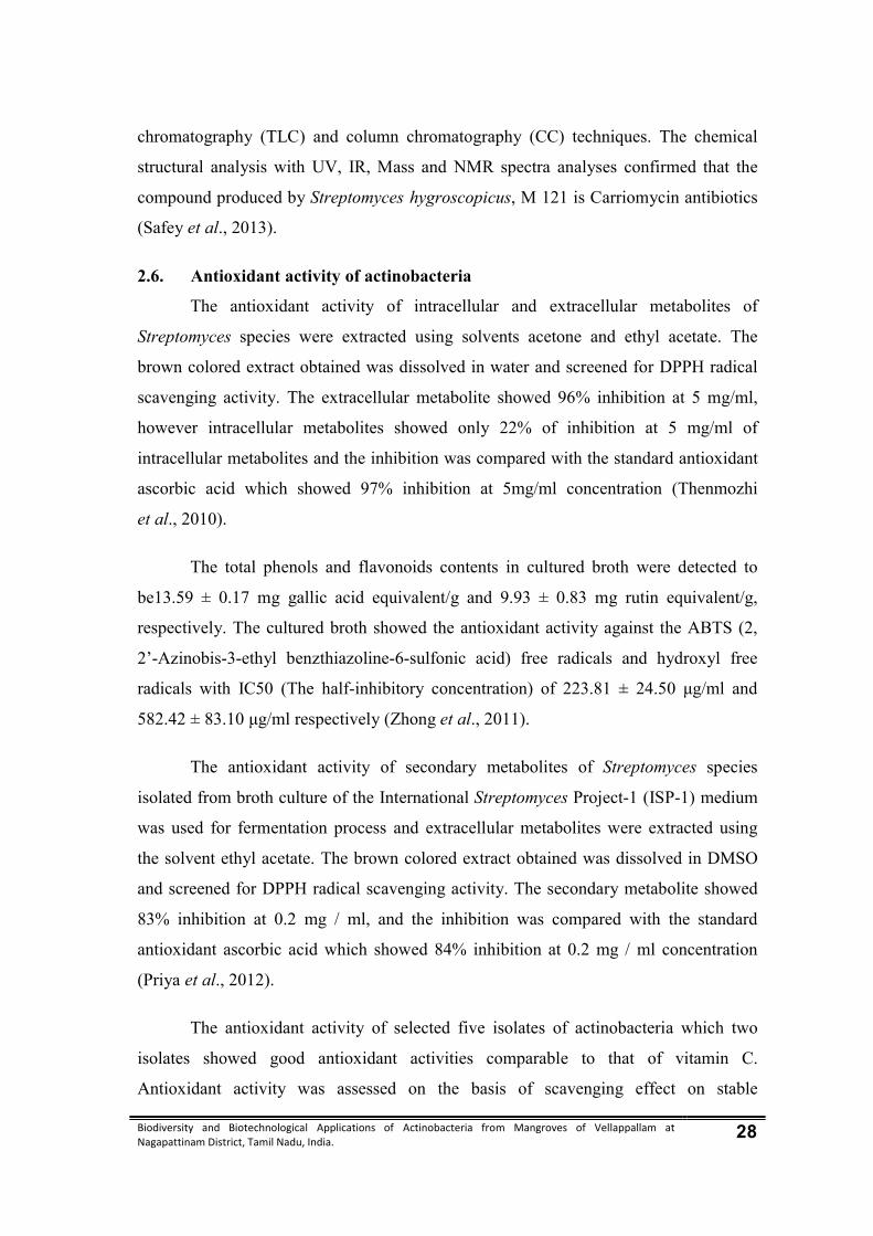
Biodiversity and Biotechnological Applications of Actinobacteria from Mangroves of Vellappallam at Nagapattinam District, Tamil Nadu, India. 28
chromatography (TLC) and column chromatography (CC) techniques. The chemical
structural analysis with UV, IR, Mass and NMR spectra analyses confirmed that the
compound produced by Streptomyces hygroscopicus, M 121 is Carriomycin antibiotics
(Safey et al., 2013).
2.6. Antioxidant activity of actinobacteria
The antioxidant activity of intracellular and extracellular metabolites of
Streptomyces species were extracted using solvents acetone and ethyl acetate. The
brown colored extract obtained was dissolved in water and screened for DPPH radical
scavenging activity. The extracellular metabolite showed 96% inhibition at 5 mg/ml,
however intracellular metabolites showed only 22% of inhibition at 5 mg/ml of
intracellular metabolites and the inhibition was compared with the standard antioxidant
ascorbic acid which showed 97% inhibition at 5mg/ml concentration (Thenmozhi
et al., 2010).
The total phenols and flavonoids contents in cultured broth were detected to
be13.59 ± 0.17 mg gallic acid equivalent/g and 9.93 ± 0.83 mg rutin equivalent/g,
respectively. The cultured broth showed the antioxidant activity against the ABTS (2,
2’-Azinobis-3-ethyl benzthiazoline-6-sulfonic acid) free radicals and hydroxyl free
radicals with IC50 (The half-inhibitory concentration) of 223.81 ± 24.50 μg/ml and
582.42 ± 83.10 μg/ml respectively (Zhong et al., 2011).
The antioxidant activity of secondary metabolites of Streptomyces species
isolated from broth culture of the International Streptomyces Project-1 (ISP-1) medium
was used for fermentation process and extracellular metabolites were extracted using
the solvent ethyl acetate. The brown colored extract obtained was dissolved in DMSO
and screened for DPPH radical scavenging activity. The secondary metabolite showed
83% inhibition at 0.2 mg / ml, and the inhibition was compared with the standard
antioxidant ascorbic acid which showed 84% inhibition at 0.2 mg / ml concentration
(Priya et al., 2012).
The antioxidant activity of selected five isolates of actinobacteria which two
isolates showed good antioxidant activities comparable to that of vitamin C.
Antioxidant activity was assessed on the basis of scavenging effect on stable

Biodiversity and Biotechnological Applications of Actinobacteria from Mangroves of Vellappallam at Nagapattinam District, Tamil Nadu, India. 29
1, 1-Diphenyl-2- Picryl Hydrazyl (DPPH) free radicals. The scavenging effect of crude
ethyl acetate extracts of the two isolates on DPPH radicals dose-dependently increased
and was found to be 71.77% and 77.52% (Spandana et al., 2012).
The in vitro antioxidant activity (total antioxidant activity, total reducing power,
and scavenging of hydrogen peroxide and nitric oxide radical scavenging activity of
ethyl acetate extracts) of seven isolates (KRCR1 to KRCR7) was studied. Extract of the
actinobacterium, KRCR1 showed maximum of total antioxidant activity (0.599), total
reducing power (0.15), and scavenging of hydrogen peroxide (80.7) and nitric oxide
radical scavenging activity (88.5) (Poongodi et al., 2012).
2.7. Anticancer activity of actinobacteria
The mechercharmycin “A” was isolated from marine-derived
Thermoactinomyces sp. YM3-251. The structure of mechercharmycin “A” was
determined by an X-ray crystallographic analysis to be cyclic peptide-like and bearing
four oxazoles and a thiazole. Mechercharmycin “B”, a linear congener of
mechercharmycin “A” was also isolated from the same bacterium. Mechercharmycin
“A” exhibited relatively strong antitumor activity, whereas mechercharmycin “B”
exhibited almost no such activity (Kanoh et al., 2005).
A novel cyclic peptide was isolated from the cultured mycelia of marine-
derived Thermoactinomycetaceae bacterium Mechercharimyces asporophorigenens
YM11-542. The peptide was purified by solvent extraction, silica gel chromatography,
ODS flash chromatography, and finally by preparative HPLC. Urukthapelstatin A dose-
dependently inhibited the growth of human lung cancer A549 cells with an IC50 value
of 12 nM. Urukthapelstatin A also showed potent cytotoxic activity against a human
cancer cell line panel (Matuo et al., 2007).
Several analogues of the cytotoxic thiopeptide IB-01211 or mechercharmycin A
have been synthesized. The cytotoxicity and the synthesized analogues were evaluated
against a panel of three human tumor cell lines. Thiopeptide 1 and the most active
derivatives 2 and 3 were chosen for further studies on effects on cell cycle progression
and induction of apoptosis. Interestingly, the inhibition of cell division and activation of
a programmed cell death by apoptosis were detected (Hernandez et al., 2008).

Biodiversity and Biotechnological Applications of Actinobacteria from Mangroves of Vellappallam at Nagapattinam District, Tamil Nadu, India. 30
The biological activity of asukamycin have been limited to its role as an
antibacterial and antifungal agent. By using five different tumor cell lines demonstrate
antineoplastic activity of asukamycin. It inhibited cell growth at concentrations similar
to other members of the manumycin family (IC50 1-5 μM). Cytotoxicity of asukamycin
was accompanied by activation of caspases 8 and 3 and was diminished by SB 202190,
a specific p38 mitogen-activated protein kinase (MAPK) inhibitor (Shipley et al.,
2009).
2.8. Silver nanoparticles from actinobacteria
Material Scientists are conducting research to develop novel materials with
better properties, more functionality and lower cost than the existing ones. Several
physical, chemical and biological synthesis methods have been developed to enhance
the performance of nanoparticles displaying improved properties with the aim to have a
better control over the particle size, distribution and morphology. Synthesis of
nanoparticles to have a better control over particles size, distribution, morphology,
purity, quantity and quality, by employing environment friendly economical processes
has always been a challenge for the researchers (Shankar et al., 2003).
The silver is a non-toxic, safe inorganic antibacterial agent being used for
centuries and is capable of killing about 650 microorganisms that cause diseases.
Silver has been described as being ‘oligodynamic’, that is, its ions are capable of
causing a bacteriostatic (growth inhibition) or even a bactericidal (antibacterial) impact.
Therefore, it has the ability to exert a bactericidal effect at minute concentration. It has
a significant potential for a wide range of biological application such as antibacterial
agents for antibiotic resistant bacteria, preventing infections, healing wounds and anti-
inflammatory (Rosi and Mirkin, 2005).
Studied the intracellular biosynthesis of silver nanoparticles by using
Streptomyces species was studied. The different species of Streptomyces viz.,
Streptomyces rameus NBR and Streptomyces sp. LD021 were exposed to aqueous
AgNO3 solution at 28ºC ± 0.5ºC for 72 hours under optimum conditions synthesizing
silver nanoparticles intracellularly, particle size is in the range of 12 nm to 22 nm
(Sapkal et al., 2009).

Biodiversity and Biotechnological Applications of Actinobacteria from Mangroves of Vellappallam at Nagapattinam District, Tamil Nadu, India. 31
Biosynthesis of silver nanoparticles from a novel actinobacteria strain
Streptomyces glaucus 71 MD isolated from a soy rhizosphere in Georgiais. The
appearance of yellowish brown color indicated the formation of silver nanoparticles.
The silver nanoparticles were spherical shaped and do not create big agglomerates by
SEM analysis (Tsibakhashvili et al., 2011).
The silver nanoparticles synthesized by using marine isolate Streptomyces
albidoflavus were studied. The produced particles were spherical shaped and
monodispersive in nature and showed a single surface plasmon resonance peak at 410
nm. FT-IR spectra of nanoparticles showed N-H, C-H, and C-N stretching vibrations,
denoting the presence of amino acid or peptide compounds on the surface of silver
nanoparticles produced by S. albidoflavus (Prakasham et al., 2012).
The biological synthesis of silver nanoparticles by Streptomyces cavourensis
was studied. The extracellular production of silver nanoparticles (AgNPs) confirmed by
color change and characteristic absorption spectra at 420 nm. The AgNPs were
determined to be spherical (20 - 70 nm) in shape. The isolated marine Streptomyces
cavourensis with potential to produce extracellular silver nanoparticles can be exploited
for bulk production, reproducible, monodispersive and spherical silver nanoparticles
(Subashini and Kannabiran, 2012).

Biodiversity and Biotechnological Applications of Actinobacteria from Mangroves of Vellappallam at Nagapattinam District, Tamil Nadu, India. 32
3. MATERIALS AND METHODS
3.1. Description of sampling sites
Vellappallam is one of the village in Thalanayar taluk in Nagapattinam District
in Tamil Nadu State. Vellappallam is located 7.9 km distance from its taluk main town
Thalanayar. Vellapallam is 29.8 km far from its District main city Nagapattinam. It is
286 km far from its state main city Chennai. It is located between latitude 10°55’289 N
and longitude 79°82’796 E. The population of the village is 5000 and almost all of
them depend on fishing (Fig. 1).
Fig 1. Map showing the sampling stations
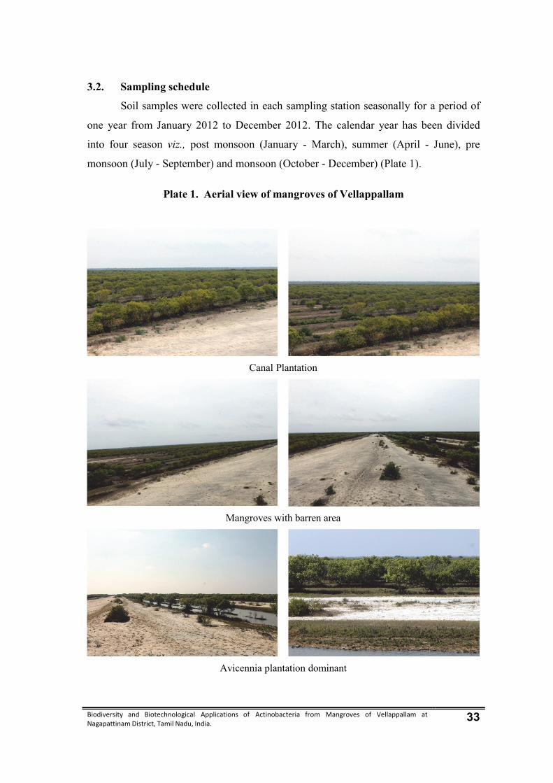
Biodiversity and Biotechnological Applications of Actinobacteria from Mangroves of Vellappallam at Nagapattinam District, Tamil Nadu, India. 33
3.2. Sampling schedule
Soil samples were collected in each sampling station seasonally for a period of
one year from January 2012 to December 2012. The calendar year has been divided
into four season viz., post monsoon (January - March), summer (April - June), pre
monsoon (July - September) and monsoon (October - December) (Plate 1).
Plate 1. Aerial view of mangroves of Vellappallam
Canal Plantation
Mangroves with barren area
Avicennia plantation dominant
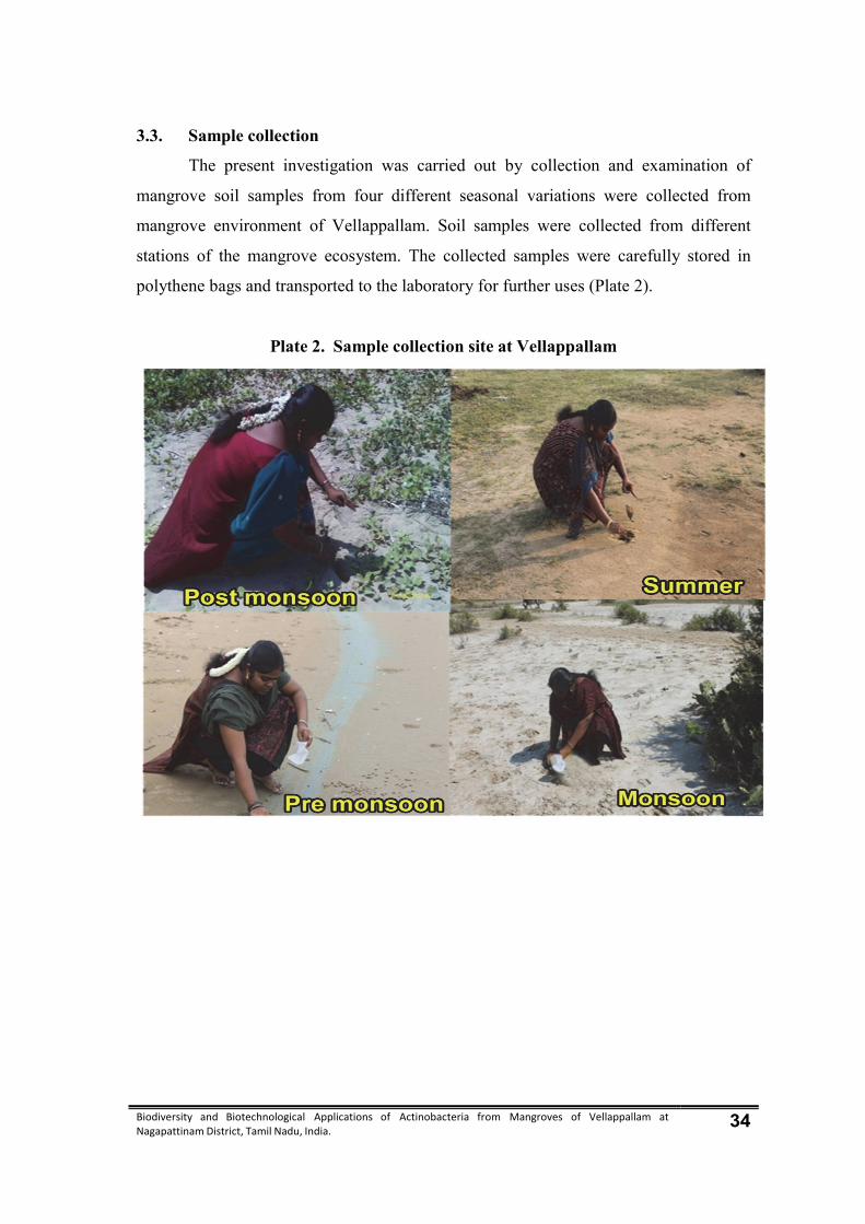
Biodiversity and Biotechnological Applications of Actinobacteria from Mangroves of Vellappallam at Nagapattinam District, Tamil Nadu, India. 34
3.3. Sample collection
The present investigation was carried out by collection and examination of
mangrove soil samples from four different seasonal variations were collected from
mangrove environment of Vellappallam. Soil samples were collected from different
stations of the mangrove ecosystem. The collected samples were carefully stored in
polythene bags and transported to the laboratory for further uses (Plate 2).
Plate 2. Sample collection site at Vellappallam
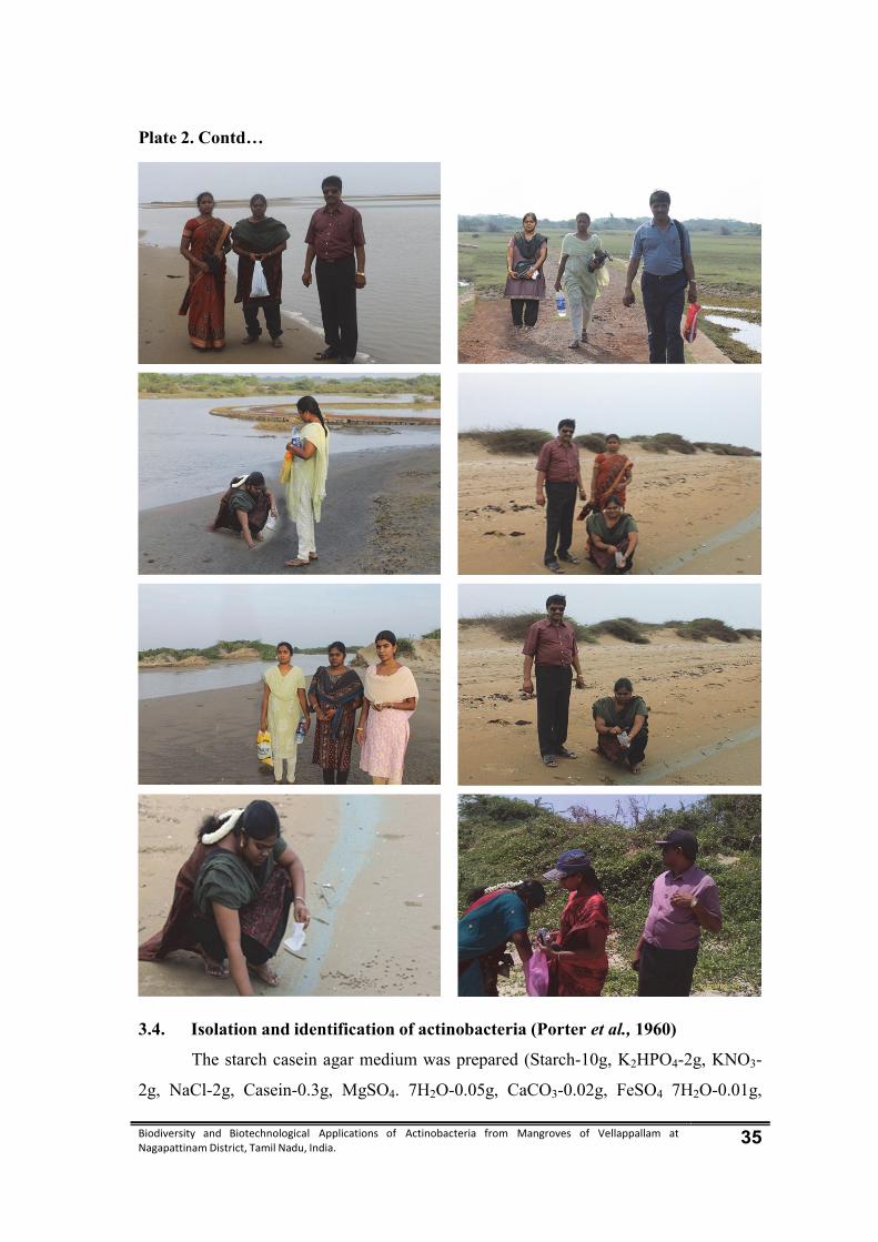
Biodiversity and Biotechnological Applications of Actinobacteria from Mangroves of Vellappallam at Nagapattinam District, Tamil Nadu, India. 35
Plate 2. Contd…
3.4. Isolation and identification of actinobacteria (Porter et al., 1960)
The starch casein agar medium was prepared (Starch-10g, K2HPO4-2g, KNO3-
2g, NaCl-2g, Casein-0.3g, MgSO4. 7H2O-0.05g, CaCO3-0.02g, FeSO4 7H2O-0.01g,

Biodiversity and Biotechnological Applications of Actinobacteria from Mangroves of Vellappallam at Nagapattinam District, Tamil Nadu, India. 36
Agar-20g, Water (50% sea water and 50% distilled water) -1000 ml, pH-7.2). The
medium containing flask was sterilized by using autoclave 121º C at 15 lbs for 15 min.
Isolation of actinobacteria was performed by plating technique using starch
casein agar medium (Kuster and Williams, 1964). Then medium was supplemented
with griseofulvin and streptomycin 50 µg/l to prevent the bacterial and fungal growth.
The medium was poured into the sterile petriplates. The collected soil samples were
diluted up to 10-6 and 0.1ml of the diluted samples (dilution factor - 10-6) was spread
over the agar plates. The inoculated plates were incubated at 28 ± 2°C for 7 - 10 days.
Three replicates were maintained for each dilution. After incubation, actinobacteria
colonies were observed and used for further investigation.
3.4.1. Purification of actinobacteria (Kokare et al., 2004)
Streak plate method was used to get pure colonies of actinobacteria. After
inoculation, the plates were incubated at 28 ± 2°C for 7 - 10 days and pure culture of
actinobacteria were maintained in starch casein agar slant and they were stored at 4°C
for further investigation.
3.4.2. Microscopic observation by coverslip culture technique (Pridham et al.,
1958)
Actinobacteria culture plates was prepared and 2-4 sterile coverslips were
inserted at an angle of 45°. The actinobacteria culture was slowly released at the
intersection of medium and coverslips. The plates were incubated at 28 ± 2°C for 4-8
days. The coverslips were removed and observed under the high power magnification.
The photomicrography was taken using Nikon microscope. The morphological features
of spores, sporangia, aerial and substrate mycelium were observed and recorded.
3.5. Analysis of physico-chemical characteristics of the soil (Jackson, 1973)
The sediment soil samples were collected in zip-lock polythene bags from
selected Study site for a period of one year. The collected sediment soil samples were
first air dried at room temperature, then crushed using a porcelain mortar and pestle and
then sieved for further analysis. The pH of the suspension was read using pH meter
(Systronics, India), to find out the soil pH. Electrical conductivity of soil was
determined in the filtrate of the water extract using Conductivity Bridge and Cation
Biodiversity and Biotechnological Applications of Actinobacteria from Mangroves of Vellappallam at Nagapattinam District, Tamil Nadu, India. 37
exchange capacity (CEC) of the soil was determined by using 1 N ammonium acetate
solution as described by Jackson (1973).
Organic carbon content was determined by adopting chromic acid wet digestion
method as described by Walkley and Black (1934), available nitrogen was estimated by
alkaline permanganate method as described by Subbiah and Asija (1956) and available
phosphorus by Brayl method as described by Bray and Kutz (1945). Available
potassium was extracted from soil with neutral 1 N ammonium acetate (1:5) and the
potassium content in the extract was determined by using flame photometer, calcium
(Neutral 1 N NH4 OAC extractable 1:5) was extracted with neutral 1 N ammonium
acetate and the available calcium in the extract was determined by Versenate method
(Jackson, 1973). Other nutrient based parameters i.e. available phosphate and total
nitrogen were estimated using standard methods of APHA (1998).
Available micronutrients such as Zn, Cu and Mn were determined in the
diethylene triamine penta acetic extract of soil using Perkin-Elmer model 2280 Atomic
Absorption Spectrophotometer. Other nutrients such as magnesium, sodium and
available iron were analyzed following the method of Muthuvel and Udayasoorian
(1999). The AR grade reagents and double distilled water were used for preparation of
solutions.
3.6. Statistical analysis
Pearson’s correlation analysis was used to assess the relationship between
physico-chemical parameters and total actinobacteria colonies. The data were computed
and analyzed using Statistical Package for Social Sciences (SPSS) software.
3.7. Characterization and Identification of Actinobacteria
3.7.1 Morphological characterization
Morphological characterization was performed with a magnified lens on
actinobacteria strains were grown for 3 to 14 days on starch casein agar plate. Colony
morphology was recorded with respect to colour, aerial mycelium, size, nature of
colony, reverse side colour and pigmentation.
Biodiversity and Biotechnological Applications of Actinobacteria from Mangroves of Vellappallam at Nagapattinam District, Tamil Nadu, India. 38
3.7.2 Light Microscopy
A cover slip culture technique was adopted for light microscopic studies
(Pridham et al., 1958). Actinobacteria culture plate was prepared and 5 to 6 sterile
cover slips were placed at an angle of 45°. The actinobacteria culture was slowly
released at the intersection of medium and cover slip. The plates were incubated at
28±2°C for 4-8 days. Afterwards, the cover slips were removed and observed under the
high power magnification. The morphological features of spores, sporangia and aerial
and substrate mycelium were observed and recorded.
3.7.3 Biochemical characterization (Pridham and Gottilibe, 1948)
3.7.3.1 Diffusible pigment test
The culture were inoculated in glycerol-asparagine agar medium and incubated
at 28±2ºC for 14 days. The formation of colour such as yellow-brown, blue, green, red,
orange, ash and grey to violet was recorded.
3.7.3.2 Melanin pigment production test
Melanin production was considered to cause browning of organic media
containing tyrosine and it was carried out with tyrosine agar medium. The agar medium
was transferred in to test tube, sterilized and made into slants. The slants were
inoculated with active cultures and incubated at 28±2ºC. After 2-4 days, the production
of soluble pigments and the colour of the vegetative and aerial mycelium in the slants
were observed.
3.7.3.3 Indole test
Peptone broth was prepared and the actinobacteria cultures were inoculated.
After incubation the indole production was tested with Kovac’s reagent. Red colour
ring formation indicated positive reaction where as yellow colour ring indicated
negative result.
3.7.3.4 Methyl red test
The actinobacteria cultures were grown in MR-VP broth and after the
incubation it was added with methyl red indicator. Red colouration of the broth
indicated positive reaction. Yellow colour development indicated negative result.
Biodiversity and Biotechnological Applications of Actinobacteria from Mangroves of Vellappallam at Nagapattinam District, Tamil Nadu, India. 39
3.7.3.5. Voges proskauer test
The actinobacteria cultures were grown in MR-VP broth and after overnight
incubation, it was added with Barrit’s reagent A and B. Development of red colour
indicated positive reaction and the yellow colour development indicated negative result.
3.7.3.6. Citrate utilization test
Sterile Simmon’s Citrate agar slants were streaked with the actinobacteria
cultures and incubated at 28±2ºC for 4 days. Change in colour from green to blue
indicated positive reaction. No colour change indicated negative result.
3.7.3.7. Nitrate reduction test
Nitrate broth was prepared and dispensed into test tubes. The test tubes were
sterilized and one loop full of cultures were inoculated and incubated at 28±2ºC for 4
days. After incubation, few drops of alpha naphthalamine and sulphanilic acid were
added. The red colour formation indicated positive result.
3.7.3.8. Urease test
Sterile Christensen’s urea slants were streaked with the actinobacteria cultures
and incubated at 28±2ºC for 4 days. Change in colour from yellow to pink indicated
positive reaction.
3.7.3.9. Catalase test
A clear slide was taken and drop of culture suspension was placed on it. Few
drops of hydrogen peroxide was added to the cultures. The evolution of air bubbles
from the suspension indicated a positive reaction.
3.7.3.10. Oxidase test
The cultures were rubbed over the filter paper containing a reagent N-N
tetramethyl paraphenylene diamine dihydrochloride. Purple colour indicated positive
result.
3.7.3.11. Starch hydrolysis
Nutrient starch agar plates were prepared and sterilized. The plates were
inoculated with the actinobacteria isolates as a single line streaks and incubated at
28±2ºC for 7 days. A positive reaction was indicated by the formation of zone of

Biodiversity and Biotechnological Applications of Actinobacteria from Mangroves of Vellappallam at Nagapattinam District, Tamil Nadu, India. 40
clearance of the medium around the colonies, which was further visualized by adding
Lugol’s iodine.
3.7.3.12. Casein hydrolysis
Skim milk agar plates were prepared and sterilized. Then the medium was
poured in to petriplates. After solidification, the cultures were streaked as a single line
and incubated. The formation of zone of clearance around the colonies indicated the
positive result.
3.7.3.13. Lipid hydrolysis
Spirit blue agar was prepared and tributyrin was added as the substrate for
lipase activity. The substrate mixture was homogenized in the magnetic thermal stirrer
and sterilized. The medium was then inoculated with the actinobacteria isolates in a
zigzag manner and incubated. A positive lipase activity was determined by the
reduction of dye around the colonies.
3.8. Screening of actinobacteria for antibacterial efficacy (Liu et al., 2006)
The selected actinobacteria were screened for antibacterial efficacy by agar well
diffusion method. The five Gram positive bacteria (Bacillus subtilis MTCC 739,
Enterobacter aerogenes MTCC 1272, Enterococcus faecalis MTCC 1144,
Staphylococcus aureus MTCC 1688 and Streptococcus pyogenes MTCC 1425) and five
Gram negative bacteria (Escherichia coli MTCC 109, Klebsiella oxytoca MTCC 2658,
K. pneumoniae MTCC 1023, Salmonella typhi MTCC 6633, and Vibrio cholerae
MTCC 1037) were obtained from Microbial Type Culture Collection (MTCC),
Chandigarh, India.
3.8.1. Mass production, extraction of antibacterial compound from
actinobacteria isolate (Bredholt et al., 2008)
The conical flask was taken and 150 ml of Starch casein broth was prepared in
the conical flask. The selected actinobacteria cultures were inoculated in each of the
conical flasks separately and incubated at 37°C for 7 - 10 days. After incubation, the
actinobacteria biomass were taken from each of the flask and put into the each of the
beakers. To this, each of the solvents (diethyl ether, ethyl acetate and distilled water)
Biodiversity and Biotechnological Applications of Actinobacteria from Mangroves of Vellappallam at Nagapattinam District, Tamil Nadu, India. 41
was added separately, crushed and centrifuged at 10,000rpm for 15 mins. The
actinobacteria biomass extracts were tested against human pathogenic bacteria.
3.8.2. Antibacterial Assay
Diethyl ether, ethyl acetate and distilled water extracts were tested for their
antibacterial efficacy against the bacterial pathogens.
All the ingredients were weighed and put into the conical flask containing 1000
ml distilled water. The flask was sterilized by using an autoclave at 121°C for 20 min
at15 lbs pressure. The nutrient agar medium (Beef extract - 3 gms, Peptone - 5 gms,
Sodium chloride - 5 gms, Agar - 15gms, Distilled water – 1000 ml, pH – 7) was poured
into the sterile petriplates and allowed to solidify. The test bacterial cultures were
evenly spreaded over the media by sterile cotton swabs. Then wells (6 mm) were made
in the medium using sterile cork borer. 200 µl actinobacterial extracts were transferred
into the separate wells. The standard antibiotics (Ampicillin, Streptomycin and
Tetracycline) and solvents (diethyl ether, ethyl acetate and distilled water) were used as
positive and negative controls respectively. Then the plates were incubated at 37ºC for
24 hrs. After the incubation the plates were observed for the formation of clear
inhibition zone around the well indicated the presence of antibacterial activity. The
zone of inhibition was calculated by measuring the diameter of the inhibition zone
around the well.
3.9. Molecular characterization of actinobacteria
3.9.1. Isolation of chromosomal DNA (Wilson, 1990)
Isolates of actinobacteria were grown upto the late exponential phase in starch
casein broth at 28±2°C, washed twice with Tris EDTA buffer. Chromosomal DNA was
isolated by resuspending 0.5-1.0g of cells with 5ml lysis buffer (25 mM Tris; 25 mM
EDTA, pH 8.0; 10 – 15 µg lysozyme and 50 µg/ml Rnase) and incubated for 30 – 80
min at 37°C, followed by the addition of 500µl of 5 M NaCl solutions. The suspension
was agitated on a vortex mixer until the cell suspension became translucent. Cells were
lysed by the addition of 1.2 ml of 10% SDS. The lysates were incubated for 15-30 min
at 65°C. After addition of 2.4 ml of 5 M potassium acetate, the solution was mixed and
kept on ice for 20 min. The precipitate was removed by centrifugation for 30 min at
Biodiversity and Biotechnological Applications of Actinobacteria from Mangroves of Vellappallam at Nagapattinam District, Tamil Nadu, India. 42
6,000 rpm and the volume of the supernatant was adjusted to 8ml. The DNA was
recovered by precipitation with two volumes of isopropanol. The precipitate was
dissolved in 700 µl/g 50 mM Tris/10 mM EDTA (pH 8.0). Any insoluble substances
were spun off and the aqueous phase was transferred to a 1.5ml microfuge tube.
Subsequently, 75µl 3M sodium acetate and 500µl isopropanol were added and the
solution was centrifuged for 30 seconds to 2 min. The precipitate was washed with cold
70% ethanol, dried and dissolved in 100 µl TE (10 mM Tris/1 mM EDTA, pH 8.0).
3.9.2. Amplification of 16S rRNA gene in actinobacteria chromosomal DNA
(Hastono et al., 2009)
Microbial 16S rRNA gene was amplified from the extracted genomic DNA
using the following universal eubacterial 16S rRNA gene primers:
forward primer 5'-AGAGTTTGATCCTGGCTCAG-3' and reverse primer
5' ACGGCTACCTTGTTACGACTT-3' PCR was performed in a 50μl reaction mixture
containing 2μl (10mg) of DNA as the template, each primer at a concentration of
0.5mM, 1.5mM MgCl2 and each deoxynucleoside triphosphate at a concentration of
50mM, as well as Taq polymerase. PCR conditions consisted of an initial denaturation
for 3 minutes at 94oC, 37 cycles consisting of denaturation at 94oC for 1minute,
annealing at 55oC for 1min. and extension at 72oC for 2 minutes, and a final extension
step of 5 minutes at 72oC were carried out (Mastercycler Personal, Eppendorf,
Germany). The amplification of 16S rRNA gene was confirmed by running the
amplification product in 1.5% agarose gel in 1xTris-Borate- EDTA buffer and purified
by using Q1A quick PCR clean up kit with the protocol suggested by Qiagen Inc. The
complete 16S rRNA gene was sequenced by using PCR products directly as sequencing
template with above mentioned primers. All sequencing reactions were carried out with
ABI 377 Automated DNA sequence.
3.9.3. Sequencing of 16S rRNA gene
The amplified PCR products were purified using a QIA quick PCR purification
kit (Qiagen GmBh, Germany) as recommended by the manufacturer. The sequences of
the PCR products were determined by using the Big Dye Terminator Cycle Sequencing
v2.0 kit on an ABI310 automatic DNA sequence (Applied Biosystems, CA and USA)
Biodiversity and Biotechnological Applications of Actinobacteria from Mangroves of Vellappallam at Nagapattinam District, Tamil Nadu, India. 43
according to the manufacturer’s instructions. The determined 16S rRNA gene
sequences were deposited in the GenBank database.
3.9.4. Phylogenetic analysis (Tamura et al., 2007)
The sequences of 16S rRNA gene of potential actinobacteria isolate were
compared against the sequences available from GenBank using the BLAST program
and were aligned using CLUSTAL W software developed by Higgins et al. (1992).
Phylogenetic analysis was constructed using the Neighbour-joining method (Saitou and
Nei, 1987). Bootstrap analysis was done based on 1000 replications (Felsenstein,
1985). All these analysis were performed by MEGA4 package.
3.9.5. Restriction site analysis in 16S rRNA gene
The restriction sites in 16S rRNA gene of potential actinobacteria isolate were
analyzed by using NEB cutter program version 2.0 tools in online
(www.neb.com.NEBCutter2/index.php).
3.9.6. Secondary structure prediction in 16S rRNA gene
The secondary structure of 16S rRNA gene in actinobacteria was predicted
using gene bee tool (www.genebee.msu.su/service/ma2-reduced.html).
3.10. Separation of bioactive compounds from actinobacteria (Wagner, 1995)
The separation of the bioactive compounds from the crude extracts of selected
actinobacteria was performed by TLC.
3.10.1. Thin Layer Chromatography (TLC)
The stationary phase (silica gel) was prepared as slurry with water (or) buffer at
1:2 ratio (w/v) and it was applied to a glass plate. The thickness for analytical
separation was 0.2mm and 2.5mm for preparative separations.
Calcium sulfate (CaSO4 2H2O) (Gypsum) (10.15%) was incorporated to the
adsorbent it was a binder, as it facilitates the adhension of the adsorbent to the plate.
After application of adsorbent, the plates were air dried for 10-15mins. The process is
also known as activation of the adsorbent. The plates could be used immediately or
stored in desicators.
Biodiversity and Biotechnological Applications of Actinobacteria from Mangroves of Vellappallam at Nagapattinam District, Tamil Nadu, India. 44
3.10.2. Preparation of Samples
Alkaloids (Wagner and Bladt, 1996)
About two gram of actinobacteria biomass was extracted with equal volume of
petroleum ether 40-60oC and shake vigorously. The ether fraction was evaporated to
dryness under vaccum using either a water pump or rotary evaporator at 40oC and was
dissolved in a known volume of absolute ethanol for analysis.
Flavonoids (Wagner and Bladt, 1996)
About two gram of actinobacteria biomass was extracted with 10ml of ethanol.
Then the plate was heated for few min and 100 µl of filtrate was applied on the silica
gel plates.
Phenols (Harborne, 1998)
About two gram of actinobacteria biomass was extracted with 10ml methanol or
rotary shaker (180 thaws / min) for 24 hours. Then these extract was filtered by using
Whatmann No.1 filter paper. The condensed filtrate was used for TLC.
Saponins (Wagner and Bladt, 1996)
About two gram of actinobacteria biomass was extracted with 10ml of 70%
ethanol by refluxing for 10 min. Then these extract was filtered by using Whatmann
No.1 filter paper. The filtrate was condensed, enriched with saturated n-butanol, and
thoroughly mixed. The butanol was retained, condensed and used for thin layer
chromatography
Sterols (Wagner and Bladt, 1996)
About two gram of actinobacteria biomass was extracted with 10ml of methanol
was kept in water bath at 80oC/15min. The condensed filtrate was used for TLC.
3.10.3. Sample application
Drawn a line lightly with a pencil about 1.5-2.0 cm from the bottom. If the thin
layer is too soft to draw a pencil line place a scale at the bottom and mark a spot at a
distance of 1cm, the samples were spotted using capillary tubes at 1.5cm distance
between them. For preparative TLC, the samples were applied as a band across the
layer, rather than as a spot.
Biodiversity and Biotechnological Applications of Actinobacteria from Mangroves of Vellappallam at Nagapattinam District, Tamil Nadu, India. 45
3.10.4. Solvent preparation
Alkaloids (Wagner and Bladt, 1996)
Alkaloids were separated by using chloroform, methanol and water (4:3:2)
solvent mixture.
Flavonoids (Wagner and Bladt, 1996)
Flavonoids were separated by using butanol, acetic acid and water (4:1:5)
solvent mixture.
Phenols (Harborne, 1998)
The phenols were separated by using chloroform and methanol (27:3) solvent
mixture.
Saponins (Wagner and Bladt, 1996)
The saponins were separated by using chloroform, glacial acetic acid, methanol
and water in the ratio of 64:34:12:8 solvent mixtures.
Sterols (Wagner and Bladt, 1996)
The sterols were separated by using acetone, glacial acetic acid, methanol and
water (64:34:12:8) solvent mixture.
3.10.5. Plate development
The chromatographic tank was filled in with developing solvent to a depth of
1.5cm and equilibrated for about an hour. The thin layer plate was placed gently in the
tank and allowed to stand for about 60 min. The separation of the compound occurs as
the solvent moves upward. As the solvent reached about 1.2cm from the top of the
plate was removed, solvent front was marked with a pencil immediately and allowed to
air dry placing the plate upside down.
3.10.6. Component detection
Several methods were available to detect the separated compounds. Different
types of specifying reagents were used to detect the different compounds.

Biodiversity and Biotechnological Applications of Actinobacteria from Mangroves of Vellappallam at Nagapattinam District, Tamil Nadu, India. 46
Alkaloids
The presence of alkaloid in the chromatogram was detected by spraying the
freshly prepared Wagner’s reagents (0.67g iodine and 1g of potassium iodine were
dissolved in 2.5 ml of distilled water). Formation of brown spot indicated as positive
reactions.
Flavonoids
The presence of flavonoid in the developed chromatogram was detected by the
formation of yellow colour spot.
Phenols
The presence of phenol in the developed chromatogram was detected by
spraying the Folin - ciocalteu’s reagent. The plates were heated at 80oC for 10 min. A
positive reaction was indicated by the formation of blue colour spot.
Sterols
The presence of sterol in the developed chromatogram was detected by spraying
the Folin - ciocalteu’s reagent. The plates were heated at 80oC for 10min. A positive
reaction was indicated by the formation of blue spots.
Saponins
The presence of saponins in the developed chromatograms was detected by
iodine vapours. A positive reaction was formation of yellow colour spot.
3.10.7. Determination of Rf value
The Rf values of the various bioactive compounds were calculated using the
following formula.
Distance travelled by solute (Measured to centre of the spot) Rf = –––––––––––––––––––––––––––––––––––––––––––––––––––
Distance travelled by solvent
3.10.8. Purification of bioactive compounds (Reynolds and Dweck, 1999)
TLC using various solvent systems to separate bioactive compounds in the
ethanolic and methanolic extract into visible fractions with retention factor value. The
fraction plus the origin were purified from the developed duplicate plate by collecting
Biodiversity and Biotechnological Applications of Actinobacteria from Mangroves of Vellappallam at Nagapattinam District, Tamil Nadu, India. 47
the silica placing it into a polypropylene test tube and resolving the fraction in
chloroform: methanol (1:1). Centrifuged to remove silica particles and the supernatant
was collected for further antibacterial activity.
3.10.9. Screening for antibacterial potentials of isolated bioactive compounds from
actinobacteria
The isolated bioactive compounds such as alkaloids, flavonoids, phenols,
saponins and sterols were purified and screened for antibacterial activity by agar well
diffusion method.
Sensitivity test of ten different pathogenic bacterial strains to various bioactive
compounds extract was measured in turns of zone of inhibition using agar well
diffusion assay. The petriplates were washed and placed in an autoclave for
sterilization. After sterilization, nutrient agar medium was poured into each sterile
petriplate and allowed to solidify in a laminar air flow chamber. After solidification,
using a sterile cotton swabs, fresh bacterial culture with known population count was
spread over the plate by spread plate technique.
One well of 6 mm size made in the agar plates with the help of sterile cork
borer, the wells were loaded with 200 µl of bioactive compounds extracts. All the
plates were incubated at 37°C for 24 hours. After incubation, the plates were observed
for formation of clear inhibition zone around the well indicated the presence of
antibacterial activity. The zone of inhibition was calculated by measuring the diameters
of the inhibition zone around the well.
3.11. UV –Visible spectroscopic analysis of bioactive compounds (Hasegawa et al.,
1983)
Fractionated bioactive compounds were dissolved in acetonitrile and then
detected UV absorption values with lambda 35 ultra violet scanners.
3.11.1. Fourier Transform - Infrared (FT – IR) analysis of bioactive compounds
(Naumann et al., 1991)
TLC fractions that showed antibacterial activity were further subjected to
spectroscopic analysis for identification of the functional groups in the bioactive
Biodiversity and Biotechnological Applications of Actinobacteria from Mangroves of Vellappallam at Nagapattinam District, Tamil Nadu, India. 48
compounds. A known weight of TLC‐purified fractions (1 mg) was taken in a mortar
and pestle and ground with 2.5 mg of dry potassium bromide (KBr). The powder so
obtained was filled in a 2 mm internal diameter micro‐cup and loaded onto FT - IR set
at 26°C ± 1°C. The samples were scanned using infrared in the range of 4000–400 cm‐1 using Fourier Transform Infrared Spectrometer (Thermo Nicolet Model‐6700). The
spectral data obtained were compared with the reference chart to identify the functional
groups present in the sample.
3.12. Screening for antioxidant activity of actinobacteria (Ravikumar et al.,
2008)
3.12.1. Sample preparation
Each culture was incubated for 7 days and filtered to separate the actinobacteria
biomass. The biomass were air dried and then extracted with 50 ml of methanol,
ethanol and distilled water for 3 days to give the biomass extract. The extracted
solution was evaporated under reduced pressure by rotary evaporator to give 2 ml of
concentrate.
3.12.2. Assay for 2, 2-Diphenyl-1-pycrylhydrazyl (DPPH) free radical scavenging
activity
The control, standard and samples (each extract of mycelium) were individually
added to 3 ml of 0.004% MeOH solution of DPPH. Absorbance at 517 nm was
measured under constant mixing at room temperature. After 30 min and percent
inhibitory activity (free radical scavenging activity) was calculated.
Percentage of free radical scavenging activity = [(A0–A1)/A0] x100
=
A0 is the absorbance of the control (MeOH ).
A1 is the absorbance of the samples or standard (in MeOH).
The standards used for positive DPPH free radical scavengers were ascorbic acid and
selenium at 1 mg/ ml-1.
Biodiversity and Biotechnological Applications of Actinobacteria from Mangroves of Vellappallam at Nagapattinam District, Tamil Nadu, India. 49
3.12.3. Determination of total phenolic content (Woisky and Salatino, 1998)
Total phenolic content (TPC) of actinobacteria extracts were estimated using the
Folin-Ciocalteu colorimetric method. Briefly, the appropriate dilutions of the sample
(0.5 mL) were oxidized with 2.5 ml Folin-Ciocalteu reagent (1:10) for 3-8 minutes at
room temperature. Then the reaction was neutralized with 4% saturated sodium
carbonate. The absorbance of the resulting blue colour was measured at 740 nm after
incubation for 2 hours at room temperature in darkness. Gallic acid was employed as
the standard. All tests were carried out in triplicate and results were expressed as gallic
acid equivalent (GAE), i.e., mg gallic acid/100 ml culture or g gallic acid/100 g
Distilled Water.
3.13. Submerged fermentation and Mechercharmycin isolation from
Thermoactinomyces vulgaris DKP01 (Matuo et al., 2006)
The Thermoactinomyces vulgaris DKP01 was carried out in different
modifications of a P2 medium (10-30 g of Protein, 1.0 g of yeast extract, 1.0 g of
sucrose and 0.1 g of Fe·citrate-nH2O in 75% seawater at pH 7.6). Each culture was
incubated at 30°C for 5-14 days on a rotary shaker at 100 rpm. In most cases, 100-ml
flasks containing 40 ml of the medium or 1000-ml baffled flasks (Shibata Scientific
Technology) containing 500 ml of the medium were used. The starch casein broth
(SCB) was also prepared for screening the test actinobacteria for Mechercharmycin
production. The discs of 3 agar plugs (5 mm diameter) containing biomass were used as
inoculum. The organisms were grown at 24±2°C under still condition for 3 weeks in a
light chamber with 16 h of light, followed by 8 h of dark cycles. The blank cultures
(uninoculated sterile medium) were also maintained. After 3 weeks, the culture fluid
was passed through four layers of cheese cloth to remove solids. Extra-cellular
Mechercharmycin was extracted from the culture medium by using dichloromethane.
The solvent was then removed by evaporation under reduced pressure at 35°C in a
rotary vacuum evaporator. The solid residue was dissolved in 1 ml of dichloromethane
and placed on a 1.5×30 cm column of silica gel (40 μ). Elution of the column was
performed in a step-wise manner starting with 70 ml of 100% dichloromethane,
followed by methylene chloride:ethylacetate at different proportions (viz., 20:1 v/v,
10:1 v/v, 6:1 v/v, 3:1 v/v, 1:1 v/v). Fractions with same mobility as the standard
Mechercharmycin were combined and evaporated to dryness. The residue was
Biodiversity and Biotechnological Applications of Actinobacteria from Mangroves of Vellappallam at Nagapattinam District, Tamil Nadu, India. 50
subjected to chromatographic and spectroscopic analyses. The solvents used for the
analyses were high performance liquid chromatography (HPLC).
3.14. Chromatographic and spectroscopic analyses
The thin layer chromatographic (TLC) analysis was carried out on Merck 1 mm
(20×20 cm) silica gel pre-coated plate developed in a solvent A, Chloroform:Methanol,
(7:1, v/v) followed by solvent B, Chloroform:Acetonitrile (7:3, v/v); solvent C, Ethyl
acetate:2-propanol (95:5, v/v); solvent D, Methylene chloride:Tetrahydrofuran (6:2,
v/v); solvent E, Methylene chloride:Methanol:Dimethylformamide (90:9:1, v/v/v),
respectively. Mechercharmyces was detected with 1% (w/v) vanillin in sulphuric acid
reagent after gentle heating (Cardellina, 1991). It appeared as a bluish spot fading to
dark grey after 24 h. The RF values of the samples were calculated and compared with
authentic Mechercharmyces. The area of the plate containing putative
Mechercharmyces was carefully removed by scraping off the silica and eluted with
acetonitrile. The ultra-violet (UV) absorption of the samples was carried out with
methanol at 273 nm (Wani et al., 1971) in a Backman DU-40 spectrophotometer.
Samples were ground with infra-red (IR) grade potassium bromide pressed into discs
under vacuum using spectra lab pelletiser and the spectrum was recorded in a Bruker
Optics Vertex 80v FT-IR spectrometer.
Study on HPLC was conducted on HP1100 series using C18 reverse phase
column (Alltech Econosil, 250 mm×4.4 mm×10 μm) with an isocratic mobile phase
consisting of methanol: water (80:20) at flow rate of 1 ml/min. Each sample of 10 μl
was injected with the help of a micro syringe. Registration of peak and retention time
was recorded on UV at 320 nm. Based on the HPLC analysis, Mechercharmyces was
quantified by comparing the peak area of the samples with that of the
Mechercharmyces standard.
Standard concentration total area of sample peak
Amount of Mechercharmycin = –––––––––––––––––––––––––––––––––––––––––––––––– Total area of the standard peak (authentic compound)
Biodiversity and Biotechnological Applications of Actinobacteria from Mangroves of Vellappallam at Nagapattinam District, Tamil Nadu, India. 51
3.15. Screening of anticancer activity of Mechercharmycin isolated from
Thermoactinomyces vulgaris DKP01
3.15.1. Experimental protocol
For the in vivo studies the experimental animals were divided into four groups,
each group consists of six animals.
Group 1 - Control rats fed with standard diet and water.
Group 2 - Mechercharmycin - alone treated rats (Rats were orally given
(8mg / kg body weight) daily once a day for 16 weeks.
Group 3 - DEN Induced group rats received single dose of DEN (ip 100
mg/ kg) & two weeks after Phenobarbital (Oral 250 mg/ kg) was
given to rats for 8 weeks for enhancing the cancer development.
Group 4 - Rats received single dose of DEN and / kg) & two weeks after
Phenobarbital (Oral 250 mg/ kg) was given to rats for 8 weeks.
Then, rats were treated with Mechercharmycin (8mg / kg body
weight) and continued till the end of the experimental period.
The induction processes were terminated after 16 weeks and all the animals
were killed by cervical dislocation after an overnight fasting. The blood and tissue
sample was collected for clinical and histopathological analysis.
3.15.2. Estimation of biochemical parameters in experiment animal blood
Estimation of protein
Protein was estimated by the method of Lowry et al., (1951).
Reagents
1. Alkaline copper reagent
Solution A: 2% sodium carbonate in 0.1 N NaOH
Solution B: 0.5% copper sulphate in 1% sodium potassium tartarate
50ml of solution A was mixed with 1ml of solution B just before use.
2. Folin's phenol reagent (commercial reagent, 1:2 dilutions)
3. Bovine serum albumin (BSA)
Biodiversity and Biotechnological Applications of Actinobacteria from Mangroves of Vellappallam at Nagapattinam District, Tamil Nadu, India. 52
Procedure
To 0.1 ml of serum, 0.9 ml of water and 4.5 ml of alkaline copper reagent were
added and kept at room temperature for 10 min. To this 0.5 ml of Folin’s reagent was
added and the blue colour developed was read after 20 min at 640 nm. Protein level
was expressed as mg/ml for serum.
Estimation of blood glucose
Blood glucose level was estimated by the method of Sasaki and Matui, (1972). To
0.1 ml of blood, 1.9 ml of 10% TCA solution was added to precipitate proteins and then
centrifuged. One ml of the supernatant was mixed with 4.0 ml of O-toluidine reagent and
was kept in a boiling water bath for 15 minutes. The green colour developed was read at
600 nm in a Shimadzu spectrophotometer. A series of standard glucose solutions
(1 mg/ml) were also treated similarly. The values were expressed as mg of glucose/dl of
whole blood.
Estimation of total Bilurubin (Autozyme kit)
To 50 µl of serum, 1000 µl of total bilurubin reagent and 20 µl of respective
activator reagent were added. The reaction mixture were mixed well and incubated for
10 minutes at 370C. At the same time, blank and standard solution was prepared. The
absorbance of sample against reagent blank was read at 546 nm. The activity was
calculated by using the formula:
Total bilurubin = O.D. of sample – O.D. of blank / O.D. of standard × 10
Estimation of total Albumin (Autozyme kit)
To 0.01 ml of serum, 1.0 ml of working solution was added and incubated the
assay mixture for 1minute at 37o C. After completion of incubation period, the
absorbance was measured at 600 nm. The activity was calculated by using the formula
Total albumin in g % = Absorbance of sample/Absorbance of standard x 5.
Estimation of total Cholesterol
The amount of Total Cholesterol was estimated according to the method of
Parekh and Jung, (1970).

Biodiversity and Biotechnological Applications of Actinobacteria from Mangroves of Vellappallam at Nagapattinam District, Tamil Nadu, India. 53
Reagents
Stock ferric chloride: 840 mg of pure dry ferric chloride was weighed and
dissolved in 100 ml of glacial acetic acid.
Ferric chloride precipitating reagent: 10 ml of stock ferric chloride reagent was
taken in 100 ml of standard flask and made up to the mark with pure glacial
acetic acid.
Ferric chloride diluting reagent: 8.5 ml of stock ferric chloride was diluted to
100 ml with pure glacial acetic acid.
Standard cholesterol solution: 100 mg of cholesterol was dissolved in 100 ml
with glacial acetic acid. The concentration of working standard is 100 μg /ml.
Working standard: 10 ml of stock was dissolved in 0.85 ml of stock ferric
chloride reagent and made up to 100 ml with glacial acetic acid. The
concentration of working standard is 100 μg/ml.
Procedure
To 0.1ml of serum, 4.9 ml of ferric chloride precipitating reagent was added
and centrifuged with 2.5 ml of supernatant and 2.5 ml of ferric chloride diluting
reagent. Then 4.0ml of concentrated sulphuric acid was added. The blank was prepared
simultaneously by taking 5.0 ml of diluting reagent and 4.0 ml of concentrated
sulphuric acid. A set of standards (0.5 - 2.5 ml) were taken and made up to 5.0 ml with
FeCl2 diluting reagent. Then 4.0 ml of con. H2SO4 was added. After 30 min, the
intensity of the colour developed was read at 540 nm against reagent blank.
The amount of cholesterol in the serum was expressed as mg / dl.
Estimation of Triglycerides (TG)
The amount of Triglycerides was estimated according to the method of Rice,
(1970).
Reagents
Chloroform-methanol mixture (2:1).
Activated silicic acid: Silicic acid washed with 4 N or 2 N HCl and then with
water until the washings become neutral. After drying ether was added and
Biodiversity and Biotechnological Applications of Actinobacteria from Mangroves of Vellappallam at Nagapattinam District, Tamil Nadu, India. 54
stirred well. The supernatant was discarded, silicic acid was added and then
dried at 60oC and activated at 100oC over night prior to use.
0.2 N H2SO4
Saponification reagent: 5.0 g KOH/60 ml water and added 40 ml isopropanol.
Sodium metaperiodate reagent: To 77 g of anhydrous ammonium acetate in 700
ml water, added 60 ml acetic acid and 650 mg of sodium metaperiodate.
Dissolved and diluted in 1000 ml with distilled water.
Acetyl acetone reagent: 0.75 ml of acetyl acetone was added to 20 ml of
isopropanol and mixed well. Then 80 ml of distilled water was added and
mixed.
Tripalmitin standard containing 100 g/ml in chloroform.
Procedure
Took 0.1 ml of the serum and made up the volume to 4.0 ml with isopropanol.
Mixed well and added 400 mg of silicic acid. Placed them in a mechanical shaker and
centrifuged.
To 2.0 ml of the supernatant, 0.6 ml of saponification reagent added and
incubated at 60-70°C for 15 min. After cooling 1.0 ml of sodium metaperiodate added
and mixed well. Then added 0.5 ml of acetyl acetone reagent and mixed again. The
tubes were incubated at 50°C for 30 mins. After cooling read the colour at 405 nm.
Standard tripalmitin (20-100 μg) were taken in tubes and treated similarly.
Triglycerides were expressed as mg/100 ml in serum.
Assay of Aspartate Transaminase
The activity of AST was estimated according to the method of Reitman and
Frankel, (1957).
Reagents
Phosphate buffer : 0.1M, pH 7.5.
Sol A : 0.1M solution of monobasic sodium phosphate.
Sol B : 0.1M solution of dibasic sodium phosphate
Mixed 16 ml of A and 84 ml of B, diluted to a total of 200 ml.
Biodiversity and Biotechnological Applications of Actinobacteria from Mangroves of Vellappallam at Nagapattinam District, Tamil Nadu, India. 55
Substrate: Dissolved 146 mg of α-ketoglutarate and 13.3 gm of aspartic acid in
1N NaOH with constant stirring. Adjusted the pH to 7.4 and made upto 1000 ml
with phosphate buffer.
Standard Pyruvate, 2 mM: Dissolved 22 mg of sodium pyruvate in 100 ml of
phosphate buffer 0.2 ml of standard contain 0.4 mM of sodium pyruvate.
Dinitrophenyl hydrazine reagent, 1mmol / L: 200mg/ L in 1mol / L HCl.
0.4N NaOH: Dissolved 16 gm of NaOH in 1L distilled water.
Procedure
0.2 ml of sample and 1.0 ml of the buffered substrate was incubated for 60 mins
at 37°C. To the control tubes, enzyme was added after arresting the reaction with
1.0 ml of DNPH and the tubes were kept at room temperature for 20 mins. Then 10 ml
of 0.4N NaOH was added. A set of standard pyruvate was also treated in a similar
manner. The color developed was read at 520 nm.
The enzyme activity in serum was expressed as μ moles of pyruvate liberated/L.
Assay of Alanine Transaminase
The activity of ALT was estimated according to the method of Reitman and
Frankel, (1957).
Reagents
Phosphate buffer: 0.1M, pH 7.5.
Substrate: Dissolved 146mg of ketoglutarate and 17.8gm of L-alanine in 1N
NaOH with constant stirring. Adjusted the pH to 7.4 and made up to 1000ml
with phosphate buffer.
Standard pyruvate, 2mM: Dissolved 22mg of sodium pyruvate in 100ml of
phosphate buffer. 0.2ml of standard contains 0.4mM of sodium pyruvate.
Dinitrophenyl hydrazine reagent, 1mmol/L: 200mg/L in 1mol / L HCl.
0.4N NaOH: Dissolved 16gm of NaOH in 1L distilled water.
Procedure
0.2 ml of sample and 1.0 ml of the buffered substrate were incubated for 30
mins at 37°C. To the control tubes, enzyme was added after arresting the reaction with
Biodiversity and Biotechnological Applications of Actinobacteria from Mangroves of Vellappallam at Nagapattinam District, Tamil Nadu, India. 56
1.0 ml of Dinitrophenyl hydrazine and the tubes were kept at room temperature for 20
mins. Then 10 ml of 0.4N NaOH was added. A set of standard pyruvate was also
treated in a similar manner. The color developed was read at 520 nm.
The enzyme activity in serum was expressed as μ moles of pyruvate liberated/L.
Assay of Alkaline Phosphatase
The activity of ALP was estimated according to the method of King and
Armstrong, (1934).
Reagents
Sodium carbonate - Sodium bicarbonate buffer, 100 mmol/L: Dissolved 6.36g
anhydrous sodium carbonate and 3.36g sodium bicarbonate in water and made
to a litre.
Disodium phenyl phosphate, 100 mmol/L: Dissolved 1g in water, heated to
boil, cooled and made to a litre. Added 1.0ml of chloroform and stored in the
refrigerator.
Buffered - Substrate: Prepared by mixing equal volume of the above two
solution. This has a pH of 10.
Folin - ciocalteau reagent: Mixed 1.0 ml of reagent with 2.0 ml of water.
Sodium carbonate solution, 20 %: Dissolved 20 g of anhydrous sodium
carbonate in 100 ml of water.
Standard phenol solution, 1g/L: Dissolved 1 g pure crystalline phenol in 100
mmol/L HCl and made to litre with the acid.
Working standard solution: Added 100 ml of dilute phenol reagent to 5.0 ml of
stock standard and diluted to 500 ml with water. This contained 10 μg
phenol / ml.
Procedure
Pipetted 4.0 ml of the buffered substrate into a test tube and incubated at 37°C
for 5 mins. Added 0.2 ml of sample and incubated further for exact 15 mins. Removed
and immediately added 1.8 ml of diluted phenol reagent. At the same time a control set
up was containing 4.0 ml buffered substrate and 0.2 ml sample, to which 1.8ml phenol
reagent was added immediately. Mixed well and centrifuged. To 4.0 ml of supernatant,

Biodiversity and Biotechnological Applications of Actinobacteria from Mangroves of Vellappallam at Nagapattinam District, Tamil Nadu, India. 57
2.0 ml of sodium carbonate was added. To 40 ml of working standard solution, blank
solution was added. Then 3.2 ml of water and 0.8 ml of phenol reagent was added.
Then 2.0 ml of sodium carbonate was added. Incubated all the tubes at 37°C for 15
min. Read the colour developed at 700 nm. The activity of serum alkaline phosphatase
was expressed in μ moles of phenol liberated/ L.
Assay of Lactate Dehydrogenase
The activity of LDH was estimated according to the method of King, (1965).
Reagents
Glycine buffer, 0.1 M, pH 10: 7.505 g of glycine and 5.85 g of sodium chloride
were dissolved in 1 litre of water.
Buffered substrate: 125ml of glycine buffer and 75ml of 0.1N NaOH were
added to 4 g of lithium lactate and mixed well.
Nicotinamide Adenine Dinucleotide: 10 mg of NAD was dissolved in 2 ml of
water.
2, 4 - Dinitrophenyl hydrazine: 200 mg of DNPH was dissolved in 100ml of 1N
HCl.
0.4 N NaOH.
Standard pyruvate, 1μmol/ml: 11 mg of sodium pyruvate was dissolved in
100ml of buffered substrate (1μmole of pyruvate /ml).
NADH solution, 1μmol/ml: 8.5 mg/10ml buffered substrate.
Procedure
Placed 1.0 ml buffered substrate and 0.1ml sample into each of two tubes. 0.2
ml water and then 0.2 ml of NAD was added. Mixed and incubated at 37°C for 15
mins. Exactly after 15 mins, 1.0 ml of dinitrophenyl hydrazine was added to each (test
and control). Then 10 ml of 0.4N Sodium hydroxide was added and the color
developed was read immediately at 440 nm. A standard curve with sodium pyruvate
solution with the concentration range 0.1 -1.0 μmole was taken.
LDH activity in serum was expressed as μ moles of pyruvate liberated / L.
Biodiversity and Biotechnological Applications of Actinobacteria from Mangroves of Vellappallam at Nagapattinam District, Tamil Nadu, India. 58
Assay of Lipid Peroxidation (LPO) (Buge and Aust, 1978)
Principle
Malondialdehyde has been identified as the product of lipid peroxidation that
reacts with thiobarbituric acid to give a red color absorbing at 535 nm.
Reagents
TCA-TBA-HCl reagent: 15% w/v trichloroacetic acid, 0.375 w/v thiobarbituric
acid and 0.25 N HCl. The solution was heated mildly to assist the dissolution of
the TBA.
Procedure
To 1.0 ml of the sample, 2.0 ml of TCA- TBA-HCl reagent was added and
mixed thoroughly. The solution was heated for 15 min in a boiling water bath. After
cooling, the flocculent precipitate was removed by centrifugation at 1,000 g for 10 min.
The absorbance was determined at 535nm against a blank that contains all the reagents
except the sample. The results were expressed as µ moles of MDA formed/min/mg
protein using an extinction coefficient of the chromophore 1.56 x 10⁵ Mcm and
expressed as µ moles of MDA formed/min/mg protein.
Assay of Reduced Glutathione
Reduced glutathione (GSH) in erythrocyte lysate of control and experimental
groups were estimated by the method of Sedlak and Lindsay, (1968).
Reagents
1. 0.2 M phosphate buffer, pH 8.0
(A) 0.2M solution of monobasic sodium phosphate (27.8g 1000ml)
(B) 0.2M solution of dibasic sodium phosphate (53.65g of Na2HPO4.
7H2O)
Solution A 5.3 ml + Solution B 94.7 ml
2. 10% (w/v) TCA
3. Ellman’s reagent: 40 mg 5, 5'-dithio-bis [2-nitrobenzoic acid]
DTNB) in 10 ml of 0.1 M phosphate buffer
4. Stock standard: 100 mg GSH dissolved in 100 ml of distilled water
5. Working standard: Stock was diluted with distilled water to get a
concentration of100 µg/ml
Biodiversity and Biotechnological Applications of Actinobacteria from Mangroves of Vellappallam at Nagapattinam District, Tamil Nadu, India. 59
Procedure
1 ml of sample was precipitated with 2 ml of TCA. To 1 ml of the supernatant,
3 ml phosphate buffer and 0.5 ml Ellman’s reagent were added. The yellow colour
developed was read in a colorimeter at 412 nm. A series of standards (20-100 µg) were
treated in a similar manner along with a blank containing 1 ml buffer. The amount of
GSH is expressed as µg/dl erythrocyte lysate.
Assay of Vitamin C (Ascorbic acid)
Ascorbic acid was measured according to the method of Omaye et al., (1979).
Reagents
1. 10% (w/v) TCA
2. 65% (v/v) H2SO4
3. DNPH-thiourea-copper sulphate reagent (DTC): This reagent was
prepared by dissolving 0.4g thiourea, 0.05g copper sulphate and 3g
DNPH in 100 ml of 9 N H2SO4
4. Stock standard: 100 mg L-ascorbic acid was dissolved in 100 ml of 5%
(w/v) TCA.
5. Working standard: 1 in 10 dilutions with 5% (w/v) TCA were made to
obtain a concentration of 0.1 mg/ml
Procedure
To 0.5 ml of serum, 1.5 ml ice-cold TCA was added, mixed and centrifuged for
10 min at 1800 rpm. To 0.5 ml of the supernatant, 0.1 ml DTC reagent was added,
mixed well and the tubes were incubated at 37°C for 3h. To this 0.75 ml of ice-cold
65% H2SO4 was added and the tubes were allowed to stand at room temperature for an
additional 30 min. A set of standards containing 10-50 µg ascorbic acid was made upto
0.5 ml and were processed in a similar manner along with a blank containing 0.5 ml
TCA. The colour developed was read at 520 nm. The amount of ascorbic acid was
expressed as mg/dL plasma.
Biodiversity and Biotechnological Applications of Actinobacteria from Mangroves of Vellappallam at Nagapattinam District, Tamil Nadu, India. 60
Assay of Vitamin E
Vitamin E in plasma was estimated by the method of Varley, (1976).
Reagents
1. Bathophenanthroline reagent: 0.2% solution of 4, 7-diphenyl-1,10-
phenanthroline in absolute ethanol
2. Ferric chloride reagent: 0.001M FeCl3 in purified absolute ethanol
3. Orthophosphoric acid reagent: 0.001M of O-phosphoric acid made in purified
absolute ethanol
4. Standard: 1-10 g/ml - tocopherols in purified absolute ethanol
Procedure
3 ml aliquot of the hexane extract was evaporated to dryness. To the residue,
1 ml ethanol, 0.2 ml bathophenanthroline reagent, 0.2 ml FeCl3 reagent were added and
mixed thoroughly. After 1 min, 0.2 ml O-phosphoric acid reagent was added, mixed
and read colorimetrically at 536 nm. The amount of vitamin E is expressed as mg/dl
plasma.
Assay of Superoxide Dismutase
The superoxide dismutase (SOD) activity was assayed by the method of Misra
and Fridovich (1972).
Reagents
1. 0.052 M sodium pyrophosphate buffer, pH 8.3
2. 186 μM phenazine methosulphate (PMS)
3. 300 μM Nitrosoblue tetrazolium
4. 780 μM reduced nicotinamide adenine dinucleotide (NADH)
Procedure
To 0.5 ml erythrocyte lysate, 1 ml water followed by 2.5 ml ethanol and 1.5 ml
chloroform (chilled reagents) were added, shaken for 90 sec at 4ºC and then
centrifuged at 2000 rpm. The enzyme activity in the supernatant was determined as
follows: The assay mixture contained 1.2 ml sodium pyrophosphate buffer, 0.1 ml PMS
0.3 ml NBT and appropriately diluted enzyme preparation in a total volume of 3 ml.
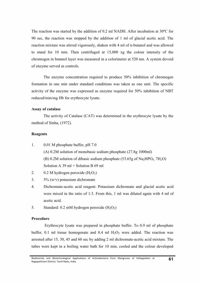
Biodiversity and Biotechnological Applications of Actinobacteria from Mangroves of Vellappallam at Nagapattinam District, Tamil Nadu, India. 61
The reaction was started by the addition of 0.2 ml NADH. After incubation at 30ºC for
90 sec, the reaction was stopped by the addition of 1 ml of glacial acetic acid. The
reaction mixture was stirred vigorously, shaken with 4 ml of n-butanol and was allowed
to stand for 10 min. Then centrifuged at 15,000 xg the colour intensity of the
chromogen in butanol layer was measured in a colorimeter at 520 nm. A system devoid
of enzyme served as controls.
The enzyme concentration required to produce 50% inhibition of chromogen
formation in one min under standard conditions was taken as one unit. The specific
activity of the enzyme was expressed as enzyme required for 50% inhibition of NBT
reduced/min/mg Hb for erythrocyte lysate.
Assay of catalase
The activity of Catalase (CAT) was determined in the erythrocyte lysate by the
method of Sinha, (1972).
Reagents
1. 0.01 M phosphate buffer, pH 7.0
(A) 0.2M solution of monobasic sodium phosphate (27.8g 1000ml)
(B) 0.2M solution of dibasic sodium phosphate (53.65g of Na2HPO4. 7H2O)
Solution A 39 ml + Solution B 69 ml
2. 0.2 M hydrogen peroxide (H2O2)
3. 5% (w/v) potassium dichromate
4. Dichromate-acetic acid reagent: Potassium dichromate and glacial acetic acid
were mixed in the ratio of 1:3. From this, 1 ml was diluted again with 4 ml of
acetic acid.
5. Standard: 0.2 mM hydrogen peroxide (H2O2)
Procedure
Erythrocyte lysate was prepared in phosphate buffer. To 0.9 ml of phosphate
buffer, 0.1 ml tissue homogenate and 0.4 ml H2O2 were added. The reaction was
arrested after 15, 30, 45 and 60 sec by adding 2 ml dichromate-acetic acid mixture. The
tubes were kept in a boiling water bath for 10 min, cooled and the colour developed

Biodiversity and Biotechnological Applications of Actinobacteria from Mangroves of Vellappallam at Nagapattinam District, Tamil Nadu, India. 62
was read at 590 nm. Standards in the concentration of 20-100 µM were processed for
test. The specific activity of the enzyme was expressed as μ moles of H2O2
utilized/min/mg Hb for erythrocyte lysate.
Assay of Glutathione Peroxidase
The activity of Glutathione peroxidase (Gpx) was assayed in the erythrocyte
lysate by the method of Rotruck et al., (1973).
Reagents
1. 0.4 M Tris-HCl buffer, pH 7.0
2. 10 mM Sodium azide
3. 10% (w/v) TCA.
4. 0.4 mM EDTA.
5. 1 mM H2O2
6. 2 mM reduced glutathione (GSH)
Procedure
To 0.2 ml of Tris-HCl buffer, 0.2 ml EDTA, 0.1 ml sodium azide and 0.2 ml
enzyme preparation (erythrocyte lysate) were added and mixed well. To this, 0.2 ml
GSH followed by 0.1 ml H2O2 were added. The contents were mixed and incubated at
37ºC for 10 min. Then the reaction was arrested by the addition of 0.5 ml TCA. The
tubes were subjected for 2000 rpm and the remaining GSH was determined
colorimetrically at 340 nm. The activity of GPx is expressed as μ moles of GSH
utilized/min/mg Hb for erythrocyte lysate.
3.15. 3. Histological studies (Kleiner et al., 2005)
The classic paraffin sectioning and haematoxylin eosin staining techniques were
used for the histological studies. The various steps involved in the preparation of
tissues for histological studies are as follows:
Fixation
A bit of tissue from each organ was cut and fixed in bouin’s fluid immediately
after removal from the animal body. Bouins fluid, is the commonly used fixative, was
prepared by mixing the following chemicals. The tissues were fixed in bouin’s fluid for

Biodiversity and Biotechnological Applications of Actinobacteria from Mangroves of Vellappallam at Nagapattinam District, Tamil Nadu, India. 63
about 24 hrs. The tissues were then taken and washed in tap water for a day to remove
excess of picric acid.
Dehydration
The term dehydration means the removal of water from the tissues by alcohol of
varying grades. Ethanol was used for dehydration. The tissues were kept in the
following solutions for an hour each
30% alcohol,
50% alcohol
70% alcohol
100% alcohol
Inadequately dehydrated tissues cannot be satisfactorily in filtered with paraffin.
At the same time over dehydration results in making the tissues brittle, which would be
difficult for sectioning. So the tissues were carefully dehydrated.
Clearing
Dealcoholization or replacement of alcohol from the tissues with a clearing agent
is called as clearing. Xylene was used as the clearing agent for one or two hours, two or
three times. Since, the clearing agent is miscible with both dehydration and embedding
agents, it permits paraffin in to filtrate the tissues. So, the clearing was carried out as
the next step after dehydration to permit tissue spaces to be filled with paraffin. The
tissues were kept in the clearing agent till they become transparent and impregnated
with xylene.
Impregnation
In this process the clearing agent xylene was placed by paraffin wax. The tissues
were taken out of xylene and were kept in molten paraffin embedding bath, which
consists of metal pots filled with molten wax maintained at about 50o C. The tissues
were given three changes in the molten wax at half an hour intervals.
Embedding
The paraffin wax used for embedding should be fresh and heated upto the
optimum melting point at about 56o C- 58 o C. A clear glass plate was smeared with

Biodiversity and Biotechnological Applications of Actinobacteria from Mangroves of Vellappallam at Nagapattinam District, Tamil Nadu, India. 64
glycerine. L-shaped mould was placed on it to from a rectangular cavity. The molten
paraffin wax was poured and air bubbles were removed by using a hot needle. The
tissue was placed in the paraffin and oriented with the surface to be sectioned. Then
the tissue was pressed gently towards the glass plate to make settle uniformly with a
metal pressing rod and allowed the wax to settle and solidify at room temperature. The
paraffin block was kept in cold water for cooling.
Section cutting
Section cutting was done with a rotatory microtome. The excess of paraffin
around the tissue was removed by trimming, leaving ½ cm around the tissue. Then the
block was attached to the gently heated holder. Additional support was given by some
extra wax, which was applied along the sides of the block. Before sectioning, all set
screws holding the object holder and knife were hand tightened to avoid vibration. To
produce uniform sections, the microtome knife was adjusted to the proper angle in the
knife holder with only the cutting edge coming in contact with the paraffin block. The
tissue was cut in 7 μ thickness.
Flattening and mounting of sections
This was carried out in tissue flotation warm water bath. The sections were
spread on a warm water bath after they were detached from the knife with the help of
hair brush. Dust free clean slides were coated with egg albumin over the whole
surface. Required sections were spread on clean slide and kept at room temperature.
Staining
The sections were stained as follows; deparaffinization with xylene two times
each for five minutes
Dehydration through descending grades of ethyl alcohol
100% alcohol (absolute) - 2 minute
90% alcohol - 1minute
50% alcohol - 1 minute

Biodiversity and Biotechnological Applications of Actinobacteria from Mangroves of Vellappallam at Nagapattinam District, Tamil Nadu, India. 65
Staining with Ehrlich’s haemaoxylin for 15-20 minutes was done. Thoroughly
washed in tap water for 10minutes. Rinsed with distilled water and stained with eosin.
Dehydration was done again with ascending grades of alcohol.
70% alcohol - 2minute
90% alcohol - 2minute
100% alcohol - 1minute
(Clearing with xylene two times, each for about 3 minutes interval).
Mounting
On the stained slide, DPX mountant was applied uniformly and microglass
cover slides were spread. The slides were observed in Nikon microscope and
microphotographs were taken.
3.16. Synthesis of silver nanoparticle from actinobacteria (Sadowski et al., 2008)
The selected actinobacterial biomass were washed thrice in deionized water to
remove the unwanted material. Approximately 3.5 gm of actinobacterial biomass were
taken in a conical flask containing 100 ml deionized water. 10-1mM AgNO3 was added
then it was incubated at room temperature,colour change was observed.
3.16.1. SEM analysis of silver nanoparticles synthesized by Thermoactinomyces
vulgaris DKP01
Silver nanoparticle synthesized actinobacterial biomass were allowed to dry
completely and ground well. Since the specimen was at high vacuum, Fixation was
usually performed by incubation in a solution of a buffered chemical fixative
glutaraldehyde. The dry specimen was mounted on a specimen stub using an adhesive
epoxy resin or electrically-conductive double-sided adhesive tape and sputter coated
with gold palladium alloy before examination in the microscope.
3.16.2. UV-Visible spectroscopic analysis of silver nanoparticles synthesized by
Thermoactinomyces vulgaris DKP01
The bioreduction of pure AgNO3 were monitored using UV-Visible
spectroscopic at regular intervals. During the reduction, 0.1ml of sample was taken and
diluted several times with Millipore water. After dilution, it was centrifuged at 800 rpm

Biodiversity and Biotechnological Applications of Actinobacteria from Mangroves of Vellappallam at Nagapattinam District, Tamil Nadu, India. 66
for 5 min. The supernatant was scanned by UV-300 spectrophotometer (UNICAM) for
UV-Vis 1601 Schimodzu spectrophotometer, operated at a resolution of 420-1000 nm.
3.16.3. FT–IR analysis of silver nanoparticles synthesized by Thermoactinomyces
vulgaris DKP01
A known weight of sample (1 mg) was taken in a mortar and pestle and ground
with 2.5 mg of dry potassium bromide (KBr). The powder so obtained was filled in a
2 mm internal diameter micro‐cup and loaded onto FT-IR set at 26°C ± 1°C. The
samples were scanned using infrared in the range of 4000–400 cm‐1 using Fourier
Transform Infrared Spectrometer (Thermo Nicolet Model‐6700). The spectral data
obtained were compared with the reference chart to identify the functional groups
present in the sample.
3.16.4. Screening for antibacterial activity of silver nanoparticles synthesized by
Thermoactinomyces vulgaris DKP01
The antibacterial activity of silver nanoparticles synthesized from actinobacteria
were evaluated by agar well diffusion method.

Biodiversity and Biotechnological Applications of Actinobacteria from Mangroves of Vellappallam at Nagapattinam District, Tamil Nadu, India. 67
4. RESULTS
The present investigation deals with isolation and identification of
actinobacteria from mangrove environment, antibacterial efficacy of actinobacteria,
separation and characterization of bioactive compounds, screening of antioxidant,
anticancer activity and synthesize of silver nanoparticles.
Actinobacteria are best known for their ability to produce antibiotics and are
Gram positive bacteria which comprise a group of branching unicellular
microorganisms. They produce branching mycelium which may be of two kinds viz.,
substrate mycelium and aerial mycelium. Among actinobacteria, the Streptomyces are
the dominant. The non Streptomyces are called rare actinobacteria, comprising
approximately 100 genera. Members of the actinobacteria, which live in marine
environment, are poorly understood particularly from mangroves.
4.1 Biodiversity of actinobacteria
Totally 56 actinobacteria isolates belonging to 17 genera were isolated from
mangrove soil samples. The total isolates were distinguished on the basis of cultural
characteristics in starch casein agar. The colony colour, size, shape, margin, diffusible
pigment, aerial and substrate mycelium appearance well observed and recorded. The
actinobacteria isolates were presented in table 1 and plate 3 & 4.
4.1.1 Species composition
In general, among the 17 genera were recorded, the genus of Micromonospora
(9 isolates) was dominant followed by Streptomyces, Streptoverticillium and Nocardia
(5 isolates each) Actinobispora, Actinomadura and Jonesia (2 isolates each),
Glycomyces and Nocardiopsis, Actinosynnema, Catellatospora, Dactylosporangium,
Micropolyspora, Microtetraspora, Streptoverticillium and Thermoactinomyces all other
genera were represented by one isolate each.
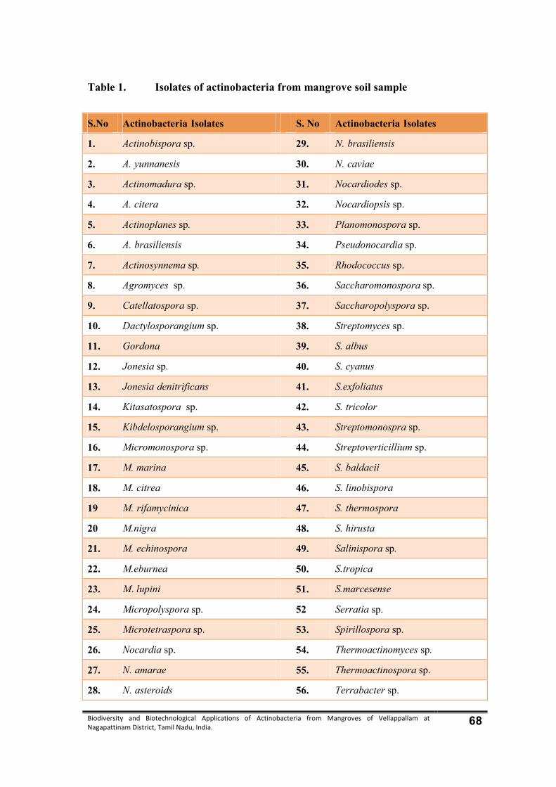
Biodiversity and Biotechnological Applications of Actinobacteria from Mangroves of Vellappallam at Nagapattinam District, Tamil Nadu, India. 68
Table 1. Isolates of actinobacteria from mangrove soil sample
S.No Actinobacteria Isolates S. No Actinobacteria Isolates
1. Actinobispora sp. 29. N. brasiliensis
2. A. yunnanesis 30. N. caviae
3. Actinomadura sp. 31. Nocardiodes sp.
4. A. citera 32. Nocardiopsis sp.
5. Actinoplanes sp. 33. Planomonospora sp.
6. A. brasiliensis 34. Pseudonocardia sp.
7. Actinosynnema sp. 35. Rhodococcus sp.
8. Agromyces sp. 36. Saccharomonospora sp.
9. Catellatospora sp. 37. Saccharopolyspora sp.
10. Dactylosporangium sp. 38. Streptomyces sp.
11. Gordona 39. S. albus
12. Jonesia sp. 40. S. cyanus
13. Jonesia denitrificans 41. S.exfoliatus
14. Kitasatospora sp. 42. S. tricolor
15. Kibdelosporangium sp. 43. Streptomonospra sp.
16. Micromonospora sp. 44. Streptoverticillium sp.
17. M. marina 45. S. baldacii
18. M. citrea 46. S. linobispora
19 M. rifamycinica 47. S. thermospora
20 M.nigra 48. S. hirusta
21. M. echinospora 49. Salinispora sp.
22. M.eburnea 50. S.tropica
23. M. lupini 51. S.marcesense
24. Micropolyspora sp. 52 Serratia sp.
25. Microtetraspora sp. 53. Spirillospora sp.
26. Nocardia sp. 54. Thermoactinomyces sp.
27. N. amarae 55. Thermoactinospora sp.
28. N. asteroids 56. Terrabacter sp.
Biodiversity and Biotechnological Applications of Actinobacteria from Mangroves of Vellappallam at Nagapattinam District, Tamil Nadu, India. 69
4.1.2. Characterization and identification of actinobacteria
Among the 56 isolated actinobacteria, one isolate was found to possess a broad
spectrum antimicrobial activity, it was justifiably chosen for further taxonomic
characterization. The different parameters namely, morphological, biochemical,
physiological characters were used for the characterization and identification of
actinobacteria isolates.
4.1.3. Morphological characterization
Actinobacteria show a notable array of macroscopic features such as
pigmentation of spores, aerial and substrate mycelium and diffusible extracellular
pigments. Morphology has played a major role in distinguishing actinobacteria from
the total actinobacteria groups. The microscopic view of actinobacteria showed tight
spirals of smooth spore surface.
Actinobispora sp.
Long, irregularly branched, vegetative mycelium does not fragment, smooth
walled spores, aerial hyphae, gray in colour.
Actinopolyspora sp.
Branching vegetative hyphae mostly fragmented long chain of smooth – walled,
aerial hyphae cylindrical.
Actinoplanes sp.
Non fragmenting branching mycelium aerial mycelium is absent, highly
colored, spores are produced within sporangia, spherical to very irregular.
Actinomadura sp.
Branching vegetative hyphae, non fragmenting substrate mycelium, aerial
mycelium is moderately developed. Short, smooth and warty. Aerial mycelium are
yellow coloured.
Actinosynnema sp.
Aerial hyphae, done like bodies or flat colonies on surface, motile spores,
melanoid pigments.

Biodiversity and Biotechnological Applications of Actinobacteria from Mangroves of Vellappallam at Nagapattinam District, Tamil Nadu, India. 70
Agromyces sp.
Branched, slender filamentous, coccoid and irregular non motile.
Catellatospora sp.
Vegetative hyphae one branched but not fragmented. No true aerial mycelium
produced, short chains of non motile spores.
Dactylosporangium sp.
The substrate hyphae irregularly branched, finger shaped to claviform
sporangia. The colour of the mycelium pale orange to deep orange.
Gordona sp.
Colony morphology varies slimy smooth and glossy to irregular and rough.
Jonesia sp.
Vegetative and aerial hyphae. Ovoid to bacillary non motile elements. Zig-zag
morphology, sporulation.
Micromonospora sp.
Well developed, branched, septate mycelium, non motile spores, aerial
mycelium is absent. Some culture appears irregularly as a restricted while or grayish
bloom.
Micropolyspora sp.
Branched mycelium, longitudinal pairs or aerial mycelium not usually formed
on the substrate mycelium, spherical to oval, non motile, surface in smooth.
Microtetraspora sp.
Branched, vegetative mycelium, aerial hyphae, spores chain are straight, hooked
or tightly closed spirals, irregular smooth in colour.
Nocardiopsis sp.
Substrate mycelium is well developed, long and densely branched, aerial
hyphae completely fragment into spores of various lengths.
Biodiversity and Biotechnological Applications of Actinobacteria from Mangroves of Vellappallam at Nagapattinam District, Tamil Nadu, India. 71
Saccharomonospora sp.
Vegetative and aerial hyphae. Ovoid to bacillary non motile elements. Zig-zag
morphology, sporulation.
Saccharopolyspora sp.
Vegetative mycelium well developed branched septate, aerial mycelium and rod
shaped.
Streptomyces sp.
Vegetative hyphae, branched mycelium aerial mycelium at maturity forms
chains, three to many spores, few species bear short chains of spores on the substrate
mycelium, spores are non motile, leathery or butyrous colonies. Smooth surface, later
they develop a weft or aerial mycelium, granular, powdery, wide variety of pigments
responsible for the colour of the vegetative and aerial mycelia.
Thermomoactinomyces sp.
Produce single, heat sensitive non motile, aerial hyphae, branched non
fragmenting vegetative hyphae, leathery colonies usually covered with aerial
mycelium. Unbranched is branched sporophores.
4.1.3 Biochemical Characterization
The physiological tests are indispensable tools for classification and
identification of actinobacteria and influencing the growth rate of actinobacteria
exhibited to catalase production and citrate utilization, urease, nitrate reduction, starch
hydrolysis, casein hydrolysis, indole, methyl red and vogues-proskauer tests. The
details of biochemical characteristics of the isolate are given in Table 2.
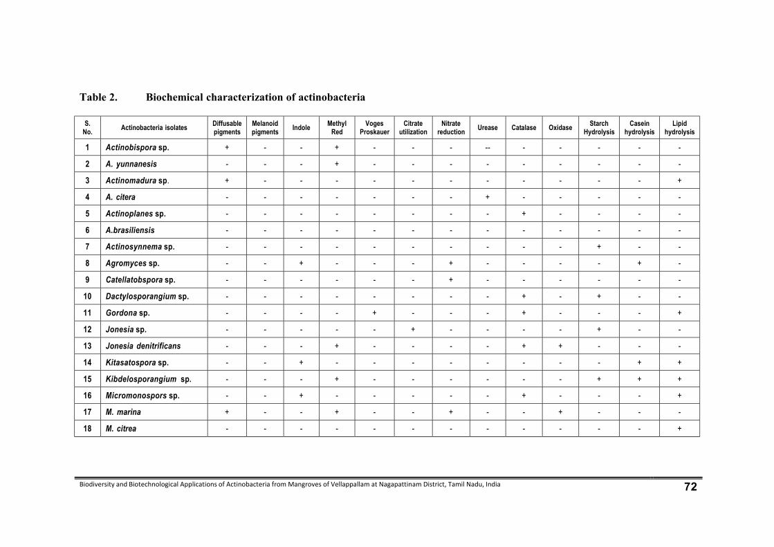
Biodiversity and Biotechnological Applications of Actinobacteria from Mangroves of Vellappallam at Nagapattinam District, Tamil Nadu, India 72
Table 2. Biochemical characterization of actinobacteria
S. No.
Actinobacteria isolates Diffusable pigments
Melanoid pigments
Indole Methyl Red
Voges Proskauer
Citrate utilization
Nitrate reduction
Urease Catalase Oxidase Starch Hydrolysis
Casein hydrolysis
Lipid hydrolysis
1 Actinobispora sp. + - - + - - - -- - - - - -
2 A. yunnanesis - - - + - - - - - - - - -
3 Actinomadura sp. + - - - - - - - - - - - +
4 A. citera - - - - - - - + - - - - -
5 Actinoplanes sp. - - - - - - - - + - - - -
6 A.brasiliensis - - - - - - - - - - - - -
7 Actinosynnema sp. - - - - - - - - - - + - -
8 Agromyces sp. - - + - - - + - - - - + -
9 Catellatobspora sp. - - - - - - + - - - - - -
10 Dactylosporangium sp. - - - - - - - - + - + - -
11 Gordona sp. - - - - + - - - + - - - +
12 Jonesia sp. - - - - - + - - - - + - -
13 Jonesia denitrificans - - - + - - - - + + - - -
14 Kitasatospora sp. - - + - - - - - - - - + +
15 Kibdelosporangium sp. - - - + - - - - - - + + +
16 Micromonospors sp. - - + - - - - - + - - - +
17 M. marina + - - + - - + - - + - - -
18 M. citrea - - - - - - - - - - - - +

Biodiversity and Biotechnological Applications of Actinobacteria from Mangroves of Vellappallam at Nagapattinam District, Tamil Nadu, India 73
S. No.
Actinobacteria isolates Diffusable pigments
Melanoid pigments
Indole Methyl Red
Voges Proskauer
Citrate utilization
Nitrate reduction
Urease Catalase Oxidase Starch Hydrolysis
Casein hydrolysis
Lipid hydrolysis
19 M. rifamycinia + + - - - + - - - + - - -
20 M. nigra - - - - + - - - - - + - -
21 M. echinospora - - - + - - + - - - + + -
22 M. eburnean + - - - + - - - + - + + +
23 M. lupine - + - - + + - - - - - - -
24 Micropolyspora sp. + - - - - + - - - - + + +
25 Microtetraspora sp. - - - - - - - - - + - - -
26 Nocardia sp. + - - - - - + + - - - - -
27 N.amarae + + - - - - - - - - + - -
28 N.asteroids + + - - - - + - - + - - -
29 N.brasiliiensis + + - - - - - - - - + + +
30 N.caviae + + - - + - - + - - - - -
31 Nocardiodes sp. - + - - - + + - - + - - -
32 Nocardiopsis sp. - - - - - - - - - - - - -
33 Planomonospora sp. + - - - - - - - - - + + +
34 Pseudonocardia sp. - - + - - - - - + - - - -
35 Rhodococcus sp. - - - - - - - - - + - - -
36 Saccharomonospora sp. - - - + - - - - - - - + -
37 Saccharopolyspora sp. + - - - - - - - - - - - +
38 Streptomyces sp. + + - - + - + - - + - - -
39 S.albus + + - - - - - - - - - + -
40 S. cyanus + + - - - - - - - - - + -

Biodiversity and Biotechnological Applications of Actinobacteria from Mangroves of Vellappallam at Nagapattinam District, Tamil Nadu, India 74
S. No.
Actinobacteria isolates Diffusable pigments
Melanoid pigments
Indole Methyl Red
Voges Proskauer
Citrate utilization
Nitrate reduction
Urease Catalase Oxidase Starch Hydrolysis
Casein hydrolysis
Lipid hydrolysis
41 S. exfoliates + + - - - - - - - - - - -
42 S. tricolor + + - - - - + - - - - - -
43 Streptomonospora sp. - - - - - + - - - - - - -
44 Streptoverticillium sp. - + - - - - - - - - + - +
45 S. baldacii - - - - - + - - - - - - -
46 S. linobispora - - - - - + - - + - - - -
47 S.thermospora - - - - - + - - + - - - -
48 S.hirusta - - - - - + - - + - - - -
49 Salinispora sp. - - - - - - - - + - - - -
50 S. tropica + + - - + - - - - - - + -
51 S. marcesense + + - - - - + - - - - - +
52 Serratia sp. - - - - - - + - - - - - +
53 Spirillospora sp. - - - - - + - - - - - + -
54 Thermoactinomyces sp. - - + - - - - + - - + + +
55 Thermoactinospora sp. + + - - + - - - - - + + +
56 Terrabacter sp. - - - + - - + - - + - - -
+ - Present; – - Absent

Biodiversity and Biotechnological Applications of Actinobacteria from Mangroves of Vellappallam at Nagapattinam District, Tamil Nadu, India 75
Plate 3. ISOLATION OF ACTINOBACTERIA FROM
MANGROVES OF VELLAPPALLAM
Actinobiospora sp. A. citera Actinosynnema sp.
A. yamnanesis Actinoplanes sp. Agromyces sp.
Actinomodura sp. A. brasiliensis Catellabspora sp.
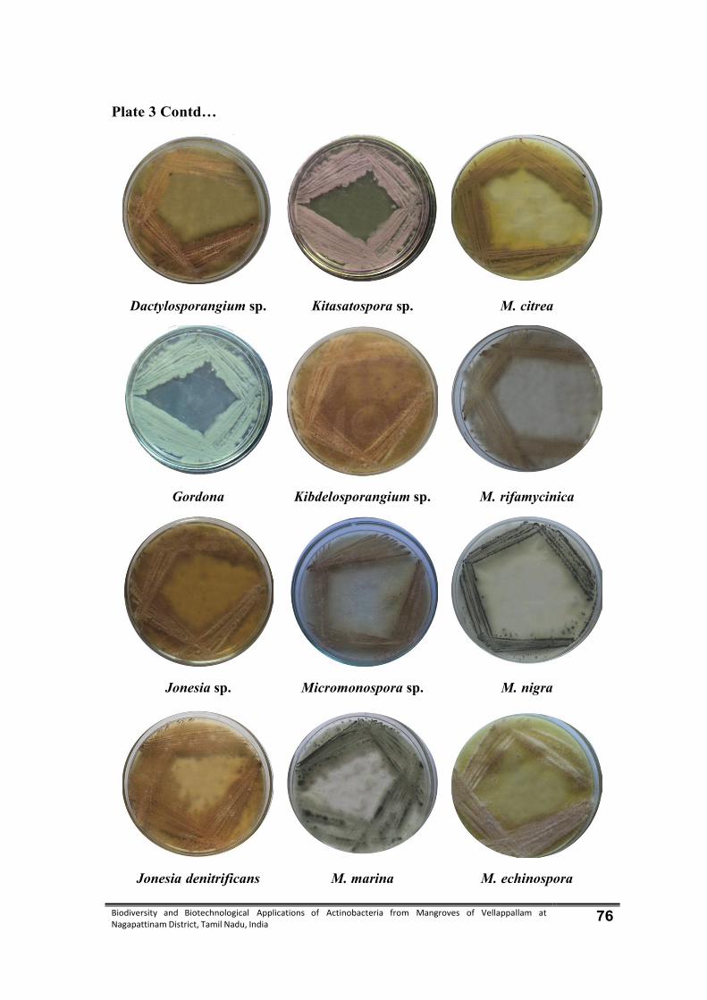
Biodiversity and Biotechnological Applications of Actinobacteria from Mangroves of Vellappallam at Nagapattinam District, Tamil Nadu, India 76
Plate 3 Contd…
Dactylosporangium sp. Kitasatospora sp. M. citrea
Gordona Kibdelosporangium sp. M. rifamycinica
Jonesia sp. Micromonospora sp. M. nigra
Jonesia denitrificans M. marina M. echinospora
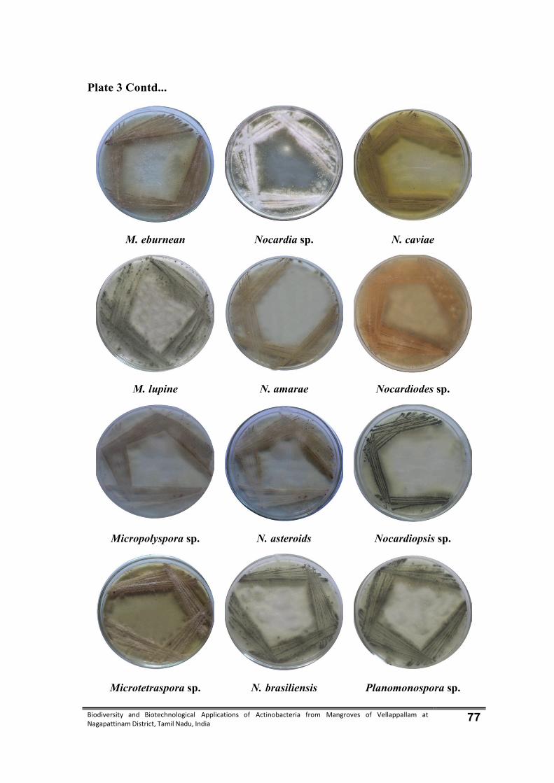
Biodiversity and Biotechnological Applications of Actinobacteria from Mangroves of Vellappallam at Nagapattinam District, Tamil Nadu, India 77
Plate 3 Contd...
M. eburnean Nocardia sp. N. caviae
M. lupine N. amarae Nocardiodes sp.
Micropolyspora sp. N. asteroids Nocardiopsis sp.
Microtetraspora sp. N. brasiliensis Planomonospora sp.
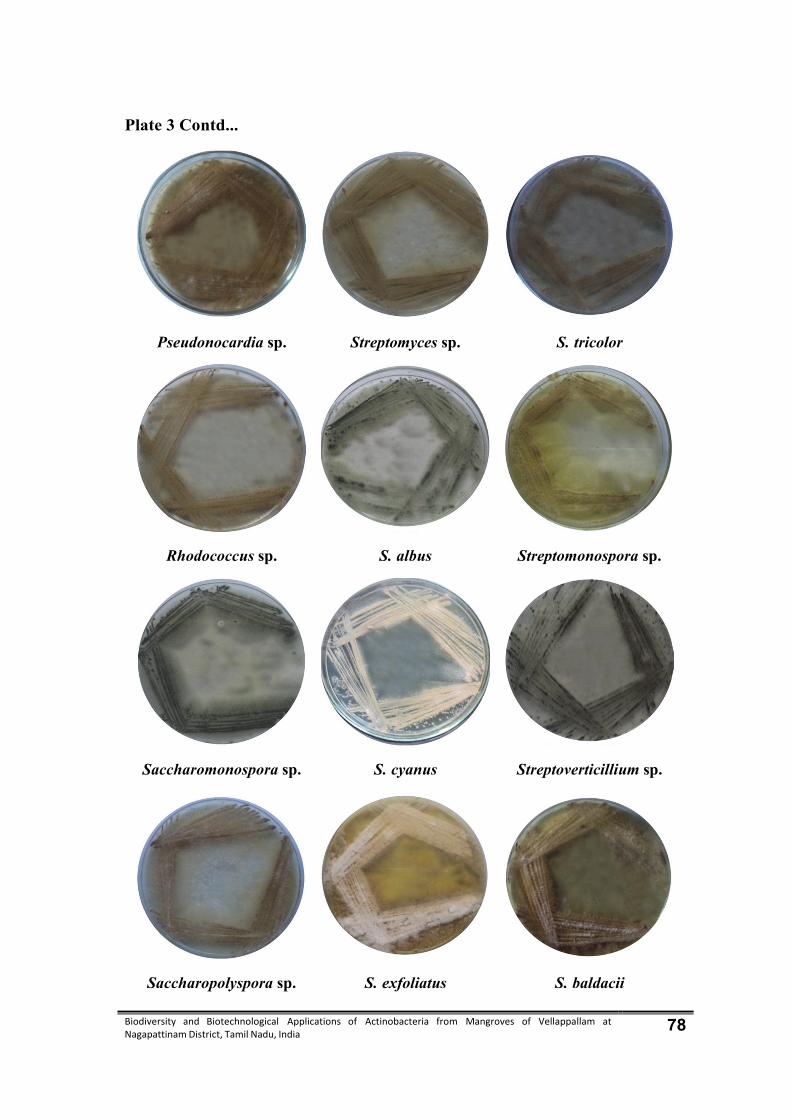
Biodiversity and Biotechnological Applications of Actinobacteria from Mangroves of Vellappallam at Nagapattinam District, Tamil Nadu, India 78
Plate 3 Contd...
Pseudonocardia sp. Streptomyces sp. S. tricolor
Rhodococcus sp. S. albus Streptomonospora sp.
Saccharomonospora sp. S. cyanus Streptoverticillium sp.
Saccharopolyspora sp. S. exfoliatus S. baldacii

Biodiversity and Biotechnological Applications of Actinobacteria from Mangroves of Vellappallam at Nagapattinam District, Tamil Nadu, India 79
Plate 3 Contd…
S. linobispora S. tropica Thermoactinomyces sp.
S. thermospora S. marcesense Thermoactinospora sp.
S. hirusta Serratia sp. Terrabacter sp.
Salinispora sp. Spirillospora sp.
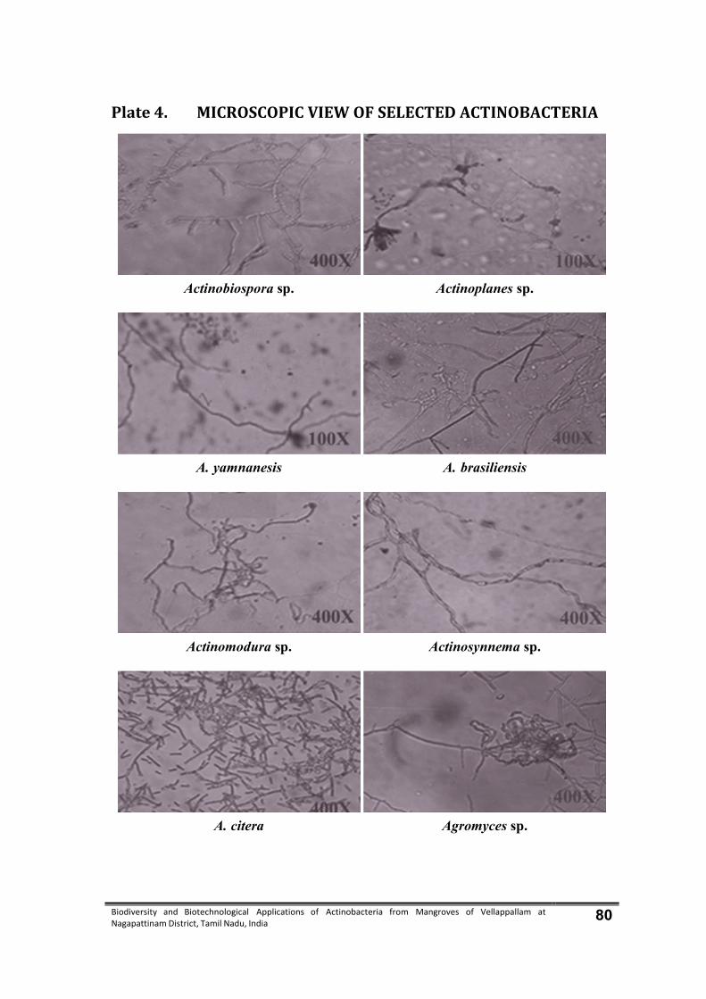
Biodiversity and Biotechnological Applications of Actinobacteria from Mangroves of Vellappallam at Nagapattinam District, Tamil Nadu, India 80
Plate 4. MICROSCOPIC VIEW OF SELECTED ACTINOBACTERIA
Actinobiospora sp. Actinoplanes sp.
A. yamnanesis A. brasiliensis
Actinomodura sp. Actinosynnema sp.
A. citera Agromyces sp.

Biodiversity and Biotechnological Applications of Actinobacteria from Mangroves of Vellappallam at Nagapattinam District, Tamil Nadu, India 81
Plate 4 Contd…
Catellabspora sp. Jonesia denitrificans
Dactylosporangium sp. Kitasatospora sp.
Gordona Kibdelosporangium sp.
Jonesia sp. Micromonospora sp.
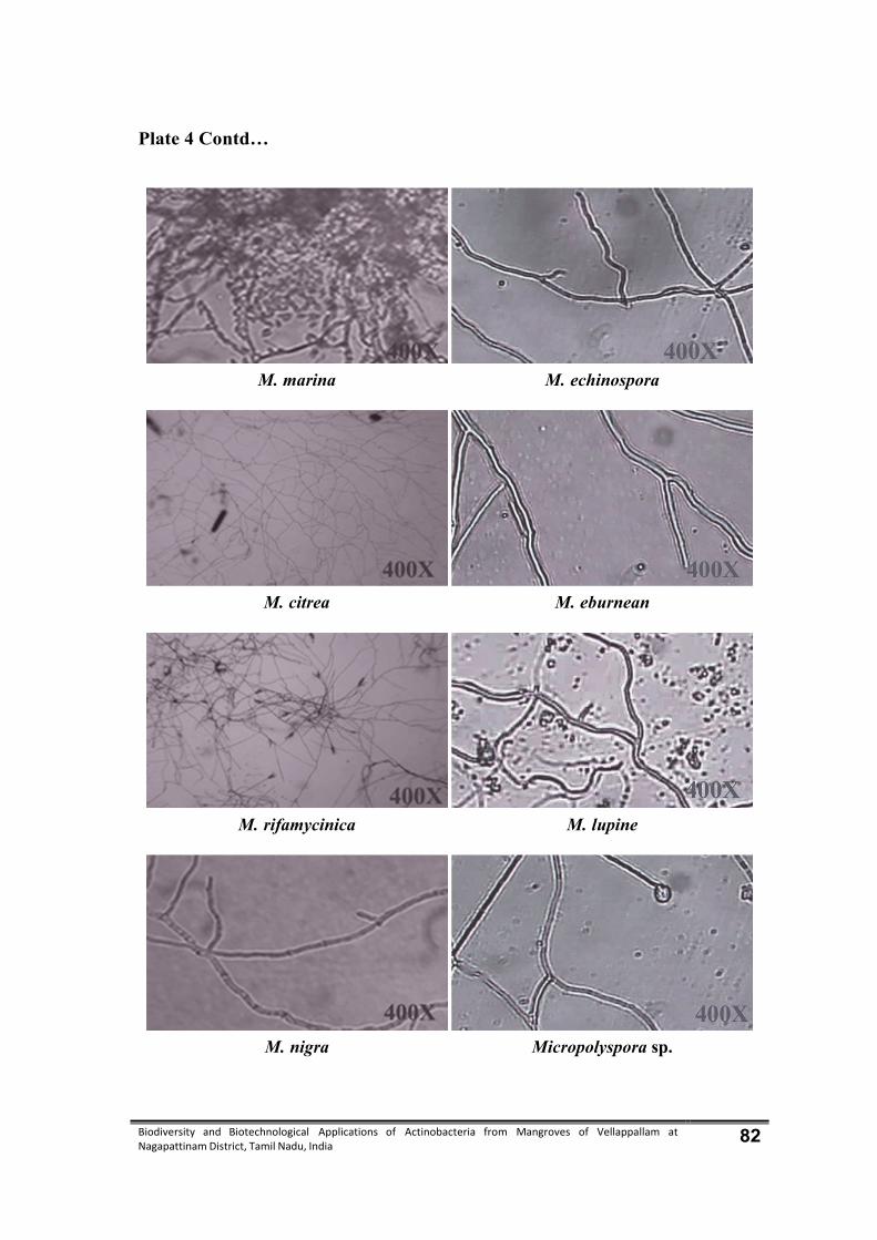
Biodiversity and Biotechnological Applications of Actinobacteria from Mangroves of Vellappallam at Nagapattinam District, Tamil Nadu, India 82
Plate 4 Contd…
M. marina M. echinospora
M. citrea M. eburnean
M. rifamycinica M. lupine
M. nigra Micropolyspora sp.

Biodiversity and Biotechnological Applications of Actinobacteria from Mangroves of Vellappallam at Nagapattinam District, Tamil Nadu, India 83
Plate 4 Contd…
Microtetraspora sp. N. brasiliensis
Nocardia sp. N. caviae
N. amarae Nocardiodes sp.
N. asteroids Nocardiopsis sp.
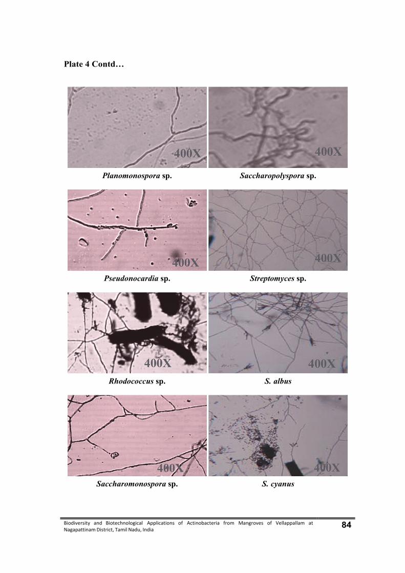
Biodiversity and Biotechnological Applications of Actinobacteria from Mangroves of Vellappallam at Nagapattinam District, Tamil Nadu, India 84
Plate 4 Contd…
Planomonospora sp. Saccharopolyspora sp.
Pseudonocardia sp. Streptomyces sp.
Rhodococcus sp. S. albus
Saccharomonospora sp. S. cyanus

Biodiversity and Biotechnological Applications of Actinobacteria from Mangroves of Vellappallam at Nagapattinam District, Tamil Nadu, India 85
Plate 4 Contd…
S. exfoliatus S. baldacii
S. tricolor S. linobispora
Streptomonospora sp. S. thermospora
Streptoverticillium sp. S. hirusta
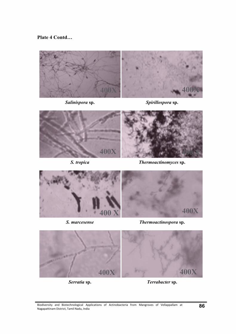
Biodiversity and Biotechnological Applications of Actinobacteria from Mangroves of Vellappallam at Nagapattinam District, Tamil Nadu, India 86
Plate 4 Contd…
Salinispora sp. Spirillospora sp.
S. tropica Thermoactinomyces sp.
S. marcesense Thermoactinospora sp.
Serratia sp. Terrabacter sp.

Biodiversity and Biotechnological Applications of Actinobacteria from Mangroves of Vellappallam at Nagapattinam District, Tamil Nadu, India 87
4.1.4. Actinobacteria population mean density
Population mean density of actinobacteria varied from 21.2 to 36.7×10⁶ CFU/g
with the minimum in the samples were collected during monsoon season and maximum
in the samples collected during pre monsoon season in 2012 (Table 3).
4.1.5. Percentage contribution
Percentage contribution of the individual species to the total actinobacteria
population at four different seasonal variations. The maximum percentage contribution
of 14.2 % was found with Micromonospora and Streptomyces (10.9%), the minimum
percentage contribution was with Actinobispora, Streptomonospora, Nocardia 1.45%
were present in the population density.
4.1.6. Percentage frequency
Frequencies of identified genera of actinobacteria in different seasons were
fluctuated. The frequency of the genus Streptomyces, Micromonospora, Nocardia,
Saccharomonospora, Saccharopolyspora, Sterptomonospora, Streptopolyspora,
Thermoactinomycetes were the common species, which showed 100% frequency.
A. Yunnanesis, Actinomadura, A. citera, Actinoplanes, A.brasiliensis, Actinosynnema,
Agromyces (75% each), Actinobispora, Micromonospora, Streptoverticillium
(50% each) and Thermoactinospora, Terrabacter (25%) were rare in occurrence
(Table 4).

Biodiversity and Biotechnological Applications of Actinobacteria from Mangroves of Vellappallam at Nagapattinam District, Tamil Nadu, India 88
Table 3. Total number of colonies, mean density (CFU/g) and percentage contribution of Actinobacteria recorded during different seasons from Mangrove soil sample at Vellappallam, Nagapattinam District.
S. No
Name of the organisms
Postmonsoon Summer Premonsoon Monsoon Total No. of
colonies
% contri-bution
TNC MD TNC MD TNC MD TNC MD
1 Actinobispora sp. 2 0.66 1 0.33 1 0.33 2 0.66 6 1.7
2 A. yunnanesis 3 1 2 0.66 3 1 1 0.33 8 2.3
3 Actinomadura sp. 2 0.66 3 1 2 0.66 - - 7 2.0
4 A. citera 1 0.33 1 0.33 2 0.66 - - 4 1.1
5 Actinoplanes sp. - - 1 0.33 2 0.66 1 0.33 4 1.1
6 A.brasiliensis - - 1 0.33 5 1.66 2 0.66 7 2.0
7 Actinosynnema sp. 3 1 1 0.33 4 1.33 - - 8 2.3
8 Agromyces sp. 2 0.66 2 0.66 - - 1 0.33 5 1.4
9 Catellatobspora sp. 2 0.66 1 0.33 1 0.33 - - 4 1.1
10 Dactylosporangium sp. - - - - 2 0.66 1 0.33 3 0.8
11 Gordona sp. 2 0.66 3 1 2 0.66 1 0.33 8 2.3
12 Jonesia sp. 3 1 2 0.66 1 0.33 2 0.66 8 2.3
13 Jonesia denitrificans - - - - 3 1 - - 3 0.8
14 Kitasatospora sp. 3 1 2 0.66 4 1.33 1 0.33 10 2.9
15 Kibdelosporangium sp. 1 0.33 1 0.33 1 0.33 2 0.66 3 0.8
16 Micromonospors sp. 2 0.66 - - 2 0.66 - - 4 1.1
17 M. marina 3 1 2 0.66 1 0.33 - - 6 1.7
18 M. citrea 2 0.66 3 1 4 1.33 1 0.33 10 2.9
19 M. rifamycinia 1 0.33 1 0.33 1 0.33 2 0.66 5 1.4
20 M. nigra 2 0.66 1 0.33 1 0.33 2 0.66 6 1.7
21 M. echinospora 4 1.33 2 0.66 1 0.33 2 0.66 9 2.6
22 M. eburnean 2 0.66 1 0.33 2 0.66 - - 5 1.4
23 M. lupine 1 0.33 1 0.33 2 0.66 1 0.33 5 1.4
24 Micropolyspora sp. 2 0.66 - - 2 0.66 1 0.33 5 1.4
25 Microtetraspora sp. 1 0.33 2 0.66 1 0.33 - - 4 1.1
26 Nocardia sp. 2 0.66 1 0.33 1 0.33 - - 4 1.1
27 N.amarae 1 0.33 1 0.33 1 0.33 1 0.33 4 1.1
28 N.asteroids 4 1.33 1 0.33 2 0.66 1 0.33 8 2.3

Biodiversity and Biotechnological Applications of Actinobacteria from Mangroves of Vellappallam at Nagapattinam District, Tamil Nadu, India 89
29 N.brasiliiensis 1 0.33 2 0.66 1 0.33 1 0.33 5 1.4
30 N.caviae 2 0.66 1 0.33 1 0.33 2 0.66 6 1.7
31 Nocardiodes sp. 2 0.66 1 0.33 1 0.33 - - 4 1.1
32 Nocardiopsis sp. 1 0.33 1 0.33 1 0.33 2 0.66 5 1.1
33 Planomonospora sp. 4 1.33 1 0.33 1 0.33 2 0.66 8 2.3
34 Pseudonocardia sp. 2 0.66 1 0.33 1 0.33 2 0.66 6 1.7
35 Rhodococcus sp. 4 1.33 1 0.33 1 0.33 2 0.66 8 2.3
36 Saccharomonospora sp. 2 0.66 4 1.33 5 1.66 1 0.33 12 3.5
37 Saccharopolyspora sp. 4 1.33 1 0.33 5 1.66 4 1.33 11 3.2
38 Streptomyces sp. 2 0.66 3 1 1 0.33 1 0.33 7 2.0
39 S.albus 2 0.66 1 0.33 1 0.33 1 0.33 5 1.4
40 S. cyanus 1 0.33 4 1.33 4 1.33 2 0.66 11 3.2
41 S. exfoliates 2 0.66 1 0.33 3 1 2 0.66 7 2.0
42 S. tricolor 1 0.33 1 0.33 4 1.33 2 0.66 8 2.3
43 Streptomonospora sp. 2 0.66 3 1 1 0.33 1 0.33 7 2.0
44 Streptoverticillium sp. 1 0.33 3 1 - - - - 4 1.1
45 S. baldacii 1 0.33 1 0.33 1 0.33 2 0.66 5 1.4
46 S. linobispora 3 1 1 0.33 1 0.33 2 0.66 7 2.0
47 S.thermospora 1 0.33 1 0.33 2 0.66 - - 4 1.1
48 S.hirusta 1 0.33 1 0.33 3 1 2 0.66 7 2.0
49 Salinispora sp. 2 0.66 1 0.33 4 1.33 2 0.66 9 2.6
50 S. tropica 1 0.33 1 0.33 3 1 - - 5 1.4
51 S. marcesense 2 0.66 1 0.33 1 0.33 - - 4 1.1
52 Serratia sp. 3 1 1 0.33 1 0.33 2 0.66 7 2.0
53 Spirillospora 1 0.33 - - 2 0.66 1 0.33 4 1.1
54 Thermoactinomyces 4 1.33 2 0.66 3 1 4 1.33 11 3.2
55 Thermoactinospora 2 0.66 - - 1 0.33 1 0.33 4 1.1
56 Terrabacter 2 0.66 1 0.33 1 0.33 1 0.33 5 1.4
Total 106 35.7 78 23.8 108 36.7 67 22.1 340 107.9
TNC – Total Number of Colonies; MD – Mean Density
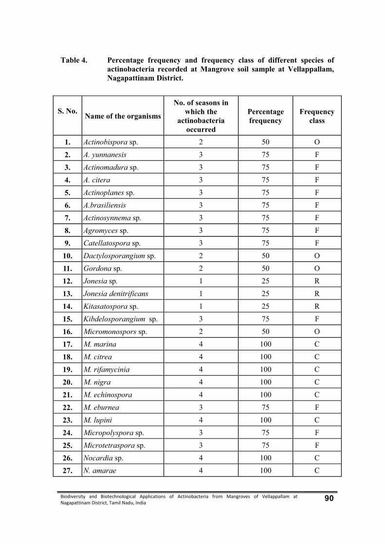
Biodiversity and Biotechnological Applications of Actinobacteria from Mangroves of Vellappallam at Nagapattinam District, Tamil Nadu, India 90
Table 4. Percentage frequency and frequency class of different species of actinobacteria recorded at Mangrove soil sample at Vellappallam, Nagapattinam District.
S. No.
Name of the organisms
No. of seasons in which the
actinobacteria occurred
Percentage frequency
Frequency class
1. Actinobispora sp. 2 50 O
2. A. yunnanesis 3 75 F
3. Actinomadura sp. 3 75 F
4. A. citera 3 75 F
5. Actinoplanes sp. 3 75 F
6. A.brasiliensis 3 75 F
7. Actinosynnema sp. 3 75 F
8. Agromyces sp. 3 75 F
9. Catellatospora sp. 3 75 F
10. Dactylosporangium sp. 2 50 O
11. Gordona sp. 2 50 O
12. Jonesia sp. 1 25 R
13. Jonesia denitrificans 1 25 R
14. Kitasatospora sp. 1 25 R
15. Kibdelosporangium sp. 3 75 F
16. Micromonospors sp. 2 50 O
17. M. marina 4 100 C
18. M. citrea 4 100 C
19. M. rifamycinia 4 100 C
20. M. nigra 4 100 C
21. M. echinospora 4 100 C
22. M. eburnea 3 75 F
23. M. lupini 4 100 C
24. Micropolyspora sp. 3 75 F
25. Microtetraspora sp. 3 75 F
26. Nocardia sp. 4 100 C
27. N. amarae 4 100 C
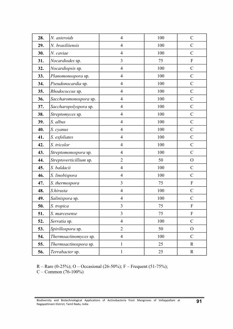
Biodiversity and Biotechnological Applications of Actinobacteria from Mangroves of Vellappallam at Nagapattinam District, Tamil Nadu, India 91
28. N. asteroids 4 100 C
29. N. brasiliiensis 4 100 C
30. N. caviae 4 100 C
31. Nocardiodes sp. 3 75 F
32. Nocardiopsis sp. 4 100 C
33. Planomonospora sp. 4 100 C
34. Pseudonocardia sp. 4 100 C
35. Rhodococcus sp. 4 100 C
36. Saccharomonospora sp. 4 100 C
37. Saccharopolyspora sp. 4 100 C
38. Streptomyces sp. 4 100 C
39. S. albus 4 100 C
40. S. cyanus 4 100 C
41. S. exfoliates 4 100 C
42. S. tricolor 4 100 C
43. Streptomonospora sp. 4 100 C
44. Streptoverticillium sp. 2 50 O
45. S. baldacii 4 100 C
46. S. linobispora 4 100 C
47. S. thermospora 3 75 F
48. S.hirusta 4 100 C
49. Salinispora sp. 4 100 C
50. S. tropica 3 75 F
51. S. marcesense 3 75 F
52. Serratia sp. 4 100 C
53. Spirillospora sp. 2 50 O
54. Thermoactinomyces sp. 4 100 C
55. Thermoactinospora sp. 1 25 R
56. Terrabacter sp. 1 25 R
R – Rare (0-25%); O – Occasional (26-50%); F – Frequent (51-75%); C – Common (76-100%)
Biodiversity and Biotechnological Applications of Actinobacteria from Mangroves of Vellappallam at Nagapattinam District, Tamil Nadu, India 92
4.1.7. Physico-chemical characteristics
Physico-chemical characteristics of the soil samples were collected from four
different seasonal variations from mangrove environment revealed the following
features. (Table 5).
pH
pH was alkaline in soil samples collected during all the seasons. The minimum
of 7.41 was recorded in the samples were collected during summer season. The
maximum was found in the sample collected during premonsoon season.
Electrical conductivity (EC)
Electrical conductivity pronounced considerable variation between the samples.
The minimum of 0.36 dsm-1 was recorded in the sample collected during post monsoon
and the maximum of 0.40 dsm-1 was recorded in the sample collected during summer.
Organic carbon
The organic carbon showed variation between the samples. The minimum of
0.71 % was recorded in the sample collected during pre monsoon season. The
maximum of 0.78 % was recorded in the sample collected during summer.
Organic matter
The organic matter content of soil ranged from 0.71 to 0.77 %. The minimum
was recorded in the samples collected from post monsoon season and the maximum
was recorded from summer season.
Available nitrogen
Available nitrogen content of the soil showed higher variation, it was ranged
between 72.5 and 98.2 (mg/kg). The minimum value was recorded in the samples were
collected from summer season and the maximum value was recorded during monsoon
season.

Biodiversity and Biotechnological Applications of Actinobacteria from Mangroves of Vellappallam at Nagapattinam District, Tamil Nadu, India 93
Available phosphorus (mg/kg)
The available phosphorus content of soil ranged from 1.13 to 1.25 mg/kg. The
minimum of 1.13mg/kg was recorded in the samples collected during the post monsoon
season and the maximum 1.25mg/kg was recorded at during the summer season.
Available potassium (mg/kg)
Available potassium content was showed variation between 93.6 and 97.4
mg/kg. The minimum of 93.6 mg/kg was recorded during the pre monsoon season and
the maximum of 97.4 mg/kg was recorded at during the summer season.
Available zinc (ppm)
Available zinc was ranged from 3.25 to 4.28 ppm in the samples were collected
from during pre monsoon and the maximum was recorded post monsoon season.
Available copper (ppm)
The copper content was 1.59 and 2.52 ppm, was observed in post and pre
monsoon season respectively.
Available iron
Available iron content of the soil was recorded between 4.16 and 4.28 ppm in
all samples analysed. The minimum was recorded during post monsoon season and the
maximum was recorded during pre monsoon season.
Available manganese
Available manganese content ranged from 2.51 to 2.58 ppm. Minimum
manganese content was recorded during post monsoon season and the maximum during
summer season.
Cation exchange capacity (CEC)
Cation exchange capacity showed variations from 53.62 to 54.68 (c.mol
proton+/kg) in the samples. The minimum was recorded during the summer season and
the maximum was recorded during the season of pre monsoon season.

Biodiversity and Biotechnological Applications of Actinobacteria from Mangroves of Vellappallam at Nagapattinam District, Tamil Nadu, India 94
Calcium
Calcium content exhibited considerable variations between the samples
collected from different stations. The range varied from 8.1 to 10.2 mg/kg with the
minimum in the samples collected during post monsoon season and maximum during
monsoon season.
Magnesium
Magnesium content showed variation in the soil samples collected from
different stations, which was in the range of 5.1 -8.3 mg/kg with the minimum in the
samples collected during post monsoon season and the maximum during pre monsoon
season.
Sodium
Sodium content was ranged from 0.78 to 2.36 mg/kg in the samples collected
during post monsoon season and maximum during pre monsoon season.
Potassium
Potassium content of the soil showed lesser variations, 0.09 to 0.28 mg/kg
during post monsoon and maximum during summer season.
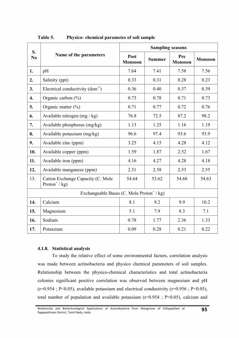
Biodiversity and Biotechnological Applications of Actinobacteria from Mangroves of Vellappallam at Nagapattinam District, Tamil Nadu, India 95
Table 5. Physico- chemical parametes of soli sample
S. No
Name of the parameters
Sampling seasons
Post Monsoon
Summer Pre
Monsoon Monsoon
1. pH 7.64 7.41 7.58 7.56
2. Salinity (ppt) 0.33 0.31 0.28 0.23
3. Electrical conductivity (dsm-1) 0.36 0.40 0.37 0.39
4. Organic carbon (%) 0.73 0.78 0.71 0.73
5. Organic matter (%) 0.71 0.77 0.72 0.76
6. Available nitrogen (mg / kg) 76.8 72.5 87.2 98.2
7. Available phosphorus (mg/kg) 1.13 1.25 1.16 1.19
8. Available potassium (mg/kg) 96.6 97.4 93.6 93.9
9. Available zinc (ppm) 3.25 4.15 4.28 4.12
10. Available copper (ppm) 1.59 1.87 2.52 1.67
11. Available iron (ppm) 4.16 4.27 4.28 4.18
12. Available manganese (ppm) 2.51 2.58 2.53 2.55
13. Cation Exchange Capacity (C. Mole Proton+ / kg)
54.64 53.62 54.68 54.63
Exchangeable Bases (C. Mole Proton+ / kg)
14. Calcium 8.1 9.2 9.9 10.2
15. Magnesium 5.1 7.9 8.3 7.1
16. Sodium 0.78 1.77 2.36 1.33
17. Potassium 0.09 0.28 0.21 0.22
4.1.8. Statistical analysis
To study the relative effect of some environmental factors, correlation analysis
was made between actinobacteria and physico chemical parameters of soil samples.
Relationship between the physico-chemical characteristics and total actinobacteria
colonies significant positive correlation was observed between magnesium and pH
(r=0.954 ; P<0.05), available potassium and electrical conductivity (r=0.956 ; P<0.05),
total number of population and available potassium (r=0.954 ; P<0.05), calcium and

Biodiversity and Biotechnological Applications of Actinobacteria from Mangroves of Vellappallam at Nagapattinam District, Tamil Nadu, India 96
available iron (r=0.956 ; P<0.05) and the significant of negative correlation was
observed potassium and organic matter (r=-0.962 ; P<0.05), cation exchange capacity
and available nitrogen (r=-0.476; P<0.05), potassium and available nitrogen
(r=-0.993 ; P<0.01) and magnesium and available zinc (r=-0.999 ; P<0.01) in table 6.
Table 6. Correlation between total actinobacteria and physico chemical
parameters
pH EC OC OM AN AP K AZ AC AI AM CEC C M S P TNC
pH 1
EC 0.786 1
OC 0.089 -0.202 1
OM 0.089 -0.202 10.000** 1
AN 0.800 0.949 -0.476 0.476 1
AP 0.878 0.395 0.051 0.051 0.447 1
AK 0.909 0.965** -0.263 0.263 0.967* 0.602 1
AZ 0.243 0.390 -0.412 0.412 0.365 0.669 0.184 1
AC 0.938 -0.711 -0.260 0.260 -0.625 0.854 0.801 0.350 1
AI 0.237 0.751 -0.539 0.539 0.763 0.237 0.618 0.880 0.069 1
AM 0.954** 0.383 0.705 0.705 0.075 0.163 0.197 0.274 0.338 0.218 1
CEC 0.550 0.081 -0.028 0.028 -0.042 0.876 0.173 0.888 0.515 0.593 0.484 1
C 0.763 0.994** 0.105 0.105 -0.910 0.368 0.942 0.390 0.724 -0.730 -0.479 0.120 1
M 0.414 -0.884 0.348 0.348 -0.836 0.069 0.755 0.776 0.309 0.962* -0.402 0.507 0.879 1
S 0.332 0.642 0.533 0.533 0.369 0.017 0.482 0.328 0.531 0.406 0.954* 0.402 0.720 0.607 1
P 0.573 -0.908 0.552 0.552 -0.948 0.141 0.856 0.640 0.385 -0.928 -0.133 0.262 0.873 0.948 -0.395 1
TNC 0.160 -0.485 -0.637 0.637 -0.188 0.115 0.303 0.317 0.400 -0.305 0.994** 0.477 0.575 0.494 0.981* 0.241 1
*. Correlation is significant at the 0.05 level; **. Correlation is significant at the 0.01 level
TNC - Total number of colonies, EC - Electrical conductivity, OC - Organic carbon, OM - Organic matter, AN - Available nitrogen, AP - Available phosphorus, AK - Available potassium, AZ - Available zinc, AC- Available copper, AI - Available iron, AM - Available manganese, CEC - Cation exchange capacity, C – Calcium, M – Magnesium, S- Sodium, P – Potassium
4.2. Antibacterial activity of actinobacteria
The antibacterial activity of the selected actinobacteria were evaluated by agar
well diffusion method. The tested bacterial pathogens were five Gram positive bacteria
namely Bacillus subtilis, Enterobacter aerogenes, Enterococcus faecalis,
Staphylococcus aureus and Streptococcus pyogenes and five Gram negative bacteria
namely Escherichia coli, Klebsiella oxytoca, K. pneumoniae, Salmonella typhi and
Vibrio cholerae.
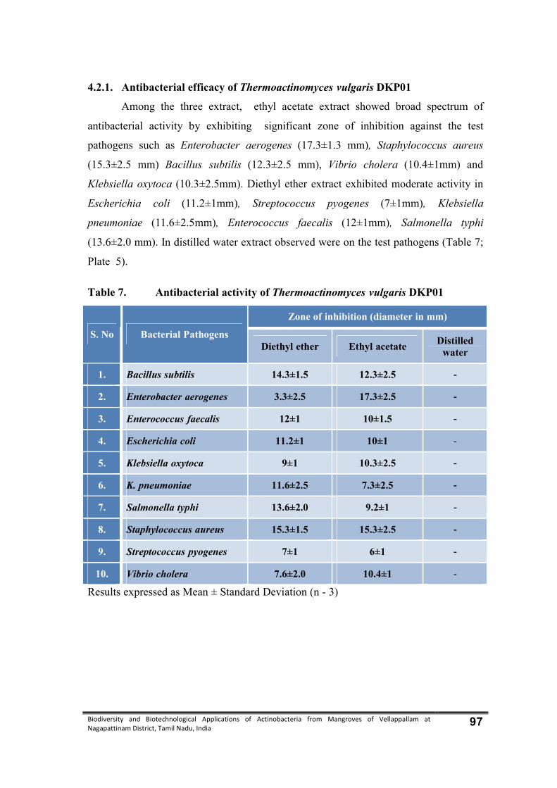
Biodiversity and Biotechnological Applications of Actinobacteria from Mangroves of Vellappallam at Nagapattinam District, Tamil Nadu, India 97
4.2.1. Antibacterial efficacy of Thermoactinomyces vulgaris DKP01
Among the three extract, ethyl acetate extract showed broad spectrum of
antibacterial activity by exhibiting significant zone of inhibition against the test
pathogens such as Enterobacter aerogenes (17.3±1.3 mm), Staphylococcus aureus
(15.3±2.5 mm) Bacillus subtilis (12.3±2.5 mm), Vibrio cholera (10.4±1mm) and
Klebsiella oxytoca (10.3±2.5mm). Diethyl ether extract exhibited moderate activity in
Escherichia coli (11.2±1mm), Streptococcus pyogenes (7±1mm), Klebsiella
pneumoniae (11.6±2.5mm), Enterococcus faecalis (12±1mm), Salmonella typhi
(13.6±2.0 mm). In distilled water extract observed were on the test pathogens (Table 7;
Plate 5).
Table 7. Antibacterial activity of Thermoactinomyces vulgaris DKP01
S. No Bacterial Pathogens Zone of inhibition (diameter in mm)
Diethyl ether Ethyl acetate Distilled water
1. Bacillus subtilis 14.3±1.5 12.3±2.5 -
2. Enterobacter aerogenes 3.3±2.5 17.3±2.5 -
3. Enterococcus faecalis 12±1 10±1.5 -
4. Escherichia coli 11.2±1 10±1 -
5. Klebsiella oxytoca 9±1 10.3±2.5 -
6. K. pneumoniae 11.6±2.5 7.3±2.5 -
7. Salmonella typhi 13.6±2.0 9.2±1 -
8. Staphylococcus aureus 15.3±1.5 15.3±2.5 -
9. Streptococcus pyogenes 7±1 6±1 -
10. Vibrio cholera 7.6±2.0 10.4±1 -
Results expressed as Mean ± Standard Deviation (n - 3)

Biodiversity and Biotechnological Applications of Actinobacteria from Mangroves of Vellappallam at Nagapattinam District, Tamil Nadu, India 98
Plate 5. Antibacterial activity of Thermoactinomyces vulgaris – DKP01
Bacillus subtilis Enterococcus faecalis Klebsiella pneumoniae
Staphylococcus aureus Salmonella typhi Enterobacter aerogenes
Escherichia coli Klebsiella oxytoca
Streptococcus pyogenes Vibrio cholerae
1 – Ethyl Acetate; 2 – Diethyl ether ; 3 – Distilled water
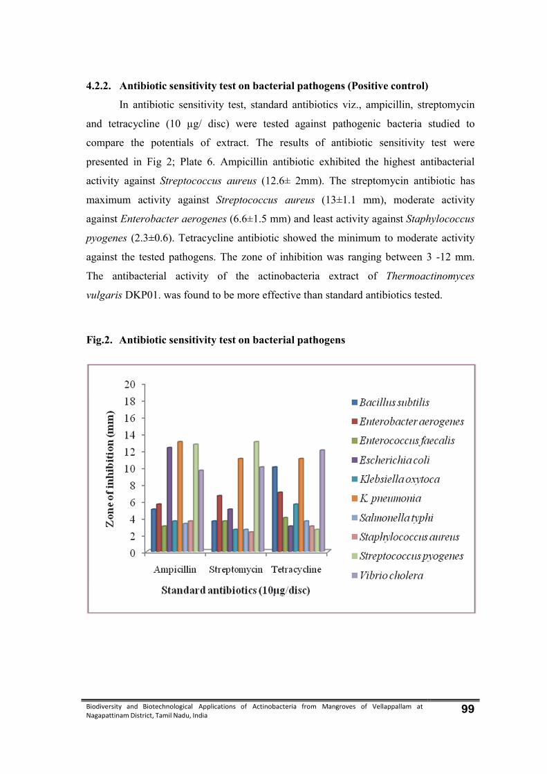
Biodiversity and Biotechnological Applications of Actinobacteria from Mangroves of Vellappallam at Nagapattinam District, Tamil Nadu, India 99
4.2.2. Antibiotic sensitivity test on bacterial pathogens (Positive control)
In antibiotic sensitivity test, standard antibiotics viz., ampicillin, streptomycin
and tetracycline (10 µg/ disc) were tested against pathogenic bacteria studied to
compare the potentials of extract. The results of antibiotic sensitivity test were
presented in Fig 2; Plate 6. Ampicillin antibiotic exhibited the highest antibacterial
activity against Streptococcus aureus (12.6± 2mm). The streptomycin antibiotic has
maximum activity against Streptococcus aureus (13±1.1 mm), moderate activity
against Enterobacter aerogenes (6.6±1.5 mm) and least activity against Staphylococcus
pyogenes (2.3±0.6). Tetracycline antibiotic showed the minimum to moderate activity
against the tested pathogens. The zone of inhibition was ranging between 3 -12 mm.
The antibacterial activity of the actinobacteria extract of Thermoactinomyces
vulgaris DKP01. was found to be more effective than standard antibiotics tested.
Fig.2. Antibiotic sensitivity test on bacterial pathogens
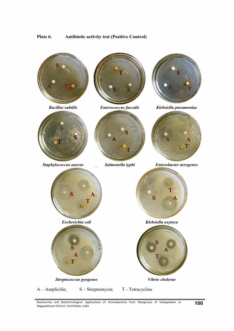
Biodiversity and Biotechnological Applications of Actinobacteria from Mangroves of Vellappallam at Nagapattinam District, Tamil Nadu, India 100
Plate 6. Antibiotic activity test (Positive Control)
Bacillus subtilis Enterococcus faecalis Klebsiella pneumoniae
Staphylococcus aureus Salmonella typhi Enterobacter aerogenes
Escherichia coli Klebsiella oxytoca
Streptococcus pyogenes Vibrio cholerae
A – Amplicilin; S – Streptomycin; T - Tetracycline
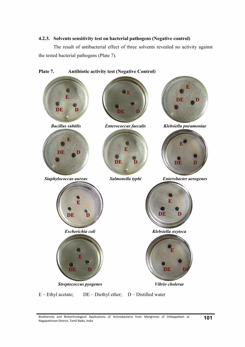
Biodiversity and Biotechnological Applications of Actinobacteria from Mangroves of Vellappallam at Nagapattinam District, Tamil Nadu, India 101
4.2.3. Solvents sensitivity test on bacterial pathogens (Negative control)
The result of antibacterial effect of three solvents revealed no activity against
the tested bacterial pathogens (Plate 7).
Plate 7. Antibiotic activity test (Negative Control)
Bacillus subtilis Enterococcus faecalis Klebsiella pneumoniae
Staphylococcus aureus Salmonella typhi Enterobacter aerogenes
Escherichia coli Klebsiella oxytoca
Streptococcus pyogenes Vibrio cholerae
E – Ethyl acetate; DE – Diethyl ether; D – Distilled water

Biodiversity and Biotechnological Applications of Actinobacteria from Mangroves of Vellappallam at Nagapattinam District, Tamil Nadu, India 102
4.3 Molecular characterization of Thermoactinomyces vulgaris DKP01
4.3.1. Nucleotide sequence accession number
The 16S rRNA gene sequences of the isolate Thermoactinomyces vulgaris
DKP01 was deposited in GenBank and obtained the accession number KF849478
(Plate 8).
Plate 8. 16S rRNA gene sequences of Thermoactinomyces vulgaris DKP01
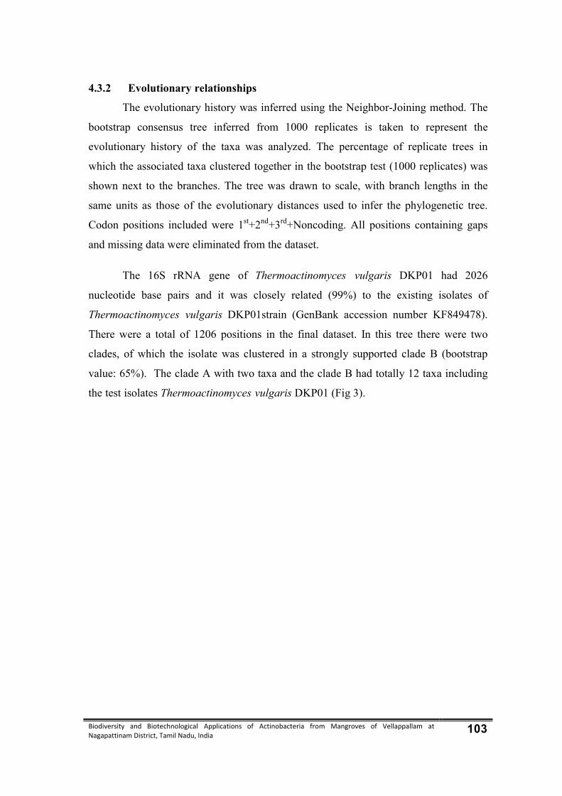
Biodiversity and Biotechnological Applications of Actinobacteria from Mangroves of Vellappallam at Nagapattinam District, Tamil Nadu, India 103
4.3.2 Evolutionary relationships
The evolutionary history was inferred using the Neighbor-Joining method. The
bootstrap consensus tree inferred from 1000 replicates is taken to represent the
evolutionary history of the taxa was analyzed. The percentage of replicate trees in
which the associated taxa clustered together in the bootstrap test (1000 replicates) was
shown next to the branches. The tree was drawn to scale, with branch lengths in the
same units as those of the evolutionary distances used to infer the phylogenetic tree.
Codon positions included were 1st+2nd+3rd+Noncoding. All positions containing gaps
and missing data were eliminated from the dataset.
The 16S rRNA gene of Thermoactinomyces vulgaris DKP01 had 2026
nucleotide base pairs and it was closely related (99%) to the existing isolates of
Thermoactinomyces vulgaris DKP01strain (GenBank accession number KF849478).
There were a total of 1206 positions in the final dataset. In this tree there were two
clades, of which the isolate was clustered in a strongly supported clade B (bootstrap
value: 65%). The clade A with two taxa and the clade B had totally 12 taxa including
the test isolates Thermoactinomyces vulgaris DKP01 (Fig 3).
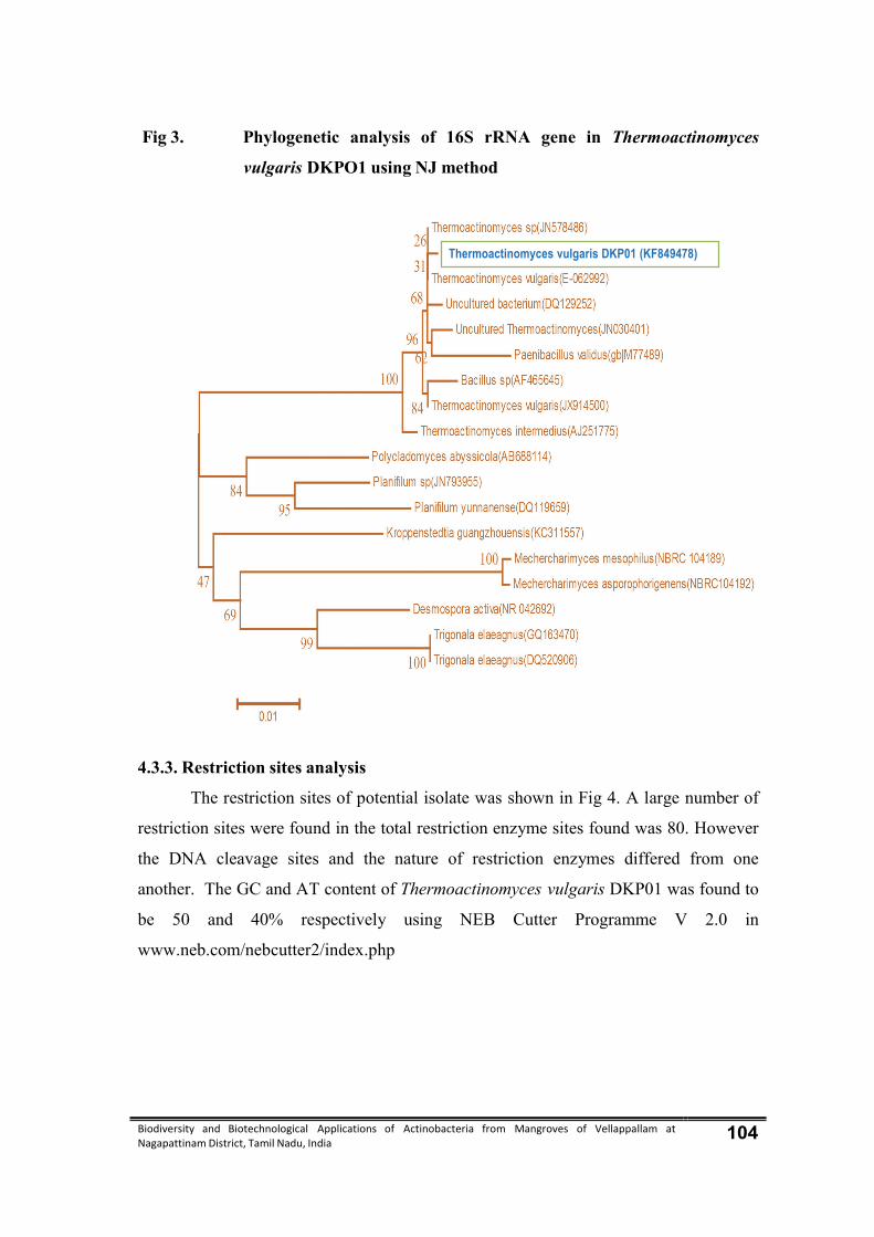
Biodiversity and Biotechnological Applications of Actinobacteria from Mangroves of Vellappallam at Nagapattinam District, Tamil Nadu, India 104
Fig 3. Phylogenetic analysis of 16S rRNA gene in Thermoactinomyces
vulgaris DKPO1 using NJ method
4.3.3. Restriction sites analysis
The restriction sites of potential isolate was shown in Fig 4. A large number of
restriction sites were found in the total restriction enzyme sites found was 80. However
the DNA cleavage sites and the nature of restriction enzymes differed from one
another. The GC and AT content of Thermoactinomyces vulgaris DKP01 was found to
be 50 and 40% respectively using NEB Cutter Programme V 2.0 in
www.neb.com/nebcutter2/index.php
Thermoactinomyces vulgaris DKP01 (KF849478)
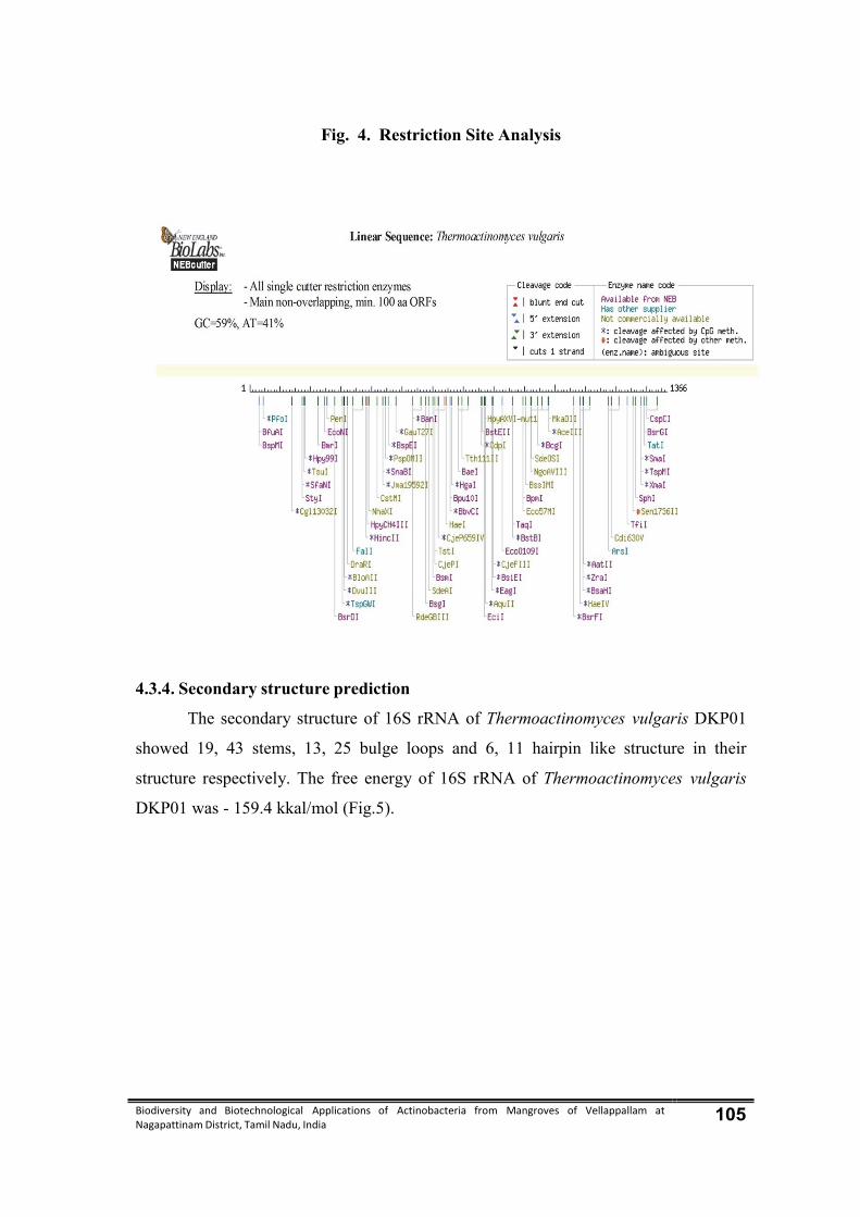
Biodiversity and Biotechnological Applications of Actinobacteria from Mangroves of Vellappallam at Nagapattinam District, Tamil Nadu, India 105
Fig. 4. Restriction Site Analysis
4.3.4. Secondary structure prediction
The secondary structure of 16S rRNA of Thermoactinomyces vulgaris DKP01
showed 19, 43 stems, 13, 25 bulge loops and 6, 11 hairpin like structure in their
structure respectively. The free energy of 16S rRNA of Thermoactinomyces vulgaris
DKP01 was - 159.4 kkal/mol (Fig.5).

Biodiversity and Biotechnological Applications of Actinobacteria from Mangroves of Vellappallam at Nagapattinam District, Tamil Nadu, India 106
Fig. 5. Secondary structure prediction
Biodiversity and Biotechnological Applications of Actinobacteria from Mangroves of Vellappallam at Nagapattinam District, Tamil Nadu, India 107
4.4. Separation of bioactive compounds from Thermoactinomyces vulgaris
DKP01
The bioactive compounds such as alkaloids, flavonoids, phenols, saponins and
sterols of selected actinobacteria were separated by using thin layer chromatography
technique. The Rf values of alkaloids, flavonoids, phenols, saponins and sterols were
presented in table 8.
Table 8. Separation of bioactive compounds from Thermoactinomyces
vulgaris DKP01 by TLC
S. No
Bio active Compounds
Test applied / Reagents
used Observation Results
Rf – Value
Thermoactinomyces vulgaris DKP01
1 Alkaloids Wagner’s reagent
Orange color spot + 0.7±0.2
2 Flavonoids Spot test Yellow color spot + 0.9±0.1
3 Phenols Folin’s –
ciocalteu’s reagent
Blue color spot + 0.6±0.1
4 Saponins Iodine vapours
Yellow color spot + 0.7±0.1
5 Sterols Folin’s
ciocalteu’s reagent
Blue color spot + 0.7±0.1
Results expressed as Mean ± Standard Deviation (n - 3)
4.4.1. Antibacterial activity of bioactive compounds from Thermoactinomyces
vulgaris DKP01
The results of antibacterial efficacy of bioactive compounds from
Thermoactinomyces vulgaris DKP01 were given in table 8; Plate 9. The maximum
antibacterial activity showed by alkaloids against Enterobacter aerogenes (10.6±2.0
mm). The flavonoid compounds exhibited highest activity against Escherichia coli
(12.5±2.5mm) and least activity against Staphylococcus aureus (7.6±2.5mm). The
phenolic compounds were showed maximum inhibitory activity against Bacillus
subtilis (9±1 mm) followed by Enterobacter aerogenes (9±1mm), Streptococcus
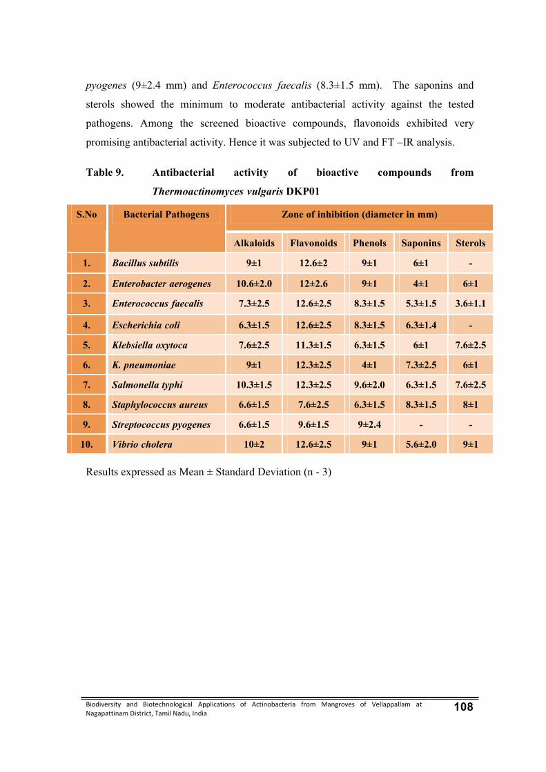
Biodiversity and Biotechnological Applications of Actinobacteria from Mangroves of Vellappallam at Nagapattinam District, Tamil Nadu, India 108
pyogenes (9±2.4 mm) and Enterococcus faecalis (8.3±1.5 mm). The saponins and
sterols showed the minimum to moderate antibacterial activity against the tested
pathogens. Among the screened bioactive compounds, flavonoids exhibited very
promising antibacterial activity. Hence it was subjected to UV and FT –IR analysis.
Table 9. Antibacterial activity of bioactive compounds from
Thermoactinomyces vulgaris DKP01
S.No Bacterial Pathogens Zone of inhibition (diameter in mm)
Alkaloids Flavonoids Phenols Saponins Sterols
1. Bacillus subtilis 9±1 12.6±2 9±1 6±1 -
2. Enterobacter aerogenes 10.6±2.0 12±2.6 9±1 4±1 6±1
3. Enterococcus faecalis 7.3±2.5 12.6±2.5 8.3±1.5 5.3±1.5 3.6±1.1
4. Escherichia coli 6.3±1.5 12.6±2.5 8.3±1.5 6.3±1.4 -
5. Klebsiella oxytoca 7.6±2.5 11.3±1.5 6.3±1.5 6±1 7.6±2.5
6. K. pneumoniae 9±1 12.3±2.5 4±1 7.3±2.5 6±1
7. Salmonella typhi 10.3±1.5 12.3±2.5 9.6±2.0 6.3±1.5 7.6±2.5
8. Staphylococcus aureus 6.6±1.5 7.6±2.5 6.3±1.5 8.3±1.5 8±1
9. Streptococcus pyogenes 6.6±1.5 9.6±1.5 9±2.4 - -
10. Vibrio cholera 10±2 12.6±2.5 9±1 5.6±2.0 9±1
Results expressed as Mean ± Standard Deviation (n - 3)

Biodiversity and Biotechnological Applications of Actinobacteria from Mangroves of Vellappallam at Nagapattinam District, Tamil Nadu, India 109
Plate 9. Antibacterial activity of bioactive compounds Thermoactinomyces vulgaris – DKP01
Bacillus subtilis Enterococcus faecalis Klebsiella pneumonia
Staphylococcus aureus Salmonella typhi Enterobacter aerogenes
Escherichia coli Klebsiella oxytoca
Streptococcus pyogenes Vibrio cholerae
1 – Alkaloids; 2 – Flavanoids; 3 – Phenols; 4 – Saponins; 5 - Steroids
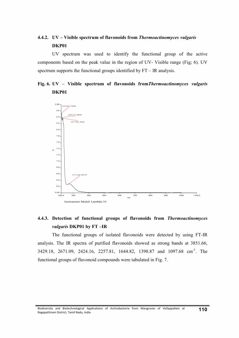
Biodiversity and Biotechnological Applications of Actinobacteria from Mangroves of Vellappallam at Nagapattinam District, Tamil Nadu, India 110
4.4.2. UV – Visible spectrum of flavonoids from Thermoactinomyces vulgaris
DKP01
UV spectrum was used to identify the functional group of the active
components based on the peak value in the region of UV- Visible range (Fig; 6). UV
spectrum supports the functional groups identified by FT – IR analysis.
Fig. 6. UV – Visible spectrum of flavonoids fromThermoactinomyces vulgaris
DKP01
4.4.3. Detection of functional groups of flavonoids from Thermoactinomyces
vulgaris DKP01 by FT –IR
The functional groups of isolated flavonoids were detected by using FT-IR
analysis. The IR spectra of purified flavonoids showed as strong bands at 3851.66,
3429.18, 2671.09, 2424.16, 2257.81, 1644.82, 1398.87 and 1097.68 cm-1. The
functional groups of flavonoid compounds were tabulated in Fig. 7.
Instrument Model: Lambda 35
220.0 300 400 500 600 700 800 900 1000 1100.0
0.00
0.2
0.4
0.6
0.8
1.0
1.2
1.4
1.6
1.8
2.0
2.2
2.4
2.6
2.80
nm
A
273.14,0.29239
231.70,2.3696
228.37,2.4050
223.68,2.5440

Biodiversity and Biotechnological Applications of Actinobacteria from Mangroves of Vellappallam at Nagapattinam District, Tamil Nadu, India 111
Fig. 7. FT –IR spectrum of flavonoids from Thermoactinomyces vulgaris DKP01
4.5. Antioxidant activity of selected Actinobacteria
The antioxidant activity of diethyl ether, ethyl acetate and distilled water
extracts of Thermoactinomyces vulgaris DKP01 were determined by DPPH method.
The scavenging effects on DPPH radicals were determined by measuring the decay, in
absorbance at 517 nm due to the DPPH radical reduction, indicating the antioxidant
activity of the extract in a short time.
The ethyl acetate extract of Thermoactinomyces vulgaris DKP01 showed the
highest scavenging activity (77.5±2.5 %) followed by diethyl ether extract (74.5±2.5%)
and distilled water extract (56±1.4 %). The values are also comparable with
commercial antioxidant selenium (81.3±2.1 %) and ascorbic acid (80.5±1.2%)
(Table 10).
4000.0 3000 2000 1500 1000 400.0
0.0
10
20
30
40
50
60
70
80
90
100.0
cm-1
%T
3878.05
3768.09
3431.82
2914.06
2454.32
2101.78
1640.411391.60
1067.46
599.94
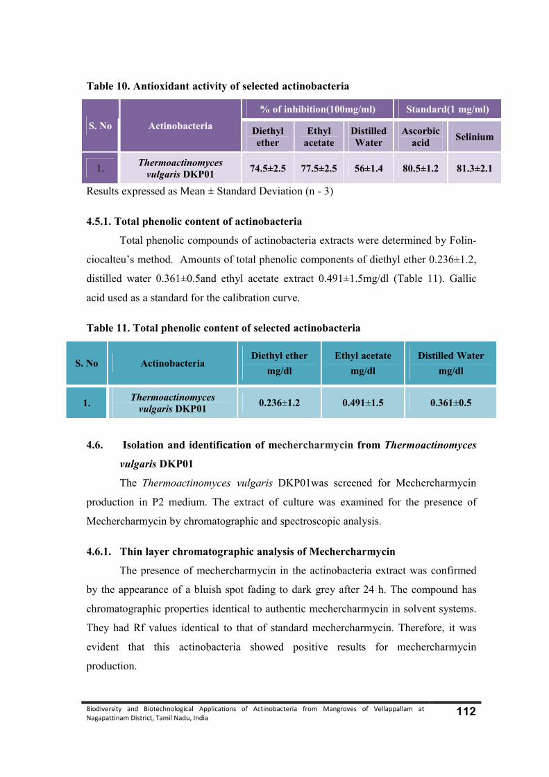
Biodiversity and Biotechnological Applications of Actinobacteria from Mangroves of Vellappallam at Nagapattinam District, Tamil Nadu, India 112
Table 10. Antioxidant activity of selected actinobacteria
S. No Actinobacteria
% of inhibition(100mg/ml) Standard(1 mg/ml)
Diethyl ether
Ethyl acetate
Distilled Water
Ascorbic acid
Selinium
1. Thermoactinomyces
vulgaris DKP01 74.5±2.5 77.5±2.5 56±1.4 80.5±1.2 81.3±2.1
Results expressed as Mean ± Standard Deviation (n - 3)
4.5.1. Total phenolic content of actinobacteria
Total phenolic compounds of actinobacteria extracts were determined by Folin-
ciocalteu’s method. Amounts of total phenolic components of diethyl ether 0.236±1.2,
distilled water 0.361±0.5and ethyl acetate extract 0.491±1.5mg/dl (Table 11). Gallic
acid used as a standard for the calibration curve.
Table 11. Total phenolic content of selected actinobacteria
S. No Actinobacteria Diethyl ether
mg/dl Ethyl acetate
mg/dl Distilled Water
mg/dl
1. Thermoactinomyces vulgaris DKP01 0.236±1.2 0.491±1.5 0.361±0.5
4.6. Isolation and identification of mechercharmycin from Thermoactinomyces
vulgaris DKP01
The Thermoactinomyces vulgaris DKP01was screened for Mechercharmycin
production in P2 medium. The extract of culture was examined for the presence of
Mechercharmycin by chromatographic and spectroscopic analysis.
4.6.1. Thin layer chromatographic analysis of Mechercharmycin
The presence of mechercharmycin in the actinobacteria extract was confirmed
by the appearance of a bluish spot fading to dark grey after 24 h. The compound has
chromatographic properties identical to authentic mechercharmycin in solvent systems.
They had Rf values identical to that of standard mechercharmycin. Therefore, it was
evident that this actinobacteria showed positive results for mechercharmycin
production.

Biodiversity and Biotechnological Applications of Actinobacteria from Mangroves of Vellappallam at Nagapattinam District, Tamil Nadu, India 113
4.6.2. Ultra Violet - Visible (UV) spectroscopic analysis of mechercharmycin
The presence of mechercharmycin in the Thermoactinomyces vulgaris DKP01
extract was further confirmed by UV Spectroscopy. After chromatographic separation,
the area of the TLC plate containing putative mechercharmycin was carefully removed
by scrapping off the silica at the appropriate Rf value and exhaustively eluting it with
methanol. The UV spectral analysis of the Thermoactinomyces vulgaris DKP01 extract
was examined and the spectrum was compared the standard mechercharmycin 340 nm
(Fig. 8 & 9).
Fig. 8. UV - Visible spectrum of the mechercharmycin isolated from
Thermoactinomyces vulgaris DKP01
Instrument Model: Lambda 35
230.0 300 400 500 600 700 800 900 1000 1100.0
-0.05
0.2
0.4
0.6
0.8
1.0
1.2
1.4
1.6
1.8
2.0
2.2
2.4
2.6
2.8
3.00
nm
A
993.93,-0.014889
900.84,-0.0099183
706.10,-0.013328
355.88,0.088108

Biodiversity and Biotechnological Applications of Actinobacteria from Mangroves of Vellappallam at Nagapattinam District, Tamil Nadu, India 114
Fig. 9. UV - Visible spectrum of the standard mechercharmycin
4.6.3. FT –IR analysis of mechercharmycin
Mechercharmycin was further confirmed by IR fingerprints recorded between
400 and 4000cm-1, which were also identical in comparison to the standard
mechercharmycin. Fig. 10 & 11 showed the IR spectrum of mechercharmycin and
authentic mechercharmycin. The IR spectrum showed a broad peak at 3440.32 cm-1,
which was assigned for the presence of the O-group in the compound, as evidenced by
its OH stretch. The registration peak observed at 2361.75 cm-1 and 2336.62 cm-1 was
due to the presence of C ≡ N starching vibrations. The C=O (keto group) stretch was
positioned at 1637. 22 cm-1. The peaks at the range 672.88 cm-1 it was due to the
presence of aromatic groups.
Instrument Model: Lambda 35
190.0 300 400 500 600 700 800 900 1000 1100.0
-0.08
0.0
0.1
0.2
0.3
0.4
0.5
0.6
0.7
0.8
0.9
1.0
1.1
1.20
nm
A
328.03,0.019061
195.87,0.94108
194.07,0.94688
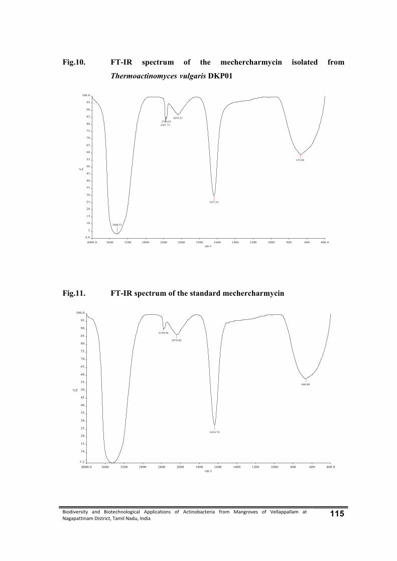
Biodiversity and Biotechnological Applications of Actinobacteria from Mangroves of Vellappallam at Nagapattinam District, Tamil Nadu, India 115
Fig.10. FT-IR spectrum of the mechercharmycin isolated from
Thermoactinomyces vulgaris DKP01
Fig.11. FT-IR spectrum of the standard mechercharmycin
4000.0 3600 3200 2800 2400 2000 1800 1600 1400 1200 1000 800 600 400.0
0.0
5
10
15
20
25
30
35
40
45
50
55
60
65
70
75
80
85
90
95
100.0
cm-1
%T
3440.32
2361.752336.62
2076.33
1637.22
672.88
4000.0 3600 3200 2800 2400 2000 1800 1600 1400 1200 1000 800 600 400.0
3.2
10
15
20
25
30
35
40
45
50
55
60
65
70
75
80
85
90
95
100.0
cm-1
%T
2358.94
2076.82
1636.78
666.80
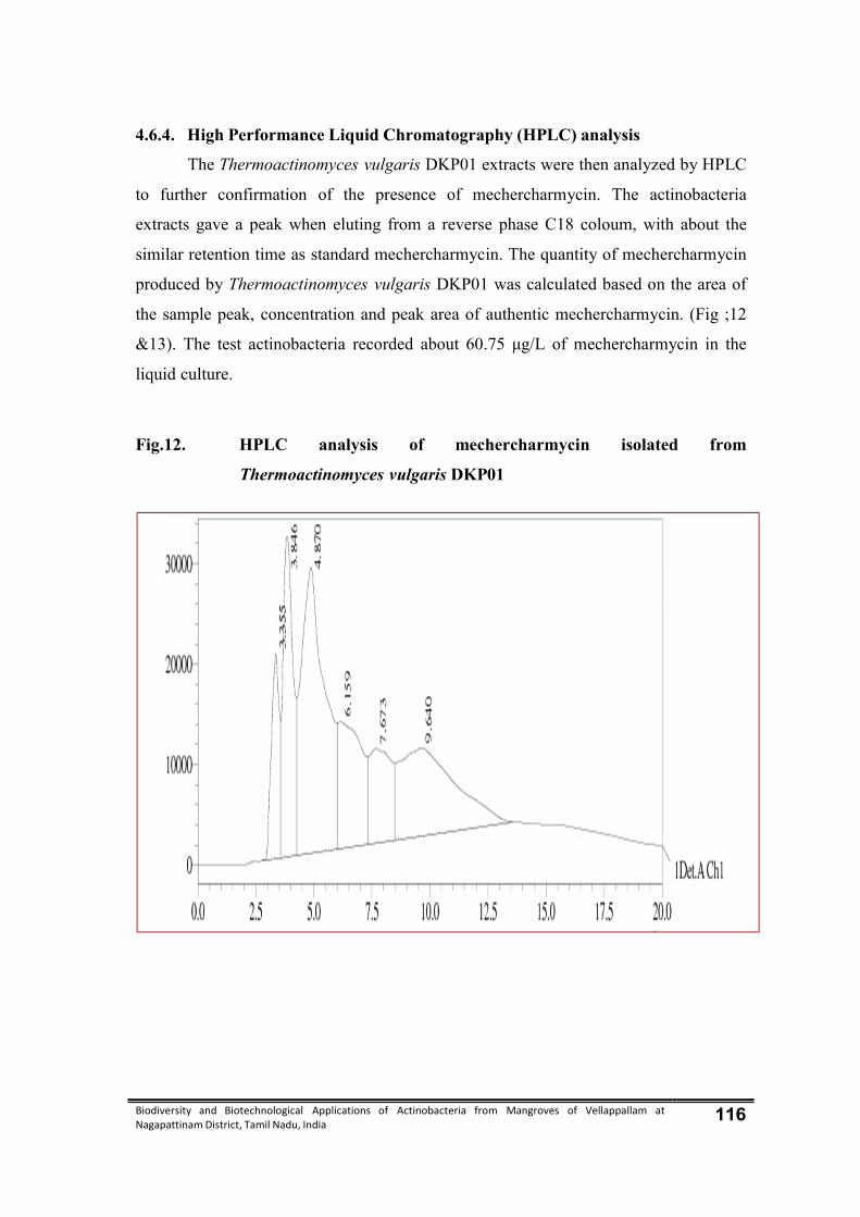
Biodiversity and Biotechnological Applications of Actinobacteria from Mangroves of Vellappallam at Nagapattinam District, Tamil Nadu, India 116
4.6.4. High Performance Liquid Chromatography (HPLC) analysis
The Thermoactinomyces vulgaris DKP01 extracts were then analyzed by HPLC
to further confirmation of the presence of mechercharmycin. The actinobacteria
extracts gave a peak when eluting from a reverse phase C18 coloum, with about the
similar retention time as standard mechercharmycin. The quantity of mechercharmycin
produced by Thermoactinomyces vulgaris DKP01 was calculated based on the area of
the sample peak, concentration and peak area of authentic mechercharmycin. (Fig ;12
&13). The test actinobacteria recorded about 60.75 μg/L of mechercharmycin in the
liquid culture.
Fig.12. HPLC analysis of mechercharmycin isolated from
Thermoactinomyces vulgaris DKP01
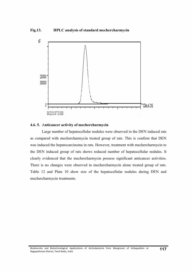
Biodiversity and Biotechnological Applications of Actinobacteria from Mangroves of Vellappallam at Nagapattinam District, Tamil Nadu, India 117
Fig.13. HPLC analysis of standard mechercharmycin
4.6. 5. Anticancer activity of mechercharmycin
Large number of hepatocellular nodules were observed in the DEN induced rats
as compared with mechercharmycin treated group of rats. This is confirm that DEN
was induced the hepatocarcinoma in rats. However, treatment with mechercharmycin to
the DEN induced group of rats shows reduced number of hepatocellular nodules. It
clearly evidenced that the mechercharmycin possess significant anticancer activities.
There is no changes were observed in mechercharmycin alone treated group of rats.
Table 12 and Plate 10 show size of the hepatocellular nodules during DEN and
mechercharmycin treatments.

Biodiversity and Biotechnological Applications of Actinobacteria from Mangroves of Vellappallam at Nagapattinam District, Tamil Nadu, India 118
Table 12. Effect of mechercharmycin and size of hepatocellular nodules during N, N-diethylnitrosamine (DEN) induced hepatocarcinogenesis
S.No Particulars Control Mechercharmycin DEN Mechercharmycin+
DEN
1. Number of rats examined (n)
6 6 6 6
2. Total number of nodules
0 0 103 82
3.
Average number of nodules/nodule bearing liver
0 0 22.30±8.27 14.8±10.86
Plate 10. Effect of mechercharmycin on hepatocellular nodules during DEN induced hepatocarcinogenesis
A - Control , B – mechercharmycin alone, C – DEN alone, D - DEN+ mechercharmycin treatment
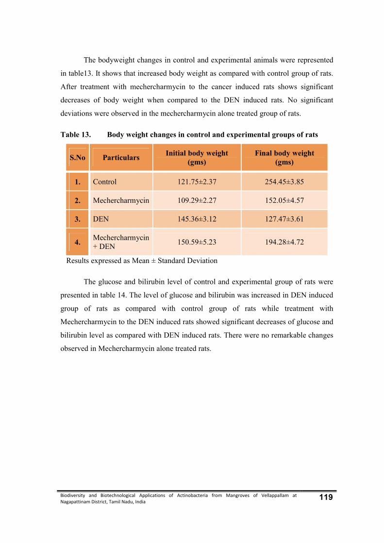
Biodiversity and Biotechnological Applications of Actinobacteria from Mangroves of Vellappallam at Nagapattinam District, Tamil Nadu, India 119
The bodyweight changes in control and experimental animals were represented
in table13. It shows that increased body weight as compared with control group of rats.
After treatment with mechercharmycin to the cancer induced rats shows significant
decreases of body weight when compared to the DEN induced rats. No significant
deviations were observed in the mechercharmycin alone treated group of rats.
Table 13. Body weight changes in control and experimental groups of rats
S.No Particulars Initial body weight
(gms) Final body weight
(gms)
1. Control 121.75±2.37 254.45±3.85
2. Mechercharmycin 109.29±2.27 152.05±4.57
3. DEN 145.36±3.12 127.47±3.61
4. Mechercharmycin + DEN
150.59±5.23 194.28±4.72
Results expressed as Mean ± Standard Deviation
The glucose and bilirubin level of control and experimental group of rats were
presented in table 14. The level of glucose and bilirubin was increased in DEN induced
group of rats as compared with control group of rats while treatment with
Mechercharmycin to the DEN induced rats showed significant decreases of glucose and
bilirubin level as compared with DEN induced rats. There were no remarkable changes
observed in Mechercharmycin alone treated rats.
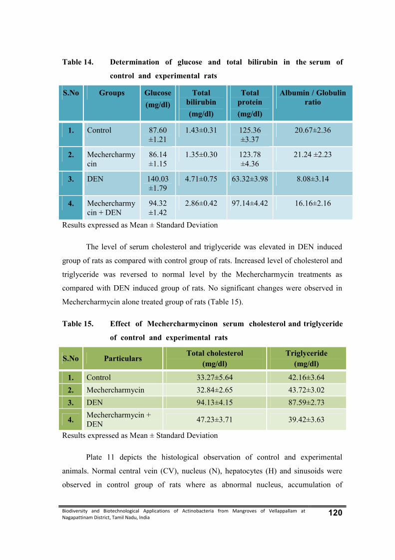
Biodiversity and Biotechnological Applications of Actinobacteria from Mangroves of Vellappallam at Nagapattinam District, Tamil Nadu, India 120
Table 14. Determination of glucose and total bilirubin in the serum of
control and experimental rats
S.No Groups Glucose
(mg/dl)
Total bilirubin
(mg/dl)
Total protein
(mg/dl)
Albumin / Globulin ratio
1. Control 87.60 ±1.21
1.43±0.31 125.36 ±3.37
20.67±2.36
2. Mechercharmycin
86.14 ±1.15
1.35±0.30 123.78 ±4.36
21.24 ±2.23
3. DEN 140.03 ±1.79
4.71±0.75 63.32±3.98 8.08±3.14
4. Mechercharmycin + DEN
94.32 ±1.42
2.86±0.42 97.14±4.42 16.16±2.16
Results expressed as Mean ± Standard Deviation
The level of serum cholesterol and triglyceride was elevated in DEN induced
group of rats as compared with control group of rats. Increased level of cholesterol and
triglyceride was reversed to normal level by the Mechercharmycin treatments as
compared with DEN induced group of rats. No significant changes were observed in
Mechercharmycin alone treated group of rats (Table 15).
Table 15. Effect of Mechercharmycinon serum cholesterol and triglyceride
of control and experimental rats
S.No Particulars Total cholesterol
(mg/dl) Triglyceride
(mg/dl)
1. Control 33.27±5.64 42.16±3.64
2. Mechercharmycin 32.84±2.65 43.72±3.02
3. DEN 94.13±4.15 87.59±2.73
4. Mechercharmycin + DEN
47.23±3.71 39.42±3.63
Results expressed as Mean ± Standard Deviation
Plate 11 depicts the histological observation of control and experimental
animals. Normal central vein (CV), nucleus (N), hepatocytes (H) and sinusoids were
observed in control group of rats where as abnormal nucleus, accumulation of

Biodiversity and Biotechnological Applications of Actinobacteria from Mangroves of Vellappallam at Nagapattinam District, Tamil Nadu, India 121
inflammatory (IF) cell around central vein, damaged central vein, and improper
sinusoids nature and granular in the cytoplasm was observed in DEN induced rats. In
Mechercharmycin treated rats, shows almost normal architecture of liver cells such as
central vein, reduced number of inflammatory cell, hepatocytes and nucleus. Same
architecture was observed in Mechercharmycin alone treated rats.
Plate 11. Histological observation of liver in control and experimental
animals
1 – Control; 2 – Mercharchamycin alone; 3 – DEN Alone; 4 – DEN + Mecharcharmycin treatment H and E stained portion of liver from A- Control, B – Mechercharmycin alone, C – DEN alone, D- DEN+ Mechercharmycin treated central vein (CV), nucleus (N), hepatocytes (H) sinusoids (S) and inflammatory (IF)

Biodiversity and Biotechnological Applications of Actinobacteria from Mangroves of Vellappallam at Nagapattinam District, Tamil Nadu, India 122
4.7. Biosynthesis of silver nanoparticles by Thermoactinomyces vulgaris DKP01
Silver nanoparticles were synthesized by using a reduction of aqueous Ag+ with
the actinobacteria biomass extracts of Thermoactinomyces vulgaris DKP01 at room
temperature. It was generally recognized that silver nanoparticles produced brown
solution in water, due to the surface plasmon resonances (SPR) effect and reduction of
AgNO3. After the addition of AgNO3 solution, the Thermoactinomyces vulgaris
DKP01 biomass extracts changed from light yellow to pink colour in a few hours,
while no color change was observed in the Thermoactinomyces vulgaris DKP01
biomass extract without AgNO3 (Plate 12). Thus, color change of the solution clearly
indicated the formation of silver nanoparticles. The color intensity of the
Thermoactinomyces vulgaris DKP01 biomass extracts with AgNO3 was sustained even
after 24 h incubation, which indicated that the particles were well dispersed in the
solution, and there was no obvious aggregation.
Plate 12. Silver Nanoparticle synthesized using Thermoactinomyces vulgaris –
DKP01
Control Silver Nanoparticle synthesized using
Thermoactinomyces vulgaris
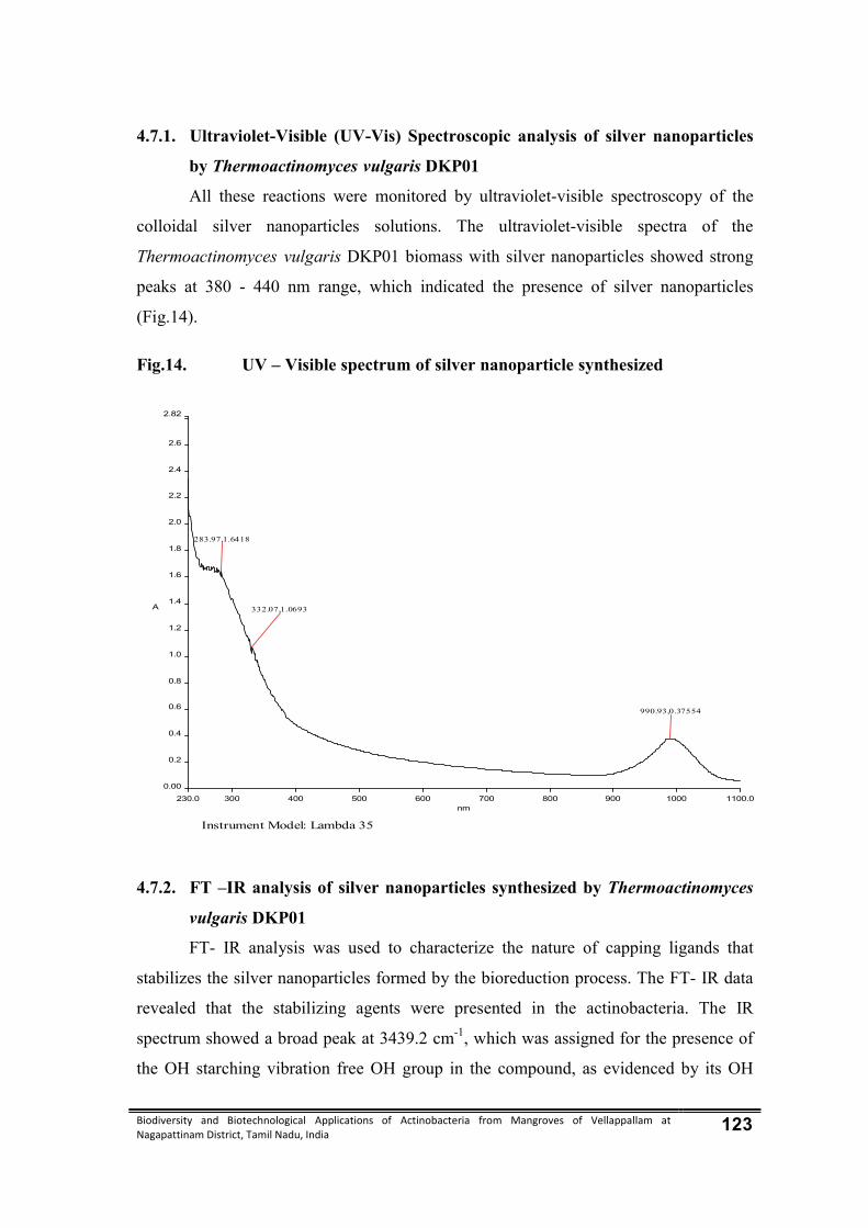
Biodiversity and Biotechnological Applications of Actinobacteria from Mangroves of Vellappallam at Nagapattinam District, Tamil Nadu, India 123
4.7.1. Ultraviolet-Visible (UV-Vis) Spectroscopic analysis of silver nanoparticles
by Thermoactinomyces vulgaris DKP01
All these reactions were monitored by ultraviolet-visible spectroscopy of the
colloidal silver nanoparticles solutions. The ultraviolet-visible spectra of the
Thermoactinomyces vulgaris DKP01 biomass with silver nanoparticles showed strong
peaks at 380 - 440 nm range, which indicated the presence of silver nanoparticles
(Fig.14).
Fig.14. UV – Visible spectrum of silver nanoparticle synthesized
4.7.2. FT –IR analysis of silver nanoparticles synthesized by Thermoactinomyces
vulgaris DKP01
FT- IR analysis was used to characterize the nature of capping ligands that
stabilizes the silver nanoparticles formed by the bioreduction process. The FT- IR data
revealed that the stabilizing agents were presented in the actinobacteria. The IR
spectrum showed a broad peak at 3439.2 cm-1, which was assigned for the presence of
the OH starching vibration free OH group in the compound, as evidenced by its OH
Instrument Model: Lambda 35
230.0 300 400 500 600 700 800 900 1000 1100.0
0.00
0.2
0.4
0.6
0.8
1.0
1.2
1.4
1.6
1.8
2.0
2.2
2.4
2.6
2.82
nm
A
990.93,0.37554
332.07,1.0693
283.97,1.6418
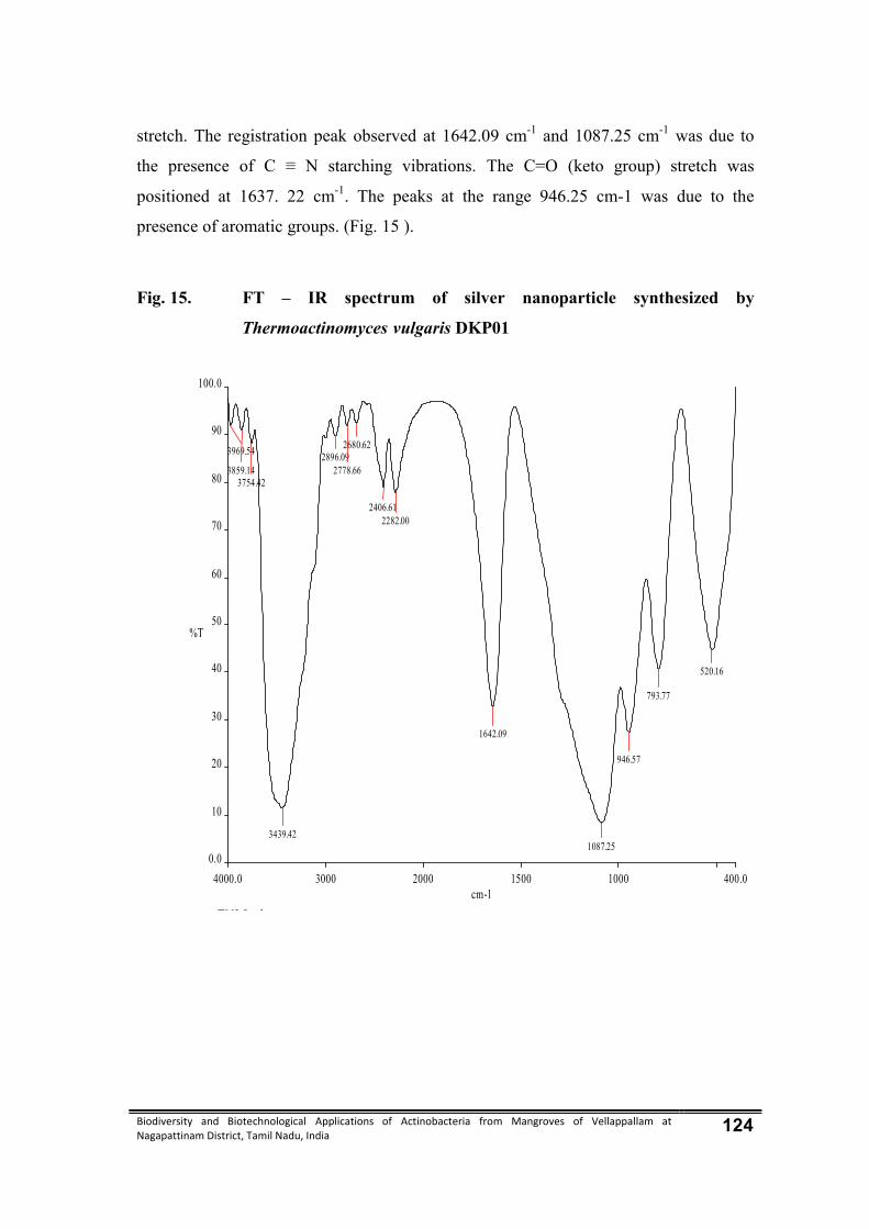
Biodiversity and Biotechnological Applications of Actinobacteria from Mangroves of Vellappallam at Nagapattinam District, Tamil Nadu, India 124
stretch. The registration peak observed at 1642.09 cm-1 and 1087.25 cm-1 was due to
the presence of C ≡ N starching vibrations. The C=O (keto group) stretch was
positioned at 1637. 22 cm-1. The peaks at the range 946.25 cm-1 was due to the
presence of aromatic groups. (Fig. 15 ).
Fig. 15. FT – IR spectrum of silver nanoparticle synthesized by
Thermoactinomyces vulgaris DKP01
p p
TNJ L k
4000.0 3000 2000 1500 1000 400.0
0.0
10
20
30
40
50
60
70
80
90
100.0
cm-1
%T
3969.54
3859.143754.42
3439.42
2896.092778.66
2680.62
2406.612282.00
1642.09
1087.25
946.57
793.77
520.16

Biodiversity and Biotechnological Applications of Actinobacteria from Mangroves of Vellappallam at Nagapattinam District, Tamil Nadu, India 125
4.7.3. Scanning Electron Microscopic (SEM) analysis of silver nanoparticle
synthesized by Thermoactinomyces vulgaris DKP01
The SEM micrographs of the present study were taken at different
magnifications. The silver nanoparticles synthesized by Thermoactinomyces vulgaris
DKP01in plate 13 depicts that the spherical in shape of the silver nanoparticles at
2000X, 5000X and 10,000X magnifications. The nanoparticles sizes were ranging from
50-60 nm. The morphology of the nanoparticles was highly variable.
Plate 13. Scanning Electron Microscopic (SEM) analysis of silver
nanoparticle synthesized by Thermoactinomyces vulgaris
DKP01

Biodiversity and Biotechnological Applications of Actinobacteria from Mangroves of Vellappallam at Nagapattinam District, Tamil Nadu, India 126
4.7.4. Antibacterial activity of silver nanoparticle synthesized by
Thermoactinomyces vulgaris DKP01
In vitro antibacterial efficacy of silver nanoparticles synthesized by
Thermoactinomyces vulgaris DKP01 was investigated by agar well diffusion method.
The silver nanoparticle synthesized by Thermoactinomyces vulgaris DKP01 extract
showed effective inhibitory activity against Klebsiella oxytoca (18.2±2.6 mm),
moderate activity against Enterobacter aerogenes (15.1±1.5mm) and least activity
against Vibrio cholera (11.6±1.5mm). Compared with the control, the diameters of
inhibition zones increased for all the test pathogens (Fig.16; Plate 14).
Fig.16. Antibacterial activity of silver nanoparticles synthesized using
Thermoactinomyces vulgaris DKP01
0
2
4
6
8
10
12
14
16
18
20
Crude sample Siver nanoparticlessynthesized sample
Zon
e of
In
hii
bit
ion
in
mm
Bacillus subtilis
Enterobacter aerogenes
Enterococcus faecalis
Escherichia coli
Klebsiella oxytoca
Klebsiella pneumoniae
Salmonella typhi
Staphylococcus aureus
Streptococcus pyogenes
Vibrio cholerae
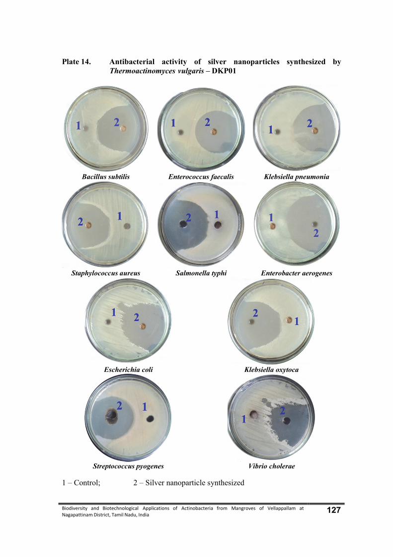
Biodiversity and Biotechnological Applications of Actinobacteria from Mangroves of Vellappallam at Nagapattinam District, Tamil Nadu, India 127
Plate 14. Antibacterial activity of silver nanoparticles synthesized by Thermoactinomyces vulgaris – DKP01
Bacillus subtilis Enterococcus faecalis Klebsiella pneumonia
Staphylococcus aureus Salmonella typhi Enterobacter aerogenes
Escherichia coli Klebsiella oxytoca
Streptococcus pyogenes Vibrio cholerae
1 – Control; 2 – Silver nanoparticle synthesized
Biodiversity and Biotechnological Applications of Actinobacteria from Mangroves of Vellappallam at Nagapattinam District, Tamil Nadu, India 128
5. DISCUSSION
5.1. Actinobacteria
Actinobacteria are prokaryotes with extremely high metabolic potentiality.
They produce numerous substances essential for health such as antibiotics, enzymes,
immunomodulators, etc. During the last few decades actinobacteria have become the
most fruitful source for antibiotics. Natural organic compounds produced by
microorganisms are an important screening target for a variety of bioactive substances
of actinobacteria origin, in particular, have been valuable in the field of bioactive
substances. (Imada et al.,2007).
Actinobacteria play a pivotal role in maintaining a satisfactory biological
balance in soil (Strohl, 2004). Actinobacteria are potent producers of wide variety of
secondary metabolites with diverse biological activities, which includes therapeutically
and agriculturally important compounds (Suzuki et al., 1991; Balagurunathan, 1992;
Tanaka and Omura, 1993; Lange and Sanchez Lopez, 1996).
Among actinobacteria, the members of the genus Streptomyces are considered
economically important because they alone constituted 50% of the total soil
actinobacteria population (Xu et al., 1996) and 75% of total bioactive molecules are
produced by this genus (Demain, 2000).The Streptomycetes produce an array of
secondary metabolites such as enzyme inhibitors, herbicides and large number of
antibiotics (Omura, 1992; Lange and Sanchez Lopez, 1996 and Demain, 1999).
Hence, the present investigation discussed the diversity, antibacterial activity,
synthesis of silver nanoparticles, and biotechnological applications of selected
actinobacteria.
5.2. Biodiversity of marine actinobacteria
Members of the actinobacteria, which live in marine environment, are poorly
understood compared with terrestrial environment.The first report in marine

Biodiversity and Biotechnological Applications of Actinobacteria from Mangroves of Vellappallam at Nagapattinam District, Tamil Nadu, India 129
actinobacteria was made by Nadson (1903), from the salt muds.It was well documented
that actinobacteria isolated from marine sediments (Walker and Colwell, 1975;
Goodfellow and Haynes, 1984; Pisano et al., 1986; Ellaiah and Reddy, 1987; Barcina
et al., 1987; Weyland and Helmke, 1988; Pisano et al., 1989; Takizawa et al.,
1993;Dhanasekaran et al., 2005a; Vijayakumar et al., 2007; Dhanasekaran et al., 2009).
In addition, because of actinobacteria are common soil bacteria, produce resistant
spores, and are known to be salt tolerant (Tresner et al., 1968; Okazaki and Okami,
1975; Okami and Okazaki, 1978; Kuster and Neumeier, 1981).
Remarkably, majority of the marine actinobacteria were Streptomyces (Sujatha
et al., 2005; Sathiyaseelan and Stella, 2011a; b; Chacko Vijai Sharma and David, 2012;
Parthasarathi et al., 2012) and chemotaxonomic investigation using isomeric
diaminopimelic acid (DAP) configuration was already established (Becker et al., 1964;
Lechevalier and Lechevalier, 1970). It has been reported that the Streptomyces are
common inhabitants of marine environments (Kokare et al., 2004a; Fiedler et al., 2005;
Ramesh et al., 2006; 2009), though other actinobacteria are also present (Jensen
et al.,1991;Mincer et al., 2002; Magarvey et al., 2004; Malarvizhi, 2006; Maldonado
et al., 2005, 2008).
Lacey & Cross (1989) described the genus Thermoactinomyces as the only
genus of Thermoactinomycetes, which was placed in the family Bacillaceae
(Stackebrandt & Woese, 1981). More recently, members of the genus
Thermoactinomyces were divided into four genera, Thermoactinomyces, Laceyella,
Thermoflavimicrobium and Seinonella (Yoon & Park, 2000; Yoon et al., 2005).
Hot spring sediment and soil samples from West Anatolia in Turkey were
investigated for the occurrence of thermophilic Actinobacteria (Yallop et al. 1997).
Among these thermophilic Actinomycetes, the genus Thermoactinomyces has industrial
and clinical importance. Some Thermoactinomyces strains are known as potent protease
producers
Biodiversity and Biotechnological Applications of Actinobacteria from Mangroves of Vellappallam at Nagapattinam District, Tamil Nadu, India 130
The mangrove sediments can be a potential source for isolating diversified
marine actinobacteria, rather than other marine samples. This might be due to the
favourable niche of the mangrove habitat that provides nutrients for saprophytic
actinobacteria (Lakshmanaperumalsamy, 1978; Rathana Kala and Chandrika, 1993;
Vikineswary et al., 1997; Sivakumar, 2001; Sateesh et al., 2011;Baskaran et al., 2011).
Muthupet mangrove actinobacterial distribution and its mosquito larvicidal potential
was reported by Dhanasekaran et al. (2010).
Previous studies on the screening of salt pan actinobacteria for its antimicrobial
potentialities were carried out by several workers (Pathiranana et al., 1991;
Jensen et al., 1991; Sorza et al., 2000; Kokare et al., 2000; Dhanasekaran et al., 2005b;
Gayathri et al., 2011). However, still it has not been fully explored and there is
tremendous potential to identify novel organisms with various biological properties
from mangrove and salt pan ecosystems. The present research has been initiated to
identify novel actinobacteria isolated from mangrove environment. The mangrove soil
samples were collected from Vellappallam at four different seasons.
In the present investigation, 56 actinobacteria were isolated from four different
seasonal by using sea water starch casein agar (SCA). It has already been reported that
the seawater amended media were used to isolate and maintain the marine
actinobacteria. Although, a number of selective media (Kuster and Williams, 1964;
Hayakava and Nonomura, 1987; Crawford et al., 1993; Duangmal et al., 2005) were
developed for the isolation of actinobacteria. Among the various media, SCA was very
good selective medium, because in this medium the development of bacterial and
fungal colony was very much suppressed, allowing only the actinobacteria to grow.
In the present study, the colonies of actinobacteria were elevated, convex and
powdery in nature. Many of such morphological characteristics are common in most of
the Streptomyces (Lo et al., 2002; Fourati Ben Fguira et al., 2005; Sujatha et al., 2005).
Most of the marine actinobacteria exhibited different mycelial conditions. The spore
morphology was considered as one of the important characteristics for the identification
of Streptomyces and it greatly varies among the species. This is similar to

Biodiversity and Biotechnological Applications of Actinobacteria from Mangroves of Vellappallam at Nagapattinam District, Tamil Nadu, India 131
Tresner et al., (1961) findings. It has been found that the majority of the marine isolates
produced aerial coiled mycelia and the spores arranged in chains as already reported by
Mukherjee and Sen (2004) and Roes and Meyer (2005).
Previously, studies on the actinobacteria of the mangrove environments is less
and hence the present study was carried out in the different mangrove environment of
Bhitarkanikka, Orissa Rajkumar et al., (2012). A total of 116 actinobacterial colonies
were recorded from 30 mangrove and marine sediment samples of Bhitherkanikka
mangrove environment east coast of Orissa. Among them, 67 isolates of were
morphologically distinct on the basis of colour of spore mass riverside colour, Aerial
and substrate mycelia format production of diffusible pigment sporophore morphology.
Classical approaches for classification make use of morphological,
physiological and biochemical characters. The classical method described in the
identification key by Nonomura (1974) and Bergey’s Manual of Determinative
Bacteriology (Buchanan and Gibbons, 1974) is very much useful in the identification of
Streptomyces. These characteristics have been commonly employed in taxonomy of
Streptomycetes for many years. They are quite useful in routine identification.
Colony formation vegetative and aerial mycelium structure of sporophores and
spores are the most important features of identification of Actinomycetes (Waksman,
1957, Krasilnikov, 1960, Waksman, 1961, Kuster, 1963), Pridham and Tresner (1974)
reported that the colour of aerial mycelium is considered to be an important character
for the grouping and identification of actinomycetes.
Identification of microorganisms especially actinomycetes could be confirmed
by morphological, cultural, biochemical and physiological. In the present investigation
actinobacteria, were identified by morphological, cultural, biochemical and
physiological characteristics. The white, pale yellow, light green, Dark green, brown,
and Ash coloured isolates were found predominant such as dominance of members of
green and white colour actinobacteria.
Biochemical characteristics of the actinomycetes were also used as the
characters of identification (Nikolova et al., 2004, Gottlieb, 1961, Kim and
Biodiversity and Biotechnological Applications of Actinobacteria from Mangroves of Vellappallam at Nagapattinam District, Tamil Nadu, India 132
Goodfellow, 2002). The production of citrate, urease, catalase, oxidase and
β-lactamase are considered for characterizing Streptomyces (Nitsch and Kutzer, 1969,
Gotoh et al., 1982).
Various biochemical characteristics of the Streptomyces were used for their
identification (Waksman, 1957, 1959, 1961; Kuster, 1963, Jones and Bradley, 1959;
Rajendra and Maskey 2003). In the present study with the help of biochemical
characteristics such as Indole, methyl red, Voges proskauer, citrate, urease, nitrate, and
catalase tests were characterized for actinobacteria.
5.3. Physico – chemical characteristics of the soil
Actinobacteria have a worldwide distribution which indicates their plasticity
and adoptability to various extreme environments. In spite of the fact that the
actinobacteria have wide distribution and they showed variation in population
dynamics. In the present investigation, it was found that there was correlation between
physico - chemical properties of soil and total actinobacteria population (TAP). It
revealed a significant positive correlation between TAP and available nitrogen
(r=0.928; p<0.01); TAP and available phosphorous (r=0.955; p<0.01) TAP and
available manganese (r=0.954; p<0.01). There was a significant negative correlation
between TAP and pH (r=-0.638; p<0.05) and TAP and AN (r=-0.993; p<0.05). Similar
type of study was already reported by Saadoun and Al-Momani, (1996); Dhanasekaran
et al.,(2008). Jiang and Xu (1990) have studied the pH, organic matter, nitrogen and
phosphorous content of the soils as correlated with actinobacteria population.
In the present study, 10 actinobacteria were isolated from the pH - 7.02 in
monsoon season, whereas pH was slightly increased in summer season. There was 18
actinobacteria were isolated. The present study has an agreement with previous findings
of Taber (1960) demonstrated that most of the actinobacteria prefer neutral or slightly
alkaline soils for their growth. Similarly Lee and Hwang (2002) observed that
Streptomyces were predominant in soils with a pH range of 5.1 – 6.5.
Biodiversity and Biotechnological Applications of Actinobacteria from Mangroves of Vellappallam at Nagapattinam District, Tamil Nadu, India 133
Ghanem et al. (2000) found that the variation in temperature, pH and dissolved
phosphate insignificant values, but the variation in total nitrogen and organic matter
were significant in the actinobacteria population of Alexandria. The correlation
between salinity, pH and organic content of marine sediments and TAP has been
reported by Jensen et al., (1991) and Ndonde and Semu (2000). Lee and Hwang (2002)
reported that the soil pH, moisture and organic matter infuence the dominance of
Streptomyces in the soils of Western part of Korea.
Satheeshkumar and Anisa Khan, (2009) reported that the seasonal variation of
physico-chemical parameters were studied at four different stations in Pondicherry
mangroves, Southeast coast of India. Atmospheric and surface water temperatures (ºC)
varied from17.9-41.7 and 16.66-37.91 respectively. Annual rainfall and relative
humidity ranges were 1.1-808 mm and 37 – 100 % respectively. Seasonal variations of
different parameters investigated were as follows: salinity (6.36-36.77ppt), dissolved
oxygen (3.45-5.49 mg/l), pH (7.11-8.52), electrical conductivity (26.65-52 ms-1),
sulphide (2.76-47.16 mg/l), soil parameters sand (63.69-87.31%), silt (9.89-29.32 %),
clay (3.06-17.98 %) and organic matter (0.94-3.94 %). pH, temperature, salinity, sand,
silt, clay and organic matter indicated a correlation at P<0.01. Multivariate statistical
technique was applied to evaluate the temporal/spatial variations in mangrove water
quality of Pondicherry mangroves.
Physico-chemical parameters in the water and soil of Vedaranyam mangroves
during the year 2008-2009 at four-seasonal intervals. In the present study N, P, K and
Na, were maximum in summer season. The total amount of N, P, K, Na, Ca and Mg
were maximum in the monsoon and minimum in summer season. The micronutrients
such as zinc, copper, iron and manganese also present in moderate level in all the
season was reported by Ramamurthy et al. (2012).
5.4. Antibacterial activity of actinobacteria
Infectious diseases are the leading cause of death world-wide. Antibiotic
resistance has become a global concern (Westh et al., 2004). The clinical efficacy of
many existing antibiotics is being threatened by the emergence of multidrug resistant

Biodiversity and Biotechnological Applications of Actinobacteria from Mangroves of Vellappallam at Nagapattinam District, Tamil Nadu, India 134
pathogens (Bandow et al., 2003). There is a continuous and urgent need to discover
new antimicrobial compounds with diverse chemical structures and novel mechanisms
of action for new and reemerging infectious diseases.
The screening of actinobacteria for antimicrobial activity has shown that
Sterptomycesrepresent a potential source of novel antibiotics (Li et al., 2005;Xu et al.,
2005; Sukanyanee et al., 2006; Firákóva et al., 2007; Xu et al., 2008; Tayung and
Jha, 2010; Thalavaipandian et al., 2011; Tayung et al., 2011). The presence of
antibacterial substances in the actinobacteria is well established (Zou et al., 2000;
Hellwig et al., 2002; Raviraja et al., 2006; Denise et al., 2008; Ramasamy et al.,
2010). Antibacterial activity depends on the actinobacteria and efficiency on extraction
of their active principles.
In the present investigation involving actinobacteria isolated from mangrove
soil samples. The actinobacteria showed significant antibacterial activity against Gram
positive bacteria as well as Gram negative bacteria. The Thermoactinomyces vulgaris
DKP01 extracts differ significantly in their activity against tested bacterial pathogens.
These differences may be attributed the fact that the occurrence of different
antimicrobial compounds with different solvents.
In the case of test bacteria, the basis for their differences in susceptibility might
be due to the differences in the cell wall composition of Gram positive and Gram
negative bacteria (Grosvenor et al., 1995 and Yao et al., 1995).
In the present study, among the three extract tested, ethyl acetate extract of
Thermoactinomyces vulgaris DKP01 showed broad spectrum of antibacterial activity
against the tested pathogens. Diethyl ether extract exhibited moderate activity and
distilled water extract showed slight inhibitory effects on the test pathogens.
Antibacterial potential of the genus Actinobispora and Streptoverticillium proved by
Maria et al. (2005); Nirjanta Devi and Wahab (2012) was coincide with the present
study.
Biodiversity and Biotechnological Applications of Actinobacteria from Mangroves of Vellappallam at Nagapattinam District, Tamil Nadu, India 135
To determine the effectiveness of the extraction methods, three different
solvents were used for the extraction of antimicrobial metabolites from the culture
filtrate of the selected actinobacteria. Distilled water extracts showed least antibacterial
activity while ethyl acetate extractions of actinobacteria showed higher antibacterial
activity than diethyl ether extracts. It is accepted widely that the use of organic solvents
always provides a higher efficiency in extracting the antimicrobial compounds when
compared with water extraction (Rosell and Srivastava, 1987). Similarly there are
numerous reports emphasis, that the use of ethyl acetate for the extraction of
antimicrobial compounds from actinobacteria is an effective method (Dalsgaard et al.,
2005; Lin et al., 2005; Li et al., 2006 and Gallardo et al., 2006).
In the present study, diethyl ether extracts of Thermoactinomyces vulgaris
DKP01 showed promising antibacterial activity when compared to other extracts. Ethyl
acetate extract of minimum to moderate activity against the tested pathogens.
5.5. Molecular characterization of potential actinobacteria
The most powerful approaches to taxonomy are through the study of nucleic
acids. Because these are either direct gene products or the genes themselves, and
comparisons of nucleic acids yield considerable information about true relatedness.
Molecular systematics, which includes both classification and identification, has
its origin in the early nucleic acid hybridization studies, but has achieved a new status
following the introduction of nucleic acid sequencing techniques (O’Donnell et al.,
1993). Significance of phylogenetic studies based on 16S rDNA sequences is
increasing in the systematics of bacteria and actinobacteria (Yokota, 1997). Sequences
of 16S rDNA have provided actinobacteriologists with a phylogenetic tree that allows
the investigation of evolution of actinobacteria and also provides the basis for
identification.
Correspondingly, the isolate SRA14 was identified as the genus Streptomyces
on the basis of its structural and morphological characteristics (Shirling and Gottlieb,
1966). Analysis of the 16S rDNA sequences showed that SRA14 was closely related to
Biodiversity and Biotechnological Applications of Actinobacteria from Mangroves of Vellappallam at Nagapattinam District, Tamil Nadu, India 136
S. hygroscopicus has 98% similarity with GenBank database accession number
AB184760 (Getha and Vikineswary, 2002). In the present investigation, among the
three potential actinobacteria isolate one efficient actinobacteria was further justifiably
choosen for molecular taxonomic characterization and identification. Besides on the
morphology and molecular properties Thermoactinomyces vulgaris DKP01. The 1475
base pairs of 16S rRNA gene sequences of Thermoactinomyces vulgaris DKP01
revealed that it was closely (99%) related to existing strain of Thermoactinomyces
vulgaris DKP01 (99%) similarity (Genbank Accession No. KF849478) by Blast
analysis.
In the present investigation, distinct variation in the secondary structure, G+C
content, presence of restriction enzymes sites in 16S rRNA gene sequences of
Thermoactinomyces vulgaris DKP01 were observed which confirmed molecular level
specificity of each and every individual isolates. The similar study was also reported in
soil streptomyces by Dhanasekaran et al. (2012).
5.6. Bioactive compounds from actinobacteria
Due to the special attributes of the marine environment, marine actinobacteria
are thought to have distinct physiological, morphological and chemotaxonomical
characteristics and unique production of secondary metabolites and bioactive
compounds. Secondary metabolites produced by marine actinobacteria have distinct
chemical structures, which may form the basis for the synthesis of new drugs (Solanki
et al., 2008).
The isolation of the antifungal metabolite mildiomycin from a culture of
Streptoverticillium rimofaciens Niida was reported in 1978, also by Takeda scientists
(Iwasa et al., 1978). Mildiomycin is strongly active against several powdery mildews
on various crops (Harada and Kishi 1978), acting as an inhibitor of the fungal protein
biosynthesis (Feduchi et al., 1985).
Two secondary metabolites of Streptomyces sp. PM5 have been isolated,
purified, coded as SPM5C-1 and SPM5C-2 and characterized by Prabavathy(2005).

Biodiversity and Biotechnological Applications of Actinobacteria from Mangroves of Vellappallam at Nagapattinam District, Tamil Nadu, India 137
Out of two compounds, SPM5C-1 was shown to be highly effective against P. oryzae
and R. solani as it remarkably inhibited their growth, especially in comparison to
SPM5C-2. This was comparable with the results of dual culture test in which,
Streptomyces sp. PM5 was effectively inhibited the mycelial growth of many fungal
pathogens including the above rice pathogens. Further, it has been observed that the
culture filtrate of Streptomyces sp. PM5 completely inhibited the conidial and sclerotial
germination of P. oryzae and R. solani, respectively. Previously, Omura et al. (1984)
discovered irumamycin, a 20 membered macrolide produced by Streptomyces flavus
sub sp. irumaensis with potent activity against P. oryzae. Interestingly, dapiramycin, an
antifungal metabolite obtained from Micromonospora sp. exhibited only weak activity
against P. oryzae although it effectively suppressed the growth of R. solani (Nishizawa
et al., 1984).
In the present investigation, the bioactive compounds namely alkaloids,
flavonoids, phenols, saponins and sterols were separated from Thermoactinomyces
vulgaris DKP01 by using TLC.The production of antifungal compounds has already
been reported by many species of Streptomyces (Xiao et al., 2002; Fourati Ben Fguira
et al., 2005 and Taechowisan et al., 2005).
The FT – IR profile of bioactive compounds of the present study indicated that
the presence of nine prominent peaks. The functional groups of saponin compounds
were N-H stretching vibrations secondary bonded, one bond, alkyne mono substituted,
C – H stretching two bonds, β diketones (enolic) and O - H – bonding and C-O
stretching vibrations, primary alcohol groups by FT – IR analysis.
Rothrock and Gottlieb (1984) presented evidence that the antibiotic
geldanamycin is produced in soil by S. hygroscopicusvar.geldanus and that the
antibiotic accounts for the antagonism of S.hygroscopicus var. geldanus to Rhizoctonia
solani in soil. Streptomyces hygroscopicusvar.geldanus inhibited the growth of
R. solani.
Biodiversity and Biotechnological Applications of Actinobacteria from Mangroves of Vellappallam at Nagapattinam District, Tamil Nadu, India 138
5.7. Antioxidant activity of Thermoactinomyces vulgaris DKP01
Free radicals are often generated as byproducts of biological reactions or from
exogenous factors. The involvement of free radicals in the pathogenesis of a large
number of diseases is well documented. A potent scavenger of free radicals may serve
as a possible preventive intervenetion for the diseases (Gyamfi et al., 1999).
Free radicals contribute to more than one hundred disorders in humans
including atherosclerosis, arthritis, and ischemia reperfusion injury of many tissues,
central nervous system injury, gastritics, cancer and AIDS (Cook and Samman, 1996;
Kumpulainen and Salonen, 1999).
Antioxidants may protect the body against ROS toxicity either by the formation
of ROS by bringing disruption in ROS attack, by converting them to less reactive
molecules or by scavenging the reactive metabolites (Sen, 1995; Hegde and Joshi,
2009). The natural antioxidants were characterized from the actinobacteria compounds
(Sun et al., 2004).
Antioxidant based drug formulations are used for the prevention and treatment
of complex diseases like atherosclerosis, stroke, diabetes, Alzheimer’s disease and
cancer (Devasagayam et al., 2004). The majority of the antioxidant activity is due to
the flavones, isoflavones, flavonoids, anthocyanin, coumarin lignans, catechins and
isocatechins (Aqil et al., 2006). Due to depletion of immune system natural
antioxidants in different malady, consuming antioxidants as free radical scavengers
may be necessary (Kuhnan, 1976; Younes, 1981; Halliwell, 1994; Kumpulainen and
Salonen, 1999). DPPH radical scavenging assay is a classic, simple, sensitive and rapid
method of assessing antioxidant activity (Moreno et al., 1998; Gulçin et al., 2005).
The number of reports on the antioxidants activity of the actinobacteria has
increased immensely during the last decade (Harper et al., 2003; Song et al., 2005;
Huang et al., 2007a; Huang et al., 2007b; Srinivasan et al., 2010; Pei Yuan et al., 2010;
Zeng et al., 2011; Jayanthi et al., 2011; Nithya et al., 2011; Nath et al., 2012;
Ravindran et al., 2012 and Dhankhar et al., 2012).

Biodiversity and Biotechnological Applications of Actinobacteria from Mangroves of Vellappallam at Nagapattinam District, Tamil Nadu, India 139
In the present investigation, the antioxidant activity of diethyl ether, distilled
water and ethyl acetate extracts of Thermoactinomyces vulgaris DKP01 were evaluated
by DPPH method. In the present investigation, ethyl acetate was the best solvent for the
extraction of radical scavenging compounds from actinobacteria. Similar work was
done by Jayanthi et al. (2011) that the ethyl acetate extracts of Sterptomyces sp.
GJJM07 showed significant DPPH radical scavenging activity.
Phenolic compounds seem to have an important role in stabilizing lipid
oxidation and are associated with antioxidant activity, which is emphasized in several
reports (Yanishlieva Maslarova, 2001; Huang et al., 2007b; Srinivasan et al., 2010 and
Nath et al., 2012). Therefore, in this study, were determined the total phenolic content
of the actinobacteria. Amounts of total phenolics was maximum in ethyl acetate
extract of Thermoactinomyces vulgaris DKP01 and followed by diethyl ether and
distilled water extracts.
Many researchers have reported a positive relation between the phenolic
contents to antioxidant activity (Saboo et al., 2010; Hoelz et al., 2010). According to
Huang coworkers Huang et al., (2005), phenolic content were the major antioxidant
constituents of the actinobacteria.
5.8. Anticancer activity
Chemotherapy is one of the main treatments used to combat cancer. A great
number of antitumor compounds are natural products or their derivatives, mainly
produced by microorganisms. In particular, actinobacteria are the producers of a large
number of natural products with different biological activities, including antitumor
properties. The Mechercharmycin has been used to cure many malignant tumors, such
as breast cancer, ovarian cancer, choriocarcinoma and hysterommyoma (Woo et al.,
1996; Jones et al., 1996; Puldduinen et al., 1996).
Hence in the present study, the Thermoactinomyces vulgaris DKP01 was
screened for Mechercharmycin production in P2 medium. The extract of actinobacteria
culture was examined for the presence of Mechercharmycin by chromatographic and
Biodiversity and Biotechnological Applications of Actinobacteria from Mangroves of Vellappallam at Nagapattinam District, Tamil Nadu, India 140
spectroscopic analysis. Thermoactinomyces vulgaris DKP01 produced the
Mechercharmycin confirmed by the appearance of a bluish spot into dark grey colour
after 24 hours by TLC. This result was compared with standard Mechercharmycin. The
RF values of both standard and Mechercharmycin was identical. The UV spectral
analysis of the actinobacterial extract was examined and the spectrum was compared
the authentic Mechercharmycin at 273 nm. The HPLC analysis shows almost same
retention time for both standard and Mechercharmycin. The amount of
Mechercharmycin present in the actinobacteria extract was 70.23 μg/L. Most probably
UV, TLC and HPLC analysis were used to confirm the incidence of Mechercharmycin
(Chen et al., 2004; Zhongjing et al., 2007; Zhongjing et al., 2010; Zhang et al., 2010).
5.9. Synthesis of silver nanoparticles by actinobacteria
In the recent past, various chemical and physical methods have been employed
for the synthesis of metal nanoparticles (Shiv Shankar et al., 2004 and Panacek et al.,
2006), but these methods have certain disadvantages due to involvement of toxic
chemicals and radiation. It is well known that many microorganisms like algae, bacteria
and fungi produce nanoparticles either intracellularly (Frankel and Blakemore, 1991;
Mann, 1993; Holmes et al., 1995; Klaus et al., 1999; Nair and Pradeep, 2002;
Mukherjee et al., 2002; Sushil and Mamta, 2003; Husseiny et al., 2007; Singaravelu et
al., 2007 and Shiying et al., 2007) or extracellularly (Ahmad et al., 2002, 2003; Bansal
et al., 2004, 2005; Duran et al., 2005; Bhainsa and D’souza,2006; Riddin et al., 2006
Anilkumar et al. 2007; Dhanasekaran et al., 2011).
Biological synthesis of nanoparticles is a green chemistry approach. Microbial
properties of bioaccumulation, biosorption, biodetoxification, and biomineralization
have been regarded as opportunity to use them as nanofactories (Dickson, 1999; Pum
and Sleytr, 1999; Milligan and Morel, 2002; Narayanan and Sakthivel, 2010). In this
context, several microbial strains or plant cell extracts have been exploited as a simple
and viable alternative to chemical and physical approaches of synthesis. It was well
documented that silver nanoparticle production could be possible using the cell mass of
certain bacteria, fungi and yeasts strains, either extracellularly or intracellularly.

Biodiversity and Biotechnological Applications of Actinobacteria from Mangroves of Vellappallam at Nagapattinam District, Tamil Nadu, India 141
However, microbe-specific variation in nanoparticle properties has been observed
(Sanghi and Verma, 2009; Vigneshwaran et al., 2006). For example, the time required
for completion of nanoparticle production varied from 24 to 120h. In addition, the size,
stability, and dispersion properties of produced nanoparticles varied with the type of
microbial strain employed.
The production of pyramidal and 5-200 nm sized silver nanoparticles by
Phaenerochaete chrysosporium was reported (Vigneshwaran et al., 2006), whereas
Coriolus versicolor (Sanghi and Verma, 2009) produced spherical and 25-75nmsized
particles, and Penicillium brevicompactum synthesized spherical shaped particles of
58.35±18nm size which indicated that the biochemical and genetic nature of microbial
strain employed plays a significant role in controlling the nanoparticle biogenic
processes (Hemanth Naveen et al., 2010). The AgNP was produced using extracellular
metabolites of Agaricus bisporus (Dhanasekaran et al ., 2012).
Hence, scientific researchers worldwide are exploring microbial strains from
xenobiotic environments to study the biosynthesis of nanoparticles for industrial
exploitation.
In the present investigation, the synthesis of silver nanoparticles by
Thermoactinomyces vulgaris DKP01 was analyzed.The colour of silver nanoparticles
was changed from watery to reddish brown in color. The time duration for the synthesis
of silver nanoparticles was found to be 24 hrs. The SEM analysis of silver nanoparticle
revealed the spherical shaped, well distributed without aggregation in the solution with
the average size of about 20- 50nm.
To understand the produced nanoparticle physical properties, surface plasmon
resonance spectra recorded in the range of 420nm further suggested the presence of a
single peak. This suggested that the produced silver nanoparticles are spherical in
shape. This is based on the fact that according to Mie’s theory (Mie, 1908), colloidal
particle shape determines the number of surface plasmon resonance peaks and a single
peak corresponds to spherical particles, whereas two or more peaks in this range are
Biodiversity and Biotechnological Applications of Actinobacteria from Mangroves of Vellappallam at Nagapattinam District, Tamil Nadu, India 142
attributed to disc or triangular shape, respectively. Sosa et al., (2003) also reported that
when number of surface plasmon resonance peaks increase the symmetry of the
nanoparticles decreases. Such single surface plasmon resonance in the recorded spectral
ranges suggested that the produced silver nanoparticles were in spherical shape,
characterized with a monodispersive character.
Brause et al. (2002) investigated that the silver colloids in aqueous solution,
reported that optical absorption spectra of metal nanoparticles are mainly dominated by
surface plasmon resonance, and the absorption peak has relationship with particle size.
The present study also concluded that the surface plasmon resonance peak of silver
nanoparticles in aqueous solution shifts to longer wavelengths with increase in particle
size.
In the context of the above, the analysis of surface plasmon resonance spectra of
silver nanoparticles produced by the marine isolate Thermoactinomyces vulgaris
DKP01 revealed an absorption peak at 420 nm, which was coincided with previous
report of Prakasham et al. (2012). Natarajan et al. (2010) also, reported a surface
plasmon resonance peak at 410 nm for silver nanoparticles produced by bacterial strain
E. coli, whereas a maximum peak at 420nm for silver nanoparticles was observed by
Pal et al. (2007).
The present study clearly revealed the potential properties of actinobacteria
isolated from mangrove soil sample and further study is needed to develop the
technology for the large scale utilization of the secondary metabolites for the human
welfare. Marine organisms have attracted special attention in the recent years for their
ability to produce interesting pharmacological lead compounds. On the basis of
investigation made on marine actinobacteria, the thesis includes following
Actinobacterial diversity in mangrove habitat of Vellappllam, Nagapattinam
district variation is due to nutritional composition of terrertrial habitat of mangrove.

Biodiversity and Biotechnological Applications of Actinobacteria from Mangroves of Vellappallam at Nagapattinam District, Tamil Nadu, India 143
Diversity of actinobacteria, and their species composition, density, seasonal
frequence in mangrove habitat are influenced by physical and chemical properties of
soil.
Seasonal variation and composition of actinobacteria abdunce in mangrove soil
are usually influenced by nitrogen, phosphorous. The diversity analysis of
actinobacterial distribution as dominant Micromonospora followed by Streptomyces,
Streptoverticillium and Nocardia and the dominant genus Thermoactinomyces,
followed by rare species Actinosynnema, Dactylosporangium and Micropolyspora.
The morphological, cultural characterized such as aerial, substarte mycelium,
arrangement spores, arthospores, sporangiospores, colony colour, margin, texture were
used identify the actinobacteria.
16S rRNA gene sequencing and phylogenetic analysis, in silico secondary
structure prediction, restriction site analysis of 16S rRNA gene in Thermoactinomyces
vulgaris DKP01 is helps to determine the genetic and biochemical plasticity of
actinobacteria.
The extracellular secondary metabolites of mangrove actinobacteria is helpful to
inhibit the growth of Gram positive and Gram negative bacterial pathogens.
The antibacterial antioxidant, anticancer potential of Thermoactinomyces
vulgaris DKP01 is due to its bioactive compounds.
Thermoactinomyces vulgaris DKP01 is bioresources for green synthesis of
silver nanoparticles. The AgNP synthesis will be the alternatives for the inhibition of
drug resistant bacterial pathogens.
Biodiversity and Biotechnological Applications of Actinobacteria from Mangroves of Vellappallam at Nagapattinam District, Tamil Nadu, India 144
6. SUMMARY AND CONCLUSIONS The present investigation entitled “Biodiversity and Biotechnological
Applications of Actinobacteria from Mangroves of Vellappallam at Nagapattinam
District, Tamil Nadu, India.” deals with the isolation of actinobacteria, screening of
antibacterial efficacy, isolate the bioactive compounds, analyze the antioxidant activity,
synthesize of silver nanoparticles, anticancer activity of Mechercharmycin and
molecular characterization of actinobacteria.
Totally 56 actinobacteria isolates belonging to 17 genera were isolated from
mangrove soil samples. The total isolates were distinguished on the basis of cultural
characteristics in starch casein agar. The colony colour, size, shape, margin, diffusible
pigment, aerial and substrate mycelium appearance well observed and recorded.
In general, among the 17 genera were recorded, the genus of Micromonospora
(9 isolates) was dominant followed by Streptomyces, Streptoverticillium and Nocardia
(5 isolates each) Actinobispora, Actinomadura and Jonesia (2 isolates each),
Glycomyces and Nocardiopsis, Actinosynnema, Catellatospora, Dactylosporangium,
Micropolyspora, Microtetraspora, and Streptoverticillium and Thermoactinomyces all
other genera were represented by one isolate each.
Population mean density of actinobacteria varied from 21.2 to 36.7×10⁶ CFU/g
with the minimum in the samples were collected during monsoon season and maximum
in the samples collected during pre monsoon season in 2012.
Actinobacteria diversity and distribution were correlated with physico -
chemical properties of soil. Correlation coefficient (r) values revealed the significant
negative correlation between available manganese and total number of actinobacteria
colonies (r = 0.994; P < 0.01) and the positive correlation with nitrogen and phosphorus
content of mangrove soil.
Biodiversity and Biotechnological Applications of Actinobacteria from Mangroves of Vellappallam at Nagapattinam District, Tamil Nadu, India 145
The dominant actinobacteria genera are Micromonospora, Streptomyces
followed by co dominant genera Saccharopolyspora and rare genera Actinoplanes,
Actinomadura, Agromyces and Thermomoactinomyces in mangrove sediment.
Antibacterial efficacy of actinobacteria was screened against five Gram positive
bacteria namely Bacillus subtilis, Enterobacter aerogenes, Enterococcus faecalis,
Staphylococcus aureus and Streptococcus pyogenes and five Gram negative bacteria
namely Escherichia coli, Klebsiella oxytoca, K. pneumoniae, Salmonella typhi and
Vibrio cholerae.
The molecular characteristic of Thermoactinomyces vulgaris DKP01 was
evaluated by PCR amplification of 16S rRNA gene. The 16S rRNA gene sequences of
actinobacteria isolate were submitted to GenBank under the accession number KF
849478. Based on the morphology and molecular phylogenetic analysis of isolate
DKP01 was identified as Thermoactinomyces vulgaris DKP01.
Among the tested Thermoactinomyces vulgaris DKP01 exhibited very
promising antibacterial activity. Bioactive compounds of actinobacteria were separated
by TLC characterized by UV – Visible spectroscopic and FT – IR analysis.
The ethyl acetate extract of Thermoactinomyces vulgaris DKP01 (77.5%)
showed highest antioxidant activity. The antioxidant capacities of tested actinobacteria
cultures were significantly correlated with their total phenolic content.
The Thermoactinomyces vulgaris DKP01 was examined for Mechercharmycin
production in P2 medium. The presence of Mechercharmycin extract was confirmed by
TLC, UV- Visible Spectroscopic and FT –IR analysis. The HPLC analysis revealed that
the quantity of Mechercharmycin was about 70.23 μg/L in the liquid culture.
Anticancer activity of Mechercharmycin isolated from Thermoactinomyces vulgaris
DKP01 were evaluated using rat.
Silver nanoparticles synthesized using Thermoactinomyces vulgaris DKP01
were studied and characterized by UV, FT-IR and SEM analysis. The nanoparticles
were in the size ranging from 20-100 nm.The morphology of the nanoparticles was
Biodiversity and Biotechnological Applications of Actinobacteria from Mangroves of Vellappallam at Nagapattinam District, Tamil Nadu, India 146
highly variable. The silver nanoparticle synthesized by actinobacteria extracts showed
effective inhibitory antibacterial activity against the tested pathogens.
The overall investigations can be concluded that the biotechnological potentials
of actinobacteria isolated from mangrove soils have been scientifically validated and
actinobacteria are one of the most significant groups of organisms to be exploited for
novel drugs. The Thermoactinomyces vulgaris DKP01 is a source for antibacterial,
anticancer, antioxidant compound in addition to AgNP biosynthesis. These findings
will be extended for mass production, purification and in vivo evaluvation for future
course of action.

Biodiversity and Biotechnological Applications of Actinobacteria from Mangroves of Vellappallam at Nagapattinam District, Tamil Nadu, India 147
REFERENCES
Ahmad, A., Mukherjee, P., Mandal, D., Senapati, S., Khan, M.I., Kumar, R. and Sastry,
M.J., 2002. Enzyme mediated extracellular synthesis of CdS nanoparticles by
actinobacteria., Am. Chem. Soc., 124:12108–12109.
Ahmad, A., P. Mukherjee, S. Senapati, D. Mandal, M.I. Khan, R. Kumar and M. Sastry, 2003. Extracellular biosynthesis of silver nanoparticles using the fungus
Fusarium oxysporum. Colloids and Surfaces B: Biointerfaces, 8: 313-318.
Anilkumar, S., Abyaneh, M.K., Gosavi, S.W., Kulkarni, S.K., Pasricha, R., Ahmad, A. and Khan, M.I., 2007. Nitrate reductase mediated synthesis of silver
nanoparticles from AgNO3. Biotechnol. Lett., 29:439–445.
APHA. 1998. Standard methods for examination of soil and wastewater. American
Public Health Association, 20th ed. Washington D.C.
Aqil, F., Ahmed, I. and Mehmood, Z., 2006. Antioxidant and free radical scavenging
properties of twelve traditionally used Indian medicinal plants. Turk. J. Biol., 30: 177-183.
Ara, I., Najat A., Bukhari, N., Aref, M., Mahera, M., Shinwari, A., and Bakir, M.A., 2012. Antiviral activities of Streptomycetes against tobacco mosaic virus
(TMV) in Datura plant. Evaluation of different organic compounds in their
metabolites. Afri. J. of Biotech ., 11(8) : 2130-2138.
Attimarad, S.L., Ediga, G.N., Karigar, A.A., Karadi, R., Chandrashekhar, N. and Shivanna, C., 2012. Screening, isolation and purification of antibacterial agents
from marine actinobacteria. Int. Curr. Pharmaceut. J.,1(12): 394-402.
Balagurunathan, R., 1992. Studies on antagonistic actinobacteria from Indian shallow
sea sediments with references to γ- lactone type of antibiotic from Streptomyces
griseobrunneus (P-33). Ph D Thesis, Annamalai University, India.
Bandow, J.E., Brotz, H. and Leichert, L.I.O., 2003. Proteomic approach to understanding antibiotic action. Antimicrob. Agents. Chemother., 47: 948-955.
Bansal, V., Rautaray, D., Ahmad, A. and Sastry, M., 2004. Biosynthesis of zirconia nanoparticles using the fungus Fusarium oxysporum. J. Mater. Chem., 14:3303–
3305.

Biodiversity and Biotechnological Applications of Actinobacteria from Mangroves of Vellappallam at Nagapattinam District, Tamil Nadu, India 148
Bansal, V., Rautaray, D., Bharde, A., Ahire, K., Sanyal, A., Ahmad, A. and Sastry, M., 2005. Fungus mediated biosynthesis of silica and titania particles. J. Mater.
Chem.,15:2583–2589.
Barcina, I., Iriberri, J. and Egea, L., 1987. Enumeration, isolation and some physiological properties of actinobacteria from seawater and sediments. Syst.
Appl. Microbiol.,10: 85-91.
Baskaran, R., Vijayakumar, R. and Mohan, P.M., 2011. Enrichment method for the
isolation of bioactive actinobacteria from mangrove sediments of Andaman
Islands, India. Malaysian J. Microbiol., 7(1): 26-32,
Becker, B., Lechevalier, M.P., Gordon, R.E. and Lechevalier, H.A., 1964. Rapid differentiation between Nocardia and Streptomyces by paper chromatography of
whole cell hydrolysate. Appl. Microbiol., 12: 421-424.
Bhainsa, C.K. and D’Souza, F.S., 2006. Extracellular biosynthesis of silver nanoparticles using the fungus Aspergillus funigatus. Colloids Surf. B
Biointerfaces,47:160–164.
Biswas, A., Aktas, O.C., Schumann, U., Saeed, U., Zaporjtchenko, V. and Faupel, F., 2004. Tunable multiple plasmon resonance wavelengths response from multicomponent polymer metal nanocoposite systems. Appl. Phys. Lett.,84:
2655 - 2657.
Blunt J.W., Copp, B.R., Munro, M.H.G., Northcote, P.T and Prinsep, M.R., 2004.
Marine natural products. Nat Prod Rep., 21: 1 – 49.
Brause, R., Moeltgen, H. and Kleinermanns, K., 2002. Characterization of laser ablated and chemically reduced silver colloids in aqueous solution by UV/Vis
spectroscopy and STM/SEM microscopy. Appl. Phys., 75: 711-716.
Bredholt, H., Fjaervik, E., Johnsen, G and Zotchev, S.B., 2008. Actinobacteria from sediments in the trondneim Fjord, Norway: Diversity and biological acitivity.
Marg. Drugs.,6:12-24.
Brown, B.E. 1997. Integrated coastal Management: South Asia. Dept Marine sciences
and coastal Management, Univ. Newcastle, Newcastle upon Tyne, UK.
Buchanan, R.E. and Gibbons, N.E., 1974. Bergey’s manual of determinative bacteriology. (Eighth edition), The Williams and Wilkin’s Co., Baltimore, 747 – 842.

Biodiversity and Biotechnological Applications of Actinobacteria from Mangroves of Vellappallam at Nagapattinam District, Tamil Nadu, India 149
Buge, J. and Aust S.D., 1978. Microsomal Lipid Peroxidation. In Colowick S.P and Kaplan N.O (eds), Methods in Enzymology. New York: Academic Press. 302-
311.
Bull, A.T., Stach, J.E.M., Ward, A.C. and Goodfellow, M., 2006. Marine actinobacteria: perspectives, challenges, future directions. Antonie Van
Leeuwenhoek, 2005, 87: 65-79.
Capon, R.J., Skene, C., Lacey, E., Gill, J.H., Wicker, J., Heiland, K., and Friedel, T.
2000. Lorneamides A and B: two new aromatic amides from a Southern
Australian marine actinobacteria. J. Nat.Prod.,63, 1682–1683.
Cardellina, J.H., 1991. HPLC separation of bioactive compounds and cephalomannine. J. Chromatogr., 14: 659-665.
Chacko Vijay Sharma, S. and David, E., 2012. A comparative study on selected marine actinobacteria from Pulicat, Muttukadu, and Ennore estuaries. Asian Pacific J.
Tropical Biomed., S1827-S1834.
Chau, C.F., Wu, S.H. and Yen, G.C., 2007. The development of regulations for food nanotechnology. Trends Food Sci. Technol., 18: 269-280.
Chen, J.H., Liu, J.J., Zang, G.G., Li, Y.J. and Zhao, L.N., 2004. Screening of
compounds producing actinobacteria and regulation of fermentation conditions.
J. Central South Univ., 35: 65-69.
Cook, N.C. and Samman, S., 1996. Flavonoids- chemistry, metabolism, cardioprotective effects, and dietary sources. Nutr. Biochem., 7: 66- 76.
Crawford, D.I., Lynch, J.M., Whipps, J.M. and Ousley, M.A., 1993. Isolation and characterization of actinobacteria antagonistic to a fungal root pathogen. Appl.
Environ. Microbiol.,59: 3899-3909.
Cuzzocrea, S., Riley, D.P., Caputi, A.P. and Salvemini, D., 2001. Antioxidant therapy:
a new pharmacological approach in shock, inflammation, and
Ischemia/Reperfusion Injury. Pharmacol. Rev., 53(1): 135-159.
Dalsgaard, P.W., Larsen, T.O. and Christophersen, C., 2005. Bioactive cyclic peptides from the psychrotolerant fungus Penicillium algidum. J. Antibiot., 58(2): 141-
144.
Das, S., Minati, D., Ganguly, D., Maiti, T.N., Mukherjee, A., Jana, T.K and Tarun K.D., 2012. Depth integrated microbial community and physico - chemical

Biodiversity and Biotechnological Applications of Actinobacteria from Mangroves of Vellappallam at Nagapattinam District, Tamil Nadu, India 150
properties in mangrove soil of sundarban, India., Advances in Microbiology, 2 :
234-240.
Davin, L., 2013. Bioactive potential and its innovative perspectives of various marine soil actinobacteria. Am. J. PharmTech Res. 3(1) : 431-438.
Demain, A.L., 1999. Pharmaceutically active secondary metabolites of
microorganisms. Appl.Microbiol. Biotechnol.,52: 455–463.
Demain, A.L., 2000. Small bugs, big business: the economic power of the microbe. Biotechnol.Adv.,18: 499–514.
Denise, O., Guimaraes Warley, S., Borges Cristina, Y., Kawano Patricia, H., Ribeiro Gustavo, H., Goldman Auro Nomizo Otavio, H., Thiemann Glaucius Oliva
Norberto, P. and Lopes Monica, T. 2008. Biological activities from extracts of
Viguiera arenaria and Tithonia diversifolia. FEMS Immunology and Medical
Microbiology, 52: 134-144.
Devasagayam, T.P.A., Tilak, J.C. Boloor, K.K., Sane, K.S. Ghaskadbi, S.S. and Lele,
R.D. 2004. Review: Free radicals and antioxidants in human health: Current status and future prospects. J. Assoc. Phys. India, 52: 794- 804.
Dhanasekaran, D, V. Sakthi, N. Thajuddin and A. Panneerselvam. 2010. Preliminary
evaluation of Anopheles mosquito larvicidal efficacy of mangrove
actinobacteria. Inter. J. of App. Biol and Pharma Tech., 1(2): 374-381.
Dhanasekaran, D. and Thangaraj, R. 2013. Evaluation of larvicidal activity of biogenic nanoparticles against filariasis causing Culex mosquito vector, Asian
Pacific J. of Trop. Disease, 3(3): 174-179.
Dhanasekaran, D., Panneerselvam, A. and Thajuddin, N, 2008. An antifungal compound:4' phenyl -1-napthyl - phenylacetamide from Streptomyces sp.
DBTB16. Facta Universitatis Series: Medicine and Biology, 15(1): 7 – 12.
Dhanasekaran, D., Panneerselvam, A. and Thajuddin, N. 2008. Distribution and
ecobiology of antagonistic Streptomyces from agriculture and coastal soil of
Tamil Nadu, India, J.of Culture collections, 6,10-20.
Dhanasekaran, D., Panneerselvam, A. and Thajuddin, N., 2005a. Antifungal actinobacteria in marine soils of Tamilnadu, Geobios,32: 37-40.
Dhanasekaran, D., Rajakumar, G., Sivamani, P., Selvamani, A., Panneerselvam, A. and Thajuddin, N., 2005b. Screening of salt pans actinobacteria for antibacterial
agents. Internet. J. Microbiol.,1(2):1-10.

Biodiversity and Biotechnological Applications of Actinobacteria from Mangroves of Vellappallam at Nagapattinam District, Tamil Nadu, India 151
Dhanasekaran, D., S.Latha, S. Saha, N.Thajuddin and A. Panneerselvam. 2012. Biosynthesis and Antimicrobial Potential of Metal Nanoparticles. Inter. J. of
Green Nanotech., 3:72–82,
Dhanasekaran, D., Selvamani,S., Panneerselvam, A. and Thajuddin, N., 2009. Isolation and characterization of actinobacteria in Vellar Estuary, Annagkoil,
Tamil Nadu. Afr. J. Biotechnol., 4159-1162.
Dhanasekaran. D. 2010. Applications of Streptomyces sp. in Pharmaceutical Industry in
Industrial Exploitation of Microorganisms, I.K. International Publishing Co.,
New Delhi. Editor D.K. Maheshwari, R.C. Dubey, R. Saravanamurthu. Pp-166-
198. ISBN No: 978-93-80026-53-4.
Dhankhar, S., Kumar, S., Dhankhar, S. and Yadav, J.P., 2012. Antioxidant activity of endophytes actinobacteria isolated from mangroves., Int. J. Pharm. Pharm. Sci.,
4(2): 380-385.
Dickson, D.P.E., 1999. Nanostructured magnetism in living systems. J. Magn. Mater., 203: 46-49.
Duangmal, K., Ward, C.A. and Goodfellow, M., 2005. Selective isolation of members of the Streptomyces violaceoruber clad from soil. Microbiol. Lett.,245: 321-327.
Duran, N., Marcato, D.P., Alves, L.O., De Souza, G. and Esposito, E., 2005.
Mechanical aspect of biosynthesis of silver nanoparticles by several Fusarium
oxysporum strains. J. Nano biotechnol., 3: 8–15.
Ellaiah, P. and Reddy, A.P.C., 1987. Isolation of actinobacteria from marine sediments off Visakhapatnam, East Coast of India. Indian J. Mar. Sci.,16: 134-135.
Feduchi, E., Cosin, M. and Carrasco, L., 1985. Mildiomycin: a nucleoside antibiotic that inhibits protein synthesis. J. Antibiot., 38: 415-419.
Felsenstein, J., 1985. Confidence limits on phylogenies: an approach using the
bootstrap. Evalution, 39: 783- 701.
Fenical W., Baden D., Burg M., de Goyet C.V., Grimes J.D., Katz M., Marcus N.H., Pomponi S., Rhines P., Tester P. and Vena J., 1999. Marine derived
pharmaceuticals and related bioactive compounds. In: from monsoons to
microbes: understanding the ocean’s role in human health. [Fenical W., (ed)] National Academics Press; Washington, DC, USA. pp. 71–86.
Biodiversity and Biotechnological Applications of Actinobacteria from Mangroves of Vellappallam at Nagapattinam District, Tamil Nadu, India 152
Fiedler, H-P., Bruntner, C., Bull, A.T., Ward, A.C., Goodfellow, M., Potterat, O., Puder, C., and Mihm, G., 2005. Marine actinobacteria as a source of novel
secondary metabolites. Antonie Van Leeuwenhoek,87: 37-42.
Firakova S., Sturdíková M. and Múcková M. 2007. Bioactive secondary metabolites produced by microorganism associated with plants. Biologia., 62: 251-257.
Fourati Ben Fguira, L., Fotso, S., Ben Ameur Mehdi, R., Mellouli, L. and Laatsch, H.,
2005. Purification and structure elucidation of antifungal and antibacterial
activities of newly isolated Streptomyces sp. Strain US80. Res. Microbiol., 156
(3): 341-347.
Frankel, R.B. and Blakemore, R.P., 1991. Bioactive secondary metabolites produced by microorganism associated with plants. Biologia., 62: 251-257.
Gallardo, G.L., Butler, M., Gallo, M.L., Rodriguez, M.A., Eberlin, M.N. and Cabrera, G.M., 2006. Antimicrobial metabolites produced by an intertidal Acremonium
furcatum. Phytochemistry, 67: 2403-2410.
Gayathri, A., Madhanraj, P. and Panneerselvam, A., 2011. Diversity, antibacterial activity and molecular characterization of actinobacteria isolated from salt pan
region of Kodiakarai, Nagapattinam District. Asian J. Pharm. Tech., 1(3): 79-81.
Gesheva, V., Ivanova, V. and Gesheva, R., 2005. Effects of nutrients on the production
of AK-111-81 macrolide antibiotic by Streptomyces hygroscopicus. Microbiol
Res., 160: 243-248.
Getha, K. and Vikineswary, S., 2002. Antagonistic effects of Streptomyces violaceusniger strain G10 on Fusarium oxysporum f. sp. cubense race 4:
Indirect evidence for the role of antibiosis in the antagonistic process. J. Ind.
Microbiol. Biotechnol., 28: 303–10.
Ghanem, N.B., Sabry, S.A., El-Sherif, Z.M. and Abu El-Ela, G.A., 2000. Isolation and enumeration of marine actinomycetes from seawater and sediments in
Alexandria. J. Gen. Appl. Microbiol., 46(3): 105-111.
Gong, P., Li, H., He, X., Wang, K., Hu, J., Zhang, S. and Yang, X., 2007. Preparation
and antibacterial activity of Fe2O4 at Ag nanoparticles. J. Nanotech., 18: 28.
Goodfellow, M., and Haynes, J. A., 1984. Actinobacteria in marine sediments. In Biological, biochemical and biomedical aspects of actinobacteria. [L.Ortiz–
Ortiz et al. (eds.)], Academic Press, Orlando, pp 453–472.

Biodiversity and Biotechnological Applications of Actinobacteria from Mangroves of Vellappallam at Nagapattinam District, Tamil Nadu, India 153
Goodfellow, M., and Williams, G.M., 1983. Actinobacteria in marine sediments, In: Biological biochemical and biomedical aspects of Actinobacteria, (Eds.),
New York, London. Acad. Press. 453-463.
Gotoh, T., Nakahara, K., Iwami, M., Aoki, H. and Ikmanaka, H., 1982. Studies on a new immuno active peptide. Marine Drugs. FK. 156.
Gottlieb, D., 1961. An evaluation of Criteria and procedures used in the description
and characterization of the Streptomyces. Appl. Microbiol. 9: 55 - 65.
Govindraju, K., Kiruthiga, V. and Singaravelu, G., 2008. Evaluation of biosynthesized silver nanoparticles against fungal pathogens of mulberry Morus indica. J.
Biopest., 1(1): 101 - 104.
Govindraju, K., Kiruthiga, V., Ganesh Kumar, V. and Singaravelu, G., 2009. Extracellular synthesis of silver nanoparticles by a marine alga, Sargassum
wightii Grevilli and their antibacterial effects. J. Nanosci. Nanotech.,9: 5497 -
5501.
Grosvenor, P.W., Supriono, A. and Gray, D.O., 1995. Medicinal plants from Riau Province, Sumatra, Indonesia. Antibacterial and antifungal activity.
J. Ethnopharmacol., 45: 97–111.
Gulçin, I., 2005. The antioxidant and radical scavenging activities of black pepper
(Piper nigrum) seeds. International J. Food Sci. Nutr., 56 (7): 491-499.
Gulve, R.M., and Deshmukh,A.M., 2012. Antimicrobial activity of the marine actinobacteria. Int. Multi. Res. J., 2(3):16-22.
Gurung, T., D., Sherpa,C., Agrawal, V.A., and Lekhak,B., 2009. Isolation and Characterization of Antibacterial Actinobacteria from Soil Samples of
Kalapatthar, Mount Everest Region. Nepal J. of Sci. and Techn.,10 : 173-182.
Gyamfi, M.A., Yonamine, M. and Aniya, Y., 1999. Free radical scavenging action of
medicinal herbs from Ghana: Thonningia sanguinea on experimentally induced
liver injuries. Gen. Pharmacol., 32: 661-667.
Halliwell, B., 1994. Free radicals, antioxidants, and human disease: curiosity, cause, or consequence. Lancet., 344: 721- 724.
Harada, S., and T. Kishi. 1978. Isolation and characterization of mildiomycin, a new nucleoside antibiotic. J. Antibiot., 31: 519.
Harborne, J.B., 1998. Phytochemical methods: A guide to Modern Techniques of
plant analysis, 2nd ed. London. Chapran and Hall. 54-84.

Biodiversity and Biotechnological Applications of Actinobacteria from Mangroves of Vellappallam at Nagapattinam District, Tamil Nadu, India 154
Harper, J.K., Arif, A.M., Ford, E.J., Strobel, G.A., Porco, J.A., Tomer, D.P., Oneill, K.L., Heider, E.M. and Grant, D.M., 2003. Pestacin: a 1,3-dihydro
isobenzofuran from Pestalotiopsis microspora possessing antioxidant and antimycotic activities. Tetrahdron, 59: 2471-2476.
Hasegawa T., Takizawa M. and Tanida S.A., 1983. Rapid analysis for chemical grouping of aerobic actinobacteria, J. Gen. Appl. Microbiol., 29 : 319-322.
Hastono, Widada, J. and Kabirun, S., 2009. 16S rRNA sequence analysis and
ammonium excretion ability of nitrogen fixing bacteria isolated from mineral
acid soil. Indonesian J. Biotechnol., 14 (2): 1179-1187.
Hayakawa, M. and Nonomura, H., 1987. Humic acid vitamin agar, a new medium for the selective isolation of soil actinomycetes. J. Ferment. Technol., 65: 501-509.
Hegde, K. and Joshi, A.B., 2009. Hepatoprotective effect of Carissa carandas Linn root extract against CCl4 and paracetamol induced hepatic oxidative stress. Ind.
J. Exp. Biol., 47: 660-667.
Hellwig, V., Grothe, T., Mayer Bartschmid, A., Endermann, R., Geschke, F.U., Henkel, T. and Stadler, M. 2002. Altersetin a new antibiotic form culture of
streptomyces sp. taxonomy, fermentation, isolation, structure elucidation and biological activities. J. Antibiotics., 55: 881 - 892.
Hemanth Naveen, K., Gaurav Kumar, S., Karthik, L. and Bhaskara Rao, K.V., 2010.
Extracellular biosynthesis of silver nanoparticles using the filamentous fungus
Penicillium sp. Arch. Appl. Sci. Res., 2: 161-167.
Hernandez, D., Altuna, M., Cuevas, C., Aligue, R., Albericio, F., and Alvarez, M., 2008. Synthesis and Antitumor Activity of Mechercharmycin A Analogues.
J. Med. Chem., 51:5722 – 5730.
Higgins, D.G., Bleasby, A.J. and Fuchs, R., 1992. CLUSTAL W: Improved software for multiple sequence alignment. Comput. Bio Sci.,8: 189-191.
Hoelz, L.V.B., Horta, B.A.C., Araújo, J.Q., Albuquerque, M.G., de Alencastro, R.B.
and da Silva, J.F.M., 2010. Quantitative structure activity relationships of
antioxidant phenolic compounds. J. Chem. Pharm. Res., 2 (5):291-306.
Holmes, J.D., Smith, P.R., Evans Gowing, R., Richardson, D.J., Russell, D.A. and Sodeau, J.R., 1995. Energy dispersive X-ray analysis of the extracellular cadmium sulfide crystallites of Klebsiella aerogenes. Arch. Microbiol.,
163:143–147.
Biodiversity and Biotechnological Applications of Actinobacteria from Mangroves of Vellappallam at Nagapattinam District, Tamil Nadu, India 155
Hong, B., Kai, J., Ren, Y., Han, J., Zou, J., Ahn, C.H. and Kang, K.A., 2008. Highly sensitive, rapid, reliable and automatic, cardiovascular disease diagnosis with
nanoparticle fluorescence enhancer and mems. Adv. Exp. Med. Biol., 614: 265-273.
Huang, D., Ou, B. and Prior, R. L., 2005. The chemistry behind antioxidant capacity assays. J. Agric. Food Chem., 53: 1841–1856.
Huang, H., She, Z., Lin, Y., Vrijmoed, L.L. and Lin, W., 2007a. Cyclic peptides from
an endophytic fungus obtained from a mangrove leaf (Kandelia candel). J. Nat.
Prod., 70:1696-1699.
Huang, W.Y., Cai, Y.Z., Xing, J., Corke, H. and Sun, M., 2007b. A potential antioxidant resource: Endophytic fungi from medicinal plants. Economic Bot.,
61 (1): 14-30.
Husseiny, M.I., Ei Aziz, M.A., Badr, Y. and Mahmoud, M.A., 2007. Biosynthesis of gold nanoparticles using Pseudomonas aeruginosa. Spectrochim. Acta.,
67:1003–1006.
Imada, C., Koseki, N., Kamata, M., Kobayashi, T. and Hamadasato, N., 2007. Isolation and characterization of antibacterial substances produced by marine actinobacteria in the presence of sea water. Actinomycetologica.21: 27-31.
Iwasa, T., Suetomi, K. and Kusuka, T., 1978. Taxonomic study and fermentation of
producing organism and antimicrobial activity of mildiomycin. J. Antibiot., 31:
511-518.
Jackson, M.L., 1973. Soil chemical analysis. Prentice Hall of India Pvt. Ltd., New Delhi.
Jain, D., Kumar Daima, S., Kachhwaha, S and Kothari, S.L., 2009. Synthesis of plant mediated silver nanoparticles using papaya fruit extract and evaluation of their
antimicrobial activities. Digest J. Nanomat. Biostru., 4(3): 557-563.
Jayanthi, G., Kamalraj, S., Karthikeyan, K., and Muthumary, J., 2011. Antimicrobial
and antioxidant activity of the Streptomyces sp. GJJM07 isolated from Mesua
ferrea. Int. J. Curr. Sci., 1: 85-90.
Jemimah, Naine, S., Nasimunislam, S., Vaishnavi, N., Mohanasrinivasan, B., Subathra, V. and Devi, C., 2012. Isolation of soil actinobacteria inhabiting Amrithi forest for the potential source of bioactive compounds. Asian J. Pharm Clin. Res.,
5(3): 189-192.

Biodiversity and Biotechnological Applications of Actinobacteria from Mangroves of Vellappallam at Nagapattinam District, Tamil Nadu, India 156
Jensen, P., Dwight, R. and Fenical, W., 1991. The distribution of actinobacteria in near-shore tropical marine sediments. Appl. Environ. Microbiol.57: 1102-1108.
Jensen, P.R., Mincer, T.J., Williams, P.G. and Fenical, W., 2005. Marine actinobacteria diversity and natural product discovery. Antonie Van Leeuwenhoek, 87: 43-48.
Jiang, C. L. and Xu, L.H., 1990. Characteristics of the populations of
soil actinomycetes in Yunnan. Actinomycetes, 1(3): 67-74.
Jihani, S., Brodolin, K., Iraqui, M., Ibnsouda, S. and Haggoud, A., 2012. Isolation and molecular identification of antibiotic-producing actinobacteria from an old
house in the medina of Fez, Morocco. Afr. J. Microbiol. Res.,6(47): 7370-7376.
Jones, L.A. and Bradley, S.G., 1959. Relationships among Streptomycetes, Nocardiae, Mycobacteria and other actinomycetes, Mycologia. 56: 505 – 513.
Jones, W.B., Schneider, J. and Shapiro, F., 1996. Treatment of resistant gestational
chorlocarcinoma with mechercharmycin: a report of two cases. Gynecol.
Oncol., 61(1): 126-30.
Kala, R. and Chandrika, V., 1995. Microbial production of antibiotics from mangrove ecosystem, Central Marine Fisheries Research Institute Spl. Publ., 61: 117-122.
Kalyani, A.L.T., Ramya Sravani, K.M. and Annapurna, J.B.,2012. Isolation and characterization of antibiotic producing actinobacteria from marine soil
samples. Inter.J. Curr. Pharm. Res.,4(2): 109-112.
Kanoh, K., Matuo, Y., Adachi, K., Imagawa, H., Nishizawa, M, and Shizuri, Y., 2005. Mechercharmycins A and B, cytotoxic substances from marine-derived
Thermoactinomyces sp. YM3-251. J. Antibiot., 58(4): 289 - 292.
Karthik L, Gaurav Kumar and Bhaskara Rao K. V., 2010. Diversity of marine
actinobacteria from Nicobar marine sediments and its antifungal activity. Inter.
J. of Pharm. and Pharmaceuti. Sci., 2 (1) :199-203.
Kathiresan, K., Balagurunathan, R. and Selvam, M., 2005. Fungicidal activity of marine actinobacteria against phytopathogenic fungi. Indian J. Biotechnol.,4:
271-276.
Kathiresan, N.K and Bingham, B.L, 2001. Biology of Mangroves and mangrove ecosystems., Advances in Marine Biology.,40:81-251.
Kaviyarasi, K., Kanimozhi, K., Madhanraj, P., Panneerselvam, A., and Ambikapathy,
V., 2011. Isolation, identification and molecular characterization of phosphate

Biodiversity and Biotechnological Applications of Actinobacteria from Mangroves of Vellappallam at Nagapattinam District, Tamil Nadu, India 157
solubilizing actinobacteria isolated from the coastal region of Manora,
Thanjavur (Dt.), Asian J. Pharm. Tech., 1(4): 119-122.
Kerkar, S. and Loka Bharathi, P.A., 2011. G model re-visited: Seasonal changes in the kinetics of sulphate reducing activity in the salterns of Ribander, Goa, India.
Geomicrobiol. J., 28(3): 187-197.
Kharat., K.R., Kharat, A., and B. P. Hardikar, B. P.,2009.Antimicrobial and cytotoxic
activity of Streptomyces sp. from Lonar Lake. Afri. J. of Biotech.,8 (23) : 6645-
6648.
Kim, B.S. and Good fellow, M., 2002. Streptomyces avermitilis sp. nov. nom rev. H taxonomic home for the avermectin producing Streptomycin. Int. J. Sys. Evol.
Microbiol. 52(6): 2011 – 2014.
King, E.J. and Armstrong, A.R., 1934. Determination of serum and bile phosphatases activity. Canad. Med. Ass. J., 31:56-63.
King, J., 1965. The Dehydrogenase or Oxidoreductase – Lactate dehydrogenase.
In: Practical Clinical Enzymology Van, D. Eds., Norstand Company Ltd., 83-93.
Kiranmayi Mangamuri, U., Muvva, V., Poda, S. and Kamma, S., 2012. Isolation,
identification and molecular characterization of rare actinobacteria from
mangrove ecosystem of Nizampatnam. Malaysian J. Microbiol., 8(2): 83-91.
Klaus, T., Joerger, R., Olsson, E. and Granqvist, C.G., 1999. Silver based crystalline nanoparticles, microbially fabricated. Proc. Nat. Acad. Sci.,96:13611–13614.
Kleiner, D.E., Brunt, E.M., Van Natta, M., Behling, C., Contos, M.J., Cummings, O.W., Ferrell, L.D., Liu, Y.C., Mc Cullough, A.J. and Sanyal, A.J., 2005.
Design and validation of a histological scoring system for nonalcoholic fatty
liver disease. Hepatology., 41:1313-1321.
Kokare, C.R., Mahadik, K.R., Kadam, S.S. and Chopade, B.A., 2000. Isolation,
characterization and antimicrobial activity of marine halophilic
Actinopoloyspora species AH1 from the west coast of India, 86(4): 593-597.
Kokare, C.R., Mahadik, K.R., Kadam, S.S. and Chopade, B.A., 2004. Isolation and characterization and antimicrobial activity of marine halophilic Actinopolyspora
species AH1 from the west coast of India. Curr. Sci., 86: 593-597.

Biodiversity and Biotechnological Applications of Actinobacteria from Mangroves of Vellappallam at Nagapattinam District, Tamil Nadu, India 158
Kokare, C.R., Mahadik, K.R., Kadam, S.S. and Chopade, B.A., 2004a. Isolation and characterization and antimicrobial activity of marine halophilic Actinopolyspora
species AH1 from the west coast of India. Curr. Sci.,86: 593-597.
Krasilnikov, N.A., 1960. Rules for the classification of antibiotic producing Actinomycetes. J. Bacterial. 76: 75 – 80.
Krishnaraj, M. and Mathivanan, N. 2009. Antimicrobial potential of marine
actinobacteria isolated from the Bay of Bengal. Envis Newsletter:
Microorganisms and Environmental management, 7: 1-8.
Kuhnan, J., 1976. The flavonoids a class of semi essential food components; their role in human nutrition. World Rev. Nutrit. Diab., 24: 117- 191.
Kumar, R.S., Muthumary, J. and Hur, B.J., 2005. Isolation and identification of an anticancer drug, from actinobacteria., Microbiol., 47(1): 40-49.
Kumpulainen, J.T. and Salonen, J.T., 1999. Natural Antioxidants and Anticarcinogens
in Nutrition, Health and Disease. Royal Soc. Chem., 178-187.
Kuster, E. and Neumeier, W., 1981. Halotolerance in some Streptomycetes producing tetracyclines. Zentralbl. Bakteriol. Suppi., 11: 315-319.
Kuster, E. and Williams, S., 1964. Selection of media for the isolation of Streptomyces. Nature, 202: 928-929.
Kuster, E., 1963. Morphological and Physiological aspects for the taxonomy of Strptomyces, Microbial Esponola. 16: 193 – 202.
Lacey, J. and Cross, T. 1989. Genus Thermoactinomyces Tsiklinsky 1899, 501AL. In
Bergey’s Manual of Systematic Bacteriology, vol. 4, pp. 2574–2585. Edited
by S. T. Williams, M. E. Sharpe & J. G. Holt. Baltimore: Williams & Wilkins.
Lakshmanaperumalsamy, P., 1978. Studies on actinobacteria with special reference to antagonistic Streptomyces from sediment of Porto novo coastal zone. Ph.D.
Thesis, Annamalai University, Tamil Nadu.
Lange, L. and Sanchez Lopez, C., 1996. Micro-organisms as a source of biologically active secondary metabolites. In Crop protection agents from nature: natural
products and analogues.[Copping, L.G. (Ed.)].The Royal Society of
Chemistry, Cambridge, UK, pp. 1–26.
Lechevalier, H.A. and Lechevalier, M.P., 1970. A critical evaluation of the genera of
aerobic actinobacteria. In The actinobacteria. [L Prauser, H., (Ed)] Gustav
Fischer-Verlag, Jena, pp: 393-405.

Biodiversity and Biotechnological Applications of Actinobacteria from Mangroves of Vellappallam at Nagapattinam District, Tamil Nadu, India 159
Lee, J. Y., and Hwang, B.K., 2002. Diversity of antifungal actinomycetes in various vegetative soils of Korea. Can. J. Mircobiol., 48(5): 407-417.
Lee, J., Koo, N. and Min, D.B., 2004. Comprehensive review in food science and food safety. Inst. Food Technol., 3: 21-33.
Li, M., Wu, Y., Jiang, F., Yu, X., Tang, K. and Miao Z., 2005. Isolation, identification
and anticancer activity of an Juglans mandshurica. Zhongguo Zhong Yao Za
Zhi., 34(13):1623-1627.
Li, X., Kim, S.K., Nam, K.W., Kang, J.S., Choi, H.D. and Son, B.W., 2006. A new antibacterial dioxopiperazine alkaloid related to gliotoxin from a marine isolate
of the fungus Pseudal lescheria. J. Antibiot., 59(4): 248-250.
Lin, X., Huang, M., Wang, J., Zheng, Z. and Su, W., 2005. Cytotoxic and antimicrobial metabolites from marine ilgnicolous fungi, Diaporthe sp. FEMS Microbiol.
Lett., 251(1): 578-581.
Liu, Z. P., Wu, J. F., Liu, Z. H. and Liu, S. J. (2006). Pseudonocardia ammonioxydans
sp. nov., isolated from coastal sediment. Inter. J.of Syste. and Evolution. Microbiol., 56, 555–558.
Lo, C.W., Lai, N.S., Cheah, H.Y., Wong, N.K.I. and Ho, C.C., 2002. Asian review of
biodiversity and environmental conservation. http: // www. arbc. com. my/ pdf/
art21 july sep 02.pdf.
Lowry, J.H., Rosenbrough, R.N., Farr, A.L. and Randall, R.J., 1951. Protein measurement with the folin phenol reagent. Biochem. J., 193: 265-275.
Lu, J.M., Peter, H., Lin, B., Yao, Q.B. and Chen, C.B., 2010. Chemical and molecular mechanisms of antioxidants: Experimental approaches and model systems.J.
Cell Mol. Med., 14(4): 840–860.
Magarvey, N.A., Keller, J.M., Bernan, V., Dworkin, M. and Sherman, D.H., 2004.
Isolation and characterization of novel marine-derived actinobacteria taxa rich
in bioactive metabolites. Appl.Environ. Microbiol.,70: 7520-7529.
Malarvizhi, K., 2006. Biodiversity and antagonistic potential of soil actinobacteria from South India: Isolation, purification and characterization of antimicrobial
metabolites produced by Streptomyces sp. MML1042. Ph.D. Thesis, University
of Madras, Chennai, India.
Maldonado, L.A., Fenical, W., Jensen, P.R., Kauffman, C.A., Mincer, T.J., Wrad, A.C., Bull, A.T. and Goodfellow, M., 2005. Salinispora arenicolagen. nov., and

Biodiversity and Biotechnological Applications of Actinobacteria from Mangroves of Vellappallam at Nagapattinam District, Tamil Nadu, India 160
Salinispora tropica nov., obligate marine actinobacteria belonging to the family
Micromonosporacear. Int. J. Syst.Evol. Microbiol.,55: 1759-1766.
Maldonado, L.A., Fragoso-Yanez, D., Perez-Garcia, A., Rosellon-Druker, J. and Quintana, E.T., 2008. Actinobacterial diversity from marine sediments collected
in Mexico. Antonie Van Leeuwenhoek, 95: 111-120.
Maleki, H., and Mashinchian, O., 2011.Characterization of Streptomyces isolates with
UV, FTIR spectroscopy and HPLC analyses., Bio Impacts.,1(1) :47-52.
Malet L, Canedo LM, Cuevas C, Fernando RJ and Romero P, 2005. New cytotoxic depsipeptides. WO 2005/000880 A2, January 6,
Mann, S., 1993. Molecular tectonics in biomineralization and biomimetic materials chemistry. Nature, 365:499–505.
Maria, G.L., Sridhar, K.R. and Raviraja, N.S., 2005. Antimicrobial and enzyme activity
of mangrove endophytic fungi of southwest coast of India. J. Agricult. Technol.,
1: xx-xx.
Martinez Castanon, G.A., Nino-Martinez, N., Martinez-Gutierrez, F. Martinez-Mondoza, J.R. and Ruiz, F., 2008. Synthesis and antibacterial activity of silver nanoparticles with different sizes. J. Nanopart. Res., 10: 1343-1348.
Maskey, R.P., Halmke, E., Kayser, O., Fiebig, H.H., Maier, A., Busche, A., and Laatsch, H. 2004. Anticancer and antibacterial trioxacarcins with antimalaria
activity from a marine Streptomycete and their absolute stereochemistry. J.
Nat.Prod., 57: 771-779.
Mathew, A. and Philip, R., 2003. Marine actinobacteria as antagonistic agents to bacterial prawn pathogens. In Aquaculture medicine. [B.I.S. Singh. et al.
(eds)]. Center for fish disease diagnostic and management, school of
environmental studies, Cochin University of Science and Technology,
Earnakulam, pp 69–79.
Matuo, Y., Kanoh, K., Yamori, T., Kasai, H., Katsuta, A., Adachi, K., Kazuo, S., and
Yoshikazu, S., 2007. Urukthapelstatin A, a novel cytotoxic substance from
marine-derived Mechercharimyces asporophorigenens YM11-542. J. Antibiot.,
60(4): 251–255.
Mie, G., 1908. Contributions to the optics of turbid media, particularly of colloidal metal solutions. Ann. Phys, 25: 377-445.
Biodiversity and Biotechnological Applications of Actinobacteria from Mangroves of Vellappallam at Nagapattinam District, Tamil Nadu, India 161
Milligan, J. and Morel, F.M.M., 2002. A proton buffering role for silica in diatoms. Sci., 297: 1848-1850.
Mincer, T.J., Jensen, P.R., Kauffman, C.A. and Fenical, W., 2002. Wide spread and persistant populations of a major new marine actinobacteria taxon in ocean
sediments. Appl. Environ.Microbiol.,68: 5005-5011.
Misra, H.P and Fridovich, I., 1972. The role of superoxide anion in the antioxidation of
epinephrine and a simple assay for superoxide dismutase. J. Biol. Chem.,
247:3170-3175.
Mohseni, M., Norouzi, H., Hamedi, J and Roohi,A., 2013. Screening of Antibacterial Producing Actinobacteria from Sediments of the Caspian Sea. Int. J. Mol. Cell.
Med. Spring., 2(6): 65-71.
Moreno, C., Larrauri, J.A. and Calixto, F., 1998. A procedure to measure the antiradical efficiency of polyphenols. J. Sci. Food Agri., 76: 270-276.
Mukherjee, G. and Sen, S.K., 2004. Characterization and identification of chitinase
producing Streptomyces venezulae P10. Indian J. Exp. Biol., 42: 541-544.
Mukherjee, P., Senapati, S., Mandal, D., Ahmad, A., Khan, M.I., Kumar, R. and Sastry, M., 2002. Extracellular synthesis of gold nanoparticles by the fungus Fusarium
oxysporum. Chem .biochem., 3:461–463.
Mundhe, K. S., Kale, A. A., Gaikwad, S. A., Deshpande, N. R. and Kashalkar, R. V. J., 2011. Evaluation of phenol, flavonoid contents and antioxidant activity of
Polyalthia longifolia. Chem.Pharm. Res., 3(1):764-769.
Nadson, G. A., 1903. Microorganismi shit geologitsheskie dieiatieli St. Petersbourg: Comm. Inv. Slavian Mineral Lakes. 3(4): 232- 236.
Naikpatil, S.V. and Rathod, J.L., 2011. Antimicrobial and cytotoxic activity of
actinobacteria from Karwar coast, West coast of India. World J. Sci. Technol.,
1(1):7-10.
Nair, B. and Pradeep, T., 2002. Coalescence of nanoclusters and formation of submicron crystallites assisted by Lactobacillus strains. Cryst Growth
Des.,2:293–298.
Nandini, J., Praveen, G., Dimple, S. and Swarnjeet, K., 2012. Molecular characterization of secondary metabolite producing Streptomyces species. J.
Del. Therapeut., 2(5): 75-78.
Biodiversity and Biotechnological Applications of Actinobacteria from Mangroves of Vellappallam at Nagapattinam District, Tamil Nadu, India 162
Narayanan, K.B. and Sakthivel, N., 2010. Biological synthesis of metal nanoparticles by microbes. Adv. Colloid Interface Sci., 156: 1-13.
Natarajan, K., Selvaraj, S. and Ramachandra Murthy, V., 2010. Microbial production of silver nanoparticles. Dig. J. Nanomater. Biostruct., 5: 135-140.
Nath, A., Raghunatha, P. and Joshi1, S. R., 2012. Diversity and biological activities of
endophytic fungi of Emblica officinalis, an ethnomedicinal plant of India.
Mycobiol., 40(1): 8-13.
Naumann, D., Helm, D., Labischinski, H. and Giesbrecht, P., 1991. The characterization of microorganisms by Fourier-Transform Infrared
Spectroscopy (FTIR). In Modern Techniques for Rapid Microbiological
Analysis, W.H. Nelson (Ed.), VCH Publishers, NewYork, 43–96,
Nie, S. and Emory, S.R., 1997. Probing single molecules and single nanoparticles by surface enhanced Raman Scattering. Sci.,275: 1102 - 1106.
Nikolova, S.A., Tzekova, N. and Yocheva, L., 2004. Taxonomy of Streptomyces sp.,
strain 3B. J. Culture Coll. 4: 36 – 42.
Nirjanta Devi, N. and Wahab, F., 2012. Antimicrobial properties of endophytic fungi isolated from medicinal plant Camellia sinesis. Int. J. Pharm. Bio. Sci., 3(3):
420 – 427.
Nishizawa, N., Kondo, V., Koyama, M., Umoto, S. and Iwata, M., 1984. Studies on a new nucleocide antibiotic, dapiramycin II. Isolation, physicochemical and
biological characterization. J. Antibiot., 37: 1–5.
Nithya, K. and Muthumary, J., 2009. Growth studies of Colletotrichum gloeosporioides (Penz.) Sacc. - a taxol producing endophytic fungus from
Plumeria acutifolia. Indian J.Sci.Technol., 2(11): 14-19.
Nithya, K. M., Pushpalatha, K.C. and Joshi, C.G., 2011. Antioxidant activity and
phytochemical analysis of endophytic fungi isolated from Lobelia
nicotianifolia. J. Chem. Pharm. Res., (5):218-225.
Nitsch, B. and Kutzner, H.J., 1969. Decomposition of oxalic acid and other organic acid by Streptomyces as a taxanomic acid. Zeitschift tar Alleme. Microbiology.
9: 613 – 632.
Nonomura, H., 1974. Key for classification and identification of 458 species of the streptomycetes include in ISP. J. Ferment. Technol. 52(2): 78 – 92.

Biodiversity and Biotechnological Applications of Actinobacteria from Mangroves of Vellappallam at Nagapattinam District, Tamil Nadu, India 163
O’Donnell, A.G., Embley, T.M. and Goodfellow, M., 1993. Future of bacterial systematic. In Handbook of new bacterial systematic. Academic Press,
London. pp. 513 – 524.
Ogundele, and Fatai Olakunle., 2012. Variation in the physico-chemical properties of badagry and ikorodu soils, lagos Nigeria.Inter. J. of Human. and Social
Sci.,2(8): 244-258.
Okami, Y., 1984. Marine microorganisms as a source of bioactive agents. In Current
perspective in microbial ecology. [M.J. Klug and C.A. Reddy (eds.)].
American Society for Microbiology, Washington DC, pp: 615–655.
Okami, Y., and T. Okazaki. 1978. Actinobacteria in marine environments. In Nocardia and Streptomycetes. [Mordarski, M., et al. (Ed.),], Gustav Fisher Verlag,
Stuttgart, Germany. pp: 145-151.
Okazaki, T., and Okami, Y., 1975. Actinobacteria tolerant to increased NaCl concentration and their metabolites. J. Ferment. Technol., 53: 833-840.
Omaye, S., Turnball, T. and Sallberlich, H., 1979. Selected methods for determination of ascorbic acid in animal cells, tissues, and fluids. Methods Enzymol., 62:1-11.
Omura, S., 1992. The expanded horizon for microbial metabolites - a review.
Gene,115: 141–149.
Omura, S., Tanaka, Y., Takahashi, Y., Chias, I. and Inoue, M., 1984. Irumamycin, an antifungal 20 membered macrolide produced by Streptomyces: Taxonomy
fermentation and biological properties. J. Antibiot., 37: 157–1578.
Pal, S., Tak, Y.K. and Song, J.M., 2007. Does the antibacterial activity of silver nanoparticles depend on the shape of the nanoparticles depend on the shape of
the nanoparticle? A study of the Gram-negative bacterium Escherichia coli.
Appl. Environ. Microbiol., 73: 1712-1720.
Panacek, A., Kvitek, L., Prucek, R., Kolar, M., Vecerova, R. and Pizurova, N., 2006.
Silver colloid nanoparticles: synthesis, characterization, and their antibacterial
activity. J. Phys. Chem. B., 110:16248–16253.
Parameswari, E., Udayasoorian, C., Paul Sebastian, S. and Jayabalakrishnan, R.M., 2010. The bactericidal potential of silver nanoparticles. Int. Res. J.
Biotech.,1(3): 44-49.
Parekh, A.C. and Jung, D.H., 1970. Cholestrol determination with ferric acetate uranil acetate and sulphuric acid ferrus sulphate reagents. Anal. Chem., 42:1423-1427.

Biodiversity and Biotechnological Applications of Actinobacteria from Mangroves of Vellappallam at Nagapattinam District, Tamil Nadu, India 164
Parthasarathi, S., Sathya, S., Bupesh, G., Manikandan, M., Kim, C.J., Manikandan, T. and Balakrishnan, K., 2012. Isolation, characterization and extraction of
antimicrobial compound from marine actinobacteria Streptomyces hygroscopicus BDUS 49, Res. J. Biotech., 8(3):40-49.
Pathiranana, C.D., Tapiolas, P.R., Jensen, R., Dwight and Fenical, W., 1991. Structure determination of moduralide: A new 24 – membered ring macrolide glycoside
produced by a marine bacterium (actinomycetales). Tetrahedron Lett., 32 :
2323-2326.
Patil, R., Jeyasekaran G., Shanmugam, S.A. and Jeya Shakila R., 2001. Control of
bacterial pathogens, associated with fish diseases, by antagonistic marine
actinomyctes isolated from marine sediments. Indian J. Mar. Sci.,30: 264-267.
Pei Yuan, Z., Xiong Hao, L. and Qiao Yan, C., 2010. Analysis of antioxidant activity of endophytic fungi from Scapania verrucosa Heeg. by DPPH. J. Fujian Univ.
Trad. Chinese Med., 10 (4): 3169 -3179.
Pisano, M.A, Sommer, M.J. and Brancaccio, L., 1989. Isolation of bioactive actinobacteria from marine sediments using rifampicin. Appl. Microbiol.
Biotechnol.,31: 609-612.
Pisano, M.A., Sommer, M.J. and Lopez, M.M., 1986. Application of pretreatments for the isolation of bioactive actinobacteria from marine sediments. Appl.
Microbiol. Biotechnol.,25: 285-288.
Poongodi, S., Karuppiah, V., Sivakumar, K., and Kannan, L., 2012. Marine
actinobacteria of the coral reef environment of the gulf of mannar biosphere
reserve, India a search for antioxidant property.Int J Pharm Pharm Sci., 4(5)
:316-321.
Porter, J.N., Wilhelm, J.J. and Tresner, M.D., 1960. Method for preferential isolation of actinobacteria from soils. Appl. Microbial,8: 174 – 178.
Prabavathy, V.R., 2005. Isolation, purification and characterization of antimicrobial metabolites produced by Streptomyces sp. and evaluation against blast and
sheath blight diseases of rice. Ph.D. Thesis, University of Madras, Chennai,
India.
Prabhu, N., Divya, T.R., Yamuna Gowri, K., Ayisha Siddiqua, S. and Joseph Puspha,
D., 2010. Silver phytho nanoparticles and their antibacterial efficacy. Digest J. Nanomat. Biostruct., 5: 185-189.
Biodiversity and Biotechnological Applications of Actinobacteria from Mangroves of Vellappallam at Nagapattinam District, Tamil Nadu, India 165
Prakasham, R.S., Buddana, S.K., Yannam, S.K. and Guntuku, G.S., 2012. Characterization of silver nanoparticles synthesized by using marine isolate
Streptomyces albidoflavus. J. Microbiol. Biotechnol., 22(5): 614-621.
Prem Anand, T., Chellaram, C., Kumaran, S. and Felicia Shanthini, C., 2010. Biochemical composition and antioxidant activity of Pleuroploca trapezium
meat. J. Chem. Pharm. Res.,2(4):526-535.
Pridham, T.G., and Gottllibe, D., 1948. The utilization of carbon compounds by some
actinobacteriaales as an aid for species determination. J. Bacteriol., 56 : 107-
114.
Pridham, T.G., Hesseltine, C.W. and Penedict, R.G., 1958. A guide for the classification of Streptomyces according to selected group. Appl. Microbiol.,6:
52-79.
Pridham, T.G., Hessetline, C.W. and Tresner, R.G., 1974. A guide for the classification of Streptomyces. Appl. Microbiology. 8: 102 – 112.
Priya, A., Sagadevan, E., Dhanalakshmi, P., Vijayan, S,. Karthikeyan, V., and Arumugam, P., 2012. Detection of antioxidant and antimicrobial activities in
marine actinobacteria isolated from puducherry coastal region. Journal of Modern Biotechnology.,1(2): 63-69.
Puldduinen, J.O., Elomma, L. and Hetsal, J., 1996. Paclitaxel induced apoptotoc
chantes followed by tie-lapse video microscopy in cell linis established from
head and neck cancer. J. cancer Res. Clin. Oncol., 122(4): 214-8.
Pum, D. and Sleytr, U.B., 1999. The application of bacterial S-layers in molecular nanotechnology. Trends Biotechnol., 17: 8-12.
Puyate Y.T., and Rim-Rukeh., 2008. Some physico-chemical and biological characteristics of soil and water samples of part of the Niger Delta area,
Nigeria.J. Appl. Sci. Environ. Manage., 12(2) : 135 - 141.t
Radhakrishnan, M., Gopikrishnan, V., Suresh, A., Selvakumar, N., Balagurunathan, R.
and Kumar, V., 2013. Characterization and phylogenetic analysis of
antituberculous compound producing actinobacteria strain D25 isolated from
Thar Desert soil, Rajasthan. Bioinformation, 9(1): 018-022.
Rahman, A. and Uislam, A., 2012. Molecular characterization of actinomyhcin D producing Streptomyces strain isolated from soil samples. Bangladesh
Pharmaceut. J., 15(2): 113-117.

Biodiversity and Biotechnological Applications of Actinobacteria from Mangroves of Vellappallam at Nagapattinam District, Tamil Nadu, India 166
Rai, M., Yadav, A. and Gade, A., 2009. Silver nanoparticles as a new generation of antimicrobials. Biotech. Advan., 27(1): 38 -43.
Raja Sekhar Reddy, A., and Janardhan, A., 2012. Evaluation of anti-microbial and anti-cancer activity of secondary metabolites from marine actinobacteria.
Journal of Pharmacy Research., 5(1) : 391-393.
Rajendra, P., Maskey and 2003. Chandrananimycin A and C: Production of novel
anticancer antibiotics from a marine Actinomadura isolation Mo48 by variation
of medium composition and growth conditions. J. of Marine Bacteria. 12 – 14.
Rajesh , C., Mule, A,D., Mali, G.V., Tamboli, R.R., Khobragade, R.M., Gaikwad, S.K., Katchi, V.I. and Patil, D., 2011. Isolation of marine actinobacteria from
the mangrove swamps for biotechnological exploration. J. Life Sci., 5: 1030-
1036.
Rajkumar, J., Swarnakumar, S., Sivakumar, K., Thangaradjou, and L. Kannan., 2012. Actinobacterial diversity of mangrove environment of the Bhitherkanika
mangroves, east coast of Orissa, India. Inter. J. of Sci and Resea Publicat,.2(2) :
1-6.
Ramamurthy,V., Radhika, K., Kavitha Amirthanayagi, A., and Raveendran, S., 2012.Physico-chemical analysis of soil and water of Vedaranyam mangrove
forest, Tamil Nadu, India. Inter. J. of Advanc. Life Sci., 2: 65-71. In
Ramasamy, K., Lim, S.M., Bakar, H.A., Ismail, N., Ismail, M.S., Ali, M.F., Weber,
J.F.F. and Cole, E.A.L.J., 2010. Antimicrobial and cytotoxic activities of
Malaysian endophytes. Phytother Res., 24: 640-643.
Ramesh, S., Rajesh, M. and Mathivanan, N., 2009. Characterization of a thermostable alkaline protease produced by marine Streptomyces fungicidius MML1614.
Bioprocess and Biosyst. Eng., 32:791-800.
Rathna Kala, R. and Chandrika, V. 1993. Effect of different media for isolation, growth and maintenance of actinobacteria from mangrove sediments. Indian J. mar.
sci., 22: 297 299.
Ravikumar, S. and Suganthi, P., 2011. Biodiversity of actinobacteria along the South
East Coast of Tamil Nadu, India. World Appl. Sci. J.,13(1): 119-124.
Ravikumar, S., Inbaneson, S.J., Uthiraselvam, M., Rajini Priya, S., Ramu, A., and Banerjee, B.M., 2011. Diversity of endophytic actinobacteria from Karangkadu
mangrove ecosystem and its antibacterial potential against bacterial pathogens.
Journal of Pharmacy Research., 4(1): 294-296.

Biodiversity and Biotechnological Applications of Actinobacteria from Mangroves of Vellappallam at Nagapattinam District, Tamil Nadu, India 167
Ravikumar, Y.S., Mahadevan, K.M., Kumaraswamy, M.N., Vaidya, V.P., Manjunatha, H., Kumar, V. and Satyanarayana, N.D. 2008. Antioxidant, Cytotoxic and
Genotoxic evaluation of alcoholic extract of Polyalthia cerasoides (roxb) Bedd. Environ Toxicol Pharmacol., 26:142- 146.
Ravikumar,S., Krishnakumar,S., Inbaneson and Gnanadesigan,M., 2010.Antagonistic activity of marine actinobacteria from Arabian Sea coast.Archives of Applied
Science Research., 2 (6): 273-280.
Ravikumar. S., Fredimoses. M., and Gokulakrishnan. R., 2011. Biodiversity of
actinobacteria in Manakkudi mangrove ecosystem, Southwest coast of
India.Scholars Research Library Annals of Biological Research.,2 (1) :76-82.
Ravindran, C., Naveenan, T., Varatharajan, G.R., Rajasabapathy, R. and Meena, R,M., 2012. Antioxidants in mangrove plants and endophytic fungal associations. Bot.
Mar., 55: 269-279.
Raviraja, N.S., Maria, G.L. and Sridhar, K.R. 2006. Antimicrobial evaluation of actinobacteria inhabiting medicinal plants of the Western Ghats of India. Eng.
Life Sci., 6: 515-520.
Reitman, S and Frankel, A.S., 1957. A colorimetric method for the determination of serum glutamic oxaloacetic and glutamic pyruvic transaminase. Am. J. Clin.
Pathol., 28:53-56.
Remya, M. and Vijayakumar, R., 2008. Isolation and characterization of marine
antagonistic actinobacteria from West Coast of India. Med. Biol., 15(1): 13 –
19.
Reynolds, T. and Dweck, A.C., 1999. Aloe vera leaf gel: a review update.J. Ethnopharmacol., 68(1-3): 3-37.
Rice, E.W., 1970. Triglycerides in serum. In: Roderick, P., MacDc aid, C.H. (eds). Standard methods of clinical chemistry 6th Ed. New York. Academic Press,
215-222.
Riddin, T., Gericke, M. and Whiteley, C., 2006. Analysis of inter and extracellular
formation of platinum nanoparticles by Fusarium oxysporum f. sp. lycopersici
using response surface methodology. Nanotechnology, 17:3482–3489.
Roe, D., Karandikar, B., Bonn-Savage, N., Gibbins, B. and Roullet, J. B,. 2008. Antimicrobial surface functionalization of plastic catheters by silver
nanoparticles. J. of. Antimicrobial Chemotherapy., 61: 869 - 76.

Biodiversity and Biotechnological Applications of Actinobacteria from Mangroves of Vellappallam at Nagapattinam District, Tamil Nadu, India 168
Roes, L.M. and Meyer, P.R., 2005. Streptomyces pharetrae sp. nov., isolated from soil from the semi-arid Karoo region. Syst. Appl. Microbiol., 28: 488-493.
Rosell, K.G. and Sirvastava, L.M., 1987. Fatty acids as antimicrobial substances in brown algae. Hydrobiologia, 152: 471-475.
Rosi, N. L., and Mirkin, C.A,. 2005. Nanostructures in biodiagnostics. Chemical
Review. 105,1547.
Rothrock, C.S. and Gottlieb, D., 1984. Role of antibiosis in antagonism of Streptomyces hygroscopicus var. geldanus to Rhizoctonia solani in soil. Can J.
Microbiol., 30: 1440–1447
Rotruck, J.T., Pope, A.L., Ganther, H.E., Swanson, A.B., Hafeman, D.G and Hoekstra, W.G.O., 1973. Selenium, biochemical role as a component of glutathione
peroxidase purification and assay. Sci.,179:588-590.
Saadoun, I. and Al-momani, F., 1996. Bacterial and Streptomyces flora of some Jordan
valley soils. Actinomycetes, 7:95-99.
Saboo, S., Tapadiya, R., Khadabadi, S. S. and Deo, U. A., 2010. In vitro antioxidant activity and total phenolic, flavonoid contents of the crude extracts of Pterospermum acerifolium wild leaves (Sterculiaceae). J. Chem. Pharm. Res.,
2(3):417- 423.
Sadowski, Z., Maliszewska, I.H., Grochowalska, I., Polowczyk, T and Koźlecki, B., 2008. Synthesis of silver nanoparticles using microorganisms. Materials
Science - Poland, 26(2) : 419-421.
Safey, E.M., Houssam, M., and AlJaralah.K.M., 2013. Antibiotic Production by Streptomyces hygroscopicus, M 121 Isolated from Kingdom of Saudi Arabia.,
Life Science Journal 10(2):1157-1163.
Saifuddin, N., Wong, C.W. and Yasumira, A.A.N., 2009. Rapid biosynthesis of silver
nanoparticles using culture supernatant of bacteria with microwave irradiation.
Electr. J. Chem., 6(1): 61-70.
Saitou, N. and Nei, M., 1987. The neighbor-joining method: a new method for reconstructing phylogenetic trees. Mol. Biol. Evol.,4(4): 406-425.
Sanghi, R. and Verma, P., 2009. Biomimetic synthesis and characterization of protein capped silver nanoparticles. Biores. Technol., 100: 501-504.
Sankaram, A., 1996. A-Laboratory Manual for Agricultural Chemistry, Asia Publishing
House, New Delhi, pp. 340.
Biodiversity and Biotechnological Applications of Actinobacteria from Mangroves of Vellappallam at Nagapattinam District, Tamil Nadu, India 169
Sapkal, M.R., Sapkal, R.T. and Deshmukh, A.M., 2009. Intracellular biosynthesis of silver nanoparticles using Streptomyces species. Res. J. Biotech., 4 (3):56-58.
Sasaki, T and Matui, S., 1972. Effect of acetic acid concentration on the colour reaction in the O-toluidine boric acid method for blood glucose estimation. Rinsh
Kagaku., 1:346-353.
Sateesh, V., Naikpati, and Rathod, J.L., 2011. Selective isolation and antimicrobial
activity of rare actinobacteria from mangrove sediment of Karwar. J.
Ecobiotechnol.,3(10): 48-53.
Satheeshkumar, P., and Anisa K., B., 2009. Seasonal Variations in Physico-Chemical Parameters of Water and Sediment Characteristics of Pondicherry Mangroves.
African Journal of Basic & Applied Sciences.,1 (1-2): 36-43.
Sathiyaseelan, K. and Stella, D., 2011a. Isolation, identification and antagonistic activity of marine actinobacteria isolated from the Muthupet Mangrove
Environment. J. Pharmaceutical Biol. Arch.,2(5): 1464-1468.
Sathiyaseelan, K. and Stella, D., 2011b. Isolation, identification and antimicrobial activity of marine actinomycetes isolated from Parangipettai. Recent Res. Sci.
Technol., 3(9): 74-77.
Saurav, K., and Kannabiran,K., 2012. In vitro activity of 5-(2,4-dimethylbenzyl)
pyrrolidin-2-one extracted from marine Streptomyces VITSVK5 spp. against
fungal and bacterial human pathogens. Rev. Iberoam. Micol.29(1): 2933.
Sedlak, J. and Lindsay, R.H., 1968. Estimation of total, protein-bound, and nonprotein sulfhydryl groups in tissue with Ellman's reagent. Anal. Biochem., 25:192-205.
Selvakumar, D., Arun, K., Suguna, S., Kumar, D and Dhevendaran, K., 2010. Bioactive potential of Streptomyces against fish and shellfish pathogens. Iran. J.
Microbiol., 2 (3) : 157-164.
Sen, C.K., 1995. Oxygen toxicity and antioxidants: state of the art. Ind. J. Physiol.
Pharmacol., 39: 177-196.
Shankar, S. S., Ahmad, A., and Sastry M. (2003). Geranium leaf assisted biosynthesis of silver nanoparticles, Biotechnol. Prog. 19, 1627-1635.
Shipley, P.R., Caitlin, C.A., Donnelly, C.H., Ashley D., Bernauer, S and Klegeris, A., 2009.Antitumor activity of asukamycin, a secondary metabolite from the
actinobacteria bacterium Streptomyces nodosus subspecies asukaensis. Inter. J.
of Molecular Medicine.,24 : 711-715.
Biodiversity and Biotechnological Applications of Actinobacteria from Mangroves of Vellappallam at Nagapattinam District, Tamil Nadu, India 170
Shipway, A.N. and Willner, I., 2001. Nanoparticles as structural and functional units in
surface confined architectures. Chem. Commun. (Camb).,20 : 2035-2045.
Shirling, E.B. and Gottlieb, D., 1966. Methods for characterization of Streptomyces
species. Int. J. Syst. Bacteriol.,16: 313–40.
Shiv Shankar, S., Rai, A., Ahmad, A. and Sastry, M.J., 2004. Rapid synthesis of Au,
Ag and bimetallic Au shell nanoparticles using neem. J. Colloid. Interface
Sci.,275:496–502.
Shiying, H., Zhirui, G., Zhang, Y., Zhang, S., Wang, J. and Gu, N., 2007. Biosynthesis
of gold nanoparticles using the bacteria Rhodopseudomonas capsulata. Mater.
Lett., 61:3984–3987.
Shrivastava, S., Bera, T., Roy, A., Singh, G., Ramachandrarao, P and Dash, D. (2007).
Characterization of enhanced antibacterial effects of novel silver nanoparticles.
Nanotechnology.18,9
Singaravelu, G., Arockiamary, J.S., Kumar, V.G. and Govindaraju, K., 2007. A novel
extracellular synthesis of monodisperse gold nanoparticles using marine alga,
Sargassum wightii Greville. Colloids Surf. B. Biointerfaces,57:97–101.
Singh, A., Jain, D., Upadhyay, M.K., Khandelwal and Verma, H.N., 2010. Green
synthesis of silver nanoparticles using Argemone mexicana leaf extracts and
evaluation of their antimicrobial activities. Digest. J. Nanomat. Biostruct., 5:
483-489.
Sinha, A.K., 1972. Colorimetric assay of catalase. Anal. Biochem.,47:380-395.
Sivakumar, K., 2001. Actinomycetes of an Indian Mangrove (Pichavaram)
environment: An Inventory. Ph.D. Thesis, Annamalai University, India.
Sivakumar, K., Sahu, M.K. and Kathiresan, K., 2005. Isolation of actinobacteria from
the mangrove environment of the south east coast of India. Eco. Env.
Cons.,11(3–4): 29–31.
Solanki, R., Khanna, M. and Rup Lal, R., 2008. Bioactive compounds from marine
actinomycetes. Indian J. Microbiol., 48: 410-431.
Biodiversity and Biotechnological Applications of Actinobacteria from Mangroves of Vellappallam at Nagapattinam District, Tamil Nadu, India 171
Song, F.L., Gan, R.Y., Zhang, Y., Xiao, Q. Kuang, L. and Li, H.B., 2010. Total
phenolic contents and antioxidant capacities of selected Chinese medicinal
plants. Int. J. Mol. Sci., 11(6):2362-2372.
Song, J.H., 2008. What's new on the antimicrobial horizon?. Int. J. Antimicrob. Agents, 324: S 207-13.
Song, Y.C., Huang, W.Y., Sun, C., Wang, F.W. and Tan, R.X., 2005. Characterization
of graphislactone a as the antioxidant and free radical-scavenging substance
from the culture of Cephalosporium sp IFB-E001, Streptomyces sp. Boil.
Pharm. Bull., 28: 506-509.
Sorza, J.D., Vaidhya, R. and De Souza, N., 2000. Screening estuarine actinomycetes for antibiotic production. Asian. J. Microbiol. Biotech. Env. Sc., 2(3-4): 201-207.
Sosa, I.O., Noguez, C. and Barrera, R.G., 2003. Optical properties of metal nanoparticles with arbitrary shapes. J. Phys. Chem., 107: 6269-6275.
Sosovele. M. E., Hosea, K.M., and Lyimo, T. J., 2012. In vitro antimicrobial activity of
crude extracts from marine Streptomyces isolated from mangrove sediments of Tanzania. J BiochemTech., 3(4): 431-435.
Spandana, M., Ratna Madhuri, T., Aruna Kumari, D and Susmitha, K., 2012.
Investigation on antioxidant and antimicrobial activities of marine
actinobacteria. International Journal of Pharma World Research.,3 (2) : 1-20.
Srinivasan, K., Jagadish, L.K., Shenbhagaraman, R. and Muthumary, J., 2010. Antioxidant activity of endophytic fungus Phyllosticta sp. isolated from
Guazuma tomentosa. J. Phytol., 2(6): 37–41.
Stach, J.E.M., Maldonado, L.A., Ward, A.C., Goodfellow, M. and Bull A.T., 2003. New primers for the class Actinobacteria: application to marine and terrestrial
environments. Environ.Microbiol.,5: 828-841.
Stackbrandt, K, Sako, T., Morioka, M., Nagai, K., Abe, K., Yanaguchi, H. and Saito,
T., 1997. Tetrazomine, a new antibiotic produced by an actinobacteria strain.
Taxonomy, fermentation, isolation and characterization. J. Antibiot.,44: 479-
483.
Stackebrandt, E. & Woese, C. R. 1981. Towards a phylogeny of the actinomycetes and related organisms. Curr Microbiol., 5: 197–202.
Strobel, G., Daisy, B., Castillo, U. and Harper, J., 2004. Natural products from microorganisms. J. Nat. Prod., 67(2): 257-268.

Biodiversity and Biotechnological Applications of Actinobacteria from Mangroves of Vellappallam at Nagapattinam District, Tamil Nadu, India 172
Strohl, W.R., 2004. Antimicrobials. In Microbial diversity and bioprocessing. [Bull, A.T. (Ed.)] American Society for Microbiology, Washington DC, pp. 336–355.
Subashini, E. and Kannabiran, K., 2012. Screening of antibacterial activity and biological synthesis of silver nanoparticles by Streptomyces cavourensis.Int. Bio
Conference & Event., 12-13.
Subbiah N.H., and Asija, D.K., 1956. Some physico-chemical parameters of soil and
water of Sundarban mangrove forest, Bangladesh. J. Biol. Sci., 5 (3): 354 – 357.
Subhasish Saha, D. Dhanasekaran, S. Shanmugapriya and S. Latha, 2013. Nocardiopsis sp. SD5: A potent feather degrading rare actinobacterium isolated from feather
waste in Tamil Nadu, India. J. of Basic Microbiol., 53: 608 – 616.
Sujatha, P., Bapi Raju, K.V.V.S.N. and Ramana, T., 2005. Studies on a new marine Streptomyces BT-408 producing polyketide antibiotic SBR-22 effective against
methicillin resistant Staphylococcus aureus. Microbiol. Res.,160: 119-126.
Sukanyanee, C., Piapukiew, J., Thienhirun, S., Sihanonth, P. and Whalley, A.I.S., 2006.
Teak leaves Tectona grandis L. and rain tree leaves Samanea saman Merr. World J. Microbiol. Biotechnol., 22: 481 - 486.
Sun, C., Wang, J.W., Fang, L., Gao, X.D. and Tan, R.X., 2004. Free radical scavenging
and antioxidant activities of EPS2, an exopolysaccharide produced by a marine
filamentous fungus Keissleriella sp. YS 4108. Life Sci., 75: 1063-1073.
Sunitha, K, Park, Y.S. and Oh, T.K., 1999. Synthesis of alkaline protease by catabolite repression-resistant Thermoactinomyces sp. E79 mutant. Biotech Lett 21: 155-
158.
Sushil, K.S. and Mamta, P., 2003. Microbially synthesized bioactive nanoparticles and their formulation active against human pathogenic fungi. Rev. Adv. Mater.
Sci.,5:501–509.
Suzuki, K, Sako, T., Morioka, M., Nagai, K., Abe, K., Yanaguchi, H. and Saito, T.,
1991. Tetrazomine, a new antibiotic produced by an actinobacteria strain.
Taxonomy, fermentation, isolation and characterization. J. Antibiot.,44: 479-
483.
Sweetline,C., Usha, R., and Palaniswamy, M., 2012.Antibacterial Activity of Actinobacteria from Pichavaram Mangrove of Tamil Nadu. Applied Journal of Hygiene., 1 (2): 15-18.
Biodiversity and Biotechnological Applications of Actinobacteria from Mangroves of Vellappallam at Nagapattinam District, Tamil Nadu, India 173
Taber, W.A., 1960. Evidence for the existence of acid sensitive actinomycetes in soil. Can. J. Microbiol., 6: 503.
Taechowisan, T., Lu, C., Shen, Y. and Lumyong, S., 2005. Secondary metabolites from endophytic Streptomyces aureofaciens CMUAc130 and their antifungal activity.
Microbiol., 151: 1691–5.
Takizawa, M., R.R. Colwell and R.T. Hill, 1993. Isolation and diversity of
actinobacteria in the Chesapeake Bay. Appl. Environ. Microbiol., 59: 997-1002.
Tamura, K., Dudley, J., Nei, M. and Kumar, S., 2007. MEGA4: Molecular evolutionary genetics analysis (MEGA) software version 4.0. Mol. Biol. Evolution,24:1596-
1599.
Tan, R. X., and W. X. Zou. 2001. Endophytes: a rich source of functional metabolites. Nat. Prod. Rep., 18:448-459.
Tanaka, Y.T. and Omura, S., 1993. Agroactive compounds of microbial origin. Annu.
Rev.Microbiol.,47: 57-87.
Tayung, K. and Jha, D.K., 2010. Antimicrobial evaluation of some mangrove samples isolated from bark of Himalayan yew. World. J. Agri Sci., 2: 489.
Tayung, K., Barik, B.P., Jagadev, P.N. and Mohapatra, U.B., 2011. Phylogentic investigation of endophytic Fusarium strain producing antimicrobial metabolite
isolated from Himalayan yew Bark. Malaysian J. Microbiol., 7(1):1-6.
Thalavaipandian, A., Arivudainambi, U.S., Bagya lakshmi, E. and Rajendran, A., 2011. Antimicrobial Potential of Colletotrichum gloeosporioides associated with
Madhuca longifolia L. Adv. Appl. Res., 3(1):1-7.
Thamizhmani, R., Vimal Raj, R., and Sivakumar, T., 2013. Abundance and diversity
of fungi in salt pan and marine ecosystem of the east coast of Tamil Nadu,
India. Int. J. Curr. Microbiol. App. Sci., 2(2): 67-75.
Thenmozhi, M., Sindhura, S., and Kannabiran, K., 2010. Characterization of antioxidant activity of Streptomyces species VITTK3 isolated from Puducherry
Coast, India. J.Adv.Sci.Res,1(2) ; 46-52.
Tresner, H.D., Davies, M.C. and Backus, E.J., 1961. Electron microscopy of Streptomyces spore morphology and its role in species differentiation. J. Bacteriol., 81:70-80.
Biodiversity and Biotechnological Applications of Actinobacteria from Mangroves of Vellappallam at Nagapattinam District, Tamil Nadu, India 174
Tresner, H.D., Hayes, J.A. and Backus, E.J., 1968. Differential tolerance of Streptomycetes to sodium chloride as a taxonomicaid. Appl. Microbiol.,16:
1134-1136.
Tsibakhashvili, N.Y., Kirkesali, E.I., Pataraya, D.T., Gurielidze, M.A., Kalabegishvili, T.L., Gvarjaladze, D.N., Tsertsvadze, G.I., Frontasyeva, M.V., Zinicovscaia,
I.I., Wakstein, M.S., Khakhanov, S.N., Shvindina, N.V. and Shklover, V.Y.,
2011. Microbial synthesis of silver nanoparticles by Streptomyces glaucus and
Spirulina platensis. Nanomaterials: Applications and Properties, 2(2): 306-
310.
Ullah, I., Arshad, M., Iqbal Chuadhry, M.J. Noureen, U., Azeem Jadoon, W.M. and
Muhammad Ayub J., 2012. Actinobacteria screening for bioactive potential
isolated from the moist forest soils of Pakistan. Rec. Zool. Surv. Pakistan, 21:
10-13.
Varley, H., 1976. In: Practical clinical Biochemistry, Fourth Edition, William Heinemann publishers Pvt. Ltd., New York. 452.
Velayudham,S and Murugan, K., 2012. Diversity and antibacterial screening of actinobacteria from Javadi Hill Forest Soil, Tamilnadu, India., J. of
Microbiology Research., 2(2): 41-46.
Verma, V.C., Kharwa, R.N. and Gange, A.C., 2010. Biosynthesis of antimicrobial silver nanoparticles by the endophytic fungus Aspergillus clavatus. J.
Nanomed., 5(1): 33-40.
Vigneshwaran, N., Arati Kathe, N., Varadarajan, P.V., Rajan Nachane, P. and
Balasubramanya, R.H., 2006. Biomimetics of silver nanoparticles by white rot
fungus. Phaenerochaete chrysosporium. Colloids Surf. B Interfaces, 53: 55-59.
Vijayakumar, R., Murugesan, S., Cholarajan, A. and Sakthi, V., 2010. Larvicidal potentiality of marine actinobacteria isolated from Muthupet Mangrove,
Tamilnadu, India. Int. J. Microbiol. Res.,1(3): 179-183.
Vijayakumar, R., Muthukumar, C., Thajuddin, N. Panneerselvam, A. and Saravanamuthu, R., 2007. Studies on the diversity of actinobacteria in the Palk
Strait region of Bay of Bengal, India. Actinomycetologica,21: 59-65.
Vikineswary, S., Nadaraj, P., Wong, W.H. and Balabaskaran, S., 1997. Actinobacteria
from a tropical mangrove ecosystem antifungal activity of selected strains. Asian Pac. J. Mol. Biol.Biotechnol.,5: 81-86.
Biodiversity and Biotechnological Applications of Actinobacteria from Mangroves of Vellappallam at Nagapattinam District, Tamil Nadu, India 175
Vimal, V., Rajan, B.M., and Kannabiran, K., 2009.Antimicrobial activity of Marine Actinobacteria, Nocardiopsis sp. VITSVK 5 (FJ973467).Asian J. of Medical
Sciences1(2): 57-63.
Wagner, 1995. Analysis of quantitative phytochemicals for bioactive compounds in TLC methods. 221.
Wagner, H., and Bladt, S., 1996. Essential drug analysis. A thin layer
chromatography. Atlas, 5-49.
Waksman, S.A., 1957. Species concept among the actinomycetes with special reference to the genus Streptomyces. Bact. Rev. 21: 1- 29.
Waksman, S.A., 1959. The Actinomycetes Nature, occurrence and activities Vol. 4. Williams, & Wilkins. Co., Baltimore U.S.A. pp: 242.
Waksman, S.A., 1961. The Actinomycetes classification identification and description
of genera and species Vol.II Williams & Wilkins Co., Baltimore, U.S.A., pp:
363.
Walker, J.P. and Colwell, R.R., 1975. Factors affecting enumeration and isolation of actinobacteria from Chesapeake Bay and southeastern Atlantic Ocean sediments. Mar. Biol.,30: 193-201.
Wang, C., Flynn, N.T. and Langer, R., 2004. Controlled structure and properties of thermoresponsive nanoparticle hydrogel composites. J. Adv. Mater., 16: 1074 –
1079.
Wang, P., 2006. Nanoscale biocatalyst systems. Curr. Opin. Biotechnol., 17: 574-579.
Wani, C., Taylor, H.L., Wall, M.E., Coggon, P. and McPhail, A.T., 1971. Plant
antitumor agents. VI. The isolation and structure of a novel antileukemic and
antitumor agent from Taxus brevifolia. J. Amer. Chem. Soc., 93(9): 2325-2327.
Westh, H., Zinn, C.S. and Rosdahl, V.T., 2004. An international multicenter study of antimicrobial consumption and resistance in Staphylococcus aureus isolates
from 15 hospitals in 14 countries. Microb. Drug Resist., 10: 169-176.
Weyland, H. and Helmke, E., 1988. Actinobacteria in the marine environment. InThe biology of actinobacteria. [Okami, Y. et al. (Ed.)] Proceedings of the 7th
International Symposium on the Biology of Actinobacteria. Japan Scientific Society Press, Tokyo.pp:294-299.

Biodiversity and Biotechnological Applications of Actinobacteria from Mangroves of Vellappallam at Nagapattinam District, Tamil Nadu, India 176
Williams, S.T., and Cross, T., 1971. Actinobacteria isolation from soil, In Methods in microbiology. [Booth, C., (Ed)], Academic press, London, New York, pp-275-
334.
Willner, B., Basnar, B. and B. Willner, 2007. Nanoparticle enzyme hybrid systems for nanobiotechnology. FEBS J., 274: 302-309.
Wilson, K., 1990. Preparation of genomic DNA from bacteria. In Current Protocols in
Molecular Biology. [Ausubel, F.A., et al., (Eds).], Greene Publishing and
Wiley-Interscience, New York.
Wlakley, A., and Black, T.A., 1934. An examination of the degriareff and method for determining soil organic matter and proposed modification of the chromic acid
titration, Soil Sci., 37: 29-38.
Woisky,D.H., and Khondkar P. 1998. In vitro antibacterial activity of bioactive metabolite and crude extract from a new Streptomyces sp. Streptomyces
rajshahiensis. Int J PharmTech Res. 2(1): 644-648.
Woo, H.L., Swenerton, K.D. and Hoskins, P.J., 1996. Bio active compounds is active in platinum resistant endometrial adenocarcinoma. Ann. J. Clin. Oncol., 19(3):
290-291.
Xiao, K., Kinkel, L.L. and Samac, D.A., 2002. Biological control of Phytophthora root
rots on alfalfa and soybean with Streptomyces. Biol. Control, 23: 285–295.
Xu, L., Zhou, L., Zhao, J., Li, J., Li, X. and Wang, J.,2008. Dioscorea zingiberensis rhizomes and their antibacterial activity. Lett. Appl. Microbiol.,
46(1):68–72.
Xu, L.H., Li, Q.R. and Jiang, C.L., 1996. Diversity of soil actinobacteria in Yunnan, China. Appl. Environ. Microbiol.,62: 244–248.
Xu, Q.Y., Huang, Y.J., Zheng, Z.H. and Song, S.Y. 2005. Purification, elucidation and
activities study of cytosporone B. J. Xiamen Univ. Nat. Sci., 44(3): 425 – 428.
Yallop C.A, Edwards C and Williams S.T.1997. Isolation and growth physiology of novel thermoactinomycetes. J Appl Microbiol., 83: 685-692.
Yanishlieva Maslarova, N. V., 2001. Antioxidants in food: practical applications. Inhibiting oxidation. In: Pokorny, J.; Yanishlieva, N.; Gordon, M. H. (Ed.)., Cambridge, CRC Press, 22-70.
Biodiversity and Biotechnological Applications of Actinobacteria from Mangroves of Vellappallam at Nagapattinam District, Tamil Nadu, India 177
Yao, J. and Moellering, R., 1995. Antibacterial agents. In: Manual of Clinical Microbiology, Murray, P., Baron, E., Pfaller, M., Tenover, F. and Yolken, R.
(Eds.) ASM, Washington DC. 1281-1290.
Yokota, A., 1997. Phylogenetic relationship of actinomycetes. Atlas of actinomycetes, Asakura Publishing Co. Ltd., Japan, pp.194 – 197.
Yoon, J.H. and Park, Y.H. 2000. Phylogenetic analysis of the genus
Thermoactinomyces based on 16S rDNA sequences. Int J Syst Evol Microbiol.,
50: 1081–1086.
Yoon, J.H., Kim, I.G., Shin, Y.K. and Park, Y.H. 2005. Proposal of the genus Thermoactinomyces sensu stricto and three new genera, Laceyella,
Thermoflavimicrobium and Seinonella, on the basis of phenotypic, phylogenetic
and chemotaxonomic analyses. Int J Syst Evol Microbiol., 55, 395 – 400.
You, J.L., Cao, L.X., Liu, G.F., Zhou, S.N., Tan, H.M., and Lin, Y.C., P.R., 2005. Isolation and characterization of actinobacteria antagonistic to pathogenic
Vibrio sp. from nearshore marine sediments. W. J. Microbiol. Biotechnol., 21:
679 – 682.
Younes, M., 1981. Inhibitory action of some flavonoids on enhanced spontaneous lipid peroxidation following glutathione depletion. Planta Med., 43: 240- 245.
Yu, H., Zhang, L., Li, L., Zheng, C., Guo, L., Li, W., Sun, P. and Qin, L., 2010. Recent
developments and future prospects of antimicrobial metabolites produced by
endophytes. Microbiological Res., 165(6): 437-449.
Zeng, F., Hou, C., Wu, S. Z., Liu, X. X., Tong, Z., and Yu, S. N. 2007. Silver nanoparticles directly formed on natural macroporous matrix and their anti-
microbial activities. Nanotechnology. 18 : 1–8.
Zeng, P.Y., Wu, J.G., Liao, L.M., Chen, T.Q., Wu, J.Z. and Wong, K.H., 2011. In vitro antioxidant activities of endophytic fungi isolated from the liverwort Scapania
verrucosa. Genet. Mol. Res., 10(4):3169-79.
Zhang, H.W., Song, Y.C. and Tan, R.X. 2006. Biology and chemistry of endophytes.
Nat. Prod. Rep., 23: 753-771.
Zhang, J.Y., Tao, L.Y., Liang, Y.J., Chen, L.M., Mi, Y.J. and Zheng, L.S., 2010. Anthracenedione derivatives as anticancer agents isolated from secondary metabolites of the mangrove actinobacteria. Mar. Drugs, 8(4): 1469-1481.
Biodiversity and Biotechnological Applications of Actinobacteria from Mangroves of Vellappallam at Nagapattinam District, Tamil Nadu, India 178
Zhang, L., Gu, F.X., Chan, J.M., Wang, A.Z., Langer, R.S. and Farokhzad, O.C., 2008. Nanoparticles in medicine: Therapeutic applications and developments. Clin.
Pharmacol. Ther., 83: 761-769.
Zhong, K., Gao, X.L., Xu,Z.J., Li1, L.H., Chen,R.J., Deng, X.J., Gao, H., Jiang, K., and Yamaguchi, I., 2011. Isolation and characterization of a novel
Streptomyces strain Eri11exhibiting antioxidant activity from therhizosphere of
Rhizoma Curcumae Longae. Afri. J. of Microbiology Research., 5(11) : 1291-
1297.
Zhongjing, H., Changlun, S., Yiguang, C., Zhigang, S., Yong, L. and Shining, Z., 2007.
A new isocoumarin from mangrove endophytic actinobacteria (no. dz17) on the
South China Sea Coast. Chem. Nat. Comp., 43(6): 655-658.
Zhongjing, H., Rulyun, Y., Zhiyong, G., Zhigang, S. and Yongcheng, L., 2010. A new Xanthone derivative from mangrove endophytic actinobacteria No.ZSU-H16.
Chem. Nat. Comp., 46(3): 348-351.
Zou, W.X., Meng, J.C., Lu, H., Chen, G.X., Shi, G.X., Zhang, T.Y. and Tan, R.X. 2000. Metabolites of Colletotrichum gloeosporioides an Artemisia mongolica.
J. Nat. Prod., 63: 1529-1530.
JPR:BioMedRx: An Internatioanal Journal Vol.1 Issue 6 .June 2013
S.Deepaet al. /JPR:BioMedRx: An Internatioanal Journal 2013,1(6),614-617
614-617
Research Article Available online throughwww.jpronline.info
*Corresponding author.S.DeepaPG and Research Dept. of Botany and Microbiology,A.V.V.M Sri Pushpam College, (Autonomous),Poondi- 613503,Thanjavur,Tamil nadu, India.
Studies on biodiversity of Actinobacteria isolated from Mangroves of Vellappallam, Tamilnadu part of Bay of Bengal, India.
S.Deepa, K.Kanimozhi and A.PanneerselvamPG and Research Dept. of Botany and Microbiology,A.V.V.M Sri Pushpam College,
(Autonomous), Poondi- 613503,Thanjavur,Tamil nadu, India.
Received on:17-02-2013; Revised on:20-03-2013; Accepted on:23-04-2013
ABSTRACTMarine environment is a potential source of secondary metabolites which provides an encouraging source for development of novel naturalpharmaceuticals. Among the marine organisms, actinomycetes are a group of bacteria that are widely distributed and are known to play a verysupporting role in the degradation of organic matter. The mangrove soil samples were collected from various locations in Vellappallam, andaround Vedharanyam,[near Point Calimere, Lat. 10_ 18’ N and Long. 79_ 51’ E (seashore)], at Nagapattinam Dt. Tamil Nadu India. Soil sampleswere collected from the study site at randomly during the study period and physicochemical characteristics of sediment soil in marineenvironment of Tamilnadu part of Bay of Bengal, India. Various stations at different parts of the marine sites were selected for sampling andthe following physicochemical parameters were recorded at different seasonal intervals. Totally 35 strains were isolated from marine sedimentsamples of Tamilnadu part of Bay of Bengal, India. Among them, 35 isolates were morphologically distinct on the basis of colour of sporemass, melanin pigment, reverse side pigment, soluble pigment, aerial and substrate mycelium formation and sporophore morphology. 35isolates were identified as genus Streptomyces, (5) Actinopolyspora (10), Actinomadura (5), Nocardiopsis (7), Micromonospora (8) andActinomyces (5).
Key words: Diversity, Actinobacteria, physicochemical characteristics ,Marine Source,
ISSN: 2321-4988
1. INTRODUCTIONMangrove forests are among the world’s most productive ecosystemthatenriches coastal waters, yields commercial forest products, pro-tect coastlines and support coastal fisheries. However, mangrovesexist under condition of high salinity extreme tides, strong winds,high temperature and muddy, anaerobic soils. There may be no othergroup of plants with such highly developed morphological, biologi-cal, ecological and physiological adaptations to extreme conditions.Mangroves are woody plants that grow at the interface between landand sea in tropical and subtropical latitudes. These plants, and theassociated microbes, fungi, plants and animals, constitute the man-grove forest community1 . Mangroves provide nursery habitat forcommercial fish, crustaceans and wildlife species that contribute tosustaining the survival of local fish and shellfish populations2 . Man-grove systems support a very wide range of wildlife species includ-ing crocodile, birds,tigers, deer, monkeys and honey bees3. Manyanimals find shelter either in the roots and branches of mangroves.
The oceans cover more than 70% of the earth’s surface,and little isknown about the microbial diversity of marine sediments, which is aninexhaustible resource that has not been properly exploited. How-ever, the full potential of this domain as the basis for biotechnology,particularly in India, remains largely unexplored. India with a long
trial conditions9. Among all microorganisms, actinomycetes are note-worthy as broad antibiotic spectrum producers has antibacterial, an-tiviral, antitumor etc. Streptomyces sp. covers around 80% of totalantibiotic production10,11 .
Actinomycetes are potential source of antibiotics and gram positivebacteria with high G+C (>55%) content in their DNA. The name “Ac-tinomycetes” was derived from Greek “atkis”(a ray) and “mykes” (fun-gus), which comprise a group of branching unicellular microorgan-isms. Actinomycetes comprise 10% of the total bacteria colonizingmarine aggregates4. Marine habitat has been proved as an outstand-ing and fascinating resource for innovating new andpotent bioactive
Marine actinomycetes produce different types of antibiotics, becauseenvironmental conditions of the ocean greatly different from terres
producing microorganisms 5-7. Marine microbes are particularly at-tractive because they have the high potency required for bioactivecompounds to be effective in the marine environment, due to thediluting effect of sea water8 .

JPR:BioMedRx: An Internatioanal Journal Vol.1 Issue 6 .June 2013
S.Deepaet al. /JPR:BioMedRx: An Internatioanal Journal 2013,1(6),614-617
614-617
coastal line of over 7,500 km an area of 2.02 million sq km in ourexclusive economic zone, with very rich biodiversity, gives us anopportunity to investigate the mankind and ultimately for the eco-nomic uplift of India. The Palk Strait region has diverse marine habi-tats such as seashore, hyper saline lakes, estuaries, saltpans and avariety of soil habitats. This paper deals with the actinobacteria iso-lated from the marine soil sediments in mangroves of vellappallam,Tamilnadu part of Bay of Bengal their distribution pattern and tax-onomy.
Fig : 1 Map showing the sampling site
(CEC);Most laboratories offen nitrogen (N), sulfur (S), and micronu-trient analyses for additional cost.
2.3 Isolation of actinobacteria from soil samplesIsolation of actinobacteria was performed by serial dilution and plat-ing technique using starch casein agar medium. One gram of this soilsample was suspended in 25 ml sterile water in a conical flask, stirredthoroughly with the help of a glass rod and left for some time. Dis-tilled water (9ml) was taken in each of the 7 test tubes and labelled
2. MATERIALS AND METHODS
2.1 Collection of SamplesThe mangrove soil samples were collected from various locations inVellappallam, and around Vedharanyam,[near Point Calimere,Nagapattinam District, Lat. 10_ 18’ N and Long. 79_ 51’ E (seashore)],(Fig. 1) at Nagapattinam Dt. Tamil Nadu India.Soil samples were col-lected from the study site at random during the study period. Thesamples were made at a depth within 10-15 cm from the surface of thesoil. Soil sample (approx. 500 g) were collected using some clean, dryand sterile polythene bags along with sterile spatula, marking penrubber band and other accessories.Samples were stored in iceboxesand transported to the laboratory where they were kept in refrigeratorat 40C until analysis.
2.2 Physicochemical Parameters of Soil AnalysisA soil test determines the soil’s nutrient supplying capacity bymixingsoil during the analysis with a very strong extracting solution(often an acid or a combination of acids). The soil reacts with theextracting solution, releasing some of the nutrients.Standard or rou-tine soil tests vary from laboratory to laboratory, but generally in-clude soil texture; electrical conductivity (EC, a measure of soil salin-ity); soil pH; available phosphorus (P), potassium (K), calcium (Ca),and magnesium (Mg); sodium (Na); cation exchange capacity
3. RESULTS AND DISCUSSION
3.1 Isolation and enumeration of ActinobacteriaDistribution and diversity of actinobacteria have been reported frommarine habitats such marine sediments by Jensen et al., (1991)7 . TheActinobacteria sp. was isolated from the soil samples of two differentseasons of Mangroves in Vellappallam, East costal region of TamilNadu. The sediment soil sample was analysed by Serial dilution tech-niques and thirty five pure actinobacteria isolates were obtained bySpread plates technique and maximum population was recorded indense mangroves (23.29 CFU / g) (Table 1). The strains were identi-fied on the basis of their physiological and biochemical characteris-tics.
2.4 Characterization and Identification of Actinobacteria
2.4.1 Microscopic observationGram staining, acid fast staining was performed to check the mor-phology of the cells and spore chain morphology was identified bycover slip culture technique.
from 1 to 7. The supernatant liquid from the dissolved soil sample wastransferred into the test tubes so as to achieve the serial dilutions of10-1, 10-2, 10-3, 10-4, 10-5, 10-6 and 10-7. 1 ml of the diluted sample wasinoculated in the starch casein agar medium plates from each dilution.The Petri plates are then rotated to spread the sample uniformly.
Plates were then incubated at room temperature (28 to 30ºC) for 7days12-14.
2.4.3 Physiological and cultural characterizationThe ability to grow at various temperatures (10-40°C), range of pH7-9
and in different concentrations of Nacl (2-16g/l) on medium was alsotested. The organism was also tested for its ability to utilize carbonsources such as dextrose, fructose, glucose, inositol, lactose, mal-tose, mannitol, rhamnose, starch, sucrose and xylose in modifiedBennett broth13. Cultural characteristics of the strain were determinedfollowing incubation for 10-15 days at 28-30°C. After incubation thegrowth, colour of spore mass and diffusible pigment production wereobserved.
2.4.2 Biochemical characterizationActinomycetes isolates are characterized using citrate utilization,starch hydrolysis, casein hydrolysis, urease production, indole pro-duction, methyl red, voges prauskauer, nitrate reduction, H
2S pro-
duction, catalase, and oxidase and gelatin liquefaction tests accord-ing to International Streptomyces Project15.

JPR:BioMedRx: An Internatioanal Journal Vol.1 Issue 6 .June 2013
S.Deepaet al. /JPR:BioMedRx: An Internatioanal Journal 2013,1(6),614-617
614-617
S.No Actinobacteria Total actinobacteriastrain no population in
Vellappallam (CFU/ g)
1 . VA 1 10.132 . VA 2 11.673 . VA 3 5.134 . VA 4 1.005 . VA 5 1.336 . VA 6 0.677 . VA 7 1.678 . VA 8 1.679 . VA 9 0.671 0 VA 10 11.2111 . VA 11 11.3312 . VA 12 11.6713 . VA 13 13.0014 . VA 14 7.6715 . VA 15 11.2116 . VA 16 4.6717 . VA 17 12.3318 . VA 18 23.2919 . VA 19 12.1320 . VA 20 11.1721 . VA 21 9.6722 . VA 22 11.2323 . VA 23 11.3324 . VA 24 12.6725 . VA 25 12.2326 . VA 26 14.6727 . VA 27 12.2328 . VA 28 11.2329 . VA 29 14.6730 . VA 30 11.6731 . VA 31 10.1232 . VA 32 11.3333 . VA 33 11.0034 . VA 34 16.6735 . VA 35 10.67
Table 1:Enumeration of actinobacteria
The strains were identified on the basis of their physiological andbiochemical characteristics. The cultural and microscopiccharacterizations of actinobacteria were recorded in Table 2. Aerialmass colour of the substrate mycelium was determined by observingthe plates after 7 to 10 days. It was done only after observing theheavy spore mass surface. The common colours found in the strainswere White (W), Yellow (Y), Grey (G) and dark ash (DA).
This isolate VA 18 showedwhite aerial mass colour. The strains weredivided into two groups according to their ability to produce pigmentson the reverse side of the colony, namely distinctive (+) and notdistinctive or none (-). Reverse side pigments and melanoidpigmentation was observed by the formation of greenish brown,brownish black or distinct brown pigment. The colours observed fornot distinctive isolates were pale yellow, olive or yellowish browncolour marked as (-). Spore chain morphology was done by theCoverslip culture technique. The slides were examined undermicroscope of 400X.
The ability of different actinomycetes strains for utilizing variouscarbon compounds as source of energy was done by following themethod recommended in International Streptomyces Project. After
comparing growth with negative and positive control, it was observedthat mannitol was the most assimilated carbon source by all strains ofActinomycetes and the arabinose was least assimilated carbon source.After obtaining all the results from the experiment done were matchedwith the keys given for 458 species of actinomycetes included in ISP(International Streptomyces Project). The match was done on thebasis of maximum percentage of resemblance of characteristics.
S.No Iso la tes Aerial mass M elanoid Reverse Spore chaincolour pigm e enta tion side morphology
pig m en ts
1 . VA 1 W + - S2 . VA 2 G(W) - - S3 . VA 3 W - - S4 . VA 4 Y - - S5 . VA 5 GY (W) - + S6 . VA 6 A - + S7 . VA 7 GR - + S8 . VA 8 Y + + S9 . VA 9 GY(W) + + S1 0 VA 10 W + + S11 . VA 11 Y - - S12 . VA 12 GY(W) - - RA13 . VA 13 Y - - S14 . VA 14 GY(W) - - RA15 . VA 15 W - - S16 . VA 16 Y - - RA17 . VA 17 GY(W) - - S18 . VA 18 W - + RA19 . VA 19 Y - - S20 . VA 20 GY(W) - - RA21 . VA 21 Y + - S22 . VA 22 Y + - S23 . VA 23 Y + - S24 . VA 24 W + + RA25 . VA 25 GY(W) + + RA26 . VA 26 W + + S27 . VA 27 W + + S28 . VA 28 Y - + S29 . VA 29 GY(W) - - S30 . VA 30 GY(W) - - S31 . VA 31 GR - - S32 . VA 32 Y - - RA33 . VA 33 W - - RA34 . VA 34 Y - - RA35 . VA 35 W - - S
4.CONCLUSIONThe present investigation concludes that the biochemical and physi-ological characteristics of actinobacteria varied depending on theavailable nutrients in the medium and the physical conditions. Presentstudy was an attempt to identify and pick out versatile strains ofactinobacteriafrom the regions ofvellappallam. Further the purifica-tion and characterization of the secondary metabolites can be carriedout.
AcknowledgementWe gratefully acknowledge the research and technical support pro-vided by the college and the department throughout the work.
5.REFERENCES1. Bredholt, H., Fjaervik, E., Jhonsen, G. and Zotechev, S. B.
Table2:send us title of table 2

JPR:BioMedRx: An Internatioanal Journal Vol.1 Issue 6 .June 2013
S.Deepaet al. /JPR:BioMedRx: An Internatioanal Journal 2013,1(6),614-617
614-617
Source of support: Nil, Conflict of interest: None Declared
(2008). Actinomycetesfrom sediments in the TrondheinFjrod,Norway: Diversity and biological activity. Journal of MarineDrugs6, 12-24.
2. Brown, B.E. (1997). Integratedcoastal Management: SouthAsia.Dept Marine sciences andcoastal Management, Univ.Newcastle, Newcastle upon Tyne, UK.
3. Kathiresan, N.K and Bingham, B.L, (2001), Biology of Man-groves and MangroveEcosystems.Advances in MarineBiologyvol40:81-251.
4. Field and C.D, (1998). Rehabilitation of mangrove ecosys-tems: an overview. MarinePollution Bulletin 37:383-392.
5. Wang L, Yun BS, Geirge NP, Wendt-Pienkowshi E,Galm U,Ob TJ, Coughlin JM, Zhang G, Tao M and ShenB,(2007).Glycopeptideantitumor antibiotic zobramycinfromStreptomyces flavovirdirATCC21892, strain improvementand structure elucidation. J. Nat. Prod., 70, 402-406.
6. Donia M and Hasmann MT. (2003). Marine natural productsand their potential applications as anti-infectiveagents.Laitert Infect Dis., 3, 338-348.
7. Bull AT., StachJEM., Ward AC. and GoodfellowM.(2005).Marine actinobacteria perspectives, challenges,futuredirections.Antonie Van Leeuwenhock, 87, 65-79.
8. Zhang H., Lee YK., Zhang W. and Lee HK.(2006).Culturalactinobacteria from marine spongeHymeniacidonPerleve: Isolation and physiological diver-sity by 16S rRNA gene RFLP analysis. Antonie VanLeeuwenhock, 50, 59- 169.
9. Elliah P, Ramana T, BapiRaju KVVS, Sujatha P, UmaShankerAM.(2004). Investigation on marine actinomycetes
from Bay of Bengal near Karnataka coast of AndraPradesh.Asian J MicrobialBiotechnol Environ Sci; 6: 53-56.
10. Lemons ML, Toranzo AE, Barja JL. (1985). Antibiotic activ-ity of epiphytic bacteria isolated from intertidalseaweeds.Microbial Ecol.11: 149-163.
11. Sambamurthy R, Ellaiah P. (1974). A new Streptomycin pro-ducing neomycin (B and C) complex, Streptomycesmarinensis(part-1). Hind AntibiotBull;17: 24-28.
12. Hayakawa M, Momose Y, Kajiura T, Younazaki T, Tamura T,Hatano K, and Nonomura.(1995). A selective isolation methodfor Actinomaduraviridis in soils.J. Ferment. Bioeng.;79(3):287-289.
13. Hayakawa M, Tamura T, Lino H, Nonomura H. (1991). Pol-len-baiting and drying method for the highly selective isola-tion of Actinoplanesspp. from soil. J. Ferment. Bioeng72(6):433-438.
14. Augustine S.K, Bhavsar S.P, Baserisaleni M, Kapadnis B.P.(2004). Isolation, Characterization and optimization of anti-fungal activity of an actinomycete of soil Origin. Indian JExp Biol. 42: 928-932.
15. Shirling EB, Gottlieb D,(2005). Methods for characterizationof Streptomyces species.I nt J SystBactetiol.16:312-40.
16. Mishra S.K, Gordon R.E, Barnett D.A, (1980). Identificationof nocardia and Streptomycetesof medicalimportance.ClinMicrobiol.; 11:728-36.
17. Jensen P, Dwight R, Fenical W. (1991). The distribution ofactinomycetes in near-shore tropical marine sediments.ApplEnviron Microbiol.,57:1102–1108.