Review of Prodromal Symptoms In Parkinson’s Disease Detected By MRI, EEG And
Microbiome
Short title: Review Of Prodromal Symptoms In Parkinson’s Disease
Isabel Cristina Echeverri O1,
Maria de la Iglesia Vayá2,
Jose Mateo Molina3,
Francia Restrepo de Mejia4
, Belarmino Segura Giraldo5
1Group of automática, group of Neuroaprendizaje 2Joint Research Unit of Biomedical Imaging, Valencia, Spain 3Centre for
Biomaterials and Tissue Engineering Universitat Politècnica de València, Valencia, Spain 4 group of Neuroaprendizaje
Universidad Autónoma de Manizales, Manizales, Colombia 5 PCM Computational Applications, Universidad Nacional de
Colombia, Manizales, Colombia
Abstract:
Context:
Parkinson’s disease (PD) is catalogued as a disorder that causes motor symptoms; the evidence of
literature shows the PD starts with non-motor signs, which can be detected in prodromal phases.
These previous phases can be analyzed and studied through magnetic resonance images (MRI),
electroencephalography (EEG) and microbiome.
Objective: To systematically review the areas of the brain and brain-gut axis which affect in early
Parkinson’s disease that can possibly be visualized and analyzed by MRI, EEG and the microbiome.
Evidence acquisition: Pubmed and Embase databases were used until July 30, 2018 as to search for
early Parkinson’s disease at its earliest non-motor symptoms stage by using MRI, EEG, and
microbiome. The search was performed according to the requirements of a systematic review. In order
to identify reports, we evaluated them following the Quality Assessment of Diagnostic Accuracy
Studies (QUADAS-2) criteria.
Evidence synthesis: MRI and EEG have provided the advances to find features for PD over the last
decade. Those techniques identify motor symptoms on substantia nigra where the patient shows a
dopamine deficiency. However, over recent years, researchers have found that PD has prodromal
phases, that is, PD is not simply a neurodegenerative disorder characterized by the dysfunction of
dopaminergic. Thus, high field MRI, event-related potential (ERP) and microbiota data shows a
significant change on the brain cortex, white and grey matter, the extrapyramidal system, brain signals
and the gut.
Conclusion: The structural MRI is a useful technique in detecting the stages of motor symptoms on
the substantia nigra in patients with PD. The use of magnetic resonance as an early detector requires
a high magnetic field, as to identify the areas which diagnose that the patient could be in the premotor
stages. On the other hand, EEG performed well in detecting PD features. Furthermore, microbiome
sequencing might include the classification of bacterial families that could help to detect PD in its
prodromal phase. Thus, the combination of all these techniques can support the possibility of
diagnosing PD in its very early stages.
Key words: Parkinson disease, event-related potentials, electroencephalograpjy, magnetic resonance
image, microbiome, non-motor symptoms.
1. Introduction
Parkinson’s disease is commonly associated with the degeneration of substantia nigra, where the
dopamine cells die and then, motor symptoms appear in the patient. However, researchers have been
questioning the evolution of PD before the motor symptoms manifest themselves (Kelly Del Tredici,
Udo Rüb, Rob A.I de Vos, Jürgen R.E. Bohl, 2002) and how to obtain an indicator to evaluate these
prodromal conditions of the disease.
Therefore, in the literature many Scientifics have been reported this clinic-pathological concept of
the PD that is questioned by numerous positions of evidence: Firstly, it has been noted that before
motor symptoms manifested in the patient, 40% of the dopaminergic cell neurons in the nervous
system (NS) are lost (Qiao, Shi, Jiang, Gao, & Niu, 2017). Secondly, Braak and collogues have
suggested that neurodegeneration of PD is initiated in the lower brainstem and anterior olfactory
structures before ascending to the basal ganglia (Barber, Klein, Mackay, & Hu, 2017) (Hamm-
clement & Sandmann-keil, 2002). Thirdly, a prominent hypothesis concerning the neuropathological
progression of PD suggests that the Lewy body deposition originates in the enteric and peripheral
nervous system, before appearing in the brain stem and then progressing to the midbrain, forebrain
and neocortex (Ziegler et al., 2013). Indeed, constipation, olfactory loss, depression and sleep disorder
have been strongly reported to go along with a significantly increased risk in developing PD
(Mahlknecht, Seppi, & Poewe, 2015).
In this context, the studies of image-signal brain are becoming more relevant to observe and find the
characteristics of PD. The magnetic resonance image (MRI) is a common technique that includes
structural magnetic resonance imaging, functional MRI (fMRI), Diffusion Tensor Imaging (DTI), and
among others; their features are quantitative which reflects the incidence of the disease and it has the
capacity of showing electrical brain activity that can be indicative of PD. Moreover, these techniques
have offered several features that can help in identifying the disease process (Pyatigorskaya, Gallea,
Garcia-lorenzo, & Vidailhet, 2014). EEG might also be a clue in finding early alterations through
ERP such as emotional, olfactory and sleeping disorders, all of which are events associated with the
non-motor symptoms of Parkinson’s disease.
In recent years, microbiome has taken an important role in discovering alternative features within the
brain-gut axis which could be related to PD (Caputi & Giron, 2018). Since, studies have reported that
the enteric nervous system (ENS) is able to communicate with the central nervous system (CNS)
through the vagal nerve. Therefore, this brain-gut axis has brought a hypothesis related to
neurodegeneration diseases, such as Parkinson’s disease (Gershanik, 2017).
The advantage of MRI, ERP and microbiome are that they are non-invasive techniques which are not
going to accelerate nor yield other symptoms in the patient; in fact, they are tremendous evidence in
determining clue features of PD. Moreover, these techniques are relatively economics when it comes
to conducting clinical or research test.
In this systematical review, we evaluated the evidences of obtaining by MRI, EEG, and microbiome
the early Parkinson’s disease, particularly focused on non-motor symptoms, with the aim of assessing
such techniques as an early indicator to characterize the non-motor symptoms. We required detecting
studies with the following criteria: MRI in cortical thickness, white and grey matter, extrapyramidal
system, EEG recording with event related potentials (ERP), and evidences of PD in microbiome in
order to obtain a set of prodromal biomarkers of the disease.
2. Literature systematic review
This present review is aimed to show how the non-motor symptoms can manifest through the brain
signals, MRI and microbiome; with the aim of providing more information about this issues and
highlight future work. This systematic review used the phase proposed in The QUADAS-2 guidelines,
which were used to asses study quality of all the chose publications (Penny F.Whiting; Anne W.S
Rutjes; Marie E. Westwood; Susan Mallett; Jonathan J. Deeks; Jahannes B. Reitsma; Mariska M.G.
Leeflang; Jonathan A.C. Sterne; Patrick M.M. Bossuyt; and the group of quadas-2, 2011)
2.1 Research question
A systematic review has a fundamental knowledge with questions that scientific production, as such
as journals, conferences proceedings in order to obtain the status of non-motor symptoms of
Parkinon’s disease techniques of ERP, MRI and microbiome. All these ideas led us to several
fundamental research question
Can MRI techniques find features of Parkinson’s disease in early stages? Can EEG and event-related potential provide features of Parkinson’s disease in early stages?
Can the microbiome data show features of early Parkinson’s disease?
2.2 Data sources
The automatic search performed in Embase and Pubmed databases. After this search, a detailed
analysis obtained papers in order to consider the relevance of the studies and avoid repetitions.
The search for information considered authors, with the aim of obtaining a list of articles which
explain the features of the non-motor symptoms of Parkinson’s disease.
The terms chosen for this search were:
Parkinson disease.
Magnetic Resonance Image.
Event-related potential.
Microbiome.
According with the terms mention above, we made the search string which are complemented with
the identifiers “OR” and “AND” to improve the results. The search process was limited to papers
published in English and in journals or proceedings between 2009 and 2019. (In the supplementary
document can visualize the whole search string that we use that referred to the questions we
formulated in the research questions).
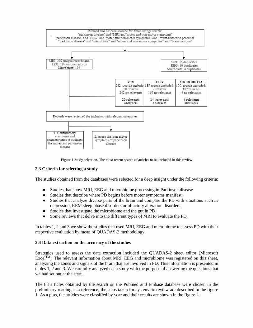
Figure 1 Study selection. The most recent search of articles to be included in this review
2.3 Criteria for selecting a study
The studies obtained from the databases were selected for a deep insight under the following criteria:
● Studies that show MRI, EEG and microbiome processing in Parkinson disease. ● Studies that describe where PD begins before motor symptoms manifest. ● Studies that analyze diverse parts of the brain and compare the PD with situations such as
depression, REM sleep phase disorders or olfactory alteration disorders. ● Studies that investigate the microbiome and the gut in PD. ● Some reviews that delve into the different types of MRI to evaluate the PD.
In tables 1, 2 and 3 we show the studies that used MRI, EEG and microbiome to assess PD with their
respective evaluation by mean of QUADAS-2 methodology.
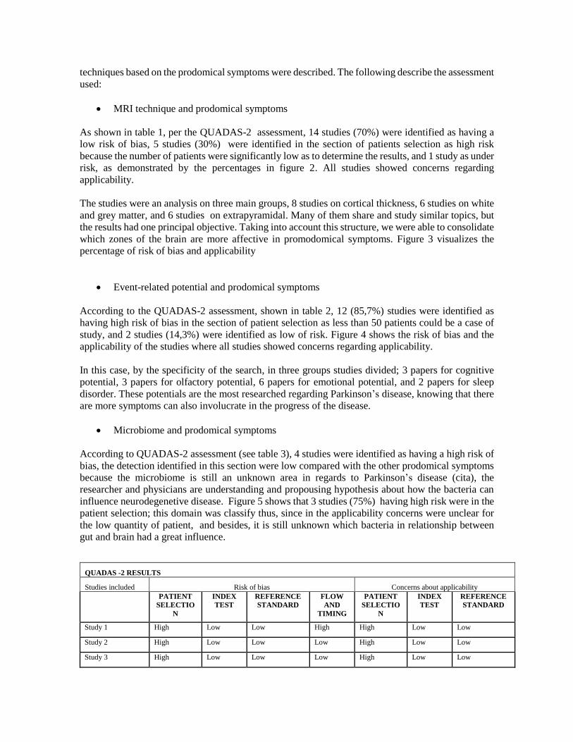
2.4 Data extraction on the accuracy of the studies
Strategies used to assess the data extraction included the QUADAS-2 sheet editor (Microsoft
ExcelTM). The relevant information about MRI, EEG and microbiome was registered on this sheet,
analyzing the zones and signals of the brain that are involved in PD. This information is presented in
tables 1, 2 and 3. We carefully analyzed each study with the purpose of answering the questions that
we had set out at the start.
The 88 articles obtained by the search on the Pubmed and Embase database were chosen in the
preliminary reading as a reference; the steps taken for systematic review are described in the figure
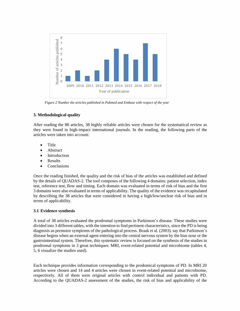
1. As a plus, the articles were classified by year and their results are shown in the figure 2.
3. Methodological quality
After reading the 88 articles, 38 highly reliable articles were chosen for the systematical review as
they were found in high-impact international journals. In the reading, the following parts of the
articles were taken into account:
Title
Abstract
Introduction
Results
Conclusions
Once the reading finished, the quality and the risk of bias of the articles was established and defined
by the details of QUADAS-2. The tool composes of the following 4 domains: patient selection, index
test, reference test, flow and timing. Each domain was evaluated in terms of risk of bias and the first
3 domains were also evaluated in terms of applicability. The quality of the evidence was recapitulated
by describing the 38 articles that were considered in having a high/low/unclear risk of bias and in
terms of applicability.
3.1 Evidence synthesis
A total of 38 articles evaluated the prodromal symptoms in Parkinson’s disease. These studies were
divided into 3 different tables, with the intention to find pertinent characteristics, since the PD is being
diagnosis as premotor symptoms of the pathological process. Braak et al. (2003), say that Parkinson’s
disease begins when an external agent entering into the central nervous system by the bias nose or the
gastrointestinal system. Therefore, this systematic review is focused on the synthesis of the studies in
prodromal symptoms in 3 great techniques: MRI, event-related potential and microbiome (tables 4,
5, 6 visualize the studies used).
Each technique provides information corresponding to the prodomical symptoms of PD. In MRI 20
articles were chosen and 14 and 4 articles were chosen in event-related potential and microbiome,
respectively. All of them were original articles with control individual and patients with PD.
According to the QUADAS-2 assessment of the studies, the risk of bias and applicability of the
0
1
2
3
4
5
6
7
8
2009 2010 2011 2012 2013 2014 2015 2016 2017 2018
Num
ber
of
arti
cles
pub
lish
ed
Year of publication
Figure 2 Number the articles published in Pubmed and Embase with respect of the year
techniques based on the prodomical symptoms were described. The following describe the assessment
used:
MRI technique and prodomical symptoms
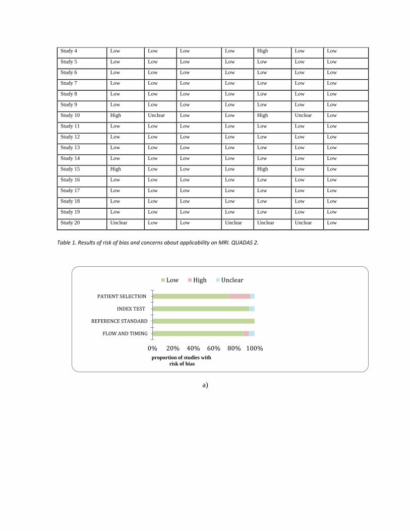
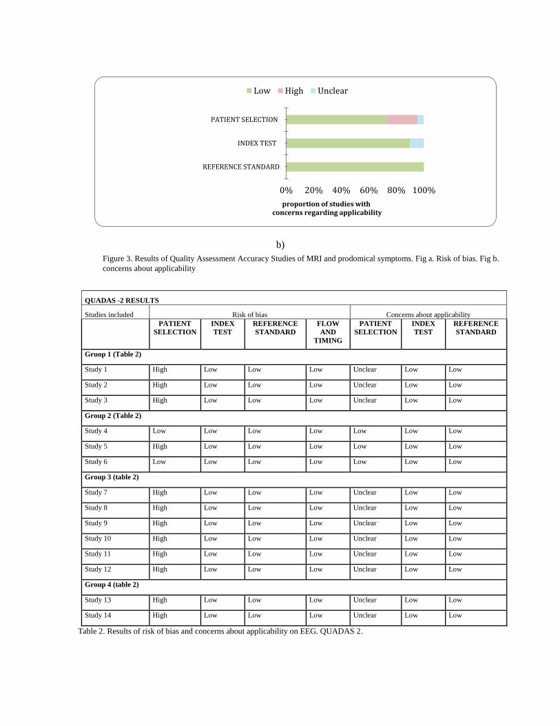
As shown in table 1, per the QUADAS-2 assessment, 14 studies (70%) were identified as having a
low risk of bias, 5 studies (30%) were identified in the section of patients selection as high risk
because the number of patients were significantly low as to determine the results, and 1 study as under
risk, as demonstrated by the percentages in figure 2. All studies showed concerns regarding
applicability.
The studies were an analysis on three main groups, 8 studies on cortical thickness, 6 studies on white
and grey matter, and 6 studies on extrapyramidal. Many of them share and study similar topics, but
the results had one principal objective. Taking into account this structure, we were able to consolidate
which zones of the brain are more affective in promodomical symptoms. Figure 3 visualizes the
percentage of risk of bias and applicability
Event-related potential and prodomical symptoms
According to the QUADAS-2 assessment, shown in table 2, 12 (85,7%) studies were identified as
having high risk of bias in the section of patient selection as less than 50 patients could be a case of
study, and 2 studies (14,3%) were identified as low of risk. Figure 4 shows the risk of bias and the
applicability of the studies where all studies showed concerns regarding applicability.
In this case, by the specificity of the search, in three groups studies divided; 3 papers for cognitive
potential, 3 papers for olfactory potential, 6 papers for emotional potential, and 2 papers for sleep
disorder. These potentials are the most researched regarding Parkinson’s disease, knowing that there
are more symptoms can also involucrate in the progress of the disease.
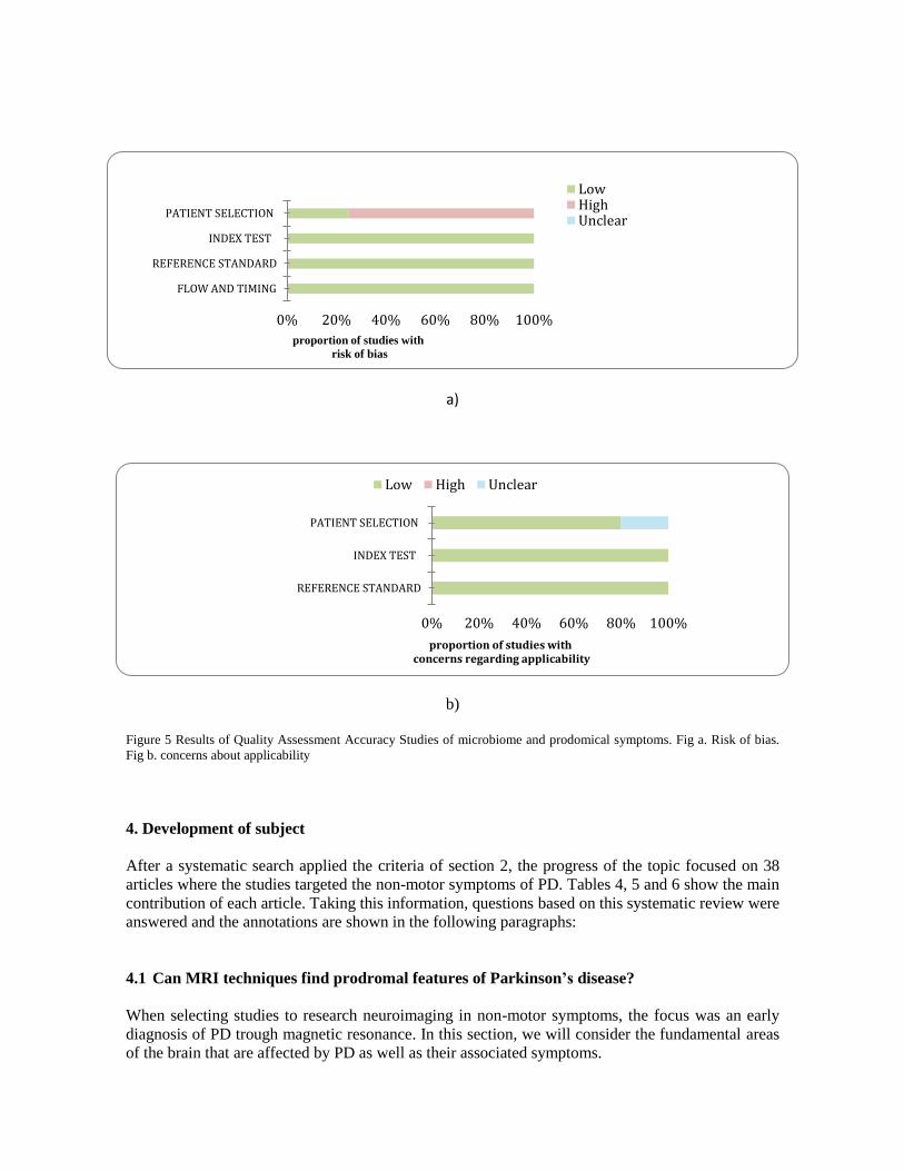
Microbiome and prodomical symptoms
According to QUADAS-2 assessment (see table 3), 4 studies were identified as having a high risk of
bias, the detection identified in this section were low compared with the other prodomical symptoms
because the microbiome is still an unknown area in regards to Parkinson’s disease (cita), the
researcher and physicians are understanding and propousing hypothesis about how the bacteria can
influence neurodegenetive disease. Figure 5 shows that 3 studies (75%) having high risk were in the
patient selection; this domain was classify thus, since in the applicability concerns were unclear for
the low quantity of patient, and besides, it is still unknown which bacteria in relationship between
gut and brain had a great influence.
QUADAS -2 RESULTS
Studies included Risk of bias Concerns about applicability
PATIENT
SELECTIO
N
INDEX
TEST
REFERENCE
STANDARD
FLOW
AND
TIMING
PATIENT
SELECTIO
N
INDEX
TEST
REFERENCE
STANDARD
Study 1 High Low Low High High Low Low
Study 2 High Low Low Low High Low Low
Study 3 High Low Low Low High Low Low
Study 4 Low Low Low Low High Low Low
Study 5 Low Low Low Low Low Low Low
Study 6 Low Low Low Low Low Low Low
Study 7 Low Low Low Low Low Low Low
Study 8 Low Low Low Low Low Low Low
Study 9 Low Low Low Low Low Low Low
Study 10 High Unclear Low Low High Unclear Low
Study 11 Low Low Low Low Low Low Low
Study 12 Low Low Low Low Low Low Low
Study 13 Low Low Low Low Low Low Low
Study 14 Low Low Low Low Low Low Low
Study 15 High Low Low Low High Low Low
Study 16 Low Low Low Low Low Low Low
Study 17 Low Low Low Low Low Low Low
Study 18 Low Low Low Low Low Low Low
Study 19 Low Low Low Low Low Low Low
Study 20 Unclear Low Low Unclear Unclear Unclear Low
Table 1. Results of risk of bias and concerns about applicability on MRI. QUADAS 2.
a)
0% 20% 40% 60% 80% 100%
PATIENT SELECTION
INDEX TEST
REFERENCE STANDARD
FLOW AND TIMING
proportion of studies with
risk of bias
Low High Unclear
b)
QUADAS -2 RESULTS
Studies included Risk of bias Concerns about applicability
PATIENT
SELECTION
INDEX
TEST
REFERENCE
STANDARD
FLOW
AND
TIMING
PATIENT
SELECTION
INDEX
TEST
REFERENCE
STANDARD
Gruop 1 (Table 2)
Study 1 High Low Low Low Unclear Low Low
Study 2 High Low Low Low Unclear Low Low
Study 3 High Low Low Low Unclear Low Low
Group 2 (Table 2)
Study 4 Low Low Low Low Low Low Low
Study 5 High Low Low Low Low Low Low
Study 6 Low Low Low Low Low Low Low
Group 3 (table 2)
Study 7 High Low Low Low Unclear Low Low
Study 8 High Low Low Low Unclear Low Low
Study 9 High Low Low Low Unclear Low Low
Study 10 High Low Low Low Unclear Low Low
Study 11 High Low Low Low Unclear Low Low
Study 12 High Low Low Low Unclear Low Low
Group 4 (table 2)
Study 13 High Low Low Low Unclear Low Low
Study 14 High Low Low Low Unclear Low Low
Table 2. Results of risk of bias and concerns about applicability on EEG. QUADAS 2.
0% 20% 40% 60% 80% 100%
PATIENT SELECTION
INDEX TEST
REFERENCE STANDARD
proportion of studies with concerns regarding applicability
Low High Unclear
Figure 3. Results of Quality Assessment Accuracy Studies of MRI and prodomical symptoms. Fig a. Risk of bias. Fig b.
concerns about applicability
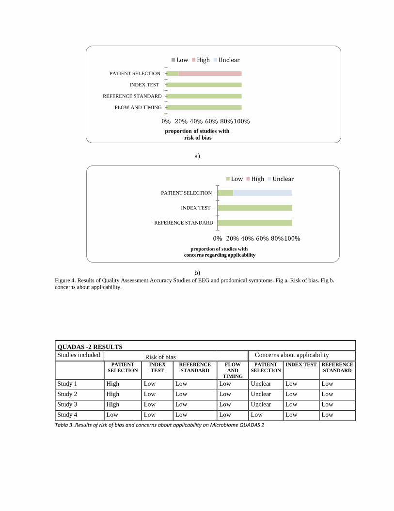
a)
b) Figure 4. Results of Quality Assessment Accuracy Studies of EEG and prodomical symptoms. Fig a. Risk of bias. Fig b.
concerns about applicability.
QUADAS -2 RESULTS
Studies included Risk of bias Concerns about applicability
PATIENT
SELECTION
INDEX
TEST
REFERENCE
STANDARD
FLOW
AND
TIMING
PATIENT
SELECTION
INDEX TEST REFERENCE
STANDARD
Study 1 High Low Low Low Unclear Low Low
Study 2 High Low Low Low Unclear Low Low
Study 3 High Low Low Low Unclear Low Low
Study 4 Low Low Low Low Low Low Low
Tabla 3 .Results of risk of bias and concerns about applicability on Microbiome QUADAS 2
0% 20% 40% 60% 80%100%
PATIENT SELECTION
INDEX TEST
REFERENCE STANDARD
proportion of studies with
concerns regarding applicability
Low High Unclear
0% 20% 40% 60% 80%100%
PATIENT SELECTION
INDEX TEST
REFERENCE STANDARD
FLOW AND TIMING
proportion of studies with
risk of bias
Low High Unclear
a)
b)
Figure 5 Results of Quality Assessment Accuracy Studies of microbiome and prodomical symptoms. Fig a. Risk of bias.
Fig b. concerns about applicability
4. Development of subject
After a systematic search applied the criteria of section 2, the progress of the topic focused on 38
articles where the studies targeted the non-motor symptoms of PD. Tables 4, 5 and 6 show the main
contribution of each article. Taking this information, questions based on this systematic review were
answered and the annotations are shown in the following paragraphs:
4.1 Can MRI techniques find prodromal features of Parkinson’s disease?
When selecting studies to research neuroimaging in non-motor symptoms, the focus was an early
diagnosis of PD trough magnetic resonance. In this section, we will consider the fundamental areas
of the brain that are affected by PD as well as their associated symptoms.
0% 20% 40% 60% 80% 100%
PATIENT SELECTION
INDEX TEST
REFERENCE STANDARD
FLOW AND TIMING
proportion of studies with
risk of bias
LowHighUnclear
0% 20% 40% 60% 80% 100%
PATIENT SELECTION
INDEX TEST
REFERENCE STANDARD
proportion of studies with concerns regarding applicability
Low High Unclear
The influence of age with onset PD is still unknown. Some studies examine the correlation of some
brains structures with age. However, it may be necessary to search for biomarkers of cognitive
impairment in Parkinson’s disease. The cortex is a clue of the brain’s neurodegeneration, since this
zone is the principal source of motor fibers of the pyramidal tract.
In order to apply techniques to investigate the progression of PD based on non-motor symptoms, data
synthesis showed that patients may have Unified Parkinson’s Disease Rating Scale (UPDRS) score
between III and IV. After performing the MRI, the dataset had to develop a quality control in order
to normalize the intensity and other characteristics that make images comparable.
4.1.1 Cortical Thickness
(Cerasa et al., 2013) analyzed the cortical thickness, a vertex by vertex multiple linear expression
analysis, this was carried out to investigate the relationship between regional cortical thickness and
scores of Abnormal Involuntary Movement Scale (May, et al, 1983). Dyskinetic PD patients mainly
showed significantly thicker cortices, nevertheless, early-onset dyskinetic patients showed increased
volume in a large cluster of the midbrain enclosed substantia nigra and red nucleus. (Mak et al., 2015)
is a relevant research due to neuropsychological assessments at baseline. They compared regional
percentage change of cortical thickness and subcortical atrophy over 18 months. These features
showed significantly reduced cortical thickness in the frontal, parietal and occipital cortices: left
supramarginal cortex, bilateral rostral middle frontal cortex, left isthmus cingulate and right posterior
cingulate cortices, and the right lateral occipital cortex. On the other hand, (Kim et al., 2014) divided
the brain into two areas, left-side-disease onset and right-side-disease onset. They modeled local
cortical thickness as a linear relationship with the motor symptoms. In the first group, it found a
cluster including the right primary sensory motor cortex and paracentral lobule, as well as another
two clusters in bilateral parahippocampal gyrus. In the second group, there was just a cluster located
in the left lingual gyrus. Moreover, they concluded that there is not relationship between the severity
of motor symptoms and cortical thickness. (Potgieser et al., 2014) focused the research in the exposes
that deteriorate the cortex in PD. In this case, the total gray matter per patient was calculated as to
compare between PD patients and controls; the first instance reduction of regional gray matter density
was not shown in anterior temporal regions. However, atrophy was observed in the left anterior
temporal and inferior frontal region, while in the right hemisphere, posterolateral frontal atrophy was
pronounced in a dorsal position.
(Wenzel et al., 2018) this work approach a rapid segmentation of subcortical brain structures in T1-
weight MRI by utilizing a shape-constrained deformable surface model. The advantage of this model
is that can use of automated segmentation and volume quantification of different neurodegeneration
disease.
4.1.2. White and Gray matter
Some of those studies performed functional magnetic resonance image (fMRI) in order to observe
changes on the different areas of the brain while it is performing several tasks. (Canu et al., 2015)
performed structural and functional magnetic resonance image. In this study, gray matter did not have
atrophy in PD with freezing of gait; whereas white matter showed damage of the pedunculopontine
tract, corpus callosum, corticospinal tract, cingulum, superior longitudinal fasciculus. (Gallagher et
al., 2013) shows that the evolution of Parkinson’s disease has been considered as an illness in which
white matter abnormalities contribute to non-motor symptoms (Bohnen & Albin, 2011). A magnetic
resonance diffusion tensor was used as a measure of white matter microstructural integrity. This
technique reported changes in hemispheric white matter in PD and these abnormalities contribute to
cognitive deficits. (Zanigni et al., 2016) also analyses white matter, yet, this research differentiates
progressive supranuclear palsy (PSP) from Parkinson disease. PSP is also a neurodegenerative disease
that causes death in some areas of the brain and its symptoms are similar to PD. The results of that
study show a high accuracy in differentiating between PSP and PD. (Agosta et al., 2013) researched
on the damage to white and grey matter at different stages of PD. To achieve the results, the
researchers analyzed the whole brain of patients and healthy controls. Patients divided into early PD
stage and moderate PD. According to this classification, they found:
(1) PD patients had a very little gray matter atrophy, (2) white matter showed microstructural damage
in PD patients, (3) the greatest difference evidenced in the pattern of white matter damage with
moderate PD when compared to mild PD cases, and (4) the severity of white matter degeneration
correlates with cognitive status.
(Amoroso, La Rocca, Monaco, Bellotti, & Tangaro, 2018) made a classification model, the
development is consisted in a brain connectivity based on grey and white matter voxel distribution;
one that the segmentation threw the region interest, then they measure how different brain regions are
correlated and for each region measure topological quantity. This model provides two important
methodological understandings. Firstly, it associates to each node quantitative measurements
which characterizes its role and importance within the network; secondly, it enables a description of
the whole brain from a global perspective.
(Li, Xing, Martin-Bastida, Piccini, & Auer, 2018) study the grey matter of PD, specifically on UPDRS
III because some motor symptoms manifested at this point on the scale, but it is not still association
with parkinsonian motor symptoms and neurodegeneration as reflected by grey matter. The results
showed are not a correlation with UPDRS III score and putamen and caudate grey matter. However,
rigidity sub-scores are associated with lower anterior striatal grey matter.
4.1.3 Extrapyramidal system
The extrapyramidal system is the zone most affected in PD; the pathway is the central nervous system
of nigrostriatal, the basal ganglia, the cerebellum, the vestibular nuclei, and different sensory zones
of the cerebral cortex (Zhang et al., 2015).
(Qiao, Shi, Jiang, Gao, & Niu, 2017) analyzed the sequence of susceptibility-weighted imaging (SWI)
and T weighted Imaging (T1wI) and T2wI to evaluate the contrast between the midbrain, basal nuclei,
and the surrounding tissue, as well as the boundary clarity. The results showed a reduction on the
signal intensities in the PD group indicated that there was a significant iron deposition in the nuclei
of the PD patients.
On the other hand (Hutchinson & Raff, 2008) and (Chen et al., 2014) segmented the zone of substantia
nigra image in order to quantify the gradient of the pathological change. In the study of Hutchinson
and Raff, there was a large evidence of differences between the patient and control group. Chen et al
used a technique to quantify neuromelanin in the locus coeruleus and substantia nigra. This
segmentation was performed in three steps: 1) ROIs were referenced with circles 2) Voxel intensities
of the ROIs (substantia nigra and the mean) were calculated, 3) the intensity differences were
calculated to create a binary map using a given criterion. Additionally, the volume of the locus
coeruleus and substantia nigra was calculated in the same way. This technique demonstrated
advantage in the image acquisition, image pre-processing and quantitative image.
(Lee et al., 2014) applied a technique that pretended to measure the volume of substantia nigra and
basal forebrain with early Parkinson disease. In order to do that, the researchers divided the patients
into three groups, 1) patients with stage 1 PD, 2) patients with stage 2 PD, and 3) patients with stage
3 PD. The results highlighted that the volume of substantia nigra was smaller on the left hemisphere
in patients with stage 1 PD compared with the control group. The patients with stage 2 and 3 showed
a smaller volume of the substantia nigra. Concerning the basal forebrain, its volume was not reduced
in patients with stage 1 PD. On the other hand, the patients with stages 2 and 3 showed a significant
reduction when compared to the controls.
(Takahashi et al., 2018) the study is about quantify nigral changes and neuromelanin values in whole
substantia nigra pas compact containing the entire nigrosome and dorsolateral. The results showed,
in both substantia nigra pars compact, quantify nigral changes were lower in PD patients. This
affirmation, the MRI assessment of the abnormality of nigrosomes can produce an excellent
diagnostic for early-stage PD.
(García-Lorenzo et al., 2013) the majority of PD patients’origin of rapid eye movement sleep behavior
disorder, this non-motor symptom do not yet know how produce. For that reason, they use
polysomnography and MRI 3T to assess the locus subcoeruleus in the brain stem, the zones which
are implicated with the rapid eye movement sleep behavior disorder. The results confirmed that in
Parkinson’s disease, this complex is affected, and there is a gradual damage of the structure.
(Ziegler et al., 2013) focused on the hypothesis that the degeneration of substantia nigra pars compact
yield that of the cholinergic basal forebrain in PD. The patients were assessed with Hoehn and Yahr
(H&Y) stages I-III (Perlmutter, 2009). Following the MRI protocol they used, the images provide a
study window on the subcortical structure that are concerned with PD, but this protocol cannot be
carried out with conventional MRI.
(Rolheiser et al., 2011) proposed a study that assess the olfactory non-motor symptom and diffusion
tensor image; in the MRI examined olfactory tract and substantia nigra. During the olfactory test,
shown impairment in the test and the diffusion tensor image showed differences between olfactory
region and substantia nigra. (Moessnang et al., 2011) also related with olfactory disorder as non-
motors symptom. The researchers combined olfactory test and fMRI to analyze the activation
olfactory network of PD patients. PD patients showed network dysfunction that need to be studied
further.
4.2 Can potential event-related EEG show early signs of Parkinson’s disease?
The selection of studies that carry out EEG related to events can provide a different set of features for
Parkinson’s disease. These measurements, through previously stipulated brain stimulations, show
peaks which appear in the EEG in response to the occurrence of an event. Now we classify the
different studies depending on the nature of the stimulus.
4.2.1. Cognitive event.
This cognitive section of Parkinson’s disease focuses on Event Related Potential ERP with the purpose of characterizing the response to cognitive events (Özmüş et al., 2017). The following studies use different techniques to measure the signal, but their respective conclusions can be compared. (Yuvaraj, Rajendra Acharya, & Hagiwara, 2018) and (Oh et al., 2018) related to the computational analysis of the signals, and the third is about the cognitive impairment of PD. Computational automated techniques are able to aid in the early detection of PD. In the casa of Yuvaraj, a high level technique was used to contribute to the diagnosis of the PD, in the developing of such techniques, the discrimination of the abnormalities signal and normal report the explicitly of PD signal.. (Oh et al.,

2018) used automated classification of EEG signal in their study. In this case, they classified signals using the technique of the convolutional neuronal network to identify early symptoms of abnormalities. (Özmüş et al., 2017) studied brain dynamics of early PD patients and controls using event-related potential. The patients in this case diagnosed neurological tests to be cognitively normal. However, in EEG after applying P300 amplitude, results indicate that PD patient’s signals were significantly lower at the F3, Fz, Cz, Cz, P4, and Pz electrode sites.
4.2.2. Olfactory event.
The olfactory loss is an ambivalent non-motor symptom of PD because a patient with respiratory
issues may also have hyposmia. However, years before the motor symptom appears, a manifestation
of hyposmia can appear in PD, which can be an alert for this type of neurodegenerative illness. For
that reason, in this systematic review, we took into consideration the articles of the second group in
table 1 (Iannilli, Stephan, Hummel, Reichmann, & Haehner, 2017) (Versace et al., 2017) (Cozac et
al., 2017). These studies are based on EEG-derived ERP, which are changes in voltage that occur at
a given moment while a stimulus is applied (Iannilli et al., 2017). In this case, the stimulus is odor
and the amplitude and latencies of response are measured. This study was able to show that there is a
reduction of olfactory sensitivity in PD patients, which was observed at EEG-derived ERP. These
responses could be detected on specific brain cortex areas: the right angular gyrus, the right
parahippocampal gyrus and the right cingulate gyrus. (Versace et al., 2017) applied two techniques:
Short latency Afferent Inhibition (SAI) to study the cholinergic function and olfactory event related
potential to evaluate the olfactory system. Cholinergic function focused on electrical stimuli to a
peripheral nerve with the purpose to assess sensorimotor system (Turco, C. V., El Sayes, J., Locke,
M. B., Chen, R., Baker, S., & Nelson, 2018). Using both techniques, they could observe a significant
reduction in this putative marker of central cholinergic activity in PD patients. The Olfactory Event
Related Potential (OERP) abnormalities indicated cognitive deterioration. Thus, provided findings
support the fact that cholinergic denervation is a robust determinant of hyposmia, and raises the
possibility that the presence of olfactory dysfunction may indicate increased risk of cognitive
impairment in patients with PD. In (Cozac et al., 2017), the objective was to identify the mutual
influence of olfactory sensitivity decrease and EEG changes in PD. Within the research, they
discriminate three relevant aspects:
Olfactory lost is considerably greater in PD patients than in healthy controls; this decline in PD
is yet to be completely understood.
There is an association between odor impairment and motor degeneration, more specifically with
gait and rigidity. It may be explained by the projections from the olfactory regions to the brain
structure (Wilson DA, Chaouis J, 2015).
There is no association between olfactory loss and the resting-state EEG power spectrum. The
principal reason for this fact is the different rates of neurodegeneration (Domellöf ME, Lundin
K-F, Edström M, 2017).
4.2.3. Emotional event.
The response of the emotional component has an important role in the organism. These events may
be internal (thoughts, memories, sensations) and external (stimulus, people´s behavior, a change of
situation) (Gray HM, 2010). From the physiological point of view, this leads to the fact of activation
of neurotransmitters in the autonomic nervous system, which are associated with emotional states.
The emotional changes can be even more problematic than motor decline in PD. In the literature,
scientifics have reported discrepancies in emotional process (Sotgiu, I., & Rusconi, 2013). These
include various changes on it, but mostly in the recognition of emotions
In this context, electrophysiology measures may be a method to assess the problematic on PD patients
(Stimuli Hiroyuki Oya, Hiroto Kawasaki, Matthew A. Howard, 2002). The articles evaluated in this
section use EEG-derived ERP to asses emotional process while showing short videos or image that
express happiness, surprise, anger,sadness or fear.
(Garrido-Vásquez, Pell, Paulmann, Sehm, & Kotz, 2016) carried out a neuronal analysis using event-
related potential. The applied stimulus was a dynamic facial display that produced emotional
sentences in a happy, angry and neutral voice. This study reported that left Parkinson Disease patients,
whose right hemisphere is predominantly affected by neural degeneration, exhibited impairments
during the first 200ms of face processing.
In this systematic review, we reported an author (Yuvaraj) who has worked in the emotional field
with 4 studies (Yuvaraj & Murugappan, 2016) (Yuvaraj et al., 2016) (Yuvaraj, Murugappan,
Mohamed Ibrahim, et al., 2014) (Yuvaraj, Murugappan, Ibrahim, et al., 2014). In these studies, the
emotional stimuli was caused by emotions such us sadness, happiness, fear, anger and surprise.
(Yuvaraj & Murugappan, 2016) reported a nonlinear analysis of EEG during emotion processing in
PD patients. In this case, they analyze the emotional processing in right-side affected and left-side
affected patients. The authors found that in order to classify it is better to differentiate between high
frequencies (alpha, beta, and gamma bands) than low frequencies (delta and theta band). These results
reported that neuronal degeneration in PD could contribute to the decline of emotional recognition.
However, lateralization of emotion has been debated and asymmetric effects on explicit emotion have
been reported (Clark, Neargarder, & Cronin-Golomb, 2008) (Ariatti A, Benuzzi F, 2008) (Ventura
MI, Baynes K, Sigvardt KA, Unruh AM, Acklin S, Kirsch HE, 2012).
(Yuvaraj et al., 2016) further investigated the emotion recognition in PD using EEG and based Brain
Functional Connectivity (BFC) patterns. In order to make the BFC, the researchers used a correlation
coefficient to analyze the emotional state, the degree of coherence in the band EEG report since the
coherence of each emotion can change and the synchronization index between right and left frontal
sites while viewing the emotions. In view of the performance of these values, they compared the BFC
index using computational techniques. The results showed PD patients show a decline in the
functional connectivity indices during emotional stimulation.
(Yuvaraj, Murugappan, Mohamed Ibrahim, et al., 2014) focused on the detection of six emotions in
PD patients when compared to healthy individuals with individuals classified using a computational
model. The classification supported the assessment of emotional impairment associated to non-motor
symptoms of PD. The categorization was developed using computational methods, applying machine
learning to emotion categories. This process found that the easiest emotions to classify were happiness
and surprise while the most difficult were disgust and sadness. An intermediate difficulty was found
for anger and fear. Those successes can demonstrate dysfunction in specific neuronal circuits. These
include amygdala and the ventral striatum which are located within the basal ganglia’s limbic loop.
(Yuvaraj, Murugappan, Ibrahim, et al., 2014) examined the PD patient’s emotion-processing deficit
in EEG signal (theta, gamma, betha and alpha). The results suggest the likelihood of the presence of
a distinctive neurobiological substrate of PD patients during emotional information processing.
Moreover, it was found an increase in power in the theta and gamma bands for PD patients. The last
article of Table 2 group 3 was (Dietz et al., 2013). Its objective was to investigate the
electrophysiology indices of brain response during emotional process. This could be carried out
measuring emotions during picture processing in the amplitude of the late positive potential (LPP).
The pictures used to measure LPP were pleasant, neutral, and unpleasant pictures. The results showed
a reduction in LLP amplitude when PD patients viewed unpleasant, compared to pleasant pictures
and healthy controls.
4.3 Sleep disorder.
The period of premotor symptoms of Parkinson’s disease manifests on the brainstem and midbrain,
while the symptomatology is located in these zones. The symptoms could be as many as autonomic
changes, depression, olfactory dysfunction, and sleep disorder. Idiopathic rapid behavior disorder
(iRBD) has confirmed to be a preclinical feature because iRBD patients develop a synucleinopathy
(YuanYuan, 2017). (Hansen, Marcussen, Christensen, Jennum, & Sorensen, 2013) classified subjects
dependent on the power of EEG signal at different frequency bands. This classification was made
using K-means and Bayesian classifiers. As a result, the study obtained Bayesian classifiers that
reached 90% of sensitivity and specificity. Regarding the brain, five features attained a worthy
classification. All came from the O1-A2 and F3-A2 signals, both located in the left hemisphere of the
brain. The paper of (Iwanami, Miyamoto, Miyamoto, Hirata, & Takada, 2010) provides reliable
information because the study performed three techniques to assess two non-motor symptoms as PD
olfactory loss and iRDB. The techniques were Polysomnography (PSG), odor identification and
Sonography. Results showed substantia nigra hyperechogenicity and abnormal functional anosmia or
hyposmia.
4.4 Can microbiome show early signs of Parkinson’s disease?
Modern civilization is confronted with a progressive increase in mental diseases, such as anxiety and
depression. As a hypothesis, inflammation of the gut has a relationship with those situations. The
gut-brain-axis refers to communication between central nervous system and gut microbiome (Clapp
et al., 2017). According to that, Parkinson disease may start in enteric nervous system and spread via
the vagal nerve to the brainstem.
(F, Hopfner, Künstner A, Müller SH, Künzel S, Zeuner KE, Margraf NG, Deuschl G, Baines JF,
2017) determined whether PD is related to qualitative or quantitative changes in the gut microbiota.
In these results, Lactobacillaceae, Barnesiellaceae and Enterococcacea were more abundant in
patients with Parkinson’s disease. Nevertheless, gut microbiome requires more investigation and it is
necessary to establish the role of the microbiome. According to (Agata Mulak, Slawomir Budrewicz,
Magdalena Panek-Jeziorna, Magdalena Koszewicz, Maria Jasinska, Beata Marczak-Karpina,
Krzysztof Slotwinski, Ryszard Podemski, 2017) Parkinson’s disease is characterized by alpha-
synucleinopathy at all levels of the brain-gut axis, whose alterations in enteric nervous system ENS
contribute to the manifesting of PD. Therefore, increased levels of calprotectin and zonulin in stool
samples found in 60% and 40% of PD patients respectively. The stool ELISA that confirmed the
changes in the intestinal microbiome of PD patients. (Unger et al., 2016) hypothesized that a shift in
gut microbiome might be associated with short chain fatty acids (SCFA) that are in the colon and
produce bacterial carbohydrate fermentation (Joseph E.Pizzorno, Michael T.Murray, 2016). The
results report that stool SCFA concentrations reduced in PD patients. Regarding the bacterial family
found, Bacteroidetes and Prevotellaceae had decreased concentrations while Enterobacteriaceae were
more abundant. However, (Scheperjans et al., 2015) report that PD gut microbiome has not been
sufficiently researched and there is no definite evidence of any specific microbe being linked to PD.
The purpose of the study was to compare the composition of the whole stool microbiome between
PD patients and healthy individuals. The results reported that the bacterial family concentration in
feces had a low quantity of prevotellaceae and an increased abundance of enterobacteriaceae.
5. Discussion
Our systematic review found that MRI, EPR and microbiome are techniques very influential in
analyzing the non-motor symptoms of Parkinson’s disease because of the ability to try and obtain
relevant results to determine prodromal features. This paper has selected n = 38 studies with of a total
of n= 5,592 PD patients and a total of healthy controls n=538. Our results, however, had several
restrictions depending on the techniques. Neuroimaging was restricted to individual imaging
modalities (structural MRI), the EEG restriction was ERP (cognitive, olfactory and emotional) and
the restriction of microbiome was the study of fecal samples. None of these techniques employed
simultaneously for the same study, so the possible synergy given by the combination of the three is
still to be determined in the future.
The results of these studies indicate a higher sensitivity associated with fMRI data as related to brain
functionality (Uribe et al., 2018). On the other hand, using structural MRI is possible to observe
changes in cortex zone, basal nucleus (when motor symptoms already manifested on the body).
Furthermore, structural MRI allows the construction of a brain volume in order to observe changes
as a tool to detect changes in several areas of the brain. However, this complex work is still to be
totally done. Furthermore, fMRI may be a relevant method because it can be used with protocols of
olfaction, emotional with propose to analyze the sensitivity of the brain through an event. This
application of fMRI will facilitate the comparison of results obtained by ERP by simply sharing
similar protocols that can be used in both techniques and can provide a general point of view
(Matsuda, Matsuura, & Ohkubo, 2002).
The most common technique to evaluate and measure non-motor symptoms is diffusion tensor image
(DFI). The majority of included studies used it, since DTI estimated regions through magnetic field
directions to create an image that is briefed in a particular direction. A recent study showed that by
using this technique, relevant longitudinal connectomes compared with other studies can be
understood (Peña-nogales, Ellmore, Luis-garcía, & Lambert, 2019).
We observe substantial results concerning the use of ERP to investigate the effect of different stimulus
in order to analyze latencies and amplitudes. It has been reported that non-motor symptoms in PD are
associated with oscillations in all frequencies ranges (Bočková & Rektor, 2018). However, there is
still a lack of information in the scale brain-networks about non-motor symptoms. In the search, we
found evidence of olfactory, emotional and cognitive assess, which are principal Non-motor
Symptoms (NMS). For example, in emotional stimulus, frontal areas play an important role as
revealed by the P100 and P200 amplitudes (A. Semertzidou, C.A. Frantzidis, A. Ladas, M.
Karagianni, M. Tsolaki, 2012). The olfactory bulb is affected in the early stages of PD, with olfactory
stimulus with patients who have Parkinson disease at stage III evidenced having the latency
substantially diminished. It should be noted that there is still a need for investigations on the ERP
technique on prodromal symptoms in PD.
Mollenhauer (Mollenhauer et al., 2016) mentions that could be the most common non-motor
symptoms that can affect the development of PD on a person. In their protocols, they examined a
non-motor Symptom Scale. To develop this study, they selected the following NMS: cognitive
function, REM sleep behavior disorder by polysomnography, voxel-based morphometry (VBM) of
the brain by MRI, and Cerebrospinal Fluid (CSF). The study concluded that the sleep and imaging
measures as other NMP are necessary in adequate scales and may lead to obtaining more features to
quantify the progression of PD.

Our review also researched microbiome. Microbiome is still an unknown topic to research and there
is much to study. Brain-gut axis seems to have a stretch relationship with neurodegeneration diseases,
and mostly with PD. It would be interesting to conduct tests to investigate questionnaire data and
stool samples to realize the relationship between constipation symptoms, bacterium families and PD.
Moreover, the association between the abundance of microbiome and medication is also interesting
in PD levodopa because pharmacological treatment regularly causes gastrointestinal side effects.
The combination of these three techniques could become increasingly useful in the diagnosis of early
stage Parkinson’s disease; subsequently it would contribute unified information that can interpret data
from the gut and the brain. The literature says these huge organs communicate between each other
which could provide the influence of some microbes in Parkinson’s disease to somehow cause the
death of dopamine cells.
6. Conclusions
In the past decade, significant improvement has been made on numerous fronts in combined EEG
and MRI studies of the Parkinson’s disease. These include optimal study design that goes from the
acquisition to the analysis of the data. An area that clearly needs further research is the development
and validation of procedures for microbiome in PD. Such models will help researchers to better
integrate the microbes with the central nervous system. Having this information could incorporate
this information with MRI and EEG signal.
Alongside this, we analyzed some published papers that assess PD focusing on motor and non-motor
symptoms using MRI, ERP and microbiome. The evidence of motor symptoms is large and by means
of MRI is already feasible to know and observe which features are affected in the brain. Nowadays,
through the use of computational tools, it is possible to study the progression of PD, but the evidence
of non-motors symptoms on MRI is still a field that needs to be explored. Although, there is already
a timeline and list of the non-motor symptoms that perhaps could produce the disease on the basal
nucleus. However, the physicians do not have all the predominant features to diagnose PD as soon as
possible. Furthermore, there are no baseline studies that observe the changes on patients with early
Parkinson’s disease.
On the other hand, using ERP allows detecting PD features in a very reliable way. Investigating
whether PD patients have trouble to distinguish emotions and how depression affects the brain’s
signals allows for the detection of cognitive impairment. Concerning olfactory loss, in order to have
an accuracy research, the researchers need to focus on known odors for Parkinson's disease patients
in order to determine if there is a real loss of smell.
Finally, microbiome might include the classification of bacterial families and this may increase
accuracy in the exploration of fecal samples to find potential features of Parkinson's disease. For all
these reasons, we consider that the combination of the previous techniques, with the support of
computation, can provide a set of biomarkers that can help in the diagnosis of Parkinson’s disease in
the earlier stages.
7. REFERENCES
A. Semertzidou, C.A. Frantzidis, A. Ladas, M. Karagianni, M. Tsolaki, P. D. B. (2012). How aging
affects emotional processing? Neurophysiological evidence using passive emotion evocative
stimuli selected from the International Affective Picture System (IAPS). Poster
Abstracts/International Journal of Psychophysiology, 85(3), 361–430.
https://doi.org/10.1016/j.ijpsycho.2012.07.122
Agata Mulak, Slawomir Budrewicz, Magdalena Panek-Jeziorna, Magdalena Koszewicz, Maria
Jasinska, Beata Marczak-Karpina, Krzysztof Slotwinski, Ryszard Podemski, L. P. (2017).
Fecal Biomarkers of Gut Inflammation and Intestinal Barrier Dysfunction in Parkinson’s
Disease. Gastroenterology, 152, S924. https://doi.org/10.1016/S0016-5085(17)33152-9
Agosta, F., Canu, E., Stojković, T., Pievani, M., Tomić, A., Sarro, L., … Filippi, M. (2013). The
topography of brain damage at different stages of parkinson’s disease. Human Brain Mapping,
34(11), 2798–2807. https://doi.org/10.1002/hbm.22101
Amoroso, N., La Rocca, M., Monaco, A., Bellotti, R., & Tangaro, S. (2018). Complex networks
reveal early MRI markers of Parkinson’s disease. Medical Image Analysis, 48, 12–24.
https://doi.org/10.1016/j.media.2018.05.004
Barber, T. R., Klein, J. C., Mackay, C. E., & Hu, M. T. M. (2017). Neuroimaging in pre-motor
Parkinson’s disease. NeuroImage: Clinical, 15(April), 215–227.
https://doi.org/10.1016/j.nicl.2017.04.011
Bočková, M., & Rektor, I. (2018). Impairment of brain functions in Parkinson’s disease reflected by
alterations in neural connectivity in EEG studies: a viewpoint. Clinical Neurophysiology, 130,
239–247. https://doi.org/10.1016/J.CLINPH.2018.11.013
Braak, H., Del, K., Rüb, U., Vos, R. A. I. De, Jansen, E. N. H., & Braak, E. (2003). Staging of brain
pathology related to sporadic Parkinson ’ s disease, 24, 197–211.
Canu, E., Agosta, F., Sarasso, E., Volontè, M. A., Basaia, S., Stojkovic, T., … Filippi, M. (2015).
Brain structural and functional connectivity in Parkinson’s disease with freezing of gait.
Human Brain Mapping, 36(12), 5064–5078. https://doi.org/10.1002/hbm.22994
Caputi, V., & Giron, M. C. (2018). Microbiome-Gut-Brain Axis and Toll-Like Receptors in
Parkinson ’ s Disease. International Journal of Molecular Sciences, 1–19.
https://doi.org/10.3390/ijms19061689
Cerasa, A., Salsone, M., Morelli, M., Pugliese, P., Arabia, G., Gioia, C. M., … Quattrone, A.
(2013). Age at onset influences neurodegenerative processes underlying PD with levodopa-
induced dyskinesias. Parkinsonism and Related Disorders, 19(10), 883–888.
https://doi.org/10.1016/j.parkreldis.2013.05.015
Chen, X., Huddleston, D. E., Langley, J., Ahn, S., Barnum, C. J., Factor, S. A., … Hu, X. (2014).
Simultaneous imaging of locus coeruleus and substantia nigra with a quantitative
neuromelanin MRI approach. Magnetic Resonance Imaging, 32(10), 1301–1306.
https://doi.org/10.1016/j.mri.2014.07.003
Clapp, M., Aurora, N., Herrera, L., Bhatia, M., Wilen, E., & Wakefield, S. (2017). Gut microbiota’s
effect on mental health: the gut-brain axis. Clinics and Practice, 7(4).
https://doi.org/10.4081/cp.2017.987
Cozac, V. V., Auschra, B., Chaturvedi, M., Gschwandtner, U., Hatz, F., Meyer, A., … Fuhr, P.
(2017). Among early appearing non-motor signs of parkinson’s disease, alteration of olfaction
but not electroencephalographic spectrum correlates with motor function. Frontiers in
Neurology, 8(OCT), 1–6. https://doi.org/10.3389/fneur.2017.00545
Dietz, J., Bradley, M. M., Jones, J., Okun, M. S., Perlstein, W. M., & Bowers, D. (2013). The late
positive potential, emotion and apathy in Parkinson’s disease. Neuropsychologia, 51(5), 960–
966. https://doi.org/10.1016/j.neuropsychologia.2013.01.001
F, Hopfner, Künstner A, Müller SH, Künzel S, Zeuner KE, Margraf NG, Deuschl G, Baines JF, K.
G. (2017). Gut microbiota in Parkinson disease in a northern German cohort. Brain Research,
1667, 41–45. https://doi.org/10.1016/j.brainres.2017.04.019
Gallagher, C., Bell, B., Bendlin, B., Palotti, M., Okonkwo, O., Sodhi, A., … Alexander, A. (2013).
White matter microstructural integrity and excutive fuctions in parkinson’s disease. Political
Science, 5(2), 361–362. https://doi.org/10.1017/Sl
García-Lorenzo, D., Longo-Dos Santos, C., Ewenczyk, C., Leu-Semenescu, S., Gallea, C.,
Quattrocchi, G., … Lehericy, S. (2013). The coeruleus/subcoeruleus complex in rapid eye
movement sleep behaviour disorders in Parkinson’s disease. Brain, 136(7), 2120–2129.
https://doi.org/10.1093/brain/awt152
Garrido-Vásquez, P., Pell, M. D., Paulmann, S., Sehm, B., & Kotz, S. A. (2016). Impaired neural
processing of dynamic faces in left-onset Parkinson’s disease. Neuropsychologia, 82, 123–
133. https://doi.org/10.1016/j.neuropsychologia.2016.01.017
Gershanik, O. S. (2017). Does Parkinson ’ s disease start in the gut ? Arq Nueropsiquiatr, (October),
67–70.
Hamm-clement, J., & Sandmann-keil, D. (2002). Staging of the intracerebral inclusion body
pathology associated with idiopathic Parkinson ’ s disease ( preclinical and clinical stages ).
Journal of Neurology, 1–5. https://doi.org/10.1007/s00415-002-1301-4
Hansen, I. H., Marcussen, M., Christensen, J. A. E., Jennum, P., & Sorensen, H. B. D. (2013).
Detection of a sleep disorder predicting Parkinson’s disease. Proceedings of the Annual
International Conference of the IEEE Engineering in Medicine and Biology Society, EMBS,
(from 2004), 5793–5796. https://doi.org/10.1109/EMBC.2013.6610868
Hutchinson, M., & Raff, U. (2008). Detection of Parkinson’s disease by MRI: Spin-lattice
distribution imaging. Movement Disorders, 23(14), 1991–1997.
https://doi.org/10.1002/mds.22210
Iannilli, E., Stephan, L., Hummel, T., Reichmann, H., & Haehner, A. (2017). Olfactory impairment
in Parkinson’s disease is a consequence of central nervous system decline. Journal of
Neurology, 264(6), 1236–1246. https://doi.org/10.1007/s00415-017-8521-0
Iwanami, M., Miyamoto, T., Miyamoto, M., Hirata, K., & Takada, E. (2010). Relevance of
substantia nigra hyperechogenicity and reduced odor identification in idiopathic REM sleep
behavior disorder. Sleep Medicine, 11(4), 361–365.
https://doi.org/10.1016/j.sleep.2009.12.006
Kelly Del Tredici, Udo Rüb, Rob A.I de Vos, Jürgen R.E. Bohl, and H. B. (2002). Where Does
Parkinson Disease Pathology Begin in the Brain ? Journal of Neuropathology and
Experimental Neurology, 61(5), 413–426.
Kim, J. S., Yang, J. ju, Lee, J. min, Youn, J., Kim, J. min, & Cho, J. W. (2014). Topographic pattern
of cortical thinning with consideration of motor laterality in Parkinson disease. Parkinsonism
and Related Disorders, 20(11), 1186–1190. https://doi.org/10.1016/j.parkreldis.2014.08.021
Lee, H. M., Kwon, K. Y., Kim, M. J., Jang, J. W., Suh, S. il, Koh, S. B., & Kim, J. H. (2014).
Subcortical grey matter changes in untreated, early stage Parkinson’s disease without
dementia. Parkinsonism and Related Disorders, 20(6), 622–626.
https://doi.org/10.1016/j.parkreldis.2014.03.009
Li, X., Xing, Y., Martin-Bastida, A., Piccini, P., & Auer, D. P. (2018). Patterns of grey matter loss
associated with motor subscores in early Parkinson’s disease. NeuroImage: Clinical,
17(November 2017), 498–504. https://doi.org/10.1016/j.nicl.2017.11.009
Mahlknecht, P., Seppi, K., & Poewe, W. (2015). The Concept of Prodromal Parkinson ’ s Disease.
The Journal of Parkinson’s Disease, 5, 681–697. https://doi.org/10.3233/JPD-150685
Mak, E., Su, L., Williams, G. B., Firbank, M. J., Lawson, R. A., Yarnall, A. J., … O’Brien, J. T.
(2015). Baseline and longitudinal grey matter changes in newly diagnosed Parkinson’s
disease: ICICLE-PD study. Brain, 138(10), 2974–2986. https://doi.org/10.1093/brain/awv211
Matsuda, T., Matsuura, M., & Ohkubo, T. (2002). Influence of arousal level for functional magnetic
resonance imaging ( fMRI ) study : Simultaneous recording of fMRI and
electroencephalogram. Psychiatry and Clinical Neurosciences, 56, 289–290.
https://doi.org/10.1046/j.1440-1819.2002.01016.x
Moessnang, C., Frank, G., Bogdahn, U., Winkler, J., Greenlee, M. W., & Klucken, J. (2011).
Altered activation patterns within the olfactory network in Parkinson’s disease. Cerebral
Cortex, 21(6), 1246–1253. https://doi.org/10.1093/cercor/bhq202
Oh, S. L., Hagiwara, Y., Raghavendra, U., Yuvaraj, R., Arunkumar, N., Murugappan, M., &
Acharya, U. R. (2018). A deep learning approach for Parkinson’s disease diagnosis from EEG
signals. Neural Computing and Applications, (August). https://doi.org/10.1007/s00521-018-
3689-5
Özmüş, G., Yerlıkaya, D., Gökçeoğlu, A., Emek Savaş, D. D., Çakmur, R., Dönmez Çolakoğlu, B.,
& Yener, G. G. (2017). Demonstration of early cognitive impairment in Parkinson’s disease
with visual p300 responses. Noropsikiyatri Arsivi, 54(1), 21–27.
https://doi.org/10.5152/npa.2016.12455
Peña-nogales, Ó., Ellmore, T. M., Luis-garcía, R. De, & Lambert, C. (2019). Longitudinal
Connectomes as a Candidate Progression Marker for Prodromal Parkinson ’ s Disease.
Frontier in Neuroscience, 12(January), 1–13. https://doi.org/10.3389/fnins.2018.00967
Penny F.Whiting; Anne W.S Rutjes; Marie E. Westwood; Susan Mallett; Jonathan J. Deeks;
Jahannes B. Reitsma; Mariska M.G. Leeflang; Jonathan A.C. Sterne; Patrick M.M. Bossuyt;
and the group of quadas-2. (2011). QUADAS-2: A Revised Tool for the Quality Assessment
of Diagnostic Accuracy Studies. Ann Intern Med, (4), 529–536. Retrieved from
https://annals.org/aim/fullarticle/474994/quadas-2-revised-tool-quality-assessment-diagnostic-
accuracy-studies
Perlmutter, J. S. (2009). Assessment of Parkinson Disease Manifestations. In Current protocols in
neuroscience (pp. 1–16). https://doi.org/10.1002/0471142301.ns1001s49.Assessment
Potgieser, A. R. E., Van Der Hoorn, A., Meppelink, A. M., Teune, L. K., Koerts, J., & De Jong, B.
M. (2014). Anterior temporal atrophy and posterior progression in patients with parkinson’s
disease. Neurodegenerative Diseases, 14(3), 125–132. https://doi.org/10.1159/000363245
Pyatigorskaya, N., Gallea, C., Garcia-lorenzo, D., & Vidailhet, M. (2014). A review of the use of
magnetic resonance imaging in Parkinson ’ s disease, 206–220.
https://doi.org/10.1177/1756285613511507
Qiao, P. F., Shi, F., Jiang, M. F., Gao, Y., & Niu, G. M. (2017). Application of high-field magnetic
resonance imaging in Parkinson’s disease. Experimental and Therapeutic Medicine, 13(5),
1665–1670. https://doi.org/10.3892/etm.2016.3551
Rolheiser, T. M., Fulton, H. G., Good, K. P., Fisk, J. D., McKelvey, J. R., Scherfler, C., …
Robertson, H. A. (2011). Diffusion tensor imaging and olfactory identification testing in early-
stage Parkinson’s disease. Journal of Neurology, 258(7), 1254–1260.
https://doi.org/10.1007/s00415-011-5915-2
Scheperjans, F., Aho, V., Pereira, P. A. B., Koskinen, K., Paulin, L., Pekkonen, E., … Auvinen, P.
(2015). Gut microbiota are related to Parkinson’s disease and clinical phenotype. Movement
Disorders, 30(3), 350–358. https://doi.org/10.1002/mds.26069
Takahashi, H., Watanabe, Y., Tanaka, H., Mihara, M., Mochizuki, H., Liu, T., … Tomiyama, N.
(2018). Quantifying changes in nigrosomes using quantitative susceptibility mapping and
neuromelanin imaging for the diagnosis of early-stage Parkinson’s disease. The British
Journal of Radiology, 20180037. https://doi.org/10.1259/bjr.20180037
Unger, M. M., Spiegel, J., Dillmann, K. U., Grundmann, D., Philippeit, H., Bürmann, J., … Schäfer,
K. H. (2016). Short chain fatty acids and gut microbiota differ between patients with
Parkinson’s disease and age-matched controls. Parkinsonism and Related Disorders, 32, 66–
72. https://doi.org/10.1016/j.parkreldis.2016.08.019
Uribe, C., Segura, B., Baggio, H. C., Abos, A., Garcia-Diaz, A. I., Campabadal, A., … Junque, C.
(2018). Gray/White matter contrast in Parkinson’s disease. Frontiers in Aging Neuroscience,
10(MAR), 1–8. https://doi.org/10.3389/fnagi.2018.00089
Versace, V., Langthaler, P. B., Sebastianelli, L., Höller, Y., Brigo, F., Orioli, A., … Nardone, R.
(2017). Impaired cholinergic transmission in patients with Parkinson’s disease and olfactory
dysfunction. Journal of the Neurological Sciences, 377, 55–61.
https://doi.org/10.1016/j.jns.2017.03.049
Wenzel, F., Meyer, C., Stehle, T., Peters, J., Siemonsen, S., Thaler, C., & Zagorchev, L. (2018).
Rapid fully automatic segmentation of subcortical brain structures by shape-constrained
surface adaptation. Medical Image Analysis, 46, 146–161.
https://doi.org/10.1016/j.media.2018.03.001
YuanYuan, L. K. L. Z. L. Z. F. D. S. C. J. L. (2017). Predictive markers for early conversion of
iRBD to neurodegenerative synucleinopathy diseases. Neurology, 88, 1493–1500.
https://doi.org/10.1212/WNL.0000000000003838
Yuvaraj, R., & Murugappan, M. (2016). Hemispheric asymmetry non-linear analysis of EEG during
emotional responses from idiopathic Parkinson’s disease patients. Cognitive Neurodynamics,
10(3), 225–234. https://doi.org/10.1007/s11571-016-9375-3
Yuvaraj, R., Murugappan, M., Acharya, U. R., Adeli, H., Ibrahim, N. M., & Mesquita, E. (2016).
Brain functional connectivity patterns for emotional state classification in Parkinson’s disease
patients without dementia. Behavioural Brain Research, 298, 248–260.
https://doi.org/10.1016/j.bbr.2015.10.036
Yuvaraj, R., Murugappan, M., Mohamed Ibrahim, N., Sundaraj, K., Omar, M. I., Mohamad, K., &
Palaniappan, R. (2014). Detection of emotions in Parkinson’s disease using higher order
spectral features from brain’s electrical activity. Biomedical Signal Processing and Control,
14(1), 108–116. https://doi.org/10.1016/j.bspc.2014.07.005
Yuvaraj, R., Murugappan, M., Omar, M. I., Ibrahim, N. M., Sundaraj, K., Mohamad, K., & Satiyan,
M. (2014). Emotion processing in Parkinson’s disease: An EEG spectral power study.
International Journal of Neuroscience, 124(7), 491–502.
https://doi.org/10.3109/00207454.2013.860527
Yuvaraj, R., Rajendra Acharya, U., & Hagiwara, Y. (2018). A novel Parkinson’s Disease Diagnosis
Index using higher-order spectra features in EEG signals. Neural Computing and Applications,
30(4), 1225–1235. https://doi.org/10.1007/s00521-016-2756-z
Zanigni, S., Calandra-Buonaura, G., Manners, D. N., Testa, C., Gibertoni, D., Evangelisti, S., …
Tonon, C. (2016). Accuracy of MR markers for differentiating Progressive Supranuclear Palsy
from Parkinson’s disease. NeuroImage: Clinical, 11, 736–742.
https://doi.org/10.1016/j.nicl.2016.05.016
Ziegler, D. A., Wonderlick, J. S., Ashourian, P., Hansen, L. A., Young, J. C., Murphy, A. J., …
Corkin, S. (2013). Substantia nigra volume loss before basal forebrain degeneration in early
parkinson disease. JAMA Neurology, 70(2), 241–247.
https://doi.org/10.1001/jamaneurol.2013.597
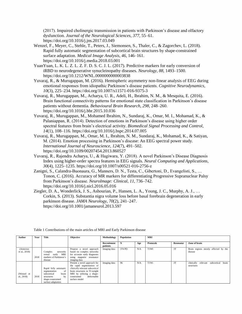
Table 1 Contributions of the main articles of MRI and Early Parkinson disease
Author Year Title Objective Methodology Population MRI
Recruitment
patients
N Age Protocols Resonator Zone of brain
(Amoroso
et al., 2018)
2018
Complex networks
reveal early MRI
markers of Parkinson’s
disease
Propose a novel approach
based on complex networks
for accurate early diagnoses
using magnetic resonance
imaging data
Imaging data 374 PD N/A T1WI 3T Brain regions mostly affected by the
disease
(Wenzel et
al., 2018)
2018
Rapid fully automatic
segmentation of
subcortical brain
structures by
shape-constrained
surface adaptation
Present a novel approach for
the rapid segmentation of
clinically relevant subcortical
brain structures in TI-weight
MRI by utilizing a shape-
constrained deformable
surface model
Imaging data 96 N/A T1Wi 3T clinically relevant subcortical brain
structures
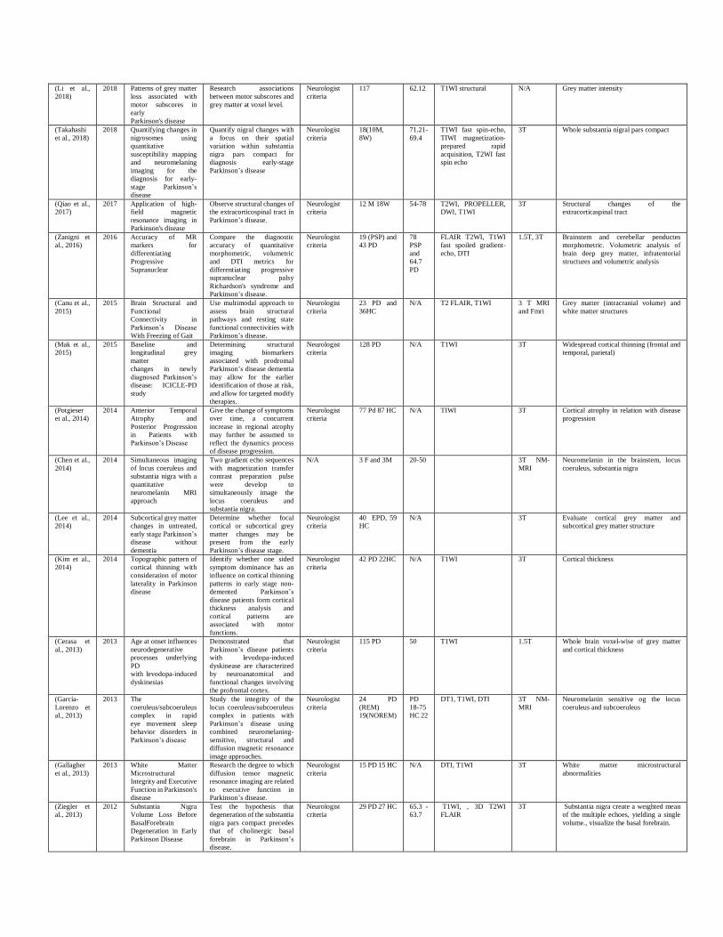
(Li et al.,
2018)
2018 Patterns of grey matter
loss associated with
motor subscores in
early
Parkinson's disease
Research associations
between motor subscores and
grey matter at voxel level.
Neurologist
criteria
117 62.12 T1WI structural N/A Grey matter intensity
(Takahashi
et al., 2018)
2018 Quantifying changes in
nigrosomes using
quantitative
susceptibility mapping
and neuromelaning
imaging for the
diagnosis for early-
stage Parkinson’s
disease
Quantify nigral changes with
a focus on their spatial
variation within substantia
nigra pars compact for
diagnosis early-stage
Parkinson’s disease
Neurologist
criteria
18(10M,
8W)
71.21-
69.4
T1WI fast spin-echo,
TIWI magnetization-
prepared rapid
acquisition, T2WI fast
spin echo
3T Whole substantia nigral pars compact
(Qiao et al.,
2017)
2017 Application of high-
field magnetic
resonance imaging in
Parkinson's disease
Observe structural changes of
the extracorticospinal tract in
Parkinson’s disease.
Neurologist
criteria
12 M 18W 54-78 T2WI, PROPELLER,
DWI, T1WI
3T Structural changes of the
extracorticaspinal tract
(Zanigni et
al., 2016)
2016 Accuracy of MR
markers for
differentiating
Progressive
Supranuclear
Compare the diagnostic
accuracy of quantitative
morphometric, volumetric
and DTI metrics for
differentiating progressive
supranuclear palsy
Richardson's syndrome and
Parkinson’s disease.
Neurologist
criteria
19 (PSP) and
43 PD
78
PSP
and
64.7
PD
FLAIR T2WI, T1WI
fast spoiled gradient-
echo, DTI
1.5T, 3T Brainstem and cerebellar penductes
morphometric. Volumetric analysis of
brain deep grey matter, infratentorial
structures and volumetric analysis
(Canu et al.,
2015)
2015 Brain Structural and
Functional
Connectivity in
Parkinson’s Disease
With Freezing of Gait
Use multimodal approach to
assess brain structural
pathways and resting state
functional connectivities with
Parkinson’s disease.
Neurologist
criteria
23 PD and
36HC
N/A T2 FLAIR, T1WI 3 T MRI
and Fmri
Grey matter (intracranial volume) and
white matter structures
(Mak et al.,
2015)
2015 Baseline and
longitudinal grey
matter
changes in newly
diagnosed Parkinson’s
disease: ICICLE-PD
study
Determining structural
imaging biomarkers
associated with prodromal
Parkinson’s disease dementia
may allow for the earlier
identification of those at risk,
and allow for targeted modify
therapies.
Neurologist
criteria
128 PD N/A T1WI 3T Widespread cortical thinning (frontal and
temporal, parietal)
(Potgieser
et al., 2014)
2014 Anterior Temporal
Atrophy and
Posterior Progression
in Patients with
Parkinson’s Disease
Give the change of symptoms
over time, a concurrent
increase in regional atrophy
may further be assumed to
reflect the dynamics process
of disease progression.
Neurologist
criteria
77 Pd 87 HC N/A TIWI 3T Cortical atrophy in relation with disease
progression
(Chen et al.,
2014)
2014 Simultaneous imaging
of locus coeruleus and
substantia nigra with a
quantitative
neuromelanin MRI
approach
Two gradient echo sequences
with magnetization transfer
contrast preparation pulse
were develop to
simultaneously image the
locus coeruleus and
substantia nigra.
N/A 3 F and 3M 20-50 3T NM-
MRI
Neuromelanin in the brainstem, locus
coeruleus, substantia nigra
(Lee et al.,
2014)
2014 Subcortical grey matter
changes in untreated,
early stage Parkinson’s
disease without
dementia
Determine whether focal
cortical or subcortical grey
matter changes may be
present from the early
Parkinson’s disease stage.
Neurologist
criteria
40 EPD, 59
HC
N/A 3T Evaluate cortical grey matter and
subcortical grey matter structure
(Kim et al.,
2014)
2014 Topographic pattern of
cortical thinning with
consideration of motor
laterality in Parkinson
disease
Identify whether one sided
symptom dominance has an
influence on cortical thinning
patterns in early stage non-
demented Parkinson’s
disease patients form cortical
thickness analysis and
cortical patterns are
associated with motor
functions.
Neurologist
criteria
42 PD 22HC N/A T1WI 3T Cortical thickness
(Cerasa et
al., 2013)
2013 Age at onset influences
neurodegenerative
processes underlying
PD
with levodopa-induced
dyskinesias
Demonstrated that
Parkinson’s disease patients
with levodopa-induced
dyskinease are characterized
by neuroanatomical and
functional changes involving
the profrontal cortex.
Neurologist
criteria
115 PD 50 T1WI 1.5T Whole brain voxel-wise of grey matter
and cortical thickness
(García-
Lorenzo et
al., 2013)
2013 The
coeruleus/subcoeruleus
complex in rapid
eye movement sleep
behavior disorders in
Parkinson’s disease
Study the integrity of the
locus coeruleus/subcoeruleus
complex in patients with
Parkinson’s disease using
combined neuromelaning-
sensitive, structural and
diffusion magnetic resonance
image approaches.
Neurologist
criteria
24 PD
(REM)
19(NOREM)
PD
18-75
HC 22
DT1, T1WI, DTI 3T NM-
MRI
Neuromelanin sensitive og the locus
coeruleus and subcoeruleus
(Gallagher
et al., 2013)
2013 White Matter
Microstructural
Integrity and Executive
Function in Parkinson's
disease
Research the degree to which
diffusion tensor magnetic
resonance imaging are related
to executive function in
Parkinson’s disease.
Neurologist
criteria
15 PD 15 HC N/A DTI, T1WI 3T White matter microstructural
abnormalities
(Ziegler et
al., 2013)
2012 Substantia Nigra
Volume Loss Before
BasalForebrain
Degeneration in Early
Parkinson Disease
Test the hypothesis that
degeneration of the substantia
nigra pars compact precedes
that of cholinergic basal
forebrain in Parkinson’s
disease.
Neurologist
criteria
29 PD 27 HC 65.3 -
63.7
T1WI, , 3D T2WI
FLAIR
3T Substantia nigra create a weighted mean
of the multiple echoes, yielding a single
volume., visualize the basal forebrain.
(Agosta et
al., 2013)
2012 The Topography of
Brain Damage at
Different
Stages of Parkinson’s
Disease
Research gray matter and
white matter damage at
different clinical stages of
Parkinson’s disease to
differentiate the trajectories
of tissue injury in this
condition.
Neurologist
criteria
89 PD 42 HC N/A T1-weighted
magnetization
prepared rapid
acquisition gradient
echo
1.5T White matter and grey matter
(Rolheiser
et al., 2011)
2011 Diffusion tensor
imaging and olfactory
identification testing
in early-stage
Parkinson’s disease
Compared
newly diagnosed Parkinson’s
disease patients with a
matched
control group using both
olfactory testing and
diffusion
tensor imaging of the
substantia nigra and anterior
olfactory
structures
Neurologist
criteria
8 M, 6 F 50-64 T1WI, T2WI 1.5T The olfactory tract and the substantia
nigra
(Moessnang
et al., 2011)
2010 Altered Activation
Patterns within the
Olfactory Network in
Parkinson’s Disease
Obtain in-depth insight into
olfactory network
dysfunction in Parkinson’s
disease patients.
Neurologist
criteria
16 (8 W,
8M)
58.4-
57.4
fMRI T2WI, DTI 3T For each hemisphere. Mesolimbic
olfactory structures encompass the
piriform cortex, amygdala, para
hippocampal cortex, hippocampus
(Hutchinson
& Raff,
2008)
2008 Detection of
Parkinson’s Disease by
MRI:
Spin-Lattice
Distribution Imaging
Develop an advance MRI
technique for detecting
Parkinson disease which
depends on an images from
two inversion recovery
sequence
Neurologist
criteria
12 PD 12HC 61.4
and
59.2
Gray matter and white
matter
1.5T Measure of disease severity in substantia
nigra
Table 2 Contributions of the main articles of EEG and Early Parkinson disease
Author Year Title Objective Methodology Population Results
Recruitme
nt patients
Non-
motor
sympto
m
Type of test N Age
Group 1: Cognitive
(Rajamanickam
Yuvaraj,
Rajendra
Acharya, &
Hagiwara,
2018)
2016 A novel
Parkinson’s
disease
diagnosis
index using
higher-order
spectra
features in
EEG signals
To explore the
application of
higher-order
spectra for
automated
diagnosis of
Parkinson’s disease
using
electroencephalogr
aphy signals
Neurologist
criteria
cognitive The EEG signals were recorded for 5
minutes eyes-closed resting state on 14-
channel (AF3, AF4, F3,F4, F5, F6, F7, F8,
T7, T8, P7, P8, O1, and O2) wireless(2.4
GHz band) Emotiv EPOC neuroheadset at
128 Sampling rate.
20 PD from
45 to
65
years
The HOS-based method, proposed by the
authors of this paper, has given the superior
performance compared to all available
modalities. The method yielded an optimum
mean accuracy: 99.62%, sensitivity: 100%, and
specificity: 99.25% using spectrum feature.
(Oh et al., 2018) 2018 A deep
learning
approach for
Parkinson’s
disease
diagnosis
from EEG
signals
To use the
electroencephalogr
aphy signals of
twenty PD and
twenty HC
employing
convolutional
neural network
Neurologist
criteria
cognitive An emotive EPOC neuroheadset of 14
channels were used. The participants were
asked to sit comfortably in a quiet room and
were informed before the recording to
refrain from body movements (e.g.,
blinking of eyes) during the recording
session. After the recording, the signals
were segmented into 2-seconds window
length
20 PD
and 20
HC
from
45 to
65
years
The evaluation parameters namely the accuracy,
sensitivity and specificity were used. The best
diagnostic performance is achieved with the
learning rate of0.0001. The proposed CNN
model yielded an accuracy of 88.25%,
sensitivity and specificity of 84.71% and
91.77% respectively.
(Özmüş et al.,
2017)
2017 Demonstratio
n of Early
cognitive
impairment in
Parkinson’s
disease with
visual P300
responses
To research the
brain dynamic of
cognitively normal
PD patients and
healthy elderly
control using event-
related potential
and to evaluate
their relationship
with
neuropsychological
test
Neurologic
al criteria
Cognitiv
e
A classical oddball paradigm was used in
the experiment. The visual stimulus has a
time off 10ms r/f a duration of 1 sec. The
participant were asked to mentally count
the target stimuli.
18 PD
and 18
HC
from
65 to
68
years
There is a significant difference between the
visual P300 amplitude values of the healthy
control group and PD patient.
Group 2: Olfactory
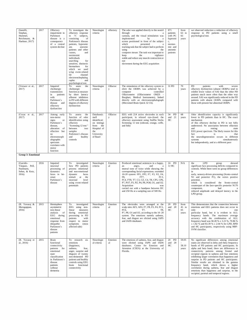
(Iannilli,
Stephan,
Hummel,
Reichmann, &
Haehner, 2017)
2017 Olfactory
impairment in
Parkinson
disease is a
consequence
of a central
system decline
To investigate the
olfactory response
in 71 subjects,
consisting of
Parkinson's disease
patients, hyposmic
ans anosmic
patients and other
causes, and
normosmic
individuals
searching for
sensitive, distinctic
biomarkers for
which we used
sclap event-related
64 channel
electroenchephalog
raphic and
psychological test.
Neurologist
criteria
olfactory The odor was applied directly to the nostril
through a
cannula, and the visual stimulation was
implemented by a
screen positioned in front of the subject
which presented a
tracking task that the subject had to perform
using the
computer mouse. The task was important to
keep the vigilance
stable and reduce any muscle contraction or
eye
movement during the EEG acquisition
871
patients
with PC
hyposmi
c,
normos
mic and
anosmic
patients
from
63 to
65
years
we observed reduction a reduction of olfactory
response in PD patients using a smell
psychological test.
(Versace et al.,
2017)
2017 Impaired
cholinergic
transmission
in patients
with
Parkinson’s
disease and
olfactory
dysfunction
To asses the
cholinergic
function as measure
by short latency
afferent inhibition
in PD with different
degrees of olfactory
dysfunction
Neurologist
criteria
Olfactor
y
The stimulation of the olfactory system to
elicit the OERPs was achieved by a
computer controlled
Olfactometer (Olfactometer OM2S -
Burghart, Medical Instruments), linked
directly with an electroencephalograph
(Micromed Brain Quick 32 Ch).
31 PD 70
years
PD patients with severe
olfactory dysfunction (absent OERPs) tend to
exhibit lower values of SAI than the other PD
patients much more often than the other way
around. SAI was significantly reduced in the PD
patients with absent OERPs compared with
those with present but abnormal OERPs
(Cozac et al.,
2017)
2017 Among early
non-motor
signs or
Parkinson’s
disease,
alteration of
olfaction but
no
electroenceph
alographic
spectrum
correlates with
motor
function
To assess the
function of odor
identification using
olfactory
"Screening 12 test "
between two
samples
Parkinson’s disease
and healthy
controls
Database(s
mell
identificati
on testing)
from the
Hospital of
the
University
of Basel
Olfactor
y
EEG with 214 active electrodes in each
participant, in relaxed eye-closed. the
olfactory assessment using Sniffin Sticks
Screening 12 test (odorant, orange, coffe,
and fish)
54 PD
and 21
HC
68
years
Odor identification capacity is significantly
lower in PD patients than in HC. The exact
mechanism
of this olfactory decline in PD is not fully
understood. No association between olfaction
and resting state
EEG power spectrum. The likely reason for this
fact is that
the neurodegeneration occurs in different
systems simultaneously
but independently, and at a different pace
Group 3: Emotional
(Garrido-
Vásquez, Pell,
Paulmann,
Sehm, & Kotz,
2016)
2016 Impaired
neuronal
processing of
dynamics
faces in let-
onset
Parkinson’s
disease
To investigated
how PD patients
process emotional
and non-emotional
dynamic faces
stimuli at the
neuronal level
using event-related
potentials
Neurologist
criteria
Emotion
al
Produced emotional sentences in a happy,
an angry, and a
neutral tone of voice while showing the
corresponding facial expressions. extended
10-20 system: FP1, FP2, F7, F3, FZ, F4,
F8, FT7, FC3,
FC4, FT8, T7, C3, CZ, C4, T8, CP5, CP6,
P7, PO7, P3, PZ, P4, P8, PO8, O1, and O2.
Acquisition was
carried out with a bandpass between DC
and 250 Hz at a sampling rate of 500 Hz.
24 PD
12 HC
N/A the LPD group showed
significant face processing deficits compared to
controls. While there were no group differences
in
early, sensory-driven processing (fronto-central
N1 and posterior P1), the vertex positive
potential,
which is considered the fronto-central
counterpart of the face-specific posterior N170
component, had a
reduced amplitude and delayed latency in the
LPD group.
(R. Yuvaraj &
Murugappan,
2016)
2015 Hemisphere
asymmetric
non-linear
analysis of
EEG during
emotional
response from
idiopathic
Parkinson’s
disease
patients
To investigated
EEG using non-
linear measures
during emotional
processing in PD
patients with
respect to motor
symptoms (most
affected side)
Neurologist
criteria
Emotion
al
The electrodes were arranged at the
scalp sites AF3, AF4, F7, F8, F3, F4, FC5,
FC6, T7, T8,
P7, P8, O1 and O2, according to the 10–20
system. The emotions namely sadness,
fear, and disgust are elicited using IAPS
and IADS databases
20 PD
and 20
HC
from
45 to
65
years
This demonstrates that the connection between
emotions and EEG patterns does not occur in
only one
particular band, but it is evident in ALL
frequency bands. The maximum average
accuracy with the combination of ALL
frequency band was 58.28 % ± 3.23 %, 70.86 %
± 1.82 % and 83.39 % ± 2.04 % for LPD, RPD
and HC participants, respectively using RBF-
SVM classifier.
(R. Yuvaraj et
al., 2016)
2015 Brain
functional
connectivity
patterns for
emotional
state
classification
in Parkinson’s
disease
patients
without
dementia
To research six
emotions
(happiness,
sadness, fear,
anger, surprise and
disgust) of twenty
non-demented PD
patients and healthy
controls using EEG
brain functional
connectivity
Neurologic
al criteria
Emotion
al
The emotions of sadness, fear, and disgust
were elicited using IAPS and IADS
databases. Center for Emotion and
Attention (CSEA) at the University of
Florida.
20 PD
and 20
HC
59.05
-
58.10
years
No significant differences among emotional
states are observed in delta and theta frequency
bands of PD patients and HC participants. In
alpha and beta band, there are differences in
connectivity patterns among frontal and
occipital regions in sadness and fear emotions
exhibiting larger correlation than happiness and
surprise in PD patients and HC participants.
Similar results are obtained in the gamma
frequency band, which shows a higher
correlation during sadness, fear and disgust
emotions than happiness and surprise, in the
occipital, parietal and temporal regions.
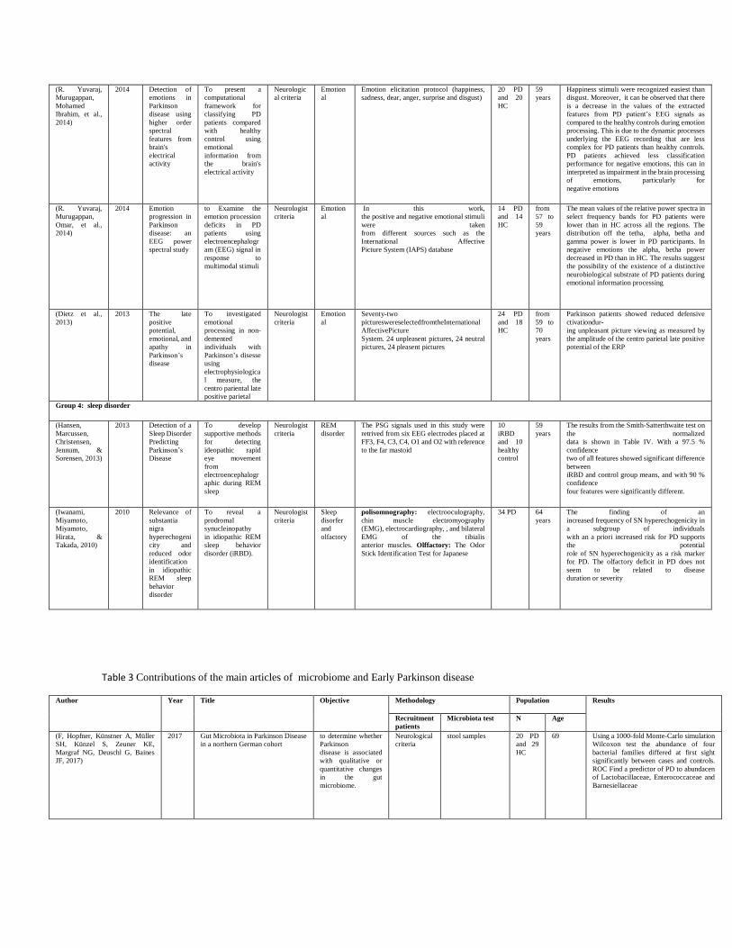
(R. Yuvaraj,
Murugappan,
Mohamed
Ibrahim, et al.,
2014)
2014 Detection of
emotions in
Parkinson
disease using
higher order
spectral
features from
brain's
electrical
activity
To present a
computational
framework for
classifying PD
patients compared
with healthy
control using
emotional
information from
the brain's
electrical activity
Neurologic
al criteria
Emotion
al
Emotion elicitation protocol (happiness,
sadness, dear, anger, surprise and disgust)
20 PD
and 20
HC
59
years
Happiness stimuli were recognized easiest than
disgust. Moreover, it can be observed that there
is a decrease in the values of the extracted
features from PD patient’s EEG signals as
compared to the healthy controls during emotion
processing. This is due to the dynamic processes
underlying the EEG recording that are less
complex for PD patients than healthy controls.
PD patients achieved less classification
performance for negative emotions, this can in
interpreted as impairment in the brain processing
of emotions, particularly for
negative emotions
(R. Yuvaraj,
Murugappan,
Omar, et al.,
2014)
2014 Emotion
progression in
Parkinson
disease: an
EEG power
spectral study
to Examine the
emotion procession
deficits in PD
patients using
electroencephalogr
am (EEG) signal in
response to
multimodal stimuli
Neurologist
criteria
Emotion
al
In this work,
the positive and negative emotional stimuli
were taken
from different sources such as the
International Affective
Picture System (IAPS) database
14 PD
and 14
HC
from
57 to
59
years
The mean values of the relative power spectra in
select frequency bands for PD patients were
lower than in HC across all the regions. The
distribution off the tetha, alpha, betha and
gamma power is lower in PD participants. In
negative emotions the alpha, betha power
decreased in PD than in HC. The results suggest
the possibility of the existence of a distinctive
neurobiological substrate of PD patients during
emotional information processing
(Dietz et al.,
2013)
2013 The late
positive
potential,
emotional, and
apathy in
Parkinson’s
disease
To investigated
emotional
processing in non-
demented
individuals with
Parkinson’s disesse
using
electrophysiologica
l measure, the
centro pariental late
positive parietal
Neurologist
criteria
Emotion
al
Seventy-two
pictureswereselectedfromtheInternational
AffectivePicture
System. 24 unpleasent pictures, 24 neutral
pictures, 24 pleasent pictures
24 PD
and 18
HC
from
59 to
70
years
Parkinson patients showed reduced defensive
ctivationdur-
ing unpleasant picture viewing as measured by
the amplitude of the centro parietal late positive
potential of the ERP
Group 4: sleep disorder
(Hansen,
Marcussen,
Christensen,
Jennum, &
Sorensen, 2013)
2013 Detection of a
Sleep Disorder
Predicting
Parkinson’s
Disease
To develop
supportive methods
for detecting
ideopathic rapid
eye movement
from
electroencephalogr
aphic during REM
sleep
Neurologist
criteria
REM
disorder
The PSG signals used in this study were
retrived from six EEG electrodes placed at
FF3, F4, C3, C4, O1 and O2 with reference
to the far mastoid
10
iRBD
and 10
healthy
control
59
years
The results from the Smith-Satterthwaite test on
the normalized
data is shown in Table IV. With a 97.5 %
confidence
two of all features showed significant difference
between
iRBD and control group means, and with 90 %
confidence
four features were significantly different.
(Iwanami,
Miyamoto,
Miyamoto,
Hirata, &
Takada, 2010)
2010 Relevance of
substantia
nigra
hyperechogeni
city and
reduced odor
identification
in idiopathic
REM sleep
behavior
disorder
To reveal a
prodromal
synucleinopathy
in idiopathic REM
sleep behavior
disorder (iRBD).
Neurologist
criteria
Sleep
disorfer
and
olfactory
polisomnography: electrooculography,
chin muscle electromyography
(EMG), electrocardiography, , and bilateral
EMG of the tibialis
anterior muscles. Olffactory: The Odor
Stick Identification Test for Japanese
34 PD 64
years
The finding of an
increased frequency of SN hyperechogenicity in
a subgroup of individuals
with an a priori increased risk for PD supports
the potential
role of SN hyperechogenicity as a risk marker
for PD. The olfactory deficit in PD does not
seem to be related to disease
duration or severity
Table 3 Contributions of the main articles of microbiome and Early Parkinson disease
Author Year Title Objective Methodology Population Results
Recruitment
patients
Microbiota test N Age
(F, Hopfner, Künstner A, Müller
SH, Künzel S, Zeuner KE,
Margraf NG, Deuschl G, Baines
JF, 2017)
2017 Gut Microbiota in Parkinson Disease
in a northern German cohort
to determine whether
Parkinson
disease is associated
with qualitative or
quantitative changes
in the gut
microbiome.
Neurological
criteria
stool samples 20 PD
and 29
HC
69 Using a 1000-fold Monte-Carlo simulation
Wilcoxon test the abundance of four
bacterial families differed at first sight
significantly between cases and controls.
ROC Find a predictor of PD to abundacen
of Lactobacillaceae, Enterococcaceae and
Barnesiellaceae
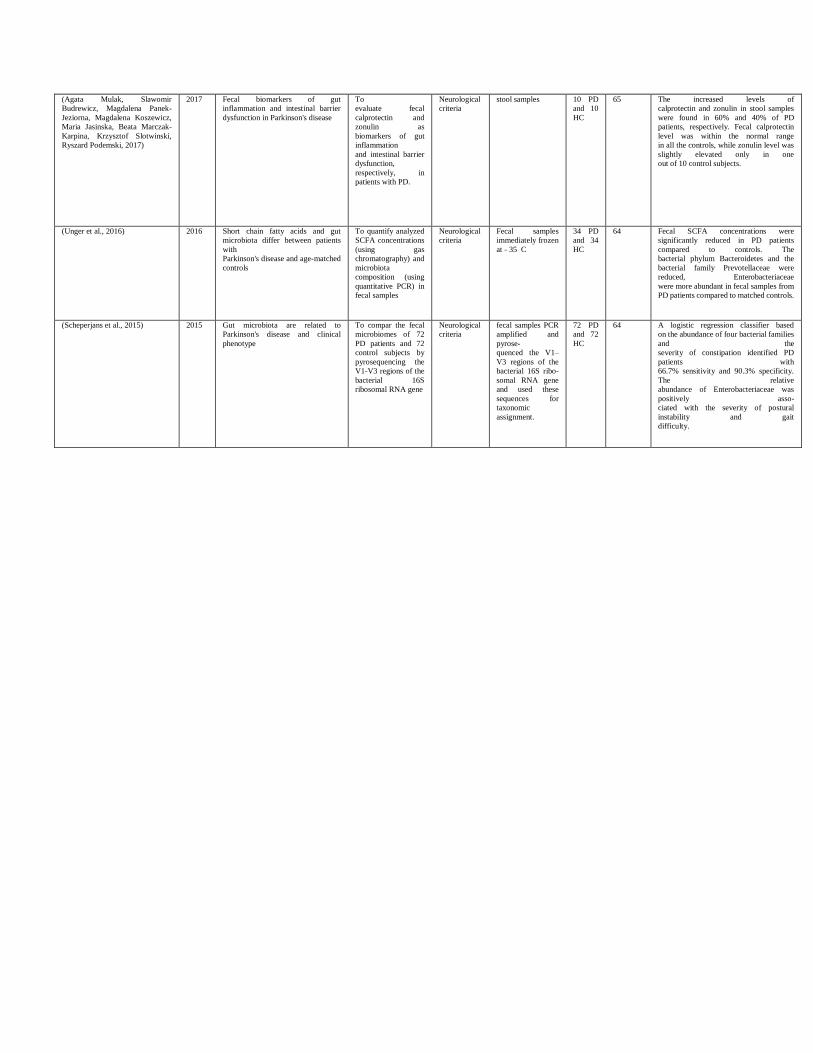
(Agata Mulak, Slawomir
Budrewicz, Magdalena Panek-
Jeziorna, Magdalena Koszewicz,
Maria Jasinska, Beata Marczak-
Karpina, Krzysztof Slotwinski,
Ryszard Podemski, 2017)
2017 Fecal biomarkers of gut
inflammation and intestinal barrier
dysfunction in Parkinson's disease
To
evaluate fecal
calprotectin and
zonulin as
biomarkers of gut
inflammation
and intestinal barrier
dysfunction,
respectively, in
patients with PD.
Neurological
criteria
stool samples 10 PD
and 10
HC
65 The increased levels of
calprotectin and zonulin in stool samples
were found in 60% and 40% of PD
patients, respectively. Fecal calprotectin
level was within the normal range
in all the controls, while zonulin level was
slightly elevated only in one
out of 10 control subjects.
(Unger et al., 2016) 2016 Short chain fatty acids and gut
microbiota differ between patients
with
Parkinson's disease and age-matched
controls
To quantify analyzed
SCFA concentrations
(using gas
chromatography) and
microbiota
composition (using
quantitative PCR) in
fecal samples
Neurological
criteria
Fecal samples
immediately frozen
at - 35 C
34 PD
and 34
HC
64 Fecal SCFA concentrations were
significantly reduced in PD patients
compared to controls. The
bacterial phylum Bacteroidetes and the
bacterial family Prevotellaceae were
reduced, Enterobacteriaceae
were more abundant in fecal samples from
PD patients compared to matched controls.
(Scheperjans et al., 2015) 2015 Gut microbiota are related to
Parkinson's disease and clinical
phenotype
To compar the fecal
microbiomes of 72
PD patients and 72
control subjects by
pyrosequencing the
V1-V3 regions of the
bacterial 16S
ribosomal RNA gene
Neurological
criteria
fecal samples PCR
amplified and
pyrose-
quenced the V1–
V3 regions of the
bacterial 16S ribo-
somal RNA gene
and used these
sequences for
taxonomic
assignment.
72 PD
and 72
HC
64 A logistic regression classifier based
on the abundance of four bacterial families
and the
severity of constipation identified PD
patients with
66.7% sensitivity and 90.3% specificity.
The relative
abundance of Enterobacteriaceae was
positively asso-
ciated with the severity of postural
instability and gait
difficulty.