Cell Reports
Article
Cholesterol Regulates Syntaxin 6 Traffickingat trans-Golgi Network Endosomal BoundariesMeritxell Reverter,1,11 Carles Rentero,1,11 Ana Garcia-Melero,1 Monira Hoque,2 Sandra Vila de Muga,1
Anna Alvarez-Guaita,1 James R.W. Conway,3 Peta Wood,2 Rose Cairns,2 Lilia Lykopoulou,4 Daniel Grinberg,5
Lluısa Vilageliu,5 Marta Bosch,6 Joerg Heeren,7 Juan Blasi,8 Paul Timpson,3 Albert Pol,1,6,9 Francesc Tebar,1,6
Rachael Z. Murray,10 Thomas Grewal,2,* and Carlos Enrich1,6,*1Departament de Biologia Cel$lular, Immunologia i Neurociencies, Facultat de Medicina, Universitat de Barcelona, 08036 Barcelona, Spain2Faculty of Pharmacy, University of Sydney, Sydney, NSW 2006, Australia3Garvan Institute of Medical Research and Kinghorn Cancer Centre, Cancer Research Program, St. Vincent’s Clinical School,
Faculty of Medicine, University of New South Wales, Sydney, NSW 2010, Australia4First Department of Pediatrics, University of Athens, Aghia Sofia Children’s Hospital, 11527 Athens, Greece5Departament de Genetica, Facultat de Biologia, Universitat de Barcelona, CIBERER, IBUB, 08028 Barcelona, Spain6Centre de Recerca Biomedica CELLEX, Institut d’Investigacions Biomediques August Pi i Sunyer (IDIBAPS), 08036 Barcelona, Spain7Department of Biochemistry and Molecular Biology II. Molecular Cell Biology, University Medical Center Hamburg-Eppendorf,
20246 Hamburg, Germany8Department of Pathology and Experimental Therapeutics, IDIBELL-University of Barcelona, L’Hospitalet de Llobregat, 08907 Barcelona,
Spain9Institucio Catalana de Recerca i Estudis Avacats (ICREA), 08010 Barcelona, Spain10Tissue Repair and Regeneration Program, Institute of Health and Biomedical, Innovation, Queensland University of Technology, Brisbane,QLD 4095, Australia11Co-first author
*Correspondence: [email protected] (T.G.), [email protected] (C.E.)http://dx.doi.org/10.1016/j.celrep.2014.03.043
This is an open access article under the CC BY-NC-ND license (http://creativecommons.org/licenses/by-nc-nd/3.0/).
SUMMARY
Inhibition of cholesterol export from late endosomescauses cellular cholesterol imbalance, includingcholesterol depletion in the trans-Golgi network(TGN). Here, using Chinese hamster ovary (CHO)Niemann-Pick type C1 (NPC1) mutant cell lines andhuman NPC1 mutant fibroblasts, we show thataltered cholesterol levels at the TGN/endosomeboundaries trigger Syntaxin 6 (Stx6) accumulationinto VAMP3, transferrin, and Rab11-positive recy-cling endosomes (REs). This increases Stx6/VAMP3interaction and interferes with the recycling of aVb3and a5b1 integrins and cell migration, possibly in aStx6-dependent manner. In NPC1 mutant cells,restoration of cholesterol levels in the TGN, but notinhibition of VAMP3, restores the steady-state local-ization of Stx6 in the TGN. Furthermore, elevation ofRE cholesterol is associated with increased amountsof Stx6 in RE. Hence, the fine-tuning of cholesterollevels at the TGN-RE boundaries together with a sub-set of cholesterol-sensitive SNAREproteinsmay playa regulatory role in cell migration and invasion.
INTRODUCTION
The intracellular trafficking, distribution, and concentration of
cellular cholesterol contributes to regulate lipid and protein
transport between cellular compartments and organizes
membrane microdomains, such as lipid rafts, at the plasma
membrane and in endo-/exocytic pathways (Maxfield and van
Meer, 2010; Simons and Ikonen, 2000). In general, cells obtain
cholesterol through endocytosis of low-density lipoproteins
(LDLs). The subsequent delivery of LDL cholesterol to endolyso-
somes and then to other subcellular compartments is facilitated
by a complex transport machinery, consisting of vesicular and
nonvesicular pathways (Ikonen, 2008; Mesmin and Maxfield,
2009). Deregulation of these cholesterol transport pathways is
associated with human disorders, including lysosomal storage
diseases, neurological disorders, and cardiovascular events
(Ikonen, 2006).
In the context of membrane trafficking, cholesterol is also
essential for the functioning of a subset of SNARE proteins along
secretory and endocytic pathways. We and others showed that
cholesterol modulates the clustering and the location of several
SNARE proteins in membranes, such as the t-SNARES SNAP23
and Stx4 (Reverter et al., 2011) or SNAP25 and Stx1A (Lang
et al., 2001; Veale et al., 2011). Syntaxin 6 (Stx6) is another
t-SNARE linked to cholesterol transport, contributing to the de-
livery of lipids and proteins required for caveolae endocytosis
(Choudhury et al., 2006). Stx6 is a cholesterol-binding protein
(Hulce et al., 2013), predominantly localized at the trans-Golgi
network (TGN) (Bock et al., 1997) involved in the regulation of
cholesterol-rich domains that determine the levels of cell-sur-
face-associated a5b1 integrin, focal adhesion kinase (FAK), focal
adhesion sites, and directional migration toward fibronectin (FN)
(Tiwari et al., 2011). However, how cholesterol affects Stx6-
dependent trafficking mechanisms still remains unclear.
Cell Reports 7, 883–897, May 8, 2014 ª2014 The Authors 883
(legend on next page)
884 Cell Reports 7, 883–897, May 8, 2014 ª2014 The Authors
Several SNAREs have been implicated in integrin trafficking
and related to the migratory ability of cells. Cholesterol controls
aVb3 integrin signal complex formation (Green et al., 1999), cell
adhesion, and migration onto FN (Ramprasad et al., 2007). This
includes VAMP2/VAMP3, Stx3/Stx4, and SNAP23 participating
in b1 integrin recycling (Day et al., 2011; Powelka et al., 2004;
Proux-Gillardeaux et al., 2005; Skalski and Coppolino, 2005;
Veale et al., 2010) and VAMP3 together with Stx6 determining
cell-surface-associated levels of a5b1 integrin and FAK (Riggs
et al., 2012; Tiwari et al., 2011).
Integrins are cell-surface receptors that mediate and coordi-
nate cellular responses to the extracellular matrix (ECM). Integ-
rins display their principal functions at the plasma membrane
but reside and traffic through the endocytic compartment, in
particular in recycling endosomes (RE) (Jones et al., 2006; Pelli-
nen and Ivaska, 2006). Certain integrin receptors, such as aVb3,
are recycled rapidly to the plasma membrane through a short
loop, whereas a5b1 returns to the cell surface through a transi-
tory recycling compartment (long-loop) (Roberts et al., 2001).
The recycling of integrins depends upon many endocytic
regulatory proteins that also control the transport of other inter-
nalized receptors. For instance, the guanosine triphosphate
(GTP)-binding proteins Rab4 (Roberts et al., 2001), Rab11, Arf6
(Powelka et al., 2004), Rab21, Rab5 (Pellinen et al., 2006), and
more recently Rab25 (Dozynkiewicz et al., 2012) have been
implicated in integrin recycling. Based on these studies, other
regulators of endocytic recycling and/or proteins (SNAREs) or
lipids that physically and/or functionally interact with certain
Rab proteins could play a role in the regulation of integrin
trafficking.
Using Niemann-Pick type C1 (NPC1) mutant Chinese hamster
ovary (CHO) cell lines (CHOM12 and CHO 2-2) (Dahl et al., 1992;
Millard et al., 2000), pharmacological U18666A treatment (Lis-
cum and Faust, 1989), and loss-of-function mutant NPC1-
P692S (Du et al., 2011; Millard et al., 2005; Ohgami et al.,
2004), all of which are models shown to accumulate cholesterol
in late endosomes (LE) with concomitant cholesterol depletion in
Golgi membranes, we provide insights into how cholesterol
pools at theGolgi/endosomal boundaries regulate cell migration.
Mechanistically, diminution of Golgi cholesterol perturbs traf-
ficking between RE and TGN to trigger Stx6 accumulation in
Rab11-containing RE. This correlates with a diminished cell-
surface expression of integrins in NPC1 mutant models leading
to reduced cell migration and invasion in two- and three-dimen-
sional environments.
Implicating physiological relevance, the human NPC1 mutant
fibroblast cell line (GM03123; Choudhury et al., 2004) and pri-
mary fibroblasts from a NPC1 patient (G1) (Rodrıguez-Pascau
et al., 2012) display translocation of Stx6 into RE, as well as
diminished cell-surface expression of aV, a5, and b3 integrins
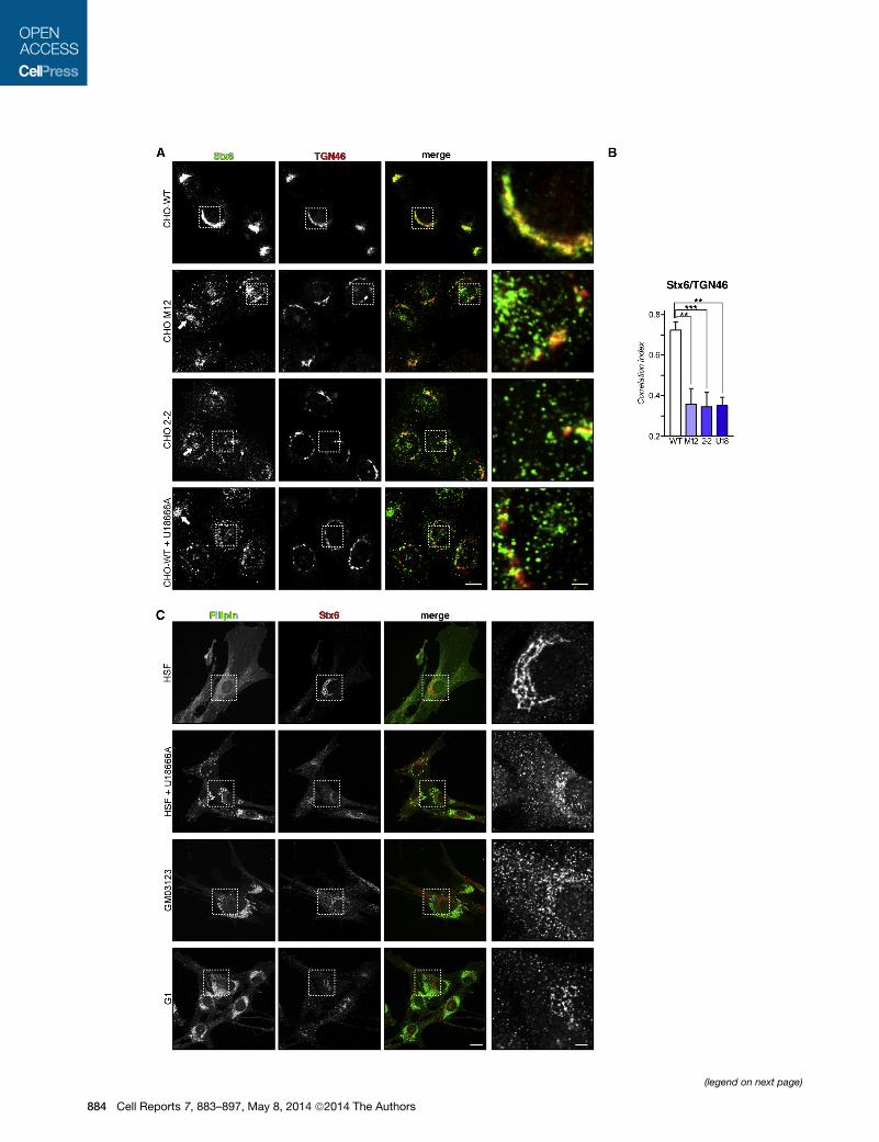
Figure 1. Stx6 Localization in NPC1 Mutant Cells
(A) CHO-WT, CHO M12, CHO 2-2, and U18666A-treated CHO-WT cells were fi
sentative images and insets (right panels) showStx6 staining details at the Golgi an
or U18666A-treated cells. The scale bars represent 10 and 2 mm (insets).
(B) Quantification of Stx6/TGN46 colocalization using the correlation index (see
(C) Stx6 localization in human skin fibroblasts (HSF), U18666A-treated HSF, and
stained with filipin. The scale bars represent 20 and 5 mm (insets).
and, consequently, reduced cell migration. This study provides
evidence that modulation of cholesterol levels at the interface
of TGN and RE determines Stx6 localization and ability to
interact with VAMP4 or VAMP3, thereby possibly regulating
cell migration through Stx6-dependent aVb3 and a5b1 integrin
trafficking.
RESULTS
Cholesterol-Dependent Stx6 Translocation into Rab11/VAMP3-Recycling EndosomesNPC1 mutant cell lines CHO M12 and CHO 2-2 or U18666A-
treated wild-type CHO (CHO-WT) cells are well characterized
for their cholesterol accumulation in LE, as judged by filipin-
positive LE structures (Figure S1). Impaired LE cholesterol
(LE-chol) export leads to a reduction of Golgi cholesterol
(Blanchette-Mackie et al., 1988; Coxey et al., 1993), interferes
with post-Golgi transport (Cubells et al., 2007, 2008; Pol et al.,
2005; Reverter et al., 2011; Wang et al., 2000), and sequesters
the t-SNAREs SNAP23 and Stx4 in the Golgi (Reverter et al.,
2011). Here, we investigated if cholesterol imbalance could
affect localization and function of another member of the SNARE
family, Stx6, which has been linked to cholesterol and caveolin
transport (Choudhury et al., 2006; Urano et al., 2008).
In CHO-WT cells, Stx6 was located in perinuclear TGN
membranes, where the vast majority of Stx6 colocalizes with
the Golgi marker TGN46 (Figure 1A) (Bock et al., 1997; Choud-
hury et al., 2006) and with VAMP4, a v-SNARE predominantly
localized in the TGN (see Figures 5A and 5B). In addition, a
distinct population of Stx6 vesicles not colocalizing with
TGN46 and probably representing early endosomes (EE) and/
or secretory structures was observed.
In contrast, Stx6 was predominantly located in scattered cyto-
plasmic structures in CHO M12 and CHO 2-2 as well as
U18666A-treated CHO-WT cells (Figure 1A; see quantification
in Figure 1B). In addition, approximately 30%–40% of these
NPC1 mutant cell lines also contained Stx6 in sometimes prom-
inent, perinuclear recycling endosomes (perinuclear recycling
endosomal compartment [PNRE], arrows). Importantly, this
Stx6-positive compartment did not colocalize with TGN46 or
VAMP4 but contained internalized transferrin and endogenous
Rab11 (see below), indicative of the recycling compartment.
Similarly, in human skin fibroblasts (HSF), most of Stx6
labeling consisted of a distinctive perinuclear network of Golgi
membranes (Figure 1C), whereas Stx6 was predominantly
located in scattered punctate structures in the NPC1 mutant
GM03123 cell line; in primary fibroblasts from a NPC1 patient,
NPC1-G1 (G1); and in U18666A-treated HSF fibroblasts.
The loss of Golgi-associated Stx6 was not due to alterations
in Golgi morphology in NPC1 mutant models or human NPC1
xed and immunolabeled with anti-Stx6 (green) and anti-TGN46 (red). Repre-
d in perinuclear recycling endosomes (PNRE, arrows) in NPC1mutant cell lines
the Experimental Procedures).
G03123 and G1 fibroblasts. Cells were immunolabeled with anti-Stx6 (red) and
Cell Reports 7, 883–897, May 8, 2014 ª2014 The Authors 885
(legend on next page)
886 Cell Reports 7, 883–897, May 8, 2014 ª2014 The Authors
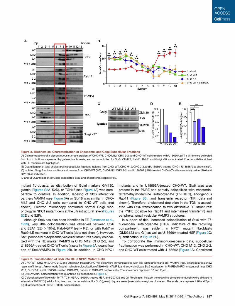
Figure 3. Biochemical Characterization of Endosomal and Golgi Subcellular Fractions
(A) Cellular fractions of a discontinuous sucrose gradient of CHO-WT, CHOM12, CHO 2-2, and CHO-WT cells treated with U18666A (WT + U18) were collected
from top to bottom, separated by gel electrophoresis, and immunoblotted for Stx6, VAMP3, Rab11, Rab7, and Golgin-97 as indicated. Fractions 6–8 enriched
with RE markers are highlighted.
(B) Quantification of total cholesterol in subcellular fractions isolated fromCHO-WT, CHOM12, CHO2-2, and U18666A-treated (CHO+U18666A) as shown in (A).
(C) Isolated Golgi fractions and total cell lysates from CHO-WT (WT), CHOM12, CHO 2-2, and U18666A (U18)-treated CHO-WT cells were analyzed for Stx6 and
GM130 as indicated.
(D and E) Quantification of Golgi-associated Stx6 and cholesterol, respectively.
mutant fibroblasts, as distribution of Golgi markers GM130,
giantin (Figures S2A–S2D), or TGN46 (see Figure 1A) was com-
parable to controls. In addition, labeling of Stx6 interaction
partners VAMP4 (see Figure 5A) or Stx16 was similar in CHO-
M12 and CHO 2-2 cells compared to CHO-WT cells (not
shown). Electron microscopy confirmed normal Golgi mor-
phology in NPC1 mutant cells at the ultrastructural level (Figures
S2E and S2F).
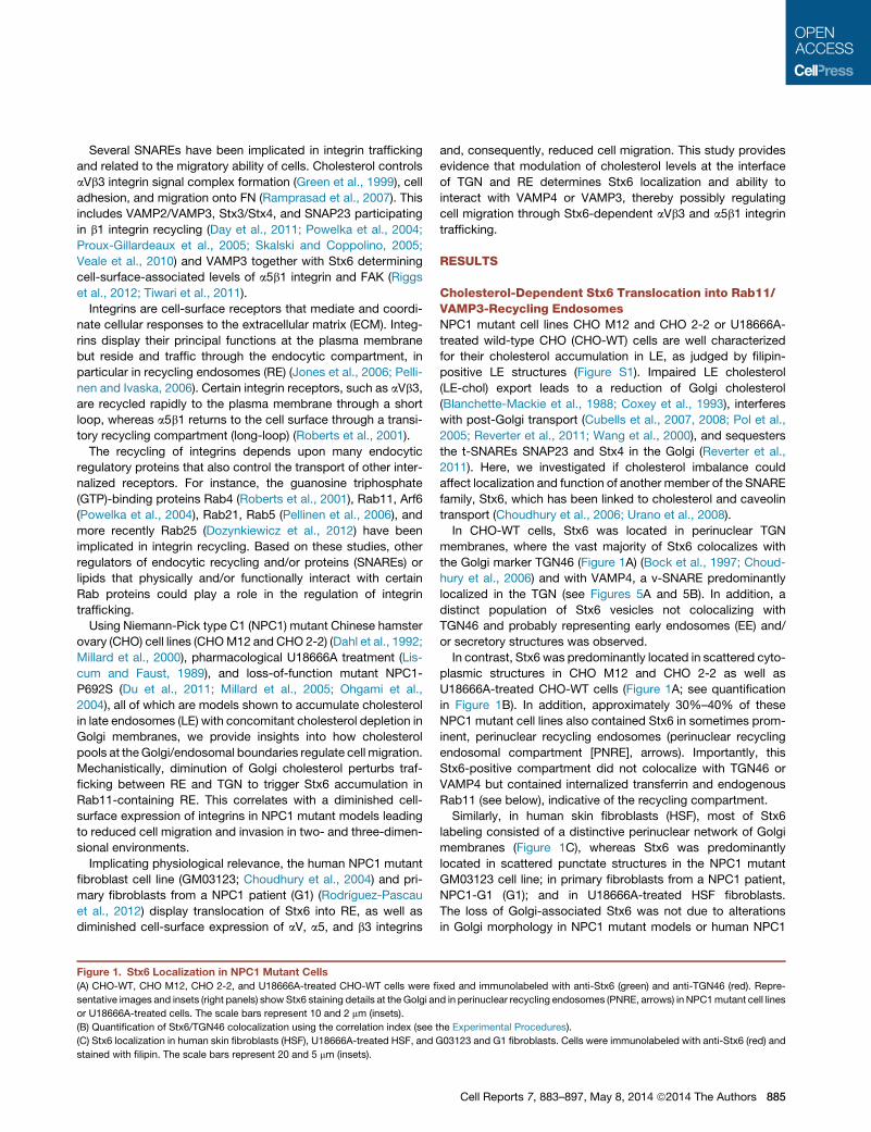
Although Stx6 has also been identified in EE (Simonsen et al.,
1999), very little colocalization was observed between Stx6
and EEA1 (EE) (�10%), Rab4-GFP (early RE), or with Rab7 or
Rab9 (LE markers) in CHO-WT cells (data not shown). However,
Stx6 peripheral cytoplasmic vesicular structures clearly colocal-
ized with the RE marker VAMP3 in CHO M12, CHO 2-2, and
U18666A-treated CHO-WT cells (insets in Figure 2A; quantifica-
tion of Stx6/VAMP3 in Figure 2B). In addition, in CHO-NPC1
Figure 2. Translocation of Stx6 into RE in NPC1 Mutant Cells
(A) CHO-WT, CHO M12, CHO 2-2, and U18666A-treated CHO-WT cells were imm
regions of interest. Arrowheads (insets) indicate colocalization of Stx6 with VAMP
M12, CHO 2-2, and U18666A-treated CHO-WT, but not in CHO-WT control cells
(B) Stx6/VAMP3 colocalization was quantified as described in Figure 1.
(C) Colocalization of Stx6with Tf-TRITC in HSF, U18666A-treated HSF, andG0312
internalize Tf-TRITC (red) for 1 hr, fixed, and immunostained for Stx6 (green). Squa
(D) Quantification of Stx6/Tf-TRITC colocalization.
mutants and in U18666A-treated CHO-WT, Stx6 was also
present in the PNRE and partially colocalized with transferrin-
tetramethylrhodamine isothiocyanate (Tf-TRITC), endogenous
Rab11 (Figure S3), and transferrin receptor (TfR; data not
shown). Therefore, cholesterol depletion in the TGN is associ-
ated with Stx6 translocation to two distinctive RE structures:
the PNRE (positive for Rab11 and internalized transferrin) and
peripheral, small vesicular VAMP3 structures.
In support of this, increased colocalization of Stx6 with Tf-
fluorescein isothiocyanate (FITC), indicative of the recycling
compartment, was evident in NPC1 mutant fibroblasts
(GM03123 and G1) as well as U18666A-treated HSF (Figure 2C;
quantification in Figure 2D).
To corroborate the immunofluorescence data, subcellular
fractionation was performed in CHO-WT, CHO M12, CHO 2-2,
and CHO-WT cells treated with U18666A (Figure 3A). Consistent
unolabeled with anti-Stx6 (green) and anti-VAMP3 (red). Enlarged areas show
3, and arrows indicate Stx6 localization in PNRE of NPC1mutant cell lines CHO
. The scale bars represent 10 and 2 mm.
3 andG1 fibroblasts. To label the recycling compartment, cells were allowed to
re areas (insets) show regions of interest. The scale bars represent 20 and 2 mm.
Cell Reports 7, 883–897, May 8, 2014 ª2014 The Authors 887
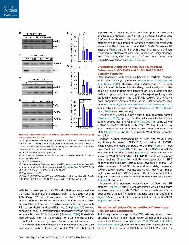
Figure 4. Characterization of Stx6-Containing SNARE Complexes in
NPC Mutant CHO Cells
(A) Cell lysates fromCHO-WT (WT), CHOM12, CHO 2-2, and U18666A-treated
CHO-WT (WT + U18) cells were immunoprecipitated with anti-VAMP4 or
control antibody (normal rabbit serum [NRS]) and analyzed for coimmuno-
precipitation of Stx6. IP, immunoprecipitate.
(B) Quantification in percentage of (A).
(C) Immunoprecipitation of VAMP4 and coimmunoprecipitation of NSF is
shown as indicated.
(D) Quantification of (C).
(E) Characterization of Stx6-containing VAMP3 immunoprecipitates from RE-
enriched subcellular fractions (see nos. 6–8 in Figure 3A) from CHO-WT, CHO
M12, CHO 2-2, and U18666A-treated CHO-WT.
(F) Quantification of (E).
(G) Total Stx6, VAMP4, VAMP3, and NSF levels in cell lysates from CHO-WT,
CHO M12, CHO 2-2, and CHO-WT treated with U18666A (WT + U18).
with the microscopy, in CHO-WT cells, Stx6 appears mostly in
the heavy fractions of the gradient (nos. 10–13), together with
TGN (Golgin-97) and plasma membrane (Na+K+-ATPase; not
shown) markers. However, in all NPC1 mutant models, Stx6
accumulated in fractions 7–9, which were highly enriched with
RE markers (Rab11 and VAMP3 in nos. 6–8) (Figure 3A). Hence,
although subcellular fractionation methods have limited ability to
separate TGN and RE in CHO cells (Hao et al., 2002), these find-
ings correlate with the translocation of Stx6 into RE of NPC
mutant cells observed by immunofluorescence microscopy.
The distribution of cholesterol along the sucrose gradient was
in agreement with published data. In CHO-WT cells, cholesterol
888 Cell Reports 7, 883–897, May 8, 2014 ª2014 The Authors
was elevated in heavy fractions containing plasma membrane
and Golgi membranes (nos. 10–13). In contrast, NPC1 mutant
CHO cell lines showed a diminution of cholesterol in the plasma
membrane and Golgi fractions, whereas cholesterol levels were
elevated in Rab7-positive LE and Rab11/VAMP3-positive RE
fractions (Figure 3B). In line with these findings, a significant
reduction of cholesterol and Stx6 in isolated Golgi fractions
from CHO M12, CHO 2-2, and CHO-WT cells treated with
U18666A was observed (Figures 3C–3E).
Cholesterol Distribution at the TGN-RE InterfaceDetermines Stx6/VAMP4 and Stx6/VAMP3 SNAREComplex FormationStx6 associates with various SNAREs at multiple locations
in endo- and exocytic pathways (Murray et al., 2005; Wendler
and Tooze, 2001). Because Stx6 accumulated in RE upon
diminution of cholesterol in the Golgi, we investigated if this
could be linked to possible alterations of SNARE complex for-
mation in post-Golgi and retrograde transport pathways. We
particularly focused on the v-SNAREs VAMP3 and VAMP4,
both recognized partners of Stx6 at the TGN-endosome inter-
face (Ganley et al., 2008; Mallard et al., 2002; Tran et al., 2007)
and involved in integrin recycling (Riggs et al., 2012; Skalski
et al., 2010) (see next section).
VAMP4 is a v-SNARE protein with a TGN retention domain
(Zeng et al., 2003), cycling from the cell surface to the TGN via
sorting endosomes and RE (Tran et al., 2007) to complex with
t-SNAREs Stx6/Stx16/Vti1a in the TGN. We hypothesized that
NPC1-mutant-induced reduction of cholesterol and Stx6 in the
TGN (Figures 1, 2, and 3) could modify VAMP4/Stx6 complex
formation.
Indeed, coimmunoprecipitation of Stx6 and VAMP4 was
significantly decreased in CHO M12, CHO 2-2, and U18666A-
treated CHO-WT cells compared to controls (Figure 4A; see
quantification in Figure 4B). Total amounts of Stx6 and VAMP4
were comparable in all cell lines (Figure 4G). Decreased colocal-
ization of VAMP4 and Stx6 in CHO-NPC1 mutant cells support
these findings (Figure 5A). VAMP4 overexpression in NPC
mutant models did not restore Stx6 localization at the TGN
(data not shown). In all NPC1-mutant-like models, decreased
VAMP4/Stx6 interaction was associated with low N-ethylmalei-
mide-sensitive factor (NSF) levels in the immunoprecipitates,
suggesting less functional VAMP4/Stx6 complexes in the Golgi
(Figures 4C and 4F).
Moreover, the increased colocalization of Stx6 with RE
markers (Figures 2A and 2B) was associated with a significantly
increased amount of VAMP3/Stx6 immunocomplexes when a
pool of RE-enriched membrane fractions (see fractions 6–8 in
Figure 3A) was used for immunoprecipitation with anti-VAMP3
(Figures 4E and 4F).
Modulation of Various Cholesterol Pools DifferentiallyRegulate Stx6 LocalizationAs further proof of concept, in CHO-WT cells, expression of loss-
of-function NPC1 mutant P692S, which cannot bind cholesterol
and inhibits LE-chol export (Du et al., 2011; Millard et al., 2005;
Ohgami et al., 2004), led to Stx6 accumulation in vesicular struc-
tures. On the contrary, in CHO M12 and CHO 2-2 cells, the
ectopic expression of wild-type NPC1 restored cellular choles-
terol distribution and rescued the prominent steady state of
Stx6 in the TGN (85% in CHO M12 and 65% in CHO 2-2 cells)
(Figure S4).
Trafficking of cholesterol derived from LDLs or high-density li-
poproteins (HDLs) follows different intracellular routes. Whereas
internalized LDL cholesterol can be found in the Golgi at later
time points (Garver et al., 2002), even in NPC1 mutant cells
(Coxey et al., 1993), HDL-derived cholesterol rapidly enters the
recycling compartment (Heeren et al., 2001, 2004; Rohrl et al.,
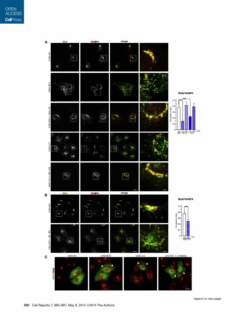
2012). Therefore, we addressed if loading with LDL cholesterol
could abrogate Stx6 mislocation into RE in NPC mutant cells.
Indeed, loading CHOM12 and CHO 2-2 with LDL for 24 hr re-es-
tablished the steady-state Stx6 TGN staining pattern (>88%)
(Figure 5A). In contrast, incubation of CHO-WT cells with HDL
for 30 min resulted in a Stx6 distribution reminiscent of the
NPC1 phenotype, with Stx6 being much more dispersed in
punctate structures and associated with diminished VAMP4 co-
localization (Figure 5B; quantification of colocalization between
Stx6 and VAMP4 ± LDL/HDL is given).
To address if the lipidic microdomain organization of TGN
membranes could trigger Stx6 translocation, CHO-WT cells
were treated with D-ceramide-C6, known to interfere with
sphinghomyelin (SM) levels and formation of SM-rich domains
in Golgi membranes (Duran et al., 2012). However, Stx6 location
remained unchanged after 30 or 60 min treatment with D-cer-
amide-C6 (Figure S5), further implicating cholesterol levels in
RE and/or TGN being responsible for Stx6 translocation.
To study if increased interaction of Stx6 with VAMP3 was
responsible for the pronounced engagement of Stx6 in RE,
NPC mutant cell lines where transfected with the catalytic light
chain of tetanus neurotoxin (L-TeTx), which selectively cleaves
and inhibits VAMP3 (McMahon et al., 1993). Yet, upon L-TeTx
overexpression and concomitant VAMP3 inhibition, Stx6 re-
mained partially scattered (Figure 5C) and still colocalized with
TfR in the recycling compartment of CHO M12, CHO 2-2, and
U18666A-treated CHO-WT cells (data not shown).
All together, our data support the hypothesis that cholesterol
levels in Golgi and RE membranes fine-tune Stx6 localization
and Stx6/VAMP4/VAMP3 complex formation at the TGN/endo-
some interface.
Stx6 Accumulation in RE Inhibits Integrin RecyclingTo examine the potential functional consequences of cholesterol
imbalance causing Stx6 mislocation, we determined trafficking
of integrins (Riggs et al., 2012; Tiwari et al., 2011). Integrins
consist of a and b subunits that bind ECM proteins to regulate
cell adhesion and migration (Caswell and Norman, 2008). Integ-
rins undergo endo-/exocytic transport, and surface integrin re-
cycling regulates cell migration (Caswell and Norman, 2006;
Caswell et al., 2009; Muller et al., 2009). Importantly, recycling
of the FN receptor integrins aVb3 and a5b1 is regulated by
Stx6 in several cellular models (Riggs et al., 2012; Tiwari et al.,
2011; Zhang et al., 2008).
RNAi knockdown experiments confirmed that Stx6 regulates
integrin localization in CHO cells. Whereas Stx6 depletion in
CHO-WT significantly reduced aV and a5 integrin cell-surface
expression (65% ± 5% and 20% ± 2.2%, respectively) (Fig-
ure S6A, compare lanes 3 and 4), Stx4 depletion (50% ± 8%)
did not alter aV and a5 integrin cell-surface localization (Figures
S6A and S6B, compare lanes 2 and 4). Flow cytometry to
compare cell-surface expression of integrins in control CHO-
WT, NPC1 mutant cells (CHO M12 and CHO 2-2), and CHO-
WT treated with U18666A showed a significant reduction in
cell surface aV, a5, and b3 integrins in all NPC1 mutant models
compared to controls (35% for aV and 25% for a5 and b3 in
CHO; Figure 6A).
In support of these data, cell microscopy identified a5 integrin
mainly at the plasma membrane in CHO-WT (Figure 6B) but
increasingly in TfR- or endogenous Rab11-positive (data not
shown) perinuclear structures in CHO M12, CHO 2-2, and
U18666A-treated CHO-WT (Figure 6B). Thus, upon decreased
cholesterol levels in the Golgi, a5, b1, and possibly other integ-
rins accumulate in the PNRE (see squares in Figure 6B).
To substantiate these findings, aV integrin recycling in control
and NPC1 mutant CHO cells was compared. Cell-surface pro-
teins, including aV integrins, were biotinylated (Figure 6C, lanes
1 and 5) and then internalized for 30 min. Residual surface biotin
was removed (Figure 6C, lanes 2 and 6), and cells were allowed
to recycle the internal pool of biotinylated proteins for 30 min
(lanes 3 and 7). After removal of recycled plasma membrane
biotin, the remaining pool of nonrecycled internal aV integrins
could be detected (lanes 4 and 8) (Roberts et al., 2001; Veale
et al., 2010). Almost all aV integrins were recycled in controls
(lane 4), whereas approximately 10%–20% of biotinylated aV
integrins remained in RE in CHO M12, CHO 2-2, and
U18666A-treated CHO-WT cells (Figure 6D; quantification).
Hence, diminution of TGN cholesterol in NPC1mutant CHO cells
interferes with aV integrin recycling.
Further validating that lowering cholesterol levels in the Golgi
interfered with integrin recycling, NPC1-P692S-GFP expression
in CHO-WT cells strongly reduced cell-surface levels of aV, a5,
and b3 integrins as judged by flow cytometry (Figure S6C). More-
over, in CHO M12 and CHO 2-2, wild-type NPC1 robustly
reduced the perinuclear accumulation of a5 integrin, which cor-
responds to the RE compartment (Figure S6D), further reinforc-
ing the involvement of LE-chol for integrin cell-surface
expression.
To provide physiological relevance of the data sets derived
from cellular and pharmacological rodent NPC1 mutant
models, integrin and Stx6 localization in human NPC1 mutant
fibroblasts were analyzed. In agreement with the results
obtained from NPC1 mutant CHO models, cell-surface expres-
sion of aV, a5, and b3 integrins in GM03123 and G1 fibroblasts
was reduced (�50% for aV and �40% for a5 and b3 integrins;
Figure 6E). a5b1 integrins were located on the cell surface
of HSF control fibroblasts but accumulated in intra-
cellular, VAMP3-containing RE structures of GM03123, G1,
and U18666A-treated HSF (Figure 6F). It should be noted
that the RE compartment is more dispersed in HSF and
NPC1 mutant fibroblasts compared to CHO cells (Choudhury
et al., 2004). As shown for the rodent NPC1 mutant models,
profound integrin (and Stx6) accumulation in vesicular RE
structures was not associated with altered Golgi morphology
in GM03123, G1, or U18666A-treated HSF fibroblasts (Figures
S2C, S2D, and S2F).
Cell Reports 7, 883–897, May 8, 2014 ª2014 The Authors 889
(legend on next page)
890 Cell Reports 7, 883–897, May 8, 2014 ª2014 The Authors
Cholesterol Imbalance Inhibits Cell MigrationTo address the impact of cholesterol-mediated Stx6 trans-
location into RE and the concomitant inhibition of integrin
recycling on cell migration, scratch-wound-healing assays
were performed in NPC1 mutant CHO cell models. Whereas
control cells efficiently migrated into scratched areas, all
NPC1 mutant-like models displayed a significantly reduced
(40%–60%) capacity to migrate (Figure 7A). Using time-lapse
video microscopy, individual cell mobility was examined. All
NPC1 mutant CHO models as well as U18666A-treated
CHO-WT cells showed a considerably lower cell velocity
compared to controls (�30% for CHO; Figure 7B). Further
supporting a role for Stx6 in regulating integrin recycling
and cell migration, small interfering RNA (siRNA)-mediated
depletion of Stx6 dramatically reduced cell migration in live-
cell imaging studies and strongly reduced velocity in CHO-
WT cells (�60%) (Figure 7C). Closer examination showed
that, 2 hr postseeding, cell spreading was also significantly
reduced in all NPC1 mutant cells (Figure 7D). Cell spread of
CHO-WT and all NPC1 mutant models 12 hr after seeding
was similar and consistent with other studies (Skalski and Cop-
polino, 2005), excluding cellular dimensions as a potential
inducer of reduced spreading in NPC1 mutants or upon drug
treatment.
Next, we looked at the potential correlation of Stx6 en-
gagement in RE with cell invasion. Using transwell migration
and Matrigel invasion chambers, large numbers of control
CHO-WT cells were observed migrating/invading into the
wells. In contrast, CHO M12 and CHO 2-2 cells displayed
significantly reduced migratory/invasive capacity compared
to controls (Figure 7E). We then compared the invasiveness
of CHO-WT, CHO M12, and CHO 2-2 cells in in vivo-like
settings using three-dimensional organotypic matrices that
more closely recapitulate a tumor stromal environment
(Timpson et al., 2011a, 2011b). CHO-WT cells moved into
organotypic matrices in considerable numbers over a 21-day
period. By contrast, and after normalization to cell number,
invasion of CHO M12 and CHO 2-2 cells was significantly
reduced by 50% ± 3% (Figure 7F). Finally, we compared
cell migration in HSF from controls and NPC1 patients
GM03123 and G1 (Choudhury et al., 2004; Rodrıguez-Pascau
et al., 2012). Supporting the data from rodent NPC1
mutant models, cell migration (�40%–60%), velocity (�20%),
and cell spreading (�30%) were significantly reduced
in GM03123 and G1 fibroblasts and U18666A-treated
HSF, compared to controls (Figures 7G–7I). Taken together,
diminution of cholesterol in the Golgi leads to a significant
reduction in cell spreading, migration, and invasiveness of
cells.
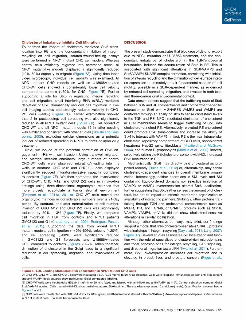
Figure 5. LDL Loading Modulates Stx6 Localization in NPC1 Mutant C
(A) CHO-WT, CHOM12, and CHO 2-2 cells were incubated ± LDL (0.05 mg/ml) fo
and anti-VAMP4 (red); squares show perinuclear Golgi compacted labeling.
(B) CHO-WT cells were incubated ± HDL (0.1 mg/ml) for 30 min, fixed, and label
Stx6/VAMP4 labeling. Cells treated with HDL show partially scattered Stx6 stainin
Figures 1 and 2.
(C) CHO cells were transfected with pIRES2-L-TeTx for 48 hr (green) and then fixed
in NPC1 mutant cells. The scale bar represents 10 mm.
DISCUSSION
The present study demonstrates that blockage of LE-chol export
due to NPC1 mutation or U18666A treatment, and the con-
comitant imbalance of cholesterol in the TGN/endosomal
boundaries, induces the accumulation of Stx6 in RE. This is
associated with significant alterations in Stx6/VAMP3 and
Stx6/VAMP4 SNARE complex formation, correlating with inhibi-
tion of integrin recycling and the diminution of cell-surface integ-
rin expression to ultimately impair fundamental aspects of cell
motility, possibly in a Stx6-dependent manner, as evidenced
by reduced cell spreading, migration, and invasion in both two-
and three-dimensional environmental context.
Data presented here suggest that the trafficking route of Stx6
between TGN and RE compartments and compartment-specific
interaction of Stx6 with v-SNARES VAMP3 and VAMP4 are
controlled through an ability of Stx6 to sense cholesterol levels
in the TGN and RE. NPC1-mediated diminution of cholesterol
in TGN membranes seems to trigger trafficking of Stx6 into
cholesterol-enriched RE. Alternatively, elevated RE cholesterol
could promote Stx6 translocation and increase the ability of
Stx6 to interact with VAMP3. In fact, RE is the main intracellular
cholesterol repository compartment of CHO cells, nonpolarized
hepatoma HepG2 cells, fibroblasts (Maxfield and McGraw,
2004), and human B lymphocytes (Mobius et al., 2003). Indeed,
selectively raising the RE cholesterol content with HDL increased
Stx6 localization in RE.
Mechanistically, Stx6 may directly bind cholesterol as pro-
posed recently (Hulce et al., 2013) or, alternatively, may sense
cholesterol-dependent changes in overall membrane organi-
zation. Interestingly, neither alterations in SM levels and SM
containing liquid-ordered domains nor selective inhibition of
VAMP3 or VAMP4 overexpression altered Stx6 localization,
further suggesting that Stx6 rather senses the amount of choles-
terol, but not its impact on intrinsic membrane organization or
availability of interacting partners. Strikingly, other proteins traf-
ficking through TGN and endosomal compartments such as
M6PR, TfR, and TGN46, or SNARE proteins such as Stx16,
VAMP3, VAMP4, or Vti1a did not show cholesterol-sensitive
alterations in cellular localization.
Although other alternative pathways may exist, our findings
support a model that links cholesterol-sensitive SNARE proteins
with final steps in integrin recycling (Day et al., 2011; Lang, 2007;
Figure S7). Several studies associate Stx6 localization and func-
tion with the role of specialized cholesterol-rich microdomains
and focal adhesion sites for integrin recycling, FAK signaling,
and directional migration toward FN (Tiwari et al., 2011). Further-
more, Stx6 overexpression increases cell migration and is
elevated in breast, liver, and prostate cancers (Riggs et al.,
HO Cells
r 24 hr as indicated. Cells were fixed and immunolabeled with anti-Stx6 (green)
ed with anti-Stx6 and anti-VAMP4 as in (A). Control cells show compact Golgi
g. The scale bars represent 10 and 2 mm (insets). Quantification as described in
and stained with anti-Stx6 (red). Arrowheads point at disperse Stx6 structures
Cell Reports 7, 883–897, May 8, 2014 ª2014 The Authors 891
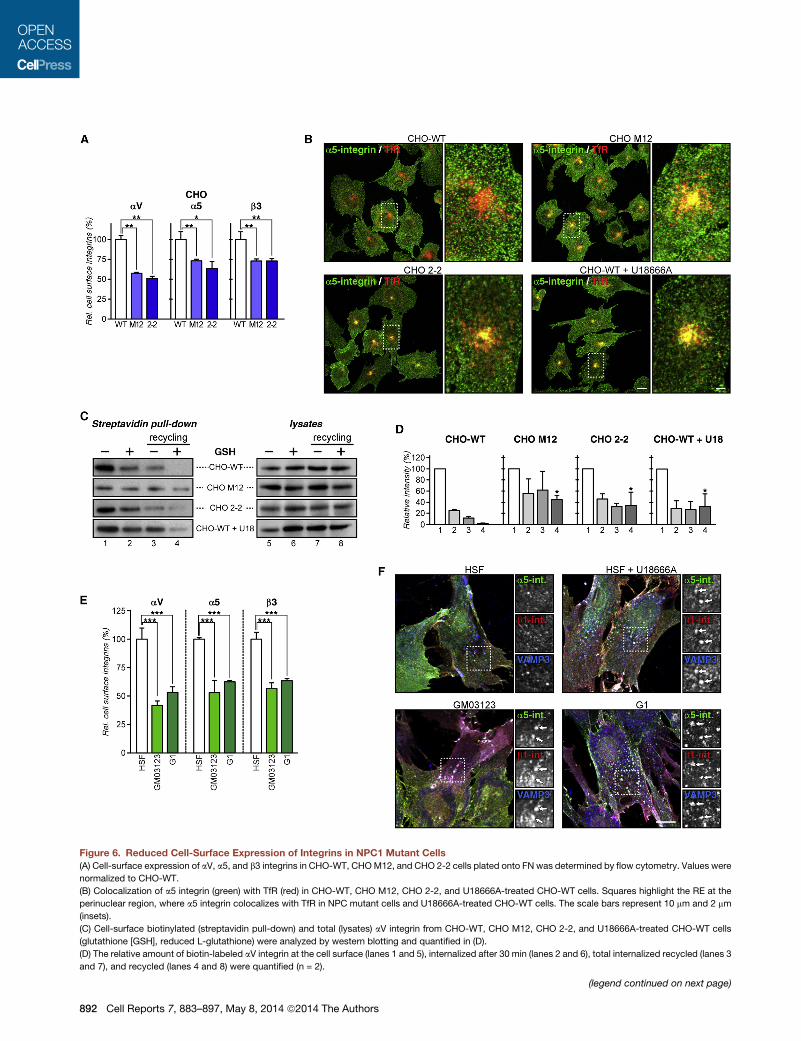
Figure 6. Reduced Cell-Surface Expression of Integrins in NPC1 Mutant Cells
(A) Cell-surface expression of aV, a5, and b3 integrins in CHO-WT, CHOM12, and CHO 2-2 cells plated onto FN was determined by flow cytometry. Values were
normalized to CHO-WT.
(B) Colocalization of a5 integrin (green) with TfR (red) in CHO-WT, CHO M12, CHO 2-2, and U18666A-treated CHO-WT cells. Squares highlight the RE at the
perinuclear region, where a5 integrin colocalizes with TfR in NPC mutant cells and U18666A-treated CHO-WT cells. The scale bars represent 10 mm and 2 mm
(insets).
(C) Cell-surface biotinylated (streptavidin pull-down) and total (lysates) aV integrin from CHO-WT, CHO M12, CHO 2-2, and U18666A-treated CHO-WT cells
(glutathione [GSH], reduced L-glutathione) were analyzed by western blotting and quantified in (D).
(D) The relative amount of biotin-labeled aV integrin at the cell surface (lanes 1 and 5), internalized after 30 min (lanes 2 and 6), total internalized recycled (lanes 3
and 7), and recycled (lanes 4 and 8) were quantified (n = 2).
(legend continued on next page)
892 Cell Reports 7, 883–897, May 8, 2014 ª2014 The Authors
2012). Earlier work implicated cholesterol in the formation of
signaling complexes containing aVb3, CD47, and G proteins
(Green et al., 1999) and control of cell adhesion and migration
onto FN (Ramprasad et al., 2007). This possibly requires
Rab11, which modulates cholesterol transport and homeostasis
(Holtta-Vuori et al., 2002) and facilitates the recycling of b1 integ-
rin (Powelka et al., 2004). Recent reports showing increased
cholesterol requirements for breast cancer and A431 cell inva-
sion (Freed-Pastor et al., 2012) and impaired A431 invasion
upon inhibition of integrin recycling (Muller et al., 2009) support
this model.
Delivery of LE-chol is critical for cholesterol homeostasis in
the endoplasmic reticulum and maintaining cholesterol levels
in other compartments. NPC1 affects cholesterol delivery to
the plasma membrane, and a substantial amount of LE-chol
being transported via NPC1 appears to traffic through Golgi
membranes en route to the plasma membrane in human fibro-
blasts (Urano et al., 2008). However, cell-specific intracellular
differences in cholesterol routes seem to exist, as BODIPY
cholesterol did not label the Golgi apparatus in A431 cells
(Kanerva et al., 2013).
Interestingly, as shown here and previously, prolonged LDL
treatment in NPC mutant cells still delivered cholesterol to other
compartments, including the Golgi, indicating alternative LDL
cholesterol trafficking routes or incomplete blockage of choles-
terol egress from LE in NPC1 mutant cells. In fact, in NPC1-
deficient fibroblasts, cholesterol accumulates in trans-Golgi
cisternae with the TGN remaining relatively cholesterol-deficient
(Garver et al., 2002).
The pathways that deliver and control cholesterol levels in
the RE are poorly understood. Major routes likely involve non-
vesicular and vesicular trafficking from and to the plasma
membrane (Mesmin and Maxfield, 2009). Although current
fractionation methods to purify RE have limitations (Hao et al.,
2002), cholesterol levels in Rab11/VAMP3-enriched fractions
(fractions Nos. 7 and 8) were slightly elevated in NPC1 mutant
cell models. Actually, prolonged LDL cholesterol loading, known
to reach the Golgi, abrogated Stx6 localization in RE of NPC1
mutant models, indicating that cholesterol levels in the Golgi,
and probably not in the RE, determine Stx6 localization. Also,
addition of exogenous cholesterol to elevate plasma membrane
and RE cholesterol did not alter Stx6 location at the TGN (not
shown). Intriguingly, methyl-b-cyclodextrin mediated cholesterol
depletion at the plasmamembrane accentuated Stx6 accumula-
tion in RE (not shown). As removal of cell-surface cholesterol
induces translocation of SNAP23 and Stx4 from the plasma
membrane to the Golgi (Reverter et al., 2011), the possibility of
SNAP23/VAMP4 association in this compartment cannot be
excluded, perhaps competing with Stx6/VAMP4 assembly
(Skalski et al., 2010).
Beyond the scope of this study, other mechanisms include
oxysterol-binding protein (OSBP) and OSBP-related proteins,
which have been implicated in the distribution of sterols among
(E) Cell-surface expression of aV, a5, and b3 integrins in HSF, G03123, and G1
normalized to HSF.
(F) Colocalization of VAMP3 (blue) with a5 (green) and b1 integrins (red) in HSF, U1
a5 and b1 integrins is indicated by small white arrows). The scale bar represents
intracellular organelles. OSBP regulates levels of PI(4)P,
ceramide, but also cholesterol in the Golgi (Duran et al., 2012)
and is essential for the localization of intra-Golgi v-SNAREs
GS28 and GS15 (Nishimura et al., 2013).
Utilizing VAMP3-targeting neurotoxins (L-TeTx) or RNAi
knockdown approaches (Luftman et al., 2009; Proux-Gillardeaux
et al., 2005; Skalski and Coppolino, 2005), VAMP3 has been
shown to be essential for cell migration, spreading, and integ-
rin-dependent cell adhesion. However, as shown here and by
others (Zylbersztejn and Galli, 2011), other SNAREs as well as
additional modulators, such as cholesterol, also contribute to
regulate integrin-trafficking pathways and, consequently, cell
migration.
Up to date, only two studies have analyzed and linked traf-
ficking (and function) of Stx6 with the recycling of integrins and
cell migration (Riggs et al., 2012; Tiwari et al., 2011). However,
these studies did not reveal a possible role for cholesterol in
the regulation of Stx6 location and function. In line with data
shown here, both studies showed that Stx6 loss of function inter-
fered with integrin trafficking: diminution of cell-surface integrin
or increased ubiquitination of integrins and diversion into the
degradation pathway (Riggs et al., 2012; Tiwari et al., 2011). In
HeLa cells, Stx6 at the TGN was required for the trafficking of
a3b1 integrins (Riggs et al., 2012). In endothelial cells, Stx6
may play a role in themaintenance of lipid or protein composition
and/or domain organization of Rab GTPases on the endosome
membrane, thereby facilitating sorting from EE (Tiwari et al.,
2011).
Although our results do not completely identify cholesterol-
imbalance-triggered Stx6 mislocalization as the sole cause of
inhibited integrin recycling, data presented here strongly indicate
that both a diminution of cholesterol in TGNmembranes, caused
by NPC1 loss of function, or alternatively increasing cholesterol
levels in recycling endosomes, e.g., by exposure to HDL, triggers
Stx6 accumulation in RE, which is associated with increased
association with VAMP3 and Rab11. Therefore, similar to the
postulated role of VAMP3 in RE (Proux-Gillardeaux et al., 2005;
Tayeb et al., 2005), Stx6 in the TGN may be necessary to ensure
accurate dynamics of integrin recycling. Importantly, this study
suggests that the levels of cholesterol in these trafficking com-
partments ultimately contribute to control mechanisms that
regulate the recycling of integrins. Hence, this study identifies
a regulatory circuit of cholesterol-sensitive SNARE interactions
that potentially drive integrin-dependent cell migration and
invasion.
Lipid storage disorders associated with NPC1/NPC2
mutations manifest in neurological disorders, hepato- and/or
splenomegaly, and cardiovascular complications. Remarkably,
NPC1-dependent cholesterol availability has now also been
identified critical for proper cell movement in early zebrafish
morphogenesis (Schwend et al., 2011). This regulatory role of
NPC1 identifies a link between a cholesterol-sensitive member
of the SNARE family, which eventually may affect integrin
fibroblasts plated onto FN was determined by flow cytometry. Values were
8666A-treated HSF, G03123, and G1 fibroblasts (colocalization of VAMP3 with
20 mm.
Cell Reports 7, 883–897, May 8, 2014 ª2014 The Authors 893
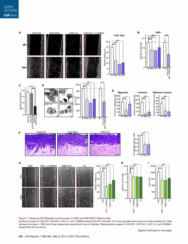
Figure 7. Reduced Cell Migration and Invasion in CHO and HSF NPC1 Mutant Cells
(A) Wound closure of CHO-WT, CHO M12, CHO 2-2, and U18666A-treated CHO-WT cells after 16 hr was calculated and is given as relative healing (%). Data
represent the mean ± SEM from three independent experiments done in triplicate. Representative images of CHO-WT, CHO M12, CHO 2-2, and U18666A-
treated CHO-WT are shown.
(legend continued on next page)
894 Cell Reports 7, 883–897, May 8, 2014 ª2014 The Authors
trafficking to regulate cell-surface integrin expression and,
consequently, cell migration.
EXPERIMENTAL PROCEDURES
Details of Experimental Procedures can be found in the Supplemental Exper-
imental Procedures.
Reagents and Antibodies
For a complete list of reagents, antibodies (Table S1), and more detailed infor-
mation on cell culture, transfections, cholesterol measurements, U18666A
treatment, immunoprecipitations, statistics, and subcellular fractionation,
see the Supplemental Experimental Procedures.
Microscopic Techniques and Image Analysis
The preparation of cells for immunofluorescence and the acquisition and
quantification of images is explained in detail in the Supplemental Experi-
mental Procedures.
Flow Cytometry
Cells were harvested in PBS and 0.5 mM EDTA, resuspended in ice-cold PBS,
1% fetal calf serum (fluorescence-activated cell sorting [FACS] buffer), and
incubated in FACS buffer with anti-integrins (0.01 mg/ml a5 and b3;
0.02 mg/ml aV) for 1 hr at 4�C. Cells were washed, incubated with fluorescently
tagged secondary antibody for 1 hr, and then washed. Cell-surface fluores-
cence was measured using a BD FACS CantoII cytometer.
Analysis of Cell Migration
Spreading, organotypic invasion, and Matrigel migration/invasion assays are
explained in detail in the Supplemental Experimental Procedures.
SUPPLEMENTAL INFORMATION
Supplemental information includes Supplemental Experimental Procedures,
seven figures, and one table and can be found with this article online at
http://dx.doi.org/10.1016/j.celrep.2014.03.043.
ACKNOWLEDGMENTS
This study was supported by grants BFU2012-36272 and CSD2009-00016
fromMinisterio de Economıa y Competitividad (MEC) and PI042182 from Fun-
dacio Marato TV3 (Spain) to C.E. T.G. is supported by the National Health and
Medical Research Council of Australia (NHMRC; 510294) and the University of
Sydney (2010-02681). R.Z.M. acknowledges support through the NHMRC
Fellowship (457247). P.T. acknowledges support from the NHMRC, Australian
Research Council, and Cancer Institute New South Wales (CINSW). We are
thankful to M. Calvo, N. Cortadellas, and A. Garcıa (Centres Cientıfics i
(B) Individual cell tracks of CHO-WT, CHOM12, CHO 2-2, U18666A-treated CHO
time-lapse video microscopy for 12 hr. Individual cell tracks were generated, an
(C) CHO-WT expressing siRNA-targeting Stx6 or scrambledwere plated on FN, an
(D) Cell spreading areas were determined for CHO-WT, CHOM12, CHO 2-2, U18
seeding onto FN-coated plates. Areas of 100 cells per experiment were quantifie
(E) Transwell migration and invasion of CHO-WT, CHOM12, and CHO 2-2 cells. M
migration and invasion was calculated.
(F) Organotypic invasion assay with CHO-WT, CHO M12, and CHO 2-2 cells pla
invade for 21 days, fixed, and processed for hematoxylin and eosin staining. The
by completematrix enfoldment, as opposed to those on the surface of thematrix.
bar represents 50 mm.
(G) Relative wound closure (healing) of HSF, U18666A-treated HSF, and G03123
SEM from three independent experiments done in triplicate.
(H) Individual cell tracking of HSF, U18666A-treated HSF, and G03123 and G1 fib
Velocity (speed) of individual cell migration tracks were quantified.
(I) Cell spreading area of HSF, U18666A-treated HSF, and G03123 and G1 fibrob
experiment were quantified.
Tecnologics, Universitat de Barcelona) for their help in confocal and electron
microscopy; M. Sawicka (Medical School of Silesia, Poland) and T. Nishizumi
(University of Miyasaki, Japan) for help in the spreading assay; and M. Molinos
and H. Gutierrez for technical assistance. M.R. and A.A.G. are grateful to MEC
for a short-term fellowship in Sydney, Australia at the University of Sydney and
the University of New South Wales, respectively.
Received: June 18, 2013
Revised: December 30, 2013
Accepted: March 17, 2014
Published: April 17, 2014
REFERENCES
Blanchette-Mackie, E.J., Dwyer, N.K., Amende, L.M., Kruth, H.S., Butler, J.D.,
Sokol, J., Comly, M.E., Vanier, M.T., August, J.T., Brady, R.O., et al. (1988).
Type-C Niemann-Pick disease: low density lipoprotein uptake is associated
with premature cholesterol accumulation in the Golgi complex and excessive
cholesterol storage in lysosomes. Proc. Natl. Acad. Sci. USA 85, 8022–8026.
Bock, J.B., Klumperman, J., Davanger, S., and Scheller, R.H. (1997). Syntaxin
6 functions in trans-Golgi network vesicle trafficking. Mol. Biol. Cell 8, 1261–
1271.
Caswell, P.T., and Norman, J.C. (2006). Integrin trafficking and the control of
cell migration. Traffic 7, 14–21.
Caswell, P., and Norman, J. (2008). Endocytic transport of integrins during cell
migration and invasion. Trends Cell Biol. 18, 257–263.
Caswell, P.T., Vadrevu, S., and Norman, J.C. (2009). Integrins: masters and
slaves of endocytic transport. Nat. Rev. Mol. Cell Biol. 10, 843–853.
Choudhury, A., Sharma, D.K., Marks, D.L., and Pagano, R.E. (2004). Elevated
endosomal cholesterol levels in Niemann-Pick cells inhibit rab4 and perturb
membrane recycling. Mol. Biol. Cell 15, 4500–4511.
Choudhury, A., Marks, D.L., Proctor, K.M., Gould, G.W., and Pagano, R.E.
(2006). Regulation of caveolar endocytosis by syntaxin 6-dependent delivery
of membrane components to the cell surface. Nat. Cell Biol. 8, 317–328.
Coxey, R.A., Pentchev, P.G., Campbell, G., and Blanchette-Mackie, E.J.
(1993). Differential accumulation of cholesterol in Golgi compartments of
normal and Niemann-Pick type C fibroblasts incubated with LDL: a cytochem-
ical freeze-fracture study. J. Lipid Res. 34, 1165–1176.
Cubells, L., Vila de Muga, S., Tebar, F., Wood, P., Evans, R., Ingelmo-Torres,
M., Calvo, M., Gaus, K., Pol, A., Grewal, T., and Enrich, C. (2007). Annexin A6-
induced alterations in cholesterol transport and caveolin export from the Golgi
complex. Traffic 8, 1568–1589.
Cubells, L., Vila de Muga, S., Tebar, F., Bonventre, J.V., Balsinde, J., Pol, A.,
Grewal, T., and Enrich, C. (2008). Annexin A6-induced inhibition of cytoplasmic
phospholipase A2 is linked to caveolin-1 export from the Golgi. J. Biol. Chem.
283, 10174–10183.
-WT, and CHO-WT expressing NPC1-P692S, plated onto FN, and analyzed by
d the speed (velocity) was calculated.
d individual cell tracks were analyzed by time-lapse videomicroscopy for 12 hr.
666A-treated CHO-WT, and NPC1-P692S-transfected CHO-WT cells 2 hr after
d. Representative fields of CHO cell lines are shown.
igrating and invading cells from six fields/cell line were quantified, and relative
ted on three-dimensional matrices of rat-tail collagen I. Cells were allowed to
invasion index was calculated as a ratio of the proportion of cells characterized
Representative images of three independent experiments are shown. The scale
and G1 fibroblasts after 24 hr was calculated. Data represent mean values ±
roblasts plated on FN was analyzed by time-lapse video microscopy for 12 hr.
lasts, plated on FN, was determined 2 hr after seeding. One hundred cells per
Cell Reports 7, 883–897, May 8, 2014 ª2014 The Authors 895
Dahl, N.K., Reed, K.L., Daunais, M.A., Faust, J.R., and Liscum, L. (1992). Isola-
tion and characterization of Chinese hamster ovary cells defective in the intra-
cellular metabolism of low density lipoprotein-derived cholesterol. J. Biol.
Chem. 267, 4889–4896.
Day, P., Riggs, K.A., Hasan, N., Corbin, D., Humphrey, D., and Hu, C. (2011).
Syntaxins 3 and 4 mediate vesicular trafficking of a5b1 and a3b1 integrins and
cancer cell migration. Int. J. Oncol. 39, 863–871.
Dozynkiewicz, M.A., Jamieson, N.B., Macpherson, I., Grindlay, J., van den
Berghe, P.V., von Thun, A., Morton, J.P., Gourley, C., Timpson, P., Nixon,
C., et al. (2012). Rab25 and CLIC3 collaborate to promote integrin recycling
from late endosomes/lysosomes and drive cancer progression. Dev. Cell 22,
131–145.
Du, X., Kumar, J., Ferguson, C., Schulz, T.A., Ong, Y.S., Hong, W., Prinz, W.A.,
Parton, R.G., Brown, A.J., and Yang, H. (2011). A role for oxysterol-binding
protein-related protein 5 in endosomal cholesterol trafficking. J. Cell Biol.
192, 121–135.
Duran, J.M., Campelo, F., van Galen, J., Sachsenheimer, T., Sot, J., Egorov,
M.V., Rentero, C., Enrich, C., Polishchuk, R.S., Goni, F.M., et al. (2012). Sphin-
gomyelin organization is required for vesicle biogenesis at the Golgi complex.
EMBO J. 31, 4535–4546.
Freed-Pastor, W.A., Mizuno, H., Zhao, X., Langerød, A., Moon, S.H., Rodri-
guez-Barrueco, R., Barsotti, A., Chicas, A., Li, W., Polotskaia, A., et al.
(2012). Mutant p53 disrupts mammary tissue architecture via the mevalonate
pathway. Cell 148, 244–258.
Ganley, I.G., Espinosa, E., and Pfeffer, S.R. (2008). A syntaxin 10-SNARE
complex distinguishes two distinct transport routes from endosomes to the
trans-Golgi in human cells. J. Cell Biol. 180, 159–172.
Garver, W.S., Krishnan, K., Gallagos, J.R., Michikawa, M., Francis, G.A., and
Heidenreich, R.A. (2002). Niemann-Pick C1 protein regulates cholesterol
transport to the trans-Golgi network and plasma membrane caveolae.
J. Lipid Res. 43, 579–589.
Green, J.M., Zhelesnyak, A., Chung, J., Lindberg, F.P., Sarfati, M., Frazier,
W.A., and Brown, E.J. (1999). Role of cholesterol in formation and function
of a signaling complex involving alphavbeta3, integrin-associated protein
(CD47), and heterotrimeric G proteins. J. Cell Biol. 146, 673–682.
Hao, M., Lin, S.X., Karylowski, O.J., Wustner, D., McGraw, T.E., and Maxfield,
F.R. (2002). Vesicular and non-vesicular sterol transport in living cells. The
endocytic recycling compartment is a major sterol storage organelle. J. Biol.
Chem. 277, 609–617.
Heeren, J., Grewal, T., Jackle, S., and Beisiegel, U. (2001). Recycling of apoli-
poprotein E and lipoprotein lipase through endosomal compartments in vivo.
J. Biol. Chem. 276, 42333–42338.
Heeren, J., Grewal, T., Laatsch, A., Becker, N., Rinninger, F., Rye, K.A., and
Beisiegel, U. (2004). Impaired recycling of apolipoprotein E4 is associated
with intracellular cholesterol accumulation. J. Biol. Chem. 279, 55483–55492.
Holtta-Vuori, M., Tanhuanpaa, K., Mobius, W., Somerharju, P., and Ikonen, E.
(2002). Modulation of cellular cholesterol transport and homeostasis by
Rab11. Mol. Biol. Cell 13, 3107–3122.
Hulce, J.J., Cognetta, A.B., Niphakis, M.J., Tully, S.E., andCravatt, B.F. (2013).
Proteome-wide mapping of cholesterol-interacting proteins in mammalian
cells. Nat. Methods 10, 259–264.
Ikonen, E. (2006). Mechanisms for cellular cholesterol transport: defects and
human disease. Physiol. Rev. 86, 1237–1261.
Ikonen, E. (2008). Cellular cholesterol trafficking and compartmentalization.
Nat. Rev. Mol. Cell Biol. 9, 125–138.
Jones, M.C., Caswell, P.T., and Norman, J.C. (2006). Endocytic recycling
pathways: emerging regulators of cell migration. Curr. Opin. Cell Biol. 18,
549–557.
Kanerva, K., Uronen, R.L., Blom, T., Li, S., Bittman, R., Lappalainen, P., Pera-
nen, J., Raposo, G., and Ikonen, E. (2013). LDL cholesterol recycles to the
plasma membrane via a Rab8a-Myosin5b-actin-dependent membrane trans-
port route. Dev. Cell 27, 249–262.
896 Cell Reports 7, 883–897, May 8, 2014 ª2014 The Authors
Lang, T. (2007). SNARE proteins and ‘membrane rafts’. J. Physiol. 585,
693–698.
Lang, T., Bruns, D., Wenzel, D., Riedel, D., Holroyd, P., Thiele, C., and Jahn, R.
(2001). SNAREs are concentrated in cholesterol-dependent clusters that
define docking and fusion sites for exocytosis. EMBO J. 20, 2202–2213.
Liscum, L., and Faust, J.R. (1989). The intracellular transport of low density
lipoprotein-derived cholesterol is inhibited in Chinese hamster ovary cells
cultured with 3-beta-[2-(diethylamino)ethoxy]androst-5-en-17-one. J. Biol.
Chem. 264, 11796–11806.
Luftman, K., Hasan, N., Day, P., Hardee, D., and Hu, C. (2009). Silencing of
VAMP3 inhibits cell migration and integrin-mediated adhesion. Biochem.
Biophys. Res. Commun. 380, 65–70.
Mallard, F., Tang, B.L., Galli, T., Tenza, D., Saint-Pol, A., Yue, X., Antony, C.,
Hong, W., Goud, B., and Johannes, L. (2002). Early/recycling endosomes-
to-TGN transport involves two SNARE complexes and a Rab6 isoform.
J. Cell Biol. 156, 653–664.
Maxfield, F.R., and McGraw, T.E. (2004). Endocytic recycling. Nat. Rev. Mol.
Cell Biol. 5, 121–132.
Maxfield, F.R., and van Meer, G. (2010). Cholesterol, the central lipid of
mammalian cells. Curr. Opin. Cell Biol. 22, 422–429.
McMahon, H.T., Ushkaryov, Y.A., Edelmann, L., Link, E., Binz, T., Niemann, H.,
Jahn, R., and Sudhof, T.C. (1993). Cellubrevin is a ubiquitous tetanus-toxin
substrate homologous to a putative synaptic vesicle fusion protein. Nature
364, 346–349.
Mesmin, B., and Maxfield, F.R. (2009). Intracellular sterol dynamics. Biochim.
Biophys. Acta 1791, 636–645.
Millard, E.E., Srivastava, K., Traub, L.M., Schaffer, J.E., and Ory, D.S. (2000).
Niemann-pick type C1 (NPC1) overexpression alters cellular cholesterol
homeostasis. J. Biol. Chem. 275, 38445–38451.
Millard, E.E., Gale, S.E., Dudley, N., Zhang, J., Schaffer, J.E., and Ory, D.S.
(2005). The sterol-sensing domain of the Niemann-Pick C1 (NPC1) protein
regulates trafficking of low density lipoprotein cholesterol. J. Biol. Chem.
280, 28581–28590.
Mobius, W., van Donselaar, E., Ohno-Iwashita, Y., Shimada, Y., Heijnen, H.F.,
Slot, J.W., and Geuze, H.J. (2003). Recycling compartments and the internal
vesicles of multivesicular bodies harbor most of the cholesterol found in the
endocytic pathway. Traffic 4, 222–231.
Muller, P.A., Caswell, P.T., Doyle, B., Iwanicki, M.P., Tan, E.H., Karim, S.,
Lukashchuk, N., Gillespie, D.A., Ludwig, R.L., Gosselin, P., et al. (2009).
Mutant p53 drives invasion by promoting integrin recycling. Cell 139, 1327–
1341.
Murray, R.Z., Kay, J.G., Sangermani, D.G., and Stow, J.L. (2005). A role for the
phagosome in cytokine secretion. Science 310, 1492–1495.
Nishimura, T., Uchida, Y., Yachi, R., Kudlyk, T., Lupashin, V., Inoue, T., Tagu-
chi, T., and Arai, H. (2013). Oxysterol-binding protein (OSBP) is required for the
perinuclear localization of intra-Golgi v-SNAREs. Mol. Biol. Cell 24, 3534–
3544.
Ohgami, N., Ko, D.C., Thomas, M., Scott, M.P., Chang, C.C., and Chang, T.Y.
(2004). Binding between the Niemann-Pick C1 protein and a photoactivatable
cholesterol analog requires a functional sterol-sensing domain. Proc. Natl.
Acad. Sci. USA 101, 12473–12478.
Pellinen, T., and Ivaska, J. (2006). Integrin traffic. J. Cell Sci. 119, 3723–3731.
Pellinen, T., Arjonen, A., Vuoriluoto, K., Kallio, K., Fransen, J.A., and Ivaska, J.
(2006). Small GTPase Rab21 regulates cell adhesion and controls endosomal
traffic of beta1-integrins. J. Cell Biol. 173, 767–780.
Pol, A., Martin, S., Fernandez, M.A., Ingelmo-Torres, M., Ferguson, C., Enrich,
C., and Parton, R.G. (2005). Cholesterol and fatty acids regulate dynamic
caveolin trafficking through the Golgi complex and between the cell surface
and lipid bodies. Mol. Biol. Cell 16, 2091–2105.
Powelka, A.M., Sun, J., Li, J., Gao, M., Shaw, L.M., Sonnenberg, A., and Hsu,
V.W. (2004). Stimulation-dependent recycling of integrin beta1 regulated by
ARF6 and Rab11. Traffic 5, 20–36.
Proux-Gillardeaux, V., Gavard, J., Irinopoulou, T., Mege, R.M., and Galli, T.
(2005). Tetanus neurotoxin-mediated cleavage of cellubrevin impairs epithelial
cell migration and integrin-dependent cell adhesion. Proc. Natl. Acad. Sci.
USA 102, 6362–6367.
Ramprasad, O.G., Srinivas, G., Rao, K.S., Joshi, P., Thiery, J.P., Dufour, S.,
and Pande, G. (2007). Changes in cholesterol levels in the plasma membrane
modulate cell signaling and regulate cell adhesion and migration on fibro-
nectin. Cell Motil. Cytoskeleton 64, 199–216.
Reverter, M., Rentero, C., de Muga, S.V., Alvarez-Guaita, A., Mulay, V., Cairns,
R., Wood, P., Monastyrskaya, K., Pol, A., Tebar, F., et al. (2011). Cholesterol
transport from late endosomes to the Golgi regulates t-SNARE trafficking,
assembly, and function. Mol. Biol. Cell 22, 4108–4123.
Riggs, K.A., Hasan, N., Humphrey, D., Raleigh, C., Nevitt, C., Corbin, D., and
Hu, C. (2012). Regulation of integrin endocytic recycling and chemotactic cell
migration by syntaxin 6 and VAMP3 interaction. J. Cell Sci. 125, 3827–3839.
Roberts, M., Barry, S., Woods, A., van der Sluijs, P., and Norman, J. (2001).
PDGF-regulated rab4-dependent recycling of alphavbeta3 integrin from early
endosomes is necessary for cell adhesion and spreading. Curr. Biol. 11, 1392–
1402.
Rodrıguez-Pascau, L., Toma, C., Macıas-Vidal, J., Cozar, M., Cormand, B.,
Lykopoulou, L., Coll, M.J., Grinberg, D., and Vilageliu, L. (2012). Characterisa-
tion of two deletions involving NPC1 and flanking genes in Niemann-Pick type
C disease patients. Mol. Genet. Metab. 107, 716–720.
Rohrl, C., Meisslitzer-Ruppitsch, C., Bittman, R., Li, Z., Pabst, G., Prassl, R.,
Strobl, W., Neumuller, J., Ellinger, A., Pavelka, M., and Stangl, H. (2012).
Combined light and electron microscopy using diaminobenzidine photooxida-
tion to monitor trafficking of lipids derived from lipoprotein particles. Curr.
Pharm. Biotechnol. 13, 331–340.
Schwend, T., Loucks, E.J., Snyder, D., and Ahlgren, S.C. (2011). Requirement
of Npc1 and availability of cholesterol for early embryonic cell movements in
zebrafish. J. Lipid Res. 52, 1328–1344.
Simons, K., and Ikonen, E. (2000). How cells handle cholesterol. Science 290,
1721–1726.
Simonsen, A., Gaullier, J.M., D’Arrigo, A., and Stenmark, H. (1999). The Rab5
effector EEA1 interacts directly with syntaxin-6. J. Biol. Chem. 274, 28857–
28860.
Skalski, M., and Coppolino, M.G. (2005). SNARE-mediated trafficking of
alpha5beta1 integrin is required for spreading in CHO cells. Biochem. Biophys.
Res. Commun. 335, 1199–1210.
Skalski, M., Yi, Q., Kean, M.J., Myers, D.W., Williams, K.C., Burtnik, A., and
Coppolino, M.G. (2010). Lamellipodium extension and membrane ruffling
require different SNARE-mediated trafficking pathways. BMCCell Biol. 11, 62.
Tayeb, M.A., Skalski, M., Cha, M.C., Kean, M.J., Scaife, M., and Coppolino,
M.G. (2005). Inhibition of SNARE-mediated membrane traffic impairs cell
migration. Exp. Cell Res. 305, 63–73.
Timpson, P., McGhee, E.J., Erami, Z., Nobis, M., Quinn, J.A., Edward, M., and
Anderson, K.I. (2011a). Organotypic collagen I assay: a malleable platform to
assess cell behaviour in a 3-dimensional context. J. Vis. Exp. 56, e3089.
Timpson, P., McGhee, E.J., Morton, J.P., von Kriegsheim, A., Schwarz, J.P.,
Karim, S.A., Doyle, B., Quinn, J.A., Carragher, N.O., Edward, M., et al.
(2011b). Spatial regulation of RhoA activity during pancreatic cancer cell inva-
sion driven by mutant p53. Cancer Res. 71, 747–757.
Tiwari, A., Jung, J.J., Inamdar, S.M., Brown, C.O., Goel, A., and Choudhury, A.
(2011). Endothelial cell migration on fibronectin is regulated by syntaxin
6-mediated alpha5beta1 integrin recycling. J. Biol. Chem. 286, 36749–36761.
Tran, T.H., Zeng, Q., and Hong, W. (2007). VAMP4 cycles from the cell surface
to the trans-Golgi network via sorting and recycling endosomes. J. Cell Sci.
120, 1028–1041.
Urano, Y., Watanabe, H., Murphy, S.R., Shibuya, Y., Geng, Y., Peden, A.A.,
Chang, C.C., and Chang, T.Y. (2008). Transport of LDL-derived cholesterol
from the NPC1 compartment to the ER involves the trans-Golgi network and
the SNARE protein complex. Proc. Natl. Acad. Sci. USA 105, 16513–16518.
Veale, K.J., Offenhauser, C., Whittaker, S.P., Estrella, R.P., and Murray, R.Z.
(2010). Recycling endosome membrane incorporation into the leading edge
regulates lamellipodia formation and macrophage migration. Traffic 11,
1370–1379.
Veale, K.J., Offenhauser, C., and Murray, R.Z. (2011). The role of the recycling
endosome in regulating lamellipodia formation and macrophage migration.
Commun. Integr. Biol. 4, 44–47.
Wang, Y., Thiele, C., and Huttner, W.B. (2000). Cholesterol is required for the
formation of regulated and constitutive secretory vesicles from the trans-Golgi
network. Traffic 1, 952–962.
Wendler, F., and Tooze, S. (2001). Syntaxin 6: the promiscuous behaviour of a
SNARE protein. Traffic 2, 606–611.
Zeng, Q., Tran, T.T., Tan, H.X., and Hong, W. (2003). The cytoplasmic domain
of Vamp4 and Vamp5 is responsible for their correct subcellular targeting: the
N-terminal extenSion of VAMP4 contains a dominant autonomous targeting
signal for the trans-Golgi network. J. Biol. Chem. 278, 23046–23054.
Zhang, Y., Shu, L., and Chen, X. (2008). Syntaxin 6, a regulator of the protein
trafficking machinery and a target of the p53 family, is required for cell adhe-
sion and survival. J. Biol. Chem. 283, 30689–30698.
Zylbersztejn, K., and Galli, T. (2011). Vesicular traffic in cell navigation. FEBS J.
278, 4497–4505.
Cell Reports 7, 883–897, May 8, 2014 ª2014 The Authors 897