PLEASE SCROLL DOWN FOR ARTICLE
This article was downloaded by: [ETH Zuerich]On: 1 December 2010Access details: Access Details: [subscription number 917202149]Publisher Taylor & FrancisInforma Ltd Registered in England and Wales Registered Number: 1072954 Registered office: Mortimer House, 37-41 Mortimer Street, London W1T 3JH, UK
Spectroscopy LettersPublication details, including instructions for authors and subscription information:http://www.informaworld.com/smpp/title~content=t713597299
Quantum Chemical Scaling and Its Importance: The Infrared and RamanSpectra of 5-BromouracilM. Alcolea Palafoxa; Jéssica Talayaa; A. Guerrero-Martíneza; G. Tardajosa; Hitesh Kumarb; J. K. Vatsb; V.K. Rastogib
a Departamento de Química-Física I, Facultad de Ciencias Químicas, Universidad Complutense,Madrid, Spain b Department of Physics, C.C.S. University, Meerut, India
Online publication date: 19 January 2010
To cite this Article Palafox, M. Alcolea , Talaya, Jéssica , Guerrero-Martínez, A. , Tardajos, G. , Kumar, Hitesh , Vats, J. K.and Rastogi, V. K.(2010) 'Quantum Chemical Scaling and Its Importance: The Infrared and Raman Spectra of 5-Bromouracil', Spectroscopy Letters, 43: 1, 51 — 59To link to this Article: DOI: 10.1080/00387010903261149URL: http://dx.doi.org/10.1080/00387010903261149
Full terms and conditions of use: http://www.informaworld.com/terms-and-conditions-of-access.pdf
This article may be used for research, teaching and private study purposes. Any substantial orsystematic reproduction, re-distribution, re-selling, loan or sub-licensing, systematic supply ordistribution in any form to anyone is expressly forbidden.
The publisher does not give any warranty express or implied or make any representation that the contentswill be complete or accurate or up to date. The accuracy of any instructions, formulae and drug dosesshould be independently verified with primary sources. The publisher shall not be liable for any loss,actions, claims, proceedings, demand or costs or damages whatsoever or howsoever caused arising directlyor indirectly in connection with or arising out of the use of this material.
Quantum Chemical Scaling and ItsImportance: The Infrared and Raman
Spectra of 5-BromouracilM. Alcolea Palafox1,
Jessica Talaya1,
A. Guerrero-Martınez1,
G. Tardajos1,
Hitesh Kumar2,
J. K. Vats2, and
V. K. Rastogi2
1Departamento de
Quımica-Fısica I, Facultad de
Ciencias Quımicas, Universidad
Complutense, Madrid, Spain2Department of Physics, C.C.S.
University, Meerut, India
ABSTRACT This work describes the interest and necessity of scaling to
correct the deficiencies in the calculation of the harmonic vibrational wave
numbers. The use of adequate quantum-chemical methods and scaling
procedures reduces the risk in the assignment and can also accurately deter-
mine the contribution of the different modes in an observed band. As an
example, the IR and laser-Raman spectra of the 5-bromouracil biomolecule
are shown.
KEYWORDS 5-bromouracil, harmonic vibrational wave numbers, quantum
chemical methods, scaling
INTRODUCTION
The simulation of vibrational spectra is of practical importance for the
identification of known and unknown compounds and has become an
important part of spectrochemical and quantum-chemical investigations.[1]
Thus the past decades have been highly productive in the interpretation
of vibrational experimental spectra by means of quantum-chemical
methods. The reliable prediction of the vibrational spectra, particularly in
synthetic and natural product chemistry, can be used to calculate the
expected spectra of proposed structures, confirming the identity of a pro-
duct or of a completely new molecule. Other advantages of vibrational
spectroscopy are the identification of experimentally observed reactive
intermediates for which the theoretically predicted wave numbers can serve
as fingerprints and the derivation of thermochemical and kinetic information
through statistical thermodynamics.
In general, the motivation for predicting vibrational spectra is to make
vibrational spectroscopy a more practical tool. If a method that could predict
vibrational spectra reliably is found, it could be used to calculate the
expected spectra of proposed structures. Comparison with the observed
spectra would then confirm the identity of a product, even that of a comple-
tely new molecule, and in some cases also its conformation.[2]
Computational methods can also be used to assign the bands of the spec-
tra. Until recently, the chemical spectroscopists have attempted to interpret
the vibrational spectra of more complex molecules by a transposition of the
Address correspondence toV. K. Rastogi, Department of Physics,C.C.S. University, Meerut-250 004,India. E-mail: [email protected]
Spectroscopy Letters, 43:51–59, 2010Copyright # Taylor & Francis Group, LLCISSN: 0038-7010 print=1532-2289 onlineDOI: 10.1080/00387010903261149
51
Downloaded By: [ETH Zuerich] At: 16:32 1 December 2010

results of normal coordinate analysis of simpler
molecules, often aided by qualitative comparisons
of the spectra of isotopically substituted species,
and the polarizations of the Raman bands. Thus it
has become an accepted practice to include tables
of these vibrational assignments in publications on
the infrared and Raman spectra of larger molecules.
However, to make such assignments for all the bands
in the spectra is risky, due to the fact that while some
of the assignments may be credible, others can be
highly speculative. Furthermore, the modes assigned
to these vibrations are often grossly oversimplified in
an attempt to describe them as group wave numbers
in localized bond systems.[2]
The problem for small molecules is different from
that for large molecules. For small molecules, the
experimental vibrational spectra can be assigned
without difficulty in comparison with the theoretical
wave numbers. Due to the small number of vibra-
tions, the possibility of mistake is very little. More-
over, with small molecules, the calculations can be
carried out at high theoretical level, that is, with
smaller error in the calculated wave numbers. In this
case, the scaling is not necessary. The most expen-
sive methods available today are accurate enough
without empirical corrections to predict spectra to
the required accuracy. Further advancement in com-
puter hardware and theoretical methods may well
make it possible to predict accurate vibrational spec-
tra of larger molecules without empirical corrections.
However, for large polyatomic molecules, the
computation of the vibrational spectra is lengthy.
The number of vibrations can be huge, and therefore
the possibility of mistake in the assignment can be
very high in comparison with the experimental spec-
tra, due to the nearness of many vibrations. Thus the
larger the molecule is, the more accuracy is required
in the prediction of the wave numbers. In spite of the
tremendous advances made both in theoretical meth-
ods and computer hardware, the more accurate
quantum chemical methods are still too expensive
and cumbersome to apply as routine research. Thus
one may be forced to work at a low level, and con-
sequently one must expect a large overestimation of
the calculated vibrational wave numbers.
This overestimation may be due to many different
factors that are usually not even considered in the
theory, such as anharmonicity, errors in the com-
puted geometry, Fermi resonance, solvent effects,
and that can be remarkably reduced with the use
of transferable empirical parameters for the force
fields or for the calculated wave numbers. These
empirical parameters, called the scaled factors,
are therefore designed to correct the calculated
harmonic wave numbers to be compared with the
anharmonic wave numbers found by the experi-
ment. The scaled factors are a consequence of the
deficiency of theoretical approach and potentially
allow vibrational wave numbers (and theoretical
information) of useful accuracy to be obtained from
procedures of moderate computational cost only.[1,2]
The use of adequate quantum-chemical methods
and scaling procedures remarkably reduce the
risk in the assignment and can also accurately
determine the contribution of the different modes
in an observed band. Now, this procedure appears
in the journals of vibrational spectroscopy as used
extensively.
MOLECULE STUDIED
The theoretical methods predict the vibrational
spectra in gas phase. If the vibrational spectra of
the molecule selected can be carried out in gas
phase, it can be compared directly with the scaled
spectra with certain accuracy. However, the differ-
ences are higher in the comparison with spectra in
the solid state. This fact requires the use of a very
accurate procedure of scaling the wave numbers to
avoid a mistake in the assignment. This procedure
of scaling to predict the accurate vibrational spectra
is explained here by considering the example of
5-bromouracil molecule.[3]
The essential biological importance of 5-BrU is
that it is one of the well-known uncommon nucleo-
tide bases and has the ability to coordinate with
metals or to bind to tissues via metals, which inter-
face with the growth of cancer cells. Molecule
5-BrU is used to treat inflammatory tissues.[4,5] A clear
prediction of the vibrational spectra of 5-BrU is
essential in the analyses of the spectra of its more
complex derivatives, like nucleosides and nucleo-
tides and their polymers, which play an important
role in some basic biochemical processes frequently
monitored by means of vibrational spectroscopy.
From the spectroscopy point of view, the vibrational
spectra of 5-BrU have been studied before without
any theoretical support: The Raman spectra was
M. A. Palafox et al. 52
Downloaded By: [ETH Zuerich] At: 16:32 1 December 2010
studied by Rai,[6] and FT-IR has been studied in an
Ar-matrix by Graindourze et al.,[7,8] Gusakova et al.,[9]
Srivastava et al.,[10] and Rastogi et al.[11] However,
there are doubts in the assignment of several bands.
ERROR IN THE CALCULATED WAVE
NUMBERS BY QUANTUM-CHEMICALMETHODS
The vibrational wave numbers are usually
calculated using the simple harmonic-oscillator
model. Therefore, they are typically larger than the
fundamentals observed experimentally.[12] The possi-
ble reasons for the deficiency in the calculations are
the following:
The Zero Point Vibrational Energy (ZPVE) applies.
Anharmonicity in the vibrational potential energy
surface applies.
Basis sets are too small.
Electron correlation is neglected.
The potential energy curve is too steep and therefore
wave numbers are too high.
In general, the calculated wave numbers are over-
estimated. This overestimation using the basis set in
the range from 6-31G� to 6-311þþG(2d,p) is about
9–12% at the Hartree-Fock level, about 5–8% at the
MP2 level, and 3–5% at the B3LYP level.[1,7,8] The
overestimation in the wave numbers also depends
on the type of vibrational mode and on the wave-
number range, varying between 1% and 12%. Thus
for modes that appear at high wave number, the dif-
ference between the harmonic-oscillator prediction
and the exact or Morse-potential–like behavior is
about 10%. However, at a very low wave number,
below a few hundred wave numbers, this difference
can be reduced by a large amount.
SCALING
General
The use of a single overall scale factor[13] is the
simplest procedure of scaling, and it is the procedure
generally used in the literature.[3,14,15] However, the
reduction of the error in the scaled values is in gen-
eral not enough on some modes and molecules, and
it impedes a clear and accurate assignment. Thus,
ones observes frequently in the literature some
mistakes in the correlation with the experimental
wave numbers. To avoid these errors and get a very
accurate assignment, one should use some other pro-
cedures of scaling that are more accurate, such as the
scaling equation, or specific scale factors for each
mode.[1,8] In both cases, the scaling is performed in
the basic skeleton of molecules from which are
extracted scaling equations or specific scale factors
to be transferred to related molecules or to their
derivatives. In previous articles,[15,16] the accuracy
of these procedures has been shown in several
molecules.
In this way, the absolute errors obtained in the
scaled wave numbers are in general lower than
20 cm�1, reducing the mistakes in the assignments.
Moreover, they also lead to a remarkable improve-
ment in the predicted wave numbers in the low–
wave-number region, compared with the results
when a unique scale factor is used.
Determination of the ScalingEquations from Uracil Molecule
We studied the uracil molecule previously, by
extracting scaling parameters to be used in their deri-
vatives, for example, 5-BrU. A list of the calculated
and experimental wave numbers in uracil and
5-BrU is collected in Table 1. The labeling of the
atoms is plotted in Fig. 1. The errors obtained in
the calculated wave numbers of uracil are shown
in the 2nd–4th columns of Table 2. As can be
observed, they are too large.
The scaling equations are of this general form:
nexperimental ¼ aþm � xcalculated
By using the calculated and experimental wave num-
bers of the uracil molecule, one obtains the scaling
equations of Table 3 at the different levels. In general
a good relationship is observed with correlation
coefficients (r) close to unity, especially with DFT
methods. To check the accuracy that these equations
raise, we introduce the calculated wave numbers of
the uracil molecule in them. In this case they appear
as follows:
nscaled ¼ aþm � xcalculated
For each wave number, the errors (nexperimental�xcalculated) obtained are analyzed. The largest values
53 Quantum Chemical Scaling and Its Importance
Downloaded By: [ETH Zuerich] At: 16:32 1 December 2010

are listed in Table 2. A summary of the calculated rms
errors in the wave numbers from these scale equa-
tions and the two procedures of scaling is also col-
lected in Table 2. These values indicate (1) the
maximum accuracy that can be raised with these
equations when they are used in uracil derivatives
and (2) the best procedure of scaling and the best
theoretical level used.
A detailed analysis of Table 2 gives rise to the
following conclusions.
The most cost-effective procedures for predicting
vibrational wave numbers are HF and the B3-based
DFT procedures. MP2 does not appear to offer a
significant improvement in performance over HF
and occasionally shows a high degree of error. For
this reason and because of the excessive time and
computer-memory consumption, it is preferable to
use another method instead of MP2.
In DFT methods, the use of the scaling equations
reduces the errors to about 30% of those found with
an overall scale factor, showing that the errors in the
calculated wave numbers with DFT methods are
systematic and partially associated with the kind of
molecules studied; and therefore they can be
TABLE 1 Calculated Vibrational Wave Numbers With the B3LYP Method and Experimental Ones (cm�1)
5-bromouracilb
Characterization Ring mode no.e Uracil expa 6-31G�� 6-311þG (2d,p) exp.c
puckering N3 1 153 148.7
puckering N1 2 185 w 395 397.0 390
d(OCNCO) 3 374 vw 392 388.1 532
c(C¼C�H12) 4 395 w 536 539.5 594
d(ring) 5 512 w 598 597.6 548?
d(ring)þ d(C=O) 6 588 w 567 551.3 656
das(ring)þ d(C=O) 7 545 w 681 667.5 760
c(N1�H) 8 659.5 w 776 773.5 753
c(N3�H) 9 717.4 vw 748 757.8 785f
c(C4=O)þ c(C5�X) 10 756.5 w 777 778.5 962?
c(C2=O) 11 802 w 971 970.8 906?
n(ring) 12 952 w 928 926.0 1048d
c(C5�X)þ c(C4=O) 13f 972 sh 1053 1055.2 1154d
n(C�C)þ d(N�H) 14 990 sh 1170 1156.1 1189d
c(C6�H) 15 1082 m 1196 1191.6 1327
d(NCC)þ d(C5�X) 16g 1172 s 1353 1346.6 1377d
n(ring)þ d(C5�X) 17 1228 m 1396 1399.8 1390d
n(C�N)þ d(C6H,N1H) 18 1356 sh 1411 1408.8 1458d
d(C5�X)þ d(N�H) 19 1387 s 1493 1486.1 1635
d(N3�H)þ d(C�H) 20 1400 s 1679 1662.0 1729
n(C�N)þ d(N3�H) 21 1461 s 1808 1759.1 1761
d(C6�H)þ d(N�H) 22 1641 s 1847 1793.3 3058
d(N1�H)þ n(N1�C) 23 1688 vs 3237 3213.7 626
n(C=C) 24 1756 vs 626 629.2 3425d
n(C4=O) 25 3076 w 3616 3589.5 3471d
n(C2=O) 26 3124 m 3653 3632.7
n(C6�H) 27 3436 s
n(C5�X) 28 3484 s
n(N3�H) 29
n(N1�H) 30
a[18].b[3].cIn Ar matrix,[19].d[11].e [18e].fc(C5–X) mode.g
d(C5–X) mode.
M. A. Palafox et al. 54
Downloaded By: [ETH Zuerich] At: 16:32 1 December 2010

reduced employing scaling equations determined in
related molecules. The B-based DFT procedures,
while not performing quite as well as the corre-
sponding B3-based procedures, have the attraction
of standard wave-number scale factors close to
unity, meaning that they can often be used without
scaling. The LYP functional method is superior in
precision to the P86 and PW91 functional methods.
Thus combining the most accurate exchange with
the correlation functional method leads to B3-LYP,
which gives the lowest errors in uracil molecule
(and derivatives), and therefore it is the recom-
mended method.
The accuracy of the results obtained with the
scaling is similar to that in using the much more
expensive anharmonic computations, although in
several specific bands these computations improve
the results.[17]
Regarding these features, the 5-BrU molecule is
studied.
Scaling of the 5-bromouracil Molecule
The results for the 5-BrU molecule are shown
in Table 4. In the 5-BrU molecule, the use of a
scaling equation or the specific scaling equation
procedure remarkably reduces the error to a value
similar to that of uracil. This low error facilitates the
TABLE 2 Errors Obtained in the Calculated and Scaled Wave Numbers of the Uracil Modes by the Different Procedures and Methods
Calculated wave
numbers
Scaled wave numbers
with an overall factor
Scaled wave numbers with
the scaling equations
Largest error Largest error Largest error
Method rms Positive Negative rms Positive Negative rms Positive Negative
HF=6-31G�� 184 427 (29) 6 (2) 23 53 (25,26) 37 (12) 22.6 46 (26) 57 (28)
HF=6-31þþG�� 177 418 (29) 11 (2) 37 53 (9) 95 (15) 16.7 27 (19) 50 (28)
MP2=6-31G� 82 187 (27) 44 (12) 33 56 (29) 50 (11) 25.4 50 (10) 66 (15)
BP86=6-31G�� 35 86 (29) 44 (11) 34 54 (29) 54 (15) 18.1 34 (9) 32 (21)
BLYP=6-31G�� 34 73 (29) 49 (11,15) 24 46 (29) 44 (15) 19.6 34 (9) 36 (21)
B3P86=6-31G�� 77 207 (29) 14 (2) 21 44 (29) 40 (12) 15.0 32 (26) 26 (15)
B3LYP=6-31G�� 66 184 (29) 15 (2) 25 50 (29) 41 (15) 13.8 24 (9) 23 (21)
B3LYP=6-311þG(2d,p) 54 156 (29) 20 (2) 13.7 21 (29) 34 (25)
B3PW91=6-31G�� 75 206 (29) 14 (2) 14.9 30 (26) 26 (22)
FIGURE 1 Labeling of the atoms in 5-BrU.
TABLE 3 Scaling Equations nexperimental¼aþb �xcalculated
From the Uracil Molecule
Methods a b
HF=6-31G� 4.6 0.8924
HF=6-31G�� 5.7 0.8867
HF=6-31þþG�� 10.5 0.8938
MP2=6-31G� 34.5 0.9372
BP86=6-31G�� 46.0 0.9678
BLYP=6-31G�� 46.4 0.9718
B3P86=6-31G� 29.9 0.9412
B3P86=6-31G�� 34.1 0.9389
B3LYP=6-31G�� 30.8 0.9468
B3LYP=6-31G�� 34.6 0.9447
B3LYP=6-311þG(2d,p) 30.8 0.9538
B3LYP=6-311þþG(3df,pd) 31.9 0.9512
B3LYP=aug-cc-pVDZ 28.6 0.9543
B3LYP=CEP 33.1 0.9589
B3LYP=SDAll 16.3 0.9535
B3LYP=DGDZVP 39.2 0.9472
B3PW91=6-31G�� 34.9 0.9393
MPW1PW91=6-31G�� 32.7 0.9334
.
55 Quantum Chemical Scaling and Its Importance
Downloaded By: [ETH Zuerich] At: 16:32 1 December 2010
one-by-one correspondence between the experi-
mental wave numbers and the calculated values,
and therefore an accurate assignment can be
reached.
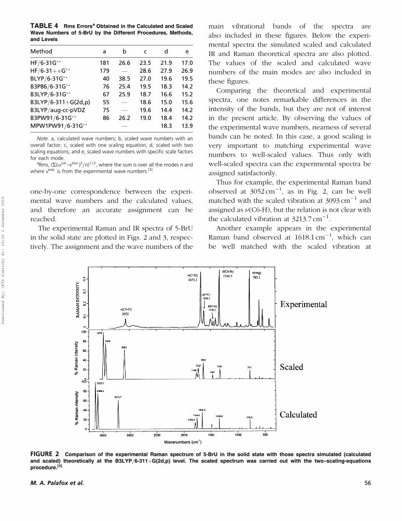
The experimental Raman and IR spectra of 5-BrU
in the solid state are plotted in Figs. 2 and 3, respec-
tively. The assignment and the wave numbers of the
main vibrational bands of the spectra are
also included in these figures. Below the experi-
mental spectra the simulated scaled and calculated
IR and Raman theoretical spectra are also plotted.
The values of the scaled and calculated wave
numbers of the main modes are also included in
these figures.
Comparing the theoretical and experimental
spectra, one notes remarkable differences in the
intensity of the bands, but they are not of interest
in the present article. By observing the values of
the experimental wave numbers, nearness of several
bands can be noted. In this case, a good scaling is
very important to matching experimental wave
numbers to well-scaled values. Thus only with
well-scaled spectra can the experimental spectra be
assigned satisfactorily.
Thus for example, the experimental Raman band
observed at 3052 cm�1, as in Fig. 2, can be well
matched with the scaled vibration at 3093 cm�1 and
assigned as n(C6-H), but the relation is not clear with
the calculated vibration at 3213.7 cm�1.
Another example appears in the experimental
Raman band observed at 1618.1 cm�1, which can
be well matched with the scaled vibration at
TABLE 4 Rms Errorsa Obtained in the Calculated and Scaled
Wave Numbers of 5-BrU by the Different Procedures, Methods,
and Levels
Method a b c d e
HF=6-31G�� 181 26.6 23.5 21.9 17.0
HF=6-31þþG�� 179 — 28.6 27.9 26.9
BLYP=6-31G�� 40 38.5 27.0 19.6 19.5
B3P86=6-31G�� 76 25.4 19.5 18.3 14.2
B3LYP=6-31G�� 67 25.9 18.7 16.6 15.2
B3LYP=6-311þG(2d,p) 55 — 18.6 15.0 15.6
B3LYP=aug-cc-pVDZ 75 — 19.6 14.4 14.2
B3PW91=6-31G�� 86 26.2 19.0 18.4 14.2
MPW1PW91=6-31G�� — 18.3 13.9
Note. a, calculated wave numbers; b, scaled wave numbers with anoverall factor; c, scaled with one scaling equation; d, scaled with twoscaling equations; and e, scaled wave numbers with specific scale factorsfor each mode.
aRms, (R(xcal.–vexp.)2=n)1=2, where the sum is over all the modes n andwhere vexp. is from the experimental wave numbers.[3]
FIGURE 2 Comparison of the experimental Raman spectrum of 5-BrU in the solid state with those spectra simulated (calculated
and scaled) theoretically at the B3LYP=6-311þG(2d,p) level. The scaled spectrum was carried out with the two–scaling-equations
procedure.[3]
M. A. Palafox et al. 56
Downloaded By: [ETH Zuerich] At: 16:32 1 December 2010

1632 cm�1 and assigned as n(C=C). However, the
relation is not clear with the calculated strong-
intensity vibration at 1662.0 cm�1, because it could
be poorly matched with the very strong Raman band
at 1675.3 cm�1 and assigned as n(C=O).
Finally, in Table 5 are collected the rms errors
obtained for other uracil derivatives. It can be noted
that always the scaling equation procedure and the
specific scale factor procedure lead to the lowest
errors, and therefore they are the procedures recom-
mended for scaling.
SUMMARY AND CONCLUSIONS
The accuracy of several of the quantum chemical
methods is determined in the wave numbers of the
uracil normal modes. To improve the calculated
wave numbers, two accurate procedures can be
used. The scaling equations procedure gives rise to
improvement in the predicted wave numbers that is
slightly greater than when a single overall scale fac-
tor is used. Although the specific scale factor proce-
dure gives the lowest error, we recommend using the
scaling equation procedure, mainly because of its
simplicity. A list of scaling equations that we used
for uracil derivatives is shown in Table 3.
The procedure selected for scaling depends on
the size of the organic molecule and the accuracy
required for the predicted wave numbers. With
larger organic molecules, but less than 20 heavy
atoms, HF, MP2, and DFT methods and large basis
sets can be used for calculating wave numbers. If
the accuracy required is not very high (the errors in
the predicted wave numbers could be 0–4%), then
the use of one or two scale factors with the calcu-
lated wave numbers is the simplest and easiest pro-
cedure. In this case, among the HF, MP2 and DFT
methods, the most cost-effective are the HF- and
B3-based. If the accuracy required is high, then at
FIGURE 3 Comparison of the experimental IR spectrum of 5-BrU in the solid state with those spectra simulated (calculated and scaled)
theoretically at the B3LYP=6-311þG(2d,p) level. The scaled spectrum was carried out with the two-scaling-equations procedure.[3]
TABLE 5 Rms Errors Obtained in the Calculated and Scaled
Wave Numbers of Several Uracil Derivatives at the B3LYP/
6–31G�� Level
Molecules a b c d
Uracil 66.4 21.4 13.8 –
5-fluorouracil 70.3 29.8 23.5 14.7
5-bromouracil 76.2 29.2 18.5 13.7
5-methyluracil 59.8 21.5 18.4 13.1
5-nitrouracil 71.7 26.1 16.5 13.0
1-methyluracil 69.2 27.0 17.9 15.8
2-thiouracil 79.0 26.5 15.5 11.5
3-methyluracil 63.2 22.8 15.6 11.0
1,3-dimethyluracil 49.4 23.1 16.5 12.1
Note. a, Calculated wave numbers; b, scaled wave numbers with anoverall factor; c, scaled wave numbers with the scaling equations; d,scaled wave numbers with specific scale factors.
57 Quantum Chemical Scaling and Its Importance
Downloaded By: [ETH Zuerich] At: 16:32 1 December 2010

the same level, scale factors for each mode should
have been calculated previously from related and
simpler molecules.
For uracil molecule the best predicted wave num-
bers for the ring modes were obtained using HF- and
B3-based methods. Thus in this molecule and in
related derivatives, these methods should be used.
With molecules larger than 20 atoms, semiempiri-
cal methods and HF and DFT methods with small
basis sets can be used for calculating wave numbers.
However, the cost-effective ratio with HF and DFT
methods is very high relative to those of semiempi-
rical methods, and therefore their use is not
recommended. In contrast the AM1 and SAM1 semi-
empirical methods, when a specific scale factor for
each mode is used, give good predicted wave
numbers, with error lower than 5%. We found no
advantage in the newer SAM1 method relative to that
of AM1.
ACKNOWLEDGMENTS
M. Alcolea Palafox, Jessica Talaya, A. Guerrero-
Martınez, and G. Tardajos are grateful to the UCM
of Spain for financial support through UCM-BSCH
GR58=08 grant 921628. V. K. Rastogi is grateful to
Professor S. K. Kak, Vice Chancellor, C.C.S.
University, Meerut, India, for motivation and encour-
agement during the course of this work.
REFERENCES
1. (a) Palafox, M. A. The prediction of vibrational spectra: The use ofscale factors. Recent Res. Developed in Physical Chem. 1998, 2,213–232; (b) Palafox, M. A.; Rastogi, V. K. Some procedures for pre-dicting the wavenumbers of the spectra: The scaling. In Perspectivesin Modern Optics and Optical Instrumentation; Sharma, A., Joby, J.,Rastogi, V .K., Eds.; Anita: New Delhi, Ghaziabad, India, 2002;91–98; (c) Palafox, M. A.; Nunez, J. L.; Gill, M.; Rastogi, V. K. Scalingprocedures for the prediction of vibrational spectra: The benzene andaniline molecules, and some derivatives. In Perspectives in Engineer-ing Optics; Singh, K., Rastogi, V. K., Eds.; Anita: New Delhi,Ghaziabad, India, 2002; 356–391; (d) Palafox, M. A.; Nunez, J. L.;Gill, M.; Rastogi, V. K.; Mittal, L.; Sharma, R. Scaling factors for theprediction of vibrational spectra: Part II. The aniline molecule andseveral derivatives. Int. J. Quantum Chem. 2005, 103, 394–421;(e) Palafox, M. A. Scaling procedures for the prediction of vibrationalspectra: Part I. The benzene molecule and some derivatives. AsianChem. Letts. 1998, 7(4), 785–816.
2. (a) Hameka, H. F.; Famini, G. R.; Jensen, J. O.; Newhouse, E. I.Computations of vibrational infrared frequencies of selected amines.Gov. Rep. Announce Index 1990, 90, 13. (b) Hameka, H. F.; Famini,G. R.; Jensen, J. O.; Jensen, J. L. Theoretical prediction of vibrationalinfrared frequencies of tertiary amines. Gov. Rep. Announce Index1991, 91, 15. (c) Hameka, H. F.; Jensen, J. O. Theoretical studies ofthe methyl rotational barrier in toluene. J. Mol. Struct. (Theochem.)1996, 362, 325–330.
3. Rastogi, V. K.; Palafox, M. A.; Mittal, L.; Peica, N.; Kiefer, W.; Lang,K.; Ojha, S. P. FT-IR and FT-Raman spectra and density functionalcomputations of the vibrational spectra, molecular geometry andatomic charges of the biomolecule: 5-Bromouracil. J. Raman Spec-trosc. 2007, 38, 1227–1241.
4. Henderson, J. P.; Byun, J.; Takeshita, J.; Heinecke, J. W. Phagocytesproduce 5-chlorouracil and 5-bromouracil, two mutagenic productsof myeloperoxidase, in human inflammatory tissue. J. Biol. Chem.2003, 278, 23522–23528.
5. Jiang, Q.; Bloun, B. C.; Ames, B. N. 5-Chlorouracil, a marker of DNAdamage from hypochlorous acid during inflammation. J. Biol. Chem.2003, 278, 32834–32840.
6. Rai, J. N. Vibrational Raman spectra of some substituted uracils.Procd. Ind. Acad. Sci. Chem. Sci. 1990, 102, 687–691.
7. Graindourze, M.; Grootaers, T.; Smets, J.; Zeegers-Huyskens, T.;Maes, G. FT-IR spectroscopic study of uracil derivatives and theirhydrogen bonded complexes with proton donors: Part II. MonomerIR absorptions of thiouracils and 5-halogeno-uracils in argonmatrices. J. Mol. Struct. 1990, 237, 389–410.
8. Graindourze, M.; Grootaers, T.; Smets, J.; Zeegers-Huyskens, T.;Maes, G. FT-IR spectroscopic study of uracil derivatives and theirhydrogen bonded complexes with proton donors: Part III. Hydrogenbonding of uracils with H2O in argon matrices. J. Mol. Struct.1991, 243, 37–60.
9. Gusakova, G. V.; Kulbida, A. I.; Plekhova, G. N.; Smolyanskii, A. L.IR spectra and proton-acceptor capability of dimethyluracil andits halogenated derivatives. J. Appl. Spectrosc. 1987, 46(4),381–385.
10. Srivastava, S. L.; Prasad, M.; Rohitashava; Pandey, V. S. Electronic andvibrational spectra of some dihydroxypyrimidines. Spectrochim. Acta1984, 40(7), 675–679.
11. Rastogi, V. K.; Arora, S.; Gupta, S. L.; Sharma, D. K. Spec. Publ. Roy.Soc. Chem. 1991, 94, 401 (Spectrosc. Biol. Mol.).
12. Hehre, W. J.; Radom, L.; Schleyer, P. V. R.; Pople, J. A. Ab initioMolecular Orbital Theory; Wiley: New York, 1986.
13. Scott, A. P.; Radom, L. Harmonic vibrational frequencies: Anevaluation of Hartree-Fock, Muller-Plesset, quadratic configurationinteraction, density functional theory, and semiempirical scalefactors. J. Phys. Chem. 1996, 100, 16502.
14. (a) Palafox, M. A.; Rastogi, V. K. Quantum-chemical predictions ofthe vibrational spectra of polyatomic molecules: The uracil moleculeand two derivatives. Spectrochim. Acta 2002, 58A, 411–440; (b)Palafox, M. A. Scaling factors for the prediction of vibrational spectra.Part I: Benzene molecule. Int. J. Quantum Chem. 2000, 77, 661–684.
15. (a) Palafox, M. A.; Nunez, J. L.; Gil, M. Theoretical quantum chemicalstudy of benzoic acid: Geometrical parameters and vibrational wave-numbers. Int. J. Quantum Chem. 2002, 89, 1–24; (b) Palafox, M. A.;Rastogi, V. K.; Singh, C.; Tanwar, R. P. Ab initio calculations, FT-IRand Raman spectra of 2,3-difluorobenzonitrile. Spectrochim. Acta2001, 57A, 2373–2389; (c) Rastogi, V. K.; Palafox, M. A.; Singh,S.; Singhal, S. K. FT-IR and Raman spectra of 2,4-difluorobenzonitrile.Asian J. Phys. 1998, 7(2), 229–249. (d) Palafox, M. A.; Gil, M.; Nunez,J. L.; Tardajos, G. Study of phenothiazine and N-methyl phenothia-zine by infrared, Raman, 1H-, and 13C-NMR, spectroscopies. Int. J.Quantum Chem. 2002, 89, 147–171. (e) Palafox, M. A. Scalingfactors for the prediction of the frequencies of the ring modes inbenzene derivatives. J. Phys. Chem. 1999, 103 A, 11366–11377.
16. (a) Rastogi, V. K.; Jain, V.; Yadav, R. A.; Singh, C.; Palafox, M. A.Fourier transform Raman spectrum and ab initio and density func-tional computations of the vibrational spectrum, molecular geometry,atomic charges and some molecular properties of the anticarcino-genic drug 5-fluorouracil. J. Raman Spectrosc. 2000, 31, 595–603;(b) Rastogi, V. K.; Singh, C.; Jain, V.; Palafox, M. A. FT-IR andFT-Raman spectra of 5-methyluracil (thymine). J. Raman Spectrosc.2000, 31, 1005–1012; (c) Palafox, M. A.; Tardajos, G.; Kim, J. J.;Neilson, O. F.; Lodhi, R.; Rastogi, V. K. Predicting wavenumbers ofvibrational spectra, its use in spectroscopy. Asian J. Phys. 2006, 15,281–286.
M. A. Palafox et al. 58
Downloaded By: [ETH Zuerich] At: 16:32 1 December 2010
17. Barone, V.; Festa, G.; Grandi, A.; Rega, N.; Sanna, N. Accurate vibra-tional spectra of large molecules by density functional computationsbeyond the harmonic approximation: The case of uracil and2-thiouracil. Chem. Phys. Letts. 2004, 388, 279–283.
18. (a) Colarusso, P.; Zhang, K.; Guo, B.; Bernath, P. F. The infrared spec-tra of uracil, thymine, and adenine in the gas phase. Chem. Phys.Letts. 1997, 269, 39–48; (b) Ivanov, A. Y.; Plokhotnichenko, A. M.;Radchenko, E. D.; Sheina, G. G.; Blagoi, Y. P. FT-IR spectroscopy ofuracil derivatives isolated in Kr, Ar and Ne matrices: Matrix effectand Fermi resonance. J. Molec. Struct. 1995, 372, 91–100; (c)Harsanyi, L.; Csaszar, P.; Csaszar, A.; Boggs, J. E. Interpretation ofthe vibrational spectra of matrix-isolated uracil from scaled Ab initio
quantummechanical force fields. Int. J. Quantum Chem. 1986, 29(4),799–815; (d) Zhiping, W.; Fengshou, Z.; Xianghua, Z.; Hongyu, Z.;Bin, G.; Wei, C. Vibrational properties of uracil. Chinese Sci. Bull.2006, 51(15), 1804–1810; (e) Palafox, M. A.; Iza, N.; Gil, M. Thehydration effect on the uracil frequencies: An experimental andquantum chemical study. J. Molec. Struct. (Theochem.) 2002, 585,69–92; (f) Palafox, M. A.; Nielsen, O. F.; Lang, K.; Garg, P.; Rastogi,V. K. Geometry and vibrational spectra of 5-substituted uracils. AsianChem. Letts. 2004, 8(1), 81–93.
19. Dobrowolski, J. C.; Rode, J. E.; Kolos, R.; Jamroz, M. H.; Bajdor, K.;Mazurek, A. P. Ar-matrix IR spectra of 5-halouracils interpreted bymeans of DFT calculations. J. Phys. Chem. A 2005, 109, 2167–2182.
59 Quantum Chemical Scaling and Its Importance
Downloaded By: [ETH Zuerich] At: 16:32 1 December 2010