PYK10, a b-glucosidase located in the endoplasmaticreticulum, is crucial for the beneficial interaction betweenArabidopsis thaliana and the endophytic fungusPiriformospora indica
Irena Sherameti1, Yvonne Venus1, Corinna Drzewiecki1, Swati Tripathi2, Vipin Mohan Dan2, Inke Nitz3, Ajit Varma2,
Florian M. Grundler3 and Ralf Oelmuller1,*
1Friedrich-Schiller-Universitat Jena, Institut fur Allgemeine Botanik und Pflanzenphysiologie,
Dornburger Str. 159, 07743 Jena, Germany,2Amity Institute of Herbal and Microbial Studies, Sector 125, Noida 201303, UP, India, and3Institute of Plant Protection, Department of Applied Plant Sciences and Plant Biotechnology,
BOKU – University of Natural Resources and Applied Life Sciences Vienna, Peter Jordan-Strasse 82, A-1190 Vienna, Austria
Received 13 December 2007; revised 16 January 2008; accepted 18 January 2008.*For correspondence (fax +49 3641 949232; e-mail [email protected]).
Summary
Piriformospora indica, an endophyte of the Sebacinaceae family, promotes growth and seed production of
many plant species, including Arabidopsis. Growth of a T-DNA insertion line in PYK10 is not promoted and the
plants do not produce more seeds in the presence of P. indica, although their roots are more colonized by the
fungus than wild-type roots. Overexpression of PYK10 mRNA did not affect root colonization and the response
to the fungus. PYK10 codes for a root- and hypocotyl-specific b-glucosidase/myrosinase, which is implicated to
be involved in plant defences against herbivores and pathogens. Expression of PYK10 is activated by the basic
helix-loop-helix domain containing transcription factor NAI1, and two Arabidopsis lines with mutations in the
NAI1 gene show the same response to P. indica as the PYK10 insertion line. PYK10 transcript and PYK10
protein levels are severely reduced in a NAI1 mutant, indicating that PYK10 and not the transcription factor
NAI1 is responsible for the response to the fungus. In wild-type roots, the message level for a leucine-rich
repeat protein LRR1, but not for plant defensin 1.2 (PDF1.2), is upregulated in the presence of P. indica. In
contrast, in lines with reduced PYK10 levels the PDF1.2, but not LRR1, message level is upregulated in the
presence of the fungus. We propose that PYK10 restricts root colonization by P. indica, which results in the
repression of defence responses and the upregulation of responses leading to a mutualistic interaction
between the two symbiotic partners.
Keywords: growth promotion, NAI1, Piriformospora indica, plant/microbe interaction, PYK10, Sebacinaceae.
Introduction
The endophytic fungus Piriformospora indica, a basidio-
mycete of the Sebacinaceae family, interacts with many
plant species, including Arabidopsis. Like other members of
the Sebacinaceae, P. indica colonizes the roots, grows inter-
and intracellularly, and forms pear-shaped spores that
accumulate in the roots as well as on the root surface. The
endophyte promotes nutrient uptake, allows plants to sur-
vive under water and salt stress, confers resistance to toxins,
heavy metal ions and pathogenic organisms, and stimulates
growth and seed production (cf. Oelmuller et al., 2004, 2005;
Peskan-Berghofer et al., 2004; Pham et al., 2004; Sahay and
Varma, 1999; Shahollari et al., 2005, 2007; Sherameti et al.,
2005; Varma et al., 1999, 2001; Verma et al., 1998; Waller
et al., 2005). P. indica is a cultivable fungus and can grow on
synthetic media without a host (Peskan-Berghofer et al.,
2004; Varma et al., 2001). The host range includes trees,
agricultural, horticultural and medicinal plants, monocots
and dicots, and mosses (Barazani et al., 2005; Glen et al.,
428 ª 2008 The AuthorsJournal compilation ª 2008 Blackwell Publishing Ltd
The Plant Journal (2008) 54, 428–439 doi: 10.1111/j.1365-313X.2008.03424.x

2002; Peskan-Berghofer et al., 2004; Shahollari et al., 2005,
2007; Sherameti et al., 2005; Varma et al., 2001; Waller et al.,
2005; Weiss et al., 2004), suggesting that the interaction is
based on general recognition and signalling processes.
Our goal was to identify plant genes that are targeted by
the fungus. Thus, we screened for Arabidopsis mutants that
do not respond to the fungus with regard to growth
promotion and enhanced seed production (Oelmuller et al.,
2004; Shahollari et al., 2007). Here we describe Arabidopsis
mutants that are impaired in the expression of PYK10, a gene
for an abundant myrosinase located in the endoplasmatic
reticulum (ER; Nitz et al., 2001; Matsushima et al., 2004).
PYK10 has recently been identified as a target of P. indica in
Arabidopsis roots (Peskan-Berghofer et al., 2004). Matsushi-
ma et al. (2003a) have shown that PYK10 is a major protein
in spindle-shaped structures of�10 lm in length and�1 lm
in width, which they named ER bodies (cf. Haseloff et al.,
1997; Hawes et al., 2001; Hayashi et al., 1999; Ridge et al.,
1999). Similar structures have been reported for more than
50 plant species (Behnke and Eschlbeck, 1978; Bones et al.,
1989; Bonnett and Newcomb, 1965; Gunning, 1998; Iversen,
1970). ER bodies are surrounded by ribosomes (Hayashi
et al., 1999) and are highly enriched in cotyledons, hypotoc-
yls and in the roots of young seedlings (Matsushima et al.,
2002). During later phases, the ER bodies decrease in the
cotyledons, whereas they remain constant in hypocotyls and
roots. ER bodies can also be induced in rosette leaves by
jasmonate (McConn et al., 1997), and the jasmonate-insen-
sitive coronatine insensitive1 (coi1; Xie et al., 1998) mutant
does not form ER bodies (Matsushima et al., 2002).
The physiological role and natural substrate(s) of PYK10
are unknown at present. b-Glucosidases and myrosinases
hydrolyze b-glucosidic bonds of aryl and alkyl b-D-gluco-
sides, as well as glucosides with carbohydrate moieties such
as cellobiose and other b-linked oligosaccharides (Esen,
1993). In particular, myrosinases hydrolyse non-toxic gluc-
osinolates to biologically active isothiocyanates, thiocya-
nates, nitriles or epithio nitriles (cf. Bones and Rossiter, 1996;
Poulton, 1990; Rask et al., 2000; Wittstock and Halkier, 2002),
and it is believed that the biological function of a myrosinase
depends upon the nature of the aglycon moieties released
from the substrates. The best-studied role of these agylcons
is their involvement in plant defence against herbivores and
microbes (Rask et al., 2000; Stotz et al., 1999, 2000; Tierens
et al., 2001), although they are also involved in the synthesis
of naturally occurring pesticides (Bones and Rossiter, 1996;
Poulton, 1990), the activation of glycosylated plant hor-
mones (Brzobohaty et al., 1993; Schliemann, 1984; Smith
and van Staden, 1978) and cell-wall catabolism (cf. Dharma-
wardhana et al., 1995; Leah et al., 1995; and references
therein). The phosphate inducibility of the PSR3.2 b-gluco-
sidase from Arabidopsis (Malboobi and Lefebvre, 1997) also
suggests an involvement of these enzymes in phosphate
metabolism. Furthermore, Zeng et al. (2003) have shown
that myrosinase activity stimulates the growth of ectomy-
corrhiza fungi. Here, we present evidence that PYK10 is
required for P. indica-mediated growth promotion and
higher seed yield in Arabidopsis because the enzyme
restricts root colonization by the fungal hyphae.
Results
Growth of mutants impaired in PYK10 accumulation
is not promoted by P. indica
In order to identify genes and proteins in Arabidopsis that
are required for growth stimulation and higher seed yield
induced by P. indica, we isolated mutants that grow in the
presence of the fungus like the uninfected wild-type plants
(cf. Oelmuller et al., 2004, 2005; Shahollari et al., 2007).
Three of these mutants are described here. One of them has
a T-DNA insertion in PYK10 (At3g09260, N871638). In the
presence of the fungus, seedlings of this line grow like wild-
type seedlings without the fungus (Figure 1), and after
transfer to soil the plants do not produce significantly more
seeds than the uncolonized wild-type controls (Table 1).
Wild-type plants produce > 20% more seeds in the presence
of the fungus (cf. Shahollari et al., 2007). Comparable results
were obtained for Sebacina, another member of the Seba-
cinaceae (data not shown). This suggests that PYK10, a
glycosyl b-D-hydroxylase/myrosinase located in the ER, is
required for beneficial effects induced by P. indica and
Sebacina in Arabidopsis.
Further analysis of the PYK10 insertion line found that root
growth of the seedlings was not stimulated by P. indica over
a period of 22 days of co-cultivation (Figure 1b). The mor-
phology of the roots as well as the ratio of lateral to main
roots was comparable with the wild type. The branching of
the roots of 22-day-old wild-type seedlings grown in the
absence of P. indica (58.3 � 4.4 lateral roots on 17.4 � 0.5-
cm-long main roots) was almost identical to that of the roots
of the PYK10 insertion line grown in the absence or presence
of P. indica (absence, 56.3 � 4.2 lateral roots on 17.0 � 0.6-
cm-long main roots; presence, 58.4 � 5.0 lateral roots on
16.9 � 0.7-cm-long main roots). When the mutant seedlings
were transferred to soil, some growth promotion was
observed; however, the response was less compared with
the wild type (Figure 2).
The importance of PYK10 for the beneficial interaction
between Arabidopsis and P. indica is further supported by
an ethylmethane–sulfonate (EMS) mutant, called P. indica-
insensitive-4 (pii-4). Similar to the PYK10 insertion line, pii-4
did not respond to P. indica at the seedlings stage (Fig-
ure 1a), and root growth was comparable with uncolonized
control seedlings (Figure 1b). Adult plants responded to the
fungus, although less than the wild-type plants (Figure 2).
Furthermore, the PYK10 mRNA level was severely reduced
in pii-4 (Figure 3), although the PYK10 gene was not affected
PYK10 in P. indica–A. thaliana interaction 429
ª 2008 The AuthorsJournal compilation ª 2008 Blackwell Publishing Ltd, The Plant Journal, (2008), 54, 428–439

by the EMS mutagenesis (data not shown). PYK10 is located
on chromosome 3, whereas the pii-4 mutation was mapped
on chromosome 2 using the pARMS set (Schaffner, 1996).
The CAPS markers PhyB (hy3) and T9D9 were used to
position the mutation in the middle of the chromosome,
within a 4.78-million-bp region, between the positions
8.146713 and 12.930159. This region contains NAI1. As the
NAI1 mRNA level was at the detection limit in roots of the pii-
4 mutant (Figure 3), we sequenced a genomic PCR product
of NAI1 from pii-4. No difference from the wild-type
sequence within the coding region and the introns could
be detected. This suggests that regulatory elements
required for the expression/transcription of NAI1 or for the
stability of the NAI1 message might be mutated in pii-4. We
sequenced approximately 900 bp both upstream and down-
stream of the ATG and stop codons, respectively, and found
an 8-bp deletion directly upstream of the ATG codon (NAI1
in pii-4, gtcaaaagagttcttgtaATG; NAI1 in wild type, gtcaaaa-
gagaaaaagagttcttgtaATG; ATG is the start codon). Whether
this deletion is responsible for the low NAI1 mRNA level in
pii-4 (Figure 3) remains to be determined.
To confirm that NAI1 is required for the response of
Arabidopsis seedlings to P. indica and the expression of
PYK10, we analysed a T-DNA insertion line (N397417). The
Table 1 Seed production (expressed in mg/plants) of wild-typeArabidopsis plants as well as of N871638 (T-DNA insertion line inPYK10), pii-4, N397417 (T-DNA insertion line in NAI1), and N341573(T-DNA insertion line in PBP1) grown in the presence and absence ofPiriformospora indica
Seeds (%)
)P. indica þP. indica
Wild type 100.00 � 2.02 121.49 � 3.32N871638 (DPYK10) 104.11 � 2.35 110.21 � 2.42pii-4 97.56 � 2.34 101.18 � 3.14N397417 (DNAI1) 96.37 � 3.13 100.56 � 3.24N341573 (DPBP1) 103.66 � 4.01 129.66 � 4.73
Seed production of wild-type plants was taken as 100%(154.9 � 3.3 mg seeds plant)1), and the other values were expressedrelative to it. In all cases, 1000 seeds weighed 18.23 � 0.03 mg.Stimulation of seed production by P. indica was significantly lower(P < 0.01) for the N871638, pii-4 and N397417 mutants compared withthe wild-type and the N341573 mutant.
(a)
(b)
Figure 1. N871638 (T-DNA insertion in PYK10), pii-4 and N397417 (T-DNA
insertion in NAI1) seedlings do not respond to Piriformospora indica.
(a) Wild-type, N871638, pii-4 and N397417 seedlings, which were grown in the
absence () P. indica) or presence (+ P. indica) of P. indica for 10 days.
(b) Analysis of the growth response of Arabidopsis roots [DPYK10 (N871638,
A), pii-4 (B) and DNAI1 (N397417, C)] cultivated on agar with a nylon net in
glass jars. Closed (open) symbols, seedlings grown in the presence (absence)
of P. indica: re, wild-type controls; h, mutants. Data are based on eight
independent experiments (number of plants per experiment was 50); bars
represent SEs.
430 Irena Sherameti et al.
ª 2008 The AuthorsJournal compilation ª 2008 Blackwell Publishing Ltd, The Plant Journal, (2008), 54, 428–439
homozygote mutant contained no NAI1, and severely
reduced PYK10 transcript levels in the roots (Figure 3). The
response to P. indica was identical to that of the PYK10
insertion line and pii-4: no growth promotion in response to
P. indica was observed at the seedlings stage (Figure 1a),
and P. indica-colonized adult plants showed little growth
response to the fungus after transfer to soil (Figure 2). In
addition, seed production was not significantly higher
compared with the uncolonized control, again comparable
with the results obtained for the PYK10 insertion line and pii-
4 (Table 1). As the PYK10 mRNA level was also reduced in
the NAI1 T-DNA insertion line (Figure 3), it is likely that
PYK10, and not NAI1, is primarily required for P. indica-
mediated growth promotion in Arabidopsis.
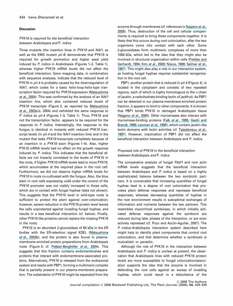
Analysis of an extract enriched in plasma membrane
proteins from roots by two-dimensional gel electrophoresis
uncovered several spots that were severely reduced in pii-4
compared with the wild type. Most obvious was the
reduction of an abundant spot of approximately 60 kDa
and with a pI value of 6.5 (Figure 4, marked ‘1’). Mass
spectrometrical analysis uncovered that this spot
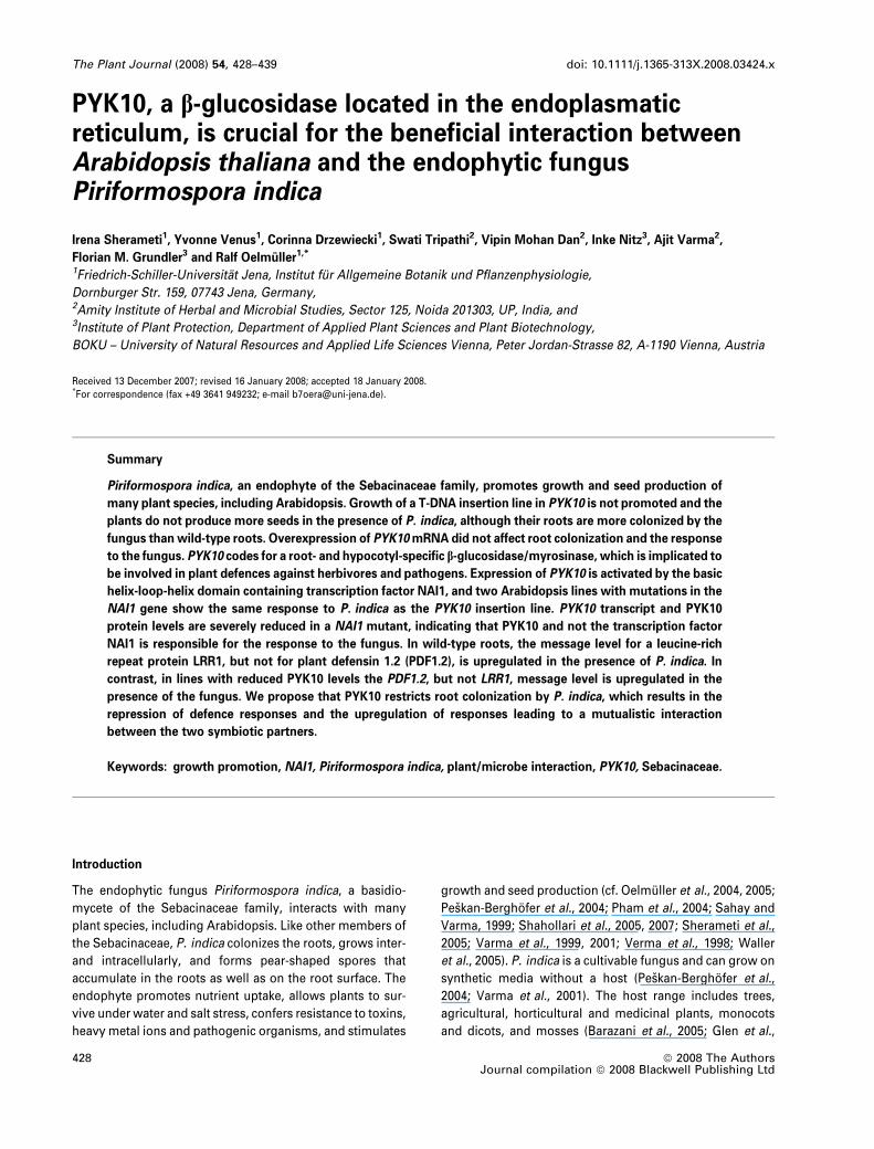
Figure 2. Phenotypes of Arabidopsis plants co-cultivated with Piriformospora
indica [wild type as well as the DPYK10 (N871638), pii-4 and DNAI1 (N397417)
mutants] 4 weeks after transfer to soil. ) P. indica, uncolonized control plants;
+ P. indica, plants that were checked for root colonization before transfer to
soil. Transfer to soil occurred after 20 days of co-cultivation with the fungus in
glass jars; at that time point, P. indica-colonized wild-type seedlings were
bigger than the uncolonized control (cf. Fig. 1), whereas the mutant seedlings
grown in the absence or presence of P. indica were indistinguishable.
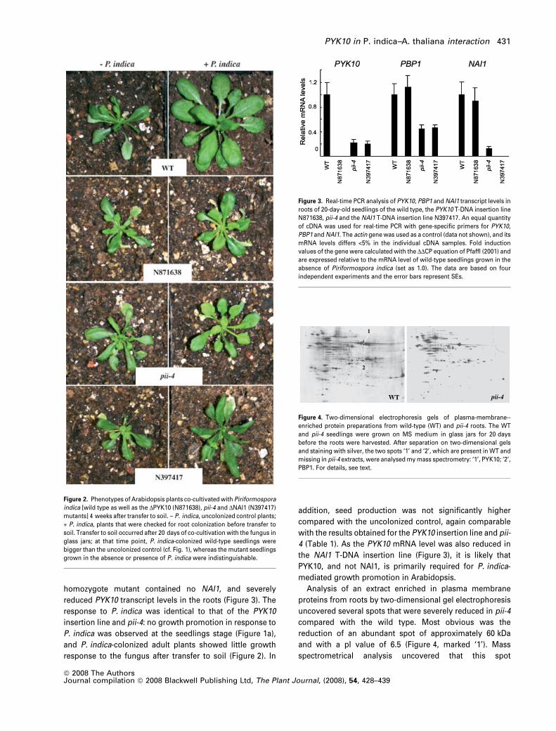
Figure 3. Real-time PCR analysis of PYK10, PBP1 and NAI1 transcript levels in
roots of 20-day-old seedlings of the wild type, the PYK10 T-DNA insertion line
N871638, pii-4 and the NAI1 T-DNA insertion line N397417. An equal quantity
of cDNA was used for real-time PCR with gene-specific primers for PYK10,
PBP1 and NAI1. The actin gene was used as a control (data not shown), and its
mRNA levels differs <5% in the individual cDNA samples. Fold induction
values of the gene were calculated with the DDCP equation of Pfaffl (2001) and
are expressed relative to the mRNA level of wild-type seedlings grown in the
absence of Piriformospora indica (set as 1.0). The data are based on four
independent experiments and the error bars represent SEs.
1
2
WT pii-4
Figure 4. Two-dimensional electrophoresis gels of plasma-membrane--
enriched protein preparations from wild-type (WT) and pii-4 roots. The WT
and pii-4 seedlings were grown on MS medium in glass jars for 20 days
before the roots were harvested. After separation on two-dimensional gels
and staining with silver, the two spots ‘1’ and ‘2’, which are present in WT and
missing in pii-4 extracts, were analysed my mass spectrometry: ‘1’, PYK10; ‘2’,
PBP1. For details, see text.
PYK10 in P. indica–A. thaliana interaction 431
ª 2008 The AuthorsJournal compilation ª 2008 Blackwell Publishing Ltd, The Plant Journal, (2008), 54, 428–439

corresponds to PYK10 [calculated molecular weight (pI) of
processed protein, 57.2 kDa (6.3); identified peptides,
IGIAHSPAWFEAHDLADSQDGASIDR; EYADFVFQEYGGK;
SGYEAYLVTHNLLISHAEAVEAYR; At3g09260). A second
spot was also reduced in pii-4 (Figure 4, marked ‘2’). The
two identified peptides (QLTAFGSDDGTVWDDGAYVGV and
STLLGFEEFVLDYSEYITAVDGTYD) correspond to a recently
identified PYK10-binding protein (PBP1, At3g16420; Nagano
et al., 2005). PBP1 has a calculated molecular weight of
approximately 32 kDa and a pI of 5.5, and is a jacalin lectin
family protein. Although PYK10 is located in the ER, PBP1 is
a cytoplasmic protein (Nagano et al., 2005). Real-time PCR
analysis for pii-4 demonstrated that not only the reduction in
PYK10, but also a reduction in PBP1 protein levels, might be
caused by fewer messages (Figure 3). The PYK10 message
level is reduced by approximately 80%, and that of PBP1 is
reduced by more than 50%. As a homozygote knock-out line
of At3g16420 (insertion in an exon; N341573), which codes
for PBP1, responded to P. indica in a similar way as the wild
type (data not shown), it appears that PYK10 rather than
PBP1 is crucial for the interaction.
P. indica promotes growth of PYK10 overexpressors
The PYK10 cDNA was overexpressed in Arabidopsis under
the control of the 35S promoter. Two of the lines that
showed the highest PYK10 mRNA levels were further anal-
ysed. The mRNA levels in the shoots were significantly
higher than in the wild type, because the low expression
level of the PYK10 gene under the control of its own
promoter was circumvented by the use of the 35S CaMV
promoter. A significant increase in the PYK10 mRNA level
was also observed in roots, although the stimulatory effect
was less because of the high activity of the endogenous
promoter in the root tissue (Figure 5a). Growth and the
phenotype of the overexpressors did not differ from the wild
type. Furthermore, promotion of root and shoot growth
by the fungus was comparable with the wild type (Fig-
ure 5b). We conclude that the higher PYK10 mRNA levels in
the overexpressor lines have no effect on P. indica-induced
growth promotion in Arabidopsis.
PYK10 restricts root colonization
To test whether modulation of the PYK10 level has an effect
on root colonization, we determined the quantity of the
fungal translation elongation factor 1 (Pitef1) mRNA relative
to the plant actin mRNA in the roots of colonized seedlings
with altered PYK10 levels. We noticed that the Pitef1/actin
mRNA ratio was higher in roots with reduced PYK10 mRNA
levels when the seedlings were co-cultivated with the fungus
for more than 5 days. At this time point, the analysed roots
were surrounded by a thin layer of fungal mycelia on the
agar plates. Quantitative data based on real-time PCR anal-
yses were obtained from roots that were co-cultivated with
the fungus for10 days. Roots with reduced PYK10 mRNA
levels contain significantly more Pitef1 mRNA when
compared with the wild type and with the overexpressor
lines ox-1 and ox-2 (Figure 6b). We propose that lower PYK10
levels result in better root colonization.
Furthermore, the message level for the leucine-rich-
repeat containing atypical receptor protein LRR1 is tran-
siently upregulated in wild-type roots in response to
P. indica and Sebacina (Shahollari et al., 2005, 2007). Real-
time PCR analysis demonstrates that this response does
not occur in the three mutant lines with reduced PYK10
mRNA levels (Figure 7). In contrast, the message level for
plant defensin 1.2 (PDF1.2), which codes for an antimicro-
bial defensin (Penninckx et al., 1996), does not respond to
P. indica in wild type, but is upregulated in these mutants
in the presence of the fungus (Figure 7). This indicates
that PYK10 restricts the expression of PDF1.2 and allows
the upregulation of the fungus-inducible LRR1 gene (see
Discussion).
PYK10 and PYK10-like proteins in the
Arabidopsis–P. indica interaction
PYK10 and PYK10-like proteins are encoded by a least 19
genes in Arabidopsis; however, transcripts for only 11 of
them can be detected in roots. Their absolute mRNA
Figure 5. Analysis of the PYK10 overexpressor line. Upper panel: RT-PCR
products of PYK10 isolated from the shoots and roots of 20-day-old PYK10-
overexpressor-1 (ox-1) and wild-type (WT) seedlings, based on equal quan-
tities of actin mRNA, which is not shown. Lower panel: the PYK10 overex-
pressor was grown in the absence () P. indica) or presence (+ P. indica) of
Piriformospora indica for 10 days. Results are representative for four
independent experiments.
432 Irena Sherameti et al.
ª 2008 The AuthorsJournal compilation ª 2008 Blackwell Publishing Ltd, The Plant Journal, (2008), 54, 428–439

levels differ substantially. Approximately 60% of all tran-
scripts for PYK10 and PYK10-like proteins in roots derive
from PYK10 (At3g09260; Figure 8). Among the 11 genes is
also PENETRATION2 (PEN2, At2g44490), which codes for
an enzyme that restricts pathogen entry into plant leaf
cells (Lipka et al., 2005; cf. Discussion); however the PEN2
mRNA level in the roots represents less than 10% of the
total PYK10 and PYK10-like mRNA level. None of the
transcript levels was significantly regulated by P. indica in
the wild type (Figure 8). The same was observed for the
PBP1 and NAI1 transcript levels (data not shown). Also,
transgenic lines expressing the uidA gene under the
control of the PYK10 (At3g09260) promoter did not show
a higher GUS activity in Arabidopsis roots co-cultivated
with the fungus (data not shown). The identified peptides
for PYK10, which are missing in pii-4, matched only with
At3g09260. Thus, it appears that PYK10 cannot be
replaced by PYK10-like proteins in the Arabidopsis–P.
indica interaction. This might be because PYK10 is the
most abundant b-glucosidase in the roots, or because the
different enzymes catalyze different reactions.
(a)
(b)
Figure 6. The transcript levels of the fungal translation elongation factor 1
(cPitef1) and genomic DNA (gPitef1) in the roots of colonized Arabidopsis
seedlings was compared with the levels of the plant actin nucleic acids.
(a) RNA and/or DNA were isolated from the roots of N871638 (DPYK10), pii-4,
N397417 (DNAI1), wild type (wt) and the overexpressor lines ox-1 and ox-2 (cf.
Experimental procedures). Co-cultivation of both organisms was performed
for 10 days. After reverse transcription, cPitef1 (38 cycles) and actin
(20 cycles) were amplified (left six lanes). For the right three lanes, genomic
DNA was amplified with the same primers. The sizes of the fragments are
given.
(b)To obtain quantitative data, real-time PCR was performed. The actin-
mRNA-normalized Pitef1 transcript levels of the different lines are expressed
relative to the level in wild-type roots, which was taken as 1.0. Data are based
on four independent experiments, and dots indicate a significant difference to
WT at P < 0.05 (•) and P < 0.001 (••).
WT N871638 pii-4 N397417
3(a)
(b)
2
1
0
Rel
ativ
e P
DF
1.2
mR
NA
leve
lsR
elat
ive
LRR
1 m
RN
A le
vels
WT N871638 pii-4 N397417
20
15
10
5
0
Figure 7. The LRR1 (At5g16590) message level is upregulated in response to
Piriformospora indica in Arabidopsis roots, but not in pii-4 and the insertion
lines N871638 (DPYK10) and N397417 (DNAI1).
The PDF1.2 mRNA level is regulated in the opposite direction. Arabidopsis
seedlings were grown in the absence (black bars) or presence (white bars) of
P. indica. After 6 days of co-cultivation, RNA was isolated from the roots and
used for cDNA synthesis. An equal quantity of cDNA was used for real-time
PCR with gene-specific primers for LRR1, PDF1.2 and the actin gene (Pfalz
et al., 2006). Fold induction values of the gene were calculated with the DDCP
equation of Pfaffl (2001), and are expressed relative to the mRNA level of wild-
type (WT) seedlings grown in the absence of P. indica (set as 1.0). The actin
values (not shown) differed less than 5% in the individual cDNA samples. The
data are based on four independent experiments and the error bars represent
SEs.
5000
4000
3000
2000
1000
0
At3g09
260
At1g66
280
At1g52
400
At2g44
480
At2g44
450
At3g03
640
PEN2
At3g60
140
At3g60
130
At2g32
860
At1g26
560
Sign
al
Figure 8. Relative expression of PYK10 and PYK10-related genes in Arabid-
opsis roots that were co-cultivated with Piriformospora indica for either 2 or
6 days.
Based on three (2 days) and two (6 days) independent microarray analyses,
the data represent average values. The absence of the response to P. indica
was also confirmed by semi-quantitative PCR analyses for the five most
abundantly expressed PYK10 and PYK10-related genes (data not shown).
Black (light grown), 2 days without (with) P. indica; white (dark grown),
6 days without (with) P. indica.
PYK10 in P. indica–A. thaliana interaction 433
ª 2008 The AuthorsJournal compilation ª 2008 Blackwell Publishing Ltd, The Plant Journal, (2008), 54, 428–439
Discussion
PYK10 is required for the beneficial interaction
between Arabidopsis and P. indica
Three mutants (the insertion lines in PYK10 and NAI1, as
well as the EMS mutant pii-4) demonstrate that PYK10 is
required for growth promotion and higher seed yield
induced by P. indica in Arabidopsis (Figures 1–2, Table 1),
whereas higher PYK10 mRNA levels did not affect the
beneficial interaction. Gene mapping data, in combination
with sequence analyses, indicate that the reduced level of
PYK10 in pii-4 is probably caused by the downregulation of
NAI1, which codes for a basic helix-loop-helix-type tran-
scription factor required for PYK10 expression (Matsushima
et al., 2004). This was confirmed by the analysis of an NAI1
insertion line, which also contained reduced levels of
PYK10 transcripts (Figure 3, as reported by Matsushima
et al., 2003a,b, 2004) and exhibited the same response to
P. indica as pii-4 (Figures 1–3, Table 1). Thus, PYK10 and
not the transcription factor, appears to be required for the
response to P. indica. Interestingly, the response to the
fungus is identical in mutants with reduced PYK10 tran-
script levels (in pii-4 and the NAI1 insertion line) and in the
mutant that lacks PYK10 transcripts completely because of
an insertion in a PYK10 exon (Figures 1–4). Also, higher
PYK10 mRNA levels had no effect on the growth response
induced by P. indica. This indicates that the beneficial ef-
fects are not linearily correlated to the levels of PYK10 in
the roots, if higher PYK10 mRNA levels lead to more PYK10,
which accumulates at the required place in the root cell.
Furthermore, we did not observe higher mRNA levels for
PYK10 in roots co-cultivated with the fungus. Also, the blue
stain in root cells expressing uidA under the control of the
PYK10 promoter was not visibly increased in those cells,
which are in contact with fungal hyphae (data not shown).
This suggests that the PYK10 level in wild-type roots is
sufficient to protect the plant against over-colonization;
however, severe reduction in the PYK10 protein level leaves
the cells unprotected against invading fungal hyphae, and
results in a less beneficial interaction (cf. below). Finally,
other PYK10-like proteins cannot replace the missing PYK10
in the roots.
PYK10 is an abundant b-glucosidase of 65 kDa in the ER
bodies with the ER-retention signal KDEL (Matsushima
et al., 2003b), and the protein is also found in plasma-
membrane-enriched protein preparations from Arabidopsis
roots (Figure 4; cf. Peskan-Berghofer et al., 2004). This
suggests that this fraction contains endomembranes and
proteins that interact with endomembrane-associated pro-
teins. Alternatively, PYK10 is released from the endosomal
system and reacts with PBP1, forming a multimeric complex
that is partially present in our plasma-membrane prepara-
tion. The substrate(s) of PYK10 might be separated from the
enzyme through membranes (cf. references in Nagano et al.,
2005). Thus, destruction of the cell and cellular compart-
ments is required to bring these components together. It is
likely that this occurs during root colonization, after the two
organisms come into contact with each other. Some
b-glucosidases form multimeric complexes of more than
1000 kDa, which led to the idea that they might also be
involved in structural organization within cells (Fieldes and
Gerhardt, 1994; Kim et al., 2000; Nisius, 1988; Selmar et al.,
1987). This might also play a role in our interaction system,
as hosting fungal hyphae requires substantial reorganiza-
tion in the root cell.
PBP1, another protein that is reduced in pii-4 (Figure 4), is
located in the cytoplasm and consists of two repeated
regions, each of which is highly homologous to the a chain
of jacalin, a carbohydrate-binding lectin of jackfruit. As PBP1
can be detected in our plasma-membrane-enriched protein
fraction, it appears to bind to other components. It is known
that PBP1 binds PYK10 in damaged Arabidopsis tissue
(Nagano et al., 2005). Other myrosinases also interact with
myrosinase-binding proteins (Falk et al., 1995; Geshi and
Brandt, 1998; Lenman et al., 1990), which contain jacalin-like
lectin domains with lectin activities (cf. Taipalensuu et al.,
1997). However, inactivation of PBP1 did not affect the
beneficial interaction between Arabidopsis and P. indica.
Proposed role of PYK10 in the beneficial interaction
between Arabidopsis and P. indica
The comparative analysis of fungal Pitef1 and root actin
mRNA levels suggests that the beneficial interaction
between Arabidopsis and P. indica is based on a highly
sophisticated balance between the two symbiotic part-
ners. It is conceivable that increasing quantities of fungal
hyphae lead to a degree of root colonization that pro-
vokes plant defense responses and represses beneficial
responses, whereas decreasing quantities of hyphae in
the root environment results in suboptimal exchanges of
information and nutrients between the two partners. This
resembles mycorrhizal symbioses, in which initially acti-
vated defense responses against the symbiont are
reduced during later phases of the interaction, or are even
actively repressed (cf. Pozo and Azcon-Aguilar, 2007). The
P. indica–Arabidopsis interaction system described here
might help to identify plant components that control root
colonization, and that determine whether a symbiosis is
mutualistic or parasitic.
Although the role of PYK10 in the interaction between
Arabidopsis and P. indica is unclear at present, the obser-
vation that Arabidopsis lines with reduced PYK10 protein
levels are more susceptible to fungal colonization/associ-
ation supports the idea that the enzyme is involved in
defending the root cells against an excess of invading
hyphae, which could result in a disturbance of the
434 Irena Sherameti et al.
ª 2008 The AuthorsJournal compilation ª 2008 Blackwell Publishing Ltd, The Plant Journal, (2008), 54, 428–439
balanced mutualistic interaction. Two lines of evidence
support this idea. (i) PYK10 exhibits striking sequence
similarities to PEN2, a glycosyl hydrolase, which restricts
pathogen entry of two ascomycete powdery mildew fungi
into Arabidopsis leaf cells (Lipka et al., 2005). Like PEN2,
PYK10 belongs to the class of glycosyl hydrolase family 1,
both proteins are located in intracellular organellar struc-
tures (PYK10 in ER bodies and PEN2 in peroxisomes), and
both proteins share a high degree of sequence similarity.
The catalytic domains of both proteins contain two
conserved nucleophilic glutamates. Lipka et al. (2005) have
shown that glutamate183 is required for PEN2 function
in vivo, which suggests that PEN2 catalytic activity is
required for restricting pathogen entry. Thus, PYK10 might
have a similar biological function in our system. (ii) The
beneficial traits in this symbiosis are highly dependent on
the density of the hyphae in and around the root.
Increasing quantities of hyphae in our co-cultivation sys-
tem resulted in a suboptimal interaction, and marker genes
for the beneficial interaction (such as LRR1) were down-
regulated and those for defence processes (such as
PDF1.2) were upregulated in the roots in a dose-dependent
manner (Oelmuller, 2008). Similar response patterns were
observed here (see Figure 7). In order to maintain a
mutualistic interaction with benefits for both partners, the
degree of root colonization might be controlled by activat-
ing PYK10-dependent defence responses, when too many
hyphae colonize the roots and the cells become damaged
or wounded by hyphal penetration. In barley, for instance,
less-defended root cells undergo cell death after coloniza-
tion with P. indica (Deshmukh et al., 2006). To further
elucidate the role of PYK10 in this interaction, Arabidopsis
lines in which better characterized defence compounds are
manipulated, can be analysed. Furthermore, because PEN2
is also expressed in roots, manipulation of the PEN2 level
might have an influence on root colonization. Finally, the
identification of PYK10 product(s) and the characterization of
its (their) role(s) in this interaction appears to be possible, for
example by comparing the composition of glucosinolates
and of other secondary metabolites in the roots of Arabid-
opsis lines with manipulated PYK10 levels growing in the
presence or absence of P. indica.
Experimental procedures
Growth conditions of plants and fungus
Wild-type Arabidopsis thaliana seeds, EMS mutant seeds (Colum-bia; Lehle, http://www.arabidopsis.com), seeds from the homozy-gote T-DNA insertion lines and lines expressing the uidA geneunder the control of the PYK10 promoter (Nitz et al., 2001), orexpressing PYK10 under the control of the 35S CaMV promoter,were surface-sterilized and placed on Petri dishes containing MSnutrient medium (Murashige and Skoog, 1962). After cold treatmentat 4�C for 48 h, plates were incubated for 7 days at 22�C under
continuous illumination (100 lmol m)2 sec)1) to allow growth ofthe seedlings without P. indica. P. indica was cultured as describedpreviously (Peskan-Berghofer et al., 2004; Verma et al., 1998) onaspergillus-minimal medium (Kaldorf et al., 2005). For solidmedium, 1% (w/v) agar was included.
To quantify root development, the seedlings were grown onsolid MS medium in sterile glass jars (ø 9 cm; height, 5 cm). Aftercounting the lateral roots, the lengths of the main root and theweight of the total root were determined (cf. Shahollari et al.,2007).
Co-cultivation experiments and estimation of plant growth
Nine days after the beginning the experiments, A. thaliana seed-lings were transferred to nylon discs (mesh-size, 70 lm) and placedon top of a modified PNM culture medium (5 mM KNO3, 2 mM
MgSO4, 2 mM Ca(NO3)2, 0.01 lM FeSO4, 70 lM H3BO3, 14 lM MnCl2,0.5 lM CuSO4, 1 lM ZnSO4, 0.2 lM Na2MoO4, 0.01 lM CoCl2,10.5 g l)1 agar, pH 5.6) in glass jars. One seedling was used per jar.After 24 h, fungal plugs of approximately 5 mm in diameter wereplaced at a distance of 1 cm from the roots. The uninfected controlplants received the same plugs without the fungus. The jars wereincubated at 22�C under continuous illumination from the side(80 lmol m)2 sec)1). Fresh weights were determined directly afterseedlings were removed from the jars.
Experiments on soil
For the experiments on soil, Arabidopsis seedlings were germi-nated on MS medium in Petri dishes without the fungus. Afterinfection with the fungus and co-cultivation for additional 20 days injars, they were transferred to sterile soil. Uninfected controls weretreated in the same way, except that the plugs introduced to the jarswere without the fungus. For experiments with the fungus, the soilwas mixed carefully with the fungus (1%, w/v). Although growthpromotion and higher seed yield also occur in uninfected soil, wenoticed that the response is more homogenous in inoculated soil.The fungal mycelium was obtained from liquid cultures after themedium was removed, and the mycelium was washed with anexcess of distilled water. Before being transferred to soil, the rootswere examined under the microscope to ensure that hyphae andspores had developed within and around the roots. Cultivationoccurred in multi-trays with Aracon tubes in a temperature-con-trolled growth chamber at 22�C under long-day conditions (16 hlight, 8 h dark; light intensity, 80 lmol m)2 sec)1). The sizes of theplants were monitored daily. For the mutant screen, the heights ofEMS mutant plants grown in the presence of P. indica were com-pared with those of control plants. Seeds were collected from theplants that were shorter than the wild type in the presence ofP. indica, but not shorter than the wild type without the fungus. Thereduced response to P. indica was confirmed in the next two gen-erations. The physiological results for pii-4 presented here wereobtained from the M3 and M4 generations. Seed production(g seeds per plant) was monitored by collecting seeds from indi-vidual plants grown under the standardized conditions describedabove. Seeds were dried for 4 weeks in paper bags before theweight was determined.
Staining fungal hyphae and spores
Small parts of the roots from seedlings that were co-cultivatedwith P. indica were transferred to 10% KOH and boiled for
PYK10 in P. indica–A. thaliana interaction 435
ª 2008 The AuthorsJournal compilation ª 2008 Blackwell Publishing Ltd, The Plant Journal, (2008), 54, 428–439
10 min. After washing with water for 1 min, the roots were putinto a 0.01% acid fuchsin-lactic acid solution and boiled againfor 10 min. Excess dye was removed with water prior tomicroscopy.
Fluorescence measurements
Autofluorescence in the developing root hairs was detected with theLSM 510 META microscope (Carl Zeiss Inc., http://www.zeiss.com).Relative values (550 nm) were obtained for the emission spectra (cf.Peskan-Berghofer et al., 2004).
Isolation of plasma-membrane-enriched protein fractions
A 20-g portion of Arabidopsis roots were used to isolate micro-somes. The material was homogenized in a buffer containing50 mM Tris/HCl, pH 7.4, 330 mM sucrose, 3 mM EDTA, 1 mM 1,4-dithiothreitol and 5% (w/v) polyvinylpolypyrrolidone. Thehomogenate was filtered through four layers of cheesecloth andcentrifuged for 20 min at 10 000 g. The supernatant was thencentrifuged at 50 000 g for 60 min to pellet the microsomes.Plasma membranes were prepared from three microsome prep-arations by two-phase partitioning with 6.4% (w/w) dextraneT-500 and 6.4% (w/w) polyethylene glycol (average molecularweight, 3350) (Briskin et al., 1987; Larsson et al., 1987; Peskanet al., 2000). The plasma membranes were resuspended in abuffer containing 50 mM Tris/HCl, pH 7.4; 3 mM EDTA and 1 mM
1,4-dithiothreitol.Preparation of protein extracts from plasma-membrane prepara-
tions, two-dimensional gel electrophoresis, staining of the gels andextraction of the protein spots was described in Sherameti et al.(2004).
Mass spectrometry
Aliquots of the eluted protein fractions were used for mass spec-trometry. Trypsin digestion of protein mixtures was performedaccording to Sherameti et al. (2004). Peptide analysis by couplingliquid chromatography with electrospray ionization mass spec-trometry (ESI-MS) and tandem mass spectrometry (MS-MS) wasdescribed previously (Shahollari et al., 2004; Sherameti et al., 2004;Stauber et al., 2003).
Protein identification
The measured MS-MS spectra were matched with the amino-acidsequences of tryptic peptides from the A. thaliana database inFASTA format. Cys modification by carbamidomethylation (+57 Da)was taken into account, and known contaminants were filtered out.Raw MS-MS data were analyzed by the Finnigan Sequest/TurboSequest software (revision 3.0; ThermoQuest, San Jose, CA, USA).The parameters for the analysis by the Sequest algorithm were setaccording to Stauber et al. (2003). The similarity between the mea-sured MS-MS spectrum and the theoretical MS-MS spectrum,reported as the cross-correlation factor (Xcorr), was equal or above1.5, 2.5 and 3.5 for singly, doubly or triply charged precursor ions,respectively. In order to identify corresponding loci, identified pro-tein sequences were subjected to BLAST searches at NCBI (http://www.ncbi.nlm.nih.gov) and FASTA searches by using the AGIprotein database at TAIR (http://www.arabidopsis.org). Conserveddomains and signal peptides were identified using SMART (Schultzet al., 1998).
RNA analysis
RNA was isolated with an RNA isolation kit (RNeasy; Qiagen, http://www.qiagen.com). For quantitative RT-PCR (cf. legend to Figure 6),RNA from Arabidopsis roots grown in the absence or presence ofP. indica was used with gene-specific and several control primerpairs (Sambrook et al., 1989). RT-PCR was performed by reversetranscription of 5 g of total RNA with gene-specific reverse primers.First-strand synthesis was performed with a kit (#K1631) from MBIFermentas (http://www.fermentas.com). After PCR, the productswere analyzed on 1.5% agarose gels and stained with ethidiumbromide, and visualized bands were quantified with the ImageMaster Video System (Amersham, GE Life Sciences, http://www.gelifesciences.com).
Real-time PCR
Real-time quantitative RT-PCR was performed using the iCycler iQreal-time PCR detection system and iCycler software version 2.2(Bio-Rad, http://www.bio-rad.com). Total RNA was isolated fromat least four independent replicates of Arabidopsis roots. For theamplification of the PCR products, iQ SYBR Supermix (Bio-Rad)was used according to the manufacturer’s instructions in a finalvolume of 20 ll. The iCycler was programmed to 95�C for 2 min,35 cycles of 95�C for 30 sec, 55�C for 40 sec, 72�C for 45 sec, and72�C for 10 min, followed by a melting-curve programme (55–95�Cin increasing steps of 0.5�C). All reactions were repeated at leasttwice. The mRNA levels for each cDNA probe were normalizedwith respect to the actin message level. Fold induction valueswere calculated with the DDCP equaltion of Pfaffl (2001) and werecompared with the mRNA level in the target genes in wild-typeroots, which were defined as 1.0. The following primer pairs wereused: LRR1-for, CGGCGAGTTTGATCTTGATGG, LRR1-rev,CTCAGGAACCACGACATCTCTC; PYK10-for, CGCATTTCCGG-TAAGCTTC, PYK10-rev, AAAGGCACCTGGTCGTTGCT; PBP1-for,GGATCCGATGAGGGTACTCA, PBP1-rev, GGCAGGAGTCAACG-GAGTTG; NAI1-for, CCGGGTTTGAGTTGCTAGC, NAI1-rev,GGAGACCCAAATGAGATCAC; PDF1.2a (At5g44420)-for, AT-GGTCAGGGGTTTGCGGAAA, PDF1.2a-rev, AT-GGTCAGGGGTTTGCGGAAA; P. indica was monitored with aprimer pair for Pitef1 (Butehorn et al., 2000), AC-CGTCTTGGGGTTGTATCC and TCGTCGCTGTCAACAAGATG. Thecolonized (and control) roots were removed from the agar plate,rinsed 12 times with an excess of sterile water (50 ml each) toremove the loosely attached fungal hyphae, and were then frozenin liquid nitrogen for RNA or DNA extraction.
Microarray analysis
Arabidopsis seedlings, grown as described above, were co-culti-vated (or mock-treated) with P. indica for either 2 or 6 days. RNAwas extracted from 70 mg of root material with the RNeasy PlantMini Kit (Qiagen), followed by an On-Column DNAse treatment(Qiagen). Microarray hybridization was performed with the Ara-bidopsis Genome Array ATH1 from Affymetrix (http://www.affymetrix.com), and the data were analysed with GCOS1.4 soft-ware (Affymetrix).
Miscellaneous
DNA extraction and sequence analysis were performed according tostandard protocols (Stockel and Oelmuller, 2004). For cloning ofPCR products, the PCR cloning kit from Quiagen was used. To
436 Irena Sherameti et al.
ª 2008 The AuthorsJournal compilation ª 2008 Blackwell Publishing Ltd, The Plant Journal, (2008), 54, 428–439
assign the mutant pii-4 locus to one of the Arabidopsis chromo-somes, a segregating F2 progeny was generated by crossing malepollen donor plants with homozygote lines of pii-4. Restrictionfragment length polymorphism analyses of the F2 plants were per-formed with the pARMS set (Schaffner, 1996). The PYK10 cDNA wascloned into a modified pMO9819 vector (Puzio, 1997) and intro-duced into Arabidopsis via Agrobacterium tumefaciens. Elevenplants with the PYK10 cDNA expressed in sense orientation wereregenerated and initially analysed. Two of them with the highestPYK10 mRNA levels were used for this study (ox-1 = 3c/38 andox-2 = 3f/42). All statistical analyses were performed by one-wayANOVAS.
Acknowledgements
We thank the Salk Institute Genomic Analysis Laboratory for pro-viding the sequence-indexed Arabidopsis T-DNA insertion mutants.Work was supported by the SFB 604, a grant from the DFG (Oe133/19-1), the BMBF (IND 03/013), the Friedrich-Schiller-University Jenaand the IMPRS Jena.
References
Barazani, O., Benderoth, M., Groten, K., Kuhlemeier, C. and
Baldwin, I.T. (2005) Piriformospora indica and Sebacinavermifera increase growth performance at the expense ofherbivore resistance in Nicotiana attenuata. Oecologia, 146,234–243.
Behnke, H.-D. and Eschlbeck, G. (1978) Dilated cisternae in Cappa-rales – an attempt towards the characterization of a specificendoplasmic reticulum. Protoplasma, 97, 351–363.
Bones, A.M. and Rossiter, J.T. (1996) The myrosinase-glucosinolatesystem, its organization and biochemistry. Physiol. Plant. 97, 194–208.
Bones, A.M., Evjen, K. and Iversen, T.-H. (1989) Characterization anddistribution of dilated cisternae of the endoplasmic reticulum inintact plants, protoplasts, and calli of Brassicaceae. Isr. J. Bot. 38,177–192.
Bonnett, H.T., Jr and Newcomb, E.H. (1965) Polyribosomes andcisternal accumulations in root cells of radish. J. Cell Biol. 27, 423–432.
Briskin, D.P., Leonard, R.T. and Hodges, T.K. (1987) Isolation ofplasma membrane: membrane markers and general principles.Methods Enzymol. 148, 542–568.
Brzobohaty, B., Moore, I., Kristoffersen, P., Bako, L., Campos, N.,
Schell, J. and Palme, K. (1993) Release of active cytokinin by ab-glucosidase localized to the maize root meristem. Science, 262,1051–1054.
Butehorn, B., Rhody, D. and Franken, P. (2000) Isolation and char-acterization of Pitef1 encoding the translation elongation factorEF-1a of the root endophyte Piriformospora indica. Plant Biol. 2,687–692.
Deshmukh, S., Huckelhoven, R., Schafer, P., Imani, J., Sharma, M.,
Weiss, M., Waller, F. and Kogel, K.H. (2006) The root endophyticfungus Piriformospora indica requires host cell death for prolif-eration during mutualistic symbiosis with barley. Proc. Natl Acad.Sci. USA, 103, 18450–18457.
Dharmawardhana, D.P., Ellis, B.E. and Carlson, J.E. (1995) A b-glu-cosidase from lodgepole pine xylem specific for the ligninprecursor coniferin. Plant Physiol. 107, 331–339.
Esen, A. (1993) b-glucosidases: overview. In B-Glucosidases, Bio-chemistry and Molecular Biology (Esen, A., ed). Washington, DC:American Chemical Society, pp. 1–14.
Falk, A., Taipalensuu, J., Ek, B., Lenman, M. and Rask, L. (1995)Characterization of rapeseed myrosinase-binding protein. Planta,195, 387–395.
Fieldes, M.A. and Gerhardt, K.E. (1994) An examination of theb-glucosidase (linamarase) banding pattern in flax seedlingsusing Ferguson plots and sodium dodecyl sulphate-polyacryl-amide gel electrophoresis. Electrophoresis, 15, 654–661.
Geshi, N. and Brandt, A. (1998) Two jasmonate-inducible myrosin-ase-binding proteins from Brassica napus L. seedlings withhomology to jacalin. Planta, 204, 295–304.
Glen, M., Tommerup, I.C., Bougher, N.L. and O’Brien, P.A. (2002)Are Sebacinaceae common and widespread ectomycorrhizalassociates of Eucalyptus species in Australian forests? Mycor-rhiza, 12, 243–247.
Gunning, B.E.S. (1998) The mystery organelles in Arabidopsisexpressing GFP. Trends Plant Sci. 3, 417.
Haseloff, J., Siemering, K.R., Prasher, D.C. and Hodge, S. (1997)Removal of a cryptic intron and subcellular localization of greenfluorescent protein are required to mark transgenic Arabidopsisplants brightly. Proc. Natl Acad. Sci. USA, 94, 2122–2127.
Hawes, C., Saint-Jore, C., Martin, B. and Zheng, H.-Q. (2001) ERconfirmed as the location of mystery organelles in Arabidopsisplants expressing GFP. Trends Plant Sci. 6, 245–246.
Hayashi, M., Toriyama, K., Kondo, M., Hara-Nishimura, I. and
Nishimura, M. (1999) Accumulation of a fusion protein containing2S albumin induces novel vesicles in vegetative cells of Arabid-opsis. Plant Cell Physiol. 40, 263–272.
Iversen, T.-H. (1970) Cytochemical localization of myrosinase(b-thioglucosidase) in root tips of Sinapis alba. Protoplasma, 71,451–466.
Kaldorf, M., Koch, B., Rexer, K.H., Kost, G. and Varma, A. (2005)Patterns of interaction between Populus Esch5 and Piriformos-pora indica: a transition from mutualism to antagonism. PlantBiol. 7, 210–218.
Kim, Y.-W., Kang, K.-S., Kim, S.-Y. and Kim, I.-S. (2000) Formation offibrillar multimers of oat b-glucosidase isoenzymes is mediatedby the As-Glu1 monomer. J. Mol. Biol. 303, 831–842.
Larsson, C., Widell, S. and Kjellbom, P. (1987) Preparation of high-purity plasma membranes. Methods Enzymol. 148, 558–568.
Leah, R., Kigel, J., Svendsen, I. and Mundy, J. (1995) Biochemicaland molecular characterization of a barley seed b-glucosidase.J. Biol. Chem. 270, 15789–15797.
Lenman, M., Rodin, J., Josefsson, L.-G. and Rask, L. (1990) Immu-nological characterization of rapeseed myrosinase. Eur. J.Biochem. 194, 747–753.
Lipka, V., Dittgen, J., Bednarek, P. et al. (2005) Pre- and postinva-sion defenses both contribute to nonhost resistance in Arabid-opsis. Science, 310, 1180–1183.
Malboobi, M.A. and Lefebvre, D.D. (1997) A phosphate-starvationinducible b-glucosidase gene (psr3.2) isolated from Arabidopsisthaliana is a member of a distinct subfamily of the BGA family.Plant Mol. Biol. 34(1), 57–68.
Matsushima, R., Hayashi, Y., Kondo, M., Shimada, T., Nishimura, M.
and Hara-Nishimura, I. (2002) An endoplasmic reticulum-derivedstructure that is induced under stress conditions in Arabidopsis.Plant Physiol. 130, 1807–1814.
Matsushima, R., Kondo, M., Nishimura, M. and Hara-Nishimura, I.
(2003a) A novel ER-derived compartment, the ER body, selec-tively accumulates a b-glucosidase with an ER retention signal inArabidopsis. Plant J. 33, 493–502.
Matsushima, R., Hayashi, Y., Yamada, K., Shimada, T., Nishimura,
M. and Hara-Nishimura, I. (2003b) The ER body, a novel endo-plasmic reticulum-derived structure in Arabidopsis. Plant CellPhysiol. 44, 661–666.
PYK10 in P. indica–A. thaliana interaction 437
ª 2008 The AuthorsJournal compilation ª 2008 Blackwell Publishing Ltd, The Plant Journal, (2008), 54, 428–439
Matsushima, R., Fukao, Y., Nishimura, M. and Hara-Nishimura, I.
(2004) NAI1 gene that encodes a basic-helix-loop-helix-typeputative transcription factor that regulates the formation of a novelER-derived structure, the ER body. Plant Cell, 16, 1536–1549.
McConn, M., Creelman, R.A., Bell, E., Mullet, J.E. and Browse, J.
(1997) Jasmonate is essential for insect defense in Arabidopsis.Proc. Natl Acad. Sci. USA, 94, 5473–5477.
Murashige, T. and Skoog, F. (1962) A revised medium for rapidgrowth and bioassays with tobacco tissue culture. Physiol. Plant,15, 473–497.
Nagano, A.J., Matsushima, R. and Hara-Nishimura, I. (2005) Acti-vation of an ER-body-localized b-glucosidase via a cytosolicbinding partner in damaged tissues of Arabidopsis thaliana. PlantCell Physiol. 46, 1140–1148.
Nisius, A. (1988) The stromacentre in Avena plastids: an aggrega-tion of b-glucosidase responsible for the activation of oat-leafsaponins. Planta, 173, 474–481.
Nitz, I., Berkefeld, H., Puzio, P.S. and Grundler, F.M.W. (2001) Pyk10,a seedling and root specific gene and promoter from Arabidopsisthaliana. Plant Sci. 161, 337–346.
Oelmuller, R. (2008) Interaction between Piriformospora indica withArabidopsis thaliana: early plant signaling events involved in abeneficial symbiosis with a growth- and seed yield-promotingendophytic fungus. In Biology of Molecular Plant–Microbe Inter-actions. Vol. 6, (Matteo, L., Woo, S. and Scala, F., eds). Springer(in press).
Oelmuller, R., Shahollari, B., Peskan-Berghofer, T., Trebicka, A.,
Giong, P.H., Sherameti, I., Oudhoff, M., Venus, Y., Altschmied, L.
and Varma, A. (2004) Molecular analyses of the interactionbetween Arabidopsis roots and the growth-promoting fungusPiriformospora indica. Endocytobiosis Cell Res. 15, 504–517.
Oelmuller, R., Peskan-Berghofer, T., Shahollari, B., Sherameti, I. and
Varma, A. (2005) MATH-domain containing proteins represent anovel gene family in Arabidopsis thaliana and are involved inplant/microbe interactions. Physiol. Plant. 124, 152–166.
Penninckx, I.A.M.A., Eggermont, K., Terras, F.R.G., Thomma,
B.P.H.J., De Samblanx, G.W., Buchala, A., Metraux, J.-P., Manners,
J.M. and Broekaert, W.F. (1996) Pathogen-induced systemicactivation of a plant defensin gene in Arabidopsis follows asalicylic acid–independent pathway. Plant Cell, 8, 2309–2323.
Peskan, T., Westermann, M. and Oelmuller, R. (2000) Identificationof low-density Triton X-100 insoluble plasma membrane micro-domains in higher plants. Eur. J. Biochem. 267, 6989–6995.
Peskan-Berghofer, T., Shahollari, B., Giong, P.H., Hehl, S., Markert,
C., Blanke, V., Varma, A.K. and Oelmuller, R. (2004) Association ofPiriformospora indica with Arabidopsis thaliana roots representsa novel system to study beneficial plant-microbe interactions andinvolves early plant protein modifications in the endoplasmaticreticulum and at the plasma membrane. Physiol. Plant, 122, 465–477.
Pfaffl, M.W. (2001) A new mathematical model for relative quanti-fication in real-time RT-PCR. Nucleic Acids Res. 29, 2002–2007.
Pfalz, J., Liere, K., Kandlbinder, A., Dietz, K.J. and Oelmuller, R.
(2006) pTAC2, -6, and -12 are components of the transcriptionallyactive plastid chromosome that are required for plastid geneexpression. Plant Cell, 18, 176–197.
Pham, G.H., Kumari, R., Singh, An. et al. (2004) Axenic cultures ofPiriformospora indica. In Plant Surface Microbiology (Varma, A.,Abbott, L., Werner, D. and Hampp, R., eds). Germany: Springer-Verlag, pp. 593–616.
Poulton, J.E. (1990) Cyanogenesis in plants. Plant Physiol. 94, 401–405.
Pozo, M.J. and Azcon-Aguilar, C. (2007) Unraveling mycorrhiza-induced resistance. Curr. Opin. Plant Biol. 10, 393–398.
Puzio, P.S. (1997) Identifizierung und Charakterisierung nemato-denresponsiver regulatorischer und kodierender DNA-Sequen-zen in der Pflanze Arabidopsis thaliana. PhD Thesis, Kiel,Germany: Christian-Alberts Universitat.
Rask, L., Andreasson, E., Ekbom, B., Eriksson, S., Pontoppidan, B.
and Meijer, J. (2000) Myrosinase: gene family evolution andherbivore defense in Brassicaceae. Plant Mol. Biol. 42, 93–113.
Ridge, R.W., Uozumi, Y., Plazinski, J., Hurley, U.A. and Williamson,
R.E. (1999) Developmental transitions and dynamics of the corti-cal ER of Arabidopsis cells seen with green fluorescent protein.Plant Cell Physiol. 40, 1253–1261.
Sahay, N.S. and Varma, A. (1999) Piriformospora indica: a newbiological hardening tool for micropropagated plants. FEMSMicrobiol. Lett. 181, 297–302.
Sambrook, J., Fritsch, E.F. and Maniatis, T. (1989) Molecular Clon-ing. A Laboratory Manual, Second Edition. New York: Cold SpringHarbor Laboratory.
Schaffner, A. (1996) pARMS, multiple marker containing plasmidsfor easy RFLP analysis in Arabidopsis thaliana. Plant Mol. Biol.Rep. 14, 11–16.
Schliemann, W. (1984) Hydrolysis of conjugated gibberellins byb-glucosidases from dwarf rice (Oryza sativa L. cv. Tan-ginbozu)..J. Plant Physiol. 116, 123–132.
Schultz, J., Milpetz, F., Bork, P. and Ponting, C.P. (1998) SMART, asimple modular architecture research tool: identification ofsignaling domains. Proc. Natl Acad. Sci. USA, 95, 5857–5864.
Selmar, D.R., Lieberei, B., Biehl, B. and Voigt, J. (1987) Hevea lina-marase-A nonspecific b-glucosidase. Plant Physiol. 83, 557–563.
Shahollari, B., Peskan-Berghofer, T., Varma, A. and Oelmuller, R.
(2004) Receptor kinases with leucine-rich repeats are enriched inTriton X-100 insoluble plasma membrane microdomains fromplants. Physiol. Plant. 122, 397–403.
Shahollari, B., Varma, A. and Oelmuller, R. (2005) Expression of areceptor kinase in Arabidopsis roots is stimulated by the basid-iomycete Piriformospora indica and the protein accumulates inTriton X-100 insoluble plasma membrane microdomains. J. PlantPhysiol. 162, 945–958.
Shahollari, B., Vadassery, J., Varma, A. and Oelmuller, R. (2007) Aleucine-rich repeat protein is required for growth promotion andenhanced seed production mediated by the endophytic fungusPiriformospora indica in Arabidopsis thaliana. Plant J. 50, 1–13.
Sherameti, I., Shahollari, B., Landsberger, M., Westermann, M.,
Cherepneva, G., Kusnetsov, V. and Oelmuller, R. (2004) Cytokininstimulates polyribosome loading of nuclear-encoded mRNAs forthe plastid ATP synthase in etioplasts of Lupinus luteus: thecomplex accumulates in the inner-envelope membrane with theCF1 moiety located towards the stromal space. Plant J. 38, 578–593.
Sherameti, I., Shahollari, B., Venus, Y., Altschmied, L., Varma, A.
and Oelmuller, R. (2005) The endophytic fungus Piriformosporaindica stimulates the expression of nitrate reductase and thestarch-degrading enzyme glucan-water dikinase in tobacco andArabidopsis roots through a homeodomain transcription factorwhich binds to a conserved motif in their promoters. J. Biol.Chem. 280, 2641–2647.
Smith, A.R. and van Staden, J. (1978) Changes in endogenouscytokinin levels in kernels of Zea mays L. during imbibition andgermination. J. Exp. Bot. 29, 1067–1075.
Stauber, E.J., Fink, A., Markert, C., Kruse, O., Johanningmeier, U.
and Hippler, M. (2003) Proteomics of Chlamydomonas reinhardtiilight-harvesting proteins. Eukaryotic Cell, 2, 978–994.
Stockel, J. and Oelmuller, R. (2004) A novel protein for photosystemI biogenesis. J. Biol. Chem. 279, 10243–10251.
Stotz, H.U., Kroymann, J. and Mitchell-Olds, T. (1999) Plant–insectinteractions. Curr. Opin. Plant Biol. 2, 268–272.
438 Irena Sherameti et al.
ª 2008 The AuthorsJournal compilation ª 2008 Blackwell Publishing Ltd, The Plant Journal, (2008), 54, 428–439
Stotz, H.U., Pittendrigh, B.R., Kroymann, J., Weniger, K., Fritsche,
J., Bauke, A. and Mitchell-Olds, T. (2000) Induced plant defenseresponses against chewing insects. Ethylene signaling reducesresistance of Arabidopsis against cotton worm but not dia-mondback moth. Plant Physiol. 124, 1007–1017.
Taipalensuu, J., Andreasson, E., Eriksson, S. and Rask, L. (1997)Regulation of the wound-induced myrosinase-associated proteintranscript in Brassica napus plants. Eur. J. Biochem. 247(3), 963–971.
Tierens, K.F., Thomma, B.P., Brouwer, M., Schmidt, J., Kistner, K.,
Porzel, A., Mauch-Mani, B., Cammue, B.P. and Broekaert, W.F.
(2001) Study of the role of antimicrobial glucosinolate-derivedisothiocyanates in resistance of Arabidopsis to microbial patho-gens. Plant Physiol. 125, 1688–1699.
Varma, A., Verma, S., Sudha, S., Sahay, N.S., Butehorn, B. and
Franken, P. (1999) Piriformospora indica, a cultivable plantgrowth promoting root endophyte. Appl. Environ. Microbiol. 65,2741–2744.
Varma, A., Singh, A., Sudha, S. et al. (2001) Piriformospora indica: acultivable mycorrhiza-like endosymbiotic fungus. In Mycota IX(Hock, B., ed). Germany: Springer Series, pp. 123–150.
Verma, S.A., Varma, A., Rexer, K.-H., Hassel, A., Kost, G., Sarbhoy,
A., Bisen, P., Butehorn, B. and Franken, P. (1998) Piriformosporaindica, gen. et sp. nov., a new root-colonizing fungus. Mycologia90, 898–905.
Waller, F., Achatz, B., Baltruschat, H. et al. (2005) The endophyticfungus Piriformospora indica reprograms barley to salt-stresstolerance, disease resistance, and higher yield. Proc. Natl Acad.Sci. USA, 102, 13386–13391.
Weiss, M., Selosse, M.A., Rexer, K.H., Urban, A. and Oberwinkler, F.
(2004) Sebacinales: a hitherto overlooked cosm of heterobasid-iomycetes with a broad mycorrhizal potential. Mycol. Res. 108,1003–1010.
Wittstock, U. and Halkier, B.A. (2002) Glucosinolate research in theArabidopsis era. Trends Plant Sci. 7, 263–270.
Xie, D.-X., Feys, B.F., James, S., Nieto-Rostro, M. and Turner, J.G.
(1998) COI1: an Arabidopsis gene required for jasmonate-regu-lated defense and fertility. Science, 280, 1091–1094.
Zeng, R.S., Mallik, A.U. and Setliff, E. (2003) Growth stimulation ofectomycorrhizal fungi by root exudates of Brassicaceae plants:role of degraded compounds of indole glucosinolates. J. Chem.Ecol. 29, 1337–1355.
PYK10 in P. indica–A. thaliana interaction 439
ª 2008 The AuthorsJournal compilation ª 2008 Blackwell Publishing Ltd, The Plant Journal, (2008), 54, 428–439