Revised 6/15 TM033
T E C H N I C A L M A N U A L
pGL3 Luciferase Reporter VectorsInstructions For Use of Products E1741, E1751, E1761 and E1771
Promega Corporation · 2800 Woods Hollow Road · Madison, WI 53711-5399 USA · Toll Free in USA 800-356-9526 · 608-274-4330 · Fax 608-277-2516 1www.promega.com TM033 · Revised 6/15
All technical literature is available at: www.promega.com/protocols/ Visit the web site to verify that you are using the most current version of this Technical Manual.
E-mail Promega Technical Services if you have questions on use of this system: [email protected]
pGL3 Luciferase Reporter Vectors
1. Description .........................................................................................................................................2
2. Product Components and Storage Conditions ........................................................................................2
3. pGL3 Vector Maps and Sequence Reference Points ................................................................................23.A. pGL3-Basic Vector ......................................................................................................................33.B. pGL3-Enhancer Vector ................................................................................................................43.C. pGL3-Promoter Vector ................................................................................................................53.D. pGL3-Control Vector ..................................................................................................................6
4. Cloning Methods .................................................................................................................................74.A. Cloning Strategies ......................................................................................................................74.B. Preparation of pGL3 Vectors and Insert DNA for Cloning ..............................................................84.C. Transformation Protocols for pGL3 Vectors ..................................................................................84.D. Isolation of Plasmid DNA ............................................................................................................8
5. Transfection of Mammalian Cells .........................................................................................................9
6. Assay of Luciferase Activity ..................................................................................................................9
7. Sequencing of Luciferase Reporter Vectors .......................................................................................... 11
8. Appendix .......................................................................................................................................... 128.A. Common Structural Elements of the pGL3 Luciferase Reporter Vectors ........................................ 128.B. Advantages of the pGL3 Vectors ................................................................................................. 138.C. The pGL3 Vectors luc+ Gene ...................................................................................................... 148.D. Mapping Genetic Elements Located Within DNA Fragments ........................................................ 168.E. Composition of Buffers and Solutions ......................................................................................... 168.F. References ............................................................................................................................... 178.G. pGL3-Basic Vector Restriction Sites ........................................................................................... 188.H. pGL3-Enhancer Vector Restriction Sites .................................................................................... 208.I. pGL3-Promoter Vector Restriction Sites .................................................................................... 238.J. pGL3-Control Vector Restriction Sites ....................................................................................... 268.K. Related Products ...................................................................................................................... 28
9. Summary of Changes ......................................................................................................................... 29

2 Promega Corporation · 2800 Woods Hollow Road · Madison, WI 53711-5399 USA · Toll Free in USA 800-356-9526 · 608-274-4330 · Fax 608-277-2516TM033 · Revised 6/15 www.promega.com
1. Description
The pGL3 Luciferase Reporter Vectors(a) provide a basis for the quantitative analysis of factors that potentially regulate mammalian gene expression. These factors may be cis-acting, such as promoters and enhancers, or trans-acting, such as various DNA-binding factors. The backbone of the pGL3 Luciferase Reporter Vectors is designed for increased expression, and contains a modified coding region for firefly (Photinus pyralis) luciferase that has been optimized for monitoring transcriptional activity in transfected eukaryotic cells. The assay of this genetic reporter is rapid, sensitive and quantitative. In addition, these Luciferase Reporter Vectors contain numerous features aiding in the structural characterization of the putative regulatory sequences under investigation.
2. Product Components and Storage Conditions
P R O D U C T S I Z E C AT. #
pGL3-Control Vector 20µg E1741
pGL3-Basic Vector 20µg E1751
pGL3-Promoter Vector 20µg E1761
pGL3-Enhancer Vector 20µg E1771
Information on related products, including the Luciferase Assay System, is provided in Sections 4–7 and 8.K.
Storage Conditions: Store the pGL3 Luciferase Reporter Vectors at –20°C.
3. pGL3 Vector Maps and Sequence Reference Points
The listings of restriction sites for the pGL3 Luciferase Reporter Vectors are provided in Section 8.G–J.
Note: The specific transcriptional characteristics of the pGL3 Vectors will vary for different cell types. This may be particularly true for COS cells, which contain the SV40 large T antigen. The SV40 large T antigen promotes replication from the SV40 origin, which is found in the promoter of the pGL3-Promoter and pGL3-Control Vectors. The combination of large T antigen and SV40 origin will result in a higher copy number of these vectors in COS cells, which in turn may result in increased expression of the reporter gene compared to other cell and vector combinations.
Promega Corporation · 2800 Woods Hollow Road · Madison, WI 53711-5399 USA · Toll Free in USA 800-356-9526 · 608-274-4330 · Fax 608-277-2516 3www.promega.com TM033 · Revised 6/15
3.A. pGL3-Basic Vector
The pGL3-Basic Vector lacks eukaryotic promoter and enhancer sequences, allowing maximum flexibility in cloning putative regulatory sequences. Expression of luciferase activity in cells transfected with this plasmid depends on insertion and proper orientation of a functional promoter upstream from luc+. Potential enhancer elements can also be inserted upstream of the promoter or in the BamHI or SalI sites downstream of the luc+ gene.
XbaI 1742
Ampr
KpnISacIMluINheISmaIXhoIBgIIIHindIII
pGL3-BasicVector
(4818bp)
f1 ori
ori
SalIBamHI NarI 121
NcoI 86luc+
SV40 late poly(A) signal
(for luc+ reporter)HpaI 1902
Synthetic poly(A)signal / transcriptional pause site(for background reduction)
0746
VA
08_4
A
511152128323653
20102004
Figure 1. pGL3-Basic Vector circle map. Additional description: luc+, cDNA encoding the modified firefly luciferase; Ampr, gene conferring ampicillin resistance in E. coli; f1 ori, origin of replication derived from filamentous phage; ori, origin of replication in E. coli. Arrows within luc+ and the Ampr gene indicate the direction of transcription; the arrow in the f1 ori indicates the direction of ssDNA strand synthesis.
pGL3-Basic Vector Sequence Reference Points:
Promoter (none)
Enhancer (none)
Multiple cloning region 1–58
Luciferase gene (luc+) 88–1740
GLprimer2 binding site 89–111
SV40 late poly(A) signal 1772–1993
RVprimer4 binding site 2080–2061
ColE1-derived plasmid replication origin 2318
β-lactamase gene (Ampr) 3080–3940
f1 origin 4072–4527
upstream poly(A) signal 4658–4811
RVprimer3 binding site 4760–4779

4 Promega Corporation · 2800 Woods Hollow Road · Madison, WI 53711-5399 USA · Toll Free in USA 800-356-9526 · 608-274-4330 · Fax 608-277-2516TM033 · Revised 6/15 www.promega.com
3.B. pGL3-Enhancer Vector
The pGL3-Enhancer Vector contains an SV40 enhancer located downstream of luc+ and the poly(A) signal. This aids in the verification of functional promoter elements because the presence of an enhancer will often result in transcription of luc+ at higher levels.
SV40 Enhancer
XbaI 1742
KpnISacIMluINheISmaIXhoIBgIIIHindIII
511152128323653
pGL3-EnhancerVector
(5064bp)
f1 ori
ori
SalIBamHI
22562250
NarI 121NcoI 86
luc+
Synthetic poly(A) signal / transcriptional pause site(for background reduction)
SV40 latepoly(A) signal
(for luc+ reporter)HpaI 1902
Ampr
0745
VA
08_4
A
Figure 2. The pGL3-Enhancer Vector circle map. Additional description: luc+, cDNA encoding the modified firefly luciferase; Ampr, gene conferring ampicillin resistance in E. coli; f1 ori, origin of replication derived from filamentous phage; ori, origin of plasmid replication in E. coli. Arrows within luc+ and the Ampr gene indicate the direction of transcription; the arrow in f1 ori indicates the direction of ssDNA strand synthesis.
pGL3-Enhancer Vector Sequence Reference Points:
Promoter (none)
Multiple cloning region 1–58
Luciferase gene (luc+) 88–1740
GLprimer2 binding site 89–111
SV40 late poly(A) signal 1772–1993
Enhancer 2013–2249
RVprimer4 binding site 2307–2326
ColE1-derived plasmid replication origin 2564
β-lactamase gene (Ampr) 3329–4186
f1 origin 4318–4773
upstream poly(A) signal 4904–5057
RVprimer3 binding site 5006–5025

Promega Corporation · 2800 Woods Hollow Road · Madison, WI 53711-5399 USA · Toll Free in USA 800-356-9526 · 608-274-4330 · Fax 608-277-2516 5www.promega.com TM033 · Revised 6/15
3.C. pGL3-Promoter Vector
The pGL3-Promoter Vector contains an SV40 promoter upstream of the luciferase gene. DNA fragments containing putative enhancer elements can be inserted either upstream or downstream of the promoter-luc+ transcriptional unit.
XbaI 1934
Ampr
KpnISacIMluINheISmaIXhoIBgIII
5111521283236
f1 ori
ori
SalIBamHI
22022196
NcoI 278
HindIII 245luc+
SV40 Promoter
pGL3-PromoterVector
(5010bp)
SV40 latepoly(A) signal(for luc+ reporter)
Synthetic poly(A) signal / transcriptional pause site(for background reduction)
0748
VA
08_4
A
HpaI 2094
Figure 3. The pGL3-Promoter Vector circle map. Additional description: luc+, cDNA encoding the modified firefly luciferase; Ampr, gene conferring ampicillin resistance in E. coli; f1 ori, origin of replication derived from filamentous phage; ori, origin of plasmid replication in E. coli. Arrows within luc+ and the Ampr gene indicate the direction of transcription; the arrow in f1 ori indicates the direction of ssDNA strand synthesis.
pGL3-Promoter Vector Sequence Reference Points:
Enhancer (none)
Multiple cloning region 1–41
Promoter 48–250
GLprimer2 binding region 281–303
Luciferase gene (luc+) 280–1932
SV40 late poly(A) signal 1964–2185
RVprimer4 binding region 2253–2272
ColE1-derived plasmid replication origin 2510
β-lactamase gene (Ampr) 3272–4132
f1 origin 4264–4719
Upstream poly(A) signal 4850–5003
RVprimer3 binding region 4952–4971

6 Promega Corporation · 2800 Woods Hollow Road · Madison, WI 53711-5399 USA · Toll Free in USA 800-356-9526 · 608-274-4330 · Fax 608-277-2516TM033 · Revised 6/15 www.promega.com
3.D. pGL3-Control Vector
The pGL3-Control Vector contains SV40 promoter and enhancer sequences, resulting in strong expression of luc+ in many types of mammalian cells. This plasmid is useful in monitoring transfection efficiency, in general, and is a convenient internal standard for promoter and enhancer activities expressed by pGL3 recombinants.
SV40 Enhancer
XbaI 1934
Ampr
KpnISacIMluINheISmaIXhoIBgIII
5111521283236
pGL3-ControlVector
(5256bp)
f1 ori
ori
SalIBamHI
24482442
NarI 313NcoI 278
HindIII 245
luc+
SV40 Promoter
SV40 latepoly(A) signal
(for luc+ reporter)HpaI 2094
Synthetic poly(A) signal / transcriptional pause site(for background reduction)
0747
VA
08_4
A
Figure 4. pGL3-Control Vector circle map. Additional description: luc+, cDNA encoding the modified firefly luciferase; Ampr, gene conferring ampicillin resistance in E. coli; f1 ori, origin of replication derived from filamentous phage; ori, origin of plasmid replication in E. coli. Arrows within luc+ and the Ampr gene indicate the direction of transcription; the arrow in f1 ori indicates the direction of ssDNA strand synthesis.
pGL3-Control Vector Sequence Reference Points:
Multiple cloning region 1–41
Promoter 48–250
Luciferase gene (luc+) 280–1932
GLprimer2 binding site 281–303
SV40 late poly(A) signal 1964–2185
Enhancer 2205–2441
RVprimer4 binding site 2499–2518
ColE1-derived plasmid replication origin 2756
β-lactamase gene (Ampr) 3518–4378
f1 origin 4510–4965
upstream poly(A) signal 5096–5249
RVprimer3 binding site 5198–5217
Promega Corporation · 2800 Woods Hollow Road · Madison, WI 53711-5399 USA · Toll Free in USA 800-356-9526 · 608-274-4330 · Fax 608-277-2516 7www.promega.com TM033 · Revised 6/15
CATTCCGGTACTGTTGGTAAAGCCACCATGGAAGACGCCAAAAACATAAAG . . . (1892bp) . . . GGATCCGTCGAC
RVprimer3
RVprimer4
5′ . . . CTAGCAAAATAGGCTGTCCCCAGTGCAAGTGCAGGTGCCAGAACATTTCTCTATCGATA
GGTACCGAGCTCTTACGCGTGCTAGCCCGGGCTCGAGATCTGCGATCTAAGTAAGCTTGG . . .
KpnIAcc65I
SacI MluI
NcoI
NheI XmaISmaI
XhoI BglII HindIII
SV40Promoter
SV40Enhancerluc+ Coding Region
Start
CGATGCCCTTGAGAGCCTTCAACCCAGTCAGCTCCTTCCGGTGGGCGCGGGGCATGACTATCGTC . . . 3′
GLprimer2 BamHI SalI
0756
MA
08_4
A
Figure 5. pGL3 Vector multiple cloning regions. Shown are the upstream and downstream cloning sites and the locations of the sequencing primers (GLprimer2, RVprimer3 and RVprimer4). The large primer arrows indicate the direction of sequencing. The positions of the promoter (in the pGL3-Promoter and pGL3-Control Vectors) and the enhancer (in the pGL3-Enhancer and pGL3-Control Vectors) are shown as insertions into the sequence of the pGL3-Basic Vector. (Note that the promoter replaces four bases [AAGT] of the pGL3-Basic Vector.) The sequence shown is of the DNA strand generated from the f1 ori.
4. Cloning Methods
4.A. Cloning Strategies
The restriction sites for XhoI and SalI have compatible ends, as do BglII and BamHI. Therefore, cloning into the XhoI or BglII sites upstream of luc+, or the downstream SalI or BamHI sites, allows easy interchange of DNA inserts between upstream and downstream positions relative to the luciferase reporter gene. Thus, positional effects of a putative genetic element may be readily tested. Cloning fragments into a single site will generally yield both possible orientations relative to the reporter gene, making these effects also readily testable.
The other upstream restriction sites may be used for cloning. However, note that some of the sites are required for generating nested deletions (see Section 8.D). Specifically, the KpnI or SacI site is needed to generate a 3´ overhang upstream of the insert.
! Please refer to Sections 8.G–J for additional information on XhoI digestion.

8 Promega Corporation · 2800 Woods Hollow Road · Madison, WI 53711-5399 USA · Toll Free in USA 800-356-9526 · 608-274-4330 · Fax 608-277-2516TM033 · Revised 6/15 www.promega.com
4.B. Preparation of pGL3 Vectors and Insert DNA for Cloning
The fragment and vector DNA should be digested with restriction enzymes that will generate compatible ends for cloning. In some cases, the ends of the DNA fragment may require modification, either by using synthetic linkers, by a PCR amplification using primers containing sites for appropriate restriction enzymes, or by filling in the restriction site overhangs. It may be advantageous to treat the vector DNA with calf intestinal alkaline phosphatase (CIAP; Cat.# M2825) or TSAP Thermosensitive Alkaline Phosphatase (Cat.# M9910) to remove 5´ phosphate groups, thus preventing reclosure of the vector on itself without an insert. Sufficient DNA should be prepared to perform control reactions for digestion, ligation and transformation steps.
To ensure capture of the correct insert DNA, the desired restriction fragment can be purified by electrophoresis on an acrylamide or agarose gel and then recovered from the gel by one of several methods, such as using the Wizard® PCR Preps DNA Purification System Technical Bulletin #TB118. Alternatively, unfractionated restriction fragments can be cloned into the target plasmid, and the desired recombinant then can be identified by gel electrophoresis of plasmid DNA.
Protocols for restriction digestion, alkaline phosphatase treatment, linker ligation and transformation of competent cells can be found in Molecular Cloning, A Laboratory Manual (1).
4.C. Transformation Protocols for pGL3 Vectors
Because the Luciferase Reporter Vectors are supplied as modified DNA, E. coli hosts may be either restriction + or restriction –. The use of a recA host such as JM109 is preferred because this prevents undesirable recombination between the insert and the host chromosomal DNA. A strain that has an F´ episome is required for ssDNA production.
Grow JM109 on minimal plates (M-9) supplemented with 1.0mM thiamine-HCl prior to preparation of competent cells and transformation. This selects for the presence of the F´ episome.
4.D. Isolation of Plasmid DNA
The Wizard® Plus SV Minipreps DNA Purification System (Cat.# A1340, A1470) may be used for small-scale preparation of plasmid DNA for screening clones. DNA suitable for transfection may be purified using the PureYield™ Plasmid Midipreps System (Cat.# A2492, A2495).

Promega Corporation · 2800 Woods Hollow Road · Madison, WI 53711-5399 USA · Toll Free in USA 800-356-9526 · 608-274-4330 · Fax 608-277-2516 9www.promega.com TM033 · Revised 6/15
5. Transfection of Mammalian Cells
Transfection of DNA into eukaryotic cells may be mediated by cationic lipid compounds (2), calcium phosphate (3,4), DEAE-dextran (3,5), or electroporation (4). Transfection systems based on cationic lipids (TransFast™ Transfection Reagent, Transfectam® Reagent and Tfx™ Reagents) and calcium phosphate (Profection® Mammalian Transfection System) are available from Promega. For more information on these transfection reagents, please request the TransFast™ Transfection Reagent Technical Bulletin (#TB260), the Transfectam® Reagent Technical Bulletin (#TB116), the Tfx™-Reagents Technical Bulletin (#TB216) or the ProFection® Mammalian Transfection System Technical Manual (#TM012). All of these documents are available on our web site at: www.promega.com/protocols/
6. Assay of Luciferase Activity
Experimental strategies using firefly luciferase may involve the analysis of a few samples per day or as many as several thousand samples per hour, and equipment used to measure luminescence may vary from inexpensive, single-sample luminometers to high-end CCD luminometers. To support this wide range of applications, we have developed three luciferase assays with different, but complementary, characteristics: Luciferase Assay System (Cat.# E1500), Bright-Glo™ Luciferase Assay System (Cat.# E2610), Steady-Glo® Luciferase Assay System (Cat.# E2510), and ONE-Glo™ Luciferase Assay System (Cat.# E6110). Reagent choice depends on the relative importance of experimental format, assay sensitivity, and luminescence duration.
Table 1. Characteristics of Promega Luciferase Assay Reagents.
Bright-Glo™ Reagent
Steady-Glo® Reagent
Luciferase Assay Reagent
ONE-Glo™ Reagent
Format NH or H NH or H NH NH or H
Process continuous batch bench scale batch or continuous
Number of Steps 1 1 4 1
Sensitivity highest lower higher high
Signal Half-Life ~30 minutes ~5 hours ~12 minutes ~50 minutes
Precision High High High Highest
Cell Lysis Time ~2 minutes maximum
~5 minutes maximum
NA ~3 minutes
NH = nonhomogeneous (first create a lysate); H = homogeneous; NA = not applicable
10 Promega Corporation · 2800 Woods Hollow Road · Madison, WI 53711-5399 USA · Toll Free in USA 800-356-9526 · 608-274-4330 · Fax 608-277-2516TM033 · Revised 6/15 www.promega.com
6. Assay of Luciferase Activity (continued)
The Luciferase Assay System has long been the standard reagent for routine laboratory analysis. Before using this reagent, cells from which the luciferase is to be measured must be washed and lysed. This reagent was optimized for high sensitivity in nonhomogeneous, single-sample measurements. The Luciferase Assay System requires a luminometer fitted with injectors to efficiently measure luminescence in 96-well plates.
The Bright-Glo™, Steady-Glo® and ONE-Glo™ Reagents were developed to perform assay reactions within multiwell plates and in the presence of complete cell culture medium: no cell preparation steps such as washing or lysing are required before the luminescence reaction is initiated. All of these are single-step reagents, requiring only addition of the reagent before measuring luminescence. This makes them ideal reagents for efficient and precise quantitation in 96-, 384- and 1536-well plates.
The Bright-Glo™ and Steady-Glo® Reagents are complementary in their characteristics based on the inverse relationship between luminescence duration and assay sensitivity (6). Generally, as the half-life of the luminescence increases, assay sensitivity decreases. The Steady-Glo® Reagent provides long luminescence duration (changing only about 10% per hour); however, to achieve this long luminescence duration, the assay sensitivity must be reduced. This reagent was designed for experiments in which many microplates are processed as a batch.
In contrast, the Bright-Glo™ Reagent provides high assay sensitivity with shorter luminescence duration (<10% decrease per 5 minutes). This reagent is designed for general research applications and for experiments using robotics for continuous sample processing. Furthermore, as a result of increased sample capacity, the Bright-Glo™ Reagent provides greater assay sensitivity than the Luciferase Assay Reagent in most applications (6).
The ONE-Glo™ Reagent provides the ultimate performance for luciferase assays. It features a high-sensitivity assay with extended duration. The ONE-Glo™ Reagent also demonstrates more robust performance and provides reagent handling enhancements.
The Luciferase Assay System, Bright-Glo™ Reagent, Steady-Glo® Reagent and ONE-Glo™ Reagent provide the highest standards in assay quantitation, sensitivity and convenience. Since these reagents are based on the same underlying design principles, different reagents can be used as experimental needs change. For more information, request the Luciferase Assay System Technical Bulletin #TB281, the Steady-Glo® Luciferase Assay System Technical Manual #TM051, the Bright-Glo™ Luciferase Assay System Technical Manual #TM052, or the ONE-Glo™ Luciferase Assay System Technical Manual #TM292.
Promega Corporation · 2800 Woods Hollow Road · Madison, WI 53711-5399 USA · Toll Free in USA 800-356-9526 · 608-274-4330 · Fax 608-277-2516 11www.promega.com TM033 · Revised 6/15
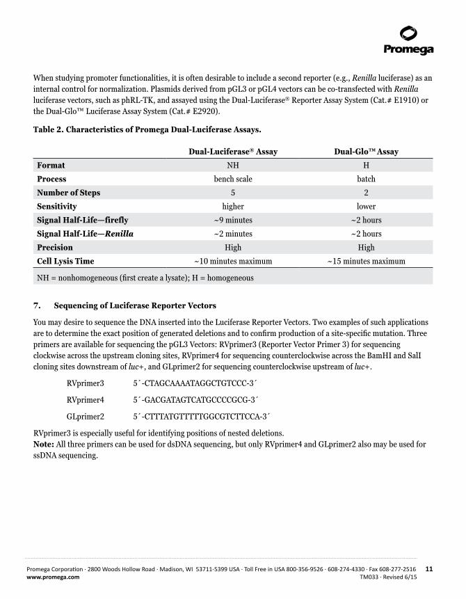
When studying promoter functionalities, it is often desirable to include a second reporter (e.g., Renilla luciferase) as an internal control for normalization. Plasmids derived from pGL3 or pGL4 vectors can be co-transfected with Renilla luciferase vectors, such as phRL-TK, and assayed using the Dual-Luciferase® Reporter Assay System (Cat.# E1910) or the Dual-Glo™ Luciferase Assay System (Cat.# E2920).
Table 2. Characteristics of Promega Dual-Luciferase Assays.
Dual-Luciferase® Assay Dual-Glo™ Assay
Format NH H
Process bench scale batch
Number of Steps 5 2
Sensitivity higher lower
Signal Half-Life—firefly ~9 minutes ~2 hours
Signal Half-Life—Renilla ~2 minutes ~2 hours
Precision High High
Cell Lysis Time ~10 minutes maximum ~15 minutes maximum
NH = nonhomogeneous (first create a lysate); H = homogeneous
7. Sequencing of Luciferase Reporter Vectors
You may desire to sequence the DNA inserted into the Luciferase Reporter Vectors. Two examples of such applications are to determine the exact position of generated deletions and to confirm production of a site-specific mutation. Three primers are available for sequencing the pGL3 Vectors: RVprimer3 (Reporter Vector Primer 3) for sequencing clockwise across the upstream cloning sites, RVprimer4 for sequencing counterclockwise across the BamHI and SalI cloning sites downstream of luc+, and GLprimer2 for sequencing counterclockwise upstream of luc+.
RVprimer3 5´-CTAGCAAAATAGGCTGTCCC-3´
RVprimer4 5´-GACGATAGTCATGCCCCGCG-3´
GLprimer2 5´-CTTTATGTTTTTGGCGTCTTCCA-3´
RVprimer3 is especially useful for identifying positions of nested deletions. Note: All three primers can be used for dsDNA sequencing, but only RVprimer4 and GLprimer2 also may be used for ssDNA sequencing.

12 Promega Corporation · 2800 Woods Hollow Road · Madison, WI 53711-5399 USA · Toll Free in USA 800-356-9526 · 608-274-4330 · Fax 608-277-2516TM033 · Revised 6/15 www.promega.com
8. Appendix
8.A. Common Structural Elements of the pGL3 Luciferase Reporter Vectors
Except for the inclusion of promoters and enhancers, the four pGL3 Luciferase Reporter Vectors are structurally identical. Each plasmid’s distinguishing features are summarized in Section 3. The pGL3 Vectors each contain a high-copy-number prokaryotic origin of replication for maintenance in E. coli, an ampicillin-resistance gene for selection, and a filamentous phage origin of replication (f1 ori) for single-stranded DNA (ssDNA) production. Restriction sites for insertion of DNA fragments are located upstream and downstream of the luciferase gene. Two of the upstream sites (XhoI and BglII) yield cohesive ends compatible with the downstream sites (SalI and BamHI, respectively), allowing the interchange of the DNA insert for rapid analysis of positional effects.
1,400
1,200
1,000
800
600
400
200
0
Improved Expression Level with the pGL3-Control Vector
Construct Transfected
pGL3-ControlVector
pGL2-ControlVector
28.5
1,350
Aver
age
Rela
tive
Ligh
t Uni
ts
Figure 6. Comparison of luciferase activities expressed in HeLa cells transfected with the pGL2-Control and pGL3-Control Reporter Vectors. The expression level of luc+ is dramatically higher with the pGL3-Control Vectors. In repeated experiments with several cell lines, we observed 20- to 100-fold higher luciferase activity from cells transfected with pGL3-Control. Luciferase activity was measured with a Turner Designs luminometer. (Absolute light values and relative expression profiles may vary between different cell types.)
Promega Corporation · 2800 Woods Hollow Road · Madison, WI 53711-5399 USA · Toll Free in USA 800-356-9526 · 608-274-4330 · Fax 608-277-2516 13www.promega.com TM033 · Revised 6/15
80
60
40
20
0
pGL2 Vector Series
Construct Transfected
pGL2-Control
pGL2-Basic
pGL2-Enhancer
pGL2-Promoter
100%
0.08% 1.24%14.5%Av
erag
e Re
lativ
e Li
ght U
nits
Aver
age
Rela
tive
Ligh
t Uni
ts
1,400
1,200
1,000
800
600
400
200
0
pGL3 Vector Series
Construct Transfected
pGL3-Control
pGL3-Basic
pGL3-Enhancer
pGL3-Promoter
100%
0.04% 1.39% 2.56%
0839
MA
11_4
A
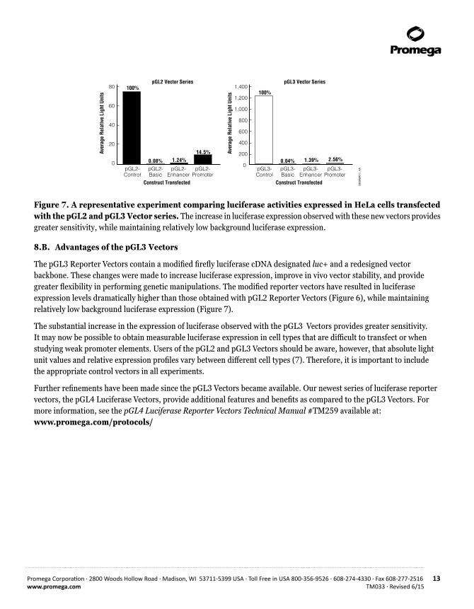
Figure 7. A representative experiment comparing luciferase activities expressed in HeLa cells transfected with the pGL2 and pGL3 Vector series. The increase in luciferase expression observed with these new vectors provides greater sensitivity, while maintaining relatively low background luciferase expression.
8.B. Advantages of the pGL3 Vectors
The pGL3 Reporter Vectors contain a modified firefly luciferase cDNA designated luc+ and a redesigned vector backbone. These changes were made to increase luciferase expression, improve in vivo vector stability, and provide greater flexibility in performing genetic manipulations. The modified reporter vectors have resulted in luciferase expression levels dramatically higher than those obtained with pGL2 Reporter Vectors (Figure 6), while maintaining relatively low background luciferase expression (Figure 7).
The substantial increase in the expression of luciferase observed with the pGL3 Vectors provides greater sensitivity. It may now be possible to obtain measurable luciferase expression in cell types that are difficult to transfect or when studying weak promoter elements. Users of the pGL2 and pGL3 Vectors should be aware, however, that absolute light unit values and relative expression profiles vary between different cell types (7). Therefore, it is important to include the appropriate control vectors in all experiments.
Further refinements have been made since the pGL3 Vectors became available. Our newest series of luciferase reporter vectors, the pGL4 Luciferase Vectors, provide additional features and benefits as compared to the pGL3 Vectors. For more information, see the pGL4 Luciferase Reporter Vectors Technical Manual #TM259 available at: www.promega.com/protocols/
14 Promega Corporation · 2800 Woods Hollow Road · Madison, WI 53711-5399 USA · Toll Free in USA 800-356-9526 · 608-274-4330 · Fax 608-277-2516TM033 · Revised 6/15 www.promega.com
8.C. The pGL3 Vectors luc+ Gene
Modifications that distinguish the luc+ gene from the native luciferase gene generally fall into four categories: i) the C-terminal tripeptide has been removed to eliminate peroxisome targeting of the expressed protein; ii) codon usage was improved for expression in plant and animal cells; iii) two potential sites of N-glycosylation were removed; and iv) several DNA sequence changes were made to disrupt extended palindromes, remove internal restriction sites, and eliminate consensus sequences recognized by genetic regulatory binding proteins, thus helping to ensure that the reporter gene itself is unaffected by spurious host transcriptional signals. (For a detailed description of the modifications to the luc+ gene, see reference 8.)
Four major modifications were made to the vector backbone: i) the SV40 early poly(A) signal has been replaced with the SV40 late poly(A) signal to increase the efficiency of transcription termination and polyadenylation of the luciferase transcripts (9); ii) a synthetic poly(A) and transcriptional pause site (10,11) have been placed upstream of the multiple cloning site to terminate spurious transcription, which may initiate within the vector backbone; iii) the small T intron has been removed to prevent reduced reporter gene expression due to cryptic RNA splicing (12,13); and iv) a Kozak consensus sequence (14) has been inserted to increase the efficiency of translation initiation of the luciferase gene (7; Table 3).
There is a newer luciferase gene available, luc2. The luc2 gene not only shares the same features as luc+, but the sequence was codon-optimized for expression in mammalian cells. For further information about the luc2 gene present in the pGL4 Luciferase Vectors, see Technical Manual #TM259 available at: www.promega.com/protocols/

Promega Corporation · 2800 Woods Hollow Road · Madison, WI 53711-5399 USA · Toll Free in USA 800-356-9526 · 608-274-4330 · Fax 608-277-2516 15www.promega.com TM033 · Revised 6/15
Table 3. Changes Made to the pGL3 Vectors.
Changes Made Purpose of Modification Reference
Modifications made to the luciferase gene (luc to luc+).
Changes eliminate peroxisome targeting of expressed protein, eliminate consensus binding sequences for various genetic regulatory proteins, improve codon usage for mammalian and plant cells, and provide convenient restriction sites.
(8)
A unique NcoI site created at 5´ end of luc+ gene. NcoI sites removed from SV40 enhancer and promoter regions.
Ability to create N-terminal gene fusions with luc+ using unique NcoI site.
Intron from SV40 small T antigen removed.
Intron from SV40 small T antigen can reduce expression when placed 3´ of certain genes due to cryptic splicing.
(12,13)
Poly(A) site for back-ground reduction changed from SV40 early site to a synthetic poly(A) and transcriptional pause site.
Avoids possible recombination between two SV40 poly(A) sequences in the same plasmid.
(9,10)
Poly(A) signal for luc+ changed from early to late SV40 poly(A) signal.
Late SV40 poly(A) signal is more efficient than early SV40 poly(A).
(7)
Kozak consensus sequence created immediately 5´ of the luc+ gene.
Provides optimal translation efficiency. (14)
Unique XbaI site created just downstream of the luc+ gene.
User convenience; facilitates subcloning of the luc+ gene.
SmaI site moved to internal position in multiple cloning region.
User convenience; blunt-ended inserts can now be cleaved on either side by restriction endonucleases.
16 Promega Corporation · 2800 Woods Hollow Road · Madison, WI 53711-5399 USA · Toll Free in USA 800-356-9526 · 608-274-4330 · Fax 608-277-2516TM033 · Revised 6/15 www.promega.com
8.D. Mapping Genetic Elements Located Within DNA Fragments
The locations of functional elements within a DNA fragment are often determined by making a set of unidirectional nested deletions following the method of Henikoff (15) and then assaying for changes in biological activity. This method takes advantage of the unique properties of Exonuclease III (Exo III), which will digest 5´ overhangs but not 3´ overhangs or α-phosphorothioate nucleotide filled-in overhangs. Nested deletions of an insert DNA can be made directly in the pGL3 family of Reporter Vectors using this method, eliminating the need for subcloning steps. The multiple cloning region of the pGL3 Vectors contains upstream KpnI and SacI restriction sites, which can be used to generate the 3´ overhangs resistant to Exo III (Figures 1–5). After treatment with Exo III, S1 nuclease is added to remove the resulting ssDNA overhangs, and T4 DNA ligase is added to reclose the vectors. Deletion clones can be screened by gel electrophoresis of miniprep DNA, and the precise deletion endpoints within the promoter region can be determined by DNA sequencing using primers designed for the Luciferase Reporter Vectors.
8.E. Composition of Buffers and Solutions
M-9 plates (1 liter) 15g agarose
Add 15g agarose to 750ml water and autoclave. Cool to 50°C. Add: 2.0ml 1M MgSO4
0.1ml 1M CaCl2
10.0ml 20% glucose (filter sterilized) 1.0ml 1M thiamine-HCl 200ml 5X M-9 salts
5X M-9 salts (1 liter) 34g Na2HPO4
15g KH2PO4
2.5g NaCl 5g NH4Cl
Dissolve in deionized water. Divide into 200ml aliquots and autoclave.

Promega Corporation · 2800 Woods Hollow Road · Madison, WI 53711-5399 USA · Toll Free in USA 800-356-9526 · 608-274-4330 · Fax 608-277-2516 17www.promega.com TM033 · Revised 6/15
8.F. References
1. Sambrook, J. et al. (1989) Molecular Cloning, A Laboratory Manual, Cold Spring Harbor Press, Cold Spring Harbor, NY.
2. Schenborn, E. and Goiffon, V. (1991) Optimization of Transfectam®-mediated transfection using a luciferase reporter system. Promega Notes 33, 8–11.
3. Cullen, B.R. (1987) Use of eukaryotic expression technology in the functional analysis of cloned genes. Methods Enzymol. 152, 684–704.
4. Ausubel, F.M. et al. (1988) Current Protocols in Molecular Biology, John Wiley and Sons, NY.
5. Rosenthal, N. (1987) Identification of regulatory elements of cloned genes with functional assays. Methods Enzymol. 152, 704–20.
6. Hawkins, E., Butler, B. and Wood, K.V. (2000) Bright-Glo™ and Steady-Glo™ Luciferase Assay Systems: Reagents for academic and industrial applications. Promega Notes 75, 3–6.
7. Groskreutz, D.J. et al. (1995) Increased expression and convenience with the new pGL3 Luciferase Reporter Vectors. Promega Notes 50, 2–8.
8. Sherf, B.A. and Wood, K.V. (1994) Firefly luciferase engineered for improved genetic reporting. Promega Notes 49, 14–21.
9. Carswell, S. and Alwine, J.C. (1989) Efficiency of utilization of the simian virus 40 late polyadenylation site: Effects of upstream sequences. Mol. Cell. Biol. 9, 4248–58.
10. Levitt, N. et al. (1989) Definition of an efficient synthetic poly(A) site. Genes and Dev. 3, 1019–25.
11. Enriquez-Harris, P. et al. (1991) A pause site for RNA polymerase II is associated with termination of transcription. EMBO J. 10, 1833–42.
12. Evans, M.J. and Scarpulla, R.C. (1989) Introns in the 3´ untranslated region can inhibit chimeric CAT and beta-galactosidase gene expression. Gene 84, 135–42.
13. Huang, M.T.F. and Gorman, C.M. (1990) The simian virus 40 small-t intron, present in many common expression vectors, leads to aberrant splicing. Mol. Cell. Biol. 10, 1805–10.
14. Kozak, M. (1989) The scanning model for translation: An update. J. Cell Biol. 108, 229–41.
15. Henikoff, S. (1987) Unidirectional digestion with exonuclease III in DNA sequence analysis. Methods Enzymol. 155, 156.
18 Promega Corporation · 2800 Woods Hollow Road · Madison, WI 53711-5399 USA · Toll Free in USA 800-356-9526 · 608-274-4330 · Fax 608-277-2516TM033 · Revised 6/15 www.promega.com
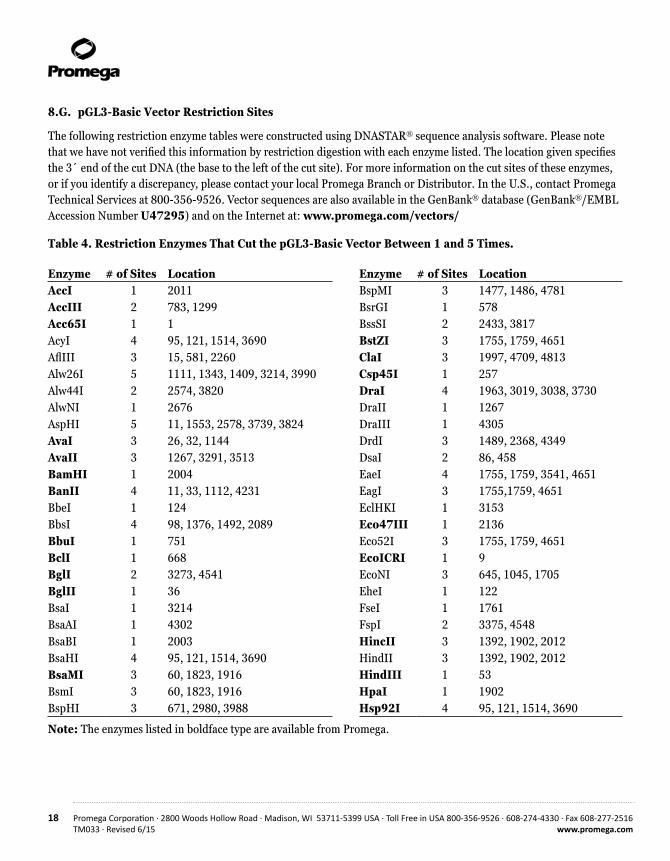
8.G. pGL3-Basic Vector Restriction Sites
The following restriction enzyme tables were constructed using DNASTAR® sequence analysis software. Please note that we have not verified this information by restriction digestion with each enzyme listed. The location given specifies the 3´ end of the cut DNA (the base to the left of the cut site). For more information on the cut sites of these enzymes, or if you identify a discrepancy, please contact your local Promega Branch or Distributor. In the U.S., contact Promega Technical Services at 800-356-9526. Vector sequences are also available in the GenBank® database (GenBank®/EMBL Accession Number U47295) and on the Internet at: www.promega.com/vectors/
Table 4. Restriction Enzymes That Cut the pGL3-Basic Vector Between 1 and 5 Times.
Enzyme # of Sites Location Enzyme # of Sites LocationAccI 1 2011 BspMI 3 1477, 1486, 4781AccIII 2 783, 1299 BsrGI 1 578 Acc65I 1 1 BssSI 2 2433, 3817 AcyI 4 95, 121, 1514, 3690 BstZI 3 1755, 1759, 4651AflIII 3 15, 581, 2260 ClaI 3 1997, 4709, 4813 Alw26I 5 1111, 1343, 1409, 3214, 3990 Csp45I 1 257Alw44I 2 2574, 3820 DraI 4 1963, 3019, 3038, 3730AlwNI 1 2676 DraII 1 1267 AspHI 5 11, 1553, 2578, 3739, 3824 DraIII 1 4305AvaI 3 26, 32, 1144 DrdI 3 1489, 2368, 4349 AvaII 3 1267, 3291, 3513 DsaI 2 86, 458BamHI 1 2004 EaeI 4 1755, 1759, 3541, 4651BanII 4 11, 33, 1112, 4231 EagI 3 1755,1759, 4651BbeI 1 124 EclHKI 1 3153 BbsI 4 98, 1376, 1492, 2089 Eco47III 1 2136 BbuI 1 751 Eco52I 3 1755, 1759, 4651BclI 1 668 EcoICRI 1 9 BglI 2 3273, 4541 EcoNI 3 645, 1045, 1705 BglII 1 36 EheI 1 122BsaI 1 3214 FseI 1 1761BsaAI 1 4302 FspI 2 3375, 4548 BsaBI 1 2003 HincII 3 1392, 1902, 2012BsaHI 4 95, 121, 1514, 3690 HindII 3 1392, 1902, 2012 BsaMI 3 60, 1823, 1916 HindIII 1 53BsmI 3 60, 1823, 1916 HpaI 1 1902BspHI 3 671, 2980, 3988 Hsp92I 4 95, 121, 1514, 3690
Note: The enzymes listed in boldface type are available from Promega.
Promega Corporation · 2800 Woods Hollow Road · Madison, WI 53711-5399 USA · Toll Free in USA 800-356-9526 · 608-274-4330 · Fax 608-277-2516 19www.promega.com TM033 · Revised 6/15
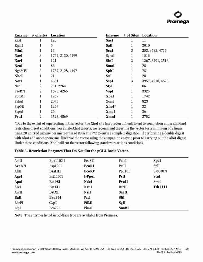
Enzyme # of Sites Location Enzyme # of Sites LocationKasI 1 120 SacI 1 11KpnI 1 5 SalI 1 2010MluI 1 15 ScaI 3 253, 3633, 4716NaeI 3 1759, 2130, 4199 SgrAI 1 1516NarI 1 121 SinI 3 1267, 3291, 3513NcoI 1 86 SmaI 1 28NgoMIV 3 1757, 2128, 4197 SphI 1 751NheI 1 21 SrfI 1 28NotI 1 4651 SspI 3 3957, 4510, 4625NspI 2 751, 2264 StyI 1 86 PaeR7I 2 1675, 4266 VspI 1 3325 PpuMI 1 1267 XbaI 1 1742 PshAI 1 2075 XcmI 1 823Psp5II 1 1267 XhoI* 1 32PspAI 1 26 XmaI 1 26 PvuI 2 3523, 4569 XmnI 1 3752
*Due to the extent of supercoiling in this vector, the XhoI site has proven difficult to cut to completion under standard restriction digest conditions. For single XhoI digests, we recommend digesting the vector for a minimum of 2 hours using 20 units of enzyme per microgram of DNA at 37°C to ensure complete digestion. If performing a double digest with XhoI and another enzyme, linearize the vector using the companion enzyme prior to carrying out the XhoI digest. Under these conditions, XhoI will cut the vector following standard reactions conditions.
Table 5. Restriction Enzymes That Do Not Cut the pGL3-Basic Vector.
AatIIAccB7IAflIIAgeIApaIAscIAvrIIBalIBbrPIBlpI
Bpu1102 IBsp120IBssHIIBst1107IBst98IBstEIIBstXIBsu36ICspIEco72I
Eco81IEcoRIEcoRVI-PpoINdeINruINsiIPacIPflMIPinAI
PmeIPmlIPpu10IPstIPvuIIRsrIISacIISfiISgfISnaBI
SpeISplISse8387IStuISwaITth111I
Note: The enzymes listed in boldface type are available from Promega.

20 Promega Corporation · 2800 Woods Hollow Road · Madison, WI 53711-5399 USA · Toll Free in USA 800-356-9526 · 608-274-4330 · Fax 608-277-2516TM033 · Revised 6/15 www.promega.com
8.G. pGL3-Basic Vector Restriction Sites (continued)
Table 6. Restriction Enzymes That Cut the pGL3-Basic Vector 6 or More Times.
AciIAluIBanIBbvI BsaOIBsaJIBsp1286I BsrIBsrSIBst71IBstOI
BstUI CfoI Cfr10IDdeI DpnIDpnIIEarIFnu4HIFokI HaeII HaeIII
HgaI HhaIHinfIHpaIIHphI Hsp92II MaeI MaeII MaeIII MboI MboII
MnlI MseI MspI MspA1I NciI NdeIINlaIII NlaIVPleIRsaI Sau3AI
Sau96I ScrFI SfaNITaqITfiI Tru9I XhoII
Note: The enzymes listed in boldface type are available from Promega.
8.H. pGL3-Enhancer Vector Restriction Sites
The following restriction enzyme tables were constructed using DNASTAR® sequence analysis software. Please note that we have not verified this information by restriction digestion with each enzyme listed. The location given specifies the 3´ end of the cut DNA (the base to the left of the cut site). For more information on the cut sites of these enzymes, or if you identify a discrepancy, please contact your local Promega Branch or Distributor. In the U.S., contact Promega Technical Services at 800-356-9526. Vector sequences are also available in the GenBank® database (GenBank®/EMBL Accession Number U47297) and on the Internet at: www.promega.com/vectors/
Table 7. Restriction Enzymes that cut the pGL3-Enhancer Vector Between 1 and 5 Times.
Enzyme # of Sites Location Enzyme # of Sites LocationAccI 1 2257 BbsI 4 98, 1376, 1492, 2335 AccIII 2 783,1299 BbuI 3 751, 2108, 2180 Acc65I 1 1 BclI 1 668AcyI 4 95, 121, 1514, 3936 BglI 2 3519, 4787 AflIII 3 15, 581 ,2506 BglII 1 36Alw26I 5 1111, 1343, 1409, 3460, 4236 BsaI 1 3460 Alw44I 2 2820, 4066 BsaAI 1 4548 AlwNI 1 2922 BsaBI 1 2003 AspHI 5 11, 1553, 2824, 3985, 4070 BsaHI 4 95, 121, 1514, 3936 AvaI 3 26, 32, 1144 BsaMI 3 60, 1823, 1916 AvaII 3 1267, 3537, 3759 BsmI 3 60, 1823, 1916 BamHI 1 2250 BspHI 3 671, 3226, 4234 BanII 4 11, 33, 1112, 4477 BspMI 3 1477, 1486, 5027 BbeI 1 124 BsrGI 1 578

Promega Corporation · 2800 Woods Hollow Road · Madison, WI 53711-5399 USA · Toll Free in USA 800-356-9526 · 608-274-4330 · Fax 608-277-2516 21www.promega.com TM033 · Revised 6/15
Enzyme # of Sites Location Enzyme # of Sites LocationBssSI 2 2679, 4063 NcoI 1 86 BstZI 3 1755, 1759, 4897 NgoMIV 3 1757, 2374, 4443 ClaI 3 1997, 4955, 5059 NheI 1 21 Csp45I 1 257 NotI 1 4897 DraI 4 1963, 3265, 3284, 3976 NsiI 2 2106, 2178DraII 1 1267 NspI 4 751, 2108, 2180, 2510 DraIII 1 4551 PaeR7I 2 1675, 4512 DrdI 3 1489, 2614, 4595 Ppu10I 2 2102, 2174DsaI 2 86, 458 PpuMI 1 1267 EaeI 4 1755, 1759, 3787, 4897 PshAI 1 2321EagI 3 1755, 1759, 4897 Psp5II 1 1267EclHKI 1 3399 PspAI 1 26Eco47III 1 2382 PvuI 2 3769, 4815Eco52I 3 1755, 1759, 4897 SacI 1 11 EcoICRI 1 9 SalI 1 2256 EcoNI 3 645, 1045, 1705 ScaI 3 253, 3879, 4962 EheI 1 122 SgrAI 1 1516FseI 1 1761 SinI 3 1267, 3537, 3759 FspI 2 3621, 4794 SmaI 1 28 HincII 3 1392, 1902, 2258 SphI 3 751, 2108, 2180 HindII 3 1392, 1902, 2258 SrfI 1 28HindIII 1 53 SspI 3 4203, 4756, 4871HpaI 1 1902 StyI 1 86Hsp92I 4 95, 121, 1514, 3936 VspI 1 3571KasI 1 120 XbaI 1 1742 KpnI 1 5 XcmI 1 823 MluI 1 15 XhoI* 1 32 NaeI 3 1759, 2376, 4445 XmaI 1 26 NarI 1 121 XmnI 1 3998
*Due to the extent of supercoiling in this vector, the XhoI site has proven difficult to cut to completion under standard restriction digest conditions. For single XhoI digests, we recommend digesting the vector for a minimum of 2 hours using 20 units of enzyme per microgram of DNA at 37°C to ensure complete digestion. If performing a double digest with XhoI and another enzyme, linearize the vector using the companion enzyme prior to carrying out the XhoI digest. Under these conditions, XhoI will cut the vector following standard reactions conditions.
Note: The enzymes listed in boldface type are available from Promega.

22 Promega Corporation · 2800 Woods Hollow Road · Madison, WI 53711-5399 USA · Toll Free in USA 800-356-9526 · 608-274-4330 · Fax 608-277-2516TM033 · Revised 6/15 www.promega.com
8.H. pGL3-Enhancer Vector Restriction Sites (continued)
Table 8. Restriction Enzymes That Do Not Cut the pGL3-Enhancer Vector.
AatIIAccB7IAflIIAgeIApaIAscIAvrIIBalIBbrPI
BlpIBpu1102IBsp120IBssHIIBst1107IBst98IBstEIIBstXIBsu36I
CspIEco72IEco81IEcoRIEcoRVI-PpoINdeINruIPacI
PflMIPinAIPmeIPmlIPstIPvuIIRsrIISacIISfiI
SgfISnaBISpeISplISse8387IStuISwaITth111I
Note: The enzymes listed in boldface type are available from Promega.
Table 9. Restriction Enzymes That Cut the pGL3-Enhancer Vector 6 or More Times.
AciIAluIBanI BbvIBsaOIBsaJIBsp1286IBsrI BsrSI Bst71I BstOI
BstUI CfoICfr10IDdeIDpnI DpnII EarI Fnu4HI FokI HaeIIHaeIII
HgaIHhaIHinfIHpaIIHphIHsp92II MaeIMaeII MaeIIIMboI MboII
MnlIMseIMspI MspA1I NciI NdeII NlaIIINlaIVPleIRsaISau3AI
Sau96IScrFISfaNITaqITfiI Tru9IXhoII
Note: The enzymes listed in boldface type are available from Promega.
Promega Corporation · 2800 Woods Hollow Road · Madison, WI 53711-5399 USA · Toll Free in USA 800-356-9526 · 608-274-4330 · Fax 608-277-2516 23www.promega.com TM033 · Revised 6/15
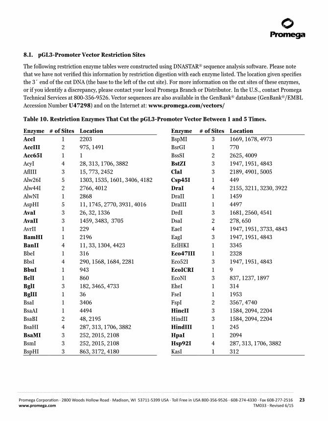
8.I. pGL3-Promoter Vector Restriction Sites
The following restriction enzyme tables were constructed using DNASTAR® sequence analysis software. Please note that we have not verified this information by restriction digestion with each enzyme listed. The location given specifies the 3´ end of the cut DNA (the base to the left of the cut site). For more information on the cut sites of these enzymes, or if you identify a discrepancy, please contact your local Promega Branch or Distributor. In the U.S., contact Promega Technical Services at 800-356-9526. Vector sequences are also available in the GenBank® database (GenBank®/EMBL Accession Number U47298) and on the Internet at: www.promega.com/vectors/
Table 10. Restriction Enzymes That Cut the pGL3-Promoter Vector Between 1 and 5 Times.
Enzyme # of Sites Location Enzyme # of Sites LocationAccI 1 2203 BspMI 3 1669, 1678, 4973AccIII 2 975, 1491 BsrGI 1 770Acc65I 1 1 BssSI 2 2625, 4009 AcyI 4 28, 313, 1706, 3882 BstZI 3 1947, 1951, 4843 AflIII 3 15, 773, 2452 ClaI 3 2189, 4901, 5005 Alw26I 5 1303, 1535, 1601, 3406, 4182 Csp45I 1 449 Alw44I 2 2766, 4012 DraI 4 2155, 3211, 3230, 3922AlwNI 1 2868 DraII 1 1459 AspHI 5 11, 1745, 2770, 3931, 4016 DraIII 1 4497 AvaI 3 26, 32, 1336 DrdI 3 1681, 2560, 4541 AvaII 3 1459, 3483, 3705 DsaI 2 278, 650AvrII 1 229 EaeI 4 1947, 1951, 3733, 4843 BamHI 1 2196 EagI 3 1947, 1951, 4843 BanII 4 11, 33, 1304, 4423 EclHKI 1 3345 BbeI 1 316 Eco47III 1 2328BbsI 4 290, 1568, 1684, 2281 Eco52I 3 1947, 1951, 4843 BbuI 1 943 EcoICRI 1 9 BclI 1 860 EcoNI 3 837, 1237, 1897 BglI 3 182, 3465, 4733 EheI 1 314 BglII 1 36 FseI 1 1953BsaI 1 3406 FspI 2 3567, 4740 BsaAI 1 4494 HincII 3 1584, 2094, 2204BsaBI 2 48, 2195 HindII 3 1584, 2094, 2204 BsaHI 4 287, 313, 1706, 3882 HindIII 1 245BsaMI 3 252, 2015, 2108 HpaI 1 2094BsmI 3 252, 2015, 2108 Hsp92I 4 287, 313, 1706, 3882 BspHI 3 863, 3172, 4180 KasI 1 312

24 Promega Corporation · 2800 Woods Hollow Road · Madison, WI 53711-5399 USA · Toll Free in USA 800-356-9526 · 608-274-4330 · Fax 608-277-2516TM033 · Revised 6/15 www.promega.com
8.I. pGL3-Promoter Vector Restriction Sites (continued)
Table 10. Restriction Enzymes That Cut the pGL3-Promoter Vector Between 1 and 5 Times (continued).
Enzyme # of Sites Location Enzyme # of Sites LocationKpnI 1 5 ScaI 3 445, 3825, 4908 MluI 1 15 SfiI 1 182 NaeI 3 1951, 2322, 4391 SgrAI 1 1708 NarI 1 313 SinI 3 1459, 3483, 3705 NcoI 1 278 SmaI 1 28 NgoMIV 3 1949, 2320, 4389 SphI 1 943 NheI 1 21 SrfI 1 28 NotI 1 4843 SspI 3 4149, 4702, 4817NspI 2 943, 2456 StuI 1 228PaeR7I 2 1867, 4458 StyI 2 229, 278 PpuMI 1 1459 VspI 1 3517 PshAI 1 2267 XbaI 1 1934 Psp5II 1 1459 XcmI 1 1015 PspAI 1 26 XhoI* 1 32 PvuI 2 3715, 4761 XmaI 1 26 SacI 1 11 XmnI 1 3944SalI 1 2202
*Due to the extent of supercoiling in this vector, the XhoI site has proven difficult to cut to completion under standard restriction digest conditions. For single XhoI digests, we recommend digesting the vector for a minimum of 2 hours using 20 units of enzyme per microgram of DNA at 37°C to ensure complete digestion. If performing a double digest with XhoI and another enzyme, linearize the vector using the companion enzyme prior to carrying out the XhoI digest. Under these conditions, XhoI will cut the vector following standard reactions conditions.
Table 11. Restriction Enzymes That Do Not Cut the pGL3-Promoter Vector.
AatIIAccB7IAflIIAgeIApaIAscIBalIBbrPIBlpI
Bpu1102IBsp120IBssHIIBst1107IBst98IBstEIIBstXIBsu36ICspI
Eco72IEco81IEcoRIEcoRVI-PpoINdeINruINsiIPacI
PflMIPinAIPmeIPmlIPpu10IPstIPvuIIRsrIISacII
SgfISnaBISpeISplISse8387ISwaITth111I
Note: The enzymes listed in boldface type are available from Promega.

Promega Corporation · 2800 Woods Hollow Road · Madison, WI 53711-5399 USA · Toll Free in USA 800-356-9526 · 608-274-4330 · Fax 608-277-2516 25www.promega.com TM033 · Revised 6/15
Table 12. Restriction Enzymes That Cut the pGL3-Promoter Vector 6 or More Times.
AciI AluI BanIBbvIBsaOIBsaJIBsp1286IBsrIBsrSIBst71IBstOI
BstUI CfoICfr10I DdeIDpnIDpnII EarIFnu4HIFokIHaeII HaeIII
HgaI HhaIHinfI HpaIIHphIHsp92II MaeIMaeII MaeIIIMboI MboII
MnlIMseIMspI MspA1INciI NdeIINlaIII NlaIVPleIRsaI Sau3AI
Sau96I ScrFI SfaNI TaqI TfiITru9I XhoII
Note: The enzymes listed in boldface type are available from Promega.

26 Promega Corporation · 2800 Woods Hollow Road · Madison, WI 53711-5399 USA · Toll Free in USA 800-356-9526 · 608-274-4330 · Fax 608-277-2516TM033 · Revised 6/15 www.promega.com
8.J. pGL3-Control Vector Restriction Sites
The following restriction enzyme tables were constructed using DNASTAR® sequence analysis software. Please note that this information has not been verified by restriction digestion with each enzyme listed. The location given specifies the 3´ end of the cut DNA (the base to the left of the cut site). For more information on the cut sites of these enzymes, or if you identify a discrepancy, please contact your local Promega Branch or Distributor. Vector sequences are also available in the GenBank® database (GenBank®/EMBL Accession Number U47296) and on the Internet at: www.promega.com/vectors/
Table 13. Restriction Enzymes That Cut the pGL3-Control Vector Between 1 and 5 Times.
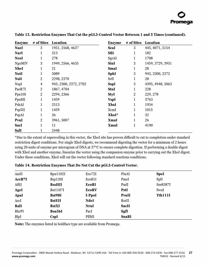
Enzyme # of Sites Location Enzyme # of Sites LocationAccI 1 2449 BsrGI 1 770AccIII 2 975, 1491 BssSI 2 2871, 4255 Acc65I 1 1 BstZI 3 1947, 1951, 5089 AcyI 4 287, 313, 1706, 4128 ClaI 3 2189, 5147, 5251AflIII 3 15, 773, 2698 Csp45I 1 449Alw26I 5 1303, 1535, 1601, 3652, 4428 DraI 4 2155, 3457, 3476, 4168Alw44I 2 3012, 4258 DraII 1 1459 AlwNI 1 3114 DraIII 1 4743 AspHI 5 11, 1745, 3016, 4177, 4262 DrdI 3 1681, 2806, 4787 AvaI 3 26, 32, 1336 DsaI 2 278, 650 AvaII 3 1459, 3729, 3951 EaeI 4 1947, 1951, 3979, 5089 AvrII 1 229 EagI 3 1947, 1951, 5089 BamHI 1 2442 EclHKI 1 3591 BanII 4 11, 33, 1304, 4669 Eco47III 1 2574 BbeI 1 316 Eco52I 3 1947, 1951, 5089 BbsI 4 290, 1568, 1684, 2527 EcoICRI 1 9 BbuI 3 943, 2300, 2372 EcoNI 3 837, 1237, 1897 BclI 1 860 EheI 1 314BglI 3 182, 3711, 4979 FseI 1 1953BglII 1 36 FspI 2 3813, 4986BsaI 1 3652 HincII 3 1584, 2094, 2450BsaAI 1 4740 HindII 3 1584, 2094, 2450BsaBI 2 48, 2195 HindIII 1 245BsaHI 4 287, 313, 1706, 4128 HpaI 1 2094BsaMI 3 252, 2015, 2108 Hsp92I 4 287, 313, 1706, 4128BsmI 3 252, 2015, 2108 KasI 1 312BspHI 3 863, 3418, 4426 KpnI 1 5BspMI 3 1669, 1678, 5219 MluI 1 15
Note: The enzymes listed in boldface type are available from Promega.
Promega Corporation · 2800 Woods Hollow Road · Madison, WI 53711-5399 USA · Toll Free in USA 800-356-9526 · 608-274-4330 · Fax 608-277-2516 27www.promega.com TM033 · Revised 6/15
Table 13. Restriction Enzymes That Cut the pGL3-Control Vector Between 1 and 5 Times (continued).
Enzyme # of Sites Location Enzyme # of Sites LocationNaeI 3 1951, 2568, 4637 ScaI 3 445, 4071, 5154 NarI 1 313 SfiI 1 182 NcoI 1 278 SgrAI 1 1708NgoMIV 3 1949, 2566, 4635 SinI 3 1459, 3729, 3951 NheI 1 21 SmaI 1 28 NotI 1 5089 SphI 3 943, 2300, 2372NsiI 2 2298, 2370 SrfI 1 28NspI 4 943, 2300, 2372, 2702 SspI 3 4395, 4948, 5063 PaeR7I 2 1867, 4704 StuI 1 228 Ppu10I 2 2294, 2366 StyI 2 229, 278PpuMI 1 1459 VspI 1 3763 PshAI 1 2513 XbaI 1 1934 Psp5II 1 1459 XcmI 1 1015 PspAI 1 26 XhoI* 1 32 PvuI 2 3961, 5007 XmaI 1 26 SacI 1 11 XmnI 1 4190SalI 1 2448
*Due to the extent of supercoiling in this vector, the XhoI site has proven difficult to cut to completion under standard restriction digest conditions. For single XhoI digests, we recommend digesting the vector for a minimum of 2 hours using 20 units of enzyme per microgram of DNA at 37°C to ensure complete digestion. If performing a double digest with XhoI and another enzyme, linearize the vector using the companion enzyme prior to carrying out the XhoI digest. Under these conditions, XhoI will cut the vector following standard reactions conditions.
Table 14. Restriction Enzymes That Do Not Cut the pGL3-Control Vector.
AatIIAccB7IAflIIAgeIApaIAscIBalIBbrPIBlpI
Bpu1102IBsp120IBssHIIBst1107IBst98IBstEIIBstXIBsu36ICspI
Eco72IEco81IEcoRIEcoRVI-PpoINdeINruIPacIPflMI
PinAIPmeIPmlIPstIPvuIIRsrIISacIISgfISnaBI
SpeISplISse8387ISwaITth111I
Note: The enzymes listed in boldface type are available from Promega.

28 Promega Corporation · 2800 Woods Hollow Road · Madison, WI 53711-5399 USA · Toll Free in USA 800-356-9526 · 608-274-4330 · Fax 608-277-2516TM033 · Revised 6/15 www.promega.com
8.J. pGL3-Control Vector Restriction Sites (continued)
Table 15. Restriction Enzymes That Cut the pGL3-Control Vector 6 or More Times.
AciI AluIBanIBbvIBsaOI BsaJI Bsp1286IBsrIBsrSIBst71I
BstOI BstUICfoI Cfr10IDdeI DpnI EarI Fnu4HIFokIHaeII
HaeIII HgaIHhaI HinfIHpaII HphIHsp92IIMaeIMaeII MaeIII
MboIMboIIMnlIMseI MspIMspA1INciINdeIINlaIIINlaIV
PleIRsaISau3AISau96I ScrFI SfaNI TaqITfiITru9IXhoII
Note: The enzymes listed in boldface type are available from Promega.
8.K. Related Products
Product Size Cat.#RVprimer3 (clockwise) 2µg E4481
RVprimer4 (counter clockwise) 2µg E4491
PureYield™ Plasmid Midiprep System 100 preps A2495
Luciferase Assay Systems
Product Size Cat.#Luciferase Assay System 100 assays E1500
Bright-Glo™ Luciferase Assay System 10ml E2610
Steady-Glo® Luciferase Assay System 10ml E2510
Dual-Luciferase® Reporter Assay System 100 assays E1910
Dual-Glo® Luciferase Assay System 10ml E2920
ONE-Glo™ Luciferase Assay System 10ml E6110
Available in additional sizes.

Promega Corporation · 2800 Woods Hollow Road · Madison, WI 53711-5399 USA · Toll Free in USA 800-356-9526 · 608-274-4330 · Fax 608-277-2516 29www.promega.com TM033 · Revised 6/15
Luminometers
Product Size Cat.#GloMax® 96 Microplate Luminometer 1 each E6501
GloMax® 20/20 Luminometer 1 each E5311
Available with single or dual injectors.
pGL4 Luciferase Vectors
Product Size Cat.#pGL4.10[luc2] Vector 20µg E6651
pGL4.11[luc2P] Vector 20µg E6661
pGL4.12[luc2CP] Vector 20µg E6671
pGL4.13[luc2/SV40] Vector 20µg E6681
pGL4.14[luc2/Hygro] Vector 20µg E6691
pGL4.17[luc2/Neo] Vector 20µg E6721
pGL4.20[luc2/Puro] Vector 20µg E6751
pGL4.23[luc2/minP] Vector 20µg E8411
pGL4.26[luc2/minP/Hygro] Vector 20µg E8441
pGL4.29[luc2P/CRE/Hygro] Vector 20µg E8471
pGL4.30[luc2P/NFAT-RE/Hygro] Vector 20µg E8481
pGL4.31[luc2P/Gal4UAS/Hygro] Vector 20µg C9351
The complete listing of pGL4 Luciferase Vectors can be found at: www.promega.com/pgl4/
9. Summary of Changes
The following change was made to the 6/15 revision of this document:
1. The patent information was updated to remove expired statements.

30 Promega Corporation · 2800 Woods Hollow Road · Madison, WI 53711-5399 USA · Toll Free in USA 800-356-9526 · 608-274-4330 · Fax 608-277-2516TM033 · Revised 6/15 www.promega.com
(a)READ THIS FIRST BEFORE OPENING PRODUCT
BY USE OF THIS PRODUCT, RESEARCHER AGREES TO BE BOUND BY THE TERMS OF THIS LIMITED USE LABEL LICENSE. If the researcher is not willing to accept the terms of this label license, and the product is unused, Promega will accept return of the unused product and provide the researcher with a full refund.
Researchers may use this product for research use only, no commercial use is allowed. “Commercial use” means any and all uses of this product and derivatives by a party for money or other consideration and may include but is not limited to use in: (1) product manufacture; and (2) to provide a service, information or data; and/or resale of the product or its derivatives, whether or not such product or derivatives are resold for use in research. Researchers shall have no right to modify or otherwise create variations of the nucleotide sequence of the luciferase gene except that researchers may: (1) create fused gene sequences provided that the coding sequence of the resulting luciferase gene has no more than four deoxynucleotides missing at the affected terminus compared to the intact luciferase gene sequence, and (2) insert and remove nucleic acid sequences in splicing research predicated on the inactivation or reconstitution of the luminescence of the encoded luciferase. No other use or transfer of this product or derivatives is authorized without the prior express written consent of Promega. In addition, researchers must either: (1) use luminescent assay reagents purchased from Promega for all determinations of luminescence activity of this product and its derivatives; or (2) contact Promega to obtain a license for use of the product and its derivatives. Researchers may transfer derivatives to others for research use provided that at the time of transfer a copy of this label license is given to the recipients and recipients agree to be bound by the terms of this label license. With respect to any uses outside this label license, including any diagnostic, therapeutic or prophylactic uses, please contact Promega for supply and licensing information. PROMEGA MAKES NO REPRESENTATIONS OR WARRANTIES OF ANY KIND, EITHER EXPRESSED OR IMPLIED, INCLUDING FOR MERCHANTABILITY OR FITNESS FOR A PARTICULAR PURPOSE WITH REGARDS TO THE PRODUCT. The terms of this label license shall be governed under the laws of the State of Wisconsin, USA. This label license relates to Promega patents and/or patent applications on improvements to the luciferase gene.
© 1994–2015 Promega Corporation. All Rights Reserved.
Dual-Glo, Dual-Luciferase, GloMax, ProFection, Steady-Glo, Transfectam and Wizard are registered trademarks of Promega Corporation. Bright-Glo, ONE-Glo, PureYield, Tfx and TransFast are trademarks of Promega Corporation.
DNASTAR is a registered trademark of of DNASTAR, Inc. GenBank is a registered trademark of the U.S. Dept. of Health and Human Services.
Products may be covered by pending or issued patents or may have certain limitations. Please visit our Web site for more information.
All prices and specifications are subject to change without prior notice.
Product claims are subject to change. Please contact Promega Technical Services or access the Promega online catalog for the most up-to-date information on Promega products.