
Pectin degradation by Botrytis cinerea:
recognition of endo-polygalacturonases by an Arabidopsis
receptor and utilization of D-galacturonic acid
Lisha Zhang
Thesis committee
Promoter
Prof. dr. ir. P.J.G.M. de Wit
Professor of Phytopathology
Wageningen University
Co-promoter
Dr. J.A.L. van Kan
Assistant professor, Laboratory of Phytopathology
Wageningen University
Other members
Prof. dr. ir. J. Bakker, Wageningen University
Prof. dr. ing. J.J.B. Keurentjes, Wageningen University / University of
Amsterdam
Dr. A.F.J.M. van den Ackerveken, Utrecht University
Dr. A.F.J. Ram, Leiden University
This research was conducted under the auspices of the Graduate School of
Experimental Plant Sciences
Pectin degradation by Botrytis cinerea:
recognition of endo-polygalacturonases by an Arabidopsis
receptor and utilization of D-galacturonic acid
Lisha Zhang
Thesis submitted in fulfilment of the requirements for the degree of doctor
at Wageningen University by the authority of the Rector Magnificus
Prof. dr. M.J. Kropff, in the presence of the
Thesis Committee appointed by the Academic Board to be defended in public
on Wednesday 5 June 2013 at 1.30 p.m. in the Aula

Lisha Zhang
Pectin degradation by Botrytis cinerea: recognition of endo-
polygalacturonases by an Arabidopsis receptor and utilization of D-
galacturonic acid, 192 pages
PhD thesis, Wageningen University, Wageningen, NL (2013)
With references, with summaries in English and Dutch
ISBN 978-94-6173-540-9
Contents
Chapter 1 General introduction 7
Chapter 2 Fungal endo-polygalacturonases are recognized as
MAMPs in Arabidopsis by the Receptor-Like Protein RBPG1 23
Chapter 3 The D-galacturonic acid catabolism in Botrytis cinerea 59
Chapter 4 Botrytis cinerea mutants deficient in D-galacturonic acid
catabolism have a perturbed virulence on Nicotiana
benthamiana and Arabidopsis, but not on tomato 83
Chapter 5 Functional analysis of putative D-galacturonic acid
transporters in Botrytis cinerea 107
Chapter 6 Pectate-induced gene expression in Botrytis cinerea and
the identification and functional analysis of cis-regulatory
D-galacturonic acid responsive elements 129
Chapter 7 General discussion 161
References 171
Summary 181
Samenvatting 183
Acknowledgements 185
Curriculum vitae 187
Publications 188
Education statement of the graduate school 189


CHAPTER 1
General introduction
Lisha Zhang
This chapter is published as part of:
Zhang, L., and van Kan, J.A.L. The contribution of cell wall degrading enzymes to virulence of fungal
plant pathogen. In: Kempken, F. (Ed.), The Mycota XI (2nd Edition), Agricultural Applications. Springer-
Verlag, Berlin, in press.

General introduction
9
Introduction
Many fungi feed in or on plant tissues, either as saprotrophs, endophytes, symbionts or as
pathogens. Saprotrophs grow on dead plant tissue and participate in its biological
decomposition and recycling. The other three types of fungi, however, proliferate in or on
living plants and often have intricate interactions with their host. These fungi must in
many cases actively pass the plant surface through the cuticle and/or the cell wall, which
collectively form a physical and chemical barrier between the environment and the
internal tissues of the plant. Cell walls not only provide plant tissues strength and
structure, but also protect against microbial invasion. Plants therefore invest substantial
resources in constructing the cell wall and maintaining its integrity. Cell wall material
makes up 50-80% of the total plant dry weight, and the vast majority of cell wall polymers
consist of carbohydrates. The large amount of carbon deposited in cell walls on the other
hand offers opportunities for fungi to utilise plant cell walls as a nutrient resource.
Regardless of their trophic lifestyle in an ecosystem (saprotrophic, endophytic, symbiotic
or pathogenic), many fungi indeed have the genetic potential to grow on plant cell wall
carbohydrates as a saprotroph (Aro et al., 2005). There are only very few fungi which are
known to have an extremely small number of genes encoding carbohydrate-degrading
enzymes, including the biotroph Ustilago maydis and the obligate powdery mildew
pathogen Blumeria graminis (Kämper et al., 2006; Spanu et al., 2010).
This chapter discusses the chemical structures of plant cell wall polysaccharides, the cell
wall-associated resistance mechanisms that plants display against pathogens, and the
microbial enzymes that are involved in cell wall decomposition. I will subsequently focus
on the plant cell wall degrading enzymes of pathogenic fungi, and illustrate with case
studies how the grey mould Botrytis cinerea decomposes pectins deposited in plant cell
walls. Finally, I will present an outline of the research described in this thesis.
Structure of plant cell walls
Plant cell walls are highly dynamic in chemistry and architecture. Their structure and
composition vary between plant species and depend on the type of cell they surround, the
stage of differentiation of the cell and the developmental stage of the plant itself. Plant
cell walls consist mainly of polysaccharides which, together with lignin and proteins, form
a complex 3-dimensional network. The main components of plant cell wall polysaccharides
are cellulose, hemicellulose and pectin. Cellulose accounts for 20-30% of the dry mass of
most primary cell walls. It consists of β-1,4-linked D-glucose residues that form
unbranched polymeric chains, which are associated by strong hydrogen bonds into
Chapter 1
10
crystalline cellulose microfibrils (Nishiyama et al., 2002). Cellulose microfibrils interact
with hemicelluloses by hydrogen bonds and the cellulose-hemicellulose complex is
physically entangled with pectins (Cosgrove, 2001). Both hemicellulose and pectin are
branched polysaccharides of varying composition.
Hemicelluloses are relatively complex polysaccharides, which have β-1,4-linked backbones
with an equatorial configuration, including xyloglucans, xylans, mannans and
glucomannans, and β-(1,3;1,4)-glucans. Xyloglucan is present both in dicot and monocot
cell walls, but it is more abundant in the walls of dicots (~20%) than in those of monocots
(~2%). Xyloglucan consists of β-1,4-linked D-glucose residues, where D-xylose is α-1,6-
linked to D-glucose chains and can be substituted at O-2 with β-D-galactose or α-L-
arabinose. Xylans constitute the major hemicellulose in the primary cell walls of monocots.
They are a diverse group of polysaccharides with a backbone of β-1,4-linked xylose
residues, which can be substituted with α-1,2-linked glucuronosyl and 4-O-methyl
glucuronosyl residues. Acetylation of xylose residues may occur at the O-2 and/or O-3
positions. The backbone of mannans consists of β-1,4-linked D-mannose residues,
whereas the backbone of glucomannans consists of glucose and mannose in a non-
alternating pattern. β-1,3;1,4-glucans consist of β-1,4-glucans with interspersed single β-
1,3-linkages (Caffall and Mohnen, 2009; Scheller and Ulvskov, 2010).
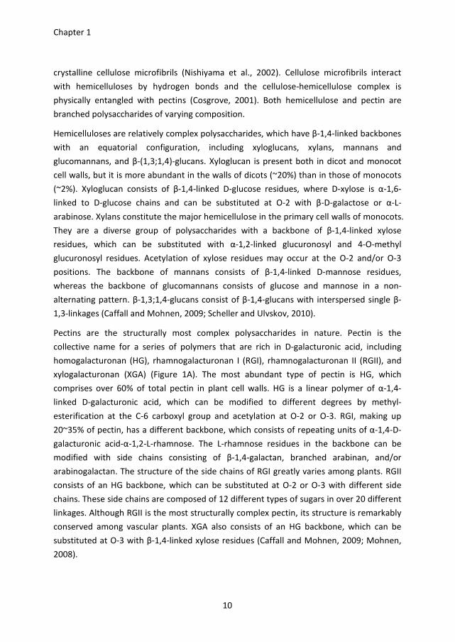
Pectins are the structurally most complex polysaccharides in nature. Pectin is the
collective name for a series of polymers that are rich in D-galacturonic acid, including
homogalacturonan (HG), rhamnogalacturonan I (RGI), rhamnogalacturonan II (RGII), and
xylogalacturonan (XGA) (Figure 1A). The most abundant type of pectin is HG, which
comprises over 60% of total pectin in plant cell walls. HG is a linear polymer of α-1,4-
linked D-galacturonic acid, which can be modified to different degrees by methyl-
esterification at the C-6 carboxyl group and acetylation at O-2 or O-3. RGI, making up
20~35% of pectin, has a different backbone, which consists of repeating units of α-1,4-D-
galacturonic acid-α-1,2-L-rhamnose. The L-rhamnose residues in the backbone can be
modified with side chains consisting of β-1,4-galactan, branched arabinan, and/or
arabinogalactan. The structure of the side chains of RGI greatly varies among plants. RGII
consists of an HG backbone, which can be substituted at O-2 or O-3 with different side
chains. These side chains are composed of 12 different types of sugars in over 20 different
linkages. Although RGII is the most structurally complex pectin, its structure is remarkably
conserved among vascular plants. XGA also consists of an HG backbone, which can be
substituted at O-3 with β-1,4-linked xylose residues (Caffall and Mohnen, 2009; Mohnen,
2008).
General introduction
11
Currently, there are two models that describe how pectic polysaccharides are linked: the
‘smooth and hairy region’ model and the ‘rhamnogalacturonan backbone’ model (Figure
1B). In the first model, pectin is composed of hairy regions, consisting of RGI decorated
with neutral sugar side chains, which are interspersed with smooth regions of HG. The
second model describes HG as a side chain of RGI, similar to the neutral sugar side chains
(Schols et al., 2009).
Figure 1. Schematic structure of pectin components in plant cell walls (A), adapted from Mohnen (2008);
and two alternative models for the organization of the pectin network (B), adapted from Schols et al.
(2009).
Chapter 1
12
Cell wall-associated resistance to plant pathogens
Plants protect their cell walls from penetration by pathogens in several ways: (i) inhibition
of plant cell wall-degrading enzymes; (ii) remodelling of cell walls at the site of attempted
penetration; (iii) perception of cell wall-derived molecules (damage-associated molecular
patterns) followed by triggering of an immune response. I will focus on the first two
aspects.
A. Inhibition of cell wall degrading enzymes
Polygalacturonase-inhibiting proteins (PGIPs) are extracellular leucine-rich repeat (eLRR)
proteins that physically interact with, and thereby inhibit, polygalacturonases (PGs)
produced by fungi, bacteria and even insects (De Lorenzo et al., 2001; Federici et al., 2006;
Juge, 2006). Secreted PGs are important virulence factors in several fungal pathogens,
including Aspergillus flavus (Shieh et al., 1997), Botrytis cinerea (ten Have et al., 1998),
Alternaria citri (Isshiki et al., 2001), and Claviceps purpurea (Oeser et al., 2002). Fungal PGs
are able to decompose pectin and thus cause cell wall degradation and tissue maceration
(see below). PGIPs are widely distributed in the plant kingdom, such as apple, bean, grape,
pepper, raspberry, soybean, tomato, leek, and Arabidopsis thaliana (De Lorenzo et al.,
2001). The potential of PGIPs to limit host tissue colonization by fungi was shown using
overexpression and gene silencing. Specifically, the overexpression of a pear PGIP in
tomato or grape plants, of bean PvPGIP2 in tobacco, and of AtPGIP1 and AtPGIP2 in A.
thaliana reduced B. cinerea infection (Aguero et al., 2005; Ferrari et al., 2003b; Powell et
al., 2000). A. thaliana plants in which the AtPGIP1 gene was silenced showed reduced PGIP
accumulation and enhanced susceptibility to B. cinerea (Ferrari et al., 2006). Furthermore,
overexpression of Phaseolus vulgaris PvPGIP2 in wheat reduced the symptoms caused by
Fusarium moniliforme and Bipolaris sorokiniana infection (Janni et al., 2008; Manfredini et
al., 2005).
PGIPs are encoded by small gene families. The isoforms within a single plant species may
exhibit differential in vitro inhibitory activities towards PGs from different fungi. The
activity of several PGIP isoforms from bean and A. thaliana was tested in vitro towards
PGs from saprotrophic and plant pathogenic fungi (Ferrari et al., 2003b), leading to
hypotheses about differential inhibition and specificity in PG-PGIP interactions (Federici et
al., 2006). It should however be noted that the in vitro interaction between PGs and PGIPs
may not be representative for the situation in planta. Joubert et al. (2007) reported that
the grapevine protein VvPGIP1 was able to inhibit the activity of B. cinerea BcPG2 in
planta, even though the two proteins were not able to interact in vitro. Thus the failure of
a given PGIP to inhibit a particular PG in vitro may not be informative about the potential
General introduction
13
of this PGIP to interact with the PG in planta and thereby confer (partial) disease
resistance.
Besides inhibitors of PGs, plants can also produce inhibitors of pectin methylesterases
(PMEIs). The inhibition of PME activity results in a markedly higher degree of methylation
of pectin, which impacts on cell wall properties and tissue texture (Jolie et al., 2010).
Overexpression of a PMEI gene from kiwifruit in wheat was shown to increase resistance
to fungal pathogens (Volpi et al., 2011). There is, however, an important conceptual
difference between PGIPs and PMEIs. PGIPs only inhibit PGs of microbes and insects that
attack the plant, but fail to inhibit endogenous plant PGs. On the contrary, PMEIs are
active only against endogenous plant PMEs and presumably inactive against non-plant
PMEs (Jolie et al., 2010).
B. Remodelling of cell walls at the site of attempted penetration
Callose, the major constituent of papillae, is an important factor contributing to the
resistance of plants against penetration and invasion by pathogenic fungi. Callose is
present at low levels throughout a plant, especially in the sieve plates of phloem cells and
in plasmodesmata. In response to biotic stress, plant cells rapidly synthesize callose in the
vicinity of the site of pathogen penetration (Adie et al., 2007; Flors et al., 2008; Garcia-
Andrade et al., 2011; Jacobs et al., 2003; Nishimura et al., 2003). The A. thaliana callose
synthase GSL5/PMR4 is required for pathogen-induced callose deposition (Jacobs et al.,
2003; Nishimura et al., 2003). gsl5/pmr4 mutant plants lack callose and show enhanced
susceptibility to the necrotrophic pathogens Alternaria brassicicola, Plectosphaerella
cucumerina and Pythium irregulare (Adie et al., 2007; Flors et al., 2008; Garcia-Andrade et
al., 2011). By contrast, gsl5/pmr4 mutant plants show increased resistance to biotrophic
pathogens Erysiphe cichoracearum, Golovinomyces orontii and Hyaloperonospora
parasitica, due to a hyperstimulation of salicylic acid-dependent defence pathways, which
remains to be understood (Jacobs et al., 2003; Nishimura et al., 2003).
Reactive oxygen species (ROS) are well known as signal molecules triggering plant defence
response (Lamb and Dixon, 1997). H2O2 is required for peroxidase-dependent lignification
and for protein cross-linking in the cell wall (Hückelhoven, 2007). Rapid oxidative cross-
linking of proline-rich proteins in the cell wall strengthens the wall, and thereby makes it
more resistant to cell wall degrading enzymes (Jacobs et al., 2003; Nishimura et al., 2003).
Chapter 1
14
Plant cell wall polysaccharide degradation
The plant cuticle and cell wall are the first barriers to pathogen invasion. Fungal plant
pathogens secrete a series of enzymes to decompose plant cell wall polysaccharides in
order to facilitate the penetration, the subsequent maceration and the acquisition of
carbon from decomposed plant tissues. Generally, these polysaccharide degrading
enzymes can be divided into two classes: exo-acting enzymes and endo-acting enzymes.
Exo-acting enzymes can be specific for the reducing end or the non-reducing end of
polysaccharides. They release monomeric or dimeric glycosyl moieties during each
catalytic event, providing the fungus with low molecular mass compounds that can be
easily taken up. Endo-acting enzymes cleave polysaccharides randomly within the chain,
resulting in a rapid decrease in the average chain length. The cleavage products, however,
are generally too large to serve as nutrients for the fungus. It is commonly observed that
any particular polysaccharide is degraded by a combination of endo-acting and exo-acting
enzymes, acting synergistically on the substrate. The plant cell wall degrading enzymes
that are secreted by fungi are all carbohydrate-active enzymes (Cantarel et al., 2009).
Fungal cellulases, hemicellulases and pectinases can be assigned to CAZy families of
glycoside hydrolases (GH), carbohydrate esterases (CE), and polysaccharide lyases (PL).
A. Cellulose degradation
Three classes of enzymes, all belonging to GH families 6 and 7, are involved in cellulose
degradation: β-1,4-endoglucanases, cellobiohydrolases and β-glucosidases. The β-1,4-
endoglucanases hydrolyse the internal bonds to disrupt the crystalline cellulose
microfibrils and expose individual cellulose polysaccharide chains. Cellobiohydrolases
cleave two glucose units from the ends of the exposed chains, resulting in the release of
the disaccharide cellobiose, which is subsequently hydrolysed by β-glucosidases into
individual D-glucose monomers.
B. Hemicellulose degradation
Hemicellulose consists of a group of relatively complex branched polysaccharides. The
various backbones of hemicelluloses are hydrolysed by the corresponding set of GH family
enzymes. Specifically, xyloglucan is decomposed by a combination of β-1,4-
endoglucanases and β-glucosidases; xylan is decomposed by a combination of β-1,4-
endoxylanases and β-xylosidases; mannan and galactomannan are decomposed by a
combination of β-1,4-endomannanases and β-mannosidases. Various side chains of
hemicelluloses are cleaved by different enzymes, which belong to GH and CE families. For
example, the α-linked D-xylose, D-galactose, D-glucuronic acid and L-arabinose residues
are cleaved by α-xylosidases, α-galactosidases, α-glucuronidases or α-
General introduction
15
arabinofuranosidases, respectively, whereas acetyl residues are cleaved by acetyl xylan
esterases.
C. Pectin degradation
Two types of backbones are present in pectin: the backbone of HG (smooth region)
consisting of α-1,4-linked D-galacturonic acid, and the backbone of RGI (hairy region)
consisting of alternating α-1,4 linked D-galacturonic acid and α-1,2-linked rhamnose
residues. Enzymes involved in degradation of the pectin backbone belong to the GH28 and
PL1 and PL3 families. Smooth regions can be hydrolysed by endo-polygalacturonases
(endo-PGs), exo-polygalacturonases (exo-PGs), pectin lyases and pectate lyases. Endo-PGs
hydrolyse the (preferentially unmethylated) backbone of HG, releasing monomeric and/or
oligomeric galacturonosyl fragments, whereas exo-PGs exclusively cleave at the non-
reducing end of HG strands, thereby releasing D-galacturonic acid monomers. Pectin
lyases and pectate lyases both cleave alternating α-1,4 linked D-galacturonic acid linkages
via β-elimination, resulting in a novel reducing end. Pectin lyases prefer substrates with a
high degree of methylesterification, whereas pectate lyases prefer substrates with a low
degree of methylesterification and require Ca2+
-ions for catalysis. The hairy regions of RGI
can be hydrolysed by rhamnogalacturonan hydrolases and rhamnogalacturonan lyases, of
which the former enzymes specifically cleave non-esterified galacturonosyl-rhamnosyl
linkages.
Various substituents occur on the backbone of pectins, therefore diverse enzymes are
involved in pectin side chain decomposition. Some of these enzymes cleave the entire side
chains from the backbone, whereas others cleave the internal or terminal linkages of side
chains. In addition, some of the enzymes not only act on pectin side chains, but also on
hemicelluloses. Specifically, α-arabinofuranosidases release L-arabinose, both from xylan,
arabinoxylan and from side chains of rhamnogalacturonan; endo/exo-arabinanases
hydrolyse α-1,5 linked arabinose residues from the arabinan side chains of pectins; β-
galactosidases release terminal D-galactose residues from the galactan side chains of
pectins; β-xylosidases can hydrolyse β-1,4-linked xylose residues, both from xyloglucan
and from side chains of xylogalacturonan. Finally, pectin acetylesterases and pectin
methylesterases remove the acetyl and methyl residues, which are present in the smooth
regions of pectins.
Chapter 1
16
Cell wall degrading enzymes in plant pathogenic fungi: a case study of
Botrytis cinerea
A. CAZymes in genomes of plant pathogenic fungi
The number of genome sequences of fungi that have been released has rapidly grown in
recent years (Martin et al., 2011). This provides opportunities to examine the repertoire of
plant cell wall degrading enzymes secreted by plant pathogenic fungi and explore
correlations between the CAZyme content in the genome, the CAZyme distribution over
different enzyme families (GH, CE, PL) and the host range of the fungal pathogen.
Plant pathogens with different lifestyles appear to have different repertoires of the
CAZymes that are involved in degrading plant cell walls. Necrotrophs and hemi-biotrophs
(in the necrotrophic infection phase) secrete large amounts of cell wall degrading enzymes
for host tissue decomposition and nutrient acquisition. The genomes of two closely
related necrotrophs, Botrytis cinerea and Sclerotinia sclerotiorum, encode respectively 118
and 106 CAZymes associated with plant cell wall degradation (Amselem et al., 2011).
These numbers are very similar to that in the saprotroph Aspergillus niger, but are lower
than in other necrotrophs (Phaeosphaeria nodorum and Pyrenophora teres f. teres) and in
the hemi-biotrophs Fusarium graminearum and Magnaporthe oryzae (Amselem et al.,
2011). By contrast, many biotrophic pathogens and symbionts have a markedly lower
content of CAZymes for cell wall degradation in their genome (Baxter et al., 2010;
Duplessis et al., 2011; Martin et al., 2010), presumably to reduce the damage to the host
and avoid the plant defence responses triggered by the release of cell wall fragments. The
most extreme examples to date are the genomes of Blumeria graminis and Ustilago
maydis, which contain only 10 and 33 genes encoding plant cell wall degrading enzymes
(Amselem et al., 2011; Kämper et al., 2006; Spanu et al., 2010).
Plant pathogens with distinct host preference seem to use different approaches to
decompose plant tissues. CAZyme analyses show that not only the contents, but also the
distribution of CAZymes differ among plant pathogens. B. cinerea and S. sclerotiorum can
both infect a wide range of dicot host plants, and prefer to infect soft plant tissues that
are rich in pectin, such as flowers and fruits. This is reflected by the observation that both
fungi grow better (in vitro) on pectic substrates than on xylan and cellulose. Their
genomes contain larger proportions of CAZymes involved in decomposition of pectin (37%
and 31%) and lower amounts of CAZymes involved in decomposition of cellulose (18% and
20%) and hemicellulose (40% and 41%). On the contrary, P. nodorum, P. teres f. teres and
M. oryzae are pathogens of wheat, barley, and rice, which all belong to commelinoid
monocots that contain less pectin in the cell wall. CAZyme analyses show that their
General introduction
17
genomes contain smaller proportions of CAZymes involved in decomposition of pectin
(18%, 17%, and 12%) and noticeably higher amounts of CAZymes involved in
decomposition of cellulose (47%, 38%, and 37%) and hemicellulose (66%, 55%, and 68%),
as compared to B. cinerea and S. sclerotiorum (Amselem et al., 2011).
B. Secretomes of plant pathogenic fungi
Releasing nutrients from plant cell wall polysaccharides requires the secretion of an
arsenal of plant cell wall degrading enzymes that act in synergy to decompose this
complex structure. In culture filtrates of Fusarium graminearum, grown in medium
containing hop cell wall material as sole carbon source, 17 different GH activities were
detected, which could collectively hydrolyse crude plant material, with monosaccharide
yields approaching 50% of the total sugars released by acid hydrolysis (Phalip et al., 2009).
Proteomics techniques are increasingly applied to study the secretomes of fungal
pathogens, either in vitro, or during their interaction with plants. Besides being useful for
annotating genomes, secretome analyses may enable to identify pathogen effectors
(reviewed by (Koeck et al., 2011)) as well as (abundant) plant cell wall degrading enzymes.
The latter information may provide leads to unravel the mechanisms that fungal
pathogens utilise to decompose plant cell wall polysaccharides and acquire nutrients from
their host. Several studies, including those in B. cinerea discussed below in detail, have
revealed that plant cell wall degrading enzymes are abundant in fungal secretomes. In S.
sclerotiorum culture filtrates, 18 secreted proteins were identified and 9 of them were
plant cell wall degrading enzymes (Yajima and Kav, 2006). Studies on F. graminearum
grown in medium containing various polysaccharide supplements, or in medium
containing wheat or barley flour, resulted in the identification of 120 and 69 proteins, in
which glycoside hydrolases represented approximately 25% of all identified proteins
(Paper et al., 2007; Yang et al., 2011). In M. oryzae, 85 proteins were identified and 19 of
them were annotated as plant cell wall hydrolases (Wang et al., 2011).
The secretome of B. cinerea has been analysed in different culture conditions (Espino et al.,
2010; Fernandez-Acero et al., 2010; Shah et al., 2009a; Shah et al., 2009b). One study
aimed to compare proteins secreted upon culturing B. cinerea in the presence of extracts
of red tomato, ripe strawberry and A. thaliana leaves. Overall, 89 B. cinerea proteins were
identified by LC-MS/MS, of which 60 contained a signal peptide in the (predicted) protein
sequence. 30 of these 60 proteins are involved in carbohydrate metabolism and transport,
and these proteins were more abundant in cultures grown in the presence of tomato and
strawberry extract, as compared to cultures containing A. thaliana leaf extract (Shah et al.,
2009a). The second study aimed to compare B. cinerea proteins induced by pectins with
different degrees of methyl esterification. A total of 126 secreted proteins were identified
Chapter 1
18
in cultures containing highly or partially esterified pectin, or sucrose. The abundance of
proteins with functions in pectin degradation was similar in both pectin containing media,
but higher as compared to sucrose containing medium (Shah et al., 2009b). In a similar
study, proteins were sampled from B. cinerea cultures grown in presence of either glucose,
starch, cellulose, pectin or tomato cell walls and submitted to two-dimensional gel
electrophoresis. 57 unique proteins were identified, of which more than 50% are involved
in plant cell wall polysaccharide decomposition (Fernandez-Acero et al., 2010). Finally,
Espino et al. (2010) focused on the early secretome of B. cinerea because the early stages
of development in planta are crucial in establishment of a successful infection. Conidia
were inoculated in minimal medium, supplemented with extracts of tomato, strawberry or
kiwifruit, and proteins were sampled after 16 h. A total of 105 proteins were identified, of
which 36 are involved in plant cell wall polysaccharide degradation; proteins involved in
pectin degradation were highly abundant (Espino et al., 2010). The lists of proteins
identified in these studies show substantial overlap, as the methodology used was often
comparable. Other culture methods, more sensitive protein identification methods and
more reliable gene models will be required to generate a more comprehensive list of
proteins identified as being secreted by B. cinerea.
The contribution of cell wall degrading enzymes to virulence of Botrytis
cinerea
Botrytis cinerea is able to infect over 200 host plant species and different tissue types:
stems, leaves, flowers and fruit. The pathogen can cause a variety of symptoms ranging
from dry, necrotic areas to water-soaked, fully macerated lesions. The ability to cause
disease on such different tissues and plant species suggests that B. cinerea has a large
weaponry to kill and invade its hosts (Choquer et al., 2007). The ultimate purpose of a
necrotrophic pathogen is not to kill its host, but to decompose the plant tissue and utilize
host-derived nutrients for its own growth. As discussed above, B. cinerea secretes a
spectrum of cell wall decomposing enzymes (including pectinases, cellulases, and
hemicellulases) to facilitate plant tissue colonization and release carbohydrates for
consumption (van Kan, 2006; Williamson et al., 2007).
A. Pectinases
B. cinerea often penetrates host leaf tissue at the anticlinal cell wall and subsequently
grows into and through the middle lamella, which consists mostly of low-methylesterified
pectin. Effective pectin degradation thus is important for virulence of B. cinerea. Several
pectinases have been found to be abundant during host infection, including pectin and
General introduction
19
pectate lyases, pectin methylesterases (PMEs), exo-polygalacturonases (exo-PGs), and
endo-polygalacturonases (endo-PGs) (Cabanne and Doneche, 2002; Kars et al., 2005b;
Kars and van Kan, 2004; Rha et al., 2001; ten Have et al., 2001). Especially the roles of
endo-PGs and PMEs in virulence have been intensively investigated.
1. Endo-polygalacturonases
The B. cinerea genome contains six genes encoding endo-PGs (Wubben et al., 1999). All
gene family members are differentially expressed in vitro and in planta (ten Have et al.,
2001; Wubben et al., 2000). Four regulatory mechanisms were proposed based on in vitro
analysis: basal, constitutive expression was observed for Bcpg1; expression of Bcpg3 was
induced by low ambient pH, irrespective of the carbon source present; expression of
Bcpg4 and Bcpg6 was induced by D-galacturonic acid; catabolite repression by glucose
was observed for Bcpg4 only. Other monosaccharides present in cell wall polymers, such
as rhamnose, arabinose, and galactose did not notably regulate the expression of Bcpg
genes. Regulation of the expression of Bcpg2 and Bcpg5 remained unclear (Wubben et al.,
2000).
Altogether this endo-PG gene family equips the fungus with a flexible pectin degrading
machinery, which provides a potential advantage for a fungus with such a broad range of
hosts and tissue types. All gene family members display various expression patterns during
infection, depending on the stage of infection and on the host (ten Have et al., 2001).
The endo-PG family members not only display diversity in their expression patterns but
also in enzymatic characteristics. Five BcPGs were produced in Pichia pastoris and their
biochemical properties were analysed (Kars et al., 2005a). All enzymes display optimal
activity at low ambient pH, which is consistent with the acidification of the environment
during the early stages of colonization by B. cinerea (Billon-Grand et al., 2012; Verhoeff et
al., 1988). BcPG1, BcPG2 and BcPG4 prefer the non-methylesterified substrate
polygalacturonic acid (PGA) to pectin, however, they show differences in substrate
affinities and hydrolysis rates. BcPG3 and BcPG6 were shown to behave as processive
endo-hydrolases, releasing monomers of D-galacturonate instead of oligomers (Kars et al.,
2005a).
The function of endo-PG gene family members in virulence has been studied by deleting
each single gene in B. cinerea. Knockout mutants ∆Bcpg1 and ∆Bcpg2 were reduced in
virulence by 25% and > 50%, respectively (Kars et al., 2005a; ten Have et al., 1998) .
2. Pectin methylesterases
The degree of methylation (DM) of pectin in plant cell walls can range from 13% to
approximately 80% (Voragen et al., 1986). Pectin methylesterases (PMEs) catalyse the
Chapter 1
20
hydrolysis of methyl esters, releasing methanol and pectate. Pectate is a preferred
substrate for many of the BcPGs (Kars et al., 2005b). This predicts that PMEs are important
for virulence on plant tissues with high DM pectin (such as in leaves), but not on tissues
with low DM pectin (such as in fruit). However, the role of PMEs in virulence of B. cinerea
is controversial. The phenotype of a Bcpme1 knockout mutant in one strain of B. cinerea
supported this hypothesis (Valette-Collet et al., 2003). However, results in a different stain
with single and double knockout mutants in two Bcpme genes, including the same Bcpme1
gene, did not support this hypothesis (Kars et al., 2005b). In addition, the Bcpme mutants
and the wild-type strain displayed better growth on 75% methylesterified pectin than on
non-methylesterified polygalacturonic acid, suggesting that pectin de-methylation by
PMEs is not important for depolymerisation in vivo (Kars et al., 2005b). The profuse
growth of B. cinerea on high DM pectin suggests that the biochemical properties of endo-
PGs determined in vitro may not reflect their behaviour in vivo, or that accessory enzymes
participate in the pectin degradation mediated by endo-PGs.
B. Other cell wall degrading enzymes
Other cell wall degrading enzymes produced by B. cinerea, such as cellulases and
hemicellulases, have also been studied. Deletion of a cellulase gene Bccel5A, encoding an
endo-β-1,4-glucanase, did not affect virulence (Espino et al., 2005), whereas the deletion
of a hemicellulase gene Bcxyn11A, encoding an endo- β-1,4-xylanase, delayed lesion
formation and reduced lesion size by more than 70% (Brito et al., 2006). The contribution
of the Bcxyn11A gene in virulence was, however, not dependent on xylanase enzyme
activity, but rather required the necrosis-inducing elicitor activity of the xylanase protein
(Noda et al., 2010).
21
Outline of the thesis
Pectin degradation plays an important role in the virulence of Botrytis cinerea. The aims of
this thesis were on the one hand, to characterize a genetic locus in Arabidopsis thaliana
that controls a necrotic response upon infiltration with BcPGs, and on the other hand, to
unravel whether the role of BcPGs in virulence (Kars et al., 2005a; ten Have et al., 1998)
relates only to a function in tissue disintegration and colonization, or also to a function in
the release of an abundant source of monosaccharide (i.e. D-galacturonic acid) nutrients
from pectic polymers. At the onset of this PhD thesis research, nothing was known about
the pathways that B. cinerea exploits to utilize D-galacturonic acid released from pectic
polymers.
Chapter 2 describes a study on the natural genetic variation in responsiveness of A.
thaliana to treatment with BcPG proteins. Among the many accessions tested, accession
Col-0 was responsive to BcPGs (resulting in a necrotic response to protein infiltration),
whereas accession Br-0 was unresponsive to BcPGs. A map-based cloning approach, in
combination with functional genomics and comparative genomics strategies, revealed that
the gene RBPG1, encoding a Receptor-Like Protein, determines the responsiveness to
BcPGs in Col-0. Furthermore, chapter 2 describes that several fungal endo-PGs act as
MAMPs and their recognition involves the formation of a complex with the receptor
protein RBPG1 and the subsequent signal transduction leading to a necrotic response is
mediated by the Receptor-Like Kinase SOBIR1.
Chapter 3 describes the functional genetic and biochemical characterization of the D-
galacturonic acid catabolic pathway in B. cinerea. The enzymatic activity of recombinant
proteins was characterized, and the function of the genes in B. cinerea was studied by
generating single and double knockout mutants and testing the mutants in vitro and in
planta. In Chapter 4, the virulence of D-galacturonic acid catabolism-deficient mutants
was further investigated on Solanum lycopersicum, Nicotiana benthamiana and A.
thaliana. The mutants displayed a reduced virulence on N. benthamiana and A. thaliana.
This phenotype was not only due to the inability of the mutants to utilize D-galacturonic
acid as nutrient, but also due to the growth inhibition by catabolic intermediates.
Chapter 5 describes the functional genetic characterization and cellular location of two
putative D-galacturonic acid transporters in B. cinerea. Chapter 6 describes an RNA
sequencing study, performed to compare genome-wide transcriptional profiles in B.
cinerea grown in media containing glucose and pectate as sole carbon sources. Conserved
sequence motifs were identified in the promoters of genes involved in pectate
decomposition and D-galacturonic acid utilization. The role of these motifs in regulating D-
galacturonic acid-induced expression was functionally analysed.

22
Chapter 7 presents a general discussion of the results in Chapters 2-6 and puts them in a
broader perspective. A model is proposed for the ways in which B. cinerea degrades pectin
and subsequently consumes the released D-galacturonic acid, and for the co-regulation of
genes involved in this process.
CHAPTER 2
Fungal endo-polygalacturonases are recognized as MAMPs in
Arabidopsis by the Receptor-Like Protein RBPG1
Lisha Zhang, Ilona Kars, Lia Wagemakers, Thomas W. H. Liebrand, Panagiota Tagkalaki,
Devlin Tjoitang, Bert Essenstam, Joyce Elberse, Guido van den Ackerveken, Jan A. L. van
Kan
Chapter 2
24
Abstract
Plants perceive microbial invaders using pattern recognition receptors (PRRs) that
recognize microbe-associated molecular patterns (MAMPs). Two well-characterised
MAMP receptors are the leucine-rich repeat receptor-like kinases (LRR-RLKs) FLS2 and EFR
that recognize bacterial flagellin and translation elongation factor EF-Tu, respectively. In
this study, we identified RBPG1, an Arabidopsis thaliana LRR receptor-like protein (LRR-
RLP) that recognizes fungal endo-polygalacturonases (endo-PGs) and thus acts as a novel
MAMP receptor. RBPG1 in particular recognizes the Botrytis cinerea BcPG3 protein.
Infiltration of the BcPG3 protein into A. thaliana accession Col-0 induced a necrotic
response, whereas accession Br-0 showed no symptoms. Heat-inactivated protein and a
catalytically inactive mutant protein retained the ability to induce necrosis. An 11-amino
acid peptide stretch was identified that is conserved among many fungal but not plant
endo-PGs. A synthetic peptide comprising this sequence was unable to induce necrosis. A
map-based cloning strategy, combined with comparative and functional genomics, led to
the identification of the RBPG1 gene, and showed that this gene is essential for
responsiveness of A. thaliana to the BcPG3 protein. Co-immunoprecipitation experiments
demonstrated that RBPG1 and BcPG3 form a complex in Nicotiana benthamiana, which
also involves the A. thaliana LRR-RLK SOBIR1. The sobir1 mutant plants were unresponsive
to BcPG3. Although transformation of RBPG1 in accession Br-0 resulted in gain of BcPG3
responsiveness, it did not alter the level of resistance to several microbial pathogens.
Recognition of fungal endo-polygalacturonases by RBPG1
25
Introduction
Microbe-associated molecular patterns (MAMPs) are molecular signatures of entire
groups of microbes and have key roles in activation of defence response in plants (Boller
and Felix, 2009; Jones and Dangl, 2006). Well characterized proteinaceous MAMPs are
bacterial flagellin, EF-Tu and Ax21, fungal xylanase, and oomycete pep13, an epitope of a
secreted transglutaminase (Boller and Felix, 2009; Monaghan and Zipfel, 2012). Plants
recognize MAMPs by means of pattern recognition receptors (PRRs), comprising a group
of leucine-rich repeat receptor-like kinases (LRR-RLKs) or leucine-rich repeat receptor-like
proteins (LRR-RLPs) located in the plasma membrane (Greeff et al., 2012; Monaghan and
Zipfel, 2012). The LRR-RLKs FLS2 and EFR recognize flg22 (the 22-amino-acid eliciting
epitope from the conserved flagellin domain) and elf18/elf26 (peptides derived from the
N-terminus of the translation elongation factor EF-Tu), respectively (Chinchilla et al., 2006;
Gomez-Gomez and Boller, 2000; Kunze et al., 2004; Zipfel et al., 2006). The fungal protein
ethylene-inducing xylanase (EIX) is recognized by the tomato LRR-RLPs LeEix1 and LeEix2,
of which only the latter mediates a necrotic response (Ron and Avni, 2004).
BRI1-associated kinase-1/Somatic embryogenesis receptor kinase-3 (BAK1/SERK3) is an
LRR-RLK acting as a common component in many RLK signalling complexes (Monaghan
and Zipfel, 2012). Although it was originally identified as a protein that interacts with the
brassinosteroid (BR) receptor BRI1 (Li et al., 2002; Nam and Li, 2002), BAK1 also forms
ligand-induced complexes with FLS2 and EFR, and contributes to disease resistance against
the pathogens Pseudomonas syringae, Hyaloperonospora arabidopsidis and Phytophthora
infestans (Chaparro-Garcia et al., 2011; Chinchilla et al., 2007; Heese et al., 2007; Roux et
al., 2011). Tomato BAK1 interacts in a ligand-independent manner with LeEix1 but not
with LeEix2, and the BAK1-LeEIX1 interaction is required for the ability of LeEix1 to
attenuate the signalling of LeEix2 (Bar et al., 2010). BAK1 has also been shown to interact
with another LRR-RLK, BIR1 (BAK1-interacting receptor-like kinase 1). The bir1 mutant
showed extensive cell death, activation of constitutive defence responses, and
impairment in the activation of the MAP kinase MPK4 (Gao et al., 2009). SOBIR1 mutants
suppress BIR1 phenotypes and over-expression of SOBIR1 triggers cell death and defence
responses (Gao et al., 2009). SOBIR1 does not physically interact with BIR1 suggesting that
SOBIR1 mediates an alternative signal transduction route.
Fungal endopolygalacturonases (endo-PGs) are a class of pectinases that decompose plant
cell walls by hydrolysing the homogalacturonan domain of pectic polysaccharides (van den
Brink and de Vries, 2011). One of the pathogenic fungi for which the role of endo-PGs has
been studied is Botrytis cinerea, the cause of grey mould disease on a wide range of plant
species and tissues (Dean et al., 2012; van Kan, 2006; Williamson et al., 2007). The B.
Chapter 2
26
cinerea genome (Amselem et al., 2011; Staats and van Kan, 2012) contains six genes
encoding endo-PGs (Wubben et al., 1999), of which BcPG1 and BcPG2 are required for full
virulence of B. cinerea (Kars et al., 2005a; ten Have et al., 1998). BcPGs, produced
heterologously in Pichia pastoris, were capable of causing necrotic responses when
infiltrated in leaves of several plant species (Kars et al., 2005a). The massive and prompt
tissue collapse and necrotic response caused by BcPG2 in broad bean leaves depends on
its enzymatic activity, since catalytically inactive mutant protein did not cause any tissue
damage (Kars et al., 2005a). In a different study, BcPG1 was reported to induce defence
responses in grapevine cell suspensions, and the defence inducing activity of BcPG1 was
independent of enzymatic activity (Poinssot et al., 2003). These two studies with
seemingly opposing conclusions were conducted with different endo-PG isozymes and on
distinct tissues of different plant species. Therefore it remained inconclusive whether the
plant responses observed after exposure to BcPGs are due to recognition of the protein,
or by the sheer structural damage resulting from hydrolysis of pectin.
Oligogalacturonides (OGAs) are hydrolytic products released upon cleavage of
homogalacturonan by endo-PGs (Caffall and Mohnen, 2009; Mohnen, 2008; van den Brink
and de Vries, 2011). OGAs have been reported to function as damage-associated
molecular patterns (DAMPs). Similar to MAMPs, DAMPs are able to activate plant defence
responses, such as an oxidative burst, cell wall lignification, phytoalexin accumulation and
changes in ion fluxes (Denoux et al., 2008; Galletti et al., 2008). The biological activity of
OGAs is related to their molecular weight and the formation of Ca2+
-dependent egg-box
confirmation (Cabrera et al., 2008; Hematy et al., 2009; Vorwerk et al., 2004). Wall-
associated kinases (WAKs), a family of cell wall-associated receptors, are able to bind
OGAs in vitro (Decreux et al., 2006; Kohorn et al., 2009; Wagner and Kohorn, 2001). The
extracellular domain of WAK1 serves as a receptor that perceives pectin damage caused
by endo-PGs, and subsequently activates intracellular kinase signalling processes (Brutus
et al., 2010). Overexpression of WAK1 in Arabidopsis thaliana enhances resistance to B.
cinerea (Brutus et al., 2010).
Here, we describe the identification of natural variation in the responsiveness of A.
thaliana to BcPGs. The accession Col-0 responded strongly to BcPGs, whereas accession
Br-0 was unresponsive to BcPGs. Cloning and functional characterization demonstrated
that the gene determining this dominant trait, which we designated RBPG1, encodes an
LRR-RLP that mediates the responsiveness of Col-0 to BcPGs. Furthermore, we
demonstrated that an endo-PG of another, non-pathogenic, fungal species can also act as
MAMPs through recognition by the receptor protein RBPG1. The LRR-RLK SOBIR1 was
found to interact with RBPG1 and is essential for responsiveness to fungal endo-PGs.
Recognition of fungal endo-polygalacturonases by RBPG1
27
Results
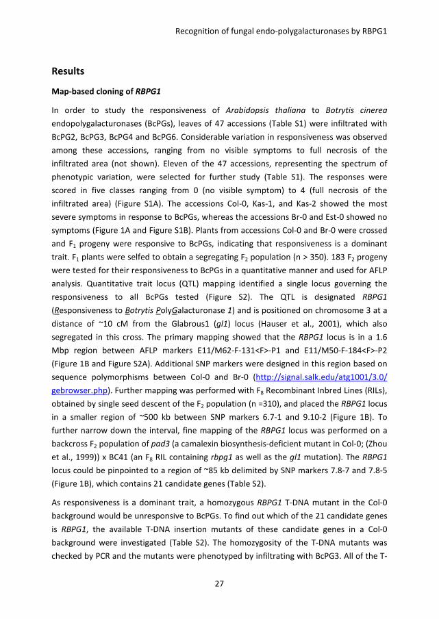
Map-based cloning of RBPG1
In order to study the responsiveness of Arabidopsis thaliana to Botrytis cinerea
endopolygalacturonases (BcPGs), leaves of 47 accessions (Table S1) were infiltrated with
BcPG2, BcPG3, BcPG4 and BcPG6. Considerable variation in responsiveness was observed
among these accessions, ranging from no visible symptoms to full necrosis of the
infiltrated area (not shown). Eleven of the 47 accessions, representing the spectrum of
phenotypic variation, were selected for further study (Table S1). The responses were
scored in five classes ranging from 0 (no visible symptom) to 4 (full necrosis of the
infiltrated area) (Figure S1A). The accessions Col-0, Kas-1, and Kas-2 showed the most
severe symptoms in response to BcPGs, whereas the accessions Br-0 and Est-0 showed no
symptoms (Figure 1A and Figure S1B). Plants from accessions Col-0 and Br-0 were crossed
and F1 progeny were responsive to BcPGs, indicating that responsiveness is a dominant
trait. F1 plants were selfed to obtain a segregating F2 population (n > 350). 183 F2 progeny
were tested for their responsiveness to BcPGs in a quantitative manner and used for AFLP
analysis. Quantitative trait locus (QTL) mapping identified a single locus governing the
responsiveness to all BcPGs tested (Figure S2). The QTL is designated RBPG1
(Responsiveness to Botrytis PolyGalacturonase 1) and is positioned on chromosome 3 at a
distance of ~10 cM from the Glabrous1 (gl1) locus (Hauser et al., 2001), which also
segregated in this cross. The primary mapping showed that the RBPG1 locus is in a 1.6
Mbp region between AFLP markers E11/M62-F-131<F>-P1 and E11/M50-F-184<F>-P2
(Figure 1B and Figure S2A). Additional SNP markers were designed in this region based on
sequence polymorphisms between Col-0 and Br-0 (http://signal.salk.edu/atg1001/3.0/
gebrowser.php). Further mapping was performed with F8 Recombinant Inbred Lines (RILs),
obtained by single seed descent of the F2 population (n =310), and placed the RBPG1 locus
in a smaller region of ~500 kb between SNP markers 6.7-1 and 9.10-2 (Figure 1B). To
further narrow down the interval, fine mapping of the RBPG1 locus was performed on a
backcross F2 population of pad3 (a camalexin biosynthesis-deficient mutant in Col-0; (Zhou
et al., 1999)) x BC41 (an F8 RIL containing rbpg1 as well as the gl1 mutation). The RBPG1
locus could be pinpointed to a region of ~85 kb delimited by SNP markers 7.8-7 and 7.8-5
(Figure 1B), which contains 21 candidate genes (Table S2).
As responsiveness is a dominant trait, a homozygous RBPG1 T-DNA mutant in the Col-0
background would be unresponsive to BcPGs. To find out which of the 21 candidate genes
is RBPG1, the available T-DNA insertion mutants of these candidate genes in a Col-0
background were investigated (Table S2). The homozygosity of the T-DNA mutants was
checked by PCR and the mutants were phenotyped by infiltrating with BcPG3. All of the T-
Chapter 2
28
DNA mutants tested were equally responsive to BcPG3 as Col-0 (Table S2). According to
these results, the 17 genes of which the homozygous T-DNA mutants were responsive to
BcPG3 could be eliminated as candidates. No T-DNA mutants were available for the genes
At3g24770, At3g24890 and At3g25020, whereas the T-DNA mutant in the At3g24800
gene could not be made homozygous. Therefore, these four genes remained candidates.
The genome sequences of several A. thaliana accessions were released by the Arabidopsis
1001 genomes project (Cao et al., 2011), including Br-0, Eden-2, Est-1, and Gy-0. Among
these accessions, Eden-2 was equally unresponsive to BcPG3 as Br-0, while Est-1 and Gy-0
were equally responsive to BcPG3 as Col-0. The different responsiveness of A. thaliana
accessions to BcPGs could be due to amino acid substitutions in RBPG1. Thus SNPs in the
21 candidate genes were compared between accessions in order to identify SNPs that are
associated with the responsive phenotype. In the 21 genes in the region, there were SNPs
in 4 genes (from At3g24770 to At3g24860) that lead to amino acid substitutions between
Col-0 and Br-0. However, none of these substitutions was specifically associated with the
phenotype in accessions Eden-2, Est-1, and Gy-0 (http://signal.salk.edu/atg1001/3.0/
gebrowser.php). Surprisingly, the sequences in the RBPG1 locus downstream of
At3g24890 were very poorly represented in accessions Br-0, Eden-2, and Gy-0, but also in
many other accessions (http://signal.salk.edu/atg1001/3.0/gebrowser.php); especially,
the region that comprises four highly homologous RLP paralogs in Col-0 is barely covered
by mappable sequence reads in the other accessions. In combination with the results of T-
DNA mutant analysis, At3g24890 and At3g25020 were considered to be the remaining
candidates for being the RBPG1 gene (Table S2).
To determine the function of At3g24890 and At3g25020, both genes (under the control of
the 35S promoter) were transformed into the BcPG-unresponsive accession Br-0.
Transgenic plants expressing At3g25020 displayed the BcPG-responsive phenotype (Figure
1A), whereas transgenic plants containing At3g24890 constructs remained equally
unresponsive to BcPG3 as the recipient accession Br-0 (Figure S3). The At3g25020 gene
encodes an LRR-RLP and is one of a family of four RLP-encoding genes that occur in a
cluster within this region of the Col-0 genome (Figure 1B). The four genes are At3g24900,
At3g24982, At3g25010, and At3g25020 and correspond to the genes RLP39-42 according
to the classification of Wang et al. (2008). Genome reassembly of raw sequence data
showed that this region in accession Br-0 contains only two RLP-encoding genes, rbpg1-1
and rbpg1-2 (Figure 1B), with over 80% identity to each other (Figure S4). Phylogenetic
analysis shows that RLP39 and RLP41 cluster with rbpg1-1, whereas RLP40 and RLP42
cluster with rbpg1-2 (Figure 1C). To determine whether the three Col-0 paralogs, RLP39,
RLP40, and RLP41 also confer responsiveness to BcPGs, these genes (under the control of
the 35S promoter) were transformed into BC41 (an F8 RIL of Col-0 x Br-0, unresponsive to
Recognition of fungal endo-polygalacturonases by RBPG1
29
BcPG3). None of the transgenic plants carrying RLP39, RLP40, and RLP41 constructs
showed a BcPG-responsive phenotype (Figure S5). These data demonstrate that RLP42 is
the only gene in this region that confers responsiveness to BcPG3 and it is hereafter
referred to as the RBPG1 gene.
Figure 1. Map-based cloning of RBPG1. A, responsiveness of Arabidopsis thaliana accessions Col-0 and Br-
0, and Br-0 transgenic plants expressing P35S:RBPG1 to BcPG3. Photographed 7 days after infiltration with
BcPG3. B, map of RBPG1 locus. C, phylogenetic tree of RBPG1 homologs in Col-0 and Br-0. The amino acid
sequences were aligned by Clustal_X 1.83 and the phylogenetic tree was generated by using Mega 4 by
the Neighbor-Joining (NJ) method with 1000 bootstrap replicates.
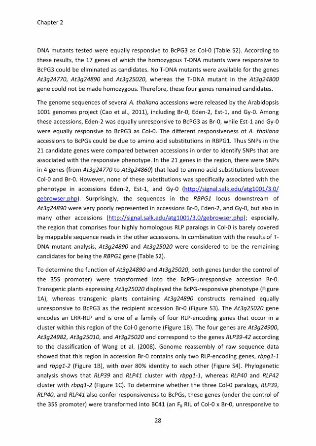
RBPG1 confers responsiveness to catalytically inactive BcPG3, but not to
oligogalacturonides
Plant cell wall fragments such as oligogalacturonides (OGAs) can serve as DAMPs (Boller
and Felix, 2009; D'Ovidio et al., 2004; Hematy et al., 2009; Rasul et al., 2012). To
distinguish whether the RBPG1-dependent responsiveness to BcPG3 is through the
recognition of the BcPG3 protein itself (acting as a MAMP) or through recognition of cell
wall fragments released in planta (acting as DAMPs), heat-inactivated BcPG3 and
catalytically inactive BcPG3 (D353E/D354N) were infiltrated into leaves of Col-0, Br-0, and
Br-0 transgenic plants expressing P35S:RBPG1. Both the heat-inactivated and the
catalytically inactive BcPG3 protein induced necrosis in Col-0 and in P35S:RBPG1 transgenic
plants, but not in Br-0 (Figure 2). Furthermore, the alcohol-insoluble residue (AIR) fraction
consisting mainly of cell wall polysaccharides, was extracted from leaves of Col-0 and Br-0
Chapter 2
30
and hydrolysed with active BcPG3. After 42 h incubation, the solubilised cell wall
fragments released from the AIR were infiltrated into Col-0 and Br-0. However, they did
not induce any visible symptom in Col-0 or Br-0. In addition, a set of distinct, partially
purified OGAs with a degree of polymerization (DP) ranging from 3 to 20, at a
concentration range from 10 µM up to 1 mM, did not induce any visible symptom upon
infiltration into leaves of Col-0 or Br-0. These results show that RBPG1 recognizes the
BcPG3 protein as a MAMP and that the response to BcPG3 does not depend on its
catalytic activity.
Figure 2. Responsiveness of Arabidopsis thaliana accessions Col-0 and Br-0, and Br-0 transgenic plants
expressing P35S:RBPG1 to fungal endo-PGs. Col-0 and P35S:RBPG1 transgenic plants are responsive to heat-
inactive BcPG3 (1.5 µM), catalytically inactive BcPG3, BcPG2 (0.1 µM), BcPG4 (3 µM), BcPG6 (1 µM) and
AnPGB (0.3 µM). Photographed 7 days after infiltration.
An 11-amino acid peptide stretch is conserved among fungal endo-PGs but fails to
induce necrosis
Plants that are responsive to BcPG3 (Col-0, as well as Br-0 transgenes expressing RBPG1)
were also responsive to BcPG2, BcPG4 and BcPG6, as well as to AnPGB from the
saprotrophic fungus Aspergillus niger (Figure 2). In the case of flagellin and EF-Tu,
conserved short peptide motifs are sufficient to trigger plant defence responses (Brunner
et al., 2002; Felix et al., 1999; Kunze et al., 2004). To avoid self-recognition these
conserved motifs should not be present in plants. Since multiple BcPGs and AnPGB were
Recognition of fungal endo-polygalacturonases by RBPG1
31
able to induce necrosis (Figure 2), we considered the possibility that a peptide motif
conserved among fungal endo-PGs, but absent in A. thaliana, might act as an epitope that
is recognized by RBPG1. Amino acid sequence alignments of several fungal endo-PGs and
three A. thaliana endo-PGs (Ogawa et al., 2009) showed that an 11-amino acid peptide
stretch, adjacent to the catalytic site, is highly conserved among the fungal endo-PGs
(fpg11). The homologous region in A. thaliana endo-PGs contains a glycine to proline
substitution (Figure 3). To investigate whether fpg11 is able to induce necrosis, a synthetic
22-amino acid peptide corresponding to BcPG3 residues 367-388 (with fpg11 in the middle)
was infiltrated in leaves of Col-0 and Br-0. In concentrations ranging from 0.01 mM to 1
mM, the peptide did not induce any symptoms till 7 days after infiltration.
Figure 3. Amino acid sequence alignment of fungal endo-PGs and Arabidopsis thaliana endo-PGs. The red
frame indicates the conserved fungal 11-amino acid peptide (fpg11). The triangles indicate the catalytic
site residues. The sequence highlighted in yellow represents the 22-amino acid synthetic peptide used for
infiltration. The numbers on the right hand side of sequences from each protein indicate the coordinate of
the last amino acid in the alignment. Accession numbers: BcPG1 (AAV84613), BcPG2 (AAV84614), BcPG3
(AAV84615), BcPG4 (AAV84616), BcPG5 (AAV84617), BcPG6 (AAV84618), AnPGII (1701294A), AnPGB
(Q9P4W3); AtADPG1 (At3g57510), AtADPG2 (At2g41850), AtQRT2 (At3g07970). The gene locus for other
species: GLRG_10528 (Colletotrichum graminicola), CH063_06619 (Colletotrichum higginsianum),
FGSG_11011 (Fusarium graminearum), FOXG_14695.2 (Fusarium oxysporum), SS1G (Sclerotinia
sclerotiorum).
The C-terminal intracellular domain of RBPG1 is not required for the responsiveness
One striking difference between RBPG1 and the three homologs from accession Col-0
(RLP39-41) and two homologs from accession Br-0 (rbpg1-1 and rbpg1-2) is the presence
of an extended C-terminal intracellular domain of 9 amino acid residues (Figure 4 and S3).
To investigate whether this domain is required for the responsiveness, two constructs
were generated in which the C-terminal domain of RBPG1 is truncated: RBPG1_Trunc1
lacking the C-terminal 18 amino acids, and RBPG1_Trunc2 lacking the C-terminal 10 amino
acids. Also a construct was generated in which the C-terminal residue of rbpg1-2 is
Chapter 2
32
replaced with the C-terminal 10 amino acids of RBPG1 (rbpg1-2_Swap1; Figure 4). These
three gene constructs (under the control of the 35S promoter) were transformed into
BC41 (an F8 RIL of Col-0 x Br-0, unresponsive to BcPG3). RBPG1_Trunc1 and RBPG1_Trunc2
conferred responsiveness of BC41 to BcPG3 similar to that of the full-length RBPG1
construct, whereas the recipient line BC41 and the plants that were transformed with
rbpg1-2_Swap1 constructs were unresponsive to BcPG3 (Figure 4). These results show
that the C-terminal intracellular domain of RBPG1 is not required for the responsiveness.
Figure 4. The C-terminal intracellular domain of RBPG1 is not required for the responsiveness to BcPG3. A,
schematic representation of the design of truncated RBPG1 and swapped rbpg1-2 constructs. B,
responsiveness of BC41 plants containing different constructs to BcPG3. Photographed 7 days after
infiltration with BcPG3.
BcPG3 interacts with RBPG1 in Nicotiana benthamiana
To determine whether BcPG3 physically interacts with RBPG1, immunoprecipitation
analysis was performed by transient co-expression of 10xMyc-tagged BcPG3 together with
RBPG1-GFP in N. benthamiana. Two days after agro-infiltration, RBPG1-GFP proteins were
immunoprecipitated using GFP Trap beads, and the purified proteins were analysed by
Western blot. BcPG3 was co-immunoprecipitated with RBPG1-GFP, but not with the GFP
beads alone (Figure 5A). GFP-tagged EFR was used as an additional negative control, to
exclude that BcPG3 interacts non-specifically with extracellular LRR-domain-containing
proteins (Figure 5B). To further confirm the interaction between BcPG3 and RBPG1, we
swapped the epitope tags and co-expressed 10xMyc-tagged RBPG1 and GFP-tagged BcPG3
in N. benthamiana. BcPG3-GFP was equally capable of immunoprecipitating RBPG1-myc
(Figure 5A).
Recognition of fungal endo-polygalacturonases by RBPG1
33
Figure 5. BcPG3 forms a complex with RBPG1 and its homologs in Nicotiana benthamiana.
Coimmunoprecipitation of (A) BcPG3 and RBPG1, (B) BcPG3 and EFR or SOBIR1, (C) BcPG3 and RLP39 or
rbpg1-2. Total proteins expressed in N. benthamiana leaves were subjected to immunoprecipitation with
GFP Trap beads followed by immunoblot analysis with anti-myc antibodies to detect BcPG3-myc and
RBPG1-myc; and anti-GFP antibodies to detect RBPG1-GFP, EFR-GFP, SOBIR1-GFP, RLP39-GFP, rbpg1-2–
GFP and BcPG3-GFP.
SOBIR1 is required for the RBPG1 dependent responsiveness to BcPG3
BAK1/SERK3 and other SERK family members play important roles in plant defence
responses by forming complexes with a number of LRR-receptor-like kinases (LRR-RLKs),
e.g. FLS2 and EFR (Chinchilla et al., 2009; Roux et al., 2011). Recently, the tomato LRR-RLK
SOBIR1 was shown to interact specifically with a number of LRR-RLPs (Liebrand et al.,
2013). Furthermore, overexpression of SOBIR1 in A. thaliana activates defence responses
(Gao et al., 2009). To test whether LRR-RLKs are involved in the response of RBPG1 to
BcPG3, A. thaliana plants carrying mutations in different LRR-RLK genes were analysed.
The efr mutant and all the serk mutants tested were unaltered in their responsiveness to
BcPG3 (Figure 6A). Interestingly, the sobir1-1 mutant did not show any symptoms
following infiltration with BcPG3 (Figure 6A). Co-immunoprecipitation analysis showed
that RBPG1 physically interacts with SOBIR1, but not with SERK2, BAK1/SERK3 or EFR
(Figure 6B). In addition, GFP-tagged SOBIR1 was incapable to immunoprecipitate 10xMyc-
tagged BcPG3 (Figure 5B).
Chapter 2
34
RBPG1 homologs physically interact with BcPG3 and SOBIR1
Among the six homologs of RBPG1 from Col-0 and Br-0, RBPG1 is the only receptor protein
that confers the responsiveness to BcPG3. To test whether the specificity is determined by
the interaction of the ligand (BcPG3) with the receptor, co-immunoprecipitation analysis
was also performed with RBPG1 homologs by transient co-expression of 10xMyc-tagged
BcPG3 with GFP-tagged RLP39, RLP40, RLP41, and rbpg1-2 in N. benthamiana (cloning of
the rbpg1-1 construct was unsuccessful). RLP39 and rbpg1-2 were able to co-
immunoprecipitate BcPG3 (Figure 5C). Moreover, GFP-tagged SOBIR1 was able to co-
immunoprecipitate 10xMyc-tagged RLP39 and rbpg1-2 (Figure 6C). Due to the very low
expression level of either GFP-tagged or 10xMyc-tagged RLP40 and RLP41 proteins (not
shown), their interaction with BcPG3 or SOBIR1 could not be studied.
Figure 6. SOBIR1 is required for RBPG1 dependent responsiveness. A, responsiveness of Arabidopsis
thaliana mutants to BcPG3. Photographed 7 days after infiltration with BcPG3. B, co-immunoprecipitation
of RBPG1 and SOBIR1, SERK2, BAK1, or EFR. C, co-immunoprecipitation of SOBIR1 and RLP39 or rbpg1-2.
Total proteins expressed in N. benthamiana leaves were subjected to immunoprecipitation with GFP Trap
beads followed by immunoblot analysis with anti-myc antibodies to detect RBPG1-myc, RLP39-myc, and
rbpg1-2-myc; and with anti-GFP antibodies to detect SOBIR1-GFP, SERK2-GFP, BAK1-GFP and EFR-GFP.
Recognition of fungal endo-polygalacturonases by RBPG1
35
RBPG1 does not contribute to disease resistance
To determine whether RBPG1 contributes to disease resistance, three independent
P35S:RBPG1 T3 transgenic Br-0 lines (homozygous T2 lines with single copy integration) and
the recipient accession Br-0 were inoculated with the necrotrophic fungal pathogen B.
cinerea, the biotrophic oomycete pathogen Hyaloperonospora arabidopsidis, the
hemibiotrophic oomycete pathogen Phytophthora capsici, and the bacterium
Pseudomonas syringae pv. tomato DC3000. All three transgenic lines were equally
susceptible as Br-0 to these tested pathogens (not shown). Despite the fact that Col-0
responds to BcPG3 and sobir1-1 mutants show loss of responsiveness, susceptibility of the
sobir1-1 mutant to pathogen infection is not altered as compared to Col-0.
Discussion
Genetic variation in response of Arabidopsis thaliana to BcPGs was studied and a single
locus, designated RBPG1, was identified by QTL mapping on the F2 population of Col-0 x
Br-0. The RBPG1 gene in the locus was identified by further map-based cloning and a
combination of functional and comparative genomics analysis. RBPG1 (RLP42) encodes an
LRR receptor-like protein (LRR-RLP) and is one of a family of four LRR-RLP-encoding genes
(RLP39, RLP40, RLP41, RLP42) that occur in a cluster (Wang et al., 2008) within the RBPG1
locus of the Col-0 genome. By contrast, accession Br-0 contains only two LRR-RLP-
encoding genes (rbpg1-1 and rbpg1-2) in this region (Figure 1B). Phylogenetic analysis
shows that RLP39 and RLP41 cluster with rbpg1-2, whereas RLP40 and RBPG1 cluster with
rbpg1-1 (Figure 1C). Among these six paralogs, RBPG1 is the only gene that confers the
responsiveness to BcPG3. It is likely that a duplication of a gene-cluster with two paralogs
occurred in the lineage to Col-0 and subsequently RBPG1 gained the responsiveness to
BcPG3. This region in the A. thaliana genome shows great diversity, which cannot easily be
unravelled. Accession Col-0 contains four paralogs with high nucleotide identity, however,
in many other accessions the region is barely covered by mappable sequence reads (Cao
et al., 2011) (http://signal.salk.edu/atg1001/3.0/gebrowser.php). In order to determine
the structure of this region in the Br-0 genome, it appeared essential to perform a de novo
assembly of Br-0 sequence reads, and manually validate the assembled contigs. Likewise,
genome assembly is probably required to study the genetic variation of these LRR-RLPs in
many different A. thaliana accessions.
Besides BcPG2, BcPG3, BcPG4, and BcPG6, also AnPGB produced by the saprotroph
Aspergillus niger induces necrosis in Col-0 (Figure 2), suggesting that fungal endo-PGs act
as microbe-associated molecular patterns (MAMPs) instead of specific pathogen-
Chapter 2
36
associated molecular patterns (PAMPs). Nevertheless, there have been controversies
whether the enzymatic activity of fungal endo-PGs is required for triggering plant
responses (Boudart et al., 2003; Kars et al., 2005a; Poinssot et al., 2003). Our data show
that catalytically inactive BcPG3 and heat-inactive BcPG3 were able to induce necrosis
similar to active BcPG3 (Figure 2). This observation suggests that RBPG1 recognizes the
BcPG3 protein itself rather than the oligogalacturonides (OGAs), hydrolytic products
released from pectin by endo-PGs (Caffall and Mohnen, 2009; Mohnen, 2008; van den
Brink and de Vries, 2011). OGAs have been reported to act as DAMPs that activate
defence responses following recognition by WAK1 (Brutus et al., 2010; Denoux et al., 2008;
Kohorn and Kohorn, 2012). We did not observe any visible symptoms upon infiltration
with several OGAs of different size ranges at concentrations up to 1 mM, however, we did
not monitor whether these OGAs induced any stress or defence response at the
transcriptional level. Our hypothesis that the BcPG3 protein itself is recognized as a MAMP
was further corroborated by a co-immunoprecipitation assay which provided evidence
that RBPG1 forms a complex with BcPG3 (Figure 5A). These data collectively suggest that
fungal endo-PGs act as MAMPs that are recognized by LRR-RLP RBPG1.
Multiple fungal endo-PGs were capable of inducing necrosis, suggesting that a conserved
motif might be involved in the recognition by RBPG1. Amino acid sequence alignment
identified an 11-amino acid peptide (fpg11) that is the longest conserved stretch among
fungal endo-PGs; other conserved sequence stretches were 7 amino acids (the catalytic
site) or shorter than 4 amino acids. However, a synthetic peptide covering fpg11 was not
capable of inducing necrosis. Either the structure of fpg11 could differ between the full-
length protein and the synthetic peptide, or the peptide is unstable after infiltration in the
plant. Notably, the corresponding region in the sequence of A. thaliana endo-PGs contains
a proline residue instead of glycine at position 1. This proline likely alters the 3-
dimensional structure of plant endo-PGs in this region, which would enable them to
escape recognition by the plant itself. Further studies are required to elucidate which
amino acid motifs in BcPG3 are crucial for the recognition.
Not only RBPG1 was able to physically interact with BcPG3, also RLP39 and rbpg1-2 are
able to form a complex with BcPG3 (Figure 5C), even though they did not confer the
responsiveness to BcPG3. This situation is analogous to the tomato LRR-RLPs LeEix1 and
LeEix2, encoded by two paralogous genes from a gene cluster, that act as receptors for
fungal ethylene-inducing xylanase (EIX). Both receptors are able to bind EIX, whereas only
LeEix2 mediates defence responses (Ron and Avni, 2004). We considered the possibility
that differences in the cytoplasmic domain between RBPG1 and its paralogs may account
for the ability of RBPG1 to transduce a signal. Genetic complementation studies, however,
demonstrated that the extended C-terminal intracellular domain of RBPG1 is not required
Recognition of fungal endo-polygalacturonases by RBPG1
37
for inducing necrosis. Several studies suggest that MAMP receptors must form a complex
with interactors for proper signal transduction to occur (Chinchilla et al., 2007; Monaghan
and Zipfel, 2012; Roux et al., 2011). An alternative reason for the inability of RLP39, RLP40,
and RLP41 to respond to BcPG3 could be the lack of interaction with other membrane-
associated proteins. We showed that the LRR-RLK SOBIR1 is an interactor of RBPG1 and is
required for the responsiveness to BcPG3. RLP39 and rbpg1-2 were also able to form a
complex with SOBIR1 in the co-immunoprecipitation assay (Figure 6). It cannot be
excluded that other (as yet unknown) components in the complex have the ability to bind
RBPG1 to determine specificity.
BAK1 is an interactor involved in diverse signalling processes (Boller and Felix, 2009;
Monaghan and Zipfel, 2012), and forms a ligand-induced complex with BRI1, FLS2 and EFR
(Chinchilla et al., 2007; Li et al., 2002; Nam and Li, 2002; Roux et al., 2011). However, BAK1
and other SERK family members do not seem to be involved in the RBPG1 mediated
signalling (Figure 6). SOBIR1 was initially identified as a suppressor of BIR1 (BAK1-
interacting receptor-like kinase 1) that positively regulates cell death (Gao et al., 2009).
More recent studies show that SOBIR1 particularly interacts with LRR-RLPs that play a role
in plant defence against fungal pathogens (Liebrand et al., 2013). Our data showed that
SOBIR1 interacts with RBPG1 in a ligand-independent manner (Figure 6).
The role of RBPG1 in disease resistance was investigated by inoculating transgenic plants
expressing RBPG1 with microbial pathogens with distinct lifestyles: the necrotrophic
fungus Botrytis cinerea, the biotrophic oomycete Hyaloperonospora arabidopsidis, the
hemibiotrophic oomycete Phytophthora capsici, and the bacterium Pseudomonas syringae
pv. tomato DC3000. Overexpression of RBPG1 did not notably enhance disease resistance.
Since there is no T-DNA insertion mutant available in the coding sequence of RBPG1, we
could not study whether knockout mutants in RBPG1 are more susceptible to these
pathogens. Since SOBIR1 is essential for RBPG1 mediated responsiveness, sobir1-1
mutants could be used as a tool to indirectly test whether RBPG1 influences disease
susceptibility. The sobir1-1 mutants showed similar susceptibility as Col-0 to the
pathogens tested, which is consistent with the previous study that sobir1 mutant did not
enhance susceptibility to Pst DC3000 (Gao et al., 2009). We did observe that accession
Col-0 is slightly less susceptible than Br-0 to these pathogens (except for B. cinerea),
suggesting that other genes in Col-0, outside the RBPG1 locus, contribute to partial
disease resistance.
Taken together, our studies provide evidence for a novel group of MAMPs (fungal endo-
PGs), which are recognized by a novel PRR (RGP1) activating cell death through SOBIR1.
Chapter 2
38
Materials and methods
Plant materials and growth conditions
The Arabidopsis thaliana plants used in this study were grown in a greenhouse at 20 °C or
in a growth chamber at 20 °C and 70% relative humidity under a 12 h light/dark cycle
(short-day conditions 8 h light/16 h dark). A. thaliana accessions were kindly provided by
Maarten Koornneef, Corry Hanhart and Joost Keurentjes (Laboratory of Genetics of
Wageningen University, The Netherlands). Transgenic seeds were grown on 1/2
Murashige and Skoog (MS) medium plates with 1% sucrose and 1% plant agar containing
20 µg/ml hygromicin or 50 µg/ml kanamycin for ~2 weeks. Antibiotic-resistant seedlings
were transferred into soil and the copy number of the transgenes was determined by qRT-
PCR. T-DNA insertion mutants were obtained from the Arabidopsis Biological Resource
Centre (Columbus, Ohio, USA) or from the Nottingham Arabidopsis Stock Centre
(Nottingham, UK). The homozygosity of the T-DNA mutants was checked by PCR using
primers listed in Table S3.
Phenotypic scoring
Six to eight-week old plants were infiltrated with BcPGs purified from culture filtrates of
Pichia pastoris expressing BcPGs, as previously described (Kars et al., 2005a). In the initial
screening, 47 A. thaliana accessions (Table S1) were infiltrated with BcPG2, BcPG3, BcPG4,
and BcPG6 at 3 U/ml (diluted in 10 mM sodium acetate buffer, pH 4.2) in duplicate.
Multiple rosette leaves per plant were infiltrated with one BcPG on either side of the mid-
vein. Eleven selected accessions (Table S1) were subsequently infiltrated with BcPG2,
BcPG3, and BcPG6 in duplicate with 3 U/ml. Plants of the F2 population of Col-0 x Br-0
were infiltrated with BcPG2 in triplicate and with BcPG3, BcPG4, and BcPG6 in duplicate.
Plants of the F2 and F3 population of BC41 x pad3 were infiltrated with BcPG3 in duplicate.
The response to each infiltration was visually scored on a scale ranging from 0 to 4, as
follows: 0, no symptoms; 1, chlorotic spots within the infiltrated zone; 2, chlorosis
covering the infiltration zone; 3, abundant chlorosis with necrotic spots; and 4, complete
necrosis (Figure S1A).
Genotyping of F2 population of Col-0 x Br-0
Genotypic data on the F2 population of Col-0 x Br-0 were generated (Keygene N.V.,
Wageningen, The Netherlands) using amplified fragment length polymorphism (AFLP)
markers (Vos et al., 1995) with the restriction enzymes EcoRI and MseI. AFLP markers
were amplified using adapter specific primers containing two (E+2) or three (M+3)
selective nucleotides. Five different E+2/M+3 primer combinations were used (Table S4).
Recognition of fungal endo-polygalacturonases by RBPG1
39
AFLP amplification reactions were performed in a Perkin Elmer 9600 thermocycler (Perkin
Elmer Corp., Norwalk, CT, USA). The amplified DNA products were separated on a
MegaBACE 1000 capillary electrophoresis system (Amersham BioSciences). Proprietary
AFLP marker analysis software (Keygene N.V.) was used to score the markers co-
dominantly on the basis of peak intensities. Data that could not be scored co-dominantly
unambiguously were scored dominantly. The genetic linkage map of the F2 population was
constructed using the JoinMap 3.0 program (Stam, 1993; van Ooijen and Voorrips, 2001),
applying the Kosambi mapping function.
Quantitative trait loci analysis
Quantitative trait locus (QTL) mapping was performed using the software packages
MapQTL version 4.0 (van Ooijen et al., 2002) and WinQTLcart version 2.5 (Wang et al.,
2006). For each BcPG, the QTL analysis was performed on the individual replicates and on
the averages of the replicates. The data were analysed using the interval mapping method
(IM), calculating the log-likelihood (LOD) values every 1 cM along the chromosome. QTL
were significant when the LOD score exceeded the significance threshold (P = 0.05), which
represents 95% confidence intervals for normally distributed data. Empirical thresholds for
interval mapping were obtained by permutations (10.000), as implemented in the package
(Churchill and Doerge, 1994). The resulting genome-wide LOD threshold for all traits in the
F2 population was 3.3 (Figure S2B).
Further mapping with the F8 RILs of Col-0 x Br-0
Individuals of the F2 population of Col-0 x Br-0 were propagated by single seed descent to
generate an F8 recombinant inbred line (RIL) population, comprising 310 RILs. Three
recombinants between markers E11/M62-F-131<F>-P1 and E11/M50-F-184<F>-P2 (Figure
S2A) were identified by PCR with SNP markers (Table S4), which were designed based on
the single nucleotide polymorphisms between the sequences of Col-0 and Br-0
(http://signal.salk.edu/atg1001/3.0/gebrowser.php).
Fine mapping with the F2 population of BC41 x pad3 mutant
To fine map RBPG1, BC41 (an F8 RIL of Col-0 x Br-0, which is homozygous for the Br-0 allele
in the RBPG1 locus and unresponsive to BcPGs) was crossed with the pad3 mutant in the
Col-0 background (Zhou et al., 1999). BC41, alike accession Br-0, is glabrous and has the
mutated gl1 gene (GL1 is involved in trichome synthesis; (Hauser et al., 2001)). The RBPG1
locus, and the GL1 locus are both located on chromosome 3. The genetic distance
between RBPG1 and GL1 is estimated to be 6~10 cM, based on the RIL population. F2
plants that had trichomes and were unresponsive to BcPG3 were putative recombinants
and were used for linkage analysis with SNP markers (Table S5). In total, over 4000 F2
Chapter 2
40
plants were grown and phenotyped; 400 of those plants were genotyped and 10 new
recombinants were identified, reducing the RBPG1 locus to a region delimited by SNP
markers 7.8-7 and 7.8-5 (Figure 1B).
Genome sequence reassembly of accession Br-0 in RBPG1 locus
The raw sequencing data of Br-0 were kindly provided by Dr. R. Schmitz (SALK Institute, La
Jolla, USA). The reads were mapped on the genome of accession Col-0 (version TAIR10),
however subtracting the sequences between coordinates 9,090,000 and 9,250,000 on
chromosome 3, which spans the region from about 8 kb upstream of At3g24890 to 100 kb
downstream of At3g25060, using Bowtie 2 version 2.0.0-beta4 (Langmead and Salzberg,
2012). Unmapped Br-0 reads were sorted and filtered accordingly using SAMtools version
0.1.18 (Li et al., 2009) and FastQC (http://www.bioinformatics.babraham.ac.uk./projects
/fastqc/; version 0.10.0). Filtered reads were assembled with Velvet version 1.1.07
(Zerbino and Birney, 2008) using the K-mer setting as 57. Six assembled nodes were
mapped to the RBPG1 locus and 14 pairs of primers (Table S6) were used to fill in gaps
between the nodes, enabling to assemble them into one contig. The entire contig was
finally confirmed by sequencing.
AIR digestion with BcPG3 and OGA preparation for infiltration in plants
Leaves of 5-6-week-old plants were freeze dried and milled. AIR was extracted with 70%
ethanol at 50 °C as described (Hilz et al., 2005). Twenty milligram of AIR was suspended in
10 mM sodium acetate pH 4.2 and incubated with 200 U BcPG3 at room temperature.
After 42 h hydrolysis, the supernatant was obtained by centrifugation at 13,000 rpm for 10
min and filtrated through an Amicon ultra 0.5 ml centrifugal filter with 3 K membrane
(Millipore), that eliminates OGAs with a degree of polymerization > 15. The collected
filtrate containing hydrolysed cell wall fragments was infiltrated into A. thaliana leaves
directly. The BcPG3 proteins that remained in the filter were also collected and diluted to
100 U/ml for leaf infiltration.
OGAs of defined length were prepared and quantified according to Kester and Visser
(1990). Samples containing pure compounds (GalpA)3 and (GalpA)4, as well as mixtures of
(GalpA)4-5, (GalpA)2-6, (GalpA)4-10 and (GalpA)7-9 were each infiltrated in leaves at final
concentrations of 0.1 mM, 0.3 mM and 1 mM. OGAs of various lengths were generated by
incubating 200 mL of 1% (w/v) PGA in 10 mM sodium acetate buffer, pH 4.2, with 25 U of
either Aspergillus niger PGII or BcPG2 for 10 min at 30°C. The partial digests were boiled
for 5 min to stop digestion and treated three times with 3 g of Dowex 50W-X8 (H+) (Bio-
Rad) to convert all pectic fragments into the acidic form. The PGA/AnPGII digest was
separated by gel filtration using a 60 x 2.6 cm Superdex 200 column (Amersham
Recognition of fungal endo-polygalacturonases by RBPG1
41
Biosciences). Elution was carried out with water and fractions containing reducing sugars
were collected in six pools. Pool 1 contained (GalpA)6-20 (~10 µM each); pool 2 contained
mainly (GalpA)4-12 (~20 µM each); pool 3 comprised primarily (GalpA)1-7 (~10 µM each);
pool 4 consisted especially of GA monomers (70 µM) and (GalpA)2-5 (~10 µM each); pool 5
and 6 primarily contained (GalpA)1-4 (~5 µM each).
Plasmid construction and transformation
For complementation analysis in the Br-0 background, the genomic DNA sequence of
At3g24890 was amplified from Col-0 with primers AT174/176 and cloned into pDONR207
(Invitrogen) by Gateway cloning to obtain the entry vector pENTR-At3g24890. Due to the
high sequence identity in the coding regions, we first amplified ~3.9 kb genomic DNA
fragments encompassing RLP39, RLP40, RLP41, and RBPG1 from Col-0 with primers
AT188/189, AT170/171, AT190/191, and AT172/173, which are respectively specific for
each gene fragment; a ~3.9 kb genomic DNA fragment containing rbpg1-2 was amplified
from Br-0 with primers AT219/204. The resulting fragments were used as templates to
amplify RLP39, RLP40, RLP41, and RBPG1, and rbpg1-2 with primers AT192/193,
AT194/195, AT196/198, AT177/178, and AT196/250 respectively, which were
subsequently cloned into pDONR207 to generate the entry vectors. Two C-terminally
truncated RBPG1 fragments (RBPG1_Trunc1 and RBPG1_Trunc2) were amplified from
pENTR-RBPG1 with primers AT177/251 and AT177/252 respectively; and the swapped
rbpg1-2_Swap1 fragment was amplified from pENTR-rbpg1-2 with primers AT196/254.
The resulting fragments were cloned into pDONR207 to obtain the entry vectors. All the
genes in entry vectors were confirmed by sequencing. Expression vectors (35S promoter)
were created by Gateway cloning of pMDC32 (Curtis and Grossniklaus, 2003) with the
corresponding entry vectors, respectively; binary constructs were transformed into
Agrobacterium tumefaciens GV3101 and subsequently into A. thaliana plants by floral
dipping (Clough and Bent, 1998).
For transient expression in Nicotiana benthamiana, Bcpg3 with a plant PR1 signal peptide
was amplified from pAT2-3 (Joubert et al., 2007) with primers At265/268 and cloned into
donor vector pDONR207 to obtain the entry vector, which was checked by sequencing.
Expression vectors were created by Gateway cloning of pSOL2095 (Liebrand et al., 2012)
or pGWB20 (Nakagawa et al., 2007) with the corresponding entry vectors and transformed
into A. tumefaciens C58C1, carrying helper plasmid pCH32 (Liebrand et al., 2012) or into A.
tumefaciens GV3101 (only for SERK2 and BAK1 constructs).
The substitutions chosen for generating the catalytically inactive Bcpg3D353E/D354N
protein
were designed based on studies of (Armand et al., 2000) on the catalytic residues of
Aspergillus niger pgB. For mutant protein production in Pichia pastoris, site-directed
Chapter 2
42
mutagenesis was carried out by amplifying two fragments of mutant allele Bcpg3D353E/D354N
from pPIC3.5-Bcpg3 (Kars et al., 2005a) with primers AT261/269 and AT270/262, which
were linked by overlap-PCR with primers AT261/262. The resulting mutant allele
Bcpg3D353E/D354N
was cloned into pGEM-T easy vector (Promega) and checked by
sequencing. To generate N-terminal Myc-tagged BcPG3, wild-type and mutant alleles of
Bcpg3 were amplified from pPIC3.5-Bcpg3 and pGEMT-Bcpg3D353E/D354N
respectively with
primers AT261/262 and linked with 10xMyc (derived from pGWB20 with primers
AT263/264) by overlap-PCR with primers AT261/264, and subsequently cloned into pGEM-
T easy vector and checked by sequencing. The EcoRI/NotI-digested fragments of Bcpg3-
myc and Bcpg3D353E/D354N
-myc derived from pGEM-T constructs were subcloned into the
corresponding sites of pPIC3.5K (Invitrogen), yielding the P. pastoris expression vectors.
The SalI linearized pPIC3.5K-Bcpg3 and pPIC3.5K-Bcpg3D353E/D354N
were used to transform P.
pastoris GS115 by electroporation (Kars et al., 2005a).
The constructs generated are listed in Table S7, and primers used for cloning are shown in
Table S8.
Transient expression in Nicotiana benthamiana
A. tumefaciens strains C58C1 (except for SERK2 and BAK1 that were in GV3101) were
grown in LB medium supplemented with appropriate antibiotics at 28 °C for overnight.
Cultures were harvested and resuspended in MMA medium (2.0% sucrose, 0.5%
Murashige and Skoog salts without vitamins, 0.2% MES, 0.2 mM acetosyringone, pH 5.6)
to OD600 = 2.0. Cultures carrying pGWB20-Bcpg3 were resuspended at an OD600 = 0.2 and
cultures carrying pZP-SERK2 or pZP-BAK1 were resuspended at an OD600 = 0.5. For co-
expression, two cultures carrying appropriate constructs were mixed in a 1:1 ratio,
incubated for 2h and infiltrated into 6-week-old N. benthamiana leaves. Samples were
collected 2 days after Agro-infiltration for co-immunoprecipitation analysis.
Immunoprecipitation and immunoblotting
Immunoprecipitation was performed as described previously (Liebrand et al., 2012). N.
benthamiana membrane fractions were extracted in extraction buffer (150 mM NaCl, 1.0%
IGEPAL CA-630 [NP-40], 0.5% sodium deoxycholate, 0.1% SDS, 50 mM Tris, pH 8.0). After
centrifugation at 14,000 rpm for 15 min, 15 µl of GFPTrap_A beads (Chromotek) was
added to the supernatant and incubated for 1 h at 4 °C. After washing the beads five times
with extraction buffer, immunoprecipitated proteins were separated by 8% SDS-PAGE gels
and electroblotted onto Immun-Blot polyvinylidene difluoride membranes (Bio-Rad) at 22
V overnight. Membranes were rinsed in TBS and blocked for 1 h in 5% skimmed milk in
TBS-Tween (0.1% [v/v]). GFP-tagged proteins were detected with 1:5000 diluted anti-GFP-
Recognition of fungal endo-polygalacturonases by RBPG1
43
HRP (MACS antibodies); whereas myc-tagged proteins were detected with 1:2000 diluted
anti-myc (cMyc 9E10, sc-40, Santa Cruz) and subsequently with 1:2000 diluted anti-Mouse
Ig-HRP (Amersham). SuperSignal west femto chemiluminescent substrate (Thermo) was
applied for signal development.
Infection assay
Botrytis cinerea strain B05.10 was used for infection on 5-6-week-old plants as described
previously (Zhang and van Kan, 2013).
Hyaloperonospora parasitica isolate Maks9 (kindly provided by Dr. E. Holub, Warwick HRI)
was maintained on A. thaliana accession Br-0. H. parasitica maintenance and infection
were performed as described previously (van Damme et al., 2005) with minor modification.
Plants were inoculated with spores (5 x 10⁴ spores/ml) by using of a spray gun (Holub et al.,
1994), air-dried for ~30 min, and incubated under a sealed lid at 100% relative humidity in
a growth chamber at 16 °C with 9 h of light per day. Sporulation levels were quantified at
6 days post inoculation (dpi) by counting the number of sporangiophores per seedling.
Phytophthora capsici strain LT3112 was cultured as described previously (Huitema et al.,
2011). Each leaf of 3-4-week old plants was inoculated with 2 µl of P. capsici zoospore
suspension (105 zoospores/ml) under a sealed lid at 100% relative humidity. Infection
symptoms were scored for each leaf at 4 dpi.
Pseudomonas syringae pv. tomato DC3000 was used for infection on 5-week-old plants as
described previously (Cabral et al., 2011). Infected leaf discs were collected at 0 and 2 days
after bacterial infiltration. A total of five biological replicates, of three leaf discs each, were
harvested per sample per time point.
Accession numbers
Sequence data from Col-0 can be found in the Arabidopsis Genome Initiative databases
under the following accession numbers: RLP39, At3g24900; RLP40, At3g24982; RLP41,
At3g25010; RBPG1, At3g25020; SOBIR1, At2g31880; EFR, At5g20480; SERK2, At1g34210;
BAK1/SERK3, AT4g33430.
Supporting information
Supplementary Figures S1, S2, S3, S4, S5 and Supplementary Table S1, S2, S3, S4, S5, S6, S7,
S8.
Chapter 2
44
Acknowledgments
We thank Maarten Koornneef, Corry Hanhart and Joost Keurentjens (WU Laboratory of
Genetics) for providing Arabidopsis thaliana accessions; Geja Krooshof for providing the
endo-PGs; Bob Schmitz and Joe Ecker (Salk Institute) for providing the Br-0 sequence reads;
Catherine Albrecht and Sacco de Vries (WU Laboratory of Biochemistry) for providing
SERK-GFP constructs; Silke Robatzek and Cyril Zipfel (Sainsbury Laboratory) for providing
the EFR entry vector and efr mutant; Yuelin Zhang (University of British Columbia) for
providing sobir1-1 mutant; Ronnie de Jonge and Luigi Faino (WU Laboratory of
Phytopathology) for assistance in genome sequence reassembly of accession Br-0; Rik van
Wijk (currently at Nickerson-Zwaan Group) for assistance in QTL mapping analysis.
Hanneke Witsenboer (Keygene N.V.) for facilitating the initial AFLP mapping. This research
was partly funded by the Technology Foundation STW (project WGC05034), by the
Technological Top Institute Green Genetics (TTI-GG, Project 2CC035RP), and the
Netherlands Graduate School Experimental Plant Sciences.
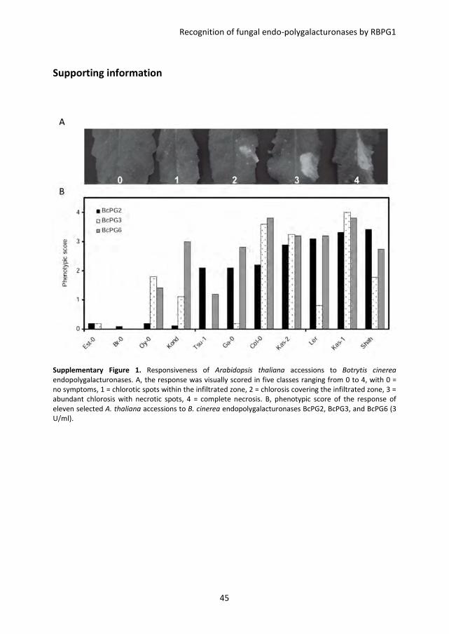
Recognition of fungal endo-polygalacturonases by RBPG1
45
Supporting information
Supplementary Figure 1. Responsiveness of Arabidopsis thaliana accessions to Botrytis cinerea
endopolygalacturonases. A, the response was visually scored in five classes ranging from 0 to 4, with 0 =
no symptoms, 1 = chlorotic spots within the infiltrated zone, 2 = chlorosis covering the infiltrated zone, 3 =
abundant chlorosis with necrotic spots, 4 = complete necrosis. B, phenotypic score of the response of
eleven selected A. thaliana accessions to B. cinerea endopolygalacturonases BcPG2, BcPG3, and BcPG6 (3
U/ml).
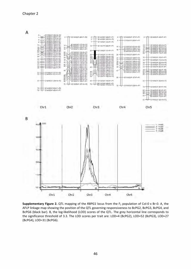
Chapter 2
46
Supplementary Figure 2. QTL mapping of the RBPG1 locus from the F2 population of Col-0 x Br-0. A, the
AFLP linkage map showing the position of the QTL governing responsiveness to BcPG2, BcPG3, BcPG4, and
BcPG6 (black bar). B, the log-likelihood (LOD) scores of the QTL. The grey horizontal line corresponds to
the significance threshold of 3.3. The LOD scores per trait are: LOD=4 (BcPG2), LOD=52 (BcPG3), LOD=27
(BcPG4), LOD=31 (BcPG6).
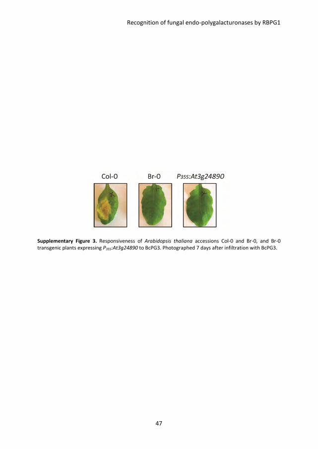
Recognition of fungal endo-polygalacturonases by RBPG1
47
Supplementary Figure 3. Responsiveness of Arabidopsis thaliana accessions Col-0 and Br-0, and Br-0
transgenic plants expressing P35S:At3g24890 to BcPG3. Photographed 7 days after infiltration with BcPG3.
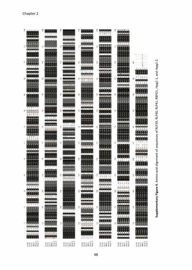
Chapter 2
48
Su
pp
lem
en
tary
Fig
ure
4.
Am
ino
aci
d a
lig
nm
en
t o
f se
qu
en
ces
of
RLP
39
, R
LP4
0,
RLP
41
, R
BP
G1
, rb
pg
1-1
, a
nd
rb
pg
1-2
.
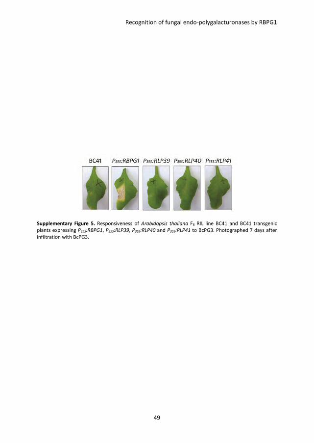
Recognition of fungal endo-polygalacturonases by RBPG1
49
Supplementary Figure 5. Responsiveness of Arabidopsis thaliana F8 RIL line BC41 and BC41 transgenic
plants expressing P35S:RBPG1, P35S:RLP39, P35S:RLP40 and P35S:RLP41 to BcPG3. Photographed 7 days after
infiltration with BcPG3.
Chapter 2
50
Supplementary Table 1. Arabidopsis thaliana accessions used in this study.
Accessions Origin Stock centre number
Br-0a Brno (Czech Republic) N6626
Bs-1 Basel (Switzerland) N6627
Cnt-1 Canterbury (UK) N1635
Co-0 Coimbra (Portugal) N6669
Col-0a Columbia (USA) N907
Col-4 Columbia (USA) N933
Ct-1 Catania (Italy) N1094
Cvi Cape Verdi Islands N8580
Cvi-1 (bas) Cape Verdi Islands N8580
Di (Dijon-G) Dijon (France) N10159
Eden-2b Eden, N Sweden (Sweden) CS76125
Ei-2 Eifel (Germany) N6689
Ema-1 East Malling (UK) N1637
En-2 Enkheim (Germany) N1138
Eri Eriengsboda (Sweden) N10042
Est-0a Estonia (Russia) N6700
Est-1b Estonia (Russia) CS76127
Ga-0a Gabelstein (Germany) N6714
Ga-2 Gabelstein (Germany) N10209
Gy-0 La Miniere (France) N6732
Ka-0 Karnten (Austria) N6752
Kas-1a Kashmir (India) N903
Kas-2a Kashmir (India) N1264
Konda Kondara (Tadjikistan) N9175
Kyoto Kyoto (Japan) N10231
Lera Landsberg (Germany) NW20
Ler Koornneef Landsberg (Germany) N8581
Li-0 Limburg (Germany) N6775
Lip-0 Lipowiec/Chrzanow (Poland) N6780
Lm-2 Le Mans (France) N6784
Lz-0 Lezoux/Puy-de-Dome (France) N6788
Ma-0 Marburg/Lahn (Germany) N6789
Nd-0 Niederzenz (Germany) N6803
Nd-1 Niederzenz (Germany) N1636
No-0 Halle (Germany) N1394
Nok-1 Noordwijk (Netherlands) N6807
Oy-0a Oystese (Norway) N6824
Pi-0 Pitztal Tirol (Austria) N6832
Rld-1 Netherlands N913
Rsch-0 Rschew/Starize (Russia) N6848
Shaha Pamiro-Alay (Tadjikistan) N929
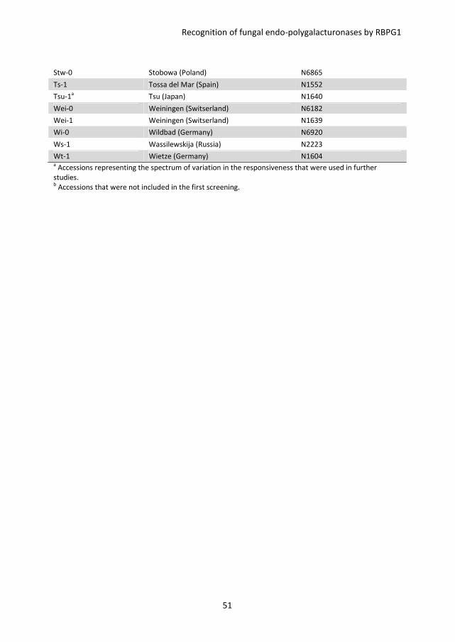
Recognition of fungal endo-polygalacturonases by RBPG1
51
Stw-0 Stobowa (Poland) N6865
Ts-1 Tossa del Mar (Spain) N1552
Tsu-1a Tsu (Japan) N1640
Wei-0 Weiningen (Switserland) N6182
Wei-1 Weiningen (Switserland) N1639
Wi-0 Wildbad (Germany) N6920
Ws-1 Wassilewskija (Russia) N2223
Wt-1 Wietze (Germany) N1604 a Accessions representing the spectrum of variation in the responsiveness that were used in further
studies. b Accessions that were not included in the first screening.
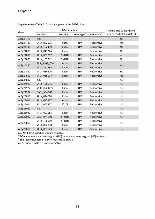
Chapter 2
52
Supplementary Table 2. Candidate genes in the RBPG1 locus.
Gene T-DNA mutant Amino acid substitutions
( between Col-0 and Br-0) Number Location Genotypea Phenotype
b
At3g24770 n.a. - - - Yes
At3g24780 SALK_082006 Exon HM Responsive No
At3g24790 SALK_152499 Exon HM Responsive No
At3g24800 SALK_049244 Exon HT Responsive No
At3g24810 SALK_089717 5’-UTR HM Responsive Yes
At3g24820 SALK_103742 5’-UTR HM Responsive No
At3g24840 SAIL_1168_C04 Intron HM Responsive
Yes SALK_122535 Exon HM Responsive
At3g24850 SALK_036306 Exon HM Responsive Yes
At3g24860 SALK_038594 Exon HM Responsive No
At3g24890 n.a. - - - n.i.
At3g24900 SALK_126505 Exon HM Responsive n.i.
At3g24927 SAIL_544_G05 Exon HM Responsive n.i.
At3g24982 GABI_564D03 Exon HM Responsive n.i.
At3g25010 SALK_024020 Exon HM Responsive n.i.
At3g25013 SALK_045377 Intron HM Responsive n.i.
At3g25014 SALK_045377 5’UTR HM Responsive n.i.
At3g25020 n.a. - - - n.i.
At3g25030 SALK_091158 Exon HM Responsive n.i.
At3g25040 GABI_044G01 5’-UTR HM Responsive n.i.
At3g25050 SALK_106019 5’-UTR HM Responsive
n.i. SALK_032898 Exon HM Responsive
At3g25060 SALK_083673 Exon HM Responsive n.i.
n.a. No T-DNA insertion mutant available. a T-DNA mutants are homozygous (HM) mutants or heterozygous (HT) mutants.
b The responsiveness of T-DNA mutants to BcPG3.
n.i. Sequence in Br-0 is not informative.
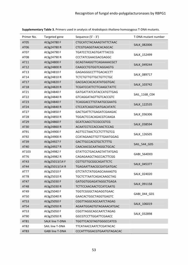
Recognition of fungal endo-polygalacturonases by RBPG1
53
Supplementary Table 3. Primers used in analysis of Arabidopsis thaliana homozygous T-DNA mutants.
Primer No. Targeted gene Sequence (5' - 3') T-DNA mutant number
AT05 At3g24780 F CTGCATCTACAAAGTATTCTAAC SALK_082006
AT06 At3g24780 R CTCGTGAGGTAACACAGCAC
AT07 At3g24790 F TGATCCTCCAGTGATTTACCG SALK_152499
AT08 At3g24790 R CCCTATCGAACGACGAGGC
AT11 At3g24800 F GCAGTAAGGTTCAGAAAACGCT SALK_049244
AT12 At3g24800 R CAAGCCTGTGGTCAGGAGTG
AT13 At3g24810 F GAGAGGGCCTTTGACACCTT SALK_089717
AT14 At3g24810 R TCTCTGTTGTTGCTGTTCTGC
AT17 At3g24820 F GACGACCACACATATGGTGAC SALK_103742
AT18 At3g24820 R TCGATCCATTCTTCAAGCTATTC
AT21 At3g24840 F GATGATTATCATACCATGTTGAG SAIL_1168_C04
AT22 At3g24840 R GTCAGGATAGTTGTCACCGTC
AT23 At3g24840 F TCAGGACCTTGTAATGCGAATG SALK_122535
AT24 At3g24840 R CTCCATCAGGTGATGACATATC
AT27 At3g24850 F GACTGATTCTGAGATCGAAGAC SALK_036306
AT28 At3g24850 R TGGACTCCACAGACGTCAAGA
AT29 At3g24860 F GCATCAACCTCCGCCGTCG SALK_038594
AT30 At3g24860 R ACAATCCTCCACCAACTCCAG
AT91 At3g24900 F AGTTCCTAACTCCTCTTTGTCG SALK_126505
AT92 At3g24900 R CCATAGAAGTTGTTTGAATGGAG
AT39 At3g24927 F GACTTGCCACGTGCTCTTTG SAIL_544_G05
AT40 At3g24927 R CAACAACGCAATAGGCTGCAC
AT103 At3g24982 F GTATTCCTGACAAGTATTATGAG GABI_564D03
AT76 At3g24982 R CAGAGAAGCTAGCCACTTCGG
AT43 At3g25013/14 F CGTTGTTGCGGCAGATTCTC SALK_045377
AT44 At3g25013/14 R TGAGAATTAACGCGATGATGAC
AT77 At3g25010 F GTCTATCTATGGAGCAAAAGTG SALK_024020
AT78 At3g25010 R TGCTCTTAATCAGACAAGCTAG
AT47 At3g25030 F GATGGTGGAGATAGGCTGAGA SALK_091158
AT48 At3g25030 R TCTTCCAACAACTCCATCAATG
AT49 At3g25040 F TGGTCGGGCTAGAGGTGAAC GABI_044_G01
AT50 At3g25040 R GAACACTGGCTAGGTGAGTC
AT53 At3g25050 F CGGTTAGGCAGCAATCTAGAG SALK_106019
AT54 At3g25050 R AGAATGGAGTGTAGAAACATGAC
AT53 At3g25050 F CGGTTAGGCAGCAATCTAGAG SALK_032898
AT90 At3g25050 R GGCGTCCTTGGATTCGAACC
AT81 SALK line T-DNA TGGTTCACGTAGTGGGCCATCG
AT82 SAIL line T-DNA TTCATAACCAATCTCGATACAC
AT83 GABI line T-DNA CCCATTTGGACGTGAATGTAGACAC
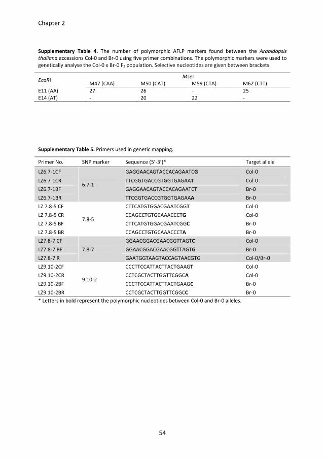
Chapter 2
54
Supplementary Table 4. The number of polymorphic AFLP markers found between the Arabidopsis
thaliana accessions Col-0 and Br-0 using five primer combinations. The polymorphic markers were used to
genetically analyse the Col-0 x Br-0 F2 population. Selective nucleotides are given between brackets.
EcoRI MseI
M47 (CAA) M50 (CAT) M59 (CTA) M62 (CTT)
E11 (AA) 27 26 - 25
E14 (AT) - 20 22 -
Supplementary Table 5. Primers used in genetic mapping.
Primer No. SNP marker Sequence (5’-3’)* Target allele
LZ6.7-1CF
6.7-1
GAGGAACAGTACCACAGAATCG Col-0
LZ6.7-1CR TTCGGTGACCGTGGTGAGAAT Col-0
LZ6.7-1BF GAGGAACAGTACCACAGAATCT Br-0
LZ6.7-1BR TTCGGTGACCGTGGTGAGAAA Br-0
LZ 7.8-5 CF
7.8-5
CTTCATGTGGACGAATCGGT Col-0
LZ 7.8-5 CR CCAGCCTGTGCAAACCCTG Col-0
LZ 7.8-5 BF CTTCATGTGGACGAATCGGC Br-0
LZ 7.8-5 BR CCAGCCTGTGCAAACCCTA Br-0
LZ7.8-7 CF
7.8-7
GGAACGGACGAACGGTTAGTC Col-0
LZ7.8-7 BF GGAACGGACGAACGGTTAGTG Br-0
LZ7.8-7 R GAATGGTAAGTACCAGTAACGTG Col-0/Br-0
LZ9.10-2CF
9.10-2
CCCTTCCATTACTTACTGAAGT Col-0
LZ9.10-2CR CCTCGCTACTTGGTTCGGCA Col-0
LZ9.10-2BF CCCTTCCATTACTTACTGAAGC Br-0
LZ9.10-2BR CCTCGCTACTTGGTTCGGCC Br-0
* Letters in bold represent the polymorphic nucleotides between Col-0 and Br-0 alleles.
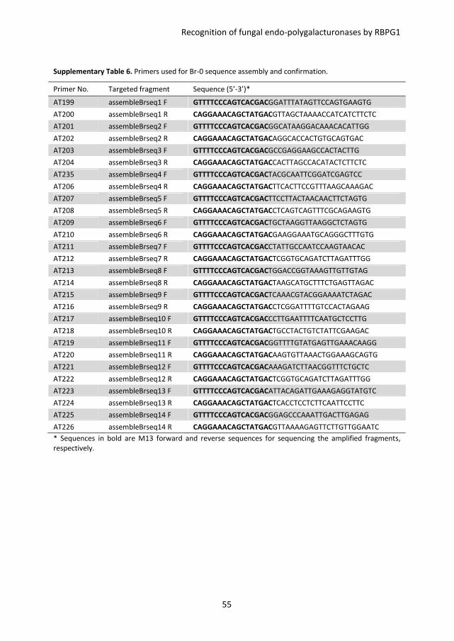
Recognition of fungal endo-polygalacturonases by RBPG1
55
Supplementary Table 6. Primers used for Br-0 sequence assembly and confirmation.
Primer No. Targeted fragment Sequence (5’-3’)*
AT199 assembleBrseq1 F GTTTTCCCAGTCACGACGGATTTATAGTTCCAGTGAAGTG
AT200 assembleBrseq1 R CAGGAAACAGCTATGACGTTAGCTAAAACCATCATCTTCTC
AT201 assembleBrseq2 F GTTTTCCCAGTCACGACGGCATAAGGACAAACACATTGG
AT202 assembleBrseq2 R CAGGAAACAGCTATGACAGGCACCACTGTGCAGTGAC
AT203 assembleBrseq3 F GTTTTCCCAGTCACGACGCCGAGGAAGCCACTACTTG
AT204 assembleBrseq3 R CAGGAAACAGCTATGACCACTTAGCCACATACTCTTCTC
AT235 assembleBrseq4 F GTTTTCCCAGTCACGACTACGCAATTCGGATCGAGTCC
AT206 assembleBrseq4 R CAGGAAACAGCTATGACTTCACTTCCGTTTAAGCAAAGAC
AT207 assembleBrseq5 F GTTTTCCCAGTCACGACTTCCTTACTAACAACTTCTAGTG
AT208 assembleBrseq5 R CAGGAAACAGCTATGACCTCAGTCAGTTTCGCAGAAGTG
AT209 assembleBrseq6 F GTTTTCCCAGTCACGACTGCTAAGGTTAAGGCTCTAGTG
AT210 assembleBrseq6 R CAGGAAACAGCTATGACGAAGGAAATGCAGGGCTTTGTG
AT211 assembleBrseq7 F GTTTTCCCAGTCACGACCTATTGCCAATCCAAGTAACAC
AT212 assembleBrseq7 R CAGGAAACAGCTATGACTCGGTGCAGATCTTAGATTTGG
AT213 assembleBrseq8 F GTTTTCCCAGTCACGACTGGACCGGTAAAGTTGTTGTAG
AT214 assembleBrseq8 R CAGGAAACAGCTATGACTAAGCATGCTTTCTGAGTTAGAC
AT215 assembleBrseq9 F GTTTTCCCAGTCACGACTCAAACGTACGGAAAATCTAGAC
AT216 assembleBrseq9 R CAGGAAACAGCTATGACCTCGGATTTTGTCCACTAGAAG
AT217 assembleBrseq10 F GTTTTCCCAGTCACGACCCTTGAATTTTCAATGCTCCTTG
AT218 assembleBrseq10 R CAGGAAACAGCTATGACTGCCTACTGTCTATTCGAAGAC
AT219 assembleBrseq11 F GTTTTCCCAGTCACGACGGTTTTGTATGAGTTGAAACAAGG
AT220 assembleBrseq11 R CAGGAAACAGCTATGACAAGTGTTAAACTGGAAAGCAGTG
AT221 assembleBrseq12 F GTTTTCCCAGTCACGACAAAGATCTTAACGGTTTCTGCTC
AT222 assembleBrseq12 R CAGGAAACAGCTATGACTCGGTGCAGATCTTAGATTTGG
AT223 assembleBrseq13 F GTTTTCCCAGTCACGACATTACAGATTGAAAGAGGTATGTC
AT224 assembleBrseq13 R CAGGAAACAGCTATGACTCACCTCCTCTTCAATTCCTTC
AT225 assembleBrseq14 F GTTTTCCCAGTCACGACGGAGCCCAAATTGACTTGAGAG
AT226 assembleBrseq14 R CAGGAAACAGCTATGACGTTAAAAGAGTTCTTGTTGGAATC
* Sequences in bold are M13 forward and reverse sequences for sequencing the amplified fragments,
respectively.
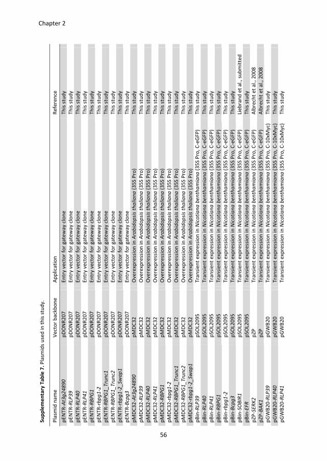
Su
pp
lem
en
tary
Ta
ble
7.
Pla
smid
s u
sed
in
th
is s
tud
y.
Pla
smid
na
me
V
ect
or
ba
ckb
on
e
Ap
plica
tio
n
Re
fere
nce
pE
NT
R-A
t3g
24
89
0
pD
ON
R2
07
E
ntr
y v
ect
or
for
ga
tew
ay c
lon
e
Th
is s
tud
y
pE
NT
R-R
LP3
9
pD
ON
R2
07
E
ntr
y v
ect
or
for
ga
tew
ay c
lon
e
Th
is s
tud
y
pE
NT
R-R
LP4
0
pD
ON
R2
07
E
ntr
y v
ect
or
for
ga
tew
ay c
lon
e
Th
is s
tud
y
pE
NT
R-R
LP4
1
pD
ON
R2
07
E
ntr
y v
ect
or
for
ga
tew
ay c
lon
e
Th
is s
tud
y
pE
NT
R-R
BP
G1
p
DO
NR
20
7
En
try v
ect
or
for
ga
tew
ay c
lon
e
Th
is s
tud
y
pE
NT
R-r
bp
g1
-2
pD
ON
R2
07
E
ntr
y v
ect
or
for
ga
tew
ay c
lon
e
Th
is s
tud
y
pE
NT
R-R
BP
G1
_T
run
c1
pD
ON
R2
07
E
ntr
y v
ect
or
for
ga
tew
ay c
lon
e
Th
is s
tud
y
pE
NT
R-R
BP
G1
_T
run
c2
pD
ON
R2
07
E
ntr
y v
ect
or
for
ga
tew
ay c
lon
e
Th
is s
tud
y
pE
NT
R-r
bp
g1
-2_
Sw
ap
1
pD
ON
R2
07
E
ntr
y v
ect
or
for
ga
tew
ay c
lon
e
Th
is s
tud
y
pE
NT
R-B
cpg
3
pD
ON
R2
07
E
ntr
y v
ect
or
for
ga
tew
ay c
lon
e
Th
is s
tud
y
pM
DC
32
-At3
g2
48
90
p
MD
C3
2
Ove
rexp
ress
ion
in
Ara
bid
op
sis
tha
lia
na
(3
5S
Pro
) T
his
stu
dy
pM
DC
32
-RLP
39
p
MD
C3
2
Ove
rexp
ress
ion
in
Ara
bid
op
sis
tha
lia
na
(3
5S
Pro
) T
his
stu
dy
pM
DC
32
-RLP
40
p
MD
C3
2
Ove
rexp
ress
ion
in
Ara
bid
op
sis
tha
lia
na
(3
5S
Pro
) T
his
stu
dy
pM
DC
32
-RLP
41
p
MD
C3
2
Ove
rexp
ress
ion
in
Ara
bid
op
sis
tha
lia
na
(3
5S
Pro
) T
his
stu
dy
pM
DC
32
-RB
PG
1
pM
DC
32
O
ve
rexp
ress
ion
in
Ara
bid
op
sis
tha
lia
na
(3
5S
Pro
) T
his
stu
dy
pM
DC
32
-rb
pg
1-2
p
MD
C3
2
Ove
rexp
ress
ion
in
Ara
bid
op
sis
tha
lia
na
(3
5S
Pro
) T
his
stu
dy
pM
DC
32
-RB
PG
1_
Tru
nc1
p
MD
C3
2
Ove
rexp
ress
ion
in
Ara
bid
op
sis
tha
lia
na
(3
5S
Pro
) T
his
stu
dy
pM
DC
32
-RB
PG
1_
Tru
nc2
p
MD
C3
2
Ove
rexp
ress
ion
in
Ara
bid
op
sis
tha
lia
na
(3
5S
Pro
) T
his
stu
dy
pM
DC
32
-rb
pg
1-2
_Sw
ap
1
pM
DC
32
O
ve
rexp
ress
ion
in
Ara
bid
op
sis
tha
lia
na
(3
5S
Pro
) T
his
stu
dy
pB
in-R
LP3
9
pS
OL2
09
5
Tra
nsi
en
t e
xpre
ssio
n in
Nic
oti
an
a b
en
tha
mia
na
(3
5S
Pro
, C
-eG
FP
) T
his
stu
dy
pB
in-R
LP4
0
pS
OL2
09
5
Tra
nsi
en
t e
xpre
ssio
n in
Nic
oti
an
a b
en
tha
mia
na
(3
5S
Pro
, C
-eG
FP
) T
his
stu
dy
pB
in-R
LP4
1
pS
OL2
09
5
Tra
nsi
en
t e
xpre
ssio
n in
Nic
oti
an
a b
en
tha
mia
na
(3
5S
Pro
, C
-eG
FP
) T
his
stu
dy
pB
in-R
BP
G1
p
SO
L20
95
T
ran
sie
nt
exp
ress
ion
in
Nic
oti
an
a b
en
tha
mia
na
(3
5S
Pro
, C
-eG
FP
) T
his
stu
dy
pB
in-r
bp
g1
-2
pS
OL2
09
5
Tra
nsi
en
t e
xpre
ssio
n in
Nic
oti
an
a b
en
tha
mia
na
(3
5S
Pro
, C
-eG
FP
) T
his
stu
dy
pB
in-B
cpg
3
pS
OL2
09
5
Tra
nsi
en
t e
xpre
ssio
n in
Nic
oti
an
a b
en
tha
mia
na
(3
5S
Pro
, C
-eG
FP
) T
his
stu
dy
pB
in-S
OB
IR1
p
SO
L20
95
T
ran
sie
nt
exp
ress
ion
in
Nic
oti
an
a b
en
tha
mia
na
(3
5S
Pro
, C
-eG
FP
) Li
eb
ran
d e
t a
l.,
sub
mit
ted
pB
in-E
FR
p
SO
L20
95
T
ran
sie
nt
exp
ress
ion
in
Nic
oti
an
a b
en
tha
mia
na
(3
5S
Pro
, C
-eG
FP
) T
his
stu
dy
pZ
P-S
ER
K2
p
ZP
T
ran
sie
nt
exp
ress
ion
in
Nic
oti
an
a b
en
tha
mia
na
(3
5S
Pro
, C
-eG
FP
) A
lbre
cht
et
al.
, 2
00
8
pZ
P-B
AK
1
pZ
P
Tra
nsi
en
t e
xpre
ssio
n in
Nic
oti
an
a b
en
tha
mia
na
(3
5S
Pro
, C
-eG
FP
) A
lbre
cht
et
al.
, 2
00
8
pG
WB
20
-RLP
39
p
GW
B2
0
Tra
nsi
en
t e
xpre
ssio
n in
Nic
oti
an
a b
en
tha
mia
na
(3
5S
Pro
, C
-10
xMyc)
T
his
stu
dy
pG
WB
20
-RLP
40
p
GW
B2
0
Tra
nsi
en
t e
xpre
ssio
n in
Nic
oti
an
a b
en
tha
mia
na
(3
5S
Pro
, C
-10
xMyc)
T
his
stu
dy
pG
WB
20
-RLP
41
p
GW
B2
0
Tra
nsi
en
t e
xpre
ssio
n in
Nic
oti
an
a b
en
tha
mia
na
(3
5S
Pro
, C
-10
xMyc)
T
his
stu
dy
Chapter 2
56
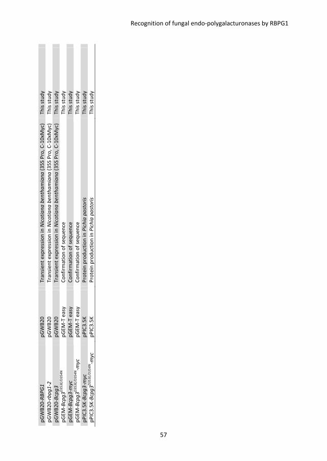
pG
WB
20
-RB
PG
1
pG
WB
20
T
ran
sie
nt
exp
ress
ion
in
Nic
oti
an
a b
en
tha
mia
na
(3
5S
Pro
, C
-10
xMyc)
T
his
stu
dy
pG
WB
20
-rb
pg
1-2
p
GW
B2
0
Tra
nsi
en
t e
xpre
ssio
n in
Nic
oti
an
a b
en
tha
mia
na
(3
5S
Pro
, C
-10
xMyc)
T
his
stu
dy
pG
WB
20
-Bcp
g3
p
GW
B2
0
Tra
nsi
en
t e
xpre
ssio
n in
Nic
oti
an
a b
en
tha
mia
na
(3
5S
Pro
, C
-10
xMyc)
T
his
stu
dy
pG
EM
-Bcp
g3
D3
53
E/D
35
4N
pG
EM
-T e
asy
C
on
firm
ati
on
of
seq
ue
nce
T
his
stu
dy
pG
EM
-Bcp
g3
-myc
p
GE
M-T
ea
sy
Co
nfi
rma
tio
n o
f se
qu
en
ce
Th
is s
tud
y
pG
EM
-Bcp
g3
D3
53
E/D
35
4N-m
yc
pG
EM
-T e
asy
C
on
firm
ati
on
of
seq
ue
nce
T
his
stu
dy
pP
IC3
.5K
-Bcp
g3
-myc
p
PIC
3.5
K
Pro
tein
pro
du
ctio
n i
n P
ich
ia p
ast
ori
s T
his
stu
dy
pP
IC3
.5K
-Bcp
g3
D3
53
E/D
35
4N-m
yc
pP
IC3
.5K
P
rote
in p
rod
uct
ion
in
Pic
hia
pa
sto
ris
Th
is s
tud
y
Recognition of fungal endo-polygalacturonases by RBPG1
57
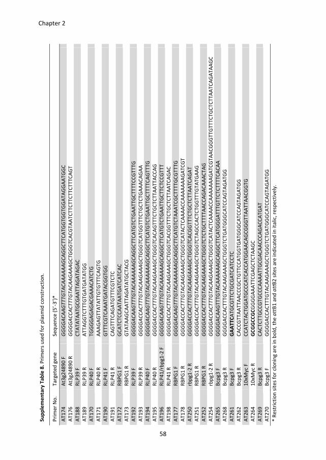
Su
pp
lem
en
tary
Ta
ble
8.
Pri
me
rs u
sed
fo
r p
lasm
id c
on
stru
ctio
n.
Pri
me
r N
o.
Ta
rge
ted
ge
ne
S
eq
ue
nce
(5
’-3
’)*
AT
17
4
At3
g2
48
90
F
GG
GG
AC
AA
GT
TT
GT
AC
AA
AA
AA
GC
AG
GC
TT
CA
TG
GT
GG
TG
GA
TA
GG
AA
TG
GC
AT
17
6
At3
g2
48
90
R
GG
GG
AC
CA
CT
TT
GT
AC
AA
GA
AA
GC
TG
GG
TC
AC
GT
AA
TC
TT
CT
TC
TT
CT
TT
CA
GT
AT
18
8
RLP
39
F
CT
AT
AT
AA
TG
CG
AA
TT
AG
AT
AG
AC
AT
18
9
RLP
39
R
AT
TA
GG
TC
TT
TG
TG
AT
GA
TA
TG
G
AT
17
0
RLP
40
F
TG
GG
GA
GA
GA
CG
AA
AC
AT
CT
G
AT
17
1
RLP
40
R
AA
AG
AG
TT
AT
AT
TG
TG
TT
TC
AG
TG
AT
19
0
RLP
41
F
GT
TT
CG
TC
AA
AT
GA
TA
CG
GT
GG
AT
19
1
RLP
41
R
CA
GT
TT
CA
GA
TG
TT
TC
GT
CT
C
AT
17
2
RB
PG
1 F
G
CA
TC
TC
CA
AT
AA
TG
AT
CA
TC
AC
AT
17
3
RB
PG
1 R
G
TA
TA
AG
CA
AT
TA
GA
TA
TG
CT
AC
G
AT
19
2
RLP
39
F
GG
GG
AC
AA
GT
TT
GT
AC
AA
AA
AA
GC
AG
GC
TT
CA
TG
TC
TG
AA
TT
GC
TT
TT
CC
GT
TT
G
AT
19
3
RLP
39
R
GG
GG
AC
CA
CT
TT
GT
AC
AA
GA
AA
GC
TG
GG
TC
AT
GG
TT
TC
TG
CT
CT
GA
AA
CA
GA
A
AT
19
4
RLP
40
F
GG
GG
AC
AA
GT
TT
GT
AC
AA
AA
AA
GC
AG
GC
TT
CA
TG
TC
TG
AA
TT
GC
TT
TT
CA
GT
TT
G
AT
19
5
RLP
40
R
GG
GG
AC
CA
CT
TT
GT
AC
AA
GA
AA
GC
TG
GG
TC
AC
AG
TT
TC
TG
CT
CT
TA
AT
TA
CC
AG
AT
19
6
RLP
41
/rb
pg
1-2
F
GG
GG
AC
AA
GT
TT
GT
AC
AA
AA
AA
GC
AG
GC
TT
CA
TG
TC
TG
AA
TT
GC
TT
CT
CC
GT
TT
AT
19
8
RLP
41
R
GG
GG
AC
CA
CT
TT
GT
AC
AA
GA
AA
GC
TG
GG
TC
AC
GG
TT
TC
TG
CT
CT
TA
AT
CA
GA
C
AT
17
7
RB
PG
1 F
G
GG
GA
CA
AG
TT
TG
TA
CA
AA
AA
AG
CA
GG
CT
TC
AT
GT
CT
AA
AT
CG
CT
TT
TG
CG
TT
TG
AT
17
8
RB
PG
1 R
G
GG
GA
CC
AC
TT
TG
TA
CA
AG
AA
AG
CT
GG
GT
CA
TA
CT
CA
AA
AC
CA
AA
AA
AA
GA
TC
GT
AT
25
0
rbp
g1
-2 R
G
GG
GA
CC
AC
TT
TG
TA
CA
AG
AA
AG
CT
GG
GT
CA
CG
GT
TT
CT
GC
TC
TT
AA
TC
AG
AT
AT
25
1
RB
PG
1 R
G
GG
GA
CC
AC
TT
TG
TA
CA
AG
AA
AG
CT
GG
GT
CT
AG
CC
AC
TC
TG
GT
TT
GT
AT
GA
AG
AT
25
2
RB
PG
1 R
G
GG
GA
CC
AC
TT
TG
TA
CA
AG
AA
AG
CT
GG
GT
CT
CT
GC
TT
TT
AA
CC
AG
AC
AA
AC
TA
G
AT
25
4
rbp
g1
-2 R
G
GG
GA
CC
AC
TT
TG
TA
CA
AG
AA
AG
CT
GG
GT
CA
TA
CT
CA
AA
AC
CA
AA
AA
AA
GA
TC
GT
AA
CG
GG
TT
GT
TT
CT
GC
TC
TT
AA
TC
AG
AT
AA
GC
AT
26
5
Bcp
g3
F
GG
GG
AC
AA
GT
TT
GT
AC
AA
AA
AA
GC
AG
GC
TT
CA
TG
GG
AT
TT
GT
TC
TC
TT
TT
CA
CA
A
AT
26
8
Bcp
g3
R
GG
GG
AC
CA
CT
TT
GT
AC
AA
GA
AA
GC
TG
GG
TC
TG
AT
GG
GC
AT
CC
AG
TA
GA
TG
G
AT
26
1
Bcp
g3
F
GA
AT
TC
AT
GC
GT
TC
TG
CG
AT
CA
TC
CT
C
AT
26
2
Bcp
g3
R
CA
CC
GT
TA
AT
TA
AC
CC
GC
TG
TT
CC
AT
GG
TG
AT
GG
GC
AT
CC
AG
TA
GA
TG
G
AT
26
3
10
xMyc
F
CC
AT
CT
AC
TG
GA
TG
CC
CA
TC
AC
CA
TG
GA
AC
AG
CG
GG
TT
AA
TT
AA
CG
GT
G
AT
26
4
10
xMyc
R
GC
GG
CC
GC
GG
GG
AA
AT
TC
GA
GC
TC
TA
AG
C
AT
26
9
Bcp
g3
R
CA
CT
CT
TG
CC
GT
GC
CC
AA
AA
TT
GC
GA
CA
AC
CC
AG
AC
CA
TG
AT
AT
27
0
Bcp
g3
F
GG
GG
AC
CA
CT
TT
GT
AC
AA
GA
AA
GC
TG
GG
TC
TG
AT
GG
GC
AT
CC
AG
TA
GA
TG
G
* R
est
rict
ion
sit
es
for
clo
nin
g a
re in
bo
ld,
the
att
B1
an
d a
ttB
2 s
ite
s a
re in
dic
ate
d i
n ita
lic,
re
spe
ctiv
ely
.
Chapter 2
58
CHAPTER 3
The D-galacturonic acid catabolic pathway in Botrytis cinerea
Lisha Zhang, Harry Thiewes and Jan A. L. van Kan
This chapter is published as:
Zhang, L., Thiewes, H. and van Kan, J.A.L. (2011) The D-galacturonic acid catabolic pathway in Botrytis
cinerea. Fungal Genet. Biol. 48, 990-997.
Chapter 3
60
Abstract
D-galacturonic acid is the most abundant component of pectin, one of the major
polysaccharide constituents of plant cell walls. D-galacturonic acid potentially is an
important carbon source for microorganisms living on (decaying) plant material. A
catabolic pathway was proposed in filamentous fungi, comprising three enzymatic steps,
involving D-galacturonate reductase, L-galactonate dehydratase, and 2-keto-3-deoxy-L-
galactonate aldolase. We describe the functional, biochemical and genetic
characterization of the entire D-galacturonate-specific catabolic pathway in the plant
pathogenic fungus Botrytis cinerea. The B. cinerea genome contains two non-homologous
galacturonate reductase genes (Bcgar1 and Bcgar2), a galactonate dehydratase gene
(Bclgd1), and a 2-keto-3-deoxy-L-galactonate aldolase gene (Bclga1). Their expression
levels were highly induced in cultures containing D-galacturonic acid, pectate, or pectin as
the sole carbon source. The four proteins were expressed in Escherichia coli and their
enzymatic activity was characterized. Targeted gene replacement of all four genes in B.
cinerea, either separately or in combinations, yielded mutants that were affected in
growth on D-galacturonic acid, pectate, or pectin as the sole carbon source. In Aspergillus
nidulans and A. niger, the first catabolic conversion only involves the Bcgar2 ortholog,
while in Hypocrea jecorina, it only involves the Bcgar1 ortholog. In B. cinerea, however,
BcGAR1 and BcGAR2 jointly contribute to the first step of the catabolic pathway, albeit to
different extent. The virulence of all B. cinerea mutants in the D-galacturonic acid
catabolic pathway on tomato leaves, apple fruit and bell peppers was unaltered.
The D-galacturonic acid catabolic pathway in Botrytis cinerea
61
Introduction
D-galacturonic acid is the major component of the plant cell wall polysaccharide pectin.
Consequently, D-galacturonic acid potentially is an important carbon source for
microorganisms living on plant material, either as a saprotroph or a pathogen (Richard and
Hilditch, 2009). For bacteria, a catabolic pathway has been described in which five
enzymes convert D-galacturonic acid to pyruvate and glyceraldehyde-3-phosphate, via the
intermediate metabolites D-tagaturonate, D-altronate, 2-keto-3-deoxy-gluconate and 2-
keto-3-deoxy-6-phospho-gluconate (Richard and Hilditch, 2009).
A different catabolic pathway occurs in filamentous fungi. Early studies in Aspergillus
nidulans cultured on D-galacturonic acid as sole carbon source showed that mutants in
pyruvate dehydrogenase or pyruvate carboxylase were unable to grow, whereas a
pyruvate kinase mutant remained able to use this carbon source. These observations
suggested that in A. nidulans, D-galacturonic acid is metabolized through a non-
phosphorylating pathway which yields glyceraldehyde and pyruvate (Hondmann et al.,
1991; Uitzetter et al., 1986; Visser et al., 1988). It was also shown that an NADPH-
dependent glycerol dehydrogenase was induced on D-galacturonic acid (Sealy-Lewis and
Fairhurst, 1992). The non-phosphorylating pathway in fungi was supported by more
recent studies on Hypocrea jecorina (anamorph Trichoderma reesei). In H. jecorina, D-
galacturonic acid is first converted to L-galactonate by GAR1, a galacturonate reductase
(Kuorelahti et al., 2005). Deletion of the gar1 gene in H. jecorina severely compromised
the ability to grow on D-galacturonic acid as the sole carbon source (Mojzita et al., 2010).
The second enzyme of the pathway is L-galactonate dehydratase (LGD1), which converts L-
galactonate to 2-keto-3-deoxy-L-galactonate. The H. jecorina lgd1 deletion mutant could
not grow on D-galacturonic acid (Kuorelahti et al., 2006). The next enzyme in the pathway
was identified as 2-keto-3-deoxy-L-galactonate aldolase (LGA1) which splits 2-keto-3-
deoxy-L-galactonate into pyruvate and L-glyceraldehyde (Hilditch et al., 2007). Deletion of
the lga1 gene resulted in the intracellular accumulation of 2-keto-3-deoxy-L-galactonate
(Wiebe et al., 2010). It was also reported that an NADPH-dependent glycerol
dehydrogenase (GLD1) is involved in the conversion of L-glyceraldehyde to glycerol as the
final step of this pathway (Liepins et al., 2006). The D-galacturonic acid catabolic pathway
in filamentous fungi, as proposed by Richard and Hilditch (2009) is summarized in Figure 1.
Transcriptome analysis of A. niger grown in liquid medium containing pectic substrates
showed that four genes, designated as gaaA, gaaB, gaaC and gaaD, were upregulated
during growth on pectin or D-galacturonic acid (Martens-Uzunova and Schaap, 2008).
Interestingly, the A. niger gaaA and gaaC genes are in the same locus, but transcribed in
opposite direction, sharing the same promoter region. The A. niger gaaA gene was shown
Chapter 3
62
to encode an active galacturonate reductase, yet the gene is not homologous to H.
jecorina gar1 and the A. niger GAAA protein has properties distinct from the H. jecorina
GAR1 protein (Martens-Uzunova and Schaap, 2008). The A. niger genome contains an
ortholog of H. jecorina gar1, designated A. niger gar1, located on a different scaffold and
its expression is not strongly induced by D-galacturonic acid (Martens-Uzunova and
Schaap, 2008). A gene that is orthologous to A. niger gaaA exists in H. jecorina, and is
designated as gar2 with unknown function (Richard and Hilditch, 2009).
Botrytis cinerea, a necrotrophic plant pathogenic fungus, is able to infect a wide range of
plant species and tissues. It often penetrates host tissues at the anticlinal cell wall and
subsequently grows into and through the middle lamella, which consists mostly of pectin.
Throughout the infection process, B. cinerea secretes a series of cell wall-degrading
enzymes (CWDEs) to break down plant cell wall polymers for plant surface penetration,
tissue invasion and nutrient release (van Kan, 2006; Williamson et al., 2007). Among these
CWDEs, several pectinases have been found to be abundant during infection, including
pectin and pectate lyases, pectin methylesterases (PMEs), exopolygalacturonases (exo-
PGs) and endopolygalacturonases (endo-PGs) (Cabanne and Doneche, 2002; Kars et al.,
2005b; Kars and van Kan, 2004; Rha et al., 2001; ten Have et al., 2001). The B. cinerea
genome contains six genes encoding endo-PGs (Wubben et al., 1999), of which Bcpg1 and
Bcpg2 are important for virulence (Kars et al., 2005a; ten Have et al., 1998). Although the
expression profiles of the Bcpg genes and the biochemical properties of the enzymes have
been well studied and the importance of pectin degradation for virulence of B. cinerea has
been documented (Kars et al., 2005a; ten Have et al., 1998), little is known about how B.
cinerea utilizes the ultimate hydrolytic product of pectin (i.e. D-galacturonic acid) for its
growth. We performed a genetic and biochemical characterization of the D-galacturonic
acid catabolic pathway in B. cinerea. The genes were identified and their expression was
investigated during growth on different carbon sources. The enzymatic activity of
recombinant proteins was characterized, and the function of the genes in B. cinerea was
studied by generating single and double deletion mutants and testing them in vitro and in
planta.
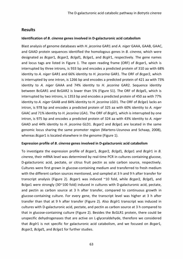
The D-galacturonic acid catabolic pathway in Botrytis cinerea
63
Results
Identification of B. cinerea genes involved in D-galacturonic acid catabolism
Blast analysis of genome databases with H. jecorina GAR1 and A. niger GAAA, GAAB, GAAC,
and GAAD protein sequences identified the homologous genes in B. cinerea, which were
designated as Bcgar1, Bcgar2, Bclgd1, Bclga1, and Bcglr1, respectively. The gene names
and locus tags are listed in Figure 1. The open reading frame (ORF) of Bcgar1, which is
interrupted by three introns, is 933 bp and encodes a predicted protein of 310 aa with 68%
identity to A. niger GAR1 and 66% identity to H. jecorina GAR1. The ORF of Bcgar2, which
is interrupted by one intron, is 1266 bp and encodes a predicted protein of 421 aa with 73%
identity to A. niger GAAA and 74% identity to H. jecorina GAR2. Sequence identity
between BcGAR1 and BcGAR2 is lower than 5% (Figure S1). The ORF of Bclgd1, which is
interrupted by two introns, is 1353 bp and encodes a predicted protein of 450 aa with 77%
identity to A. niger GAAB and 84% identity to H. jecorina LGD1. The ORF of Bclga1 lacks an
intron, is 978 bp and encodes a predicted protein of 325 aa with 60% identity to A. niger
GAAC and 71% identity to H. jecorina LGA1. The ORF of Bcglr1, which is interrupted by one
intron, is 975 bp and encodes a predicted protein of 324 aa with 43% identity to A. niger
GAAD and 44% identity to H. jecorina GLD1. Bcgar2 and Bclga1 are located in the same
genomic locus sharing the same promoter region (Martens-Uzunova and Schaap, 2008),
whereas Bcgar1 is located elsewhere in the genome (Figure 1).
Expression profile of B. cinerea genes involved in D-galacturonic acid catabolism
To investigate the expression profile of Bcgar1, Bcgar2, Bclgd1, Bclga1 and Bcglr1 in B.
cinerea, their mRNA level was determined by real-time PCR in cultures containing glucose,
D-galacturonic acid, pectate, or citrus fruit pectin as sole carbon source, respectively.
Cultures were first grown in glucose-containing medium and transferred to fresh medium
with the different carbon sources mentioned, and sampled at 3 h and 9 h after transfer for
transcript analysis (Figure 2). Bcgar1 was induced ~10 fold, while Bcgar2, Bclgd1, and
Bclga1 were strongly (30~100 fold) induced in cultures with D-galacturonic acid, pectate,
and pectin as carbon source at 3 h after transfer, compared to continuous growth in
glucose-containing culture. For every gene, the transcript level was higher at 3 h after
transfer than that at 9 h after transfer (Figure 2). Also Bcglr1 transcript was induced in
cultures with D-galacturonic acid, pectate, and pectin as carbon source at 3 h compared to
that in glucose-containing culture (Figure 2). Besides the BcGLR1 protein, there could be
unspecific dehydrogenases that are active on L-glyceraldehyde, therefore we considered
that Bcglr1 is not specific for galacturonic acid catabolism, and we focused on Bcgar1,
Bcgar2, Bclgd1, and Bclga1 for further studies.
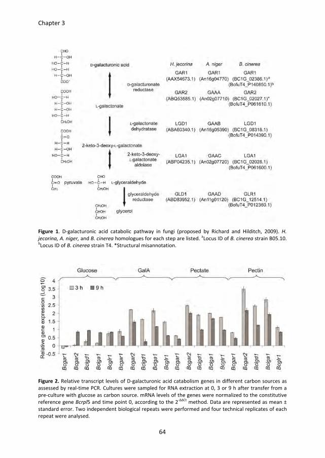
Chapter 3
64
Figure 1. D-galacturonic acid catabolic pathway in fungi (proposed by Richard and Hilditch, 2009). H.
jecorina, A. niger, and B. cinerea homologues for each step are listed. aLocus ID of B. cinerea strain B05.10.
bLocus ID of B. cinerea strain T4. *Structural misannotation.
Figure 2. Relative transcript levels of D-galacturonic acid catabolism genes in different carbon sources as
assessed by real-time PCR. Cultures were sampled for RNA extraction at 0, 3 or 9 h after transfer from a
pre-culture with glucose as carbon source. mRNA levels of the genes were normalized to the constitutive
reference gene Bcrpl5 and time point 0, according to the 2-ΔΔCt
method. Data are represented as mean ±
standard error. Two independent biological repeats were performed and four technical replicates of each
repeat were analysed.
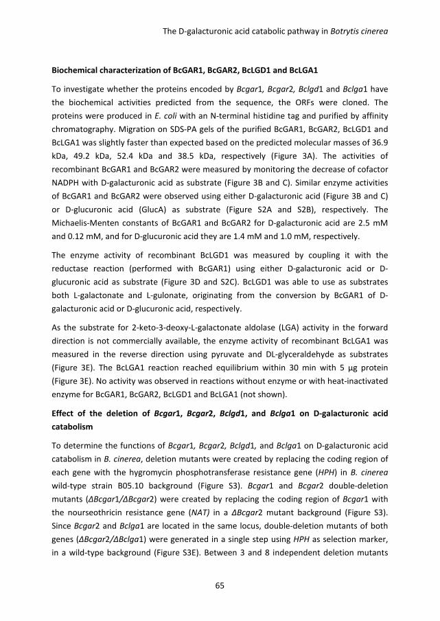
The D-galacturonic acid catabolic pathway in Botrytis cinerea
65
Biochemical characterization of BcGAR1, BcGAR2, BcLGD1 and BcLGA1
To investigate whether the proteins encoded by Bcgar1, Bcgar2, Bclgd1 and Bclga1 have
the biochemical activities predicted from the sequence, the ORFs were cloned. The
proteins were produced in E. coli with an N-terminal histidine tag and purified by affinity
chromatography. Migration on SDS-PA gels of the purified BcGAR1, BcGAR2, BcLGD1 and
BcLGA1 was slightly faster than expected based on the predicted molecular masses of 36.9
kDa, 49.2 kDa, 52.4 kDa and 38.5 kDa, respectively (Figure 3A). The activities of
recombinant BcGAR1 and BcGAR2 were measured by monitoring the decrease of cofactor
NADPH with D-galacturonic acid as substrate (Figure 3B and C). Similar enzyme activities
of BcGAR1 and BcGAR2 were observed using either D-galacturonic acid (Figure 3B and C)
or D-glucuronic acid (GlucA) as substrate (Figure S2A and S2B), respectively. The
Michaelis-Menten constants of BcGAR1 and BcGAR2 for D-galacturonic acid are 2.5 mM
and 0.12 mM, and for D-glucuronic acid they are 1.4 mM and 1.0 mM, respectively.
The enzyme activity of recombinant BcLGD1 was measured by coupling it with the
reductase reaction (performed with BcGAR1) using either D-galacturonic acid or D-
glucuronic acid as substrate (Figure 3D and S2C). BcLGD1 was able to use as substrates
both L-galactonate and L-gulonate, originating from the conversion by BcGAR1 of D-
galacturonic acid or D-glucuronic acid, respectively.
As the substrate for 2-keto-3-deoxy-L-galactonate aldolase (LGA) activity in the forward
direction is not commercially available, the enzyme activity of recombinant BcLGA1 was
measured in the reverse direction using pyruvate and DL-glyceraldehyde as substrates
(Figure 3E). The BcLGA1 reaction reached equilibrium within 30 min with 5 μg protein
(Figure 3E). No activity was observed in reactions without enzyme or with heat-inactivated
enzyme for BcGAR1, BcGAR2, BcLGD1 and BcLGA1 (not shown).
Effect of the deletion of Bcgar1, Bcgar2, Bclgd1, and Bclga1 on D-galacturonic acid
catabolism
To determine the functions of Bcgar1, Bcgar2, Bclgd1, and Bclga1 on D-galacturonic acid
catabolism in B. cinerea, deletion mutants were created by replacing the coding region of
each gene with the hygromycin phosphotransferase resistance gene (HPH) in B. cinerea
wild-type strain B05.10 background (Figure S3). Bcgar1 and Bcgar2 double-deletion
mutants (ΔBcgar1/ΔBcgar2) were created by replacing the coding region of Bcgar1 with
the nourseothricin resistance gene (NAT) in a ΔBcgar2 mutant background (Figure S3).
Since Bcgar2 and Bclga1 are located in the same locus, double-deletion mutants of both
genes (ΔBcgar2/ΔBclga1) were generated in a single step using HPH as selection marker,
in a wild-type background (Figure S3E). Between 3 and 8 independent deletion mutants
Chapter 3
66
were obtained for each target gene or combination of genes; in each case, two
independent mutants were characterized in detail at molecular level and phenotypically.
Figure S3F-G shows the verification by PCR of two independent mutants for each type of
deletion that was generated. Southern blot hybridization showed that a single
homologous integration event, without additional ectopic integration, occurred in all the
mutants except for ΔBcgar2-6 and ΔBclgd1-4 (not shown).
Figure 3. SDS-PAGE and enzymatic activity of purified proteins. A, SDS-PAGE showing Coomassie brilliant
blue R250-stained purified proteins. B and C, recombinant BcGAR1 and BcGAR2 activities were
determined by monitoring the decrease of NADPH absorbance at 340 nm. NADPH (0.25 mM) and D-
galacturonic acid (50 mM) were added at t=0. The increase in L-galactonate was proportional to the
decrease in NADPH. D and E, recombinant BcLGD1 and BcLGA1 activities were determined by monitoring
the increase of 2-keto-3-deoxy-L-galactonate. For each enzyme activity, at least two independent
measurements were performed with independently isolated and purified enzyme batches. Biological
replicates showed similar curves, of which one is presented.
Radial growth assays revealed that all the deletion mutants grew almost equally as the
wild-type strain B05.10 on glucose-containing medium. The ΔBclgd1, ΔBclga1,
ΔBcgar1/ΔBcgar2 and ΔBcgar1/ΔBclga1 mutants were however completely unable to
grow on D-galacturonic acid as the sole carbon source (Figure 4A). ΔBcgar1 mutants
showed similar growth to B05.10 and ΔBcgar2 mutants showed a slight reduction of
colony diameter compared to B05.10 on D-galacturonic acid as sole carbon source (Figure
4A). To confirm and independently quantify the growth reduction of the mutants, fungal
biomass accumulation was monitored in liquid culture containing D-galacturonic acid as
the sole carbon source. The ΔBcgar2 mutants showed > 90% reduction of biomass after 3
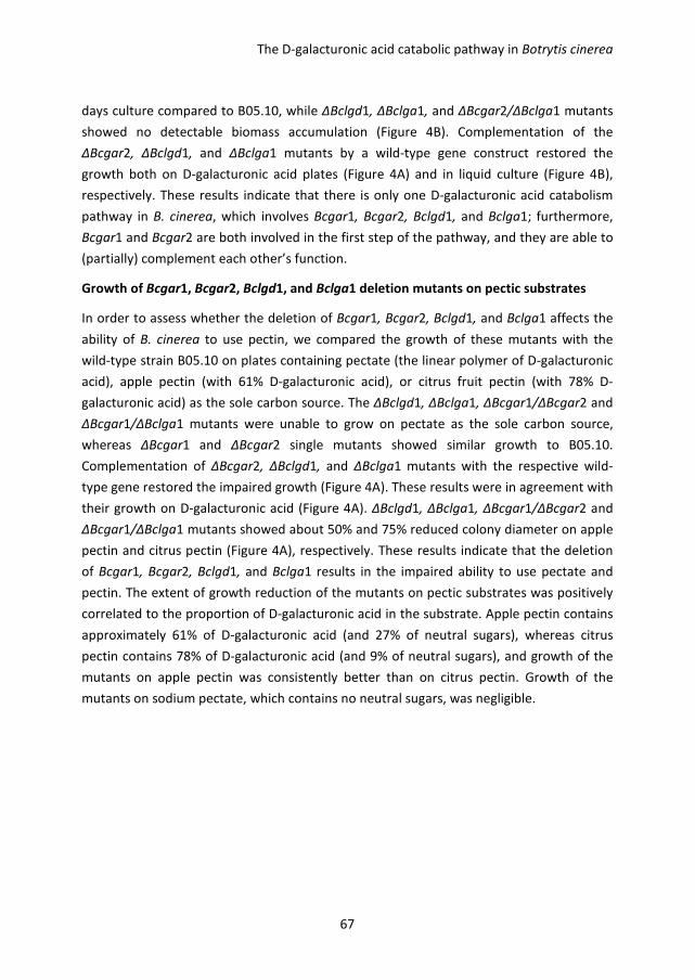
The D-galacturonic acid catabolic pathway in Botrytis cinerea
67
days culture compared to B05.10, while ΔBclgd1, ΔBclga1, and ΔBcgar2/ΔBclga1 mutants
showed no detectable biomass accumulation (Figure 4B). Complementation of the
ΔBcgar2, ΔBclgd1, and ΔBclga1 mutants by a wild-type gene construct restored the
growth both on D-galacturonic acid plates (Figure 4A) and in liquid culture (Figure 4B),
respectively. These results indicate that there is only one D-galacturonic acid catabolism
pathway in B. cinerea, which involves Bcgar1, Bcgar2, Bclgd1, and Bclga1; furthermore,
Bcgar1 and Bcgar2 are both involved in the first step of the pathway, and they are able to
(partially) complement each other’s function.
Growth of Bcgar1, Bcgar2, Bclgd1, and Bclga1 deletion mutants on pectic substrates
In order to assess whether the deletion of Bcgar1, Bcgar2, Bclgd1, and Bclga1 affects the
ability of B. cinerea to use pectin, we compared the growth of these mutants with the
wild-type strain B05.10 on plates containing pectate (the linear polymer of D-galacturonic
acid), apple pectin (with 61% D-galacturonic acid), or citrus fruit pectin (with 78% D-
galacturonic acid) as the sole carbon source. The ΔBclgd1, ΔBclga1, ΔBcgar1/ΔBcgar2 and
ΔBcgar1/ΔBclga1 mutants were unable to grow on pectate as the sole carbon source,
whereas ΔBcgar1 and ΔBcgar2 single mutants showed similar growth to B05.10.
Complementation of ΔBcgar2, ΔBclgd1, and ΔBclga1 mutants with the respective wild-
type gene restored the impaired growth (Figure 4A). These results were in agreement with
their growth on D-galacturonic acid (Figure 4A). ΔBclgd1, ΔBclga1, ΔBcgar1/ΔBcgar2 and
ΔBcgar1/ΔBclga1 mutants showed about 50% and 75% reduced colony diameter on apple
pectin and citrus pectin (Figure 4A), respectively. These results indicate that the deletion
of Bcgar1, Bcgar2, Bclgd1, and Bclga1 results in the impaired ability to use pectate and
pectin. The extent of growth reduction of the mutants on pectic substrates was positively
correlated to the proportion of D-galacturonic acid in the substrate. Apple pectin contains
approximately 61% of D-galacturonic acid (and 27% of neutral sugars), whereas citrus
pectin contains 78% of D-galacturonic acid (and 9% of neutral sugars), and growth of the
mutants on apple pectin was consistently better than on citrus pectin. Growth of the
mutants on sodium pectate, which contains no neutral sugars, was negligible.
Chapter 3
68
Figure 4. Effect of the deletion of Bcgar1, Bcgar2, Bclgd1, and Bclga1 on growth of B. cinerea on agar medium
containing various carbon sources. A, colony diameter of B05.10 and all the mutants strains on plates containing
either glucose, D-galacturonic acid, pectate, apple pectin, or citrus pectin as the sole carbon source. B, the
comparison of biomass of mutant strains and wild-type strain B05.10 in liquid culture containing D-galacturonic
acid (D-GalA) as the sole carbon source. Two independent deletion mutants of each gene or gene-combinations
were analysed and showed similar results, results of one mutant are presented.
Virulence of mutants
The virulence of ΔBcgar1/ΔBcgar2, ΔBclgd1, ΔBclga1, and ΔBcgar1/ΔBclga1 mutants was
investigated on different plant species and tissues. There was no difference in lesion sizes
between any of the tested mutants and the wild-type strain B05.10 on tomato leaves,
apple fruit or bell peppers (not shown).
The D-galacturonic acid catabolic pathway in Botrytis cinerea
69
Discussion
In this study, we have genetically and biochemically characterized the D-galacturonic acid
catabolic pathway in B. cinerea, which consists of three steps involving 4 genes: Bcgar1,
Bcgar2, Bclgd1, and Bclga1. Richard and Hilditch (2009) proposed that an L-glyceraldehyde
reductase (GLR1) also is involved in the D-galacturonic acid catabolic pathway; however,
since there might be unspecific dehydrogenases that are also active with L-glyceraldehyde,
this step is not specifically dedicated to D-galacturonic acid catabolism. Hence, Bcglr1 was
not considered as part of the D-galacturonic acid-specific catabolic pathway in B. cinerea
and we did not study it in detail. The expression of Bcgar1, Bcgar2, Bclgd1, and Bclga1 is
co-regulated by D-galacturonic acid, although transcript levels of Bcgar1 following transfer
to D-galacturonic acid-containing medium were (much) lower than of Bcgar2. These
results are in agreement with the microarray data in A. niger, where gaaA to gaaD were
reported to be highly expressed during growth on D-galacturonic acid, whereas gar1 was
not strongly induced by D-galacturonic acid (Martens-Uzunova and Schaap, 2008). Among
the genes that were co-expressed in A. niger during growth on D-galacturonic acid are two
hexose transporter genes, An14g04280 and An03g01620 (Martens-Uzunova and Schaap,
2008). It is plausible to suggest that these genes encode D-galacturonic acid transporters.
We identified the B. cinerea orthologs of these A. niger genes, BC1G_12561.1 (Bchxt15;
(Dulermo et al., 2009)) and BC1G_08389.1 (Bchxt18; our annotation), and observed that
their transcript levels were induced by D-galacturonic acid as well (not shown). Further
studies will need to elucidate whether BcHXT15 and BcHXT18 could be the B. cinerea D-
galacturonic acid transporters involved in the uptake of D-galacturonic acid from the
environment prior to its entry into the catabolic pathway.
A. nidulans mutant strain WG222 is unable to grow on D-galacturonic acid as sole carbon
source, since it has a nonsense mutation in the gaaA gene (homologous to Bcgar2). The A.
nidulans genome contains a gene homologous to Bcgar1, ANIA_05986, but this gene is
unable to compensate for the lack of D-galacturonic acid reductase activity in the gaaA
mutant (Martens-Uzunova and Schaap, 2008). By contrast, deletion of the gar1 gene in H.
jecorina led to at least six-fold lower biomass accumulation during growth on D-
galacturonic acid (Mojzita et al., 2010) and the H. jecorina gar2 gene (Richard and Hilditch,
2009) is insufficient to support substantial growth on D-galacturonic acid. In the case of B.
cinerea, however, two genes encoding non-homologous galacturonate reductases (Figure
S2) collectively are required for the first step of D-galacturonic acid catabolism. Either
ΔBcgar1 or ΔBcgar2 single mutants remained able to grow on D-galacturonic acid, while
ΔBcgar1/ΔBcgar2 double deletion mutants completely lost the ability to utilize D-
galacturonic acid. These results unequivocally show that Bcgar1 and Bcgar2 are both
Chapter 3
70
functional in the D-galacturonic acid catabolic pathway in B. cinerea, whereas a single
galacturonate reductase gene operates in A. nidulans (Martens-Uzunova and Schaap,
2008), H. jecorina and A. niger (Mojzita et al., 2010).
In spite of the involvement of both Bcgar1 and Bcgar2 in the first step of the catabolic
pathway, their roles are not equally important. BcGAR2 showed a 20-fold lower Km for D-
galacturonic acid as substrate as compared to BcGAR1. Moreover, Bcgar2 transcript levels
were induced by D-galacturonic acid to higher levels than those of Bcgar1. Collectively
these data suggest that BcGAR2 plays a more important role in D-galacturonic acid
catabolism than BcGAR1. This is consistent with the analysis of deletion mutants: ΔBcgar2
mutants showed 90% reduction in biomass accumulation on D-galacturonic acid, while
ΔBcgar1 mutants did not display any biomass reduction. The second and third step in the
catabolic pathway are performed by a single enzyme, alike in H. jecorina (Hilditch et al.,
2007; Kuorelahti et al., 2006) and A. niger (Martens-Uzunova and Schaap, 2008). The
ΔBclgd1 and ΔBclga1 mutants were entirely unable to grow on D-galacturonic acid,
indicating that B. cinerea only has one L-galactonate dehydratase and 2-keto-3-deoxy-L-
galactonate aldolase.
BcGAR1 and BcGAR2 showed similar enzymatic activity with either D-galacturonic acid or
D-glucuronic acid as substrate, while BcLGD1 showed a slightly higher enzymatic activity
on L-galactonate as substrate, as compared to L-gulonate. These results demonstrate that
BcGAR1, BcGAR2 and BcLGD1 are not specific for D-galacturonic acid but also accept D-
glucuronic acid as input to the pathway. One would expect that B. cinerea is able to
catabolise D-glucuronic acid. However, wild-type B. cinerea is unable to grow on D-
glucuronic acid as sole carbon source (not shown). This raises the possibility that D-
glucuronic acid does not induce gene expression of the pathway genes equally effective as
D-galacturonic acid. We supplemented medium containing 50 mM D-glucuronic acid as
the major carbon source with a low amount of D-galacturonic acid (50 µM), in order to
induce expression of Bcgar1, Bcgar2, Bclgd1, and Bclga1, however, this did not allow the
growth of B. cinerea on D-glucuronic acid (not shown). The alternative explanation could
be that B. cinerea lacks specific transporters for uptake of D-glucuronic acid prior to entry
into the catabolic pathway.
B. cinerea deletion mutants in the endoPG genes Bcpg1 and Bcpg2 show a severe
reduction in virulence (Kars et al., 2005a; ten Have et al., 1998), indicating that
decomposition of pectin in plant cell walls is important for B. cinerea infection.
Experiments using such ΔBcpg mutants, however, do not permit to distinguish whether
pectin decomposition by B. cinerea is important for the process of physical colonization of
plant tissue (enabled by the destructive action of the enzymes on plant cell wall integrity)
The D-galacturonic acid catabolic pathway in Botrytis cinerea
71
or for providing nutrients for fungal growth. This distinction can be made by analysing B.
cinerea mutants in the D-galacturonic acid catabolic pathway described here. Such
mutants may be anticipated to express the full spectrum of pectin depolymerizing
enzymes (endopolygalacturonases, exopolygalacturonases, pectin and pectate lyases), yet
they are unable to consume the most abundant monosaccharide released upon pectin
degradation, and utilize it for their growth. Indeed the growth rate of the D-galacturonic
acid catabolic mutants on pectic substrates was severely reduced and was inversely
correlated to the proportion on sugars other than D-galacturonic acid in the substrate
(Figure 4). However, the lesion sizes caused by ΔBcgar1/ΔBcgar2, ΔBclgd1, ΔBclga1, and
ΔBcgar1/ΔBclga1 mutants on tomato leaves, apples and peppers were indistinguishable
from the wild-type. The mutants expressed the endopolygalacturonase genes to levels
that were similar to or even slightly higher than the wild-type B. cinerea (not shown). The
presence in the plant tissues tested of simple sugars (especially sucrose) and/or non-pectic
polysaccharides (starch, cellulose, and hemicellulose) probably provided sufficient
nutrients to compensate for the inability of the mutants to utilize degradation products of
pectic substrates for growth. These data suggest that, in the plant tissues tested, the
function of pectin depolymerization in virulence of B. cinerea is primarily in the process of
tissue colonization, whereas the contribution of pectic substrates to nutrition for hyphal
growth seems limited. We currently investigate the growth of galacturonic acid
catabolism-deficient mutants on other plant tissues, especially tissues with different
pectin contents in the cell walls.
Materials and Methods
Fungal strain and growth conditions
Botrytis cinerea wild-type strain B05.10 and the mutant strains used in this study were
grown and maintained as described (Wubben et al., 1999) and are indicated in Table 1. For
radial growth assays, conidia of the strains were inoculated on Gamborg’s B5 (Duchefa,
Haarlem, The Netherlands) agarose medium supplemented with 10 mM (NH4)H2PO4 and
as carbon source either glucose (50 mM), D-galacturonic acid (50 mM), citrus fruit pectin
(1 % w/v; Sigma), apple pectin (1 % w/v; Carl Roth KG) or sodium pectate (1% w/v; Sigma).
The D-galacturonic acid content of citrus fruit pectin and apple pectin was 78% and 61%,
respectively (Kravtchenko et al., 1992). Cultures were grown at 20 °C.
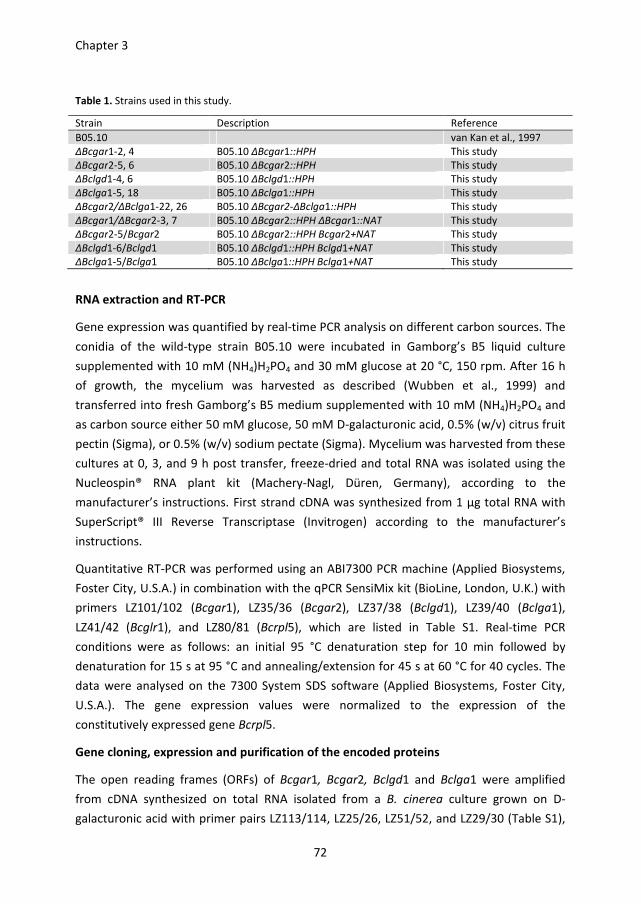
Chapter 3
72
Table 1. Strains used in this study.
Strain Description Reference
B05.10 van Kan et al., 1997
ΔBcgar1-2, 4 B05.10 ΔBcgar1::HPH This study
ΔBcgar2-5, 6 B05.10 ΔBcgar2::HPH This study
ΔBclgd1-4, 6 B05.10 ΔBclgd1::HPH This study
ΔBclga1-5, 18 B05.10 ΔBclga1::HPH This study
ΔBcgar2/ΔBclga1-22, 26 B05.10 ΔBcgar2-ΔBclga1::HPH This study
ΔBcgar1/ΔBcgar2-3, 7 B05.10 ΔBcgar2::HPH ΔBcgar1::NAT This study
ΔBcgar2-5/Bcgar2 B05.10 ΔBcgar2::HPH Bcgar2+NAT This study
ΔBclgd1-6/Bclgd1 B05.10 ΔBclgd1::HPH Bclgd1+NAT This study
ΔBclga1-5/Bclga1 B05.10 ΔBclga1::HPH Bclga1+NAT This study
RNA extraction and RT-PCR
Gene expression was quantified by real-time PCR analysis on different carbon sources. The
conidia of the wild-type strain B05.10 were incubated in Gamborg’s B5 liquid culture
supplemented with 10 mM (NH4)H2PO4 and 30 mM glucose at 20 °C, 150 rpm. After 16 h
of growth, the mycelium was harvested as described (Wubben et al., 1999) and
transferred into fresh Gamborg’s B5 medium supplemented with 10 mM (NH4)H2PO4 and
as carbon source either 50 mM glucose, 50 mM D-galacturonic acid, 0.5% (w/v) citrus fruit
pectin (Sigma), or 0.5% (w/v) sodium pectate (Sigma). Mycelium was harvested from these
cultures at 0, 3, and 9 h post transfer, freeze-dried and total RNA was isolated using the
Nucleospin® RNA plant kit (Machery-Nagl, Düren, Germany), according to the
manufacturer’s instructions. First strand cDNA was synthesized from 1 μg total RNA with
SuperScript® III Reverse Transcriptase (Invitrogen) according to the manufacturer’s
instructions.
Quantitative RT-PCR was performed using an ABI7300 PCR machine (Applied Biosystems,
Foster City, U.S.A.) in combination with the qPCR SensiMix kit (BioLine, London, U.K.) with
primers LZ101/102 (Bcgar1), LZ35/36 (Bcgar2), LZ37/38 (Bclgd1), LZ39/40 (Bclga1),
LZ41/42 (Bcglr1), and LZ80/81 (Bcrpl5), which are listed in Table S1. Real-time PCR
conditions were as follows: an initial 95 °C denaturation step for 10 min followed by
denaturation for 15 s at 95 °C and annealing/extension for 45 s at 60 °C for 40 cycles. The
data were analysed on the 7300 System SDS software (Applied Biosystems, Foster City,
U.S.A.). The gene expression values were normalized to the expression of the
constitutively expressed gene Bcrpl5.
Gene cloning, expression and purification of the encoded proteins
The open reading frames (ORFs) of Bcgar1, Bcgar2, Bclgd1 and Bclga1 were amplified
from cDNA synthesized on total RNA isolated from a B. cinerea culture grown on D-
galacturonic acid with primer pairs LZ113/114, LZ25/26, LZ51/52, and LZ29/30 (Table S1),
The D-galacturonic acid catabolic pathway in Botrytis cinerea
73
which were designed according to the gene models of B. cinerea strain T4. The ORFs were
cloned into a pGEM-T Easy vector (Promega) and confirmed by sequencing. The ORFs of
Bcgar2 and Bclga1 were re-amplified from the pGEM-T constructs with primers LZ47/48
and LZ53/54 (Table S1), respectively; and cloned into expression vector pET16b (Novagen)
by using the NdeI and BamHI restriction sites. Insert sequences were verified by DNA
sequencing analysis (Macrogen). The BamHI-restricted ORFs of Bcgar1 and Bclgd1 derived
from pGEM-T constructs were subcloned into the corresponding sites of pET16b. The
orientations were confirmed by PCR with T7 promoter primer (Invitrogen) and LZ52 or
LZ114.
For heterologous protein expression, the constructs were transformed into E. coli strain
BL21. The overnight cultures were diluted 100-fold into fresh LB medium and grown
further at 28 °C until OD600 reached ~1.0. IPTG (0.5 mM) was added to induce protein
expression and the culture was continued at 25 °C overnight. The induced cultures were
harvested by centrifugation (4000 g) for 10 min. The cell extracts were obtained by using
BugBusterTM
Protein extraction reagent (Novagen). The histidine-tagged proteins were
purified with Ni-NTA agarose beads (Qiagen) according to the manufacturer’s instructions.
Enzyme activity assays
D-galacturonic acid reductase activity was measured by monitoring the change of
absorbance of NADPH at 340 nm in reaction buffer containing 0.37/1.1 μg BcGAR1 or
0.12/0.6 μg BcGAR2; 10 mM sodium phosphate, pH 7.0; 0.25 mM NADPH; and 50 mM D-
galacturonic acid or 50 mM D-glucuronic acid, pH 5.2. Michaelis-Menten constants for D-
galacturonic acid and D-glucuronic acid, of which the concentration was ranging from 1
mM to 75 mM (for BcGAR1), or from 0.1 mM to 2.5 mM (for BcGAR2), were determined at
a NADPH concentration of 0.25 mM.
As L-galactonic acid is not commercially available, L-galactonate dehydratase activity was
measured by starting with the D-galacturonic acid reductase reaction which contained 15
μg BcGAR1; 10 mM sodium phosphate, pH 7.0; 5 mM NADPH; and 50 mM D-galacturonic
acid or 50 mM D-glucuronic acid, pH 5.2. After the overnight reaction, MgCl2 was adjusted
to a final concentration of 5 mM, and 2.5 μg BcLGD1 was added to the reaction and the
mixture was further incubated at room temperature. The production of 2-keto-3-deoxy-L-
galactonate was measured by the thiobarbituric acid method (Buchanan et al., 1999).
As 2-keto-3-deoxy-L-galactonate is not commercially available, 2-keto-3-deoxy-L-
galactonate aldolase activity was measured in the reverse direction (Hilditch et al., 2007)
by determining the production of 2-keto-3-deoxy-L-galactonate from pyruvate and
glyceraldehyde as described above. The reaction buffer contained 1/5 μg BcLGA1; 10 mM
Chapter 3
74
sodium phosphate, pH 7.0; 10 mM pyruvate; and 10 mM DL-glyceraldehyde. Reactions
without enzyme and reactions with heat-inactivated enzyme (65 °C for 15 min) were
included as negative controls. The assays were performed at room temperature.
Deletion of Bcgar1, Bcgar2, Bclgd1, and Bclga1 genes in B. cinerea
The gene replacement strategy for generating B. cinerea deletion constructs, B. cinerea
protoplast transformation and PCR-based screening of transformants were described by
Kars et al. (2005b). Primers used for amplification of gene replacement fragments are
listed in Table S1. To generate Bcgar2-Bclga1 double deletion mutant, primers LZ01, 02,
and 03 were used to amplify the 5’-flanking fragment, and primers LZ10, 11, and 12 were
used to amplify the 3’-flanking fragment. The hygromycin (HPH) and nourseothricin (NAT)
cassette, derived from vector pLOB7 (Figure S4) and pNR3 (Figure S5) with primers 20/21,
were used as selection marker to replace the target genes, as indicated in Table 1.
Genomic DNA of transformants was screened for the presence of the wild-type target
gene by PCR by amplifying the target genes Bcgar1, Bcgar2, Bclgd1, and Bclga1 with
primers LZ113/114, LZ25/26, LZ27/28, and LZ29/30, respectively. Deletion mutants were
routinely double checked by PCR using the same method. Southern hybridization was
performed to confirm the recombination, in accordance with the manufacturer’s
instructions for DIG nucleic acid detection kit (Roche, Germany). The primers for
amplification of Bcgar1, Bcgar2, Bclgd1, and Bclga1 probes are LZ110/111, LZ55/56,
LZ16/17, and LZ95/96, respectively (Table S1). The probes for detecting the HPH or NAT
cassettes were amplified using pLOB7 and pNR3 as templates with primers LZ92/93 and
21/LZ92, respectively.
Complementation construct in B. cinerea
In order to obtain the complementation plasmids rapidly and efficiently, a gateway cloning
vector pNR4 (Figure S6) was generated. The attP1P2 cassette containing the
chloramphenicol resistance gene (CmR) and ccdB gene was amplified with primers
LZ60/94 using the pDONR207 vector (Invitrogen) as template. The attP1P2 fragment was
digested with SacI/XbaI and cloned into the corresponding sites of vector pNR3 (Figure S5),
yielding the plasmid pNR4. The attP1P2 sites of pNR4 were verified by sequencing. The
complementation fragments of Bcgar2, Bclgd1, and Bclga1, including ~1500 bp upstream
and ~300 bp downstream sequences of the coding region, were amplified with primers
LZ97/98, LZ99/100, and LZ62/63 which contain attB1 and attB2 sites, respectively. The
purified fragments were recombined with pNR4 in BP reactions (Invitrogen) in the
appropriate concentration. The resulting plasmids were used for B. cinerea protoplast
transformation.
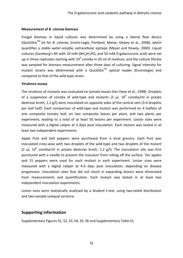
The D-galacturonic acid catabolic pathway in Botrytis cinerea
75
Measurement of B. cinerea biomass
Fungal biomass in liquid cultures was determined by using a lateral flow device
(QuickStixTM
kit for B. cinerea, Enviro-Logix, Portland, Maine; Dewey et al., 2008), which
quantifies a stable water-soluble, extracellular epitope (Meyer and Dewey, 2000). Liquid
cultures (Gamborg’s B5 with 10 mM (NH4)H2PO4 and 50 mM D-galacturonic acid) were set
up in three replicates starting with 104 conidia in 20 ml of medium, and the culture filtrate
was sampled for biomass measurement after three days of culturing. Signal intensity for
mutant strains was determined with a QuickStixTM
optical reader (Envirologix) and
compared to that of the wild-type strain.
Virulence assays
The virulence of mutants was evaluated on tomato leaves (ten Have et al., 1998). Droplets
of a suspension of conidia of wild-type and mutants (2 µl, 106 conidia/ml in potato
dextrose broth, 1.2 g/l) were inoculated on opposite sides of the central vein (3-4 droplets
per leaf half). Each comparison of wild-type and mutant was performed on 4 leaflets of
one composite tomato leaf, on two composite leaves per plant, and two plants per
experiment, leading to a total of at least 50 lesions per experiment. Lesion sizes were
measured with a digital caliper at 3 days post inoculation. Each mutant was tested in at
least two independent experiments.
Apple fruit and bell peppers were purchased from a local grocery. Each fruit was
inoculated cross-wise with two droplets of the wild-type and two droplets of the mutant
(2 µl, 106 conidia/ml in potato dextrose broth, 1.2 g/l). The inoculation site was first
punctured with a needle to prevent the inoculum from rolling off the surface. Ten apples
and 15 peppers were used for each mutant in each experiment. Lesion sizes were
measured with a digital caliper at 4-6 days post inoculation, depending on disease
progression. Inoculation sites that did not result in expanding lesions were eliminated
from measurements and quantification. Each mutant was tested in at least two
independent inoculation experiments.
Lesion sizes were statistically analysed by a Student t-test, using two-tailed distribution
and two-sample unequal variance.
Supporting information
Supplementary Figures S1, S2, S3, S4, S5, S6 and Supplementary Table S1.

Chapter 3
76
Acknowledgements
The authors acknowledge funding by the Foundation Technological Top Institute Green
Genetics (project 2CC035RP) and the Netherlands Graduate School Experimental Plant
Sciences.
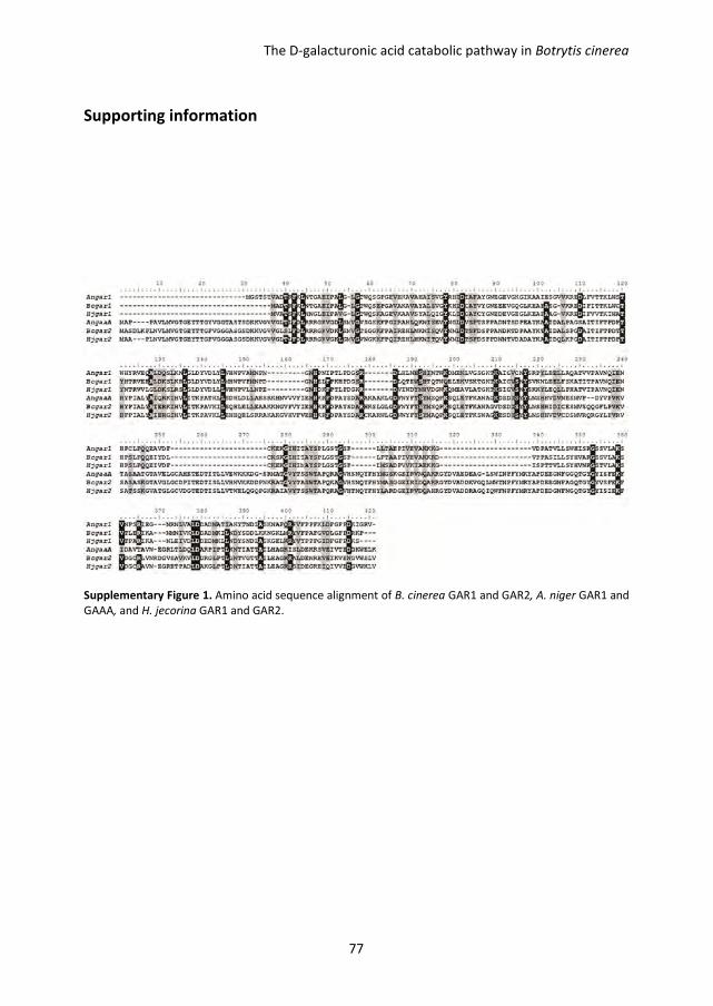
The D-galacturonic acid catabolic pathway in Botrytis cinerea
77
Supporting information
Supplementary Figure 1. Amino acid sequence alignment of B. cinerea GAR1 and GAR2, A. niger GAR1 and
GAAA, and H. jecorina GAR1 and GAR2.
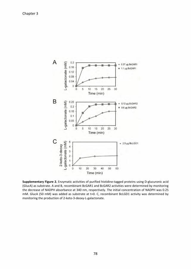
Chapter 3
78
Supplementary Figure 2. Enzymatic activities of purified histidine-tagged proteins using D-glucuronic acid
(GlucA) as substrate. A and B, recombinant BcGAR1 and BcGAR2 activities were determined by monitoring
the decrease of NADPH absorbance at 340 nm, respectively. The initial concentration of NADPH was 0.25
mM. GlucA (50 mM) was added as substrate at t=0. C, recombinant BcLGD1 activity was determined by
monitoring the production of 2-keto-3-deoxy-L-galactonate.
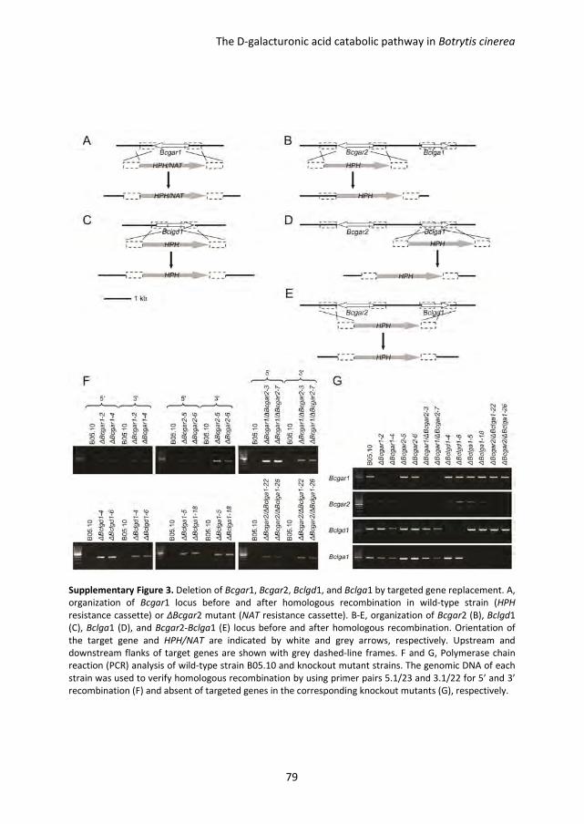
The D-galacturonic acid catabolic pathway in Botrytis cinerea
79
Supplementary Figure 3. Deletion of Bcgar1, Bcgar2, Bclgd1, and Bclga1 by targeted gene replacement. A,
organization of Bcgar1 locus before and after homologous recombination in wild-type strain (HPH
resistance cassette) or ΔBcgar2 mutant (NAT resistance cassette). B-E, organization of Bcgar2 (B), Bclgd1
(C), Bclga1 (D), and Bcgar2-Bclga1 (E) locus before and after homologous recombination. Orientation of
the target gene and HPH/NAT are indicated by white and grey arrows, respectively. Upstream and
downstream flanks of target genes are shown with grey dashed-line frames. F and G, Polymerase chain
reaction (PCR) analysis of wild-type strain B05.10 and knockout mutant strains. The genomic DNA of each
strain was used to verify homologous recombination by using primer pairs 5.1/23 and 3.1/22 for 5’ and 3’
recombination (F) and absent of targeted genes in the corresponding knockout mutants (G), respectively.
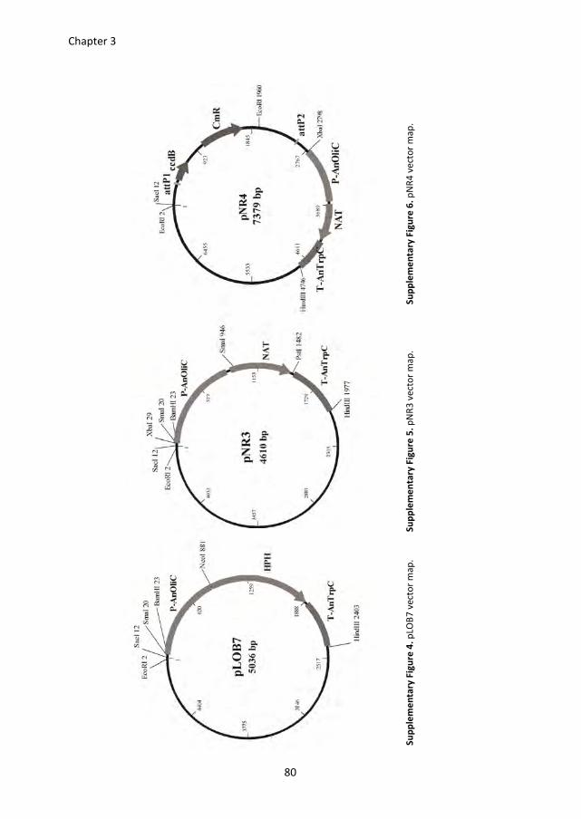
Chapter 3
80
Su
pp
lem
en
tary
Fig
ure
4.
pLO
B7
ve
cto
r m
ap
. S
up
ple
me
nta
ry F
igu
re 5
. p
NR
3 v
ect
or
ma
p.
Su
pp
lem
en
tary
Fig
ure
6.
pN
R4
ve
cto
r m
ap
.
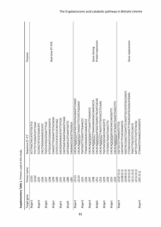
Su
pp
lem
en
tary
Ta
ble
1.
Pri
me
rs u
sed
in
th
is s
tud
y.
Ta
rge
t g
en
e
Pri
me
r n
am
e
Se
qu
en
ce (
5’-
3’)
a
Pu
rpo
se
Bcg
ar1
LZ
10
1
GC
TT
AG
CT
AC
CA
TG
TT
GC
TC
G
Re
al-
tim
e R
T-P
CR
LZ
10
2
TT
CT
TC
TT
CA
GG
TC
GT
CT
GA
G
Bcg
ar2
LZ
35
C
CC
AG
CT
AT
CC
GT
GA
AC
AT
C
LZ
36
C
AC
CT
GG
GG
AA
AG
CG
CA
TC
Bcl
gd
1
LZ3
7
TG
GT
CA
TG
GC
AT
GA
CT
TT
CA
C
LZ
38
G
TT
GC
GA
AT
CG
GA
AA
CG
AG
AT
A
Bcl
ga
1
LZ3
9
CA
AG
GT
TT
GG
GA
AT
TG
TA
CA
GA
G
LZ
40
G
TA
TC
CT
CC
AT
AT
CC
AT
AG
TA
GC
Bcg
lr1
LZ
41
G
AC
AG
GA
AA
GA
CA
TA
TT
TA
TC
AC
LZ
42
C
AG
TG
GA
TA
AG
AT
AA
AG
GT
CA
AG
Bcr
pl5
LZ
80
G
AT
GA
GA
CC
GT
CA
AA
TG
GT
TC
LZ
81
C
AG
AA
GC
CC
AC
GT
TA
CG
AC
A
Bcg
ar1
LZ
11
3
CA
CA
CA
GG
AT
CC
GA
TG
GC
TG
AT
AC
AA
GA
TT
CA
AG
C
Ge
ne
clo
nin
g
Pro
tein
exp
ress
ion
LZ
11
4
CA
CA
CA
GG
AT
CC
CT
AA
GG
CT
TT
CT
AT
CT
GG
GA
AT
Bcg
ar2
LZ
25
T
GA
TG
GC
TT
CA
GA
TT
TG
AA
AC
CC
LZ
26
T
TA
AA
CA
AG
AG
AC
CA
AA
CA
CC
A
Bcg
ar2
LZ
47
C
AC
AC
AC
AT
AT
GG
CT
TC
AG
AT
TT
GA
AA
CC
C
LZ
48
C
AC
AC
AG
GA
TC
CT
TA
AA
CA
AG
AG
AC
CA
AA
CA
CC
A
Bcl
gd
1
LZ5
1
CA
CA
CA
GG
AT
CC
GA
TG
GC
TG
AA
GT
TA
CA
AT
CA
CA
G
LZ
52
C
AC
AC
AG
GA
TC
CT
TA
GA
TC
TT
GA
TA
CC
CT
CC
AA
G
Bcl
ga
1
LZ2
9
TG
AT
GG
CG
GC
AA
CC
AA
CG
GT
TC
LZ
30
C
TA
TA
AG
CT
GA
AC
TC
AA
CC
TT
C
Bcl
ga
1
LZ5
3
CA
CA
CA
CA
TA
TG
GC
GG
CA
AC
CA
AC
GG
TT
C
LZ
54
C
AC
AC
AG
GA
TC
CC
TA
TA
AG
CT
GA
AC
TC
AA
CC
TT
C
Bcg
ar1
LZ
10
7 (
5.1
) C
GC
TG
CT
CT
TA
AA
GA
TC
TT
CG
Ge
ne
re
pla
cem
en
t
LZ
10
8 (
5.2
) C
AG
TA
GT
CT
TC
CA
CG
GA
AG
TG
LZ
10
9 (
5.3
) G
GG
TA
CC
GA
GC
TC
GA
AT
TC
CA
GA
TT
GC
CA
TG
TA
CC
GA
GT
C
LZ
11
0 (
3.3
) C
TC
GG
CG
CG
CC
GA
AG
CT
TT
GG
AC
GA
TG
CG
GA
CA
TG
AA
G
LZ
11
1 (
3.2
) T
GA
CT
TT
GC
CG
TC
TC
CA
GT
G
LZ
11
2 (
3.1
) A
AT
TC
CA
TT
CC
TC
GT
TC
TA
CA
G
Bcg
ar2
LZ
01
(5
.1)
CT
AA
AG
TG
TA
AA
GC
CG
GA
GG
TC
Botrytis cinereaThe D-galacturonic acid catabolic pathway in
81
LZ
02
(5
.2)
CA
TA
CG
TC
TT
CA
TT
CA
AG
AA
CC
LZ
03
(5
.3)
GG
GT
AC
CG
AG
CT
CG
AA
TT
CG
CG
AC
GG
TT
AA
TA
TC
TG
GG
TG
LZ
55
(3
.3)
CT
CG
GC
GC
GC
CG
AA
GC
TT
CA
CG
TC
TT
CG
CA
GA
TC
GA
AC
LZ
56
(3
.2)
GT
GG
TA
GT
TG
TT
CC
TG
AA
GT
G
LZ
57
(3
.1)
GA
AA
TC
CA
TC
AA
CT
CG
AG
GA
G
Bcl
gd
1
LZ1
3 (
5.1
) C
AA
TT
GC
CC
CA
CT
GT
TG
AG
C
LZ
14
(5
.2)
CG
AT
TA
TG
CT
CT
CT
TG
AC
TG
C
LZ
15
(5
.3)
GG
GT
AC
CG
AG
CT
CG
AA
TT
CA
GG
GA
GG
AA
AG
GG
TG
CG
TC
LZ
16
(3
.3)
CT
CG
GC
GC
GC
CG
AA
GC
TT
GG
AA
TC
GG
TG
TT
GC
AA
CT
GG
LZ
17
(3
.2)
GC
CA
TC
GT
TG
CC
CT
GC
TG
C
LZ
18
(3
.1)
CC
CA
AC
AT
GA
AA
AC
CC
TC
GA
C
Bcl
ga
1
LZ0
7 (
5.1
) A
GG
TC
AC
CG
GT
CC
TC
GG
AC
LZ
95
(5
.2)
CC
CA
CT
TT
CT
CT
TC
AT
CT
AC
AC
LZ
96
(5
.3)
GG
GT
AC
CG
AG
CT
CG
AA
TT
CA
TG
TT
GC
GG
TG
TT
TG
GC
GA
LZ
10
(3
.3)
CT
CG
GC
GC
GC
CG
AA
GC
TT
CG
GT
GG
TG
CA
AA
TG
TC
AT
GC
LZ
11
(3
.2)
CT
GC
AT
AA
CG
TG
TA
CG
AA
TG
AC
LZ
12
(3
.1)
CA
AA
AA
GA
GA
AC
GT
GG
AC
AA
CG
HP
H/N
AT
ca
sse
tte
2
0 (
cass
ett
e-5
) G
AA
TT
CG
AG
CT
CG
GT
AC
CC
GG
GG
A
2
1 (
cass
ett
e-3
) C
AA
GC
TT
CG
GC
GC
GC
CG
AG
2
2 (
scre
en
-3)
GT
AA
CC
AT
GC
AT
GG
TT
GC
CT
2
3 (
scre
en
-5)
GG
GT
AC
CG
AG
CT
CG
AA
TT
C
att
P c
ass
ett
e
LZ6
0
CA
CA
CA
GA
GC
TC
GC
TT
AG
TT
TG
AT
GC
CT
GG
CA
G
Ge
ne
co
mp
lem
en
tati
on
LZ
94
C
AC
AC
AT
CT
AG
AC
GT
TG
AA
TA
TG
GC
TC
AT
AA
CA
C
Bcg
ar2
LZ
97
G
GG
GA
CA
AG
TT
TG
TA
CA
AA
AA
AG
CA
GG
CT
TT
CC
AG
AA
AA
TC
CT
CG
CT
GA
G
LZ
98
G
GG
GA
CC
AC
TT
TG
TA
CA
AG
AA
AG
CT
GG
GT
GG
TT
TG
GG
GT
TG
AA
CG
CG
AG
Bcl
gd
1
LZ9
9
GG
GG
AC
AA
GT
TT
GT
AC
AA
AA
AA
GC
AG
GC
TA
CT
TC
TC
GG
GA
AG
CC
AA
GT
G
LZ
10
0
GG
GG
AC
CA
CT
TT
GT
AC
AA
GA
AA
GC
TG
GG
TG
TT
GA
GA
TC
AA
GG
AC
TT
GG
AC
Bcl
ga
1
LZ6
2
GG
GG
AC
AA
GT
TT
GT
AC
AA
AA
AA
GC
AG
GC
TG
AG
CA
AG
TA
GA
GA
TT
TA
CC
CA
LZ
63
G
GG
GA
CC
AC
TT
TG
TA
CA
AG
AA
AG
CT
GG
GT
CA
AA
AA
GA
GA
AC
GT
GG
AC
AA
CG
aR
est
rict
ion
sit
es
intr
od
uce
d f
or
clo
nin
g a
re u
nd
erl
ine
d,
the
att
B1
an
d a
ttB
2 s
ite
s a
re in
dic
ate
d in
ita
lic,
re
spe
ctiv
ely
.
Chapter 3
82
CHAPTER 4
Botrytis cinerea mutants deficient in D-galacturonic acid
catabolism have a perturbed virulence on Nicotiana
benthamiana and Arabidopsis, but not on tomato
Lisha Zhang and Jan A. L. van Kan
This chapter is published as:
Zhang, L. and van Kan, J.A.L. (2013) Botrytis cinerea mutants deficient in D-galacturonic acid catabolism
have a perturbed virulence on Nicotiana benthamiana and Arabidopsis, but not on tomato. Mol. Plant
Pathol. 14, 19-29.
Chapter 4
84
Abstract
D-galacturonic acid is the most abundant monosaccharide component of pectic
polysaccharides that comprise a significant part of most plant cell walls. Therefore, it is
potentially an important nutritional factor for Botrytis cinerea when it grows in and
through plant cell walls. The D-galacturonic acid catabolic pathway in B. cinerea consists of
three catalytic steps converting D-galacturonic acid to pyruvate and L-glyceraldehyde,
involving two non-homologous galacturonate reductase genes (Bcgar1 and Bcgar2), a
galactonate dehydratase gene (Bclgd1), and a 2-keto-3-deoxy-L-galactonate aldolase gene
(Bclga1). Knockout mutants in each step of the pathway (∆Bcgar1/∆Bcgar2, ∆Bclgd1, and
∆Bclga1) showed reduced virulence on Nicotiana benthamiana and Arabidopsis thaliana
leaves, but not on Solanum lycopersicum leaves. The cell walls of N. benthamiana and A.
thaliana leaves were shown to have a higher D-galacturonic acid content as compared to S.
lycopersicum. The observation that mutants displayed a reduction in virulence, especially
on plants with a high D-galacturonic acid content in cell walls, suggests that, in these hosts,
D-galacturonic acid has an important role as carbon nutrient for B. cinerea. However,
additional in vitro growth assays with the knockout mutants revealed that B. cinerea
growth is reduced when D-galacturonic acid catabolic intermediates cannot proceed
through the entire pathway, even when fructose is present as the major, alternative
carbon source. These data suggest that the reduced virulence of D-galacturonic acid
catabolism-deficient mutants on N. benthamiana and A. thaliana is not only a result of the
inability of the mutants to utilize an abundant carbon source as nutrient, but also a result
of the growth inhibition by catabolic intermediates.
Virulence of Botrytis cinerea mutants deficient in D-galacturonic acid catabolism
85
Introduction
Botrytis cinerea is a necrotrophic fungal plant pathogen infecting over 200 host plants and
causing significant economic damage (pre- and post-harvest) to crops worldwide. The
wide variety of symptoms on different tissues and plants suggests that B. cinerea
possesses a large arsenal of weapons to invade its hosts (Choquer et al., 2007). The
infection process includes the penetration of the host tissue, killing of the host cells and
lesion expansion, followed by tissue maceration and sporulation (van Kan, 2006). B.
cinerea produces a variety of compounds capable of killing plant cells, such as phytotoxic
metabolites and proteins, oxalic acid and hydrogen peroxide (van Kan, 2006). However,
the ultimate purpose of a necrotroph is not to kill its host, but to decompose the plant
tissue and utilize the host-derived nutrients for its own growth. B. cinerea secretes
multiple cell wall-degrading enzymes (including pectinases, cellulases and hemicellulases)
to facilitate plant tissue colonization and to release carbohydrates for consumption;
several of these enzymes have been demonstrated to be required for full virulence
(Choquer et al., 2007).
B. cinerea often penetrates host leaf tissue at the anticlinal cell wall and subsequently
grows into and through the middle lamella, which consists mostly of low-methyl-esterified
pectin (ten Have et al., 2002). The importance of pectin degradation for the virulence of B.
cinerea was demonstrated by targeted mutagenesis in endo-polygalacturonase (endo-PG)
genes. Strains with mutations in endo-PG genes have a reduced capacity for pectin
decomposition and, consequently, release reduced amounts of nutrients for the fungus to
potentially catabolize. Strains with a mutation in the Bcpg1 gene were reduced in
virulence by 25% (ten Have et al., 1998), whereas mutants in the Bcpg2 gene were
reduced in virulence by > 50% (Kars et al., 2005a). These experiments, however, could not
distinguish whether pectin decomposition during infection occurs for the purpose of plant
tissue colonization or for the release of monosaccharides that serve as nutrients for fungal
growth, or both. In order to distinguish between the relevance of pectin decomposition
for plant tissue colonization or for nutrient acquisition by the fungus, it is imperative to
obtain B. cinerea mutants that produce the full spectrum of plant cell wall-decomposing
enzymes, but cannot catabolize the monosaccharides that are released from the cell wall
polymers.
Current knowledge of monosaccharide utilization and catabolism by B. cinerea during
plant infection is limited. NMR spectroscopy has suggested that, during colonization of
sunflower cotyledons, B. cinerea converts glucose and fructose present in the host plant
into mannitol via pathways involving the enzymes mannitol-1-phosphate dehydrogenase
(BcMPD), mannitol-1-phosphate phosphatase and mannitol-2-dehydrogenase (BcMTDH)
Chapter 4
86
(Dulermo et al., 2010; Dulermo et al., 2009). Transcript levels of Bcmpd and Bcmtdh as
well as enzyme activities of BcMPD and BcMTDH increased during the progress of
infection (Dulermo et al., 2009). Analysis of single- and double-knockout mutants in the
Bcmpd and Bcmtdh genes, however, revealed that deletion of these genes did not abolish
mannitol metabolism and did not affect virulence on sunflower (Dulermo et al., 2010). A
different study showed that hexokinase is required for B. cinerea development and for
virulence on apple and tomato fruit. The extent of reduction in virulence of a hexokinase-
deficient mutant was correlated with the content of sugars in the fruit, in particular
fructose (Rui and Hahn, 2007).
The monosaccharide D-galacturonic acid is the most abundant component of pectin
polysaccharides (Caffall and Mohnen, 2009; Mohnen, 2008), and might constitute an
important part of the nutrition of B. cinerea when it grows in and through plant cell walls.
Recently, we have characterized the D-galacturonic acid catabolic pathway in B. cinerea,
which consists of three catalytic steps converting D-galacturonic acid to pyruvate and L-
glyceraldehyde. The pathway involves two nonhomologous galacturonate reductase genes
(Bcgar1 and Bcgar2), a galactonate dehydratase gene (Bclgd1) and a 2-keto-3-deoxy-
Lgalactonate aldolase gene (Bclga1) (Zhang et al., 2011). Their transcript levels were
induced substantially when the fungus was cultured in media containing D-galacturonic
acid, pectate or pectin as the sole carbon source. BcGAR1 and BcGAR2 jointly contribute
to the conversion of D-galacturonic acid to L-galactonate, albeit to a different extent.
BcLGD1 converts L-galactonate to 2-keto-3-deoxy-L-galactonate, which is subsequently
catalysed by BcLGA1 to pyruvate and L-glyceraldehyde. Targeted gene replacement of the
four genes in B. cinerea, either separately or in combination, yielded mutants that were
affected in growth on D-galacturonic acid or pectic substrates (pectate, apple pectin, citrus
pectin) as the sole carbon source. The extent of growth reduction of the mutants on pectic
substrates (as sole carbon source) was correlated with the proportion of D-galacturonic
acid in the substrate. The growth of the mutants on apple pectin (containing only 61% D-
galacturonic acid) was reduced by ~50%, whereas growth on citrus pectin (containing 78%
D-galacturonic acid) was reduced by ~75%, and growth on sodium pectate (containing >
99%D-galacturonic acid) was negligible (Zhang et al., 2011).
In this study, we analysed the D-galacturonic acid content in leaf cell wall extracts of three
plant species (Solanum lycopersicum, Nicotiana benthamiana and Arabidopsis thaliana)
and analysed the expression profiles of B. cinerea D-galacturonic acid catabolic genes in
planta, as well as the virulence of B. cinerea mutants in these genes.
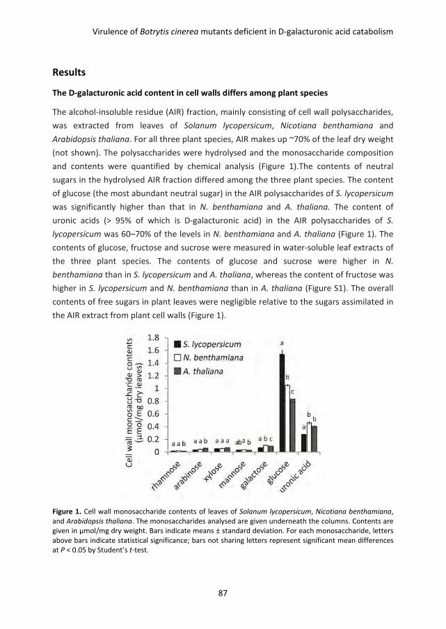
Virulence of Botrytis cinerea mutants deficient in D-galacturonic acid catabolism
87
Results
The D-galacturonic acid content in cell walls differs among plant species
The alcohol-insoluble residue (AIR) fraction, mainly consisting of cell wall polysaccharides,
was extracted from leaves of Solanum lycopersicum, Nicotiana benthamiana and
Arabidopsis thaliana. For all three plant species, AIR makes up ~70% of the leaf dry weight
(not shown). The polysaccharides were hydrolysed and the monosaccharide composition
and contents were quantified by chemical analysis (Figure 1).The contents of neutral
sugars in the hydrolysed AIR fraction differed among the three plant species. The content
of glucose (the most abundant neutral sugar) in the AIR polysaccharides of S. lycopersicum
was significantly higher than that in N. benthamiana and A. thaliana. The content of
uronic acids (> 95% of which is D-galacturonic acid) in the AIR polysaccharides of S.
lycopersicum was 60–70% of the levels in N. benthamiana and A. thaliana (Figure 1). The
contents of glucose, fructose and sucrose were measured in water-soluble leaf extracts of
the three plant species. The contents of glucose and sucrose were higher in N.
benthamiana than in S. lycopersicum and A. thaliana, whereas the content of fructose was
higher in S. lycopersicum and N. benthamiana than in A. thaliana (Figure S1). The overall
contents of free sugars in plant leaves were negligible relative to the sugars assimilated in
the AIR extract from plant cell walls (Figure 1).
Figure 1. Cell wall monosaccharide contents of leaves of Solanum lycopersicum, Nicotiana benthamiana,
and Arabidopsis thaliana. The monosaccharides analysed are given underneath the columns. Contents are
given in µmol/mg dry weight. Bars indicate means ± standard deviation. For each monosaccharide, letters
above bars indicate statistical significance; bars not sharing letters represent significant mean differences
at P < 0.05 by Student’s t-test.
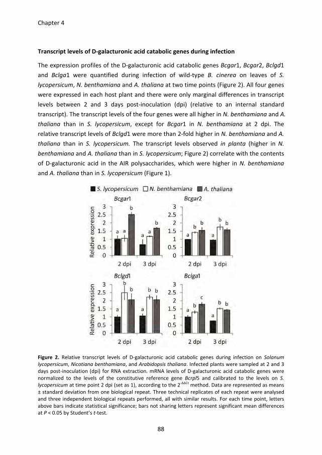
Chapter 4
88
Transcript levels of D-galacturonic acid catabolic genes during infection
The expression profiles of the D-galacturonic acid catabolic genes Bcgar1, Bcgar2, Bclgd1
and Bclga1 were quantified during infection of wild-type B. cinerea on leaves of S.
lycopersicum, N. benthamiana and A. thaliana at two time points (Figure 2). All four genes
were expressed in each host plant and there were only marginal differences in transcript
levels between 2 and 3 days post-inoculation (dpi) (relative to an internal standard
transcript). The transcript levels of the four genes were all higher in N. benthamiana and A.
thaliana than in S. lycopersicum, except for Bcgar1 in N. benthamiana at 2 dpi. The
relative transcript levels of Bclgd1 were more than 2-fold higher in N. benthamiana and A.
thaliana than in S. lycopersicum. The transcript levels observed in planta (higher in N.
benthamiana and A. thaliana than in S. lycopersicum; Figure 2) correlate with the contents
of D-galacturonic acid in the AIR polysaccharides, which were higher in N. benthamiana
and A. thaliana than in S. lycopersicum (Figure 1).
Figure 2. Relative transcript levels of D-galacturonic acid catabolic genes during infection on Solanum
lycopersicum, Nicotiana benthamiana, and Arabidopsis thaliana. Infected plants were sampled at 2 and 3
days post-inoculation (dpi) for RNA extraction. mRNA levels of D-galacturonic acid catabolic genes were
normalized to the levels of the constitutive reference gene Bcrpl5 and calibrated to the levels on S.
lycopersicum at time point 2 dpi (set as 1), according to the 2-∆∆Ct
method. Data are represented as means
± standard deviation from one biological repeat. Three technical replicates of each repeat were analysed
and three independent biological repeats performed, all with similar results. For each time point, letters
above bars indicate statistical significance; bars not sharing letters represent significant mean differences
at P < 0.05 by Student’s t-test.
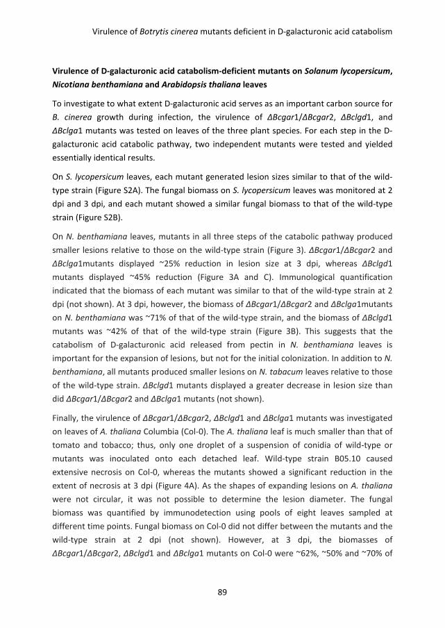
Virulence of Botrytis cinerea mutants deficient in D-galacturonic acid catabolism
89
Virulence of D-galacturonic acid catabolism-deficient mutants on Solanum lycopersicum,
Nicotiana benthamiana and Arabidopsis thaliana leaves
To investigate to what extent D-galacturonic acid serves as an important carbon source for
B. cinerea growth during infection, the virulence of ΔBcgar1/ΔBcgar2, ΔBclgd1, and
ΔBclga1 mutants was tested on leaves of the three plant species. For each step in the D-
galacturonic acid catabolic pathway, two independent mutants were tested and yielded
essentially identical results.
On S. lycopersicum leaves, each mutant generated lesion sizes similar to that of the wild-
type strain (Figure S2A). The fungal biomass on S. lycopersicum leaves was monitored at 2
dpi and 3 dpi, and each mutant showed a similar fungal biomass to that of the wild-type
strain (Figure S2B).
On N. benthamiana leaves, mutants in all three steps of the catabolic pathway produced
smaller lesions relative to those on the wild-type strain (Figure 3). ΔBcgar1/ΔBcgar2 and
ΔBclga1mutants displayed ~25% reduction in lesion size at 3 dpi, whereas ΔBclgd1
mutants displayed ~45% reduction (Figure 3A and C). Immunological quantification
indicated that the biomass of each mutant was similar to that of the wild-type strain at 2
dpi (not shown). At 3 dpi, however, the biomass of ΔBcgar1/ΔBcgar2 and ΔBclga1mutants
on N. benthamiana was ~71% of that of the wild-type strain, and the biomass of ΔBclgd1
mutants was ~42% of that of the wild-type strain (Figure 3B). This suggests that the
catabolism of D-galacturonic acid released from pectin in N. benthamiana leaves is
important for the expansion of lesions, but not for the initial colonization. In addition to N.
benthamiana, all mutants produced smaller lesions on N. tabacum leaves relative to those
of the wild-type strain. ΔBclgd1 mutants displayed a greater decrease in lesion size than
did ΔBcgar1/ΔBcgar2 and ΔBclga1 mutants (not shown).
Finally, the virulence of ΔBcgar1/ΔBcgar2, ΔBclgd1 and ΔBclga1 mutants was investigated
on leaves of A. thaliana Columbia (Col-0). The A. thaliana leaf is much smaller than that of
tomato and tobacco; thus, only one droplet of a suspension of conidia of wild-type or
mutants was inoculated onto each detached leaf. Wild-type strain B05.10 caused
extensive necrosis on Col-0, whereas the mutants showed a significant reduction in the
extent of necrosis at 3 dpi (Figure 4A). As the shapes of expanding lesions on A. thaliana
were not circular, it was not possible to determine the lesion diameter. The fungal
biomass was quantified by immunodetection using pools of eight leaves sampled at
different time points. Fungal biomass on Col-0 did not differ between the mutants and the
wild-type strain at 2 dpi (not shown). However, at 3 dpi, the biomasses of
ΔBcgar1/ΔBcgar2, ΔBclgd1 and ΔBclga1 mutants on Col-0 were ~62%, ~50% and ~70% of
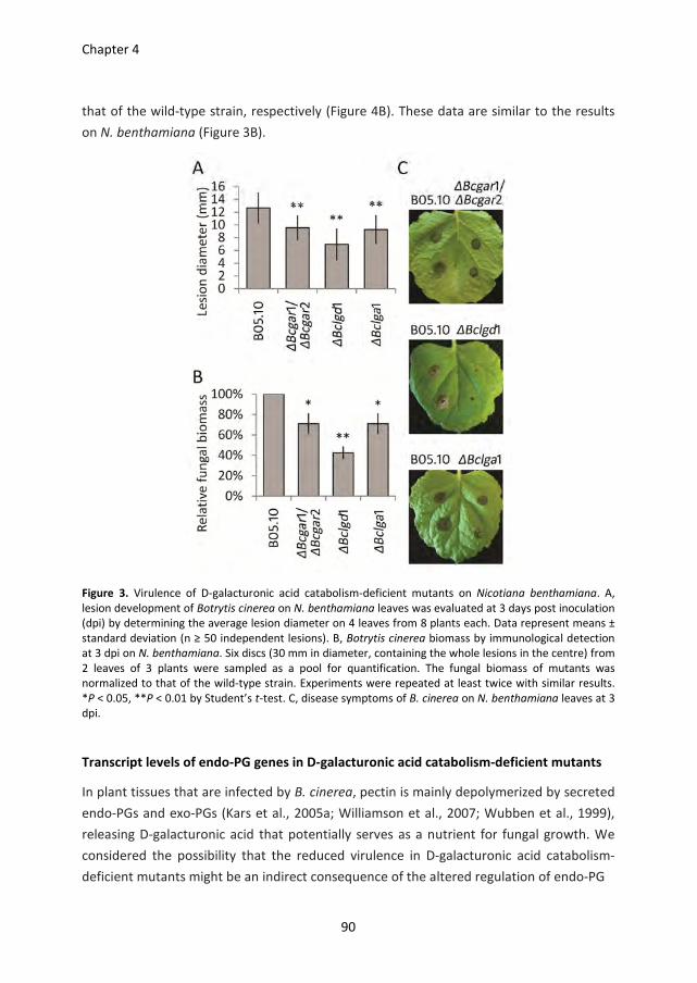
Chapter 4
90
that of the wild-type strain, respectively (Figure 4B). These data are similar to the results
on N. benthamiana (Figure 3B).
Figure 3. Virulence of D-galacturonic acid catabolism-deficient mutants on Nicotiana benthamiana. A,
lesion development of Botrytis cinerea on N. benthamiana leaves was evaluated at 3 days post inoculation
(dpi) by determining the average lesion diameter on 4 leaves from 8 plants each. Data represent means ±
standard deviation (n ≥ 50 independent lesions). B, Botrytis cinerea biomass by immunological detection
at 3 dpi on N. benthamiana. Six discs (30 mm in diameter, containing the whole lesions in the centre) from
2 leaves of 3 plants were sampled as a pool for quantification. The fungal biomass of mutants was
normalized to that of the wild-type strain. Experiments were repeated at least twice with similar results.
*P < 0.05, **P < 0.01 by Student’s t-test. C, disease symptoms of B. cinerea on N. benthamiana leaves at 3
dpi.
Transcript levels of endo-PG genes in D-galacturonic acid catabolism-deficient mutants
In plant tissues that are infected by B. cinerea, pectin is mainly depolymerized by secreted
endo-PGs and exo-PGs (Kars et al., 2005a; Williamson et al., 2007; Wubben et al., 1999),
releasing D-galacturonic acid that potentially serves as a nutrient for fungal growth. We
considered the possibility that the reduced virulence in D-galacturonic acid catabolism-
deficient mutants might be an indirect consequence of the altered regulation of endo-PG
Virulence of Botrytis cinerea mutants deficient in D-galacturonic acid catabolism
91
Figure 4. Virulence of D-galacturonic acid catabolism-deficient mutants on Arabidopsis thaliana. A, disease
symptoms of Botrytis cinerea on A. thaliana leaves at 3 days post-inoculation (dpi). Two representative
leaves are shown for each plant/strain combination. B, Botrytis cinerea biomass accumulation by
immunological detection at 3 dpi. Eight detached leaves were sampled as a pool for quantification. The
fungal biomass of mutants was normalized to that of the wild-type strain. Data represent means ±
standard deviation from two independent biological repeats. *P < 0.05, **P < 0.01 by Student’s t-test.
genes. To assess whether the expression profiles of endo-PG genes were affected in D-
galacturonic acid catabolism-deficient mutants, the transcript levels of Bcpg1–6 were
investigated in the wild-type strain and in mutants during infection on A. thaliana at 2
and3 dpi (Figure 5). Bcpg1 showed similar transcript levels in the D-galacturonic acid
catabolism-deficient mutants as in the wild-type strain at 2 and 3 dpi. Transcripts of Bcpg2,
Bcpg3 and Bcpg5 were not detected under these experimental conditions. The transcript
levels of Bcpg4 and Bcpg6 were higher in all the mutants relative to those in the wild-type
strain at 2 and 3 dpi. These results are in agreement with reports indicating that transcript
levels of Bcpg4 and Bcpg6 (but not Bcpg1, Bcpg2, Bcpg3 and Bcpg5) are induced in
cultures containing D-galacturonic acid as sole carbon source (Wubben et al., 2000). The
expression profiles of Bcpg4 and Bcpg6 suggest that D-galacturonic acid (or catabolic
pathway intermediates) accumulates in mutants during infection to a concentration
sufficient to hyperinduce Bcpg4 and Bcpg6 transcripts.

Chapter 4
92
During infection on N. benthamiana, higher transcript levels of Bcpg4 and Bcpg6 were also
observed in the D-galacturonic acid catabolism-deficient mutants in comparison with
those in the wild-type strain, especially at 3 dpi (Figure S3). By contrast, on S. lycopersicum,
Bcpg4 and Bcpg6 exhibited similar transcript levels in the D-galacturonic acid catabolism-
deficient mutants and in the wild-type, and their transcript levels of were negligible
compared with those on N. benthamiana and A. thaliana (Figure S3). These results
indicate that the deficiency of D-galacturonic acid catabolism in B. cinerea does not impair
the expression of endo-PG genes. Therefore, misregulation of endo-PG genes is not the
cause for the reduced virulence of mutants.
Figure 5. Relative transcript levels of Bcpg1, Bcpg4, and Bcpg6 in wild-type Botrytis cinerea and D-
galacturonic acid catabolism-deficient mutants during infection on Arabidopsis thaliana leaves. Infected
plants were sampled at 2 and 3 days post-inoculation (dpi) for RNA extraction. mRNA levels of Bcpg1,
Bcpg4, and Bcpg6 genes were normalized to the levels of the constitutive reference gene Bcrpl5 and
calibrated to wild-type strain B05.10 levels at time point 2 dpi (set as 1), according to the 2-∆∆Ct
method.
Data are represented as means ± standard deviation from one biological repeat. Three technical replicates
of each repeat were analysed and three independent biological repeats were performed, which showed
similar results. For each time point, letters above bars indicate statistical significance; bars not sharing
letters represent significant mean differences at P < 0.01 by Student’s t-test.
Defence responses in Arabidopsis thaliana
In order to test whether the reduced virulence of the B. cinerea mutants in A. thaliana
could be explained by altered defence responses, the expression was monitored of several
A. thaliana genes that are involved in basal resistance to pathogens, including pad3,
required for the production of camalexin (Böttcher et al., 2009; Zhou et al., 1999), PR1, a
marker gene for salicylic acid dependent defence (Cao et al., 1994; Delaney et al., 1994;
Penninckx et al., 1996), and PDF1.2, a marker for jasmonic acid/ethylene-dependent
defence (Penninckx et al., 1998; Thomma et al., 1998). Moreover, the expression was
monitored of genes which contribute to partial resistance against B. cinerea, including
AtPGIP1, AtPME3 and AtrbohD (Ferrari et al., 2003a; Raiola et al., 2011; Torres et al., 2005;
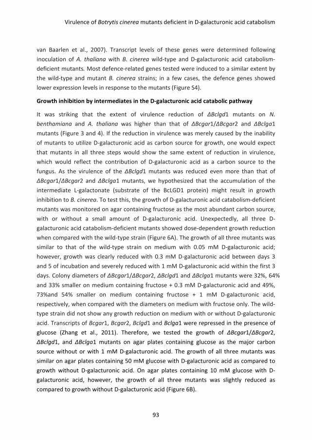
Virulence of Botrytis cinerea mutants deficient in D-galacturonic acid catabolism
93
van Baarlen et al., 2007). Transcript levels of these genes were determined following
inoculation of A. thaliana with B. cinerea wild-type and D-galacturonic acid catabolism-
deficient mutants. Most defence-related genes tested were induced to a similar extent by
the wild-type and mutant B. cinerea strains; in a few cases, the defence genes showed
lower expression levels in response to the mutants (Figure S4).
Growth inhibition by intermediates in the D-galacturonic acid catabolic pathway
It was striking that the extent of virulence reduction of ΔBclgd1 mutants on N.
benthamiana and A. thaliana was higher than that of ΔBcgar1/ΔBcgar2 and ΔBclga1
mutants (Figure 3 and 4). If the reduction in virulence was merely caused by the inability
of mutants to utilize D-galacturonic acid as carbon source for growth, one would expect
that mutants in all three steps would show the same extent of reduction in virulence,
which would reflect the contribution of D-galacturonic acid as a carbon source to the
fungus. As the virulence of the ΔBclgd1 mutants was reduced even more than that of
ΔBcgar1/ΔBcgar2 and ΔBclga1 mutants, we hypothesized that the accumulation of the
intermediate L-galactonate (substrate of the BcLGD1 protein) might result in growth
inhibition to B. cinerea. To test this, the growth of D-galacturonic acid catabolism-deficient
mutants was monitored on agar containing fructose as the most abundant carbon source,
with or without a small amount of D-galacturonic acid. Unexpectedly, all three D-
galacturonic acid catabolism-deficient mutants showed dose-dependent growth reduction
when compared with the wild-type strain (Figure 6A). The growth of all three mutants was
similar to that of the wild-type strain on medium with 0.05 mM D-galacturonic acid;
however, growth was clearly reduced with 0.3 mM D-galacturonic acid between days 3
and 5 of incubation and severely reduced with 1 mM D-galacturonic acid within the first 3
days. Colony diameters of ΔBcgar1/ΔBcgar2, ΔBclgd1 and ΔBclga1 mutants were 32%, 64%
and 33% smaller on medium containing fructose + 0.3 mM D-galacturonic acid and 49%,
73%and 54% smaller on medium containing fructose + 1 mM D-galacturonic acid,
respectively, when compared with the diameters on medium with fructose only. The wild-
type strain did not show any growth reduction on medium with or without D-galacturonic
acid. Transcripts of Bcgar1, Bcgar2, Bclgd1 and Bclga1 were repressed in the presence of
glucose (Zhang et al., 2011). Therefore, we tested the growth of ΔBcgar1/ΔBcgar2,
ΔBclgd1, and ΔBclga1 mutants on agar plates containing glucose as the major carbon
source without or with 1 mM D-galacturonic acid. The growth of all three mutants was
similar on agar plates containing 50 mM glucose with D-galacturonic acid as compared to
growth without D-galacturonic acid. On agar plates containing 10 mM glucose with D-
galacturonic acid, however, the growth of all three mutants was slightly reduced as
compared to growth without D-galacturonic acid (Figure 6B).
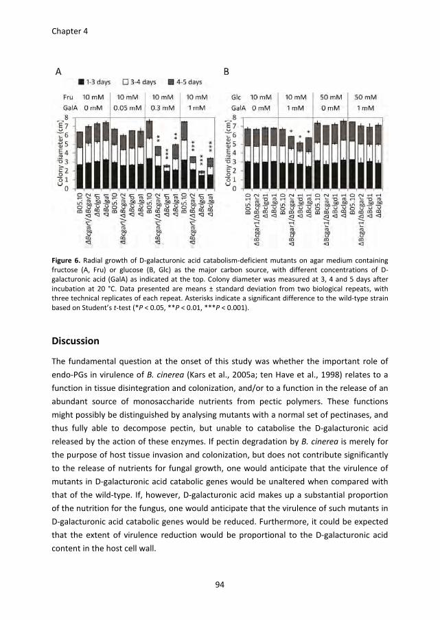
Chapter 4
94
Figure 6. Radial growth of D-galacturonic acid catabolism-deficient mutants on agar medium containing
fructose (A, Fru) or glucose (B, Glc) as the major carbon source, with different concentrations of D-
galacturonic acid (GalA) as indicated at the top. Colony diameter was measured at 3, 4 and 5 days after
incubation at 20 °C. Data presented are means ± standard deviation from two biological repeats, with
three technical replicates of each repeat. Asterisks indicate a significant difference to the wild-type strain
based on Student’s t-test (*P < 0.05, **P < 0.01, ***P < 0.001).
Discussion
The fundamental question at the onset of this study was whether the important role of
endo-PGs in virulence of B. cinerea (Kars et al., 2005a; ten Have et al., 1998) relates to a
function in tissue disintegration and colonization, and/or to a function in the release of an
abundant source of monosaccharide nutrients from pectic polymers. These functions
might possibly be distinguished by analysing mutants with a normal set of pectinases, and
thus fully able to decompose pectin, but unable to catabolise the D-galacturonic acid
released by the action of these enzymes. If pectin degradation by B. cinerea is merely for
the purpose of host tissue invasion and colonization, but does not contribute significantly
to the release of nutrients for fungal growth, one would anticipate that the virulence of
mutants in D-galacturonic acid catabolic genes would be unaltered when compared with
that of the wild-type. If, however, D-galacturonic acid makes up a substantial proportion
of the nutrition for the fungus, one would anticipate that the virulence of such mutants in
D-galacturonic acid catabolic genes would be reduced. Furthermore, it could be expected
that the extent of virulence reduction would be proportional to the D-galacturonic acid
content in the host cell wall.
Virulence of Botrytis cinerea mutants deficient in D-galacturonic acid catabolism
95
We analysed the D-galacturonic acid content in the cell wall extracts of leaves from three
plant species that are commonly used for B. cinerea infection assays: S. lycopersicum, N.
benthamiana and A. thaliana. Although substantial data are available on the leaf cell wall
composition of A. thaliana (Harholt et al., 2006; Zablackis et al., 1995; Zandleven et al.,
2007), only limited information is available on the composition of pectins and cell walls of
leaves from S. lycopersicum (Curvers et al., 2010) and none on N. benthamiana. Although
the latter two species are both Solanaceae and might be anticipated to have similar cell
wall architecture, the composition of monosaccharides released from AIR in N.
benthamiana was remarkably similar to that in A. thaliana and distinct from the
composition in S. lycopersicum. The content of D-galacturonic acid in S. lycopersicum was
only 60%–70% of that in the other two species, whereas the content of glucose was higher
in S. lycopersicum leaves when compared with that in the other two plants. The amount of
free monosaccharides in all three plants (on a mol/dry weight basis) was extremely low
when compared with the monosaccharides assimilated in cell wall polysaccharides. The
differences in D-galacturonic acid content between the three hosts might affect the
nutrients available for B. cinerea growth when it colonizes the leaves of the respective
hosts.
We have previously described a set of B. cinerea D-galacturonic acid catabolism-deficient
mutants, which are blocked in one of three enzymatic steps in the catabolic pathway
(Zhang et al., 2011). These mutants were unable to grow on polygalacturonic acid as sole
carbon source, and showed severely reduced growth on apple pectin and citrus pectin,
substrates that, in addition to D-galacturonic acid, contain neutral sugars. The extent of
residual growth of the mutants correlated with the proportion of neutral sugars in these
pectic substrates (Zhang et al., 2011). The B. cinerea D-galacturonic acid catabolism-
deficient mutants showed a significant reduction in virulence on N. benthamiana and A.
thaliana leaves when compared with the wild-type, but a similar virulence on S.
lycopersicum leaves. The differential virulence was correlated with the content of D-
galacturonic acid in the cell walls of the plants tested. The initial interpretation of the data
was that, in N. benthamiana and A. thaliana, D-galacturonic acid makes up an important
part of the nutrition for B. cinerea. The decomposition of pectin would release D-
galacturonic acid monomers that cannot be utilized by the mutants for growth, thereby
leading to significantly reduced lesion outgrowth. The observation that the virulence of
the mutants on S. lycopersicum leaves was similar to that of the wild-type was tentatively
explained by the low D-galacturonic acid content in this host, and the use of alternative
carbon sources by the mutants.
The transcript levels of D-galacturonic acid catabolic genes during infection by wild-type B.
cinerea correlated with the level of D-galacturonic acid in the leaf cell walls of the species
Chapter 4
96
tested, with relatively high transcript levels in N. benthamiana and A. thaliana, as opposed
to low transcript levels in S. lycopersicum. In individual mutant strains, the transcript levels
of the other pathway genes were not altered during leaf infections, when compared with
the levels in the wild-type. Specifically, there was no significant change in the transcript
levels of Bclgd1 and Bclga1 in the ΔBcgar1/ΔBcgar2 mutant, of Bcgar1, Bcgar2 and Bclga1
in the ΔBclgd1 mutant, or of Bcgar1, Bcgar2 and Bclgd1 in the ΔBclga1 mutant (not
shown). These data suggest that D-galacturonic acid itself is responsible for the induction
of the catabolic genes, and the catabolic intermediates derived from D-galacturonic acid
are not required for induction of transcription. This finding is in agreement with the
observation that, in an Aspergillus niger knockout mutant in the gaaA gene (orthologue of
Bcgar2), transcript levels of genes downstream in the pathway were elevated in the
presence of D-galacturonic acid, similar to that in the wild-type (Mojzita et al., 2010).
We tested whether a non-functional D-galacturonic acid catabolic pathway might affect
the in planta expression of B. cinerea endo-PG genes, which could lead indirectly to
reduction of virulence. Transcript levels of the Bcpg1 gene, which is important for
virulence of B. cinerea (Kars et al., 2005a; ten Have et al., 1998), were unaltered in the D-
galacturonic acid catabolism-deficient mutants. By contrast, the expression of the Bcpg4
and Bcpg6 genes, which can be induced in the presence of D-galacturonic acid (Wubben et
al., 2000), was elevated in the mutants relative to the wild-type strain. This suggests that
the mutants sense elevated levels of D-galacturonic acid, when compared with the wild-
type, presumably because the compound is catabolized in the wild-type strain, leading to
lower intra- and/or extracellular D-galacturonic acid concentrations. The inability to
catabolize D-galacturonic acid in the mutants leads to prolonged elevated levels of Bcpg4
and Bcpg6 transcripts. Expression analyses corroborated that the reduced virulence of D-
galacturonic acid catabolism-deficient mutants was not a result of an indirect impact on
the expression of B. cinerea endo-PG genes. Furthermore, we tested whether the reduced
virulence of B. cinerea mutants on A. thaliana could be explained by an elevation in
defence responses. All A. thaliana defence-related genes tested were induced to a similar
extent by the wild-type and mutant B. cinerea strains; some defence genes even showed
lower transcript levels in response to the mutants. The conclusion that the reduced
virulence of D-galacturonic acid catabolism-deficient mutants was not caused by an
altered regulation of B. cinerea endo-PG genes, or by a modulation of defence responses
in the host, strengthened the initial hypothesis that D-galacturonic acid catabolism
influences virulence directly, and therefore D-galacturonic acid may serve as an important
nutritional component for the fungus. This hypothesis was challenged by the striking
observation that the mutants did not all show the same reduction in virulence. The
ΔBclgd1 mutant (blocked in the second step in the pathway) showed markedly stronger
Virulence of Botrytis cinerea mutants deficient in D-galacturonic acid catabolism
97
reduction in virulence than the mutants blocked in the first and third steps. This
observation was indicative of growth inhibitory effects exerted by catabolic intermediates,
which would partly or largely explain the differences between the ΔBclgd1 mutant and the
other mutants. As the catabolic pathway intermediates (L-galactonate and 2-keto-3-
deoxy-L-galactonate, accumulating in the ΔBclgd1 and ΔBclga1 mutants, respectively) are
not commercially available, it is not feasible to evaluate directly the growth-inhibiting
effects of these compounds in B. cinerea. Therefore, experiments were performed in
which B. cinerea wild-type and mutants were grown on agar, containing fructose as the
most abundant carbon source, supplemented with different amounts of D-galacturonic
acid. The growth of all three mutants was reduced on medium containing fructose
supplemented with D-galacturonic acid when compared with growth on fructose alone.
The growth reduction of the mutants in the presence of D-galacturonic acid occurred in a
dose-dependent manner (Figure 6A). This suggests that the accumulation of all three
intermediates in the D-galacturonic acid catabolic pathway is inhibitory to B. cinerea, with
L-galactonate (product of the first step in the pathway, accumulating in the ΔBclgd1
mutant) having the most severe growth-reducing effect. The mutants grew at a similar
rate to the wild-type in the first 3 days, but then showed a severe decline in radial growth
between days 3–4 and days 4–5. The ΔBclgd1 mutant showed nearly complete growth
cessation in the presence of 0.3 and 1 mM D-galacturonic acid. These observations
provide support that the intermediates first need to be generated and accumulate before
exerting their growth-reducing effect. Growth reduction was not observed when glucose
(50 mM) was provided as the major carbon source, because the expression of D-
galacturonic acid catabolic genes is repressed by glucose (Zhang et al., 2011). In addition,
the expression of putative D-galacturonic acid transporter genes is repressed by glucose
(Chapter 5 and 6). With 10 mM glucose and 1 mM D-galacturonic acid in the medium, the
growth of mutants was reduced slightly between days 4 and 5 of incubation. This
observation can be explained by the glucose being depleted after 3–4 days of incubation,
leading to the release of repression of the D-galacturonic catabolic genes and putative D-
galacturonic acid transporter genes. This enables the fungus to switch to D-galacturonic
acid transport and consumption, resulting in the accumulation of pathway intermediates
to levels sufficient to cause growth reduction. Future studies with knockout mutants of
putative D-galacturonic acid transporter genes in the background of a ∆Bclgd1 mutant
could confirm the growth-reducing effect of catabolic pathway intermediates both in
planta and in vitro. Failure to import D-galacturonic acid would prevent the accumulation
of the inhibitory intermediates and thereby alleviate the growth reduction. Such
experiments were, however, beyond the scope of the present study. The D-galacturonic
acid catabolic pathway has been characterized in Hypocrea jecorina and Aspergillus niger
Chapter 4
98
(Martens-Uzunova and Schaap, 2008; Richard and Hilditch, 2009). In these fungi, there is
no evidence of growth-reducing effects of catabolic pathway intermediates. Intracellular
accumulation of 2-keto-3-deoxy-L-galactonate was observed in the H. jecorina lga1
mutant and the Aspergillus niger gaaC mutant (ΔBclga1 mutant in B. cinerea), but did not
appear to affect hyphal viability or sporulation (Hilditch et al., 2007; Wiebe et al., 2010). In
addition, the degradation of 2-keto-3-deoxy-L-galactonate was observed in H. jecorina and
Aspergillus niger mutant cultures (Wiebe et al., 2010), suggesting that alternative
mechanisms exist in these fungi, which prevent the accumulation of inhibitory compounds.
Such alternative pathways are either less effective or missing in B. cinerea. The observed
growth-reducing effects of catabolic pathway intermediates forced us to reconsider the
interpretation of the virulence assays. It was particularly striking that the extent of
reduction in virulence of the mutants in different steps of the catabolic pathway was
strongly correlated with the extent of growth reduction in vitro (on fructose plus D-
galacturonic acid). Furthermore, the reduced virulence was especially observed in host
species with higher contents of D-galacturonic acid in their cell walls. S. lycopersicum has
30%–40% smaller amounts of D-galacturonic acid in its cell wall when compared with N.
benthamiana and A. thaliana. This leads to less pronounced induction of the expression of
D-galacturonic acid catabolic genes in S. lycopersicum than in the other two hosts, as
corroborated by the observed expression profiles (Figure 2). The levels of glucose and
sucrose in all three plants tested are negligible when compared with the monosaccharides
deposited in cell walls. The concentrations of free monosaccharides in leaves are
insufficient to cause catabolite repression of B. cinerea D-galacturonic acid catabolic genes
(Figure S2). The consequence is that the growth-reducing effects of D-galacturonic acid
catabolic intermediates during infection on S. lycopersicum leaves are negligible and
lesions of the mutants are as large as those of the wild-type. By contrast, the higher D-
galacturonic acid levels in N. benthamiana and A. thaliana leaves cause higher expression
of D-galacturonic acid catabolic genes, and more rapid and greater accumulation of
catabolic intermediates, leading to slower lesion outgrowth. Ideally, instead of comparing
host species with distinct pectin composition, one might prefer to compare the virulence
of B. cinerea on a single wild-type host species and on an isogenic mutant with an altered
content of D-galacturonic acid in its cell wall. The A. thaliana qua1 mutant is deficient in
pectin synthesis and contains 25% less D-galacturonic acid in the cell wall (Bouton et al.,
2002). However, this mutant is severely dwarfed and displays numerous features that
would influence the interaction with B. cinerea in an unpredictable manner. Pectin is of
such crucial relevance for plant cell architecture (Mohnen, 2008) that it is not feasible to
compare the virulence of pathogens on genotypes from the same plant species with
markedly different D-galacturonic acid contents. The question at the onset of this study
Virulence of Botrytis cinerea mutants deficient in D-galacturonic acid catabolism
99
was whether the important role of endo-PGs in the virulence of B. cinerea relates to a
function in tissue disintegration and colonization, and/or to a function in the release of an
abundant source of monosaccharide nutrients from pectic polymers. The data presented
here suggest that the reduced virulence of D-galacturonic acid catabolism deficient B.
cinerea mutants on N. benthamiana and A. thaliana is only partly caused by the inability of
mutants to utilize pectic monosaccharides that serve as an important nutrient source. The
growth inhibitory effect of the D-galacturonic acid catabolic pathway intermediates might
make a more significant contribution to the reduced virulence phenotype of the mutants.
Materials and Methods
Fungal strain and growth conditions
Botrytis cinerea wild-type strain B05.10 and the mutant strains ∆Bcgar1/∆Bcgar2, ∆Bclgd1,
and ∆Bclga1 used in this study were routinely grown on Malt Extract Agar (Oxoid; 50
gram/liter) in darkness at 20 °C for 3-4 days. The plates were placed for one night under
near-UV light (350-400 nm) to promote sporulation, and were subsequently returned to
darkness. Conidia were harvested 4-7 days later in 10-20 ml water, and the suspension
was filtered over glass wool to remove mycelium fragments. The conidia suspension was
centrifuged at 800 rpm (120 g) for 5 minutes. The supernatant was discarded and the
conidia in the pellet resuspended at the desired density. For radial growth assays, conidia
of the strains were inoculated on Gamborg’s B5 (Duchefa, Haarlem, The Netherlands)
agarose medium supplemented with 10 mM (NH4)H2PO4, either fructose (10 mM) or
glucose (10 or 50 mM) as carbon source and D-galacturonic acid (0.05, 0.3, or 1 mM).
Cultures were grown at 20 °C and the colony diameter was measured after 3, 4, and 5
days of incubation.
Plant material and growth conditions
Solanum lycopersicum (Moneymaker) and Nicotiana benthamiana plants were grown in a
greenhouse at 20 °C. Arabidopsis thaliana wild-type Columbia (Col-0) plants were grown in
a growth chamber at 20 °C and 70% relative humidity under a 12 h light/dark cycle.
Plant infection
Leaves of 5-6 weeks-old S. lycopersicum, N. benthamiana and A. thaliana plants were
inoculated with B. cinerea. Droplets of a suspension of conidia of wild-type and mutants (2
µl, 106 conidia/ml in potato dextrose broth, 1.2 g/l) were inoculated on opposite sides of
the central vein (for S. lycopersicum 3-4 droplets per leaf half, for N. benthamiana 1-2
droplets per leaf half). Each comparison of wild-type and mutant was performed on 4
Chapter 4
100
leaflets of one composite tomato leaf, on two composite leaves per plant, and two plants
per experiment; or on 3-4 leaves per N. benthamiana plant, and 6 plants per experiment;
leading to a total of at least 50 lesions per experiment. Lesion diameters were measured
with a digital calliper at 3 dpi. Six discs containing the infection lesions in the centre (30
mm in diameter) from 3 leaves of 3 plants were sampled at 2 and 3 days post inoculation
(dpi) as a pool for RNA isolation and biomass quantification.
For A. thaliana infection, one droplet of a suspension of conidia of wild-type or mutants
were inoculated on one side of each detached leaf. Eight leaves of each inoculation were
sampled at 1, 2, and 3 dpi as a pool for RNA isolation and biomass quantification.
Each mutant was tested in at least two independent experiments. Lesion sizes and fungal
biomass were analysed statistically by a Student’s t-test, using two-tailed distribution and
two-sample unequal variance.
RNA extraction and quantitative reverse transcription-polymerase chain reaction (qRT-
PCR) analysis
The infected plant material was freeze-dried and partially used for RNA extraction. Total
RNA was isolated using a Nucleospin® RNA plant kit (Machery-Nagl, Düren, Germany),
according to manufacturer’s instructions. First strand cDNA was synthesized from 1 μg
total RNA with SuperScript® III Reverse Transcriptase (Invitrogen) according to the
manufacturer’s instructions.
qRT-PCR was performed using an ABI7300 PCR machine (Applied Biosystems, Foster City,
U.S.A.) in combination with the qPCR SensiMix kit (BioLine, London, U.K.). The primers to
detect the transcripts of B. cinerea and A. thaliana genes are listed in Table S1 and S2,
respectively. Real-time PCR conditions were as follows: an initial 95 °C denaturation step
for 10 min followed by denaturation for 15 s at 95 °C and annealing/extension for 45 s at
60 °C for 40 cycles. The data were analysed on the 7300 System SDS software (Applied
Biosystems, Foster City, U.S.A.). The transcript levels of target genes were normalized to
the transcript levels of the constitutively expressed gene Bcrpl5 (for B. cinerea genes) and
Atactin (for A. thaliana genes), according to the 2-∆∆Ct
method.
Measurement of B. cinerea biomass
The freeze-dried infected plant material was incubated in the extraction buffer (35 mg/ml),
which is provided in the QuickStixTM
kit for B. cinerea (Enviro-Logix, Portland, Maine). After
10 min incubation, the supernatants were used for fungal biomass quantification with a
lateral flow device (Dewey et al., 2008), which quantifies a stable water-soluble,
extracellular epitope (Meyer and Dewey, 2000). The signal intensity of the monoclonal
antibody reaction was determined with an optical reader (Envirologix) and converted into
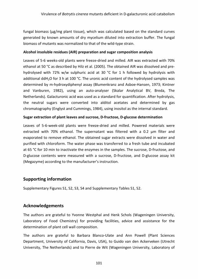
Virulence of Botrytis cinerea mutants deficient in D-galacturonic acid catabolism
101
fungal biomass (µg/mg plant tissue), which was calculated based on the standard curves
generated by known amounts of dry mycelium diluted into extraction buffer. The fungal
biomass of mutants was normalized to that of the wild-type strain.
Alcohol insoluble residues (AIR) preparation and sugar composition analysis
Leaves of 5-6 weeks-old plants were freeze-dried and milled. AIR was extracted with 70%
ethanol at 50 °C as described by Hilz et al. (2005). The obtained AIR was dissolved and pre-
hydrolysed with 72% w/w sulphuric acid at 30 °C for 1 h followed by hydrolysis with
additional ddH2O for 3 h at 100 °C. The uronic acid content of the hydrolysed samples was
determined by m-hydroxydiphenyl assay (Blumenkranz and Asboe-Hansen, 1973; Kintner
and Vanburen, 1982), using an auto-analyser (Skalar Analytical BV, Breda, The
Netherlands). Galacturonic acid was used as a standard for quantification. After hydrolysis,
the neutral sugars were converted into alditol acetates and determined by gas
chromatography (Englyst and Cummings, 1984), using inositol as the internal standard.
Sugar extraction of plant leaves and sucrose, D-fructose, D-glucose determination
Leaves of 5-6-week-old plants were freeze-dried and milled. Powered materials were
extracted with 70% ethanol. The supernatant was filtered with a 0.2 µm filter and
evaporated to remove ethanol. The obtained sugar extracts were dissolved in water and
purified with chloroform. The water phase was transferred to a fresh tube and incubated
at 65 °C for 10 min to inactivate the enzymes in the samples. The sucrose, D-fructose, and
D-glucose contents were measured with a sucrose, D-fructose, and D-glucose assay kit
(Megazyme) according to the manufacturer’s instruction.
Supporting information
Supplementary Figures S1, S2, S3, S4 and Supplementary Tables S1, S2.
Acknowledgements
The authors are grateful to Yvonne Westphal and Henk Schols (Wageningen University,
Laboratory of Food Chemistry) for providing facilities, advice and assistance for the
determination of plant cell wall composition.
The authors are grateful to Barbara Blanco-Ulate and Ann Powell (Plant Sciences
Department, University of California, Davis, USA), to Guido van den Ackerveken (Utrecht
University, The Netherlands) and to Pierre de Wit (Wageningen University, Laboratory of

Chapter 4
102
Phytopathology) for comments on a draft version of the manuscript and for fruitful
discussion. The authors acknowledge funding by the Foundation Technological Top
Institute Green Genetics (Project 2CC035RP) and the Netherlands Graduate School
Experimental Plant Sciences.
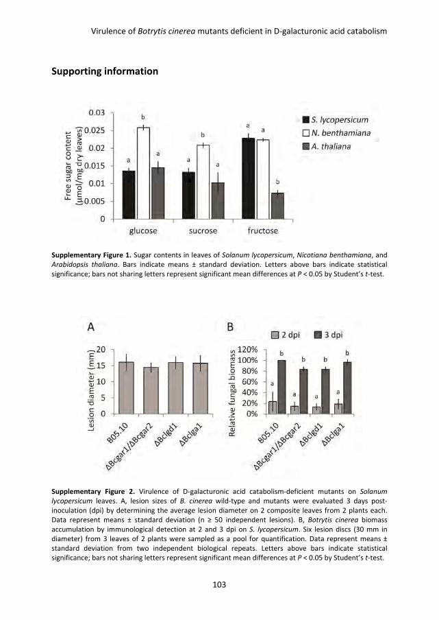
Virulence of Botrytis cinerea mutants deficient in D-galacturonic acid catabolism
103
Supporting information
Supplementary Figure 1. Sugar contents in leaves of Solanum lycopersicum, Nicotiana benthamiana, and
Arabidopsis thaliana. Bars indicate means ± standard deviation. Letters above bars indicate statistical
significance; bars not sharing letters represent significant mean differences at P < 0.05 by Student’s t-test.
Supplementary Figure 2. Virulence of D-galacturonic acid catabolism-deficient mutants on Solanum
lycopersicum leaves. A, lesion sizes of B. cinerea wild-type and mutants were evaluated 3 days post-
inoculation (dpi) by determining the average lesion diameter on 2 composite leaves from 2 plants each.
Data represent means ± standard deviation (n ≥ 50 independent lesions). B, Botrytis cinerea biomass
accumulation by immunological detection at 2 and 3 dpi on S. lycopersicum. Six lesion discs (30 mm in
diameter) from 3 leaves of 2 plants were sampled as a pool for quantification. Data represent means ±
standard deviation from two independent biological repeats. Letters above bars indicate statistical
significance; bars not sharing letters represent significant mean differences at P < 0.05 by Student’s t-test.
Chapter 4
104
Supplementary Figure 3. Relative transcript levels of Bcpg1, Bcpg4, and Bcpg6 in wild-type Botrytis
cinerea and D-galacturonic acid catabolism-deficient mutants during infection on Nicotiana benthamiana
and Solanum lycopersicum leaves. Infected plants were sampled at 2 and 3 days post-inoculation (dpi) for
RNA extraction. mRNA levels of Bcpg1, Bcpg4, and Bcpg6 genes were normalized to the levels of the
constitutive reference gene Bcrpl5 and calibrated to wild-type strain B05.10 levels at time point 2 dpi on N.
benthamiana leaves (set as 1), according to the 2-∆∆Ct
method. Data are represented as means ± standard
deviation from one biological repeat. Three technical replicates of each repeat were analysed and three
independent biological repeats were performed, which showed similar results. For each time point on
each plant leaves, letters above bars indicate statistical significance; bars not sharing letters represent
significant mean differences at P < 0.05 by Student’s t-test.
Virulence of Botrytis cinerea mutants deficient in D-galacturonic acid catabolism
105
Supplementary Figure 4. Relative transcript levels of A. thaliana defence-related genes during infection
with B. cinerea wild-type strain and D-galacturonic acid catabolism-deficient mutants. Infected leaves
were sampled at 1, 2, and 3 days post-inoculation (dpi) for RNA extraction. mRNA levels of A. thaliana
genes were normalized to the levels of the constitutive reference gene Atactin and calibrated to the levels
with B. cinerea wild-type at time point 1 dpi (set as 1), according to the 2-∆∆Ct
method. Data are
represented as means ± standard deviation from one biological repeat. Three technical replicates of each
repeat were analysed and three independent biological repeats were performed, which all showed similar
results.
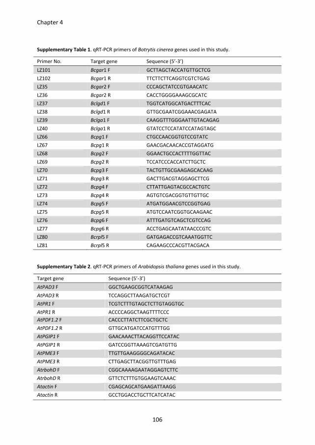
Chapter 4
106
Supplementary Table 1. qRT-PCR primers of Botrytis cinerea genes used in this study.
Primer No. Target gene Sequence (5’-3’)
LZ101 Bcgar1 F GCTTAGCTACCATGTTGCTCG
LZ102 Bcgar1 R TTCTTCTTCAGGTCGTCTGAG
LZ35 Bcgar2 F CCCAGCTATCCGTGAACATC
LZ36 Bcgar2 R CACCTGGGGAAAGCGCATC
LZ37 Bclgd1 F TGGTCATGGCATGACTTTCAC
LZ38 Bclgd1 R GTTGCGAATCGGAAACGAGATA
LZ39 Bclga1 F CAAGGTTTGGGAATTGTACAGAG
LZ40 Bclga1 R GTATCCTCCATATCCATAGTAGC
LZ66 Bcpg1 F CTGCCAACGGTGTCCGTATC
LZ67 Bcpg1 R GAACGACAACACCGTAGGATG
LZ68 Bcpg2 F GGAACTGCCACTTTTGGTTAC
LZ69 Bcpg2 R TCCATCCCACCATCTTGCTC
LZ70 Bcpg3 F TACTGTTGCGAAGAGCACAAG
LZ71 Bcpg3 R GACTTGACGTAGGAGCTTCG
LZ72 Bcpg4 F CTTATTGAGTACGCCACTGTC
LZ73 Bcpg4 R AGTGTCGACGGTGTTGTTGC
LZ74 Bcpg5 F ATGATGGAACGTCCGGTGAG
LZ75 Bcpg5 R ATGTCCAATCGGTGCAAGAAC
LZ76 Bcpg6 F ATTTGATGTCAGCTCGTCCAG
LZ77 Bcpg6 R ACCTGAGCAATATAACCCGTC
LZ80 Bcrpl5 F GATGAGACCGTCAAATGGTTC
LZ81 Bcrpl5 R CAGAAGCCCACGTTACGACA
Supplementary Table 2. qRT-PCR primers of Arabidopsis thaliana genes used in this study.
Target gene Sequence (5’-3’)
AtPAD3 F GGCTGAAGCGGTCATAAGAG
AtPAD3 R TCCAGGCTTAAGATGCTCGT
AtPR1 F TCGTCTTTGTAGCTCTTGTAGGTGC
AtPR1 R ACCCCAGGCTAAGTTTTCCC
AtPDF1.2 F CACCCTTATCTTCGCTGCTC
AtPDF1.2 R GTTGCATGATCCATGTTTGG
AtPGIP1 F GAACAAACTTACAGGTTCCATAC
AtPGIP1 R GATCCGGTTAAAGTCGATGTTG
AtPME3 F TTGTTGAAGGGGCAGATACAC
AtPME3 R CTTGAGCTTACGGTTGTTTGAG
AtrbohD F CGGCAAAAGAATAGGAGTCTTC
AtrbohD R GTTCTCTTTGTGGAAGTCAAAC
Atactin F CGAGCAGCATGAAGATTAAGG
Atactin R GCCTGGACCTGCTTCATCATAC
CHAPTER 5
Functional analysis of putative D-galacturonic acid transporters
in Botrytis cinerea
Lisha Zhang, Sayantani Chatterjee, Chenlei Hua, Jan A. L. van Kan
Chapter 5
108
Abstract
Plant pathogenic fungi use different strategies to acquire carbon sources from their host
plants. Botrytis cinerea predominantly infects plant tissues and species that are rich in
pectin, which is mainly composed of D-galacturonic acid. Effective utilization of D-
galacturonic acid is important for virulence of B. cinerea. Transcriptome data showed that
hexose transporter genes Bchxt8, Bchxt11, Bchxt13 and Bchxt15 are up-regulated in a
culture with pectate as the sole carbon source, as compared to cultures with glucose.
Additional qRT-PCR analysis showed that Bchxt15 is highly (> 180 fold) and specifically
induced by D-galacturonic acid, but not by five other carbon sources analysed, whereas
Bchxt13 is highly expressed in the presence of all carbon sources tested except for glucose.
Subcellular location of BcHXT13-GFP and BcHXT15-GFP fusion proteins expressed under
control of their native promoter was studied in a B. cinerea wild-type strain. Both genes
are expressed during growth on D-galacturonic acid and the fusion proteins are localized
in plasma membranes and intracellular vesicles. Mutants of B. cinerea, in which Bchxt13
and Bchxt15 genes were knocked out, were neither affected in their growth on D-
galacturonic acid as the sole carbon source, nor in their virulence on tomato and Nicotiana
benthamiana leaves.
Putative D-galacturonic acid transporters in Botrytis cinerea
109
Introduction
The main sugar uptake system in fungi comprises hexose transporters, which are
members of the major facilitator superfamily (MFS) and are membrane-bound proteins
containing 12 putative transmembrane domains (Marger and Saier, 1993; Pao et al., 1998).
The best characterised hexose transporters in fungi are from Saccharomyces cerevisiae, of
which the genome contains 20 genes encoding hexose transporters (HXT1 to HXT17, GAL2,
SNF3, and RGT2). None of these transporters are essential for growth on glucose due to
their functional redundancy (Ozcan and Johnston, 1999). In Aspergillus nidulans, there are
at least 17 putative hexose transporters and deletion of hxtA did not impair growth of the
fungus on a variety of carbon sources (Wei et al., 2004).
Plant pathogenic fungi cause globally severe losses of crops during growth and after
harvest every year (Fisher et al., 2012). Different fungi have developed different strategies
to acquire nutrients from their host plants. The nutrition of biotrophic pathogens relies on
living host cells, whereas necrotrophic pathogens kill plant cells and feed on dead or dying
tissues. Other pathogens (hemi-biotrophs) employ both types of nutrition, starting with
biotrophic growth and switching to necrotrophic invasion at a later stage.
A proton-dependent hexose symporter (UfHXT1) of the biotroph Uromyces fabae, the
causal agent of broad bean rust, was shown to be involved in uptake of hexoses across the
haustorial membrane (Voegele et al., 2001). A study of five hexose transporters (CgHXT1-5)
of the hemi-biotrophic pathogen Colletotrichum graminicola showed that these hexose
transporters were functionally distinct: CgHXT1 to CgHXT3 are high affinity/low capacity
transporters that accept several hexoses, whereas CgHXT5 is a low affinity/high capacity
transporter with a narrow substrate specificity, involved in uptake of glucose and
mannose only. Moreover, their expression was different at different stages of infection:
CgHXT1 and CgHXT3 were transiently up-regulated during the biotrophic stage, and
CgHXT2 and CgHXT5 were expressed exclusively during necrotrophic stages, while CgHXT4
was expressed throughout the infection (Lingner et al., 2011).
The necrotrophic fungal plant pathogen Botrytis cinerea is able to infect over 200 host
plants and causes severe damage to crops, both pre- and post-harvest (Dean et al., 2012).
The broad range of host plants suggests that B. cinerea has adapted to various nutritional
environments and possesses a complex sugar uptake system to acquire carbon nutrients
from different hosts and during different infection stages (van Kan, 2006). Studies by
Dulermo et al. (2009) on the expression profiles of 17 putative hexose transporter (Bchxt)
genes in B. cinerea during infection of sunflower, showed four types of expression profiles
for hexose transporters: (1) constitutive during the course of infection; (2) transient at a
certain time point; (3) decreased during the course of infection; (4) increased at the late
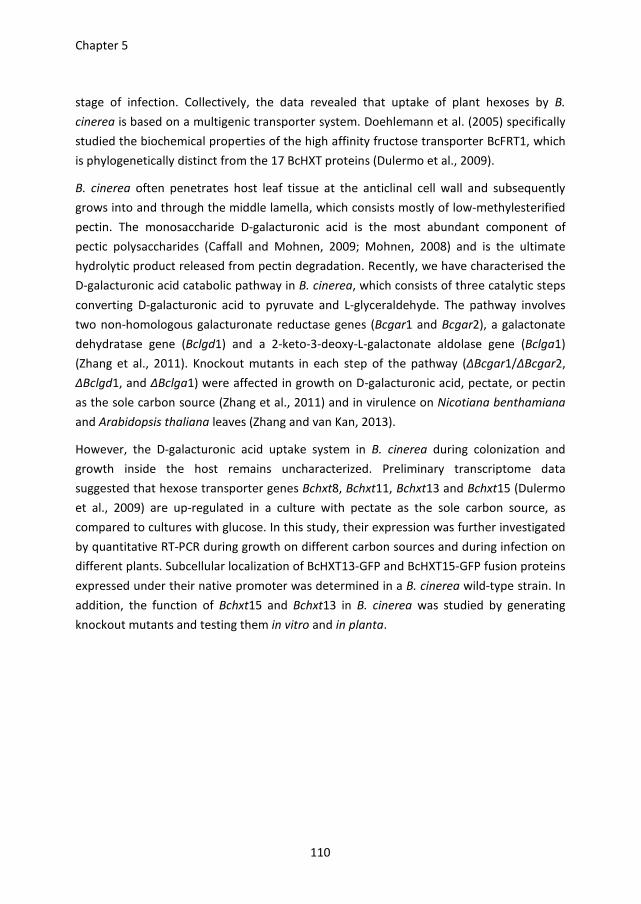
Chapter 5
110
stage of infection. Collectively, the data revealed that uptake of plant hexoses by B.
cinerea is based on a multigenic transporter system. Doehlemann et al. (2005) specifically
studied the biochemical properties of the high affinity fructose transporter BcFRT1, which
is phylogenetically distinct from the 17 BcHXT proteins (Dulermo et al., 2009).
B. cinerea often penetrates host leaf tissue at the anticlinal cell wall and subsequently
grows into and through the middle lamella, which consists mostly of low-methylesterified
pectin. The monosaccharide D-galacturonic acid is the most abundant component of
pectic polysaccharides (Caffall and Mohnen, 2009; Mohnen, 2008) and is the ultimate
hydrolytic product released from pectin degradation. Recently, we have characterised the
D-galacturonic acid catabolic pathway in B. cinerea, which consists of three catalytic steps
converting D-galacturonic acid to pyruvate and L-glyceraldehyde. The pathway involves
two non-homologous galacturonate reductase genes (Bcgar1 and Bcgar2), a galactonate
dehydratase gene (Bclgd1) and a 2-keto-3-deoxy-L-galactonate aldolase gene (Bclga1)
(Zhang et al., 2011). Knockout mutants in each step of the pathway (ΔBcgar1/ΔBcgar2,
ΔBclgd1, and ΔBclga1) were affected in growth on D-galacturonic acid, pectate, or pectin
as the sole carbon source (Zhang et al., 2011) and in virulence on Nicotiana benthamiana
and Arabidopsis thaliana leaves (Zhang and van Kan, 2013).
However, the D-galacturonic acid uptake system in B. cinerea during colonization and
growth inside the host remains uncharacterized. Preliminary transcriptome data
suggested that hexose transporter genes Bchxt8, Bchxt11, Bchxt13 and Bchxt15 (Dulermo
et al., 2009) are up-regulated in a culture with pectate as the sole carbon source, as
compared to cultures with glucose. In this study, their expression was further investigated
by quantitative RT-PCR during growth on different carbon sources and during infection on
different plants. Subcellular localization of BcHXT13-GFP and BcHXT15-GFP fusion proteins
expressed under their native promoter was determined in a B. cinerea wild-type strain. In
addition, the function of Bchxt15 and Bchxt13 in B. cinerea was studied by generating
knockout mutants and testing them in vitro and in planta.
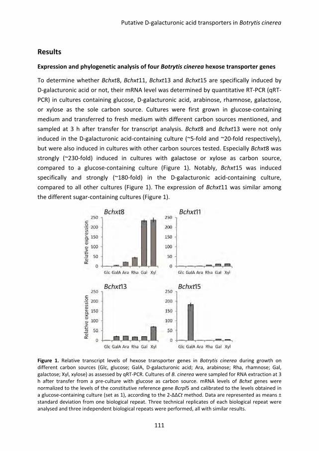
Putative D-galacturonic acid transporters in Botrytis cinerea
111
Results
Expression and phylogenetic analysis of four Botrytis cinerea hexose transporter genes
To determine whether Bchxt8, Bchxt11, Bchxt13 and Bchxt15 are specifically induced by
D-galacturonic acid or not, their mRNA level was determined by quantitative RT-PCR (qRT-
PCR) in cultures containing glucose, D-galacturonic acid, arabinose, rhamnose, galactose,
or xylose as the sole carbon source. Cultures were first grown in glucose-containing
medium and transferred to fresh medium with different carbon sources mentioned, and
sampled at 3 h after transfer for transcript analysis. Bchxt8 and Bchxt13 were not only
induced in the D-galacturonic acid-containing culture (~5-fold and ~20-fold respectively),
but were also induced in cultures with other carbon sources tested. Especially Bchxt8 was
strongly (~230-fold) induced in cultures with galactose or xylose as carbon source,
compared to a glucose-containing culture (Figure 1). Notably, Bchxt15 was induced
specifically and strongly (~180-fold) in the D-galacturonic acid-containing culture,
compared to all other cultures (Figure 1). The expression of Bchxt11 was similar among
the different sugar-containing cultures (Figure 1).
Figure 1. Relative transcript levels of hexose transporter genes in Botrytis cinerea during growth on
different carbon sources (Glc, glucose; GalA, D-galacturonic acid; Ara, arabinose; Rha, rhamnose; Gal,
galactose; Xyl, xylose) as assessed by qRT-PCR. Cultures of B. cinerea were sampled for RNA extraction at 3
h after transfer from a pre-culture with glucose as carbon source. mRNA levels of Bchxt genes were
normalized to the levels of the constitutive reference gene Bcrpl5 and calibrated to the levels obtained in
a glucose-containing culture (set as 1), according to the 2-∆∆Ct method. Data are represented as means ±
standard deviation from one biological repeat. Three technical replicates of each biological repeat were
analysed and three independent biological repeats were performed, all with similar results.
Chapter 5
112
Phylogenetic analysis was performed with BcHXT8, BcHXT11, BcHXT13, and BcHXT15, with
8 functionally characterised hexose transporters from pathogenic fungi differing in
lifestyle (Doehlemann et al., 2005; Kim and Woloshuk, 2011; Lingner et al., 2011; Voegele
et al., 2001), and with 2 putative hexose transporters from the saprophytic fungus
Aspergillus niger (Martens-Uzunova and Schaap, 2008). Figure 2 shows that three major
clades of transporters were discriminated. BcHXT8 and BcHXT11 clustered with
Colletotrichum graminicola and Uromyces fabae hexose transporters, and were most
closely related to CgHXT3 and CgHXT1, respectively, both of which are transporters with
high affinity for glucose (Lingner et al., 2011). By contrast, BcHXT13 and BcHXT15
clustered with the proteins encoded by An03g01620 and An14g04280, genes that were
induced in A. niger grown on D-galacturonic acid or pectin (Kuivanen et al., 2012; Martens-
Uzunova and Schaap, 2008). The third cluster contains BcFRT1, a fructose transporter
from B. cinerea (Doehlemann et al., 2005), and FvFST1, a putative hexose transporter
required by Fusarium verticillioides to colonize maize kernels (Kim and Woloshuk, 2011).
Collectively, the expression and phylogenetic data suggest that BcHXT13 and BcHXT15
might play a role in D-galacturonic acid uptake; especially BcHXT15 might have a
prominent role because of its strong and specific up-regulation by D-galacturonic acid.
Thus, we focused on these two genes for further study.
Figure 2. Phylogenetic analysis of fungal hexose transporters. Amino acid sequences of B. cinerea
transporters were obtained from Broad institute Botrytis cinerea database (http://www. broad-institute.
org/annotation/genome/ botrytis_cinerea/MultiHome.html) according to the locus ID (Staats and van Kan,
2012): BcHXT8: B0510_7979; BcHXT11: B0510_5073; BcHXT13: B0510_9487; BcHXT15: B0510_3996;
BcFRT1: B0510_8895; other fungal transporter sequences were obtained from GenBank according to the
accession number: CgHXT1 to CgHXT5: FN433101 to FN433105; UfHXT1: AJ310209; FvFST1: EU152990.
The amino acid sequences were aligned by Clustal_X 1.83 and the phylogenetic tree was generated by
using Mega 4 (Tamura et al., 2007) by the Neighbor-Joining (NJ) method with 1000 bootstrap replicates.
Putative D-galacturonic acid transporters in Botrytis cinerea
113
Subcellular localization of BcHXT13 and BcHXT15
To study the subcellular localization of BcHXT13 and BcHXT15, both genes were fused to
GFP and transformed into B. cinerea wild-type stain B05.10 with their native promoters.
The hyphae of transformants germinated in the presence of glucose, D-galacturonic acid,
or xylose as carbon sources, were examined by epifluorescence microscopy. In the
medium with D-galacturonic acid as carbon source, the BcHXT15-GFP expressing
transformants showed fluorescence in the plasma membrane and septa, but also in
vesicular structures which move inside the hyphae (Figure 3 and Figure 4). In most cases,
fluorescence in the apical hyphal compartment is weaker than in the subapical hyphal
region (Figure 3A). In the older hyphal region close to where the conidium germinated, the
fluorescence was concentrated in immobile compartments which are larger than the
mentioned vesicular structures (Figure 3B compared to 3A). No GFP fluorescence was
detected in the hyphae of the BcHXT15-GFP expressing transformants, germinated in the
presence of glucose or xylose (not shown). BcHXT13-GFP expressing transformants
showed the same subcellular localization of fluorescence as the BcHXT15-GFP expressing
transformants, when germinated in the medium with D-galacturonic acid or xylose as
carbon source but not with glucose (Figure 3C and D). Under the same microscopic
settings, the fluorescence of BcHXT13-GFP was in both cases weak as compared with
BcHXT15-GFP in D-galacturonic acid (not shown). These results were consistent with the
qRT-PCR data suggesting that Bchxt13 is induced by both D-galacturonic acid and xylose,
whereas Bchxt15 is strongly induced solely by D-galacturonic acid (Figure 1).
To identify the nature of the vesicular structures and large compartments in which
BcHXT15-GFP is located, the germinated hyphae of transformants were stained with
fluorescent dyes DAPI, Mitotracker, and FM4-64, which specifically label nuclei,
mitochondria and membrane structures, respectively. Figure 5 shows that the
fluorescence of BcHXT15-GFP (green) does neither co-localize with DAPI-labelled nuclei
(blue) nor with Mitotracker-labelled mitochondria (red). However, within 2 min of FM4-64
staining, the red dye is detected in the plasma membrane and in several small vesicles
that overlapped with vesicles in which BcHXT15-GFP resided (Figure 6). After 2 h staining,
FM4-64 signal was clearly observed in the membrane of the larger compartments where
BcHXT15-GFP was located (Figure 6).
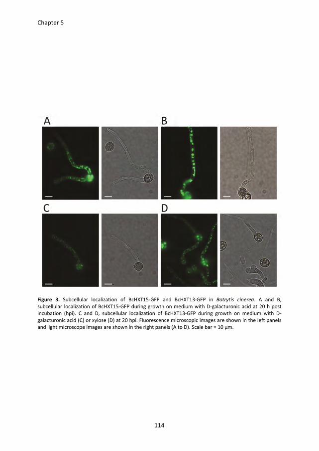
Chapter 5
114
Figure 3. Subcellular localization of BcHXT15-GFP and BcHXT13-GFP in Botrytis cinerea. A and B,
subcellular localization of BcHXT15-GFP during growth on medium with D-galacturonic acid at 20 h post
incubation (hpi). C and D, subcellular localization of BcHXT13-GFP during growth on medium with D-
galacturonic acid (C) or xylose (D) at 20 hpi. Fluorescence microscopic images are shown in the left panels
and light microscope images are shown in the right panels (A to D). Scale bar = 10 µm.
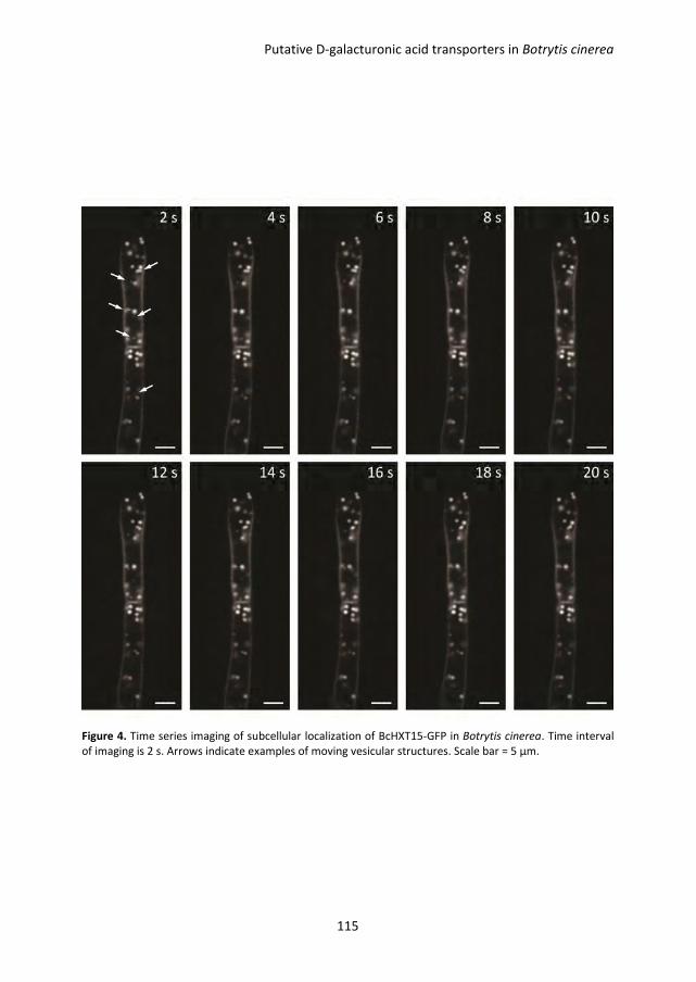
Putative D-galacturonic acid transporters in Botrytis cinerea
115
Figure 4. Time series imaging of subcellular localization of BcHXT15-GFP in Botrytis cinerea. Time interval
of imaging is 2 s. Arrows indicate examples of moving vesicular structures. Scale bar = 5 µm.
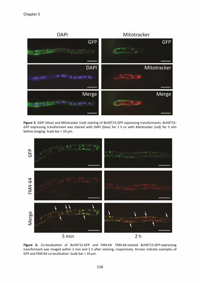
Chapter 5
116
Figure 5. DAPI (blue) and Mitotracker (red) staining of BcHXT15-GFP expressing transformants. BcHXT15-
GFP expressing transformant was stained with DAPI (blue) for 1 h or with Mitotracker (red) for 5 min
before imaging. Scale bar = 10 µm.
Figure 6. Co-localization of BcHXT15-GFP and FM4-64. FM4-64-stained BcHXT15-GFP-expressing
transformant was imaged within 2 min and 2 h after staining, respectively. Arrows indicate examples of
GFP and FM4-64 co-localization. Scale bar = 10 µm.
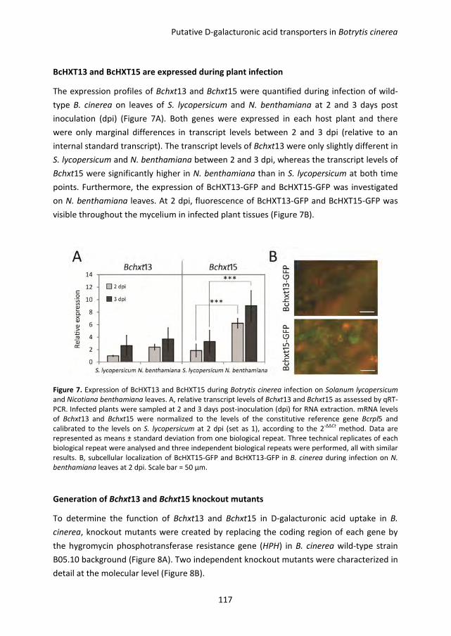
Putative D-galacturonic acid transporters in Botrytis cinerea
117
BcHXT13 and BcHXT15 are expressed during plant infection
The expression profiles of Bchxt13 and Bchxt15 were quantified during infection of wild-
type B. cinerea on leaves of S. lycopersicum and N. benthamiana at 2 and 3 days post
inoculation (dpi) (Figure 7A). Both genes were expressed in each host plant and there
were only marginal differences in transcript levels between 2 and 3 dpi (relative to an
internal standard transcript). The transcript levels of Bchxt13 were only slightly different in
S. lycopersicum and N. benthamiana between 2 and 3 dpi, whereas the transcript levels of
Bchxt15 were significantly higher in N. benthamiana than in S. lycopersicum at both time
points. Furthermore, the expression of BcHXT13-GFP and BcHXT15-GFP was investigated
on N. benthamiana leaves. At 2 dpi, fluorescence of BcHXT13-GFP and BcHXT15-GFP was
visible throughout the mycelium in infected plant tissues (Figure 7B).
Figure 7. Expression of BcHXT13 and BcHXT15 during Botrytis cinerea infection on Solanum lycopersicum
and Nicotiana benthamiana leaves. A, relative transcript levels of Bchxt13 and Bchxt15 as assessed by qRT-
PCR. Infected plants were sampled at 2 and 3 days post-inoculation (dpi) for RNA extraction. mRNA levels
of Bchxt13 and Bchxt15 were normalized to the levels of the constitutive reference gene Bcrpl5 and
calibrated to the levels on S. lycopersicum at 2 dpi (set as 1), according to the 2-∆∆Ct
method. Data are
represented as means ± standard deviation from one biological repeat. Three technical replicates of each
biological repeat were analysed and three independent biological repeats were performed, all with similar
results. B, subcellular localization of BcHXT15-GFP and BcHXT13-GFP in B. cinerea during infection on N.
benthamiana leaves at 2 dpi. Scale bar = 50 µm.
Generation of Bchxt13 and Bchxt15 knockout mutants
To determine the function of Bchxt13 and Bchxt15 in D-galacturonic acid uptake in B.
cinerea, knockout mutants were created by replacing the coding region of each gene by
the hygromycin phosphotransferase resistance gene (HPH) in B. cinerea wild-type strain
B05.10 background (Figure 8A). Two independent knockout mutants were characterized in
detail at the molecular level (Figure 8B).
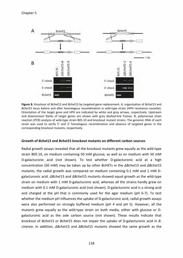
Chapter 5
118
Figure 8. Knockout of Bchxt13 and Bchxt15 by targeted gene replacement. A, organization of Bchxt13 and
Bchxt15 locus before and after homologous recombination in wild-type strain (HPH resistance cassette).
Orientation of the target gene and HPH are indicated by white and grey arrows, respectively. Upstream
and downstream flanks of target genes are shown with grey dashed-line frames. B, polymerase chain
reaction (PCR) analysis of wild-type strain B05.10 and knockout mutant strains. The genomic DNA of each
strain was used to verify 5’ and 3’ homologous recombination and absence of targeted genes in the
corresponding knockout mutants, respectively.
Growth of Bchxt13 and Bchxt15 knockout mutants on different carbon sources
Radial growth assays revealed that all the knockout mutants grew equally as the wild-type
strain B05.10, on medium containing 50 mM glucose, as well as on medium with 50 mM
D-galacturonic acid (not shown). To test whether D-galacturonic acid at a high
concentration (50 mM) may be taken up by other BcHXTs in the ΔBchxt13 and ΔBchxt15
mutants, the radial growth was compared on medium containing 0.1 mM and 1 mM D-
galacturonic acid. ΔBchxt13 and ΔBchxt15 mutants showed equal growth as the wild-type
strain on medium with 1 mM D-galacturonic acid, whereas all the strains hardly grew on
medium with 0.1 mM D-galacturonic acid (not shown). D-galacturonic acid is a strong acid
and charged at the pH that is commonly used for the agar medium (pH 6-7). To test
whether the medium pH influences the uptake of D-galacturonic acid, radial growth assays
were also performed on strongly buffered medium (pH 4 and pH 5). However, all the
mutants grew equally as the wild-type strain on both media, either with glucose or D-
galacturonic acid as the sole carbon source (not shown). These results indicate that
knockout of Bchxt13 or Bchxt15 does not impair the uptake of D-galacturonic acid in B.
cinerea. In addition, ΔBchxt13 and ΔBchxt15 mutants showed the same growth as the
Putative D-galacturonic acid transporters in Botrytis cinerea
119
wild-type strain on medium with arabinose, rhamnose, galactose, or xylose as the sole
carbon source (not shown).
Virulence of ΔBchxt13 and ΔBchxt15 mutants
The virulence of ΔBchxt13 and ΔBchxt15 mutants was investigated on S. lycopersicum and
N. benthamiana leaves. There was no difference in lesion sizes between any of the tested
mutants and the wild-type strain B05.10 (not shown).
Discussion
In this study, the transcript levels of four Botrytis cinerea hexose transporter genes were
investigated by qRT-PCR during in vitro growth in medium with different sugars as carbon
source. Bchxt15 is specifically up-regulated (~180-fold) by D-galacturonic acid while the
other hexoses at most cause a ~5-fold transcript increase. The expression pattern of
Bchxt13 (Figure 1) might be considered to result from a high basal expression level on
multiple hexoses, combined with specific induction (~3-fold) by xylose and catabolite
repression in the presence of glucose. Furthermore, the expression of BcHXT13-GFP and
BcHXT15-GFP controlled by their native promoter in B. cinerea is in agreement with the
qRT-PCR analysis; BcHXT13-GFP is expressed in medium with D-galacturonic acid or xylose
as the carbon source, whereas BcHXT15-GFP is expressed solely in D-galacturonic acid-
containing medium (Figure 3). Moreover, the phylogenetic analysis (Figure 2)
demonstrates that BcHXT13 and BcHXT15 closely grouped with the putative hexose
transporters An03g01620 and An14g04280 from Aspergillus niger, respectively, two genes
of which the expression was induced by D-galacturonic acid or pectin (Kuivanen et al.,
2012; Martens-Uzunova and Schaap, 2008). It is plausible to suggest that BcHXT13 and
BcHXT15 might be involved in D-galacturonic acid uptake with BcHXT15 likely playing a
prominent role.
In Saccharomyces cerevisiae the transcriptional regulation of HXT genes in response to
glucose is consistent with their function as low- or high-affinity transporters: for example,
HXT1 transcription is induced only by high concentrations of glucose and encodes a low-
affinity glucose transporter, whereas HXT6 and HXT7 are expressed at low concentrations
of glucose and encode high-affinity glucose transporters (Ozcan and Johnston, 1999).
However, there is no systematic study in filamentous fungi demonstrating whether the
substrate specificity of a hexose transporter is reflected in the profile of its gene
expression.
Chapter 5
120
The transcript level of Bchxt15 was significantly higher in Nicotiana benthamiana than in
Solanum lycopersicum (Figure 4A), which is correlated with the higher content of D-
galacturonic acid in the cell walls of N. benthamiana as compared to S. lycopersicum
(Zhang and van Kan, 2013). This observation is analogous to the transcript levels of D-
galacturonic acid catabolic genes during infection on these plants, with relatively high
transcript levels in N. benthamiana as opposed to low transcript levels in S. lycopersicum
(Zhang and van Kan, 2013).
Functional analysis of Bchxt13 and Bchxt15 by characterising ΔBchxt13 and ΔBchxt15
knockout mutants did not reveal any phenotypic difference when compared with the wild-
type strain. The in vitro growth of ΔBchxt13 and ΔBchxt15 mutants on medium with D-
galacturonic acid as sole carbon source was indistinguishable from the wild-type strain
B05.10. The presence of other hexose transporters probably facilitates sufficient uptake of
D-galacturonic acid (at a relatively high concentration) for fungal growth. In order to
reduce the contribution of the non-specific hexose transporters, the growth of ΔBchxt13
and ΔBchxt15 mutants was investigated on medium with D-galacturonic acid at a low
concentration (1 mM). Nevertheless, ΔBchxt13 and ΔBchxt15 mutants grew equally to the
wild-type strain. Lowering the concentration to 0.1 mM of D-galacturonic acid resulted in
complete cessation of fungal growth, even in the wild-type recipient. These experiments
were conducted in media that had a pH of 6. As D-galacturonic acid is an acid (pKa = 3.5),
the ambient pH influences the charge of the molecule and might thereby influence its
suitability as a substrate for the transporter. However, ΔBchxt13 and ΔBchxt15 mutants
still showed similar growth as the wild-type strain on D-galacturonic acid-containing
medium buffered to pH 4 and pH 5, at which the substrate is less charged. These
experiments indicate that deletion of either Bchxt13 or Bchxt15 does not influence the
uptake of D-galacturonic acid by B. cinerea, and the hexose uptake system in ΔBchxt13
and ΔBchxt15 mutants provides sufficient D-galacturonic acid for growth to the same
extent as wild-type strain. Making ΔBchxt13/ΔBchxt15 double mutants remains to be
achieved, but even such mutants might still be able to grow on D-galacturonic acid, if
other transporters can compensate for the absence of both BcHXT13 and BcHXT15.
This observation logically explains that ΔBchxt13 and ΔBchxt15 mutants were equally
virulent as the wild-type strain on S. lycopersicum and N. benthamiana leaves, not only
because the hexose uptake system transports sufficient D-galacturonic acid, but also
because the fungus can utilize other sugars, present in host plant tissues as nutrients. Also
a B. cinerea knockout mutant in a fructose transporter gene (Bcfrt1) showed normal
growth on fructose and its virulence on bean and tomato leaves was indistinguishable
from wild-type (Doehlemann et al., 2005).
Putative D-galacturonic acid transporters in Botrytis cinerea
121
Functional complementation in Saccharomyces cerevisiae is a convenient system to study
fungal hexose transporter activity (Doehlemann et al., 2005; Kim and Woloshuk, 2011;
Lingner et al., 2011). Transforming a candidate transporter gene into a S. cerevisiae strain,
such as EBY.VW4000, which lacks 20 hexose transporters (Wieczorke et al., 1999) followed
by investigating the growth of transformants on different hexoses can provide information
about the substrate specificity of the transporter of interest. The ability of transformants
expressing a candidate gene to grow on certain hexose(s) indicates that the protein
encoded by this gene has the capacity to transport that/those specific hexose(s). However,
there is no report of S. cerevisiae strains able to grow on D-galacturonic acid. Thus, we
cannot use this method to analyse the D-galacturonic acid transporter activity of BcHXT13
and BcHXT15. Alternatively, S. cerevisiae can be used to quantify the uptake of hexoses by
measuring the intracellular accumulation of radiolabeled substrates. A recent study
demonstrated that S. cerevisiae has a high-capacity D-galacturonic acid uptake system
(Souffriau et al., 2012), which suggests that it is not feasible to measure D-galacturonic
acid uptake rate by BcHXT13 and BcHXT15 in S. cerevisiae. Surprisingly, none of more than
160 single and multiple S. cerevisiae deletion mutants in channels and transporters was
affected in D-galacturonic acid uptake (Souffriau et al., 2012). In addition, genes with high
homology to the genes of filamentous fungal D-galacturonic catabolic pathway (Martens-
Uzunova and Schaap, 2008; Zhang et al., 2011) are absent in the S. cerevisiae genome.
Altogether, it suggests that S. cerevisiae and filamentous fungi might have distinct D-
galacturonic acid uptake systems and therefore the S. cerevisiae system is not suitable to
perform functional analysis of filamentous fungal D-galacturonic acid transporters.
BcHXT13-GFP and BcHXT15-GFP are both located in the B. cinerea plasma membrane and
in vesicular structures (Figure 3), in agreement with the reported location of other
transporters, such as FvFST1 in Fusarium verticillioides (Kim and Woloshuk, 2011).
Interestingly, the fluorescence of BcHXT15-GFP is weaker in the apical region (Figure 3B),
suggesting that the uptake of D-galacturonic acid by BcHXT15 predominantly occurs in the
subapical region rather than in the apical compartment. FM4-64 is a membrane-selective
dye that is used as a marker to follow endocytosis in fungal cells (Fischer-Parton et al.,
2000). The dye initially stains the plasma membrane and is subsequently internalized to
small punctate vesicles, larger vesicles and eventually to the vacuolar membrane (Fischer-
Parton et al., 2000; Hickey et al., 2004). The time courses of FM4-64 staining different cell
components are variable among different fungi and also depend on the cell type and
growth conditions tested (Hickey et al., 2004). In this study we observed the co-
localization of BcHXT15-GFP with FM4-64 dye not only within 2 min after staining but also
over a 2 h time lapse, suggesting that excess BcHXT15-GFP proteins are internalized by
endocytosis and subsequently confined to vacuoles. It was reported that several S.
Chapter 5
122
cerevisiae hexose transporters are internalized by endocytosis and degraded in vacuoles
(Krampe and Boles, 2002; van Suylekom et al., 2007).
In summary, the data presented here suggest that BcHXT13 and BcHXT15 are both hexose
transporters that localize in the plasma membrane. However, knockout mutants ΔBchxt13
and ΔBchxt15 were not impaired in their in vitro growth on D-galacturonic acid, likely due
to functional redundancy of other hexose transporters. Genome-wide transcriptome
analysis may be useful to identify the whole D-galacturonic acid uptake system in future.
Materials and methods
Fungal strain and growth conditions
Botrytis cinerea wild-type strain B05.10 and the mutant strains ΔBchxt13 and ΔBchxt15
used in this study were routinely grown on Malt Extract Agar (Oxoid, Basingstoke, UK; 50
g/L) in the dark at 20 °C for 3-4 days. The plates were placed for one night under near-UV
light (350–400 nm) to promote sporulation, and were subsequently returned to darkness.
Conidia were harvested 4-7 days later in 10–20 mL of water, and the suspension was
filtered over glass wool to remove mycelium fragments. The conidia suspension was
centrifuged at 1200 rpm for 5 min. The supernatant was discarded and the conidia in the
pellet were resuspended at the desired density.
For radial growth assays, conidia of the strains were inoculated on Gamborg’s B5 (Duchefa,
Haarlem, The Netherlands) agarose medium supplemented with 10 mM (NH4)H2PO4 and
as carbon source either D-glucose (50 mM), D-galacturonic acid (0.1, 1 or 50 mM), L-
arabinose (50 mM), D-rhamnose (50 mM), D-galactose (50 mM), or D-xylose (50 mM).
Cultures were grown at 20 °C and the colony diameter was measured after 3 to 5 days of
incubation.
RNA extraction and quantitative RT-PCR analysis
For in vitro gene expression analysis, the conidia of the wild-type strain B05.10 were
incubated in Gamborg’s B5 liquid culture supplemented with 10 mM (NH4)H2PO4 and 50
mM glucose at 20 °C, 150 rpm. After 16 h of growth, the mycelium was harvested as
described (Wubben et al., 1999) and transferred into fresh Gamborg’s B5 medium
supplemented with 10 mM (NH4)H2PO4 and as carbon source either D-glucose, D-
galacturonic acid, L-arabinose, D-rhamnose, D-galactose, or D-xylose (each at 50 mM).
Mycelium was harvested from these cultures at 3 h post transfer and freeze dried.
Putative D-galacturonic acid transporters in Botrytis cinerea
123
For in planta gene expression analysis, six discs containing the infected lesions in the
centre (30 mm in diameter) from three leaves of three plants were sampled at 2 and 3 dpi
as a pool for RNA isolation.
Total RNA was isolated using the Nucleospin® RNA plant kit (Machery-Nagl, Düren,
Germany), according to the manufacturer’s instructions. First strand cDNA was
synthesized from 1 μg total RNA with SuperScript® III Reverse Transcriptase (Invitrogen)
according to the manufacturer’s instructions.
Quantitative RT-PCR was performed using an ABI7300 PCR machine (Applied Biosystems,
Foster City, U.S.A.) in combination with the qPCR SensiMix kit (BioLine, London, U.K.). The
primers to detect the transcripts of B. cinerea genes are listed in Table S2. Real-time PCR
conditions were as follows: an initial 95 °C denaturation step for 10 min followed by
denaturation for 15 s at 95 °C and annealing/extension for 45 s at 60 °C for 40 cycles. The
data were analysed on the 7300 System SDS software (Applied Biosystems, Foster City,
U.S.A.). The transcript levels of target genes were normalized to the transcript levels of the
constitutively expressed gene Bcrpl5, according to the 2-ΔΔCt
method.
Knocking out of Bchxt13 and Bchxt15 genes in B. cinerea
The gene knockout strategy for generating B. cinerea knockout constructs, B. cinerea
protoplast transformation and PCR-based screening of transformants were described by
Kars et al. (2005). Primers used for amplification of gene knockout fragments are listed in
Table S2. The hygromycin (HPH) cassette, derived from vector pLOB7 (Zhang et al., 2011)
with primers 20/21, was used as selection marker to replace the target genes. Genomic
DNA of transformants was screened for the presence of the wild-type target gene by PCR
by amplifying the target genes Bchxt13 and Bchxt15 with primers LZ203/204 and
LZ193/194, respectively. Knockout mutants were routinely double checked by PCR using
the same method.
Plant infection assay
Solanum lycopersicum and Nicotiana benthamiana infection assay was performed as
described previously (Zhang and van Kan, 2013, Chapter 4). Each mutant was tested in at
least two independent experiments. Lesion sizes were analysed statistically by Student's t-
test using a two-tailed distribution and two-sample unequal variance.
Subcellular localization of BcHXT13 and BcHXT15 in B. cinerea
Primers used for construction of Bchxt13-gfp and Bchxt15-gfp are listed in Table S2. The
gene fragments of Bchxt13 and Bchxt15, including ~1000 bp upstream sequences of the
coding region, were amplified with primers LZ205/241 and LZ195/239 using B05.10
Chapter 5
124
genomic DNA as template. The gfp fragments were amplified from plasmid pNDH-GFP
(Chapter 6) with primers LZ240/245 and LZ242/245, which were overlapping with the stop
codons of Bchxt13 and Bchxt15, respectively. The gene fragments and gfp fragments were
fused by an overlap PCR with primers LZ205/245 and LZ195/245, respectively to generate
Bchxt13-gfp and Bchxt15-gfp. The fused fragments were cloned into the pNR4 vector
(Zhang et al., 2011) by a BP reaction (Invitrogen) in the appropriate concentration. The
resulting plasmids were checked by sequencing (Macrogen) and subsequently
transformed into B. cinerea wild-type strain B05.10 by protoplast transformation. The
nourseothricin-resistant transformants were checked by PCR for the presence of the gfp
gene and three independent transformants of each fusion were used for subcellular
localization analysis.
For microscopic analysis, conidia of the BcHXT13-GFP and BcHXT15-GFP expressing strains
were incubated at 20 °C on glass slides with Gamborg’s B5 liquid medium supplemented
with 10 mM (NH4)H2PO4 and either 50 mM of D-glucose, D-galacturonic acid, or D-xylose
as the carbon source. The glass slides were kept in a box with wet filter paper at the
bottom to prevent evaporation of the liquid medium. The slides were covered with cover
slips before investigation of GFP signal at 20 hpi. For DAPI, Mitotracker (red) and FM4-64
staining, all the stocks of dyes were diluted in liquid medium, at 1 µg/ml for DAPI, 1 µM for
Mitotracker (red) and 10 µg/ml for FM4-64, respectively. The glass slides with germinated
conidia were used directly for staining by washing with dye-containing liquid medium
since after 20 h incubation most of germinated conidia were attached to the glass slides.
Light and epifluorescence microscopy was done under a Nikon Eclipse 90i epifluorescence
microscope (Nikon, Badhoevedorp, the Netherlands). DAPI fluorescence was investigated
at least 1 h after adding the stain with DAPI filter (Ex 340-380, DM 400, BA 435-485).
Mitotracker (red) and FM4-64 can be observed within 5 min after staining with TRITIC
filter (Ex 540/25, DM 565, BA 605/55). GFP fluorescence was visualized by using a GFP-B
filter (EX460-500, DM 505, BA510-560). The NIS-Elements software package was used to
analyze digital pictures. For co-localization imaging, samples were prepared similarly but
the fluorescence was imaged by using a Nikon Eclipse Ti inverted microscope connected to
a Roper Scientific spinning disk system, consisting of a CSU-X1 spinning disk head
(Yokogawa), QuantEM:512SC CCD camera (Roper Scientific) and a 100X magnifying lens
between the spinning disk head and the camera. The DAPI, GFP and Mitotracker/FM4-64
fluorescences were excited using 405, 488 and 561nm laser lines, respectively.
Metamorph software was used to analyse images.
Putative D-galacturonic acid transporters in Botrytis cinerea
125
Supporting information
Supplementary Table S1.
Acknowledgements
The authors are grateful to Pierre de Wit (Wageningen University, Laboratory of
Phytopathology) for critical reading of the manuscript. The authors acknowledge funding
by the Foundation Technological Top Institute Green Genetics (Project 2CC035RP) and the
Netherlands Graduate School Experimental Plant Sciences.
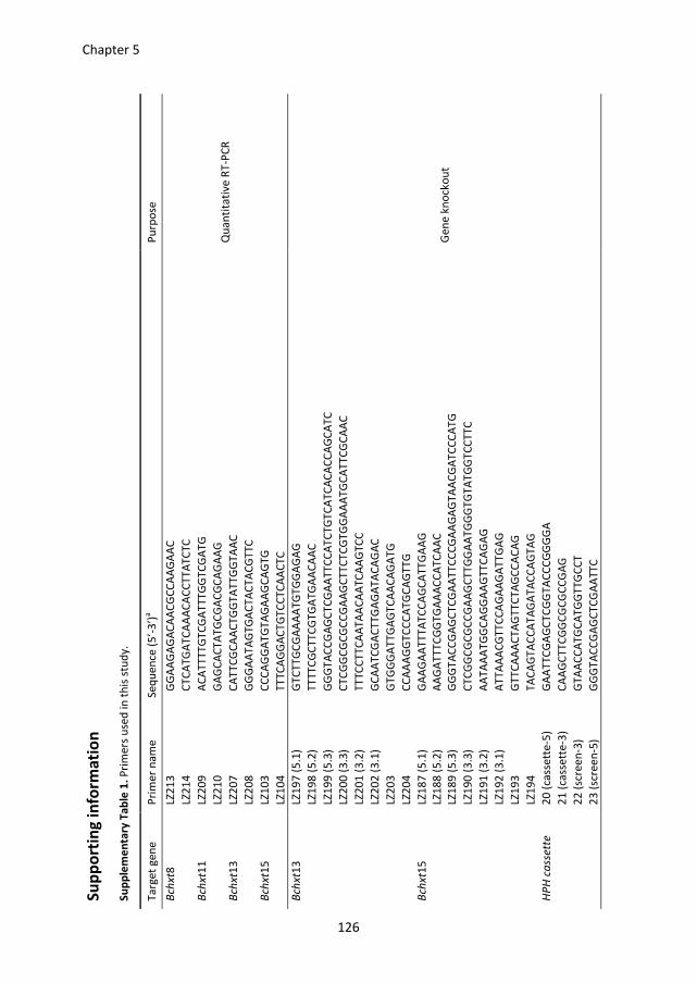
Su
pp
ort
ing
in
form
ati
on
Su
pp
lem
en
tary
Ta
ble
1.
Pri
me
rs u
sed
in
th
is s
tud
y.
Ta
rge
t g
en
e
Pri
me
r n
am
e
Se
qu
en
ce (
5’-
3’)
a
Pu
rpo
se
Bch
xt8
LZ
21
3
GG
AA
GA
GA
CA
AC
GC
CA
AG
AA
C
Qu
an
tita
tive
RT
-PC
R
LZ
21
4
CT
CA
TG
AT
CA
AA
CA
CC
TT
AT
CT
C
Bch
xt1
1
LZ2
09
A
CA
TT
TT
GT
CG
AT
TT
GG
TC
GA
TG
LZ
21
0
GA
GC
AC
TA
TG
CG
AC
GC
AG
AA
G
Bch
xt1
3
LZ2
07
C
AT
TC
GC
AA
CT
GG
TA
TT
GG
TA
AC
LZ
20
8
GG
GA
AT
AG
TG
AC
TA
CT
AC
GT
TC
Bch
xt1
5
LZ1
03
C
CC
AG
GA
TG
TA
GA
AG
CA
GT
G
LZ
10
4
TT
TC
AG
GA
CT
GT
CC
TC
AA
CT
C
Bch
xt1
3
LZ1
97
(5
.1)
GT
CT
TG
CG
AA
AA
TG
TG
GA
GA
G
Ge
ne
kn
ock
ou
t
LZ
19
8 (
5.2
) T
TT
TC
GC
TT
CG
TG
AT
GA
AC
AA
C
LZ
19
9 (
5.3
) G
GG
TA
CC
GA
GC
TC
GA
AT
TC
CA
TC
TG
TC
AT
CA
CA
CC
AG
CA
TC
LZ
20
0 (
3.3
) C
TC
GG
CG
CG
CC
GA
AG
CT
TC
TC
GT
GG
AA
AT
GC
AT
TC
GC
AA
C
LZ
20
1 (
3.2
) T
TT
CC
TT
CA
AT
AA
CA
AT
CA
AG
TC
C
LZ
20
2 (
3.1
) G
CA
AT
CG
AC
TT
GA
GA
TA
CA
GA
C
LZ
20
3
GT
GG
GA
TT
GA
GT
CA
AC
AG
AT
G
LZ
20
4
CC
AA
AG
GT
CC
CA
TG
CA
GT
TG
Bch
xt1
5
LZ1
87
(5
.1)
GA
AG
AA
TT
TA
TC
CA
GC
AT
TG
AA
G
LZ
18
8 (
5.2
) A
AG
AT
TT
CG
GT
GA
AA
CC
AT
CA
AC
LZ
18
9 (
5.3
) G
GG
TA
CC
GA
GC
TC
GA
AT
TC
CC
GA
AG
AG
TA
AC
GA
TC
CC
AT
G
LZ
19
0 (
3.3
) C
TC
GG
CG
CG
CC
GA
AG
CT
TG
GA
AT
GG
GT
GT
AT
GG
TC
CT
TC
LZ
19
1 (
3.2
) A
AT
AA
AT
GG
CA
GG
AA
GT
TC
AG
AG
LZ
19
2 (
3.1
) A
TT
AA
AC
GT
TC
CA
GA
AG
AT
TG
AG
LZ
19
3
GT
TC
AA
AC
TA
GT
TC
TA
GC
CA
CA
G
LZ
19
4
TA
CA
GT
AC
CA
TA
GA
TA
CC
AG
TA
G
HP
H c
ass
ett
e
20
(ca
sse
tte
-5)
GA
AT
TC
GA
GC
TC
GG
TA
CC
CG
GG
GA
2
1 (
cass
ett
e-3
) C
AA
GC
TT
CG
GC
GC
GC
CG
AG
2
2 (
scre
en
-3)
GT
AA
CC
AT
GC
AT
GG
TT
GC
CT
2
3 (
scre
en
-5)
GG
GT
AC
CG
AG
CT
CG
AA
TT
C
126
Chapter 5
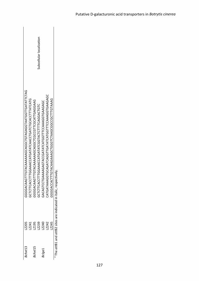
Bch
xt1
3
LZ2
05
G
GG
GA
CA
AG
TT
TG
TA
CA
AA
AA
AG
CA
GG
CT
GT
AG
AG
GT
AA
TG
GT
TG
AT
AT
TC
TA
G
Su
bce
llu
lar
loca
liza
tio
n
LZ
24
1
GC
TC
TT
CA
CC
TT
TG
GA
AA
CC
AT
GA
TA
TC
AA
CC
TG
AT
CT
GC
AC
CT
TT
AT
CA
TG
Bch
xt1
5
LZ1
95
G
GG
GA
CA
AG
TT
TG
TA
CA
AA
AA
AG
CA
GG
CT
CG
CC
GT
TC
GC
AT
TG
AG
GA
AG
LZ
23
9
GC
TC
TT
CA
CC
TT
TG
GA
AA
CC
AT
GA
TA
TC
GG
TA
CT
CT
TT
TC
AG
GA
CT
GT
C
Bcl
ga
1
LZ2
40
G
AC
AG
TC
CT
GA
AA
AG
AG
TA
CC
GA
TA
TC
AT
GG
TT
TC
CA
AA
GG
TG
AA
GA
GC
LZ
24
2
CA
TG
AT
AA
AG
GT
GC
AG
AT
CA
GG
TT
GA
TA
TC
AT
GG
TT
TC
CA
AA
GG
TG
AA
GA
GC
LZ
24
5
GG
GG
AC
CA
CT
TT
GT
AC
AA
GA
AA
GC
TG
GG
TC
TA
AG
CG
GC
CG
CT
TT
GT
AA
AG
a T
he
att
B1
an
d a
ttB
2 s
ite
s a
re in
dic
ate
d in
ita
lic,
re
spe
ctiv
ely
.
Botrytis cinereaPutative D-galacturonic acid transporters in
127

CHAPTER 6
Pectate-induced gene expression in Botrytis cinerea and the
identification and functional analysis of cis-regulatory D-
galacturonic acid responsive elements
Lisha Zhang, Joost Stassen, Sayantani Chatterjee, Maxim Cornelissen, Jan A. L. van Kan
Chapter 6
130
Abstract
The fungal plant pathogen Botrytis cinerea produces a spectrum of cell wall degrading
enzymes for the decomposition of host cell wall polysaccharides and the consumption of
the monosaccharides that are released. Especially pectin is an abundant cell wall
component, and the decomposition of pectin by B. cinerea has been extensively studied.
An effective concerted action of the appropriate pectin depolymerising enzymes,
monosaccharide transporters and catabolic enzymes is important for complete D-
galacturonic acid utilization by B. cinerea. In this study, we performed RNA sequencing to
compare genome-wide transcriptional profiles in B. cinerea grown in media containing
glucose and pectate as sole carbon source. Transcript levels of 32 genes that are induced
by pectate were further examined in cultures grown on six different monosaccharides, by
means of quantitative RT-PCR, leading to the identification of 8 genes that are specifically
induced by D-galacturonic acid. Conserved sequence motifs were identified in the
promoters of genes involved in pectate decomposition and D-galacturonic acid utilization.
The role of these motifs in regulating D-galacturonic acid-induced expression was
functionally analysed in the promoter of the Bclga1 gene, which encodes one of the key
enzymes in the D-galacturonic acid catabolic pathway. Regulation by D-galacturonic acid
required the presence of sequences with a conserved motif, designated, GAE1 and a
binding site for the pH-dependent transcriptional regulator PacC.
Pectate-induced gene expression and cis-regulatory elements
131
Introduction
The plant cell wall is the first barrier to pathogen invasion, and consists mainly of
polysaccharides that form a complex three-dimensional network together with lignin and
proteins. The main components of plant cell wall polysaccharides are cellulose,
hemicellulose, and pectin. The ability to decompose complex plant cell wall
polysaccharides is an important aspect of the lifestyle of fungal pathogens. Necrotrophic
fungal plant pathogens secrete large amounts of enzymes to decompose plant cell wall
polysaccharides in order to facilitate the penetration, the subsequent maceration and the
acquisition of carbon from decomposed plant tissues (Amselem et al., 2011). Hemi-
biotrophic pathogens also produce polysaccharide decomposing enzymes during the late,
necrotizing phase of infection (Gan et al., 2013; King et al., 2011; O'Connell et al., 2012).
By contrast, many biotrophic pathogens and symbionts have a markedly lower content of
enzymes for cell wall decomposition in their genome (Baxter et al., 2010; Duplessis et al.,
2011; Martin et al., 2010), presumably to reduce the damage to the host and avoid the
plant defence responses triggered by the release of cell wall fragments.
Botrytis cinerea is a necrotrophic fungal plant pathogen infecting more than 200 host
plants and causing severe economic damage to crops worldwide (Dean et al., 2012;
Williamson et al., 2007). B. cinerea secretes large amounts of cell wall degrading enzymes
for host tissue decomposition and nutrient acquisition. The preference for infection of
pectin-rich plants and tissues (ten Have et al., 2002) suggests that effective pectin
degradation is important for virulence of B. cinerea. The genome of B. cinerea encodes
118 Carbohydrate Active enZymes (CAZymes, www.cazy.org) (Cantarel et al., 2009)
associated with plant cell wall decomposition, of which a large proportion is involved in
the decomposition of pectin (Amselem et al., 2011). The pectin degrading capacity of B.
cinerea and the saprotroph Aspergillus niger is similar. These two unrelated fungi are not
only similar in the number of enzymes in pectin-related CAZY families that are encoded in
their genomes, but also in the ratio between pectin degrading lyases versus hydrolases
(Amselem et al., 2011).
Several B. cinerea pectin degrading enzyme activities have been detected during host
infection, including pectin and pectate lyases, pectin methylesterase (PMEs), exo-
polygalacturonases (exo-PGs), and endo-polygalacturonases (endo-PGs) (Cabanne and
Doneche, 2002; Kars et al., 2005b; Kars and van Kan, 2004; Rha et al., 2001; ten Have et al.,
2001). The importance of several pectinases for virulence of B. cinerea was investigated by
targeted mutagenesis in endo-PG genes and PME genes. Knockout mutants ∆Bcpg1 and
∆Bcpg2 were reduced in virulence by 25% and > 50%, respectively (Kars et al., 2005a; ten
Have et al., 1998). A ∆Bcpme1 mutant in one B. cinerea strain showed reduction in
Chapter 6
132
virulence (Valette-Collet et al., 2003); in a different strain, however, mutants in the same
Bcpme1 gene or the Bcpme2 gene, or even ∆Bcpme1/∆Bcpme2 double knockout mutants,
were not altered in virulence (Kars et al., 2005b). However, there is still a number of
pectinolytic genes that remain to be functionally analysed.
The monosaccharide D-galacturonic acid is the most abundant component of pectic
polysaccharides (Caffall and Mohnen, 2009; Mohnen, 2008) and is the final product
released from pectin degradation. The D-galacturonic acid catabolic pathway is conserved
in many filamentous fungi (Martens-Uzunova and Schaap, 2008; Richard and Hilditch,
2009). The pathway has been genetically and biochemically characterized in B. cinerea,
and consists of three catalytic steps involving four genes: Bcgar1, Bcgar2, Bclgd1, and
Bclga1 (Zhang et al., 2011). Their transcript levels were induced substantially when the
fungus was cultured in media containing D-galacturonic acid, pectate or pectin as the sole
carbon source (Zhang et al., 2011). Knockout mutants in each step of the four genes
(ΔBcgar1/ΔBcgar2, ΔBclgd1, and ΔBclga1) were affected in growth on D-galacturonic acid,
pectate, or pectin as the sole carbon source (Zhang et al., 2011), and in virulence on
Nicotiana benthamiana and Arabidopsis thaliana leaves (Zhang and van Kan, 2013).
Collectively, the functional analyses on the B. cinerea endo-PG genes and the D-
galacturonic acid catabolic pathway genes indicate that a concerted action of the
appropriate pectin depolymerising enzymes and catabolic enzymes is important for
complete D-galacturonic acid utilization by B. cinerea. Previous studies showed that
several genes involved in pectin decomposition and D-galacturonic acid catabolism are
induced in vitro by D-galacturonic acid and are expressed at high levels during infection in
the stage of lesion expansion, when plant cell wall degradation occurs (Wubben et al.,
2000; Zhang et al., 2011; Zhang and van Kan, 2013). Co-expression of these genes suggests
that a central regulatory mechanism is present in B. cinerea.
Current knowledge of the regulation of pectinolytic genes and D-galacturonic acid
catabolic genes in fungi is limited. Several studies showed that D-galacturonic acid is an
inducer for some pectinolytic genes and D-galacturonic acid catabolic genes (Martens-
Uzunova and Schaap, 2008; Martens-Uzunova et al., 2006; Mojzita et al., 2010; Wubben et
al., 2000; Zhang et al., 2011; Zhang and van Kan, 2013). A conserved element (TTGGNGG)
was identified in the bidirectional promoter region shared between the D-galacturonic
acid reductase and 2-keto-3-deoxy-L-galactonate aldolase genes from 18 fungal species,
including B. cinerea (Martens-Uzunova and Schaap, 2008). This conserved element is also
present in the promoters of several pectin degrading enzymes in A. niger (Martens-
Uzunova and Schaap, 2008). However, the function of this element in regulation by D-
galacturonic acid remains to be characterised.
Pectate-induced gene expression and cis-regulatory elements
133
In order to get further insight into the B. cinerea genes that participate in pectin
decomposition and D-galacturonic acid utilization, we have exploited the next generation
RNA-sequencing (RNA-seq) technology (Wang et al., 2009) to perform a genome-wide
transcriptome analysis in B. cinerea grown in media containing either glucose or pectate
as sole carbon sources. We identified a set of genes that are significantly altered in gene
expression. Promoter sequences of these genes were analysed to identify conserved
motifs that may act as cis-regulatory elements in the regulation of pectate decomposition
and D-galacturonic acid utilization. The promoter of the Bclga1 gene was functionally
analysed by fusing various promoter constructs to a reporter gene and monitoring
reporter activity in the presence of D-galacturonic acid.
Results
Pectate-induced gene expression in Botrytis cinerea
To study the transcriptome of Botrytis cinerea during degradation of pectic polymers and
D-galacturonic acid utilization, RNA-seq was performed and the transcriptome profiles
were compared between cultures containing glucose and sodium polygalacturonate
(pectate) as sole carbon source. Cultures were grown in glucose-containing medium
overnight and transferred to fresh medium with either glucose or pectate, and sampled at
6 h after transfer. RNA was isolated from three independent cultures in each condition.
One sample of each condition was used for RNA-seq and the other two were used for
quantitative RT-PCR (qRT-PCR) validation. A total of 6,800,254 and 6,462,333 read pairs
were obtained from glucose- and pectate-containing cultures, respectively, of which 90.3%
and 91.6% could be mapped to B. cinerea genome version 2 (Staats and van Kan, 2012).
The number of fragments per kilobase of transcript per million reads mapped (FPKM) was
determined for the 10,345 predicted genes as released with B. cinerea genome version 2
(Staats and van Kan, 2012), supplemented with two manually annotated gene models
representing genes known to be involved in D-galacturonic acid utilization, Bcpg2 and
Bcpme1 (Valette-Collet et al., 2003; Wubben et al., 1999). Of these 10,347 genes, 7,602
(73.5%) and 7,556 (73.0%) genes had an FPKM value > 1 in glucose- and pectate-
containing culture, respectively. The transcript level of 32 genes was significantly (q < 0.05)
up-regulated (log2 > 2) in pectate-containing culture compared to the glucose-containing
culture (Table 1). The transcript levels of these 32 genes were further determined by qRT-
PCR, and indeed showed strong induction in pectate-containing culture as compared to
the glucose-containing culture, with the exception of B0510_9368 (Table 1). For 23 of the
31 genes, the induction folds as determined by RNA-seq and by qRT-PCR, on independent

Chapter 6
134
biological samples, were in very good agreement (the difference between the log2 values
for a given sample was < 1).
In order to examine the expression of pectinolytic genes in the presence of pectate, we
generated a list of CAZymes with substrate specificity for pectic compounds, and listed
their expression values (Table S1). Among the 42 pectin-specific CAZyme encoding genes
in the B. cinerea genome, 14 genes were expressed at least 2-fold higher in pectate as
compared to glucose (with a minimum FPKM value of 1 in at least one of the samples),
comprising genes encoding enzymes assigned to 6 CAZY families: GH28, GH78, GH105,
GH115, CE8 and PL1.
Differential gene expression in cultures containing different monosaccharides
Pectate that was used as an inducer in the cultures used for the RNAseq studies is a linear,
unbranched polygalacturonate of 99% purity. We presumed that instead of the polymer
itself, the D-galacturonate monosaccharide released from the polymer would act as the
inducer of gene expression. To investigate whether the 31 pectate-induced genes are
specifically up-regulated in the presence of D-galacturonate or may also be up-regulated
by other cell wall-related monosaccharides, their transcript levels were determined by
qRT-PCR in cultures containing glucose, D-galacturonic acid, arabinose, rhamnose,
galactose, or xylose as the sole carbon source, respectively. Cultures were pre-grown in
glucose-containing medium and transferred to fresh medium with the different carbon
sources, and sampled at 3 h after transfer. The 31 pectate-induced genes could be divided
into three groups based on expression profiles: (1) genes of which transcript levels were
up-regulated in D-galacturonic acid-containing culture compared to glucose-containing
culture, and at least 2-fold higher as compared to cultures containing other
monosaccharides. This group comprises genes involved in D-galacturonic acid catabolism
(Bcgar2 (B0510_551) and Bclga1 (B0510_552; (Zhang et al., 2011)), pectin hydrolysis
(endo-polygalacturonase gene Bcpg2 and exo-polygalacturonase gene B0510_2787), as
well as members of the major facilitator superfamily (B0510_3996, encoding hexose
transporter BcHXT15 (Dulermo et al., 2009) and B0510_978, encoding a putative sugar:H+
symporter), and two additional genes: B0510_4887, encoding a putative alcohol
dehydrogenase and B0510_171, encoding a secreted protein (Figure 1); (2) genes of which
transcript levels, as compared to that in glucose-containing culture, were not only induced
by D-galacturonic acid, but also by other monosaccharide(s) to different extents (Figure
S1A); (3) genes of which transcript levels were not induced by D-galacturonic acid, as
compared to glucose (Figure S1B).
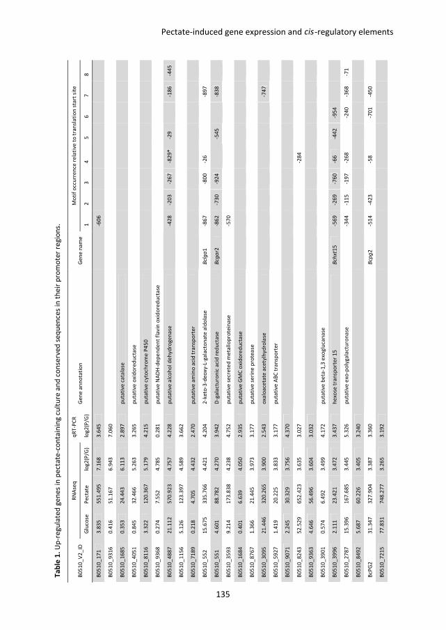
Ta
ble
1.
Up
-re
gu
late
d g
en
es
in p
ect
ate
-co
nta
inin
g c
ult
ure
an
d c
on
serv
ed
se
qu
en
ces
in t
he
ir p
rom
ote
r re
gio
ns.
B0
51
0_
V2
_ID
R
NA
seq
q
RT
-PC
R
Ge
ne
an
no
tati
on
G
en
e n
am
e
Mo
tif
occ
urr
en
ce r
ela
tive
to
tra
nsl
ati
on
sta
rt s
ite
Glu
cose
P
ect
ate
lo
g2
(P/G
) lo
g2
(P/G
) 1
2
3
4
5
6
7
8
B0
51
0_
17
1
3.8
35
5
51
.49
5
7.1
68
3
.64
5
-60
6
B0
51
0_
93
16
0
.41
6
51
.16
7
6.9
43
7
.06
0
B0
51
0_
16
85
0
.35
3
24
.44
3
6.1
13
2
.89
7
pu
tati
ve c
ata
lase
B0
51
0_
40
51
0
.84
5
32
.46
6
5.2
63
3
.26
5
pu
tati
ve o
xid
ore
du
cta
se
B0
51
0_
81
16
3
.32
2
12
0.3
67
5
.17
9
4.2
15
p
uta
tive
cyt
och
rom
e P
45
0
B0
51
0_
93
68
0
.27
4
7.5
52
4
.78
5
0.2
81
p
uta
tive
NA
DH
-de
pe
nd
en
t fl
av
in o
xid
ore
du
cta
se
B0
51
0_
48
87
2
1.1
12
5
70
.92
3
4.7
57
4
.22
8
pu
tati
ve a
lco
ho
l de
hy
dro
ge
na
se
-4
28
-2
03
-2
67
-8
29
*
-29
-18
6
-44
5
B0
51
0_
11
56
5
.12
6
12
3.3
97
4
.58
9
3.6
62
B0
51
0_
71
89
0
.21
8
4.7
05
4
.43
2
2.4
70
p
uta
tive
am
ino
aci
d t
ran
spo
rte
r
B0
51
0_
55
2
15
.67
5
33
5.7
66
4
.42
1
4.2
04
2
-ke
to-3
-de
oxy
-L-g
ala
cto
na
te a
ldo
lase
B
clg
a1
-8
67
-80
0
-26
-8
97
B0
51
0_
55
1
4.6
01
8
8.7
82
4
.27
0
3.9
42
D
-ga
lact
uro
nic
aci
d r
ed
uct
ase
B
cga
r2
-86
2
-73
0
-92
4
-5
45
-83
8
B0
51
0_
35
93
9
.21
4
17
3.8
38
4
.23
8
4.7
52
p
uta
tive
se
cre
ted
me
tall
op
rote
ina
se
-5
70
B0
51
0_
16
84
0
.40
1
6.6
39
4
.05
0
2.9
35
p
uta
tive
GM
C o
xid
ore
du
cta
se
B0
51
0_
87
67
1
.36
6
21
.44
5
3.9
73
3
.17
7
pu
tati
ve s
eri
ne
pro
tea
se
B0
51
0_
30
95
2
1.4
46
3
20
.26
5
3.9
00
2
.54
3
oxa
loa
ceta
te a
cety
lhy
dro
lase
-74
7
B0
51
0_
59
27
1
.41
9
20
.22
5
3.8
33
3
.17
7
pu
tati
ve A
BC
tra
nsp
ort
er
B0
51
0_
90
71
2
.24
5
30
.32
9
3.7
56
4
.37
0
B0
51
0_
82
43
5
2.5
29
6
52
.42
3
3.6
35
3
.02
7
-2
84
B0
51
0_
93
63
4
.64
6
56
.49
6
3.6
04
3
.03
2
B0
51
0_
39
01
0
.57
4
6.4
92
3
.49
9
4.1
72
p
uta
tive
be
ta-1
,3 e
xog
luca
na
se
B0
51
0_
39
96
2
.11
1
23
.42
1
3.4
72
3
.43
7
he
xose
tra
nsp
ort
er
15
B
chxt
15
-5
69
-2
69
-7
60
-6
6
-44
2
-95
4
B0
51
0_
27
87
1
5.3
96
1
67
.68
5
3.4
45
5
.32
6
pu
tati
ve e
xo-p
oly
ga
lact
uro
na
se
-3
44
-1
15
-1
97
-2
68
-24
0
-36
8
-71
B0
51
0_
84
92
5
.68
7
60
.22
6
3.4
05
3
.24
0
BcP
G2
3
1.3
47
3
27
.90
4
3.3
87
3
.36
0
B
cpg
2
-51
4
-42
3
-5
8
-7
01
-4
50
B0
51
0_
72
15
7
7.8
31
7
48
.27
7
3.2
65
3
.19
2
cis Pectate-induced gene expression and -regulatory elements
135
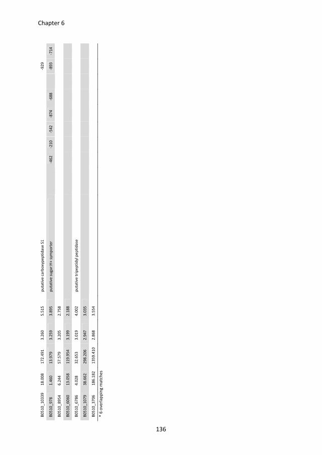
B0
51
0_
10
33
9
18
.00
8
17
2.4
91
3
.26
0
5.5
15
p
uta
tive
ca
rbo
xyp
ep
tid
ase
S1
-92
9
B0
51
0_
97
8
1.4
60
1
3.9
79
3
.25
9
3.8
95
p
uta
tive
su
ga
r:H
+ s
ym
po
rte
r
-46
2
-21
0
-54
2
-87
4
-68
8
-8
93
-7
14
B0
51
0_
89
54
6
.24
4
57
.57
9
3.2
05
2
.75
8
B0
51
0_
60
60
1
3.0
58
1
19
.95
4
3.1
99
2
.18
8
B0
51
0_
67
86
4
.02
8
32
.65
3
3.0
19
4
.00
2
pu
tati
ve t
rip
ep
tid
yl p
ep
tid
ase
B0
51
0_
10
79
3
8.6
62
2
98
.20
6
2.9
47
3
.03
5
B0
51
0_
37
06
1
86
.18
2
13
59
.41
0
2.8
68
3
.55
4
* 6
ove
rla
pp
ing
ma
tch
es
136
Chapter 6
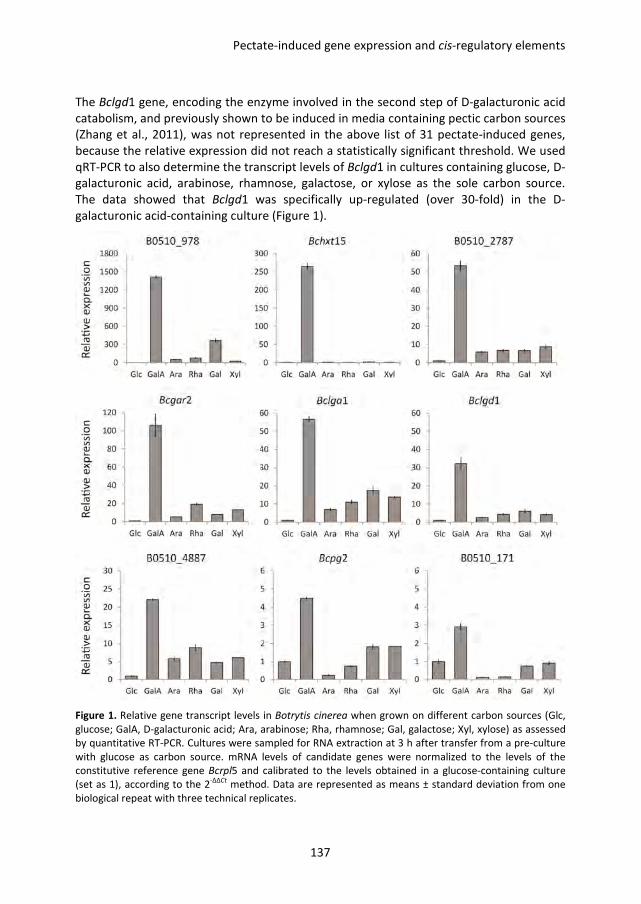
Pectate-induced gene expression and cis-regulatory elements
137
The Bclgd1 gene, encoding the enzyme involved in the second step of D-galacturonic acid
catabolism, and previously shown to be induced in media containing pectic carbon sources
(Zhang et al., 2011), was not represented in the above list of 31 pectate-induced genes,
because the relative expression did not reach a statistically significant threshold. We used
qRT-PCR to also determine the transcript levels of Bclgd1 in cultures containing glucose, D-
galacturonic acid, arabinose, rhamnose, galactose, or xylose as the sole carbon source.
The data showed that Bclgd1 was specifically up-regulated (over 30-fold) in the D-
galacturonic acid-containing culture (Figure 1).
Figure 1. Relative gene transcript levels in Botrytis cinerea when grown on different carbon sources (Glc,
glucose; GalA, D-galacturonic acid; Ara, arabinose; Rha, rhamnose; Gal, galactose; Xyl, xylose) as assessed
by quantitative RT-PCR. Cultures were sampled for RNA extraction at 3 h after transfer from a pre-culture
with glucose as carbon source. mRNA levels of candidate genes were normalized to the levels of the
constitutive reference gene Bcrpl5 and calibrated to the levels obtained in a glucose-containing culture
(set as 1), according to the 2-∆∆Ct
method. Data are represented as means ± standard deviation from one
biological repeat with three technical replicates.
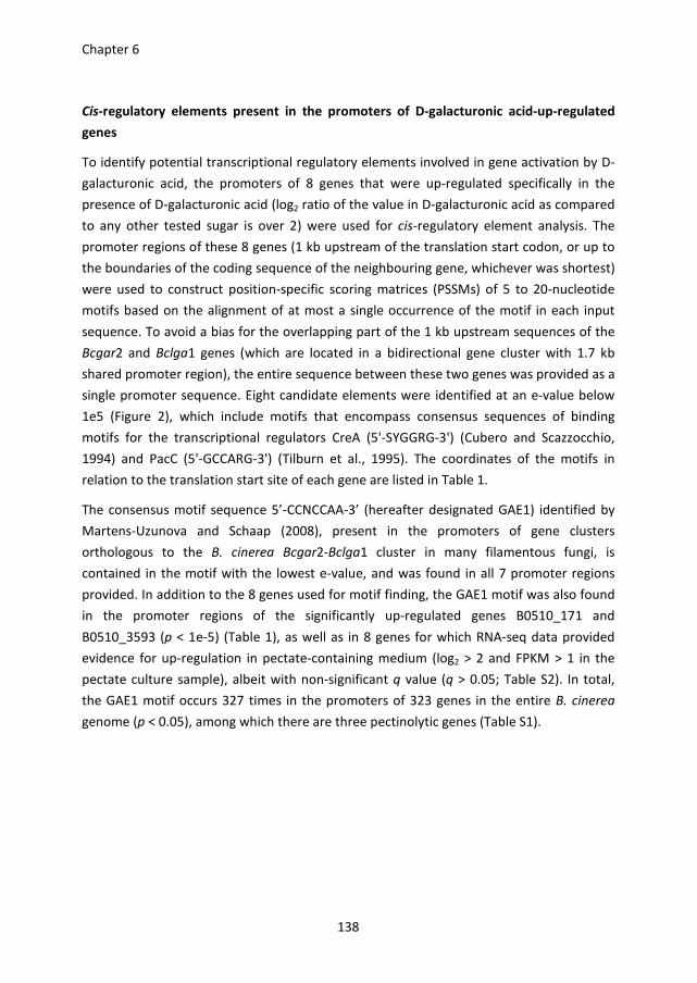
Chapter 6
138
Cis-regulatory elements present in the promoters of D-galacturonic acid-up-regulated
genes
To identify potential transcriptional regulatory elements involved in gene activation by D-
galacturonic acid, the promoters of 8 genes that were up-regulated specifically in the
presence of D-galacturonic acid (log2 ratio of the value in D-galacturonic acid as compared
to any other tested sugar is over 2) were used for cis-regulatory element analysis. The
promoter regions of these 8 genes (1 kb upstream of the translation start codon, or up to
the boundaries of the coding sequence of the neighbouring gene, whichever was shortest)
were used to construct position-specific scoring matrices (PSSMs) of 5 to 20-nucleotide
motifs based on the alignment of at most a single occurrence of the motif in each input
sequence. To avoid a bias for the overlapping part of the 1 kb upstream sequences of the
Bcgar2 and Bclga1 genes (which are located in a bidirectional gene cluster with 1.7 kb
shared promoter region), the entire sequence between these two genes was provided as a
single promoter sequence. Eight candidate elements were identified at an e-value below
1e5 (Figure 2), which include motifs that encompass consensus sequences of binding
motifs for the transcriptional regulators CreA (5'-SYGGRG-3') (Cubero and Scazzocchio,
1994) and PacC (5'-GCCARG-3') (Tilburn et al., 1995). The coordinates of the motifs in
relation to the translation start site of each gene are listed in Table 1.
The consensus motif sequence 5’-CCNCCAA-3’ (hereafter designated GAE1) identified by
Martens-Uzunova and Schaap (2008), present in the promoters of gene clusters
orthologous to the B. cinerea Bcgar2-Bclga1 cluster in many filamentous fungi, is
contained in the motif with the lowest e-value, and was found in all 7 promoter regions
provided. In addition to the 8 genes used for motif finding, the GAE1 motif was also found
in the promoter regions of the significantly up-regulated genes B0510_171 and
B0510_3593 (p < 1e-5) (Table 1), as well as in 8 genes for which RNA-seq data provided
evidence for up-regulation in pectate-containing medium (log2 > 2 and FPKM > 1 in the
pectate culture sample), albeit with non-significant q value (q > 0.05; Table S2). In total,
the GAE1 motif occurs 327 times in the promoters of 323 genes in the entire B. cinerea
genome (p < 0.05), among which there are three pectinolytic genes (Table S1).
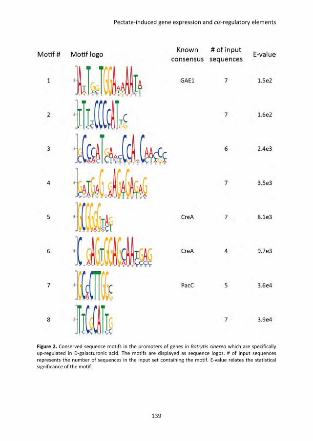
Pectate-induced gene expression and cis-regulatory elements
139
Figure 2. Conserved sequence motifs in the promoters of genes in Botrytis cinerea which are specifically
up-regulated in D-galacturonic acid. The motifs are displayed as sequence logos. # of input sequences
represents the number of sequences in the input set containing the motif. E-value relates the statistical
significance of the motif.
Chapter 6
140
Functional analysis of the cis-regulatory elements
Besides the predicted conserved GAE1 motif, there are 8 CreA binding motifs (Cubero and
Scazzocchio, 1994) and one PacC binding motif (Tilburn et al., 1995) in the promoter
region of the Bcgar2-Bclga1 gene cluster. The motifs 2, 3 and 4 (Figure 2) are each present
once around positions -1000, -800 and -26, relative to the start of the Bclga1 coding
sequence. To determine the contribution of these elements to the induction of gene
expression by D-galacturonic acid, the full-length (FL) promoter region of this gene cluster
(in the direction for Bclga1 expression) was fused with a codon-optimised gfp gene and a
set of promoter deletion constructs was generated in which different combinations of
motifs were removed (Del1-8) (Figure 3A). The FL and deletion constructs were
transformed into wild-type B. cinerea strain B05.10 and for each construct, up to 10
transformants were verified for proper integration of the construct and propagated for
reporter gene analysis. The conidia of transformants were germinated in the presence of
glucose or D-galacturonic acid for 20 h and hyphae of the germlings were subsequently
examined by epifluorescence microscopy. As shown in Figure 3, GFP fluorescence with the
FL promoter was observed in the presence of D-galacturonic acid but not in glucose. GFP
fluorescence of transformants with promoter deletion constructs Del1, Del2, and Del7 was
the same as that with the FL promoter in D-galacturonic acid, whereas no GFP
fluorescence was detected with the all other promoter deletion constructs. None of the
transformants (either with the FL promoter or the deletion constructs) showed any GFP
fluorescence when grown in glucose.
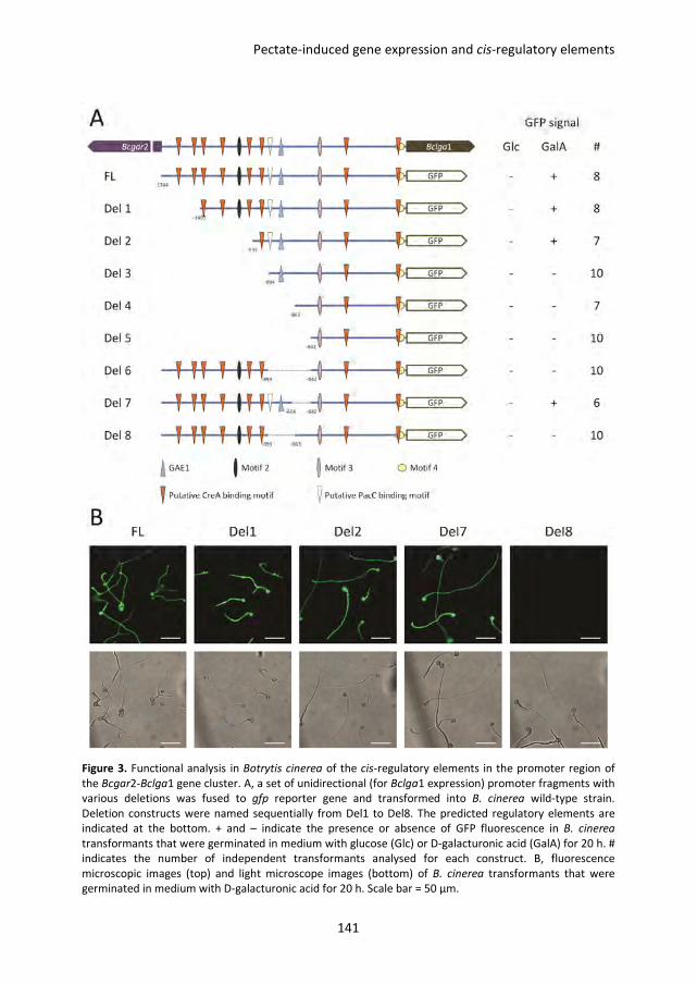
Pectate-induced gene expression and cis-regulatory elements
141
Figure 3. Functional analysis in Botrytis cinerea of the cis-regulatory elements in the promoter region of
the Bcgar2-Bclga1 gene cluster. A, a set of unidirectional (for Bclga1 expression) promoter fragments with
various deletions was fused to gfp reporter gene and transformed into B. cinerea wild-type strain.
Deletion constructs were named sequentially from Del1 to Del8. The predicted regulatory elements are
indicated at the bottom. + and – indicate the presence or absence of GFP fluorescence in B. cinerea
transformants that were germinated in medium with glucose (Glc) or D-galacturonic acid (GalA) for 20 h. #
indicates the number of independent transformants analysed for each construct. B, fluorescence
microscopic images (top) and light microscope images (bottom) of B. cinerea transformants that were
germinated in medium with D-galacturonic acid for 20 h. Scale bar = 50 µm.
Chapter 6
142
Discussion
In this study, RNA-seq technology was applied to genome-wide transcriptome analysis in B.
cinerea grown in media containing glucose or pectate as sole carbon source. 32 genes in B.
cinerea were identified that are significantly up-regulated when grown on pectate as
compared to glucose. Furthermore, the transcript levels of these genes were verified by
qRT-PCR with two additional biological repeats. The data obtained by qRT-PCR were
strongly correlated with the data from the RNA-seq experiments. Of the initially identified
32 pectate-induced genes, 31 were indeed highly up-regulated in pectate with a log2 ratio
higher than 2. For 23 of the 31 genes, the RNA-seq and qRT-PCR data (on independent
biological samples) showed less than 2-fold difference in the induction ratios, indicating
that RNA-seq is a suitable method for quantifying gene expression, even when performed
on a single biological sample. Bcgar2, Bclgd1 and Bclga1 encode enzymes involved in the
three catalytic steps of D-galacturonic acid catabolism (Zhang et al., 2011). The up-
regulation of expression of Bcgar2 and Bclga1 found in our RNA-seq analysis was in
agreement with qRT-PCR data obtained before (Zhang et al., 2011). The transcript levels of
Bclgd1 were also highly induced by pectate (Zhang et al., 2011), but this gene was not
among the 31 up-regulated genes, because it was not statistically significant at the
threshold of q < 0.05 (Bclgd1: log2 = 2.4, q = 0.159). In addition, the Bchxt15 gene,
encoding a putative D-galacturonic acid transporter (Chapter 5), was significantly up-
regulated in the RNA-seq experiment. Growth of ∆Bchxt15 mutants on D-galacturonic acid
was not different from the wild-type strain (Chapter 5), indicating that other hexose
transporters also contribute to D-galacturonic acid uptake. Indeed, the RNA-seq data
revealed that another putative sugar:H+ symporter gene, B0510_978, was highly up-
regulated by pectate. qRT-PCR analysis demonstrated that both Bchxt15 and B0510_978
were specifically up-regulated by D-galacturonic acid compared to glucose, with ~560-fold
and ~1400-fold respectively. The expression data suggest that B0510_978 might play a
prominent role in D-galacturonic acid uptake. Further studies with knockout mutants of
the B0510_978 gene in the wild-type strain and in a ∆Bchxt15 mutant background could
shed more light on its function in D-galacturonic acid uptake.
Pectate is a linear polymer of D-galacturonic acid and it is plausible to assume that pectate
hydrolysis, by a mixture of endo-PGs and exo-PGs, would provide D-galacturonic acid for
fungal growth over a fair period of time, until the substrate would be entirely degraded.
On the other hand, if B. cinerea would be provided with D-galacturonic acid
monosaccharide as sole carbon source, this would lead to a short, transient burst of
expression of genes involved in D-galacturonic acid utilization which would fade out as
soon as the bulk of monosaccharide would be absorbed from the medium. In order to
Pectate-induced gene expression and cis-regulatory elements
143
mimic gene expression during the degradation of pectate in plant tissue and the
subsequent utilization of the released monosaccharides, we decided to choose pectate as
the carbon source for RNA-seq analysis. We anticipated that the induction of gene
expression in pectate-containing medium is likely mediated by the monosaccharides
released upon hydrolysis. This would imply that genes that are inducible during growth on
pectate should also be induced during growth on D-galacturonic acid. Unexpectedly, qRT-
PCR analysis demonstrated that ~50% of the 31 pectate-up-regulated genes were not
induced by D-galacturonic acid. It should be noted that the samples for RNA-seq were
prepared at 6 h after transfer to pectate, whereas the samples for qRT-PCR were prepared
at 3 h after transfer to the monosaccharide. It is possible that 3 h was not sufficient to
induce the transcript levels of these genes. Previous studies showed that the transcript
levels of D-galacturonic acid catabolic genes were higher at 3 h after transfer than at 9 h
after transfer in medium with D-galacturonic acid or pectate as the carbon source (Zhang
et al., 2011), probably because the carbon sources were already depleted at 9 h. Further
transcript analysis with more time points will allow to compare the expression profiles of
genes in D-galacturonic acid and pectate in more detail. Also, the ambient pH was
different in the D-galacturonic acid-containing culture (adjusted to pH 5.6 with sodium
hydroxide) and pectate-containing culture (pH 7.0), and this may also have affected the
expression profiles. It has been reported that the transcript levels of several B. cinerea
genes are modulated by ambient pH, including genes encoding proteases and cell wall
degrading enzymes (Billon-Grand et al., 2012; Rolland et al., 2009). B0510_3593 and
B0510_10339, both encoding secreted proteases, and B0510_3901, encoding a beta-1,3-
exoglucanase, were up-regulated in the presence of pectate (pH 7.0) but not D-
galacturonic acid (pH 5.6), suggesting that ambient pH may have affected the expression
of these genes.
The GAE1 element is not only present in the promoters of all eight genes specifically
induced by D-galacturonic acid- and of several pectinolytic genes of B. cinerea, but also in
the promoters of several co-expressed pectinolytic genes of Aspergillus niger and in gene
clusters orthologous to the B. cinerea Bcgar2-Bclga1 cluster in many filamentous fungi
(Martens-Uzunova and Schaap, 2008). This suggests that the GAE1 motif is part of a
conserved regulatory system for pectin degradation and D-galacturonic acid utilization in
different fungi. Functional analysis of the regulatory elements in the promoter region of
the Bcgar2-Bclga1 gene cluster suggests that the promoter region containing the GAE1
element and the PacC binding motif is essential for the induction of gene expression by D-
galacturonic acid. These sequences are in very close proximity, only 15 nucleotides apart.
Constructs Del2 and Del3 differ only in the presence of one CreA binding motif and the
PacC binding motif, while fluorescence was only observed in Del2-containing
Chapter 6
144
transformants, but not in Del3-containing transformants. This result shows that the PacC
binding motif is essential, while the GAE1 motif alone is insufficient for D-galacturonate-
induced expression. Comparison of the results for constructs Del8 and FL suggest that only
35 nucleotides (between coordinates -899 and -863) are essential for D-galacturonate-
induced expression. Additional promoter deletion constructs will need to be designed to
further dissect the sequences that are important in regulation.
In summary, a set of genes was identified in B. cinerea of which the transcript levels were
specifically induced by D-galacturonic acid. These genes encode proteins involved in pectin
degradation, monosaccharide uptake and D-galacturonic acid catabolism, suggesting the
existence of an efficient, co-regulated D-galacturonic acid utilization network in B. cinerea.
The promoters of these co-expressed genes share certain cis-regulatory elements and are
therefore potentially regulated by a set of common transcription factors. It has been
reported that in Aspergillus species, several transcription factors that are involved in plant
cell wall polysaccharide degradation and in the catabolism of the derived
monosaccharides are induced by the corresponding monosaccharide (Battaglia et al., 2011;
Gruben, 2012). For example, RhaR regulates the expression of genes related to rhamnose
utilization and its expression is induced by rhamnose (Gruben, 2012). However, the list of
B. cinerea genes that are significantly up-regulated by pectate does not include any gene
encoding a putative transcription factor or DNA-binding protein. One possible explanation
is that the transcript levels of transcriptional regulator genes increase fast (reaching a
peak at an early time point) and subsequently decrease rapidly, such that induction
cannot be detected at 6 h after transfer from glucose to pectate. Alternatively, the
transcriptional regulator might be present in an inactive form, and activated in the
presence of D-galacturonic acid to promote target gene expression. Other experimental
approaches, such as a DNA-protein pull-down combined with mass spectrometry analysis,
may allow to identify the transcription factor(s) binding to crucial cis-regulatory elements
allowing a better understanding of the regulatory network of B. cinerea for pectin
degradation and D-galacturonic acid utilization.
Pectate-induced gene expression and cis-regulatory elements
145
Materials and methods
Fungal strains and growth conditions
Botrytis cinerea wild-type strain B05.10 and strains transformed with Bcgar2-Bclga1
promoter deletion constructs used in this study were routinely grown on Malt Extract Agar
(Oxoid, Basingstoke, UK; 50 g/L) in the dark at 20 °C for 3-4 days. The plates were placed
for one night under near-UV light (350–400 nm) to promote sporulation, and were
subsequently returned to darkness. Conidia were harvested 4-7 days later in 10–20 mL of
water, and the suspension was filtered over glass wool to remove mycelium fragments.
The conidia suspension was centrifuged at 1200 rpm for 5 min. The supernatant was
discarded and the conidia in the pellet were resuspended at the desired density.
RNA extraction
The conidia of the wild-type strain B05.10 were incubated in Gamborg’s B5 liquid culture
supplemented with 10 mM (NH4)H2PO4 and 50 mM glucose at 20 °C, 150 rpm. After 16 h
of growth, the mycelium was harvested and transferred into fresh Gamborg’s B5 medium
supplemented with 10 mM (NH4)H2PO4 and a carbon source. For the cultures used for
RNA-seq analysis, either 50 mM glucose or 0.5% sodium polygalacturonate (pectate) was
added; mycelium was harvested from these cultures at 6 h post transfer and freeze-dried.
For the cultures used for quantitative RT-PCR, either glucose, D-galacturonic acid, L-
arabinose, L-rhamnose, D-galactose, or L-xylose was added at 50 mM final concentration;
mycelium was harvested from these cultures at 3 h post transfer and freeze-dried.
Total RNA was isolated using the Nucleospin® RNA plant kit (Machery-Nagl, Düren,
Germany), according to the manufacturer’s instructions. First strand cDNA was
synthesized from 1 μg total RNA with SuperScript® III Reverse Transcriptase (Invitrogen)
according to the manufacturer’s instructions.
RNA-sequencing and data analysis
Twenty micrograms of total RNA for each RNA sample were prepared as described above.
cDNA synthesis, library preparation and Illumina sequencing (100 bp paired-end reads)
were performed at Beijing Genome Institute (BGI, Hong Kong). The obtained Illumina RNA-
seq reads were trimmed to remove the first 12 nucleotides using fastx trimmer
(http://hannonlab.cshl.edu/fastx_toolkit/) and mapped to annotated genes on version 2
of the B. cinerea genome (Staats and van Kan, 2012) using Tophat (version 2.0.6) with
default settings (Trapnell et al., 2009). Differentially expressed genes were then
determined using Cuffdiff (version 2.0.2) with default settings and cut-offs (Trapnell et al.,
2013).
Chapter 6
146
Quantitative RT-PCR
Quantitative RT-PCR was performed using an ABI7300 PCR machine (Applied Biosystems,
Foster City, U.S.A.) in combination with the qPCR SensiMix kit (BioLine, London, U.K.). The
primers to detect the transcripts of B. cinerea genes are listed in Table S3. Real-time PCR
conditions were as follows: an initial 95 °C denaturation step for 10 min followed by
denaturation for 15 s at 95 °C and annealing/extension for 45 s at 60 °C for 40 cycles. The
data were analysed on the 7300 System SDS software (Applied Biosystems, Foster City,
U.S.A.). The transcript levels of target genes were normalized to the transcript levels of the
constitutively expressed gene Bcrpl5 and calibrated to the levels observed in glucose
culture (set as 1), according to the 2-ΔΔCt
method.
Conserved motif elements finding
Conserved motif elements were predicted by MEME (Bailey and Elkan, 1994) with
minimum size 5 nucleotides, maximum size 20 nucleotides, zero or one occurrence per
sequence, using the promoter regions of 8 D-galacturonic acid up-regulated genes as
training set. The occurrence of motifs in the promoter regions was performed by the
scanning for matches to the position-specific scoring matrices (PSSMs) describing the
motif with PoSSuM search (Beckstette et al., 2009).
Plasmid construction and B. cinerea transformation
In order to study the regulation of the promoter of the Bcgar2-Bclga1 gene cluster in B.
cinerea, a yeast recombination-based cloning vector pNDH-GFP was generated as
described previously (Figure S2) (Schumacher, 2012). The full-length promoter region of
Bcgar2-Bclga1 and a series of promoter deletions were amplified from genomic DNA using
primers listed in Table S4. The amplified promoter regions, together with NcoI-linearized
pNDH-GFP, were co-transformed into the uracil-auxotrophic yeast strain FY834 (Winston
et al., 1995), as described by Schumacher (2012). The transformants were confirmed by
PCR with primers LZ182/183 and plasmids were isolated with Zymoprep yeast plasmid
miniprep kit I (Zymo research) according to the manufacturer’s instructions. Plasmids
were then transformed into competent Escherichia coli DH5α cells to increase the DNA
yield. The resulting plasmids were checked by sequencing (Macrogen) and subsequently
transformed into B. cinerea wild-type strain B05.10 by protoplast transformation (Kars et
al., 2005b). The hygromycin-resistant transformants were checked by PCR for the
presence of the hph gene with primers LZ92/93 and up to 10 independent transformants
of each construct were used for analysis of GFP fluorescence.
Pectate-induced gene expression and cis-regulatory elements
147
Microscopic analysis
Conidia of the strains containing different promoter constructs were incubated on glass
slides with Gamborg’s B5 liquid medium supplemented with 10 mM (NH4)H2PO4 and 50
mM of glucose or D-galacturonic acid as the carbon source at 20 °C. After 20 h incubation,
the slides were covered with cover slips and observed under a Nikon Eclipse 90i
epifluorescence microscope (Nikon, Badhoevedorp, the Netherlands) or light microscopy.
GFP fluorescence was visualized by using a GFP-B filter cube (EX460-500, DM 505, BA510-
560). The NIS-Elements software package was used to analyze digital pictures.
Supporting information
Supplementary Figures S1, S2 and Supplementary Tables S1, S2, S3, S4.
Acknowledgements
The authors are grateful to Pierre de Wit (Wageningen University, Laboratory of
Phytopathology) for critical reading of the manuscript. The authors acknowledge funding
by the Foundation Technological Top Institute Green Genetics (Project 2CC035RP) and the
Netherlands Graduate School Experimental Plant Sciences.
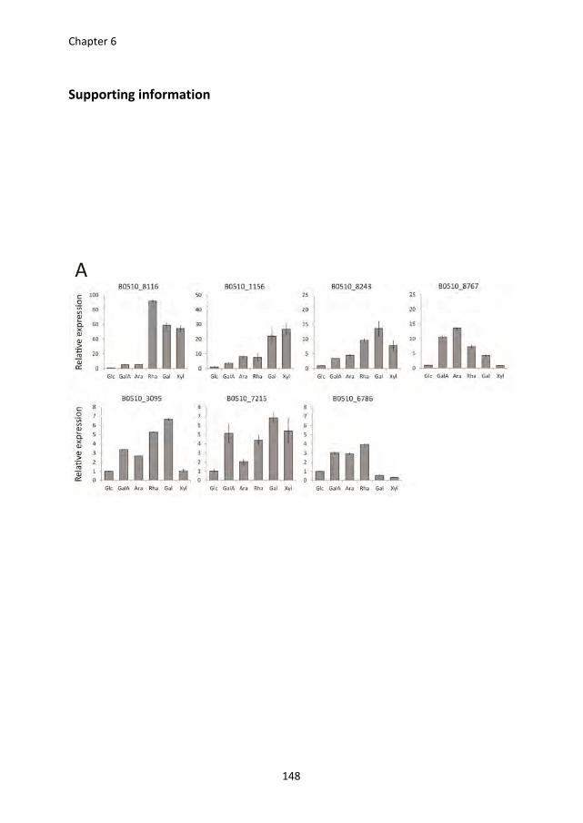
Chapter 6
148
Supporting information
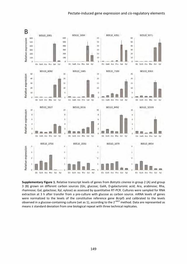
Pectate-induced gene expression and cis-regulatory elements
149
Supplementary Figure 1. Relative transcript levels of genes from Botrytis cinerea in group 2 (A) and group
3 (B) grown on different carbon sources (Glc, glucose; GalA, D-galacturonic acid; Ara, arabinose; Rha,
rhamnose; Gal, galactose; Xyl, xylose) as assessed by quantitative RT-PCR. Cultures were sampled for RNA
extraction at 3 h after transfer from a pre-culture with glucose as carbon source. mRNA levels of genes
were normalized to the levels of the constitutive reference gene Bcrpl5 and calibrated to the levels
observed in a glucose-containing culture (set as 1), according to the 2-∆∆Ct
method. Data are represented as
means ± standard deviation from one biological repeat with three technical replicates.
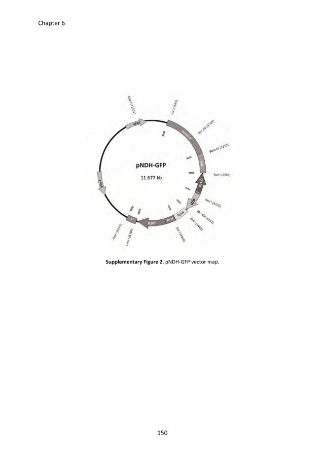
Chapter 6
150
Supplementary Figure 2. pNDH-GFP vector map.
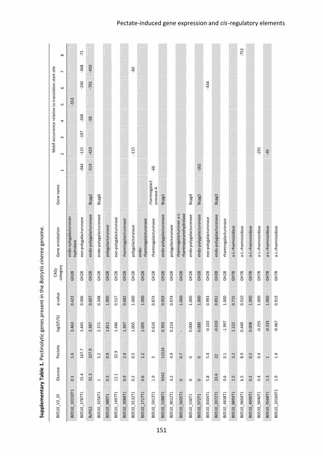
Su
pp
lem
en
tary
Ta
ble
1.
Pe
ctin
oly
tic
ge
ne
s p
rese
nt
in t
he
Bo
tryti
s ci
ne
rea
ge
no
me
.
B0
51
0_
V2
_ID
G
luco
se
Pe
cta
te
log
2(P
/G)
q-v
alu
e
CA
Zy
cate
go
ry
Ge
ne
an
no
tati
on
G
en
e n
am
e
Mo
tif
occ
urr
en
ce r
ela
tive
to
tra
nsl
ati
on
sta
rt s
ite
1
2
3
4
5
6
7
8
B0
51
0_
10
15
0T
1
0.1
1
.6
3.4
63
0
.42
2
GH
28
e
nd
o-x
ylo
ga
lact
uro
na
n
hyd
rola
se
-5
53
B0
51
0_
27
87
T1
1
5.4
1
67
.7
3.4
45
0
.00
6
GH
28
e
xo-p
oly
ga
lact
uro
na
se
-3
44
-1
15
-1
97
-2
68
-24
0
-36
8
-71
BcP
G2
3
1.3
3
27
.9
3.3
87
0
.00
7
GH
28
e
nd
o-p
oly
ga
lact
uro
na
se
Bcp
g2
-5
14
-4
23
-58
-70
1
-45
0
B0
51
0_
32
56
T1
1
5
.2
2.3
71
0
.34
8
GH
28
e
nd
o-p
oly
ga
lact
uro
na
se
Bcp
g6
B0
51
0_
98
0T
1
0.3
0
.9
1.8
11
1
.00
0
GH
28
p
oly
ga
lact
uro
na
se
B0
51
0_
14
97
T1
1
2.1
3
2.9
1
.44
6
0.5
17
G
H2
8
exo
-po
lyg
ala
ctu
ron
ase
B0
51
0_
30
88
T1
0
.9
2.4
1
.36
7
0.6
82
G
H2
8
rha
mn
og
ala
ctu
ron
ase
B0
51
0_
91
32
T1
0
.2
0.5
1
.00
5
1.0
00
G
H2
8
po
lyg
ala
ctu
ron
ase
-1
15
-6
0
B0
51
0_
17
17
T1
0
.6
1.2
1
.00
5
1.0
00
G
H2
8
en
do
-
rha
mn
og
ala
ctu
ron
ase
B0
51
0_
59
13
T1
1
.9
3
0.6
16
0
.87
4
GH
28
rh
am
no
ga
lact
uro
na
se
rha
mn
og
ala
ct
uro
na
se A
-6
6
B0
51
0_
53
88
T1
9
34
2
11
52
4
0.3
03
0
.95
9
GH
28
e
nd
o-p
oly
ga
lact
uro
na
se
Bcp
g1
B0
51
0_
90
22
T1
4
.2
4.9
0
.21
4
0.9
74
G
H2
8
po
lyg
ala
ctu
ron
ase
B0
51
0_
56
05
T1
0
0
.7
- 1
.00
0
GH
28
rh
am
no
ga
lact
uro
na
n a
-L-
rha
mn
op
yra
no
hy
dro
lase
B0
51
0_
53
6T
1
0
0
0.0
00
1
.00
0
GH
28
e
nd
o-p
oly
ga
lact
uro
na
se
Bcp
g4
B0
51
0_
97
2T
1
0
0
0.0
00
1
.00
0
GH
28
e
nd
o-p
oly
ga
lact
uro
na
se
Bcp
g5
-1
62
B0
51
0_
83
04
T1
5
.8
5.4
-0
.10
3
0.9
93
G
H2
8
exo
-po
lyg
ala
ctu
ron
ase
-4
34
B0
51
0_
20
72
T1
3
3.6
2
2
-0.6
10
0
.85
1
GH
28
e
nd
o-p
oly
ga
lact
uro
na
se
Bcp
g3
B0
51
0_
48
38
T1
0
.6
0.1
-1
.99
7
1.0
00
G
H2
8
rha
mn
og
ala
ctu
ron
ase
B0
51
0_
88
94
T1
1
.5
3.2
1
.12
2
0.7
15
G
H7
8
a-L
-rh
am
no
sid
ase
B0
51
0_
96
04
T1
6
.5
8.9
0
.44
9
0.9
10
G
H7
8
a-L
-rh
am
no
sid
ase
-7
53
B0
51
0_
46
00
T1
0
.2
0.2
0
.00
8
1.0
00
G
H7
8
a-L
-rh
am
no
sid
ase
B0
51
0_
94
40
T1
0
.4
0.4
-0
.25
5
1.0
00
G
H7
8
a-L
-rh
am
no
sid
ase
-2
92
B0
51
0_
95
68
T1
1
.3
1
-0.3
33
1
.00
0
GH
78
a
-L-r
ha
mn
osi
da
se
-45
B0
51
0_
10
16
0T
1
1.9
1
.4
-0.4
67
0
.91
9
GH
78
a
-L-r
ha
mn
osi
da
se
Pectate-induced gene expression and -regulatory elementscis
151
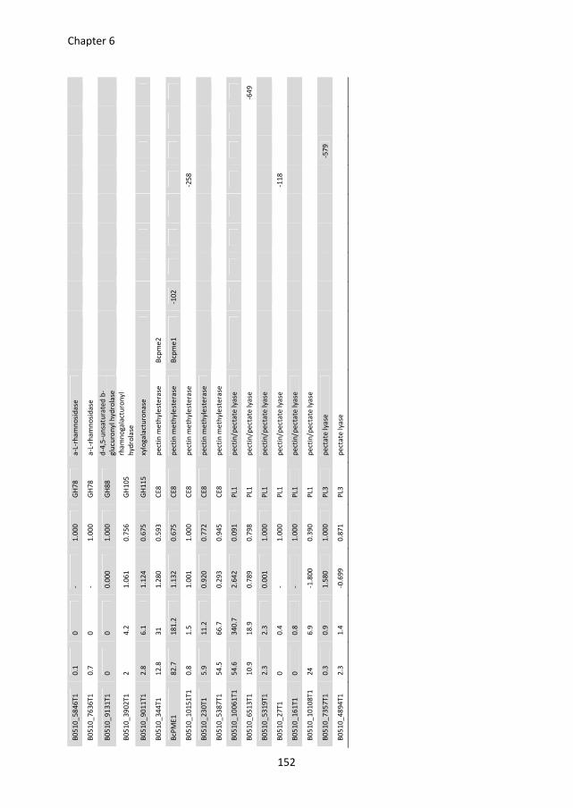
B0
51
0_
58
46
T1
0
.1
0
- 1
.00
0
GH
78
a
-L-r
ha
mn
osi
da
se
B0
51
0_
76
36
T1
0
.7
0
- 1
.00
0
GH
78
a
-L-r
ha
mn
osi
da
se
B0
51
0_
91
31
T1
0
0
0
.00
0
1.0
00
G
H8
8
d-4
,5-u
nsa
tura
ted
b-
glu
curo
ny
l hyd
rola
se
B0
51
0_
39
02
T1
2
4
.2
1.0
61
0
.75
6
GH
10
5
rha
mn
og
ala
ctu
ron
yl
hyd
rola
se
B0
51
0_
90
11
T1
2
.8
6.1
1
.12
4
0.6
75
G
H1
15
xy
log
ala
ctu
ron
ase
B0
51
0_
34
4T
1
12
.8
31
1
.28
0
0.5
93
C
E8
p
ect
in m
eth
yle
ste
rase
B
cpm
e2
BcP
ME
1
82
.7
18
1.2
1
.13
2
0.6
75
C
E8
p
ect
in m
eth
yle
ste
rase
B
cpm
e1
-1
02
B0
51
0_
10
15
1T
1
0.8
1
.5
1.0
01
1
.00
0
CE
8
pe
ctin
me
thyl
est
era
se
-2
58
B0
51
0_
23
0T
1
5.9
1
1.2
0
.92
0
0.7
72
C
E8
p
ect
in m
eth
yle
ste
rase
B0
51
0_
53
87
T1
5
4.5
6
6.7
0
.29
3
0.9
45
C
E8
p
ect
in m
eth
yle
ste
rase
B0
51
0_
10
06
1T
1
54
.6
34
0.7
2
.64
2
0.0
91
P
L1
pe
ctin
/pe
cta
te l
yase
B0
51
0_
65
13
T1
1
0.9
1
8.9
0
.78
9
0.7
98
P
L1
pe
ctin
/pe
cta
te l
yase
-6
49
B0
51
0_
53
19
T1
2
.3
2.3
0
.00
1
1.0
00
P
L1
pe
ctin
/pe
cta
te l
yase
B0
51
0_
27
T1
0
0
.4
- 1
.00
0
PL1
p
ect
in/p
ect
ate
lya
se
-1
18
B0
51
0_
16
1T
1
0
0.8
-
1.0
00
P
L1
pe
ctin
/pe
cta
te l
yase
B0
51
0_
10
10
8T
1
24
6
.9
-1.8
00
0
.39
0
PL1
p
ect
in/p
ect
ate
lya
se
B0
51
0_
73
57
T1
0
.3
0.9
1
.58
0
1.0
00
P
L3
pe
cta
te l
yase
-5
79
B0
51
0_
48
94
T1
2
.3
1.4
-0
.69
9
0.8
71
P
L3
pe
cta
te l
yase
Chapter 6
152
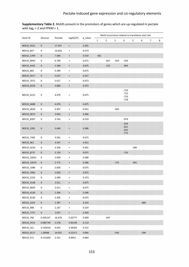
Pectate-induced gene expression and cis-regulatory elements
153
Supplementary Table 2. Motifs present in the promoters of genes which are up-regulated in pectate
with log2 > 2 and FPKM > 1.
Gene ID Glucose Pectate log2(G/P) q_value Motif occurrence relative to translation start site
1 2 3 4 5 6 7 8
B0510_9321 0 17.247 + 0.291
B0510_847 0 10.626 + 0.473
B0510_1769 0 7.584 + 0.519 -431
B0510_8943 0 6.790 + 0.675
-827 -424 -559
B0510_4442 0 5.399 + 0.675
-224
-944
B0510_805 0 5.399 + 0.675
B0510_9317 0 5.027 + 0.247
B0510_7071 0 5.017 + 0.473
B0510_8156 0 4.680 + 0.473
B0510_6151 0 4.476 + 0.675
-710
-712
-716
-718
B0510_6688 0 4.476 + 0.675
B0510_6620 0 4.307 + 0.451
-565
B0510_9073 0 3.942 + 0.306
B0510_8397 0 3.763 + 0.519
-979
B0510_2281 0 3.646 + 0.586
-619
-621
-625
-772
B0510_7392 0 3.561 + 0.675
B0510_961 0 3.547 + 0.451
B0510_6229 0 3.336 + 0.401
-300
B0510_8737 0 3.134 + 0.675
-716
B0510_10031 0 2.930 + 0.586
B0510_10076 0 2.775 + 0.586
-772
-941
B0510_1086 0 2.656 + 0.675
B0510_2981 0 2.656 + 0.675
B0510_2332 0 2.499 + 0.473
B0510_4148 0 2.411 + 0.675
B0510_8603 0 2.411 + 0.675
B0510_4226 0 2.394 + 0.586
B0510_9230 0 2.305 + 0.675
B0510_3355 0 2.287 + 0.310
-680
B0510_898 0 2.167 + 0.324
B0510_7757 0 2.057 + 0.369
B0510_760 0.505147 16.479 5.02777 0.099
-447
B0510_9315 0.082749 2.578 4.96109 0.110
B0510_161 0.193018 4.055 4.39303 0.222
B0510_8117 1.28588 24.025 4.22371 0.066
-545
-504
B0510_571 0.131669 2.251 4.0953 0.083
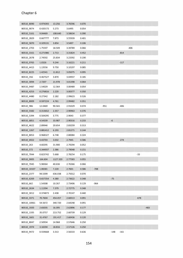
Chapter 6
154
B0510_8090 0.974393 13.256 3.76596 0.070
B0510_9074 0.420173 5.273 3.6495 0.054
B0510_5161 9.04469 108.640 3.58634 0.390
B0510_2829 0.667777 7.873 3.55939 0.401
B0510_5870 0.329131 3.854 3.5497 0.106
B0510_2703 1.75597 18.509 3.39789 0.066
-406
B0510_3161 0.271986 2.713 3.31824 0.452
-814
B0510_2678 2.74592 25.834 3.23392 0.190
B0510_9765 1.0326 9.244 3.16221 0.211
-117
B0510_4415 1.13556 9.750 3.10197 0.085
B0510_8225 1.42541 11.812 3.05075 0.095
B0510_356 0.467527 3.870 3.04937 0.185
B0510_3094 2.7207 21.978 3.01398 0.064
B0510_9487 1.54029 12.364 3.00489 0.054
B0510_4253 0.276816 2.220 3.00377 0.350
B0510_4480 0.27342 2.182 2.99623 0.526
B0510_8009 0.597224 4.761 2.99482 0.351
B0510_986 12.0469 90.565 2.91029 0.074
-951
-406
B0510_5582 0.313652 2.357 2.90963 0.376
B0510_5394 0.504295 3.775 2.9043 0.377
B0510_6851 4.54339 33.987 2.90316 0.233
-6
B0510_4622 2.89946 20.654 2.83259 0.313
B0510_1267 0.881412 6.193 2.81271 0.144
B0510_8053 0.968327 6.748 2.80084 0.324
B0510_6922 0.43703 3.032 2.7945 0.586
-274
B0510_263 4.60295 31.900 2.79294 0.052
B0510_572 0.344937 2.386 2.79048 0.151
B0510_7044 0.823742 5.668 2.78254 0.175
-33
B0510_9005 164.604 1127.320 2.77583 0.055
B0510_7045 5.98364 40.636 2.76366 0.066
B0510_10107 1.06581 7.220 2.7601 0.586 -708
B0510_2377 94.5399 636.038 2.75012 0.070
B0510_6269 0.657354 4.380 2.73622 0.348
-75
B0510_662 1.54308 10.267 2.73406 0.129 -964
B0510_2634 1.11394 7.379 2.72775 0.348
B0510_3012 0.374873 2.438 2.70147 0.440
B0510_3371 70.7604 454.457 2.68313 0.091
-678
B0510_10061 54.5672 340.720 2.64248 0.091
B0510_1920 2.65035 16.395 2.62896 0.177
-483
B0510_1335 35.0757 213.752 2.60739 0.129
B0510_1601 31.4767 191.417 2.60436 0.120
B0510_8047 2.50934 14.968 2.57646 0.250
B0510_1974 3.16594 18.816 2.57126 0.250
B0510_9472 0.559668 3.313 2.56533 0.636
-148 -161
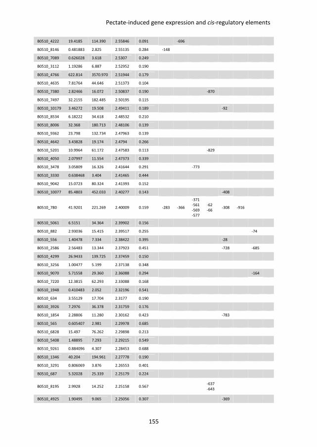
Pectate-induced gene expression and cis-regulatory elements
155
B0510_4222 19.4185 114.390 2.55846 0.091
-696
B0510_8146 0.481883 2.825 2.55135 0.284 -148
B0510_7089 0.626028 3.618 2.5307 0.249
B0510_3112 1.19286 6.887 2.52952 0.190
B0510_4766 622.814 3570.970 2.51944 0.179
B0510_4635 7.81764 44.646 2.51373 0.104
B0510_7380 2.82466 16.072 2.50837 0.190
-870
B0510_7497 32.2155 182.485 2.50195 0.115
B0510_10179 3.46272 19.508 2.49411 0.189
-92
B0510_8534 6.18222 34.618 2.48532 0.210
B0510_8006 32.368 180.713 2.48106 0.139
B0510_9362 23.798 132.734 2.47963 0.139
B0510_4642 3.43828 19.174 2.4794 0.266
B0510_5201 10.9964 61.172 2.47583 0.113
-829
B0510_4050 2.07997 11.554 2.47373 0.339
B0510_3478 3.05809 16.326 2.41644 0.291
-773
B0510_3330 0.638468 3.404 2.41465 0.444
B0510_9042 15.0723 80.324 2.41393 0.152
B0510_10077 85.4803 452.033 2.40277 0.143
-408
B0510_780 41.9201 221.269 2.40009 0.159 -283 -366
-371
-561
-569
-577
-62
-66 -308 -916
B0510_5061 6.5151 34.364 2.39902 0.156
B0510_882 2.93036 15.415 2.39517 0.255
-74
B0510_556 1.40478 7.334 2.38422 0.395
-28
B0510_2586 2.56483 13.344 2.37923 0.451
-728
-685
B0510_4299 26.9433 139.725 2.37459 0.150
B0510_3256 1.00477 5.199 2.37138 0.348
B0510_9070 5.71558 29.360 2.36088 0.294
-164
B0510_7220 12.3815 62.293 2.33088 0.168
B0510_1948 0.410483 2.052 2.32196 0.541
B0510_634 3.55129 17.704 2.3177 0.190
B0510_3926 7.2976 36.378 2.31759 0.176
B0510_1854 2.28806 11.280 2.30162 0.423
-783
B0510_565 0.605407 2.981 2.29978 0.685
B0510_6828 15.497 76.262 2.29898 0.213
B0510_5408 1.48895 7.293 2.29215 0.549
B0510_9261 0.884096 4.307 2.28453 0.688
B0510_1346 40.204 194.961 2.27778 0.190
B0510_3291 0.806069 3.876 2.26553 0.401
B0510_687 5.32028 25.339 2.25179 0.224
B0510_8195 2.9928 14.252 2.25158 0.567
-637
-643
B0510_4925 1.90495 9.065 2.25056 0.307
-369
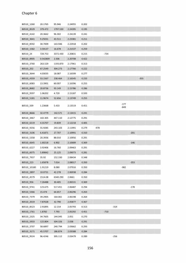
Chapter 6
156
B0510_1260 20.1765 95.946 2.24955 0.202
B0510_8529 379.472 1797.530 2.24395 0.195
B0510_4142 20.3662 96.302 2.24139 0.241
B0510_3641 9.25031 43.511 2.23381 0.211
B0510_8592 30.7009 143.946 2.22918 0.202
B0510_1582 3.59137 16.678 2.21537 0.259
B0510_24 729.753 3372.430 2.20831 0.215
-734
B0510_8905 0.542809 2.506 2.20708 0.422
B0510_3760 263.529 1193.870 2.17961 0.313
B0510_202 67.2549 304.273 2.17766 0.222
B0510_3644 4.03035 18.087 2.16599 0.277
B0510_4359 53.1567 238.464 2.16545 0.233
-355
B0510_6083 13.3901 60.007 2.16396 0.255
B0510_8682 20.8736 93.149 2.15786 0.286
B0510_9597 1.06202 4.720 2.15187 0.593
B0510_5181 11.9674 52.656 2.13749 0.255
B0510_509 1.23668 5.433 2.13519 0.451
-177
-849
B0510_8666 32.4779 142.571 2.13415 0.241
B0510_3467 102.305 447.110 2.12775 0.291
B0510_6519 4.55707 19.839 2.12218 0.405
B0510_9256 55.9285 243.103 2.11991 0.279 -978
B0510_3106 6.41871 27.707 2.10991 0.310
-201
B0510_5358 20.3936 88.010 2.10956 0.291
B0510_6645 1.60218 6.902 2.10689 0.369
-146
B0510_6227 3.92406 16.760 2.09463 0.291
B0510_6075 5.89842 25.125 2.09073 0.281
B0510_7937 35.92 152.330 2.08434 0.348
B0510_223 1.65878 7.014 2.08017 0.350
-353
B0510_10180 1.91219 8.080 2.07916 0.350
-982
B0510_2897 10.0731 42.278 2.06938 0.284
B0510_4579 1514.08 6340.290 2.0661 0.350
B0510_956 7.26488 30.405 2.06531 0.369
B0510_9741 123.675 517.451 2.06487 0.294
-178
B0510_5366 15.474 64.657 2.06296 0.263
B0510_7379 39.2905 164.061 2.06198 0.264
B0510_2019 7.87028 32.790 2.05877 0.367
B0510_8523 2.91895 12.154 2.05793 0.313
-314
B0510_1761 1.8782 7.793 2.05292 0.451
-710
B0510_2325 34.7605 144.045 2.051 0.270
B0510_5933 122.804 504.326 2.038 0.291
B0510_3707 58.6897 240.794 2.03662 0.293
B0510_3171 45.5707 186.874 2.03588 0.284
B0510_9524 96.4246 395.112 2.03479 0.289
-256
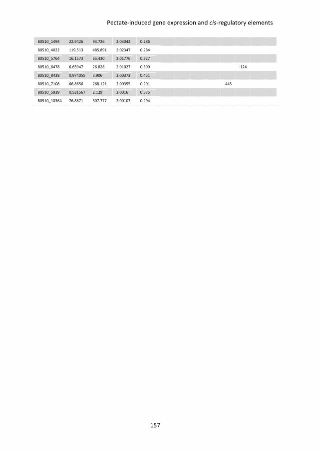
Pectate-induced gene expression and cis-regulatory elements
157
B0510_1494 22.9426 93.726 2.03042 0.286
B0510_4022 119.513 485.891 2.02347 0.284
B0510_5764 16.1573 65.430 2.01776 0.327
B0510_6478 6.65947 26.828 2.01027 0.399
-124
B0510_8438 0.974055 3.906 2.00373 0.451
B0510_7108 66.8656 268.121 2.00355 0.291
-445
B0510_5939 0.531567 2.129 2.0016 0.575
B0510_10364 76.8871 307.777 2.00107 0.294
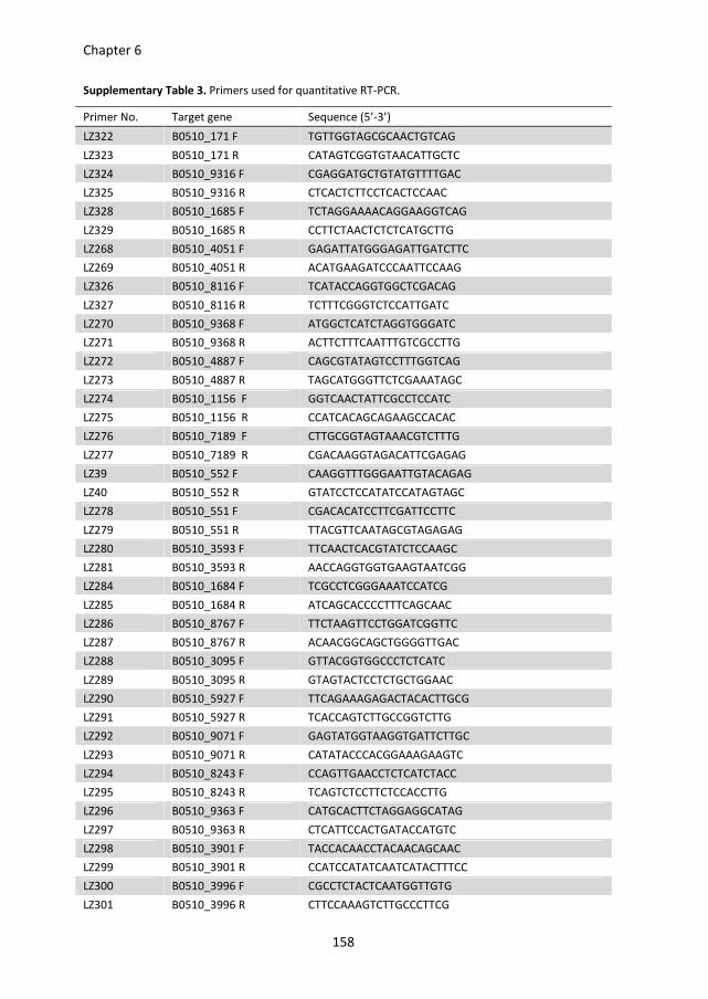
Chapter 6
158
Supplementary Table 3. Primers used for quantitative RT-PCR.
Primer No. Target gene Sequence (5’-3’)
LZ322 B0510_171 F TGTTGGTAGCGCAACTGTCAG
LZ323 B0510_171 R CATAGTCGGTGTAACATTGCTC
LZ324 B0510_9316 F CGAGGATGCTGTATGTTTTGAC
LZ325 B0510_9316 R CTCACTCTTCCTCACTCCAAC
LZ328 B0510_1685 F TCTAGGAAAACAGGAAGGTCAG
LZ329 B0510_1685 R CCTTCTAACTCTCTCATGCTTG
LZ268 B0510_4051 F GAGATTATGGGAGATTGATCTTC
LZ269 B0510_4051 R ACATGAAGATCCCAATTCCAAG
LZ326 B0510_8116 F TCATACCAGGTGGCTCGACAG
LZ327 B0510_8116 R TCTTTCGGGTCTCCATTGATC
LZ270 B0510_9368 F ATGGCTCATCTAGGTGGGATC
LZ271 B0510_9368 R ACTTCTTTCAATTTGTCGCCTTG
LZ272 B0510_4887 F CAGCGTATAGTCCTTTGGTCAG
LZ273 B0510_4887 R TAGCATGGGTTCTCGAAATAGC
LZ274 B0510_1156 F GGTCAACTATTCGCCTCCATC
LZ275 B0510_1156 R CCATCACAGCAGAAGCCACAC
LZ276 B0510_7189 F CTTGCGGTAGTAAACGTCTTTG
LZ277 B0510_7189 R CGACAAGGTAGACATTCGAGAG
LZ39 B0510_552 F CAAGGTTTGGGAATTGTACAGAG
LZ40 B0510_552 R GTATCCTCCATATCCATAGTAGC
LZ278 B0510_551 F CGACACATCCTTCGATTCCTTC
LZ279 B0510_551 R TTACGTTCAATAGCGTAGAGAG
LZ280 B0510_3593 F TTCAACTCACGTATCTCCAAGC
LZ281 B0510_3593 R AACCAGGTGGTGAAGTAATCGG
LZ284 B0510_1684 F TCGCCTCGGGAAATCCATCG
LZ285 B0510_1684 R ATCAGCACCCCTTTCAGCAAC
LZ286 B0510_8767 F TTCTAAGTTCCTGGATCGGTTC
LZ287 B0510_8767 R ACAACGGCAGCTGGGGTTGAC
LZ288 B0510_3095 F GTTACGGTGGCCCTCTCATC
LZ289 B0510_3095 R GTAGTACTCCTCTGCTGGAAC
LZ290 B0510_5927 F TTCAGAAAGAGACTACACTTGCG
LZ291 B0510_5927 R TCACCAGTCTTGCCGGTCTTG
LZ292 B0510_9071 F GAGTATGGTAAGGTGATTCTTGC
LZ293 B0510_9071 R CATATACCCACGGAAAGAAGTC
LZ294 B0510_8243 F CCAGTTGAACCTCTCATCTACC
LZ295 B0510_8243 R TCAGTCTCCTTCTCCACCTTG
LZ296 B0510_9363 F CATGCACTTCTAGGAGGCATAG
LZ297 B0510_9363 R CTCATTCCACTGATACCATGTC
LZ298 B0510_3901 F TACCACAACCTACAACAGCAAC
LZ299 B0510_3901 R CCATCCATATCAATCATACTTTCC
LZ300 B0510_3996 F CGCCTCTACTCAATGGTTGTG
LZ301 B0510_3996 R CTTCCAAAGTCTTGCCCTTCG
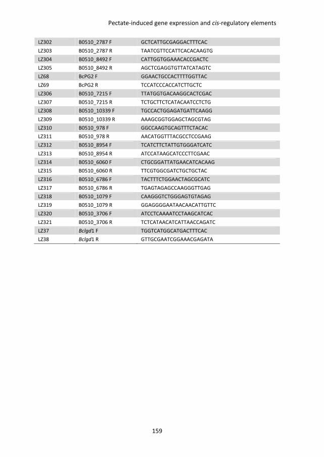
Pectate-induced gene expression and cis-regulatory elements
159
LZ302 B0510_2787 F GCTCATTGCGAGGACTTTCAC
LZ303 B0510_2787 R TAATCGTTCCATTCACACAAGTG
LZ304 B0510_8492 F CATTGGTGGAAACACCGACTC
LZ305 B0510_8492 R AGCTCGAGGTGTTATCATAGTC
LZ68 BcPG2 F GGAACTGCCACTTTTGGTTAC
LZ69 BcPG2 R TCCATCCCACCATCTTGCTC
LZ306 B0510_7215 F TTATGGTGACAAGGCACTCGAC
LZ307 B0510_7215 R TCTGCTTCTCATACAATCCTCTG
LZ308 B0510_10339 F TGCCACTGGAGATGATTCAAGG
LZ309 B0510_10339 R AAAGCGGTGGAGCTAGCGTAG
LZ310 B0510_978 F GGCCAAGTGCAGTTTCTACAC
LZ311 B0510_978 R AACATGGTTTACGCCTCCGAAG
LZ312 B0510_8954 F TCATCTTCTATTGTGGGATCATC
LZ313 B0510_8954 R ATCCATAAGCATCCCTTCGAAC
LZ314 B0510_6060 F CTGCGGATTATGAACATCACAAG
LZ315 B0510_6060 R TTCGTGGCGATCTGCTGCTAC
LZ316 B0510_6786 F TACTTTCTGGAACTAGCGCATC
LZ317 B0510_6786 R TGAGTAGAGCCAAGGGTTGAG
LZ318 B0510_1079 F CAAGGGTCTGGGAGTGTAGAG
LZ319 B0510_1079 R GGAGGGGAATAACAACATTGTTC
LZ320 B0510_3706 F ATCCTCAAAATCCTAAGCATCAC
LZ321 B0510_3706 R TCTCATAACATCATTAACCAGATC
LZ37 Bclgd1 F TGGTCATGGCATGACTTTCAC
LZ38 Bclgd1 R GTTGCGAATCGGAAACGAGATA

Su
pp
lem
en
tary
Ta
ble
4.
Pri
me
rs u
sed
fo
r g
en
era
tin
g p
rom
ote
r co
nst
ruct
s.
Pri
me
r N
o.
Ta
rge
t g
en
e
Se
qu
en
ce (
5'-
3')
D
esc
rip
tio
n
LZ1
74
F
L F
GA
TT
AC
TT
AC
CT
CG
CC
CT
TG
CT
TA
CC
AT
CG
AT
GA
TA
GT
GT
GA
AT
TG
TA
TT
TG
AA
LZ1
47
F
L R
T
GA
AA
AG
CT
CT
TC
AC
CT
TT
GG
AA
AC
CA
TG
TG
TT
AT
GA
TT
AT
AT
GT
AT
GT
GT
AG
AT
G
LZ2
53
D
el1
F
GA
TT
AC
TT
AC
CT
CG
CC
CT
TG
CT
TA
CC
AT
CT
AT
CG
GT
GT
TG
TT
CG
CT
GC
TT
W
ith
LZ
14
7 t
o a
mp
lify
De
l1 f
rag
me
nt
LZ2
54
D
el2
F
GA
TT
AC
TT
AC
CT
CG
CC
CT
TG
CT
TA
CC
AT
CT
TC
AA
AT
TG
GC
CA
CA
AC
TC
TA
C
Wit
h L
Z1
47
to
am
plify
De
l2 f
rag
me
nt
LZ1
48
D
el3
F
GA
TT
AC
TT
AC
CT
CG
CC
CT
TG
CT
TA
CC
AT
CA
TC
AG
AA
AG
CT
TA
TT
GG
TG
GA
AA
AA
T
Wit
h L
Z1
47
to
am
plify
De
l3 f
rag
me
nt
LZ1
49
D
el4
F
GA
TT
AC
TT
AC
CT
CG
CC
CT
TG
CT
TA
CC
AT
CC
CA
GC
TT
CA
AC
GA
CA
CT
CA
GA
W
ith
LZ
14
7 t
o a
mp
lify
De
l4 f
rag
me
nt
LZ1
50
D
el5
F
GA
TT
AC
TT
AC
CT
CG
CC
CT
TG
CT
TA
CC
AT
CA
TG
TT
GA
AT
CG
AC
AA
TT
TT
AG
TC
C
Wit
h L
Z1
47
to
am
plify
De
l5 f
rag
me
nt
LZ1
54
D
el6
F
CT
CT
AC
CC
CA
CG
CC
CT
TG
GA
TG
TT
GA
AT
CG
AC
AA
TT
TT
AG
TC
C
Wit
h L
Z1
47
to
am
plify
De
l6-3
’ fr
ag
me
nt
LZ1
55
D
el6
R
GG
AC
TA
AA
AT
TG
TC
GA
TT
CA
AC
AT
CC
AA
GG
GC
GT
GG
GG
TA
GA
G
Wit
h L
Z1
74
to
am
plify
De
l6-5
’ fr
ag
me
nt
LZ1
56
D
el7
F
GA
AA
GC
TT
AT
TG
GT
GG
AA
AA
AT
AG
TT
AT
GT
TG
AA
TC
GA
CA
AT
TT
TA
GT
CC
W
ith
LZ
14
7 t
o a
mp
lify
De
l7-3
’ fr
ag
me
nt
LZ1
57
D
el7
R
GG
AC
TA
AA
AT
TG
TC
GA
TT
CA
AC
AT
AA
CT
AT
TT
TT
CC
AC
CA
AT
AA
GC
TT
TC
W
ith
LZ
17
4 t
o a
mp
lify
De
l7-5
’ fr
ag
me
nt
LZ1
84
D
el8
F
CT
CT
AC
CC
CA
CG
CC
CT
TG
GT
CC
AG
CT
TC
AA
CG
AC
AC
TC
AG
A
Wit
h L
Z1
47
to
am
plify
De
l8-3
’ fr
ag
me
nt
LZ1
85
D
el8
R
TC
TG
AG
TG
TC
GT
TG
AA
GC
TG
GA
CC
AA
GG
GC
GT
GG
GG
TA
GA
G
Wit
h L
Z1
74
to
am
plify
De
l8-5
’ fr
ag
me
nt
LZ1
82
p
ND
H-G
FP
F
GT
TT
TC
CC
AG
TC
AC
GA
CC
CT
TA
AA
TC
TC
AT
GA
AC
TC
CT
TG
LZ1
83
p
ND
H-G
FP
R
CA
GG
AA
AC
AG
CT
AT
GA
CC
CT
CT
CC
GC
TG
AC
TG
AG
AA
C
LZ9
2
HP
H F
G
GT
TC
GG
CG
TA
GG
GT
TG
TT
C
LZ9
3
HP
H R
G
TG
TA
TT
GA
CC
GA
TT
CC
TT
GC
Chapter 6
160

CHAPTER 7
General discussion

General discussion
163
The plant pathogenic fungus Botrytis cinerea predominantly infects pectin-rich plant
species and tissues. Effective pectin degradation thus is important for virulence of B.
cinerea. The experimental results presented in this thesis provide more insights into the
role of pectin degradation by B. cinerea. On the one hand, we report a novel feature of B.
cinerea endo-polygalacturonases (BcPGs) that act as microbe-associated molecular
patterns (MAMPs) and describe the identification of the receptor complex in Arabidopsis
thaliana that is required for the responsiveness to BcPGs. On the other hand, we discuss
the role of pectinases and D-galacturonic acid catabolism in virulence of B. cinerea. We
subsequently discuss the co-regulation of genes involved in pectin degradation, D-
galacturonic acid uptake and catabolism and propose a model for this concerted action.
Novel features of endo-polygalacturonases in plant-pathogen interactions
BcPGs are not only important virulence factors, but are also able to induce necrotic
responses when infiltrated in leaves of several plant species (Kars et al., 2005a). The data
presented in chapter 2 demonstrate that fungal endo-PGs (including BcPGs) may act as
MAMPs. This observation is analogous to reports on the ethylene-inducing xylanase (EIX)
from the saprotrophic fungus Trichoderma and B. cinerea xylanase xyn11A (Enkerli et al.,
1999; Furman-Matarasso et al., 1999; Noda et al., 2010), which are both able to cause a
necrotic response in tobacco and tomato. Both for the B. cinerea endo-PGs and the
Trichoderma and B. cinerea xylanases, catalytically inactive proteins were equally able to
induce a necrotic response, indicating that it is the protein that is recognized, rather than
hydrolytic products of cell wall polymers also known as damage-associated molecular
patterns (DAMPs).
For bacterial MAMPs such as flagellin and EF-Tu, the full-blown plant responses can be
triggered by an oligopeptide that contains the epitope determining structure, presumably
perceived as a linear peptide stretch. Also in the Trichoderma EIX, a pentapeptide epitope
has been identified as an essential component for induction of the hypersensitive
response (HR) (Rotblat et al., 2002). Among fungal endo-PGs, an 11-amino acid sequence
is present as the longest conserved linear stretch. Unfortunately, a synthetic peptide of 22
amino acids, comprising these 11 amino acid residues was not capable of inducing
necrosis. This observation does not necessarily exclude the importance of this sequence as
an epitope for recognition. Possibly the 3-dimensional structure of the synthetic peptide
differs from the structure present in the native protein, or the peptide is unstable and
degraded by plant proteases after infiltration in the plant. Notably, the corresponding
region in the sequence of A. thaliana endo-PGs contains at position 1 a proline residue
instead of glycine. The proline most likely alters the 3-dimensional structure of plant endo-
PGs, which would enable them to escape recognition by the plant itself. Further studies
Chapter 7
164
are required to elucidate which amino acid motifs in fungal endo-PGs are crucial for
inducing necrosis.
Plants perceive potential microbial invaders by using pattern recognition receptors (PRRs)
that recognize MAMPs. As described in chapter 2, we studied the natural variation of A.
thaliana accessions in responsiveness to BcPGs, and found among others that accession
Col-0 is responsive to BcPGs, whereas accession Br-0 is not responsive. Molecular
identification and functional characterization demonstrated that the gene RBPG1,
encoding an LRR-RLP, determines responsiveness to BcPGs in Col-0. RBPG1 is one member
of a family of four RLP-encoding genes (RLP39, RLP40, RLP41 and RBPG1) that occur in a
cluster within this region of the Col-0 genome. Accession Br-0 contains only two RLP-
encoding genes (rbpg1-1 and rbpg1-2) in this region. Although RLP39, rbpg1-2 as well as
RBPG1 were able to form a complex with BcPG3, only RBPG1 is responsive and able to
induce necrosis. This situation is analogous to the tomato LRR-RLPs LeEix1 and LeEix2,
encoded by two paralogous genes from a gene cluster, that act as receptors for EIX. Both
receptors are able to bind EIX, whereas only LeEix2 mediates necrotic responses (Ron and
Avni, 2004). Further studies demonstrated that LeEix1, but not LeEix2 interacts with
tomato BAK1 in a ligand-independent manner, and the LeEIX1-BAK1 interaction is
required for the ability of LeEix1 to attenuate the signalling of LeEix2 (Bar et al., 2010).
RBPG1, however, does not form a complex with BAK1 in the co-immunoprecipitation
assay. We demonstrate that a different LRR-RLK, named SOBIR1 (Gao et al., 2009), is an
inBteractor of RBPG1 and this interaction is required for the induction of necrosis upon
application of BcPG3. RBPG1 and rbpg1-2 were also able to form a complex with SOBIR1.
Further studies are required to investigate whether other (as yet unknown) components in
the complex have the ability to bind RBPG1 to determine specificity.
HR-associated cell death usually enhances susceptibility of plants to B. cinerea (Govrin and
Levine, 2000). However, we demonstrate that RBPG1 has no effect on susceptibility nor on
disease resistance against B. cinerea. The full lesion development of B. cinerea on A.
thaliana leaves occurs within 3 days post inoculation, whereas the response of A. thaliana
to BcPG proteins is visible 3 days after infiltration. This suggests that the necrotic response
is possibly too slow to have any impact on the infection by B. cinerea. Furthermore, RBPG1
does not contribute to disease resistance to other microbial pathogens like
Hyaloperonospora arabidopsidis, Phytophthora capsici and Pseudomonas syringae pv.
tomato DC3000, probably because RBPG1 does not recognize the endo-PGs produced by
these pathogens, due to the lack of the conserved epitope fpg11. The role of RBPG1 in
disease resistance to other pathogens that contain the epitope fpg11 in their endo-PGs (e.
g. Colletotrichum higginsianum) is not known yet and requires future studies.
General discussion
165
The role of pectinases in virulence of Botrytis cinerea
The B. cinerea genome contains six genes encoding endo-PGs (Wubben et al., 1999). All
gene family members are differentially expressed in planta (ten Have et al., 2001; Zhang
and van Kan, unpublished). Bcpg1 is expressed in all tissues tested although differences in
transcript levels occur. Bcpg2 is expressed early in the infection of tomato, broad bean,
and courgette, but not in tobacco, A. thaliana, and apple. Bcpg3 and Bcpg5 are mainly
expressed in apple fruit tissue, in agreement with the in vitro inducibility of Bcpg3 at low
pH and of Bcpg5 by apple pectin. Bcpg4 and Bcpg6 are mostly expressed in late stages of
infection, when extensive tissue maceration occurs, in agreement with in vitro inducibility
of Bcpg4 and Bcpg6 by D-galacturonic acid.
BcPG1 seems to be universally required for full virulence. Knockout mutants in the Bcpg1
gene were reduced in virulence on tomato leaves, tomato fruit, apple fruit (ten Have et al.,
1998), as well as on leaves of broad bean (ten Have and van Kan, unpublished), Nicotiana
benthamiana and A. thaliana (Zhang and van Kan, unpublished). This can be explained by
the constitutive expression of Bcpg1 in planta (ten Have et al., 2001) and the observation
that the BcPG1 protein is abundant in B. cinerea secretomes in different media (Espino et
al., 2010; Shah et al., 2009a). Mutants in the Bcpg2 gene were reduced in virulence on
tomato as well as broad bean leaves (Kars et al., 2005a), but not on N. benthamiana
leaves and A. thaliana leaves (Zhang and van Kan, unpublished). This is consistent with the
observation that expression of Bcpg2 can be detected in tomato leaves but not in N.
benthamiana and A. thaliana leaves. Individual knockout mutants in Bcpg3, Bcpg4, Bcpg5,
and Bcpg6 have been generated, but all of them displayed similar virulence as the wild-
type strain on tomato, broad bean (Kars, 2007) and N. benthamiana leaves (Zhang and van
Kan, unpublished). The targeted mutation studies have indicated that none of the single
Bcpg genes is absolutely essential for penetration and host colonization. This is likely due
to overlapping activities of the individual enzymes, resulting in functional redundancy. In
view of the partial reduction in virulence of single Bcpg1 and single Bcpg2 mutants, it was
of interest to generate double knockout mutants in both Bcpg1 and Bcpg2, and check
whether a further reduction of virulence could be obtained. These double mutants,
however, were still virulent on tomato and N. benthamiana (Zhang and van Kan,
unpublished). Silencing or deletion of multiple Bcpg genes would be required to
investigate functional overlap among BcPG isozymes in more detail.
Besides endo-PGs, the joint action of other pectinases may also be important for
successful infection. RNA-seq analyses revealed that the transcript levels of genes
encoding an exo-polygalacturonase (exo-PG) and two pectin/pectate lyases were
abundant during infection on tomato (Zhang and van Kan, unpublished). Deletion of these
Chapter 7
166
candidate genes, possibly in combination with the endo-PG genes, would be required to
study their contribution to virulence of B. cinerea.
The role of D-galacturonic acid uptake and utilization in virulence of
Botrytis cinerea
The monosaccharide D-galacturonic acid is the ultimate hydrolytic product released by the
joint action of endo-PGs and exo-PGs. In view of the large amount of carbon deposited in
host cell walls, and the high proportion of pectin in these walls, D-galacturonic acid may
constitute an important carbon supply for nutrition of B. cinerea when it colonizes and
grows inside a host. Several genes involved in pectin decomposition and D-galacturonic
acid catabolism are induced in vitro by D-galacturonic acid (Wubben et al., 2000; Zhang et
al., 2011, Chapter 3), as is the expression of the putative D-galacturonic acid transporters
(Chapter 5 and 6). Furthermore, most of these genes are expressed at higher levels during
infection in the stage of lesion expansion, when host tissue decomposition occurs at high
rates (Wubben et al., 2000; Zhang et al., 2011, Chapter 3). These observations collectively
are indicative of a continuous release, uptake and consumption of D-galacturonic acid
during infection.
The individual knockout mutants in the putative D-galacturonic acid transporters Bchxt13
and Bchxt15 displayed normal growth on D-galacturonic acid (Chapter 5), suggesting that
the hexose uptake system in ΔBchxt13 and ΔBchxt15 mutants provides sufficient D-
galacturonic acid for growth to the same extent as the wild-type strain. The expression
analysis described in Chapter 6 identified another putative hexose transporter that was
up-regulated even stronger than Bchxt15. Generating knockout mutants in this gene
needs to be performed to investigate its possible function in D-galacturonic acid uptake.
Once D-galacturonic acid is taken up by the fungus, there should be a catabolic pathway in
place to consume it. As in many other ascomycetes (Martens-Uzunova and Schaap, 2008),
the D-galacturonic acid catabolic pathway in B. cinerea consists of three catalytic steps
converting D-galacturonic acid to pyruvate and L-glyceraldehyde. The pathway involves
two non-homologous galacturonate reductase genes (Bcgar1 and Bcgar2), a galactonate
dehydratase gene (Bclgd1), and a 2-keto-3-deoxy-L-galactonate aldolase gene (Bclga1)
(Zhang et al., 2011, Chapter 3). The transcript levels of all these genes were induced
substantially when the fungus was cultured in media containing D-galacturonic acid,
pectate, or pectin as the sole carbon source. Deletion of these four genes in B. cinerea,
either separately or in combinations, affected growth on D-galacturonic acid or pectic
substrates (pectate, apple pectin, and citrus pectin) as the sole carbon source. The extent
of growth reduction of the mutants on pectic substrates was correlated with the
General discussion
167
proportion of D-galacturonic acid content in the substrate. Growth of the mutants on
apple pectin (containing only 61% D-galacturonic acid) was better than on citrus pectin
(containing 78% D-galacturonic acid), while growth on sodium pectate (containing > 99%
D-galacturonic acid) was negligible (Zhang et al., 2011, Chapter 3).
The deletion of these four genes in B. cinerea did not affect virulence on tomato leaves,
apples, and peppers (Zhang et al., 2011, Chapter 3), but reduced virulence on N.
benthamiana and A. thaliana leaves (Zhang and van Kan, 2013, Chapter 4). The extent of
reduction in virulence of mutants in the D-galacturonic acid catabolic pathway was
correlated with the content of D-galacturonic acid in the cell wall of the host plants tested.
This suggested that D-galacturonic acid released from pectin in plant cell walls makes up
an important part of the nutrition of B. cinerea. However, more detailed studies revealed
that the B. cinerea mutants were retarded in growth due to inhibitory activity by catabolic
pathway intermediates that accumulate in the D-galacturonic acid catabolic mutants
(Zhang and van Kan, 2013, Chapter 4). However, it is not possible to directly test the
growth inhibitory activity of the intermediates, since they are not commercially available.
The D-galacturonic acid catabolic pathway has also been characterized in Aspergillus niger
and Hypocrea jecorina (Martens-Uzunova and Schaap, 2008; Richard and Hilditch, 2009).
However, in these fungi there is no growth inhibition by catabolic pathway intermediates
(Kuivanen et al., 2012; Wiebe et al., 2010). The degradation of intermediate 2-keto-3-
deoxy-L-galactonate was observed in A. niger and H. jecorina mutant cultures (Wiebe et
al., 2010), suggesting that these fungi possess an enzyme activity that converts the
compound into an, as yet unknown, metabolite that is no longer inhibitory, whereas such
an enzyme is less effective or missing in B. cinerea. Alternatively, the intermediates might
not be toxic to the fungus. But it might be possible that through another enzymatic
reaction, the intermediates are converted into toxic products, which are produced in B.
cinerea but not in A. niger and H. jecorina. However, the exact physiological causes that
determine growth inhibition remain to be resolved.
Collectively, the functional analyses on the Bcpg genes and the D-galacturonic acid
catabolic pathway genes indicate that B. cinerea secretes endo-PGs primarily for the
purpose of pectin decomposition, which facilitates the penetration and colonization of
host tissues. However, our data suggest that the pectin breakdown products might
contribute only little to the increase in fungal growth and biomass production.
Chapter 7
168
Transcription factors regulating cell wall degrading enzymes in Botrytis
cinerea
The full consumption of available carbon sources by B. cinerea requires a concerted action
of the appropriate depolymerising enzymes, monosaccharide transporters and catabolic
enzymes. The expression of the genes encoding these enzymes were up-regulated in
medium containing D-galacturonic acid, including Bcgar2, Bclga1 (Zhang et al., 2011,
Chapter 3), Bcpg2, B0510_2787 (encoding an exo-PG), Bchxt15 (Chapter 5) and
B0510_978 (encoding a putative sugar:H+ symporter) (Chapter 6). The co-expression
pattern of these genes suggests the presence of common cis-regulatory elements in their
promoter regions, which mediate their regulation by (a) common transcription factor(s).
Conserved motifs are indeed present in the promoters of several pectinolytic genes and D-
galacturonic acid catabolic genes in B. cinerea (Chapter 6). Functional analysis of
regulatory elements present in the promoter region of Bclga1 suggests that a sequence
stretch of 35 nucleotides encompassing the PacC binding site and GAE1 motif are required
for the up-regulation by D-galacturonic acid (Chapter 6). Further experiments need to be
performed to assign the key sequence for regulation and to identify D-galacturonic acid-
responsive transcription factor(s) that operate in regulating the pectinolytic complex of B.
cinerea. Two potential mechanisms of gene activation may be considered (Figure 1B and
1C). Firstly, the transcription factor regulating pectinolytic genes might be present in an
inactive form in B. cinerea in the absence of D-galacturonic acid, either in the cytoplasm or
in the nucleus. The extracellular decomposition of pectin and subsequent uptake of D-
galacturonic acid by the fungus leads to elevated levels of D-galacturonic acid in the
cytoplasm. D-galacturonic acid might, directly or indirectly, activate the transcriptional
regulator leading to its ability to promote transcription of pectinolytic genes (Figure 1B).
An alternative mechanism that might be operating is that the transcription factor which
regulates the co-expression of pectinolytic genes in B. cinerea is not expressed in the
absence of D-galacturonic acid. The presence of D-galacturonic acid, either at the hyphal
periphery or at low levels inside the cytoplasm might, through an unknown regulatory
mechanism, induce expression of the D-galacturonic acid-responsive transcription factor(s)
in an active form (Figure 1C). Both regulatory mechanisms are, at this point in time,
hypothetical and will require further study.
Conclusions and perspectives
Plant cell walls are complex chemical structures. Many fungi, including plant pathogens
have the genetic potential to decompose cell walls by means of concerted actions of
different enzymes (CAZymes). The degradation of plant cell wall polysaccharides releases
General discussion
169
monosaccharides that can be used by fungi for growth. Genome sequences of plant
pathogenic fungi have provided insights into the cell wall decomposing potential of fungi.
Some of these enzymes are being exploited for industrial application, to decompose
organic matter and release useful breakdown products. B. cinerea is not only a pathogenic
fungus of great economic relevance (Dean et al., 2012), but also serves as an good
exemplary case to illustrate various aspects of plant cell wall decomposition by
(pathogenic) fungi.
Figure 1 illustrates our current view of the different steps of pectin decomposition and
monosaccharide consumption by B. cinerea. One endo-PG (BcPG1) and as yet functionally
uncharacterized exo-PG and pectin/pectate lyase are constitutively expressed and
secreted to the hyphal periphery. These enzymes serve as pioneers to explore the
environment (Figure 1A). When a pectic substrate is present in the vicinity of hyphae, it is
cleaved by the concerted action of these hydrolases. This cleavage will release
monosaccharides that are taken up into the hyphae by a (possibly ligand-specific)
monosaccharide transporter protein, expressed at low basal level. Accumulation of D-
galacturonate monosaccharide in the fungal cytoplasm is evidence for the presence of a
pectic substrate in the environment and acts as a signal to boost the decomposing
machinery for this particular polysaccharide. The presence of the monosaccharide
activates a transcriptional regulator (Figures 1B and 1C), which induces the coordinated
transcription of three distinct groups of genes, encoding additional secreted
depolymerising enzymes, transporter proteins and monosaccharide catabolic enzymes,
which collectively facilitate the complete degradation and consumption of pectin (Figure
1D).
Many conceptual features and steps that are illustrated in Figure 1 for the decomposition
of pectin by B. cinerea (degradation by concerted actions of endo- and exo-hydrolases;
uptake of monosaccharides by plasma membrane transporters; entry of the
monosaccharide into the catabolic pathway; co-regulation of genes involved in
degradation of one particular type of cell wall polysaccharide) may also be operational in
other fungi, and may be valid for decomposition of other plant cell wall polysaccharides as
well. Different levels of complexity may be observed and connections are likely to exist
between pathways involved in decomposing the different types of plant cell wall
polysaccharides. The insights into the components and the regulation of the plant cell wall
decomposing machinery in B. cinerea may provide new leads for designing a rational
control strategy for this devastating pathogen. Interfering with the plant cell wall
decomposing machinery of B. cinerea will lead to reduced host tissue decomposition and
delayed outgrowth of the fungus, eventually causing a significant reduction in host
damage.
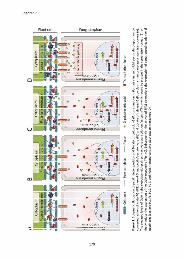
Chapter 7
170
Fig
ure
1.
Sch
em
ati
c illu
stra
tio
n o
f p
ect
in d
eco
mp
osi
tio
n a
nd
D-g
ala
ctu
ron
ic a
cid
(G
alA
) co
nsu
mp
tio
n i
n B
otr
yti
s cin
ere
a.
Init
ial
pe
ctin
de
com
po
siti
on
by
con
cert
ed
act
ion
of
en
do
-PG
(P
G1
), e
xo-P
G a
nd
pe
ctin
/pe
cta
te l
ya
se (
PL)
an
d u
pta
ke
of
rele
ase
d G
alA
by
pla
sma
me
mb
ran
e-a
sso
cia
ted
tra
nsp
ort
ers
(A
).
Th
e e
leva
ted
le
ve
ls o
f G
alA
in
th
e c
yto
pla
sm e
ith
er
dir
ect
ly a
ctiv
ate
tra
nsc
rip
tio
n f
act
or(
s) (
TF(s
)) w
hic
h c
ou
ld b
e p
rese
nt
in t
he
cyto
pla
sm/
nu
cle
us
(B),
or
firs
tly i
nd
uce
th
e e
xpre
ssio
n o
f th
e G
alA
-re
spo
nsi
ve
TF
(s)
(C).
Su
bse
qu
en
tly,
the
act
iva
ted
TF
(s)
co-r
eg
ula
te t
he
exp
ress
ion
of
ge
ne
s e
nco
din
g a
dd
itio
na
l
pe
ctin
ase
s (e
.g.
exo
-PG
, P
L, P
G2
, P
G4
, a
nd
PG
6),
tra
nsp
ort
ers
, a
nd
Ga
lA c
ata
bo
lic
en
zym
es
(D).
References
171
References
Adie, B. A., et al., 2007. ABA is an essential signal for plant resistance to pathogens affecting JA
biosynthesis and the activation of defenses in Arabidopsis. Plant Cell. 19, 1665-1681.
Aguero, C. B., et al., 2005. Evaluation of tolerance to Pierce's disease and Botrytis in transgenic
plants of Vitis vinifera L. expressing the pear PGIP gene. Mol Plant Pathol. 6, 43-51.
Amselem, J., et al., 2011. Genomic analysis of the necrotrophic fungal pathogens Sclerotinia
sclerotiorum and Botrytis cinerea. PLoS Genet. 7, e1002230.
Armand, S., et al., 2000. The active site topology of Aspergillus niger endopolygalacturonase II as
studied by site-directed mutagenesis. J Biol Chem. 275, 691-696.
Aro, N., et al., 2005. Transcriptional regulation of plant cell wall degradation by filamentous fungi.
FEMS Microbiol Rev. 29, 719-739.
Bailey, T. L., Elkan, C., 1994. Fitting a mixture model by expectation maximization to discover motifs
in biopolymers. Proc Int Conf Intell Syst Mol Biol. 2, 28-36.
Bar, M., et al., 2010. BAK1 is required for the attenuation of ethylene-inducing xylanase (Eix)-
induced defense responses by the decoy receptor LeEix1. Plant J. 63, 791-800.
Battaglia, E., et al., 2011. Analysis of regulation of pentose utilisation in Aspergillus niger reveals
evolutionary adaptations in Eurotiales. Stud Mycol. 69, 31-38.
Baxter, L., et al., 2010. Signatures of adaptation to obligate biotrophy in the Hyaloperonospora
arabidopsidis genome. Science. 330, 1549-1551.
Beckstette, M., et al., 2009. Significant speed up of database searches with HMMs by search space
reduction with PSSM family models. Bioinformatics. 25, 3251-3258.
Billon-Grand, G., et al., 2012. pH modulation differs during sunflower cotyledon colonization by the
two closely related necrotrophic fungi Botrytis cinerea and Sclerotinia sclerotiorum. Mol
Plant Pathol. 13, 568-578.
Blumenkranz, N., Asboe-Hansen, G., 1973. New method for quantitative determination of uronic
acids. Anal Biochem. 54, 484-489.
Boller, T., Felix, G., 2009. A renaissance of elicitors: perception of microbe-associated molecular
patterns and danger signals by pattern-recognition receptors. Annu Rev Plant Biol. 60,
379-406.
Böttcher, C., et al., 2009. The multifunctional enzyme CYP71B15 (PHYTOALEXIN DEFICIENT3)
converts cysteine-indole-3-acetonitrile to camalexin in the indole-3-acetonitrile metabolic
network of Arabidopsis thaliana. Plant Cell. 21, 1830-1845.
Boudart, G., et al., 2003. Elicitor activity of a fungal endopolygalacturonase in tobacco requires a
functional catalytic site and cell wall localization. Plant Physiol. 131, 93-101.
Bouton, S., et al., 2002. QUASIMODO1 encodes a putative membrane-bound glycosyltransferase
required for normal pectin synthesis and cell adhesion in Arabidopsis. Plant Cell. 14, 2577-
2590.
Brito, N., et al., 2006. The endo-beta-1,4-xylanase xyn11A is required for virulence in Botrytis cinerea.
Mol Plant Microbe Interact. 19, 25-32.
Brunner, F., et al., 2002. Pep-13, a plant defense-inducing pathogen-associated pattern from
Phytophthora transglutaminases. EMBO J. 21, 6681-6688.
Brutus, A., et al., 2010. A domain swap approach reveals a role of the plant wall-associated kinase 1
(WAK1) as a receptor of oligogalacturonides. Proc Natl Acad Sci U S A. 107, 9452-9457.
Buchanan, C. L., et al., 1999. An extremely thermostable aldolase from Sulfolobus solfataricus with
specificity for non-phosphorylated substrates. Biochem J. 343 Pt 3, 563-570.
References
172
Cabanne, C., Doneche, B., 2002. Purification and characterization of two isozymes of
polygalacturonase from Botrytis cinerea. Effect of calcium ions on polygalacturonase
activity. Microbiol Res. 157, 183-189.
Cabral, A., et al., 2011. Identification of Hyaloperonospora arabidopsidis transcript sequences
expressed during infection reveals isolate-specific effectors. PLoS One. 6, e19328.
Cabrera, J. C., et al., 2008. Egg box conformation of oligogalacturonides: the time-dependent
stabilization of the elicitor-active conformation increases its biological activity.
Glycobiology. 18, 473-482.
Caffall, K. H., Mohnen, D., 2009. The structure, function, and biosynthesis of plant cell wall pectic
polysaccharides. Carbohydr Res. 344, 1879-1900.
Cantarel, B. L., et al., 2009. The Carbohydrate-Active EnZymes database (CAZy): an expert resource
for Glycogenomics. Nucleic Acids Res. 37, D233-D238.
Cao, H., et al., 1994. Characterization of an Arabidopsis mutant that is nonresponsive to inducers of
systemic acquired resistance. Plant Cell. 6, 1583-1592.
Cao, J., et al., 2011. Whole-genome sequencing of multiple Arabidopsis thaliana populations. Nat
Genet. 43, 956-963.
Chaparro-Garcia, A., et al., 2011. The receptor-like kinase SERK3/BAK1 is required for basal
resistance against the late blight pathogen Phytophthora infestans in Nicotiana
benthamiana. PLoS One. 6, e16608.
Chinchilla, D., et al., 2006. The Arabidopsis receptor kinase FLS2 binds flg22 and determines the
specificity of flagellin perception. Plant Cell. 18, 465-476.
Chinchilla, D., et al., 2009. One for all: the receptor-associated kinase BAK1. Trends Plant Sci. 14,
535-541.
Chinchilla, D., et al., 2007. A flagellin-induced complex of the receptor FLS2 and BAK1 initiates plant
defence. Nature. 448, 497-500.
Choquer, M., et al., 2007. Botrytis cinerea virulence factors: new insights into a necrotrophic and
polyphageous pathogen. FEMS Microbiol Lett. 277, 1-10.
Churchill, G. A., Doerge, R. W., 1994. Empirical threshold values for quantitative trait mapping.
Genetics. 138, 963-971.
Clough, S. J., Bent, A. F., 1998. Floral dip: a simplified method for Agrobacterium-mediated
transformation of Arabidopsis thaliana. Plant J. 16, 735-743.
Cosgrove, D. J., 2001. Plant cell walls: wall-associated kinases and cell expansion. Curr Biol. 11, R558-
R559.
Cubero, B., Scazzocchio, C., 1994. Two different, adjacent and divergent zinc finger binding sites are
necessary for CREA-mediated carbon catabolite repression in the proline gene cluster of
Aspergillus nidulans. EMBO J. 13, 407-415.
Curtis, M. D., Grossniklaus, U., 2003. A gateway cloning vector set for high-throughput functional
analysis of genes in planta. Plant Physiol. 133, 462-469.
Curvers, K., et al., 2010. Abscisic acid deficiency causes changes in cuticle permeability and pectin
composition that influence tomato resistance to Botrytis cinerea. Plant Physiol. 154, 847-
860.
D'Ovidio, R., et al., 2004. Polygalacturonases, polygalacturonase-inhibiting proteins and pectic
oligomers in plant-pathogen interactions. Biochim Biophys Acta. 1696, 237-244.
De Lorenzo, G., et al., 2001. The role of polygalacturonase-inhibiting proteins (PGIPs) in defense
against pathogenic fungi. Annu Rev Phytopathol. 39, 313-335.
Dean, R., et al., 2012. The Top 10 fungal pathogens in molecular plant pathology. Mol Plant Pathol.
13, 414-430.
References
173
Decreux, A., et al., 2006. In vitro characterization of the homogalacturonan-binding domain of the
wall-associated kinase WAK1 using site-directed mutagenesis. Phytochemistry. 67, 1068-
1079.
Delaney, T. P., et al., 1994. A central role of salicylic Acid in plant disease resistance. Science. 266,
1247-1250.
Denoux, C., et al., 2008. Activation of defense response pathways by OGs and Flg22 elicitors in
Arabidopsis seedlings. Mol Plant. 1, 423-445.
Dewey, F. M., et al., 2008. Quantification of Botrytis and laccase in winegrapes. Am J Enol Vitic. 59,
47-54.
Doehlemann, G., et al., 2005. Molecular and functional characterization of a fructose specific
transporter from the gray mold fungus Botrytis cinerea. Fungal Genet Biol. 42, 601-610.
Dulermo, T., et al., 2010. Novel insights into mannitol metabolism in the fungal plant pathogen
Botrytis cinerea. Biochem J. 427, 323-332.
Dulermo, T., et al., 2009. Dynamic carbon transfer during pathogenesis of sunflower by the
necrotrophic fungus Botrytis cinerea: from plant hexoses to mannitol. New Phytol. 183,
1149-1162.
Duplessis, S., et al., 2011. Melampsora larici-populina transcript profiling during germination and
timecourse infection of poplar leaves reveals dynamic expression patterns associated with
virulence and biotrophy. Mol Plant Microbe Interact. 24, 808-818.
Englyst, H. N., Cummings, J. H., 1984. Simplified method for the measurement of total non-starch
polysaccharides by gas-liquid chromatography of constituent sugars as alditol acetates.
Analyst. 109, 937-942.
Enkerli, J., et al., 1999. The enzymatic activity of fungal xylanase is not necessary for its elicitor
activity. Plant Physiol. 121, 391-397.
Espino, J. J., et al., 2005. Botrytis cinerea endo-beta-1,4-glucanase Cel5A is expressed during
infection but is not required for pathogenesis. Physiol Mol Plant Pathol. 66, 213-221.
Espino, J. J., et al., 2010. The Botrytis cinerea early secretome. Proteomics. 10, 3020-3034.
Federici, L., et al., 2006. Polygalacturonase inhibiting proteins: players in plant innate immunity?
Trends Plant Sci. 11, 65-70.
Felix, G., et al., 1999. Plants have a sensitive perception system for the most conserved domain of
bacterial flagellin. Plant J. 18, 265-276.
Fernandez-Acero, F. J., et al., 2010. 2-DE proteomic approach to the Botrytis cinerea secretome
induced with different carbon sources and plant-based elicitors. Proteomics. 10, 2270-
2280.
Ferrari, S., et al., 2006. Antisense expression of the Arabidopsis thaliana AtPGIP1 gene reduces
polygalacturonase-inhibiting protein accumulation and enhances susceptibility to Botrytis
cinerea. Mol Plant Microbe Interact. 19, 931-936.
Ferrari, S., et al., 2003a. Arabidopsis local resistance to Botrytis cinerea involves salicylic acid and
camalexin and requires EDS4 and PAD2, but not SID2, EDS5 or PAD4. Plant J. 35, 193-205.
Ferrari, S., et al., 2003b. Tandemly duplicated Arabidopsis genes that encode polygalacturonase-
inhibiting proteins are regulated coordinately by different signal transduction pathways in
response to fungal infection. Plant Cell. 15, 93-106.
Fischer-Parton, S., et al., 2000. Confocal microscopy of FM4-64 as a tool for analysing endocytosis
and vesicle trafficking in living fungal hyphae. J Microsc. 198, 246-259.
Fisher, M. C., et al., 2012. Emerging fungal threats to animal, plant and ecosystem health. Nature.
484, 186-194.
Flors, V., et al., 2008. Interplay between JA, SA and ABA signalling during basal and induced
resistance against Pseudomonas syringae and Alternaria brassicicola. Plant J. 54, 81-92.
References
174
Furman-Matarasso, N., et al., 1999. A point mutation in the ethylene-inducing xylanase elicitor
inhibits the beta-1-4-endoxylanase activity but not the elicitation activity. Plant Physiol.
121, 345-351.
Galletti, R., et al., 2008. The AtrbohD-mediated oxidative burst elicited by oligogalacturonides in
Arabidopsis is dispensable for the activation of defense responses effective against
Botrytis cinerea. Plant Physiol. 148, 1695-1706.
Gan, P., et al., 2013. Comparative genomic and transcriptomic analyses reveal the hemibiotrophic
stage shift of Colletotrichum fungi. New Phytol. 197, 1236-1249.
Gao, M., et al., 2009. Regulation of cell death and innate immunity by two receptor-like kinases in
Arabidopsis. Cell Host Microbe. 6, 34-44.
Garcia-Andrade, J., et al., 2011. Arabidopsis ocp3 mutant reveals a mechanism linking ABA and JA to
pathogen-induced callose deposition. Plant J. 67, 783-794.
Gomez-Gomez, L., Boller, T., 2000. FLS2: an LRR receptor-like kinase involved in the perception of
the bacterial elicitor flagellin in Arabidopsis. Mol Cell. 5, 1003-1011.
Govrin, E. M., Levine, A., 2000. The hypersensitive response facilitates plant infection by the
necrotrophic pathogen Botrytis cinerea. Curr Biol. 10, 751-757.
Greeff, C., et al., 2012. Receptor-like kinase complexes in plant innate immunity. Front Plant Sci. 3,
209.
Gruben, B. S., 2012. Novel transcriptional activators of Aspergillus involved in plant biomass
utilization. PhD thesis, Utrecht University, Microbiology.
Harholt, J., et al., 2006. ARABINAN DEFICIENT 1 is a putative arabinosyltransferase involved in
biosynthesis of pectic arabinan in Arabidopsis. Plant Physiol. 140, 49-58.
Hauser, M. T., et al., 2001. Trichome distribution in Arabidopsis thaliana and its close relative
Arabidopsis lyrata: molecular analysis of the candidate gene GLABROUS1. Mol Biol Evol. 18,
1754-1763.
Heese, A., et al., 2007. The receptor-like kinase SERK3/BAK1 is a central regulator of innate immunity
in plants. Proc Natl Acad Sci U S A. 104, 12217-12222.
Hematy, K., et al., 2009. Host-pathogen warfare at the plant cell wall. Curr Opin Plant Biol. 12, 406-
413.
Hickey, P. C., et al., 2004. Live-cell imaging of filamentous fungi using vital fluorescent dyes and
confocal microscopy. Methods in Microbiology. 34, 63-88.
Hilditch, S., et al., 2007. The missing link in the fungal D-galacturonate pathway: identification of the
L-threo-3-deoxy-hexulosonate aldolase. J Biol Chem. 282, 26195-26201.
Hilz, H., et al., 2005. Cell wall polysaccharides in black currants and bilberries-characterisation in
berries, juice, and press cake. Carbohyd. Polym. 59, 477-488.
Holub, E. B., et al., 1994. Phenotypic and genotypic characterization of interactions between isolates
of Peronospora parasitica and accessions of Arabidopsis thaliana. Mol Plant Microbe
Interact. 7, 223-239.
Hondmann, D. H., et al., 1991. Glycerol catabolism in Aspergillus nidulans. J Gen Microbiol. 137, 629-
636.
Hückelhoven, R., 2007. Cell wall-associated mechanisms of disease resistance and susceptibility.
Annu Rev Phytopathol. 45, 101-127.
Huitema, E., et al., 2011. A straightforward protocol for electro-transformation of Phytophthora
capsici zoospores. Methods Mol Biol. 712, 129-135.
Isshiki, A., et al., 2001. Endopolygalacturonase is essential for citrus black rot caused by Alternaria
citri but not brown spot caused by Alternaria alternata. Mol Plant Microbe Interact. 14,
749-757.
References
175
Jacobs, A. K., et al., 2003. An Arabidopsis callose synthase, GSL5, is required for wound and papillary
callose formation. Plant Cell. 15, 2503-2513.
Janni, M., et al., 2008. The expression of a bean PGIP in transgenic wheat confers increased
resistance to the fungal pathogen Bipolaris sorokiniana. Mol Plant Microbe Interact. 21,
171-177.
Jolie, R. P., et al., 2010. Pectin methylesterase and its proteinaceous inhibitor: a review. Carbohydr
Res. 345, 2583-2595.
Jones, J. D., Dangl, J. L., 2006. The plant immune system. Nature. 444, 323-329.
Joubert, D. A., et al., 2007. A polygalacturonase-inhibiting protein from grapevine reduces the
symptoms of the endopolygalacturonase BcPG2 from Botrytis cinerea in Nicotiana
benthamiana leaves without any evidence for in vitro interaction. Mol Plant Microbe
Interact. 20, 392-402.
Juge, N., 2006. Plant protein inhibitors of cell wall degrading enzymes. Trends Plant Sci. 11, 359-367.
Kämper, J., et al., 2006. Insights from the genome of the biotrophic fungal plant pathogen Ustilago
maydis. Nature. 444, 97-101.
Kars, I., 2007. The role of pectin degradation in pathogenesis of Botrytis cinerea. PhD thesis,
Wageningen University, Phytopathology.
Kars, I., et al., 2005a. Necrotizing activity of five Botrytis cinerea endopolygalacturonases produced
in Pichia pastoris. Plant J. 43, 213-225.
Kars, I., et al., 2005b. Functional analysis of Botrytis cinerea pectin methylesterase genes by PCR-
based targeted mutagenesis: Bcpme1 and Bcpme2 are dispensable for virulence of strain
B05.10. Mol Plant Pathol. 6, 641-652.
Kars, I., van Kan, J. A. L., 2004. Extracellular enzymes and metabolites involved in pathogenesis of
Botrytis. Botrytis: biology, pathology and control (Y. Elad, B. Williamson, P. Tudzynski and
N. Delen, eds.) Kluwer Academic Publishers, The Netherlands. 99-118.
Kester, H. C., Visser, J., 1990. Purification and characterization of polygalacturonases produced by
the hyphal fungus Aspergillus niger. Biotechnol Appl Biochem. 12, 150-60.
Kim, H., Woloshuk, C. P., 2011. Functional characterization of fst1 in Fusarium verticillioides during
colonization of maize kernels. Mol Plant Microbe Interact. 24, 18-24.
King, B. C., et al., 2011. Arsenal of plant cell wall degrading enzymes reflects host preference among
plant pathogenic fungi. Biotechnol Biofuels. 4, 4.
Kintner, P. K., Vanburen, J. P., 1982. Carbohydrate interference and its correction in pectin analysis
using the m-hydroxydiphenyl method. J Food Sci. 47, 756-759.
Koeck, M., et al., 2011. The role of effectors of biotrophic and hemibiotrophic fungi in infection. Cell
Microbiol. 13, 1849-1857.
Kohorn, B. D., et al., 2009. Pectin activation of MAP kinase and gene expression is WAK2 dependent.
Plant J. 60, 974-982.
Kohorn, B. D., Kohorn, S. L., 2012. The cell wall-associated kinases, WAKs, as pectin receptors. Front
Plant Sci. 3, 88.
Krampe, S., Boles, E., 2002. Starvation-induced degradation of yeast hexose transporter Hxt7p is
dependent on endocytosis, autophagy and the terminal sequences of the permease. FEBS
Lett. 513, 193-196.
Kravtchenko, T. P., et al., 1992. Analytical comparison of three industrial pectin preparations.
Carbohydr Polym. 18, 17-25.
Kuivanen, J., et al., 2012. Engineering filamentous fungi for conversion of d-galacturonic acid to L-
galactonic acid. Appl Environ Microbiol. 78, 8676-8683.
Kunze, G., et al., 2004. The N terminus of bacterial elongation factor Tu elicits innate immunity in
Arabidopsis plants. Plant Cell. 16, 3496-3507.
References
176
Kuorelahti, S., et al., 2006. L-galactonate dehydratase is part of the fungal path for D-galacturonic
acid catabolism. Mol Microbiol. 61, 1060-1068.
Kuorelahti, S., et al., 2005. Identification in the mold Hypocrea jecorina of the first fungal D-
galacturonic acid reductase. Biochemistry. 44, 11234-11240.
Lamb, C., Dixon, R. A., 1997. The oxidative burst in plant disease resistance. Annu Rev Plant Physiol
Plant Mol Biol. 48, 251-275.
Langmead, B., Salzberg, S. L., 2012. Fast gapped-read alignment with Bowtie 2. Nat Methods. 9, 357-
359.
Li, H., et al., 2009. The Sequence Alignment/Map format and SAMtools. Bioinformatics. 25, 2078-
2079.
Li, J., et al., 2002. BAK1, an Arabidopsis LRR receptor-like protein kinase, interacts with BRI1 and
modulates brassinosteroid signaling. Cell. 110, 213-222.
Liebrand, T. W., et al., 2012. Endoplasmic reticulum-quality control chaperones facilitate the
biogenesis of Cf receptor-like proteins involved in pathogen resistance of tomato. Plant
Physiol. 159, 1819-1833.
Liebrand, T. W., et al., 2013. The receptor-like kinase SOBIR1/EVR interacts with receptor-like
proteins in plant immunity against fungal infection. submitted.
Liepins, J., et al., 2006. Enzymes for the NADPH-dependent reduction of dihydroxyacetone and D-
glyceraldehyde and L-glyceraldehyde in the mould Hypocrea jecorina. Febs J. 273, 4229-
4235.
Lingner, U., et al., 2011. Hexose transporters of a hemibiotrophic plant pathogen: functional
variations and regulatory differences at different stages of infection. J Biol Chem. 286,
20913-20922.
Manfredini, C., et al., 2005. Polygalacturonase-inhibiting protein 2 of Phaseolus vulgaris inhibits
BcPG1, a polygalacturonase of Botrytis cinerea important for pathogenicity, and protects
transgenic plants from infection. Physiol Mol Plant Pathol. 67, 108-115.
Marger, M. D., Saier, M. H., 1993. A major superfamily of transmembrane facilitators that catalyze
uniport, symport and antiport. Trends Biochem Sci. 18, 13-20.
Martens-Uzunova, E. S., Schaap, P. J., 2008. An evolutionary conserved d-galacturonic acid metabolic
pathway operates across filamentous fungi capable of pectin degradation. Fungal Genet
Biol. 45, 1449-1457.
Martens-Uzunova, E. S., et al., 2006. A new group of exo-acting family 28 glycoside hydrolases of
Aspergillus niger that are involved in pectin degradation. Biochem J. 400, 43-52.
Martin, F., et al., 2011. Sequencing the fungal tree of life. New Phytol. 190, 818-821.
Martin, F., et al., 2010. Perigord black truffle genome uncovers evolutionary origins and mechanisms
of symbiosis. Nature. 464, 1033-1038.
Meyer, U., Dewey, F., 2000. Efficacy of different immunogens for raising monoclonal antibodies to
Botrytis cinerea. Mycol Res. 104, 979-987.
Mohnen, D., 2008. Pectin structure and biosynthesis. Curr Opin Plant Biol. 11, 266-277.
Mojzita, D., et al., 2010. Metabolic engineering of fungal strains for conversion of D-galacturonate to
meso-galactarate. Appl Environ Microbiol. 76, 169-175.
Monaghan, J., Zipfel, C., 2012. Plant pattern recognition receptor complexes at the plasma
membrane. Curr Opin Plant Biol. 15, 349-357.
Nakagawa, T., et al., 2007. Development of series of gateway binary vectors, pGWBs, for realizing
efficient construction of fusion genes for plant transformation. J Biosci Bioeng. 104, 34-41.
Nam, K. H., Li, J., 2002. BRI1/BAK1, a receptor kinase pair mediating brassinosteroid signaling. Cell.
110, 203-212.
References
177
Nishimura, M. T., et al., 2003. Loss of a callose synthase results in salicylic acid-dependent disease
resistance. Science. 301, 969-972.
Nishiyama, Y., et al., 2002. Crystal structure and hydrogen-bonding system in cellulose Ibeta from
synchrotron X-ray and neutron fiber diffraction. J Am Chem Soc. 124, 9074-9082.
Noda, J., et al., 2010. The Botrytis cinerea xylanase Xyn11A contributes to virulence with its
necrotizing activity, not with its catalytic activity. BMC Plant Biol. 10, 38.
O'Connell, R. J., et al., 2012. Lifestyle transitions in plant pathogenic Colletotrichum fungi deciphered
by genome and transcriptome analyses. Nat Genet. 44, 1060-1065.
Oeser, B., et al., 2002. Polygalacturonase is a pathogenicity factor in the Claviceps purpurea/rye
interaction. Fungal Genet Biol. 36, 176-186.
Ogawa, M., et al., 2009. ARABIDOPSIS DEHISCENCE ZONE POLYGALACTURONASE1 (ADPG1), ADPG2,
and QUARTET2 are polygalacturonases required for cell separation during reproductive
development in Arabidopsis. Plant Cell. 21, 216-233.
Ozcan, S., Johnston, M., 1999. Function and regulation of yeast hexose transporters. Microbiol Mol
Biol Rev. 63, 554-569.
Pao, S. S., et al., 1998. Major facilitator superfamily. Microbiol Mol Biol Rev. 62, 1-34.
Paper, J. M., et al., 2007. Comparative proteomics of extracellular proteins in vitro and in planta
from the pathogenic fungus Fusarium graminearum. Proteomics. 7, 3171-3183.
Penninckx, I. A., et al., 1996. Pathogen-induced systemic activation of a plant defensin gene in
Arabidopsis follows a salicylic acid-independent pathway. Plant Cell. 8, 2309-2323.
Penninckx, I. A., et al., 1998. Concomitant activation of jasmonate and ethylene response pathways
is required for induction of a plant defensin gene in Arabidopsis. Plant Cell. 10, 2103-2113.
Phalip, V., et al., 2009. Plant cell wall degradation with a powerful Fusarium graminearum enzymatic
arsenal. J Microbiol Biotechnol. 19, 573-581.
Poinssot, B., et al., 2003. The endopolygalacturonase 1 from Botrytis cinerea activates grapevine
defense reactions unrelated to its enzymatic activity. Mol Plant Microbe Interact. 16, 553-
564.
Powell, A. L., et al., 2000. Transgenic expression of pear PGIP in tomato limits fungal colonization.
Mol Plant Microbe Interact. 13, 942-950.
Raiola, A., et al., 2011. Pectin methylesterase is induced in Arabidopsis upon infection and is
necessary for a successful colonization by necrotrophic pathogens. Mol Plant Microbe
Interact. 24, 432-440.
Rasul, S., et al., 2012. Nitric oxide production mediates oligogalacturonide-triggered immunity and
resistance to Botrytis cinerea in Arabidopsis thaliana. Plant Cell Environ. 35, 1483-1499.
Rha, E., et al., 2001. Expression of exo-polygalacturonases in Botrytis cinerea. FEMS Microbiol Lett.
201, 105-109.
Richard, P., Hilditch, S., 2009. D-galacturonic acid catabolism in microorganisms and its
biotechnological relevance. Appl Microbiol Biotechnol. 82, 597-604.
Rolland, S., et al., 2009. pH controls both transcription and post-translational processing of the
protease BcACP1 in the phytopathogenic fungus Botrytis cinerea. Microbiology. 155, 2097-
2105.
Ron, M., Avni, A., 2004. The receptor for the fungal elicitor ethylene-inducing xylanase is a member
of a resistance-like gene family in tomato. Plant Cell. 16, 1604-1615.
Rotblat, B., et al., 2002. Identification of an essential component of the elicitation active site of the
EIX protein elicitor. Plant J. 32, 1049-1055.
Roux, M., et al., 2011. The Arabidopsis leucine-rich repeat receptor-like kinases BAK1/SERK3 and
BKK1/SERK4 are required for innate immunity to hemibiotrophic and biotrophic pathogens.
Plant Cell. 23, 2440-2455.
References
178
Rui, O., Hahn, M., 2007. The Botrytis cinerea hexokinase, Hxk1, but not the glucokinase, Glk1, is
required for normal growth and sugar metabolism, and for pathogenicity on fruits.
Microbiology. 153, 2791-2802.
Scheller, H. V., Ulvskov, P., 2010. Hemicelluloses. Annu Rev Plant Biol. 61, 263-289.
Schols, H. A., et al., 2009. Revealing pectin's structure. Pectins and pectinases (H.A. Schols, R. G. F.
Visser, and A. G.J. Voragen, eds) Wageningen Academic Publishers, The Netherlands. 19-
34.
Schumacher, J., 2012. Tools for Botrytis cinerea: New expression vectors make the gray mold fungus
more accessible to cell biology approaches. Fungal Genet Biol. 49, 483-497.
Sealy-Lewis, H. M., Fairhurst, V., 1992. An NADP(+)-dependent glycerol dehydrogenase in Aspergillus
nidulans is inducible by D-galacturonate. Curr Genet. 22, 293-296.
Shah, P., et al., 2009a. Comparative proteomic analysis of Botrytis cinerea secretome. J Proteome
Res. 8, 1123-1130.
Shah, P., et al., 2009b. A proteomic study of pectin-degrading enzymes secreted by Botrytis cinerea
grown in liquid culture. Proteomics. 9, 3126-3135.
Shieh, M. T., et al., 1997. Molecular genetic evidence for the involvement of a specific
polygalacturonase, P2c, in the invasion and spread of Aspergillus flavus in cotton bolls.
Appl Environ Microbiol. 63, 3548-3552.
Souffriau, B., et al., 2012. Evidence for rapid uptake of D-galacturonic acid in the yeast
Saccharomyces cerevisiae by a channel-type transport system. FEBS Lett. 586, 2494-2499.
Spanu, P. D., et al., 2010. Genome expansion and gene loss in powdery mildew fungi reveal tradeoffs
in extreme parasitism. Science. 330, 1543-1546.
Staats, M., van Kan, J. A. L., 2012. Genome update of Botrytis cinerea strains B05.10 and T4.
Eukaryot Cell. 11, 1413-1414.
Stam, P., 1993. Construction of integrated genetic-linkage maps by means of a new computer
package - Joinmap. Plant J. 3, 739-744.
Tamura, K., et al., 2007. MEGA4: Molecular Evolutionary Genetics Analysis (MEGA) software version
4.0. Mol Biol Evol. 24, 1596-1599.
ten Have, A., et al., 2001. Botrytis cinerea endopolygalacturonase genes are differentially expressed
in various plant tissues. Fungal Genet Biol. 33, 97-105.
ten Have, A., et al., 1998. The endopolygalacturonase gene Bcpg1 is required for full virulence of
Botrytis cinerea. Mol Plant Microbe Interact. 11, 1009-1016.
ten Have, A., et al., 2002. The contribution of the cell wall degrading enzymes to pathogenesis of
fungal plant pathogens. The Mycota XI, Agricultural applications (F. Kempken, ed.)
Springer-Verlag, Germany. 341-358.
Thomma, B. P., et al., 1998. Separate jasmonate-dependent and salicylate-dependent defense-
response pathways in Arabidopsis are essential for resistance to distinct microbial
pathogens. Proc Natl Acad Sci U S A. 95, 15107-15111.
Tilburn, J., et al., 1995. The Aspergillus PacC zinc finger transcription factor mediates regulation of
both acid- and alkaline-expressed genes by ambient pH. EMBO J. 14, 779-790.
Torres, M. A., et al., 2005. Pathogen-induced, NADPH oxidase-derived reactive oxygen intermediates
suppress spread of cell death in Arabidopsis thaliana. Nat Genet. 37, 1130-1134.
Trapnell, C., et al., 2013. Differential analysis of gene regulation at transcript resolution with RNA-
seq. Nat Biotechnol. 31, 46-53.
Trapnell, C., et al., 2009. TopHat: discovering splice junctions with RNA-Seq. Bioinformatics. 25,
1105-1111.
Uitzetter, J. H., et al., 1986. Characterization of Aspergillus nidulans mutants in carbon metabolism
isolated after D-galacturonate enrichment. J Gen Microbiol. 132, 1167-1172.
References
179
Valette-Collet, O., et al., 2003. Disruption of Botrytis cinerea pectin methylesterase gene Bcpme1
reduces virulence on several host plants. Mol Plant Microbe Interact. 16, 360-367.
van Baarlen, P., et al., 2007. Histochemical and genetic analysis of host and non-host interactions of
Arabidopsis with three Botrytis species: an important role for cell death control. Mol Plant
Pathol. 8, 41-54.
van Damme, M., et al., 2005. Identification of Arabidopsis loci required for susceptibility to the
downy mildew pathogen Hyaloperonospora parasitica. Mol Plant Microbe Interact. 18,
583-592.
van den Brink, J., de Vries, R. P., 2011. Fungal enzyme sets for plant polysaccharide degradation.
Appl Microbiol Biotechnol. 91, 1477-1492.
van Kan, J. A. L., 2006. Licensed to kill: the lifestyle of a necrotrophic plant pathogen. Trends Plant
Sci. 11, 247-253.
van Kan, J. A. L., et al., 1997. Cutinase A of Botrytis cinerea is expressed, but not essential, during
penetration of gerbera and tomato. Mol Plant Microbe Interact. 10, 30-38.
van Ooijen, J. W., et al., 2002. MapQTL® Version 4.0, Software for the calculation of QTL positions on
genetic maps. Plant Research International, Wageningen, The Netherlands.
van Ooijen, J. W., Voorrips, R. E., 2001. Joinmap® 3.0, Software for the calculation of genetic linkage
maps. Plant Research International, Wageningen, The Netherlands.
van Suylekom, D., et al., 2007. Degradation of the hexose transporter Hxt5p in Saccharomyces
cerevisiae. Biol Cell. 99, 13-23.
Verhoeff, K., et al., 1988. Changes in pH and the production of organic-acids during colonization of
tomato petioles by Botrytis cinerea. J Phytopathology. 122, 327-336.
Visser, J., et al., 1988. Glycerol uptake mutants of the hyphal fungus Aspergillus nidulans. J Gen
Microbiol. 134, 655-659.
Voegele, R. T., et al., 2001. The role of haustoria in sugar supply during infection of broad bean by
the rust fungus Uromyces fabae. Proc Natl Acad Sci U S A. 98, 8133-8138.
Volpi, C., et al., 2011. The ectopic expression of a pectin methyl esterase inhibitor increases pectin
methyl esterification and limits fungal diseases in wheat. Mol Plant Microbe Interact. 24,
1012-1019.
Voragen, A. G. J., et al., 1986. Determination of the degree of methylation and acetylation of pectins
by h.p.l.c. Food Hydrocolloids. 1, 65-70.
Vorwerk, S., et al., 2004. The role of plant cell wall polysaccharide composition in disease resistance.
Trends Plant Sci. 9, 203-209.
Vos, P., et al., 1995. AFLP: a new technique for DNA fingerprinting. Nucleic Acids Res. 23, 4407-4414.
Wagner, T. A., Kohorn, B. D., 2001. Wall-associated kinases are expressed throughout plant
development and are required for cell expansion. Plant Cell. 13, 303-318.
Wang, G., et al., 2008. A genome-wide functional investigation into the roles of receptor-like
proteins in Arabidopsis. Plant Physiol. 147, 503-517.
Wang, S., et al., 2006. Windows QTL Cartographer 2.5. Department of statistics, North Carolina State
University, Raleigh, NC., http://statgen.ncsu.edu/qtlcart/WQTLCart.htm.
Wang, Y., et al., 2011. Comparative secretome investigation of Magnaporthe oryzae proteins
responsive to nitrogen starvation. J Proteome Res. 10, 3136-3148.
Wang, Z., et al., 2009. RNA-Seq: a revolutionary tool for transcriptomics. Nat Rev Genet. 10, 57-63.
Wei, H., et al., 2004. A putative high affinity hexose transporter, hxtA, of Aspergillus nidulans is
induced in vegetative hyphae upon starvation and in ascogenous hyphae during
cleistothecium formation. Fungal Genet Biol. 41, 148-156.
Wiebe, M. G., et al., 2010. Bioconversion of D-galacturonate to keto-deoxy-L-galactonate (3-deoxy-L-
threo-hex-2-ulosonate) using filamentous fungi. BMC Biotechnol. 10, 63.
References
180
Wieczorke, R., et al., 1999. Concurrent knock-out of at least 20 transporter genes is required to block
uptake of hexoses in Saccharomyces cerevisiae. FEBS Lett. 464, 123-128.
Williamson, B., et al., 2007. Botrytis cinerea: the cause of grey mould disease. Mol Plant Pathol. 8,
561-580.
Winston, F., et al., 1995. Construction of a set of convenient Saccharomyces cerevisiae strains that
are isogenic to S288C. Yeast. 11, 53-55.
Wubben, J. P., et al., 1999. Cloning and partial characterization of endopolygalacturonase genes
from Botrytis cinerea. Appl Environ Microbiol. 65, 1596-1602.
Wubben, J. P., et al., 2000. Regulation of endopolygalacturonase gene expression in Botrytis cinerea
by galacturonic acid, ambient pH and carbon catabolite repression. Curr Genet. 37, 152-
157.
Yajima, W., Kav, N. N., 2006. The proteome of the phytopathogenic fungus Sclerotinia sclerotiorum.
Proteomics. 6, 5995-6007.
Yang, F., et al., 2011. Secretomics identifies Fusarium graminearum proteins involved in the
interaction with barley and wheat. Mol Plant Pathol. 13, 445-453.
Zablackis, E., et al., 1995. Characterization of the cell-wall polysaccharides of Arabidopsis thaliana
leaves. Plant Physiol. 107, 1129-1138.
Zandleven, J., et al., 2007. Xylogalacturonan exists in cell walls from various tissues of Arabidopsis
thaliana. Phytochemistry. 68, 1219-1226.
Zerbino, D. R., Birney, E., 2008. Velvet: algorithms for de novo short read assembly using de Bruijn
graphs. Genome Res. 18, 821-829.
Zhang, L., et al., 2011. The D-galacturonic acid catabolic pathway in Botrytis cinerea. Fungal Genet
Biol. 48, 990-997.
Zhang, L., van Kan, J. A. L., 2013. Botrytis cinerea mutants deficient in d-galacturonic acid catabolism
have a perturbed virulence on Nicotiana benthamiana and Arabidopsis, but not on tomato.
Mol Plant Pathol. 14, 19-29.
Zhou, N., et al., 1999. Arabidopsis PAD3, a gene required for camalexin biosynthesis, encodes a
putative cytochrome P450 monooxygenase. Plant Cell. 11, 2419-2428.
Zipfel, C., et al., 2006. Perception of the bacterial PAMP EF-Tu by the receptor EFR restricts
Agrobacterium-mediated transformation. Cell. 125, 749-760.
Summary
181
Summary
The necrotrophic fungal plant pathogen Botrytis cinerea is able to infect over 200 host plants
and cause severe damage to crops, both pre- and post-harvest. B. cinerea often penetrates
host leaf tissue at the anticlinal cell wall and subsequently grows into and through the middle
lamella, which consists mostly of low-methylesterified pectin. Effective pectin degradation thus
is important for virulence of B. cinerea. Chapter 1 describes the chemical structures of plant
cell wall polysaccharides, the cell wall-associated mechanisms that confer resistance against
pathogens, and the microbial enzymes involved in cell wall decomposition. It then discusses
the plant cell wall degrading enzymes of pathogenic fungi and illustrates with case studies the
process of pectin decomposition by B. cinerea.
Chapter 2 describes the molecular identification and functional characterization of a novel
MAMP receptor RBPG1, a Leucine-Rich Repeat Receptor-Like Protein (LRR-RLP), that recognizes
fungal endo-polygalacturonases (endo-PGs), in particular the B. cinerea protein BcPG3.
Infiltration of the BcPG3 protein into Arabidopsis thaliana accession Col-0 induced a necrotic
response. Heat-inactivated protein and a catalytically inactive mutant protein retained the
ability to induce necrosis. An 11-amino acid peptide stretch was identified that is conserved
among many fungal but not plant endo-PGs. A synthetic peptide comprising this sequence was
unable to induce necrosis. A map-based cloning strategy, combined with comparative and
functional genomics, led to the identification of the RBPG1 gene, which is required for
responsiveness of A. thaliana to the BcPG3 protein. Co-immunoprecipitation experiments
demonstrated that RBPG1 and BcPG3 form a complex in Nicotiana benthamiana, which also
involves the A. thaliana LRR-RLK SOBIR1. The sobir1 mutant plants no longer respond to BcPG3.
Furthermore, overexpression of RBPG1 in the BcPG3-non-responsive accession Br-0 did not
enhance resistance to a number of microbial pathogens.
Chapter 3 describes the functional, biochemical and genetic characterization of the D-
galacturonic acid catabolic pathway in B. cinerea. The B. cinerea genome contains two non-
homologous galacturonate reductase genes (Bcgar1 and Bcgar2), a galactonate dehydratase
gene (Bclgd1), and a 2-keto-3-deoxy-L-galactonate aldolase gene (Bclga1). Targeted gene
replacement of all four genes in B. cinerea, either separately or in combinations, yielded
mutants that were affected in growth on D-galacturonic acid, pectate, or pectin as the sole
carbon source. The extent of growth reduction of the mutants on pectic substrates was
positively correlated to the proportion of D-galacturonic acid present in the pectic substrate.
The virulence of these mutants on different host plants is discussed in Chapter 4. These
mutants showed reduced virulence on N. benthamiana and A. thaliana leaves, but not on
tomato leaves. The cell walls of N. benthamiana and A. thaliana leaves have a higher D-
galacturonic acid content as compared to tomato. Additional in vitro growth assays with the
knockout mutants suggested that the reduced virulence of D-galacturonic acid catabolism-
deficient mutants on N. benthamiana and A. thaliana is not only due to the inability of the
Summary
182
mutants to utilize an abundant carbon source as nutrient, but also due to the growth inhibition
by catabolic intermediates.
In Chapter 5, the functional characterization of two putative D-galacturonic acid transporters is
presented. Bchxt15 is highly and specifically induced by D-galacturonic acid, whereas Bchxt13 is
highly expressed in the presence of all carbon sources tested except for glucose. Subcellular
localization of BcHXT13-GFP and BcHXT15-GFP fusion proteins expressed under their native
promoter suggests that the fusion proteins are localized in plasma membranes and
intracellular vesicles. Knockout mutants in the Bchxt13 and Bchxt15 genes, respectively, were
neither affected in their growth on D-galacturonic acid as the sole carbon source, nor in their
virulence on tomato and N. benthamiana leaves.
Chapter 6 describes the genome-wide transcriptome analysis in B. cinerea grown in media
containing glucose and pectate as sole carbon sources. Genes were identified that are
significantly altered in their expression during growth on these two carbon sources. Conserved
sequence motifs were identified in the promoters of genes involved in pectate decomposition
and D-galacturonic acid utilization. The role of these motifs in regulating D-galacturonic acid-
induced expression was functionally analysed in the promoter of the Bclga1 gene, which
encodes one of the key enzymes in the D-galacturonic acid catabolic pathway. The regulation
by D-galacturonic acid required the presence of sequences encompassing the GAE1 motif and a
binding motif for the pH-dependent transcriptional regulator PacC.
Chapter 7 provides a general discussion of the results presented in this thesis. A model of the
concerted action of pectin degradation and subsequent monosaccharide consumption and co-
regulation of gene expression is proposed.
Samenvatting
183
Samenvatting
De necrotrofe plantenpathogene schimmel Botrytis cinerea is in staat om meer dan 200
waardplanten te infecteren, en veroorzaakt ernstige schade in land- en tuinbouwgewassen en
producten, zowel voor als na de oogst. B. cinerea dringt plantenweefsels meestal binnen via de
anticlinale celwand van epidermiscellen en groeit vervolgens in en dóór de middenlamel, die
vooral bestaat uit pectine met een lage methyl-veresteringsgraad. Effectieve pectine afbraak is
daarom belangrijk voor de virulentie van B. cinerea. Hoofdstuk 1 beschrijft de chemische
structuren van plantencelwand polysaccharides, de celwand- geassociëerde mechanismen die
een plant resistent maken tegen pathogenen, en de groepen van microbiële enzymen die zijn
betrokken bij celwandafbraak. Vervolgens worden de plantencelwand-afbrekende enzymen
van plantenpathogene schimmels in detail besproken en worden voorbeelden gegeven van
enzymen die B. cinerea gebruikt bij pectine afbraak.
Hoofdstuk 2 beschrijft de moleculaire identificatie en functionele analyse van een nieuwe
MAMP receptor RBPG1, een leucine-rijk receptor-eiwit (LRR-RLP), dat endo-polygalacturonases
(endo-PGs) van schimmels herkent, en met name het B. cinerea eiwit BcPG3. Injectie van het
BcPG3 eiwit in Arabidopsis thaliana genotype Col-0 induceert een necrotische reactie. Ook
hitte-geïnactiveerd BcPG3 eiwit en een katalytisch inactief mutant BcPG3 eiwit induceren
necrose. Een peptide fragment van 11 aminozuren is aanwezig in een groot aantal schimmel
endo-PGs, maar niet in endo-PGs van planten. Een synthetisch peptide dat deze schimmel-
specifieke sequentie bevat, induceert geen necrose. Met een kloneringsstrategie gebaseerd op
genetische kartering, vergelijkende genoomanalyse en een functionele analyse werd
uiteindelijk het RBPG1 gen geïdentificeerd. Aanwezigheid van het RBPG1 receptor eiwit is
vereist in A. thaliana om het BcPG3 eiwit te herkennen. Door middel van co-
immunoprecipitatie experimenten werd aangetoond dat RBPG1 en BcPG3 een eiwitcomplex
vormen in Nicotiana benthamiana, en in het complex is ook het A. thaliana LRR-RLK eiwit
SOBIR1 aanwezig. A. thaliana planten met een mutatie in SOBIR1 vertoonden geen necrotische
reactie meer na injectie van BcPG3. Overexpressie van RBPG1 in het BcPG3-ongevoelige A.
thaliana genotype Br-0 leidde niet tot verhoogde ziekteresistentie tegen microbiële
pathogenen.
Hoofdstuk 3 beschrijft de functionele, biochemische en genetische karakterisering van de
catabole afbraakroute voor D-galacturonzuur in B. cinerea. In het B. cinerea genoom zijn twee
niet-homologe galacturonaat reductase genen (Bcgar1 en Bcgar2), een galactonaat
dehydratase gen (Bclgd1), en een 2-keto-3-deoxy-L-galactonaat aldolase gen (Bclga1) aanwezig.
Gerichte uitschakeling van alle vier de genen in B. cinerea, ieder apart of in combinaties,
leverde mutanten op die waren verstoord in hun groei op D-galacturonzuur, pectaat of pectine
als enige koolstofbron. De mate van groeireductie van de mutanten op pectine-achtige
substraten vertoonde een positieve correlatie met het gehalte aan D-galacturonzuur in het
substraat. De virulentie van deze mutanten op verschillende waardplanten wordt beschreven
Samenvatting
184
in Hoofdstuk 4. De mutanten waren minder virulent op bladeren van N. benthamiana en A.
thaliana, maar niet op die van tomaat. De celwanden van bladeren van N. benthamiana en A.
thaliana bevatten een hoger gehalte aan D-galacturonzuur dan bladeren van tomaat.
Aanvullende in vitro groeiproeven toonden aan dat de verminderde virulentie van deze B.
cinerea mutanten in de D-galacturonzuur catabole route op N. benthamiana en A. thaliana niet
uitsluitend werd veroorzaakt door het feit dat ze zich niet kunnen voeden met deze in
overvloed aanwezige koolstofbron, maar dat er ook groeiremming optreedt door
intermediaire metabolieten van de D-galacturonzuur afbraakroute.
In Hoofdstuk 5 wordt de functionele analyse beschreven van twee mogelijke
membraantransporters die betrokken zijn bij opname van D-galacturonzuur. Bchxt15 wordt
sterk en specifiek geïnduceerd door D-galacturonzuur, terwijl Bchxt13 hoog tot expressie komt
in aanwezigheid van alle koolstofbronnen die werden getest, behalve glucose. Om de
subcellulaire lokalisatie te bepalen van BcHXT13-GFP en BcHXT15-GFP fusie eiwitten, werden
hun genen onder eigen promoter tot expressie gebracht, en werd aangetoond dat de fusie
eiwitten aanwezig zijn in plasma membranen en in intracellulaire blaasjes. Mutanten in de
genen Bchxt13 en Bchxt15 waren niet te onderscheiden van wild type wat betreft hun groei op
D-galacturonzuur als enige koolstofbron, en virulentie op tomaat en op N. benthamiana
bladeren.
Hoofdstuk 6 beschrijft een genoom-brede transcriptoom analyse in B. cinerea die is
opgekweekt in medium met glucose of pectaat als enige koolstofbron. Er werden genen
geïdentificeerd die significant hoger tot expressie kwamen tijdens groei in pectaat dan in
medium zonder pectaat. Geconserveerde DNA sequentie motieven zijn aanwezig in de
promoter gebieden van genen die zijn betrokken bij pectaat afbraak en de groei op D-
galacturonzuur. De rol van deze sequentie motieven in de regulatie van genexpressie door D-
galacturonzuur werd bestudeerd door fragmenten van de promoter van het Bclga1 gen, dat
codeert voor een van de enzymen in de catabole route voor D-galacturonzuur, te fuseren aan
GFP. De regulatie door D-galacturonzuur vereiste de aanwezigheid van een zogenaamd ‘GAE1
motief’ en het bindingsmotief voor de pH-afhankelijke transcriptionele regulator PacC.
Hoofdstuk 7 bevat een algemene discussie van de resultaten in dit proefschrift. Een model
wordt gepresenteerd dat beschrijft hoe een gezamenlijke actie van meerdere genen en
enzymen leidt tot een effectieve pectine afbraak, gevolgd door opname en consumptie van de
vrijgekomen monosaccharides en hoe dit proces gereguleerd wordt op gen- en enzym-niveau.

Acknowledgements
185
Acknowledgements
I came to the Netherlands on February 2nd, 2009 to start my PhD research. After 4 years
and 5 months, I am very pleased with the completion of my PhD thesis. During these years,
I got help and support from many people both for my research and for my living. Here I
would like to take this opportunity to express my appreciation to all these people.
First of all, I would like to express my sincere gratitude to my co-promoter and supervisor
Dr. Jan van Kan for your trust and for offering me the PhD position. You provided me
excellent guidance and so many constructive suggestions during my work and writing.
With your kind supervision, I could always raise my own scientific questions and follow
them up confidently. After meaningful discussions with you I got past many tough
situations during the work. I am also very happy to have had opportunities by your
recommendation to assist courses, supervise students in the lab and collaborate with
excellent research groups for my project. I benefited a lot from all these activities.
I am grateful to my promoter Prof. Pierre de Wit. Thanks for the annual discussion on my
research progress and for offering constructive comments on the thesis writing.
I am grateful to my external supervisor Dr. Guido van den Ackerveken. Thanks for offering
generous help to my work including experimental materials and discussion on the
research progress.
I thank Prof. Francine Govers for recommending me for this project. Without your trust, I
could not have had the opportunity to be a PhD candidate, and to enjoy the nice time in
the Netherlands with Chenlei.
I thank Prof. Zhengguang Zhang (张正光), for supervising my MSc thesis and for giving me
technical training, with which I could quickly get used to a new fungal research system in
the Netherlands. I thank Prof. Yuanchao Wang (王源超) and Dr. Yuling Bai (白玉玲) for
giving me positive comments in an interview in Nanjing during my application for the PhD
position.
I am very happy to have worked with all the colleagues in the laboratory of
Phytopathology, in which I experienced a welcoming and inspiring atmosphere. I thank all
of you, especially Joost Stassen, Ronnie de Jonge and Luigi Faino for the help with
bioinformatic analysis; Harrold van de Burg, Thomas Liebrand, Dirk Jan Valkenburg,
Guodong Wang (王国栋) and Zhao Zhang (张钊) for the help with experiments and
materials; Ali Ormel for the great help with all kinds of paperwork; the students, Panagiota
Tagkalaki, Devlin Tjoitang, Harry Thiewes, Shamsun Nahar, Pol Rey Serra, Maxim

Acknowledgements
186
Cornelissen and Sayantani Chatterjee, for their contribution to my PhD project and for
sharing with me cultures and stories from different countries and places.
I also received a lot of technical support from people outside the laboratory of
Phytopathology and I gratefully acknowledge Yvonne Westphal and Henk Schols for
providing facilities, advice and assistance to determine the plant cell wall composition,
Joyce Elberse for assistance with the plant disease assays and Julia Schumacher for
providing experimental materials.
It was a great honour to be offered a scholarship for Outstanding Self-financed Students
Abroad from the China Scholarship Council. I felt very fortunate to receive this award in
the Chinese embassy in the Netherlands and got encouragement from Ambassador Jun
Zhang (张军), Counsellor Qingchao Fang (方庆朝) and Secretary Lei Xia (夏磊).
It was really a great pleasure to make many friends in Wageningen. With all of you,
Chenlei and I had a very colourful and enjoyable time. I want to first thank Shutong Wang
(王树桐), Ningwen Zhang (张凝文), Ke Lin (林柯), Na Li (李娜) and Wei Liu (刘巍) for
kindly sub-renting us nice apartments, with which we could build up a very warm and
comfortable home. I thank Chunxu Song (宋春旭), Wei Qin (覃伟), Xu Cheng (程旭), Yanru
Song (宋彦儒), Feng Zhu (朱峰) and Minghui Fei (费明慧) for sharing all kinds of living
experience in the Netherlands, fascinating and unforgettable journeys in Europe, and for
helping organizing everything around the thesis ceremony. I thank Lisong Ma (马利松),
Fang Xu (徐方), Ke Peng (彭珂), Fengfeng Wang (王枫枫), Chunhui Zhou (周春晖),
Yuanchuan Zhang (张塬川) and Xin Li (李歆) for sharing with us their personal stories,
which benefited our living, studying and working. I thank Yan Wang (王燕), Yu Du (杜羽),
Miao Han (韩淼), Guozhi Bi (毕国志) and Yin Song (宋银) for many warm and delicious
dinners. I thank Ting Hieng Ming (陈贤明), Ya-Fen Lin (林雅芬), Qing Liu (刘庆), Ting Yang
(杨婷), Wei Song (宋伟), Ying Zhang (张莹), Chunting Lang (郎春婷), Weicong Qi (戚维聪),
Xi Chen (陈曦), Lemeng Dong (董乐萌), Juan Du (杜鹃), Chunzhao Zhao (赵春钊), Junyou
Wang (王俊友), Lijin Tian (田利金), Hui Li (李慧) and many other people for introducing
me to all kinds of scientific knowledge.
Finally, I express my deepest gratitude to my family for their full support and
understanding, especially to my dear husband, Chenlei Hua (华辰雷), for your endless love,
support and patience.
Curriculum vitae
187
Curriculum vitae
Lisha Zhang was born on December 23rd, 1982 in Baoding,
China. She has attended a bachelor study in biological
sciences at Hebei University (Baoding, China) in 2001.
After graduation in 2005, she was enrolled in a master
programme in Nanjing Agricultural University (Nanjing,
China) under the supervision of Prof. Zhengguang Zhang.
In 2008, she obtained her MSc degree with a thesis
entitled ‘Cloning and functional analysis of hexose kinase
and inhibitor of apoptosis protein in Magnaporthe grisea’.
From 2009 to 2013, she conducted her PhD research in
the laboratory of Phytopathology at Wageningen
University (Wageningen, The Netherlands). She worked on pectin degradation by Botrytis
cinerea described in this thesis under the supervision of Dr. J.A.L. van Kan.
Publications
188
Publications
Zhang, L., Hua, C., Stassen, J., Chatterjee, S., Cornelissen, M., van Kan, J.A.L. D-galacturonic
acid utilization by Botrytis cinerea. In preparation.
Zhang, L., Kars, I., Wagemakers, L., Liebrand, T.W.H., Tagkalaki, P., Tioitang, D., Essenstam,
B., Elberse, J., van den Ackerveken, G., and van Kan, J.A.L. Fungal endo-polygalacturonases
are recognized as MAMPs in Arabidopsis by the Receptor-Like Protein RBPG1. In
preparation.
Nafisi, M.*, Stranne, M.*, Zhang, L.*, Bromley, J., van Kan, J.A.L., and Sakuragi, Y. Roles of
arabinan and its degradation enzyme in plant-microbe interaction: arabinan is required
the full defence capacity in Arabidopsis thaliana and the endo-arabinanase BcARA1 is a
novel virulence factor of Botrytis cinerea. In preparation.
Zhang, L., and van Kan, J.A.L. The contribution of cell wall degrading enzymes to virulence
of fungal plant pathogen. In: Kempken, F. (Ed.), The Mycota XI (2nd Edition), Agricultural
Applications. Springer-Verlag, Berlin, in press.
Zhang, L. and van Kan, J.A.L. (2013) Botrytis cinerea mutants deficient in D-galacturonic
acid catabolism have a perturbed virulence on Nicotiana benthamiana and Arabidopsis,
but not on tomato. Mol. Plant Pathol. 14, 19-29.
Zhang, L., Thiewes, H. and van Kan, J.A.L. (2011) The D-galacturonic acid catabolic pathway
in Botrytis cinerea. Fungal Genet. Biol. 48, 990-997.
Zhang, L., Lv, R., Dou, X., Qi, Z., Hua, C., Zhang, H., Wang, Z., Zheng, X. and Zhang, Z. (2011)
The function of MoGlk1 in integration of glucose and ammonium utilization in
Magnaporthe oryzae. PLoS One. 6, e22809.
Gan, Y., Zhang, L., Zhang, Z., Dong, S., Li, J., Wang, Y. and Zheng, X. (2008) The LCB2 subunit
of the sphingolip biosynthesis enzyme serine palmitoyltransferase can function as an
attenuator of the hypersensitive response and Bax-induced cell death. New Phytol. 181,
127-146.
*Those authors contributed equally.
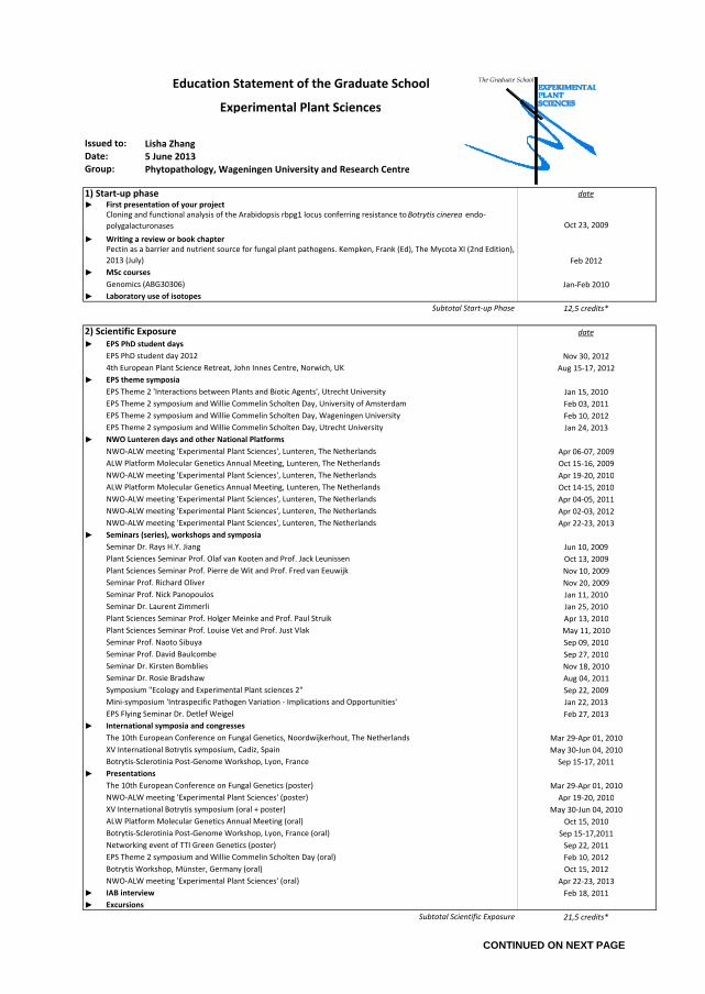
Lisha Zhang
5 June 2013
Phytopathology, Wageningen University and Research Centre
date
►
Oct 23, 2009
►
Feb 2012
►
Jan-Feb 2010
►
12,5 credits*
date
►
Nov 30, 2012
Aug 15-17, 2012
►
Jan 15, 2010
Feb 03, 2011
Feb 10, 2012
Jan 24, 2013
►
Apr 06-07, 2009
Oct 15-16, 2009
Apr 19-20, 2010
Oct 14-15, 2010
Apr 04-05, 2011
Apr 02-03, 2012
Apr 22-23, 2013
►
Jun 10, 2009
Oct 13, 2009
Nov 10, 2009
Nov 20, 2009
Jan 11, 2010
Jan 25, 2010
Apr 13, 2010
May 11, 2010
Sep 09, 2010
Sep 27, 2010
Nov 18, 2010
Aug 04, 2011
Sep 22, 2009
Jan 22, 2013
Feb 27, 2013
►
Mar 29-Apr 01, 2010
May 30-Jun 04, 2010
Sep 15-17, 2011
►
Mar 29-Apr 01, 2010
Apr 19-20, 2010
May 30-Jun 04, 2010
Oct 15, 2010
Sep 15-17,2011
Sep 22, 2011
Feb 10, 2012
Oct 15, 2012
Apr 22-23, 2013
► Feb 18, 2011
►
21,5 credits*
Seminar Prof. Nick Panopoulos
Mini-symposium 'Intraspecific Pathogen Variation - Implications and Opportunities'
Seminar Prof. David Baulcombe
Seminar Dr. Kirsten Bomblies
Seminar Dr. Rosie Bradshaw
Seminar Prof. Naoto Sibuya
Plant Sciences Seminar Prof. Louise Vet and Prof. Just Vlak
Plant Sciences Seminar Prof. Holger Meinke and Prof. Paul Struik
Seminar Dr. Laurent Zimmerli
Symposium "Ecology and Experimental Plant sciences 2"
Presentations
Botrytis Workshop, Münster, Germany (oral)
Botrytis-Sclerotinia Post-Genome Workshop, Lyon, France
EPS Flying Seminar Dr. Detlef Weigel
International symposia and congresses
The 10th European Conference on Fungal Genetics, Noordwijkerhout, The Netherlands
XV International Botrytis symposium, Cadiz, Spain
NWO-ALW meeting 'Experimental Plant Sciences' (oral)
EPS Theme 2 symposium and Willie Commelin Scholten Day (oral)
The 10th European Conference on Fungal Genetics (poster)
Botrytis-Sclerotinia Post-Genome Workshop, Lyon, France (oral)
Networking event of TTI Green Genetics (poster)
ALW Platform Molecular Genetics Annual Meeting (oral)
XV International Botrytis symposium (oral + poster)
NWO-ALW meeting 'Experimental Plant Sciences' (poster)
Subtotal Scientific Exposure
Plant Sciences Seminar Prof. Pierre de Wit and Prof. Fred van Eeuwijk
NWO-ALW meeting 'Experimental Plant Sciences', Lunteren, The Netherlands
Seminars (series), workshops and symposia
Seminar Dr. Rays H.Y. Jiang
NWO Lunteren days and other National Platforms
EPS Theme 2 symposium and Willie Commelin Scholten Day, Wageningen University
EPS Theme 2 symposium and Willie Commelin Scholten Day, Utrecht University
ALW Platform Molecular Genetics Annual Meeting, Lunteren, The Netherlands
Plant Sciences Seminar Prof. Olaf van Kooten and Prof. Jack Leunissen
ALW Platform Molecular Genetics Annual Meeting, Lunteren, The Netherlands
NWO-ALW meeting 'Experimental Plant Sciences', Lunteren, The Netherlands
NWO-ALW meeting 'Experimental Plant Sciences', Lunteren, The Netherlands
Education Statement of the Graduate School
Experimental Plant Sciences
First presentation of your project
Subtotal Start-up Phase
1) Start-up phase
Cloning and functional analysis of the Arabidopsis rbpg1 locus conferring resistance to Botrytis cinerea endo-
polygalacturonases
Pectin as a barrier and nutrient source for fungal plant pathogens. Kempken, Frank (Ed), The Mycota XI (2nd Edition),
2013 (July)
MSc courses
Writing a review or book chapter
Issued to:
Genomics (ABG30306)
Date:
Group:
Laboratory use of isotopes
Excursions
IAB interview
2) Scientific Exposure
4th European Plant Science Retreat, John Innes Centre, Norwich, UK
EPS theme symposia
EPS Theme 2 'Interactions between Plants and Biotic Agents', Utrecht University
EPS PhD student day 2012
EPS PhD student days
EPS Theme 2 symposium and Willie Commelin Scholten Day, University of Amsterdam
NWO-ALW meeting 'Experimental Plant Sciences', Lunteren, The Netherlands
Seminar Prof. Richard Oliver
NWO-ALW meeting 'Experimental Plant Sciences', Lunteren, The Netherlands
CONTINUED ON NEXT PAGE
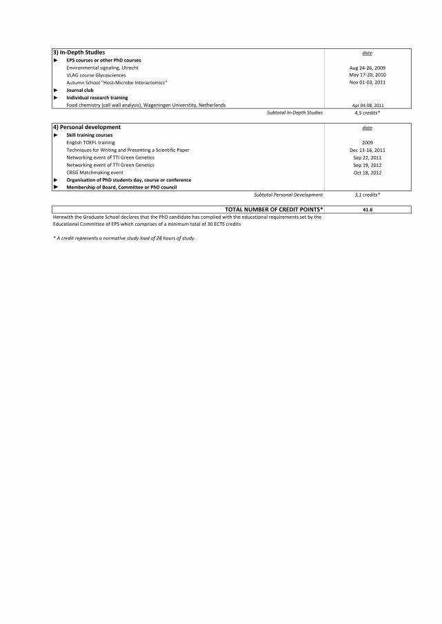
date
►
Aug 24-26, 2009
May 17-20, 2010
Nov 01-03, 2011
►
►
Apr 04-08, 2011
4,5 credits*
date
►
2009
Dec 13-16, 2011
Sep 22, 2011
Sep 19, 2012
Oct 18, 2012
►
►
3,1 credits*
41.6
Skill training courses
Techniques for Writing and Presenting a Scientific Paper
Food chemistry (cell wall analysis), Wageningen Universtity, Netherlands
Autumn School "Host-Microbe Interactomics"
3) In-Depth Studies
* A credit represents a normative study load of 28 hours of study.
TOTAL NUMBER OF CREDIT POINTS*
Herewith the Graduate School declares that the PhD candidate has complied with the educational requirements set by the
Educational Committee of EPS which comprises of a minimum total of 30 ECTS credits
Subtotal Personal Development
EPS courses or other PhD courses
4) Personal development
Membership of Board, Committee or PhD council
Organisation of PhD students day, course or conference
Individual research training
Journal club
Networking event of TTI Green Genetics
Networking event of TTI Green Genetics
CBSG Matchmaking event
VLAG course Glycosciences
English TOEFL training
Environmental signaling, Utrecht
Subtotal In-Depth Studies

This research was conducted in the Laboratory of Phytopathology of Wageningen
University and was financially supported by the graduate school Experimental Plant
Sciences (EPS) and the Technological Top Institute Green Genetics (TTI-GG).
Cover & Layout by: Lisha Zhang
Printed by: Proefschriftmaken.nl || Uitgeverij BOXPress