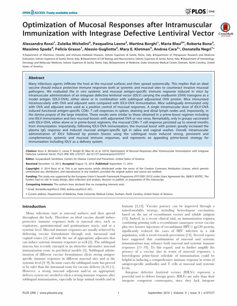
Optimization of Mucosal Responses after IntramuscularImmunization with Integrase Defective Lentiviral VectorAlessandra Rossi1, Zuleika Michelini2, Pasqualina Leone2, Martina Borghi1, Maria Blasi3¤, Roberta Bona2,
Massimo Spada4, Felicia Grasso1, Alessio Gugliotta2, Mary E. Klotman5, Andrea Cara2*, Donatella Negri1*
1 Department of Infectious, Parasitic and Immune-mediated Diseases, Istituto Superiore di Sanita, Rome, Italy, 2 Department of Therapeutic Research and Medicine
Evaluation, Istituto Superiore di Sanita, Rome, Italy, 3 Department of Cell Biology and Neuroscience, Istituto Superiore di Sanita, Rome, Italy, 4 Department of Hematology,
Oncology and Molecular Medicine, Istituto Superiore di Sanita, Rome, Italy, 5 Department of Medicine, Duke University Medical Center, Durham, North Carolina, United
States of America
Abstract
Many infectious agents infiltrate the host at the mucosal surfaces and then spread systemically. This implies that an idealvaccine should induce protective immune responses both at systemic and mucosal sites to counteract invasive mucosalpathogens. We evaluated the in vivo systemic and mucosal antigen-specific immune response induced in mice byintramuscular administration of an integrase defective lentiviral vector (IDLV) carrying the ovalbumin (OVA) transgene as amodel antigen (IDLV-OVA), either alone or in combination with sublingual adjuvanted OVA protein. Mice immunizedintramuscularly with OVA and adjuvant were compared with IDLV-OVA immunization. Mice sublingually immunized onlywith OVA and adjuvant were used as a positive control of mucosal responses. A single intramuscular dose of IDLV-OVAinduced functional antigen-specific CD8+ T cell responses in spleen, draining and distal lymph nodes and, importantly, inthe lamina propria of the large intestine. These results were similar to those obtained in a prime-boost regimen includingone IDLV immunization and two mucosal boosts with adjuvanted OVA or vice versa. Remarkably, only in groups vaccinatedwith IDLV-OVA, either alone or in prime-boost regimens, the mucosal CD8+ T cell response persisted up to several monthsfrom immunization. Importantly, following IDLV-OVA immunization, the mucosal boost with protein greatly increased theplasma IgG response and induced mucosal antigen-specific IgA in saliva and vaginal washes. Overall, intramuscularadministration of IDLV followed by protein boosts using the sublingual route induced strong, persistent andcomplementary systemic and mucosal immune responses, and represents an appealing prime-boost strategy forimmunization including IDLV as a delivery system.
Citation: Rossi A, Michelini Z, Leone P, Borghi M, Blasi M, et al. (2014) Optimization of Mucosal Responses after Intramuscular Immunization with IntegraseDefective Lentiviral Vector. PLoS ONE 9(9): e107377. doi:10.1371/journal.pone.0107377
Editor: Suryaprakash Sambhara, Centers for Disease Control and Prevention, United States of America
Received December 13, 2013; Accepted August 15, 2014; Published September 11, 2014
Copyright: � 2014 Rossi et al. This is an open-access article distributed under the terms of the Creative Commons Attribution License, which permitsunrestricted use, distribution, and reproduction in any medium, provided the original author and source are credited.
Funding: This study was supported by the European Union’s Seventh Framework Programme [FP7/2007 2013] under Grant Agreement No: 280873 ADITEC. Thefunders had no role in study design, data collection and analysis, decision to publish, or preparation of the manuscript.
Competing Interests: The authors have declared that no competing interests exist.
* Email: [email protected] (DN); [email protected] (AC)
¤ Current address: Department of Medicine, Duke University Medical Center, Durham, North Carolina, United States of America
Introduction
Many infections start at mucosal surfaces and then spread
throughout the body. Therefore an ideal vaccine should induce
protective immune responses both at mucosal sites, such as
respiratory, gastrointestinal and genitourinary tracts, and at a
systemic level. Mucosal immune responses are usually achieved by
delivering vaccine formulations through oral, intranasal and
vaginal routes [1] and with the use of appropriate adjuvants that
can induce systemic immune responses as well [2]. The sublingual
mucosa has recently emerged as an attractive alternative mucosal
immunization route in preclinical models [3]. Sublingual admin-
istration of different vaccine formulations elicits strong antigen-
specific immune responses in different mucosal sites and at the
systemic level [4–9]. In some cases the sublingual route has proved
to be safer than the intranasal route for vaccine delivery [4,10,11].
However, a strong mucosal adjuvant and/or an appropriate
delivery system are needed to elicit a strong immune response after
sublingual immunization, especially in large animal models and in
humans [2,12]. Vaccine potency can be improved through a
mixed-modality strategy, including heterologous vaccination,
based on the use of recombinant vectors and soluble antigens
[13]. Indeed, in a recent clinical trial, an immunization regimen
combining priming with a recombinant canarypox vector vaccine
plus two booster injections of recombinant HIV-1 gp120 protein,
significantly reduced the cases of HIV infection in a risk
population, with a trend towards prevention [14]. Several reports
have suggested that combinations of mucosal and systemic
immunizations may enhance both mucosal and systemic immune
responses [15–19]. To this regard, and to further amplify the
potency of a vaccine also in terms of mucosal responses, a
heterologous prime-boost schedule of immunization could be
helpful in inducing a comprehensive immune response in terms of
antigen-specific antibodies and T cells at mucosal and systemic
levels.
Integrase defective lentiviral vectors (IDLVs) represent a
powerful tool to deliver foreign genes. IDLVs are safer than their
integrase competent counterparts, since they lack integrase
PLOS ONE | www.plosone.org 1 September 2014 | Volume 9 | Issue 9 | e107377
activity, and transgene expression is efficiently driven from
unintegrated circular forms of the vector genome [20–22]. Several
reports have shown that IDLVs are suitable for delivery of vaccine
antigens in preventive vaccine strategies [23–29], demonstrating
that immunization with IDLVs induced strong and protective
antigen-specific immune responses, in absence of vector integra-
tion. Moreover, we recently demonstrated that therapeutic
vaccination with IDLV expressing HPV-E7 as a tumor antigen
results in eradication of TC-1 derived tumor in tumor-bearing
mice [30]. However, while the efficacy of intramuscular immu-
nization with IDLV at inducing systemic antigen-specific immune
responses after a single immunization is well established, no data
are available concerning the mucosal immune responses. Some
reports showed that in addition to systemic T cell response,
intramuscular immunization was able to induce CD8+ T cell
mediated antigen-specific immunity in gut mucosa, an important
portal of entry of many infectious pathogens [31,32].
In the present study, we analyzed the antigen-specific immune
response using different immunization protocols, by focusing on
mucosal cellular and antibody responses induced by a single
intramuscular administration of IDLV expressing ovalbumin
protein (OVA) as a model antigen either alone or in combination
with sublingual adjuvanted OVA in prime-boost regimens. Results
indicate that intramuscular immunization with IDLV is sufficient
to induce a persistent CD8+ T cell response in the lamina propriaof large intestine, while a mucosal protein boost is necessary for the
induction of mucosal IgA.
Materials and Methods
Vector construction and productionTransfer vector plasmid pTY2CMV-GFPW expressing GFP
has been already described [33]. For construction of transfer
vector expressing OVA protein, the coding sequence for OVA was
excised from plasmid pOVA, kindly provided by Dr Piergiuseppe
de Berardinis (I.B.P., C.N.R., Naples, Italy), using SnaBI/XbaI
and cloned into the transfer vector pTY2CMV-GFPW by
replacing the GFP coding sequence, thus obtaining the transfer
vector plasmid pLenti-OVA. The HIV-based packaging plasmid
IN defective (pcHelp/IN-) and the pseudotyping VSV.G envelope-
expressing (pMD.G) plasmids have been already described
[23,34,35].
For production of recombinant IDLV expressing OVA (IDLV-
OVA), 293T cells were transiently transfected on 10 cm Petri
dishes using the Calcium Phosphate-based Profection Mammalian
Transfection System (Promega Corporation, Madison WI, USA)
as previously described [23]. For concentration, vector containing
supernatants were ultracentrifuged (Beckman Coulter, Inc., Full-
erton, CA, USA) on a 20% sucrose gradient (Sigma Chemical Co.
St. Louis, MO, USA) and viral pellets were resuspended in 16PBS. Viral titers for IDLV-OVA were performed by the reverse
transcriptase (RT) activity assay [36] over standards of known
infectivity and the vector-associated RT activity were compared
with the ones of IDLV-GFP virions of known infectious titers and
RT activity, thus allowing for the determination of their infectious
titer units [37].
Western blot, DNA isolation and PCRTo verify the expression of ovalbumin (OVA), 293T cells were
seeded in 10 cm plates and transduced with IDLV-OVA or
IDLV-GFP at 37uC in atmosphere containing 5% CO2. Thirty-
six hours post-transduction supernatants and cells were collected.
Equivalent amounts of cells transduced with IDLV-OVA or
IDLV-GFP were lysed in lysis buffer (20 mM HEPES, 50 mM
NaCl, 10 mM EDTA, 2 mM EGTA, 0.5%, NP-40, 50 mM NaF,
1 mM orthovanadate, 1 mM PMSF, 5 mg/ml of aprotin and
5 mg/ml of leupeptin). Proteins of cell lysates and supernatants
were separated on 12% SDS polyacrylamide gel along with the
purified OVA protein (Sigma), used as a positive control and
transferred to a nitrocellulose membrane (GE HEALTHCARE).
The filters were saturated overnight with 5% non fat dry milk
(NFDM) in PBST (PBS with 0.1% Tween 20) and then incubated
with a rabbit anti-OVA polyclonal antibody (AB1225, Millipore)
for 1 hr at room temperature, followed by incubation for 1 hr at
room temperature with an anti-rabbit HRP-conjugated IgG
(Sigma). The immunocomplexes were visualized using chemilu-
minescence ECL detection system (Luminata Crescendo Western
HRP Substrate, Millipore) [30].
DNA from muscle, splenocytes and large intestine was extracted
using the SV Total RNA Isolation System protocol, modified for
DNA preparation (Promega Corporation, Madison, WI) [38]. All
samples supported the amplification of the mouse glyceraldehyde
3-phosphate dehydrogenase gene (G3PDH), (GlymoFor: 5’-
TGAAGGTCGGTGTGAACGGATTTGGC-3’; GlymoRev: 5’-
CATGTAGGCCATGAGGTCCACCAC-3’) and were included
in subsequent PCR analysis to detect the presence of the vector
DNA sequence using 500 ng of DNA and a primer pair spanning
the LTR region at the 39 end of the vector (PPTs: 59-
CAGCTGTAGATCTTAGCCACT-39; AA55: 59-CTGC TA-
GAGATTTTCCACACTGAC-39), as described [23]. PCR pa-
rameters were: 1 cycle of 5 min at 94uC, followed by 40 cycles of
30 sec at 94uC, 30 sec at 60uC, 30 sec at 72uC with a final
extension step of 10 min at 72uC in a 9700 Perkin-Elmer
Thermocycler.
Mice and immunization scheduleEthics statement. Animals were maintained under specific
pathogen-free conditions in the animal facilities at the Istituto
Superiore di Sanita and treated according to European Union
guidelines and Italian legislation (Decreto Legislativo 116/92,
implementing the 86/609/CEE Directive on laboratory animal
protection). All animal studies were reviewed and approved by the
Service for Biotechnology and Animal Welfare at the Istituto
Superiore di Sanita (ISS registration n. 3138 of 26/01/2012). All
animals were euthanized by CO2 inhalation using approved
chambers, and efforts were made to minimize suffering and
discomfort.
C57/Bl6 female mice were purchased from Charles River
Laboratories, Calco, Italy. A scheme of immunization protocols is
described in Table 1. Mice were immunized by intramuscular
(i.m.) injection with IDLV expressing OVA (IDLV-OVA) either
alone (group A) or in combination with two doses of OVA protein
(OVAp) plus E.coli heat-labile enterotoxin adjuvant (LT), deliv-
ered sublingually either after or before IDLV-OVA (group B and
C, respectively). Other groups included mice immunized once
intramuscularly with OVAp plus LT alone (group D) or in
combination with two sublingual (s.l.) doses of OVAp + LT
(groups E). A group of mice representing the positive control for
mucosal immune responses was sublingually immunized with 4
doses of OVAp + LT (group F); the number of doses was selected
based on our previous data [6] and in order to obtain a strong
positive control for mucosal humoral and cellular responses.
Naıve, non-immunized mice were kept for parallel analysis. All
immunizations were given 2 weeks apart. IDLV-OVA (1.16107
RT units total/mouse) was administered in both left and right
thigh. The same dose of OVAp (20 mg/mouse per dose) + LT
(1 mg/mouse per dose) was injected either intramuscularly in one
thigh or sublingually. Sublingually immunized mice (5 ml/mouse
Mucosal Responses by Using IDLV
PLOS ONE | www.plosone.org 2 September 2014 | Volume 9 | Issue 9 | e107377

per dose) were deeply anesthetized with ketamine (2 mg/mouse)
and xylazine (0.17 mg/mouse), in order to avoid the swallowing of
saliva during the immunization, as already described [6].
The cellular immune responses were analyzed at 2 weeks and at
6 months after the last immunization, by sacrificing 4 mice per
group at each time point and each experiment was repeated at
least two times. Anti-OVA IgG and IgA antibodies (Abs) were
measured in plasma and mucosal secretions at 2 weeks after each
immunization and at 6 months after the last immunization.
Plasma samples were obtained from blood collected from the
retro-orbital plexus of mice with heparin-treated glass Pasteur
pipettes and stored at 220uC until assayed. Saliva was collected
after intraperitoneal injection of pilocarpine (160 mg/mouse).
Vaginal washes were obtained introducing 50 ml of PBS each for
three times into the vaginal tract of mice using a Gilson pipette. At
the time of sacrifice, spleen, lymph nodes (submandibular,
mesenteric, inguinal) and large intestine were recovered for the
analysis of cellular immune responses.
Preparation of single-cell suspensionsSplenocytes and lymph node derived cells were prepared by
mechanical disruption and passage through cell strainers (BD
Pharmingen, San Diego, CA, USA) and resuspended in RPMI
1640 (Euroclone) containing 10% fetal bovine serum (FBS)
(Lonza), 100 units/ml of penicillin–streptomycin–glutamine (Euro-
clone), non-essential aminoacids (Euroclone), sodium pyruvate
1 mM (Euroclone), HEPES buffer solution 25 mM (Euroclone)
50 mM 2-mercaptoethanol (Sigma Chemicals). In order to isolate
lamina propria (LP) lymphocytes, the large intestine surgically
removed from sacrificed mice was cleaned, cut longitudinally and
then sliced into small pieces with a scalpel. Tissue fragments were
incubated shaking with 15 ml of Hank’s balanced salt solution
(HBSS) (Euroclone), 10% FBS, Hepes buffer solution 25 mM
(Euroclone), EDTA 5 mM (Sigma Chemicals) and dithiothreitol
(DTT) 1 mM (Sigma Chemicals) for 15 minutes at 37uC.
Supernatants were discarded and left fragments were spun down,
resuspended in liberase (800 units/sample, Roche) and DNAse I
(40 units/sample, Roche, Monza, Italy) and incubated shaking for
1 hour at 37uC. After incubation both supernatants and pellets
were filtered through 100 mm cell strainers, resuspended in 20 ml
Percoll 30%-EDTA 1 mM (Sigma Chemicals) and centrifuged at
290xg for 25 minutes. LP lymphocytes were recovered in the
pellet. Cells obtained from the same immunization group were
pooled to ensure there was a sufficient number for subsequent
tests.
IFNc ELISPOT and dextramer stainingThe IFNc ELISPOT assay was performed using the BD
ELISPOT kit reagents and protocol (BD Biosciences). Briefly,
single cell suspensions from spleen and lymph nodes were seeded
at a density of 26105/well in 96 well plates and stimulated
overnight either with 2 mg/ml of the H-2Kb restricted OVA 8mer
peptide (SIINFEKL) or with 5 mg/ml of concanavalin A (Sigma
Chemicals) used as a positive control. Complete medium treated
cells were used as negative controls. Spot Forming Cells (SFC)
were counted with an ELISPOT reader (A.EL.VIS, Hannover,
Germany) and results expressed as IFNc secreting cells/106 cells.
For dextramer staining, cells from lymph nodes and large
intestine lamina propria were washed once with 2 ml of PBS-5%
bovine serum albumin (BSA) (Sigma Chemicals) in 5 ml polysty-
rene tubes and centrifuged. After discarding the supernatant,
16106 cells were resuspended in residual volume (50 ml) and 10 ml
of H-2Kb-SIINFEKL R-PE conjugated dextramer (Immudex,
Copenhagen, Denmark) was added in each sample for 10 minutes
at room temperature in the dark. Cells were washed again and
PerCP-Cy5.5 conjugated anti-mouse CD8a (BD Pharmigen) and
anti-mouse CD3 FITC (Immunological Sciences, Rome, Italy)
were added for 20 minutes on ice in the dark. Cells were washed
twice, resuspended in 0.5 ml PBS-1% paraformaldeyde and
analyzed at the FACScalibur (BD Biosciences).
Intracellular staining for cytokinesSplenocytes were either cultured in the presence of OVA-
specific 8mer peptide (5 mg/ml) or left untreated in the presence of
anti-mouse CD28 mAb (BD Pharmigen) at 2 mg/ml. PMA
(10 ng/ml) (Sigma Chemicals) in combination with Ionomicin
(1 mg/ml) (Sigma Chemicals) were used as positive control. One
hour after stimulation, 10 mg/ml of Brefeldin A (Sigma Chemicals)
was added to the culture to inhibit cytokine secretion and cells
were incubated overnight at 37uC. After blocking of Fc receptors
by treatment with anti-mouse CD16/CD32 (BD Pharmigen) cells
were stained with fluorochrome conjugates FITC anti-mouse CD3
(Immunological Sciences), PE anti-mouse CD4 (Immunological
Sciences) and PerCP Cy 5.5 anti-mouse CD8a (BD Pharmigen).
Cells were washed, fixed with 4% paraformaldehyde (Sigma
Chemicals), permeabilized in PBS-0.5% saponin (Sigma Chemi-
cals) and stained with APC conjugated anti-mouse IFNc and PE-
Cy 7 conjugated anti-mouse TNFa or their isotype-matched
controls (BD Pharmigen). Samples were washed and analyzed by
FACScanto (BD Biosciences)
Table 1. Vaccine regimens.
Group 1st immunization week 0 2nd immunization week 2 3rd immunization week 4 4th immunization week 6
A i.m. IDLV-OVA - - -
B i.m. IDLV-OVA s.l. OVAp + LT s.l OVAp + LT -
C s.l. OVAp + LT s.l. OVAp + LT i.m. IDLV-OVA -
D i.m. OVAp + LT - - -
E i.m. OVAp + LT s.l. OVAp + LT s.l. OVAp + LT -
F s.l. OVAp + LT s.l. OVAp + LT s.l. OVAp + LT s.l. OVAp + LT
Naive - - - -
i.m.: intramuscular; s.l.: sublingual; OVAp: ovalbumin protein; LT: E.coli heat-labile enterotoxin.doi:10.1371/journal.pone.0107377.t001
Mucosal Responses by Using IDLV
PLOS ONE | www.plosone.org 3 September 2014 | Volume 9 | Issue 9 | e107377

Measurement of OVA-specific IgG and IgA antibodiesPlasma, saliva and vaginal washes were tested for the presence
of anti-OVA IgG or IgA antibodies by a standard ELISA. Ninety-
six well plates (Greiner bio-one, Germany) were coated with
0.5 mg/well of OVA overnight at 4uC. After washing and blocking
for 2 hrs with 200 ml of PBS containing 1% BSA (Sigma
Chemicals), serial dilutions of plasma and mucosal secretions
from individual mice were added to wells in duplicate and
incubated for 2 hrs at room temperature. The plates were washed
and biotin-conjugated goat anti-mouse IgG (Southern Biotech,
Birmingham, AL, USA) or IgA (Southern Biotech) was added to
the wells for 2 hrs at room temperature. The plates were washed
again before the addition of horse radish peroxidase (HRP)-
conjugated streptavidin (AnaSpec, Fremont, CA, USA) for 30 min
at room temperature. The antigen–antibody reaction was
measured by using the 3.3,5.5-tetramethylbenzidine substrate
(SurModics BioFX, Edina, MN, USA) and the reaction was
stopped with 50 ml of H2SO4 1M. Endpoint titers were
determined as the reciprocal of the highest dilution giving an
absorbance value at least equal to threefold that of background
(biological sample from naıve mice). For each group of immuni-
zation, results were expressed as mean titer 6 standard deviation.
Statistical analysisThe immune responses were expressed as averages 6 standard
deviation. Statistical significance was determined by unpaired two
tailed t-Student test. Paired two tailed t-Student test was used
when appropriate and specified in the text; p,0.05 was considered
statistically significant.
Results
Ovalbumin is efficiently expressed from IDLVTo confirm expression of ovalbumin (OVA) from IDLV-OVA,
293T cells were transduced with IDLV-OVA or IDLV-GFP as a
control and cell lysates were analyzed by Western blotting assay.
As shown in Figure 1, a band corresponding to the full length
OVA was detected in cells transduced with IDLV-OVA but not in
cells transduced with IDLV-GFP. Importantly, 293T cells
transduced with IDLV-OVA released OVA protein in the
supernatant. These results demonstrated that OVA protein was
efficiently expressed in vitro from IDLV, validating IDLV-OVA as
a suitable candidate for in vivo vaccination studies.
Intramuscular administration of IDLV-OVA either alone orin prime-boost regimens induces strong systemicantigen-specific T cell responses
Groups of mice were immunized according to the schedule of
immunization shown in Table 1. Two weeks after the final
immunization mice from all groups were sacrificed, lymphoid
organs were removed and the presence of antigen-specific T cells
was evaluated by IFNc ELISPOT assay. Splenocytes derived from
all immunized groups showed IFNc secreting cells upon stimula-
tion with the H-2Kb restricted OVA 8mer peptide (Figure 2A). In
particular, mice immunized with IDLV-OVA either alone, as a
prime or as a boost (groups A, B and C, respectively) showed high
numbers of antigen-specific IFNc-producing cells (13956150,
11226158 and 16656191 SFC/106cells, groups A, B and C,
respectively). Of note, two s.l. boosts with OVAp + LT (group B)
did not increase the number of IFNc-producing T cells compared
to group A, while IDLV boost in group C induced the highest
number of IFNc secreting cells compared to groups A and B (p,
0.05 C vs B; p.0.05 A vs C). The lowest response was detected in
animals intramuscularly immunized once with OVAp + LT (group
D, 136651 SFC/106 cells). This response increased when animals
were sublingually boosted twice with the protein (group E,
600634 SFC/106 cells), without reaching the levels observed in
IDLV-immunized animals.
In mice sublingually immunized four times (group F) the cellular
immune response was higher (8236126 SFC/106cells) than that
observed in group D (p,0.05) and group E, but significantly lower
than that present in the IDLV-immunized groups (p,0.05).
Splenocytes derived from untreated naıve mice did not show any
specific IFNc response (data not shown).
Lymph node-derived cells from mice of the same groups were
also utilized for the analyses, and the results are summarized in
Figure 2B. In inguinal lymph nodes (ING LN), draining the thigh
muscle region (injection site), groups A and C showed a similar
number of antigen-specific IFNc producing T cells (425682 and
512651 SFC/106cells, respectively), which was significantly
higher than that present in mice from all other groups (198642,
13663, 93635 and 6064 SFC/106cells, B, D, E and F groups,
respectively). In submandibular lymph nodes (SM LN), draining
the sublingual site, mice receiving OVA sublingually showed a
higher response (22266, 378656, 170638 and 42468 SFC/
106cells, B, C, E and F groups, respectively) compared to the
animals immunized with IDLV-OVA alone (93632 SFC/
106cells). IFNc producing T cells were also detected in mesenteric
lymph nodes (MES LN), which represents a distal site from
immunizations. In this case all the IDLV-immunized groups
(327625, 222617 and 331617 SFC/106cells, A, B and C,
respectively) showed a significantly higher response compared to
the other groups (3464, 89645 and 70627 SFC/106cells, D, E
and F, respectively). Lymph node derived cells from untreated
naıve mice did not show any specific IFNc response (data not
shown).
These results indicate that all immunized mice have a systemic
antigen-specific T cell response and that mice immunized
intramuscularly with IDLV-OVA showed overall higher responses
compared to the animals immunized with the adjuvanted OVAp,
administered either i.m. or s.l.
Figure 1. Expression of ovalbumin from IDLV. Western blotanalysis of cell lysates and supernatants from 293T cells transduced withIDLV-OVA or IDLV-GFP as negative control, using an anti-OVApolyclonal antibody. Ovalbumin protein (OVAp, predicted size 47 kD)was used as positive control. Samples included supernatants showingsecreted OVA (lane 1 and lane 2, 14 ml and 7 ml from 293T cells culturedat 56105/ml, respectively), and cell lysates (lane 3 and lane 4,corresponding to 36105 and 0.756105 cells equivalent, respectively).Supernatants (lane 5, 14 ml) and cell lysates (lane 6, 36105 cellsequivalent) from IDLV-GFP transduced cells did not produce OVA. OVAprotein (OVAp) at indicated amounts was used as positive control.doi:10.1371/journal.pone.0107377.g001
Mucosal Responses by Using IDLV
PLOS ONE | www.plosone.org 4 September 2014 | Volume 9 | Issue 9 | e107377
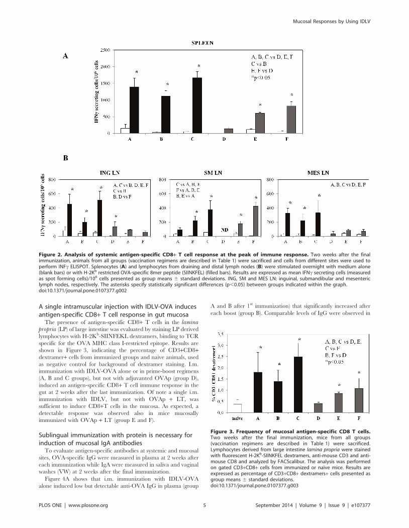
A single intramuscular injection with IDLV-OVA inducesantigen-specific CD8+ T cell response in gut mucosa
The presence of antigen-specific CD8+ T cells in the laminapropria (LP) of large intestine was evaluated by staining LP derived
lymphocytes with H-2Kb-SIINFEKL dextramers, binding to TCR
specific for the OVA MHC class I-restricted epitope. Results are
shown in Figure 3, indicating the percentage of CD3+CD8+dextramer+ cells from immunized groups and naive animals, used
as negative control for background of dextramer staining. I.m.
immunization with IDLV-OVA alone or in prime-boost regimens
(A, B and C groups), but not with adjuvanted OVAp (group D),
induced an antigen-specific CD8+ T cell immune response in the
gut at 2 weeks after the last immunization. Of note a single i.m.
immunization with IDLV, but not with OVAp + LT, was
sufficient to induce CD8+T cells in the mucosa. As expected, a
detectable response was observed also in mice mucosally
immunized with OVAp + LT (group E and F).
Sublingual immunization with protein is necessary forinduction of mucosal IgA antibodies
To evaluate antigen-specific antibodies at systemic and mucosal
sites, OVA-specific IgG were measured in plasma at 2 weeks after
each immunization while IgA were measured in saliva and vaginal
washes (VW) at 2 weeks after the final immunization.
Figure 4A shows that i.m. immunization with IDLV-OVA
alone induced low but detectable anti-OVA IgG in plasma (group
A and B after 1st immunization) that significantly increased after
each boost (group B). Comparable levels of IgG were observed in
Figure 2. Analysis of systemic antigen-specific CD8+ T cell response at the peak of immune response. Two weeks after the finalimmunization, animals from all groups (vaccination regimens are described in Table 1) were sacrificed and cells from different sites were used toperform INFc ELISPOT. Splenocytes (A) and lymphocytes from draining and distal lymph nodes (B) were stimulated overnight with medium alone(blank bars) or with H-2Kb restricted OVA-specific 8mer peptide (SIINKFEL) (filled bars). Results are expressed as mean IFNc secreting cells (measuredas spot forming cells)/106 cells presented as group means 6 standard deviations. ING, SM and MES LN: inguinal, submandibular and mesentericlymph nodes, respectively. The asterisks specify statistically significant differences (p,0.05) between groups indicated within the graph.doi:10.1371/journal.pone.0107377.g002
Figure 3. Frequency of mucosal antigen-specific CD8 T cells.Two weeks after the final immunization, mice from all groups(vaccination regimens are described in Table 1) were sacrificed.Lymphocytes derived from large intestine lamina propria were stainedwith fluorescent H-2Kb-SIINKFEL dextramers, anti-mouse CD3 and anti-mouse CD8 and analyzed by FACScalibur. The analysis was performedon gated CD3+CD8+ cells from immunized or naıve mice. Results areexpressed as percentage of CD3+CD8+ dextramers+ cells presented asgroup means 6 standard deviations.doi:10.1371/journal.pone.0107377.g003
Mucosal Responses by Using IDLV
PLOS ONE | www.plosone.org 5 September 2014 | Volume 9 | Issue 9 | e107377

plasma of mice immunized twice mucosally with OVAp + LT and
boosted intramuscularly with IDLV-OVA (Group C). A single i.m.
immunization with adjuvanted OVAp (group D) induced a
significantly higher level of IgG compared to group A. The titer
further increased after two s.l. boosts (group E). Finally, in group F,
a single s.l. immunization induced anti-OVA IgG levels compa-
rable to those obtained after a single i.m. IDLV-OVA immuni-
zation (group A and B) but significantly lower than those observed
in group D and E. Overall, i.m. priming and s.l. boosts with
OVAp + LT (group E) induced the highest levels of IgG in plasma
after each immunization, when compared to the other groups. In
all cases, within the same group, IgG titers significantly increased
after each boost (p,0.05, paired two tailed t-Student test was
used).
IgA measurement in mucosal secretions showed a more
composite picture (Fig. 4B). In accordance with our previous
study [6], multiple s.l. immunizations (group F) induced antigen-
specific IgA antibodies both in saliva and vaginal washes. I.m.
immunization alone either with IDLV or with protein and
adjuvant did not induce anti-OVA IgA antibodies (group A and D,
data not shown). IgA were induced after two sublingual boosts
with adjuvanted OVA in both mucosal secretions from group B
mice, while only in vaginal washes and at a lower level from group
E animals. Interestingly, while anti-OVA IgA were present in
group B mice, receiving i.m. IDLV-OVA followed by two s.l.
boost with OVAp + LT, they were absent in group C mice (data
not shown), receiving two s.l. adiminstration of OVAp + LT
followed by i.m. IDLV-OVA. This was unexpected, since both
groups received IDLV-OVA and OVAp + LT, delivered
sublingually, although using different prime-boost regimens.
Intramuscular injection with IDLV-OVA induces longlasting systemic and mucosal antigen-specific CD8+ T cellresponse
Persistence of antigen-specific cell-mediated immunity was
analyzed in all groups of mice at 6 months after the final
immunization by IFNc ELISPOT assay, intracellular staining
(ICS) on IFNc and TNFa producing CD8+ T cells, and detection
of antigen-specific CD8+ T cells by dextramer staining. The IFNc
Figure 4. Analysis of antigen-specific antibodies in plasma and mucosal secretions. (A) Kinetics of plasma anti-OVA IgG titers in micebelonging to different groups (vaccination regimens are described in Table 1) at 2 weeks after each immunization. Results are expressed as meantiter presented as group means 6 standard deviations. The statistical analysis is described and discussed in the text. (B) Analysis of anti-OVA IgA titerin saliva and vaginal washes (VWs) collected 2 weeks after the final immunization. Results are expressed as mean titer presented as group means 6standard deviations.doi:10.1371/journal.pone.0107377.g004
Mucosal Responses by Using IDLV
PLOS ONE | www.plosone.org 6 September 2014 | Volume 9 | Issue 9 | e107377

ELISPOT assay showed that IFNc producing OVA-specific cells
were still detectable in splenocytes from all groups of mice
(Fig. 5A). In particular, mice receiving IDLV-OVA (groups A, B
and C) showed the highest number of IFNc producing cells in
spleens (averages of 11586133, 10526311 and 14166272 SFC/
106 splenocytes, respectively; C vs B p,0.05), which were
significantly higher than the values observed in groups D, E and
F (group D 101616 SFC/106, group E 103626 SFC/106, group
F, 5066219 SFC/106; p,0.05). This was in line with the results
obtained at 2 weeks from the last immunization (Fig. 2A). To
verify the functionality of OVA-specific CD8+ T cells at several
months from the last immunization, multifunctional OVA-specific
CD8+ T cell responses were analyzed by ICS for IFNc and TNFa.
A representative experiment is shown in Figure 5B. CD8+ T cells
producing both cytokines were evident in all groups upon
stimulation with the specific OVA peptide, confirming the
ELISPOT data. Groups A, B and C showed remarkable
percentages of multifunctional CD8+ T cells, which were
significantly higher than those present in all other groups. We
did not find evidence of CD8 negative T cells producing either
IFNc or TNFa upon stimulation with the H-2Kb specific OVA
peptide (data not shown).
To evaluate the persistence of vaccine-induced CD8+ T cell
responses at systemic distal and mucosal sites, we measured the
frequency of OVA-specific CD8+ T cells in mesenteric lymph
nodes and in LP lymphocytes by dextramer staining (Fig. 6).
OVA-specific CD8+ T cells were found in mesenteric lymph
nodes from all groups of mice immunized with IDLV-OVA
(groups A, B and C) (Fig. 6A), but not in those derived from
groups D, E and F. The same analysis was performed on LP
derived lymphocytes (Fig. 6B). CD8+ dextramer+ lymphocytes
were present in all IDLV-OVA immunized mice (groups A, B and
C) but no longer detectable in the other groups that were positive
at 2 weeks after immunization (groups E and F, cf. Fig. 3).
Given the prolonged immune response observed after IDLV-
OVA immunization, persistence of the vector at systemic and
mucosal sites was evaluated by DNA-PCR in mice vaccinated with
IDLV-OVA at 6 months after the single immunization (group A).
As shown in Figure 7A, while the presence of the vector was
detected in all muscle samples (injection site), we did not find
evidence of lentiviral vector sequences at systemic sites, including
spleen and gut (large intestine). No vector sequences were detected
in tissue samples from naıve mice. This is in line with previous
studies showing persistence of IDLV sequences at the injection site
[23,25].
Finally, the persistence of antibody response was evaluated in
plasma and in mucosal secretions. Plasma anti-OVA IgG were still
present in all groups of mice at 6 months after the final
immunization, although at titers lower than those observed at 2
weeks after the final immunization (Figure 7B). Conversely, no
detectable anti-OVA IgA were found in mucosal samples from the
immunized mice, regardless of the immunization protocol (data
not shown).
Discussion
Whereas the efficacy of IDLV immunization at inducing
prolonged systemic antigen-specific immune responses is now well
established, data are still lacking concerning the IDLV-induced
mucosal immune responses. We previously demonstrated that a
single intramuscular injection of IDLV induced systemic antigen-
specific CD8+ T cell responses that were detectable up to 3
months in spleen, draining LN and bone marrow derived cells
[23,25,39]. Here we showed for the first time that antigen-specific
CD8+ T cells induced after a single i.m. immunization with
IDLV-OVA were detectable at both systemic and mucosal sites,
including distal LN and the lamina propria of large intestine, for
up to 6 months after the immunization. These results are in
agreement with data demonstrating that recombinant adenoviral
vectors, an efficient vaccine delivery system, induced mucosal
cellular response after intramuscular immunization in mice and
non human primates [40].
The cellular immune response induced by IDLV was compared
in heterologous prime-boost vaccine strategies, based on the
immunization with the same antigen delivered as a soluble protein
either i.m. or s.l. together with LT as an adjuvant. It has been
extensively demonstrated that LT is a potent adjuvant for vaccines
delivered through both systemic and mucosal routes of immuni-
zation [41,42]. Our data clearly demonstrated that i.m. immuni-
zation with IDLV induced the highest T cell responses and showed
an overall superior efficacy in terms of persistence at mucosal site,
where it appears necessary for the maintenance of antigen-specific
CD8+ T cells. Interestingly, the s.l. boosts with OVAp + LT did
not increase the T cell response induced by IDLV priming, while
in the mice s.l. immunized twice with OVAp +LT, the boost with
IDLV resulted in a significantly higher T cell response both in the
spleen and in draining LN. Since IDLV immunization induced
high levels of effector CD8+ T cells [23], we can hypothesize that a
boost given during the peak of response could have been
detrimental. Further experiments aimed at analyzing this phe-
nomenon are warranted, such as analysis of exhaustion markers on
T cells and use of longer intervals between prime and boost(s). On
the other hands, the frequency of antigen-specific CD8+ T cells
measured several months after the last s.l. boost was still high in
spleen, mesenteric LN and in LP of large intestine, and
comparable to that observed in the other groups immunized with
IDLV-OVA alone or as a boost.
In addition to T cell responses, we also evaluated the antibody
response at systemic and mucosal sites. Results clearly indicated
that intramuscular injection of IDLV-OVA alone induced low
levels of antigen specific antibodies. This is consistent with our
previous work using plasmid DNA or IDLV expressing HIV-Env
[23,39] and in line with data showing that DNA or other vector-
based immunizations are poor inducers of antibody response
[43,44], especially compared to i.m. immunizations with soluble
antigens delivered in combination with strong adjuvants. In fact,
our results showed that i.m. immunization with 20 mg of soluble
OVA together with the potent adjuvant LT induced higher
antibody titers than i.m. immunization with IDLV (Fig. 4). When
the same dose of protein and adjuvant was given once through the
s.l. route, known to be less efficient than i.m. route, anti-OVA IgG
levels in plasma were comparable to those found in mice
immunized with IDLV. As expected, priming with IDLV-OVA
followed by two s.l. doses of LT-adjuvanted soluble OVAp
strongly increased the systemic antibody response, although at
levels significantly lower than those observed in mice intramuscu-
larly primed with OVAp + LT and boosted twice mucosally.
However, only by using the mucosal route of immunization we
were able to detect anti-OVA IgA in mucosal secretions.
Interestingly, in mice primed with IDLV and boosted twice
sublingually, mucosal anti-OVA IgA were produced at levels
similar to those present in mice immunized sublingually four times,
considered our positive control for mucosal responses. Our results
also suggest that IDLV as a prime (group B) but not as a boost
(group C) was able to drive the mucosal IgA response in
combination with mucosal immunizations. Since the vector
persists at the site of injection, we can speculate that the persistent
antigen expression from IDLV in mice primed with the vector
Mucosal Responses by Using IDLV
PLOS ONE | www.plosone.org 7 September 2014 | Volume 9 | Issue 9 | e107377

helped in driving a better production of both systemic and mucosal
antibodies when boosted with OVAp + LT. We did not evaluate
the presence of antibody secreting cells at mucosal sites, therefore
we cannot exclude that IgA present in mucosal secretions might
originate from blood. Since it has been demonstrated that s.l.
immunizations using bacterial toxins as adjuvants in combination
with different antigens induced local production of IgA [4,5], we
can only speculate that, also in this study, IgA in mucosal
secretions were produced locally.
We also showed that at 6 months after the final immunization
anti-OVA IgG antibodies were still present in plasma in all groups
of immunized mice, suggesting that all vaccine strategies used in
this study efficiently induced long term systemic antibodies.
Conversely, mucosal IgA antibodies were transient, and became
undetectable in all groups at later time points (data not shown),
suggesting that further mucosal boost(s) may be necessary to
maintain the mucosal IgA antibody response. Alternatively,
immunization schedules with longer intervals may induce higher
mucosal and systemic antibody responses, as already demonstrated
in different settings [16].
We previously demonstrated that 3 months after a single IDLV
immunization the vector was still detectable in muscle samples
from the injection site [23,25]. Here we showed that the vector is
detectable at 6 months from the immunization only at the site of
injection (Fig. 7B). This is in agreement with reports showing that
IDLV persists up to 9 months from the inoculum in non-dividing
cells or tissues [45–49]. It can be hypothesized that transduction of
non-proliferating cell populations at the injection site, including
Figure 5. Persistence of systemic antigen-specific CD8+ T cell response. Six months after the last immunization mice from all groups(vaccination regimens are described in Table 1) were sacrificed and splenocytes used for the analysis of OVA-specific T cells. (A) IFNc ELISPOT.Splenocytes were stimulated overnight with medium alone (blank bars) or with H-2Kb restricted OVA-specific 8mer peptide (SIINFEKL) (filled bars).IFNc-producing T cells are expressed as the number of spot forming cells per 106 cells. Results are presented as group means 6 standard deviations.The asterisks indicate statistically significant differences (p,0.05) between indicated groups. (B) Analysis of multifunctional antigen-specific CD8+ Tlymphocytes by intracellular assay for IFNc and TNFa production. A representative experiment is shown. The analysis was performed on gated CD3+CD8+ cells from immunized or naıve mice. The percentages of single or double-cytokine producing cells were calculated and are indicated within thedot plots.doi:10.1371/journal.pone.0107377.g005
Mucosal Responses by Using IDLV
PLOS ONE | www.plosone.org 8 September 2014 | Volume 9 | Issue 9 | e107377

muscle fibers and professional antigen presenting cells, ensures
persistence of the vaccine antigen and consequently prolonged
presentation to naıve T cells in draining lymph nodes. However,
although IDLV-OVA is expression competent and it is an ideal
tool for induction of CD8 T cell response, the amount of protein it
produces may not be sufficient to induce or boost high levels of
antibodies. Development of heterologous prime-boost regimens
combining IDLV-OVA with soluble protein + adjuvant could be
useful to this aim. Other groups showed that following immuni-
zation with lentiviral vectors, the antigen presentation to CD8 T
cells persists more than 40 days in an adoptive transfer model
[26,50]. This feature may be responsible for the induction of the
long term immune response induced by IDLV. Further charac-
terization of the microenvironment and the mechanism of antigen
presentation associated with IDLV injection could help to better
clarify this issue. Concerning safety issues, IDLVs are non-
replicating and non-integrating lentiviral vectors, produced by
incorporating a mutated form of the integrase protein in the
recombinant lentiviral particles. Absence of integration has been
demonstrated in several murine models in vivo and in cell culture
model systems [51]. Importantly, a recent report showed that,
compared to the parental integrating counterpart, the genotoxicity
of IDLV-associated insertional mutagenesis was negligible, making
IDLV highly attractive from a biosafety standpoint [52].
In this study we showed that systemic immunization with IDLV
can overcome immune compartmentalization and generate potent
and durable mucosal cellular immunity. Further analysis of
mucosal responses after immunization with IDLV using different
mucosal routes should be attempted in order to evaluate the ability
of IDLV in inducing local mucosal responses, in the absence of
toxicity. In this context, we recently demonstrated that two
intranasal, but not intramuscular administrations of IDLV
expressing Influenza NP were able to protect mice from challenge
with a heterosubtypic Influenza virus [53]. However, mucosal
responses were not evaluated.
In conclusion, this is the first report demonstrating that i.m.
immunization with IDLV induced antigen-specific CD8+ T cells
in lamina propria of large intestine, an important immune effector
site against mucosal associated pathogens, and that the CD8-
specific response lasts for a prolonged period of time from the
Figure 6. Persistence of mucosal antigen-specific CD8+ T cell response. Six months after the last immunization mice from all groups(vaccination regimens are described in Table 1) were sacrificed. (A) Mesenteric lymph node-derived lymphocytes were stained with H-2Kb-SIINKFELdextramers, anti-mouse CD3 and anti-mouse CD8. Results are expressed as percentage of CD3+CD8+ dextramers+ cells presented as group means 6standard deviations. The asterisks indicate statistically significant differences (p,0.05) between indicated groups. (B) Lymphocytes derived from largeintestine lamina propria (LP) of mice from indicated group were stained with fluorescent H-2Kb-SIINKFEL dextramers, anti-mouse CD3 and anti-mouseCD8. The analysis was performed on gated CD3+CD8+ cells from immunized or naıve mice. Results are expressed as percentage of CD3+CD8+dextramers+ cells presented as group means 6 standard deviations. The asterisks indicate statistically significant differences (p,0.05) betweenindicated groups.doi:10.1371/journal.pone.0107377.g006
Mucosal Responses by Using IDLV
PLOS ONE | www.plosone.org 9 September 2014 | Volume 9 | Issue 9 | e107377

immunization. The response was superior to s.l. or i.m.
immunizations with adjuvanted protein. On the other hand,
priming with IDLV-OVA alone induced very low plasma anti-
OVA IgG levels and failed to induce IgA in the mucosal fluids
analyzed, for which mucosal administration of vaccine antigen was
required. Among the prime-boost schedules of immunization used
in this study the one that better optimized the IDLV-elicited
immune response was the IDLV-OVA intramuscular prime and
the adjuvanted OVA mucosal boosts. Indeed it induced a more
comprehensive immune response in terms of antigen-specific
antibodies and CD8+ T cells at mucosal and systemic levels. Our
results are preliminary to further studies focused on more relevant
antigens in order to assess a possible application for vaccines
against important mucosal pathogens.
Acknowledgments
The authors wish to thank Maria Teresa De Magistris for her support and
Valeria Morante, Antonella Riccomi, Daniele Macchia, Armando Cesolini
and Stefano Fidanza for their valuable technical assistance.
Author Contributions
Conceived and designed the experiments: AR AC DN. Performed the
experiments: AR ZM PL M. Borghi M. Blasi RB MS FG AG. Analyzed
the data: AR AC DN. Wrote the paper: AR MEK AC DN.
Figure 7. Persistence of IDLV in the immunized mice. (A) PCR analysis for evaluation of vector presence in DNA extracted from indicated tissuesamples at 6 months from IDLV-OVA injection. The 293 cell line stably transduced with the TY2-GFP-IRES-Neo vector (293/LV-Neo) was used asstandard for evaluating vector presence, as already described [54]. DNA quality and integrity of all samples was evaluated by PCR amplification ofGAPDH on 200 ng of DNA. PCR samples were run on a 2% agarose gel. G3PDH, glyceraldehyde 3-phosphate dehydrogenase; NT, muscle from naıvemice. Persistence of anti-OVA IgG in plasma. (B) Anti-OVA IgG titer in plasma samples collected from immunized mice at 6 months after the finalimmunization (vaccination regimens are described in Table 1). Results are expressed as mean titer presented as group means 6 standard deviations.doi:10.1371/journal.pone.0107377.g007
Mucosal Responses by Using IDLV
PLOS ONE | www.plosone.org 10 September 2014 | Volume 9 | Issue 9 | e107377

References
1. Lycke N (2012) Recent progress in mucosal vaccine development: potential and
limitations. Nat Rev Immunol 12: 592–605.
2. Pavot V, Rochereau N, Genin C, Verrier B, Paul S (2012) New insights inmucosal vaccine development. Vaccine 30: 142–154.
3. Shim BS, Choi Y, Cheon IS, Song MK (2013) Sublingual delivery of vaccines
for the induction of mucosal immunity. Immune Netw 13: 81–85.
4. Cuburu N, Kweon MN, Song JH, Hervouet C, Luci C, et al. (2007) Sublingualimmunization induces broad-based systemic and mucosal immune responses in
mice. Vaccine 25: 8598–8610.
5. Cuburu N, Kweon MN, Hervouet C, Cha HR, Pang YY, et al. (2009)Sublingual immunization with nonreplicating antigens induces antibody-forming
cells and cytotoxic T cells in the female genital tract mucosa and protects againstgenital papillomavirus infection. J Immunol 183: 7851–7859.
6. Negri DR, Riccomi A, Pinto D, Vendetti S, Rossi A, et al. (2010) Persistence of
mucosal and systemic immune responses following sublingual immunization.
Vaccine 28: 4175–4180.7. Hervouet C, Luci C, Cuburu N, Cremel M, Bekri S, et al. (2010) Sublingual
immunization with an HIV subunit vaccine induces antibodies and cytotoxic T
cells in the mouse female genital tract. Vaccine 28: 5582–5590.
8. Buffa V, Klein K, Fischetti L, Shattock RJ (2012) Evaluation of TLR agonists aspotential mucosal adjuvants for HIV gp140 and tetanus toxoid in mice. PLoS
One 7(12): e50529.
9. Singh S, Yang G, Schluns KS, Anthony SM, Sastry J (2014) SublingualVaccination Induces Mucosal and Systemic Adaptive Immunity for Protection
against Lung Tumor Challenge. PLoS One 9(3): e90001.
10. Mutsch M, Zhou W, Rhodes P, Bopp M, Chen RT, et al. (2004) Use of theinactivated intranasal influenza vaccine and the risk of Bell’s palsy in
Switzerland. N Engl J Med 350: 896–903.
11. Song JH, Nguyen HH, Cuburu N, Horimoto T, Ko SY, et al. (2008) Sublingualvaccination with influenza virus protects mice against lethal viral infection. Proc
Natl Acad Sci USA 105: 1644–1649.
12. Huo Z, Bissett SL, Giemza R, Beddows S, Oeser C, et al. (2012) Systemic andmucosal immune responses to sublingual or intramuscular human papilloma
virus antigens in healthy female volunteers. PLoS One 7(3): e33736.
13. Liu MA (2010) Immunologic basis of vaccine vectors. Immunity 33: 504–515.
14. Rerks-Ngarm S, Pitisuttithum P, Nitayaphan S, Kaewkungwal J, Chiu J, et al.(2009) Vaccination with ALVAC and AIDSVAX to prevent HIV-1 infection in
Thailand. N Engl J Med 361: 2209–2220.
15. Mantis NJ, Kozlowski PA, Mielcarz DW, Weissenhorn W, Neutra MR. (2001)Immunization of mice with recombinant gp41 in a systemic prime/mucosal
boost protocol induces HIV-1- specific serum IgG and secretory IgA antibodies.
Vaccine 19: 3990–4001.16. Srivastava I, Goodsell A, Zhou F, Sun Y, Burke B, et al. (2008) Dynamics of
acute and memory mucosal and systemic immune responses against HIV-1
envelope following immunizations through single or combinations of mucosaland systemic routes. Vaccine. 26: 2796–806.
17. Belyakov IM, Ahlers JD, Nabel GJ, Moss B, Berzofsky JA. (2008) Generation of
functionally active HIV-1 specific CD8 CTL in intestinal mucosa followingmucosal, systemic or mixed prime boost immunization. Virology 381: 106–115.
18. Barnett SW, Burke B, Sun Y, Kan E, Legg H, et al. (2010) Antibody-mediated
protection against mucosal simian-human immunodeficiency virus challenge ofmacaques immunized with alphavirus replicon particles and boosted with
trimeric envelope glycoprotein in MF59 adjuvant. J Virol. 84: 5975–5985.
19. Belyakov IM, Ahlers JD. (2011) Simultaneous approach using systemic, mucosaland transcutaneous routes of immunization for development of protective HIV-1
vaccines. Curr Med Chem 18: 3953–3962.
20. Negri DR, Michelini Z, Cara A (2010) Toward integrase defective lentiviralvectors for genetic immunization. Curr HIV Res 8: 274–281.
21. Negri DR, Michelini Z, Bona R, Blasi M, Filati P, et al. (2011) Integrase-
defective lentiviral-vector-based vaccine: a new vector for induction of T cell
immunity. Expert Opin Biol Ther 11: 739–750.22. Hu B, Tai A, Wang P (2011) Immunization delivered by lentiviral vectors for
cancer and infectious diseases. Immunol Rev 239: 45–61.
23. Negri DR, Michelini Z, Baroncelli S, Spada M, Vendetti S, et al. (2007)Successful immunization with a single injection of non-integrating lentiviral
vector. Mol Ther 15: 1716–1723.
24. Coutant F, Frenkiel MP, Despres P, Charneau P (2008) Protective antiviral
immunity conferred by a nonintegrative lentiviral vector-based vaccine. PLoSOne 3(12): e3973.
25. Michelini Z, Negri DR, Baroncelli S, Spada M, Leone P, et al. (2009)
Development and use of SIV-based Integrase defective lentiviral vector forimmunization. Vaccine 27: 4622–4629.
26. Karwacz K, Mukherjee S, Apolonia L, Blundell MP, Bouma G, et al. (2009)
Nonintegrating lentivector vaccines stimulate prolonged T-cell and antibodyresponses and are effective in tumor therapy. J Virol 83: 3094–3103.
27. Hu B, Dai B, Wang P (2010) Vaccines delivered by integration-deficient
lentiviral vectors targeting dendritic cells induces strong antigen-specificimmunity. Vaccine 28: 6675–6683.
28. Coutant F, Sanchez David RY, Felix T, Boulay A, Caleechurn L, et al. (2012) A
nonintegrative lentiviral vector-based vaccine provides long-term sterileprotection against malaria. PLoS One 7(11): e48644.
29. Deng Y, Guan J, Wen B, Zhu N, Chen H, et al. (2013) Induction of broadly
neutralising HCV antibodies in mice by integration-deficient lentiviral vector-
based pseudotyped particles. PLoS One 8(4): e62684.
30. Grasso F, Negri DR, Mochi S, Rossi A, Cesolini A, et al. (2013) Successful
therapeutic vaccination with integrase defective lentiviral vector expressing
nononcogenic human papillomavirus E7 protein. Int J Cancer 132: 335–
344.
31. Kaufman DR, Liu J, Carville A, Mansfield KG, Havenga MJ, et al. (2008)
Trafficking of antigen-specific CD8+ T lymphocytes to mucosal surfaces
following intramuscular vaccination. J Immunol 181: 4188–4198.
32. Sircar P, Furr KL, Letvin NL (2013) Systemic vaccination induces clonally
diverse SIV-specific CD8+ T-cell populations in systemic and mucosal
compartments. Mucosal Immunol 6: 93–103.
33. Negri DR, Bona R, Michelini Z, Leone P, Macchia I, et al. (2010) Transduction
of human antigen-presenting cells with integrase-defective lentiviral vector
enables functional expansion of primed antigen-specific CD8(+) T cells. Hum
Gene Ther 21: 1029–1035.
34. Naldini L, Blomer U, Gallay P, Ory D, Mulligan R, et al. (1996) In vivo gene
delivery and stable transduction of nondividing cells by a lentiviral vector.
Science 272: 263–267.
35. Mochizuki H, Schwartz JP, Tanaka K, Brady RO, Reiser J (1998) High-titer
human immunodeficiency virus type 1-based vector systems for gene delivery
into nondividing cells. J Virol 72: 8873–8883.
36. Weiss S, Konig B, Morikawa Y, Jones I (1992) Recombinant HIV-1
nucleocapsid protein p15 produced as a fusion protein with glutathione
Stransferase in Escherichia coli mediates dimerization and enhances reverse
transcription of retroviral RNA. Gene 121: 203–212.
37. Berger G, Durand S, Goujon C, Nguyen XN, Cordeil S, et al. (2011) A simple,
versatile and efficient method to genetically modify human monocyte-derived
dendritic cells with HIV-1-derived lentiviral vectors. Nat Protoc 6: 806–
816.
38. Otto P, Kephart D, Bitner R, Huber S, Volkerding K (1998). Separate isolation
of genomic DNA and total RNA from single samples using the SV total RNA
isolation system. Promega Notes 69: 19–24.
39. Negri DR, Michelini Z, Baroncelli S, Spada M, Vendetti S, et al. (2010)
Nonintegrating Lentiviral Vector-Based Vaccine Efficiently Induces Functional
and Persistent CD8+ T Cell Responses in Mice. J Biomed Biotechnol 2010:
534501.
40. Kaufman DR, Liu J, Carville A, Mansfield KG, Havenga MJ, et al. (2008)
Trafficking of antigen-specific CD8+ T lymphocytes to mucosal surfaces
following intramuscular vaccination. J Immunol 181: 4188–4198.
41. Rappuoli R, Pizza M, Douce G, Dougan G (1999) Structure and mucosal
adjuvanticity of cholera and Escherichia coli heat-labile enterotoxins. Immunol
Today 20: 493–500.
42. Braga CJ, Rodrigues JF, Medina-Armenteros Y, Farinha-Arcieri LE, Ventura
AM, et al. (2014) Parenteral Adjuvant Effects of an Enterotoxigenic Escherichia
coli Natural Heat-Labile Toxin Variant. Front Immunol 7; 4: 487.
43. Li J, Valentin A, Kulkarni V, Rosati M, Beach RK, et al. (2013) HIV/SIV
DNA vaccine combined with protein in a co-immunization protocol elicits
highest humoral responses to envelope in mice and macaques. Vaccine 31:
3747–3755.
44. Chmielewska AM, Naddeo M, Capone S, Ammendola V, Hu K, et al. (2014)
Combined adenovirus vector and hepatitis C virus envelope protein prime-boost
regimen elicits T cell and neutralizing antibody immune responses. J Virol 88:
5502–5510.
45. Yanez-Munoz RJ, Balaggan KS, MacNeil A, Howe SJ, Schmidt M, et al. (2006)
Effective gene therapy with nonintegrating lentiviral vectors. Nat Med 12: 348–
353.
46. Apolonia L, Waddington SN, Fernandes C, Ward NJ, Bouma G, et al. (2007)
Stable gene transfer to muscle using non-integrating lentiviral vectors. Mol Ther
15: 1947–1954.
47. Bayer M, Kantor B, Cockrell A, Ma H, Zeithaml B, et al. (2008) A large U3
deletion causes increased in vivo expression from a nonintegrating lentiviral
vector. Mol Ther 16: 1968–1976.
48. Saenz DT, Loewen N, Peretz M, Whitwam T, Barraza R, et al. (2004)
Unintegrated lentivirus DNA persistence and accessibility to expression in
nondividing cells: analysis with class I integrase mutants. J Virol 78: 2906–2920.
49. Rahim AA, Wong A, Howe SJ, Buckley SM, Acosta-Saltos AD, et al.(2009)
Efficient gene delivery to the adult and fetal CNS using pseudotyped non-
integrating lentiviral vectors. Gene Ther 16: 509–520.
50. Liu Y1, Peng Y, Mi M, Guevara-Patino J, Munn DH, et al. (2009) Lentivector
Immunization Stimulates Potent CD8 T Cell Responses against Melanoma Self-
Antigen Tyrosinase-Related Protein 1 and Generates Antitumor Immunity in
Mice. J Immunol 182: 5960–5969.
51. Wanisch K, Yanez-Munoz RJ (2009) Integration-deficient lentiviral vectors: a
slow coming of age. Mol Ther 17: 1316–1332.
52. Cesana D, Ranzani M, Volpin M, Bartholomae C, Duros C, at al. (2014)
Uncovering and dissecting the genotoxicity of self-inactivating lentiviral vectors
in vivo. Mol Ther 22: 774–785.
Mucosal Responses by Using IDLV
PLOS ONE | www.plosone.org 11 September 2014 | Volume 9 | Issue 9 | e107377

53. Fontana JM, Christos PJ, Michelini Z, Negri D, Cara A, et al. (2014) Mucosal
immunization with integrase-defective lentiviral vectors protects againstinfluenza virus challenge in mice. PLoS One 9(5): e97270.
54. Negri DR, Rossi A, Blasi M, Michelini Z, Leone P, et al. (2012) Simian
immunodeficiency virus-Vpx for improving integrase defective lentiviral vector-based vaccines. Retrovirology 22; 9: 69.
Mucosal Responses by Using IDLV
PLOS ONE | www.plosone.org 12 September 2014 | Volume 9 | Issue 9 | e107377