Molecular Cell
Article
Oncogenic B-RAF Negatively Regulatesthe Tumor Suppressor LKB1to Promote Melanoma Cell ProliferationBin Zheng,1,2,* Joseph H. Jeong,4 John M. Asara,1,3 Yuan-Ying Yuan,1 Scott R. Granter,5 Lynda Chin,4
and Lewis C. Cantley1,2,*1Division of Signal Transduction, Beth Israel Deaconess Medical Center, Boston, MA 02115, USA2Department of Systems Biology3Department of Pathology
Harvard Medical School, Boston, MA 02115, USA4Department of Medical Oncology, Dana-Farber Cancer Institute, Boston, MA 02115, USA5Department of Pathology, Brigham and Women’s Hospital, Boston, MA 02115, USA*Correspondence: [email protected] (B.Z.), [email protected] (L.C.C.)
DOI 10.1016/j.molcel.2008.12.026
SUMMARY
The LKB1-AMPK signaling pathway serves as a crit-ical cellular sensor coupling energy homeostasis tocell growth, proliferation, and survival. However,how tumor cells suppress this signaling pathway togain growth advantage under conditions of energystress is largely unknown. Here, we show thatAMPK activation is suppressed in melanoma cellswith the B-RAF V600E mutation and that downregula-tion of B-RAF signaling activates AMPK. We find thatin these cells LKB1 is phosphorylated by ERK andRsk, two kinases downstream of B-RAF, and thatthis phosphorylation compromises the ability ofLKB1 to bind and activate AMPK. Furthermore,expression of a phosphorylation-deficient mutant ofLKB1 allows activation of AMPK and inhibits mela-noma cell proliferation and anchorage-independentcell growth. Our findings provide a molecular linkagebetween the LKB1-AMPK and the RAF-MEK-ERKpathways and suggest that suppression of LKB1function by B-RAF V600E plays an important role inB-RAF V600E-driven tumorigenesis.
INTRODUCTION
The RAF-MEK-ERK protein kinase signaling cascade is a central
pathway that regulates cell growth, proliferation, differentiation,
and survival in response to extracellular stimuli (Chong et al.,
2003; Wellbrock et al., 2004). Somatic mutations in B-RAF,
a member of the RAF kinase family, have been found in �6%
of human cancer (Davies et al., 2002), with the highest incidence
in malignant melanoma (50%–70%), papillary thyroid cancer
(�30%), serous ovarian cancer (�30%), and colorectal cancer
(�15%) (Dhomen and Marais, 2007; Garnett and Marais, 2004;
Tuveson et al., 2003). More recently, germline mutations of
B-RAF have also been identified in cardio-facio-cutaneous
M
syndrome (Schubbert et al., 2007). More than 90% of the onco-
genic B-RAF mutations (Ikenoue et al., 2003) occur as V600E,
which induces constitutively active ERK signaling (Wan et al.,
2004). The oncogenic B-RAF V600E mutant has been shown
to be important for tumor induction, growth, maintenance, and
progression, but the detailed molecular mechanisms remain to
be elucidated (Dhomen and Marais, 2007; Gray-Schopfer
et al., 2005).
The tumor suppressor LKB1 is a serine/threonine protein
kinase mutated in autosomal dominantly inherited Peutz-Jegh-
ers syndrome (PJS), a disease characterized by increased risk
of benign and malignant tumors in multiple tissues, harmartom-
atous polyps in the gastrointestinal tract, and mucocutaneous
pigmentation (for reviews, see Alessi et al., 2006; Katajisto
et al., 2007). Somatic mutations in LKB1 have also been
observed frequently in sporadic lung adenocarcinomas (San-
chez-Cespedes et al., 2002), and its inactivation in the mouse
promotes development of metastatic lung adenocarcinomas (Ji
et al., 2007). Genetic studies have shown that LKB1 modulates
cell growth, cell proliferation, and cell survival in response to
stress. Mouse embryonic fibroblasts lacking LKB1 fail to sen-
esce in culture (Bardeesy et al., 2002) but more readily undergo
apoptosis in response to energy stress (Shaw et al., 2004b). In
addition, LKB1 has been implicated in the control of epithelial
cell polarity based on C. elegans and Drosophila genetics and
on mammalian cell culture (Baas et al., 2004; Martin and St John-
ston, 2003; Watts et al., 2000).
The recently discovered role for LKB1 in activation of AMP-
dependent protein kinase (AMPK) (Hawley et al., 2003; Shaw
et al., 2004b; Woods et al., 2003) has begun to explain many of
the phenomena associated with loss of LKB1. LKB1 directly
phosphorylates AMPK at Thr172 in the activation loop of this
enzyme, and accumulation of phosphate at this position in
response to elevation of cellular AMP is required for the activa-
tion of AMPK in most cellular contexts. The failure of AMPK to
be activated in response to energy stress has been invoked to
explain the failure of LKB1�/� cells to undergo cell-cycle arrest
and to suppress protein synthesis and other macromolecular
syntheses in response to energy stress conditions, such as those
olecular Cell 33, 237–247, January 30, 2009 ª2009 Elsevier Inc. 237

Molecular Cell
Oncogenic B-RAF Inhibits LKB1 Signaling to AMPK
observed in tumor growth (Inoki et al., 2003; Jones et al., 2005;
Luo et al., 2005; Motoshima et al., 2006; Shaw et al., 2004a).
Of particular interest, the phosphorylation of tuberin and
RAPTOR by AMPK has been shown to play a role in suppressing
mTOR signaling in response to energy stress (Gwinn et al., 2008;
Inoki et al., 2003; Shaw et al., 2004a). A host of AMPK substrates
have been identified in recent years, and many of these play crit-
ical roles in regulating macromolecule synthesis and cellular
energy (Carling, 2004; Hardie, 2005; Kahn et al., 2005; Motosh-
ima et al., 2006; Shaw, 2006). It is likely that other targets of
LKB1, including the AMPK-related MARK family protein kinases
(Lizcano et al., 2004), also contribute to the various defects in
cellular regulation in cells lacking LKB1.
This recent insight into the critical role played by the LKB1-
AMPK axis in suppressing cell growth and cell-cycle entry raises
interesting possibilities for pharmaceutical intervention to
suppress tumor growth through activation of this pathway (Har-
die, 2007) and also raises questions about how tumor cells
suppress this pathway to allow continued growth under condi-
tions of energy stress. While somatic loss-of-function mutations
in LKB1 are not frequent in human cancers other than lung
adenocarcinoma, epigenetic mechanisms for suppression of
the expression of genes in this pathway are being uncovered
(Tiainen et al., 1999). Here we address a posttranslational mech-
anism for suppression of the LKB1-AMPK pathway in tumor
cells. We find that, in melanoma cells transformed by mutations
in oncogenic B-RAF kinase, LKB1 becomes phosphorylated at
two sites that compromise the ability of this enzyme to bind to
and phosphorylate AMPK. More importantly, we show that this
suppression of LKB1 function in B-RAF-transformed melanoma
cells plays an important role in mediating the oncogenic activity
of B-RAF.
RESULTS
Melanoma Cells with the B-RAF V600E OncogenicMutation Have Impaired AMPK ActivationAICAR (5-aminoinidazole-4-carboxamideribonucleoside), an
AMP mimetic, has been shown to activate AMPK and inhibit
cell proliferation in several different human tumor cell lines,
including MCF-7 (breast cancer), C6 (glioma), PC3, and LNCaP
(prostate cancer) cells (Rattan et al., 2005; Xiang et al., 2004).
During the investigation of the potential effects of AICAR on
melanoma cells, we observed that AICAR stimulated phosphor-
ylation of AMPK at Thr172 in the human melanoma cell lines
MeWo and SK-MEL-31, as expected, but failed to cause phos-
phorylation of AMPK in several other melanoma cell lines,
SK-MEL-28, UACC62, and UACC257 (Figure 1A). Consistent
with this observation, AICAR stimulated phosphorylation of the
AMPK substrate, Acetyl CoA Carboxylate (ACC) at Ser79 in the
MeWo and SK-Mel-31 cells, but not in the other three cell lines
(Figure 1A). Total AMPK levels were similar in all five cell lines
(Figure 1A). The failure to phosphorylate AMPK in the SK-Mel-
28, UACC62, and UACC257-4 cells was not due to lack of
LKB1, as judged by western blots with an LKB1 antibody
(Figure 1A).
While MeWo and SK-Mel-31 cells express wild-type B-RAF,
SK-MEL-28, UACC62, and UACC 257 cells contain the B-RAF
238 Molecular Cell 33, 237–247, January 30, 2009 ª2009 Elsevier In
V600E mutation, raising the possibility that activating mutations
of B-RAF may suppress LKB1-AMPK signaling. To test this
hypothesis, we generated mouse Ink4a/Arf null melanocyte
C140 cells stably transfected with WT B-RAF or V600E mutant
and found that cells transfected with the B-RAF V600E showed
a reduction in AICAR-induced phosphorylation of AMPK (Fig-
ure 1B). Transfection with B-RAF V600E enhanced ERK1/2
phosphorylation as expected but did not affect the total level
of endogenous AMPK or LKB1 compared to WT B-RAF (Fig-
ure 1B). Expression of the B-RAF V600E mutant but not WT
B-RAF in Cos-7 cells also suppressed activation of AMPK in
response to AICAR (see Figure S1 available online). These results
together indicate that expression of B-RAF V600E inhibits the
activity of AMPK. It’s noteworthy that suppression of AMPK acti-
vation correlated with the B-RAF mutational status of the mela-
noma cell lines better than with the level of ERK1/2 phosphory-
lation. For example, AICAR activates AMPK in SK-Mel-31 cells,
which have elevated ERK1/2 phosphorylation but lack B-RAF
mutations (Figure 1A). This result, discussed in more detail
below, suggests that B-RAF activation may channel down-
stream signaling to the suppression of AMPK more than other
ERK1/2 activation mechanisms.
A
B
72
72
72
19043
43
55
72
72
43
43
55
kDa
kDa
Figure 1. B-RAF V600E Suppresses AMPK Activity
(A) Phosphorylation of AMPK and ACC in human melanoma cells containing
WT B-RAF or V600E mutant. Cells were treated with or without 1 mM AICAR
for 1 hr. Cell lysates were used for western blotting with indicated antibodies.
(B) Expression of B-RAF V600E attenuates AMPK activation in C140 melano-
cytes. C140 stably expressed B-RAF WT or V600E mutants were treated with
indicated concentration of AICAR.
c.

Molecular Cell
Oncogenic B-RAF Inhibits LKB1 Signaling to AMPK
Downregulation of Oncogenic B-RAF SignalingStimulates AMPK ActivationTo further examine the role of B-RAF in the regulation of AMPK
activation, we used RNA interference to knock down the expres-
sion of B-RAF (Hingorani et al., 2003) in a melanoma cell line
containing the B-RAF V600E mutation. As shown in Figure 2A,
downregulation of B-RAF expression by using two different
shRNA constructs in SK-MEL-28 cells led to a decrease in
ERK1/2 phosphorylation, as expected, and also resulted in
increased phosphorylation of AMPK at Thr172, further support-
ing the inhibitory effect of oncogenic B-RAF on AMPK activation.
To evaluate the role of B-RAF downstream effectors (MEK and
ERK) in the regulation of AMPK, we used retroviral shRNA
constructs specifically targeting MEK1 or ERK2 in SK-MEL-28
cells and found that downregulation of either MEK1 (Figure 2B)
or ERK2 (Figure S5) led to activation of AMPK. Similar activation
of AMPK was also observed when SK-MEL-28 (Figure 2C) and
UACC62 cells were treated with the MEK inhibitors U0126,
PD98059, or CI-1040 (also known as PD184352) (Figures S3
and S4). Taken together, our results suggest that the activity of
AMPK is negatively regulated by the oncogenic B-RAF V600E
mutant, probably through its downstream MEK-ERK kinase
signaling cascade.
Phosphorylation of LKB1 by ERK and p90Rsk,Two Kinases Downstream of B-RAFThe negative regulation of AMPK by B-RAF signaling suggests
that AMPK itself or its upstream kinases may be targets of the
RAF-MEK-ERK protein kinase signaling cascade. Because
LKB1, an upstream activator of AMPK, is known to be phosphor-
ylated on multiple sites in vivo (Sapkota et al., 2001, 2002a,
2002b), we first examined whether the effect of U0126 treatment
on AMPK activity is dependent on the presence of LKB1. As
shown in Figure 3A, unlike the immortalized mouse embryonic
Figure 2. Downregulation of B-RAF Signaling Activates AMPK
(A) Knockdown of B-RAF expression by RNA interference activates AMPK.
SK-Mel-28 cells were infected with retrovirus containing two different shRNA
constructs in pSUPER-retro against B-RAF or pSUPER-retro empty vector.
(B) Knockdown of MEK1 expression by RNA interference activates AMPK. SK-
Mel-28 cells were infected with retrovirus containing two different shRNA
constructs in pSM2C against MEK1 or control empty vector.
(C) Induction of AMPK phosphorylation by various inhibitors against the RAF-
MEK-ERK signaling cascade. SK-MEL-28 cells were treated with DMSO,
20 mM U0126, or 50 mM PD98059 for 1 hr.
M
fibroblast (MEF) cells originated from Lkb1+/+ mouse, U0126
treatment did not cause activation of AMPK in Lkb1�/� MEFs.
Similarly, knockdown of LKB1 in SK-Mel-28 cells by using two
different shRNA constructs impaired the activation of AMPK in
response to U0126 in these cells (Figure 3B), suggesting that
the presence of LKB1 is critical for the effect of the MEK inhibitor
on AMPK activity.
Next, we examined the degree of LKB1 phosphorylation upon
modulation of the RAF-MEK-ERK pathway. HEK293 cells were
transfected with FLAG-tagged LKB1 and treated with the phor-
bol ester PMA, a known activator of the RAF-MEK-ERK cascade,
in the presence or absence of U0126. LKB1 was immunopurified
using anti-FLAG M2 agarose beads (Figure 4A), digested with
trypsin or chymotrypsin and subjected to LC-MS/MS analysis
to assess phosphorylation status. Two peptides containing
phosphorylated Ser325 and Ser428 were consistently found in
the PMA-treated samples but rarely detected in LKB1 from the
untreated cells or cells treated with U0126. The relative quanti-
ties of the peptides containing phospho-Ser325 and phospho-
Ser428 to the same peptides in their unphosphorylated state
were analyzed using ratios of the total ion current (TIC) over
the LC elution peaks (Asara et al., 2008; Tsay et al., 2000). This
analysis revealed that treatment with U0126dramatically reduced
the fraction of LKB1 phosphorylated at Ser325 and Ser428 by
approximately 80% and 65%, respectively (Figure S6). A previous
study demonstrated that Ser428 could be phosphorylated by
PKA or by p90Rsk in vitro and in vivo (Sapkota et al., 2001), and
the results presented here indicate that Ser428 phosphorylation
occurs downstream of MEK signaling, consistent with p90Rsk
being the kinase involved in this case. Although the S428 phos-
pho-specific antibody is not sufficiently sensitive to detect endog-
enous LKB1 in SK-Mel-28 cells or MEFs by western blotting
(Figure S7), this antibody detected phospho-S428 in SK-MEL-
28 cells stably expressing FLAG-LKB1, and the phosphorylation
was reduced by treatment with the MEK inhibitors U0126 and
PD98059, or by knockdown of B-RAF expression by RNAi
(Figures 4B and 4C). These results further support that Ser428
is a target of the RAF-MEK-ERK signaling cascade.
Figure 3. Activation of AMPK by U0126 Is Dependent on the
Presence of LKB1
(A) U0126-induced activation of AMPK is dependent on LKB1 in MEFs. Immor-
talized Lkb1+/+ and Lkb1�/�MEFs were serum starved and treated with 20 mM
of U0126 for 2 hr.
(B) U0126-induced activation of AMPK is dependent on LKB1 in SK-Mel-28
cells. SK-Mel-28 cells were infected with lentivirus encoding two different
shRNA against LKB1 (sh2 and sh3) or control shRNA (sh1), serum starved
and treated with 20 mM of U0126 for 2 hr.
olecular Cell 33, 237–247, January 30, 2009 ª2009 Elsevier Inc. 239

Molecular Cell
Oncogenic B-RAF Inhibits LKB1 Signaling to AMPK
Figure 4. Phosphorylation of Ser325 and
Ser428 of LKB1 by Two Downstream
Kinases of B-RAF, ERK, and p90Rsk,
Respectively
(A) Identification of phosphorylated LKB1 peptides
containing Ser325 and Ser428 by LC-MS/MS
analysis. HEK293 cells transfected with FLAG-
LKB1 were serum starved and pretreated with or
without 20 mM of U0126 for 2 hr before the addition
of 200 nM PMA for 20 min. FLAG-LKB1 proteins
were immunoprecipitated using anti-FLAG M2
agarose beads and subjected to trypsin or chymo-
trypsin digestion followed by the LC-MS/MS
analysis.
(B) Inhibition of LKB1 Ser325 and Ser428 phos-
phorylation by MEK inhibitors U0126 and
PD98059. Cell lysates from SK-Mel-28 stably ex-
pressing FLAG-LKB1 were immunoprecipitated
with anti-FLAG M2 agarose beads followed by
western blotting using indicated antibodies.
Numbers indicate relative intensity as quantified
by image J analysis.
(C) Attenuation of LKB1 Ser325 and Ser428 phos-
phorylation upon knockdown of B-RAF expres-
sion. SK-Mel-28 cells stably expressing FLAG-
LKB1 were infected with retrovirus containing
two different shRNA constructs in pSUPER-retro
against B-RAF or pSUPER-retro empty vector.
Cell lysates were immunoprecipitated with anti-
FLAG M2 agarose beads followed by western
blotting using indicated antibodies. Numbers indi-
cate relative intensity as quantified by imageJ
analysis.
(D) Ser325 is critical for phosphorylation of LKB1
by ERK in vitro. HA-LKB1 WT and S325A mutant
were immunoprecipitated from HEK293 cells and
incubated with recombinant ERK proteins. Protein
from the assays and HEK293 cell lysates were
used for western blotting analysis with phospho-
S325 LKB1 antibody and HA antibody, respec-
tively.
(E) ERK directly phosphorylates LKB1 in vitro.
GST-LKB1 (D194A) proteins were expressed in
E. coli, purified, and incubated with active re-
combinant ERK proteins in the presence of
g-32P-ATP. Autoradiography was performed.
(F) HA-LKB1 coimmunoprecipitates with FLAG-
ERK2. HEK293 cells were transfected with
HA-LKB1 together with FLAG-ERK2 wild-type or
kinase-dead mutant. Cell lysates were immuno-
precipitated with anti-FLAG M2 agarose beads
followed by western blotting with HA antibody.
(G) HA-ERK2 coimmunoprecipitates with FLAG-
LKB1-N, but no FLAG-LKB1-C. Cos-7 cells were
transfected with HA-ERK2 together with FLAG-
LKB1 full-length (FL), N (aa 1–309), C (aa 310–
433), or control vector. Cell lysates were immunoprecipitated with anti-FLAG M2 agarose beads followed by western blotting with HA antibody.
(H) HA-LKB1 coimmunoprecipitates with B-RAF V600E, but not WT B-RAF. HEK293 cells were transfected with FLAG-BRAF, HA-LKB1, or empty vectors as
indicated. Cell lysates were immunoprecipitated with anti-FLAG M2 agarose beads followed by immunoblotting with indicated antibodies.
(I) Expression of BRAF V600E enhances the association between LKB1 and ERK. HEK293 cells were transfected with FLAG-LKB1 and HA-ERK together with
control vector, FLAG-B-RAF WT, or B-RAF V600E constructs as indicated. Cell lysates were immunoprecipitated with anti-HA antibodies followed by immuno-
blotting with indicated antibodies.
Ser325 in mouse LKB1 was also previously found to be phos-
phorylated (Sapkota et al., 2002a), but the responsible kinase
was not identified. Sequence analysis by Scansite (Yaffe et al.,
240 Molecular Cell 33, 237–247, January 30, 2009 ª2009 Elsevier In
2001) indicated that Ser325 is a candidate phosphorylation site
by proline-dependent kinases such as cyclin-dependent kinases
and MAPKs including ERK. Moreover, Scansite predicts that
c.

Molecular Cell
Oncogenic B-RAF Inhibits LKB1 Signaling to AMPK
LKB1 contains two putative ERK-docking D domains and one
ERK1 binding domain. These predictions, together with the
sensitivity of Ser325 phosphorylation to MEK inhibitors, imply
that LKB1 could be directly phosphorylated by ERK on Ser325.
To test whether ERK can directly phosphorylate LKB1 in vitro,
GST-tagged LKB1 was expressed in E. coli, purified, and incu-
bated with active recombinant ERK protein in the presence of
g-32P-ATP. To avoid background 32P incorporation due to auto-
phosphorylation of LKB1, a kinase-dead mutant of LKB1
(D194A) was used. As shown in Figure 4E, ERK proteins were
found to phosphorylate recombinant LKB1 protein in vitro. In
addition, ERK was found to phosphorylate HA-tagged LKB1
immunoprecipitated from HEK293 cells, and mutation of
Ser325 to Ala abolished the phosphorylation (Figure 4D).
To further characterize the phosphorylation of LKB1 Ser325
in vivo, an antibody against phospho-Ser325 of LKB1 was
generated. This antibody recognizes overexpressed HA-LKB1
WT, but not the HA-LKB1 S325A mutant, and the reactivity
toward HA-LKB1 was greatly reduced upon U0126 treatment
(Figure S8). Similarly, Ser325 phosphorylation of FLAG-LKB1 in
SK-MEL-28 stable cell lines was also sensitive to the treatment
of U0126 and PD98059, or knockdown of BRAF expression by
RNA interference (Figures 4B and 4C). Moreover, HA-LKB1
coimmunoprecipitated with FLAG-ERK2 in HEK293 cells
(Figure 4F). Further mapping experiments showed that the
kinase domain of LKB1, but not the C-terminal region of LKB1,
mediates its association with ERK2 (Figure 4G). Collectively,
these results suggest that LKB1 Ser325 is a direct phosphoryla-
tion target of ERK.
The inhibitory effect of RAF-MEK-ERK signaling on AMPK acti-
vation was only observed in melanoma cell lines with B-RAF
V600E mutant, but not in those with WT B-RAF (Figure 1A). To
investigate potential mechanisms underlying this specific effect
of mutant B-RAF, we coexpressed FLAG-tagged WT B-RAF or
V600E mutant together with HA-LKB1 in HEK293 cells and found
that HA-LKB1 coimmunoprecipitated with FLAG-B-RAF V600E,
but not FLAG-BRAF WT (Figure 4H), suggesting that LKB1 pref-
erentially associates with B-RAF V600E. To further examine
whether this B-RAF V600E can channel ERK activity to LKB1,
we tested the effects of B-RAF V600E expression on the associ-
ation between LKB1 and ERK. As shown in Figure 4I, there was
a dramatic increase of FLAG-LKB1 coimmunoprecipitated with
HA-ERK2 in cells expressing B-RAF V600E compared to cells
expressing either WT B-RAF or control pCDNA3 vector. These
results further support the idea that the BRAF V600E mutant facil-
itates targeting of ERK to LKB1.
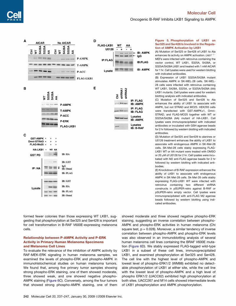
Phosphorylation of Ser325 and Ser428 Is Criticalfor the Regulation of AMPK Activation by LKB1To address the functional consequence of LKB1 phosphoryla-
tion at Ser325 and Ser428, we assessed the ability of LKB1
WT and phosphorylation-deficient mutants to activate endoge-
nous AMPK. As shown in Figure 5A, expression of LKB1
S325A, S428A, or S325A/S428A (AA) double mutants in Lkb1�/�
MEFs resulted in enhanced AMPK phosphorylation compared to
expression of WT LKB1. The difference was particularly
apparent in the absence of AICAR. Enhanced phosphorylation
of ACC was also observed in cells expressing phosphoryla-
Mo
tion-defective mutants of LKB1. These results suggest that
phosphorylation at either Ser325 or Ser428 suppresses the
ability of LKB1 to activate AMPK. Similar results were obtained
when these various constructs were expressed in HeLa cells
(Figure S10), which lack LKB1 (Tiainen et al., 1999).
To confirm that phosphorylation of LKB1 on Ser325 and
Ser428 mediates the inhibition of AMPK activity in human mela-
noma cells containing the oncogenic B-RAF V600E mutation, we
generated SK-MEL-28 cells stably expressing LKB1 wild-type,
S325A, S428A, or the AA double mutants of LKB1. As shown
in Figure 5B, SK-Mel-28 cells expressing the LKB1 AA mutant
showed increased phospho-AMPK compared to cells express-
ing wild-type LKB1. In these cells, expression of LKB1 with single
point mutations had only moderate effects on AMPK phosphor-
ylation, suggesting that phosphorylation of LKB1 at either site
may be insufficient to suppress activity. Similar results were
obtained in UACC67 cells, another B-RAF V600E-containing
melanoma cell line (Figure S11).
The full activity of LKB1 requires its interaction with two regu-
latory subunits, STRAD and MO25 (Hawley et al., 2003). In
addition, LKB1 has been shown to associate with AMPK
(Shaw et al., 2004b). To gain insight into the mechanism under-
lying the effect of LKB1 phosphorylation on AMPK activation,
we compared the ability of LKB1 WT and the phosphoryla-
tion-deficient AA mutant to associate with AMPK, STRAD,
and MO25. As shown in Figure 5C, both HA-tagged WT
LKB1 and the AA mutant bind to Omni-STRAD and FLAG-
MO25 in HEK293 cells. However, interestingly, the LKB1 AA
mutant showed stronger association with GST-AMPK than
with WT LKB1 (Figure 5C). Similarly, more endogenous AMPK
coimmunoprecipitated with FLAG-tagged LKB1 AA mutant
than with WT LKB1 in SK-Mel-28 stable cell lines (Figure 5D).
Moreover, U0126 treatment enhanced the interaction between
AMPK and WT LKB1 but did not have an additional effect on
the AA mutant (Figure 5D). Similarly, knocking down the
expression of B-RAF with two different shRNA constructs
also enhanced the interaction between AMPK and LKB1 in
SK-Mel-28 cells stably expressing LKB1 (Figure 5E). These
results suggest that phosphorylation of LKB1 on Ser325 and
Ser428 suppress its ability to bind to AMPK, thus explaining
the decreased phosphorylation of AMPK.
Expression of the LKB1 Phosphorylation-DeficientMutant Suppresses Proliferation and Anchorage-Independent Growth of Melanoma CellsTo examine the biological effects of LKB1 phosphorylation at
Ser325 and Ser428, we performed cell proliferation assays on
SK-MEL-28 cells stably expressing LKB1 WT or the phosphory-
lation-deficient AA mutant. We found that cells expressing the
AA mutant of LKB1 had significantly reduced proliferation rates
compared to cells expressing WT LKB1, suggesting that phos-
phorylation of LKB1 at Ser325 and Ser428 is important for
cell proliferation in BRAF V600E-expressing melanoma cells
(Figure 6A).
To determine the role of phosphorylation in anchorage-inde-
pendent cell growth, we performed soft agar assays on SK-
MEL-28 cells expressing either WT or the AA mutant of LKB1.
As shown in Figure 6B, cells expressing the LKB1 AA mutant
lecular Cell 33, 237–247, January 30, 2009 ª2009 Elsevier Inc. 241
Molecular Cell
Oncogenic B-RAF Inhibits LKB1 Signaling to AMPK
242 Molecular Cell 33, 237–247, January 30, 2009 ª2009 Elsevier Inc.
Figure 5. Phosphorylation of LKB1 on
Ser325 and Ser428 Is Involved in the Regula-
tion of AMPK Activation by LKB1
(A) Mutation of Ser325 or Ser428 of LKB1 to Ala
enhances its activity on AMPK activation. Lkb1�/�
MEFs were infected with retrovirus containing the
vector control, WT LKB1, S325A, S428A, or
S325A/S428A LKB1 and treated with 1 mM AICAR
for 1 hr. Cell lysates were used for western blotting
with indicated antibodies.
(B) Expression of LKB1 S325A/S428A mutant
stimulates AMPK in SK-MEL-28 cells. SK-MEL-
28 cells were infected with retrovirus containing
WT LKB1, S428A, S325A, or S325A/S428A (AA)
LKB1 mutants. Cell lysates were used for western
blotting analysis with indicated antibodies.
(C) Mutation of Ser325 and Ser428 to Ala
enhances the ability of LKB1 to associate with
AMPK, but not STRAD and MO25. HEK293 cells
were transfected with GST-AMPKa1, Omni-
STRAD, and FLAG-MO25 together with WT or
S325A/S428A (AA) mutant of HA-LKB1. Cell
lysates were immunoprecipitated with indicated
antibodies or incubated with GSH agarose beads
for 2 hr followed by western blotting with indicated
antibodies.
(D) Mutation of Ser325 and Ser428 to alanines or
U0126 treatment enhances the ability of LKB1 to
associate with endogenous AMPK in SK-Mel-28
cells. SK-Mel-28 cells stably expressing FLAG-
LKB1 WT or AA mutant were treated with DMSO
or 20 mM of U0126 for 2 hr. Cell lysates were incu-
bated with M2 anti-FLAG agarose beads for 2 hr
followed by western blotting with indicated anti-
bodies.
(E) Knockdown of B-RAF expression enhances the
ability of LKB1 to associate with endogenous
AMPK in SK-Mel-28 cells. Sk-Mel-28 cells stably
expressing FLAG-LKB1 WT were infected with
retrovirus containing two different shRNA
constructs in pSUPER-retro against B-RAF or
pSUPER-retro empty vector. Cell lysates were
immunoprecipitated with anti-FLAG M2 agarose
beads followed by western blotting using indi-
cated antibodies.
formed fewer colonies than those expressing WT LKB1, sug-
gesting that phosphorylation at Ser325 and Ser428 is important
for cell transformation in B-RAF V600E-expressing melanoma
cells.
Relationship between P-AMPK Activity and P-ERKActivity in Primary Human Melanoma Specimensand Melanoma Cell LinesTo evaluate the relevance of the inhibition of AMPK activity by
RAF-MEK-ERK signaling in human melanoma samples, we
examined the levels of phospho-ERK and phospho-AMPK in
immunohistochemical studies on human melanoma tumors.
We found that, among five primary tumor samples showing
strong phospho-ERK staining, one of them showed moderate,
three showed weak, and one showed negative phospho-
AMPK staining (Figure 6C). Conversely, among the four tumors
that showed strong phospho-AMPK staining, one of them
showed moderate and three showed negative phospho-ERK
staining, suggesting an inverse correlation between phospho-
AMPK and phospho-ERK activities in human melanoma (Chi
square test, p = 0.028). Moreover, a similar tendency of inverse
correlation between phospho-AMPK and phospho-ERK levels
was also observed in an immunoblotting analysis of several
human melanoma cell lines containing the BRAF V600E muta-
tion (Figure 6D). We stably expressed FLAG-tagged wild-type
LKB1 in a subset of these cell lines, immunoprecipitated
LKB1, and examined phosphorylation at Ser325 and Ser428.
The cell line with the highest level of phospho-AMPK and
lowest level of phospho-ERK1/2 (WM88) exhibited no detect-
able phosphorylation of LKB1 at either site, while the cell line
with the lowest level of phospho-AMPK and a high level of
phospho ERK1/2 (UACC62) exhibited high phosphorylation at
both sites. UACC267 and M14 cells showed intermediate levels
of LKB1 phosphorylation and AMPK phosphorylation.

Molecular Cell
Oncogenic B-RAF Inhibits LKB1 Signaling to AMPK
DISCUSSION
The LKB1-AMPK signaling axis plays a central role in bridging
cellular metabolic sensing and regulation of cell growth, prolifer-
ation, and survival (Hardie, 2005; Luo et al., 2005; Motoshima
et al., 2006; Shaw, 2006). The activity of AMPK is regulated by
the ratio of cytosolic AMP to ATP, which affects the phosphory-
lation of Thr172. The steady-state level of phospho-Thr172 is
determined by the relative rate of phosphorylation by kinases
and dephosphorylation by phosphatases. Recent studies
suggest that AMP binding to the gamma subunit of AMPK
decreases the rate of dephosphorylation at Thr172, resulting in
increased AMPK activation (Steinberg et al., 2006). The results
we present here indicate that phosphorylation of LKB1 by
kinases downstream of B-RAF impairs the ability of LKB1 to
associate with and phosphorylate AMPK at Thr172, thereby
blocking the activation of AMPK even under conditions of
elevated AMP. The phosphorylation of LKB1 by ERK and
p90RSK and subsequent inhibition of its ability to phosphorylate
and activate AMPK may partially mediate the oncogenic effects
of BRAF V600E, as expression of a phosphorylation-deficient
mutant of LKB1 (S325A/S428A) inhibited melanoma cell prolifer-
Figure 6. Phosphorylation of LKB1 on Ser325 and
Ser428 Is Critical for Cell Proliferation and
Anchorage-Independent Growth
(A) Expression of LKB1 S325A/S428A (AA) mutant inhibits cell
proliferation. Cell proliferation curves of SK-MEL-28 cells
stably expressing WT LKB1 or AA mutant were measured.
One representative from three independent experiments is
shown.
(B) Expression of LKB1 S325A/S428A (AA) mutant inhibits cell
transformation. SK-MEL-28 cells stably expressing LKB1 WT
LKB1 or AA mutant were used in soft agar assays. Colonies
were photographed after 28 days of growth. Error bars indi-
cate SD. One representative from three independent experi-
ments is shown.
(C) Inverse correlation between phospho-ERK and phospho-
AMPK activities in human melanoma tumor samples. Repre-
sentative images of human melanoma tumor samples stained
with phospho-AMPK or phospho-ERK antibodies in immuno-
histochemical analysis.
(D) Inverse correlation between phospho-ERK and phospho-
AMPK activities in human melanoma cells containing B-RAF
V600E mutation. Total cell lysates from several human mela-
noma cell lines were used in western blotting analysis with
indicated antibodies.
(E) Phosphorylation levels of LKB1 Ser325 and Ser428 in
human melanoma cells containing B-RAF V600E mutation.
Various melanoma cell lines were stably transfected with
pBabe-FLAG-LKB1 WT, and cell lysates were immunoprecip-
itated with anti-FLAG M2 agarose beads followed by western
blotting using indicated antibodies.
ation (Figure 7). This model provides a mechanism
for downregulation of the cellular activity of the
tumor suppressor LKB1 in cancer cells through
posttranslational modification.
LKB1 contains a central serine-threonine kinase
domain, an N-terminal region with a nuclear locali-
zation signal, and a C-terminal regulatory region
(Alessi et al., 2006). Both Ser325 and Ser428 of LKB1 are located
in the C-terminal region. While Ser428 was shown previously to
be phosphorylated by p90Rsk and PKA in response to different
stimuli, there was no evidence that this phosphorylation affected
the in vivo or in vitro function of LKB1 (Sapkota et al., 2001).
Phosphorylation at Ser325 was also previously detected, but
the kinase responsible for this phosphorylation was not deter-
mined (Sapkota et al., 2001). In this report, we show that muta-
tion of Ser325 and Ser428 to Ala enhanced the association of
LKB1 with AMPK but did not alter the association with STRAD
or MO25. This observation is consistent with previous findings
that the C-terminal region of LKB1 is involved in AMPK interac-
tion (Forcet et al., 2005), while the kinase domain mediates the
interaction with STRAD and MO25 (Boudeau et al., 2004). It
remains to be seen whether phosphorylation at these residues
would regulate the interaction between LKB1 and other LKB1
downstream kinases.
Intriguingly, a loss-of-function missense mutation of LKB1 has
been identified at codon 324 (Pro to Leu) in the germline of a PJS
patient and in a sporadic carcinoma (Alessi et al., 2006).
Biochemical studies showed that this mutation impairs LKB1’s
ability to activate AMPK and its downstream signaling (Forcet
Molecular Cell 33, 237–247, January 30, 2009 ª2009 Elsevier Inc. 243

Molecular Cell
Oncogenic B-RAF Inhibits LKB1 Signaling to AMPK
et al., 2005), supporting the idea that regulation of AMPK
signaling is critical to the activity of this LKB1 mutant. Pro324
is adjacent to the Ser325 ERK phosphorylation site, and the
change of Pro to Leu is predicted to make LKB1 a better
substrate for ERK2, based on the phosphorylation motif of
ERK2 (Yaffe et al., 2001), which is consistent with the lower
activity of this LKB1 mutant on AMPK activation (Forcet et al.,
2005). In addition, it is likely that a specific local conformation
in this region is required for binding to AMPK and that phosphor-
ylation of Ser325 or mutation of Pro324 to Leu disrupts the local
structure needed for this interaction.
In this study, we observed an inverse correlation between the
activities of ERK and AMPK in melanoma cells harboring the
B-RAF V600E mutation (Figures 1A and 6). However, some mela-
noma cell lines that lack B-RAF mutations have elevated phos-
pho-ERK without a major suppression of AMPK activation, i.e.,
SK-Mel-31 cells (Figure 1A). Hence, suppression of AMPK acti-
vation correlates better with B-RAF mutations than with total
ERK activation in the melanoma cells. Intriguingly, we found
that LKB1 coimmunoprecipitates with B-RAF V600E mutant,
but not with WT B-RAF, and that expression of B-RAF V600E
dramatically enhanced the association between LKB1 and ERK
(Figures 4H and 4I). These results strongly suggest that in mela-
noma cells that harbor B-RAF mutations activated ERK is more
efficiently channeled to the substrate, LKB1. Consistent with
this concept, previous studies have shown that melanoma with
B-RAF mutation and those without B-RAF mutations are wired
differently in terms of the regulation of RAS-RAF-MEK-ERK
signaling network. For example, melanomas with Ras mutations
have been shown to utilize C-RAF rather than B-RAF to activate
ERK, and the consequence is a disruption of cAMP signaling
specifically in the Ras mutant melanomas (Dumaz et al., 2006).
In addition, although both Ras mutations and B-RAF mutations
activate ERK and cause cell transformation, MEK inhibitors
only inhibit growth of B-RAF mutant cell lines (Solit et al., 2006).
Both LKB1 and AMPK have been shown to suppress cell
growth and proliferation under conditions of energy stress. It’s
conceivable that tumor cells must turn off this signaling pathway
to gain a growth advantage. Mechanisms to suppress LKB1-
Figure 7. A Model for Negative Regulation of LKB1/AMPK Activity by
BRAF V600E Signaling
244 Molecular Cell 33, 237–247, January 30, 2009 ª2009 Elsevier In
AMPK signaling include maintaining high levels of cellular ATP
through upregulation of HIF-1 and subsequent transcriptional
activation of genes involved in glucose uptake and glycolysis-
dependent ATP synthesis (Ashrafian, 2006; Gatenby and Gillies,
2004; Shaw, 2006). A second mechanism involves deletion or
loss-of-function mutations of the LKB1 gene, as occur in PJS
and in non-small-cell lung carcinomas (Sanchez-Cespedes
et al., 2002), and less frequently in malignant melanomas and
colon, breast, ovarian, and brain cancers (Alessi et al., 2006;
Guldberg et al., 1999; Katajisto et al., 2007; Rowan et al.,
1999). Our findings here suggest that phosphorylation of LKB1
represents a way to ‘‘override’’ the energy brake set by LKB1
and AMPK. We show that AMPK is consistently suppressed in
B-RAF V600E-transformed cells through phosphorylation and
inactivation of LKB1, and that downregulation of B-RAF signaling
by pharmacological inhibitors or RNA interference relieved this
inhibition. It would be interesting to study whether similar mech-
anisms of suppressing LKB1-AMPK signaling through post-
translational modifications on LKB1 or AMPK occur in other
cancer cells driven by other mechanisms.
Drugs targeting the RAF-MEK-ERK pathway, such as MEK
and RAF inhibitors, are intensively being developed and under
clinical trails for various human cancers, such as melanoma,
renal cell carcinoma, and colorectal cancer (Beeram et al.,
2005; Gray-Schopfer et al., 2007; Schreck and Rapp, 2006;
Thompson and Lyons, 2005). Meanwhile, several current
prescribed drugs used for metabolic disorders, such as metfor-
min and thiazolidinediones, have been found to activate the
LKB1-AMPK pathway (Hardie, 2007; Shaw, 2006; Shaw et al.,
2005). The molecular linkage between the RAF-MEK-ERK and
LKB1-AMPK signaling pathways reported here has several
implications for the potential uses of these drugs in treating
cancer and metabolic disorders. First, AMPK activators have
been proposed for the use of cancer therapy (Hardie, 2007;
Motoshima et al., 2006). Our findings here suggest that AMPK
activators and MEK/RAF inhibitors may have synergistic effects
on inhibiting proliferation of B-RAF V600E-transformed tumor
cells. Second, our findings suggest that MEK and RAF inhibitors
may be useful in the treatment of PJS by activating LKB1 when
an intact LKB1 allele is present. Recent studies have shown
that haploinsufficiency of LKB1 is sufficient for the formation of
hamartomatous polyps in mouse models, and there is evidence
that a wild-type allele of LKB1 can be found in the harmatoma-
tous polyps from PJS patients (Hernan et al., 2004). Lastly, in
addition to direct AMPK activators, strategies based on modu-
lating posttranslational modifications of LKB1, such as MEK
and RAF inhibitors, that activate AMPK indirectly, could also
be considered for the treatment of metabolic disorders.
In summary, our findings reveal an intriguing molecular linkage
between LKB1-AMPK and RAF-MEK-ERK, two important
protein kinase signaling pathways involved in cancer. In a recent
survey on patterns of somatic mutations in human cancer
genomes (Greenman et al., 2007), BRAF and LKB1/STK11
were listed as the second and third of all the protein kinases
mutated in cancers (in terms of gene-specific selection pres-
sures). Further understanding of the role of this molecular linkage
in tumorigenesis could potentially provide great therapeutic
opportunities for cancer treatment.
c.

Molecular Cell
Oncogenic B-RAF Inhibits LKB1 Signaling to AMPK
EXPERIMENTAL PROCEDURES
Materials
Anti-phospho-AMPK (Thr172), anti-AMPK, anti-phospho-ERK1/2 (Thr202/
Tyr204), anti-ERK, anti-phospho-ACC, and anti-phospho-LKB1 (Ser428) anti-
bodies were purchased from Cell Signaling Technology. Anti-phosopho-LKB1
(Ser325) antibodies were generated at Cell Signaling Technology. Anti-FLAG
M2 affinity gel, anti-FLAG M2 monoclonal antibodies, and U0126 were
purchased from Sigma. Anti-Omni, anti-B-RAF, and anti-CRAF antibodies
were obtained from Santa Cruz Biotechnology. Anti-HA (11) and anti-LKB1
(Ley37D/G6) monoclonal antibodies were purchased from Covance and
Abcam, respectively. AICAR was obtained from Toronto Research Chemicals
(Downsview, ON, Canada). PD98059 was from obtained from Cell Signaling
Technology. LKB1 and AMPK cDNA constructs have been described previ-
ously (Shaw et al., 2004b; Zheng and Cantley, 2007). LKB1 S325A, S428A,
and S325/S428A mutants were generated using PCR mutagenesis and
verified by sequencing. FLAG-ERK2 construct was kindly provided by
Dr. Melanie Cobb.
Cell Culture, Transfection, and Retroviral Infection
SK-Mel-28, UACC62, UACC257, SK-Mel-31, and MeWo Cells were cultured in
RPMI medium (MediaTech) containing 10% fetal bovine serum (FBS) (Gemini
Bio-products). C140 cells stably expressing B-RAF WT or V600E mutant (Kim
et al., 2006) were cultured in RPMI medium containing 10% FBS and 2 mg/ml
doxycycline (Clontech). Immortalized wild-type and LKB1-deficient MEFs
were gifts from Dr. Nabeel Bardeesy (Massachusetts General Hospital) and
cultured in DMEM medium containing 10% FBS. Cos-7 and HeLa cells were
obtained from ATCC and cultured in DMEM medium containing 10% FBS.
For retroviral transfection, amphotropic retrovirus was generated as described
previously (Zheng and Cantley, 2007). When indicated, stable populations
were obtained and maintained by selection with 2 mg/ml puromycin (Sigma).
For B-RAF RNA interference, pSuper-retro containing short hairpin RNAs
against B-RAF (Mu-A and com-4, sh1 and sh2) was obtained from Dr. David
Tuveson and described previously (Hingorani et al., 2003). For MEK1 and
ERK2 knockdown, pSM2C containing shRNAs were gifts from Dr. Stephen
Elledge. For LKB1 interference, pLKO constructs containing shRNAs against
human LKB1 were purchased from Sigma. For drug treatment, cells were
replaced with fresh media before addition of various drugs as indicated.
Western Blotting and Immunoprecipitation
Cell lysates were prepared using lysis buffer containing HEPES (pH 7.0),
150 mM NaCl, 1% NP-40, 1 mM EDTA, 50 mM NaF, 10 mM b-glycero-phos-
phate, 10 nM calyculin A, 1 mM Na3VO4, and protease inhibitors and normal-
ized by protein concentrations using the Bradford method (Bio-Rad). For
western blotting, protein samples were separated on 8%–12% SDS-PAGE
and transferred to PVDF membrane (Millipore). Membranes were blocked in
TBST containing 5% nonfat milk, incubated with primary antibodies according
to the antibody manufacturer’s instructions, followed by incubation with horse-
radish peroxidase-conjugated goat anti-rabbit or anti-mouse IgG (Chemicon)
and enhanced chemiluminescence detection (Perkin Elmer). For immunopre-
cipitation, cell lysates were incubated with primary antibodies or anti-FLAG
M2 affinity gel overnight at 4�C, followed by incubation with protein A/G Se-
pharose for an additional 1 hr at 4�C, when applicable. Beads were washed
three times with lysis buffer and boiled in Laemmli sample buffer, and immune
complexes were analyzed by SDS-PAGE and western blotting. For GST
pulldown, cell lysates were incubated with GSH agarose beads (Pharmacia)
overnight.
Mass Spectrometry
Coomassie-stained SDS-PAGE gel bands containing FLAG-LKB1 isolated
from HEK293 cells with different treatments were excised and subjected to
in-gel digestion and reversed-phase microcapillary LC/MS/MS using a LTQ
2D linear ion trap mass spectrometer (ThermoScientific) in data-dependent
acquisition and positive ion mode. MS/MS spectra were searched against
the concatenated target and decoy (reversed) Swiss-Prot protein database
using Sequest (Proteomics Browser, ThermoScientific) with differential modi-
fications for Ser/Thr/Tyr phosphorylation (+79.97) and the sample processing
Mo
artifacts Met oxidation (+15.99) and Cys alkylation (+57.02). Phosphorylated
and unphosphorylated peptide sequences were identified if they initially
passed the following Sequest scoring thresholds: 1+ ions, Xcorr R 2.0 Sf R
0.4, p R 5; 2+ ions, Xcorr R 2.0, Sf R 0.4, p R 5; 3+ ions, Xcorr R 2.60,
Sf R 0.4, p R 5 against the target protein database. Passing MS/MS spectra
were manually inspected to be sure that all b� and y� fragment ions aligned
with the assigned sequence and modification sites. Determination of the exact
sites of phosphorylation were aided using GraphMod software (Proteomics
Browser). For relative quantification of phosphorylation peptide signal levels,
an isotope-free (label-free) method was used by first integrating the total ion
current (TIC) for each MS/MS sequencing event during a targeted ion MS/
MS (TIMM) experiment or a data-dependant acquisition. For each phosphor-
ylation site (Ser325 and Ser428), a ratio of phosphorylated peptide signal
(TIC of phosphorylated form) to the total peptide signal (TIC of phosphorylated
form + TIC of nonphosphorylated form) from both the PMA- and U0126-
treated samples were calculated according to the following equation:
TICPO4=ðTICPO4 + TICnonPO4Þ = ratio of phosphopeptide signal
The ratios of the Ser325 and Ser428 sites from the PMA-treated samples
were then compared to the same phosphopeptide ratios from both the
untreated and U0126-treated samples according to the following equation:
½ðRPO4U0126=RPO4PMAÞ � 1�3 100 =
% change in phosphorylation level upon treatment
While a direct comparison of phosphopeptide signals between different
experiments is not accurate due to different total protein levels and sample
environments, a comparison of the ratio of the phosphorylated to nonphos-
phorylated peptide forms is an accurate measure of signal level change since
the total peptide signal (modified and unmodified) is measured. The above
calculations were performed manually using Microsoft Excel and with auto-
mated in-house-developed software named Protein Modification Quantifier
v1.0 (Beth Israel Deaconess Medical Center, Boston, MA).
In Vitro MAP Kinase Assay
Recombinant GST-LKB1 (D194A) proteins were expressed, purified from
E. coli, and incubated with recombinant active ERK protein (Stratagene)
in the presence of g-32P-ATP at 37�C for 30 min. For kinase assays using
HA-LKB1, HEK293 cells were transfected with HA-LKB1 WT or S325A
constructs, and immunoprecipitated HA-LKB1 proteins were used in the
kinase assays.
Cell Proliferation and Soft Agar Assays
For cell proliferation assays, 2 3 105 cells were seeded in triplicate on a 6-well
plate and grown in full medium. At 24 hr intervals for 4 days, cells were trypsi-
nized and the numbers of cells in each well were counted by a Coulter counter.
For soft agar assays, cells were suspended in 0.3% agarose in complete
medium and plated on a layer of 0.6% agarose in complete medium in
6-well culture plates (3 3 104 cells/well). After 3 weeks, the colonies were
stained with iodonitrotetrazolium chloride (Sigma) and photographed. The
numbers of colonies were counted using NIH image software.
Immunohistochemical Analysis
Human melanoma tissue microarray was obtained from US Biomax (Rockville
MD). Formalin-fixed paraffin-embedded tissue microarray was deparaffinized
in xylenes and hydrated in a graded series of alcohols. Heat-induced epitope
antigen retrieval using citrate buffer in a pressure cooker was used. Staining
was performed using DAKO LSAB+ Alkaline Phosphatase detection system
and Permanent Red as the chromogen. The results were evaluated by pathol-
ogist S.R.G. The immunoreactivity of phospho-ERK and phospho-AMPK
was scored on a scale from 0 to 3+ for negative, weak, moderate, and strong
staining.
SUPPLEMENTAL DATA
TheSupplementalData include13figuresandcanbefoundwith thisarticle online
at http://www.cell.com/molecular-cell/supplemental/S1097-2765(09)00002-1.
lecular Cell 33, 237–247, January 30, 2009 ª2009 Elsevier Inc. 245

Molecular Cell
Oncogenic B-RAF Inhibits LKB1 Signaling to AMPK
ACKNOWLEDGMENTS
We thank Drs. Reuben Shaw, Nabeel Bardeesy, David Tuveson, and Melanie
Cobb for reagents; Lisa Freimark for assistance with the mass spectrometry
analysis; Li Zhang and Szexian Lee for technical assistance; and Dr. Yang-
Qing Xu and members of the Cantley Lab for helpful discussion. We also would
like to thank Shailender Nagpal for helping to create software for quantitative
mass spectrometry analysis. B.Z. is supported by a Charles A. King Trust post-
doctoral fellowship from the Charles A. King Trust, Bank of America, Co-
Trustee. This work is supported by National Institutes of Health Grant
GM56203 and CA102694 to L.C.C., K99CA133245 to B.Z., and UO1
CA84313 and RO1 CA93947 to L.C.
Received: October 9, 2007
Revised: August 28, 2008
Accepted: December 30, 2008
Published: January 29, 2009
REFERENCES
Alessi, D.R., Sakamoto, K., and Bayascas, J.R. (2006). LKB1-dependent
signaling pathways. Annu. Rev. Biochem. 75, 137–163.
Asara, J.M., Christofk, H.R., Freimark, L.M., and Cantley, L.C. (2008). A label-
free quantification method by MS/MS TIC compared to SILAC and spectral
counting in a proteomics screen. Proteomics 8, 994–999.
Ashrafian, H. (2006). Cancer’s sweet tooth: the Janus effect of glucose metab-
olism in tumorigenesis. Lancet 367, 618–621.
Baas, A.F., Kuipers, J., van der Wel, N.N., Batlle, E., Koerten, H.K., Peters, P.J.,
and Clevers, H.C. (2004). Complete polarization of single intestinal epithelial
cells upon activation of LKB1 by STRAD. Cell 116, 457–466.
Bardeesy, N., Sinha, M., Hezel, A.F., Signoretti, S., Hathaway, N.A., Sharpless,
N.E., Loda, M., Carrasco, D.R., and DePinho, R.A. (2002). Loss of the Lkb1
tumour suppressor provokes intestinal polyposis but resistance to transforma-
tion. Nature 419, 162–167.
Beeram, M., Patnaik, A., and Rowinsky, E.K. (2005). Raf: a strategic target for
therapeutic development against cancer. J. Clin. Oncol. 23, 6771–6790.
Boudeau, J., Scott, J.W., Resta, N., Deak, M., Kieloch, A., Komander, D., Har-
die, D.G., Prescott, A.R., van Aalten, D.M., and Alessi, D.R. (2004). Analysis of
the LKB1-STRAD-MO25 complex. J. Cell Sci. 117, 6365–6375.
Carling, D. (2004). The AMP-activated protein kinase cascade—a unifying
system for energy control. Trends Biochem. Sci. 29, 18–24.
Chong, H., Vikis, H.G., and Guan, K.L. (2003). Mechanisms of regulating the
Raf kinase family. Cell. Signal. 15, 463–469.
Davies, H., Bignell, G.R., Cox, C., Stephens, P., Edkins, S., Clegg, S., Teague,
J., Woffendin, H., Garnett, M.J., Bottomley, W., et al. (2002). Mutations of the
BRAF gene in human cancer. Nature 417, 949–954.
Dhomen, N., and Marais, R. (2007). New insight into BRAF mutations in cancer.
Curr. Opin. Genet. Dev. 17, 31–39.
Dumaz, N., Hayward, R., Martin, J., Ogilvie, L., Hedley, D., Curtin, J.A., Bas-
tian, B.C., Springer, C., and Marais, R. (2006). In melanoma, RAS mutations
are accompanied by switching signaling from BRAF to CRAF and disrupted
cyclic AMP signaling. Cancer Res. 66, 9483–9491.
Forcet, C., Etienne-Manneville, S., Gaude, H., Fournier, L., Debilly, S., Salmi,
M., Baas, A., Olschwang, S., Clevers, H., and Billaud, M. (2005). Functional
analysis of Peutz-Jeghers mutations reveals that the LKB1 C-terminal region
exerts a crucial role in regulating both the AMPK pathway and the cell polarity.
Hum. Mol. Genet. 14, 1283–1292.
Garnett, M.J., and Marais, R. (2004). Guilty as charged: B-RAF is a human
oncogene. Cancer Cell 6, 313–319.
Gatenby, R.A., and Gillies, R.J. (2004). Why do cancers have high aerobic
glycolysis? Nat. Rev. Cancer 4, 891–899.
Gray-Schopfer, V.C., da Rocha Dias, S., and Marais, R. (2005). The role of
B-RAF in melanoma. Cancer Metastasis Rev. 24, 165–183.
246 Molecular Cell 33, 237–247, January 30, 2009 ª2009 Elsevier In
Gray-Schopfer, V., Wellbrock, C., and Marais, R. (2007). Melanoma biology
and new targeted therapy. Nature 445, 851–857.
Greenman, C., Stephens, P., Smith, R., Dalgliesh, G.L., Hunter, C., Bignell, G.,
Davies, H., Teague, J., Butler, A., Stevens, C., et al. (2007). Patterns of somatic
mutation in human cancer genomes. Nature 446, 153–158.
Guldberg, P., thor Straten, P., Ahrenkiel, V., Seremet, T., Kirkin, A.F., and
Zeuthen, J. (1999). Somatic mutation of the Peutz-Jeghers syndrome gene,
LKB1/STK11, in malignant melanoma. Oncogene 18, 1777–1780.
Gwinn, D.M., Shackelford, D.B., Egan, D.F., Mihaylova, M.M., Mery, A., Vas-
quez, D.S., Turk, B.E., and Shaw, R.J. (2008). AMPK phosphorylation of raptor
mediates a metabolic checkpoint. Mol. Cell 30, 214–226.
Hardie, D.G. (2005). New roles for the LKB1/AMPK pathway. Curr. Opin. Cell
Biol. 17, 167–173.
Hardie, D.G. (2007). AMP-activated protein kinase as a drug target. Annu. Rev.
Pharmacol. Toxicol. 47, 185–210.
Hawley, S.A., Boudeau, J., Reid, J.L., Mustard, K.J., Udd, L., Makela, T.P.,
Alessi, D.R., and Hardie, D.G. (2003). Complexes between the LKB1 tumor
suppressor, STRAD alpha/beta and MO25 alpha/beta are upstream kinases
in the AMP-activated protein kinase cascade. J. Biol. 2, 28.
Hernan, I., Roig, I., Martin, B., Gamundi, M.J., Martinez-Gimeno, M., and Car-
ballo, M. (2004). De novo germline mutation in the serine-threonine kinase
STK11/LKB1 gene associated with Peutz-Jeghers syndrome. Clin. Genet.
66, 58–62.
Hingorani, S.R., Jacobetz, M.A., Robertson, G.P., Herlyn, M., and Tuveson,
D.A. (2003). Suppression of BRAF(V599E) in human melanoma abrogates
transformation. Cancer Res. 63, 5198–5202.
Ikenoue, T., Hikiba, Y., Kanai, F., Tanaka, Y., Imamura, J., Imamura, T., Ohta,
M., Ijichi, H., Tateishi, K., Kawakami, T., et al. (2003). Functional analysis of
mutations within the kinase activation segment of B-Raf in human colorectal
tumors. Cancer Res. 63, 8132–8137.
Inoki, K., Zhu, T., and Guan, K.L. (2003). TSC2 mediates cellular energy
response to control cell growth and survival. Cell 115, 577–590.
Ji, H., Ramsey, M.R., Hayes, D.N., Fan, C., McNamara, K., Kozlowski, P., Tor-
rice, C., Wu, M.C., Shimamura, T., Perera, S.A., et al. (2007). LKB1 modulates
lung cancer differentiation and metastasis. Nature 448, 807–810.
Jones, R.G., Plas, D.R., Kubek, S., Buzzai, M., Mu, J., Xu, Y., Birnbaum, M.J.,
and Thompson, C.B. (2005). AMP-activated protein kinase induces a p53-
dependent metabolic checkpoint. Mol. Cell 18, 283–293.
Kahn, B.B., Alquier, T., Carling, D., and Hardie, D.G. (2005). AMP-activated
protein kinase: ancient energy gauge provides clues to modern understanding
of metabolism. Cell Metab. 1, 15–25.
Katajisto, P., Vallenius, T., Vaahtomeri, K., Ekman, N., Udd, L., Tiainen, M., and
Makela, T.P. (2007). The LKB1 tumor suppressor kinase in human disease.
Biochim. Biophys. Acta 1775, 63–75.
Kim, M., Gans, J.D., Nogueira, C., Wang, A., Paik, J.H., Feng, B., Brennan, C.,
Hahn, W.C., Cordon-Cardo, C., Wagner, S.N., et al. (2006). Comparative onco-
genomics identifies NEDD9 as a melanoma metastasis gene. Cell 125,
1269–1281.
Lizcano, J.M., Goransson, O., Toth, R., Deak, M., Morrice, N.A., Boudeau, J.,
Hawley, S.A., Udd, L., Makela, T.P., Hardie, D.G., and Alessi, D.R. (2004).
LKB1 is a master kinase that activates 13 kinases of the AMPK subfamily,
including MARK/PAR-1. EMBO J. 23, 833–843.
Luo, Z., Saha, A.K., Xiang, X., and Ruderman, N.B. (2005). AMPK, the meta-
bolic syndrome and cancer. Trends Pharmacol. Sci. 26, 69–76.
Martin, S.G., and St Johnston, D. (2003). A role for Drosophila LKB1 in anterior-
posterior axis formation and epithelial polarity. Nature 421, 379–384.
Motoshima, H., Goldstein, B.J., Igata, M., and Araki, E. (2006). AMPK and cell
proliferation—AMPK as a therapeutic target for atherosclerosis and cancer.
J. Physiol. 574, 63–71.
Rattan, R., Giri, S., Singh, A.K., and Singh, I. (2005). 5-Aminoimidazole-4-car-
boxamide-1-beta-D-ribofuranoside inhibits cancer cell proliferation in vitro
c.
Molecular Cell
Oncogenic B-RAF Inhibits LKB1 Signaling to AMPK
and in vivo via AMP-activated protein kinase. J. Biol. Chem. 280, 39582–
39593.
Rowan, A., Bataille, V., MacKie, R., Healy, E., Bicknell, D., Bodmer, W., and
Tomlinson, I. (1999). Somatic mutations in the Peutz-Jeghers (LKB1/STK11)
gene in sporadic malignant melanomas. J. Invest. Dermatol. 112, 509–511.
Sanchez-Cespedes, M., Parrella, P., Esteller, M., Nomoto, S., Trink, B., Eng-
les, J.M., Westra, W.H., Herman, J.G., and Sidransky, D. (2002). Inactivation
of LKB1/STK11 is a common event in adenocarcinomas of the lung. Cancer
Res. 62, 3659–3662.
Sapkota, G.P., Kieloch, A., Lizcano, J.M., Lain, S., Arthur, J.S., Williams, M.R.,
Morrice, N., Deak, M., and Alessi, D.R. (2001). Phosphorylation of the protein
kinase mutated in Peutz-Jeghers cancer syndrome, LKB1/STK11, at Ser431
by p90(RSK) and cAMP-dependent protein kinase, but not its farnesylation
at Cys(433), is essential for LKB1 to suppress cell growth. J. Biol. Chem.
276, 19469–19482.
Sapkota, G.P., Boudeau, J., Deak, M., Kieloch, A., Morrice, N., and Alessi, D.R.
(2002a). Identification and characterization of four novel phosphorylation sites
(Ser31, Ser325, Thr336 and Thr366) on LKB1/STK11, the protein kinase
mutated in Peutz-Jeghers cancer syndrome. Biochem. J. 362, 481–490.
Sapkota, G.P., Deak, M., Kieloch, A., Morrice, N., Goodarzi, A.A., Smythe, C.,
Shiloh, Y., Lees-Miller, S.P., and Alessi, D.R. (2002b). Ionizing radiation
induces ataxia telangiectasia mutated kinase (ATM)-mediated phosphoryla-
tion of LKB1/STK11 at Thr-366. Biochem. J. 368, 507–516.
Schreck, R., and Rapp, U.R. (2006). Raf kinases: oncogenesis and drug
discovery. Int. J. Cancer 119, 2261–2271.
Schubbert, S., Bollag, G., and Shannon, K. (2007). Deregulated Ras signaling
in developmental disorders: new tricks for an old dog. Curr. Opin. Genet. Dev.
17, 15–22.
Shaw, R.J. (2006). Glucose metabolism and cancer. Curr. Opin. Cell Biol. 18,
598–608.
Shaw, R.J., Bardeesy, N., Manning, B.D., Lopez, L., Kosmatka, M., DePinho,
R.A., and Cantley, L.C. (2004a). The LKB1 tumor suppressor negatively regu-
lates mTOR signaling. Cancer Cell 6, 91–99.
Shaw, R.J., Kosmatka, M., Bardeesy, N., Hurley, R.L., Witters, L.A., DePinho,
R.A., and Cantley, L.C. (2004b). The tumor suppressor LKB1 kinase directly
activates AMP-activated kinase and regulates apoptosis in response to energy
stress. Proc. Natl. Acad. Sci. USA 101, 3329–3335.
Shaw, R.J., Lamia, K.A., Vasquez, D., Koo, S.H., Bardeesy, N., Depinho, R.A.,
Montminy, M., and Cantley, L.C. (2005). The kinase LKB1 mediates glucose
homeostasis in liver and therapeutic effects of metformin. Science 310,
1642–1646.
Mo
Solit, D.B., Garraway, L.A., Pratilas, C.A., Sawai, A., Getz, G., Basso, A., Ye,
Q., Lobo, J.M., She, Y., Osman, I., et al. (2006). BRAF mutation predicts sensi-
tivity to MEK inhibition. Nature 439, 358–362.
Steinberg, G.R., Michell, B.J., van Denderen, B.J., Watt, M.J., Carey, A.L.,
Fam, B.C., Andrikopoulos, S., Proietto, J., Gorgun, C.Z., Carling, D., et al.
(2006). Tumor necrosis factor alpha-induced skeletal muscle insulin resistance
involves suppression of AMP-kinase signaling. Cell Metab. 4, 465–474.
Thompson, N., and Lyons, J. (2005). Recent progress in targeting the
Raf/MEK/ERK pathway with inhibitors in cancer drug discovery. Curr. Opin.
Pharmacol. 5, 350–356.
Tiainen, M., Ylikorkala, A., and Makela, T.P. (1999). Growth suppression by
Lkb1 is mediated by a G(1) cell cycle arrest. Proc. Natl. Acad. Sci. USA 96,
9248–9251.
Tsay, Y.G., Wang, Y.H., Chiu, C.M., Shen, B.J., and Lee, S.C. (2000). A
strategy for identification and quantitation of phosphopeptides by liquid chro-
matography/tandem mass spectrometry. Anal. Biochem. 287, 55–64.
Tuveson, D.A., Weber, B.L., and Herlyn, M. (2003). BRAF as a potential thera-
peutic target in melanoma and other malignancies. Cancer Cell 4, 95–98.
Wan, P.T., Garnett, M.J., Roe, S.M., Lee, S., Niculescu-Duvaz, D., Good, V.M.,
Jones, C.M., Marshall, C.J., Springer, C.J., Barford, D., and Marais, R. (2004).
Mechanism of activation of the RAF-ERK signaling pathway by oncogenic
mutations of B-RAF. Cell 116, 855–867.
Watts, J.L., Morton, D.G., Bestman, J., and Kemphues, K.J. (2000). The
C. elegans par-4 gene encodes a putative serine-threonine kinase required
for establishing embryonic asymmetry. Development 127, 1467–1475.
Wellbrock, C., Karasarides, M., and Marais, R. (2004). The RAF proteins take
centre stage. Nat. Rev. Mol. Cell Biol. 5, 875–885.
Woods, A., Johnstone, S.R., Dickerson, K., Leiper, F.C., Fryer, L.G., Neumann,
D., Schlattner, U., Wallimann, T., Carlson, M., and Carling, D. (2003). LKB1 is
the upstream kinase in the AMP-activated protein kinase cascade. Curr. Biol.
13, 2004–2008.
Xiang, X., Saha, A.K., Wen, R., Ruderman, N.B., and Luo, Z. (2004). AMP-acti-
vated protein kinase activators can inhibit the growth of prostate cancer cells
by multiple mechanisms. Biochem. Biophys. Res. Commun. 321, 161–167.
Yaffe, M.B., Leparc, G.G., Lai, J., Obata, T., Volinia, S., and Cantley, L.C.
(2001). A motif-based profile scanning approach for genome-wide prediction
of signaling pathways. Nat. Biotechnol. 19, 348–353.
Zheng, B., and Cantley, L.C. (2007). Regulation of epithelial tight junction
assembly and disassembly by AMP-activated protein kinase. Proc. Natl.
Acad. Sci. USA 104, 819–822.
lecular Cell 33, 237–247, January 30, 2009 ª2009 Elsevier Inc. 247


![Review Article Regulation of the Ras-MAPK and PI3K-mTOR ... · Cytosolic kinase SK Tumor suppressor/oncogenic isoforms, activates/inhibits mTORC. Breast, lung [ , ] Cytosolic kinase](https://static.cupdf.com/doc/110x72/6080c0d51308b03b786a8817/review-article-regulation-of-the-ras-mapk-and-pi3k-mtor-cytosolic-kinase-sk.jpg)


![Radiofrequency Ablation and Excision of Multiple Cutaneous … · 2017-06-19 · of neurofibromin as a tumor suppressor, possibly through modu-lation of the oncogenic pathways [2,10,11].](https://static.cupdf.com/doc/110x72/5e607bfb21da97009701a99f/radiofrequency-ablation-and-excision-of-multiple-cutaneous-2017-06-19-of-neurofibromin.jpg)