©Ken L Schreibman, PhD/MD 11/10/08 www.schreibman.info
page 1 of 18Easily Missed Fracturesof the Ankle & Foot
©Ken L Schreibman, PhD/MD 2008 schreibman.info
Easily Missed
Fractures
Foot & Ankle
©Ken L Schreibman, PhD/MD 2008 schreibman.info
Easily Missed FracturesSearch for on EVERY Ankle view:
1) MM & LM (Weber)2) OLT (OCD)3) 5th MT (Jones, Avulsion)4) LPT
Search for on EVERY Foot view:5) APC6) MT7) Lisfranc
Previous
Talk
This
Talk Anatomy: Tarsal
Bones & Joints
but first…
©Ken L Schreibman, PhD/MD 2008 schreibman.info
Anatomy: Tarsal Bones & JointsTalusLatin: “Ankle”Center of Ankle Joint
©Ken L Schreibman, PhD/MD 2008 schreibman.info
Anatomy: Tarsal Bones & JointsTalus Dome
Head
Body
©Ken L Schreibman, PhD/MD 2008 schreibman.info
Anatomy: Tarsal Bones & Joints
DomeAnkle Joint
Plafond
pla·fond\plà-fōn\ n [fr. plat flat + fond bottom]
ceiling formed by the underside of a floor
Mortise
Anatomy: Tarsal Bones & Joints
©Ken L Schreibman, PhD/MD 2008 schreibman.info
Mortise: Woodworking Term
MORTISE
TENON

©Ken L Schreibman, PhD/MD 11/10/08 www.schreibman.info
page 2 of 18Easily Missed Fracturesof the Ankle & Foot
©Ken L Schreibman, PhD/MD 2008 schreibman.info
Anatomy: Tarsal Bones & JointsTalus Tibia
Navicular
Calcaneus
©Ken L Schreibman, PhD/MD 2008 schreibman.info
Anatomy: Tarsal Bones & JointsTalo-Navicular JointNavicular sits on Head like a hat
Headof
TalusHead
of Talus
NavicularN
avicular
Talo-Navicularsubluxation Posterior Tibial Tendon(PTT) Disfunction
©Ken L Schreibman, PhD/MD 2008 schreibman.info
Anatomy: Tarsal Bones & Joints
Talus
Calcaneus
Sub-TalarJoint
©Ken L Schreibman, PhD/MD 2008 schreibman.info
Anatomy: Tarsal Bones & JointsSub-Talar
Joint3 Facets Middle
Posterior
Anterior
Sustentaculum Tali
* Latin: “a supporting structure”
*
©Ken L Schreibman, PhD/MD 2008 schreibman.info
Anatomy: Tarsal Bones & JointsSub-Talar
Joint
©Ken L Schreibman, PhD/MD 2008 schreibman.info
Anatomy: Tarsal Bones & JointsCalcaneus
Talus
Cuboid

©Ken L Schreibman, PhD/MD 11/10/08 www.schreibman.info
page 3 of 18Easily Missed Fracturesof the Ankle & Foot
©Ken L Schreibman, PhD/MD 2008 schreibman.info
Anatomy: Tarsal Bones & JointsCalcaneo-Cuboid Joint
Cuboid Calcaneus
©Ken L Schreibman, PhD/MD 2008 schreibman.info
Anatomy: Tarsal Bones & JointsChopart Joint
Cuboid
LateralProcess
AnteriorProcessCalcaneus
Talus
©Ken L Schreibman, PhD/MD 2008 schreibman.info
Anatomy: Tarsal Bones & Joints
Navicular Cuboid
TalusThe NavicularDOES NOT
articulate withthe Calcaneus
©Ken L Schreibman, PhD/MD 2008 schreibman.info
Anatomy: Tarsal Bones & Joints
Navicular Cuboid
Talus
Navicular
1 23
12 3
III
III IV V
Cuboid
IV
V
©Ken L Schreibman, PhD/MD 2008 schreibman.info
Anatomic Divisions
N Cu
CaTa
1 2 3
META-TARSALS
TARSALS
FORE-FOOT
MID-FOOT
HIND-FOOT
LISFRANC
CHOPART
©Ken L Schreibman, PhD/MD 2008 schreibman.info
Anatomy: 3D

©Ken L Schreibman, PhD/MD 11/10/08 www.schreibman.info
page 4 of 18Easily Missed Fracturesof the Ankle & Foot
©Ken L Schreibman, PhD/MD 2008 schreibman.info
Anatomy: Cross-SectionalCT: Axial Plane
Ti
Fi
Syndesmosis
©Ken L Schreibman, PhD/MD 2008 schreibman.info
MM
LM
Ta
Anatomy: Cross-SectionalCT: Axial Plane
©Ken L Schreibman, PhD/MD 2008 schreibman.info
Ta
N
TNJ
P-STJM-STJ
Anatomy: Cross-SectionalCT: Axial Plane
©Ken L Schreibman, PhD/MD 2008 schreibman.info
Anatomy: Cross-SectionalCT: Axial Plane
1
N
Ca
23
Cu
©Ken L Schreibman, PhD/MD 2008 schreibman.info
Ca
Cu
Anatomy: Cross-SectionalCT: Axial Plane
CCJ
©Ken L Schreibman, PhD/MD 2008 schreibman.info
Anatomy: Cross-SectionalCT: Axial Plane
GOOD FOR:SyndesmosisTalonavicular JointCalcaneocuboid JointNavicular-cuneiform JointsTarsal-metatarsal Joints
NOT GOOD FOR:Ankle JointSubtalar Joint
©Ken L Schreibman, PhD/MD 11/10/08 www.schreibman.info
page 5 of 18Easily Missed Fracturesof the Ankle & Foot
©Ken L Schreibman, PhD/MD 2008 schreibman.info
Anatomy: Cross-SectionalCT: Sagittal PlaneReformatted off Axial reference image
Medial Slice
ST
M-STJ
Ta
N1
I
TNJTi
©Ken L Schreibman, PhD/MD 2008 schreibman.info
Anatomy: Cross-SectionalCT: Sagittal PlaneReformatted off Axial reference image
Middle Slice
Ta
N
TNJ
M-STJ
P-STJ
Ca
Ti
©Ken L Schreibman, PhD/MD 2008 schreibman.info
Anatomy: Cross-SectionalCT: Sagittal PlaneReformatted off Axial reference image
Lateral Slice
Ta
Ca
TiP-STJ
Cu
CCJ
AJ
LPTAPC
©Ken L Schreibman, PhD/MD 2008 schreibman.info
Anatomy: Cross-SectionalCT: Sagittal Plane
SECONDARY PLANE FOR:Ankle JointSub-Talar JointTalo-Navicular JointCalcaneo-Cuboid JointNavicular-Cuneiform JointsTarsal-Metatarsal Joints
NOT GOOD FOR:Syndesmosis
©Ken L Schreibman, PhD/MD 2008 schreibman.info
Anatomy: Cross-SectionalCT: Coronal Plane (2 schemes)1) Mortise Coronal: Reformatted off AxialAligned between MalleoliGOOD FOR:Distal Tibial FracturesMalleoliTillaux, TriplanePilon
Talar Dome Fxs (OLT)
(Mortise Sagittal, perpendicular to this)
MM
LM
©Ken L Schreibman, PhD/MD 2008 schreibman.info
Anatomy: Cross-SectionalCT: Coronal Plane (2 schemes)2) Oblique Coronal: Reformat off SagittalPerpendicular to P-STJGOOD FOR:Hindfoot FxsSub-Talar JointTalusCalcaneus
TarsalCoalitions

©Ken L Schreibman, PhD/MD 11/10/08 www.schreibman.info
page 6 of 18Easily Missed Fracturesof the Ankle & Foot
©Ken L Schreibman, PhD/MD 2008 schreibman.info
UW 2-Page CT Protocol Sheets
www.radiology.wisc.edu ©Ken L Schreibman, PhD/MD 2008 schreibman.info
Anatomy: Cross-SectionalCT: Oblique Coronal PlaneSagittal ref image perpendic to P-STJ
Ta
Ca
Ti
FiAJ
P-STJ
©Ken L Schreibman, PhD/MD 2008 schreibman.info
Anatomy: Cross-SectionalCT: Oblique Coronal PlaneSagittal ref image perpendic to P-STJ
P-STJ
©Ken L Schreibman, PhD/MD 2008 schreibman.info
Anatomy: Cross-SectionalCT: Oblique Coronal PlaneSagittal ref image perpendic to P-STJ
M-STJST
©Ken L Schreibman, PhD/MD 2008 schreibman.info
Anatomy: Cross-SectionalCT: Oblique Coronal PlaneSagittal ref image perpendic to P-STJ
A-STJ
©Ken L Schreibman, PhD/MD 2008 schreibman.info
Anatomy: Cross-SectionalCT: Oblique Coronal Plane
GOOD FOR:Sub-Talar Joint Ankle Joint
NOT GOOD FOR:Talo-Navicular JointCalcaneo-Cuboid JointNavicular-Cuneiform JointsTarsal-Metatarsal Joints

©Ken L Schreibman, PhD/MD 11/10/08 www.schreibman.info
page 7 of 18Easily Missed Fracturesof the Ankle & Foot
©Ken L Schreibman, PhD/MD 2008 schreibman.info
Easily Missed FracturesSearch for on EVERY Ankle view:
1) MM & LM (Weber, Adolescent)2) OLT (OCD)3) 5th MT (Jones & Avulsion)4) LPT
©Ken L Schreibman, PhD/MD 2008 schreibman.info
Lateral Process Talus
©Ken L Schreibman, PhD/MD 2008 schreibman.info
Lateral Process Talus Fracture17 yo F gymnast,
landed wrongafter vault
Lateral View
©Ken L Schreibman, PhD/MD 2008 schreibman.info
Lateral Process Talus Fracture29 yo F,s/p MVA
Lateral View
©Ken L Schreibman, PhD/MD 2008 schreibman.info
Lateral Process Talus Fracture40 yo M, s/pmotorcycleaccidentLateral View
APView
©Ken L Schreibman, PhD/MD 2008 schreibman.info
Lateral Process Talus Fracture24 yo M, construction
worker fell 15 feet
Lateral View
AP View

©Ken L Schreibman, PhD/MD 11/10/08 www.schreibman.info
page 8 of 18Easily Missed Fracturesof the Ankle & Foot
©Ken L Schreibman, PhD/MD 2006 schreibman.info
Fracture of the lateral process of thetalus is a common, yet frequentlymissed, injury. It is the second mostcommon fracture of the talar body,accounting for 24% of these injuries,yet it has been documented that 40% offractures of the lateral process of thetalus are missed at initial presentation.
Falling75%
Twisting12%
Collisionwith Tree
8%
Gettingoff Lift
4%
Collisionwith Skier
1%Gettingon Lift
1%
Mechanism of foot/ankle injuries related to snowboarding
The Snowboarder’s Foot and AnkleKirkpatrick, et al Am J Sports Med 1998: 26 271-277
Ankle Injuries 5-6% of all alpine skiing injuries
12-38% of all snowboarding injuries
LPT Fractures
15% of snowboarding ankle injuries
2% of all snowboarding injuries
The Snowboarder’s Foot and AnkleKirkpatrick, et al Am J Sports Med 1998: 26 271-277 ©Ken L Schreibman, PhD/MD 2008 schreibman.info
Lateral Process Talus Fracture29 yo snow boarder
CT: Axial
Courtesy of Paul Hsieh, Colorado
CT: SagittalCT: Coronal
©Ken L Schreibman, PhD/MD 11/10/08 www.schreibman.info
page 9 of 18Easily Missed Fracturesof the Ankle & Foot
©Ken L Schreibman, PhD/MD 2008 schreibman.info
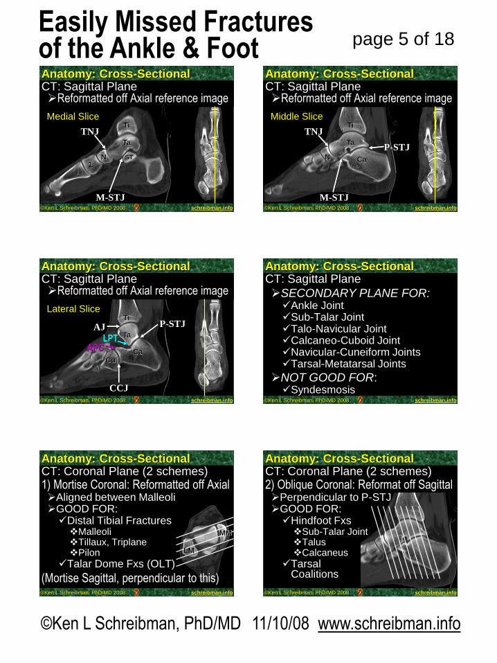
Easily Missed FracturesSearch for on EVERY Ankle view:
1) MM & LM (Weber)2) OLT (OCD)3) 5th MT (Jones, Avulsion)4) LPT
Search for on EVERY Foot view:5) APC
©Ken L Schreibman, PhD/MD 2008 schreibman.info
Anterior Process Calcaneus
©Ken L Schreibman, PhD/MD 2008 schreibman.info
Anterior Process Calcaneus Fx
©Ken L Schreibman, PhD/MD 2008 schreibman.info
APC FxAPC Fx: Search for on every foot viewAPObl29 yo F, tripped down some steps
6 months later…
©Ken L Schreibman, PhD/MD 2008 schreibman.info
APC Fx: Best seen on CT
RIGHT LEFT
Acute margins
RIGHT LEFT
29 yo F, tripped down some steps
CT day of injury
Sclerotic margins
CT 6-months after injury
©Ken L Schreibman, PhD/MD 2008 schreibman.info
APC Fx: May require fixation29 yo F, tripped down some steps
9-months post-surgery

©Ken L Schreibman, PhD/MD 11/10/08 www.schreibman.info
page 10 of 18Easily Missed Fracturesof the Ankle & Foot
©Ken L Schreibman, PhD/MD 2008 schreibman.info
Elongated APCUnlike the normal triangular APC.
Elongated APC has blunt tiplike an anteater’s snout.
Elongated APC: “Anteater sign”
©Ken L Schreibman, PhD/MD 2008 schreibman.info
Elongated APC: “Anteater sign”1) More easily fractured
Even from minor trauma
51 yo M, Jumping with child
©Ken L Schreibman, PhD/MD 2008 schreibman.info
Elongated APC: “Anteater sign”
Sagittal CT
Axial CT
51 yo M, Jumping with child
2) Tarsal Coalitioni) Calcaneo-Navicular
©Ken L Schreibman, PhD/MD 2008 schreibman.info
Tarsal CoalitionsCause of foot pain in adolescent
“Rigid (peroneal) flat foot”
Abnormal hindfoot biomechanics “Talar Beak”, seen on lateral
12 yo M
©Ken L Schreibman, PhD/MD 2008 schreibman.info
Tarsal Coalitions
12 yo M
Occur at 2 sitesi) Calcaneo-Navicular
Can be seen on oblique view
C
N
©Ken L Schreibman, PhD/MD 2008 schreibman.info
Tarsal CoalitionsOccur at 2 sites
ii)Middle Facet Sub-Talar JointBest seen on Oblique Coronal CT
29 yo F, long h/o Bilateral foot pain, worse on RightRIGHT LEFT
Non-osseousCoalition
OsseousCoalition
©Ken L Schreibman, PhD/MD 11/10/08 www.schreibman.info
page 11 of 18Easily Missed Fracturesof the Ankle & Foot
©Ken L Schreibman, PhD/MD 2008 schreibman.info
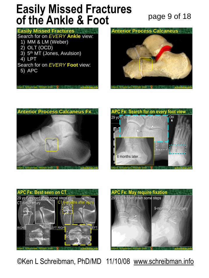
APC: Fx vs Os Calcaneus Secondarius
©Ken L Schreibman, PhD/MD 2008 schreibman.info
APC: Fx vs Os Calcaneus Secondarius
©Ken L Schreibman, PhD/MD 2008 schreibman.info
APC: Fx vs Os Calcaneus Secondarius
©Ken L Schreibman, PhD/MD 2008 schreibman.info
APC: Fx vs Os Calcaneus Secondarius
©Ken L Schreibman, PhD/MD 2008 schreibman.info
APC: Fx vs Os Calcaneus Secondarius
APC
N
©Ken L Schreibman, PhD/MD 2008 schreibman.info
APC: Fx vs Os Calcaneus Secondarius
LEFTRIGHT
OCS
11 yo F with R foot pain

©Ken L Schreibman, PhD/MD 11/10/08 www.schreibman.info
page 12 of 18Easily Missed Fracturesof the Ankle & Foot
©Ken L Schreibman, PhD/MD 2008 schreibman.info
Easily Missed FracturesSearch for on EVERY Ankle view:
1) MM & LM (Weber, Adolescent)2) OLT (OCD)3) 5th MT (Jones & Avulsion)4) LPT
Search for on EVERY Foot view:5) APC6) MT
©Ken L Schreibman, PhD/MD 2008 schreibman.info
CAN BE VERY SUBTLENeed AP & Oblique ViewsMagnification Helps
Metatarsal Fractures
©Ken L Schreibman, PhD/MD 2008 schreibman.info
Metatarsal Fractures
©Ken L Schreibman, PhD/MD 2008 schreibman.info
CAN BE VERY SUBTLENeed AP & Oblique ViewsMagnification Helps
FATIGUE FRACTURES ARE COMMON“March Fracture”2nd/3rd/4th MTsLook Closely for Periosteal ReactionSuggest Follow-Up Radiographs
Metatarsal Fractures
©Ken L Schreibman, PhD/MD 2008 schreibman.info
40 yo F
Metatarsal Fractures3 weeks later
“March Fracture”
©Ken L Schreibman, PhD/MD 2008 schreibman.info
“March Fracture”21 yo M

©Ken L Schreibman, PhD/MD 11/10/08 www.schreibman.info
page 13 of 18Easily Missed Fracturesof the Ankle & Foot
©Ken L Schreibman, PhD/MD 2008 schreibman.info
CAN BE VERY SUBTLENeed AP & Oblique ViewsMagnification Helps
FATIGUE FRACTURES ARE COMMON“March Fracture”2nd/3rd/4th MTsLook Closely for Periosteal ReactionSuggest Follow-Up Radiographs
CHRONIC PAIN w/NEG RADIOGRAPHS……MRI
Metatarsal Fractures
©Ken L Schreibman, PhD/MD 2008 schreibman.info
Metatarsal Fractures - Occult14 yo M
©Ken L Schreibman, PhD/MD 2008 schreibman.info
Metatarsal Fractures - Occult14 yo M 6 weeks later
©Ken L Schreibman, PhD/MD 2008 schreibman.info
Metatarsal Fractures - Occult14 yo M T1 T2
FS
©Ken L Schreibman, PhD/MD 2008 schreibman.info
Metatarsal Stress Fracture22 yo F, pain during 1 week vacation wearing sandals
©Ken L Schreibman, PhD/MD 2008 schreibman.info
Metatarsal Stress Fracture22 yo F, pain during 1 week vacation wearing sandals
T1 T2fs
m
IR
©Ken L Schreibman, PhD/MD 11/10/08 www.schreibman.info
page 14 of 18Easily Missed Fracturesof the Ankle & Foot
©Ken L Schreibman, PhD/MD 2008 schreibman.info
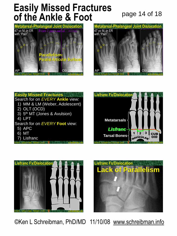
Metatarsal-Phalangeal Joint Dislocation47 yo M, in ER with “Pain”
AP AP Obl
Priors 2 years earlier… normal
Parallelism:Parallel Articular Surfaces
©Ken L Schreibman, PhD/MD 2008 schreibman.info
Metatarsal-Phalangeal Joint Dislocation47 yo M, in ER with “Pain”
AP
Obl Obl
©Ken L Schreibman, PhD/MD 2008 schreibman.info
Easily Missed FracturesSearch for on EVERY Ankle view:
1) MM & LM (Weber, Adolescent)2) OLT (OCD)3) 5th MT (Jones & Avulsion)4) LPT
Search for on EVERY Foot view:5) APC6) MT7) Lisfranc
©Ken L Schreibman, PhD/MD 2008 schreibman.info
Lisfranc Fx/Dislocation
1 2 3CUB
OID
Lisfranc
Tarsal Bones
Metatarsals
©Ken L Schreibman, PhD/MD 2008 schreibman.info
Lisfranc Fx/Dislocation
©Ken L Schreibman, PhD/MD 2008 schreibman.info
Lack of ParallelismLisfranc Fx/Dislocation

©Ken L Schreibman, PhD/MD 11/10/08 www.schreibman.info
page 15 of 18Easily Missed Fracturesof the Ankle & Foot
©Ken L Schreibman, PhD/MD 2008 schreibman.info
Lisfranc Fx/DislocationOblAP
©Ken L Schreibman, PhD/MD 2008 schreibman.info
Lisfranc Fx/Dislocation
1 2 3CUB
OID
HOMOLATERAL
1 2
I II
©Ken L Schreibman, PhD/MD 2008 schreibman.info
Lisfranc Fx/Dislocation
1 2 3CUB
OID
DIVERGENT
©Ken L Schreibman, PhD/MD 2008 schreibman.info
Lisfranc
Lisfranc Fx/DislocationLisfranc Joint
Lisfranc Ligament
1 2 3CUB
OID
©Ken L Schreibman, PhD/MD 2008 schreibman.info
Lisfranc Fx/DislocationLisfranc Joint
Lisfranc Ligament
©Ken L Schreibman, PhD/MD 2008 schreibman.info
66 yo M h/o DiabetesPresents in Sept swollen footMR is requested to “r/o Osteo”
Are there radiographs?Yes
…3 months ago
Repeat radiographs obtained now, prior to MR, reveal…
Common siteneuropathiccollapseCan occur
rapidly
Lisfranc Fx/Dislocation: Diabetes
June September
Neuropathic destruction of the Lisfranc joint
Normal Lisfranc joint
©Ken L Schreibman, PhD/MD 11/10/08 www.schreibman.info
page 16 of 18Easily Missed Fracturesof the Ankle & Foot
©Ken L Schreibman, PhD/MD 2008 schreibman.info
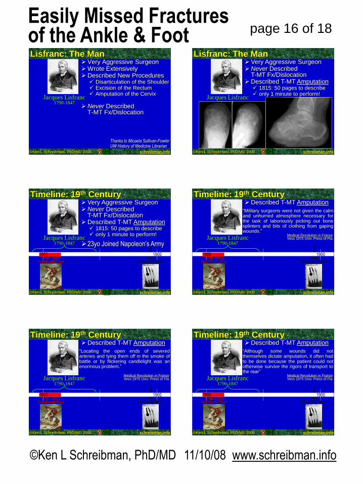
Jacques Lisfranc1790-1847
Very Aggressive SurgeonWrote ExtensivelyDescribed New Procedures Disarticulation of the Shoulder Excision of the Rectum Amputation of the Cervix
Never Described T-MT Fx/Dislocation
Lisfranc Fx/Dislocation
Thanks to Micaela Sullivan-FowlerUW History of Medicine Librarian
Lisfranc: The Man
©Ken L Schreibman, PhD/MD 2008 schreibman.info
Lisfranc: The Man
Jacques Lisfranc1790-1847
Very Aggressive SurgeonNever Described
T-MT Fx/DislocationDescribed T-MT Amputation 1815: 50 pages to describe only 1 minute to perform!
©Ken L Schreibman, PhD/MD 2008 schreibman.info
Lisfranc: The ManTimeline: 19th Century
1800 1900
Jacques Lisfranc1790-1847
1895Roentgen Rays
1796-1815NapoleonicWars
1800
Very Aggressive SurgeonNever Described
T-MT Fx/DislocationDescribed T-MT Amputation 1815: 50 pages to describe only 1 minute to perform!
23yo Joined Napoleon‟s Army
©Ken L Schreibman, PhD/MD 2008 schreibman.info
Lisfranc: The ManTimeline: 19th Century
1800 1900
Jacques Lisfranc1790-1847
1895Roentgen Rays
1796-1815NapoleonicWars
1800
Described T-MT Amputation
“Military surgeons were not given the calmand unhurried atmosphere necessary forthe task of laboriously picking out bonesplinters and bits of clothing from gapingwounds.”
Medical Revolution in FranceVess 1975 Univ. Press of Fla.
©Ken L Schreibman, PhD/MD 2008 schreibman.info
Lisfranc: The ManTimeline: 19th Century
1800 1900
Jacques Lisfranc1790-1847
1895Roentgen Rays
1796-1815NapoleonicWars
1800
Described T-MT Amputation
Medical Revolution in FranceVess 1975 Univ. Press of Fla.
“Locating the open ends of severedarteries and tying them off in the smoke ofbattle or by flickering candlelight was anenormous problem.”
©Ken L Schreibman, PhD/MD 2008 schreibman.info
Lisfranc: The ManTimeline: 19th Century
1800 1900
Jacques Lisfranc1790-1847
1895Roentgen Rays
1796-1815NapoleonicWars
1800
Described T-MT Amputation
Medical Revolution in FranceVess 1975 Univ. Press of Fla.
“Although some wounds did notthemselves dictate amputation, it often hadto be done because the patient could nototherwise survive the rigors of transport tothe rear”

©Ken L Schreibman, PhD/MD 11/10/08 www.schreibman.info
page 17 of 18Easily Missed Fracturesof the Ankle & Foot
©Ken L Schreibman, PhD/MD 2008 schreibman.info
Lisfranc: The ManTimeline: 19th Century
1800 1900
Jacques Lisfranc1790-1847
1895Roentgen Rays
1796-1815NapoleonicWars
1800
Described T-MT Amputation
Medical Revolution in FranceVess 1975 Univ. Press of Fla.
“The mind did not have time to reason.Experience and coldbloodedness countedfor more than talent. Everything had to bedone with prompt and decisive action.”
1846Ether
©Ken L Schreibman, PhD/MD 2008 schreibman.info
Lisfranc: The ManTimeline: 19th Century
1800 1900
Jacques Lisfranc1790-1847
1895Roentgen Rays
1796-1815NapoleonicWars
1800
Medical Revolution in FranceVess 1975 Univ. Press of Fla.
1846Ether
Percy, surgeon-in-chief, complained ofhaving too many “pseudosurgeons whocounted their battle actions only by thenumber of arms and legs they had cut off.”
©Ken L Schreibman, PhD/MD 2008 schreibman.info
Lisfranc: The ManTimeline: 19th Century
1800 1900
Jacques Lisfranc1790-1847
1895Roentgen Rays
1796-1815NapoleonicWars
1800
Medical Revolution in FranceVess 1975 Univ. Press of Fla.
1846Ether
Percy called Lisfranc, “so obsessive ascalpel-wielder that he (Lisfranc) lamentedthe passing of the Napoleonic age that hadprovided him with so many splendidopportunities for amputations”
©Ken L Schreibman, PhD/MD 2008 schreibman.info
Lisfranc: The ManTimeline: 19th Century
1800 1900
Jacques Lisfranc1790-1847
1895Roentgen Rays
1796-1815NapoleonicWars
18001846Ether
Dr Oliver Wendell Homes (US physician, poet,humorist, Dean of Harvard Medical School):
“As for Lisfranc, I can say little more of himthan he was a great drawer ofblood…ordering a wholesale bleeding ofhis patients, right and left, whatever mightbe the matter with them.”
Guillaume Dupuytren: A Surgeon in His Place and Time Barsky. Vantage Press
©Ken L Schreibman, PhD/MD 2008 schreibman.info
Lisfranc: The ManTimeline: 19th Century
1800 1900
Jacques Lisfranc1790-1847
1895Roentgen Rays
1796-1815NapoleonicWars
18001846Ether
Dr Oliver Wendell Homes (US physician, poet,humorist, Dean of Harvard Medical School):
Guillaume Dupuytren: A Surgeon in His Place and Time Barsky. Vantage Press
[quoting Lisfranc]:
“the splendid guardsmen of the old Empire
had such magnificent thighs to amputate.”
©Ken L Schreibman, PhD/MD 2008 schreibman.info
Jacques Lisfranc1790-1847
1796-1815Napoleonic Wars
18001846Ether
1895Roentgen Rays
Sir Robert Jones1857-1933
1900 2000
Timeline: 19th CenturyTimeline: 19th & 20th Centuries Born 1857, N. WalesDied 1933, age 76 1887 (age 30)
Apprenticed w/uncle, Hugh Owen Thomas
1888 – Appointed chief surgeon for the Manchester Ship Canal
–Established a chain of smallhospitals along the length ofthe canal construction
–This was the first organizedfracture and injury servicein England

©Ken L Schreibman, PhD/MD 11/10/08 www.schreibman.info
page 18 of 18Easily Missed Fracturesof the Ankle & Foot
©Ken L Schreibman, PhD/MD 2008 schreibman.info
Timeline: 19th & 20th Centuries
1895Roentgen Rays
1900 2000
Sir Robert Jones1857-1933
1914-1919WW I
Jacques Lisfranc1790-1847
1796-1815Napoleonic Wars
18001846Ether
During WWI, revolutionized the care of wounded soldiers– Established network
of field hospitals– Rehab hospitals– Mortality rate for
open fractures wasreduced from 80% to 20%
1925-“To him and his practical teaching and
influence we owe it that our streets today show relatively so few
war cripples”©Ken L Schreibman, PhD/MD 2008 schreibman.info
Timeline: 19th & 20th Centuries
1895Roentgen Rays
1900 2000
Sir Robert Jones1857-1933
1914-1919WW I
Jacques Lisfranc1790-1847
1796-1815Napoleonic Wars
18001846Ether Pioneer in the care of
crippling diseases of children. Established the first long-stay orthopœdic hospitals for children.
©Ken L Schreibman, PhD/MD 2008 schreibman.info
Jones: Early X-Ray Proponent
18001895
Roentgen Rays
1900 2000
Sir Robert Jones1857-1933
Dec 28 1895: Röntgen publishes “On a New Kind of Rays”
Feb 22 1896: Jones publishes in Lancet,“The Discovery of a Bullet Lost in the Wrist by Mean of the Roentgen Ray”
Arguably the first published case history in which x-rays were used as a diagnostic tool
Jones: Early X-Ray Proponent“In 1896 we were all dancing in a circle round the tentpole. Robert Jones‟ ankle seemed to give…he said he had strained such and such a muscle or tendon, exclaiming:
‘Most interesting, most painful. I had no idea it could be so painful. Most interesting!’
“At that time he (Jones) had what might almost be described as a new toy – an X-ray apparatus, the first in England.
“He wondered whether it would not be possible for the X-ray to show the torn or swollen muscle, and on experimenting the plate showed to his amazement that a small bone was fractured.
“This disability gave him immense satisfaction. To one patient who came to him with mysterious symptoms he said, after a brief examination,
‘Madam, you could have paid me no greater compliment – this is a genuine Jones fracture.’”
Frederick Waston: “The Life ofSir Robert Jones” p98, 1934
Jones: Early X-Ray Proponent“Radiography here, as in allbranches of medicine, is anessential aid to diagnosis. Nomatter how experienced we may be,we cannot afford to dispense with it,even in the apparently simple andobvious case. Not only should weinsist upon procuring a film, but it isequally important that we shouldwelcome the radiologist‟s reading ofit. Some surgeons resent this andsay, „Give me the film so that I canread it myself,‟ but this is an arrogantand stupid attitude, and not thepatient‟s advantage.”
Jones: Manipulation as a therapeutic measure, Proc Roy Soc Med 24: 1405, 1931-2
©Ken L Schreibman, PhD/MD 2008 schreibman.info
Jones: The Inspiration
Peltier: “Eponymic fractures: Robert Jones and Jones’s fracture”Surgery1972 v71 p522
Memorial Tablet:
“Great surgeon:
greater man”
Obituary:
“As a teacher he was pre-eminent, not in the role of didactic pedagogue, but in the role of leader able to enthuse men, and through them he advanced the art and science of his specialty”