Neuron
Article
Neuregulin-1 Enhances Depolarization-InducedGABA ReleaseRan-Sook Woo,1,7 Xiao-Ming Li,1,2,7 Yanmei Tao,1 Ezekiel Carpenter-Hyland,1 Yang Z. Huang,3
Janet Weber,4 Hannah Neiswender,1 Xian-Ping Dong,1 Jiong Wu,5 Martin Gassmann,6 Cary Lai,4
Wen-Cheng Xiong,1 Tian-Ming Gao,2,* and Lin Mei1,2,*1Program of Developmental Neurobiology, Institute of Molecular Medicine and Genetics, Department of Neurology,Medical College of Georgia, Augusta, GA 30912, USA2Department of Anatomy and Neurobiology, Southern Medical University, Guangzhou, 510515, China3Department of Neurobiology, Duke University Medical Center, Durham, NC 27710, USA4Molecular and Integrative Neuroscience Department, Scripps Research Institute, La Jolla, CA 92037, USA5Cell Signaling Technology, Inc., 3 Trask Lane, Danvers, MA 01923, USA6 Institute of Physiology, University of Basel, CH-4056 Basel, Switzerland7These authors contributed equally to this work.
*Correspondence: [email protected] (T.-M.G.), [email protected] (L.M.)DOI 10.1016/j.neuron.2007.04.009
SUMMARY
Neuregulin-1 (NRG1), a regulator of neural de-velopment, has been shown to regulate neuro-transmission at excitatory synapses. AlthoughErbB4, a key NRG1 receptor, is expressed in glu-tamic acid decarboxylase (GAD)-positive neu-rons, little is known about its role in GABAergictransmission. We show that ErbB4 is localizedat GABAergic terminals of the prefrontal cortex.Our data indicate a role of NRG1, both endoge-nous and exogenous, in regulation of GABAergictransmission. This effect was blocked by inhibi-tion or mutation of ErbB4, suggesting the in-volvement of ErbB4. Together, these resultsindicate that NRG1 regulates GABAergic trans-mission via presynaptic ErbB4 receptors, identi-fying a novel function of NRG1. Because bothNRG1 and ErbB4 have emerged as susceptibilitygenes of schizophrenia, these observations maysuggest a mechanism for abnormal GABAergicneurotransmission in this disorder.
INTRODUCTION
Neuregulin-1 (NRG1), a family of polypeptides that plays
an important role in neural development, is implicated in
nerve cell differentiation, neuron migration, neurite out-
growth, and synapse formation (Buonanno and Fischbach,
2001; Corfas et al., 2004). NRG1 and its receptor ErbB
tyrosine kinases are expressed not only in the developing
nervous system, but also in adult brain. In the adult, ErbB
receptors are concentrated at the postsynaptic density
(PSD), presumably via interaction with PDZ domain-
containing proteins including PSD-95 and erbin (Garcia
et al., 2000; Huang et al., 2000, 2001; Ma et al., 2003).
NRG1 suppresses induction of LTP at Schaffer collateral-
CA1 synapses in the hippocampus without affecting basal
synaptic transmission (Huang et al., 2000; Ma et al., 2003).
Subsequently, NRG1 was shown to reverse LTP and re-
duce whole-cell NMDA receptor currents in pyramidal
neurons of prefrontal cortex, and was also shown to de-
crease NMDA receptor-mediated EPSCs in prefrontal cor-
tex slices (Gu et al., 2005; Kwon et al., 2005). Interestingly,
the NRG1 gene is strongly associated with schizophrenia
in diverse populations in Iceland, Scotland, China, Japan,
and Korea (Fukui et al., 2006; Kim et al., 2006; Stefansson
et al., 2002, 2003; Yang et al., 2003).
ErbB4 mRNA is enriched in regions where interneurons
are clustered in adult brains (Lai and Lemke, 1991). GAD-
positive neurons from the embryonic hippocampus ex-
press ErbB4 (Huang et al., 2000). During development,
loss of NRG1/ErbB4 signaling alters tangential migration
of cortical interneurons, leading to a reduction in the num-
ber of GABAergic interneurons in the cortex (Anton et al.,
2004; Flames et al., 2004). In adult mice, deletion of ErbB4
in the central nervous system (CNS) resulted in lower levels
of spontaneous motor activity, reduced grip strength, and
altered cue use in performing a maze task (Golub et al.,
2004). The ErbB4 gene is also associated with schizophre-
nia (Law et al., 2006; Nicodemus et al., 2006).
g-Aminobutyric acid (GABA) is the principal inhibitory
neurotransmitter in the mammalian forebrain. GABAergic
inhibitory interneurons are essential to the proper func-
tioning of the CNS (McBain and Fisahn, 2001). GABAergic
dysfunction is implicated in several neurological disor-
ders, including Huntington’s chorea, Parkinson’s disease,
and epilepsy, and in psychiatric disorders such as anxiety,
depression, and schizophrenia (Coyle, 2004).
This study investigates the role of NRG1 in GABAergic
neurotransmission. We find that ErbB4 is expressed in
GABAergic presynaptic terminals in the cerebral cortex.
Treatment with NRG1 had no effect on basal GABA re-
lease, but it increased evoked release in cortical slices in a
manner dependent on ErbB4. These observations identify
a novel function of NRG1 and may suggest a mechanism
Neuron 54, 599–610, May 24, 2007 ª2007 Elsevier Inc. 599

Neuron
Neuregulin Regulation of GABAergic Transmission
Figure 1. NRG1 and ErbB4 Are Expressed throughout Cortical Layers
(A–C) In situ hybridization in adult rat brain coronal sections using radiolabeled antisense RNA probes. (A) ErbB4-specific hybridization was detected
in scattered cells throughout the cortex (layers 2–6b) in the rostral forebrain (left panel). Prominent hybridization is observed in scattered cells through-
out the cortex and hippocampus (Hi), in the medial habenula (MHb), in the reticular nucleus of the thalamus (Rt), and in the intercalated masses of the
amygdala (Amyg) in a more caudal section (right panel). Scale bar, 1 mm. (B) In the rostral forebrain (left panel), NRG1 type I/II-specific hybridization
was detected in layers 2-3 and 6b of the cortex and in the piriform cortex (Pir). In caudal sections (right panel), NRG1 type I/II transcripts were detected
in cortical layer 6b, in the reticular nucleus of the thalamus (Rt), in all fields of the hippocampus (Hi), and in scattered large cells in the globus pallidus
(GP). (C) In the rostral forebrain (left panel), NRG1 type III-specific hybridization was detected in cortical layer 5 and in the piriform cortex (Pir). In more
caudal sections (right panel), it was present in cortical layer 5, the reticular nucleus of the thalamus (Rt), and all fields of hippocampus (Hi).
for abnormal GABAergic neurotransmission in schizo-
phrenia and epilepsy.
RESULTS
Localization of ErbB4 in GABAergic
Presynaptic Terminals
ErbB4 transcripts were expressed throughout cortical
layers 2–6b (Figure 1A) (Lai and Lemke, 1991; Yau et al.,
2003). In addition, ErbB4 transcripts were identified at
high levels in the medial habenula, the reticular nucleus
of the thalamus, and in the intercalated masses of the
amygdala. These observations are consistent with the no-
tion that ErbB4 is expressed in interneurons. In further
agreement, ErbB4 was shown to be present in GAD-posi-
tive neurons isolated from the hippocampus (Huang et al.,
2000). To determine the in vivo subcellular localization of
ErbB4 in GAD-positive neurons, we stained prefrontal
sections of GFP-expressing inhibitory neurons (GIN) mice
that express GFP under the control of the gad1 promoter
that directs specific expression in GABA interneurons,
especially those that are somatostatin positive, in the hip-
pocampus (Oliva et al., 2000). Presynaptic terminals of
GABAergic neurons appear as discrete puncta-rings in
the prefrontal cortex, surrounding the soma of postsynap-
tic neurons in cortical layers 2–6 (Figure 2A, arrows) (Pillai-
Nair et al., 2005). The anti-ErbB4 antibody 0618 (Zhu et al.,
1995) and sc-283 specifically recognized ErbB4 because
their immunoreactivity was diminished in ErbB4 mutant
mice (Figures 2G and 2H). As shown in Figure 2A, ErbB4
was detected in puncta-rings and neuropils, colocalizing
with GFP. Quantitatively, about 90% of puncta-rings and
neuropils in the prefrontal cortex expressed ErbB4 (Fig-
ure 2B). These results suggest that ErbB4 is present at
terminals of GABAergic neurons, including somatostatin
neurons. To test this hypothesis further, we determined
600 Neuron 54, 599–610, May 24, 2007 ª2007 Elsevier Inc.
whether ErbB4 colocalizes with GAD65 and vesicular
GABA transporter (VGAT), both well-characterized markers
of GABAergic terminals (Tafoya et al., 2006). The ErbB4
immunoreactivity colocalized with GAD65 and VGAT in
puncta-ring-like structures (Figures 2C and 2D). Twenty-
three percent of GAD65 clusters and forty-seven percent
of VGAT clusters were ErbB4-positive, suggesting ErbB4
localization at specific subsets of GABA terminals (Figures
2E and 2F). On the other hand, 26% and 70% of ErbB4
clusters colocalized with GAD65 and VGAT, respectively,
in agreement with the notion that ErbB4 is also localized
at non-GABAergic synapses (Huang et al., 2000). Taken
together, these results indicate that ErbB4 is present at
groups of presynaptic terminals of GABAergic neurons in
the cerebral cortex.
Increase in Depolarization-Evoked GABA
Release by NRG1
The presynaptic localization of ErbB4 in GABAergic neu-
rons suggested to us that NRG1 may regulate GABAergic
neurotransmission. To test this hypothesis, we determined
effects of NRG1 on GABA release in cortical slices by both
biochemical and electrophysiological approaches. Basal
[3H]GABA release was low, at a rate of 3.75% ± 0.35%
(n = 8) of total radioactivity per 10 min (Figure 3A). Treat-
ment of slices with 20 mM KCl, a treatment known to depo-
larize neurons, increased [3H]GABA release by 2.5- to 3.5-
fold within 10 min (Figure 3A). NRG1 had no effect on basal
[3H]GABA release; by contrast, it increased depolariza-
tion-evoked GABA release in a dose-dependent manner
(Figures 3A and 3B and Figure S1A in the Supplemental
Data). This effect was not inhibited by antagonists of gluta-
mate receptors, suggesting that the increase in GABA
release does not require glutamatergic signaling (Fig-
ure S1B). To demonstrate that NRG1 regulates the
physiological function of GABA transmission, inhibitory
Neuron
Neuregulin Regulation of GABAergic Transmission
postsynaptic currents (IPSCs) were recorded from pre-
frontal cortical slices. As shown in Figures 3C–3F, NRG1
did not appear to affect the frequency, amplitude, and de-
cay times of miniature IPSCs (mIPSCs) that were blockable
by bicuculline, a GABA-A receptor antagonist (Figure S1C
and data not shown). These results are in agreement with
observations above that basal GABA release was not af-
fected. By contrast, as shown in Figure 3G, it enhanced
evoked IPSCs (eIPSCs) that were sensitive to bicuculline
(Figure S1D). The increase in eIPSCs had a similar dose-
response curve to that of evoked [3H]GABA release
(Figure 3H) and was abolished when NRG1 was heat-
denatured (Figure 3I). Furthermore, the NRG1 regulation
remained unchanged in the presence of antagonists of
metabotropic glutamate receptors, cholinergic receptors,
serotonin receptors, adrenergic receptors, dopamine re-
ceptors, or some combination thereof (Figure S1E). As a
control, BDNF decreased depolarization-evoked GABA
release and eIPSCs in cortical slices, in agreement with
earlier studies (Canas et al., 2004; Frerking et al., 1998).
These results indicate that NRG1 increases evoked GABA
release without affecting basal release, likely via a direct
effect on GABAergic presynaptic terminals.
NRG1 Effects on GABAergic Presynaptic Terminals
To further determine whether NRG1 regulates GABA re-
lease directly at presynaptic terminals, we performed the
following two experiments. First, we investigated whether
NRG1 is able to regulate [3H]GABA release from synapto-
somes in the absence of their neural circuit. As shown in
Figure 4A, NRG1 increased depolarization-evoked GABA
release from synaptosomes while having no effect on
basal GABA release. Moreover, this effect was concentra-
tion-dependent, with a maximal response of 28% ± 1.5%
(n = 6), similar to that observed in cortical slices (Figure 4A).
Second, we characterized the paired-pulse ratios (PPRs)
of control and NRG1-affected eIPSCs in response to two
stimulations. At inhibitory synapses, a second stimulation
generates a smaller eIPSC because of depletion of vesi-
cles in the releasable pool by the first stimulation (Lambert
and Wilson, 1994). Figure 4B (left panel) shows averaged
traces of eight consecutive eIPSCs induced by paired
stimuli at different interpulse intervals. The PPRs at 25
ms intervals were reduced from 0.86 ± 0.07 in control to
0.68 ± 0.05 in NRG1-treated slices (n = 6, p < 0.01). The
reduction in PPRs remained even at 200 ms intervals.
The depression effect of NRG1 on the amplitudes of the
second eIPSCs provides further evidence that NRG1 reg-
ulates evoked GABA release by a presynaptic mecha-
nism. In addition, these results also suggest that NRG1
may increase the probability of GABA release in response
to depolarization.
Endogenous NRG1 Is Necessary to Maintain
Activity-Dependent GABA Release
NRG1 is expressed in various regions in the brain (Law
et al., 2004). NRG1 type I/II transcripts were detected
prominently in cortical layer 6b and at lower levels in layers
2–3 (Figure 1B). In comparison, NRG1 type III transcripts
were primarily detected in cortical layer 5 (Figure 1C). Hy-
bridization of NRG1 type I/II was also observed in the re-
ticular nucleus of the thalamus and in cholinergic interneu-
rons in the globus pallidus. NRG1 type III was expressed in
the reticular nucleus of the thalamus. Both NRG1 isoforms
were also observed in the piriform cortex and throughout
the hippocampus. Notably, the distinct isoforms of NRG1
appear to be expressed in a laminar-specific and largely
nonoverlapping manner in the cortex. These observations
indicate that NRG1 is available in various areas in the brain
including the cerebral cortex. To determine whether en-
dogenous NRG1 regulates GABA release, we generated
ecto-ErbB4, which contains the entire extracellular region
of ErbB4 fused to an FC fragment. Ecto-ErbB4 binds to
and thus prevents NRG1 from interacting with ErbB recep-
tor kinases. As shown in Figure 5A and Figure 5B, treatment
with ecto-ErbB4 inhibited NRG1 activation of ErbB4 in
GAD-positive neurons (see Figure S2 for characterization
of the anti-phospho-ErbB4 antibody). Such treatment
blocked NRG1 potentiation of eIPSCs in a dose-depen-
dent manner (Figures 5C and 5D), demonstrating the
neutralizing ability of ecto-ErbB4. NRG1-enhanced evoked
GABAreleasewasalso inhibitedbyecto-ErbB4 (Figure5D).
Remarkably, treatment with ecto-ErbB4 alone reduced
both evoked GABA release and eIPSCs in the absence of
exogenous NRG1 (Figure 5D). These observations and
results from studies of inhibitors of ErbB4 suggest a role
for endogenous NRG1 in regulating evoked GABA release.
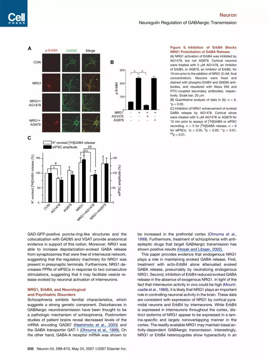
ErbB4 Is Necessary for NRG1 Enhancement
of Evoked GABA Release
Of the three ErbB kinases, ErbB2 and ErbB4, but not
ErbB3, are catalytically active (Citri and Yarden, 2006). To
determine which ErbB is involved in NRG1 regulation of
evoked GABA release, cortical neurons were treated with
AG879 and AG1478, specific inhibitors of ErbB2 and
ErbB4, respectively (Fukazawa et al., 2003). ErbB4 tyro-
sine phosphorylation in response to NRG1 was blocked
in neurons pretreated with AG1478, but not AG879 (Fig-
ures 6A and 6B). Treatment with AG1478 prevented
NRG1 from increasing evoked GABA release and increas-
ing amplitude of eIPSCs in cortical slices (Figure 6C).
These results suggest a role of ErbB4 in NRG1 regulation
of GABAergic transmission. As observed with ecto-
ErbB4, AG1478 alone decreased depolarization-evoked
[3H]GABA release and the amplitude of eIPSCs (Figure 6C),
providing further evidence that endogenous NRG1 activity
may be necessary to maintain GABA release elicited by
neuronal activation. In a control experiment, treatment
with AG879 had no detectable effect on evoked GABA
release and the amplitude of eIPSCs in the presence or ab-
sence of exogenous NRG1 (Figure 6C). Taken together,
these observations demonstrate that activation of ErbB4,
but not ErbB2, is required for NRG1’s effect.
To investigate the involvement of ErbB4 further, we
characterized evokedGABA release inErbB4 mutant mice.
ErbB4 null mutant mice die around E11. The embryonic
Neuron 54, 599–610, May 24, 2007 ª2007 Elsevier Inc. 601

Neuron
Neuregulin Regulation of GABAergic Transmission
Figure 2. ErbB4 Is Present at Presynaptic Terminals of GABAergic Neurons
(A) Coronal sections of prefrontal cortex of GIN-GFP mice were stained with anti-ErbB4 antibody 0618 (top panels) or with the antibody sc-283 (bot-
tom panels). Immunoactivity was visualized by Alexa 594-conjugated secondary antibody. GAD-positive terminals (expressing GFP) were visualized
by excitation at 488 nm. Arrows, GFP-positive puncta-ring structures surrounding pyramidal neurons; arrowheads, neuropils; inset, enlarged areas.
(B) Quantitative analysis of puncta-rings and neuropils that are positive for ErbB4. The antibody used for quantification was 0618. Shown are means ±
SEM; n = 60 for puncta-rings and n = 10 for neuropils of 20 independent sections.
(C and D) Coronal sections of prefrontal cortex were stained with anti-ErbB4 antibody 0618 and anti-GAD65 (G1166) and anti-VGAT (131003) anti-
bodies. Immunoactivity was visualized by Alexa 488- and Alexa 594-conjugated secondary antibodies, respectively. Arrowheads, colocalization of
ErbB4 and GAD65 or VGAT; arrows, ErbB4-positive alone; hallow arrows, GAD65- or VGAT-positive alone.
602 Neuron 54, 599–610, May 24, 2007 ª2007 Elsevier Inc.

Neuron
Neuregulin Regulation of GABAergic Transmission
Figure 3. NRG1 Increases Depolarization-Evoked [3H]GABA Release and eIPSC Amplitude
(A) Cortical slices were preloaded with [3H]GABA for 30 min in the presence of b-alanine (1 mM), an inhibitor of [3H]GABA uptake by glial cells, amino-
oxyacetic acid (0.1 mM), an inhibitor of GABA degradation, and nipecotic acid (1 mM), an inhibitor of the GABA transporter in neurons. Basal and
depolarization (20 mM KCl)-evoked release of [3H]GABA were monitored sequentially. The sum of the basal release, depolarization-evoked release,
and the residual [3H]GABA was taken as 100%. In comparison with controls (open circles), NRG1 (closed circles) had no effect on basal [3H]GABA
release, but increased depolarization-evoked [3H]GABA release.
(B) Dose-dependent potentiation of evoked [3H]GABA release. Raw data of a representative experiment are presented in Figure S1A.
(C) Representative traces of mIPSCs in pyramidal neurons in prefrontal cortical slices.
(D) Cumulative plots of mIPSC amplitudes.
(E) Cumulative plots of mIPSC frequencies.
(F) No effect of NRG1 on mIPSCs in pyramidal neurons in prefrontal cortical slices (n = 12).
(G) Increased eIPSCs in NRG1-treated slices. (Top) Representative eIPSCs of control, NRG1-treated, or NRG1-treated/washed slices. (Bottom)
Quantitative analysis of eIPSC amplitudes. n = 12, *p < 0.01.
(H) Dose-dependent effect of NRG1 on eIPSCs. n = 6, *p < 0.05, **p < 0.01.
(I) Denatured NRG1 failed to increase depolarization-evoked [3H]GABA release and eIPSC amplitude. n = 8 for [3H]GABA release; for eIPSCs, n = 6 for
control, NRG1, and denatured NRG1, and n = 4 for BDNF. *p < 0.05, #p < 0.05; **p < 0.05, ##p < 0.01.
lethality can be genetically rescued by expressing ErbB4
under a cardiac-specific myosin promoter (Tidcombe
et al., 2003). This line of mice (ErbB4�/�ht+), however,
does not express ErbB4 in the brain (Figures 7A and 7B)
or other noncardiac tissues (data not shown). Ablation of
the ErbB4 gene had no effect on basal and depolariza-
tion-evoked [3H]GABA release (Figure 7C). However, un-
like in control slices, NRG1 was unable to increase evoked
(E and F) Quantitative analysis of ErbB4 clusters with GAD65 and with VGAT, and VGAT and GAD65 clusters with ErbB4. More than 1100 clusters of
five independent sections were scored. Shown are means ± SEM.
(G and H) Specificity characterization of anti-ErbB4 antibodies. Coronal sections of prefrontal cortex of ErbB4+/+ht+ and ErbB4�/�ht+ mice were
incubated with the anti-ErbB4 antibodies 0618 and sc-283. Immunoactivity was visualized by Alexa-conjugated secondary antibodies.
Neuron 54, 599–610, May 24, 2007 ª2007 Elsevier Inc. 603

Neuron
Neuregulin Regulation of GABAergic Transmission
Figure 4. Effects of NRG1 on Presynaptic
Terminals
(A) NRG1 increases depolarization-evoked
[3H]GABA release from synaptosomes.
[3H]GABA-loaded cortical synaptosomes were
treated with 5 nM NRG1 with (evoked) or with-
out (basal) 20 mM KCl. [3H]GABA release was
assayed 10 min after NRG1 stimulation. Shown
are means ± SEM of six individual experiments
in triplicate. *p < 0.05, **p < 0.01.
(B) NRG1 reduces PPRs of GABAergic trans-
mission in the prefrontal cortex. (Left) Averaged
traces of eight consecutive recordings induced
by paired stimuli (10 s apart) separated by indi-
cated interpulse intervals. (Right) PPRs as
a function of interpulse intervals. The ampli-
tudes of the first and second IPSCs were mea-
sured as indicated in the inset. n = 6, *p < 0.05.
[3H]GABA release and eIPSC amplitude in ErbB4�/�ht+
slices (Figures 7C and 7D). These observations identify
an important role of ErbB4 in NRG1 regulation of evoked
GABA release.
DISCUSSION
The major findings of this study are as follows. First, ErbB4,
a receptor for NRG1, is present in GABAergic terminals of
the prefrontal cortex. Second, NRG1 facilitates evoked
release of GABA from slices of the prefrontal cortex, but
has no effect on basal GABA release. Third, the potentia-
tion effect of NRG1 must require ErbB4 because it was
blocked by the ErbB4 inhibitor AG1478 and was abolished
in cortical slices of ErbB4 mutant mice. In addition, we pro-
vided evidence that evoked GABA release and eIPSCs in
the absence of exogenous NRG1 were blocked by inhibi-
tors of NRG1 signaling, suggesting a role of endogenous
NRG1 in regulating GABA neurotransmission. Together,
these results identify a novel function of NRG1—regulation
of GABAergic transmission via presynaptic ErbB4 recep-
tors. These results suggest that NRG1 may regulate the
activity of cortical interneurons, providing insight into po-
tential mechanisms by which this trophic factor regulates
synaptic plasticity and pathogenesis of schizophrenia
and epilepsy.
NRG1 and Neurotransmission at Excitatory
and Inhibitory Synapses
NRG1 has been shown to regulate differentiation of neural
cells, neuronal navigation, and neuron survival in develop-
ing CNS (Buonanno and Fischbach, 2001; Corfas et al.,
2004). In the peripheral nervous system, NRG1 signaling
604 Neuron 54, 599–610, May 24, 2007 ª2007 Elsevier Inc.
is implicated in Schwann cell differentiation and myelina-
tion, muscle spindle development, and synapse-specific
expression of AChR subunit genes (Adlkofer and Lai,
2000; Fischbach and Rosen, 1997; Hippenmeyer et al.,
2002; Si et al., 1996). Interestingly, NRG1 and its receptor
ErbB kinases are continuously expressed in various brain
regions, including the prefrontal cortex, hippocampus,
cerebellum, oculomotor nucleus, superior colliculus, red
nucleus, substantia nigra, and pars compacta (Lai and
Lemke, 1991; Law et al., 2004; Yau et al., 2003). Moreover,
ErbB4 colocalizes with PSD-95 and NMDA receptors in
hippocampal neurons (Garcia et al., 2000; Huang et al.,
2000). Furthermore, NRG1 signaling may be increased
by the interaction of ErbB4 with PSD-95 (Huang et al.,
2000). These observations suggest that NRG1 may play
a role in synaptic plasticity, maintenance or regulation of
synaptic structure, or some combination thereof in adult
brain. Indeed, we found that NRG1 blocks induction of
long-term potentiation (LTP) at Schaffer collateral-CA1
synapses (Huang et al., 2000). NRG1 can depotentiate
LTP at hippocampal CA1 synapses and reduce whole-
cell NMDA receptor, but not AMPA receptor, currents in
prefrontal cortex pyramidal neurons (Gu et al., 2005;
Kwon et al., 2005). Recently, ErbB4 has been shown to
play a key role in activity-dependent maturation and plas-
ticity of excitatory synaptic structure and function (Li et al.,
2007).
This study provides evidence that ErbB4 is present
at GABAergic terminals in the prefrontal cortex. The iden-
tification of the subtype or subtypes of GABA interneurons
that express ErbB4 will require further investigation.
Interestingly, ErbB4 colocalizes with GAD-GFP in GIN
mice. An earlier study demonstrated that hippocampal

Neuron
Neuregulin Regulation of GABAergic Transmission
Figure 5. Suppression of NRG1-Enhanced GABA Release by Ecto-ErbB4
(A) Ecto-ErbB4 inhibition of NRG1 activation of ErbB4 in GAD65-positive cortical neurons. Cortical neurons were pretreated with ecto-ErbB4 for
10 min prior to the addition of NRG1 (5 nM, final concentration) for another 10 min, fixed, and stained with anti-p-ErbB4 and anti-GAD65 antibodies
that were visualized with Alexa 594 and FITC-coupled secondary antibodies, respectively. Scale bar, 20 mm.
(B) Quantitative analysis of data in (A). Images were captured with a Zeiss LSM confocal microscope and analyzed by Image J software (NIH). Ecto-
ErbB4 treatment inhibits NRG1-induced ErbB4 phosphorylation. n = 7, *p < 0.05.
(C) Ecto-ErbB4 inhibition of eIPSCs. Cortical slices were treated with sequential addition of NRG1 (5 nM) and ecto-ErbB4 (1 mg/ml and 2 mg/ml) (all final
concentrations). eIPSCs were recorded as in Figure 3G. Shown are data from a representative experiment. On the top are averaged traces before (a)
and after (b) NRG1, and after different dosages of ecto-ErbB4 ([c] and [d], 1 and 2 mg/ml, respectively).
(D) Inhibition by ecto-ErbB4 of depolarization-evoked GABA release and eIPSCs. Cortical slices were treated with 1 or 2 mg/ml ecto-ErbB4 for 10 min
prior to assays of [3H]GABA and eIPSCs. n = 5 for [3H]GABA release, n = 6 for eIPSCs. *p < 0.01 and #p < 0.01 for [3H]GABA release and eIPSCs,
respectively.
GAD-GFP-labeled neurons of these mice are mostly so-
matostatin positive (Oliva et al., 2000). Whether GFP-
labeled neurons in the prefrontal cortex are somatostatin
positive was not characterized in detail. Nevertheless,
we found that NRG1 activates ErbB4 and regulates
GABAergic transmission. This trophic factor has no effect
on basal GABA release but increases GABA release
evoked by neuronal activation. More work is needed to
determine whether NRG1 regulates neurotransmission of
other GABAergic neurons. Because glutamatergic neuro-
transmission can be regulated by NRG1 (Gu et al., 2005; Li
et al., 2007) and because glutamatergic activity is known
to increase GABAergic transmission (Belan and Kostyuk,
2002), it is possible that NRG1 regulation of evoked
GABA release may be mediated by a glutamatergic mech-
anism. Our results, however, suggest otherwise; NRG1
enhancement of evoked [3H]GABA release was not atten-
uated by inhibitors of NMDA and AMPA receptors. More-
over, NRG1 enhanced eIPSCs in the presence of these
inhibitors. Therefore, we propose that NRG1 regulates
GABA release by directly activating ErbB4 receptors
on presynaptic terminals. The presence of ErbB4 in
Neuron 54, 599–610, May 24, 2007 ª2007 Elsevier Inc. 605
Neuron
Neuregulin Regulation of GABAergic Transmission
Figure 6. Inhibition of ErbB4 Blocks
NRG1 Potentiation of GABA Release
(A) NRG1 activation of ErbB4 was inhibited by
AG1478, but not AG879. Cortical neurons
were treated with 5 mM AG1478, an inhibitor
of ErbB4, or AG879, an inhibitor of ErbB2, for
10 min prior to the addition of NRG1 (5 nM, final
concentration). Neurons were fixed and
stained with phospho-ErbB4 and GAD65 anti-
bodies, and visualized with Alexa 594 and
FITC-coupled secondary antibodies, respec-
tively. Scale bar, 20 mm.
(B) Quantitative analysis of data in (A). n = 8,
*p < 0.05.
(C) Inhibition of NRG1 enhancement of evoked
GABA release by AG1478. Cortical slices
were treated with 5 mM AG1478 or AG879 for
10 min prior to assays of [3H]GABA or eIPSC
recording. n = 5 for [3H]GABA release, n = 6
for eIPSCs. *p < 0.05, #p < 0.05; **p < 0.01,##p < 0.01.
GAD-GFP-positive puncta-ring-like structures and the
colocalization with GAD65 and VGAT provide anatomical
evidence in support of this notion. Moreover, NRG1 was
able to increase depolarization-evoked GABA release
from synaptosomes that were free of interneural network,
suggesting that the regulatory machinery for NRG1 was
present in presynaptic terminals. Furthermore, NRG1 de-
creases PPRs of eIPSCs in response to two consecutive
stimulations, suggesting that it may facilitate vesicle re-
lease evoked by neuronal activation of interneurons.
NRG1, ErbB4, and Neurological
and Psychiatric Disorders
Schizophrenia exhibits familial characteristics, which
suggests a strong genetic component. Disturbances in
GABAergic neurotransmission have been thought to be
a pathologic mechanism of schizophrenia. Postmortem
studies of patient brains reveal decreased levels of the
mRNA encoding GAD67 (Hashimoto et al., 2003) and
the GABA transporter GAT-1 (Ohnuma et al., 1999). On
the other hand, GABA-A receptor mRNA was shown to
606 Neuron 54, 599–610, May 24, 2007 ª2007 Elsevier Inc.
be increased in the prefrontal cortex (Ohnuma et al.,
1999). Furthermore, treatment of schizophrenia with anti-
epileptic drugs that target GABAergic transmission has
shown positive results (Hosak and Libiger, 2002).
This paper provides evidence that endogenous NRG1
plays a role in maintaining evoked GABA release. First,
treatment with ecto-ErbB4 alone attenuated evoked
GABA release, presumably by neutralizing endogenous
NRG1. Second, inhibition of ErbB4 reduced evoked GABA
release in the absence of exogenous NRG1. In light of the
fact that interneuron activity in vivo could be high (Mount-
castle et al., 1969), it is likely that NRG1 plays an important
role in controlling neuronal activity in the brain. These data
are consistent with expression of NRG1 by cortical pyra-
midal neurons and ErbB4 by interneurons. While ErbB4
is expressed in interneurons throughout the cortex, dis-
tinct isoforms of NRG1 appear to be expressed in a lam-
ina-specific and largely nonoverlapping manner in the
cortex. The readily available NRG1 may maintain basal ac-
tivity-dependent GABAergic transmission. Interestingly,
NRG1 or ErbB4 heterozygotes show hyperactivity in an

Neuron
Neuregulin Regulation of GABAergic Transmission
Figure 7. NRG1 Potentiation of GABA
Release Was Diminished in ErbB4 Mutant
Mice
(A) Genotyping of heart-rescued ErbB4�/�
mice. Transgenic mice (ht+) expressing ErbB4
under the control of the MHC promoter
were crossed with ErbB4+/� mice to generate
ErbB4�/�;ErbB4HEART (ErbB4�/�ht+) (Tid-
combe et al., 2003). The ErbB4 wild-type
allele yields�150 bp, whereas the mutant allele
yields �320 bp. The heart rescue transgene
yields �500 bp.
(B) Western blots showing that ErbB4 was not
expressed in the brains from ErbB4�/�ht+
mice. Equal loading was shown by immuno-
blotting for actin.
(C) NRG1 enhancement of depolarization-
evoked GABA release was abolished in
ErbB4�/�ht+ cortical slices. [3H]GABA release
was assayed as in Figure 3A. n = 6.
(D) NRG1 potentiation of eIPSCs was lost in
ErbB4 mutant mice. Cortical slices of control
(ErbB4+/+ht+) and ErbB4�/�ht+ mice were re-
corded for eIPSCs. Shown are normalized
eIPSC amplitudes. n = 6, *p < 0.05. The eIPSC
amplitudes in ErbB4+/+ht+ and ErbB4�/�ht+
were 1014 ± 170 and 598 ± 160 pA, respec-
tively. n = 17, p < 0.01.
open field (Gerlai et al., 2000; Stefansson et al., 2002). Fur-
ther investigation of NRG1’s role in regulating GABA trans-
mission could be useful in understanding the pathogene-
sis of schizophrenia and epilepsy.
EXPERIMENTAL PROCEDURES
Reagents and Animals
The NRG1 used is a recombinant polypeptide containing the entire
EGF domain of the b-type NRG1 (rHRG b177–244) from Dr. Mark
Sliwkowski (Holmes et al., 1992). It was prepared in 1% bovine serum
albumin (BSA). BDNF was a gift from Regeneron Pharmaceuticals.
The ectodomain of ErbB4 (aa 1–659, ecto-ErbB4) was subcloned
into pC4DNA/Fc to generate pErbB4ex/Fc. Stable HEK293 cells ex-
pressing ecto-ErbB4 were generated and cultured in IgG-low medium
for condition media collection. ErbB4ex/Fc was purified by a HiTrap
column (Amersham). AG1478 and AG879 were from Calbiochem;
poly-L-lysine, nipecotic acid, b-alanine and TMPH (2,2,6,6,-Tetrame-
thylpiperidin-4-yl heptanoate) from Sigma; DL-AP5, CNQX, TTX, bicu-
culline, LY341495, ipratropium, nicergoline, sotalol, metergoline, MDL
72222, RS 23597-190, and L-741742 from Tocris Bioscience; and
aminooxyacetic acid from Chemika. When necessary, chemicals were
dissolved in dimethylsulfoxide (DMSO, Sigma); the final concentration
of DMSO was 0.001% or less when applied to brain slices. Antibodies
were from Sigma (GAD65, G1166); Cell Signaling Technology [ErbB4,
#4795; p-ErbB4 (Y1284), #4757]; Transduction Labs (phosphotyro-
sine, 610024); NeoMarkers (ErbB2, MS-303-PO; ErbB3, MS-229-PO);
Santa Cruz Biotechnology (ErbB4, sc-283); and Synaptic Systems
(VGAT, 131003). ErbB4�/�ht+ mice were kindly provided by Martin
Gassmann (Tidcombe et al., 2003). GAD-GFP mice were from the
Jackson Lab.
[3H]GABA Release
[3H]GABA release from cerebral cortical slices was assayed as de-
scribed previously (Woo et al., 2002). Briefly, male Sprague-Dawley
rats (200–250 g) or ErbB4+/+ht+ and ErbB4�/�ht+ mice were decapi-
tated; cerebral cortices were dissected out and sliced with a McIlwain
tissue chopper. Slices (0.25 3 0.25 mm) were preincubated for 15 min
at 37�C in 10 ml of oxygenated Krebs-HEPES buffer (KHB, pH 7.4) con-
taining 25 mM HEPES-sodium salt, 100 mM NaCl, 5 mM KCl, 1.2 mM
MgCl2, 2.5 mM CaCl2, and 10 mM glucose. Slices were incubated
for 30 min with 50 nM [3H]GABA (Perkin-Elmer Life Sciences, 33.7
Ci/mmol) in KHB containing 1 mM b-alanine to prevent [3H]GABA up-
take by glial cells. For basal GABA release, slices were incubated in
KHB for 10 min, after which aliquots of the medium were collected.
For depolarization-evoked GABA release, slices were incubated with
KHB containing 20 mM KCl for 10 min in the presence or absence of
NRG1 (5 nM, unless otherwise indicated). In some experiments, inhib-
itors or vehicle were added 10 min prior to NRG1 stimulation. Medium
was collected and counted in scintillation solution by a b counter. Sli-
ces were incubated in 0.2 N HCl for 45 min to extract residual radioac-
tivity. The sum of basal release, the release in the presence of KCl
(evoked), and the residual [3H]GABA was taken as 100%. Aminooxy-
acetic acid (0.1 mM), an inhibitor of GABA degradation, and nipecotic
acid (1 mM), an inhibitor of the GABA transporter in neurons, were
present in all solutions.
To measure GABA release from synaptosomes, cerebral cortex was
isolated from adult rats and homogenized in 10 volumes of the homog-
enization buffer (0.32 M sucrose, 5 mM HEPES-NaOH [pH 7.4], and
1 mM EDTA) with glass-Teflon homogenizer (Turner and Goldin, 1989).
Homogenates were cleared by low-speed centrifugation (1000 3 g
for 10 min) to remove nuclear fractions and cell debris. The superna-
tant was centrifuged at 14,500 3 g for 20 min and the resulting synap-
tosomal pellet (P2) was resuspended in ice-cold oxygenated KHB
buffer to 2 mg protein/ml. Synaptosomes were incubated at 37�C for
10 min before the addition of [3H]GABA (33.7 Ci/mmol, 50 nM) in oxy-
genated KHB for 10 min. The loading reaction was stopped by a cen-
trifugation at 12,000 3 g for 1 min and the pellet resuspended to 1 mg
protein/ml with ice-cold oxygenated KHB. To measure [3H]GABA re-
lease, synaptosomes (50 mg protein in 100 ml) were stimulated without
(basal) or with (evoked) 20 mM KCl at 37�C for 10 min and centrifuged
at 12,000 3 g for 1 min at 4�C. Aliquots of the supernatant and SDS-
solubilized pellets were counted. The sum of the radioactivity in the
supernatant and pellets was taken as 100%.
Neuron 54, 599–610, May 24, 2007 ª2007 Elsevier Inc. 607
Neuron
Neuregulin Regulation of GABAergic Transmission
Electrophysiological Recordings in Slices
Transverse prefrontal cortical slices (0.3 mm) were prepared from P28–
P36 mice using a Vibroslice (Leica VT 1000S) in the ice-cold solution,
which contained 2.5 mM KCl, 1.25 mM NaH2PO4, 10 mM MgSO4,
0.5 mM CaCl2, 26 mM NaHCO3, 10 mM glucose, and 230 mM sucrose.
Slices were allowed to recover for at least 2 hr in ACSF (1 hr at 34�C
followed by 1 hr at 22�C) in a solution containing 126 mM NaCl,
2.5 mM KCl, 1.25 mM NaH2PO4, 2 mM MgSO4, 2 mM CaCl2, 26 mM
NaHCO3, and 10 mM glucose. Slices were placed in the recording
chamber and superfused (1.5 ml/min) with ACSF at 34�C. All solutions
were saturated with 95% O2/5% CO2. Neurons were visualized with an
IR-sensitive CCD camera with a 403 water-immersion lens (Zeiss, Ax-
ioskop2 Fsplus) and recorded using whole-cell voltage-clamp tech-
niques (MultiClamp 700B Amplifier, Digidata 1320A analog-to-digital
converter) and pClamp 9.2 software (Axon Instruments). Glass pi-
pettes were filled with the solution containing 125 mM Cs-gluconate,
10 mM CsCl, 1 mM MgCl2, 10 mM HEPES, 1 mM EGTA, 0.1 mM CaCl2,
10 mM sodium phosphocreatine, 4 mM Mg-ATP, 0.3 mM GTP, 0.2 mM
leupeptin, and 5 mM lidocaine N-ethylchloride (QX314) (pH 7.2, with
the osmolarity adjusted to 280 mOsm with sucrose). The resistance
of pipettes was 2–3 MU. For mIPSC recording, QX314 was omitted
in the pipette filling solution, whereas 1 mM TTX was included in the
superfusing solution. eIPSCs were generated with a two-concentric
bipolar stimulating electrode (25 mm pole separation; FHC, ME) posi-
tioned �100 mm from the neuron under recording. Single or paired
pulses of 0.2 ms were delivered at 0.1 Hz and synchronized using a Ma-
ter-8 stimulator (A.M.P.I). The holding potential for both mIPSCs and
eIPSCs was �65 mV. All experiments were done at 34�C in the pres-
ence of 6-cyano-7-nitroquinoxaline-2,3-dione (CNQX, 10 mM) and
AP-5 (50 mM) to block AMPA/NMDA receptors. Data were collected
when series resistance fluctuated within 15% of initial values (8–15
MU), they were filtered at 2 kHz, and they were sampled at 10 kHz.
Cell Culture
Primary cortical neurons were cultured as described previously (Huang
et al., 2000). Briefly, cerebral cortex was dissected out of Sprague-
Dawley rat embryos (E18) and dissociated by gentle trituration in PBS
(Cellgro). Cells were seeded on poly-L-lysine-coated 12-well plates
and cultured in Neurobasal media (Gibco). Experiments were per-
formed 14 days after seeding (DIV14). C2C12 cells were obtained
from E. S. Ralston (NIH) and cultured as previously described (Si
et al., 1996). To generate ecto-ErbB4, HEK293 cells were cotransfected
with pC4-B4Ex/Fc, which expresses the entire ectodomain fused with
the Fc fragment, and pEGFP-C1, which contains the neomycin resis-
tance gene at a ratio of 10:1. Cells resistant to G418 (0.4 mg/ml) were
cloned. Cells were cultured in 2% low Ig fetal bovine serum to collect
condition medium. Ecto-ErbB4 was purified by chromatography using
HiTrap protein G beads (Amersham).
Immunoprecipitation and Western Blotting
Immunoprecipitation was carried out as previously described (Huang
et al., 2000). Briefly, cell lysates (1 mg of protein) were incubated
with indicated antibodies (1–2 mg) at 4�C for 1 hr with constant rocking
in 1 ml of the modified RIPA buffer (50 mM Tris-HCl [pH 7.4], 150 mM
NaCl, 1% NP-40, 0.25% sodium-deoxycholate, 1 mM PMSF, 1 mM
EDTA, 1 mg/ml aprotinin, leupeptin, and pepstatin protease inhibitors).
Samples were then incubated at 4�C for 1 hr with agarose beads (1:1
slurry, 50 ml) conjugated with protein A (for rabbit antibodies) or G (for
mouse antibodies). Bound proteins were resolved by SDS-PAGE and
transferred to nitrocellulose membrane, which was blocked with TBS
containing 5% nonfat dry milk and 0.05% Tween 20 for 1 hr. The mem-
brane was then incubated overnight at 4�C with primary antibodies
and developed by horseradish peroxidase-conjugated secondary
antibodies and enhanced chemiluminescence system (Amersham
Pharmacia).
608 Neuron 54, 599–610, May 24, 2007 ª2007 Elsevier Inc.
In Situ Hybridization
In situ hybridization was performed essentially as previously described
(Simmons et al., 1989), with minor modifications. Adult Sprague-Daw-
ley rats were perfused for 20 min with 4% paraformaldehyde in 0.1 M
sodium borate buffer (pH 9.5). Sagittal sections (30 mm) were cut on
a sliding microtome and mounted on gelatin and poly-L-lysine-coated
slides. Tissue sections were fixed for 30 min in 10% buffered formalin
and washed in 50 mM KPBS prior to prehybridization. ErbB4 sequence
#1009-1931 (accession # NM-021687), NRG1 type I/II sequence #345-
845 (accession # NM-031588), and NRG1 type III sequence #555-1321
(accession #AF194438) were subcloned in pCRScript. Plasmids were
digested with NotI, SpeI, and EcoRI, respectively, for the production of
individual antisense RNAs using T7 RNA polymerase. Transcriptions
were performed using 125 mCi 33P-UTP (2000–4000 Ci/mmole, NEN).
After hybridization, the sections were defatted in xylene, rinsed in
100% ethanol and then 95% ethanol, air dried, and dipped in NTB2
emulsion (Kodak) diluted 1:1 with water. The slides were exposed for
2–5 weeks and developed in Kodak D-19 developer. All images were
captured with a Hamamatsu Orca ER CCD camera using dark-field mi-
croscopy on an Olympus BX-51 microscope at 1.25 3 magnification.
Immunostaining
Immunostaining of rat cortical neurons (E17, DIV14) was performed as
previously described (Huang et al., 2000). Briefly, neurons were fixed
with 4% paraformaldehyde and 4% sucrose in PBS for 20 min, and
permeabilized by incubation in PBS containing 1% BSA and 0.1% Tri-
ton X-100 for 30 min at room temperature. After washing, neurons
were incubated in the buffer containing antibodies against phospho-
ErbB4 (1:200), GAD65 (1:200), or both for 1 hr at room temperature.
Brain sections (20 mm) were fixed with 10% formaldehyde and blocked
in 5% BSA/1% normal goat serum (Ren et al., 2004). Sections were in-
cubated overnight at 4�C in PBS containing rabbit anti-ErbB4 with or
without anti-GAD65 or VGAT. Fluorochrome-conjugated secondary
antibodies were used to visualize the immunoreactivity with a confocal
microscope.
Statistical Analysis
Data were presented as mean ± SEM of three or more independent ex-
periments. For multiple group comparisons, statistical differences
were calculated by one-way ANOVA followed by Dunnett’s test. For
comparison of means from the same group of cells, Student’s paired
t test was used. mIPSCs were analyzed by the Kolmogorov-Smirnov
(K-S) test. Values of p < 0.05 were considered significant.
Supplemental Data
The Supplemental Data for this article can be found online at http://
www.neuron.org/cgi/content/full/54/4/599/DC1/.
ACKNOWLEDGMENTS
We thank Ren-ping Zhou for providing the pC4 vector, M. Slikowski for
NRG1, and Xiangdong Zhu for TAT protein constructs. This work was
supported in part by grants from NIH (L.M. and W.C.X.), MDA (L.M.),
and NSFC (#30330240 and U0632007, T.M.G.). T.M.G. and L.M. are
Chang Jiang Scholars. R.S.W. was supported in part by the Korea
Research Foundation Grant funded by the Korean Government
(MOEHRD, KRF-2004-214-H00004).
Received: December 14, 2005
Revised: November 14, 2006
Accepted: April 3, 2007
Published: May 23, 2007
REFERENCES
Adlkofer, K., and Lai, C. (2000). Role of neuregulins in glial cell devel-
opment. Glia 29, 104–111.

Neuron
Neuregulin Regulation of GABAergic Transmission
Anton, E.S., Ghashghaei, H.T., Weber, J.L., McCann, C., Fischer, T.M.,
Cheung, I.D., Gassmann, M., Messing, A., Klein, R., Schwab, M.H.,
et al. (2004). Receptor tyrosine kinase ErbB4 modulates neuroblast
migration and placement in the adult forebrain. Nat. Neurosci. 7,
1319–1328.
Belan, P.V., and Kostyuk, P.G. (2002). Glutamate-receptor-induced
modulation of GABAergic synaptic transmission in the hippocampus.
Pflugers Arch. 444, 26–37.
Buonanno, A., and Fischbach, G.D. (2001). Neuregulin and ErbB
receptor signaling pathways in the nervous system. Curr. Opin. Neuro-
biol. 11, 287–296.
Canas, N., Pereira, I.T., Ribeiro, J.A., and Sebastiao, A.M. (2004).
Brain-derived neurotrophic factor facilitates glutamate and inhibits
GABA release from hippocampal synaptosomes through different
mechanisms. Brain Res. 1016, 72–78.
Citri, A., and Yarden, Y. (2006). EGF-ERBB signalling: towards the sys-
tems level. Nat. Rev. Mol. Cell Biol. 7, 505–516.
Corfas, G., Roy, K., and Buxbaum, J.D. (2004). Neuregulin 1-erbB
signaling and the molecular/cellular basis of schizophrenia. Nat. Neu-
rosci. 7, 575–580.
Coyle, J.T. (2004). The GABA-glutamate connection in schizophrenia:
which is the proximate cause? Biochem. Pharmacol. 68, 1507–1514.
Fischbach, G.D., and Rosen, K.M. (1997). ARIA: a neuromuscular junc-
tion neuregulin. Annu. Rev. Neurosci. 20, 429–458.
Flames, N., Long, J.E., Garratt, A.N., Fischer, T.M., Gassmann, M.,
Birchmeier, C., Lai, C., Rubenstein, J.L., and Marin, O. (2004). Short-
and long-range attraction of cortical GABAergic interneurons by neu-
regulin-1. Neuron 44, 251–261.
Frerking, M., Malenka, R.C., and Nicoll, R.A. (1998). Brain-derived neu-
rotrophic factor (BDNF) modulates inhibitory, but not excitatory, trans-
mission in the CA1 region of the hippocampus. J. Neurophysiol. 80,
3383–3386.
Fukazawa, R., Miller, T.A., Kuramochi, Y., Frantz, S., Kim, Y.D., March-
ionni, M.A., Kelly, R.A., and Sawyer, D.B. (2003). Neuregulin-1 protects
ventricular myocytes from anthracycline-induced apoptosis via erbB4-
dependent activation of PI3-kinase/Akt. J. Mol. Cell. Cardiol. 35, 1473–
1479.
Fukui, N., Muratake, T., Kaneko, N., Amagane, H., and Someya, T.
(2006). Supportive evidence for neuregulin 1 as a susceptibility gene
for schizophrenia in a Japanese population. Neurosci. Lett. 396,
117–120.
Garcia, R.A., Vasudevan, K., and Buonanno, A. (2000). The neuregulin
receptor ErbB-4 interacts with PDZ-containing proteins at neuronal
synapses. Proc. Natl. Acad. Sci. USA 97, 3596–3601.
Gerlai, R., Pisacane, P., and Erickson, S. (2000). Heregulin, but not
ErbB2 or ErbB3, heterozygous mutant mice exhibit hyperactivity in
multiple behavioral tasks. Behav. Brain Res. 109, 219–227.
Golub, M.S., Germann, S.L., and Lloyd, K.C. (2004). Behavioral char-
acteristics of a nervous system-specific erbB4 knock-out mouse.
Behav. Brain Res. 153, 159–170.
Gu, Z., Jiang, Q., Fu, A.K., Ip, N.Y., and Yan, Z. (2005). Regulation of
NMDA receptors by neuregulin signaling in prefrontal cortex. J. Neuro-
sci. 25, 4974–4984.
Hashimoto, T., Volk, D.W., Eggan, S.M., Mirnics, K., Pierri, J.N., Sun,
Z., Sampson, A.R., and Lewis, D.A. (2003). Gene expression deficits
in a subclass of GABA neurons in the prefrontal cortex of subjects
with schizophrenia. J. Neurosci. 23, 6315–6326.
Hippenmeyer, S., Shneider, N.A., Birchmeier, C., Burden, S.J., Jessell,
T.M., and Arber, S. (2002). A role for neuregulin1 signaling in muscle
spindle differentiation. Neuron 36, 1035–1049.
Holmes, W.E., Sliwkowski, M.X., Akita, R.W., Henzel, W.J., Lee, J.,
Park, J.W., Yansura, D., Abadi, N., Raab, H., Lewis, G.D., et al.
(1992). Identification of heregulin, a specific activator of p185erbB2.
Science 256, 1205–1210.
Hosak, L., and Libiger, J. (2002). Antiepileptic drugs in schizophrenia:
a review. Eur. Psychiatry 17, 371–378.
Huang, Y.Z., Won, S., Ali, D.W., Wang, Q., Tanowitz, M., Du, Q.S.,
Pelkey, K.A., Yang, D.J., Xiong, W.C., Salter, M.W., and Mei, L.
(2000). Regulation of neuregulin signaling by PSD-95 interacting with
ErbB4 at CNS synapses. Neuron 26, 443–455.
Huang, Y.Z., Wang, Q., Xiong, W.C., and Mei, L. (2001). Erbin is a pro-
tein concentrated at postsynaptic membranes that interacts with PSD-
95. J. Biol. Chem. 276, 19318–19326.
Kim, J.W., Lee, Y.S., Cho, E.Y., Jang, Y.L., Park, D.Y., Choi, K.S., Jeun,
H.O., Cho, S.H., Jang, S.Y., and Hong, K.S. (2006). Linkage and asso-
ciation of schizophrenia with genetic variations in the locus of neuregu-
lin 1 in Korean population. Am. J. Med. Genet. B. Neuropsychiatr.
Genet. 141, 281–286.
Kwon, O.B., Longart, M., Vullhorst, D., Hoffman, D.A., and Buonanno,
A. (2005). Neuregulin-1 reverses long-term potentiation at CA1 hippo-
campal synapses. J. Neurosci. 25, 9378–9383.
Lai, C., and Lemke, G. (1991). An extended family of protein-tyrosine
kinase genes differentially expressed in the vertebrate nervous sys-
tem. Neuron 6, 691–704.
Lambert, N.A., and Wilson, W.A. (1994). Temporally distinct mecha-
nisms of use-dependent depression at inhibitory synapses in the rat
hippocampus in vitro. J. Neurophysiol. 72, 121–130.
Law, A.J., Shannon Weickert, C., Hyde, T.M., Kleinman, J.E., and Har-
rison, P.J. (2004). Neuregulin-1 (NRG-1) mRNA and protein in the adult
human brain. Neuroscience 127, 125–136.
Law, A.J., Kleinman, J.E., Weinberger, D.R., and Weickert, C.S. (2006).
Disease associated intronic variants in the erbb4 gene are related to
altered erbb4 splice variant expression in the brain in schizophrenia.
Hum. Mol. Genet. 16, 129–141. Published online December 12,
2006. 10.1093/hmg/ddl449.
Li, B., Woo, R.S., Mei, L., and Malinow, R. (2007). The neuregulin 1
receptor ErbB4 controls glutamatergic synapse maturation and plas-
ticity. Neuron 54, this issue, 583–597.
Ma, L., Huang, Y.Z., Pitcher, G.M., Valtschanoff, J.G., Ma, Y.H., Feng,
L.Y., Lu, B., Xiong, W.C., Salter, M.W., Weinberg, R.J., and Mei, L.
(2003). Ligand-dependent recruitment of the ErbB4 signaling complex
into neuronal lipid rafts. J. Neurosci. 23, 3164–3175.
McBain, C.J., and Fisahn, A. (2001). Interneurons unbound. Nat. Rev.
Neurosci. 2, 11–23.
Mountcastle, V.B., Talbot, W.H., Sakata, H., and Hyvarinen, J. (1969).
Cortical neuronal mechanisms in flutter-vibration studied in unanes-
thetized monkeys. Neuronal periodicity and frequency discrimination.
J. Neurophysiol. 32, 452–484.
Nicodemus, K.K., Luna, A., Vakkalanka, R., Goldberg, T., Egan, M.,
Straub, R.E., and Weinberger, D.R. (2006). Further evidence for asso-
ciation between ErbB4 and schizophrenia and influence on cognitive
intermediate phenotypes in healthy controls. Mol. Psychiatry 11,
1062–1065.
Ohnuma, T., Augood, S.J., Arai, H., McKenna, P.J., and Emson, P.C.
(1999). Measurement of GABAergic parameters in the prefrontal cortex
in schizophrenia: focus on GABA content, GABA(A) receptor alpha-1
subunit messenger RNA and human GABA transporter-1 (HGAT-1)
messenger RNA expression. Neuroscience 93, 441–448.
Oliva, A.A., Jr., Jiang, M., Lam, T., Smith, K.L., and Swann, J.W. (2000).
Novel hippocampal interneuronal subtypes identified using transgenic
mice that express green fluorescent protein in GABAergic interneu-
rons. J. Neurosci. 20, 3354–3368.
Pillai-Nair, N., Panicker, A.K., Rodriguiz, R.M., Gilmore, K.L., Demya-
nenko, G.P., Huang, J.Z., Wetsel, W.C., and Maness, P.F. (2005). Neu-
ral cell adhesion molecule-secreting transgenic mice display
Neuron 54, 599–610, May 24, 2007 ª2007 Elsevier Inc. 609

Neuron
Neuregulin Regulation of GABAergic Transmission
abnormalities in GABAergic interneurons and alterations in behavior.
J. Neurosci. 25, 4659–4671.
Ren, X.R., Ming, G.L., Xie, Y., Hong, Y., Sun, D.M., Zhao, Z.Q., Feng,
Z., Wang, Q., Shim, S., Chen, Z.F., et al. (2004). Focal adhesion kinase
in netrin-1 signaling. Nat. Neurosci. 7, 1204–1212.
Si, J., Luo, Z., and Mei, L. (1996). Induction of acetylcholine receptor
gene expression by ARIA requires activation of mitogen-activated pro-
tein kinase. J. Biol. Chem. 271, 19752–19759.
Simmons, D.M., Arriza, J.L., and Swanson, L.W. (1989). A complete
protocol for in situ hybridization of messenger RNAs in brain and other
tissues with radiolabeled single-stranded RNA probes. J. Histotech-
nol. 12, 169–181.
Stefansson, H., Sigurdsson, E., Steinthorsdottir, V., Bjornsdottir, S.,
Sigmundsson, T., Ghosh, S., Brynjolfsson, J., Gunnarsdottir, S., Ivars-
son, O., Chou, T.T., et al. (2002). Neuregulin 1 and susceptibility to
schizophrenia. Am. J. Hum. Genet. 71, 877–892.
Stefansson, H., Sarginson, J., Kong, A., Yates, P., Steinthorsdottir, V.,
Gudfinnsson, E., Gunnarsdottir, S., Walker, N., Petursson, H., Crom-
bie, C., et al. (2003). Association of neuregulin 1 with schizophrenia
confirmed in a Scottish population. Am. J. Hum. Genet. 72, 83–87.
Tafoya, L.C., Mameli, M., Miyashita, T., Guzowski, J.F., Valenzuela,
C.F., and Wilson, M.C. (2006). Expression and function of SNAP-25
610 Neuron 54, 599–610, May 24, 2007 ª2007 Elsevier Inc.
as a universal SNARE component in GABAergic neurons. J. Neurosci.
26, 7826–7838.
Tidcombe, H., Jackson-Fisher, A., Mathers, K., Stern, D.F., Gass-
mann, M., and Golding, J.P. (2003). Neural and mammary gland
defects in ErbB4 knockout mice genetically rescued from embryonic
lethality. Proc. Natl. Acad. Sci. USA 100, 8281–8286.
Turner, T.J., and Goldin, S.M. (1989). Multiple components of synapto-
somal [3H]-gamma-aminobutyric acid release resolved by a rapid
superfusion system. Biochemistry 28, 586–593.
Woo, R.S., Park, E.Y., Shin, M.S., Jeong, M.S., Zhao, R.J., Shin, B.S.,
Kim, C.J., Park, J.W., and Kim, K.W. (2002). Mechanism of nicotine-
evoked release of 3H-noradrenaline in human cerebral cortex slices.
Br. J. Pharmacol. 137, 1063–1070.
Yang, J.Z., Si, T.M., Ruan, Y., Ling, Y.S., Han, Y.H., Wang, X.L., Zhou,
M., Zhang, H.Y., Kong, Q.M., Liu, C., et al. (2003). Association study of
neuregulin 1 gene with schizophrenia. Mol. Psychiatry 8, 706–709.
Yau, H.J., Wang, H.F., Lai, C., and Liu, F.C. (2003). Neural develop-
ment of the neuregulin receptor ErbB4 in the cerebral cortex and the
hippocampus: preferential expression by interneurons tangentially mi-
grating from the ganglionic eminences. Cereb. Cortex 13, 252–264.
Zhu, X., Lai, C., Thomas, S., and Burden, S.J. (1995). Neuregulin re-
ceptors, erbB3 and erbB4, are localized at neuromuscular synapses.
EMBO J. 14, 5842–5848.