Please cite this article in press as: Castelo-Branco et al., Neural Stem Cell Differentiation Is Dictated by Distinct Actions of Nuclear ReceptorCorepressors and Histone Deacetylases, Stem Cell Reports (2014), http://dx.doi.org/10.1016/j.stemcr.2014.07.008
Stem Cell Reports
ArticleNeural Stem Cell Differentiation Is Dictated by Distinct Actions of NuclearReceptor Corepressors and Histone Deacetylases
Goncalo Castelo-Branco,1,2,* Tobias Lilja,1 Karolina Wallenborg,1 Ana M. Falcao,1,2 Sueli C. Marques,2
Aileen Gracias,1 Derek Solum,3 Ricardo Paap,1 Julian Walfridsson,1 Ana I. Teixeira,1 Michael G. Rosenfeld,3
Kristen Jepsen,3 and Ola Hermanson1,*1Linnaeus Center in Developmental Biology for Regenerative Medicine (DBRM), Department of Neuroscience, Karolinska Institutet, 17177 Stockholm,
Sweden2Laboratory of Molecular Neurobiology, Department of Medical Biochemistry and Biophysics, Karolinska Institutet, 17177 Stockholm, Sweden3Howard HughesMedical Institute, Department ofMedicine, University of California, San Diego (UCSD), 9500 GilmanDrive, La Jolla, CA 92093-0648, USA
*Correspondence: [email protected] (G.C.-B.), [email protected] (O.H.)
http://dx.doi.org/10.1016/j.stemcr.2014.07.008
This is an open access article under the CC BY-NC-ND license (http://creativecommons.org/licenses/by-nc-nd/3.0/).
SUMMARY
Signaling factors including retinoic acid (RA) and thyroid hormone (T3) promote neuronal, oligodendrocyte, and astrocyte differentia-
tion of cortical neural stem cells (NSCs). However, the functional specificity of transcriptional repressor checkpoints controlling these
differentiation programs remains unclear. Here, we show by genome-wide analysis that histone deacetylase (HDAC)2 and HDAC3
show overlapping and distinct promoter occupancy at neuronal and oligodendrocyte-related genes in NSCs. The absence of HDAC3,
but not HDAC2, initiated a neuronal differentiation pathway in NSCs. The ablation of the corepressor NCOR or HDAC2, in conjunction
with T3 treatment, resulted in increased expression of oligodendrocyte genes, revealing a direct HDAC2-mediated repression of Sox8 and
Sox10 expression. Interestingly, Sox10was required also for maintaining themore differentiated state by repression of stem cell program-
ming factors such as Sox2 and Sox9. Distinct and nonredundant actions of NCORs and HDACs are thus critical for control of lineage pro-
gression and differentiation programs in neural progenitors.
INTRODUCTION
Histone-deacetylase-associated (HDAC-associated) DNA-
binding transcription factors, including REST (also known
as neuron-restrictive silencer factor [NRSF]) and coregula-
tors such as the nuclear receptor corepressor (NCOR/
Ncor1) and silencing mediator of retinoic acid and thyroid
hormone receptors (SMRT/Ncor2), have been shown to be
essential regulators of neural proliferation and differentia-
tion during brain development, as they actively repress
lineage-characteristic gene expression and are required
for self-renewal or progression of cell fate specification
(Andreu-Agullo et al., 2009; Ballas et al., 2005; Hermanson
et al., 2002b; Jepsen et al., 2007; Johnson et al., 2008; Lilja
et al., 2013a; Miller and Gauthier, 2007; Wang et al., 2010).
Mammalian HDACs can be divided into several classes,
with the class I HDACs most strongly associated with
NCOR and SMRT (Yang and Seto, 2008). NCOR and/or
SMRT-HDAC complexes regulate histone modifications
occurring at single lysines of histone tails that seem to
display progenitor-specific signatures (Spivakov and Fisher,
2007). Thesemodified lysines in turn function asmolecular
beacons recruiting specific complexes for regulation of
gene expression (Perissi et al., 2010; Ruthenburg et al.,
2007). Studies using inhibitors of HDACs, including val-
proic acid (VPA), have established that such compounds
can affect the differentiation of embryonic and adult neu-
ral progenitors both negatively and positively and also
Stem
exert toxic effects on neuroectodermal cells (Hsieh et al.,
2004; Laeng et al., 2004;Marin-Husstege et al., 2002; Salmi-
nen et al., 1998). Genetic models of HDAC1 and HDAC2
ablation also support roles for class I HDACs in regulating
forebrain neural progenitor characteristics, although the
roles for individual HDACs remain unclear (Montgomery
et al., 2009; Ye et al., 2009).
Despite the neurodevelopmental phenotypes and neural
progenitor aberrations of mice harboring genetic deletions
ofNcor versus Smrt being distinct (Hermanson et al., 2002b;
Jepsen et al., 2000, 2007), functional and nonredundant
specificity regarding the associated HDACs is not well
understood (Perissi et al., 2010). HDAC activity has been
shown to both be required and repressive for neurogenesis
(Hsieh et al., 2004; Montgomery et al., 2009). Likewise,
HDAC activity can be required for oligodendrocyte differ-
entiation and proper myelination (Ye et al., 2009). How-
ever, HDAC inhibitors such as VPA have also been shown
to have positive effects on oligodendrocyte generation
and function (Liu et al., 2012), and the alleviation of tran-
scription factors associated with HDAC complexes, such as
REST (NRSF), results in increased expression of oligoden-
drocyte genes (Covey et al., 2012). Only a few studies
have addressed the roles for individual HDACs and histone
acetyl transferases (HATs) in oligodendrocyte differentia-
tion of embryonic forebrain progenitors using genetic
mouse models (Wang et al., 2010; Ye et al., 2009). The dele-
tion of Hdac1 or Hdac2 individually in NSCs only had
Cell Reports j Vol. 3 j 1–14 j September 9, 2014 j ª2014 The Authors 1
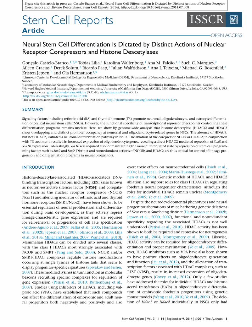
Figure 1. Single-Gene and Genome-wide ChIP Reveal Overlapping and Nonoverlapping Occupancies of HDAC2 and HDAC3 andHDAC3, but Not HDAC2, Knockdown Results in Increased Neuronal Differentiation of NSCs(A) Genome browser images of normalized densities of HDAC2 and HDAC3 on regions of chromosomes 3 and 7 in the vicinity of genesencoding Cebpb, Ovol2, Hoxd4, and Zfp7 in cortically derived NSCs.
(legend continued on next page)
2 Stem Cell Reports j Vol. 3 j 1–14 j September 9, 2014 j ª2014 The Authors
Stem Cell ReportsNCORs and HDACs Regulate Neural Differentiation
Please cite this article in press as: Castelo-Branco et al., Neural Stem Cell Differentiation Is Dictated by Distinct Actions of Nuclear ReceptorCorepressors and Histone Deacetylases, Stem Cell Reports (2014), http://dx.doi.org/10.1016/j.stemcr.2014.07.008
limited effects, while simultaneous deletion of the two
factors resulted in a loss of markers of oligodendrocyte
differentiation in cortical progenitors (Ye et al., 2009). Para-
doxically, genetic and RNA knockdown of the HAT Cbp
(also known as Crebbp) in the embryonic forebrain also
resulted in decreased expression of oligodendrocyte
markers in cortical NSCs, and Cbp haploinsufficiency leads
to aberrant development of the corpus callosum (Wang
et al., 2010).
To elucidate the roles for these factors in differentiation
of cortical progenitors, we undertook an investigation of
the functional roles for class I HDACs and nuclear receptor
corepressors in the enforcement of NSC repression check-
points that are subsequently released during differentia-
tion to neurons and glia. By analysis of NSCs derived
from rodents with gene deletions and/or specific RNA
knockdown in wild-type primary-derived NSCs, we have
revealed a series of distinct functional roles for the class I
HDACs, HDAC2, and HDAC3, alone and in concert with
NCOR or SMRT in the regulation of NSC differentiation
into neurons and oligodendrocytes.
RESULTS
HDAC2 and HDAC3 Show Both Unique and
Overlapping Binding to Promoter Regions of Genes
Associated with Neuronal and Oligodendrocyte
Differentiation
To investigate specific roles for HDAC2 and HDAC3, we
performed chromatin immunoprecipitation sequencing
(ChIP-seq) (Gene Expression Omnibus [GEO] accession
GSE57232; see Fullgrabe et al., 2013; Heldring et al.,
2014) and subsequent single-gene ChIP analysis (see Lilja
et al., 2013b) in neural stem cells (NSCs) derived from em-
bryonic cortices of rats at embryonic day 15 (E15), which
produce HDAC2 and HDAC3, but not HDAC1 (Figure 1C).
HDAC2 and HDAC3 were determined to be present in the
vicinity of a number of genes associated with transcrip-
(B) qPCR analysis of regions in the vicinity of Sox8 and Pax6 after Ch(C) Immunoblotting of NSC protein lysates with antibodies to classHDAC2, 60 kDa; HDAC3, 50 kDa.(D) Quantification of the numbers of TuJ1-positive cells after treatme(E) Immunofluorescence micrographs depicting NSCs stained for acesiHDAC2+3.(F) qPCR of the Bdnf IV promoter following ChIP with anti-acetyl-H3K9(siECFP) or Hdac3 siRNA.(G) qRT-PCR analysis of Bdnf mRNA levels following transfection with(H) qRT-PCR analysis of Bdnf mRNA levels in NSCs cultured from wild(I) qPCR of the Bdnf IV promoter following ChIP with anti-SMRT andThe scale bar represents 40 mm (E). All experiments were performed iError bars, SEM.
Stem
tional regulation of differentiation (Figure 1A). Several of
the regions identified by ChIP-seq were confirmed by sub-
sequent single-gene ChIP-quantitative PCR (ChIP-qPCR)
experiments that demonstrated that HDAC2 and HDAC3
could bind both in an overlapping and distinct fashion
near genes associated with development and differentia-
tion, including Cebpb (C/EBPb), Hoxd4, Ovol2, and Zfp7
(Figure 1A; data not shown). A more detailed analysis re-
vealed differences in enrichment at certain genes critically
involved in NSC differentiation. Whereas a significant
enrichment of HDAC3 was found on the promoters of
both Pax6 and Sox8, genes encoding transcription factors
associated with neuronal and oligodendrocyte differentia-
tion of NSCs, increased enrichment of HDAC2 was only
found on the Sox8 promoter (Figure 1B). This observation
was of particular interest due to a previous report demon-
strating that class I HDACs, including HDAC2, are essential
for proper progression of embryonic oligodendrocyte
development (Ye et al., 2009).
Similar to SMRT, HDAC3 Represses Neuronal Genes in
Embryonic NSCs
While previous analyses of the functional roles for HDAC1
and HDAC2 by genetic mouse models have confirmed
redundant roles for the two factors in embryonic develop-
ment of the nervous system, our ChIP analysis suggested
that there could be functional differences between
HDAC2 and HDAC3 in embryonic NSCs. To examine the
individual role(s) of these class I HDACs in NSCs, specific
small interference RNA (siRNA) pools were used to rapidly
and conditionally knockdown Hdac2 and Hdac3 mRNA in
the cortically derived NSCs (see Figures S1A and S1B avail-
able online for efficiency of the siRNAs). NSCs transfected
with siHDAC3 or a combination of siHDAC2 and siHDAC3
showed global hyperacetylation of H3K9 (Figure 1E) and a
significant increase in TuJ1-positive cells (Figure 1D), as
well as an increased H3K9 acetylation on the well-estab-
lished HDAC target Bdnf IV promoter as compared to con-
trol siRNA (Figure 1F). In contrast, transfection of siHDAC2
IP for HDAC2 and HDAC3 in NSCs.I HDAC proteins. Molecular weights (approximately): HDAC1 and
nt with control siRNA (siECFP), siHDAC2, siHDAC3, and siHDAC2+3.tylated H3K9 after treatment with siECFP, siHDAC2, siHDAC3, and
and anti-acetyl-H3K14 antibodies following transfection of control
siECFP, siHDAC2, or siHDAC3.-type or Smrt�/� embryos.anti-HDAC3 antibodies in NSCs.n n = 3–5 independent experiments, *p < 0.05, **p < 0.01 (t test).
Cell Reports j Vol. 3 j 1–14 j September 9, 2014 j ª2014 The Authors 3
Stem Cell ReportsNCORs and HDACs Regulate Neural Differentiation
Please cite this article in press as: Castelo-Branco et al., Neural Stem Cell Differentiation Is Dictated by Distinct Actions of Nuclear ReceptorCorepressors and Histone Deacetylases, Stem Cell Reports (2014), http://dx.doi.org/10.1016/j.stemcr.2014.07.008
had no significant effect on these parameters (Figures 1D–
1F). Analysis of gene expression by quantitative RT-PCR
(qRT-PCR) showed that siHDAC2 treatment had no sig-
nificant effect on Bdnf mRNA levels in NSCs, whereas
siHDAC3 delivery alone was sufficient to induce a signifi-
cant increase in Bdnf gene expression (Figure 1G). As the
gene encoding TUBB3, the protein detected by the anti-
body TuJ1, is a direct target for the HDAC-associated
repressor REST, it is possible that the increase in TuJ1-posi-
tive cells could be due to a direct regulation and increased
acetylation at the Tubb3 gene. We however also noted an
increase in expression of the proneuronal gene Neurog2
6–24 hr after siHDAC3 delivery (data not shown), pointing
to multiple putative mechanisms that could underlie
increased neuronal differentiation and survival after
siHDAC3 delivery. Together, these results demonstrate
that HDAC3, but not HDAC2, is a nonredundant repressor
of H3K9 acetylation, Bdnf gene expression, and neuronal
differentiation in cortical NSCs.
To shed further light on the mechanisms underlying the
functional specificity of HDAC2 and HDAC3, we turned to
the closely associated nuclear receptors NCOR and SMRT.
These HDAC-associated corepressors have in spite of their
sequence homology previously been shown to play distinct
functional roles in NSCs as spontaneous neuronal and
astrocytic differentiation has been noted only in NSCs
derived from Smrt-deficient mice, while only astrocytic
and no neuronal differentiation has been reported in
NSCs from Ncor-deficient or double-heterozygote mice
(Hermanson et al., 2002b; Jepsen et al., 2007). ChIP anal-
ysis revealed that both SMRT and HDAC3 were enriched
at the Bdnf IV promoter, and qRT-PCR investigation of
Bdnf gene expression levels in Smrt�/� NSCs revealed a sig-
nificant (>4-fold) increase compared to wild-type cells (Fig-
ures 1H and 1I). In contrast, Bdnf mRNA levels in Ncor�/�
NSCs, which do not differentiate down the neuronal
pathway, remained unchanged (data not shown; see
further below). This result suggests that HDAC3, rather
than HDAC2, is involved in at least a subset of the critical
SMRT-mediated repressive events in NSCs.
Absence of the T3-Associated Corepressor NCOR
Promotes Oligodendrocyte-Associated Gene
Expression
NSCs derived from mice gene-deleted for either Ncor or
Smrt, or from double-heterozygote animals, spontaneously
differentiate into astrocytes, suggesting that regulation of
this pathway depends on proper gene dosage of both
Ncor and Smrt (Hermanson et al., 2002b; Jepsen et al.,
2007). Interestingly, a substantial subset of the Ncor�/�
NSCs display neither the astrocytic marker GFAP nor the
stem cell marker nestin or neuronal marker TuJ1 (Herman-
son et al., 2002b), suggesting the possibility that NCOR
4 Stem Cell Reports j Vol. 3 j 1–14 j September 9, 2014 j ª2014 The Author
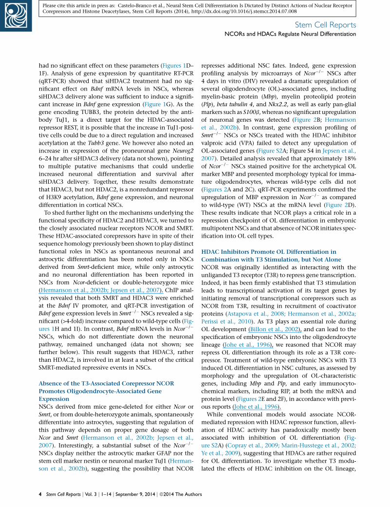
represses additional NSC fates. Indeed, gene expression
profiling analysis by microarrays of Ncor�/� NSCs after
4 days in vitro (DIV) revealed a dramatic upregulation of
several oligodendrocyte (OL)-associated genes, including
myelin-basic protein (Mbp), myelin proteolipid protein
(Plp), beta tubulin 4, and Nkx2.2, as well as early pan-glial
markers such as S100b, whereas no significant upregulation
of neuronal genes was detected (Figure 2B; Hermanson
et al., 2002b). In contrast, gene expression profiling of
Smrt�/� NSCs or NSCs treated with the HDAC inhibitor
valproic acid (VPA) failed to detect any upregulation of
OL-associated genes (Figure S2A; Figure S4 in Jepsen et al.,
2007). Detailed analysis revealed that approximately 18%
of Ncor�/� NSCs stained positive for the archetypical OL
marker MBP and presented morphology typical for imma-
ture oligodendrocytes, whereas wild-type cells did not
(Figures 2A and 2C). qRT-PCR experiments confirmed the
upregulation of MBP expression in Ncor�/� as compared
to wild-type (WT) NSCs at the mRNA level (Figure 2D).
These results indicate that NCOR plays a critical role in a
repression checkpoint of OL differentiation in embryonic
multipotent NSCs and that absence of NCOR initiates spec-
ification into OL cell types.
HDAC Inhibitors Promote OL Differentiation in
Combination with T3 Stimulation, but Not Alone
NCOR was originally identified as interacting with the
unliganded T3 receptor (T3R) to repress gene transcription.
Indeed, it has been firmly established that T3 stimulation
leads to transcriptional activation of its target genes by
initiating removal of transcriptional corepressors such as
NCOR from T3R, resulting in recruitment of coactivator
proteins (Astapova et al., 2008; Hermanson et al., 2002a;
Perissi et al., 2010). As T3 plays an essential role during
OL development (Billon et al., 2002), and can lead to the
specification of embryonic NSCs into the oligodendrocyte
lineage (Johe et al., 1996), we reasoned that NCOR may
repress OL differentiation through its role as a T3R core-
pressor. Treatment of wild-type embryonic NSCs with T3
induced OL differentiation in NSC cultures, as assessed by
morphology and the upregulation of OL-characteristic
genes, including Mbp and Plp, and early immunocyto-
chemical markers, including RIP, at both the mRNA and
protein level (Figures 2E and 2F), in accordance with previ-
ous reports (Johe et al., 1996).
While conventional models would associate NCOR-
mediated repression with HDAC repressor function, allevi-
ation of HDAC activity has paradoxically mostly been
associated with inhibition of OL differentiation (Fig-
ure S2A) (Copray et al., 2009; Marin-Husstege et al., 2002;
Ye et al., 2009), suggesting that HDACs are rather required
for OL differentiation. To investigate whether T3 modu-
lated the effects of HDAC inhibition on the OL lineage,
s
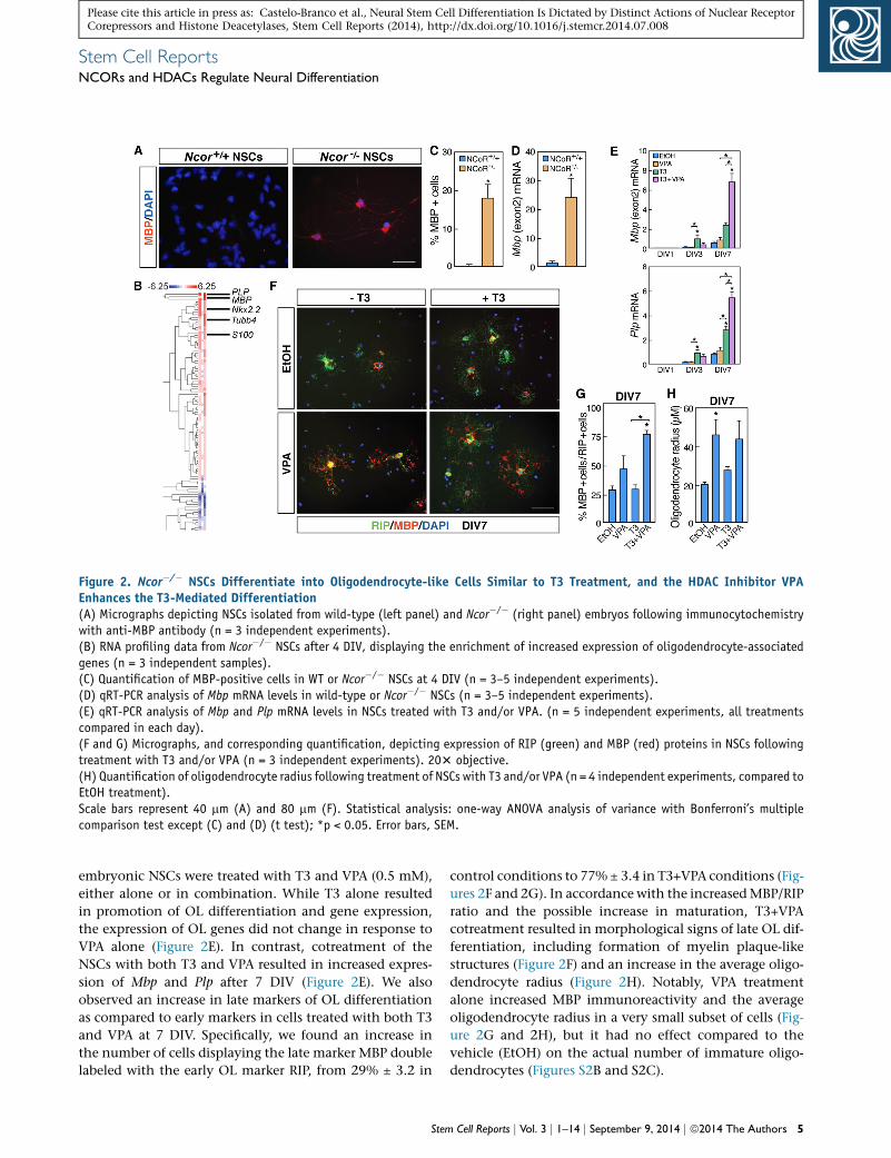
Figure 2. Ncor�/� NSCs Differentiate into Oligodendrocyte-like Cells Similar to T3 Treatment, and the HDAC Inhibitor VPAEnhances the T3-Mediated Differentiation(A) Micrographs depicting NSCs isolated from wild-type (left panel) and Ncor�/� (right panel) embryos following immunocytochemistrywith anti-MBP antibody (n = 3 independent experiments).(B) RNA profiling data from Ncor�/� NSCs after 4 DIV, displaying the enrichment of increased expression of oligodendrocyte-associatedgenes (n = 3 independent samples).(C) Quantification of MBP-positive cells in WT or Ncor�/� NSCs at 4 DIV (n = 3–5 independent experiments).(D) qRT-PCR analysis of Mbp mRNA levels in wild-type or Ncor�/� NSCs (n = 3–5 independent experiments).(E) qRT-PCR analysis of Mbp and Plp mRNA levels in NSCs treated with T3 and/or VPA. (n = 5 independent experiments, all treatmentscompared in each day).(F and G) Micrographs, and corresponding quantification, depicting expression of RIP (green) and MBP (red) proteins in NSCs followingtreatment with T3 and/or VPA (n = 3 independent experiments). 203 objective.(H) Quantification of oligodendrocyte radius following treatment of NSCs with T3 and/or VPA (n = 4 independent experiments, compared toEtOH treatment).Scale bars represent 40 mm (A) and 80 mm (F). Statistical analysis: one-way ANOVA analysis of variance with Bonferroni’s multiplecomparison test except (C) and (D) (t test); *p < 0.05. Error bars, SEM.
Stem Cell ReportsNCORs and HDACs Regulate Neural Differentiation
Please cite this article in press as: Castelo-Branco et al., Neural Stem Cell Differentiation Is Dictated by Distinct Actions of Nuclear ReceptorCorepressors and Histone Deacetylases, Stem Cell Reports (2014), http://dx.doi.org/10.1016/j.stemcr.2014.07.008
embryonic NSCs were treated with T3 and VPA (0.5 mM),
either alone or in combination. While T3 alone resulted
in promotion of OL differentiation and gene expression,
the expression of OL genes did not change in response to
VPA alone (Figure 2E). In contrast, cotreatment of the
NSCs with both T3 and VPA resulted in increased expres-
sion of Mbp and Plp after 7 DIV (Figure 2E). We also
observed an increase in late markers of OL differentiation
as compared to early markers in cells treated with both T3
and VPA at 7 DIV. Specifically, we found an increase in
the number of cells displaying the late marker MBP double
labeled with the early OL marker RIP, from 29% ± 3.2 in
Stem
control conditions to 77% ± 3.4 in T3+VPA conditions (Fig-
ures 2F and 2G). In accordance with the increasedMBP/RIP
ratio and the possible increase in maturation, T3+VPA
cotreatment resulted in morphological signs of late OL dif-
ferentiation, including formation of myelin plaque-like
structures (Figure 2F) and an increase in the average oligo-
dendrocyte radius (Figure 2H). Notably, VPA treatment
alone increased MBP immunoreactivity and the average
oligodendrocyte radius in a very small subset of cells (Fig-
ure 2G and 2H), but it had no effect compared to the
vehicle (EtOH) on the actual number of immature oligo-
dendrocytes (Figures S2B and S2C).
Cell Reports j Vol. 3 j 1–14 j September 9, 2014 j ª2014 The Authors 5
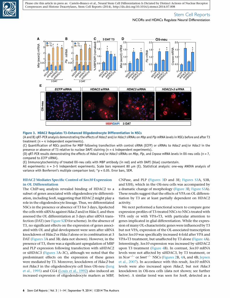
Figure 3. HDAC2 Regulates T3-Enhanced Oligodendrocyte Differentiation in NSCs(A and B) qRT-PCR analysis demonstrating the effects of Hdac2 and/or Hdac3 siRNAs onMbp and PlpmRNA levels in NSCs before and after T3treatment (n = 4 independent experiments).(C) Quantification of NSCs positive for MBP following transfection with control siRNA (ECFP) or siRNAs to Hdac2 and/or Hdac3 in thepresence or absence of T3 relative to nuclear DAPI staining (n = 4 independent experiments).(D) qRT-PCR results demonstrating the effects of Hdac2 and/or Hdac3 siRNAs on Mbp, Plp, and Cnpase mRNA levels in Oli-neu cells (n = 7,compared to ECFP siRNA).(E) Immunocytochemistry of treated Oli-neu cells with MBP antibody (in red) and with DAPI (blue) counterstain.All experiments: n = 3–5 independent experiments. Scale bars represent 80 mm (E). Statistical analysis: one-way ANOVA analysis ofvariance with Bonferroni’s multiple comparison test; *p < 0.05. Error bars, SEM.
Stem Cell ReportsNCORs and HDACs Regulate Neural Differentiation
Please cite this article in press as: Castelo-Branco et al., Neural Stem Cell Differentiation Is Dictated by Distinct Actions of Nuclear ReceptorCorepressors and Histone Deacetylases, Stem Cell Reports (2014), http://dx.doi.org/10.1016/j.stemcr.2014.07.008
HDAC2Mediates Specific Control of Sox10 Expression
in OL Differentiation
The ChIP-seq analysis revealed binding of HDAC2 to a
subset of genes associated with oligodendrocyte differenti-
ation, including Sox8, suggesting that HDAC2might play a
role in the oligodendrocyte lineage. Thus, we differentiated
NSCs in the presence or absence of T3 for 3 days, lipofected
the cells with siRNAs againstHdac2 and/orHdac3, and then
assessed the OL differentiation at 3 days after siRNA trans-
fection (DAT) (see Figure S2D for scheme). In the absence of
T3, no significant effects on the expression of genes associ-
ated with OL and glial development were seen after siRNA
knockdown ofHdac2 orHdac3 alone or in combination at 3
DAT (Figures 3A and 3B; data not shown). However, in the
presence of T3, there was a significant upregulation of MBP
and PLP expression following transfection with siHDAC2
or siHDAC3 (Figures 3A–3C), although we noted that the
predominant effects on the expression of these genes
were mediated by T3. Moreover, knockdown of Hdac2 but
not Hdac3 in the oligodendrocyte cell lines Oli-neu (Jung
et al., 1995) and CG4 (Louis et al., 1992) also induced an
increased expression of oligodendrocyte markers as MBP,
6 Stem Cell Reports j Vol. 3 j 1–14 j September 9, 2014 j ª2014 The Author
CNPase, and PLP (Figures 3D and 3E; Figures S3A, S3B,
and S3H), which in the Oli-neu cells was accompanied by
a dramatic change of morphology (Figure 3E; Figure S3A).
These results suggest that the effects of VPA onOL differen-
tiation by T3 are at least partially dependent on HDAC2
activity.
We next performed a functional screen to compare gene
expression profiles of T3-treated NSCs to NSCs treated with
VPA only or with VPA+T3, with particular attention to
genes implicated in glial differentiation. While the expres-
sion ofmanyOL-characteristic geneswere influenced by T3
but not VPA, expression of the OL-associated transcription
factor Sox10 was specifically increased 4-fold after VPA and
VPA+T3 treatment, but unaffected by T3 alone (Figure 4A).
Interestingly, Sox10 expression was increased by siHDAC2
upon T3 treatment (Figure 4B). In contrast, Sox10 mRNA
levels were not affected by siHDAC3, by T3 treatment, or
in Ncor�/� or Smrt�/� NSCs (Figures 2B, 4A, and 4B; Jepsen
et al., 2007). In accordance with this result, Sox10 mRNA
levels were also increased upon Hdac2, but not Hdac3
knockdown in Oli-neu cells (data not shown; see further
below). A similar trend was seen for Sox8, detected as a
s
Figure 4. HDAC2 Regulates Sox10 Expression, and Sox10 Knockdown in NSCs and Oli-neu Cells Prevents Effects of T3 and VPA onOligodendrocyte Differentiation(A) qRT-PCR analysis of Sox10 mRNA levels in NSCs treated with T3 and/or VPA.(B) qRT-PCR analysis of Sox10mRNA levels in NSCs transfected with control siRNA (ECFP) or siRNAs to Hdac2 and/or Hdac3 in the presenceor absence of T3.(C) qRT-PCR analysis of Hes5 mRNA levels in NSCs treated with T3 and/or VPA.(D) Quantification of NSCs expressing MBP following transfection with control siRNA (ECFP) or Sox10 siRNAs in the presence of T3 and VPArelative to nuclear DAPI staining.(E) qRT-PCR demonstrating the effects of Sox10 siRNA on expression of Mbp, Plp, Sox8, Cnpase, and Sox9 following VPA and/or T3treatment.All experiments: n = 3–5 independent experiments. Statistical analysis: one-way ANOVA analysis of variance with Bonferroni’s multiplecomparison test; *p < 0.05. Error bars, SEM.
Stem Cell ReportsNCORs and HDACs Regulate Neural Differentiation
Please cite this article in press as: Castelo-Branco et al., Neural Stem Cell Differentiation Is Dictated by Distinct Actions of Nuclear ReceptorCorepressors and Histone Deacetylases, Stem Cell Reports (2014), http://dx.doi.org/10.1016/j.stemcr.2014.07.008
direct HDAC2 target in the ChIP-seq experiments (Fig-
ure 1B). Transcriptional inhibitors of OL differentiation,
such as Hes5, were not affected by treatment with VPA
alone in the NSCs (Figure 4C), in contrast to what has
been observed in postnatal oligodendrocyte precursor cells
(OPCs) (Lyssiotis et al., 2007; Shen et al., 2008).
SOX10 is required for terminal OL differentiation as null
mutant mice for this SoxE protein generate normal
numbers of OL progenitors that fail to terminally differen-
tiate (Stolt et al., 2002). To test whether SOX10 could
mediate the additive effects of VPA in T3-mediated OL dif-
ferentiation of embryonic NSCs, NSCs were differentiated
in the presence of VPA and/or T3 and lipofected at 3 days
with siRNA against Sox10 (Roh et al., 2006) (Figure S2D; Fig-
ure S1B for efficiency of the siRNA). Gene expression was
then analyzed 3 DAT. Knockdown of Sox10 partially pre-
Stem
vented the upregulation of MBP and PLP and blocked the
terminal OL differentiation induced by the cotreatment
of VPA and T3 at 3 DAT (Figures 4D and 4E). Sox10 knock-
down also reduced themRNA levels ofCnpase and Sox8 at 3
DAT, but importantly did not affect earlier markers,
including Sox9, in differentiating NSCs (Figure 4E). We
conclude that SOX10 is required for the properOL differen-
tiation effects observed when treating NSCs with VPA or
siHDAC2 and T3.
To investigate specific roles for class I HDACs in the VPA-
mediated potentiation of OL differentiation of NSCs, we
next investigated the presence of HDAC2 and HDAC3 at
previously characterized regulatory regions ofMbp (Farhadi
et al., 2003) and Sox10 (Werner et al., 2007) in the embry-
onic NSCs (Figure 5A). ChIP-qPCR analysis indicated that
while both HDAC2 and HDAC3 were present in the
Cell Reports j Vol. 3 j 1–14 j September 9, 2014 j ª2014 The Authors 7
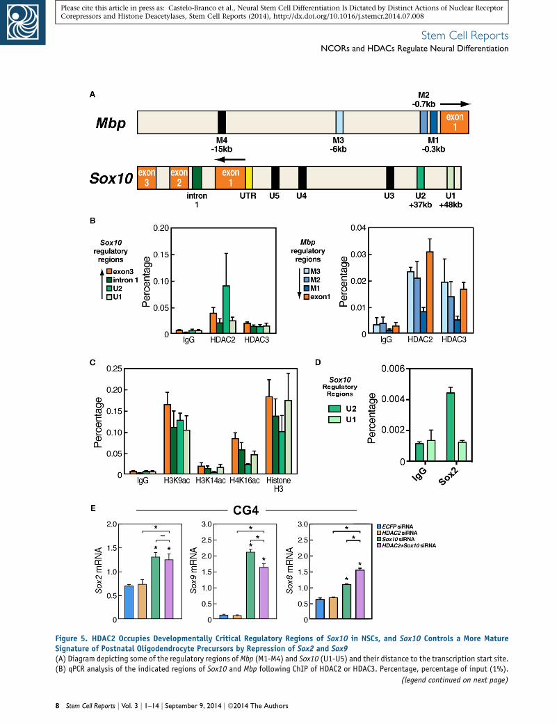
Figure 5. HDAC2 Occupies Developmentally Critical Regulatory Regions of Sox10 in NSCs, and Sox10 Controls a More MatureSignature of Postnatal Oligodendrocyte Precursors by Repression of Sox2 and Sox9(A) Diagram depicting some of the regulatory regions of Mbp (M1-M4) and Sox10 (U1-U5) and their distance to the transcription start site.(B) qPCR analysis of the indicated regions of Sox10 and Mbp following ChIP of HDAC2 or HDAC3. Percentage, percentage of input (1%).
(legend continued on next page)
8 Stem Cell Reports j Vol. 3 j 1–14 j September 9, 2014 j ª2014 The Authors
Stem Cell ReportsNCORs and HDACs Regulate Neural Differentiation
Please cite this article in press as: Castelo-Branco et al., Neural Stem Cell Differentiation Is Dictated by Distinct Actions of Nuclear ReceptorCorepressors and Histone Deacetylases, Stem Cell Reports (2014), http://dx.doi.org/10.1016/j.stemcr.2014.07.008
regulatory regions of the Mbp gene in NSCs and Oli-neu
cells (Figure 5B; Figure S3D), HDAC2 was specifically
enriched in the U2 enhancer of Sox10 (Figure 5B, left
panel). This HDAC2 enrichment was observed both in
comparison to other regions bound by HDAC2 as well as
in comparison to the occupancy of HDAC3 at the same re-
gion (Figure 5B). Similar results were obtained in Oli-neu
cells (Figures S3C and S3F). Indeed, an analysis of the acet-
ylation state of the regulatory regions of Sox10 revealed
relatively low acetylation levels of H3K14 and H4K16, but
not H3K9, at the U2 enhancer in NSCs and to an extent
also in the Oli-neu cells, strengthening the observation of
increased HDAC2 occupancy at this specific enhancer (Fig-
ure 5C; Figure S3E). The U2 enhancer is an element that
previously has been shown to regulate Sox10 expression
specifically in the oligodendrocyte lineage starting at em-
bryonic midgestation (Kuspert et al., 2011; Werner et al.,
2007). Together with our data, this suggests that HDAC2
may be specifically required for Sox10 gene regulation in
early oligodendrocyte development. Class I HDACs and
NCORs have been identified as binding partners of SOX2
(Engelen et al., 2011), and an in silico analysis revealed a
putative binding site for SOX2 in the U2 enhancer of
Sox10 (Figure S4). We therefore investigated whether this
T3 and NCoR-independent HDAC2-mediated repression
of Sox10 could be mediated in association with SOX2.
Indeed, we found a significant enrichment of SOX2 occu-
pancy on the U2, but not U1, enhancer compared to con-
trol regions (Figure 5D; data not shown). siRNA-mediated
knockdown of Sox2, however, did not result in any signifi-
cantly increased expression of Sox10 (data not shown), sug-
gesting that other factors are sufficient to recruit HDAC2 to
the U2 enhancer and mediate the essential repression.
Sox10, but Not HDACs, Is Required for Maintenance of
the Differentiated State in Postnatal OL Progenitors
The differentiation of OPCs begins in early postnatal stages
in the forebrain, although its peak occurs a few days later
(Wegner, 2008). It is noteworthy that Hdac2 expression
has been reported to decrease in cells of the oligodendro-
cyte lineage at these stages (Shen et al., 2005). Interestingly,
we did not observe any morphological signs of premature
differentiation of the proliferating postnatal CG4 upon
siHDAC2 and/or siHDAC3 delivery (data not shown), in
contrast to what we observed in embryonic Oli-neu cells
(C) qPCR of the indicated regions of Sox10 following ChIP of acetylatedfor ChIP efficiency.(D) qPCR of the indicated regions after ChIP of SOX2 revealed that SO(E) qRT-PCR demonstrating the effects of Sox10 siRNA and/or Hdac2 siROPCs) after Sox10 RNA knockdown.All experiments: n = 2–3 independent experiments. Statistical analyscomparison test; *p < 0.05. Error bars, SEM.
Stem
or T3-treated NSCs (Figure 2; Figure S3A). Moreover,
although moderate increases in gene expression levels of
Mbp and Plp were observed following transfection with
siHDAC2 in the CG4 cells (Figure S3H), these were found
not to be associated with an siHDAC2-mediated Sox10
derepression as Sox10 levels were indeed rescued by
siHDAC2, but Mbp and Plp expression levels were not in
these cells (Figure S3H; data not shown). These results
suggested that SOX10 could play alternative roles at this
late stage of OL differentiation. Intriguingly, Sox10 siRNA
delivery at this stage instead resulted in a dramatic increase
in gene expression associated with the stem cell state
(Pevny and Nicolis, 2010; Scott et al., 2010), including
Sox2, Sox9, Sox8, and Hes5, without affecting markers of
neuronal differentiation (Figure 5E; data not shown). This
result stands in sharp contrast to the mild effects of Sox10
siRNA on the expression of these genes in NSCs and a
reversible increase of Sox9 expression in Oli-neu cells (Fig-
ure 4E; data not shown). Taken together, our observations
suggest that Sox10 expression is required for efficient pro-
gression of oligodendrocyte differentiation and expression
of late OL characteristic genes and at a later stage serves to
maintain the differentiated state and suppress the expres-
sion of genes associated with more undifferentiated pro-
genitors and stem cells (Figure 6).
DISCUSSION
Transcriptional corepressors such as NCOR and SMRT have
distinct roles in repressing differentiation in NSCs, and we
therefore hypothesized that associated HDACs also have
specific functions albeit the reports so far have been
unclear. Here we demonstrate that whereas HDAC3 and
SMRT, and not HDAC2 or NCoR, are part of a repression
checkpoint for neuronal differentiation in embryonic
NSCs, NCoR and HDAC2 constitute specific repressor
checkpoints in control of oligodendrocyte differentiation
(Figure 6). The mechanism by which HDAC2 inhibits
oligodendrocyte differentiation is at least in part, via
repression of the key transcription factor SOX10, coin-
ciding with binding of HDAC2 to the SOX2-occupied U2
enhancer of the Sox10 gene (Figure 6). Together, these
data reveal nonredundant roles for class I HDACs and asso-
ciated factors in NSC differentiation.
H3K9, H3K14, or H4K16 from NSCs. Histone H3 is shown as a control
X2 is bound to the U2, but not the U1, enhancer of the Sox10 gene.NA on the expression of Sox2, Sox8, and Sox9 in CG4 cells (postnatal
is: one-way ANOVA analysis of variance with Bonferroni’s multiple
Cell Reports j Vol. 3 j 1–14 j September 9, 2014 j ª2014 The Authors 9
Figure 6. NCOR and HDACs Play Nonredundant Roles in the Regulation of Neural Stem Cell DifferentiationIn multipotent embryonic NSCs, SMRT and HDAC3 are nonredundantly repressing neuronal differentiation, at least in part by repressingBdnf expression required for the cell survival during the progression of neuronal differentiation. Astrocytic differentiation is repressed byboth NCOR and SMRT. Oligodendrocyte differentiation is repressed by NCOR and HDAC2 in neural stem cells by repressing different OL-associated genes. In Oli-neu cells (prenatal oligodendrocyte progenitors), a repression of Sox10 is maintained by HDAC2 that occupies theregulatory U2 enhancer of Sox10. In CG4 cells (postnatal oligodendrocyte precursors), Sox10 is not only required for the progression of OLdifferentiation but also for retaining a more differentiated state by being required for efficient repression of Sox2 along with Sox9, Hes5,and other progenitor associated genes.
Stem Cell ReportsNCORs and HDACs Regulate Neural Differentiation
Please cite this article in press as: Castelo-Branco et al., Neural Stem Cell Differentiation Is Dictated by Distinct Actions of Nuclear ReceptorCorepressors and Histone Deacetylases, Stem Cell Reports (2014), http://dx.doi.org/10.1016/j.stemcr.2014.07.008
Our results suggest that specific HDACs play distinct
roles in the differentiation of NSCs. Chemical or genetic
ablation of class I HDAC activity have been reported to
inhibit oligodendrocyte and Schwann cell differentiation
and promote neuronal differentiation (Copray et al.,
2009; Hsieh et al., 2004; Marin-Husstege et al., 2002). In
these studies, HDACs are suggested to repress inhibitors
of OL differentiation, thereby being required for proper
oligodendrocyte differentiation. However, deletion of the
HAT CREB-binding protein also results in decreased oligo-
dendrocyte differentiation and corpus callosum deficits
(Wang et al., 2010), and HDACs have also been shown to
be required for neurogenesis (Montgomery et al., 2009),
blurring a general consensus statement of the function of
these factors in NSC differentiation. In the present study,
loss of HDAC2 activity in the presence of T3 resulted in
enhanced oligodendrocytic differentiation in embryonic
cortical NSCs. Strikingly, HDAC2 does repress directly the
key OL transcription factor Sox10, in contrast to the indi-
rect effects of HDACs previously reported. Thus, the effects
of HDAC inhibition on NSCs appear to be distinct depend-
10 Stem Cell Reports j Vol. 3 j 1–14 j September 9, 2014 j ª2014 The Autho
ing on cell-type specificity, the temporal window of action,
and the presence of additional factors. These cell-type and
temporal specific effects of HDACs are in linewithwhat has
been reported for components of the NOTCH and WNT
signaling, which can have both repressing and activating
functions in oligodendrocyte development (Popko, 2003;
Tawk et al., 2011).
Class B1 Sox proteins such as SOX2 are established re-
pressors of differentiation and required to retain stem cell
characteristics during development and in the adult
(Arnold et al., 2011; Bylund et al., 2003; Engelen et al.,
2011; Graham et al., 2003), but it has been shown that
SOX2 can also act as a transcriptional activator in certain
developmental contexts (Hoffmann et al., 2014). Our
data on NCOR, HDAC2, and SOX10 suggest a transcrip-
tional ‘‘transmission’’ mechanism where the appropriate
‘‘gear’’ controls the level of progenitor state and early and
late progression of oligodendrocyte differentiation. In
NSCs and embryonic OPCs, HDAC2 were found to bind
to the critical U2 enhancer regulating Sox10 in association
with SOX2, and alleviation of HDAC2 led to an increase in
rs
Stem Cell ReportsNCORs and HDACs Regulate Neural Differentiation
Please cite this article in press as: Castelo-Branco et al., Neural Stem Cell Differentiation Is Dictated by Distinct Actions of Nuclear ReceptorCorepressors and Histone Deacetylases, Stem Cell Reports (2014), http://dx.doi.org/10.1016/j.stemcr.2014.07.008
Sox10 expression. OLIG2 has recently been shown to acti-
vate the U2 enhancer (Kuspert et al., 2011) and as such
could also be a candidate for the upregulation of Sox10
expression. Nevertheless, in contrast to SOX2 (Figure 5D),
OLIG2 is present not only at the U2 enhancer in immature
oligodendrocytes but also at the U1, U3, U4, and U5 Sox10
enhancers (Yu et al., 2013), suggesting a more general role
in the transcriptional regulation of SOX10.While OLIG2 is
an important regulator of this system, we do not observe an
upregulation of Olig2 upon VPA+T3 treatment or Hdac2
and/orHdac3 siRNA in NSCs and Oli-neu cells, respectively
(Figures S3G and S3I). It will be of immediate interest to
elucidate additional factors responsible for HDAC2 recruit-
ment to the U2 enhancer in NSCs and prenatal oligoden-
drocyte progenitors. Indeed, since Sox2 knockdown failed
to induce a significant increase in Sox10 expression even
in the presence of T3, it is plausible that other class B1
Sox factors such as SOX1/3 could be involved, and the
role for class B2 factors binding similar sequences but
with alternative actions, such as SOX14 or SOX21, should
be further investigated.
In postnatal OPCs, increased levels of SOX10 seem to
repress Sox2 expression and consequently promote late
progression of oligodendrocyte differentiation. Intrigu-
ingly, forced Sox2 expression mediates dedifferentiation
of maturely differentiated oligodendrocytes (Kondo and
Raff, 2000, 2004) and SOX9 has been shown to promote
neural stem cell state (Scott et al., 2010). Whereas there
are few previous demonstrations of such key factors main-
taining differentiated state in the central nervous system,
the requirement of SOX10 for retaining a more differenti-
ated state partly resembles the master role of PAX5 main-
taining the differentiated state of mature B lymphocytes
(Cobaleda et al., 2007). Our present observations that
SOX10 is required for repression of Sox2 and Sox9 in late
oligodendrocyte precursor cells, in addition to the distinct
requirements for NCOR, SMRT, and HDAC-regulated
repressor actions in the progression of neural stem and pro-
genitor differentiation, suggest a distinct order of repressor
and corepressor action to mediate the serial events that
details the neuronal and oligodendrocyte lineages, with
expression of Sox10 required to prevent reexpression of
gene products promoting stem cell characteristics.
EXPERIMENTAL PROCEDURES
Embryonic Neural Stem Cell CulturesRat embryonic neural stem cells were derived as previously
described in detail elsewhere (Hermanson et al., 2002a; Johe
et al., 1996; Joseph et al., 2009). In brief, rat cortical tissue from
embryonic day 15.5was dissociated and8003 103 cellswere plated
per 10 cm dish, previously coated with poly-L-ornithine and fibro-
nectin (both from Sigma-Aldrich). Neural stem cells were then
Stem
expanded in N2 media with 10 ng/ml of basic fibroblastic growth
factor (R&DSystems) andpassagedby light dissociation in thepres-
ence of HBSS, NaHCO3, and HEPES (from Invitrogen and Sigma-
Aldrich). For differentiation assays, neural stem cells (passage 1)
were plated at the following densities: 150 3 103 cells in 35 mm
plates (Corning) or 300 3 103 cells in 60 mm plates (Corning).
NSCs were differentiated for up to 7 DIV in the absence of FGF-2
(N2), in the presence of VPA (0.5 mM, a concentration at which
it specifically inhibits class I HDACs, Sigma-Aldrich) (Gottlicher
et al., 2001) or its vehicle 70% ethanol (EtOH), andwith or without
thyroid hormone (T3—50 ng/ml, Sigma-Aldrich). Factors were
added daily and media was changed every second day. After 1, 3,
or 7 DIV, cells were fixed with 10% formalin (Sigma-Aldrich) and
analyzed by immunocytochemistry or qRT-PCR. For ChIP assays,
NSCs were expanded after the first passage until confluency,
without contact between adjacent colonies. Animals were treated
in accordance with institutional and national guidelines, with all
required ethical permits. In Figure S2, FGF2 (10 ng/ml) expanded
NSCs were treated for 3 DIV with FGF2 (10 ng/ml), IGF-2
(5 ng/ml), and PDGF-BB (10 ng/ml) (OPC media).
OPC CulturesThe embryonic OPC cell line Oli-neu and the postnatal OPC cells
were cultured as previously described (Jung et al., 1995; Louis et al.,
1992); for details see Supplemental Information.
Transfection-LipofectionsiRNAs or pcDNAEF1-eGFP (Amaxa) were lipofected using Lipo-
fectamine 2000 (Invitrogen), following themanufacturer’s instruc-
tions. Three micrograms of siRNA or 1 mg pcDNAEF1-eGFP was
used per 35-mm plate. Opti-MEM I reduced serum medium was
used to prepare the complexes. Cells were changed to Dulbecco’s
modified Eagle’s medium (Invitrogen) and incubated with the
complexes for 4 hr before returning to their original media. Cells
were collected for immunocytochemistry or qRT-PCR analysis
after 1 or 3 DIV. Lipofection of siRNA in differentiated conditions
was performed after 3 DIV differentiation. The following target
sequences for siRNAs (A4 synthesis, Dharmacon) were used.
ECFP Control
50-GAAGAACGGCAUCAAGGCC-30.Sox10
50- CUGCUGUUCCUUCUUGACC-3 (Roh et al., 2006).
Hdac2
(1) 50-AAA UGU CGC UGA UCA UAA GAA-30.(2) 50-AAG GUG UUC AAA UGC AAG CUA-30.Hdac3(1) 50-AAU AGC CUA GUC CUG CAU UAU-30.(2) 50-AAC ACA GCU AAA CAA UAA GAU-30
Optimal results for differentiation and gene expression in NSCs
by HDAC2 and HDAC3 siRNAs were titrated to knockdowns in
the range of 50%–75%. Higher efficiency resulted in increased
cell death along with previous observations, and lower efficiency
yielded less significant results.
Chromatin Immunoprecipitation and qPCRChIP-IT Express (ActiveMotif) was used according to the supplier’s
recommendations. Cells were crosslinked using 1% formaldehyde
Cell Reports j Vol. 3 j 1–14 j September 9, 2014 j ª2014 The Authors 11
Stem Cell ReportsNCORs and HDACs Regulate Neural Differentiation
Please cite this article in press as: Castelo-Branco et al., Neural Stem Cell Differentiation Is Dictated by Distinct Actions of Nuclear ReceptorCorepressors and Histone Deacetylases, Stem Cell Reports (2014), http://dx.doi.org/10.1016/j.stemcr.2014.07.008
for 10 min. Cells were rinsed twice with cold PBS, collected by
scraping and pelleted at 2,000 rpm for 4 min at 4�C. Frozen
pelleted cells were resuspended in lysis buffer and centrifuged for
10 min at 4�C, and the pelleted nuclei were resuspended in the
shearing buffer. Chromatin was then sonicated using a Bioruptor
200 (Diagenode) at high frequency, 0.5 min/0.5min, for 12 min.
Sonicated chromatin was analyzed in a 3% agarose gel to confirm
efficient sonication. Input was collected for further analysis. Five
micrograms of chromatin was incubated for 1 hr at 4�C with pro-
tein G magnetic beads and then immunoprecipitated overnight
at 4�C with the following antibodies: a-H3K9ac (07-352; Upstate,
5 ml), a-H3K14ac (06-911; Upstate, 5 ml), a-H4K16ac (07-329;
Upstate, 5 ml), a-HDAC2 (sc-7899x; Santa Cruz, 5 mg), a-HDAC3
(sc-11417x; Santa Cruz, 5 mg), a-SOX2 (sc-17320x; Santa Cruz,
1 mg), RNA polymerase II (Active Motif, positive control for ChIP-
IT Express, 2 mg), Histone H3 (Abcam, ab1791, 2 mg), rabbit IgG
(Active Motif, negative control, 2 mg; Santa Cruz, sc-2027, 1 mg),
and goat IgG (Santa Cruz, sc-2028, 1 mg). After three washes with
ChIP buffer 1 and two washes with ChIP buffer 2, the IP DNA
was reverse crosslinked and resuspended in a final volume of
130 ml. Purified DNA and 1% input were analyzed by qPCR, using
5-fold dilutions of the input for standard curves and triplicates per
sample. When primer dimers were detected, the relative quantity
for the specific sample was considered to be zero. As the standard
curve method was used, potential differences in primer efficiency
were taken in account and occupancy in different regulatory re-
gions could be compared for a specific antibody.
RNA ProfilingRNA profiling was performed as previously described (Jepsen et al.,
2007); for details see Supplemental Information.
Statistical AnalysisStatistical analysis and graphs were performed using the software
Prism 4 (Graph Pad). When comparing to a hypothetical control
value (100%), one sample t test was used. When two groups were
compared, statistical analysis was performed using two-tailed
unpaired t test. When comparing three or more groups, one-way
ANOVA analysis of variance with Bonferroni’s multiple compari-
son test was used. Comparisons were performed between all treat-
ments, unless stated otherwise. The threshold value for statistical
significance (a value) was set at 0.05 (*p < 0.05). In all graphs, re-
sults are expressed as mean ± SEM. The ‘‘n’’ in the figure legends
represents the number of independent experiments.
ACCESSION NUMBERS
The ChIP-seq data are freely accessible in the GEO database under
accession number GSE57232. Additional descriptions of qRT-PCR,
immunocytochemistry and ChIP-seqmethods can be found in the
Supplemental Information.
SUPPLEMENTAL INFORMATION
Supplemental Information includes Supplemental Experimental
Procedures, four figures, and one table and can be found
with this article online at http://dx.doi.org/10.1016/j.stemcr.
2014.07.008.
12 Stem Cell Reports j Vol. 3 j 1–14 j September 9, 2014 j ª2014 The Autho
ACKNOWLEDGMENTS
Wewould like to thank Dr. J.C. Louis (Amgen) for the CG4 cell line
(Louis et al., 1992), Dr. J. Trotter (University of Mainz) for the
Oli-neu cell line (Jung et al., 1995), J. Hightower for artwork, and
Dr. M. Gotz and members of the O.H. lab for valuable discussions.
M.G.R. is a Howard Hughes Medical Institute Investigator. This
study was supported by NIH grants (to M.G.R.), a Scientist Devel-
opment Grant from the American Heart Association (to K.J.), and
grants from the Swedish Brain Foundation (Hjarnfonden), David
and Astrid Hagelen Foundation, SSMF (Svenska Sallskapet for Med-
icinsk Forskning), the Marie Curie Integration Grant, Seventh
Framework Programme, European Union (to G.C.-B.), the Swedish
ResearchCouncil (VR-MH), Karolinska Institutet Research Founda-
tions, the Swedish Society of Medicine (SLS) (to G.C.-B. and O.H.),
the Swedish Cancer Society (CF), the KI Cancer Network, the
Swedish Foundation for Strategic Research (SSF), and the Swedish
Childhood Cancer Foundation (BCF) (to O.H.).
Received: June 20, 2013
Revised: July 20, 2014
Accepted: July 21, 2014
Published: August 28, 2014
REFERENCES
Andreu-Agullo, C., Morante-Redolat, J.M., Delgado, A.C., and
Farinas, I. (2009). Vascular niche factor PEDF modulates Notch-
dependent stemness in the adult subependymal zone. Nat.
Neurosci. 12, 1514–1523.
Arnold, K., Sarkar, A., Yram, M.A., Polo, J.M., Bronson, R., Sen-
gupta, S., Seandel, M., Geijsen, N., and Hochedlinger, K. (2011).
Sox2(+) adult stem and progenitor cells are important for tissue
regeneration and survival of mice. Cell Stem Cell 9, 317–329.
Astapova, I., Lee, L.J., Morales, C., Tauber, S., Bilban, M., and Hol-
lenberg, A.N. (2008). The nuclear corepressor, NCoR, regulates thy-
roid hormone action in vivo. Proc. Natl. Acad. Sci. USA 105,
19544–19549.
Ballas, N., Grunseich, C., Lu, D.D., Speh, J.C., and Mandel, G.
(2005). REST and its corepressors mediate plasticity of neuronal
gene chromatin throughout neurogenesis. Cell 121, 645–657.
Billon, N., Jolicoeur, C., Tokumoto, Y., Vennstrom, B., and Raff, M.
(2002). Normal timing of oligodendrocyte development depends
on thyroid hormone receptor alpha 1 (TRalpha1). EMBO J. 21,
6452–6460.
Bylund, M., Andersson, E., Novitch, B.G., and Muhr, J. (2003).
Vertebrate neurogenesis is counteracted by Sox1-3 activity. Nat.
Neurosci. 6, 1162–1168.
Cobaleda, C., Jochum, W., and Busslinger, M. (2007). Conversion
of mature B cells into T cells by dedifferentiation to uncommitted
progenitors. Nature 449, 473–477.
Copray, S., Huynh, J.L., Sher, F., Casaccia-Bonnefil, P., and
Boddeke, E. (2009). Epigenetic mechanisms facilitating oligo-
dendrocyte development, maturation, and aging. Glia 57, 1579–
1587.
Covey, M.V., Streb, J.W., Spektor, R., and Ballas, N. (2012). REST
regulates the pool size of the different neural lineages by restricting
rs
Stem Cell ReportsNCORs and HDACs Regulate Neural Differentiation
Please cite this article in press as: Castelo-Branco et al., Neural Stem Cell Differentiation Is Dictated by Distinct Actions of Nuclear ReceptorCorepressors and Histone Deacetylases, Stem Cell Reports (2014), http://dx.doi.org/10.1016/j.stemcr.2014.07.008
the generation of neurons and oligodendrocytes fromneural stem/
progenitor cells. Development 139, 2878–2890.
Engelen, E., Akinci, U., Bryne, J.C., Hou, J., Gontan, C., Moen, M.,
Szumska, D., Kockx, C., van Ijcken,W., Dekkers, D.H., et al. (2011).
Sox2 cooperates with Chd7 to regulate genes that are mutated in
human syndromes. Nat. Genet. 43, 607–611.
Farhadi, H.F., Lepage, P., Forghani, R., Friedman, H.C., Orfali, W.,
Jasmin, L., Miller, W., Hudson, T.J., and Peterson, A.C. (2003). A
combinatorial network of evolutionarily conserved myelin basic
protein regulatory sequences confers distinct glial-specific pheno-
types. J. Neurosci. 23, 10214–10223.
Fullgrabe, J., Lynch-Day, M.A., Heldring, N., Li, W., Struijk, R.B.,
Ma, Q., Hermanson, O., Rosenfeld, M.G., Klionsky, D.J., and
Joseph, B. (2013). The histone H4 lysine 16 acetyltransferase
hMOF regulates the outcome of autophagy. Nature 500, 468–471.
Gottlicher, M., Minucci, S., Zhu, P., Kramer, O.H., Schimpf, A., Gia-
vara, S., Sleeman, J.P., Lo Coco, F., Nervi, C., Pelicci, P.G., and
Heinzel, T. (2001). Valproic acid defines a novel class of HDAC in-
hibitors inducing differentiation of transformed cells. EMBO J. 20,
6969–6978.
Graham, V., Khudyakov, J., Ellis, P., and Pevny, L. (2003). SOX2
functions to maintain neural progenitor identity. Neuron 39,
749–765.
Heldring, N., Nyman, U., Lonnerberg, P., Onnestam, S., Herland,
A., Holmberg, J., and Hermanson, O. (2014). NCoR controls glio-
blastoma tumor cell characteristics. Neuro-oncol. 16, 241–249.
Hermanson, O., Glass, C.K., and Rosenfeld, M.G. (2002a). Nuclear
receptor coregulators: multiple modes of modification. Trends
Endocrinol. Metab. 13, 55–60.
Hermanson, O., Jepsen, K., and Rosenfeld, M.G. (2002b). N-CoR
controls differentiation of neural stem cells into astrocytes. Nature
419, 934–939.
Hoffmann, S.A., Hos, D., Kuspert, M., Lang, R.A., Lovell-Badge, R.,
Wegner, M., and Reiprich, S. (2014). Stem cell factor Sox2 and its
close relative Sox3 have differentiation functions in oligodendro-
cytes. Development 141, 39–50.
Hsieh, J., Nakashima, K., Kuwabara, T., Mejia, E., and Gage, F.H.
(2004). Histone deacetylase inhibition-mediated neuronal differ-
entiation of multipotent adult neural progenitor cells. Proc. Natl.
Acad. Sci. USA 101, 16659–16664.
Jepsen, K., Hermanson, O., Onami, T.M., Gleiberman, A.S., Lu-
nyak, V., McEvilly, R.J., Kurokawa, R., Kumar, V., Liu, F., Seto, E.,
et al. (2000). Combinatorial roles of the nuclear receptor core-
pressor in transcription and development. Cell 102, 753–763.
Jepsen, K., Solum, D., Zhou, T., McEvilly, R.J., Kim, H.-J., Glass,
C.K., Hermanson, O., and Rosenfeld, M.G. (2007). SMRT-mediated
repression of an H3K27 demethylase in progression from neural
stem cell to neuron. Nature 450, 415–419.
Johe, K.K., Hazel, T.G., Muller, T., Dugich-Djordjevic, M.M., and
McKay, R.D. (1996). Single factors direct the differentiation of
stem cells from the fetal and adult central nervous system. Genes
Dev. 10, 3129–3140.
Johnson, R., Teh, C.H., Kunarso, G., Wong, K.Y., Srinivasan, G.,
Cooper, M.L., Volta, M., Chan, S.S., Lipovich, L., Pollard, S.M.,
Stem
et al. (2008). REST regulates distinct transcriptional networks in
embryonic and neural stem cells. PLoS Biol. 6, e256.
Joseph, B., Andersson, E.R., Vlachos, P., Sodersten, E., Liu, L., Teix-
eira, A.I., and Hermanson, O. (2009). p57Kip2 is a repressor of
Mash1 activity and neuronal differentiation in neural stem cells.
Cell Death Differ. 16, 1256–1265.
Jung, M., Kramer, E., Grzenkowski, M., Tang, K., Blakemore, W.,
Aguzzi, A., Khazaie, K., Chlichlia, K., von Blankenfeld, G., Ketten-
mann, H., et al. (1995). Lines of murine oligodendroglial precursor
cells immortalized by an activated neu tyrosine kinase show
distinct degrees of interaction with axons in vitro and in vivo.
Eur. J. Neurosci. 7, 1245–1265.
Kondo, T., and Raff, M. (2000). Oligodendrocyte precursor cells re-
programmed to become multipotential CNS stem cells. Science
289, 1754–1757.
Kondo, T., and Raff,M. (2004). Chromatin remodeling andhistone
modification in the conversion of oligodendrocyte precursors to
neural stem cells. Genes Dev. 18, 2963–2972.
Kuspert, M., Hammer, A., Bosl, M.R., andWegner, M. (2011). Olig2
regulates Sox10 expression in oligodendrocyte precursors through
an evolutionary conserved distal enhancer. Nucleic Acids Res. 39,
1280–1293.
Laeng, P., Pitts, R.L., Lemire, A.L., Drabik, C.E.,Weiner, A., Tang,H.,
Thyagarajan, R.,Mallon, B.S., andAltar, C.A. (2004). Themood sta-
bilizer valproic acid stimulates GABA neurogenesis from rat fore-
brain stem cells. J. Neurochem. 91, 238–251.
Lilja, T., Heldring, N., and Hermanson, O. (2013a). Like a rolling
histone: epigenetic regulation of neural stem cells and brain devel-
opment by factors controlling histone acetylation and methyl-
ation. Biochim. Biophys. Acta 1830, 2354–2360.
Lilja, T., Wallenborg, K., Bjorkman, K., Albage, M., Eriksson, M.,
Lagercrantz, H., Rohdin, M., and Hermanson, O. (2013b). Novel
alterations in the epigenetic signature of MeCP2-targeted pro-
moters in lymphocytes of Rett syndrome patients. Epigenetics 8,
246–251.
Liu, X.S., Chopp,M., Kassis, H., Jia, L.F., Hozeska-Solgot, A., Zhang,
R.L., Chen, C., Cui, Y.S., and Zhang, Z.G. (2012). Valproic acid
increases white matter repair and neurogenesis after stroke.
Neuroscience 220, 313–321.
Louis, J.C., Magal, E., Muir, D., Manthorpe, M., and Varon, S.
(1992). CG-4, a new bipotential glial cell line from rat brain, is
capable of differentiating in vitro into either mature oligodendro-
cytes or type-2 astrocytes. J. Neurosci. Res. 31, 193–204.
Lyssiotis, C.A.,Walker, J., Wu, C., Kondo, T., Schultz, P.G., andWu,
X. (2007). Inhibition of histone deacetylase activity induces devel-
opmental plasticity in oligodendrocyte precursor cells. Proc. Natl.
Acad. Sci. USA 104, 14982–14987.
Marin-Husstege, M., Muggironi, M., Liu, A., and Casaccia-Bonne-
fil, P. (2002). Histone deacetylase activity is necessary for oligoden-
drocyte lineage progression. J. Neurosci. 22, 10333–10345.
Miller, F.D., and Gauthier, A.S. (2007). Timing is everything:
making neurons versus glia in the developing cortex. Neuron 54,
357–369.
Montgomery, R.L., Hsieh, J., Barbosa, A.C., Richardson, J.A., and
Olson, E.N. (2009). Histone deacetylases 1 and 2 control the
Cell Reports j Vol. 3 j 1–14 j September 9, 2014 j ª2014 The Authors 13
Stem Cell ReportsNCORs and HDACs Regulate Neural Differentiation
Please cite this article in press as: Castelo-Branco et al., Neural Stem Cell Differentiation Is Dictated by Distinct Actions of Nuclear ReceptorCorepressors and Histone Deacetylases, Stem Cell Reports (2014), http://dx.doi.org/10.1016/j.stemcr.2014.07.008
progression of neural precursors to neurons during brain develop-
ment. Proc. Natl. Acad. Sci. USA 106, 7876–7881.
Perissi, V., Jepsen, K., Glass, C.K., and Rosenfeld, M.G. (2010).
Deconstructing repression: evolvingmodels of co-repressor action.
Nat. Rev. Genet. 11, 109–123.
Pevny, L.H., and Nicolis, S.K. (2010). Sox2 roles in neural stem
cells. Int. J. Biochem. Cell Biol. 42, 421–424.
Popko, B. (2003). Notch signaling: a rheostat regulating oligoden-
drocyte differentiation? Dev. Cell 5, 668–669.
Roh, J., Cho, E.A., Seong, I., Limb, J.K., Lee, S., Han, S.J., and Kim, J.
(2006). Down-regulation of Sox10 with specific small interfering
RNA promotes transdifferentiation of Schwannoma cells into
myofibroblasts. Differentiation 74, 542–551.
Ruthenburg, A.J., Li, H., Patel, D.J., and Allis, C.D. (2007). Multiva-
lent engagement of chromatin modifications by linked binding
modules. Nat. Rev. Mol. Cell Biol. 8, 983–994.
Salminen, A., Tapiola, T., Korhonen, P., and Suuronen, T. (1998).
Neuronal apoptosis induced by histone deacetylase inhibitors.
Brain Res. Mol. Brain Res. 61, 203–206.
Scott, C.E., Wynn, S.L., Sesay, A., Cruz, C., Cheung, M., Gomez
Gaviro, M.V., Booth, S., Gao, B., Cheah, K.S., Lovell-Badge, R.,
and Briscoe, J. (2010). SOX9 induces and maintains neural stem
cells. Nat. Neurosci. 13, 1181–1189.
Shen, S., Li, J., and Casaccia-Bonnefil, P. (2005). Histone modifica-
tions affect timing of oligodendrocyte progenitor differentiation in
the developing rat brain. J. Cell Biol. 169, 577–589.
Shen, S., Sandoval, J., Swiss, V.A., Li, J., Dupree, J., Franklin, R.J.,
and Casaccia-Bonnefil, P. (2008). Age-dependent epigenetic con-
trol of differentiation inhibitors is critical for remyelination effi-
ciency. Nat. Neurosci. 11, 1024–1034.
Spivakov, M., and Fisher, A.G. (2007). Epigenetic signatures of
stem-cell identity. Nat. Rev. Genet. 8, 263–271.
14 Stem Cell Reports j Vol. 3 j 1–14 j September 9, 2014 j ª2014 The Autho
Stolt, C.C., Rehberg, S., Ader, M., Lommes, P., Riethmacher, D.,
Schachner,M., Bartsch,U., andWegner,M. (2002). Terminal differ-
entiation of myelin-forming oligodendrocytes depends on the
transcription factor Sox10. Genes Dev. 16, 165–170.
Tawk, M., Makoukji, J., Belle, M., Fonte, C., Trousson, A., Hawkins,
T., Li, H., Ghandour, S., Schumacher, M., and Massaad, C. (2011).
Wnt/beta-catenin signaling is an essential and direct driver of
myelin gene expression and myelinogenesis. J. Neurosci. 31,
3729–3742.
Wang, J., Weaver, I.C., Gauthier-Fisher, A., Wang, H., He, L., Yeo-
mans, J., Wondisford, F., Kaplan, D.R., and Miller, F.D. (2010).
CBP histone acetyltransferase activity regulates embryonic neural
differentiation in the normal and Rubinstein-Taybi syndrome
brain. Dev. Cell 18, 114–125.
Wegner, M. (2008). A matter of identity: transcriptional control in
oligodendrocytes. J. Mol. Neurosci. 35, 3–12.
Werner, T., Hammer, A., Wahlbuhl, M., Bosl, M.R., andWegner, M.
(2007). Multiple conserved regulatory elements with overlapping
functions determine Sox10 expression in mouse embryogenesis.
Nucleic Acids Res. 35, 6526–6538.
Yang, X.J., and Seto, E. (2008). The Rpd3/Hda1 family of lysine de-
acetylases: from bacteria and yeast tomice andmen. Nat. Rev.Mol.
Cell Biol. 9, 206–218.
Ye, F., Chen, Y., Hoang, T., Montgomery, R.L., Zhao, X.H., Bu, H.,
Hu, T., Taketo, M.M., van Es, J.H., Clevers, H., et al. (2009).
HDAC1 and HDAC2 regulate oligodendrocyte differentiation by
disrupting the beta-catenin-TCF interaction. Nat. Neurosci. 12,
829–838.
Yu, Y., Chen, Y., Kim, B.,Wang, H., Zhao, C., He, X., Liu, L., Liu,W.,
Wu, L.M., Mao, M., et al. (2013). Olig2 targets chromatin remodel-
ers to enhancers to initiate oligodendrocyte differentiation. Cell
152, 248–261.
rs


![[Disability] Justice Dictated by the Surfeit of Love ... · rights promote a kind of alienated and bourgeois ... [Disability] Justice Dictated by the Surfeit of ... to be directed](https://static.cupdf.com/doc/110x72/5aefa1057f8b9abc788c3d9a/disability-justice-dictated-by-the-surfeit-of-love-promote-a-kind-of-alienated.jpg)