Polymers 2022, 14, 1339. https://doi.org/10.3390/polym14071339 www.mdpi.com/journal/polymers
Review
Natural Melanin: Current Trends, and Future Approaches,
with Especial Reference to Microbial Source
Noura El-Ahmady El-Naggar 1,* and WesamEldin I. A. Saber 2,*
1 Department of Bioprocess Development, Genetic Engineering and Biotechnology Research Institute,
City of Scientific Research and Technological Applications (SRTA-City), Alexandria 21934, Egypt 2 Microbial Activity Unit, Microbiology Department, Soils, Water and Environment Research Institute,
Agricultural Research Center, Giza 12619, Egypt
* Correspondence: [email protected] (N.E.-A.E.-N.); [email protected] (W.I.A.S.)
Abstract: Melanin is a universal natural dark polymeric pigment, arising in microorganisms, ani-
mals, and plants. There is a couple of pieces of literature on melanin, each focusing on a different
issue, the goal of the present review is to focus on microbial melanin. It has numerous benefits with
very few drawbacks. The current situation and expected trends are discussed. Intriguing, numerous
studies have provoked a serious necessity for a comprehensive assessment of microbial melanin
pigments. So that, such review would help scholars from diverse backgrounds to realize the im-
portance of melanin pigments isolated from microorganisms, with this aim in mind, information,
and hypothesis from this review could be the paradigm for studies on melanin in the next era.
Keywords: microbial melanin; optimization; fermentation; detection; biosynthesis; artificial
intelligence; recombinant microbes
1. Introduction
The natural pigment, melanin, is abundantly detected in a wide array of higher or-
ganisms (animals, and plants), and microorganisms (fungi and bacteria), as well, where
such pigments play vital and multi-function roles. They are generated by the oxidative
polymerization of phenolic or indolic molecules [1]. Physically, melanin is amorphous
with a dark brown to black, as the predominant color, sometimes the reddish, and yel-
lowish colors have also been observed. In humans, melanin determines skin color [2,3].
Chemically, they have a high molecular weight with a negatively charged hydropho-
bic nature, forming complex polymers that resist the concentrated acids, light, and reduc-
ers, what is more, they are very thermostable, some can resist thermolysis up to 600 °C
[1,4]. But, on the other side, susceptible to oxidizing agents (bleaching processes), and
soluble in both alkali and phenols [5].
Collectively, the physicochemical features of melanin allow them to carry out multi-
tasks, as a result, melanin has potential multi-applications in a variety of biological, envi-
ronmental, and technological fields, for instance, as antitumor, scavengers of free radicals,
antimicrobial, neuroprotector, synthesizer of nanoparticles, remediator of radioactive re-
siduals, antivenin stimulator, liver protectant, anti-inflammatory, and protector of the di-
gestive system [3,6–11]. Therefore, melanogenesis is a vital process for living organisms.
Although melanin protects the pigmented cells and tissues by adsorbing potentially
harmful substances (drug and chemicals) in pigmented tissues, this process may represent
a drawback, since long-term exposure, may accumulate high levels of chemicals, which
ultimately may cause degeneration in the melanin-containing cells, and secondary lesions
in surrounding tissues [12].
Owing to the increasing demand for the melanin pigment, conducting studies on the
production of melanin and selection of new unordinary sources are a must. Fortunately, the
Citation: El-Naggar, N.E.-A.;
Saber, W.I.A. Natural Melanin:
Current Trends, and Future
Approaches, with Especial Reference
to Microbial Source. Polymers 2022,
14, 1339. https://doi.org/10.3390/
polym14071339
Academic Editors: Helena Felgueiras
Received: 05 January 2022
Accepted: 24 February 2022
Published: 25 March 2022
Publisher’s Note: MDPI stays neu-
tral with regard to jurisdictional
claims in published maps and institu-
tional affiliations.
Copyright: © 2022 by the authors. Li-
censee MDPI, Basel, Switzerland.
This article is an open access article
distributed under the terms and con-
ditions of the Creative Commons At-
tribution (CC BY) license (https://cre-
ativecommons.org/licenses/by/4.0/).
Polymers 2022, 14, 1339 2 of 29
microorganism can efficiently perform the task. Microbial melanin proved its substantial
importance in various fields. Several modern approaches are expected to share extensively
in the next scenarios in microbial melanin production e.g., application of the statistical ap-
proach and artificial intelligence [13], using economy substrate [14], recombinant microbes
and/or random mutagenesis [15], and green nano-melanin’s synthesis approach [16].
The current review is spotting the light on natural melanin with special referencing to
the microbial source. The future perspectives of such life-keeper pigments were highlighted.
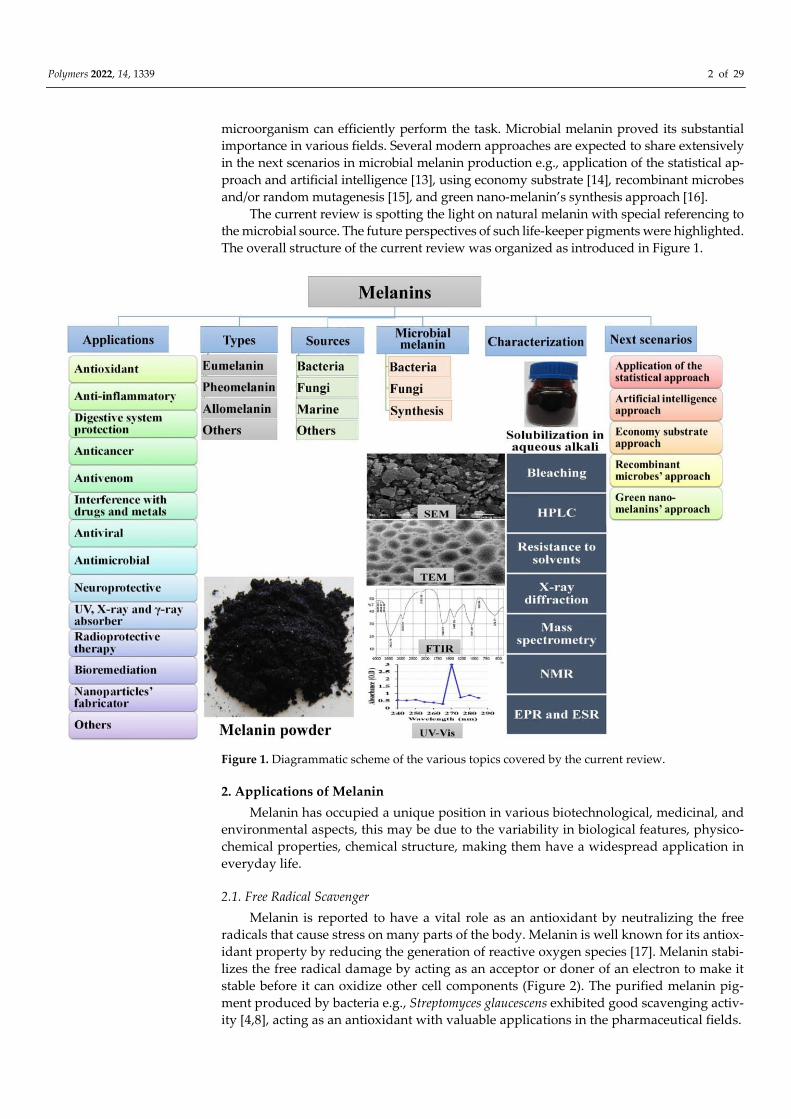
The overall structure of the current review was organized as introduced in Figure 1.
Figure 1. Diagrammatic scheme of the various topics covered by the current review.
2. Applications of Melanin
Melanin has occupied a unique position in various biotechnological, medicinal, and
environmental aspects, this may be due to the variability in biological features, physico-
chemical properties, chemical structure, making them have a widespread application in
everyday life.
2.1. Free Radical Scavenger
Melanin is reported to have a vital role as an antioxidant by neutralizing the free
radicals that cause stress on many parts of the body. Melanin is well known for its antiox-
idant property by reducing the generation of reactive oxygen species [17]. Melanin stabi-
lizes the free radical damage by acting as an acceptor or doner of an electron to make it
stable before it can oxidize other cell components (Figure 2). The purified melanin pig-
ment produced by bacteria e.g., Streptomyces glaucescens exhibited good scavenging activ-
ity [4,8], acting as an antioxidant with valuable applications in the pharmaceutical fields.

Polymers 2022, 14, 1339 3 of 29
Figure 2. Mode of action of melanin as an antioxidant.
2.2. Anti-Inflammatory Activity
The anti-inflammatory behavior of microbial melanin was reported [9]. Inflammation
is the body’s natural response induced by tissue injury or infection and interrelates to
antioxidation. Melanin is well known for its antioxidant property, which can minimize
the generation of the reactive oxygen species and thereby help in reducing the inflamma-
tory response. The topical application of melanin against formalin-induced rat-paw
edema reported a strong anti-inflammatory action of melanin and thus can be a potential
anti-inflammatory medication [10]. Melanin can inhibit the activity of cyclooxygenase
(COX), Lipoxygenase (LOX) enzymes, which have significant roles in the regulation of
inflammatory responses. COX is a limiting enzyme in inflammation as it is involved in
the conversion of arachidonic acid to prostaglandins, which are associated with many in-
flammatory diseases. LOX produces leukotrienes, thus is important in the pathophysiol-
ogy of inflammatory diseases. Another antioxidant activity is accompanied by a reduction
of inflammation as both these properties are interrelated to each other [17].
2.3. Digestive System Protection
Lipid peroxidation is the main cause of cell damage, through the oxidative degradation
of lipids, free radicals take electrons from the lipids in cell membranes, thus cell destruction
occurs. The lipid peroxidation inhibition ability of melanin was reported. For example, inhibi-
tion of eye lipid peroxidation in the eye by melanin effectively prevented uveitis [10]. When
exposed to free radicals, lipid peroxidation of liver cells was inhibited by melanin extracted
obtained from black tea [11]. Additionally, melanin effectively hinders lipid peroxidation of
liver microsomal membranes, and further protects hepatic peroxidation that occurs in adju-
vant-induced disease in rats, the observed effect was activation by melanin. Another hepato-
protective effect of melanin was stated against hydrazine-induced liver injury since melanin
plays a vital role during liver intoxication by heavy metals through preventing the develop-
ment of liver intoxication and improving liver functions as well [10,11].
Further beneficial gastrointestinal health of melanin was mentioned. Previous stud-
ies on gastric ulcers revealed that melanin constrained the secretion of gastric juice and
thus prevent ulceration in gastric mucosa in rats, another important replenishment role,
melanin can drive the renewal of the mucus levels in ethanol-depleted gastric cell walls,
and powerfully defends against ulcers induced by alcohol, indomethacin, stress or the
combined ulcerogenic activity of both aspirin, and stress [10,12].
2.4. Anti-Cancer Activity
During cancer-curing, melanin is used as a cancer remedy, also to protect patients
undergoing radiation doses against the harmful effects of gamma rays [4,18]. In vitro,
melanin exhibited potent cytotoxic activity against HFB4 skin cancer cell line was reported
with an additional benefit of little cytotoxicity towards normal non-cancerous cells,

Polymers 2022, 14, 1339 4 of 29
supporting that melanin pigments can be applied as promise anti-malignant tumor [4].
However, the cytotoxicity of black melanin was concentration-dependent. In comparison
to Doxorubicin (4.05 μg), melanin inhibited the growth of the cell lines HEPG-2, HCT-116,
and MCF-7 with IC50 values of 6.15, 5.54, and 7.91 μg, respectively [19]. In-vitro, 60 μg
melanin recorded 53% inhibition of the hepatocellular carcinoma cell line [18].
2.5. Melanin as Antivenom
Melanin demonstrated a neutralization effect against several venoms. A study per-
formed on model animals injected immediately, in the same place of venom, with melanin
after the venom administration (3 mg per mouse). The greatest antivenom impact was
discovered against Japanese mamushi snake venom. Melanin’s low venomousness, along
with its antagonistic action against various venoms, may enable successful life-saving
therapy for snakebites [11].
2.6. Interference with Drugs and Metals
Melanin may perform a task in protecting against antimicrobial drugs, therefore, in
humans, the lack of melanin leads to several abnormalities and diseases [3,12]. Broad
pharmacological and physicochemical investigations concentrate on the inter-relation-
ships of melanin with metals and medications, which promptly link to melanin and are
held in pigmented tissues for significantly longer periods, in this way, melanin may be
used as a vehicle or channel for drug delivery, acting as a drug carrier or transporter to
the site of action, especially through interacting with orally administered drugs [10]. For
example, iron-deficiency anemia was treated using melanin-iron complex, this led to a
significant reduction in symptoms, greater bioavailability of iron, and rarer side impacts
than the treatment with standard drugs does, signifying that melanin-iron complexes
might assist in improving hematopoietic performance and could be exploited as a safe,
efficient new iron tonic [20].
Another, melanin’s pigmented cells and their adjacent tissues were found to be pro-
tected from harmful substances, where melanin can absorb the harmful substances, pre-
venting or neutralizing the potential damage that occurred by such substances. The phys-
iological role of the metallic-binding feature of melanin is not clear, but possibly melanin
gradually released the binding harmful substances in nontoxic concentrations [10].
In the biomedical field, the human antigenic response is among the main obstacles
usually linked to the used biomaterials, along with the lack of stability under certain phys-
iological conditions. This challenge had been overcome in the case of melanin due to the
lack of enzymes that are capable of decomposing melanin, as well as biostability and a
lesser likelihood of long-term accumulation in organs, which is normally linked with var-
ious harmful effects [9,16].
2.7. Antiviral Feature
The replication of human immune-deficiency viruses (HIV) can be inhibited by the
synthetic soluble melanin, furthermore, the selective antiviral activity of synthetic soluble
melanin against SARS-CoV2 and HIV was revealed without toxicity to the host cells [17].
Melanin complexes also exert various favorable effects on both in vitro and in vivo animal
models, these melanin complexes could serve as a supply of biopolymers for the creation
of new medications with wide applications in infectious viral diseases [21]. A pioneering
study proved that melanin precursors like 5,6-dihydroxyindole-2-carboxylic acid and l-
3,4-dihydroxyphenylalanine strongly interact with the spike protein of the SARS-CoV2,
confirming that melanin and its precursors could be utilized as effective antiviral com-
pounds [22].

Polymers 2022, 14, 1339 5 of 29
2.8. Antimicrobial Action
Crude melanin pigment can usefully manage microbial diseases [23]. For instance,
melanin of Streptomyces sp. had proved antibacterial behavior against Lactobacillus vul-
garis, and Escherichia coli [24]. Melanin complexes were reported to have the same antimi-
crobial effects on pure cultures of Candida albicans, and Helicobacter pylori [21], suggesting
the success of melanin-based therapy as anti-microbial pathology.
2.9. Neuroprotective Agent
Melanin promotes chemical transport, resulting in faster nerve impulses and further
acting as neuroprotection [25]. These particular features of melanin elucidate their incidence
in organs, and tissues that are connected with impulse transmission systems, like body skin,
nervous system, and retina [26]. Therefore, melanin metabolism abnormalities may have a
role in the etiology (genesis) of illnesses such as Parkinsonism, which was shown to be as-
sociated with melanin metabolism disorders. Studies on the post-mortem brains of Parkin-
son’s disease patients, animal models, and in vitro cultures indicate the presence of pro-
grammed cell death (apoptosis) in the substantia nigra region, which was confirmed
through DNA fragmentation and typical morphological changes in the brain’s cell [27].
After induced destruction, bacterial melanin supports the survival of neurons in the
substantia nigra pars compacta and preserves dopaminergic neurons, suppressing sec-
ondary inflammation in damaged brain tissue. Low concentrations of bacterial melanin
are non-toxic and do not produce side effects. Moreover, bacterial melanin has been eval-
uated as a strong activator of regeneration and motion recovery in rat models subjected
to unilateral depletion of the substantia nigra pars compacta. It stimulates the healing of
a damaged motion tract and the regeneration of a damaged peripheral nerve fiber, pro-
duces capillary dilatation in the lesion area, and so improves blood flow in brain tissue
[25,28]. Another protective mechanism of neuromelanin pigments is that they attach and
sequester the toxic metals to form stable complexes that prevent neuronal toxicity [27].
Another study confirmed that the bacterial melanin has been used as a neuroprotector. It
aids in the recovery and regeneration processes following central nervous system lesions.
The role of bacterial melanin after the destruction of substantia nigra is of great interest.
The firing rate of substantia nigra pars compacta dopaminergic neurons is significantly
increased by bacterial melanin. An increase in the firing rate of substantia nigra neurons
can aid in the recovery of the substantia nigra after neuronal degeneration [29].
2.10. UV X-, and γ-rays Protective
Melanin is used in pharmaceuticals and cosmetics, especially in photoprotective-
based creams that are utilized to protect against ultraviolet (UV) radiation [30], X-ray, and
γ-ray [31], particularly in optical lenses as eye protectants [22].
Almost all living creatures are exposed to UV radiation that spreads on the earth’s
surface regularly. The UV spectrum (between 200 and 400 nm) is generally split into three
regions; UV-A (320–400 nanometers), UV-B (280–320 nanometers), and UV-C (280–200
nm). The potential damage of UV radiation increases exponentially with decreasing wave-
length. Sunburn, premature skin photoaging, skin malignancy, and immune system sup-
pression have been related to UV radiation exposure [8].
Melanin in the skin does not entirely prevent UV from accessing and harming cells;
rather, it decreases UV transmission to the nuclei. Consequently, studies are being con-
ducted to give enough UV protection against the development of skin cancer. Sunscreen
products, unlike melanin, frequently utilize a mixture of chemical filters, which raises the
risk of adverse effects, melanin, on the other hand, is considered superior UV-sunscreens
as a component of photoprotective creams since their maximal absorption is often within
the UV spectrum [8].

Polymers 2022, 14, 1339 6 of 29
2.11. Radioprotective Therapy
Oral melanin-based products are used for the protection of people who expose to radia-
tion. The oral-radioprotective efficacy of melanin is used as edible or drinkable for alleviating
and/or preventing one or more side effects associated with radiation exposure. In a study on
mice, one-hour post-eumelanin feeding (15 mg/kg body weight), the mice were subjected to a
total body irradiation dose of 9 Gy, the defensive action of melanin was due to hindering the
radiation-induced hematopoietic harms. Melanin prevents apoptosis in splenic tissue, dimin-
ishes oxidative stress in hepatic tissue, and abrogates immune imbalance [32,33].
During cancer treatment, radiation therapy can damage the normal cells. Therefore,
internal administration by the radioprotective materials is applied to shield normal organs
without protecting the cancer tissue, leading to enhancement of the effectiveness of radi-
ation treatment by permitting higher tumoricidal dosages. In this case, melanin protects
against the harmful effects of gamma rays by preventing the formation and/or scavenging
the free radicals, which instigate DNA damage [32].
Novel melanin nano-shells, used as radioprotector approach, were reported, at-
tempting to apply the melanin coated silica nanoparticles for protection of bone marrow
from ionizing radiation during cancer radiation therapy, implying a thinkable future med-
ical implication of melanin for radioprotection in humans [34]. Melanin nano-shells are
also utilized to defend against radiation. It can be made in or on clothes, safety gear, or
packaging material, for example. The material can be incorporated into the wall of a room,
ceiling, floor, building, vehicle, aircraft, ship, spaceship, and submarine [32].
2.12. Bioremediation of Radioactive Residuals
There is considerable worry about the health dangers connected with the extensive
development of nuclear projects in energy generation, medicine, agriculture, and indus-
try. Bacterial melanin was utilized to immobilize uranium in uranium-polluted soils. For
fostering such in-situ procedure of uranium bio-immobilization, the one-time addition of
tyrosine is applied to soil, This, in turn, exploits the ability of native microbes to produce
melanin, resulting in uranium immobilization. Consequently, melanin-delivering micro-
organisms can be employed in the bioremediation of radioactive waste like uranium [35].
Species of melanized fungi is another group of melanin producers that have been
found to thrive in regions of high-radiation capacity as Chernobyl, and in cooling pool
water of nuclear reactors, similarly, numerous laboratory investigations have shown that
melanized fungi are tolerant to different levels of UV and ionizing radiation [32]. It was
determined that melanin’s radioprotective effect in microorganisms was owed to a mix-
ture of physical shielding, and physiological activities that quenching the harmful nature
of free radicals [33,35].
2.13. Nanoparticles’ Fabricator
The biopolymer of melanin might function as a reducing, and stabilizing mediator in
the production of silver and gold nanostructures. Silver nitrate and chloroauric acid were
converted to silver and gold nanostructures by the L-DOPA-melanin pigment, respec-
tively. The silver nanoparticles were shown to have strong antifungal activities and fur-
ther may be used as efficient paint additives [36].
2.14. Food Industry
The microbial pigment, melanin has received considerable attention because of its use-
ful biological activities in food. Melanin is used as a food colorant and nutritional supple-
ments as natural ingredients, replacing the chemically synthesized pigments which cause
harmful effects in the natural environment [37,38]. In this respect, melanin pigment pro-
duced by sponge-associated actinobacterium Nocardiopsis alba was used for the environ-
mentally benign synthesis of silver nanostructures, which was useful for food packaging
materials [39]. The characteristics of the broad spectrum of activity against food pathogens

Polymers 2022, 14, 1339 7 of 29
of silver nanostructures give an insight into their potential applicability in the incorporation
of food packaging materials and antimicrobials for stored fruits and foods [8].
2.15. Other Uses
Other biological, environmental, and technological applications for melanin pig-
ments have been mentioned. Melanogenesis plays a vital role in many clusters of free-
living organisms to protect them from ecological stress conditions that improve the sur-
vival and competitiveness of the organisms [15]. In plants, for example, melanin is incor-
porated in the cell walls as strengtheners [40].
One of the most common features of melanin pigments is the capacity of eumelanin
and pheomelanin to bind different metal ions [41]. Ecologically, fungal melanin showed
potential metal-ions chelating ability [10]. Melanin is an extremely effective, and fast ion
exchange molecule that binds toxins, chemicals, and heavy metals, serving as a radical
sink [5]. This feature is important biologically because it allows melanin to chelate sub-
stances and control their entrance into cells [42]. These pigments are thought to act as a
reservoir or trap for metal ions, such as Ca (II), Zn (II), Cu (II), and Fe (III) [43].
Another detoxification role was reported by microbial melanin, by which mycotoxin
secretion was inhibited [9]. In agriculture, microbial melanin has the potential to be used
as bioinsecticides [44]. Finally, melanin has various industrial applications such as bio-
plastics, paints, and varnishes [45], as well as, in bioelectronics, melanin could be useful
as semiconductors [46].
3. Types of Melanin
Because melanin polymer is an assemblage of smaller molecules, the quantities and
bonding patterns of these molecules influence the kind of melanin to a considerable ex-
tent. The main four kinds of melanin were identified based on color and structural classi-
fications i.e., eumelanin, pheomelanin, allomelanin, and others that were occasionally cre-
ated (Figure 3).
Figure 3. Part of the structural formula of the most common types of melanins; eumelanin (A), and
pheomelanin (B).

Polymers 2022, 14, 1339 8 of 29
3.1. Eumelanin
Eumelanin is mostly black to dark brown pigments that are formed in human hair
and skin and can also be generated by some bacteria and fungi [47]. Eumelanin is created
by the oxidative polymerization of tyrosine and/or phenylalanine into L-3,4-dihydroxy-
phenylalanine (L-DOPA), which is subsequently transformed into dopachrome and fi-
nally melanin [48,49].
3.2. Pheomelanin
Pheomelanin is a red or yellow pigment found, mainly, in red human hair that is
originally formed similarly to eumelanin, but L-DOPA undergoes integration of cysteine
in the polymer (cysteinylation), and therefore, pheomelanin contain sulfur [15].
3.3. Allomelanin
Allomelanin is a heterogeneous pigment found in many fungi and plants that feature
a nitrogen-free heterogeneous category of polymers. They are derived from many sources,
including dihydrofolate, homogentisic acid, catechols, and others [4,9]. This type includes
melanin, formed from 1,8-dihydroxy naphthalene (DHN) compounds, and water-soluble
pyomelanin created when homogentisic acid, a by-product of the tyrosine breakdown
pathway, accumulates and polymerizes [50].
3.4. Other Types
Other less frequent melanin are occasionally created as a result of a divergence from
the preceding melanin types. Trichochrome pigments (previously known as trichosiderins)
are formed in the same metabolic route as eumelanin and pheomelanin, by having a lower
molecular weight than the original melanin molecules [51]. Neuromelanin is dark insoluble
pigments generated in a distinct region of catecholaminergic neurons in the brain. Neuro-
melanin granules should have a mixture of pheomelanin in the core and eumelanin in the
surface [52]. Humans contain the most neuromelanin, which is present in smaller levels in
several other non-human primates but is completely lacking in many lower species’ brains
[53]. Human neuromelanin has been demonstrated to bind transition metals like iron as well
as other potentially hazardous compounds. As a result, it may perform critical roles in apop-
tosis, neurodegeneration, and Parkinson’s disease [54]. Individuals with Parkinson’s disease
had half the quantity of neuromelanin in the substantia nigra as compared with the patients
of the same age who did not have Parkinson’s. The concentration of neuromelanin increases
with age, implying a function in neuroprotection [53,54].
4. Source of Melanin
The biological supply of melanin is miscellaneous. Melanin can be found in a variety
of biological and synthetic forms. The majority of which are available in a commercial
form for purchase. Synthetic melanin polymers are generated by auto-oxidation and
polymerization of phenolic or indolic compounds (e.g., catechols). During the process,
melanin absorption spectra raise monotonically from 700 to 250 nm [55]. Another route,
by the catalytic action of tyrosinase on tyrosine or 3,4-dihydroxyphenylalanine [40].
The largest producers, however, are marine cephalopods, this is the best-studied mel-
anin so far, which is expelled out by Sepia as a defense strategy against their adversaries
[56]. Plants are another source, in which the vegetable melanin are obtained by a chemi-
cally treated process, whereby a vegetable crude material-containing polymers or mono-
meric units of the flavonoids is processed to extract melanin [40].
Another key source of melanin is microorganisms (bacteria and fungus). Microbes’ abil-
ity to synthesize melanin is largely determined by their virulence to host associations and their
ability to protect themselves from environmental stresses such as temperature extremes, ul-
traviolet rays, and solar radiation, as well as adverse chemical stress conditions, e.g., oxidant-
mediated damage, lytic-enzymes, heavy metal toxicity, and antimicrobial medications [9,57].
Polymers 2022, 14, 1339 9 of 29
5. Why Microbial Melanin?
The synthetic melanin polymers cannot be compared with natural melanin from the
environmental point of view, and the quality, as well. The latter is safe and cost-effective
than the artificial one, especially if the proposed scenarios in the current review are con-
sidered. However, microorganisms have recently gained popularity as an alternative to
chemical melanin production. Therefore, several attempts have been done to obtain mi-
crobial strains qualified to synthesize a high quantity of melanin pigment for several rea-
sons. Melanin obtained from microbes has great advantages over synthetic, and those
from animals and plants melanin.
In contrast to the other biological-based methods (marine and vegetable melanin),
microorganisms do not get influenced by the problems of seasonal fluctuation during the
production progression and they can modify their mechanisms according to the medium
composition and growth conditions provided [58]. Meaning that the biosynthesis of mel-
anin pigments by microbes is independent of meteorological conditions that interfere with
the production process. The ease and speed of growth with no detrimental consequences
and their ability to grow on low-cost substrates are other merits [4,57,58].
Another important merit of microbial melanin is the diverse microbial sources. Sev-
eral reports stated various bacterial and fungal melanin, have various characteristics, and
hence various applications. Under laboratory conditions, microbial species with melano-
genic capacity have been examined and/or optimized for developing production pro-
cesses (Table 1). For instance, Aeromonas media and Pseudomonas maltophilia were found to
produce authentic 3,4-dihydroxyphenylalanine melanin pigment. Although melanin
from both bacteria shares many biophysical properties, the yield of Aeromonas media was
significantly higher and showed to be more effective in the protection of a bioinsecticide
against ultraviolet or solar radiation than melanin of Pseudomonas maltophilia [44]. Another
study on Actinomycetes described a newly isolated strain, Streptomyces glaucescens NEAE-
H, capable of producing a high amount of black melanin pigment on an optimized me-
dium of peptone-yeast extract iron agar. The in vitro trial showed anticancer activity of
melanin against skin cancer cell lines [4].
For another group of microorganisms, the pathogenic Cryptococcus neoformans was
able to accumulate black melanin pigments, in a medium containing phenolic substrates
such as L-dopa. Pigmented and non-pigmented mutants of Cryptococcus neoformans cells
were studied with transmission electron microscopy and electron spin resonance, both
revealed a stable free-radical population in pigmented cells. The melanin deficient was
less virulent for mice, and the melanized cells were also more resistant to antibody-medi-
ated phagocytosis and the antifungal effects of murine macrophages than non-melanized
cells, concluding that melanin pigment production is associated with microbial virulence
by protecting cells against attack by immune effector cells [59].
During an extensive search to find out cost-effective alternative sources to the con-
ventional sources of melanin production, 102 fungal isolates were tested, and only Amor-
photheca resinae was chosen as a promising melanin producer on peptone yeast extract
glucose broth. Amorphotheca resinae produced the melanin rapidly, reaching up to 4.5 g/L
within 14 days. Structural characterization of the purified fungal melanin indicates that it
is like the eumelanin and has a high antioxidant activity because of free radical scavenging
assays, suggesting a promising fungal candidate for scalable production of industrially
applicable melanin [60]. However, the general procedure of microbial melanin production
is introduced in Figure 4.

Polymers 2022, 14, 1339 11 of 29
Table 1. List of some bacteria and fungi that produce microbial melanin pigments.
Group Microorganism Objective Main Finding Reference
Bacteria
Bacillus cereus Detection of melanin produced by a
wild-type strain of Bacillus cereus
Melanin produced by the wild bacterium
was firstly identified and its UV protection to insecticidal pro-
teins was approved
Zhang et al. [61]
Bacillus thuringiensis Melanin pigment formation in high
temperature
The bacterial cell was able to produce melanin in the presence of
L-tyrosine at elevated temperature (42 °C). Ruan et al. [62]
Burkholderia cepacia Attenuation of monocyte respiratory
burst activity
Melanin-producing B. cepacia may derive protection from the
free-radical-scavenging properties of this pigment. Zughaier et al. [63]
Klebsiella sp. GSK Purification and physicochemical
characterization of melanin pigment
A bacterium capable of producing a high
amount of melanin from L-tyrosine within 3 days of incubation. Sajjan et al. [64]
Pseudomonas stutzeri
Melanin production from Pseudomonas
stutzeri isolated from red seaweed
Hypnea musciformis
The marine Pseudomonas stutzeri strain produces significant
amounts of melanin of about 6·7 g l−1 without L-tyrosine supple-
mentation in the sea-water production medium.
Ganesh Kumar et al. [65]
Pseudomonas maltophilia
Aeromonas media
Novel strain producing high levels of
DOPA-melanin and assessment of the
photoprotective role of the melanin
A novel melanin-producing bacterium was isolated. The melanin
produced by this strain offers effective photoprotection of a com-
mercial bioinsecticide against UV and solar radiation.
Wan et al. [44]
Stenotrophomonas malto-
philia
Isolation of Stenotrophomonas malto-
philia from clinical samples and pro-
duction of melanin pigment
Stenotrophomonas maltophilia was reported as a possible melanin
source in the clinical environment, and the isolated bacteria
showed production of melanin pigment with rates of strong,
moderate, weak, and lack of pigment.
Amoli et al. [66]
Actinomycetes
Nocardiopsis dassonvillei
Extract bioactive melanin pigment
from marine actinobacteria, which is
not a widespread occurrence.
First report on the production and characterization of melanin
from marine by Nocardiopsis dassonvillei. Kamarudheen et al. [67]
Streptomyces cyaneus
Optimization of medium conditions
using response surface methodology
for melanin production by Streptomy-
ces cyaneus and synthesis of copper ox-
ide nanoparticles using gamma radia-
tion
The unprecedented achievement was realized for melanin pig-
ment production, (9.898 mg/mL) was obtained by optimized cul-
ture condition. Also, 2.0% faba bean’s seed peel maximized mela-
nin (9.953 mg/mL) and hence super-yield (11.113 mg/mL) was
produced by a stimulus from gamma irradiation (2.5 kGy).
El-Batal et al. [68]

Polymers 2022, 14, 1339 12 of 29
Streptomyces spp.
Separation, identification, and analy-
sis of melanin production in Strepto-
myces
The study reveals that the method of testing melanin production
by L-tyrosine or L-dopa as a substrate may be a good criterion for
the identification and classification of Streptomyces.
Dastager et al. [69]
Yeasts
Cryptococcus neoformans melanin role in Cryptococcus neofor-
mans virulence mechanism of action
Melanin appears to contribute to virulence by protecting fungal
cells against attack by immune effector cells. Wang et al. [59]
Yarrowia lipolytica
Characterization of a nontoxic
pyomelanin pigment produced by the
yeast Yarrowia lipolytica
The ability of the yeast Yarrowia lipolytica W29 to produce high
yield (0.5 mg/mL) extracellular melanin was reported in a culture
medium supplemented with L-tyrosine. The purified pigment
was found embedded with antioxidant properties
Ben Tahar et al. [70]
Hortaea werneckii Melanin is crucial for Hortaea werneckii
growth in a hypersaline environment
Melanin has an important role in the ability of the black fungus
Hortaea werneckii to survive in hypersaline environments. Kejžar et al. [71]
Fungi
Amorphotheca resinae
Production and characterization of
melanin pigments derived from Amor-
photheca resinae
Amorphotheca resinae produced melanin in the peptone yeast ex-
tract glucose broth, reaching up 4.5 g/L within 14 days. The struc-
tural properties of melanin are similar to eumelanin.
Oh et al. [60]
Aspergillus bridgeri Physicochemical characterization and
antioxidant activity of melanin
The extracellular pigment was alkali-soluble, acid-resistant, and
insoluble in organic solvents and water. The pigment was precip-
itated and characterized and showed good free radical scaveng-
ing activity.
Kumar et al. [72]
Aspergillus fumigatus Production of pyomelanin via the ty-
rosine degradation pathway
The fungus was able to produce pyomelanin, by a different path-
way, starting from L-tyrosine. Proteome analysis indicated that
the l-tyrosine degradation enzymes are synthesized when the
fungus is grown with L-tyrosine in the medium. Homogentisic
acid is the major intermediate, and the L-tyrosine degradation
pathway leading to pyomelanin is similar to that in humans lead-
ing to alkaptomelanin.
Schmaler-Ripcke et al. [73]
Aspergillus nidulans Characterization of fungal melanin
pigment
The characterization of this pigment indicated the presence of in-
dolic units, which were also found in synthetic DOPA-melanin.
The analyses of the elemental composition showed that the pig-
ment extracted from these mutants has a high percentage of nitro-
gen and, therefore, it cannot be DHN-melanin, which presents
only a trace of nitrogen. Taken together, the results obtained in
this study indicate that melanin produced by these mutants is
Gonçalves et al. [74]

Polymers 2022, 14, 1339 13 of 29
DOPA type, representing the first report on the characterization
of this type of melanin in A. nidulans.
Auricularia auricula Auricularia auricula melanin and its
molecular structure
The nutritional control was very important to promote melanin
production, deficiency of tyrosine in the medium led to weak se-
cretion of melanin. Meanwhile, the molecular and structural for-
mulae concluded the presence of eumelanin
Sun et al. [75]
Cryomyces antarcticus
Multidisciplinary characterization of
melanin pigments from the black fun-
gus Cryomyces antarcticus
The fungus possesses the ability to produce both 1,8-dihy-
droxynaphthalene (DHN) and L 3–4 dihydroxyphenylalanine (L-
DOPA) melanins, opening interesting scenarios for the protective
role against radiation.
Pacelli et al. [76]
Phyllosticta capitalensis Characterization of fungal endophyte
melanin
First report of Phyllosticta melanin. Melanin in the hyphae of P.
capitalensis may be responsible for the success of this fungus as a
cosmopolitan endophyte since melanin is known to enhance the
survival capability of fungi in stressful environments.
Suryanarayanan et al. [77]
Pleurotus cystidiosus Isolation and characterization of mela-
nin pigment from Pleurotus cystidiosus
First report on isolation and characterization of melanin obtained
from Pleurotus cystidiosus var. formosensis. The black pigment was
confirmed as melanin based on UV, IR, and EPR spectra
Selvakumar et al. [78]
Spissiomyces endophytica Characterization and production of
melanin by an endophytic fungus
The pigment was extracted, purified, and identified from the
dried fungal biomass. The highest fungal pigment yield was ob-
served in glucose yeast extract peptone medium at an initial pH
value of 6.0 and 25 °C over three weeks of cultivation, represent-
ing the first report on the production and characterization of mel-
anin obtained from the genus Spissiomyces.
Suwannarach et al. [57]

Polymers 2022, 14, 1339 14 of 29
Figure 4. The general procedure of microbial melanin production. The graph was designed based
on the data extracted from a previous study.
5.1. Bacterial Melanin
The synthesis of black pigments in bacteria was realized long-time ago, since then
several bacteria are reported to produce diverse groups of melanin through specialized
pathways or using enzymatic imbalances in modified metabolic channels [9]. The regula-
tion of the melanin synthesis mechanism in bacteria includes transcriptional and meta-
bolic regulation, on the other side, several biosynthesis pathways remain still mostly

Polymers 2022, 14, 1339 15 of 29
unspecified [1]. However, the biosynthesis of melanin exists in Gram-positive and Gram-
negative bacteria, such as Streptomyces griseus [79], Bacillus licheniformis [19], Ralstonia sol-
anacearum [80]. In particular, diverse species of Streptomyces produce melanin that is
thought to be a useful criterion for taxonomical investigations [47]. The physiological
growth attributes of some bacteria were well described such as these of thermo-al-
kaliphilic Streptomyces sp. That required alkaline pH (9.0), higher temperature (45 °C), and
3% of sodium chloride [9].
5.2. Fungal Melanin
Fungi have been stated to possess all types of melanin discovered. Melanin in fungi
is considered a secondary metabolite and their presence can be naturally constitutive. Alt-
hough melanin is not essential for the growth and development of fungi but can perform
an extensive spectrum of biological jobs such as increased virulence in many fungi path-
ogenic to humans and plants [48]. In most melanized fungi the pigment is principally lo-
calized as granules or layered in fibrils in the outermost layer or embedded within the cell
wall, or bound to cell wall chitin, or secreted extracellularly [8]. Several fungi were re-
ported to secrete melanin e.g., Aspergillus niger, Aspergillus nidulans, Alternaria alternata,
Cladosporium carionii, Fonsecaea compacta, Exophiala jeanselmei, Hendersonula toruloidii, and
Phaeoannellomyces wernickii [48].
5.3. Microbial Synthesis of Melanin
The biosynthesis mechanism of microbial melanin occurs by oxidative polymeriza-
tion of phenolic compounds, principally by two distinctive pathways, mainly through L-
DOPA, leading to the generation of various types of melanin i.e., eumelanin,
pheomelanin. A variety of important enzymes are involved in microbial melanogenesis.
Melanin is often synthesized by microbes using phenoloxidases such as tyrosinases, lac-
cases, polyketide synthase, and p-hydroxyphenylpyruvate hydroxylase. Tyrosinase (EC
1.14.18.1) is a copper protein that belongs to the polyphenol oxidases family, it catalyzes
the oxidation of L-tyrosine to L-DOPA, which is then converted to dopachrome, that
transformed to melanin by a sequence of non-enzymatic oxidoreduction reactions [81].
Laccases (EC 1.10.3.2) are metalloproteins with an active site containing 1–4 copper atoms.
They are not linked to tyrosinases, but rather to the family of blue copper [82]. Polyketide
synthases are multidomain proteins that create DHN melanin via the polyketide pathway.
They are linked to animal fatty acid synthases [83]. Many microbes use these enzymes to
make pigments, antibiotics, poisons, and other intermediate metabolic molecules [84]. Fi-
nally, p-hydroxyphenylpyruvate hydroxylase is a crucial enzyme in pyomelanin biosyn-
thesis that catalyzes the creation of homogentisinic acid. This enzyme is found in many
living species and belongs to the phenylalanine and tyrosine degradation pathways [8].
Microbial melanin is generated through the metabolites resulting from the two main
pathways. The most widespread pathway involves melanin precursors derived from aro-
matic amino acids, such as tyrosine transformations. This monohydroxylated molecule is
oxidized to diphenol, (yielding dihydroxylated derivatives), in which the amino group
can be conserved, giving rise to L-DOPA, or eliminated before oxidation, creating mole-
cules like homogentisate (2,5-dihydroxyphenylacetate), or homoprotocatechuate (3,4-di-
hydroxyphenylacetate). These molecules are oxidized directly or by certain enzymes, giv-
ing rise to benzoquinones, or dopaquinones. The autopolymerization of such quinones
leads to the formation of melanin. These synthetic pathways are mediated by copper-
based enzymes (tyrosinases and laccases), resulting in the yellow-red pheomelanin, the
brown-black eumelanin, and a heterogeneous group of allomelanin, including
pyomelanin [1,15,45].
In addition, some microorganisms can synthesize melanin from malonyl-CoA, a process
mediated by polyketide synthases [1]. This pathway was first described in Streptomyces griseus.
The pathway includes the successive decarboxylative condensation of 5 molecules of malonyl-
coenzyme A, which is managed by the homodimeric type III polyketide synthase RppA,

Polymers 2022, 14, 1339 16 of 29
forming 1, 3, 6, 8-tetrahydroxynaphthalene (THN). A member of the cytochrome P450 family,
co-transcribed with RppA, administers the oxidative dimerization of two-THN subunits to
compose hexahydroxyperylenequinone (HPQ). The autopolymerization of such unstable pre-
cursor leads to the establishment of brownish HPQ melanin [1,8,15,45].
6. Current Trends in Melanin Characterization
Despite their marked abundance in the global biomass, some unknown aspects about the
complete chemical structure of some melanin types had to be unveiled. Because of the com-
plex polymerization, amorphous and insoluble nature, current biochemical and biophysical
practices are unable to offer a decisive chemical construction. Therefore, several identification
tests of melanin have been established, including physicochemical properties (resistance to
solvents, bleaching, and solubilization in aqueous alkali), surface morphology studies (scan-
ning electron microscopy, and transmission electron microscopy), and structural elucidation
i.e., electron paramagnetic resonance (EPR), electron spin resonance (ESR), X-ray photoelec-
tron spectroscopy (XPS), fourier transform infrared spectroscopy, UV-Visible spectroscopy,
nuclear magnetic resonance spectroscopy, mass spectrometry, gas chromatography-mass
spectrometry, and high-performance liquid chromatography (Figure 5), these methods aid
each other to introduce a complementary perspective of the melanin’s structure.
Figure 5. Various methods for characterization of melanin. Electron paramagnetic resonance (EPR),
electron spin resonance (ESR) spectroscopy, fourier transform infrared spectroscopy (FT-IR), gas
chromatography-mass spectrometry (GC-MS), high-performance liquid chromatography (HPLC),
transmission electron microscopy (TEM), scanning electron microscopy (SEM), and nuclear mag-
netic resonance spectroscopy (NMR).
Polymers 2022, 14, 1339 17 of 29
Recently, several studies and techniques were performed for the identification of
melanin. Raman spectroscopy was used in the past for analyzing melanin in vivo [85]. By
which, eumelanin exhibits specific Raman scattering properties that are measurable in
situ, thereby providing a novel, nondestructive means for optical measurement and char-
acterization of melanin according to their occurrence in different biological environments.
The atomic force microscope (AFM) is a high-resolution imaging technique that im-
plements 3D topographical data for the measurement of intermolecular forces with atomic
resolution. AFM has important advantages over other microscopic methods because it
provides measurements at the nanometer scale [86].
Electron spectroscopy for chemical analysis (ESCA), also known as XPS, is an ele-
mental analysis technique, which has helped in providing the chemical natures of the ni-
trogen and sulfur atoms is also used to determine the quantitative atomic composition
and chemistry of melanin [87].
Matrix-assisted laser desorption ionization (MALDI) analysis is a mass spectrometry
coupled with fast atom bombardment, laser desorption/ionization (LDI), and matrix-as-
sisted laser desorption/ionization. MALDI mass spectrometry was used to follow melanin
degradation trends [86]. This tool proves its usefulness in analyzing natural melanin pig-
ment, characterization of the polymer, and identification of the melanin type by detecting
the monomeric units [86]. Based on MALDI, the darker a melanin, the higher its molecular
mass, possibly due to a higher degree of polymerization of the molecule [87].
Due to their polymeric-like structure, melanin pigments have been extensively ana-
lyzed by pyrolysis gas chromatography coupled with mass spectrometry detection (py-GC-
MS). This technique degrades macromolecules by heating them to temperatures high
enough to cause bond dissociation. The pyrograms obtained are being characteristic of the
original analyte. Moreover, the pyrolysate retains the original material structure infor-
mation, permitting differentiation between pure analytes and a mixture, or copolymers [86].
Liquid chromatography analysis (LC-MS) can serve as a quantification method for
eumelanin and pheomelanin by measuring their degradation products. The oxidation of
eumelanin and pheomelanin yields specific markers, such as PTCA and pyrrole-3,5-dicar-
boxylic acid for eumelanin. These molecules were used to quantitatively assess the mela-
nin in various samples, such as hair, skin tissue, fungi, melanocytes, urine, fossils, and
sepia ink [86].
Other methods employed to partially characterize melanin include EPR and ESR,
which show a population of stable free radicals in these molecules. Melanin polymers are
known to have a paramagnetic character and o-semiquinone free radical with spin (S =
1/2). These unpaired electrons of free radicals obey the EPR effect [88].
6.1. Physicochemical Properties
6.1.1. Resistance to Solvents
Melanin is mostly insoluble in water (cold or boiled), concentrated acids (hot or cold),
and common organic solvents like methanol, ethanol, benzene, ethyl acetate, chloroform,
propanol, acetone, and petroleum ether [57].
6.1.2. Solubilization in Aqueous Alkali
Melanin solubility varies according to the origin, state of hydration or purity, and/or
polymerization status [57]. Solubility factors also include the ionization state of the pig-
ment’s carboxylic, phenolic and aminic groups, its polyelectrolyte characters, and its amino
acid contents [89]. The melanin’s solubilization in 1M KOH, and the precipitation detected
when their alkaline solutions were acidified by 7M HCl can be explained by the state of
aggregation of melanin being affected by the pH. Lowering the pH of a melanin solution
encourages the formation of aggregates and sedimentation [90] while increasing the pH
causes the granules to break down into minor particles-oligomers with a poorer degree of
polymerization. The presence of ionizable units and hydrophobic interactions within the
Polymers 2022, 14, 1339 18 of 29
molecule is responsible for this behavior [41]. Water bounds to the melanin molecule in large
amounts (30% of its weight in some cases) and is likely important in maintaining its solvent-
swollen state. Once the polymer is dehydrated, it becomes further aggregated and almost
completely misses its capacity for physicochemical interactions [90].
6.1.3. Bleaching
The bleaching of melanin solutions by oxidizing molecules such as sodium hypo-
chlorite (NaOCl), and hydrogen peroxide (H2O2) has been related to pigment deteriora-
tion [90]. Solubilization of melanin in a strongly alkaline solution is determinate for the
bleaching process, as this generates ionization of the hydroxyl phenolic units of melanin
that favors reactivity with the oxidizing agent [10]. The decolorization of the pigment is
mainly due to the degradation of melanin. The reaction mechanism, with H2O2, includes
a nucleophilic attack of OOH− ions, which induces a ring-opening reaction leading to the
formation of quinone epoxides responsible for the bleaching of melanin [15,90].
6.2. Surface Morphology
To investigate the structure of natural and synthetic melanin, transmission and scan-
ning electron microscopy were generally utilized. The researchers have stated that the
synthetic eumelanin appears to be amorphous solids, while the natural eumelanin looks
to be minute spheres [47]. Moreover, the electron microscopy investigation shows that the
particles of natural melanin are non-porous spherical (around 30 nm in diameter) that
tend to join as aggregates. Then these aggregates associate with loose clusters of melanin
agglomerates [91].
6.3. Structural Elucidation
6.3.1. UV-Visible Spectroscopy
A characteristic property of melanin, wavelength scanning shows a peak in the UV
region, ranging from 200–700 nm of extracted Streptomyces glaucescens melanin pigment,
and no peaks were reported out of this region, especially the visible region [4]. The highest
peak absorption was perceived in the UV region at 250 nm, which then declined with the
progress towards the visible area. However, the exact peak absorbance of melanin varies
according to the source, and each source has maximum UV-Vis absorption peaks. For ex-
ample, the purified Chroogomphus rutilus melanin has an extreme absorption peak at 212
nm [92], while Streptomyces bikiniensis M8 melanin at 230 nm [93]. Whereas wavelength
scan of melanin synthesized by Actinoalloteichus sp. MA-32 displayed the highest absorp-
tion peak, being at 300 nm [7].
6.3.2. FT-IR Spectroscopy
The FT-IR spectrum has been widely used for the characterization of fungal melanin.
FT-IR is considered to be the most informative, well-resolved, and non-destructive
method, providing information on functional groups such as aromatic ring CH stretching,
hydroxyl group, and N–H stretch, reflecting a detailed structural analysis of melanin
[57,78].
6.3.3. EPR and ESR Spectroscopy
Electron paramagnetic resonance (EPR) and electron spin resonance (ESR) are defin-
ing features of all melanin, by which the presence of stable groups of organic free radicals
could be detected, these free radicals result in characteristic EPR behavior, confirming the
melanin feature [78]. However, the typical melanin spectrum that indicates the presence
of free radicals falls between 3300 and 3500 gausses [64].

Polymers 2022, 14, 1339 19 of 29
6.3.4. Mass Spectrometry
Usually, the mass spectrometer is utilized to quantify known compounds, to identify
an unknown compound, determination of the compound’s molecular weight, and iden-
tify the structural and chemical properties. Investigation of melanin with mass spectrom-
etry coupled with fast atom bombardment, laser desorption/ionization, and matrix-as-
sisted laser desorption/ionization mass spectrometry proved that the darker melanin
types have higher molecular masses, the degree of polymerization possibly is positively
correlated to higher darkness of the molecule [94].
6.3.5. High-Performance Liquid Chromatography (HPLC)
Chemical or thermal degradation using drastic procedures that produce an extensive
breakdown of the pigment, followed by the identification of the end products by HPLC
or gas chromatography coupled with mass spectrometry, have been used to recognize the
monomeric items [95]. Recently, an improved HPLC technique has been applied for the
detection of the two major classes of melanin pigments (eumelanin and pheomelanin), in
biological samples, the method proved to be simple, specific, and reproducible [96].
6.3.6. X-ray Diffraction
Generally, X-ray diffraction has been used for the characterization of several natural
and synthetic types of purified melanin. X-ray diffraction of melanin shows the lack of
crystalline structure in the diffraction pattern i.e., no significant crystallinity in melanin
could be detected. The X-ray diffraction pattern was used to classify various kinds of bio-
logical melanin from the diffraction pattern of synthetic melanin [97]. Another work on
natural and synthetic melanin reported the presence of high resemblance in the scattering
intensity profiles, signifying that both kinds of melanin may be essentially similar in local
atomic arrangements [98].
6.3.7. Nuclear Magnetic Resonance (NMR) Spectroscopy
Little is known about NMR spectroscopy of melanin pigment, this limitations of
NMR in melanin identification may back to the presence of free radicals, molecular com-
plexity, and the scarce solubility, causing significant restriction on obtaining structural
information of melanin [78]. However, NMR can be used for the determination of some
melanin structures, i.e., the key functional groups in melanin such as the ketone, alkene,
ester, alkane, alcohol, and indole units [99].
7. Current Obstacles
Owing to its unique features, melanin is dominant in potential applications in diverse
life forms, especially in the medical sciences, and this, consequently, urges researchers to
find out alternative efficient microbes and procedures for boosting the biosynthesis pro-
cess to minimize the chasm between production and demand.
Natural melanin, like those generated from cuttlefish and fungi, are insoluble in wa-
ter, thus, require severe treatments e.g., boiling in strong alkali or using strong oxidants
for converting them into water-soluble form are applied. The harsh use for solubilization
of the insoluble melanin frequently destroys the natural pigments and limits their usage.
Synthetic soluble melanin can be manufactured enzymatically by converting melanin
precursors into pigments [9]. Unfortunately, these precursors are costly, resulting in a
higher cost of artificial melanin. Similarly, vegetable melanin is a different source of true
or eumelanin and is also expensive. One more significant limitation in utilizing natural
melanin in biotechnology has been the low return of and the related extraction difficulties,
as that occurs when separating melanin under brutal conditions, boiling utilizing NaOH
for example [69,89]. On the other side, microorganisms secrete bulky quantities of extra-
cellular melanin in aqueous media and have remarkable potential in biotechnological ap-
plications in contrast to insoluble melanin. Attaining low-cost soluble natural melanin can

Polymers 2022, 14, 1339 20 of 29
essentially promote and speed up the utilization of melanin in medicine, cosmetics, and
several other fields.
So, trials are in progress on microbial strains that produce soluble pure melanin.
However, the next proposals (approaches) could be mentioned in the way to achieve the
target. It is important to note that, till now some of these proposals are rarely used and
others are not applied yet.
8. Next Scenarios
8.1. Application of the Statistical Approach
Conventional methods of optimization of medium conditions, using one variable at-
a-time, are laborious, boring, and generate conflicting yield as they disregard interface
among the tested production factors. The recently emerging tools of statistical experi-
mental designs for optimization of the biosynthesis conditions of melanin have overcome
the limitation of the conventional methods.
The statistical modeling approach starts with the screening of the significant factors
among multiple tested factors concurrently. Plackett-Burman designs (PBD) are the most
applied methods to signify the important factors, the nonsignificant factor(s) that shows
a very low effect on response values (melanin for example) are omitted from further ex-
periments, whereas the significant ones are selected for extra optimization tests. Follow-
ing the screening design and the related results, the response surface methodology (RSM)
approach is applied to the significant factors to explore the relationship between the tested
variable and response (melanin production). RSM contains several fixable designs to meet
a variety of experimental conditions such as the number and concentration of the tested
factors, nature of the design space, and the number of trials used. Central composite de-
sign (CCD) and the Box-Behnken design are the two main common approaches for such
optimization and maximization process. As could be seen this modeling approach re-
quires only two steps, followed by validation of the overall process [15,100].
However, rare studies on using PBD and RSM for melanin biosynthesis were applied,
mostly in the last decade. As an example, among 17 independent factors, PBD was con-
ducted, and three significant factors affecting melanin production by Streptomyces glau-
cescens were selected to study and optimize their interaction using CCD, leading to a max-
imum melanin production of 310.650 μg/1 mL [4]. Similarly, the boost in the melanin bio-
synthesis by Auricularia auricula was studied, engaging PBD, and CCD approaches, under
the obtained optimized circumstances, the melanin yield was 1.08 g/L contrasted to 306.52 mg/L at suboptimal conditions, indicating a 3.52-fold increase [101].
The two abovementioned examples concluded that the statistical approach develops
a low-cost and fast fermentation process with enhances melanin biosynthesis. This, in
turn, encourages the scientific community to broaden the use of such techniques in the
next era.
8.2. Artificial Intelligence Approach
Artificial intelligence is the distinguished sign of the next age. The melanin produc-
tion process could be undergone optimization of the factor controlling the biosynthesis
process. The amount and rapid development of data in recent years have motivated sci-
entists to ponder how to obtain valuable information from the large quantity of gathered
data through handling and analysis. For the past couple of years, artificial intelligence (AI)
approaches have been quickly evolved and applied practically in a wide range of indus-
tries and biotechnology. By the same token, microbial melanin production can be magni-
fied using AI. AI behaves like the human brain through learning, solving problems, per-
ception, understanding, reasoning, critical thinking, and awareness of surroundings [102].
Among the regular and significant AI strategies, artificial neural networks (ANN) have
drowned the greatest consideration regarding the ability in dealing with huge infor-
mation, plan their nonlinear connections, and give outcome expectations [103]. ANN has

Polymers 2022, 14, 1339 21 of 29
been broadly applied in various fields, this employment benefits from the great self-learn-
ing ability and precision of ANN in modeling a complex relationship between input
(tested factors) and output data (target product) without accommodating complicated nu-
merical equations [104].
The core principle of ANN is based on the human brain’s learning behavior. ANN
utilizes technology solutions to mimic the structures and functions of the human neural
system, where learning in the human brain requires changing neurons, and synaptic in-
teractions to achieve a specific target [102]. In this sense, ANN is amongst the scientific
modeling tactics that employ neural network constructions to mimic the corresponding
physical systems. The idea of ANN is that it provides a good nonlinear mapping capabil-
ity, allowing to solve the challenge of mapping data from one set to another [105].
Anyhow, two main classes of ANN were identified, feedforward and feedback neu-
ral networks, both of which contain different frameworks, reflecting the wide elasticity
and applicability in solving a variety of problems, the two classes simulate the synaptic
performance among brain neurons by utilizing a huge number of nonlinear, and parallel
processors to attain the function of learning [13]. The learning process of ANN forms
mainly a relationship between input and output data, rather than focusing on the detailed
interactions of physical and/or chemical conditions, and that is why ANN exactly gener-
ates the nonlinear mapping between inputs (tested variables) and outputs (target mole-
cule e.g., melanin) to achieve the maximum prediction performance, usually, better than
other modeling approaches like RSM [105].
ANNs are, in this sense, black-box models with great simulation accuracy, and they
are often selected when the system’s intrinsic physical mechanics are highly complex or
insignificant. With these explanations, ANNs could be applied in the modeling of the pre-
vious and current data, as well as, planning the future investigation on melanin biosyn-
thesis on AI-base. This will expect to solve many melanin-related issues. To the authors’
knowledge, there is no literature dealing with the application of ANN during the optimi-
zation of the production process of melanin
8.3. Economy Substrate Approach
Employing diverse aromatic precursors in the melanin production medium can gener-
ate various types of pure melanin. Despite this advantage, the relatively high cost of em-
ploying pure melanin precursors is a significant drawback [14]. Consequently, more efforts
are required for the economization of melanin production by using agro-industrial byprod-
ucts as an eco-friendly alternative. Studies should be more directed towards semi-industrial
production of melanin using an uncomplicated cultivation process by avoiding the utiliza-
tion of expensive chemicals e.g., purified tyrosinase, complicated methods, and the cumber-
some withdrawal procedure of melanin polymers. Future investigations need to be more
focused on the pharmacological activity of melanin pigments which would be very helpful
in designing a novel strategy for the management of some diseases like cancer.
Another rarely used approach is the utilization of agro-industrial residues as a fer-
mentation substrate. Besides being readily available, the residues of agro-industrial bio-
mass, as fermentation substrates, make them an economically reasonable choice for mi-
crobial melanin biosynthesis. In this connection, fruit residues extract, as a carbon source
for bacterial fermentation, is an example of an inexpensive culture medium, which was
validated in Bacillus safensis without additional nitrogen source, which was, apparently,
15% greater than the average yield, being 6.96 g/L [47].
8.4. Recombinant Microbes’ Approach
The experimental procedures, known as genetic engineering methods, utilize the al-
teration of microorganisms’ genetic components to increase biosynthesis or create new
certain compounds. It is now feasible to genetically modify a wide range of microbes, and
the number is rising all the time. Manipulation of culture conditions, in conjunction with
Polymers 2022, 14, 1339 22 of 29
recombinant technology, has been proven in studies to enhance melanin yield in large-
scale manufacturing [14,15].
Therefore, attempts are being made for obtaining microbial strains that produce pure
soluble melanin. For example, recombinant microorganisms, like E. coli, have been used
for the biosynthesis of soluble melanin [9,106]. Another example, Streptomyces glaucescens
successfully produced a water-soluble and optically clear, dark solution of melanin in a
relatively short (2 days) incubation period [4,9], suggesting amenability to a wide variety
of uses.
8.4.1. Expression of Genes Encoding Tyrosinases
Gene cloning or transferring is an old-new procedure. The organism is modified to
express genes encoding tyrosinases received from another organism. An early study re-
ported that the recombinant E. coli exhibited eumelanin biosynthesis from L-tyrosine on
agar-plates, and liquid medium after transferring the mel gene from Streptomyces antibioti-
cus [107].
Expression of genes encoding tyrosinases can be performed through induction of the
microorganisms that contain the melanin gene. In an early model, Bacillus thuringiensis
was displayed to deliver melanin when cultured for several hours with L-tyrosine at 42
°C [62]. These outcomes indicate that the bacteria should be induced during the fermen-
tation protocol since it contains a gene encoding a tyrosinase in its genome that was in-
duced by the substrate (tyrosine) during the microbial development in the cultivation me-
dium. Furthermore, the concentration of copper, which functions as a cofactor of tyrosi-
nases and laccases, is necessary for the biosynthesis of both DHN- and DOPA-melanin,
and so, copper can modulate the melanin synthesis pathway. As a result, copper can
change melanin synthesis by controlling the expression of both enzymes. With the same
given, cancellation of the copper-transporting ATPase gene was found to control the
melanization process in Botrytis cinerea [108].
Another, physical conditions (e.g., pH, temperature, incubation periods) and specific
media components can control gene expression, hence the biosynthesis process. There-
fore, these conditions are usually altered according to the individual melanogenic strains
[15]. Modulation of the growth conditions can positively or negatively change the genes’
expression and activate or stop the cryptic biosynthetic pathways of the pigments. Ac-
cordingly, genetic modification is strongly correlated to fermentation conditions, both are
salient features for the enhancement of melanin biosynthesis.
In parallel, the symbiotic bacterium, Rhizobium etli, can fix nitrogen by forming sym-
biotic nodules in the root of Phaseolus vulgaris. The gene encoding tyrosinase (melA) has
been detected on the symbiotic plasmid. The melA was cloned, in the expression vector
pTrc99A, under control of the strong promoter (Trc), then the resultant plasmid (pTrc-
melA) was transformed in E. coli strain, which bio-synthesized eumelanin in the L-tyro-
sine-containing medium Interestingly when compared to the original wild strain, the re-
combinant bacterium exhibited extensively higher survival rate [15,109].
8.4.2. Random Mutagenesis
For gaining a novel melanogenic strain, another route, of recombinant microbes, can
be gone through, which is random mutagenesis. To discover more about the role of mel-
anogenesis genes, Pseudomonas putida strain grown in a medium containing L-tyrosine
was used as a model for transposon mutagenesis study. This resultant mutant had high
melanin biosynthesis ability, being a 6-fold increase, and superior resistance to UV light,
and H2O2 than the wild strain. Genetic investigation revealed that the transposon muta-
genesis method disrupted a gene encoding homogentisic acid 1,2 -dioxygenase that con-
verts homogentisic acid (originated from the L-tyrosine biosynthetic pathway) into 4 -maleylacetoacetate as part of a catalytic pathway. This indicates that homogentisic acid is
the precursor of allomelanin in this mutant strain [15].

Polymers 2022, 14, 1339 23 of 29
A significant benefit of melanogenic species is the visual ease with which mutants
can be identified, and more, the generated novel genes involved in the melanogenesis
process could be discovered. A random mutagenesis is a straightforward approach for
strain improvement, but, on the other hand, it is confined to species that already can pro-
duce melanin. Furthermore, the genetic alterations induced by random mutagenesis
might be unstable, causing the strain to return to a low producer phenotype. A solution
to this issue can be built on genome sequencing, to obtain enough information about the
type of mutation as well as the genes and pathways involved in the observed new pheno-
type. This can help in recovering the identified mutations if lost and helps also in the sep-
aration of the genetic alterations that are responsible for the newly developed phenotype
from those resulting from genetic instability [14].
8.4.3. Metabolic Engineering
One of the drawbacks that appear in the employment of complex media components,
is the lowered purity of melanin. Therefore, the efforts for the purification of melanin be-
come a determinant issue on the industry level. Metabolic engineering proposes the ap-
plication of simpler carbon substrates, during production, to induce the biosynthesis of
both melanin and its precursor by the microorganism. For instance, a metabolic engineer-
ing method was applied to induce E. coli strain to synthesize the eumelanin precursor; L-
tyrosine, from glucose. To direct the carbon flow from central metabolism into the com-
mon aromatic and the L-tyrosine biosynthetic pathways, feedback inhibition resistant ver-
sions of key enzymes were expressed in an engineered strain lacking the sugar phos-
photransferase system and TyrR repressor. The expressed tyrosinase consumed intracel-
lular L-tyrosine, causing growth impairment. To avoid this issue, a two-phase production
process was devised, where tyrosinase activity was controlled by the delayed addition of
the cofactor Cu. Following this procedure, melanin was produced with a simple carbon
source as glucose. This strain had the potential for synthesizing eumelanin from glucose,
with the aid of metabolic engineering, the strain was metabolically modified to overex-
press the genes responsible for carbon flow to the L-tyrosine biosynthetic pathway
[15,110], which is a precursor for melanin biosynthesis. The process reduced the produc-
tion cost compared with employing L-tyrosine as raw material.
8.5. Green Nano-Melanin’s Approach
The green synthesis of biomolecules, such as melanin, is based on green biomaterials
obtained from nature. In comparison to other synthetic melanin polymers, melanin from
biological origin has been demonstrated to be very promising and growing in the research
community among distinct research areas, such as biomedicine [16].
The current approach applies melanin nanoparticles (NPs) in the biomedical field, to
which promising additional capabilities have been attributed compared to the natural
form. NPs are proving to be a viable option for the development of novel agents, such as
drugs. Unlike microparticles, NPs have a larger surface area and are capable of surface
modification, giving them higher selectivity and specificity for a particular target [111]. In
medicine, for example, melanin nanocarrier is not applied only as a diagnostic tool but
also photothermal therapy, and controlled drug release through chemotherapy, this dou-
ble action is known as theranostics [16]. What is more, through surface modification of
nano-melanin, it is possible to add certain specific molecules to target certain kinds of
cells, (tumor tissue or bacteria, for example). Melanin NPs are also able to enhance the
drug loading capacity and to stimulate a controlled drug delivery, since, once in the vas-
cular system, these NPs structures can enter cells by receptor-mediated transcytosis, or
endocytosis, which rises the delivery of the NPs into the cells. Besides, melanin NPs can
be expelled through the common organs, such as liver and kidney pathways, easier than
higher-sized particles, presenting robust biocompatibility, and lower toxic effects emerg-
ing from the long-term accumulation in organs [16,28].
Polymers 2022, 14, 1339 24 of 29
Consequently, microbial melanin NPs is a virgin area of study, to the best of our
knowledge, no previous work was performed on the conversion of microbial melanin into
nano form using green chemistry, or through a microbial factory, encouraging extensive
studies in this biotechnological approach.
9. Drawbacks and Limitations of Melanin
Various drugs and other chemicals, such as organic amines, metals, etc., are bound
to melanin and retained in pigmented tissues for long periods. The physiological signifi-
cance of the binding is not evident, but it has been suggested that melanin protects the
pigmented cells and adjacent tissues by adsorbing potentially harmful substances, which
then are slowly released in nontoxic concentrations. Long-term exposure, on the other
hand, may build up high levels of noxious chemicals, stored on the melanin, which ulti-
mately may cause degeneration in the melanin-containing cells, and secondary lesions in
surrounding tissues. In the eye, e.g., and in the inner ear, the pigmented cells are located
close to the receptor cells, and melanin binding may be an important factor in the devel-
opment of some ocular and inner ear lesions. In the brain, neuromelanin is present in
nerve cells in the extrapyramidal system, and the melanin affinity of certain neurotoxic
agents may be involved in the development of parkinsonism, and possibly tardive dyski-
nesia. In recent years, various carcinogenic compounds have been found to accumulate
selectively in the pigment cells of experimental animals, and there are many indications
of a connection between the melanin affinity of these agents and the induction of malig-
nant melanoma [112]. The mechanism behind the development of lesions in the pig-
mented cells is probably a combination of selective retention, due to melanin binding, and
toxicity.
The insolubility of melanin is another limitation for its absorption by cells as well as
its applications. Water-soluble melanin, on the other side, is more efficient in various bio-
technological attributes, including the medical application e.g., the antiviral activity of
soluble melanin against human immunodeficiency virus [21,22]. So, it is critical for mela-
nin to be water-soluble. To extend the applicability of melanin, various scenarios of re-
search have focused on the solubilization of melanin using various methods, in this re-
spect, several technologies have been used to obtain soluble melanin from insoluble ones
such as squid ink [113]. The solubilization and/or the degradation of melanin may be re-
lated to the melanin structure and the technology used.
More melanin is not always better. In addition to its photoprotective feature, melanin
can be toxic to cells and cause skin cancer. UV exposure energizes an electron in melanin,
producing reactive oxygen compounds that can lead to a break in a single strand of DNA,
and pheomelanin can generate hydrogen peroxide which may cause carcinogenic muta-
tions. Moreover, DNA damage continued in human melanocyte cells even after hours of
exposure to UV, concluding that melanin can cause skin cancer [114].
Insoluble melanin requires severe treatments such as chemicals, enzymes, boiling in
strong alkali, or the use of strong oxidants for making them water-soluble, fortunately,
the process can hardly split melanin monomers but is often associated with melanin dam-
ages [90]. For example, the application of ultrasound during the solubilization of melanin
increased the propagation of ultrasound pressure waves and resulting cavitation phenom-
enon [115], concluding that the solubility may not be related to the degradation of melanin
structure. Therefore, some supplementary means for the ultrasound degradation process,
such as combining the use of ultrasound and enzyme, or ultrasound under hydrogen per-
oxide and alkaline conditions, were used to accelerate the degradation of the polymer,
and keep its undegraded form [113,116]. In this respect, a novel method using ultrasound
degradation under alkaline conditions (0.5 M NaOH) for preparing water-soluble squid
ink melanin fractions, this combined method minimized the structural variation of the
resulting soluble melanin fractions [112].
To avoid the damage of melanin fractions by the severe treatments, another scenario
was proposed, which is the application of microorganisms in the production of the
Polymers 2022, 14, 1339 25 of 29
required type of melanin; soluble, insoluble, or both. Streptomyces lusitanus DMZ-3 is an
example of a good producer of both kinds of melanin [117].
10. Conclusions
Melanogenesis is a vital process for living organisms. The natural complex polymer
pigment, melanin, is abundantly detected in a wide array of higher organisms, and micro-
organisms, as well, where such pigments play vital and multi-function roles. Therefore, the
current review spotted some light on natural melanin with special referencing to the micro-
bial source. The current application, characterization, and drawbacks of melanin are ex-
plored. The next scenarios such as artificial intelligence, recombinant microbes, and green
nano-melanin’s synthesis are the next approaches for melanin synthesis. Finally, future per-
spectives must devote more attention to the abovementioned topics for the economization
of melanin production by using agro-industrial byproducts as an eco-friendly alternative.
Studies should be more directed towards semi-industrial production of melanin utilizing a
simple microbial cultivation process, consequently, avoiding the use of purified tyrosinase,
expensive chemical methods, and the cumbersome extraction of the polymer from the plant,
and animal tissues. Future investigations need to be more focused on the pharmacological
activity of melanin pigment and its NPs, which would be very helpful in designing a novel
strategy for the management of some diseases like cancer.
Author Contributions: N.E.-A.E.-N. and W.I.A.S. shared equally in conceptualization, methodol-
ogy, data curation, writing original draft, visualization, supervision, validation, writing, reviewing,
and editing. All authors have read and agreed to the published version of the manuscript.
Funding: This research received no external funding.
Institutional Review Board Statement: Not applicable.
Informed Consent Statement: Not applicable.
Data Availability Statement: All data were reported in the review.
Conflicts of Interest: The authors declare no conflict of interest.
References
1. Pavan, M.E.; López, N.I.; Pettinari, M.J. Melanin biosynthesis in bacteria, regulation and production perspectives. Appl.
Microbiol. Biotechnol. 2020, 104, 1357–1370. https://doi.org/10.1007/s00253-019-10245-y.
2. Langfelder, K.; Streibel, M.; Jahn, B.; Haase, G.; Brakhage, A.A. Biosynthesis of fungal melanins and their importance for
human pathogenic fungi. Fungal Genet. Biol. 2003, 38, 143–158. https://doi.org/10.1016/S1087-1845(02)00526-1.
3. Huang, H.C.; Chang, T.M. Antioxidative properties and inhibitory effect of Bifidobacterium adolescentis on melanogenesis.
World J. Microbiol. Biotechnol. 2012, 28, 2903–2912. https://doi.org/10.1007/s11274-012-1096-0.
4. El-Naggar, N.E.; El-Ewasy, S.M. Bioproduction, characterization, anticancer and antioxidant activities of extracellular mel-
anin pigment produced by newly isolated microbial cell factories Streptomyces glaucescens NEAE-H. Sci. Rep. 2017, 7, 42129.
https://doi.org/10.1038/srep42129.
5. Jacobson, E.S. Pathogenic roles for fungal melanins. Clin. Microbiol. Rev. 2000, 13, 708–717.
https://doi.org/10.1128/cmr.13.4.708.
6. Hung, Y.C.; Sava, V.; Hong, M.Y.; Huang, G.S. Inhibitory effects on phospholipase A2 and antivenin activity of melanin
extracted from Thea sinensis Linn. Life Sci. 2004, 74, 2037–2047. https://doi.org/10.1016/j.lfs.2003.09.048.
7. Manivasagan, P.; Venkatesan, J.; Senthilkumar, K.; Sivakumar, K.; Kim, S.K. Isolation and characterization of biologically
active melanin from Actinoalloteichus sp. MA-32. Int. J. Biol. Macromol. 2013, 58, 263–274. https://doi.org/10.1016/j.ijbi-
omac.2013.04.041.
8. Tran-Ly, A.N.; Reyes, C.; Schwarze, F.W.; Ribera, J. Microbial production of melanin and its various applications. World J.
Microbiol. Biotechnol. 2020, 36, 1–9. https://doi.org/10.1007/s11274-020-02941-z.
9. El-Naggar, N.E.A. Streptomyces-based cell factories for production of biomolecules and bioactive metabolites. In Microbial
Cell Factories Engineering for Production of Biomolecules; Academic Press: Cambridge, MA, USA, 2021; pp. 183–234.
https://doi.org/10.1016/B978-0-12-821477-0.00011-8.
10. ElObeid, A.S.; Kamal-Eldin, A.; Abdelhalim, M.A.K.; Haseeb, A.M. Pharmacological properties of melanin and its function
in health. Basic Clin. Pharmacol. Toxicol. 2017, 120, 515–522. https://doi.org/10.1111/bcpt.12748.
11. Sava, V.M.; Hung, Y.C.; Blagodarsky, V.A.; Hong, M.Y.; Huang, G.S. The liver-protecting activity of melanin-like pigment
derived from black tea. Food Res. Int. 2003, 36, 505–511. https://doi.org/10.1016/S0963-9969(02)00199-0.
Polymers 2022, 14, 1339 26 of 29
12. Al Mofleh, I.A.; Alhaider, A.A.; Mossa, J.S.; Al-Sohaibani, M.O.; Al-Yahya, M.A.; Rafatullah, S.; Shaik, S.A. Gastroprotective
effect of an aqueous suspension of black cumin Nigella sativa on necrotizing agents-induced gastric injury in experimental
animals. Saudi J. Gastroenterol. 2008, 14, 128–134. https://doi.org/10.4103/1319-3767.41731.
13. Abiodun, O.I.; Jantan, A.; Omolara, A.E.; Dada, K.V.; Mohamed, N.A.; Arshad, H. State-of-the-art in artificial neural network
applications: A survey. Heliyon, 2018, 4, e00938. https://doi.org/10.1016/j.heliyon.2018.e00938.
14. Martínez, L.M.; Martinez, A.; Gosset, G. Production of melanins with recombinant microorganisms. Front Bioeng. Biotechnol.
2019, 7, 285. https://doi.org/10.3389/fbioe.2019.00285.
15. Singh, S.; Nimse, S.B.; Mathew, D.E.; Dhimmar, A.; Sahastrabudhe, H.; Gajjar, A.; Ghadge, V.A.; Kumar, P.; Shinde, P.B.
Microbial melanin: Recent advances in biosynthesis, extraction, characterization, and applications. Biotechnol. Adv. 2021,
107773. https://doi.org/10.1016/j.biotechadv.2021.107773.
16. Caldas, M.; Santos, A.C.; Veiga, F.; Rebelo, R.; Reis, R.L.; Correlo, V.M. Melanin nanoparticles as a promising tool for bio-
medical applications–a review. Acta Biomater. 2020, 105, 26–43. https://doi.org/10.1016/j.actbio.2020.01.044.
17. Kurian, N.K.; Nair, H.P.; Bhat, S.G. Evaluation of anti-inflammatory property of melanin from marine Bacillus spp. BTCZ31.
EVALUATION. 2015, 8, 251–255.
18. Arun, G.; Eyini, M.; Gunasekaran, P. Characterization and biological activities of extracellular melanin produced by Schiz-
ophyllum commune (Fries). Indian J. Exp. Biol. 2015, 53, 380–387. https://doi.org/10.1007/s11274-020-02924-0.
19. Ragab, T.I.; Helal, M.M.; Esawy, M.A. Optimization of Bacillus licheniformis MAL tyrosinase: In vitro anticancer activity for
brown and black eumelanin. Heliyon 2019, 5, e01657. https://doi.org/10.1016/j.heliyon.2019.e01657.
20. Lei, M.; Xue, C.H.; Wang, Y.M.; Li, Z.J.; Xue, Y.; Wang, J.F. Effect of squid ink melanin-Fe on iron deficiency anemia remis-
sion. J. Food Sci. 2008, 73, H207–H211. https://doi.org/10.1111/j.1750-3841.2008.00930.x.
21. Seniuk, O.F.; Gorovoj, L.F.; Beketova, G.V.; Savichuk, H.O.; Rytik, P.G.; Kucherov, I.I.; Prilutskay, A.B.; Prilutsky, A.I. Anti-
infective properties of the melanin-glucan complex obtained from medicinal tinder bracket mushroom, Fomes fomentarius
(L.: Fr.) Fr.(Aphyllophoromycetideae). Int. J. Med. Mushrooms 2011, 13, 7–18. https://doi.org/10.1615/intjmedmushr.v13.i1.20.
22. Vijayababu, P.; Kurian, N.K. Melanin and its precursors as effective antiviral compounds: With a special focus on SARS
CoV2. Mol. Biol. 2021, 10, 1–3.
23. Zerrad, A.; Anissi, J.; Ghanam, J.; Sendide, K.; El Hassouni, M. Antioxidant and antimicrobial activities of melanin produced
by a Pseudomonas balearica strain. J. Biotechnol. Lett. 2014, 5, 87–94.
24. Vasanthabharathi, V.; Lakshminarayanan, R.; Jayalakshmi, S. Melanin production from marine Streptomyces. Afr. J. Biotech-
nol. 2011, 10, 11224–11234. https://doi.org/10.5897/AJB11.296.
25. Petrosyan, T. Bacterial melanin in rat models of Parkinson’s disease: A potential neuroprotective strategy. Neural Regen. Res.
2015, 10, 211–212. https://doi.org/10.4103/1673-5374.152372.
26. Gevorkyan, O.V.; Meliksetyan, I.B.; Petrosyan, T.R.; Ovsepyan, A.S.; Agadzhanyan, A.E.; Saghiyan, A.S. Study of the influ-
ence of bacterial melanin on brain plasticity. Neurochem. J. 2008, 2, 308–309. https://doi.org/10.1134/S1819712408040168.
27. Zecca, L.; Wilms, H.; Geick, S.; Claasen, J.H.; Brandenburg, L.O.; Holzknecht, C.; Panizza, M.L.; Zucca, F.A.; Deuschl, G.;
Sievers, J.; et al. Human neuromelanin induces neuroinflammation and neurodegeneration in the rat substantia nigra: Im-
plications for Parkinson's disease. Acta Neuropathol. 2008, 116, 47–55. https://doi.org/10.1007/s00401-008-0361-7.
28. Nakamura, Y.; Mochida, A.; Choyke, P.L.; Kobayashi, H. Nanodrug delivery: Is the enhanced permeability and retention
effect sufficient for curing cancer? Bioconjug. Chem. 2016, 27, 2225–2238. https://doi.org/10.1021/acs.bioconjchem.6b00437.
29. Petrosyan, T.R.; Chavushyan, V.A.; Hovsepyan, A.S. Bacterial melanin increases electrical activity of neurons in Substantia
Nigra pars compacta. J. Neural Transm. 2014, 121, 259–265 . 30. Allam, N.G.; Abd El-Zaher, E.H. Protective role of Aspergillus fumigatus melanin against ultraviolet (UV) irradiation and
Bjerkandera adusta melanin as a candidate vaccine against systemic candidiasis. Afr. J. Biotechnol. 2012, 11, 6566–6577.
https://doi.org/10.5897/AJB11.4136.
31. Ye, M.; Guo, G.Y.; Lu, Y.; Song, S.; Wang, H.Y.; Yang, L. Purification, structure and anti-radiation activity of melanin from
Lachnum YM404. Int. J. Biol. Macromol. 2014, 63, 170–176. https://doi.org/10.1016/j.ijbiomac.2013.10.046.
32. Dadachova, E.; Casadevall, A. Ionizing radiation: How fungi cope, adapt, and exploit with the help of melanin. Curr. Opin.
Microbiol. 2008, 11, 525–531. https://doi.org/10.1016/j.mib.2008.09.013.
33. Kunwar, A.; Adjhikary, B.; Jayakumar, S.; Barik, A.; Chattopadhyay, S.; Raghukumar, S.; Priyadarsini, K.I. Melanin, a prom-
ising radioprotector: Mechanisms of actions in a mice model. Toxicolo. Appl. Pharmacol. 2012, 264, 202–211.
https://doi.org/10.1016/j.taap.2012.08.002.
34. Schweitzer, A.D.; Revskaya, E.; Chu, P.; Pazo, V.; Friedman, M.; Nosanchuk, J.D.; Cahill, S.; Frases, S.; Casadevall, A.;
Dadachova, E. Melanin-covered nanoparticles for protection of bone marrow during radiation therapy of cancer. Int. J. Ra-
diat. Oncol. Biol. Phys. 2010, 78, 1494–1502. https://doi.org/10.1016/j.ijrobp.2010.02.020.
35. Turick, C.E.; Ekechukwu, A.A.; Milliken, C.E.; Casadevall, A.; Dadachova, E. Gamma radiation interacts with melanin to
alter its oxidation-reduction potential and results in electric current production. Bioelectrochemistry 2011, 82, 69–73.
https://doi.org/10.1016/j.bioelechem.2011.04.009.
36. Apte, M.; Girme, G.; Bankar, A.; RaviKumar, A.; Zinjarde, S. 3, 4-dihydroxy-L-phenylalanine-derived melanin from Yarrowia
lipolytica mediates the synthesis of silver and gold nanostructures. J. Nanobiotechnol. 2013, 11, 1–9.
https://doi.org/10.1186/1477-3155-11-2.
Polymers 2022, 14, 1339 27 of 29
37. Unagul, P.; Wongsa, P.; Kittakoop, P.; Intamas, S.; Srikiti-Kulchai, P.; Tanticharoen, M. Production of red pigments by the
insect pathogenic fungus Cordyceps unilateralis BCC 1869. J. Ind. Microbiol. Biotechnol. 2005, 32, 135–140.
38. Kim, J.K.; Park, S.M.; Lee, S.J. Novel antimutagenic pigment produced by Bacillus licheniformis SSA3. J. Microbiol. Biotechnol.
1995, 5, 48–50.
39. Kiran, G.S.; Dhasayan, A.; Lipton, A.N.; Selvin, J.; Arasu, M.V.; Al-Dhabi, N.A. Melanin-templated rapid synthesis of silver
nanostructures. J. Nanobiotechnol. 2014, 12, 1–13.
40. Riley, P.A. Melanin. Int j Biochem. Cell Boil. 1997, 29, 1235–1239. https://doi.org/10.1016/S1357-2725(97)00013-7.
41. Abbas, M.; D’Amico, F.; Morresi, L.; Pinto, N.; Ficcadenti, M.; Natali, R.; Ottaviano, L.; Passacantando, M.; Cuccioloni, M.;
Angeletti, M.; et al. Structural, electrical, electronic and optical properties of melanin films. Eur. Phys. J. E. 2009, 28, 285–291.
https://doi.org/10.1140/epje/i2008-10437-9.
42. Slominski, A.; Tobin, D.J.; Shibahara, S.; Wortsman, J. Melanin pigmentation in mammalian skin and its hormonal regula-
tion. Physiol. Rev. 2004, 84, 1155–1212. https://doi.org/10.1152/physrev.00044.2003.
43. Hong, L.; Simon, J.D. Insight into the binding of divalent cations to Sepia eumelanin from IR absorption spectroscopy. Pho-
tochem. Photobiol. 2006, 82, 1265–1269. https://doi.org/10.1562/2006-02-23-RA-809.
44. Wan, X.; Liu, H.M.; Liao, Y.; Su, Y.; Geng, J.; Yang, M.Y.; Chen, X.D.; Shen, P. Isolation of a novel strain of Aeromonas media
producing high levels of DOPA-melanin and assessment of the photoprotective role of the melanin in bioinsecticide appli-
cations. J. App. Microbiol. 2007, 103, 2533–2541. https://doi.org/10.1111/j.1365-2672.2007.03502.x.
45. Dong, C.; Yao, Y. Isolation, characterization of melanin derived from Ophiocordyceps sinensis, an entomogenous fungus en-
demic to the Tibetan Plateau. J. Biosci. Bioeng. 2012, 113, 474–479. https://doi.org/10.1016/j.jbiosc.2011.12.001.
46. Mostert, A.B. Melanin, the what, the why and the how: An introductory review for materials scientists interested in flexible
and versatile polymers. Polymers 2021, 13, 1670. https://doi.org/10.3390/polym13101670.
47. Tarangini, K.; Mishra, S. Production of melanin by soil microbial isolate on fruit waste extract: Two step optimization of key
parameters. Biotechnol. Rep. 2014, 4, 139–146. https://doi.org/10.1016/j.btre.2014.10.001.
48. Belozerskaya, T.A.; Gessler, N.N.; Aver‘yanov, A.A. Melanin Pigments of Fungi; Fungal Metabolites Reference Series in Phy-
tochemistry; Springer: Cham, Switzerland, 2017; pp. 263–291. https://doi.org/10.1007/978-3-319-25001-4_29.
49. Roy, S.; Rhim, J.W. New insight into melanin for food packaging and biotechnology applications. Crit. Rev. Food Sci. Nutr.
2021, 1878097. https://doi.org/10.1080/10408398.2021.1878097.
50. Vasanthakumar, A.; DeAraujo, A.; Mazurek, J.; Schilling, M.; Mitchell, R. Pyomelanin production in Penicillium chrysogenum
is stimulated by L-tyrosine. Microbiology 2015, 161, 1211–1218. https://doi.org/10.1099/mic.0.000030.
51. Jangir, R.; Gadre, S.R.; Argade, N.P. Sulfuryl Chloride Promoted gem-Dichlorination–Dehydrochlorination in Alkyl Benzo-
thiazinylacetates: Synthesis of the Skeleton of Trichochrome Pigments. Synthesis. 2015, 47, 2631–2634.
https://doi.org/10.1055/s-0034-1378714.
52. Bush, W.D.; Garguilo, J.; Zucca, F.A.; Albertini, A.; Zecca, L.; Edwards, G.S.; Nemanich, R.J.; Simon, J.D. The surface oxida-
tion potential of human neuromelanin reveals a spherical architecture with a pheomelanin core and a eumelanin surface.
Proc. Natl. Acad. Sci. USA 2006, 103, 4785–14789. https://doi.org/10.1073/pnas.0604010103.
53. Fedorow, H.; Tribl, F.; Halliday, G.; Gerlach, M.; Riederer, P.; Double, K.L. Neuromelanin in human dopamine neurons:
Comparison with peripheral melanins and relevance to Parkinson’s disease. Prog. Neurobiol. 2005, 75, 109–124.
https://doi.org/10.1016/j.pneurobio.2005.02.001.
54. Double, K.L. Functional effects of neuromelanin and synthetic melanin in model systems. J. Neural Transm. 2006, 113, 751–
756. https://doi.org/10.1007/s00702-006-0450-5.
55. Rosei, M.A.; Mosca, L.; Galluzzi, F. Photoelectronic properties of synthetic melanins. Synth. Met. 1996, 76, 331–335.
https://doi.org/10.1016/0379-6779(95)03483-Z.
56. Mbonyiryivuze, A.; Nuru, Z.Y.; Kotsedi, L.; Mwakikunga, B.; Dhlamini, S.M.; Park, E.; Maaza, M. Multi-scale assembly in
nano-scaled sepia melanin natural dye. Mater. Today: Proc. 2015, 2, 3988–3997. https://doi.org/10.1016/j.matpr.2015.08.028.
57. Suwannarach, N.; Kumla, J.; Watanabe, B.; Matsui, K.; Lumyong, S. Characterization of melanin and optimal conditions for
pigment production by an endophytic fungus, Spissiomyces endophytica SDBR-CMU319. PLoS ONE, 2019, 14, e0222187.
https://doi.org/10.1371/journal.pone.0222187.
58. Gao, Q.; Garcia-Pichel, F. Microbial ultraviolet sunscreens. Nat. Rev. Microbiol. 2011, 9, 791–802. https://doi.org/10.1038/nrmi-
cro2649.
59. Wang, Y.; Aisen, P.; Casadevall, A. Cryptococcus neoformans melanin and virulence: Mechanism of action. Infect. Immun. 1995,
63, 3131–3136. https://doi.org/10.1128/iai.63.8.3131-3136.1995.
60. Oh, J.J.; Kim, J.Y.; Kwon, S.L.; Hwang, D.H.; Choi, Y.E.; Kim, G.H. Production and characterization of melanin pigments
derived from Amorphotheca resinae. J. Microbiol. 2020, 58, 648–656. https://doi.org/10.1007/s12275-020-0054-z.
61. Zhang, J.; Cai, J.; Deng, Y.; Chen, Y.; Ren, G. Characterization of melanin produced by a wild-type strain of Bacillus cereus.
Front. Biol. China 2007, 2, 26–29. https://doi.org/10.1007/s11515-007-0004-8.
62. Ruan, L.; Yu, Z.; Fang, B.; He, W.; Wang, Y.; Shen, P. Melanin pigment formation and increased UV resistance in Bacillus
thuringiensis following high temperature induction. Syst. Appl. Microbiol. 2004, 27, 286–289. https://doi.org/10.1078/0723-
2020-00265.

Polymers 2022, 14, 1339 28 of 29
63. Zughaier, S.M.; Ryley, H.C.; Jackson, S.K. A melanin pigment purified from an epidemic strain of Burkholderia cepacia atten-
uates monocyte respiratory burst activity by scavenging superoxide anion. Infect. Immun 1999, 6, 908–913.
https://doi.org/10.1128/IAI.67.2.908-913.1999.
64. Sajjan, S.; Kulkarni, G.; Yaligara, V.; Lee, K.; Karegoudar, T.B. Purification and physiochemical characterization of melanin
pigment from Klebsiella sp. GSK. J. Microbiol. Biotechnol. 2010, 20, 1513–1520. https://doi.org/10.4014/jmb.1002.02006.
65. Ganesh Kumar, C.; Sahu, N.; Narender Reddy, G.; Prasad, R.B.N.; Nagesh, N.; Kamal, A. Production of melanin pigment
from Pseudomonas stutzeri isolated from red seaweed Hypnea musciformis. Lett. Appl. Microbiol. 2013, 57, 295–302.
https://doi.org/10.1111/lam.12111.
66. Amoli, R.I.; Nowroozi, J.; Sabokbar, A.; Rajabniya, R. Isolation of Stenotrophomonas maltophilia from clinical samples: An
investigation of patterns motility and production of melanin pigment. Asian. Pac. J. Trop. Biomed. 2017, 7, 826–830.
https://doi.org/10.1016/j.apjtb.2017.08.012.
67. Kamarudheen, N.; Naushad, T.; Rao, K.V.B. Biosynthesis, characterization and antagonistic applications of extracellular
melanin pigment from marine Nocardiopsis sp. Ind. J. Pharm. Educ. Res. 2019, 53, 112–120.
https://doi.org/10.5530/ijper.53.2s.55.
68. El-Batal, A.I.; El-Sayyad, G.S.; El-Ghamery, A.; Gobara, M. Response surface methodology optimization of melanin produc-
tion by Streptomyces cyaneus and synthesis of copper oxide nanoparticles using gamma radiation. J. Clust. Sci. 2017, 28, 1083–
1112. https://doi.org/10.1007/s10876-016-1101-0.
69. Dastager, S.G.; Li, W.J.; Dayanand, A.; Tang, S.K.; Tian, X.P.; Zhi, X.Y.; Xu, L.H.; Jiang, C.L. Separation, identification and
analysis of pigment (melanin) production in Streptomyces. Afr. J. Biotechnol. 2006, 5, 1131–1134.
https://doi.org/10.5897/AJB05.361.
70. Ben Tahar, I.; Kus-Liśkiewicz, M.; Lara, Y.; Javaux, E.; Fickers, P. Characterization of a nontoxic pyomelanin pigment pro-
duced by the yeast Yarrowia lipolytica. Biotechnol. Prog. 2020, 36, e2912. https://doi.org/10.1002/btpr.2912.
71. Kejžar, A.; Gobec, S.; Plemenitaš, A.; Lenassi, M. Melanin is crucial for growth of the black yeast Hortaea werneckii in its
natural hypersaline environment. Fungal Biol. 2013, 117, 368–379. https://doi.org/10.1016/j.funbio.2013.03.006.
72. Kumar, C.G.; Mongolla, P.; Pombala, S.; Kamle, A.; Joseph, J. Physicochemical characterization and antioxidant activity of
melanin from a novel strain of Aspergillus bridgeri ICTF-201. Lett. Appl. Microbiol. 2011, 53, 350–358.
https://doi.org/10.1111/j.1472-765X.2011.03116.x.
73. Schmaler-Ripcke, J.; Sugareva, V.; Gebhardt, P.; Winkler, R.; Kniemeyer, O.; Heinekamp, T.; Brakhage, A.A. Production of
pyomelanin, a second type of melanin, via the tyrosine degradation pathway in Aspergillus fumigatus. Appl. Environ. Micro-
biol. 2009, 752, 493–503. https://doi.org/10.1128/AEM.02077-08.
74. Gonçalves, R.C.R.; Lisboa, H.C.F.; Pombeiro-Sponchiado, S.R. Characterization of melanin pigment produced by Aspergillus
nidulans. World J. Microbiol. Biotechnol. 2012, 28, 1467–1474. https://doi.org/10.1007/s11274-011-0948-3.
75. Sun, S.; Zhang, X.; Sun, S.; Zhang, L.; Shan, S.; Zhu, H. Production of natural melanin by Auricularia auricula and study on
its molecular structure. Food Chem. 2016, 190, 801–807. https://doi.org/10.1016/j.foodchem.2015.06.042.
76. Pacelli, C.; Cassaro, A.; Maturilli, A.; Timperio, A.M.; Gevi, F.; Cavalazzi, B.; Stefan, M.; Ghica, D.; Onofri, S. Multidiscipli-
nary characterization of melanin pigments from the black fungus Cryomyces antarcticus. Appl. Microbiol. Biotechnol. 2020, 104,
6385–6395. https://doi.org/10.1007/s00253-020-10666-0.
77. Suryanarayanan, T.S.; Ravishankar, J.P.; Venkatesan, G.; Murali, T.S. Characterization of the melanin pigment of a cosmo-
politan fungal endophyte. Mycol. Res. 2004, 108, 974–978. https://doi.org/10.1017/S0953756204000619.
78. Selvakumar, P.; Rajasekar, S.; Periasamy, K.; Raaman, N. Isolation and characterization of melanin pigment from Pleurotus
cystidiosus (telomorph of Antromycopsis macrocarpa). World J. Microbiol. Biotechnol. 2008, 24, 2125–2131.
https://doi.org/10.1007/s11274-008-9718-2.
79. Funa, N.; Ohnishi, Y.; Fujii, I.; Shibuya, M.; Ebizuka, Y.; Horinouchi, S. A new pathway for polyketide synthesis in microor-
ganisms. Nature 1999, 400, 897–899. https://doi.org/10.1038/23748.
80. Hernández-Romero, D.; Solano, F.; Sanchez-Amat, A. Polyphenol oxidase activity expression in Ralstonia solanacearum. Appl.
Environmen. Microbiol. 2005, 71, 6808–6815. https://doi.org/10.1128/aem.71.11.6808-6815.2005.
81. Marino, S.M.; Fogal, S.; Bisaglia, M.; Moro, S.; Scartabelli, G.; De Gioia, L.; Spada, A.; Monzani, E.; Casella, L.; Mammi, S.; et
al. Investigation of Streptomyces antibioticus tyrosinase reactivity toward chlorophenols. Arch. Biochem. Biophys. 2011, 505, 67–
74. https://doi.org/10.1016/j.abb.2010.09.019.
82. Valderrama, B.; Oliver, P.; Medrano-Soto, A.; Vazquez-Duhalt, R. Evolutionary and structural diversity of fungal laccases.
Antonie Van Leeuwenhoek 2003, 84, 289–299. https://doi.org/10.1023/A:1026070122451.
83. Minami, A.; Ugai, T.; Ozaki, T.; Oikawa, H. Predicting the chemical space of fungal polyketides by phylogeny-based bioin-
formatics analysis of polyketide synthase-nonribosomal peptide synthetase and its modification enzymes. Sci. Rep. 2020, 10,
1–13. https://doi.org/10.1038/s41598-020-70177-w.
84. Snyder, R.V.; Gibbs, P.D.L.; Palacios, A.; Abiy, L.; Dickey, R.; Lopez, J.V.; Rein, K.S. Polyketide synthase genes from marine
dinoflagellates. Mar. Biotechnol. 2003, 5, 1–12. https://doi.org/10.1007/s10126-002-0077-y.
85. Wibowo, J.T.; Kellermann, M.Y.; Petersen, L.E.; Alfiansah, Y.R.; Lattyak, C.; Schupp, P.J. Characterization of an Insoluble
and soluble form of melanin produced by Streptomyces cavourensis SV 21, a Sea Cucumber Associated Bacterium. Marine
Drugs 2022, 20, 54 .
Polymers 2022, 14, 1339 29 of 29
86. Pralea, I.E.; Moldovan, R.C.; Petrache, A.M.; Ilieș, M.; Hegheș, S.C.; Ielciu, I.; Nicoară, R.; Moldovan, M.; Ene, M.; Radu, M.;
et al. From extraction to advanced analytical methods: The challenges of melanin analysis. Int. J. Mol. Sci. 2019, 20, 3943.
87. Yacout, S.M.; McIlwain, K.L.; Mirza, S.P.; Gaillard, E.R. Characterization of retinal pigment epithelial melanin and degraded
synthetic melanin using mass spectrometry and in vitro biochemical diagnostics. Photochem. Photobiol. 2019, 95, 183–191 . 88. Olaizola, C. Isolation, Characterization and Assessment of the Photoprotective Effect of Two Fungal Melanins. Doctoral
dissertation, University of British Columbia, Vancouver, BC, Canada, 2012.
89. Aghajanyan, A.E.; Hambardzumyan, A.A.; Hovsepyan, A.S.; Asaturian, R.A.; Vardanyan, A.A.; Saghiyan, A.A. Isolation,
purification and physicochemical characterization of water-soluble Bacillus thuringiensis melanin. Pigment Cell Res. 2005, 18,
130–135. https://doi.org/10.1111/j.1600-0749.2005.00211.x.
90. d'Ischia, M.; Wakamatsu, K.; Napolitano, A.; Briganti, S.; Garcia-Borron, J.C.; Kovacs, D.; Meredith, P.; Pezzella, A.; Picardo,
M.; Sarna, T.; et al. Melanins and melanogenesis: Methods, standards, protocols. Pigment Cell Melanoma Res. 2013, 26, 616–
633. https://doi.org/10.1111/pcmr.12121.
91. Cavallini, C.; Vitiello, G.; Adinolfi, B.; Silvestri, B.; Armanetti, P.; Manini, P.; Pezzella, A.; d’Ischia, M.; Luciani, G.; Meni-
chetti, L. Melanin and melanin-like hybrid materials in regenerative medicine. Nanomaterials 2020, 10, 1518.
https://doi.org/10.3390/nano10081518.
92. Hu, W.L.; Dai, D.H.; Huang, G.R.; Zhang, Z.D. Isolation and characterization of extracellular melanin produced by Chroo-
gomphus rutilus D447. Am. J. Food Technol. 2015, 10, 68–77. https://doi.org/10.3923/ajft.2015.68.77.
93. Deepthi, A.; Rosamma, P. Actinomycete isolates from Arabian Sea and Bay of Bengal: Biochemical, molecular and functional
characterization, Ph.D. Thesis. Cochin University of Science and Technology, Cochin, India 2014. Available online: http://dy-
uthi.cusat.ac.in/purl/4946 (accessed on 15 December 2021).
94. Bertazzo, A.; Costa, C.; Allegri, G.; Seraglia, R.; Traldi, P. Laser desorption/ionisation mass spectrometry in the study of
natural and synthetic melanins. III. Human hair melanins. European Mass Spectrometry 1995, 1, 305–311.
https://doi.org/10.1255/ejms.113.
95. Latocha, M.; Chodurek, E.; Kurkiewicz, S.; Świątkowska, L.; Wilczok, T. Pyrolytic GC-MS analysis of melanin from black,
gray and yellow strains of Drosophila melanogaster. J. Anal. Appl. Pyrolysis 2000, 56, 89–98. https://doi.org/10.1016/S0165-
2370(00)00082-6.
96. Ito, S.; Del Bino, S.; Hirobe, T.; Wakamatsu, K. Improved HPLC conditions to determine eumelanin and pheomelanin con-
tents in biological samples using an ion pair reagent. Int. J. Mol. Sci. 2020, 21, 5134. https://doi.org/10.3390/ijms21145134.
97. Thathachari, Y.T.; Blois, M.S. Physical studies on melanins: II. X-ray diffraction. Biophys. J. 1969, 9, 77–89.
https://doi.org/10.1016/S0006-3495(69)86370-8.
98. Cheng, J.I.N.; Moss, S.C.; Eisner, M.; Zschack, P. X-ray characterization of melanins-I. Pigment Cell Res. 1994, 7, 255–262.
https://doi.org/10.1111/j.1600-0749.1994.tb00060.x.
99. Tian, S.; Garcia-Rivera, J.; Yan, B.; Casadevall, A.; Stark, R.E. Unlocking the molecular structure of fungal melanin using 13C
biosynthetic labeling and solid-state NMR. Biochem. 2003, 42, 8105–8109. https://doi.org/10.1021/bi0341859.
100. Ghoniem, A.A.; El-Naggar, N.E.A.; Saber, W.I.A.; El-Hersh, M.S.; El-Khateeb, A.Y. Statistical modeling-approach for opti-
mization of Cu2+ biosorption by Azotobacter nigricans NEWG-1; characterization and application of immobilized cells for
metal removal. Sci. Rep. 2020, 10, 1–15. https://doi.org/10.1038/s41598-020-66101-x.
101. Zhang, M.; Xiao, G.; Thring, R.W.; Chen, W.; Zhou, H.; Yang, H. Production and characterization of melanin by submerged
culture of culinary and medicinal fungi Auricularia auricula. Appl. Biochem. Biotechnol. 2015, 176, 253–266.
https://doi.org/10.1007/s12010-015-1571-9.
102. Smuga-Kogut, M.; Kogut, T.; Markiewicz, R.; Słowik, A. Use of machine learning methods for predicting amount of bioeth-
anol obtained from lignocellulosic biomass with the use of ionic liquids for pretreatment. Energies 2021, 14, 243.
https://doi.org/10.3390/en14010243.
103. Saber, W.I.; El-Naggar, N.E.A.; El-Hersh, M.S.; El-Khateeb, A.Y.; Elsayed, A.; Eldadamony, N.M.; Ghoniem, A.A. Rotatable
central composite design versus artificial neural network for modeling biosorption of Cr6+ by the immobilized Pseudomonas
alcaliphila NEWG-2. Sci. Rep. 2021, 11, 1–15. https://doi.org/10.1038/s41598-021-81348-8.
104. Elsayed, M.S.; Eldadamony, N.M.; Alrdahe, S.S.T.; Saber, W.I.A. Definitive screening design and artificial neural network
for modeling a rapid biodegradation of date palm fronds by a new Trichoderma sp. PWN6 into citric acid. Molecules. 2021,
26, 5048. https://doi.org/10.3390/molecules26165048.
105. Xu, A.; Chang, H.; Xu, Y.; Li, R.; Li, X.; Zhao, Y. Applying artificial neural networks (ANNs) to solve solid waste-related
issues: A critical review. Waste Manag. 2021, 124, 385–402. https://doi.org/10.1016/j.wasman.2021.02.029.
106. della-Cioppa, G.; Garger Jr, S.J.; Sverlow, G.G.; Turpen, T.H.; Grill, L.K.; Chedekal, M.R. Biosource Technologies Inc. Mela-
nin production from transformed Escherichia coli. U.S. Patent 5837505, 17 November 1998.
107. della-Cioppa, G.; Garger, S.J.; Sverlow, G.G.; Turpen, T.H.; Grill, L.K. Melanin production in Escherichia coli from a cloned
tyrosinase gene. Biotechnol. 1990, 8, 634–638. https://doi.org/10.1038/nbt0790-634.
108. Saitoh, Y.; Izumitsu, K.; Morita, A.; Tanaka, C. A copper-transporting ATPase BcCCC2 is necessary for pathogenicity of
Botrytis cinerea. Mol. Genet. Genom. 2010, 284, 33–43. https://doi.org/10.1007/s00438-010-0545-4.
109. Piñero, S.; Rivera, J.; Romero, D.; Cevallos, M.A.; Martínez, A.; Bolívar, F.; Gosset, G. Tyrosinase from Rhizobium etli is in-
volved in nodulation efficiency and symbiosis-associated stress resistance. J. Mol. Microbiol. Biotechnol. 2007, 13, 35–44.
https://doi.org/10.1159/000103595.

Polymers 2022, 14, 1339 30 of 29
110. Chávez-Béjar, M.I.; Balderas-Hernandez, V.E.; Gutiérrez-Alejandre, A.; Martinez, A.; Bolívar, F.; Gosset, G. Metabolic engi-
neering of Escherichia coli to optimize melanin synthesis from glucose. Microbial Cell Factories 2013, 12, 1–12.
https://doi.org/10.1186/1475-2859-12-108.
111. Siddiqui, I.A.; Sanna, V.; Ahmad, N.; Sechi, M.; Mukhtar, H. Resveratrol nanoformulation for cancer prevention and ther-
apy. Ann. NY Acad. Sci. 2015, 1348, 20–31. https://doi.org/10.1111/nyas.12811.
112. Larsson, B.S. Interaction between chemicals and melanin. Pigment Cell Res. 1993, 6, 127–133.
113. Guo, X.; Chen, S.; Hu, Y.; Li, G.; Liao, N.; Ye, X.; Liu, D.; Xue, C. Preparation of water-soluble melanin from squid ink using
ultrasound-assisted degradation and its anti-oxidant activity. J. Food Sci. Technol. 2014, 51, 3680–3690.
114. Brenner, M.; Hearing, V.J. The protective role of melanin against UV damage in human skin. Photochem. Photobiol. 2008, 84,
539–549.
115. Vilkhu, K.; Mawson, R.; Simons, L.; Bates, D. Applications and opportunities for ultrasound assisted extraction in the food
industry—A review. Innov. Food Sci. Emerg. Technol. 2008, 9, 161–169.
116. Entezari, M.H.; Petrier, C. A combination of ultrasound and oxidative enzyme: Sono-biodegradation of phenol. Applied
Catalysis B: Environmental. 2004, 53, 257–263.
117. Madhusudhan, D.N.; Mazhari, B.B.Z.; Dastager, S.G.; Agsar, D. Production and cytotoxicity of extracellular insoluble and
droplets of soluble melanin by Streptomyces lusitanus DMZ-3. BioMed Res. Int. 2014, 2014, 306895.

![Melanin Translation[1]](https://static.cupdf.com/doc/110x72/577d22411a28ab4e1e96f1ae/melanin-translation1.jpg)