Nanobiomaterials inClinical Dentistry

Every good and perfect gift is from above, coming down from The Almighty.
Thanks be to God for his blessings and this wonderful life!
I would like to dedicate this book to my parents for making me who I am today. This is a special
moment to remember and thank all my teachers, research mentors, and professors who have been
the guiding light throughout my career. A very special thanks to Prof. G. Thomas Kluemper, Prof.
Sarandeep Huja, and Prof. James K. Hartsfield Jr. for being a source of immense motivation and
moral support.
Karthikeyan Subramani
I would like to dedicate this book to my father, Muhammed Mukhtar, and mother, Shamim Anwar,
for their unconditional love and guidance, and to my family—Rihana, Aisha, Imran, Omar, Usman,
and Adam—who have provided an environment of fun and happiness for my work to flourish.
Thank you to the most beautiful little girl in the whole wide world, my granddaughter Zoya, for
coming into my life and lighting it up with joy and happiness.
Waqar Ahmed
I would like to dedicate this book to my parents Jim and Shirley, to my loving wife Karen, our son
Kennedy of whom we are very proud, and our grandson Clayton. Special thanks to all my teachers,
faculty, students, and patients, from whom I continue to learn.
James K. Hartsfield Jr.

Nanobiomaterials inClinical Dentistry
Edited by
Karthikeyan Subramani
Waqar Ahmed
James K. Hartsfield, Jr.
AMSTERDAM • BOSTON • HEIDELBERG • LONDONNEW YORK • OXFORD • PARIS • SAN DIEGO
SAN FRANCISCO • SINGAPORE • SYDNEY • TOKYOWilliam Andrew is an imprint of Elsevier

Every good and perfect gift is from above, coming down from The Almighty.
Thanks be to God for his blessings and this wonderful life!
I would like to dedicate this book to my parents for making me who I am today. This is a special
moment to remember and thank all my teachers, research mentors, and professors who have been
the guiding light throughout my career. A very special thanks to Prof. G. Thomas Kluemper, Prof.
Sarandeep Huja, and Prof. James K. Hartsfield Jr. for being a source of immense motivation and
moral support.
Karthikeyan Subramani
I would like to dedicate this book to my father, Muhammed Mukhtar, and mother, Shamim Anwar,
for their unconditional love and guidance, and to my family—Rihana, Aisha, Imran, Omar, Usman,
and Adam—who have provided an environment of fun and happiness for my work to flourish.
Thank you to the most beautiful little girl in the whole wide world, my granddaughter Zoya, for
coming into my life and lighting it up with joy and happiness.
Waqar Ahmed
I would like to dedicate this book to my parents Jim and Shirley, to my loving wife Karen, our son
Kennedy of whom we are very proud, and our grandson Clayton. Special thanks to all my teachers,
faculty, students, and patients, from whom I continue to learn.
James K. Hartsfield Jr.

William Andrew is an imprint of Elsevier225 Wyman Street, Waltham, 02451, USAThe Boulevard, Langford Lane, Kidlington, Oxford OX5 1GB, UK
First edition 2013
Copyright r 2013 Elsevier Inc. All rights reserved
No part of this publication may be reproduced or transmitted in any form or by any means, electronic ormechanical, including photocopying, recording, or any information storage and retrieval system, withoutpermission in writing from the publisher. Details on how to seek permission, further information about thePublisher’s permissions policies and arrangements with organizations such as the Copyright Clearance Centerand the Copyright Licensing Agency, can be found at our website: www.elsevier.com/permissions.
This book and the individual contributions contained in it are protected under copyright by the Publisher(other than as may be noted herein).
NoticeKnowledge and best practice in this field are constantly changing. As new research and experience broaden ourunderstanding, changes in research methods, professional practices, or medical treatment may become necessary.
Practitioners and researchers must always rely on their own experience and knowledge in evaluating and using anyinformation, methods, compounds, or experiments described herein. In using such information or methods theyshould be mindful of their own safety and the safety of others, including parties for whom they have a professionalresponsibility.
To the fullest extent of the law, neither the Publisher nor the authors, contributors, or editors, assume any liabilityfor any injury and/or damage to persons or property as a matter of products liability, negligence or otherwise, orfrom any use operation of any methods, products, instructions, or ideas contained in the material herein.
Library of Congress Cataloging-in-Publication DataA catalog record for this book is available from the Library of Congress
British Library Cataloguing-in-Publication DataA catalogue record for this book is available from the British Library
ISBN: 978-1-4557-3127-5
For information on all Elsevier publicationsvisit our website at elsevierdirect.com
Typeset by MPS Limited, Chennai, Indiawww.adi-mps.com
Printed and bound in United States of America
13 14 15 11 10 9 8 7 6 5 4 3 2 1

Foreword by Professor C.N.R. Rao
I am glad to write a foreword for this book which, for the first time, focuses on clinical applications
of nanotechnology and nanobiomaterials in dentistry.
At a fundamental level, nanotechnology helps to manipulate individual atoms and molecules to
produce novel structures with unique properties or improved properties. It involves the production
and applications of physical, chemical, and biological systems and materials at a size scale ranging
1�100 nm. Even though nanotechnology was first introduced over half a century ago, its progress
has been slow, but in the last decade, nanotechnology has caught the imagination of scientists and
the general public. Nanotechnology offers us the ability to design materials with totally new desir-
able characteristics. Nanotechnology can be approached in two ways: “top-down” and “bottom-up”
approaches. The “top-down” approach has resulted in remarkable breakthroughs. This approach has
been responsible for the rapid development of the semiconductor industry. Future impact of this
approach will depend on how quickly we reach the limits in lithographic technologies. Much prog-
ress has been made in integrating nanostructured materials into larger systems. The “bottom-up”
approach refers to the construction of macromolecular structures from atoms or molecules that self-
assemble to form macroscopic structures. The “bottom-up” approach represents “molecular nano-
technology.” The field of nanotechnology is diverse, involving the need for a good understanding
of biology, chemistry, physics, and mathematics. Extensive research is being carried out worldwide
to understand the advantages and scientific limitations of nanotechnology and its applications to a
wide range of disciplines from materials science and biomedical research to space research.
Much has been written on the fundamental aspects of nanotechnology. This book is refreshing
because it deals with recent studies and techniques used in nanotechnology to produce newer bio-
materials for practical applications in clinical dentistry. Even though progress in the application of
nanotechnology in biological systems and medicine has been much slower, it is evident that the
“bottom-up” approach is potentially far more as we develop the ability to control and manipulate
atoms and molecules more precisely. Nature uses the bottom-up approach and builds diverse struc-
tures in biological systems. The complexity and functionality of these structures is truly amazing. If
we can control in fine detail the way in which these structures can be produced in the same way as
nature does, remarkable and rapid advances can be made in the field of medicine and dentistry.
In recent years, there has been an explosive growth in the application of nanotechnology in
medicine and dentistry. New drug delivery systems based on nanocarriers are being developed for
treating diseases such as cancer, asthma, and diabetes. These developments are likely to accelerate
in the future. The development of numerous new products may have a considerable economic
impact.
There is intense research activity in the nanotechnology in dentistry with numerous publications
appearing. Last year Subramani and Ahmed put together the first comprehensive text, Emerging
Nanotechnologies in Dentistry. This was useful and timely for both experts and novices. However,
developments in the applications of dentistry have been so rapid that they decided to work on this
text along with Professor Hartsfield.
xvii

This book brings together recognized experts from across the globe to focus on clinical applica-
tions of nanobiomaterials in a comprehensive way with 24 chapters. The authors come from a num-
ber of countries including the United States, United Kingdom, Australia, Canada, Israel, Mexico,
Germany, Brazil, Jordan, China, Taiwan, Korea, Japan, Oman, Hong Kong, Czech Republic, and
Iran and represent many laboratories in both academia and industry which are in the forefront of
the subject. Since no single person can be an expert in this vast field of nanotechnology, this book
provides information that enables everyone to learn something valuable and interesting. It is written
in a way that both experts and novices can benefit.
This comprehensive book will be a valuable addition as a textbook in university libraries and
laboratories and as a reference source for members of the scientific, industrial, and clinical
community.
The editors, Subramani, Ahmed, and Hartsfield, should be congratulated for bringing the experts
together to produce a timely, useful, and comprehensive text on nanobiomaterials in clinical den-
tistry. I hope that readers will enjoy the book and find it useful.
Professor C.N.R. RaoJawaharlal Nehru Centre for Advanced Scientific Research,
Bangalore, Karnataka, India
xviii Foreword by Professor C.N.R. Rao

Foreword by Professor Peixuan Guo
I am pleased to be writing a foreword for this book as nanotechnology is one of the most exciting
and dynamic fields to emerge over the last century. Considerable investment, effort, and time have
been spent on fundamental research and new applications of nanotechnology. New insights have
emerged from scientists from multiple disciplines working together. Newer applications have been
developed that have had a major impact in several industries such as semiconductors, aerospace,
automotive, biomedical field, and cosmetics. Recently research and development has intensified in
the field of medical nanotechnology where it is being used for drug delivery systems, medical
implants, and dental and pharmaceutical products. Major diseases such as cancer, diabetes, and
asthma are already being treated using nanotechnology for targeted controlled drug delivery sys-
tems. The “bottom-up” approach used by nature is being exploited and once we can precisely con-
trol the behavior of atoms and molecules, we will be able to make a staggering array of new
products for a far wider range of applications.
Nanotechnology has been commonly defined as the manipulation and control of materials at the
atomic and molecular level at a scale between 10 and 100 nm. This is an interesting scale because
at this level the properties of materials are being defined and interesting phenomena occur.
Japanese researchers are looking at it from a commercial perspective much more than the West.
They define nanotechnology as a technology that will produce quantum leaps in producing new
products and generating a great deal of wealth and contributing to a global economy. China is
investing huge resources and efforts into nanotechnology, and it is widely agreed that this field is
expected to have a massive impact on commercial applications in the near future.
It appears that Nobel laureate Richard P. Feynman’s vision of nanotechnology outlined in his
classic science lecture “There is Plenty of Room at the Bottom” in 1959 is finally being realized.
He envisioned nanorobots and nanomachines that can do amazing things inside the body being built
atom by atom. He predicted new materials having properties never seen before being created. This
was years before the revolutionary “microchip” was developed. You can see how this has impacted
our society by walking into any electronics store anywhere in the world or by almost everyone car-
rying mobile phones, computers and laptops, and the whole range of electronic equipment in homes
and in cars. Nanoelectronics is huge commercially.
The academic importance of nanotechnology has been realised and acknowledged by several
scientists winning Nobel prizes after Feynman for their work on nanotechnology notably Robert F.
Curl Jr., Sir Harold W. Kroto, and Richard E. Smalley for the discovery of C60 in 1996 and much
more recently in Physics 2010 to Andre Geim and Konstantin Novoselov for groundbreaking
experiments regarding the two-dimensional material graphene.
Even though nanotechnology is already having a huge impact commercially, I feel that we have
only scratched the surface and there is a vast array of new applications and products that will be
exploiting this dynamic field. In the near future almost every product on the market will be making
use of nanotechnology in one form or another. For example, research into nanotechnology in medi-
cine and dentistry has exploded with a large number of research papers appearing from all over the
xix

world. In 2012, Karthikeyan and Ahmed published the first comprehensive book on “Emerging
nanotechnologies in dentistry.” The pace of activity in nanotechnology in dentistry is so rapid that
this new book became necessary. It focuses on “Nanobiomaterials in clinical dentistry.”
Karthikeyan, Ahmed, and Hartsfield have brought together a group of international experts from
multiple backgrounds to explore a range of topics. This book contains 24 comprehensive chapters
written in the unique style of the authors. This book will be useful to dental surgeons, specialists,
engineers, scientists, physicists, chemists, biologists, materials scientists, and decision and policy
makers at undergraduate, post graduate, and specialist levels. Since no one individual can be an
expert in all aspects of nanotechnology and its applications, this book will be useful for anyone
with an interest in the field. I hope that you find this book stimulating, enjoyable, and useful, and
that it ignites further interest in this field.
Professor Peixuan GuoWilliam Farish Endowed Chair in Nanobiotechnology, Director of Nanobiotechnology Center,
College of Pharmacy, University of Kentucky, Lexington, KY, USA
xx Foreword by Professor Peixuan Guo

Acknowledgments
There has been an explosion of activity in the last few years in the research and development of
nanobiomaterials for clinical applications in dentistry. Once again, as with our first book, we have
had the pleasure and privilege of working with world-class experts who wrote the high-quality
chapters included in this book. We are grateful for their dedication and hard work in writing the
chapters in a timely manner. We would like to extend our thanks and appreciation to the following
authors for their valuable time and hard work: Abdelbary Elhissi, Seyed Shahabeddin Mirsasaani,
Mehran Hemati, Ehsan Sadeghian Dehkord, Golnaz Talebian Yazdi, Danesh Arshadi Poshtiri,
Mrinal Bhattacharya, Wook-Jin Seong, Shin-Woo Ha, M. Neale Weitzmann, George R. Beck Jr.,
Abdul Samad Khan, Maria Khan, Ihtesham Ur Rehman, Hockin H. K. Xu, Lei Cheng, Ke Zhang,
Mary Anne S. Melo, Michael D. Weir, Joseph M. Antonucci, Nancy J. Lin, Sheng Lin-Gibson,
Laurence C. Chow, Xuedong Zhou, M. Nassif, F. El Askary, M. Hannig, C. Hannig, D.B. Barbosa,
D.R. Monteiro, A.S. Takamyia, E.R. Camargo, A.M. Agostinho, A.C.B. Delbem, J.P. Pessan, R.P.
Allaker, Sarandeep Huja, G. Thomas Kluemper, Lorri Morford, Tarek El-Bialy, Meir Redlich,
Reshef Tenne, Ki Young Nam, Chul Jae Lee, Sandhra M. Carvalho, Agda A. R. Oliveira, Elke M.
F. Lemos, Marivalda M. Pereira, Sandrine Lavenus, Julie Roze, Guy Louarn, Pierre Layrolle, Kaifu
Huo, Lingzhou Zhao, Paul K. Chu, Qing Cai, Reji T. Mathew, Xiaoping Yang, R. Dziak, K.
Mohan, B. Almaghrabi, Y. Park, Shengbin Huang, Tingting Wu, Haiyang Yu, Sami Chogle,
Bassam Kinaia, Harold Goodis, Chamindie Punyadeera, Paul D. Slowey, Elizabeth Pinon-Segundo,
Nestor Mendoza-Munoz, David Quintanar-Guerrero, Yashwant Pathak, and Charles Preuss.
We were fortunate to get Forewords for this book written by Prof. C. N. R. Rao and
Prof. Peixuan Guo.
We are thankful to the entire team of Elsevier for bringing this book in its finest form and
quality.
We hope that this book inspires our readers to explore more into the science of nanobiomater-
ials and their clinical application in dentistry and that they find this work useful.
Karthikeyan Subramani, Waqar Ahmed and James K. Hartsfield, Jr.
xxi

CHAPTER
1Introduction to Nanotechnology
Waqar Ahmeda, Abdelbary Elhissia and Karthikeyan SubramanibaInstitute of Nanotechnology and Bioengineering, School of Computing, Engineering and Physical Sciences,
University of Central Lancashire, Preston, UKbDepartment of Orthodontics, University of Kentucky, Lexington, KY, USA
CHAPTER OUTLINE
1.1 Introduction ......................................................................................................................................3
1.2 Approaches to nanotechnology ..........................................................................................................4
1.3 Nanotechnology on a large scale and volume .....................................................................................5
1.3.1 Top-down approach....................................................................................................... 5
1.3.2 Bottom-up approach ..................................................................................................... 6
1.4 Applications .................................................................................................................................. 11
1.5 Future considerations..................................................................................................................... 15
1.6 Nanobiomaterials in clinical dentistry ............................................................................................. 15
References ........................................................................................................................................... 16
1.1 IntroductionNanotechnology has been around since the beginning of time. Nature routinely has always used
nanotechnology to synthesize molecular structures in the body such as enzymes, proteins, carbohy-
drates, and lipids which form components of cellular structures. However, the discovery of nano-
technology has been widely attributed to the American Physicist and Nobel Laureate Dr. Richard
Phillips Feynman [1] who presented a paper called
“There is plenty of room at the bottom”
in December 29, 1959, at the annual meeting of the American Physical Society at California Institute
of Technology. Feynman talked about the storage of information on a very small scale, writing and
reading in atoms, about miniaturization of the computer, building tiny machines, tiny factories, and
electronic circuits with atoms. He stated that “In the year 2000, when they look back at this age, they
will wonder why it was not until the year 1960 that anybody began seriously to move in this direc-
tion.” However, he did not specifically use the term nanotechnology. The first use of the word
3Nanobiomaterials in Clinical Dentistry.
© 2013 Elsevier Inc. All rights reserved.

“nanotechnology” has been attributed to Tanaguchi [2] in a paper published in 1974 “On the basic
concept of nanotechnology.” Dr. K. Eric Drexler an MIT graduate later took Feynman’s concept of a
billion tiny factories and added the idea that they could make more copies of themselves, via
computer control instead of control by a human operator, in his 1986 book Engines of Creation: The
Coming Era of Nanotechnology, to popularize the potential of nanotechnology.
Several definitions of nanotechnology have since then evolved. For example, the dictionary [3]
definition states that nanotechnology is “the art of manipulating materials on an atomic or molecular
scale especially to build microscopic devices.” Other definitions include the US government [4]
which state that “Nanotechnology is research and technology development at the atomic, molecular
or macromolecular level in the length scale of approximately 1�100 nm range, to provide a funda-
mental understanding of phenomena and materials at the nanoscale and to create and use structures,
devices and systems that have novel properties and functions because of their small and/or intermedi-
ate size.” The Japanese [5] have come up with a more focused and succinct definition. “True Nano”:
as nanotechnology which is expected to cause scientific or technological quantum jumps, or to pro-
vide great industrial applications by using phenomena and characteristics peculiar in nanolevel.
It is evident regardless of the definition used that the properties of matter are controlled at a
scale between 1 and 100 nm. For example, chemical properties take advantage of large surface to
volume ratio for catalysis, interfacial and surface chemistry is important in many applications.
Mechanical properties involve improved strength hardness in lightweight nanocomposites and nano-
materials, altered bending, compression properties, and nanomechanics of molecular structures.
Optical properties involve absorption and fluorescence of nanocrystals, single photon phenomena,
and photonic band gap engineering. Fluidic properties give rise to enhanced flow using nanoparti-
cles and nanoscale adsorbed films are also important. Thermal properties give increased thermo-
electric performance of nanoscale materials, and interfacial thermal resistance is important.
1.2 Approaches to nanotechnologyNumerous approaches have been utilized successfully in nanotechnology and as the technology
develops further, approaches may emerge. The approaches employed thus far have generally been
dictated by the technology available and the background experience of the researchers involved.
Nanotechnology is a truly multidisciplinary field involving chemistry, physics, biology, engineer-
ing, electronics, and social sciences, which need to be integrated together in order to generate the
next level of development in nanotechnology. Fuel cells, mechanically stronger materials, nanobio-
logical devices, molecular electronics, quantum devices, carbon nanotubes (CNTs), etc. have been
made using nanotechnology. Even social scientists are debating ethical use of nanotechnology.
The “top-down” approach involves fabrication of device structures via monolithic processing on
the nanoscale and has been used with spectacular success in the semiconductor devices used in
consumer electronics. The “bottom-up” approach involves the fabrication of device structures via
systematic assembly of atoms, molecules, or other basic units of matter. This is the approach nature
uses to repair cells, tissues, and organ systems in living things and indeed for life processes such as
protein synthesis. Tools are evolving which will give scientists more control over the synthesis and
characterization of novel nanostructures yielding a range of new products in the near future.
4 CHAPTER 1 Introduction to Nanotechnology

1.3 Nanotechnology on a large scale and volumeNanotechnology is being researched extensively internationally, and governments and research
organizations are spending large amounts of money and human resources on nanotechnology. This
has generated interesting scientific output and potential commercial applications, some of which
have been translated into products produced on a large scale. However, in order to realize commer-
cial benefits far more from lab-scale applications need to be commercialized, and for that to happen
nanotechnology needs to enter the realm of nanomanufacturing. This involves using the technolo-
gies available to produce products on a large scale, which is economically viable. A nanomanufac-
turing technology should be:
• capable of producing components with nanometer precision,
• able to create systems from these components,
• able to produce many systems simultaneously,
• able to structure in three dimensions,
• cost-effective.
1.3.1 Top-down approachThe most successful industry utilizing the top-down approach is the electronics industry. This
industry is utilizing techniques involving a range of technologies such as chemical vapor deposition
(CVD), physical vapor deposition (PVD), lithography (photolithography, electron beam, and X-ray
lithography), wet and plasma etching to generate functional structures at the micro- and nanoscale
(Figure 1.1). Evolution and development of these technologies have allowed the emergence of
numerous electronic products and devices that have enhanced the quality of life throughout the
world. The feature sizes have shrunk continuously from about 75 µm to below 100 nm. This has
been achieved by improvements in deposition technology and more importantly due to the develop-
ment of lithographic techniques and equipment such as X-ray lithography and electron beam
lithography.
Techniques such as electron beam lithography, X-ray lithography, and ion beam lithography,
all have advantages in terms of resolution achieved; however, there are disadvantages associ-
ated with cost, “optics,” and detrimental effects on the substrate. These methods are currently
under investigation to improve upon current lithographic processes used in the integrated
circuits (IC) industry. With continuous developments in these technologies, it is highly likely
that the transition from microtechnology to nanotechnology will generate a whole new genera-
tion of exciting products and features.
A demonstration of how several techniques can be combined together to form a “nano” wine
glass (Figure 1.2). In this example, a focused ion beam and CVD have been employed to produce
this striking nanostructure.
The top-down approach is being used to coat various coatings to give improved functionality.
For example, vascular stents are being coated using CVD technology with ultrathin diamond-like
carbon coatings in order to improve biocompatibility and blood flow (Figure 1.3). Graded a-SixCy:
H interfacial layers results in greatly reduced cracking and enhanced adhesion.
51.3 Nanotechnology on a large scale and volume

1.3.2 Bottom-up approachThe bottom-up approach involves making nanostructures and devices by arranging atom by atom.
The scanning tunneling microscope (STM) has been used to build nanosized atomic features such
as the letters IBM written using xenon atoms on nickel [8] (Figure 1.4). While this is beautiful and
exciting, it remains that the experiment was carried out under carefully controlled conditions (i.e.,
liquid helium cooling, high vacuum), and it took something like 24 h to get the letters right. Also
Etch mask
Film deposition
Substrate
Photoresist application
Etching Resist removal
Exposure
Photoresist
MaskLight
Development
Deposited film
FIGURE 1.1
A typical process sequence employed in the electronics industry to generate functional devices at the micro-
and nanoscale [6].
Beam scan
10−3 pa
12345Ga focusedion beam
Gas inlet
Source gas Depositedmaterials
Substrate
direction
FIGURE 1.2
Demonstration of three-dimensional nanostructure fabrication [7].
6 CHAPTER 1 Introduction to Nanotechnology

(A)
(C)
(E) (F)
(D)
(B)
Stainlesssteel
FIGURE 1.3
Examples of stents coated with diamond-like carbon using plasma enhanced CVD (Okpalugo, private
communication, 2007).
FIGURE 1.4
Positioning single atoms with an STM [8].
71.3 Nanotechnology on a large scale and volume

the atoms are not bonded to the surface just adsorbed and a small change in temperature or
pressure will dislodge them. Since this demonstration, significant advances have been made in
nanomanufacturing.
The discovery of the STM’s ability to image variations in the density distribution of surface
state electrons created in the artists a compulsion to have complete control of not only the atomic
FIGURE 1.5
Confinement of electrons to quantum corrals on a metal surface [8].
8 CHAPTER 1 Introduction to Nanotechnology

landscape, but also the electronic landscape [9]. Here they have positioned 48 iron atoms into a cir-
cular ring in order to “corral” some surface state electrons and force them into “quantum” states of
the circular structure (Figure 1.5). The ripples in the ring of atoms are the density distribution of a
particular set of quantum states of the corral. The artists were delighted to discover that they
could predict what goes on in the corral by solving the classic eigenvalue problem in quantum
mechanics—a particle in a hard-wall box.
Probably the most publicized material in recent years has been CNTs. CNTs, long, thin cylin-
ders of carbon, were discovered in 1991 by S. Iijima. These are large macromolecules that are
unique for their size, shape, and remarkable physical properties. They can be thought of as a
sheet of graphite (a hexagonal lattice of carbon) rolled into a cylinder. These intriguing structures
have sparked much excitement in recent years and a large amount of research has been dedicated
to their understanding. Currently, the physical properties are still being discovered and disputed.
What makes it so difficult is that nanotubes have a very broad range of electronic, thermal, and
structural properties that change depending on the different kinds of nanotube (defined by its
diameter, length, and chirality, or twist). To make things more interesting, besides having a single
FIGURE 1.6
MWNTs with a diameter of 30 nm and length of 12 µm have been formed within 2 min [10].
91.3 Nanotechnology on a large scale and volume

cylindrical wall (SWNTs), nanotubes can have multiple walls (MWNTs) cylinders inside the
other cylinders.
Bower et al. [10] have grown vertically aligned CNTs using microwave plasma enhanced CVD
system using a thin film cobalt catalyst at 825�C (Figure 1.6). The chamber pressure used was 20
Torr. The plasma was generated using hydrogen which was replaced completely with ammonia and
acetylene at a total flow rate of 200 sccm.
Lithographic methods are important for micro- and nanofabrication. Lithography: drawing or
writing on kind of yellow salty limestone so that impressions in ink can be taken and in the Oxford
Dictionary the word Lithos comes from Greek for stone. In micro- and nanofabrication we mean
pattern transfer. Due to limitations in current (and future) photolithographic processes, there is a
challenge to develop novel lithographic processes with better resolution for smaller features. One
such development is that of Dip-pen nanolithography (DPN). Dip-pen technology in which ink on
a pointed object is transported to a surface via capillary forces is approximately 4000 years old.
The difference with DPN is that the pointed object has a tip which has been sharpened to a few
atoms across in some cases. DPN is a scanning probe nanopatterning technique in which an AFM
tip is used to deliver molecules to a surface via a solvent meniscus, which naturally forms in the
ambient atmosphere. It is a direct-write technique and is reported to give high-resolution patterning
capabilities for a number of molecular and biomolecular “inks” on a variety of substrates, such as
metals, semiconductors, and monolayer functionalized surfaces.
DPN allows one to precisely pattern multiple patterns with good registration. It is both a fabri-
cation and imaging tool, as the patterned areas can be imaged with clean or ink-coated tips.
The ability to achieve precise alignment of multiple patterns is an additional advantage earned by
using an AFM tip to write as well as read nanoscopic features on a surface. These attributes make
DPN a valuable tool for studying fundamental issues in colloid chemistry, surface science, and
nanotechnology. For instance, diffusion and capillarity on a surface at the nanometer level, organi-
zation and crystallization of particles onto chemical or biomolecular templates, monolayer etching
resists for semiconductors, and nanometer-sized tethered polymer structures can be investigated
using this technique. In order to create stable nanostructures, it is beneficial to use molecules that
can anchor themselves to the substrate via chemisorption or electrostatic interactions. When alkane
thiols are patterned on a gold substrate, a monolayer is formed in which the thiol head groups form
relatively strong bonds to the gold and the alkane chains extend roughly perpendicular to surface.
Creating nanostructures using DPN is a single step process which does not require the use of
resists. Using a conventional atomic force microscope (AFM), DPN has been reported to achieve
ultrahigh-resolution features with line widths as small as 10�15 nm with approximately 5 nm spa-
tial resolution. For nanotechnological applications, it is important not only to pattern molecules in
high resolution, but also to functionalize surfaces with patterns of two or more components
(Figure 1.7).
Figure 1.8 shows the basic concept of nanomanufacturing. Individual atoms, which are given in
the periodic table, form the basis of nanomanufacturing. These can be assembled into molecules
and various structures using various methods including directed self-assembly and templating, and
may be positioned appropriately depending on the final requirements. Further along the devices
architecture, integration, in situ processing may be employed culminating in nanosystems,
molecular devices, etc.
10 CHAPTER 1 Introduction to Nanotechnology

1.4 ApplicationsOver the years, developments in dentistry have made many dental treatment procedures fast, reliable,
safe, and much less painful. New technologies such as nanotechnology, dental implantology, cosmetic
surgery, use of lasers, and digital dentistry have had great impact on dental treatment methodologies
and recovery time. Even though the concept of nanotechnology has always existed, its discovery is
attributed to Richard Feynman who won the Nobel Prize in 1959 for his theories regarding nanosized
devices. In the field of medicine, nanotechnology has been applied in diagnosis, prevention, and treat-
ment of diseases. Nanotechnology offers considerable scope in dentistry to improve dental treatment,
care and prevention of oral diseases. The following chapters in this book discuss about the recent
developments in this interdisciplinary field bridging nanotechnology and dentistry.
Nanotechnology has been in dentistry for tooth sealants and fillers that use nanosized particles
to improve their strength, luster, and resist wear. The application of nanoparticles in dental materi-
als and their synthesis has been discussed in the next chapter. Antimicrobial nanoparticles in restor-
ative composite materials are being used to prevent dental caries. For example, silver particles
as antibacterial agents when used in fillers and toothpastes can retard bacterial growth and reduce
290 nm
Conducting polymersSilicon nanostructures
55 nm
Single nanoparticle lines
Sol–gel templates
E (V)Single particle devicesUltrahigh density DNA arraysSmall organic molecules
65 nm
Protein nanoarrays
Solid substrate
Molecular transport
Writingdirection
AFM tip
Watermeniscus
Tunnel junctionsI (nA
)
1 µm
65 nm
FIGURE 1.7
Some of the potential applications of DPN (Byrne, private communication, 2006).
111.4 Applications

tooth decay. It is envisaged that in the longer term, biomimetic approaches and nanotechnology
will be used to repair and rebuild damaged enamel. Composite materials are becoming popular due
to their esthetic appearance and superior wear properties designed to replicate the properties of
enamel. The properties of these materials such as compressive strength, material flow, tensile
strength, and flexural strength have been improved using nanotechnology. Microfill composites are
made using the top-down approach to nanotechnology where materials such as ceramics, quartz,
and glasses start off as bulk materials and then they are ground into particle sizes below 100 nm.
However, nanocomposites are made using a bottom-up approach where atoms and molecules
combine to produce nanoparticles much smaller than those produced by the first approach.
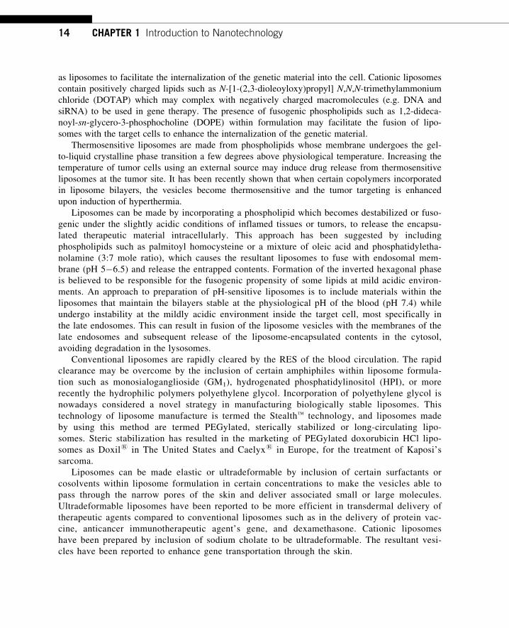
Nanoparticle-based drug delivery systems have been widely used in targeted treatment of
various forms of cancer. For example, liposomes can be used for drug delivery in oral cancer and
asthma applications. The basic structures of liposomes are shown in Figure 1.9.
- Fragmentation- Patterning- Restructuring of bulk- Lithography...
- Interfaces, field and boundary control- Positioning assembly- Integration...
- System engineering- Device architecture- Integration...
- Nanosystem biology- Emerging systems- Hierarchical integration...
Assembling
- Directed self- assembling- Templating,- New molecules
- Multiscale self- assembling- In situ processing...
- Engineering molecules as devices- Quantum control- Synthetic biology...
Passivenanostructures
Activenanostructures
− − −Systems ofnanosystems
Molecularnanosystems
Nanoproducts
FIGURE 1.8
Summary of nanotechnology [11].
12 CHAPTER 1 Introduction to Nanotechnology

Liposomes are promising drug delivery carriers owing to their safety, biocompatibility, and bio-
degradability. However, liposomes are unstable in aqueous dispersions and most of the methods
used to prepare liposomes are unsuitable for large-scale production. This review has come across a
range of technologies which may be applied to scale up the production of stable liposomes. These
include freeze drying (lyophilization) to produce powdered liposome formulations or proliposome
technologies to produce liposome precursor formulations. Various types of liposomes have been
manufactured with biological functionality. These are summarized below.
The biological functionality of liposomes is determined by liposome size and bilayer composi-
tion. Accordingly, liposomes are classified into conventional liposomes, cationic liposomes, ther-
mosensitive liposomes, pH-sensitive liposomes, long-circulating (sterically stabilized) liposomes,
and ultradeformable liposomes. Some liposome formulations may however fall under more than
one category. For instance, inclusion of certain copolymers within pH-sensitive liposomes may
enhance their escaping tendency from the blood phagocytes and hence such liposomes can be
classified as both pH sensitive and long circulating.
Conventional liposomes are multilamellar vesicles (MLVs) made of lipids having neutral or
negative charge. These liposomes are large, and because of their surface characteristics they are
readily cleared from blood circulation by reticuloendothelial system (RES) cells and hence they
have short biological half-life. Conventional liposomes are most commonly used in research to
investigate the entrapment of compounds and their release profiles. They are commonly studied as
model biological membranes.
Delivery of gene to diseased cells may repair the cause of the disease. This approach of delivery
is commonly called gene therapy. Because DNA molecules are very large, their ability to penetrate
the target cell and be expressed may be poor. This necessitates the presence of safe carriers, such
Hydrophilicheadgroup
Hydrophobicalkyl chain
MLV LUV SUV OLV
Phospholipid bilayer
FIGURE 1.9
Types of liposomes based on microscopic morphology. Liposomes bilayers (lamella) are made of phospholipid
molecules each having a cylindrical geometry. MLV5multilamellar liposome/vesicle; LUV5 large unilamellar
liposome/vesicle; SUV5 small unilamellar liposome/vesicle; OLV5 oligolamellar liposome/vesicle.
131.4 Applications
as liposomes to facilitate the internalization of the genetic material into the cell. Cationic liposomes
contain positively charged lipids such as N-[1-(2,3-dioleoyloxy)propyl] N,N,N-trimethylammonium
chloride (DOTAP) which may complex with negatively charged macromolecules (e.g. DNA and
siRNA) to be used in gene therapy. The presence of fusogenic phospholipids such as 1,2-dideca-
noyl-sn-glycero-3-phosphocholine (DOPE) within formulation may facilitate the fusion of lipo-
somes with the target cells to enhance the internalization of the genetic material.
Thermosensitive liposomes are made from phospholipids whose membrane undergoes the gel-
to-liquid crystalline phase transition a few degrees above physiological temperature. Increasing the
temperature of tumor cells using an external source may induce drug release from thermosensitive
liposomes at the tumor site. It has been recently shown that when certain copolymers incorporated
in liposome bilayers, the vesicles become thermosensitive and the tumor targeting is enhanced
upon induction of hyperthermia.
Liposomes can be made by incorporating a phospholipid which becomes destabilized or fuso-
genic under the slightly acidic conditions of inflamed tissues or tumors, to release the encapsu-
lated therapeutic material intracellularly. This approach has been suggested by including
phospholipids such as palmitoyl homocysteine or a mixture of oleic acid and phosphatidyletha-
nolamine (3:7 mole ratio), which causes the resultant liposomes to fuse with endosomal mem-
brane (pH 5�6.5) and release the entrapped contents. Formation of the inverted hexagonal phase
is believed to be responsible for the fusogenic propensity of some lipids at mild acidic environ-
ments. An approach to preparation of pH-sensitive liposomes is to include materials within the
liposomes that maintain the bilayers stable at the physiological pH of the blood (pH 7.4) while
undergo instability at the mildly acidic environment inside the target cell, most specifically in
the late endosomes. This can result in fusion of the liposome vesicles with the membranes of the
late endosomes and subsequent release of the liposome-encapsulated contents in the cytosol,
avoiding degradation in the lysosomes.
Conventional liposomes are rapidly cleared by the RES of the blood circulation. The rapid
clearance may be overcome by the inclusion of certain amphiphiles within liposome formula-
tion such as monosialoganglioside (GM1), hydrogenated phosphatidylinositol (HPI), or more
recently the hydrophilic polymers polyethylene glycol. Incorporation of polyethylene glycol is
nowadays considered a novel strategy in manufacturing biologically stable liposomes. This
technology of liposome manufacture is termed the Stealtht technology, and liposomes made
by using this method are termed PEGylated, sterically stabilized or long-circulating lipo-
somes. Steric stabilization has resulted in the marketing of PEGylated doxorubicin HCl lipo-
somes as Doxils in The United States and Caelyxs in Europe, for the treatment of Kaposi’s
sarcoma.
Liposomes can be made elastic or ultradeformable by inclusion of certain surfactants or
cosolvents within liposome formulation in certain concentrations to make the vesicles able to
pass through the narrow pores of the skin and deliver associated small or large molecules.
Ultradeformable liposomes have been reported to be more efficient in transdermal delivery of
therapeutic agents compared to conventional liposomes such as in the delivery of protein vac-
cine, anticancer immunotherapeutic agent’s gene, and dexamethasone. Cationic liposomes
have been prepared by inclusion of sodium cholate to be ultradeformable. The resultant vesi-
cles have been reported to enhance gene transportation through the skin.
14 CHAPTER 1 Introduction to Nanotechnology

1.5 Future considerationsBiomedical scientists and clinicians all over the world are working toward prevention and early
delivery of care to maintain human health. It is envisaged that nanotechnology will have a great
impact in dental research and improvement in current treatment methodologies leading to superior
oral health care in the near future.
Nanomaterials will be used far more widely and will yield superior properties and when com-
bined with biotechnology, laser and digital guided surgery will thus provide excellent dental care.
Smarter preventive measures and earlier interventions to avert craniofacial disorders using nano-
diagnostics seem a reality. Nanotechnology research will definitely pave the way for development
of tools, which would allow clinicians to diagnose and treat oral malignancies at their earliest
stage.
Biomimetics and nanotechnology have given us the knowledge to bioengineer lost tooth and
remineralization of carious lesions. This is one field which has stimulated immense interest among
the dental and nanotechnology researchers. Salivary glands can be a gateway to the body for the
delivery of precise molecular therapies using nanoparticle-based drug delivery systems with fewer
side effects. Nanofillers have improved the esthetic, physical, and mechanical properties of dental
composite materials.
Futuristic applications have been proposed on utilizing nanobots (nanoscale robots) to treat cari-
ous lesions, dentin hypersensitivity, induce dental anesthesia, teeth repositioning (using orthodontic
nanobots that could directly manipulate periodontal tissues allowing rapid, painless movement).
Dentifrobots (nanorobots in dentifrices) delivered through mouthwash or toothpaste could patrol
supra- and subgingival surfaces of tooth performing continuous plaque/calculus removal and metab-
olize trapped organic matter into harmless and odorless vapor. These proposals may seemingly
look outrageous, but inventions have always been the brainchildren of outrageous ideas of the sci-
entific community. Predictive tools like “lab-on-a-chip” can utilize saliva as a media to diagnose
dental and other physical anomalies of the human body.
1.6 Nanobiomaterials in clinical dentistryThere has been a huge surge in the number of studies over the recent few years focusing on the
clinical applications of nanobiomaterials in dentistry. This book aims to address these recent devel-
opments and is an effort to bring concepts and research studies in this interdisciplinary field under
one roof. The book has been divided into various sections to give the readers an idea about the
specific applications and uses of nanobiomaterials in various dental specialties like preventive den-
tistry, orthodontics, prosthodontics, periodontics, implant dentistry, dental tissue engineering, and
endodontics. The last section discusses the use of saliva for diagnostic purposes and the potential
use of nanoparticles as dental drug delivery systems and their biocompatibility/toxicity. While this
chapter discusses the basic concepts of nanotechnology, the second chapter gives a general over-
view of the applications of nanobiomaterials in dentistry. CNTs have been gaining increased inter-
est among the scientific community for their excellent physical and mechanical properties.
151.6 Nanobiomaterials in clinical dentistry

Different techniques of CNT manufacturing and its potential applications in dental restorative mate-
rials, bone regeneration, and gene delivery have been discussed briefly in Chapter 3. Another inter-
esting group of nanomaterials is silica-based nanomaterials. Their manufacturing techniques,
properties, and potential use for skeletal and dental applications are addressed in Chapter 4. The
applications of nanoparticles in glass ionomer cements (GICs), dental composite resin, and adhe-
sives used in dentistry are presented in Chapter 5, 6 and 7, respectively. The uses of antimicrobial
nanomaterials to prevent biofilm and caries formation are discussed in Chapters 8�10. Chapters
11�13 focus on the applications of nanobiomaterials and nanoscale imaging systems like AFM in
orthodontic materials. Potential applications of such nanobiomaterials and how they can improve
the current orthodontic armamentarium are also outlined in these chapters. The application of silver
nanoparticles incorporated into acrylic-based tissue conditioner to prevent denture stomatitis has
been discussed briefly in Chapter 14. Bioactive glass nanoparticles and their application for peri-
odontal regeneration have been presented in Chapter 15.
Chapter 16 discusses the impact of nanotechnology/nanofabrication techniques for dental
implants. Chapter 17 addresses the potential applications of titania nanotube coatings for dental
implants to enhance osseointegration. Chapter 18 discusses carbon nanotube coatings/scaffolds and
their potential applications in dental implants and bone regeneration. In Chapter 19, various nano-
structured ceramics evaluated for bone regeneration in oral and maxillofacial complex have been
reviewed briefly. Chapter 20 addresses the applications of biomimetics for periodontal and dental
tissue regeneration. The potential applications and research studies done on the utilization of nano-
biomaterials for endodontics is described in Chapter 21. Chapter 22 covers the applications of
saliva as a diagnostic material and the potential use of microelectro mechanical systems/nanoelectro
mechanical systems (MEMS/NEMS) as salivary diagnostic tool. Chapter 23 outlines the recent
advances in nanoparticles as drug delivery systems in dentistry and Chapter 24 discusses the cyto-
toxicity of orally delivered nanoparticle on systemic organs.
References[1] R.P. Feynman, There is plenty of room at the bottom, Eng. Sci. 23 (1960) 22�36 and ,www.zyvex.
com/nanotech/feynman.html/. (1959).
[2] N. Tanaguchi, On the basic concept of nanotechnology, in: 1974 Proc. ICPE.
[3] Merriam Webster dictionary 2010.
[4] US government, ,http://www.nano.gov/..
[5] K. Shimizu, INC 2, USA, 2006.
[6] B. Bushan, Springer Handbook of Nanotechnology, 2003, 147�180.
[7] T. Fujii, J. Micromech. Microeng. 15 (2005) S286�S291.
[8] D.M. Eigler, E.K. Schweizer, Positioning single atoms with a scanning tunnelling microscope, Nature
344 (1990) 524�526.
[9] M.F. Crommie, C.P. Lutz, D.M. Eigler, Confinement of electrons to quantum corrals on a metal surface,
Science 262 (1993) 218�220.
[10] C. Bower, et al., Appl. Phys. Lett. 77 (2000) 6.
[11] M.C. Roco, NSF Nanoscale Science and Engineering Grantees Conference, December 12�15, 2005.
16 CHAPTER 1 Introduction to Nanotechnology

CHAPTER
2Nanotechnology andNanobiomaterials in Dentistry
Seyed Shahabeddin Mirsasaania,b, Mehran Hematia,c, Tina Tavasolid,Ehsan Sadeghian Dehkorda, Golnaz Talebian Yazdia and Danesh Arshadi Poshtirib
aBiomaterials Group, Faculty of Biomedical Engineering (Center of Excellence),
Amirkabir University of Technology, Tehran, IranbFaculty of Dentistry, Tehran University of Medical Sciences, Tehran, Iran
cDental School, Shiraz University of Medical Sciences, Shiraz, IrandBiotechnology Engineering Department, Faculty of Engineering, University of Isfahan, Isfahan, Iran
CHAPTER OUTLINE
2.1 Introduction ................................................................................................................................... 17
2.2 Nanoscale materials ...................................................................................................................... 18
2.2.1 Nanoparticles............................................................................................................. 20
2.2.2 Characterization ......................................................................................................... 20
2.2.3 Nanofibers ................................................................................................................. 21
2.3 Nanodentistry ................................................................................................................................ 21
2.4 Nanobiomaterials in dentistry ......................................................................................................... 21
2.5 Nanobiomaterials in preventive dentistry ......................................................................................... 22
2.6 Nanobiomaterials in restorative dentistry......................................................................................... 25
2.6.1 Dental nanocomposites ............................................................................................... 25
2.6.2 Silver nanoparticles in restorative dental materials ........................................................ 29
2.7 Nanocomposites in bone regeneration............................................................................................. 29
2.8 Conclusions................................................................................................................................... 30
References ........................................................................................................................................... 30
2.1 IntroductionHumans have been using nanotechnology for a long time without realizing it. The processes of
making steel, vulcanizing rubber, and sharpening a razor all rely on manipulations of nanoparticles.
The term “nanotechnology” was coined by Prof. Eric Drexler, a lecturer and researcher of nano-
technology. “Nano” is derived from the Greek word for “dwarf.” Nanotechnology is an umbrella
term that encompasses all fields of science that operate on the nanoscale. A nanometer is one
17Nanobiomaterials in Clinical Dentistry.
© 2013 Elsevier Inc. All rights reserved.

billionth of a meter, or three to five atoms in width. It would take approximately 40,000 nan-
ometers lined up in a row to equal the width of a human hair. The basic idea of nanotechnology,
used in the narrow sense of the world, is to employ individual atoms and molecules to construct
functional structures [1].
In 1959, Nobel award winner Richard Feynman first proposed the seminal idea of nanotechnol-
ogy by suggesting the development of molecular machines. In his historic lecture in 1959, he
concluded saying, “this is a development which I think cannot be avoided” [2]. Ever since, the
scientific community has investigated the role that nanotechnology can play in every aspect of
science. The intrigue of nanotechnology comes from the ability to control material properties by
assembling such materials at the nanoscale. The tunable material properties that nanotechnology
can provide were stated in Norio Taniguchi’s paper in 1974 where the term “nanotechnology” was
first used in a scientific publication [3]. The reason for the omnipresence of the word “nano” as
one of the most attractive prefixes in the contemporary materials science is simpler than it
seems [4]. Namely, the progress of humanity is underlaid by a continual increase in sensitivity of
human interactions with their physical surrounding. As the human societies evolved, the critical
length of cutting-edge functional devices has shifted from millimeter to micrometer to nanometer
scale. With the scientific ability to control physical processes at nanometer scale, we have entered
the era of research and application of nanoscale phenomena. Finally, as material properties often
significantly alter following the micro-to-nano shift in the scale at which critical boundaries are
found, a new field was born to explain these rather strange phenomena, named nanoscience; the
application of its discoveries is known as nanotechnology [5].
Nanotechnologies are on the verge of initiating extraordinary advances in biological and biomedi-
cal sciences. These would be associated with both providing the tools for improved understanding of
fundamental building blocks of materials and tissues at the nanoscale and designing technologies for
probing, analyzing, and reconstructing them. It is not surprising that the development of novel tech-
nologies provides the foundations for creation and application of newer and more advanced ones.
Expansion of novel technologies, particularly those involved in enriching methods of research, have
already changed the way we view and define the standards of high-quality dental materials, tools,
and practices. As we see, nanotechnology has favored our understanding of dental materials at the
nanoscale and enabled the design of materials with ultrafine architecture [6].
Nanoengineering is one field of nanotechnology. Nanoengineering concerns itself with manipu-
lating processes that occur on the scale of 1�100 nm. Nanoengineering is an interdisciplinary science
that builds biochemical structures smaller than bacterium, which function like microscopic factories.
This is possible by utilizing basic biochemical processes at the atomic or molecular level. In simple
terms, molecules interact through natural processes, and nanoengineering takes advantage of those
processes by direct manipulation. Current developments are limited to the creation of nanoscale
objects for use as materials in different technologies. Material engineered using nanotechnology is
often more precise and durable because of certain properties of matter at extremely small scales [4].
2.2 Nanoscale materialsThe nanomaterial field takes a science-based approach to study materials with morphological fea-
tures on the nanoscale, and especially those that have special properties stemming from their
18 CHAPTER 2 Nanotechnology and Nanobiomaterials in Dentistry

nanoscale dimensions. Nanoscale is usually defined as smaller than one-tenth of a micrometer in at
least one dimension, though this term is sometimes also used for materials smaller than 1 µm. A
natural, incidental, or manufactured material containing particles, in an unbound state or as an
aggregate or as an agglomerate and where, for 50% or more of the particles in the number size dis-
tribution, one or more external dimensions is in the size range 1�100 nm. In specific cases and
where warranted by concerns for the environment, health, safety, or competitiveness, the number
size distribution threshold of 50% may be replaced by a threshold between 1% and 50% [7].
An important aspect of nanotechnology is the vastly increased ratio of surface area to volume pres-
ent in many nanoscale materials, which makes possible new quantum mechanical effects. One example
is the “quantum size effect” where the electronic properties of solids are altered with great reductions
in particle size. This effect does not come into play by going from macro- to microdimensions.
However, it becomes pronounced when the nanometer size range is reached. A certain number of phys-
ical properties also alter with the change from macroscopic systems. Novel mechanical properties of
nanobiomaterials are the subject of nanomechanics research. Catalytic activities also reveal new behav-
ior in the interaction with biomaterials [8]. The chemical processing and synthesis of high-performance
technological components for the private, industrial, and military sectors require the use of high-purity
ceramics, polymers, glass-ceramics, and material composites. In condensed bodies formed from fine
powders, the irregular sizes and shapes of nanoparticles in a typical powder often lead to nonuniform
packing morphologies that result in packing density variations in the powder compact [9].
Uncontrolled agglomeration of powders due to attractive Vander Waals forces can also give rise
to microstructural inhomogeneity. Differential stresses that develop as a result of nonuniform dry-
ing shrinkage are directly related to the rate at which the solvent can be removed and thus highly
dependent upon the distribution of porosity. Such stresses have been associated with a plastic-to-
brittle transition in consolidated bodies and can yield to crack propagation in the unfired body if
not relieved [10,11]. In addition, any fluctuations in packing density in the compact as it is pre-
pared for the kiln are often amplified during the sintering process, yielding inhomogeneous densifi-
cation. Some pores and other structural defects associated with density variations have been shown
to play a detrimental role in the sintering process by growing and thus limiting end-point densities.
Differential stresses arising from inhomogeneous densification have also been shown to result in
the propagation of internal cracks, thus becoming the strength-controlling flaws [12,13]. It would
therefore appear desirable to process a material in such a way that it is physically uniform with
regard to the distribution of components and porosity, rather than using particle size distributions
which will maximize density. The containment of a uniformly dispersed assembly of strongly inter-
acting particles in suspension requires total control over particle�particle interactions. It should be
noted here that a number of dispersants such as ammonium citrate (aqueous) and imidazoline or
oleyl alcohol (nonaqueous) are promising solutions as possible additives for enhanced dispersion
and deagglomeration. Monodisperse nanoparticles and colloids provide this potential [14].
Monodisperse powders of colloidal silica, for example, may therefore be stabilized sufficiently to
ensure a high degree of order in the colloidal crystal or polycrystalline colloidal solid which results
from aggregation. The degree of order appears to be limited by the time and space allowed for
longer-range correlations to be established. Such defective polycrystalline colloidal structures
would appear to be the basic elements of submicrometer colloidal materials science, and, therefore,
provide the first step in developing a more rigorous understanding of the mechanisms involved in
microstructural evolution in high-performance materials and components [15].
192.2 Nanoscale materials

2.2.1 NanoparticlesNanoparticles are nanometer-sized particles that are nanoscale in three dimensions. They include
nanopores, nanotubes, quantum dots, nanoshells, dendrimers, liposomes, nanorods, fullerenes, nano-
spheres, nanowires, nanobelts, nanorings, and nanocapsules. The applications of nanoparticles
include drug delivery systems, cancer targeting, and dentistry [2]. Nanoparticles are of great scien-
tific interest as they are effectively a bridge between bulk materials and atomic or molecular
structures. A bulk material should have constant physical properties regardless of its size, but for the
nanoscale this is often not the case. Size-dependent properties are observed such as quantum confine-
ment in semiconductor particles, surface plasmon resonance in some metal particles, and super
paramagnetism in magnetic materials [16]. Nanoparticles exhibit a number of special properties rela-
tive to bulk material. Nanoparticles often have unexpected visual properties because they are small
enough to confine their electrons and produce quantum effects. For example, gold nanoparticles
appear deep red to black in solution. The often very high surface-area-to-volume ratio of nanoparti-
cles provides a tremendous driving force for diffusion, especially at elevated temperatures. Sintering
is possible at lower temperatures and over shorter durations than for larger particles. This theoreti-
cally does not affect the density of the final product, though flow difficulties and the tendency of
nanoparticles to agglomerate do complicate matters. The surface effects of nanoparticles also reduce
the incipient melting temperature. Nanoparticles are being applied in various industries, including
medicine, due to various properties such as increased resistance to wear and the killing of bacteria,
but there are worries due to the unknown consequences to the environment and human health [17].
2.2.2 CharacterizationThe first observations and size measurements of nanoparticles were made during the first decade of
the twentieth century. They are mostly associated with the name of Zsigmondy who made detailed
studies of gold sols and other nanobiomaterials with sizes down to 10 nm and less. Zsigmondy
published a book in 1914. He used an ultramicroscope that employs a dark field method for seeing
particles with sizes much less than light wavelength. Applications began in the 1980s with the
invention of the scanning tunneling microscope and the discovery of carbon nanotubes and fuller-
enes. In 2000, the US government founded the National Nanotechnology Initiative to direct
nanotechnological development. There are traditional techniques developed during twentieth cen-
tury in Interface and Colloid Science for characterizing nanobiomaterials. These are widely used
for first generation passive nanobiomaterials [18]. These methods include several different techni-
ques for characterizing particle size distribution. This characterization is imperative because many
materials that are expected to be nanosized are actually aggregated in solutions. Some of the
methods are based on light scattering. Others apply ultrasound, such as ultrasound attenuation
spectroscopy for testing concentrated nanodispersions and microemulsions. There is also a group of
traditional techniques for characterizing surface charge or zeta potential of nanoparticles in
solutions. This information is required for proper system stabilization, preventing its aggregation or
flocculation. These methods include microelectrophoresis, electrophoretic light scattering, and
electroacoustics. Nanobiomaterials behave differently than other similarly sized particles. It is
therefore necessary to develop specialized approaches to testing and monitoring their effects on
human health and on the environment [19].
20 CHAPTER 2 Nanotechnology and Nanobiomaterials in Dentistry

2.2.3 NanofibersNanotechnology has improved the properties of various kinds of fibers. Polymer nanofibers with
diameters in the nanometer range possess a larger surface area per unit mass and permit an easier
addition of surface functionalities compared to polymer microfibers. Polymer nanofiber materials
have been studied as drug delivery systems, scaffolds for tissue engineering, and filters. Carbon
fibers with nanometer dimensions showed a selective increase in osteoblast adhesion necessary for
successful orthopedic/dental implant applications due to a high degree of nanometer surface
roughness [20�23].
2.3 NanodentistryNanodentistry is an emerging field with significant potential to yield new generation of technologi-
cally advanced clinical tools and devices for oral health care. There is a hope that nanotechnology
will likewise bring tangible benefits to dentistry, from the bench to the clinical level. As described
by Saunders [24], the subject of comparing anticipated versus realized in the transition of an emerg-
ing technology to the actual practice is not new; however, the pace of application of nanotechnol-
ogy in dentistry has been less than revolutionary. Nanotechnology has been applied in dentistry in
the early 1970s with the beginning of the era of microfills. Nanodentistry is an emerging field with
significant potential to yield new generation of technologically advanced materials in prosthodon-
tics. Nanodentistry will make possible the maintenance of comprehensive oral health by employing
nanobiomaterials [2,25]. It is noticeable that increases in the versatility of scientific knowledge and
the ability to control physical processes at a finer resolution naturally led to more information and,
henceforth, to more questions. The broader our knowledge, the more amazement arises in face of
the natural wonders [26]. The same could certainly be said for the field of dentistry. The historic
progress in this area naturally goes hand-in-hand with many new questions and challenges that pro-
vide opportunities for improvement. The comparatively moderate progressiveness of dentistry
throughout the history, admittedly, has been slower than might be considered desirable for those
who would wish to put a cutting-edge technology to clinical use. For example, early descriptions of
the extraction of teeth with the use of forceps by Hippocrates and Aristotle date back to 300�500
BC, a technique that has remained essentially unchanged up to this date. Likewise, restorations with
gold and amalgam date back to years 700 and 1746, respectively, and are still a part of our clinical
setting without much change [27].
2.4 Nanobiomaterials in dentistryTissue engineering and regeneration improve damaged tissue and organ functionality. While tissue
engineering has hinted at much promise in the last several decades, significant research is still required
to provide exciting alternative materials to finally solve the numerous problems associated with tradi-
tional materials. Nanotechnology may have the answers since only nanobiomaterials can mimic surface
properties (including topography and energy) of natural tissues. For these reasons, over the last decade,
nanobiomaterials have been highlighted as promising candidates for improving traditional tissue
212.4 Nanobiomaterials in dentistry

engineering materials. Importantly, these efforts have highlighted that nanobiomaterials exhibit super-
ior cytocompatible, mechanical, electrical, optical, catalytic, and magnetic properties compared to
conventional (or micron structured) materials. These unique properties of nanobiomaterials have helped
to improve various tissue growths over what is achievable today [28]. Recently, nanobiomaterials,
which are materials with basic structural units, grains, particles, fibers, or other constituent components
smaller than 100 nm in at least one dimension, have evoked a great amount of attention for improving
disease prevention, diagnosis, and treatment. The intrigue in nanomaterial research for regenerative
medicine is easy to see and is widespread. For example, from a material property point of view, nano-
biomaterials can be made of metals, ceramics, polymers, organic materials, and composites thereof,
just like conventional or micron structured materials. Nanobiomaterials include nanoparticles,
nanoclusters, nanocrystals, nanofibers, nanowires, and nanofilms [29].
Two types of methods exist for working with nanotechnology, each approaching the problem from
a different direction. Bottom-up methods use various processes to induce structures to self-assemble at
the scale desired. Top-down methods build a structure at a scale easily worked at to, in turn build
another structure at a smaller, unreachable scale. To date, numerous top-down and bottom-up nanofab-
rication technologies (such as electrospinning, phase separation, self-assembly processes, thin film
deposition, chemical vapor deposition, chemical etching, nanoimprinting, photolithography, and
electron beam or nanosphere lithography) are available to synthesize nanobiomaterials with ordered or
random nanotopographies. After decreasing material size into the nanoscale, dramatically increased
surface area, surface roughness, and surface-area-to-volume ratios can be created to lead to superior
physiochemical properties (i.e., mechanical, electrical, optical, catalytic, and magnetic properties).
Therefore, nanobiomaterials with such excellent properties have been extensively investigated
in a wide range of biomedical applications, in particular prosthodontics [30].
2.5 Nanobiomaterials in preventive dentistryThe purpose of modern dentistry is the early prevention of tooth decay rather than invasive restor-
ative therapy. However, despite tremendous efforts in promoting oral hygiene and fluoridation, the
prevention and biomimetic treatment of early caries lesions are still challenges for dental research
and public health, particularly for individuals with a high risk for developing caries, which is the
most widespread oral disease. Recent studies indicate that nanotechnology might provide novel
strategies in preventive dentistry, specifically in the control and management of bacterial biofilms
or remineralization of submicrometer-sized tooth decay [31�33]. Dental caries is caused by bacte-
rial biofilms on the tooth surface, and the process of caries formation is modulated by complex
interactions between acid-producing bacteria and host factors including teeth and saliva
(Figures 2.1A and B, and 2.2). On exposure to oral fluids, a proteinaceous surface coating—termed
pellicle—is formed immediately on all solid substrates [4]. This conditioning layer, which defines
the surface charge and the nature of chemical groups exposed at the surface, changes the properties
of the substrate [34]. Bacteria colonize the surface by adhering to the pellicle through
adhesion�receptor interactions and form a biofilm, known as dental plaque. Maturation of the pla-
que is characterized by bacterial interactions (such as coaggregation and quorum sensing) and
increasingly diverse bacterial populations. Each human host harbors different bacterial populations,
22 CHAPTER 2 Nanotechnology and Nanobiomaterials in Dentistry

Bacteria
Receptor Adhesion
(A) (B)
(C) (D)
Glucans
Pellicle
Enamel
Enamel
Dentin
Pulp
ACPCPP
II
II
Bacteria
Pellicle
Shear forcesin the mouth
Nanocomposite
Enamel
III
I Ca2+
FIGURE 2.1
Bioadhesion and biofilm management in the oral cavity. (A) Bioadhesion in the oral cavity. Proteins interact
with the enamel surface to form a proteinaceous pellicle layer. Bacteria adhere to this conditioning film
through calcium bridges and specific adhesion�receptor interactions. Bacteria are surrounded by an
extracellular matrix of water insoluble glucans, and they communicate through quorum sensing (arrows). (B)
Cross section of a human molar tooth showing the enamel, dentin, and pulp chamber. (C) Easy-to-clean
nanocomposite surface coating. The low-surface-free-energy coating (circles) causes poor protein�protein
binding. Shear forces in the mouth can easily detach the outer layer of the pellicle and bacterial biofilm from
the surface. (D) CPP�ACP inhibits bacterial adhesion and oral biofilm formation. CPP attaches to the pellicle
and limits bacterial adhesion. It competes with calcium for plaque�calcium binding sites (I), and decreases
the amount of calcium bridging the pellicle and bacteria, and between the bacterial cells. Specific receptor
molecules in the pellicle layer and on the bacterial surfaces are blocked; further reducing adhesion and
coadhesion (II). This affects the viability of the bacteria (III) [36].
232.5 Nanobiomaterials in preventive dentistry

and it is thought that the metabolic interactions between different bacterial species play a key role
in the maturation process of the biofilm [35]. Therefore, the number of streptococci and lactobacilli
bacteria that cause caries can increase, especially in the presence of dietary sugars [31]. These bac-
terial species produce acids as by-products from the metabolism of fermentable carbohydrates and
cause demineralization below the surface of the tooth [32,33] (Figure 2.2).
Further to conventional oral hygiene, antiadhesive surface coatings can be used to control the
formation of dental biofilms because nanostructured surface topography and surface chemistry can
both determine initial bioadhesion [37]. The classic lotus effect in ultrahydrophobic surfaces is an
example of a self-cleaning surface [38,39]. However, such nanostructured surfaces are not
suitable for application in the oral cavity because of surface wear and equilibration of the surface
nanotopography by the ubiquitous pellicle layer [34]. To prevent the pathogenic consequences of
tenacious intraoral biofilm formation over a longer interval, wear-resistant nanocomposite surface
SucroseGlucose
Bacteria
Lactate
Pellicle
Enamel
Whitearea
lesion
Ca2+ + HPO4
H+ + L−
Demineralization
2−
FIGURE 2.2
Early stages of tooth decay caused by bacterial biofilm. Bacteria metabolize sugar and other carbohydrates to
produce lactate (HL) and other acids that, in turn, dissociate to form H1 ions that demineralize the enamel
beneath the surface of the tooth; calcium and phosphate are dissolved in the process. This is known as a
white-spot lesion. Owing to reprecipitation, a pseudointact surface layer is observed on top of the body of the
carious lesion in this early stage of tooth decay. This pseudointact layer is permeable to ions [36].
24 CHAPTER 2 Nanotechnology and Nanobiomaterials in Dentistry

coatings have been developed for the modification of the tooth surface in vivo. Easy-to-clean surface
properties are achieved by integrating nanometer-sized inorganic particles into a fluoro-polymer
matrix [40]. These biocompatible surface coatings have a surface free energy of 20�25 mJ/m2—
known as theta surfaces [41]—and therefore can facilitate the detachment of adsorbed salivary pro-
teins and adherent bacteria under the influence of physiological shearing forces in the mouth
(Figure 2.1C) [40]. Easy-to-clean coatings are conceivable for patients with high caries risk, such as
those suffering from mouth dryness owing to dysfunctional salivary glands—termed xerostomia—or
for individuals who do not practice proper oral hygiene. Possible applications could be tooth sealants
as well as coatings of restorations, dentures, or transmucosal parts of implants. Even tooth fissures
sealed with this material could be cleaned more easily by the shear forces from tooth brushing.
Other nano-enabled approaches for biofilm management are oral health-care products that contain
bioinspired apatite nanoparticles, either alone or in combination with proteinaceous additives such as
casein phosphopeptides (CPP) [42,43]. CPP-stabilized amorphous calcium phosphate (ACP) nano-
complexes with a diameter of 2.12 nm [44,45] seem to play a pronounced role in biomimetic strate-
gies for biofilm management. There is in vivo evidence indicating that CPP�ACP complexes reduce
bacterial adherence by binding to the surfaces of bacterial cells, the components of the intercellular
plaque matrix, and to adsorbed macromolecules on the tooth surface (Figure 2.1D) [46,47].
CPP�ACP-treated germanium surfaces that are applied in the oral cavity for up to 1 week have been
shown to significantly delay the formation of biofilms. However, it should be emphasized that
because germanium is not a biomineral, the clinical relevance of the study remains limited. Other
in vivo experiments have shown that nonaggregated and clustered hydroxyapatite (HAP) nanocrys-
tallite particles (average size 1003 103 5 nm3) can adsorb onto the bacterial surface and interact
with bacterial adhesions to interfere with the binding of microorganisms to the tooth surface [42].
These bioinspired strategies for biofilm management are based on size-specific effects of the
apatite nanoparticles and are thought to be more effective than traditional approaches that use
micrometer-sized HAP in toothpastes. HAP has been adopted for years in preventive dentistry; how-
ever, effective interaction of the biomineral with the bacteria is only possible if nanosized particles
that are used are smaller than the microorganisms (Figure 2.1D). Finally, oral health-care products
based on bioinspired nanobiomaterials have moved from the laboratory to daily application—as a
supplement to conventional approaches—for biofilm control and remineralization of submicrometer-
sized enamel lesions. Easy-to-clean, wear-resistant, and biocompatible nanocomposite surface
coatings for biofilm management are close to being used in dental practice. However, biomimetic
restoration and filling of small clinically visible cavities with nanobiomaterials is not conceivable at
the moment and requires further extensive research with respect to clinical applicability. It should
also be kept in mind that biomimetic enamel surfaces are still susceptible to caries if patients neglect
conventional oral health care such as tooth brushing or fluoride application [48].
2.6 Nanobiomaterials in restorative dentistry2.6.1 Dental nanocompositesThe demand by patients for tooth-colored restorations, concerns regarding environmental impact,
and the adverse clinical reactions to amalgam-filling materials have accelerated research into the
development of alternative restoratives. However, despite the development of resin-based
252.6 Nanobiomaterials in restorative dentistry

composite (RBC) materials, clinical longevity of dental amalgam remains superior [49]. Dental
composite resins have been used as popular materials to restore teeth since their introduction about
50 years ago [50]. Compared to dental amalgams, they have less safety concern and possess better
esthetic property. Based on the report in 2005, the composites were used in more than 95% of all
anterior tooth direct restorations and about 50% of all posterior tooth direct restorations [51].
Dental composites are increasingly popular due to their esthetics, direct-filling ability, and
enhanced performance. Dental composites are typically composed of four major components:
organic polymer matrix (2,2-bis[p-(20-hydroxy-30methacryloxypropoxy)phenylene]propane
(BisGMA), bisphenol A ethoxylated dimethacrylate (BisEMA), triethylene glycol dimethacrylate
(TEGDMA), urethane dimethacrylate (UDMA), etc.) (Figure 2.3), inorganic filler particles,
O
O
OH OH
CH CHO
BisEMA
BisGMA
UDMA
TEGDMA
O O
O
CC
C C CC
CCC
H2CH2 H2
H2 H2
CH3
CH3
CH3
CH2
CH3
O
OO
OO
O
O
C
C CC C C C C
CC C
CC
CCC
CCC
H2C
H2C
H2
H2H2
H2
H2
H2
H2H2H2
H2CH3
CH3
CH2
CH3CH3
CH3
CH
HN
HN
CH3
OC
CH3
CH3
CO
O
O
O
O
O
C CC
H2
H2 22
CH2
CH3
O
O
C CCC
H2CH2
H2CH3
CC
CC
H2
H2
CH2
CH3
OO
O
O
CH2
H2
C
FIGURE 2.3
Chemical structures of monomers used in dental nanocomposites.
26 CHAPTER 2 Nanotechnology and Nanobiomaterials in Dentistry

coupling agents, and the initiator�accelerator system. Despite the significant improvement of RBC,
restorative composites still suffer from several key shortcomings: deficiencies of mechanical
strength and high polymerization shrinkage, which are responsible for the shorter median survival
life span of RBCs (5�7 years) in comparison with amalgam (13 years) [52], and secondary caries
and bulk fracture. Caries at the restoration margins is a frequent reason for replacement of existing
restorations, which accounts for 50�70% of all restorations.
During the past decade, more efforts have been focused on dental nanocomposite, with a
hope that contemporary nanocomposites with ceramic nanofillers should offer increased
esthetics, strength, and durability. However, research to date shows that most nanofillers provide
only incremental improvements in the mechanical properties with a few exceptions [53]. Variety
of calcium phosphates (CaPs), such as HAP, ACP, tetracalcium phosphate (TTCP), and dical-
cium phosphate anhydrous (DCPA) have been studied as fillers to make mineral releasing dental
composites. Skrtic et al. [54] conducted pioneering research to investigate the physicochemical
properties of dental composites containing unhybridized and hybridized ACP. Their research
demonstrated that hybridization of ACP fillers using agents, such as tetraethoxysilane (TEOS)
or ZrOCl2 solution, improved the mechanical properties, e.g., biaxial flexural strength, of the
composites containing ACP fillers. However, the addition of both hybridized and unhybridized
ACP fillers generally degraded the biaxial flexural strength of the resin materials [55]. It was
hypothesized that the strength degradation compared to unfilled resin is attributed to poor dis-
persion and insufficient interaction between ACP and resin. Such hypothesis has been supported
by mechanical testing of dental composites containing particles with different sizes [55]. Both
nanosized and microsized HAP particles were also studied as dental fillers and the mechanical
tests indicated that microsized instead of nanosized HAP was favored in terms of mechanical
properties [56].
From the point of view of composite mechanics, fibers are the preferred reinforced materials
compared to particles since fibers can provide larger load transfer and they can also facilitate some
well-known toughening mechanisms, such as fiber bridging and fiber pullout. Reinforcement with
high-strength inorganic fibers indeed demonstrates significant improvement on the mechanical proper-
ties of dental composite. Beyond the benefits of strengthening effects, it has been reported that fibers
can reduce the polymerization shrinkage as well [57]. The development of RBCs as an alternative to
dental amalgam has resulted in optimization of the particle size distributions and filler loading, result-
ing in an improvement in the mechanical properties [58]. In order to achieve superior esthetics,
submicron fillers were introduced to the development of RBC materials. However, filler loading of the
early “homogeneous microfill” RBC types was reduced due to a high surface-area-to-volume ratio,
thereby limiting mechanical properties. The introduction of “heterogeneous microfills” increased the
filler loading (B50 vol%), as prepolymers containing a high-volume fraction of silanated nanofillers
(B50 nm) were incorporated into a resin matrix containing discrete submicron particles. Although the
approach improved the flexural strength of “heterogeneous” RBCs (80�160 MPa) compared with
“homogeneous” microfills (60�80 MPa), the mechanical properties remained inferior to hybrid RBC
systems, which are loaded to approximately 55�65 vol% and possess flexure strengths in the region of
120�145 MPa [59].
Microfilled composites comprise silicon dioxide filler particles with less than 100 nm in
diameter in conjunction with prepolymerized organic fillers, aggregated by crushing them into
larger filler particles. Nowadays, the most commonly used resin composites, i.e., microhybrids
272.6 Nanobiomaterials in restorative dentistry

and nanofilled composites, comprise filler particles ranging from approximately 20 to 600 nm. In
composite resin technology, particle size and the amount of particles represent crucial informa-
tion in determining how best to use the composite materials. Alteration of the filler component
remains the most significant development in the evolution of composite resins [60] because filler
particle size, distribution, and the quantity incorporated dramatically affect the mechanical
properties and the clinical success of composite resins. In general, mechanical and physical prop-
erties of composites improve in relationship to the amount of filler added [61]. Many of the
mechanical properties depend upon this filler phase, including compression strength and/or
hardness, flexural strength, the elastic modulus, coefficient of thermal expansion, water absorp-
tion, and wear resistance.
Nanotechnology or molecular manufacturing may provide resin with filler particle size that is
dramatically smaller in size, can be dissolved in higher concentrations and polymerized into the
resin system with molecules that can be designed to be compatible when coupled with a polymer,
and provide unique characteristics (physical, mechanical, and optical) [62]. In addition, optimizing
the adhesion of restorative biomaterials to the mineralized hard tissues of the tooth is a decisive
factor in enhancing the mechanical strength and marginal adaptation and seal, while improving the
reliability and longevity of the adhesive restoration. Currently, the particle sizes of conventional
composites are dissimilar to the structural sizes of the HAP crystal, dental tubule, and enamel rod,
and there is a potential for compromises in adhesion between the macroscopic (40 nm to 0.7 µm)
restorative material and the nanoscopic (1 to 10 nm in size) tooth structure. However, nanotechnol-
ogy has the potential to improve this continuity between the tooth structure and the nanosized filler
particle and provide a more stable and natural interface between the mineralized hard tissues of the
tooth and these advanced restorative biomaterials [63].
The surface quality of the composite is influenced not only by the polishing instruments and
polishing pastes but also by the composition and filler characteristics of the composite. The newer
formulations of nanocomposites with smaller particle size, shape and orientation, and increased
filler concentration provide improved physical, mechanical, and optical characteristics. Although
clinical evidence of polishability with these new nanoparticle hybrids appears promising, the long-
term durability of the polish will need to be evaluated in future clinical trials [64]. Research in
modern dentistry has discovered the uses for nanoparticles for fillings and sealant, and could lead
to the creation of artificial bone and teeth. The mechano-physical properties and resultant clinical
longevity of dental composites are insufficient. To improve these properties, the ongoing develop-
ment of RBCs has sought to modify the filler size and morphology and to improve the loading and
distribution of constituent filler particles. This has resulted in the introduction of the so-called nano-
fills which possess a combination of nano- and microsized filler to produce a hybrid material.
A variation to this approach was the introduction of “nanocluster” particles, which are essentially
an agglomeration of nanosized silica and zirconia particles. Although these materials have demon-
strated a degree of clinical and experimental success, debate remains as to their specific benefit
compared with existing conventionally filled systems. The “nanoclusters” provided a distinct
reinforcing mechanism compared with the microhybrid, microfill, or nanohybrid RBC systems
resulting in significant improvements to the strength and reliability, irrespective of the environmen-
tal storage and testing conditions. Silane infiltration within interstices of the nanoclusters may mod-
ify the response to preloading induced stress, thereby enhancing damage tolerance and providing
the potential for improved clinical performance [16].
28 CHAPTER 2 Nanotechnology and Nanobiomaterials in Dentistry

2.6.2 Silver nanoparticles in restorative dental materialsSilver has a long and intriguing history as an antibiotic in human health care [65]. It has been used
in water purification, wound care, bone prostheses, reconstructive orthopedic surgery, cardiac
devices, catheters, and surgical appliances. Advancing biotechnology has enabled the incorporation
of ionizable silver into fabrics, for clinical use to reduce the risk of nosocomial infections and for
personal hygiene [66]. The antimicrobial, antifungal, and antiviral action of silver or silver com-
pounds is proportional to the amount of released bioactive silver ions (Ag1) and its availability to
interact with bacterial or fungal cell membranes [67]. Silver and inorganic silver compounds can
ionize in the presence of water, body fluids, or tissue exudates. The silver ion is biologically active
and can readily interact with proteins, especially those with thiol groups, amino acid residues, free
anions, and receptors on mammalian and eukaryotic cell membranes [68]. Bacterial sensitivity to
silver is genetically determined and relates to the levels of intracellular silver uptake and its ability
to interact and irreversibly denature key enzyme systems [66]. Bacterial biofilms are responsible
for dental diseases, such as caries and periodontitis. Due to the high frequency of recurrent caries
after restorative treatment, much attention has been paid to the therapeutic effects revealed by
direct-filling materials. Resin composites containing silver ion implanted fillers that release silver
ions have been found to have antibacterial effects on oral bacteria, e.g., Streptococcus mutans [69].
Most studies available on the antimicrobial effect of silver containing composites describe the
effect of the silver particles on different species of cariogenic bacteria or deal with modified mate-
rial properties related to the addition of silver particles. Some of these studies tested the mechanical
properties of the silver containing composite [70].
2.7 Nanocomposites in bone regenerationReplacement of tooth and bone with metal implants and plates is one of the most frequently used
and successful surgical procedures. The introduction of modern implants started with the work of
Branemark, who in 1969 observed that a piece of titanium embedded in rabbit bone became firmly
attached and difficult to remove [71]. Due to their strength and toughness, metal implants have
been used in orthopedic and dental surgeries for many years. Titanium (Ti) and its alloys have had
considerable advantages over other metals because of their inertness, which yields excellent
biocompatibility and nonsensitization of tissues. However, issues concerning the release of Ti and
alloying elements from implants and the formation of Ti debris due to wear during implantation
still remain. Metal and metal alloys (i.e., Ti, Al, V, and Ni) in implants and dental bridges have the
potential for allergic reactions. Ceramic materials are known to have excellent esthetics, corrosion
resistance, and biocompatibility; several ceramic implants have been already commercialized. The
continuing interest in the use of modern ceramics for the fabrication of dental implants is under-
scored by several presentations during recent meetings of the International Association of Dental
Research and by recent research efforts by several European and Japanese companies (Kyocera,
Dentsply, Metoxit, etc). Unfortunately, in contrast to metallic materials, most ceramics suffer from
almost a complete lack of plastic deformation; this is due to the absence of mobile dislocation
activity, although other modes of inelastic deformation, such as microcracking and in situ phase
transformation, can provide limited alternative deformation mechanisms [72]. Alumina/zirconia
292.7 Nanocomposites in bone regeneration

nanocomposites offer an example of how nanotechnology offers an attractive path to the develop-
ment of new implant materials, but ceramics, even nanocomposite ceramics, will not replicate the
unique combinations of mechanical properties of tooth tissues as they are, for example, much stiffer
and wear resistant. A possibility is to develop new hybrid organic/inorganic materials whose prop-
erties will closely match those of the tissue for which they substitute. However, the use of synthetic
composite materials as permanent replacements for bone, which generated much excitement 30�40
years ago [73], remains largely unmet due to significant challenges related to fabrication, perfor-
mance, and cost. Current hybrid organic/inorganic composites have significant problems related
mostly to their mechanical performance and their degradation in vivo. As a result, the use of
synthetic composite materials as permanent replacements for bone is nearly nonexistent. To achieve
dramatic improvements in in vivo performance of composites for dental implants and skeletal tissue
repair, new ways of approaching composite design and fabrication are needed. Ideally, these mate-
rials would be capable of self-healing, as is the case in many biological materials. In addition, teeth
have a complex structure in which several tissues (enamel, dentin, cementum, and pulp) with very
different properties and structure are arranged. An ideal artificial tooth requires the combination of
several synthetic materials with prescribed properties [74].
2.8 ConclusionsNanotechnology has achieved tremendous progress in the past several decades. It is expected that
nanotechnology will change dentistry, health care, and human life more profoundly than many
developments of the past. As with all technologies, nanotechnology carries a significant potential
for misuse and abuse on a scale and scope never seen before. However, they also have potential to
bring about significant benefits, such as improved health, better use of natural resources, and
reduced environmental pollution. Nanotechnology has been used for dental applications in several
forms, including the field of prosthodontics with the development of nanobiomaterials as a useful
tool. To date, there has been an exponential increase in studies using nanotechnology for other
dental applications. It is not too early to consider, evaluate, and attempt to shape potential effects
of nanodentistry. Nanodentistry will lead to efficient and highly effective personalized dental treat-
ments. Nanotechnology seems to be where the world is headed if technology keeps advancing and
competition practically guarantees that advance will continue. It will open a huge range of opportu-
nities of benefit for both the dentist and the patient.
References[1] T. Kaehler, Nanotechnology: basic concepts and definitions, Clin. Chem. 40 (9) (1994) 1797�1799.
[2] R.A. Freitas, Nanomedicine/Basic Capabilities, vol. 1, Landes Bioscience, Georgetown, TX, 1999,
pp. 345�347.
[3] N. Taniguchi, Proc. International Conference on Precision Engineering (ICPE), Tokyo, Japan, 1974,
pp. 18�23.
[4] T.J. Webster, Int. J. Nanomed. 2 (2007) 1.
30 CHAPTER 2 Nanotechnology and Nanobiomaterials in Dentistry

[5] V. Uskokovic, Nanotechnologies: what we do not know, Technol. Soc. 29 (2007) 43�61.
[6] V. Uskokovic, Nanomaterials and nanotechnologies: approaching the crest of this big wave, Curr.
Nanosci. 4 (2008) 119�129.
[7] C. Buzea, I. Pacheco, K. Robbie, Nanomaterials and nanoparticles: sources and toxicity, Biointerphases
2 (4) (2007) 17�71, doi:10.1116/1.2815690.
[8] Study sizes up nanomaterial toxicity, Chem. Eng. News 86 (35) (2008) 44.
[9] Y. George, J.R. Onoda, L. Larry, L.L. Hench, Ceramic Processing Before Firing, Wiley & Sons,
New York, NY, 1979, 0471654108.
[10] I.A. Aksay, F.F. Lange, B.I. Davis, Uniformity of Al2O3�ZrO2 composites by colloidal filtration, J. Am.
Ceram. Soc. 66 (10) (1983) 190.
[11] G.V. Franks, F.F. Lange, Plastic-to-brittle transition of saturated, alumina powder compacts, J. Am.
Ceram. Soc. 79 (12) (1996) 3161.
[12] A.G. Evans, R.W. Davidge, The strength and fracture of fully dense polycrystalline magnesium oxide,
Phil. Mag. 20 (164) (1969) 373.
[13] F.F. Lange, M. Metcalf, Processing-related fracture origins: II, agglomerate motion and cracklike internal
surfaces caused by differential sintering, J. Am. Ceram. Soc. 66 (6) (1983) 398.
[14] A.G. Evans, Considerations of inhomogeneity effects in sintering, J. Am. Ceram. Soc. 65 (10)
(1987) 497.
[15] G.M. Whitesides, et al., Molecular self-assembly and nanochemistry: a chemical strategy for the
synthesis of nanostructures, Science 254 (5036) (1991) 1312�1319.
[16] D.M. Dubbs, I.A. Aksay, Self-assembled ceramics, Ann. Rev. Phys. Chem. 51 (2000) 601�622.
[17] S. Iijima, C. Brabec, A. Maiti, Structural flexibility of carbon nanotubes, J. Chem. Physiol. 104 (5)
(1996) 2089�2092.
[18] R. Zsigmondy, Colloids and the ultramicroscope, Wiley & Sons, New York, NY, 1914.
[19] A.S. Dukhin, P.J. Goetz, Ultrasound for Characterizing Colloids, Elsevier, 2002.
[20] K. Jayraman, M. Kotaki, Y. Zhang, et al., Recent advances in polymer nanofibers, J. Nanosci.
Nanotechnol. 4 (52) (2004) 65�67.
[21] R.L. Price, K. Ellison, K.M. Haberstroh, et al., Nanometer surface roughness increases select osteoblasts
adhesion on carbon nanofiber compacts, J. Biomed. Mater. 70 (129) (2004) 38�40.
[22] D.S. Katti, K.W. Robinson, C.T. Laurenci, Bioresorbable nanofiber based systems for wound healing
and drug delivery: optimization of fabrication parameters, J. Biomed. Mater. 70 (282) (2004) 96�97.
[23] E.M. Reifman, Diamond teeth, in: B.C. Crandall (Ed.), Nanotechnology: Molecular Speculations on
Global Abundance, MIT Press, Cambridge, MA, 1996, p. 81.
[24] S.A. Saunders, Current practicality of nanotechnology in dentistry. Part 1: Focus on nanocomposite
restoratives and biomimetics, Clin. Cosmet. Invest. Dent. 1 (2009) 47�61.
[25] J.H. Kinney, S. Habelitz, S.J. Marshall, G.W. Marshall, The importance of intrafibrillar mineralization of
collagen on the mechanical properties of dentin, J. Dent. Res. 82 (2003) 957�961.
[26] V. Uskokovic, On the light doves and learning on mistakes, Axiomathes 19 (2009) 17�50.
[27] V. Uskokovic, On science of metaphors and the nature of systemic reasoning, World Futures 65 (2009)
241�269.
[28] R.W. Siegel, G.E. Fougere, Nanostruct. Mater. 6 (1995) 205.
[29] J.W. Freeman, L.D. Wright, C.T. Laurencin, S. Bhattacharyya, in: K.E. Gonsalves, C.R. Halberstadt,
C.T. Laurencin, L.S. Nair (Eds.), Biomedical Nanostructures, Wiley & Sons, NJ, 2008, pp. 3�24.
[30] B.D. Fahlman, Materials Chemistry, Springer, Dordrecht, The Netherlands, 2007.
[31] R.H. Selwitz, A.I. Ismail, N.B. Pitts, Dental caries, Lancet 369 (2007) 51�59.
[32] N. Takahashi, B. Nyvad, Caries ecology revisited: microbial dynamics and the caries process, Caries
Res. 42 (2008) 409�418.
31References

[33] S. Filoche, L Wong, C.H. Sissons, Oral biofilms: emerging concepts in microbial ecology, J. Dent. Res.
89 (2010) 8�18.
[34] C. Hannig, M. Hannig, The oral cavity—a key system to understand substratum-dependent bioadhesion
on solid surfaces in man, Clin. Oral Investig. 13 (2009) 123�139.
[35] P.E. Kolenbrander, et al., Bacterial interactions and successions during plaque development,
Periodontology 42 (2006) 47�79.
[36] M. Hannig, C. Hannig, Nanomaterials in preventive dentistry, Nature Nanotechnol 5 (8) (2010)
565�569.
[37] D. Khang, J. Carpenter, Y.W. Chun, R. Pareta, T.J. Webster, Nanotechnology for regenerative medicine,
Biomed. Microdevices (2008) doi:10.1007/s10544-008-9264�6.
[38] R. Blossey, Self-cleaning surfaces—virtual realities, Nat. Mater. 2 (2003) 301�306.
[39] A. Solga, Z. Cerman, B.F. Striffler, M. Spaeth, W. Barthlott, The dream of staying clean: lotus and bio-
mimetic surfaces, Bioinspir. Biomim. 2 (2007) 126�134.
[40] M. Hannig, L. Kriener, W. Hoth-Hannig, C. Becker-Willinger, H. Schmidt, Influence of nanocomposite
surface coating on biofilm formation in situ, J. Nanosci. Nanotechnol. 7 (2007) 4642�4648.
[41] R.E. Baier, Surface behaviour of biomaterials: the theta surface for biocompatibility, J. Mater. Sci.
Mater. Med. 17 (2006) 1057�1062.
[42] C. Rahiotis, G. Vougiouklakis, G. Eliades, Characterization of oral films formed in the presence of a
CPP�ACP agent: an in situ study, J. Dent. 36 (2008) 272�280.
[43] E.C. Reynolds, F. Cai, P. Shen, G.D. Walker, Retention in plaque and remineralization of enamel lesions
by various forms of calcium in a mouthrinse or sugar-free chewing gum, J. Dent. Res. 82 (2003)
206�211.
[44] E.C. Reynolds, Calcium phosphate-based remineralization systems: scientific evidence? Aust. Dent. J.
53 (2008) 268�273.
[45] E.C. Reynolds, Remineralization of enamel subsurface lesions by casein phosphopeptide-stabilized
calcium phosphate solutions, J. Dent. Res. 76 (1997) 1587�1595.
[46] K.J. Cross, N.L. Huq, E.C. Reynolds, Casein phosphopeptides in oral health—chemistry and clinical
applications, Curr. Pharm. Des. 13 (2007) 793�800.
[47] R.K. Rose, Binding characteristics of Streptococcus mutans for calcium and casein phosphopeptide,
Caries. Res. 34 (2000) 427�431.
[48] S.C. Venegas, J.M. Palacios, M.C. Apella, P.J. Morando, M.A. Blesa, Calcium modulates interactions
between bacteria and hydroxyapatite, J. Dent. Res. 85 (2006) 1124�1128.
[49] P.S.K. Lucarotti, R.L. Holder, F.J.T. Burke, Outcome of direct restorations placed within the general
dental services in England and Wales. Part 1: Variation by type of restoration and re-intervention,
J. Dent. 33 (2005) 805�815.
[50] R.L. Bowen, Properties of a silica-reinforced polymer for dental restorations, J. Am. Dental. Assoc. 66
(1963) 57�64.
[51] S. Pamela, J.S. Stein, J.E. Haubenreicb, P.B. Osborne, Composite resin in medicine and dentistry,
J. Long-Term Eff. Med. Implants 15 (2005) 641�654.
[52] S.S.H. Mirsasaani, M. Hajipour Manjili, N. Baheiraei, Dental nanomaterials, in: B. Reddy (Ed.),
Advances in Diverse Industrial Applications of Nanocomposites, INTECH, Vienna, Austria, 2011,
pp. 441�474.
[53] D.A. Terry, Direct applications of a nanocomposite resin system. Part 2: Procedures for anterior restora-
tions, Pract. Proced. Aesthet. Dent. 16 (2004) 677�684.
[54] D. Skrtic, A.W. Aailer, S. Takagi, J.M. Antonucci, E.D. Eanes, Quantitative assessment of the efficacy
of amorphous calcium phosphate/methacrylate composites in remineralizing caries-like lesions artifi-
cially produced in bovine enamel, J. Dent. Res. 75 (1996) 1679�1686.
32 CHAPTER 2 Nanotechnology and Nanobiomaterials in Dentistry

[55] S.Y. Lee, W.F. Regnault, J.M. Antonucci, D. Skrtic, Effect of particle size of an amorphous calcium
phosphate filler on the mechanical strength and ion release of polymeric composites, J. Biomed. Mater.
Res. B Appl. Biomater. 80 (2007) 11�17.
[56] C. Santos, R.L. Clarke, M. Braden, F. Guitian, K.W. Davy, Water absorption characteristics of dental
composites incorporating hydroxyapatite filler, Biomaterials 23 (2002) 1897�1904.
[57] H.K. Hockin, J.B.Q. Xu, D.T. Smith, A.A. Giuseppetti, F.C. Eichmiller, Effects of different whiskers on
the reinforcement of dental resin composites, Dent. Mater. 19 (2003) 359�367.
[58] J.L. Ferracane, Current trends in dental composites, Crit. Rev. Oral. Biol. Med. 6 (1995) 302�318.
[59] D.S. Cobb, B.S. McGreggor, M.A. Vargas, G.E. Denehy, The physical properties of package and
conventional posterior resin-based composites: a comparison, J. Am. Dent. Assoc. 131 (2000) 1610.
[60] J.F. Roulet, Degradation of Dental Polymers, first ed., S. Karger AG, Basel, Switzerland, 1987.
[61] S.S.H. Mirsasaani, M. Atai, M.M. Hasani-Sadrabadi, Photopolymerization of a dental nanocomposite as
restorative material using the argon laser, Lasers Med. Sci. 26 (5) (2011) 553�561.
[62] S.S.H. Mirsasaani, M. Sarmast Shoushtari, Dental nanocomposites, in: F.C. Calhoun (Ed.), Dental
Composites, Nova Publisher, New York, NY, 2011, p. 195.
[63] M. Muselmann, Composites make large difference in “small” medical, dental applications, Comp. Tech.
December 24�27 (2003).
[64] S.R. Jefferies, R.L. Smith, W.W. Barkmeier, et al., Comparison of surface smoothness of restorative
resin materials, J. Esthet. Dent. 1 (5) (1989) 169�175.
[65] J.W. Alexander, History of the medical use of silver, Surg. Infect. (Larchmt) 10 (2009) 289�292.
[66] A.B. Lansdown, Silver in health care: antimicrobial effects and safety in use, Curr. Probl. Dermatol. 33
(2006) 17�34.
[67] A.B. Lansdown, Silver I: Its antibacterial properties and mechanism of action, J. Wound Care 11 (2002)
125�130.
[68] A.B. Lansdown, Silver 2: Toxicity in mammals and how its products aid wound repair, J. Wound Care
11 (2002) 173�177.
[69] K. Yamamoto, S. Ohashi, M. Aono, T. Kokubo, I. Yamada, J. Yamauchi, Antibacterial activity of silver
ions implanted in SiO2 filler on oral streptococci, Dent. Mater. 12 (1996) 227�229.
[70] S.J. Ahn, S.J. Lee, J.K. Kook, B.S. Lim, Experimental antimicrobial orthodontic adhesives using nano-
fillers and silver nanoparticles, Dent. Mater. 25 (2009) 206�213.
[71] P. Worthington, Introduction: history of implants, in: WPLBR, RJE (Ed.), Osseointegration in Dentistry:
An Overview, Quintessence Publishing, IL, (2003) 2.
[72] Y. Li, C. Wong, J. Xiong, P. Hodgson, C. Wen, Cytotoxicity of titanium and titanium alloying elements,
J. Dent. Res. 89 (5) (2010) 493�497.
[73] M. Schehl, L.A. Diaz, R. Torrecillas, Alumina nanocomposites from powder�alkoxide mixtures, Acta
Mater. 50 (5) (2002) 1125�1139.
[74] J.M. Karp, R. Langer, Development and therapeutic applications of advanced biomaterials, Curr. Opin.
Biotechnol. 18 (5) (2007) 454�459.
33References

CHAPTER
3Carbon Nanotube-Based Materials—Preparation, Biocompatibility, andApplications in Dentistry
Mrinal Bhattacharyaa and Wook-Jin SeongbaDepartment of Bioproducts and Biosystems Engineering, University of Minnesota, St. Paul, MN, USA
bDivision of Prosthodontics, Department of Restorative Sciences, School of Dentistry, University of Minnesota,
Minneapolis, MN, USA
CHAPTER OUTLINE
3.1 Introduction ................................................................................................................................... 37
3.2 Preparation of CNT composites ....................................................................................................... 38
3.2.1 Melt processing of CNT composites .............................................................................. 40
3.2.2 Solution processing of CNT composites ........................................................................ 42
3.2.3 In situ polymerization technique .................................................................................. 42
3.2.4 Electrospinning .......................................................................................................... 43
3.2.5 Layer-by-layer assembly .............................................................................................. 46
3.3 Conductivity................................................................................................................................... 47
3.4 CNT cytotoxicity............................................................................................................................. 48
3.5 CNT applications in dentistry .......................................................................................................... 50
3.5.1 Dental restorative materials ......................................................................................... 50
3.5.2 Bony defect replacement therapy ................................................................................. 52
3.5.3 Protein, gene, and drug delivery................................................................................... 54
3.6 Summary and conclusions .............................................................................................................. 56
References ........................................................................................................................................... 56
3.1 IntroductionCarbon has the unique ability to assume a wide variety of different structures and forms. At the
atomic scale, carbon nanotubes (CNTs) are hexagonal sheets of graphite wrapped into single or
multiple sheets. They have unique mechanical, thermal, and electronic properties that derive from
the special property of carbon bond, their cylindrical symmetry, and their unique one-dimensional
(1D) nature. Nanotubes can also be metallic or semiconducting depending on their chirality. They
37Nanobiomaterials in Clinical Dentistry.
© 2013 Elsevier Inc. All rights reserved.

are stable up to 2800�C in vacuum, possess a thermal conductivity whose value is twice that of
diamond, and have an electric-current carrying capacity that is 1000 times that of copper [1].
Studies [2,3] have shown that nanotubes display extraordinary mechanical properties—tensile
modulus of 1 TPa, tensile strength in the range of 50�150 GPa, and a failure strain in excess of
5%. The elastic modulus and strengths are one to two orders of magnitude higher than that of the
strongest steel. They also display outstanding electrical and thermal properties. These extraordinary
properties of nanotubes have sparked an interest in using them as reinforcing materials in compo-
sites or as additives to impart novel functionalities. Given their unique properties, CNT-based mate-
rials have attracted attention in the field of biomaterials with potential applications in load-bearing
application, radiotracers, Magnetic Resonance Imaging (MRI) contrast agents, drug delivery, tissue
engineering, and sensors. In addition, CNT-based scaffolds that are electrically conducting is also
an attractive potential. However, before its wide spread usage, the safety of CNT-based materials
must be established.
3.2 Preparation of CNT compositesThere have been different techniques used to prepare composites using CNTs [4]. These include
melt blending [5], solution blending [6], in situ polymerization [7], electrospinning [8,9], and layer-
by-layer (LbL) assembly [10,11]. In melt blending, the polymer in a molten state and the nanotubes
are mixed in a shear environment in a mixing device (typically in a screw extruder). The objective
is to uniformly disperse the nanotubes in the polymer matrix for reinforcement. In solution blend-
ing, the polymer is dissolved in solution and the nanotubes are added. Since the tubes are held
together by van der Waals forces, they are separated and dispersed in solution using sonication.
Once adequate dispersion and homogeneity are obtained, the solvent is evaporated to yield the
nanotube-filled polymer. This process is mainly used where the polymer is soluble in common
organic solvents. CNT composites using in situ polymerization involve polymerizing vinyl mono-
mers and CNT. This process is very attractive for polymers that are thermally unstable or are insol-
uble in solvents. It is possible to have a high nanotube loading [12], including grafting the polymer
to nanotube surface which promotes interfacial adhesion between the polymer and the nanotube
increasing its bulk properties. Electrospinning is a technique for the production of fibers with
diameters ranging from microns to few nanometers. It was originally applied to polymers, but more
recently the process has been applied to the production of glass, metal, and ceramic [13]. In the
electrospinning process, static electric charges are induced on the polymeric solution which is
extruded through a syringe. Initially, the polymer solution is held by its surface tension in the form
of a droplet at the end of a capillary. If the charge density is high enough, the repulsive force over-
comes the surface tension. Within a few centimeters of travel from the tip, the discharged jet
undergoes bending instability and begins to whip and split into bundles of smaller fibers. In addi-
tion to bending instability, the jet undergoes elongation that causes it to become thinner. The sol-
vent evaporates leading to the solidification of the fluid jet. The fibers are collected on a collector,
usually in the form of nonwoven fabric. The LbL technique exploits the electrostatic attraction
between oppositely charged species to induce the growth of one-dimensional (1D) structure. An
important characteristic of the LbL assembly is to precisely control the thickness of individual
layers. It is also possible to incorporate a number of different materials, particularly biomolecules,
38 CHAPTER 3 Carbon Nanotube-Based Materials

into the multilayer composite, as long as alternating charge is maintained. The technique is cheap
and easy to assemble. Functionalized CNTs (f-CNT) can be incorporated into the multilayer and by
varying the number of layers, the properties of the films can be controlled.
It is widely recognized that the excellent properties of nanotubes have yet to be realized. Little
of the data reported achieve the reinforcement predicted by the rule of mixtures especially at
concentrations of 10 vol% of CNT [14]. This is because efficient load transfer depends on the inter-
facial bonding between the polymer matrix and the CNT. Because of the strong van der Waals
forces and electrostatic interactions, CNTs tend to aggregate in solvents. Although van der Waals
forces are considered to be weak intermolecular forces, they become significant at the nanoscale
due to the large surface area per unit mass of the material. This diminishes the interfacial bonding
(probably because of the smooth grapheme-like surface of nanotubes) leading to lower than
predicted properties. Agglomerated nanotubes form ropes that slip when stressed due to their poor
adhesion to the polymer matrix, affecting their elastic properties [15]. Nanotube bundles act as stress
concentration points within the polymer matrix and can, in some cases, reduce the mechanical prop-
erties of the original polymer. The reduced aspect ratio due to agglomeration of nanotubes also leads
to a reduction in reinforcement. Thus, nanotube dispersion is critical to efficient reinforcement.
Reinforcement of materials using fillers are affected by four parameters—aspect ratio, extent of
dispersion in the matrix, alignment, and interfacial stress transfer [4]. Large aspect ratio maximizes
load transfer to the nanotubes. Nanotubes must be well dispersed in the matrix to the point of indi-
vidual tubes coated by the polymer. Good dispersion helps achieve good load transfer to the
nanotube network resulting in more uniform stress distribution. Alignment while important is not
critical. While alignment maximizes modulus and strength, it makes the composite anisotropic.
Bonding between the nanotube and the polymer is essential to allow the external stress applied to
the composite to be transferred to the nanotubes, enabling them to bear most of the applied load.
Hence, CNTs are functionalized to improve their dispersibility and enable their interactions with
polymers.
There are several reviews available on the functionalization of CNTs [16,17]. Chemical functio-
nalization of nanotubes was discovered in an attempt to purify single-walled carbon nanotubes
(SWNT) with acids. Depending on the method of nanotube production, CNTs are often mixed with
impurities such as metal catalysts, amorphous carbon, and soot. Strong acids are utilized to eat
away the impurities leaving behind pure CNTs. The acid reacts with the nanotube caps, which are
reactive because of their high degree of curvature. This method of functionalization creates bonds
that are progressively oxidized, depending on the intensity of treatment to hydroxyl (aOH), car-
bonyl (.CQO) and carboxyl (aCOOH) groups [18]. The carboxylic acid groups are employed as
anchoring sites for functional groups that make the nanotubes soluble in organic solvents [19�21].
Other oxygenated functionalities include anhydrides, quinines, and esters [17]. Such functionalities
can also be introduced by treatment with ozone [22]. The formation of carboxylic acid sites has the
potential for the attachment of various moieties through amidization and esterification reactions.
Various materials have been electrostatically [23], hydrophobically [24], or covalently [25,26]
attached to the surface of CNTs. There are examples in the literature of DNA and protein functio-
nalized CNTs [27] which can be exploited in biological application.
In order to ensure a strong bond at the interface, a molecular level entanglement between the
polymer and nanotube is essential [28]. This is one of the reasons behind the use of functionalized
nanotubes in composites. Even with improved adhesion and dispersion in the polymer matrix, the
393.2 Preparation of CNT composites

nanotubes remain randomly dispersed. Attempts have been made to align nanotubes to increase
reinforcement. Alignments techniques include melt drawing [29], polymer stretching [30�33],
alternating-current electric field [34�36], surface acoustic waves [37], direct-current electric field
[35,36,38,39] and magnetic fields [40�42]. Several studies [29,33,43�46] have shown that in
composites where the nanotubes were aligned, a significant increase in the modulus was obtained
over nonaligned composites. Alignment of nanotubes in the composite also caused anisotropy with
improvement in the perpendicular direction being significantly less [43]. The use of magnetic field
as a technique to align nanotubes gave conflicting results on modulus enhancement [40]. However,
magnetic field was found to disrupt van der Waals interactions, improving dispersion and electrical
conductivity [41].
3.2.1 Melt processing of CNT compositesThere has been extensive work published in the melt processing of CNT with polymers. This pro-
cess involves heat processing the polymer and the CNT in a mixing equipment (screw extruder or
batch mixer). The mixer imparts shear and elongational stress to the process helping to break apart
the CNT agglomerates and dispersing them uniformly in the polymer matrix. The extruder is much
more versatile where by simply changing the screw configuration (in a twin-screw system) better
control of shear and mixing is obtained. Production rates and material throughputs in a continuous
extrusion process can be high. Another advantage of melt processing is that it does not require the
use of organic solvents during processing. The compounded CNT�polymer composite can be
further processed using other polymer-processing techniques such as injection molding, profile
extrusion, blow molding, and so on. Because of the large number of variables involved (tempera-
ture, screw speed, residence time, shear stress) the mixing process needs to be fine-tuned for
optimal properties.
Most of the work reported in the literature has involved polymers such as low-density polyethyl-
ene [47], high-density polyethylene [48,49], polypropylene (PP) [50], polystyrene (PS) [50], poly
(methyl methylacrylate) (PMMA) [32,51], polyamide [52], polyesters [53,54], and polycarbonate
(PC) [47]. In most instances, the mechanical, electrical, and morphological properties were evalu-
ated. There are several reviews that detail the important findings [4,12,55,56]. Melt processing has
shown modest improvement in mechanical properties. Jin et al. [57] reported a 132% increase in
Young’s modulus when 17 wt% multiwalled carbon nanotubes (MWNT) were mixed with PMMA.
MWNT (1% by weight) added to PS increased the modulus by 36�42% and strength by 25% [58].
Significant increases (15�60%) in modulus was obtained in MWNT-polyamide 6 blends [59] with
increasing nanotube concentration. Addition of amine-functionalized MWNT made nylon 6 tougher
[60]. SWNT increased the modulus of PC [61] and PP [62] by 50% and 28% for nanotube loading of
7.5 and 0.75 wt%, respectively. Results for composites made from different type of tubes show that
the reinforcement scales linearly with the total nanotube surface area in the films, indicating that
low-diameter multiwall nanotubes are the best tube type for reinforcement [63]. The properties of
several CNT�polymer composites produced by various techniques are summarized in Table 3.1.
The dispersion of nanotubes in polymer matrix is affected by material and processing para-
meters [53,54]. Varying methods of synthesis used by different manufacturers of nanotubes lead to
differing characteristics such as agglomerate structure, packing density, length to diameter ratio,
and purity. These variations affect dispersibility of the MWNT in polymeric matrix. In addition,
40 CHAPTER 3 Carbon Nanotube-Based Materials

Table 3.1 Summary of Mechanical Properties of Various Carbon Nanotubes Composites Processed Using Different Techniques
PolymerTypeof NT
Concentration(%)
ProcessingMethod
NanotubeFunctionality
Modulus(GPa)
TensileStrength(MPa) Reference
HDPE (High densitypolyethylene)
MWNT 1 Melt Acid 1.2 28 [48]
PS MWNT 1 Melt None 2.0 35 [50]
PP MWNT 1 Melt None 1.5 26 [50]
PA-12 SWNT 0�15 Melt None 2.4�13.2 2 [51]
PA-6 SWNT 2�12 Melt None 3.0�4.18 2 [58]
PA-6 MWNT 0�2 Melt Acid 2.0�3.0 35�54 [59]
PMMA MWNT 1�10 In situpolymerization
None 2 47.2�71.5 [50]
PVA (Poly Vinyl Alcohol) SWNT 0�0.8 Solution casting Hydroxyl 2.4�4.3 74�107 [64]
PVA MWNT 1.5 Solution casting Ferritin protein 7.2 2 [65]
PS MWNT 1 Solution casting Chlorinatedpolypropylene
2.63 [66]
PU MWNT 0�20 Solution casting Acid 0.05�0.42 7.6�21.3 [67]
Nylon 610 MWNT 0�1.2 In situpolymerization
Acid 0.9�1.4 36�54 [7]
Nylon 610 MWNT 0�1.5 In situpolymerization
Acid 0.9�2.4 36�52 [68]
Epoxy SWNT 0�4 In situpolymerization
Acid 2.62�3.40 83�102 [69]
41
3.2
PreparationofCNTcomposite
s

the polymer matrix (specifically the melt viscosity) also affects the degree of dispersion.
Unfortunately, the shear forces generated in most mixing equipment are not large enough to break
and disperse the CNT in the polymer matrix efficiently. Special mixers where shear rates are an
order of magnitude higher than obtained in a typical screw extruder are often used leading to better
dispersion and improved properties. However, it also has the potential for degrading the polymer
(particularly aliphatic polyesters used in tissue engineering) and the CNT.
3.2.2 Solution processing of CNT compositesProcessing of composites using solution casting remains the most popular method of producing
composites, particularly on a laboratory scale. As the name suggests, solvent casting involves the
agitation of CNTs in a polymer dissolved in a solvent before casting in a mold and evaporating the
solvent. The lower viscosity of the polymer in solution (as opposed to a melt) coupled with agita-
tion by mechanical stirrer or ultrasonication aids in the dispersion of the CNTs. The choice of
solvent is determined by the solubility of the polymer. Since it is difficult to disperse pristine nano-
tubes, a surfactant is added to aid in the dispersion before adding to the polymer solution. In
general, this leads to good wetting of the CNT surface by the polymer. Also, studies with high
CNT loading (.50 wt%) have been reported [56]. An important issue in the solution casting
system is the speed at which solvent is removed as nanotubes often reaggregate particularly at high
concentrations [70] in a low-viscosity liquid.
3.2.3 In situ polymerization techniqueA variety of CNT�polymer composite has been prepared using in situ polymerization. This tech-
nique can be used to produce both thermoset and thermoplastic materials. Free radical initiator
AIBN (2,20-azobisisobutyronitrile) led to the formation of strong interface between CNT and
PMMA matrices [51]. The tensile strength increased up to 7% by weight addition of CNT after
which it decreased. This decrease in properties at higher fractions due to nanotube agglomeration
is evidence that dispersion affects mechanical properties. Majority of the thermoset-CNT studies
have focused on CNT�epoxy [69,71] and CNT�thermosetting polyimide composites [68,72,73].
The mechanical properties of epoxy/CNT composites with and without CNT functionalization
have been the focus of several studies [12]. Here, the nanotubes are dispersed in the monomer
which is then polymerized. Dispersants may be added to assist in the deagglomeration of the nano-
tubes [74]. Alternately, functionalization [75,76] or polymer adsorption [77] techniques have been
used to aid in dispersion. Polymerization is initiated by increasing the temperature, adding chemi-
cal that initiates the reaction or by mixing two monomers. Since nanotubes are microwave absorb-
ing causing an increase in temperature, microwaves have been used to induce polymerization
[78,79]. One of the advantages of this technique is that it allows the grafting of polymer molecules
on to the walls of the tube. The technique is useful in making CNT composites with polymers that
are insoluble in most common solvents or are thermally unstable (thereby making melt processing
difficult). Some of the composites developed include polyethylene [80], PP [81], PMMA [82],
polyurethane [83,84], polycaprolactone (PCL) [85,86], and polylactide [87]. One potential problem
in making CNT�aliphatic polyester composites using this method is the ability to obtain
42 CHAPTER 3 Carbon Nanotube-Based Materials

sufficiently high molecular weight polyester. Few of the studies associated with aliphatic polyester
conducted the molecular weight studies.
3.2.4 ElectrospinningAn alternate technique to fabricate polymer/CNT composite fibers is electrospinning. This tech-
nique allows the alignment of the CNTs along the fiber axis. The diameter of electrospun polymeric
fibers ranges from tens of nanometers to several microns. Many biologically functional molecules
and cells often interact at the nanoscale level making these electrospun matrix attractive for tissue
engineering. A number of CNT/polymer composites (mostly consisting of MWNT) have been
successfully electrospun making it a versatile fiber processing technique. The alignment of the
CNT in the polymer enhances the aspect ratio for reinforcing and increases the area for interfacial
bonding [88].
The elements of a basic electrospinning unit include an electrode connected to a high voltage
power supply that is inserted into a syringe-like container containing the polymeric solution.
Connected to the syringe is a capillary. The syringe�capillary setup can be mounted vertically
[89], horizontally [90], or tilted at a defined angle [91]. A grounded collector plate, which is
connected to the other end of the electrode, is placed at a distance of 10�30 cm from the tip of the
capillary (Figure 3.1).
The polymer solution at the end of the capillary upon the application of high voltage becomes
charged. As the voltage is increased, a charge is induced on the surface of the liquid. Mutual charge
repulsion leads to the development of force directly opposite to the surface tension. A jet is ejected
Taylor coneWhippinginstability
Syringe pump
High-voltageDC supply
HVDC
Rotating and translatinggrounded collector
FIGURE 3.1
Schematic of a typical electrospinning system. If the electrostatic charge is able to overcome the surface
tension, the Taylor cone is formed and the solution is ejected from the apex of the needle. Whipping instability
depicted here further thins the fiber. It is collected on the drum which can be rotated to further align the fiber.
From Ref. [92].
433.2 Preparation of CNT composites

from the suspended liquid meniscus at the end of the capillary when the applied electric field
overcomes the surface tension of the liquid. Further increase in the electric field causes the hemi-
spherical surface of the droplet at the tip of the capillary tube to elongate and form a conical shape
known as the Taylor cone. When the repulsive electrostatic force overcomes the surface tension of
the fluid, the charged jet is ejected from the tip of the Taylor cone. Within a few centimeters of
travel from the tip, the discharged jet undergoes bending instability (Raleigh instability) and begins
to whip and splits into bundles of smaller fibers. In addition to bending instability, the jet under-
goes elongation (strain B105 and rate of strain B103 s21) which causes it to become very long and
thin (diameter in the range of nanometers to micrometers). The solvent evaporates, leading to the
formation of skin and solidification of the fluid jet followed by the collection of solid charged
polymer fibers on the collector, usually in the form of nonwoven fabric.
Parameters that affect the formation of nanofibers during the electrospinning process include (i)
solution properties—viscosity, elasticity, conductivity, and surface tension, (ii) system properties—
hydrostatic pressure in the capillary, applied voltage, distance between tip and collecting screen,
and (iii) ambient parameters—solution temperature, humidity, and air velocity [93]. Comprehensive
reviews on this topic can be found in several monologues [8,9,13,94]. Parameters that control fiber
diameter are concentration of the spinning solution, electrical conductivity of the solution, and the
feeding rate of the solution through the nozzle.
The 1D electrospun fibers can be allowed to stack on the electrode to produce a three-dimensional
(3D) fiber mesh. These 3D structures have been used in cell cultures to test for tissue engineering appli-
cations [95�97]. The orientation induced during the bending instability experienced by electrospun
fibers have shown to aid in cell growth and differentiation [98]. It is also possible to produce core/shell
fiber composite consisting of polymeric core and a low molar mass materials as core. Two dies
arranged in a concentric configuration and are connected to two reservoirs containing different spinning
solutions. The low molar mass of the core (such as water) makes it possible to serve as carriers for bio-
logical materials.
A number of biological molecules can be incorporated into electrospun fibers. Immobilization
of bacteria in electrospun nanofibers has been reported [99]. This opens up the possibility of elec-
trospun fibers to serve as carriers for drug and as controlled release agent. Drugs ranging from
antibiotics to anticancer agents and proteins [92] have been incorporated into electrospun scaffolds.
Electrospinning of core-shell fibers containing fluorescent proteins or the enzyme bovine serum
albumin has shown [100] that many of the functions of these proteins were retained.
While many natural fibers such as chitosan [101], collagen [102], silk [103], hyaluronic acid
[104,105], gelatin [106], and fibrinogen [107,108] have been electrospun into fibers, there have been
few reports of CNT and natural polymer electrospun composite. Electrospinning silk with CNT
resulted in a sevenfold increase in strength, 35-fold increase in modulus, and a fourfold increase in
electrical conductivity with the addition of 1% CNT [109]. At concentrations below percolation
threshold (see discussion on conductivity), addition of CNTs increased the mechanical properties of
PS/CNT electrospun fibers, while at threshold concentrations and above, the properties decline to
level below that of pure PS [110]. Significant increase in both modulus and tensile strength was
reported upon the addition of small amounts of MWNT to cellulose-MWNT fibers [111].
Electrospun fibers have been reported to show promise in engineering a number of tissues
[92,98,112,113]. The micro- and nanoscale features of fabricated fibers are similar to the hierarchi-
cal structure of extracellular matrix. The high surface area�volume ratio of electrospun meshes
44 CHAPTER 3 Carbon Nanotube-Based Materials

ensures significant area for cellular attachment. This enables higher density of cells to be cultured
when compared to 2D flat surface. In addition, synthetic electrospun polymer scaffolds are amena-
ble to surface modification with different functional groups that provide integrin-binding specificity
[114]. Hence, electrospun nanofibers combine topographical and biochemical cues within a single
scaffold to provide synergistic impact [113]. The material selection (biodegradable or nondegrad-
able) can be used to control over drug release kinetics—via diffusion for nondegradable polymers
or diffusion and scaffold degradation for biodegradable polymers.
Electrospinning also offers the opportunity to align the nanotubes along the axis of the fibers.
Mechanical (modulus and strength), electrical, magnetic, and optical properties of CNT compo-
sites are affected by alignment of nanotubes in the matrix. Because of the sink-like flow in the
wedge of the syringe, the nanotubes become oriented in the direction of flow. The aligned CNT
in the fiber increases the surface area for contact with the polymer matrix and enhances the
aspect ratio for reinforcement. Cells cultured on electrospun scaffolds have been shown to adhere
and elongate along the fiber axis [115�117]. Alignment can also be achieved by depositing the
fibers onto a rotating collector or to the edge of a spinning disk which orients the fiber along the
axis of rotation (Figure 3.1). Fiber orientation is relevant since many tissues such as skeletal mus-
cle tissue, ligaments, articular cartilage, and blood vessel walls intrinsically possess anisotropic
cell organization. The orientation of the fiber induces anisotropy. This is critical to mimic in vivo
the function of skeletal muscle cells, whose alignments permit the fusion of myoblasts into myo-
tubes which forms the structural building blocks of densely packed muscle fibers that generate
longitudinal muscle contraction [118]. Electrospun PCL�MWNT scaffolds seeded with primary
rat muscle cells displayed multinucleated cells with interacting actin filaments [117]. Smooth
muscle cells attached and migrated along the axis of electrospun poly(L-lactate-co-ε-caprolactone)copolymer [119]. In addition, it was reported [116] that electrically stimulated Poly (Lactic Acid)
(PLA/MWNT) enhanced osteoblast growth along the axis of aligned nanofibers. This is an exam-
ple of synergistic effect of electrical stimulation and topological cue on osteoblast growth that an
electrospun nanofiber can induce (Figure 3.2).
(A) (B) (C)
5 µm 5 µm 5 µm
FIGURE 3.2
Scanning electron micrographs of polystyrene solution with different MWCNT concentration: (A) 0.5%,
(B) 5%, and (C) 7%.
From Ref. [110].
453.2 Preparation of CNT composites

3.2.5 Layer-by-layer assemblyThe LbL technique is a powerful tool to assemble multilayer and multimaterial thin films.
The alternate deposition of oppositely charged particles to form multiple-layer thin films was initially
reported by Iller in 1966 [120] but picked up popularity after the work of Decher and coworkers
[121,122] in the mid-1990s. The technique involves immersing a negatively (or positively) charged
substrate in an oppositely charged polyelectrolyte which is adsorbed onto the substrate. After equilib-
rium is reached, the substrate is removed, rinsed, dried, and immersed in a negatively charged
polyelectrolyte solution. This process is repeated until the desired thickness is achieved. The absorp-
tion of the polyelectrolyte is irreversible and charge overcompensation leads to charge reversal at
the surface [123]. Different materials can be inserted between layers as long as they have the
opposite charge. LbL assembly can also be performed on a colloidal substrate [11,124]. It enables the
coating of various different shapes and sizes by uniformly layered materials with controllable
thickness.
Initial results from multilayered film assembly [125] showed linear growth of mass and film
thickness. Examples of linearly growing systems include poly (styrene sulfonate) and poly (allyamine
hydrochloride) [126,127]. In these films, each polyelectrolyte interpenetrates only its neighboring
layers. However, films that experience exponential growth have also been reported [128�130]. This
exponential growth pattern was attributed to the vertical diffusion of polyelectrolyte into the film.
Diffusion is controlled by the molecular weight (MW) of the polyelectrolyte, with higher MW diffus-
ing much more slowly. Other factors that affect diffusion include polymer charge density and nature
of chemical groups present on the polymer.
LbL assembly initially focused on construction films based on electrostatic interaction; subse-
quent works have focused on developing LbL composites based on hydrogen bonding [10,11],
charge�transfer interactions [131,132], coordination bonding, and covalent bonding [133,134].
Through hydrogen bonding, a number of additional materials can be incorporated into multilayered
composites in a water solution or organic phase. A number of polymers can act as donors and accep-
tors for hydrogen bonding. Hydrogen-bonding multilayer film assembly is based on the alternate
deposition of polymers containing a hydrogen bond acceptor and a hydrogen bond donor, respec-
tively. This enables the incorporation of biodegradable and biocompatible natural and synthetic
polymers to be incorporated as they cannot be assembled via the electrostatic LbL approach. The
double stranded DNA is a combination of hydrogen bonds between the bases and the π�π stacking
of the aromatic rings contained in the bases. While DNA multilayer systems have been produced
using electrostatic attractions [135,136], it does not utilize the interactions between the base pairs
which can be used to manipulate the structure of the multilayer film [137,138]. Compared to films
assembled using electrostatic attractions, the pH range where hydrogen bonded multilayer form
stable films is limited. Hence, the ability of these materials to disassemble within a narrow range of
pH offers new possibilities for drug delivery applications. Disassembly can be achieved through fine
tuning the pH by varying the hydrogen-bonding pairs or the conditions under which the layers are
assembled. However, the films can also be made stable by cross-linking using chemical, thermal, and
photochemical techniques.
Covalently bonded multilayered films have also been assembled using the LbL techniques. The
presence of covalent bonds imparts stability to the films. The strength of the composites depends
on the strong adhesion between the two polymers. The films can be assembled in organic solvents.
46 CHAPTER 3 Carbon Nanotube-Based Materials

LbL composites containing CNTs have been assembled using electrostatic interactions [139,140]
or hydrogen bonding [141�143]. Covalent cross-linking can increase the modulus of the films
[144,145] and is achieved by using polymers that have functional groups that are capable of
reacting with one another or can react with a bifunctional agent (using diamines, diimides, or
dialdehydes). It is generally believed that cell processes are affected by their surrounding micro-
environment which include their mechanical and biochemical stimuli [146�148].
In summary, the mechanical properties of CNT�polymer composites depend on the fabrication
and processing technique used. A number of materials have served as a matrix for CNT-based
composite including synthetic polymer, natural polymer, and ceramics. Irrespective of the proces-
sing technique used, two important criteria must be satisfied to effectively improve the material
properties—interfacial adhesion between the CNT and the polymeric matrix, and a homogeneous
dispersion of CNT in the matrix. CNTs have been chemically modified to incorporate functional
groups in the end or sidewalls to enhance interactions with the matrix. Several dispersion techni-
ques (e.g., screw extrusion, agitation, and ultrasonication) have been used to achieve efficient
dispersion. One of the potential drawbacks of CNT composites made using traditional polymer-
processing techniques is its inherent inability to include functional components. Biomolecules such
as proteins are unable to withstand the harsh processing conditions encountered in traditional
polymer techniques. In both the LbL assembly and electrospinning, it is possible to include bio-
molecules into the composite. Hence, the latter two techniques are more appealing for use in the
field of tissue engineering.
3.3 ConductivityComposites based on conductive polymer and conductive filler are of interest as biomaterials
[12,149]. Polyaniline, polypyrrole, and polythiopene are conductive polymers that are available.
However, conductive polymers have limited thermal and electrical stability, poor solubility in
solvents, and poor mechanical properties. CNTs, in addition to improving the mechanical properties
of composite, also make the composite more conductive. CNT�polymer composites become elec-
trically conductive when a critical CNT concentration, referred to as percolation threshold, is
attained. At percolation concentration, CNTs form an interconnected network of conductive path-
ways. The critical concentration is affected by various factors and includes polymer, nanotube type,
processing technique, and processing conditions. For a given nanotube concentration, processing
parameters during molding such as holding pressure and melt and mold temperature have a signifi-
cant effect (B5�10 orders of magnitude) on the resistivity [54,150]. Type of nanotube (single wall
versus multiple wall) affects percolation threshold [151] with SWNT requiring significantly lower
amount to reach percolation threshold than MWNT [152]. While functionalization aids in disper-
sion and enhance matrix interaction, it decreased the electrical conductivity [153]. The mechanism
for charge transport and modulus reinforcement of CNT-based composite materials are different
[154]. For increased electrical conductivity, nanotube agglomeration is preferred. Pristine CNTs
exhibited lower electrical percolation threshold than amino-functionalized ones, probably due to the
lower aspect ratio of functionalized nanotubes [55]. In addition to electrical conductivity, the ther-
mal conductivity of polymers can also be enhanced by the addition of CNTs [155].
473.3 Conductivity

The conductivity of CNT can be advantageous in many biological applications. For example,
small current can stimulate osteogenesis of fractures [156]. Electrically induced osteogenesis has
been extensively studied in vitro [157�159] and in vivo [160�164]. Blends of CNT and polylactic
acid were used as conductive composite and exposed osteoblast cells to electrical stimulation [165].
Results indicate that alternating-current electrical stimulation promotes cell proliferation, gene
expressions for collagenous and noncollagenous proteins, and enhanced calcium deposition in the
extracellular matrix.
The conductive properties of CNT composites can be exploited to offer neural stimulation by
prosthesis used to heal a damaged or diseased portion of the nervous system by delivering electrical
pulses. Electrical conductivity can affect neural signal transmission [166]. Traditional neural
electrodes are made from stable metals such as platinum, gold, titanium, and stainless steel. These
metals suffer from poor contact with tissue or scar formation. Several coating materials such as
iridium oxide [167] and conducting polymers such as polypyrrole [168] and polythiopene [169]
have emerged as materials for neural interfacing. However, they suffer from long-term instability
[170]. Recently poly (3,4-ethylenedioxythipene) doped with CNT and deposited on platinum micro-
electrodes gave promising results in terms of stability, toxicity, and in supporting the growth of
neurons [171,172]. It has been shown [173�177] that CNT microelectrodes have superior electro-
chemical properties, which are further enhanced by surface coating [178�180]. The CNT-based
microelectrodes allow the growth and differentiation of neurons.
3.4 CNT cytotoxicityGiven the remarkable properties of CNTs, there is considerable interest in its applications in the
fields of biomaterials, biosensors, drug delivery, and tissue engineering. Hence, it is paramount that
the safety of CNTs to human health and the environment be assessed. It is known that sub-micron
size particles (such as asbestos fibers) influence cell behavior [181]. The higher surface area for
small particles makes them more reactive than larger particles [182,183].
Most of the as-produced CNTs contain substantial amounts of impurities such as metal catalyst
(Co, Fe, Ni, Mo, and Pt). Some of the metals (Co, Ni, Co21) cause cytotoxic or genotoxic effects
as well as lung diseases including fibrosis and asthma [184]. Nickel is known to be cytotoxic and
carcinogenic to the human body [185]. Hence, the toxic effects of unpurified CNT were evoked by
the heavy metal residues rather than the nanotubes themselves [186]. Other factors that induce
cytotoxicity include length and size distribution, surface area, dispersion and aggregation status,
coating or functionalization, immobilization, internalization, or cellular uptake and cell type [149].
Furthermore, these factors could interact with each other. This has led to contradictory results in
the literature. While some studies have reported that CNTs are toxic to mammalian cells
[187�191], other reports have suggested that CNTs are biocompatible [192�195]. In general, the
negative results have come from research groups concerned with environmental aspects
[187,190,196]. The discrepancy lies in the source from where the nanotubes were obtained, dose,
and time of exposure.
Several techniques have been used to purify the as-processed nanotubes. These include acid
treatment [197,198], thermal oxidation [199], acid treatment with thermal oxidation [200], and a
48 CHAPTER 3 Carbon Nanotube-Based Materials

combination of ultrasonication and centrifugation using organic solvents [201]. During the purifica-
tion by strong acids, chemical oxidation occurs at the end cap of CNTs and at the wall-defect sites
resulting in the introduction of functional oxygenated groups [193]. Purified SWNTs have the most
adverse effect on cellular behavior because of its larger surface area when compared to MWNT,
carbon graphite, active carbon, and carbon black [188]. Purified SWNTs are more cytotoxic than
unmodified SWNTs, due to the functional group (carboxyl and hydroxyl) density on SWNT surface
generated by acid treatment [191,202,203]. Other studies have indicated that higher degree of side-
wall functionalization leads to reduced cytotoxicity [204]. Functionalized CNTs can cross the cell
membrane and accumulate in the cytoplasm without being toxic to cells [205]. Furthermore, these
functional groups also allow CNTs to conjugate with other biomolecules such as nucleic acids and
proteins enhancing biocompatibility [206]. Surface chemistry also affects aggregation of nanotubes
resulting from van der Waals interactions between the tubes. Unmodified CNTs cannot disperse in
water due to their hydrophobic nature and accumulate into cells, tissues, and organs leading to
toxicity [207]. Cytotoxic effects of well-dispersed SWNT were compared with the toxicity of
agglomerated SWNTs with asbestos as a reference. Rope-like agglomerated nanotubes were
reported to be the most toxic.
MWNTs with diameter of 20�40 nm and average lengths of 220 and 825 nm induce similar
activities and slight toxicities [208]. SWNTs with length shorter than B189 nm are more easily
consumed and induce greater toxicity [209]. Larger lengths of CNT (in micron range) are unable to
cross the cell membranes. Methods used to disperse CNTs affect cytotoxicity [210,211]. The type
of CNT (single wall versus multiple walls) could also affect toxicity since the surface area per unit
mass of SWNT is greater than that of MWNT. It is difficult to compare the results between toxicity
induced by SWNT versus MWNT as it is unclear whether results should be compared to same
mass concentration of CNT or the same total surface area.
In vivo toxicity studies have been conducted using CNTs. Pulmonary toxicity was attributed to
mechanical blockage of the airways in both rat [182,212] and mice [213�215] models. Warheit
et al. [182] suggested that granuloma formation within the lungs of rats occurred due to the pres-
ence of SWNT aggregates. Other studies have reported that intravenously administered low doses
of functionalized CNTs showed no toxicity even after persisting in the body for 4 months [216].
Even at high concentrations, modified MWNT showed only low acute toxicity [211]. In vivo
studies for applications in bone�tissue engineering have also yielded promising results. Usui et al.
[217] reported that MWNT implanted into mouse skull induced minimal local inflammation and
showed high bone�tissue compatibility by permitting bone repair. In one study [218], a threefold
increase in bone�tissue growth was observed in defects repaired with scaffolds containing nano-
tube composite over polymer scaffolds. CNT composites also permitted bone formation and bone
repair without signs of rejection and inflammation when implanted in critical-sized rat calvaria
[219]. Composites of MWNT/polycarbosilane implanted in the subcutaneous tissue and femur of
rats showed little inflammatory response and supported newly formed bone [220]. Nanotube-filled
nanocomposite-derived microcatheters exhibited highly reduced thrombus formation compared to
pure nylon-derived microcatheters when implanted in adult beagle dogs [221].
It is more relevant to evaluate the toxicity of scaffolds that are assembled for tissue engineering.
Here substrates are developed with the aim of directing and controlling cellular behavior on these
scaffolds capable of replacing or regenerating tissues [222�225]. The scaffolds are 3D structures
that act as substrates for cell adhesion and proliferation. Recently scaffolds based on CNTs have
493.4 CNT cytotoxicity

been explored as templates for bone and neural tissue engineering. In these circumstances, the
surrounding tissues come in contact with CNT-based composites. Unrestricted growth and prolifera-
tion of human osteoblast hFOB 1.19 cells were observed around CNT regions indicating biocompati-
bility [226]. Studies of bone cells interaction with polyurethane�CNT foams indicated no osteoblast
cytotoxicity or hindrance on osteoblast differentiation or mineralization [227]. SWNTs with negative
charge allow the nucleation and crystallization of hydroxyapatite (HA), a component of bone
[228,229]. Polyurethane�CNT composites also displayed improved anticoagulant functions [230].
Several studies have been conducted that evaluated CNTs as templates for neuronal growth
[173,174,231�233]. Neurons were observed to grow on unmodified MWNT [177], while more
branched neuritis were observed when grown on 4-hydroxynonenal-coated MWNT. Positively
charged MWNTs yielded more numerous growth cones, longer average neurite length, and elaborate
neurite branching than neutrally or negatively charged MWNTs. In a subsequent study [232], posi-
tively charged poly(ethylenimine) (PEI)-grafted SWNT had reduced neuronal growth characteristic
than a pure PEI. CNT-based composites improve the neural signal transfer probably due to the high
electrical conductivity [174].
3.5 CNT applications in dentistryThe use of CNT in the dentistry field has been explored modestly since its introduction in the early
1990s. The applications of CNT in the dental field can be categorized into three areas: (i) applica-
tion to dental restoration materials, (ii) application to bony defect replacement therapy, and (iii)
application to protein, gene, and drug delivery and cancer treatment.
3.5.1 Dental restorative materialsDental composite resin is a tooth-colored restorative material used to replace a decayed portion of
tooth structure. Its esthetic appearance is the main advantage over the conventional dental amal-
gam. Typical composite resin is composed of a resin-based matrix, such as bisphenol A-glycidyl
methacrylate and inorganic filler like silica. The filler gives the composite improved mechanical
property, wear resistance, and translucency. Functionalized SWNT has been applied to the dental
composite to increase its tensile strength and Young’s modulus to help improve the longevity of
composite restoration in oral cavity. Addition of functionalized SWNT increased its flexural
strength significantly by absorbing more stress [234]. However, further effort in development of
CNT-reinforced composite resin has been hampered because of its dark color primarily from CNT,
which is a major drawback for esthetic composite resin.
CNT has been applied to the interface of dentin and composite resin to compensate for micro-
leakage development in long-term use, which is a major cause of restoration failure. Once micro-
leakage develops between tooth and composite resin interface, it works as a nidus for bacterial
colonization; thus, secondary decay can develop. CNT has shown the potential to provide
protection against bacteria and initiates the nucleation of HA on its surface [235]. Studies have
reported that hydrophobic interaction between CNTs and exposed collagen fibers from dentin as a
mechanism for CNT’s attachment to the dentin surface [236] and that the bond strength between
CNT-coated dentin and composite resin restoration material was not affected by the presence of the
50 CHAPTER 3 Carbon Nanotube-Based Materials

CNT [235]. The presence of CNT at the interface of dentin and composite resin can reduce
the chance of secondary decay development in the long term by providing protection against decay
inducing bacteria and initiating HA nucleation on its surface. However, the gray discoloration
(Figure 3.3) at the dentin�composite resin interface due to CNT needs to be overcome to make
this application a reality.
One of the most common complications of denture prostheses is the cracking of denture base
from either accidental dropping or long-term fatigue failure. Denture base is usually made of
PMMA because of its excellent esthetics, low density, low cost, and ability to be repaired.
However, it has relatively low fracture strength which makes a denture base vulnerable to crack by
either impact or flexural fatigue under chewing [237]. Recently, MWNT (0.1�1.0 wt%) has been
incorporated into PMMA to increase flexural strength and fracture toughness of denture base mate-
rials [238]. A similar application of MWNT (0�10 wt%) to PMMA-based bone cement used in the
orthopedic area has shown to improve the fatigue performance of bone cement [239]. Authors of
both studies found that loading of MWNT in PMMA improved flexural strength and fatigue
performances of polymers in a dose-dependent manner. It was speculated that well-dispersed
MWNT was able to reinforce PMMA matrix prior to crack initiation and to arrest/retard early
phase of crack propagation. Even with the significant improvement in mechanical properties, resul-
tant black color of the denture base remains as a disadvantage of CNT application.
(A) (C)
1mm
1mm1mm
(B)
FIGURE 3.3
Photographs of tooth slices coated with CNTs. (A) Nontreated tooth slice (control), (B) transverse view of
CNT-coated tooth slice, and (C) sagittal view of CNT-coated tooth slice.
From Ref. [235].
513.5 CNT applications in dentistry

3.5.2 Bony defect replacement therapyAs dental implant treatment of replacing missing teeth becomes highly predictable, supplemental
bone augmentation therapy using synthetic and cadaveric bone biomaterials also attains increased
popularity. Insufficient volume or bony defects of alveolar bone can be caused by the periodontal
disease, tooth loss, and/or trauma. Wide range of biomaterials from bone fillers to tissue engineered
composite materials, such as calcium sulfate, calcium phosphate, HA, polymers, and CNTs have
been explored as candidates to achieve predictable bony defect replacement. It was reported that
nanoscale HA was formed on the surface of MWNT when immersed in calcium phosphate solution
of 37�C for 2 weeks, indicating CNTs potential use for bone�tissue engineering.
Guided bone regeneration is a dental surgical procedure that utilizes barrier membrane to guide
the new bone formation at sites having insufficient volumes of bone while inhibiting the epithelial
cell growth into the bony defect sites. Researchers have developed a biomembrane by electron-
spinning a suspension of poly (L-lactic acid) (PLLA), MWNT and HA to promote guided bone
regeneration. Authors found that the membrane was able to promote desirable periodontal ligament
cell (PDLC) adhesion and proliferation by 30% while inhibiting less desirable epithelial cell
proliferation (Figure 3.4) [240]. Furthermore, a new chitosan�MWNT composite has been devel-
oped which can promote osteoblast proliferation and apatite crystal formation on the surface of
MWNT while discouraging adhesion of fibroblast [241]. However, whether and how these engi-
neered membranes used for guided bone regeneration can be removed or will be resorbed
completely in a pattern similar to that of currently available Teflon-based and collagen-based
membranes are the questions that need to be answered before they are introduced to the clinic.
The key factors for the successful bony defect replacement therapy are whether sufficient blood
supply can be maintained to the grafted sites and whether the grafted materials can be immobilized
and protected to achieve a desired dimension after healing. A small bony defect which is well
surrounded by the adjacent native bone can be replaced predictably using patient’s own bone or
cadaveric/animal particulated bone graft materials. However, larger bony defects which do not
have surrounding native bone supports face challenges of achieving immobilization of the grafted
materials as well as sufficient blood supply. Engineered bone scaffolds which mimic the anatomy
of bone structure and can provide structures for new bone formation have been studied widely.
Highly porous interconnected scaffolds were fabricated using thermal-cross-linking particulate-
leaching technique for bone defect replacement therapy [242]. The porogen content was found to
dictate the porosity of scaffold and the higher porosity improved the interconnectivity of the pores.
However, compressive mechanical properties declined as porosity increased. Authors found that
nanocomposites with ultrashort SWNT showed higher compressive strength compared to poly(pro-
pylene fumarate) scaffold, indicating that highly porous polymer scaffold which mimicks natural
trabecular bone structure can be strengthened by the incorporation of CNT. Composite scaffolds of
electrospun poly(lactic-co-glycolic acid) nanofibers onto the knitted scaffold made from MWNT
yarn [243] resulted in uniform cell distribution and spanning of cells on the knotted scaffold sur-
face. This indicates the potential of knitted composite to be used as a scaffold for bone
replacement.
Mineral formation on CNT�PCL composite fabricated using LbL self-assembly technique by
human fetal osteoblast was better than mineral formation on the titanium surface (Figure 3.5) [219].
Increasing the number of layers of SWNT on PCL polymer increased the mechanical properties.
52 CHAPTER 3 Carbon Nanotube-Based Materials

Researchers have continued to work on 3D scaffold using compression molding and salt leaching
technique to mimic trabecular pattern of bone. Multiple layers of SWNT and gelatin were coated
using LbL self-assembly technique on 3D porous PCL scaffold. This technique might increase the
mechanical properties of the 3D scaffold while maintaining high levels of porosity and interconnec-
tivity, mimicking highly porous trabecular bone and possibly facilitate osteogenic cell proliferation
by utilizing CNTs electrical properties.
(A) (B) (C)
(D) (E) (F)
(G) (H) (I)
FIGURE 3.4
Morphology observation of PDLCs cultured on three kinds of membranes: (A), (B), and (C) represent confocal
laser microscope images of PDLCs on PLLA, PLLA/HA, and PLLA/MWNTs/HA membranes, respectively.
(D), (E), and (F) represent SEM images of PDLCs on PLLA, PLLA/HA, and PLLA/MWNTs/HA membranes,
respectively, at low magnification. (G), (H), and (I) represent them at high magnification.
From Ref. [240].
533.5 CNT applications in dentistry

3.5.3 Protein, gene, and drug deliveryThe findings that biomolecules such as proteins and DNA can be immobilized with CNTs
hollow cavity or on its surface have embarked significant research efforts on bioconjugated
CNTs potential role in gene therapy and drug delivery [244,245]. MWNT has higher ability to
adsorb proteins than graphite when the maturation of the osteoblast by MWNT was examined
[246,247]. This finding of CNTs ability to adsorb proteins on its surface has indicated its
potential role as a carrier for protein or gene delivery. Also, the adsorption of the recombinant
human bone morphogenetic protein-2 (rhBMP-2) on the surface of MWNT�chitosan scaffolds
was shown to be satisfactory, and researchers were able to induce ectopic bone formation
by implanting MWNT�chitosan scaffold adsorbed with the rhBMP-2 in mouse muscle
(Figure 3.6) [248].
Various functional groups can be attached to the tips or sidewalls of nanotubes to make functiona-
lized CNT which can be used for the delivery of drugs, proteins, and genes because functionalization
can improve CNTs solubility and biocompatibility [249]. During gene therapy, due to the potential
undesired immune and oncogenic side effects toward the viral vector, nonviral vectors such as
nucleic acid conjugate with cationic lipids and polymers, and nanoparticles have been explored as an
alternative to guarantee a high degree of safety [250]. Functionalized CNT was used as a gene deliv-
ery medium because the macromolecular and cationic nature of the functionalized CNT can help to
form supramolecular complexes with plasmid DNA [251]. Authors showed that supramolecular com-
plexes with plasmid DNA were successfully formed on f-SWNT, indicating the potential of functio-
nalized CNTs in gene therapy and genetic vaccination. Furthermore, it was reported that gene
expression offered by the supramolecular complexes of functionalized CNT and plasmid DNA was
five to ten times higher than that of DNA alone [252].
cpTi CNT-comp
FIGURE 3.5
Surface calcium mapping using EDS (Energy Dispersive Spectroscopy) on substrates with cells 4 weeks after
cell plating. The red dots indicate locations of calcium. All the three substrates showed uniform distribution of
calcium, while CNT-comp had higher amount of calcium compared to cpTi (commercially pure titanium)
(P, 0.05). (For interpretation of the references to color in this figure legend, the reader is referred to the web
version of this book.)
From Ref. [219].
54 CHAPTER 3 Carbon Nanotube-Based Materials

Due to its nanoscale size, solubility, reduced cellular toxicity, and cationic surface of functiona-
lized CNT, its potential use as a novel drug delivery vehicle has been studied extensively.
Functionalized CNT conjugated with peptides was able to penetrate into the cells such as HeLa
immortal cells, fibroblasts, and keratocytes, indicating the potential of functionalized CNT as a
carrier for drug delivery [253].
A group of researchers conjugated SWNT with the anticancer drug cisplatin and a receptor
ligand, epidermal growth factor (EGF) to treat squamous cell carcinoma, which is one of the most
common oral cancers [254]. By having EGF, which has a strong affinity to the cell-surface receptor
which is overexpressed in squamous cell carcinoma, the compound was able to target cancer cells
with high specificity. Authors found that functionalized and bioconjugated CNT caused endocytosis
by drawing CNT into the cell.
(A)
(C)
(B)
2 mm
100 µm 50 µm
(D)
FIGURE 3.6
Picture (A) shows the surgery implantation of rhBMP-2 adsorbed MWNT/CHI (chitosan) scaffolds into mouse
subcutaneous muscular pocket. Optical microscope micrograph (B) shows regenerated bone tissue and a
minor fraction of remaining MWNT/CHI scaffold. Optical micrograph (C) shows a detail of regenerated bone
tissue (collagen expressing cells, blue�green colored) after major disassembly of the MWNT/CHI scaffold,
surrounded by muscle tissue (pink colored). The well-limited interface between adjacent tissues is
remarkable (see black dash line). The remaining MWNT/CHI scaffold (black colored) is pointed by black
arrow. Optical micrograph (D) shows a detail of remaining scaffold plenty of fibroblasts (purple colored), prior
to its disassembly and colonization by collagen expressing cells (blue�green colored). (For interpretation of
the references to color in this figure legend, the reader is referred to the web version of this book.)
From Ref. [248].
553.5 CNT applications in dentistry

3.6 Summary and conclusionsPolymer-based composites reinforced by carbon fibers have been used for reinforcing structures.
CNTs have significantly better properties than carbon fibers making them superior fillers in compo-
sites. However, nanotubes easily agglomerate, bundle together, and entangle, thus limiting the rein-
forcing efficiency on polymer matrix. The poor dispersion along with the rope-like entanglement
leads to reduced properties of the composites. Hence, to maximize the potential of CNTs as effec-
tive reinforcements in composites, the nanotubes must be well dispersed and interact with the
matrix to prevent slippage. Dispersing nanofillers in a polymeric matrix is significantly more diffi-
cult due to their strong tendency to agglomerate. The problem is compounded if high volume frac-
tion composites are to be realized. Methods such as high-speed shearing and ultrasonication have
been attempted with limited success. To achieve strong interfacial adhesion with the surrounding
polymer matrix, the surface of the nanotubes needs to be chemically functionalized.
In addition to the traditional polymer-processing techniques (melt blending, solution casting,
and in situ polymerization) electrospinning and LbL assembly methods of compounding composites
deserve special attention. Both techniques are versatile and offer the potential to incorporate biolog-
ical entities in the composite. Electrospinning offers the potential of combining material properties
with morphological characteristics (3D) that are attractive for tissue engineering application.
Multilayered films assembled using the LbL technique appears promising for drug delivery
application.
An added advantage of using CNTs in composites is that it makes the composite conductive
which aids in bone healing and in neural stimulation. Given the importance of orientation with
respect to electrical conduction, it is important to be able to align CNTs in composites. Since higher
conductivities are desirable, it is also imperative that methods to incorporate well-dispersed nano-
tubes at higher concentration be developed.
Mixed and conflicting results have been obtained for the toxicity of CNTs. The reason lies in
the presence of impurities, degree of functionalization, types of cells used, and even the techniques
used to assess cytotoxicity. It has been suggested [255] that at least two or more independent tests
be used to test for toxicity of nanotubes. Results from studies conducted in vivo and in vitro are
difficult to compare. Before nanotubes can be widely accepted in medical applications, its long-
term toxicity needs to be evaluated.
References[1] P.G. Collins, P. Avouris, Nanotubes for electronics, Sci. Am. 283 (2000) 62�69.
[2] E.W. Wong, P.E. Sheehan, C.M. Lieber, Nanobeam mechanics: elasticity, strength, and toughness of
nanorods and nanotubes, Science 277 (1997) 1971�1975.
[3] M. Yu, et al., Strength and breaking mechanism of multiwalled carbon nanotubes under tensile load,
Science 287 (2000) 637�640.
[4] J.N. Coleman, U. Khan, Y.K. Gun’ko, Mechanical reinforcement of polymers using carbon nanotubes,
Adv. Mater. 18 (2006) 689�706.
56 CHAPTER 3 Carbon Nanotube-Based Materials

[5] W. Kaminsky, Polyolefin�carbon nanotube composites by in situ polymerization, in: T.M.a.P. Potschke
(Ed.), Polymer Carbon Nanotube Composites—Preparation Properties and Applications, Woodhead,
Cambridge, UK, 2011, p. 3.
[6] A.R. Bhattacharyya, et al., Crystallization and orientation studies in polypropylene/single wall carbon
nanotube composite, Polymer 44 (2003) 2373�2377.
[7] J. Jeong, et al., Nylon 610 functionalized multiwalled carbon nanotube composite prepared from in situ
interfacial polymerization, J. Polym. Sci. 108 (2008) 1506�1513.
[8] S. Agarwal, A. Greiner, J. Wendorff, Electrospinning of manmade and biopolymer nanofibers—progress
in techniques, materials, and applications, Adv. Funct. Mater. 19 (2009) 2863�2879.
[9] W. Teo, R. Inai, S. Ramakrishna, Technological advances in electrospinning of nanofibers, Sci. Technol.
Adv. Mater. 12 (2011) p013002 (19 pp).
[10] T. Boudou, et al., Multiple functionalities of polyelectrolyte multilayer films: new biomedical applica-
tions, Adv. Mater. 21 (2009) 1�27.
[11] G.K. Such, A.P.R. Johnston, F. Caruso, Engineered hydrogen-bonded polymer multilayers: from assem-
bly to biomedical applications, Chem. Soc. Rev. 40 (2011) 19�29.
[12] C. McClory, S.J. Chin, T. McNally, Polymer/carbon nanotube composites, Aust. J. Chem. 62 (2009)
762�785.
[13] A. Greiner, J.H. Wendorff, Functional self-assembled nanofibers by electrospinning, Adv. Polym. Sci.
219 (2008) 107�171.
[14] R. Andrews, M.C. Weisenberger, Carbon nanotube polymer composites, Curr. Opin. Solid State Mater.
Sci. 8 (2004) 31�37.
[15] J.P. Salvetat, et al., Elastic and shear moduli of single-walled carbon nanotubes ropes, Phys. Rev. Lett.
82 (1999) 944�947.
[16] S.B. Sinnott, Chemical functionalization of carbon nanotubes, J. Nanosci. Nanotechnol. 2 (2002)
113�123.
[17] J.L. Bahr, J.M. Tour, Covalent chemistry of single-walled carbon nanotubes, J. Mater. Chem. 12 (2002)
1952�1958.
[18] V. Lordi, N. Yao, J. Wei, Method for supporting platinum on single-walled carbon nanotubes for selec-
tive hydrogenation catalyst, Chem. Mater. 13 (2001) 733�737.
[19] J. Chen, et al., Solution properties of single-walled carbon nanotubes, Science 282 (1998) 95�98.
[20] J. Chen, et al., Dissolution of full-length single-walled carbon nanotubes, J. Phys. Chem. B 105 (2001)
2525�2528.
[21] M.A. Hamon, et al., Dissolution of single-walled nanotubes, Adv. Mater. 11 (1999) 834�840.
[22] D.B. Mawhinney, et al., Infrared spectral evidence for the etching of carbon nanotubes: ozone oxidation
at 298K, J. Am. Chem. Soc. 122 (2000) 2383�2384.
[23] K. Jiang, et al., Selective attachment of gold nanoparticles to nitrogen-doped carbon nanotubes, Nano
Lett. 3 (2003) 275�277.
[24] A.V. Ellis, et al., Hydrophobic anchoring of monolayer-protected gold-nanoclusters to carbon nanotubes,
Nano Lett. 3 (2003) 279�282.
[25] R. Saini, et al., Covalent sidewall functionalization of single wall carbon nanotubes, J. Am. Chem. Soc.
125 (2003) 3617�3621.
[26] V. Georgakilas, et al., Organic functionalization of carbon nanotubes, J. Am. Chem. Soc. 124 (2002)
760�761.
[27] W. Huang, et al., Attaching proteins to carbon nanotubes via diimide-activated amidation, Nano Lett. 2
(2002) 311�314.
[28] V. Lordi, N. Yao, Molecular mechanics of binding in carbon nanotube-polymer composites, J. Mater.
Res. 15 (2000) 1049�1052.
57References

[29] E.K. Thostenson, T.W. Chou, Aligned multi-walled carbon nanotube-reinforced composites: processing
and mechanical characterization, J. Phys. D Appl. Phys. 35 (2002) L77�L80.
[30] C. Bower, et al., Deformation of carbon nanotubes in nanotube polymer composites, Appl. Phys. Lett.
74 (1999) 3317�3319.
[31] L. Jin, C. Bower, O. Zhou, Alignment of carbon nanotubes in polymer matrix by mechanical stretching,
Appl. Phys. Lett. 73 (1998) 1197�1199.
[32] R. Haggenmueller, et al., Aligned single-wall carbon nanotubes in composites by melt processing
methods, Chem. Phys. Lett. 330 (2000) 219�225.
[33] R. Haggenmueller, et al., Production and characterization of polymer nanocomposites with highly
aligned single-walled carbon nanotubes, J. Nanosci. Nanotechnol. 3 (2003) 105�110.
[34] X.Q. Chen, et al., Aligning single-wall carbon nanotubes with an alternating-current electric field, Appl.
Phys. Lett. 78 (2001) 3714�3716.
[35] M.S. Kumar, et al., Influence of electric field type on the assembly of single walled carbon nanotubes,
Chem. Phys. Lett. 383 (2004) 235�239.
[36] C.A. Martin, et al., Electric field-induced aligned multi-wall carbon nanotube networks in epoxy compo-
sites, Polymer 46 (2005) 866�877.
[37] C.J. Strobl, et al., Carbon nanotube alignment by surface acoustic waves, Appl. Phys. Lett. 85 (2004)
1427�1429.
[38] M.S. Kumar, et al., DC electric field assisted alignment of carbon nanotubes on metal electrodes, Solid-
State Electron. 47 (2003) 2075�2080.
[39] P.V. Kamat, et al., Self-assembled linear bundles of single wall carbon nanotubes and their alignment
and deposition as a film in a dc field, J. Am. Chem. Soc. 126 (2004) 10757�10762.
[40] E. Camponeschi, et al., Properties of carbon nanotube-polymer composites in a magnetic field, Carbon
45 (2007) 2037�2046.
[41] B.W. Steinart, D.R. Dean, Magnetic field alignment and electrical properties of solution cast PET-
carbon nanotube composite films, Polymer 50 (2009) 898�904.
[42] H. Garmestani, et al., Polymer-mediated alignment of carbon nanotubes under high magnetic fields,
Adv. Mater. 15 (2003) 1918�1921.
[43] Q. Wang, et al., The effects of CNT alignment on electrical conductivity and mechanical properties of
SWNT/epoxy nanocomposites, Compo. Sci. Technol. 68 (2008) 1644�1648.
[44] J.H. Yang, et al., Direct formation of nanohybrid shish-kebab in the injection molded bar of
polyethylene/multiwalled carbon nanotubes composites, Macromolecules 42 (2009) 7016�7023.
[45] Y. Bin, et al., Development of highly oriented polyethylene filled with aligned carbon nanotubes by
gelation/crystallization from solutions, Macromolecules 36 (2003) 6213�6219.
[46] W. Chen, X. Tao, Production and characterization of polymer nanocomposite with aligned single wall
carbon nanotubes, Appl. Surf. Sci. 252 (2006) 3547�3552.
[47] P. Potschke, A.R. Bhattacharya, A. Janke, Morphology and electrical resistivity of melt mixed blends of
polyethylene and carbon nanotube filled polycarbonate, Polymer 44 (2003) 8061�8069.
[48] Y. Zuo, et al., Processing and properties of MWNT/HDPE composites, Carbon 42 (2004) 271�277.
[49] W. Tang, M.H. Santare, S.G. Advani, Melt processing and mechanical property characterization of
multi-walled carbon nanotube/high density polyethylene (MWNT/HDPE) composite films, Carbon 41
(2003) 2779�2785.
[50] R. Andrews, et al., Fabrication of carbon multiwall nanotube/polymer composites by shear mixing,
Macromol. Mater. Eng. 287 (2002) 395�403.
[51] Z. Jia, et al., Study on poly(methyl methacrylate): carbon nanotube composites, Mater. Sci. Eng, A 271
(1999) 395�400.
58 CHAPTER 3 Carbon Nanotube-Based Materials

[52] J.K.W. Sandler, et al., A comparative study of melt spun polyamide-12 fibres reinforced with carbon
nanotubes and nanofibres, Polymer 45 (2004) 2001�2015.
[53] T. Villmow, B. Kretzschmar, P. Potschke, Influence of screw configuration, residence time, and specific
mechanical energy in twin-screw extrusion of polycaprolactone/multi-walled carbon nanotube compo-
sites, Compos. Sci. Technol. 70 (2010) 2045�2055.
[54] T. Villmow, et al., Influence of twin-screw extrusion conditions on the dispersion of multi-walled carbon
nanotubes in poly(lactic acid) matrix, Polymer 49 (2008) 3500�3509.
[55] M.T. Byrne, Y.K. Gun’ko, Recent advances in research on carbon nanotube-polymer composites, Adv.
Mater. 22 (2010) 1672�1688.
[56] J.N. Coleman, et al., Small but strong: a review of the mechanical properties of carbon nanotube-
polymer composites, Carbon 44 (2006) 1624�1652.
[57] Z. Jin, et al., Dynamic mechanical behavior of melt-processed multi-walled carbon nanotube/poly
(methyl methacrylate) composites, Chem. Phys. Lett. 337 (2001) 43�47.
[58] D. Qian, et al., Load transfer and deformation mechanism in carbon nanotube�polystyrene composites,
Appl. Phys. Lett. 76 (2000) 2868�2870.
[59] O. Meincke, et al., Mechanical properties and electrical conductivity of carbon-nanotube filled
polyamide-6 and its blends with acrylonitrile/butadiene/styrene, Polymer 45 (2004) 739�748.
[60] G.X. Chen, et al., Multi-walled carbon nanotubes reinforced nylon 6 composites, Polymer 47 (2006) 4760�4767.
[61] P. Potschke, et al., Dispersion of carbon nanotubes into thermoplastic polymers using melt mixing, Am.
Inst. Phys. Conf. Proc. 723 (2004) 478�484.
[62] M.A. Lopez-Manchado, et al., Thermal and mechanical properties of single-walled carbon
nanotubes�polypropylene composites prepared by melt processing, Carbon 43 (2005) 1499�1505.
[63] M. Cadek, et al., Reinforcement of polymers with carbon nanotubes: the role of nanotube surface area,
Nano Lett. 4 (2004) 353�356.
[64] L. Liu, et al., Mechanical properties of functionalized single-walled carbon-nanotube/poly(vinyl alcohol)
nanocomposites, Adv. Funct. Mater. 15 (2005) 975�980.
[65] S. Bhattacharyya, et al., Protein-functionalized carbon nanotube-polymer composites, Appl. Phys. Lett.
86 (2005) 113104.
[66] R. Blake, et al., Reinforcement of poly(vinyl chloride) and polystyrene using chlorinated polypropylene
grafted carbon nanotubes, J.. Mater. Chem. 16 (2006) 4206�4213.
[67] N.G. Sahoo, et al., Effect of functionalized carbon nanotubes on molecular interaction and properties of
polyurethane composites, Macromol. Chem. Phys. 207 (2006) 1773�1780.
[68] M. Kang, S.J. Myung, H.J. Jin, Nylon 610 and carbon nanotube composite by in situ interfacial polymer-
ization, Polymer 47 (2006) 3961�3966.
[69] J. Zhu, et al., Reinforcing epoxy polymer composites through covalent integration of functionalized
nanotubes, Adv. Funct. Mater. 14 (2004) 643�648.
[70] S.D. Bergin, et al., Towards solution of single-walled carbon nanotubes in common solvents, Adv.
Mater. 20 (2008) 1876�1881.
[71] Y.J. Kim, et al., Electrical conductivity of chemically modified multiwalled carbon nanotube/epoxy com-
posites, Carbon 43 (2005) 23�30.
[72] R. Haggenmueller, et al., Interfacial in situ polymerization of single wall carbon nanotube/nylon 6,6
composites, Polymer 47 (2006) 2381�2388.
[73] J.Y. Jeong, et al., Nylon 610/functionalized multiwalled carbon nanotube composite prepared from in
situ interfacial polymerization, J. Polym. Sci. A 46 (2008) 6041�6050.
[74] Y. Geng, et al., Effects of surfactant treatment on the mechanical and electrical properties of CNT/epoxy
nanocomposites, Compos. Appl. Sci. Manuf 39 (2008) 1876�1883.
59References

[75] P.C. Ma, J.K. Kim, B.Z. Tang, Effects of silane functionalization on the properties of carbon nanotube/
epoxy nanocomposites, Compos. Sci. Technol. 67 (2007) 2965�2972.
[76] F.H. Gojny, K. Schulte, Functionalization effect on the thermo-mechanical behavior of multi-wall carbon
nanotube/epoxy composites, Compos. Sci. Technol. 64 (2004) 2303�2308.
[77] X.Q. Liu, M.B. Chan-Park, Facile way to disperse single-walled carbon nanotubes using a noncovalent
method and their reinforcing effect in poly(methyl methyacrylate) composites, J. Appl. Polym. Sci. 114
(2009) 3414�3419.
[78] S.R. Chowdhury, et al., Microwave-induced rapid nanocomposite using dispersed single-wall carbon
nanotubes as the nuclei, J. Mater. Sci. 44 (2009) 1245�1250.
[79] J.M. Yuan, et al., Preparation of polystyrene�multiwalled carbon nanotube composites with individual-
dispersed nanotubes and strong interfacial adhesion, Polymer 50 (2009) 3285�3291.
[80] M. Trujillo, et al., Thermal and morphological characterization of nanocomposites prepared by in situ poly-
merization of high-density polyethylene on carbon nanotubes, Macromolecules 40 (2007) 6268�6276.
[81] W. Kaminsky, A. Funck, In situ polymerization of olefins with nanoparticles by metallocene-catalysis,
Macromol. Symp. 260 (2007) 1�8.
[82] S.M. Kwon, et al., Poly(methyl methacrylate)/multiwalled carbon nanotube microspheres fabricated via
in situ polymerization, J. Polym. Sci. Polym. Phys. 46 (2008) 182�189.
[83] W.H. Songa, et al., The preparation of biodegradable polyurethane/carbon nanotube composite based on
in situ cross-linking, Polym. Adv. Technol. 20 (2009) 327�331.
[84] J. Kwon, H. Kim, Comparison of the properties of waterborne polyurethane/multiwalled carbon nanotube
and acid-treated multiwalled carbon nanotube composites prepared by in situ polymerization, J. Polym.
Sci. Polym. Chem. 43 (2005) 3973�3985.
[85] M. Castro, et al., Carbon nanotube/poly(e-caprolcatone) composite vapor sensors, Carbon 47 (2009)
1930�1942.
[86] D. Priftis, et al., Surface modification of multiwalled carbon nanotubes with biocompatible polymers via
ring opening and living anionic surface initiated polymerization. Kinetics and crystallization hehavior,
J. Polym. Sci. Polym. Chem. 47 (2009) 4379�4390.
[87] T. Biedron, L. Pietrzak, P. Kubisa, Ionic liquid functionalized polylactide by cationic polymerization:
synthesis and stabilization of carbon nanotube suspensions, J. Polym. Sci. Polym. Chem. 49 (2011)
5239�5244.
[88] G.M. Kim, G.H. Michler, P. Potschke, Deformation processes of ultrahigh porous multiwalled carbon
nanotubes/polycarbonate composite fibers prepared by electrospinning, Polymer 46 (2005) 7346�7351.
[89] W. Teo, et al., A dynamic liquid support system for continuous electrospun yarn fabrication, Polymer 48
(2007) 3400�3405.
[90] F. Ko, et al., Electrospinning of continuous carbon nanotuble-filled nanofiber yarns, Adv. Mater. 15
(2003) 1161�1165.
[91] J. Yu, et al., Production of aligned helical polymer nanofibers by electrospinning, Eur. Polym. J. 44
(2008) 2838�2844.
[92] T.J. Sill, H.A.v. Recum, Electrospinning: applications in drug delivery and tissue engineering,
Biomaterials 29 (2008) 1989�2006.
[93] J. Doshi, D.H. Reneker, Electrospinning process and applications of electrospun fibers, J. Electrostat. 35
(1995) 151�160.
[94] L.Y. Yeo, J.R. Friend, Electrospinning carbon nanotube polymer composite nanofibers, J. Exp. Nanosci.
1 (2006) 177�209.
[95] U. Boudriot, et al., Electrospinning approaches towards scaffold engineering—a brief overview, Artif.
Organs. 30 (2006) 785�792.
60 CHAPTER 3 Carbon Nanotube-Based Materials

[96] L. Moroni, et al., 3D fiber-deposited electrospun integrated scaffolds enhance cartilage tissue formation,
Adv. Funct. Mater. 18 (2008) 53�60.
[97] J.H. Kim, et al., Hybrid process for fabricating 3D hierarchical scaffolds combining rapid prototyping
and electrospinning, Macromol. Rapid Commun. 29 (2008) 1577�1581.
[98] R. Murugan, S. Ramakrishna, Design strategies of tissue engineering scaffolds with controlled fiber
orientation, Tissue Eng. 13 (2007) 1845�1866.
[99] M. Gensheimer, et al., Novel biohybrid materials by electrospinning: nanofibers of poly(ethylene oxide)
and living bacteria, Adv. Mater. 19 (2007) 2480�2482.
[100] Y.Z. Zhang, et al., Coaxial electrospinning of (fluorescein isothiocyanate-conjugated bovine serum
albumin)-encapsulated poly(ε-caprolactone) nanofibers for sustained release, Biomacromolecules 7
(2006) 1049�1057.
[101] K. Okhawa, et al., Electrospinning of chitosan, Macromol. Rapid. Commun. 25 (2004) 1600�1605.
[102] J.A. Matthews, et al., Electrospinning of collagen nanofibers, Biomacromolecules 3 (2002) 232�238.
[103] J.S. Stephens, et al., Effects of electrospinning and solution casting protocols on the secondary structure
of a genetically engineered dragline spider silk analogue investigated via Fourier transform Raman
spectroscopy, Biomacromolecules 6 (2005) 1405�1413.
[104] J. Li, et al., Electro-spinning and electro-blowing of hyaluronic acid, Biomacromolecules 5 (2004)
1428�1436.
[105] E.K. Brenner, et al., Electrospinning of hyaluronic acid nanofibers from aqueous ammonium solutions,
Carbohydr. Polym. 87 (2012) 926�929.
[106] M. Stotak, et al., Electrospun cross-linked gelatin fibers with controlled diameter: the effect of matrix
stiffness on proliferative and biosynthetic activity of chondrocytes cultured in vitro, J. Biomed. Res.
95A (2010) 826�828.
[107] M.C. McManus, et al., Electrospun fibrinogen: feasibility as a tissue engineering scaffold in a rat cell
culture model, J. Biomed. Res. 81A (2007) 299�309.
[108] G.E. Wnek, et al., Electrospinning of nanofiber fibrogen structures, Nano Lett. 3 (2003) 213�216.
[109] M. Gandhi, et al., Post-spinning modification of electrospun nanofiber nanocomposite from Bombyx
mori silk and carbon nanotubes, Polymer 50 (2009) 1918�1924.
[110] S. Mazinani, A. Ajji, C. Dubois, Morphology, structure and properties of conductive PS/CNT nanocom-
posite electrospun mat, Polymer 50 (2009) 3329�3342.
[111] P. Lu, Y.L. Hsieh, Multiwalled carbon nanotube (MWCNT) reinforced cellulose fibers by electrospin-
ning, Appl. Mater. Interfaces 2 (2010) 2413�2420.
[112] W. Ji, et al., Bioactive electrospun scaffolds delivering growth factors and genes for tissue engineering
applications, Pharm. Res. 28 (2011) 1259�1272.
[113] S.H. Lim, H.Q. Mao, Electrospun scaffolds for stem cell engineering, Adv. Drug. Deliv. Rev. 61
(2009) 1084�1096.
[114] B.G. Keselowsky, D.M. Collard, A.J. Garcia, Surface chemistry modulates focal adhesion composition
and signaling through changes in integrin binding, Biomaterials 25 (2004) 5947�5954.
[115] H. Zhang, Z. Chen, Fabrication and characterization of electrospun PLGA/MWNTs/hydroxyapatite bio-
composite scaffolds for bone tissue engineering, J. Bioact. Compat. Polym. 25 (2010) 241�259.
[116] S. Shao, et al., Osteoblast function on electrically conductive electrospun PLA/MWCNTs nanofibers,
Biomaterials 32 (2011) 2821�2833.
[117] K.D. McKeon-Fischer, D.H. Flagg, J.W. Freeman, Coaxial electrospun poly(e-caprolactone), multi-
walled carbon nanotubes, and polyacrylic acid/polyvinyl alcohol for skeletal muscle tissue engineering,
J. Biomed. Res. A 99 (2011) 493�499.
[118] J.C. Chen, D.J. Goldhamer, Skeletal muscle stem cells, Reprod. Biol. Endocrinol. 1 (2003) 101.
61References

[119] C.Y. Xu, et al., Aligned biodegradable nanofibrous structure: a potential scaffold for blood vessel engi-
neering, Biomaterials 24 (2004) 866�877.
[120] R.K. Iller, Multilayers of colloidal layers, J. Colloid. Interface Sci. 21 (1966) 569�594.
[121] G. Decher, Fuzzy nanoassemblies: toward layered polymeric multicomposites, Science 277 (1997)
1232�1237.
[122] G. Decher, et al., Highly ordered ultrathin LC multilayer films on solid substrates, Adv. Mater. 3
(1991) 617�619.
[123] F. Caruso, R.A. Caruso, H. Mohwald, Nanoengineering of inorganic and hybrid hollow spheres by col-
loidal templating, Science 282 (1998) 1111�1114.
[124] A.L. Becker, A.P.R. Johnston, F. Caruso, Layer-by-layer-assembled capsules and films for therapeutic
delivery, Small 6 (2010) 1836�1852.
[125] G. Decher, J.D. Hong, J. Schmitt, Buildup of ultrathin multilayer films by a self-assembly process: III.
Consecutively alternating adsorption of anionic and cationic polyelectrolytes on charged surfaces, Thin
Solid Films 210 (1992) 831�835.
[126] F. Caruso, et al., Ultrathin multilayer polyelectrolyte films on gold: construction and thickness determi-
nation, Langmuir 13 (1997) 3422�3426.
[127] C. Picart, et al., Determination of structural parameters characterizing thin films by optical methods:
a comparison between scanning angle reflectometry and optical waveguide lightmode spectroscopy,
J. Chem. Phys. 115 (2001) 1086�1095.
[128] C. Picart, et al., Buildup mechanism for poly(L-lysine)/hyaluronic acid films onto a solid surface,
Langmuir 17 (2001) 7414�7424.
[129] D.L. Elbert, C.B. Herbert, J.A. Hubbell, Thin polymer layers formed by polyelectrolyte multilayer tech-
niques on biological surfaces, Langmuir 15 (1999) 5355�5362.
[130] L. Richert, et al., Layer by layer buildup of polysaccharide films: physical chemistry and cellular adhe-
sion aspects, Langmuir 20 (2004) 448�458.
[131] F. Wang, et al., Halogen bonding as a new driving force for layer-by-layer assembly, Langmuir 23
(2007) 9540�9542.
[132] Y. Shimazaki, et al., Preparation of layer-by-layer deposited ultrathin film based on charge-transfer
interactions, Langmuir 13 (1997) 1385�1387.
[133] C.R. Kinnane, et al., Peptide-functionalized, low-biofouling click multilayers for promoting cell adhe-
sion and growth, Small 5 (2005) 444�448.
[134] P. Kohli, G.J. Blanchard, Applying polymer chemistry to interfaces: layer-by-layer and spontaneous
growth of covalently bound multilayers, Langmuir 16 (2000) 4655�4661.
[135] D. Philip, J.F. Stoddart, Self-assembly in natural and unnatural systems, Angew. Chem. Int. Ed. 35
(1996) 1154�1196.
[136] C.S. Peyratout, L. Daehne, Tailor-made polyelectrolyte microcapsules: from multilayers to smart
containers, Angew. Chem. Int. Ed. 43 (2004) 3762�3783.
[137] A.P.R. Johnston, et al., Composition and structural engineering of DNA multilayer films, Langmuir 22
(2006) 3251�3258.
[138] A.P.R. Johnston, E.S. Read, F. Caruso, DNA multilayer films on planar and colloidal supports: sequen-
tial assembly of like-charged polyelectrolytes, Nano Lett. 5 (2005) 953�956.
[139] J.H. Rouse, P.T. Lillehei, Electrostatic assembly of polymer/single walled carbon nanotube multilayer
films, Nano Lett. 3 (2003) 59�62.
[140] A.B. Artukhin, et al., Layer-by-layer electrostatic self-assembly of polyelectrolyte nanoshells on indi-
vidual carbon nanotube templates, Langmuir 20 (2004) 1442�1448.
[141] J.S. Jan, P.J. Chen, Y.H. Ho, Bioassisted synthesis of catalytic gold/silica nanotubes using layer-by-
layer assembled polypeptide templates, J. Colloid Interface Sci. 358 (2011) 409�415.
62 CHAPTER 3 Carbon Nanotube-Based Materials
[142] V. Kozlovskaya, et al., Hydrogen-bonded LbL shells for living cell surface engineering, Soft Matter 7
(2011) 2364�2372.
[143] D. Chen, et al., Electrically conductive poly(vinyl alcohol) hybrid films containing graphene and lay-
ered double hydroxide fabricated via layer-by-layer self-assembly, Appl. Mater. Interfaces 2 (2010)
2005�2011.
[144] A.A. Mamedov, et al., Molecular design of strong single-wall carbon nanotube/polyelectrolyte multi-
layer composites, Nat. Mater. 1 (2002) 190�194.
[145] G. Francius, et al., Effect of crosslinking on the elasticity of polyelectrolyte multilayer films measured
by colloidal probe AFM, Microsc. Res. Tech. 69 (2006) 84�92.
[146] M.M. Stevens, J.H. George, Exploring and engineering the cell surface interface, Science 310 (2005)
1135�1139.
[147] A.A. Chen, et al., Modulation of hepatocyte phenotype in vitro via chemomechanical tuning of poly-
electrolyte multilayers, Biomaterials 30 (2009) 1113�1120.
[148] D.E. Discher, P. Jamaney, Y.L. Wang, Tissue cells feel and respond to the stiffness of the substrate,
Science 310 (2005) 1139�1143.
[149] H.F. Cui, et al., Interfacing carbon nanotubes with living mammalian cells and cytotoxicity issues,
Chem. Res. Toxicity 23 (2010) 1131�1147.
[150] G. Kasaliwal, A. Godel, P. Potschke, Influence of processing conditions in small-scale melt mixing and
compression molding on the resistivity and morphology of polycarbonate�MWNT composites, J. Appl.
Polym. Sci. 112 (2009) 3494�3509.
[151] A. Celzard, et al., Critical concentration in percolating systems containing a high-aspect-ratio filler,
Phys. Rev. B 53 (1996) 6209�6214.
[152] P. Potschke, et al., Melt mixing as method to disperse carbon nanotubes into thermoplastic polymers,
Fullerenes, Nanotubes, Carbon Nanostruct. 13 (2005) 211�214.
[153] F.H. Gojny, et al., Evaluation and identification of electrical and thermal conduction mechanisms in
carbon nanotube/epoxy composites, Polymer 47 (2006) 2036�2045.
[154] I. Alig, D. Lellinger, T. Skipa, Influence of thermo-rheological history on electrical and rheological
properties of polymer�carbon nanotube composite, in: T.M.a.P. Potschke (Ed.), Polymer�Carbon
Nanotube Composites, Woodhead, Cambridge, UK, 2011, pp. 295�328.
[155] P. Bonnet, et al., Thermal properties and percolation in carbon nanotube-polymer composites, Appl.
Phys. Lett. 91 (2007) 210910.
[156] J. Black, T.J. Baranowski, C.T. Brighton, Electrochemical aspects of d.c. stimulation of osteogenesis,
Bioeletrochem. Bioenerg. 12 (1984) 323�327.
[157] R. Korenstein, et al., Capacitative pulsed electric simulation of bone cells. Induction of cyclic-AMP
and DNA synthesis, Biochem. Biophys. Acta 803 (1984) 302�307.
[158] R.A. Luben, et al., Effects of electromagnetic stimuli on bone and bone cells in vitro: inhibition of
responses to parathyroid hormone by low-frequency fields, Proc. Nat. Acad. Sci. U.S.A. 79 (1982)
4180�4184.
[159] G.A. Rodan, L.A. Bourret, L.A. Norton, DNA synthesis in cartilage cells in stimulated by oscillating
electric fields, Science 199 (1978) 690�692.
[160] Z.B. Friedenberg, et al., Stimulation of fracture healing by direct current in rabbit fibula, J. Bone Joint
Surg. 53A (1971) 1400�1408.
[161] Z.B. Friedenberg, et al., The response of non-traumatized bone to direct current, J. Bone Joint Surg.
56A (1974) 1023�1030.
[162] D.D. Levy, B. Rubin, Inducing bone growth in vivo by pulse stimulation, Clin. Orthop. 88 (1972)
218�222.
[163] B.T. O’Connor, et al., Effect of electric current in bone in vivo, Nature 222 (1969) 162�163.
63References

[164] J.A. Spadaro, S.A. Albanese, S.E. Chase, Electromagnetic effects on bone formation at implants in the
medullary canal in rabbits, J. Orthop. Res. 8 (1990) 685�693.
[165] P.R. Supronowicz, et al., Novel current-conducting composite substrates for exposing osteoblasts to
alternating current stimulation, J. Biomed. Res. 59 (2002) 499�506.
[166] V. Lovatt, et al., Carbon nanotube substrates boost neuronal electronic signalling, Nano Lett. 5 (2005)
1107�1110.
[167] J.D. Weiland, D.J. Anderson, Chronic neural stimulation with thin-film, iridium oxide electrodes, IEEE
Trans. Biomed. Eng. 47 (2000) 911�918.
[168] X.Y. Cui, et al., Electrochemical deposition and characterization of conducting polymer polypyrrole/
PSS on multichannel neural probes, Sens. Actuators A Phys. 93 (2001) 8�18.
[169] A.S. Widge, et al., Self-assembled monolayers of polythiophene conductive polymers improve
biocompatibility and electrical impedance of neural electrodes, Biosens. Bioelectron. 22 (2007)
1723�1732.
[170] S.F. Cogan, et al., Over-pulsing degrades activated iridium oxide films used for intracortical neural
stimulation, J. Neurosci. Methods 137 (2004) 141�150.
[171] B.L. Groenendaal, et al., Poly(3,4-ethylenedioxythiophene) and its derivaties: past, present, and future,
Adv. Mater. 12 (2000) 481�494.
[172] X.T. Cui, D.D. Zhou, Poly (3,4-ethylenedioxythiophene) for chronic neural stimulation, IEEE Trans.
Neural Syst. Rehabil. Eng. 15 (2007) 502�508.
[173] E. Jan, et al., Layered carbon nanotube-polyelectrolyte electrodes outperform traditional neural inter-
face materials, Nano Lett. 9 (2009) 4012�4018.
[174] V. Lovat, et al., Carbon nanotube substrates boost neuronal electrical signaling, Nano Lett. 5 (2005)
1107�1110.
[175] E.W. Keefer, et al., Carbon nanotube coatings improves neuronal recordings, Nat. Nanotechnol. 21
(2008) 139�152.
[176] K. Matsumoto, et al., Stimulation of neuronal neurite outgrowth using functionalized carbon nanotubes,
Nanotechnology 21 (2010) 115101.
[177] M.P. Mattson, R.C. Haddon, A.M. Rao, Molecular functionalization of carbon nanotubes and use as
substrates for neuronal growth, J. Mol. Neurosci. 14 (2000) 175�182.
[178] X. Luo, et al., Highly stable carbon nanotube doped poly(3,4-ethylenedioxythiopene) for chronic neural
stimulation, Biomaterials 32 (2011) 5551�5557.
[179] J.H. Zou, et al., Transparent carbon nanotube/poly (3,4-ethylenedioxythiopene) composite electrical
conductors, Soft Mater. 7 (2009) 355�365.
[180] X.Y. Cui, D.C. Martin, Electrochemical deposition and characterization of poly (3,4-ethylenedioxythio-
phene) on neural microelectrode arrays, Sens. Actuators B Chem. 89 (2003) 92�102.
[181] J. Niklinski, et al., The epidemiology of asbestos-related diseases, Lung Cancer 45 (Suppl. 1) (2004)
S7�S15.
[182] D.B. Warheit, et al., Comparitive pulmonary toxicity assessment of single-wall carbon nanotubes in
rats, Toxicol. Sci. 77 (2004) 117�125.
[183] D.B. Warheit, et al., Pulmonary toxicity study in rats with three forms of ultrafine�TiO2 particles:
different responses related to surface properties, Toxicology 230 (2007) 90�104.
[184] R. Colognato, et al., Comparative genotoxicity of cobalt nanoparticles and ions on human peripheral
leukocytes in vitro, Mutagenesis 23 (2008) 377�382.
[185] M. Taira, et al., Studies on cytotoxicity of nickel ions using C3H10T1/2 fibroblast cells, J. Oral
Rehabil. 27 (2000) 1068�1072.
[186] V.E. Kagan, et al., Direct and indirect effects of single walled carbon nanotubes on RAW264.7 macro-
phages: role of iron, Toxicol. Lett. 165 (2006) 88�100.
64 CHAPTER 3 Carbon Nanotube-Based Materials

[187] D. Cui, et al., Effect of single wall carbon nanotubes on human HEK293 cells, Toxicol. Lett. 155
(2005) 73�85.
[188] F. Tian, et al., Cytotoxicity of single-walled carbon nanotubes on human fibroblasts, Toxicol. In Vitro
20 (2006) 1202�1212.
[189] A.A. Shvedova, et al., Exposure to carbon nanotube material, assessment of nanotube cytoxicity using
human keratinocyte cells, J. Toxicol. Environ. Health A 66 (2003) 1909�1926.
[190] N.A. Monteiro-Riviere, et al., Multi-walled carbon nanotubes interactions with human epidermal kerati-
nocytes, Toxicol. Lett. 155 (2005) 377�384.
[191] M. Bottini, et al., Multi-walled carbon nanotubes induce T lymphocyte apoptosis, Toxicol. Lett. 160
(2006) 121�126.
[192] S. Garibaldi, et al., Carbon nanotube biocompatibility with cardiac muscle cells, Nanotechnology 17
(2006) 391�397.
[193] X. Chen, et al., Interfacing carbon nanotubes with living cells, J. Am. Chem. Soc. 128 (2006)
6292�6293.
[194] H. Dumortier, et al., Functionalized carbon nanotubes are non-cytotoxic and preserve the functionality
of primary immune cells, Nano Lett. 6 (2006) 1522�1528.
[195] E. Flahaut, et al., Investigation of the cytotoxicity of CCVD carbon nanotubes towards human umbilical
vein endothelial cells, Carbon 44 (2006) 1093�1099.
[196] G. Jia, et al., Cytotoxicity of carbon nanomaterials: single-wall nanotube, multi-wall nanotube, and ful-
lerene, Environ. Sci. Technol. 39 (2005) 1378�1383.
[197] J. Liu, et al., Fullerene pipes, Science 280 (1998) 1253�1256.
[198] A.G. Rinzler, et al., Large-scale purification of single-wall carbon nanotubes: process, product, and
characterization, Appl. Phys. A: Mater. Sci. Proc. 67 (1998) 29�37.
[199] Y.S. Park, et al., High yield purification of multiwalled carbon nanotubes by selective oxidation during
thermal annealing, Carbon 39 (2001) 655�661.
[200] S.H. Tana, et al., Purification of single-walled carbon nanotubes using a fixed bed reactor packed with
zirconia beads, Carbon 46 (2008) 245�254.
[201] B. Fei, et al., Solubilization, purification and functionalization of carbon nanotubes using polyoxometa-
late, Nanotechnology 17 (2006) 1589�1593.
[202] A. Magrez, et al., Cellular toxicity of carbon-based nanomaterials, Nano Lett. 6 (2006) 1121�1125.
[203] A. Jos, et al., Cytotoxicity of carboxylic acid functionalized single wall carbon nanotubes on the human
intestinal cell line Caco-2, Toxicol. In Vitro 23 (2009) 1491�1496.
[204] C.M. Sayes, et al., Functionalization density dependence of single-walled carbon nanotubes cytotoxicity
in vitro, Toxicol. Lett. 161 (2006) 135�142.
[205] D. Pantarotto, et al., Translocation of bioactive peptides across cell membranes by carbon nanotubes,
Chem. Commun. 1 (2004) 16�17.
[206] K. Kostarelos, et al., Cellular uptake of functionalized carbon nanotubes is independent of functional
group and cell type, Nat. Nanotechnol. 2 (2007) 108�113.
[207] V.L. Colvin, The potential environmental impact of engineered nanomaterials, Nat. Biotechnol. 21
(2003) 1166�1170.
[208] Y. Sato, et al., Influence of length on cytotoxicity of multiwalled carbon nanotubes against human acute
monocytic luekemia cell line THP-1 in vitro and subcutaneous tissue of rats in vivo, Mol. Biosyst. 1
(2005) 176�182.
[209] M.L. Becker, et al., Length-dependent uptake of DNA-wrapped single-walled carbon nanotubes, Adv.
Mater. 19 (2007) 939�945.
[210] L.W. Zhang, et al., Biological interactions of functionalized single-wall carbon nanotubes in human
epidermal keratibocytes, Int. J. Toxicol. 26 (2007) 103�113.
65References

[211] H. Li, et al., In vivo evaluation of acute toxicity of water-soluble carbon nanotubes, Toxicol. Environ.
Chem. 93 (2011) 603�615.
[212] J. Muller, et al., Respiratory toxicity of multi-wall carbon nanotubes, Toxicol. Appl. Pharmacol. 207
(2005) 221�231.
[213] C.W. Lam, et al., Pulmonary toxicity of single-wall carbon nanotubes in mice 7 and 90 days after intra-
tracheal instillation, Toxicol. Sci. 77 (2004) 126�134.
[214] A.A. Shvedova, et al., Inhalation vs. aspiration of single-walled carbon nanotubes in C57BL/6 mice:
inflammation, fibrosis, oxidative stress, and mutagenesis, Am. J. Physiol. Lung Cell. Mol. Physiol. 295
(2008) L552�L565.
[215] A.A. Shvedova, et al., Unusual inflammatory and fibrogenic pulmonary responses to single-walled car-
bon nanotubes in mice, Am. J. Physiol. Lung Cell. Mol. Physiol. 289 (2005) L698�L708.
[216] M.L. Schipper, et al., A pilot toxicology study of single-walled carbon nanotubes in a small sample of
mice, Nat. Nanotechnol. 3 (2008) 216�221.
[217] Y. Usui, et al., Carbon nanotube with high bone tissue compatibility and bone-formation acceleration
effects, Small 4 (2008) 240�246.
[218] B. Sitharaman, et al., In vivo biocompatibility of ultra-short single-walled carbon nanotube/biodegrad-
able polymer nanocomposite for bone tissue engineering, Bone 43 (2008) 362�370.
[219] M. Bhattacharya, et al., Bone formation on carbon nanotube composite, J. Biomed. Res. 96A (2011)
75�82.
[220] W. Wang, et al., Mechanical properties and biological behavior of carbon nanotube/polycarbosilane
composites for implant materials, J. Biomed. Res. B Appl. Biomater. 82B (2007) 223�230.
[221] S. Koyama, et al., Medical application of carbon nanotube-filled nanocomposites: the microcatheter,
Small 2 (2006) 1406�1411.
[222] R. Langer, J.P. Vacanti, Tissue engineering, Science 260 (1993) 920�926.
[223] J. Meng, et al., Using single-walled carbon nanotubes nonwoven films as scaffolds to enhance long-
term cell proliferation in vitro, J. Biomed. Res. 79A (2006) 298�306.
[224] M.A. Correa-Duarte, et al., Fabrication and biocompatibility of carbon nanotube-based 3D networks as
scaffolds for cell seeding and growth, Nano Lett. 4 (2004) 2233�2236.
[225] R.A. MacDonald, et al., Collagen-carbon nanotube composite materials as scaffolds in tissue engineer-
ing, J. Biomed. Res. 74A (2005) 489�496.
[226] K. Balani, et al., Plasma-sprayed carbon nanotube reinforced hydroxyapatite coatings and their
interaction with human osteoblasts in vitro, Biomaterials 28 (2007) 618�624.
[227] R. Verdejo, et al., Reactive polyurethane carbon nanotube foams and their interactions with osteoblasts,
J. Biomed. Res. 88 (2009) 65�73.
[228] L.P. Zanello, et al., Bone cell proliferation on carbon nanotubes, Nano Lett. 6 (2006) 562�567.
[229] B. Zhao, et al., A bone mimic based on self-assembly of hydroxyapatite on chemically functionalized
single-walled carbon nanotubes, Chem. Mater. 17 (2005) 3235�3241.
[230] J. Meng, et al., Improving the blood compatibility of polyurethane using carbon nanotubes as fillers
and its implications to cardiovascular surgery, J. Biomed. Res. 74A (2005) 208�214.
[231] J.N. Xie, et al., Somatosensory neurons grown on functionalized carbon nanotube mats, Smart Mater.
Struct. 15 (2006) N85�N88.
[232] H. Hu, et al., Polyethyleneimine functionalized single walled carbon nanotubes as substrate for neuronal
growth, J. Phys. Chem. B 109 (2005) 4285�4289.
[233] H. Hu, et al., Chemically functionalized carbon nanotubes as substrates for neuronal growth, Nano Lett.
4 (2004) 507�511.
[234] E. Zhang, et al., Surface modification and microstructure of single-walled carbon nanotubes for dental
resin-based composites, J. Biomed. Res. B Appl. Biomater. 86 (2008) 90�97.
66 CHAPTER 3 Carbon Nanotube-Based Materials

[235] T. Akasaka, et al., Modification of the dentin surface by using carbon nanotubes, Biomed. Mater. Eng.
19 (2009) 179�185.
[236] S. Karajanagi, et al., Protein-assisted solubilization of single-walled carbon nanotubes, Langmuir 22
(2006) 1392�1395.
[237] T. Meng, M. Latta, Physical properties of four acrylic denture base resins, J. Contemp. Dent. Practise
16 (2005) 93�100.
[238] Z. Zhou, Augmentation of PMMA denture base materials with multi-walled nanotubes, MS thesis,
Queen’s University, Belfast, Ireland, 2009.
[239] B. Marr, Carbon Nanotube Augmentation of a Bone Cement Polymer, University of Kentucky,
Lexington, KY, 2007.
[240] F. Mei, et al., Improved biological characteristics of poly(L-lactic acid) electrospun membrane by incor-
poration of multiwalled carbon nanotubes/hydroxyapatite nanoprticles, Biomacromolecules 8 (2007)
3729�3735.
[241] J. Yang, et al., Growth of apatite on chitosan-multiwall carbon nanotube composite membranes, Appl.
Surf. Sci. 255 (2009) 8551�8555.
[242] X. Shi, et al., Fabrication of porous ultra-short single-walled carbon nanotube nanocomposite scaffolds
for bone tissue engineering, Biomaterials 28 (2007) 4078�4090.
[243] S. Edwards, et al., Tubular micro-scale multiwalled carbon nanotube-based scaffolds for tissue engi-
neering, Biomaterials 30 (2009) 1725�1731.
[244] J. Davis, et al., The immobilization of proteins in carbon nanotubes, Inorg. Chim. Acta 272 (1998)
261�266.
[245] D. Cui, Advances and prospects on biomolecules functionalized carbon nanotubes, J. Nanosci.
Nanotechnol. 7 (2007) 1298�1314.
[246] X. Li, et al., Maturation of osteoblast-like SaoS2 induced by carbon nanotubes, Biomed. Mater. 4
(2009) 015005.
[247] X. Li, et al., Effect of carbon nanotubes on cellular functions in vitro, J. Biomed. Res. 91 (2009)
132�139.
[248] A. Abarrategi, et al., Multiwall carbon nanotube scaffolds for tissue engineering purposes, Biomaterials
29 (2008) 94�102.
[249] X. Li, Y. Fan, F. Watari, Current investigations into carbon nanotubes for biomedical application,
Biomed. Mater. 5 (2010) 022001.
[250] C. Thomas, A. Ehrhardt, M. Kay, Progress and problems with the use of viral vectors for gene therapy,
Nat. Rev. Genet. 4 (2003) 346�358.
[251] A. Bianco, et al., Biomedical applications of functionalized carbon nanotubes, Chem. Commun. 5
(2005) 571�577.
[252] D. Pantarotto, et al., Functionalized carbon nanotubes for plasmid DNA gene delivery, Angew. Chem.
Int. Ed. 43 (2004) 5242�5246.
[253] K. Park, et al., Single-walled carbon nanotubes are a new class of ion channel blockers, J. Biol. Chem.
278 (2003) 50212�50216.
[254] A. Bhirde, et al., Targeted killing of cancer cells in vivo and in vitro with EGF-directed carbon
nanotube-based drug delivery, ACS Nano 3 (2009) 307�316.
[255] J.M. Worle-Knirsch, K. Pulskamp, H.F. Krug, Oops they did it again! Carbon nanotubes hoax scientists
in viability assays, Nano Lett. 6 (2006) 1261�1268.
67References

CHAPTER
4Dental and Skeletal Applications ofSilica-Based Nanomaterials
Shin-Woo Haa, M. Neale Weitzmanna,b,c and George R. Beck, Jr.a,baEmory University School of Medicine, Department of Medicine, Division of Endocrinology,
Metabolism, and Lipids, Atlanta, GA, USAbThe Winship Cancer Institute, Emory University School of Medicine, Atlanta, GA, USA
cThe Atlanta Department of Veterans Affairs Medical Center, Decatur, GA, USA
CHAPTER OUTLINE
4.1 Introduction ................................................................................................................................... 70
4.2 Silica nanoparticles ....................................................................................................................... 70
4.3 Synthesis of silica-based nanomaterials.......................................................................................... 71
4.3.1 Methods .................................................................................................................... 72
4.3.2 Dispersibility and purification...................................................................................... 73
4.3.3 Composites and functionalization................................................................................. 74
4.4 Physicochemical properties of silica-based nanomaterials ............................................................... 75
4.4.1 Size .......................................................................................................................... 75
4.4.2 Shape........................................................................................................................ 76
4.4.3 Surface properties and modifications ........................................................................... 77
4.5 Dental applications of silica-based nanomaterials ........................................................................... 78
4.5.1 Composite resins ........................................................................................................ 78
4.5.2 Surface topography: roughness, polishing, and antimicrobial properties........................... 82
4.5.2.1 Polishing ........................................................................................................ 82
4.5.2.2 Antimicrobial properties ................................................................................... 82
4.6 Skeletal applications of silica-based nanomaterials......................................................................... 83
4.6.1 Skeletal modeling and remodeling, osteoblast, and osteoclasts....................................... 83
4.6.2 Silica and osteoblasts ................................................................................................. 84
4.6.3 Silica nanoparticles and bone metabolism .................................................................... 84
4.6.4 Osseointegration......................................................................................................... 85
4.6.5 Biocompatibility/toxicology.......................................................................................... 85
4.7 Conclusions................................................................................................................................... 85
Acknowledgments ................................................................................................................................. 86
References ........................................................................................................................................... 86
69Nanobiomaterials in Clinical Dentistry.
© 2013 Elsevier Inc. All rights reserved.

4.1 IntroductionNanoparticles, engineered or naturally occurring, are broadly defined as compounds that exist on a
scale of B1�100 nm. Advances in nanotechnology over the last decade have raised exciting possi-
bilities for the application of nanomaterials in medicine. Nanoparticles can be grouped into three
general categories: (i) environmental, such as those generated from forest fires and volcanoes, (ii)
nonengineered, which often represent by-products of human activity including those produced by
power plants and incinerators, and (iii) engineered, such as those being developed for diagnostic
imaging and as vehicles for drug delivery. Engineered materials can be synthesized as pure parti-
cles or composites and in various shapes and sizes as well as surface features which can addition-
ally be conjugated to different bioactive molecules, creating an almost infinite number of variations
with an unlimited potential for biological applications [1].
Engineered nanomaterials can be organized into five general types: (i) metal-based metal oxides
(e.g., TiO2) with biocompatibility often increased by the use of incorporating a silica coating or shell,
(ii) semiconductor nanocrystal-quantum dots which are also metal based and have silica shells to pro-
vide some degree of biocompatibility, (iii) silica-based, which are composed entirely of silica and
are considered highly biologically compatible materials, (iv) carbon-based, which are composed
entirely of carbon and come in various shapes such as hollow spheres and tubes, and (v) dendrimers,
which are three-dimensional polymer structures that can be used for drug and gene delivery [2]
(Table 4.1). Within each category, size, shape, and surface alterations such as charge and biologically
compatible/active coatings play key roles in determining the physicochemical properties of nanopar-
ticles [3]. Nanosized materials can have very different biological effects from the bulk forms based
on an increased surface-to-mass ratio. Although the application of nanomaterials to medicine (nano-
medicine) holds great promise, current biomedical applications are still in their infancy. The unique
combination of semistructured extracellular matrix, biomechanical properties, and active remodeling
makes dentin and bone unique tissues particularly suited to nanomedicine [4].
4.2 Silica nanoparticlesSilica represents a well-suited material for biomedical applications and in particular to dentistry.
Chemically, silica is an oxide of silicon (silicon dioxide, SiO2). Silica in the form of orthosilicic acid is
the form predominantly absorbed by humans and is found in numerous tissues including bone, tendons,
aorta, liver, and kidney. Dietary silica is generally presumed safe in humans and no adverse effects are
observed in rodents at doses as high as 50,000 ppm [5]. Silica is used extensively as a food additive,
used as an anticaking agent, as a means to clarify beverages, to control viscosity, as an antifoaming
agent, dough modifier, and as an inactive filler in drugs and vitamins [6]. Furthermore, the FDA classi-
fies silica as a “generally regarded as safe” (GRAS) agent, making it an ideal candidate for biomedical
applications. Being the second most prevalent element after oxygen [5], silica is abundant and cheap.
Silica has long been used for dental applications, mainly as a component of fillers because of its physi-
cal and optical properties as well as compatibility in composites [7]. The emergence of nanotechnology
has provided new opportunities to package and deliver certain elements at the nanoscale, with the intent
of enhancing biological effects or properties.
70 CHAPTER 4 Dental and Skeletal Applications of Silica-Based Nanomaterials

In this chapter, we will discuss the specific physicochemical properties of silica-based
nanomaterials including synthesis methods, size, shape, surface properties, and biocompatibil-
ity in the context of both mechanical properties and potential biological applications to
living cells.
4.3 Synthesis of silica-based nanomaterialsThe excitement surrounding the potential applications of nanotechnology to medicine in part
revolves around the almost unlimited possibilities for varying the physicochemical properties.
A key aspect of controlling these properties is the method of synthesis. Silica-based nanomaterials
can be synthesized in a number of different ways and the method is critical to the potential dental
or biomedical application. Different synthesis methods have relative advantages and disadvantages
in the control of shape, size, charge, and postsynthesis surface modifications as well as the practical
issues of scale and cost associated with manufacturing (Figure 4.1).
Table 4.1 General Types and Properties of Nanoparticles
Type Shells Cores Surfaces Shape Sizes (nm)
Carbon based None Solid, hollow OH/COOH Sphere—(C60,C70),Tube—SWNT,DWNT,MWNT
B1,10�20,30�50,.50
Metal based None, metaloxide, silica
Ag, Al, Au, C, Co, Cu,Fe, In, Mo, Nb, Ni, Pt, Sn,Ta, Ti, W, Zn1binary(mainly oxides)1 complexcompounds (above)1Cs,Cd, Co, Mg, Sr1SiO2
PEG, antibody,positive ornegativecharged,heparin, biotin
Sphere, rod 1B100
Quantumdots (metalchalcogenide)
None, othermetal sulfide
CdSe, CdS, CdSe/ZnS,CdTe
Alkyl phosphineoxide, organicacid, COOH,phospholipids,PEG
Sphere, rod 1B10
Dendrimers Generation:0�7
Thousands of potentialcores
Amines, COOH,PEG AA, CDs,sugars, biotin
Sphere 2�100 kDa
Silica based None Solid or mesoporous Sphere, rod,fiber, sheet
.10
Note: CNTs (carbon nanotubes) come in single-walled (SWNTs), double-walled (DWNTs), and multiwalled (MWNTs)varieties. AA, amino acid; CDs, cyclodextrins; PEG, polyethylene glycol.
714.3 Synthesis of silica-based nanomaterials

4.3.1 MethodsFumed silica is synthesized by the pyrolysis method in which silicon tetrachloride reacts with
oxygen in a flame, and the SiO2 seed grows in size or aggregates [8]. The sol�gel process [9,10] is
performed in a liquid phase and silica nanoparticles are synthesized with an acidic or basic catalyst
and alcoholic solvent in the presence of silicon alkoxide. When the reaction occurs under alkaline
conditions, this is referred to as the Stober method [11] (Figure 4.2). In this method, the concentra-
tion of precursor and catalyst can be used to control the size and results in a sphere, the most
stable shape in nature. Using the template method, nanomaterials can be synthesized similar to a
confined nanosized cavity that can be generated by surfactants including polymers. The surfactant,
which has a hydrophilic head (or part) and hydrophobic tail (or part) in a single molecule (or poly-
mer), forms a micelle in water or an inverse micelle (or reverse micelle) in organic solvent as
shown in Figure 4.3. This micelle size is dependent on the water-to-surfactant molar ratio
(W05 [H2O]/[surfactant]). In this case, both basic and acidic catalyst can be used [12,13].
Mesoporous silica nanoparticles (MSNs) are essentially silica particles with pores of varying
sizes. The pores allow the resulting particles to be used as carriers for therapeutics or biological
active compounds [14]. Pore width, which can be measured and analyzed by N2 adsorption/desorp-
tion [15], is classified by the International Union of Pure and Applied Chemistry (IUPAC) as
macropores—exceeding 50 nm, mesopores—2�50 nm, and micropores—not exceeding 2 nm [16].
MSNs can also be synthesized by the sol�gel process although this process tends to aggregate
MSNs that are large in size (typically greater than a micrometer). More recently MSNs in the range
of 20�100 nm have been achieved using a double surfactant system [17].
Synthetic methods
Pyrolysis Sol–gel process
Fumed or pyrogenic silica Stöber method Template method
Advantages
Disadvantages
Size controlSurface modificationDispersibilityCompositesShape controlPorosity control
Size controlSurface modificationExpenseDispersibilityComposites
Surfactant freeEase of synthesisScale of production
Size controlSurface modificationDispersibilityShape controlPorosity control
Possible surfactantShape controlPorosity control
Possible surfactantExpense
AdvantagesAdvantages
Disadvantages
Disadvantages
FIGURE 4.1
Schematic of various synthesis methods for silica-based nanoparticles. The most commonly used silica
nanoparticles are synthesized by either pyrolysis (fumed silica) or the sol�gel process (engineered
nanoparticles). Each method has advantages and disadvantages for biomedical applications including size
control, scale of synthesis, surface modification, and incorporation of other compounds.
72 CHAPTER 4 Dental and Skeletal Applications of Silica-Based Nanomaterials

4.3.2 Dispersibility and purificationDispersibility, generally defined as a uniform distribution in solution, not to be confused with solu-
bility, is an important factor for both biomaterials and biomedical applications, with monodispersed
often being the most desirable. The sol�gel process can generate highly dispersible products,
whereas fumed products do not generate a good polydispersion index, a measure of dispersibility.
Silica’s characteristics such as size distribution, surface charge, and porosity will vary depending
on their synthetic method and it is therefore important to select the synthesis method based on
application, irrespective of whether fabricated in the laboratory or purchased commercially.
OR
OR
OR
OR
OR
OR
(C)
For
mat
ion
of s
ilica
NP
s(B
) H
ydro
lysi
san
d co
nden
satio
n(A
) In
itial
ste
p
OR
OR
OR
OH−
OROROR
OR
OR
OROR
RO
RO
RO
RORO
O O–
O− O
OH
OH
HSi Si
SiSi Si Si
(1)
(2)
+
+
+
+
RO Si + Basic catalyst(such as NH4OH)
where R is –CH3 or –CH2CH3, normally
H2O
FIGURE 4.2
General synthesis of silica nanoparticles under basic conditions: (A) silica precursor and catalyst are added in
an alcoholic solution, if R is a methyl group, the precursor is TMOS and if R is an ethyl group, the precursor
is TEOS. (B) The precursor is then hydrolyzed by a hydroxyl ion (hydrolysis, B1) and the activated precursor
is condensed with another activated precursor or precursor (condensation, B2). (C) As a result of hydrolysis
and condensation, silica nanoparticles are generated.
734.3 Synthesis of silica-based nanomaterials

A second important consideration regarding the method of synthesis is the potential need to
remove residual surfactant before application. Fumed silica is essentially surfactant free following
synthesis; however, both the sol�gel and template methods will require purification, the removal
of excess surfactants or reactants. For example, the template-assisted method results in surfactants
such as CTAB (cetyl trimethylammonium bromide) and AOT (aero OT, dioctyl sulfosuccinate
sodium salt). CTAB will have to be completely removed before biological application as free
CTAB results in acute toxicity at the cellular level [18]. Purification can be as simple as centrifuga-
tion, acid extraction, or osmosis. Repeated purification steps such as washing and redispersion after
centrifugation may be important to achieve a neutral pH and/or completely remove the surfactants/
contaminants necessary to avoid unintended secondary effects on biological systems.
4.3.3 Composites and functionalizationIn addition to pure silica-based materials, silica is also used in combination with other materials to
increase biocompatibility. A common scenario is to coat a metal core (e.g., gold, silver, iron oxide,
or cobalt ferrite (see Table 4.1)), with a silica shell. The shell may function to decrease acute toxic-
ity of certain metals such as cadmium and lead (e.g., quantum dots) or allow for easy surface
modification (such as antibody conjugation). In addition, the selective removal of the core can be
used to synthesize hollow typed nanoparticles [9] suitable for delivery of compounds such as thera-
peutics. Despite masking the potentially toxic core with an inert silica shell, the potential long-term
negative effects of a metal core may preclude their general use in humans. However, these types of
(A) (B)
Organicsolvent
Organic solvent
Water
Water
: Hydrophilic part, : Hydrophobic part
FIGURE 4.3
Synthesis of template-assisted silica nanoparticles by formation of micelles and inverse micelles. (A) A micelle
in aqueous solution and (B) inverse micelle in organic solvents such as CTAB, AOT, phospholipids, and block
copolymers (PS-b-PVP, where PS is polystyrene and PVP is polyvinylpyridine) are normally used for their
formation. By adjusting the proportions of water, organic solvent, and surfactant, the size and shape of the
micelles can be tightly and reproducibly controlled.
74 CHAPTER 4 Dental and Skeletal Applications of Silica-Based Nanomaterials

particles can be very useful for more acute animal and cell studies providing a number of advan-
tages for preclinical investigations. For example, the electron dense, magnetic metal core can be
useful as a contrast agent to identify and track particles in vitro by electron microscopy and in vivo
by magnetic resonance imaging (MRI). Additionally, magnetic cores have been found to be useful
agents in both immunomagnetic isolation systems when combined with antibody conjugates as
demonstrated with microspheres [19] and even for cell targeting using magnetic fields [20].
Another key advantage of the sol�gel and template methods for biomedical applications is the
ability to incorporate dyes into the silica during synthesis [21] providing visualization and tracking
capabilities. Fluorescent dyes have been used extensively for tracking nanoparticles in biological
assays [22�26] and for in vivo pharmacokinetic studies [27]. When combined with a metal core,
the resulting core-shell nanoparticles may be multifunctional containing magnetic (metal core) and
fluorescent of luminescent (doped shell) properties. Another potential application is the incorpo-
ration of biologically active compounds, such as antimicrobials or fluoride. MSNs are candidate
silica-based particles that are being investigated for such a purpose. Although few actual therapeu-
tic successes have been reported in dentistry to date, some in vitro studies have reported success in
delivering antibacterial compounds such as nitric oxide (discussed in 4.5.2.2).
4.4 Physicochemical properties of silica-based nanomaterialsThree important physical attributes of silica nanomaterials are size, shape, and surface functionali-
zation. The ability to control these three properties results in a wide variety of potential silica-based
nanomaterials and almost infinite number of physicochemical responses.
4.4.1 SizeRelevant to biomedical applications, the most attractive size range of silica materials appears to be
in the range of 10�1000 nm. Although applications vary, one goal is to reproducibly synthesize
particles within a narrow size range and the synthesis method has a strong influence on size distri-
bution. The size of silica-based nanomaterials can be relatively easily controlled over a wide range
spanning nanometer to micrometer. Both the sol�gel and template-assisted methods have good size
controllability and reproducibility, whereas the pyrolysis (fumed silica) is less controllable. Factors
that will influence size during synthesis include silica sources such as tetramethyl orthosilicate
(TMOS), tetraethyl orthosilicate (TEOS), and sodium silicate, acidic or basic catalysts, temperature,
solvents, and surfactant. Using the template method, size can be controlled by adjusting the
amounts of surfactants, including polymers, or the ratio of water-to-surfactant or organic solvent.
The surfactant forms a micelle (or inverse micelle) similar to a nanosized cavity (Figure 4.3). The
silica deposition occurs based on the micelles’ size and keeps the spherical shape. When using
the sol�gel process, increasing the concentration of the silica source and catalyst will increase the
silica’s size in a linear manner (Figure 4.4).
Nanoparticle size is extremely important for a number of reasons. Specific materials when
synthesized at the nanosize often possess different properties to those of the bulk or macroform
because of increased surface area. In regard to dental application, the size of the particle may influ-
ence the density that can be achieved in composite resins ultimately influencing mechanical and
754.4 Physicochemical properties of silica-based nanomaterials

physical properties. Particle size might also influence the topography of a surface in the case
of either adhesion or polishing, and biocompatibility as discussed below. Related to more general
biomedical applications, different sized particles may have different capacities to enter cells and
cellular organelles. The mechanisms by which particles are endocytosed are size dependent and
likely influence the manner in which the internalized particles are processed by the cell [28]. Once
in the cell, the ability to enter or be excluded from certain organelles is likely highly size depen-
dent. In vivo, size also influences half-life with 6 nm particles rapidly removed by the kidneys [29]
and particles larger than 200 accumulating in the spleen and liver [30].
4.4.2 ShapeShape can be controlled by a number of methods; however, varying the concentration of the
primary or secondary surfactant or addition of a secondary silica source, such as aminopropyltri-
methoxysilane (APS), is common. Semiconductor, metal oxide, and metal nanoparticles can be
controlled by changing the ratio of surfactants and precursors, growing under harsh and mild condi-
tions, adding polymers, and so on [31,32]. These nanomaterials have crystallinity and because
surfactants are often more interactive with a certain crystalline facet, the less interactive facets will
result in relative faster growth. The nonuniform growth can be manipulated to synthesize nanoma-
terials with different shapes. Silica also has various crystalline forms, such as quartz. Because these
crystalline silicas are formed under high temperature or pressure, referred to as calcination, they
cannot be synthesized by the sol�gel process. Silica amorphously and entropically favors a spheri-
cal shape and therefore silica-based nanomaterials made by the sol�gel process in the absence of
PVP TEOS
Ethanol transfer NH4OHor w/dyes
(A) (B)
or
FIGURE 4.4
Synthesis of fluorescent metal core-shell nanoparticles [20]. The core, in this case a 20-nm-sized cobalt
ferrite (CoFe2O4) magnetic particle, is synthesized first (A). To increase biocompatibility the core can be
coated with silica via polyvinyl pyrrolidone (PVP) to form a silica shell on the core surface (middle panel).
A fluorescent property can be added by a chemical bonding group such as an alkoxysilane that is
incorporated into the silica shell (B). All scale bars are 100 nm in TEM images.
76 CHAPTER 4 Dental and Skeletal Applications of Silica-Based Nanomaterials

surfactant will be spherical. However, shape may be controlled by using templates such as anodic
aluminum oxide (AAO). AAO has been employed in the manufacturing of nanoparticles as a tem-
plate due to the fact that the pore size and length can be easily controlled and can be used to syn-
thesize various shapes. Metal and polymer nanorods, wires, and tubes can be created by generating
an AAO template followed by selectively removing the template [33,34]. Recently, Huang and cow-
orkers [35] demonstrated that the shape of MSNs could be controlled by changing the surfactant and
ammonia concentration, which was proportional to increased length and thickness, respectively.
Nonmesoporous silica-based nanomaterials can also be manufactured in various shapes in
addition to size including rods, fibers, and even cubes [36�38]. Shape can influence the cellular
response to the nanoparticle such as uptake efficiency and potential toxicity [39,40]. A study investi-
gating cellular uptake efficiency among various size and shape nanomaterials demonstrated that
50 nm spherical gold nanoparticles have the highest uptake efficiency and that short rods were more
effectively internalized relative to long [41]. At present the spherical particles have been demon-
strated to be mostly biocompatible regardless of composition and are usually taken up by cells
through an organized and energy-dependent endocytosis [42]. Shape also effects biodistribution and
clearance in vivo. MSNs as short rods (aspect ratio B1.5, length 1856 22 nm) were mainly detected
in the liver; however, long rods (aspect ratio B5, length 7206 65 nm) were detected preferentially in
the spleen. The clearance of the short rods was faster than the long rods [35]. The above studies have
been performed in biological systems; however, the somewhat unique applications of nanotechnol-
ogy to dentistry may allow for the use of shapes not compatible with biological systems. For
example, the rod shape may be more useful in dental applications as an antibacterial although, to
date, the effect of shape on various dental applications has not been completely explored.
4.4.3 Surface properties and modificationsOne of the most important features of a nanoparticle in terms of mechanical and biological proper-
ties is surface charge. Changes in surface charge lead to differences in dispersibility in aqueous and
organic solutions, the ability to interact with and translocate cell membranes, and the potential reac-
tivity with numerous proteins, enzymes, and surfaces. The sol�gel process results in a terminal OH
group (Si�OH or silanol group) on the surface which is relatively easily coupled with silane con-
taining compounds by condensation. The silanol group gives a hydrophilic character to the nano-
particles as shown in Figure 4.5.
The ability to modify the surface of nanoparticles is an important advantage in selective target-
ing of specific cell populations and organs. Modifications involve surface decoration of nanoparti-
cles using targeting proteins such as antibodies or ligands recognized by specific cell surface
receptors. Examples of modifications include the addition of avidin, avidin antibody conjugates,
and direct immunoglobulin conjugation. In order to link biomolecules for specific targeting,
avidin�biotin couple or antigen�antibody coupling reactions can be employed through the surface
modification with APS to generate amine groups. There are potentially infinite numbers of different
molecules that could be coupled to the surface of nanoparticles in order to target them to specific
cell populations, as would be important in the rational design of novel nanoparticles for therapeutic
applications (Figure 4.6). Changes to the surface of nanoparticles are likely to also change impor-
tant properties such as the ability to enter cells and location to specific organelles ultimately
774.4 Physicochemical properties of silica-based nanomaterials

altering their biological effects. Surface modification might also increase adherence to external sur-
faces as well as within composite resins (Figure 4.7).
Once again, the rather unique mechanical intent of dental applications suggests that the funda-
mentals which are often applied to biomedical purposes may be different. For example, a common
modification used in dentistry is addition of γ-methacryloxypropyltrimethoxysilane (γ-MPS) to
silica-based nanomaterials used to improve adhesion of the nanoparticles within the resin matrix, as
well as to reduce agglomeration, discussed in more detail below. This adhesive property may be
beneficial in dentistry; however, these types of particles would not be very useful for systemic
applications required in the development of a therapeutic for cancer, for example. For such sys-
temic applications, particles are often surface modified with polyethylene glycol (PEG or
PEGylation) which decreases phagocyte system uptake and increases in vivo half-life [46,47]. To
date, attempts to use biological active modifications of silica-based nanomaterials in dentistry have
not been reported.
4.5 Dental applications of silica-based nanomaterialsThe structured matrix and physical properties of dentition and the skeleton are very well suited for
the application of silica-based nanomaterials. Studies investigating the use of controlling the physi-
cochemical properties of silica nanomaterials for dental applications have only begun to be realized
(summarized in Table 4.2).
4.5.1 Composite resinsComposite resins generally consist of a resin polymer matrix, inorganic filler, coupling reagent, col-
oring agent, and initiator [59]. Three key properties of composite resins used in dental applications
are mechanical, physical, and esthetic qualities all of which can be enhanced by silica. Although
silica has long been used as the reinforcing filler, the potential novel properties introduced by the
OH
OH
OH
OH
OHO
HOH
SiO2
OH
Si R
O
O
O
OHO
HOH
SiO2
SiO
OO
R
Where R groups are –NH2, –PEG, –N(CH3)4, –SH, –Halogen (Cl, Br, I)...+
(A) (B)
FIGURE 4.5
The silica surface (A) has silanol groups. To modify the surface, the modified ligand, organosilane, will have a
silane group necessary for chemical bonding and a functional group (R), which can alter the surface charge
or be used for an additional coupling reaction (B).
78 CHAPTER 4 Dental and Skeletal Applications of Silica-Based Nanomaterials

(A) For the surface charge (B) For dispersing in polymer matrices
(C) For coupling reactions
1. 2.(EtO)3Si (EtO)3Si(EtO)3Si
(EtO)3Si
(EtO)3Si
(EtO)3Si
(EtO)3SiSH
OO
O
O
(EtO)3Si
(EtO)3Si
(HO)3Si
NH2
Na+
NNH2
N+2.1.
3.
4.
5.
3.
4.
3.
4.
5.
1.
2.
H
NH
NH
H
HS–R
H2N–R
NH2 +
N
NH
OH
O R
OH
S R
R
RN
O
O
OO
O
O
OO
O
S=C=N+
SH
S
+ N R
N R
R
O OP
1 or 2
n
NH2R
S
Organic/
Inorganic
dyes
Quantum
dots
(CdSe, InP, ...)
Metal,
metal oxide
(Au, Ag,
TiO2...)
Silica surface
FIGURE 4.6
Metal and metal oxide nanoparticles and quantum dots (central circles) can be incorporated to make core-
shell structure or homogeneously embedded in silica matrices. Since both have silanol (Si�OH) groups on
the surface, modifications are possible [43�45]. (A) Ligands for modifying surface charge: A1 and A3 induce
to positive charge under acidic condition, and its amine is very important in the coupling reaction. A2 also
results in a positive charge independent of pH due to quaternary ammonium salt. A4 “PEGylation” is a good
ligand for bioapplications and it helps to increase circulation time in vivo and to enhance the dispersibility of
nanoparticles in biological buffer and medium reducing the interaction between nanoparticles and proteins.
A5 provides a negative charge and introduces a phosphonate. (B) Ligands for modifying dispersion in
polymer matrices: B1 is widely used in dentistry to mix bis-GMA/TEGDMA resin. Nanoparticles having
terminal double bonds via condensation of B2 can be polymerized with monomers such as styrene. Because
most polymers such as polystyrene and polyethylene are hydrophobic, B3-modifed nanoparticles can be
readily blended into common polymers. The B4-modifed materials can make epoxy resin composites, and the
B5-modified structures can form PMMA, poly(methyl methacrylate) composites, a major component of
contact lenses. (C) Ligands for coupling reactions: the surface reaction of nanoparticles is different from a
general reaction due to possible hindrance of spatial configuration and therefore a highly effective coupling
reaction might be required. C1 is the formation of thioether. Nanoparticles condensed with B1 are reacted
with maleimide-linked protein or antibody. The amine terminated can be synthesized with A1 and A3. As
most biomolecules including proteins have an amine group, C2 reaction is very useful for tagging proteins or
used on the surface of MSNs with isothiocyanated dyes such as RITC (rhodamine B isothiocyanate) and FITC
(fluorescein isothiocyanate). C3 is frequently used in biotinylation reacted with a primary amine and the
activated biotin by N-hydroxysuccinimide (NHS) with a linker. The epoxide-terminated B4 can be used not
only for mixing polymer matrices, but also for coupling reactions with primary amine and thiol groups.

nanoscale and various synthesis and surface modifications have only begun to be explored in
dentistry. Recent studies have begun testing the effects of altering size and surface properties on
the functional properties of silica-based nanomaterials in composite resins.
Using the sol�gel process, Kim et al. [54] synthesized spherical silica nanoparticles having dif-
ferent sizes (from 5 to 450 nm) that were tested for dispersion in, and adhesion to, a resin matrix of
70 wt% bisphenol-α-glycidyl methacrylate (bis-GMA) and 30 wt% triethyleneglycol dimethacrylate
(TEGDMA). This study determined that particles with γ-MPS-modified surface were more adhe-
sive and had better dispersion than nontreated particles regardless of size. A similar study used
silica nanoparticles with a size range of 20�50 nm and filler mass fractions of 20%, 30%, 40%,
and 50% [53]. These composites were compared to a conventional composite containing 10�40 μmsilica particles. The use of nanosized silica resulted in increased mechanical properties with mass
fractions up to 40% producing an increase in fracture toughness, flexural strength, and hardness in
comparison to control. A third study recently tested similar spherical nanosilica fillers with a size
range of 10�20 nm for dispersion, surface roughness, and flexural strength [60]. Two filler ratios
were tested, 30 and 35 wt%. The surface modification of γ-MPS was determined important for use
in the resin matrix and the higher filler ratio decreased surface roughness but decreased flexural
strength relative to the lower filler ratio.
Spherical particle may not be the only shape that can be used to enhance composite resins,
as other silica-based nanomaterials are now being tested. Tian et al. [56] used fibrillar silicate
(diameter in tens of nm and length in μm) in small mass fractions (1% and 2.5%) and determined
that uniform impregnation of fibrillar silicate into dental resins significantly improved mechanical
properties such as flexural strength, elastic modulus, and work to fracture. MSNs have also recently
been explored for enhanced properties in dental composites. The particles were synthesized using
the nonsurfactant templating method in the 500 nm range and the composites prepared using
combinations of MSNs and nonporous fillers [61]. The authors concluded that including porous
fillers increased mechanical properties potentially due to the interconnecting pores. These studies
(A) (B) (C)
FIGURE 4.7
Fluorescent silica nanoparticles. (A) Transmission electron microscopy (TEM) and (B) scanning electron
microscopy (SEM) of 50 nm silica nanoparticles. The particles were incorporated with rhodamine B to allow
cellular tracking. (C) MC3T3 preosteoblasts efficiently take up the particles and the cytoplasm becomes
saturated. Note that the particles are excluded from the nucleus (dark circle).
80 CHAPTER 4 Dental and Skeletal Applications of Silica-Based Nanomaterials

Table 4.2 Dental Applications of Silica-Based Nanoparticles
Function Specification Purpose Result Reference
Polishing 60 nm silica nanoparticles A component ofslurry to makedentinal surfacesmoother
Streptococcus mutansbacteria could be easilyremoved on the smoothsilica particle coatedsurface
[48]
Antimicrobial 4�21 nm silica nanoparticles The effect of silicananoparticles bythemselves
Silica nanoparticlesreduced the attachmentand growth of C.albicans
[49]
220 and 510 nmsilica�polymer core-shellnanoparticles
A chlorine-ionsource
Increased bacteriakilling by releasing N-halamine-functionalizedparticles
[50]
30 nm flame-derived bioactiveglass 45S5
Comparison ofantimicrobial effectbetween nano-and micron-sizedbioactive glasses
Micron-sized glass didnot show anyantibacterial effect, butthe nanosize wasdecreased inEnterococcus faecaliscells after directexposure
[51]
90 and 136 nm NO-releasesilica nanoparticles
Antimicrobial effectof NO-releasesilica nanoparticles
The NO-releasenanoparticles killed over99% of cells from eachtype of biofilm,Pseudomonasaeruginosa, Escherichiacoli, Streptococcusaureus, Staphylococcusepidermidis, andC. albicans
[52]
Filler 26 nm silica and bis-GMA/TEGDMA polymer
A filler to enhancemechanicalproperties
One of restorativeresins in dentistry couldbe used by mechanicalenhancement
[53]
45 nm MPS-modified silica andbis-GMA/TEGDMA polymer
A filler to enhancemechanicalproperties
Improved dispersion inresins as well asmechanical properties
[54]
Nanosheet-shapedmontmorillonite and PMMApolymer
A filler to enhancethermal stabilityand mechanicalstrength
This could be used as adenture base materialdue to goodbiocompatibility
[55]
Crystalline fibrillar silicate (FS,100�3000 nm3 10�25 nm)and bis-GMA/TEGDMApolymer
A filler to enhancemechanicalproperties
Flexural strength, elasticmodulus, and work offracture of 1% and2.5% of compositesare reinforced
[56]
(Continued )
814.5 Dental applications of silica-based nanomaterials

identify the potential benefit of using nanosilica in composite resins and highlight the potential of
manipulating size, shape, and surface modifications for increased performance.
4.5.2 Surface topography: roughness, polishing, and antimicrobial propertiesTooth enamel is gradually but constantly damaged throughout life. The rate of damage can be
limited or exaggerated based on personal habits. The damaged enamel creates an environment for
caries through the production of lactic acid by bacteria [62]. Reducing roughness by polishing the
surface reduces the opportunities for bacteria and plaque to establish [63]. Topographical proper-
ties are known to alter bacteria and mammalian cell attachment as well as generate changes in
function [64�68].
4.5.2.1 PolishingThe ability of silica-based particles to alter topography not only as a component of resins but also
as a polishing agent represents yet another potential use of nanoparticles in dentistry. Larger silica
particles, in the micron range, have previously been used as a polishing tool. A recent study was
performed investigating the use of nanosized silica particles for polishing [48]. This study used
atomic force microscopy to measure roughness. The results of the study suggested that in fact silica
nanoparticles (606 4 nm) decreased roughness by an order of magnitude relative to regular tooth-
paste or professional prophylactic toothpaste (1�180 μm size). This study also determined that the
nanoparticle polished surface reduced bacterial adhesion.
4.5.2.2 Antimicrobial propertiesSilica-based nanoparticles might reduce bacterial damage by altering the enamel surface or releas-
ing antimicrobial agents. A study by Cousins et al. [49] examined the effect of spherical silica
nanoparticles with diameters of 4, 7, 14, or 21 nm on the attachment and/or growth of Candida
albicans. All four sized particles reduced cell attachment and growth of C. albicans on tissue
culture polystyrene substrates although the 7 and 14 nm particles were the most efficacious.
Table 4.2 (Continued)
Function Specification Purpose Result Reference
10�20 nm silica nanoparticlesand bis-GMA/TEGDMApolymer
A filler to enhancemechanicalproperties
35% filler compositeshowed betterproperties in modulusand roughness, but35% is better in flexuralstrength
[57]
20 nm, 20�50 nm, and20�80 nm silica nanoparticlesand bis-GMA/TEGDMApolymers
Mechanicalproperties of thecomposites withthree commercialsilica nanoparticles
20 nm silicananoparticles showedthe smoothest surfaceroughness
[58]
82 CHAPTER 4 Dental and Skeletal Applications of Silica-Based Nanomaterials

Waltimo et al. [51] synthesized nanometric bioactive glass 45S5 and compared the antimicrobial
activity to micron-sized bioactive glass against enterococci from root canal infections and found
higher increased killing efficacy with the nanometric glass. This was possibly due to a tenfold
increase in silica release corresponding to a tenfold increase in surface area of the nanosized glass.
Delivery of bioactive compounds is yet another mechanism by which silica nanoparticles could be
used as an antimicrobial. A recent study investigated a novel nitric oxide releasing silica nanoparti-
cle as an antimicrobial on biofilm-based microbial cells [52]. Nitric oxide (NO) has been reported
to have antimicrobial properties and in fact these NO containing particles resulted in $99% killing
of five common bacteria. Another study used N-halamine-functionalized silica core-shell nanoparti-
cles (B200�500 nm) and demonstrated increased antibacterial activity against both gram-positive
and -negative bacteria relative to bulk powder N-halamine [50].
4.6 Skeletal applications of silica-based nanomaterialsAlthough the skeleton and dentition have obvious differences, they also share some common
features such as similarities in the cells that create the mineralized matrix, osteoblasts, odontoblasts,
and cementoblasts [69]. Osteoblasts are bone forming cells of the skeleton, cementoblasts form the
mineralized tissue of the tooth root [70,71], and odontoblasts function to create dentin [72] all of
which are thought to derive from cranial neural crest mesenchymal cells [73], at least for osteo-
blasts of craniofacial bones [74]. All three cell types are active throughout life and mature cells can
be differentiated from precursors when required. Several genetic disorders also effect the skeleton
and dentin including hypophosphatemic rickets and osteogenesis imperfecta among others [75].
Genetic studies in mice have identified a number of genes that are important for both dentin and
bone formation, two examples are the alkaline phosphatase knockout mouse which presents with
malformed incisors and defective enamel [76] as well as skeletal mineralization defects [77] and
the dentin matrix protein (DMP1) knockout mouse which also presents with defects in dentin and
skeleton [78�80].
4.6.1 Skeletal modeling and remodeling, osteoblast, and osteoclastsThe skeleton is a dynamic organ that undergoes continuous regeneration. During development and
growth, the skeleton is sculpted to achieve its shape and size by the removal of bone from one site
and deposition at a different one (modeling) [81]. In contrast to modeling, bone remodeling serves
to maintain mechanical integrity of the adult skeleton and provides a mechanism by which calcium
and phosphate ions may be released from or conserved within the skeleton, a process central to
metabolic and cellular functions. In healthy young adult bone, remodeling is homeostatic, i.e., the
amount of bone resorbed is equivalent to the amount of new bone formed, with no net change in
bone mass [81].
The two main cell types involved in homeostasis of the adult skeleton are osteoblasts and osteo-
clasts. Osteoblasts are the bone building cells of the body and derive from pluripotent mesenchymal
stem cells [82]. Runt-related transcription factor-2 (Runx2) is a critical transcription factor that
drives the initial differentiation of precursors toward an osteoblast phenotype, while an additional
transcriptional regulator, osterix, is critical for continued osteoblast development [83�85].
834.6 Skeletal applications of silica-based nanomaterials

Differentiation is marked by increasing alkaline phosphatase activity and expression of osteoblast-
specific genes such as osteocalcin [86]. Opposing bone formation by osteoblasts is bone resorption
by osteoclasts. Osteoclast precursors circulate within the monocyte population and express on their
membranes receptor activator of nuclear factor kappa B (RANK), the receptor for the key osteo-
clastogenic cytokine RANK ligand (RANKL) [87]. In the presence of RANKL, the upregulation of
key transcription factors including nuclear factor kappa B (NF-κB), c-Fos, and nuclear factor of
activated T cells (NFAT) induce early osteoclast precursors to differentiate into mononucleated pre-
osteoclasts, characterized by expression of the enzyme tartrate-resistant acid phosphatase (TRAP).
These multinucleated preosteoclasts fuse together to form giant multinucleated mature bone-
resorbing osteoclasts.
4.6.2 Silica and osteoblastsSilicon has been suggested to play a physiological role in bone formation [88,89] and silica
deficiency leads to detrimental effects on the skeleton including skull and peripheral bone deformi-
ties, poorly formed joints, defects in cartilage and collagen, and disruption of mineral balance in
the femur and vertebrae [5]. Osteoblasts grown on silica-coated disks demonstrated increased
hydroxyapatite formation although alkaline phosphatase and cell number was not changed [90].
Orthosilicate (10�1000 μM) was demonstrated to increase proliferation, mineralization, and
osteoprotegerin (OPG) RNA of human osteoblast like SaOS-2 cells [91]. OPG acts as a decoy
receptor for RANKL, inhibiting RANKL-induced RANK-mediated activation of important signal
transduction pathways, including NF-κB, that are necessary for osteoclast formation. Silica has also
been incorporated into hydroxyapatite/bioceramic artificial bone scaffolds, where it is reported to
enhance osteoconductivity and proliferation [92�95]. A recent study synthesized mesoporous silica
xerogels by the sol�gel process with different surface area (401, 647 and 810 m2/g, respectively)
and found an increase in proliferation and osteoblast synthesis of attachment proteins [96]. MSNs
were not found to affect viability, proliferation, immunophenotype, or differentiation of mesenchy-
mal stem cells (osteoblast precursors) in vitro [27].
4.6.3 Silica nanoparticles and bone metabolismWe recently reported that engineered silica nanoparticles (50 nm) possess intrinsic properties that
endow them with therapeutic properties for the skeleton. The nanoparticles were demonstrated to
have a direct promoting effect on osteoblasts in culture, not only increasing mineralization but
also increasing gene expression associated with osteoblast differentiation such as osterix and osteo-
calcin [97]. These data suggested that the particles are capable of altering cell behavior and not
simply altering the matrix. We also examined the effect on bone-resorbing osteoclasts and found
a strong inhibition of osteoclast formation by the silica nanoparticles as measured by the number
of TRAP-positive multinucleated cells [97]. This study also identified NF-κB signaling as target
of the particles, resulting in decreased signaling activity in both preosteoblasts and preosteoclasts.
These in vitro studies predicted a positive effect on bone mass accrual. Intraperitoneal injection of
mice with the 50 nm fluorescent PEGylated silica nanoparticles (Figure 4.7) twice a week for 6
weeks increased bone mineral density relative to vehicle injection. The results suggested that silica
nanoparticles, absent of significant surface functionalization or deliverable cargo, are capable of
84 CHAPTER 4 Dental and Skeletal Applications of Silica-Based Nanomaterials

intrinsically altering cell behavior both in vitro and in vivo. This is significant for bone and
dentition in that it represents an additional mechanism, beyond mechanical properties, by which
silica-based nanoparticles might be used for therapeutic applications.
4.6.4 OsseointegrationOsseointegration is generally defined as the direct, functional interaction between bone and an
implant [98]. This process is particularly important to the fields of dentistry and bone biology
[98,99]. The response of the bone to the implant can be influenced by a number of factors including
the quality of the bone, surface properties of the implant, as well as topography of the implant
[100]. Furthermore, deformities caused by injury, disease, or wear in the alveolar process, the ridge
of bone that contains the tooth sockets, present problems associated with implants. In this case,
bone grafts or substitutes are often used to correct the deformities. Although titanium has been the
focus of much of the work-related implants, the physical properties of nanosilica, as discussed
above, suggest that this material could have significant beneficial effects on surface and topography
modification both altering the interface of the implant with bone and cell recruitment as well as
incorporation of bioactive molecules [100]. The studies described throughout this chapter suggest
that the use of silica-based nanomaterials on skeletal regulation may also be applicable to the for-
mation and healing/repair of the jaw in preparation of dental implants.
4.6.5 Biocompatibility/toxicologyAlthough dietary silica is considered generally safe and even beneficial, the nanoscale has the
potential to alter physicochemical properties from the bulk form of any substance. Somewhat
surprisingly, 50 nm silica nanoparticles have been detected to cross the blood�brain barrier in mice
although without apparent negative effects or toxicity [101]. The topical nature of many potential
dental applications reduces the potential significance of both the biocompatibility and toxicology
profile of novel silica nanomaterials. Long-term inhalation of silica which can occur by those
involved with various dental procedures such as milling and grinding can cause inflammation and
even silicosis. These silica particles are however generally thought to be larger crystalline forms
(0.5�10 μm). As human applications of nanomaterials move toward bioactively altering or target-
ing cells or the implant�bone interface, a more concerted effort to understand the biocompatibility
and/or toxicology of silica-based nanomaterials will be required. The same properties that make
nanoparticles so exciting; size, shape, and surface properties might also alter the biocompatibility
of the resulting novel material.
4.7 ConclusionsNanotechnology presents many opportunities in dental medicine. Because of the nature of dental
applications, mostly topical with limited systemic exposure, nanotechnology has the potential to
have a much more immediate impact than other fields of medicine. Silica-based nanomaterials are
particularly well suited to dental applications because of the mechanical and esthetic properties.
Additionally, the ease of surface modification, size control, and biocompatibility make silica an
854.7 Conclusions

ideal compound for a number of dental purposes. Potential applications include filler as a key con-
stituent of composites, an abrasive to polish and reduce bacterial attachment, as well as a delivery
vehicle for antimicrobials. Recent studies also suggest that silica nanoparticles hold the potential to
influence cell types in the tooth and possibly gum. If silica-based nanomaterials will be used in the
context of an active biological agent to influence cells, a toxicology profile will need to be estab-
lished and the environmental impact of nanoparticles, which is still not fully understood, will need
to be carefully assessed.
AcknowledgmentsThe authors are supported by grants from the NIH/NIAMS (AR056090), Georgia Research Alliance (GRA.
VL12.C2), and the Emory Center for Pediatric Nanomedicine. MNW is also supported in part by funding from
the Biomedical Laboratory Research and Development Service of the VA Office of Research and
Development (5I01BX000105) and by grants AR059364 and AR053607 from NIAMS, and AG040013 from
NIA. GRB is also supported in part by grants from the NCI (CA136059 and CA136716).
References[1] R. Hao, R. Xing, Z. Xu, Y. Hou, S. Gao, S. Sun, Synthesis, functionalization, and biomedical applica-
tions of multifunctional magnetic nanoparticles, Adv. Mater. 22 (25) (2010) 2729�2742.
[2] N.W.G. U.S. Environmental Protection Agency (U.S. EPA), Nanotechnology Whitepaper, External Draft
for Review, 2005, ,www.gov.epa/osa/nanotech.htm/..
[3] G. Oberdorster, E. Oberdorster, J. Oberdorster, Nanotoxicology: an emerging discipline evolving from
studies of ultrafine particles, Environ. Health Perspect. 113 (7) (2005) 823�839.
[4] D.A. Terry, Applications of nanotechnology, Pract. Proced. Aesthet. Dent. 16 (3) (2004) 220�222.
[5] K.R. Martin, The chemistry of silica and its potential health benefits, J. Nutr. Health Aging 11 (2)
(2007) 94�97.
[6] Q. Chaudhry, M. Scotter, J. Blackburn, B. Ross, A. Boxall, L. Castle, et al., Applications and implica-
tions of nanotechnologies for the food sector, Food Addit. Contam. Part A Chem. Anal. Control. Expo.
Risk Assess. 25 (3) (2008) 241�258.
[7] A.K. Luhrs, W. Geurtsen, The application of silicon and silicates in dentistry: a review, Prog. Mol.
Subcell. Biol. 47 (2009) 359�380.
[8] B. Buesser, S.E. Pratsinis, Design of nanomaterial synthesis by aerosol processes, Annu. Rev. Chem.
Biomol. Eng. 3 (2012) 103�127.
[9] Y. Piao, A. Burns, J. Kim, U. Wiesner, T. Hyeon, Designed fabrication of silica-based nanostructured
particle systems for nanomedicine applications, Adv. Funct. Mater. 18 (23) (2008) 3745�3758.
[10] B.L. Cushing, V.L. Kolesnichenko, C.J. O’Connor, Recent advances in the liquid-phase syntheses of
inorganic nanoparticles, Chem. Rev. 104 (9) (2004) 3893�3946.
[11] W. Stober, A. Fink, E. Bohn, Controlled growth of monodisperse silica spheres in the micron size range,
J. Colloid Interface Sci. 26 (1968) 62�69.
[12] T. Andl, K. Ahn, A. Kairo, E.Y. Chu, L. Wine-Lee, S.T. Reddy, et al., Epithelial Bmpr1a regulates
differentiation and proliferation in postnatal hair follicles and is essential for tooth development,
Development 131 (10) (2004) 2257�2268.
86 CHAPTER 4 Dental and Skeletal Applications of Silica-Based Nanomaterials

[13] N. Pinna, M. Niederberger, Surfactant-free nonaqueous synthesis of metal oxide nanostructures, Angew.
Chem. Int. Ed Engl. 47 (29) (2008) 5292�5304.
[14] Z. Li, J.C. Barnes, A. Bosoy, J.F. Stoddart, J.I. Zink, Mesoporous silica nanoparticles in biomedical
applications, Chem. Soc. Rev. 41 (7) (2012) 2590�2605.
[15] K.S.W. Sing, D.H. Everett, R.A.W. Haul, L. Moscou, R.A. Pierotti, J. Rouquerol, et al., Reporting physi-
sorption data for gas solid systems with special reference to the determination of surface-area and poros-
ity (recommendations 1984), Pure Appl. Chem. 57 (4) (1985) 603�619.
[16] L.O. Ohrnell, J.M. Hirsch, I. Ericsson, P.I. Branemark, Single-tooth rehabilitation using osseointegration.
A modified surgical and prosthodontic approach, Quintessence Int. 19 (12) (1988) 871�876.
[17] K. Suzuki, K. Ikari, H. Imai, Synthesis of silica nanoparticles having a well-ordered mesostructure using
a double surfactant system, J. Am. Chem. Soc. 126 (2) (2004) 462�463.
[18] E.E. Connor, J. Mwamuka, A. Gole, C.J. Murphy, M.D. Wyatt, Gold nanoparticles are taken up by
human cells but do not cause acute cytotoxicity, Small 1 (3) (2005) 325�327.
[19] F. Vartdal, G. Gaudernack, S. Funderud, A. Bratlie, T. Lea, J. Ugelstad, et al., HLA class I and II typing
using cells positively selected from blood by immunomagnetic isolation—a fast and reliable technique,
Tissue Antigens 28 (5) (1986) 301�312.
[20] T.J. Yoon, J.S. Kim, B.G. Kim, K.N. Yu, M.H. Cho, J.K. Lee, Multifunctional nanoparticles possessing
a “magnetic motor effect” for drug or gene delivery, Angew. Chem. Int. Ed Engl. 44 (7) (2005)
1068�1071.
[21] S.W. Ha, C.E. Camalier, G.R. Beck Jr., J.K. Lee, New method to prepare very stable and biocompatible
fluorescent silica nanoparticles, Chem. Commun. (Camb.) 20 (2009) 2881�2883.
[22] A. Vanblaaderen, A. Vrij, Synthesis and characterization of colloidal dispersions of fluorescent, mono-
disperse silica spheres, Langmuir 8 (12) (1992) 2921�2931.
[23] H. Ow, D.R. Larson, M. Srivastava, B.A. Baird, W.W. Webb, U. Wiesner, Bright and stable core-shell
fluorescent silica nanoparticles, Nano Lett. 5 (1) (2005) 113�117.
[24] L. Wang, W.H. Tan, Multicolor FRET silica nanoparticles by single wavelength excitation, Nano Lett.
6 (1) (2006) 84�88.
[25] R. Kumar, I. Roy, T.Y. Hulchanskyy, L.N. Goswami, A.C. Bonoiu, E.J. Bergey, et al., Covalently dye-
linked, surface-controlled, and bioconjugated organically modified silica nanoparticles as targeted probes
for optical imaging, Acs. Nano. 2 (3) (2008) 449�456.
[26] S.W. Bae, W.H. Tan, J.I. Hong, Fluorescent dye-doped silica nanoparticles: new tools for bioapplica-
tions, Chem. Commun. 48 (17) (2012) 2270�2282.
[27] D.M. Huang, Y. Hung, B.S. Ko, S.C. Hsu, W.H. Chen, C.L. Chien, et al., Highly efficient cellular label-
ing of mesoporous nanoparticles in human mesenchymal stem cells: implication for stem cell tracking,
FASEB J. 19 (14) (2005) 2014�2016.
[28] R.A. Petros, J.M. DeSimone, Strategies in the design of nanoparticles for therapeutic applications,
Nat. Rev. Drug. Discov. 9 (8) (2010) 615�627.
[29] H.S. Choi, W. Liu, P. Misra, E. Tanaka, J.P. Zimmer, B. Itty Ipe, et al., Renal clearance of quantum
dots, Nat. Biotechnol. 25 (10) (2007) 1165�1170.
[30] A. Albanese, P.S. Tang, W.C. Chan, The effect of nanoparticle size, shape, and surface chemistry on
biological systems, Annu. Rev. Biomed. Eng., (2012.
[31] Y.W. Jun, J.S. Choi, J. Cheon, Shape control of semiconductor and metal oxide nanocrystals through
nonhydrolytic colloidal routes, Angew. Chem. Int. Ed. 45 (21) (2006) 3414�3439.
[32] Y.G. Sun, Y.N. Xia, Shape-controlled synthesis of gold and silver nanoparticles, Science 298 (5601)
(2002) 2176�2179.
[33] G.E.J. Poinern, N. Ali, D. Fawcett, Progress in nano-engineered anodic aluminum oxide membrane
development, Materials 4 (3) (2011) 487�526.
87References

[34] A.J. Nan, X. Bai, S.J. Son, S.B. Lee, H. Ghandehari, Cellular uptake and cytotoxicity of silica nanotubes,
Nano. Lett. 8 (8) (2008) 2150�2154.
[35] X.L. Huang, L.L. Li, T.L. Liu, N.J. Hao, H.Y. Liu, D. Chen, et al., The shape effect of mesoporous silica
nanoparticles on biodistribution, clearance, and biocompatibility in vivo, Acs. Nano. 5 (7) (2011)
5390�5399.
[36] S. Huh, J.W. Wiench, J.C. Yoo, M. Pruski, V.S.Y. Lin, Organic functionalization and morphology
control of mesoporous silicas via a co-condensation synthesis method, Chem. Mater. 15 (22) (2003)
4247�4256.
[37] M.W. Zhao, W.P. Kang, L.Q. Zheng, Y.A. Gao, Synthesis of silica nanoboxes via a simple hard-
template method and their application in controlled release, Mater. Lett. 64 (8) (2010) 990�992.
[38] L.R. Kong, X.C. Liu, X.J. Bian, C Wang, Silica nanocubes with a hierarchically porous structure, Rsc.
Adv. 2 (7) (2012) 2887�2894.
[39] X.L. Huang, X. Teng, D. Chen, F.Q. Tang, J.Q. He, The effect of the shape of mesoporous silica nano-
particles on cellular uptake and cell function, Biomaterials 31 (3) (2010) 438�448.
[40] H.L. Herd, A. Malugin, H. Ghandehari, Silica nanoconstruct cellular toleration threshold in vitro,
J. Control. Release 153 (1) (2011) 40�48.
[41] B.D. Chithrani, A.A. Ghazani, W.C. Chan, Determining the size and shape dependence of gold nanopar-
ticle uptake into mammalian cells, Nano Lett. 6 (4) (2006) 662�668.
[42] J.S. Kim, T.J. Yoon, K.N. Yu, M.S. Noh, M. Woo, B.G. Kim, et al., Cellular uptake of magnetic nanoparti-
cle is mediated through energy-dependent endocytosis in A549 cells, J. Vet. Sci. 7 (4) (2006) 321�326.
[43] G.T. Hermanson, Bioconjugate Techniques, second ed., Academic Press, Amsterdam, 2008.
[44] S. Chen, Y.J. Wei, M.M. Chen, Z.T. Zhang, Bilateral treatment: a strategy for enhancing the mechanical
strength of machinable veneers, Dent. Mater. 26 (10) (2010) 961�967.
[45] H. Zou, S. Wu, J. Shen, Polymer/silica nanocomposites: preparation, characterization, properties, and
applications, Chem. Rev. 108 (2008) 3893�3957.
[46] A.S. Karakoti, S. Das, S. Thevuthasan, S. Seal, PEGylated inorganic nanoparticles, Angew. Chem. Int.
Ed. 50 (9) (2011) 1980�1994.
[47] J.V. Jokerst, T. Lobovkina, R.N. Zare, S.S. Gambhir, Nanoparticle PEGylation for imaging and therapy,
Nanomedicine 6 (4) (2011) 715�728.
[48] R.M. Gaikwad, I. Sokolov, Silica nanoparticles to polish tooth surfaces for caries prevention, J. Dent.
Res. 87 (10) (2008) 980�983.
[49] B.G. Cousins, H.E. Allison, P.J. Doherty, C. Edwards, M.J. Garvey, D.S. Martin, et al., Effects of
a nanoparticulate silica substrate on cell attachment of Candida albicans, J. Appl. Microbiol. 102 (3)
(2007) 757�765.
[50] A. Dong, J. Huang, S. Lan, T. Wang, L. Xiao, W. Wang, et al., Synthesis of N-halamine-functionalized
silica-polymer core-shell nanoparticles and their enhanced antibacterial activity, Nanotechnology 22 (29)
(2011) 295602.
[51] T. Waltimo, T.J. Brunner, M. Vollenweider, W.J. Stark, M. Zehnder, Antimicrobial effect of nanometric
bioactive glass 45S5, J. Dent. Res. 86 (8) (2007) 754�757.
[52] E.M. Hetrick, J.H. Shin, H.S. Paul, M.H. Schoenfisch, Anti-biofilm efficacy of nitric oxide-releasing
silica nanoparticles, Biomaterials 30 (14) (2009) 2782�2789.
[53] M. Hosseinalipour, J. Javadpour, H. Rezaie, T. Dadras, A.N. Hayati, Investigation of mechanical proper-
ties of experimental Bis-GMA/TEGDMA dental composite resins containing various mass fractions of
silica nanoparticles, J. Prosthodont. 19 (2) (2010) 112�117.
[54] J.W. Kim, L.U. Kim, C.K. Kim, Size control of silica nanoparticles and their surface treatment for fabri-
cation of dental nanocomposites, Biomacromolecules 8 (1) (2007) 215�222.
88 CHAPTER 4 Dental and Skeletal Applications of Silica-Based Nanomaterials
[55] J. Zheng, Q. Su, C. Wang, G. Cheng, R. Zhu, J. Shi, et al., Synthesis and biological evaluation of PMMA/
MMT nanocomposite as denture base material, J. Mater. Sci. Mater. Med. 22 (4) (2011) 1063�1071.
[56] M. Tian, Y. Gao, Y. Liu, Y. Liao, N.E. Hedin, H. Fong, Fabrication and evaluation of Bis-GMA/
TEGDMA dental resins/composites containing nano fibrillar silicate, Dent. Mater. 24 (2) (2008)
235�243.
[57] T.N.A.T. Rahim, D. Mohamad, A.R. Ismaila, H.M. Akil, Synthesis of nanosilica fillers for experimental
dental nanocomposites and their characterisations, J. Phys. Sci. 22 (1) (2011) 93�105.
[58] J. Janus, G. Fauxpoint, Y. Arntz, H. Pelletier, O. Etienne, Surface roughness and morphology of three
nanocomposites after two different polishing treatments by a multitechnique approach, Dent. Mater. 26 (5)
(2010) 416�425.
[59] M.H. Chen, Update on dental nanocomposites, J. Dent. Res. 89 (6) (2010) 549�560.
[60] T.N.A.T. Rahim, D. Mohamad, A.R. Ismail, H.M. Akil, Synthesis of nanosilica fillers for experimental
dental nanocomposites and their characterization, J. Phys. Sci. 22 (2) (2011) 93�105.
[61] S.P. Samuel, S. Li, I. Mukherjee, Y. Guo, A.C. Patel, G. Baran, et al., Mechanical properties of experi-
mental dental composites containing a combination of mesoporous and nonporous spherical silica as
fillers, Dent. Mater. 25 (3) (2009) 296�301.
[62] W.J. Loesche, Role of Streptococcus mutans in human dental decay, Microbiol. Rev. 50 (4) (1986)
353�380.
[63] C.M. Bollen, P. Lambrechts, M. Quirynen, Comparison of surface roughness of oral hard materials to the
threshold surface roughness for bacterial plaque retention: a review of the literature, Dent. Mater. 13 (4)
(1997) 258�269.
[64] B.C. Lee, G.Y. Jung, D.J. Kim, J.S. Han, Initial bacterial adhesion on resin, titanium and zirconia
in vitro, J. Adv. Prosthodont. 3 (2) (2011) 81�84.
[65] N. Mitik-Dineva, J. Wang, R.C. Mocanasu, P.R. Stoddart, R.J. Crawford, E.P. Ivanova, Impact of nano-
topography on bacterial attachment, Biotechnol. J. 3 (4) (2008) 536�544.
[66] W. Teughels, N. Van Assche, I. Sliepen, M. Quirynen, Effect of material characteristics and/or surface
topography on biofilm development, Clin. Oral. Implants Res. 17 (Suppl. 2) (2006) 68�81.
[67] K. Subramani, R.E. Jung, A. Molenberg, C.H. Hammerle, Biofilm on dental implants: a review of the
literature, Int. J. Oral. Maxillofac. Implants 24 (4) (2009) 616�626.
[68] L.D. Renner, D.B. Weibel, Physicochemical regulation of biofilm formation, MRS Bull. 36 (5) (2011)
347�355.
[69] A. Linde, Dentin matrix proteins: composition and possible functions in calcification, Anat. Rec. 224 (2)
(1989) 154�166.
[70] M. Kitagawa, H. Tahara, S. Kitagawa, H. Oka, Y. Kudo, S. Sato, et al., Characterization of established
cementoblast-like cell lines from human cementum-lining cells in vitro and in vivo, Bone 39 (5) (2006)
1035�1042.
[71] S.M. Carvalho, A.A. Oliveira, C.A. Jardim, C.B. Melo, D.A. Gomes, M.D. Leite, et al., Characterization
and induction of cementoblast cell proliferation by bioactive glass nanoparticles, J. Tissue Eng. Regen.
Med. (2011).
[72] V.E. Arana-Chavez, L.F. Massa, Odontoblasts: the cells forming and maintaining dentin, Int. J.
Biochem. Cell Biol. 36 (8) (2004) 1367�1373.
[73] S.W. Cho, H.J. Hwang, J.Y. Kim, W.C. Song, S.J. Song, H. Yamamoto, et al., Lineage of non-cranial
neural crest cell in the dental mesenchyme: using a lacZ reporter gene during early tooth development,
J. Electron. Microsc. (Tokyo) 52 (6) (2003) 567�571.
[74] F. Long, Building strong bones: molecular regulation of the osteoblast lineage, Nat. Rev. Mol. Cell.
Biol. 13 (1) (2012) 27�38.
89References

[75] S. Opsahl Vital, C. Gaucher, C. Bardet, P.S. Rowe, A. George, A. Linglart, et al., Tooth dentin defects
reflect genetic disorders affecting bone mineralization, Bone 50 (4) (2012) 989�997.
[76] K.G. Waymire, J.D. Mahuren, J.M. Jaje, T.R. Guilarte, S.P. Coburn, G.R. MacGregor, Mice lacking tis-
sue non-specific alkaline phosphatase die from seizures due to defective metabolism of vitamin B-6,
Nat. Genet. 11 (1) (1995) 45�51.
[77] K.N. Fedde, L. Blair, J. Silverstein, S.P. Coburn, L.M. Ryan, R.S. Weinstein, et al., Alkaline phosphatase
knock-out mice recapitulate the metabolic and skeletal defects of infantile hypophosphatasia, J. Bone
Miner. Res. 14 (12) (1999) 2015�2026.
[78] L. Ye, M. MacDougall, S. Zhang, Y. Xie, J. Zhang, Z. Li, et al., Deletion of dentin matrix protein-1
leads to a partial failure of maturation of predentin into dentin, hypomineralization, and expanded cavi-
ties of pulp and root canal during postnatal tooth development, J. Biol. Chem. 279 (18) (2004)
19141�19148.
[79] L. Ye, Y. Mishina, D. Chen, H. Huang, S.L. Dallas, M.R. Dallas, et al., Dmp1-deficient mice display
severe defects in cartilage formation responsible for a chondrodysplasia-like phenotype, J. Biol. Chem.
280 (7) (2005) 6197�6203.
[80] J.Q. Feng, L.M. Ward, S. Liu, Y. Lu, Y. Xie, B. Yuan, et al., Loss of DMP1 causes rickets and osteoma-
lacia and identifies a role for osteocytes in mineral metabolism, Nat. Genet. 38 (11) (2006) 1310�1315.
[81] S.C. Manolagas, Birth and death of bone cells: basic regulatory mechanisms and implications for the
pathogenesis and treatment of osteoporosis, Endocr. Rev. 21 (2) (2000) 115�137.
[82] J.E. Aubin, J.T. Triffitt, Mesenchymal stem cells and osteoblast differentiation, in: J.P. Bilezikian,
L.G. Raisz, G.A. Rodan (Eds.), Principles of Bone Biology, second ed. Vol. 1, Academic Press,
San Diego, CA, 2002, pp. 59�81.
[83] K. Nakashima, X. Zhou, G. Kunkel, Z. Zhang, J.M. Deng, R.R. Behringer, et al., The novel zinc finger-
containing transcription factor osterix is required for osteoblast differentiation and bone formation, Cell
108 (1) (2002) 17�29.
[84] P. Ducy, R. Zhang, V. Geoffroy, A.L. Ridall, G. Karsenty, Osf2/Cbfa1: a transcriptional activator of
osteoblast differentiation, Cell 89 (5) (1997) 747�754.
[85] C. Banerjee, L.R. McCabe, J.Y. Choi, S.W. Hiebert, J.L. Stein, G.S. Stein, et al., Runt homology domain
proteins in osteoblast differentiation: AML3/CBFA1 is a major component of a bone-specific complex,
J. Cell Biochem. 66 (1) (1997) 1�8.
[86] J.E. Aubin, Advances in the osteoblast lineage, Biochem. Cell Biol. 76 (6) (1998) 899�910.
[87] S.L. Teitelbaum, Bone resorption by osteoclasts, Science 289 (5484) (2000) 1504�1508.
[88] C.D. Seaborn, F.H. Nielsen, Silicon deprivation decreases collagen formation in wounds and bone, and
ornithine transaminase enzyme activity in liver, Biol. Trace Elem. Res. 89 (3) (2002) 251�261.
[89] C.D. Seaborn, F.H. Nielsen, Dietary silicon and arginine affect mineral element composition of rat femur
and vertebra, Biol. Trace Elem. Res. 89 (3) (2002) 239�250.
[90] S.I. Anderson, S. Downes, C.C. Perry, A.M. Caballero, Evaluation of the osteoblast response to a silica
gel in vitro, J. Mater. Sci. Mater. Med. 9 (12) (1998) 731�735.
[91] M. Wiens, X. Wang, U. Schlossmacher, I. Lieberwirth, G. Glasser, H. Ushijima, et al., Osteogenic poten-
tial of biosilica on human osteoblast-like (SaOS-2) cells, Calcif. Tissue Int. 87 (6) (2010) 513�524.
[92] I.R. Gibson, S.M. Best, W. Bonfield, Chemical characterization of silicon-substituted hydroxyapatite,
J. Biomed. Mater. Res. 44 (4) (1999) 422�428.
[93] P.E. Keeting, M.J. Oursler, K.E. Wiegand, S.K. Bonde, T.C. Spelsberg, B.L. Riggs, Zeolite A increases
proliferation, differentiation, and transforming growth factor beta production in normal adult human
osteoblast-like cells in vitro, J. Bone Miner. Res. 7 (11) (1992) 1281�1289.
90 CHAPTER 4 Dental and Skeletal Applications of Silica-Based Nanomaterials

[94] S. Zou, D. Ireland, R.A. Brooks, N. Rushton, S. Best, The effects of silicate ions on human osteoblast
adhesion, proliferation, and differentiation, J. Biomed. Mater. Res. B Appl. Biomater. 90 (1) (2009)
123�130.
[95] C.Q. Ning, J. Mehta, A. El-Ghannam, Effects of silica on the bioactivity of calcium phosphate compo-
sites in vitro, J. Mater. Sci. Mater. Med. 16 (4) (2005) 355�360.
[96] H. Zhou, X. Wu, J. Wei, X. Lu, S. Zhang, J. Shi, et al., Stimulated osteoblastic proliferation by
mesoporous silica xerogel with high specific surface area, J. Mater. Sci. Mater. Med. 22 (3) (2011)
731�739.
[97] G.R. Beck, Jr., S.W. Ha, C.E. Camalier, M. Yamaguchi, Y. Li, J.K. Lee, et al., Bioactive silica-based
nanoparticles stimulate bone-forming osteoblasts, suppress bone-resorbing osteoclasts, and enhance
bone mineral density in vivo, Nanomedicine, 8 (6) (2012) 793�803.
[98] P.I. Branemark, Osseointegration and its experimental background, J. Prosthet. Dent. 50 (3) (1983)
399�410.
[99] R. Branemark, P.I. Branemark, B. Rydevik, R.R. Myers, Osseointegration in skeletal reconstruction and
rehabilitation: a review, J. Rehabil. Res. Dev. 38 (2) (2001) 175�181.
[100] A.B. Novaes Jr., S.L. de Souza, R.R. de Barros, K.K. Pereira, G. Iezzi, A. Piattelli, Influence of implant
surfaces on osseointegration, Braz. Dent. J. 21 (6) (2010) 471�481.
[101] J.S. Kim, T.J. Yoon, K.N. Yu, B.G. Kim, S.J. Park, H.W. Kim, et al., Toxicity and tissue distribution
of magnetic nanoparticles in mice, Toxicol. Sci. 89 (1) (2006) 338�347.
91References

CHAPTER
5Nanoparticles, Properties, andApplications in Glass IonomerCements
Abdul Samad Khana, Maria Khanb and Ihtesham Ur RehmancaInterdisciplinary Research Centre in Biomedical Materials, COMSATS Institute of Information Technology,
Lahore, PakistanbDepartment of Dentistry, Pakistan Institute of Medical Sciences, Islamabad, Pakistan
cDepartment of Materials Science and Engineering, The Kroto Research Institute,
University of Sheffield, Sheffield, UK
CHAPTER OUTLINE
5.1 Introduction ................................................................................................................................... 93
5.2 Smart dental materials ................................................................................................................... 94
5.3 Nanotechnology and dentistry ......................................................................................................... 94
5.4 Glass ionomer cement .................................................................................................................... 95
5.5 Modified GIC.................................................................................................................................. 97
5.6 Resin-modified nano-glass ionomer composites ............................................................................... 98
5.7 Nanoparticles-based GIC ..............................................................................................................103
5.8 Conclusions................................................................................................................................. 105
References ......................................................................................................................................... 106
5.1 IntroductionDentistry is a much developed field in the last few decades. New techniques have changed the
conventional treatment methods as applications of new dental materials give better outcomes.
The current century has suddenly forced on dentistry a new paradigm regarding expected standards
for state-of-the-art patient care. Traditional methods and procedures that have served the profession
well are being questioned within the context of evidence-based rationales and emerging informa-
tion/technologies. Dental materials science for restorative dentistry is derived from material
science. Material science is classified into four categories: metals, ceramics, polymers, and compo-
sites. Each of these materials has characteristic microstructures and resulting properties [1]. A large
number of materials have been used in dentistry for a wide spectrum of applications [2].
93Nanobiomaterials in Clinical Dentistry.
© 2013 Elsevier Inc. All rights reserved.

Restorative dental materials include synthetic components, acid�base cements, amalgam, resin-
based composites, noble and base metals, ceramics, and denture polymers [3,4]. The ideal
restorative material should be biocompatible, bond permanently to tooth structure or bone, match
the natural appearance of tooth structure and other visible tissues, and be capable of initiating tissue
repair or regeneration of missing or damaged tissues [4]. The moisture environment of oral cavity
poses many challenges to restorative materials; in addition, they should withstand the effect of
masticatory forces, enzymatic attacks, variation of pH, and temperature. Bite forces can vary from
100 to 500 N depending on the position in the mouth and of the individual [5]. Microleakage
between the tooth and restoration can lead to colonization by bacteria and development of second-
ary caries, and subsequently, failure of restoration [6].
5.2 Smart dental materialsWithin the field of restorative dentistry, the incredible advances in dental materials research have
led to the current availability of esthetic adhesive restorations, conducting the profession into the
“post-amalgam era” [7]. It has been clearly established that this new biomimetic approach to restor-
ative dentistry is possible through the use of composite resins/porcelains and the generation of a
hard tissue bond. The development of nanomaterials has moved nanotechnology from its theoretical
foundations into mainstream practice [8]. Clinicians have been using certain criteria to select dental
materials, e.g., (i) analysis of the problem, (ii) consideration of requirement, and (iii) available
materials and their properties [9].
Materials demonstrating an optimum combination of smart interactions and longevity are likely
to have some combination of stable resin matrix combined with a coexistent salt matrix or discreet
phase. The rapid developments in nanotechnology propose that such features can be manufactured
into compounds using building blocks at an atomic or molecular level. Friend [10] reported that
The development of true smart materials at the atomic scale is still some way off, although the
enabling technologies are under development. These require novel aspects of nanotechnology (tech-
nologies associated with materials and processes at the nano-meter scale, 1029m) and the newly
developing science of shape chemistry.
5.3 Nanotechnology and dentistryNanotechnology has revolutionized the field of science and technology. It is the production of func-
tional materials and structures in the range of 0.1�100 nm by various physical and chemical
methods and also known as molecular nanotechnology or molecular engineering. It has led to the
development of new restorative materials containing nanoparticles. The interest in using nanomater-
ials stems from the idea that they can be used to manipulate the structure and properties of the
materials [11]. Nanotechnology is of great interest in biomaterials engineering and in the develop-
ment of dental materials [2]. The particle size of dental restorative materials is so dissimilar to the
tooth structure such as hydroxyapatite (HA) crystal, dentinal tubules and enamel rod that there is a
potential for compromises in adhesion between the macroscopic (40�0.7 nm) restorative material
94 CHAPTER 5 Nanoparticles, Properties, and Applications

and the nanoscopic (1�10 nm in size) tooth structure [12]. Nanoscale particles have more similari-
ties to natural tooth as far as crystal size is concerned. Additionally, the high surface area of the
nanoscopic particles would offer a good mechanical interlocking with the polymer matrix [13].
This is true for purpose-designed nanostructures, which can be used to produce low shrinkage, high
wear resistance, and biocompatibility of the dental materials. The fundamental application is the
resistance of nanoparticles-filled materials to the loss of substance during the propagation of micro-
fracture through cyclic fatigue loading [14]. Inorganic nanoparticles are hard and dense and these
characteristics make them interesting for improving a material’s mechanical properties. Due to
large surface area, the particles show thixotropic thickening effect and low viscosity and improve
the handling properties. Nanofillers also show smooth surface effects and volume effects as well as
high optical properties. In addition, they have higher contact surface with the organic phase when
compared to minifilled composites, consequently improving the material hardness [15].
The hybrid system of nanoparticles dispersed in polymer matrix has received extensive atten-
tion recently [16]. The major difference between nanometric (, 100 nm) and micrometric
(. 100 nm) particles is that nanoparticle facilitates the transfer of load from polymer matrix to
nanoparticles [17]. Therefore, nanoparticle-reinforced hybrid system exhibits higher stiffness and
better resistance to wear [18]. In contrast, large specific surface area can easily lead to particle
agglomeration, which makes nanoparticles more difficult to be evenly dispersed into polymer
matrix, thus resulting in a strength reduction. In restorative dentistry, there has also been a grow-
ing interest in using nanoparticles to improve the properties of dental restoratives [11]. However,
little work has been reported so far regarding use of any nanoparticles to improve dental glass
ionomer cement (GIC) [19].
5.4 Glass ionomer cementGIC was invented by Wilson and Kent in 1969 at the English Laboratory of the Government
Chemist [20]. GIC commonly known as polyalkenoate cement is water-based cement and formed
by the reaction of an acidic polymer and a basic glass in the presence of water [21,22]. The generic
name of glass ionomers is based on the original components fluorosilicate glass and polyacrylic
acid. The resulting cement is an inorganic and organic network with a highly cross-linked structure
that adheres to tooth structure and is translucent [23,24]. The first GIC introduced had the acronym
“ASPA,” and comprised alumina-silicate glass as the powder and polyacrylic acid as the liquid.
This product was first sold in Europe (De Trey Company and Amalgamated Dental Company) and
later in the United States [25]. For glass ionomers the mixing process and working time combined
should last about 2�3 min and the setting reaction should be complete several minutes after place-
ment. GIC have exceptional properties that make them useful as restorative and adhesive materials,
which include chemical bonding to tooth structure, adhesion to base metals, anticariogenic proper-
ties due to fluoride release, thermal compatibility with tooth structure, biocompatibility, and low
cytotoxicity. This material has tendency to be used as luting cements, filling materials, and lining
cements [26]; hereas, limitations of GIC include brittleness, poor fracture toughness material, and
sensitivity to moisture in the early stages of the placement. Although stronger and more esthetic
glass ionomers with improved handling characteristics are now available, low fracture toughness
955.4 Glass ionomer cement

and lack of strength are still major problems for GICs. Despite major improvements since their
invention, significant advances are still needed, e.g., chemistries that increase the degree of the
cross-linking and polysalt bridge formation improve mechanical properties, making the material a
suitable choice for both posterior tooth restorations and bone grafting material in stress-bearing
areas.
GICs contain ion leachable calcium fluoroaluminosilicate (FAS) glass to which other compo-
nents, such as lanthanum, strontium, barium, and zinc oxide, have been added that can react with
water-soluble acids such as polyacrylic acid and tartaric acid. The setting reaction is based on
acid�base reaction between the FAS glass and homo- and copolymers of polyacrylic acid [4]. The
glass ionomer powder comprises silica (SiO2), alumina (Al2O3), and calcium fluoride (CaF2) as flux
and typically sodium fluoride (NaF), cryolite (Na3AlF6) and aluminum phosphate (AlPO4), which
are soluble in acids. Phosphate and fluoride ions are used in the basic glass to modify the setting char-
acteristics of the material ([25]); however, still alumina and silica, which form the skeletal backbone
of the glass, are the main structural components [27]. These components are melted at high tempera-
ture (fusion of oxides at temperatures between 1100 and 1500�C). The use of high temperatures,
particularly on an industrial scale, consumes a huge quantity of energy since the oxide links need to
be ruptured and then formed again for the synthesis of glass. This makes production cost higher.
Alternatively, soft chemistry has been used for synthesis of glasses because this route yields more
homogeneous materials using lower processing temperatures than the conventional fusion method.
The other methods are flame spraying and inductively coupled radiofrequency plasma spraying
techniques [28,29], spray drying method [30], and sol�gel technique [31].
The three-dimensional structure of the glass particle is based on an aluminosilicate network.
The Si41 ions reside at the interstices formed by four oxygen anions, where Al31 plays a dual role
in the glass matrix. It can be substituted for a Si41, therefore, consider as a network-forming ion.
The negative charge is offset by other network-dwelling ions such as sodium (Na1) or calcium
(Ca21). Network-dwelling ions do not take part in the three-dimensional network but reside within
the glass. If sufficient sodium or calcium ions are not present to maintain charge neutrality, then
the aluminum ions will be network dwelling, presumably as an oxide, fluoride, or phosphate also
considered network dwelling [32,33]. The proper glass network should be formed before the glass
is reactive to an acidic polymer. This occurs when counter ions are present and the Al/Si ratio is
close. The glass has loosely bound negative charges that attack by the carboxylic acid from poly-
mers and disrupt the three-dimensional matrix. Al31 ions, along with other ions, are released and
form ionic bonds with polymers. The Al/Si ratio is the main factor controlling the rate of setting
reaction in GIC. The ratio of Al2O3 to SiO2 is critical for accurate reactivity and must be 1:2 or
more by mass for cement formation. Furthermore, the hydrolytic stability of GIC also depends on
Al/Si ratio; higher ratios of Al/Si increase the stability of the cement [25,34]. The entrance of Al31
ions in the network-forming sites increases the susceptibility of the glass structure to acid attack
because the negative charge on the network is increased. Other ion concentrations also play a role
in the setting and properties of GIC. The presence of Na1 ions in the glasses has adverse effects on
the hydrolytic stability. Currently, in GIC the amount of CaF2 has been decreased, and the Al2O3/
SiO2 ratio has been changed to enhance the esthetics and the degree of transparency. CaF2 has
been added as a flux to decrease the melting point (the structure can be melted at a more economi-
cal temperature below 1350�C). The addition of ions of lanthanum (La), strontium (Sr), barium
(Ba), or zinc (Zn) provides radiopacity for the cement.
96 CHAPTER 5 Nanoparticles, Properties, and Applications

The polyelectrolytes used in GIC can be described as polyalkenoic acids. Originally, the liquid
was based on 40�50% aqueous solution of polyacrylic acid, but the solution was very viscous for
optimal mixing with the powdered glass, unstable and tended to gel over time, and subsequently
lowered the setting rate. Currently manufactured polyacids include the homopolymers or copoly-
mers of unsaturated mono-, di-, or tricarboxylic acids such as maleic acid and itaconic acid to
overcome the problems associated with polyacrylic acid.
During the setting reaction, the surface of glass particles release ions that cross-link the
polymer, and inorganic matrixes form as a result of this reaction. The resultant cement is a highly
complex composite including gel of calcium and aluminum polyacrylates that contains fluoride.
The unreacted portion of the glass powders act as filler for the cement. The glass powder is partly
etched by the polyacid and the outer surface is degraded to siliceous hydrogel that contains fluorite
crystallites. Silicic acid is also released which polymerizes into a silica gel. The set cement contains
many components, a hydrogel matrix, a silica gel matrix, and glass particles containing polysalt
bridges between metallic ions and carboxylate groups [35].
In addition to the chemistry of the basic glass and the polyacid, other factors such as molecular
weight of acids, powder/liquid (P/L) ratio, and particle size and their distribution [36] control the
setting and mechanical properties of GICs. Higher molecular weight and P/L ratios increase the
setting rate and mechanical strength [37]. Particle size and their uniform distribution substantially
affect the microstructure of the cement and therefore their mechanical properties. It is reported that
small particles corresponded to higher strengths, and an increased proportion of larger particles
corresponded with a decrease in viscosity of the unset cement. The optimization of particle sizing
and distribution can lead to enhanced physical properties and life of restoration [38]. The nature of
the filler, its qualitative and quantitative analysis largely decide the mechanical properties of the
restoration material. The shape of the particles is another important aspect influencing the mechani-
cal locking of filler particles to the polymeric matrix; an irregular shape of the filler particle favors
a better physical retention in the polymeric matrix. However, irregular particles possess smaller
packing ability and therefore they cause a heterogeneous stress distribution.
5.5 Modified GICThe conventional GICs have some clinical limitations such as prolonged setting reaction time, moisture
sensitivity during initial setting, dehydration, and rough surface texture, which can affect mechanical
resistance [38]. To overcome these problems, resin-modified GICs (RMGICs) were developed which
contain monomers and photo initiators [39]. Setting reactions is based on an acid�base reaction; in
addition, light exposure causes the creation of cross-linking between polymeric chains and polymeriza-
tion of methacrylate. Metal-reinforced GICs were introduced in 1977, and the addition of silver-
amalgam alloy powder to conventional materials increases the physical strength of the cement and pro-
vides radiopacity. They can be used to restore Class II cavities by tunnel preparation, deciduous teeth
(especially Class I), core buildups, and retrograde root filling [40]. Several faster-setting, high-viscosity
conventional GICs have been developed with fine glass particles, anhydrous polyacrylic acids of high
molecular weight, and a high powder-to-liquid mixing ratio [41]. The setting reaction is the same as the
acid�base reaction of a typical conventional GIC.
975.5 Modified GIC

However, the advent of nanotechnology in recent years has made it possible to structurally
change many dental materials including impression materials, composites, and glass ionomers. This
in some cases has resulted in the overcoming of physical limitations previously thought to be insur-
mountable. Not only have the limited hardness and resistance to stress of GIC been overcome but it
has also been possible to give GICs an appearance of translucency and coloration that are a good
solution in many areas of the oral cavity. Although composite materials are now the reference
material for reconstructions, a glass ionomer system based on nanotechnology can represent a
good, or in certain conditions even better, alternative if the chemical properties, the protection of
the ablation of caries, and the constant release of fluoride are taken into consideration case by case.
5.6 Resin-modified nano-glass ionomer compositesIn addition to conventional and resin-modified GIC, a nanofilled resin-modified GIC or “nanoiono-
mer” was developed recently by 3M ESPE�Ketact N100 (KN). KN light curing nanoionomer
restorative is the first paste/paste, resin-modified glass ionomer material developed with nanotech-
nology. Because it adds benefits not usually associated with glass ionomers, it has resulted in a new
category of glass ionomer restorative: the nanoionomer. The technology of KN restorative represents
a blend of FAS technology and nanotechnology. Nanoparticle-filled RMGIC is developed by the
addition of nanoparticles (100 nm compared to 30 μm in traditional GIC, which is equivalent to
30,000 nm) to RMGIC materials. This combination offers unique characteristics of wear and polish,
and filler particle size can influence strength, optical properties, and abrasion resistance. The addition
of nanoparticles to KN would be expected to provide an improved finish and a smoother, more
esthetic restoration without adversely affecting other advantageous properties, including fluoride
release, adhesion to enamel and dentin, high early bond strength, and less susceptibility to moisture
and dehydration [42]. In vitro study demonstrates that the addition of nanofillers provides enhanced
surface wear and polish relative to some other commercially available dental materials [43].
According to 3M ESPE, by using nanosized fillers and nanoclusters, along with FAS glass, KN
restorative provides enhanced esthetics as well as the benefits of glass ionomer chemistry, such as
fluoride release. The nanoionomer is based on the acrylic and itaconic acid copolymers necessary for
the glass ionomer reaction with alumino-silicate glass and water. The nanoionomer also contains a
blend of resin monomers, bisphenol A-glycidyl methacrylate (BisGMA), triethylene glycol dimetha-
crylate (TEGDMA), and hydroxy ethylmethacrylate (HEMA), which polymerize via the free radical
addition upon curing primary curing mechanism is by light activation. The originality of this GIC is
the inclusion of nanofillers which constitute up to two-thirds of the filler content (circa 69 wt%). In a
study, the fluoride release pattern of KN was investigated and it was found that KN presented
a similar cumulative F2 release pattern compared to resin-modified glass ionomer; however, the fluo-
ride release was lesser than conventional GIC. Based on this, it might be speculated that the nanopar-
ticles presented in the tested GIC (KN) do not have influence on the cumulative F release profile.
This phenomenon could be due to low solubility of nanoparticles, and the material does not exhibit
any voids, cracks, and microporosities after immersion in saline, as found with all other GICs tested
[44]. However, it is claimed by the manufacturer that KN has the tendency for high fluoride release
that is rechargeable after being exposed to a topical fluoride source. Additionally, in vitro tests
showed that KN has the ability to create a caries inhibition zone after acid exposure [45].
98 CHAPTER 5 Nanoparticles, Properties, and Applications

It is speculated that the nanofiller components also enhance some physical properties of the
hardened restorative. However, in a study it is reported that the KN showed low hardness value
(39 KHN) when compared with other resin-modified glass ionomer, Vitremer (69.9 KHN). It is
suggested that this nanofilled GIC could be indicated to anterior teeth or cervical restorations and
does not seem to be appropriate to use in stress-bearing areas. However, according to the manufac-
turer (3M ESPE), KN is indicated for small Class I restorations, Class II and V, sandwich
technique, primary teeth restorations, and provisional restorations. It is wise to observe that the
material does not comply with the specifications of ADA (American Dental Association), which
regulates the number of Knoop hardness of ionomer material indicated for restoration in 48 KHN
[46]. Whereas, recently in another study, El Halim [47] found higher values of microhardness
(62 KHN) of KN, which are in agreement of ADA specifications.
Bond strength of nanoionomer was analyzed and it was observed that the material interacted with
dentin and enamel in a very superficial way, without evidence of demineralization and/or hybridization.
They exhibited adequate bond strength to enamel (14.46 5.8 MPa) and dentin (12.66 6.5 MPa), at the
same extent as other GICs (enamel: 12.96 3.3 MPa; dentin: 12.36 2.9 MPa), on the condition that
the surface was beforehand treated with the proprietary primer that contains the acrylic/itaconic acid
copolymer dissolved in HEMA and water. It bonded less effectively than conventional RMGIC (Fuji II
LC) (enamel (38.86 7.4 MPa) and dentin (31.46 4.3 MPa)). Its superficial interaction provides micro-
mechanical interlocking that is most likely supported by chemical interaction of the acrylic/itaconic
acid copolymer with surface HA [48]. The transmission electron microscope (TEM) images shown in
Figure 5.1 exhibited the nonhomogeneous filler distribution and there are some nanoclusters.
Another study using the shear bond strength (SBS) as an adhesion parameter showed that Er:YAG
laser dentin pretreatment results in lower bond strength values compared to acid-etching or a combined
acid-etching and laser pretreatment [49]. A study [50] on bonding orthodontic brackets showed
(A) (B) (C)
FIGURE 5.1
TEM images of the nano-RMGIC, disclosing areas of nonhomogeneous filler distribution at certain locations
(A). An area of highly packed mixed glass particles is shown (round frame), as well as a cluster of nanofillers
clearly apart (arrowheads) from the remaining microstructure. The rectangular frame delimits an area
examined in higher magnification (B), which shows the interface between a highly packed cluster of nanofiller
and the remaining microstructure (arrowheads). The rectangular frame delimits an area examined at higher
magnification (C), which displays a detail of the interface (arrowheads) between the nanofiller cluster and the
nanofillers in the typical microstructure [48].
995.6 Resin-modified nano-glass ionomer composites

significantly lower SBS for KN compared to a conventional light-cure orthodontic bonding adhesive
(Transbond XT). However, it has been suggested that this nanoionomer may be used for bonding ortho-
dontic brackets since the obtained SBS is within clinically acceptable range. The results in that study
demonstrated that Transbond XT (12.606 4.48 MPa) had higher SBS values than nanocomposite
(8.336 5.16 MPa) and nanoionomer (6.146 2.12 MPa). No statistically significant differences were
found between nanocomposite and nanoionomer (P. 0.05). The nanoionomer did not have the disad-
vantage of the nanocomposite wherein the consistency of the adhesive paste is thick, and the nanoiono-
mer easily flowed into the retention pad of the bracket base. The flowability of the nanoionomer may
make it superior to composite resins for penetrating the bracket retention features and possibly coating
the enamel during the bonding procedure. Such an attribute might reduce the possibility of caries form-
ing under brackets during treatment. Fluoride release and recharge might also reduce the possibility of
caries formation near the bonding material excess interface between the bonding material/enamel/oral
environment lines. From this perspective, KN nanoionomer should be considered a potentially useful
adhesive for bonding orthodontic brackets. Reynolds [51] suggested that minimum bond strength of
5.9�7.8 MPa is adequate for most orthodontic needs during routine clinical use. A microleakage
around Class V cavities was analyzed and results showed that Er:YAG preparation exhibited greater
microleakage than a conventional cavity preparation with a bur when a nanoionomer (KN) and a nano-
composite (Filtek Supreme XT) were used as restorative materials [52].
Recently, another nanofilled resin-modified glass ionomer composite (Equia system) has been
developed which contains inorganic nanofiller (represents 15% by weight and 80% by volume), adhe-
sive monomer, functional methacrylate, methyl methacrylate, and photochemical initiator. The fillers
are composed of silica powder with an average size of 40 nm and, being uniformly dispersed within
the solution, give the restoration a high degree of wear resistance. The nanofiller particles tend to
agglutinate in the resin matrix, which is used to form a layer with an average thickness of 35�40 μmthat seals and protects both the surfaces of the restoration and the adhesive interface between the res-
toration and the dental structure. The estimated setting time is only 3 min of which 1 min and 15 s is
for mixing and manipulation and 2 min for the hardening of the cement. It has a decisive advantage
over traditional GIC, whose hardening requires more than 5 min. The infiltration and dispersion of
the nanofilled particles protect the restoration and margins, and increase the hardness and resistance
to both flexion and wear. Furthermore, the nanofilled resin also maintains the polished surface of the
restoration for a long time because it hinders the dissolution and disintegration of the outermost layer
of the material. In contrast, traditional finishing using abrasive polishing pastes is always accompa-
nied by a certain amount of wear on the restoration, which becomes clinically evident after a while
because of the loss of translucency. The esthetic appearance has also been improved with nanofilled
resins that give the filling the same brilliance as that of a natural tooth [53]. The clinical trials showed
(Figure 5.2) that after the removal of the metallic reconstructions, teeth were reconstructed, first using
Vita shade A3 Equia. Then its surface was modeled and the G-Coat coating resin positioned. It was
therefore possible to create effective contact points. The GIC of the Equia system called Fuji IX GP
has a maturation period of 7�10 days from placement of the reconstruction.
Rehman and group [54] synthesized N-vinyl-pyrrolidone (NVP) containing polymers and altered
monomeric sequences (AA-NVP-IA) and incorporated into glass ionomer liquid formulations. It was
envisaged that NVP molecules interspersed between the itaconic and acrylic acid and would act as a
spacer to decrease the degree of steric hindrance of carboxylic acid groups. Subsequently, the probabil-
ity of ionic bond formation and polysalt bridge formation within the final set cement would be
100 CHAPTER 5 Nanoparticles, Properties, and Applications

increased significantly. In this study, nanoparticles (50�100 nm) of both HA and fluoroapatite (FA)
were added to glass ionomer powder (5 wt%) and the mechanical test results showed that both of the
glass powders had higher strength compared to the Fuji II commercial GIC. Nano-FA/ionomers had
higher values for compressive strength (CS), diametral tensile strength (DTS) and biaxial flexural
strength (BFS) (179, 23, and 35 MPa, respectively) compared to HA/ionomer (178, 19, and 32 MPa,
respectively), which can be related to the stability of FA and lower dissolution rate of FA in distilled
water compared to the dissolution rate of nano-HA. Both nano-HA and FA take part in the acid/base
reaction of the GIC and react with inorganic component of GIC network via their phosphate and cal-
cium ions. The highest values for mechanical properties were obtained when both powder and liquid
were modified, although the difference was not remarkable when it compared to the glass ionomer
samples in which only their powder (by incorporation of nanoceramic particles) or liquid (by incorpo-
ration of NVP segments) were modified. By incorporation of both NVP and nanoparticles into the liq-
uid and powder of GIC, the highest increase in strength was observed. More than 14% increase in CS
was observed and the values for DTS doubled while the BFS tripled. This suggested that these additives
have an effect on the setting reaction, mechanism, and degree of polysalt bridge formation of the glass
ionomer, which cause higher mechanical properties of final set cement. There should be some physio-
chemical interactions between the carbonyl group of NVP in the polymer structure and phosphate and
hydroxyl and fluoride ions of apatite. This kind of physical bonding is weak, but since there are a large
number of these types of bonds, they might be partly responsible for increase in the mechanical proper-
ties of the resulting experimental glasses. Moreover, the possibility of formation of hydrogen bonds is
much more because of the presence of hydroxyl, phosphate, fluoride, and carbonyl groups in the
matrix. It is expected that stronger bonds between the organic and inorganic networks of the set cement
lead to higher mechanical strength of final set cement. Another role for incorporated apatite nanoparti-
cles is their ability to react with poly acrylic acid (PAA). Due to their small size, the incorporation of
nanoparticles into glass powder of glass ionomers leads to wider particle size distribution (the average
particle size of glass ionomer particles were around 10�20 μm) which resulted in higher mechanical
values. Consequently, they can occupy the empty spaces between the glass ionomer particles and act as
a reinforcing material in the composition of the GIC.
(A) (B)
FIGURE 5.2
Teeth were restored with Equia system (Fuji IX GP) [53].
1015.6 Resin-modified nano-glass ionomer composites

Lee et al. [55] added micro-HA (5�10 μm) and nano-HA (100�150 nm) in resin-modified glass
ionomer and observed the bond strength with tooth structure, and it was found that lowest
bonding strength was observed in the resin-modified glass ionomer (0.75 MPa), followed by
micro-HA-added GIC group (1.02 MPa), and nano-HA-added GIC group presented highest bond-
ing strength (1.91 MPa). The reason for the increased bonding strength of GIC with the addition
of HA is that calcium ions from HA may participate in chemical ionic bonding between the tooth
and the material as shown in Figure 5.3. Bonding strength was greater in the nano-HA-added
GIC group compared to micro-HA added GIC group, which can be attributed to smaller particle
size of nano-HA; smaller particles have increased surface area, which contributes to enhanced
solubility and infiltration capacity to enamel surface, thereby reinforcing the bonding strength.
The demineralized enamel surface study showed that the resin-modified GIC exhibited increased
irregularities, and microporosities arise from the loss of inorganic materials from the inter-rod
space in enamel. Whereas, samples with nano-HA showed smoother enamel surface with
increased regularity compared to the micro-HA. Increased resistance to demineralization with
nano-HA samples can be explained by smaller particle size of nano-HA compared to micro-HA;
the smaller particle size enhanced its deposition into micropores in demineralized enamel. The
high solubility of nano-HA leads to effective release of calcium and phosphate ions, which fills
the demineralized micropores. HA particles and inorganic ions infiltrate into the demineralized
µµ µµ
(A) (B) (C)
FIGURE 5.3
Fractured surface observed under SEM after bonding strength test (SEM3 5000). (A) GIC only,
(B) GIC1micro-HA, (C) GIC1 nano-HA. Spherical apatite particle was not observed in the control group
(A). Decreased diameter of dentinal tubules and deposition of spherical apatite particle were observed in
experimental groups (B and C). Increased spherical apatite particle deposition was observed in nano-HA-
added GIC group (C) compared to micro-HA-added GIC group (B) [55].
102 CHAPTER 5 Nanoparticles, Properties, and Applications

surface, they obstruct the movement of calcium released from enamel surface, and therefore,
resistance to demineralization is increased.
Gu et al. [28] incorporated nano-HA/ZrO2 and developed GIC composites with improved
biocompatibility and bioactivity. The morphology of HA/ZrO2 powder was a mixture of spherical
ZrO2 particles embedded within agglomerated HA needles. The nano-HA/ZrO2�GICs prepared by
the HA/ZrO2 powders with high crystallinity showed better mechanical properties than those
prepared by lower crystalline HA/ZrO2 powders, due to the slow resorption rate for high crystallin-
ity powders. The mechanical properties of nano-HA/ZrO2�GICs were found to be much better
than those of nano-HA�GICs because ZrO2 was harder than the glass and nano-HA particles. The
mechanical properties were increased due to the continuous formation of aluminum salt bridges,
which provided the final strength of the cements. However, a decrease in the mechanical properties
was found for higher volume (28 and 40 vol%) HA/ZrO2�GICs because of the insufficient ionomer
to effectively hold the relatively large amount of HA/ZrO2 powders. It was concluded that with an
increase in the HA/ZrO2 content in the cements beyond 12 vol%, the mechanical properties were
found to deteriorate.
5.7 Nanoparticles-based GICHA has been incorporated in GIC powder to improve its biological and mechanical properties.
Rehman and group [56] synthesized nano-HA and FA using an ethanol based sol�gel technique and
incorporated the synthesized nanoparticles into commercial glass ionomer powder (Fuji II GC). The
Scanning Electron Microscope (SEM) images of nano-HA and nano-FA are given in Figure 5.4.
CS, DTS, and BFS of the modified GIC were analyzed, and it was found that after 24 h and
1 week of setting, the nano-HA/FA added cements exhibited higher CS (177�179 MPa), higher DTS
(19�20 MPa), and higher BFS (26�28 MPa) compared to control group (160 MPa in CS, 14 MPa in
(A) (B)
FIGURE 5.4
SEM images of (A) nano-HA and (B) nano-FA incorporated in GIC [56].
1035.7 Nanoparticles-based GIC

DTS, and 18 MPa in BFS). This cement also showed higher bond strength to dentin after 7 and
30 days of storage in distilled water. Gu et al. [28] mixed nanosized (5�15 nm) spherical Yttria-
stabilized ZrO2 (YSZ) powders and microsized Y2O3 stabilized ZrO2 powders (5�80 μm) with glass
ionomer. The SEM images are given in Figure 5.5. The experimental materials were characterized to
analyzed microhardness, CS and DTS. It was found that the microhardness values were in compari-
son for both materials; however, the CS and DTS of microsized particles were higher than nanosized
particles. It was due to high packing density of GIC with finely distributed large particles that can
ensure high mechanical strength. However, due to the extremely small particle size and large surface
area of the nanosized YSZ�GIC composites, there might be insufficient ionomer to hold the large
amount of nanosized YSZ powders effectively. The nanosized YSZ�GIC composites can be
(A)
(B) (C)
FIGURE 5.5
SEM micrographs of polished cross-sectional view of YSZ�GIC: (A) nanosized YSZ�GIC at low magnification;
(B) nanosized YSZσGIC at high magnification; and (C) microsized YSZ�GIC [28].
104 CHAPTER 5 Nanoparticles, Properties, and Applications

manufactured only at low powder/liquid ratios, and higher powder/liquid ratio is unattainable
because of the low packing density of the nanosized powders. Therefore, low strength values were
obtained for the nanosized YSZ�GIC composites because of the low powder/liquid ratio.
A study in which preparation of niobium FAS glass powder by the sol�gel process for use as
cement formers was conducted. The resulting powder was in a nanoscale range with surface area of
19 m2/g, which affected its manipulative properties. The microhardness (Knoop Hardness: KHN)
values were higher (19) compared to conventional GIC (16.4) [57].
A commercial glass ionomer powder (Riva SC) was blended in various proportions (1, 2, 5, 10,
15, and 25% (w/w)) with YbF3/BaSO4 nanoparticles. It was found that with incorporation of these
nanoparticles, there was significant decrease in working and setting time of cements, which indicates
a significant interaction of the nanoparticles with the glass ionomer matrix. The longer working times
at 10% and 15% incorporation of nanoparticles are most likely a result of the inhibition by YbF3 of
the initial, divalent-mediated, gelation stage of the glass ionomer reaction. The decrease in working
and initial setting time caused by the addition of BaSO4 (significant at 2% BaSO4) was due to its
contribution to the initial gelation reaction of the GIC. Because of the nanostructure of the BaSO4
particles, the barium ions are more easily available than the calcium or strontium ions, which are in
the glass. Up to 5% BaSO4 nanoparticles addition the working time decreased and a minimum work-
ing time was achieved at 15% nanoparticles. The low solubility and reactivity of the BaSO4 resulted
in dilution or rate-reducing effect on the glass ionomer reaction. The CS was also decreased with
these nanoparticles. The decrease in CS of the GIC was only significant at concentrations of 5% or
higher, indicating that 1�2% YbF3 addition had no significant deleterious effect. However, the
investigators expected the addition of YbF3 to improve the CS of the GIC significantly for at least
three reasons. First, YbF3 can be expected to act similarly to AlF3 in glass ionomer systems because
of its similar chemical and physical structure. Second, the release of a trivalent ion into the glass
ionomer matrix may speed the cross-linking, improve its insolubility, and further its reaction extent
and strength. Third, the reactivity of the YbF3 with the glass ionomer matrix indicated that the ytter-
bium ions were being incorporated into the matrix and accelerating the setting reaction [58].
5.8 ConclusionsMore research is needed to investigate other mechanical properties of the nanoionomer, its biochemical
stability in the oral environment, fluoride release, and so on. Ultimately, well-designed randomized
clinical trials will reveal the longevity and anticariogenic effect of this material in clinical conditions.
However, nanofilled glass ionomers have acquired a prominent place among the restorative materials
employed in direct techniques. Their considerable chemical linkage, smooth surface, and esthetic possi-
bilities give rise to a variety of clinical applications, which continue to grow as a result of the great
versatility of the presentations offered; also, these materials conserve the tooth structure better because
they are retained by adhesive methods rather than depending on cavity design. However, they are tech-
nique sensitive, and hence there is a need to control certain aspects such as correct indication,
good isolation, and choice of the right material for each situation. Further research in the area related
to the biochemical stability of glass ionomer materials, supported by both industry and clinicians, is
required.
1055.8 Conclusions
References[1] T.M. Roberson, H. Heymann, E.J. Swift, Sturdevant’s Art and Science of Operative Dentistry, fourth
ed., Mosby, NY, USA, 2002.
[2] N. Moszner, S. Klapdohr, Nanotechnology for dental composites, Int. J. Nanotechnol. 1 (2004) 130�156.
[3] R.G. Craig, J.M. Power, Restorative Dental Materials, eleventh ed., Mosby, NY, USA, 2002.
[4] J.K. Anusavice, Phillip’s Science of Dental Materials, eleventh ed., Elsevier Science, NY, USA, 2003.
[5] M.C. Raadsheer, T.M. van Eijden, F.C. van Ginkel, B. Prahl-Andersen, Contribution of jaw muscle size
and craniofacial morphology to human bite force magnitude, J. Dent. Res. 78 (1999) 31�42.
[6] M. Bernardo, H. Luis, M.D. Martin, B.G. Leroux, T. Rue, J. Leitao, et al., J. Am. Dent. Assoc. 138
(2007) 775�783.
[7] G.L. Adabo, C.A. dos Santos Cruz, R.G. Fonseca, L.G. Vaz, The volumetric fraction of inorganic parti-
cles and the flexural strength of composites for posterior teeth, J. Dent. 31 (2003) 353�359.
[8] N.D. Alberola, G. Merle, K. Benzarti, Unidirectional fibre-reinforced polymers: analytical morphology
approach and mechanical modelling based on the percolation concept, Polymer 40 (1999) 315�328.
[9] K.S. Anseth, Photopolymerization of multifunctional monomers: reaction mechanisms and polymer
structural evolution, Dissertation, University of Colorado at Boulder, Boulder, CO, 1994.
[10] C. Friend, Smart materials: the emerging technology, Mater. World 4 (1996) 16�18.
[11] S.B. Mitra, D. Wu, B.N. Holmes, An application of nanotechnology in advanced dental materials, J. Am.
Dent. Assoc. 134 (2003) 1382.
[12] M. Muselmann, Direct applications of a nanocomposite resin system: Part 1—The evolution of contem-
porary composite materials, Compos. Technol. 1 (2003) 24.
[13] R.W. Arcis, A.M. Macipe, M. Toledano, E. Osorio, R.R. Clemente, J. Murtra, et al., Mechanical proper-
ties of visible light-cured resins reinforced with hydroxyapatite for dental restoration, Dent. Mater. 18
(2002) 49.
[14] C.P. Turssi, J.L. Ferracane, K. Vogel, Filler feature and their effects on wear and degree of conversion
of particulate dental resin composite, Biomaterials 26 (2005) 4932.
[15] E.G. Mota, H.M.S. Oshima, L.H. Burnett Jr., L.A.G. Pires, R.S. Rosa, Evaluation of diametral tensile
strength and Knoop microhardness of five nanofilled composites in dentin and enamel shades, Stomatol.
Balt. Dent. Maxillofac. J. 8 (2006) 67�69.
[16] Y.M. Lai, J.C. Kuo, M.C. Huang, M. Chen, On the PEEK composites reinforced by surface modified
nano-silica, Mater. Sci. Eng. A 458 (2007) 158�169.
[17] M. Sumita, T. Shizuma, K. Miyasaka, K. Ishikawa, Effect of reducible properties of temperature, rate of
strain, and filler content on the tensile yield stress of nylon 6 composites filled with ultrafine particles,
J. Macromol. Sci. B Phys. 22 (1985) 601�618.
[18] M.C. Kuo, C.M. Tsai, J.C. Huang, M. Chen, PEEK composites reinforced by nano-sized SiO2 and Al2O3
particulates, Mater. Chem. Phys. 90 (2005) 185�195.
[19] J. Zhao, D. Xie, Effect of nanoparticles on wear resistance and surface hardness of a dental glass-
ionomer cement, J. Compos. Mater. 43 (2009) 2739�2752.
[20] A.D. Wilson, B.E. Kent, Formation of glass-ionomer cement based on an ion-leachable glass and poly
acrylic acid, J. Appl. Chem. Biotechnol. 21 (1971) 313.
[21] J.W. McLean, O. Gasser, Glass-cermet cements, Quint. Int. 16 (1985) 333�343.
[22] A.D. Wilson, J.W. McLean, Glass-Ionomer Cement, second ed., Quintessence Publishing Company,
Chicago, 1988.
[23] C.L. Davidson, Advances in Glass-Ionomer Cements, Quintessence Publishing Company, Chicago,
1993.
106 CHAPTER 5 Nanoparticles, Properties, and Applications

[24] A.D. Wilson, J.W. Nicholson, Acid�Base Cements: Their Biomedical and Industrial Applications,
Cambridge University Press, New York, NY, 1993.
[25] A.D. Wilson, B.E. Kent, D. Clinton, R.P. Miller, The formation and microstructure of dental silicate
cements, J. Mater. Sci. 7 (1972) 220�238.
[26] A. Moshaverinia, N. Roohpour, W.W.L. Chee, S.R. Schricker, A review of powder modifications in con-
ventional glass-ionomer dental cements, J. Mater. Chem. 21 (2011) 1319�1328.
[27] D.C. Smith, Development of glass-ionomer cements systems, Biomaterials 19 (1998) 467�478.
[28] Y.W. Gu, A.U.J. Yap, P. Cheang, Y.L. Koh, K.A. Khor, Development of zirconia-glass ionomer cement
composites, J. Non-Crys. Sol. 351 (2005) 508�514.
[29] N. Monmaturapoj, W. Soodsawang, S. Tanodekaew, Enhancement effect of pre-reacted glass on strength
of glass-ionomer cement, Dent. Mater. J. 31 (2012) 125�130.
[30] R. Vehrin, W.R. Foss, D. Lechuga-Ballesteros, Particle formation in spray drying, Aerosol. Sci. 38
(2007) 728�746.
[31] L.F.R. Garcia, F.C.P. Pires-De-Souza, J.M. Teofilo, A. Cestari, P.S. Calefi, K.J. Ciuffi, et al., Synthesis
and biocompatibility of an experimental glass ionomer cement prepared by a non-hydrolytic sol�gel
method, Braz. Dent. J. 21 (2010) 499�507.
[32] B.M. Culbertson, New polymeric materials for use in glass-ionomer cement, J. Dent. 34 (2006)
556�565.
[33] B.E. Kent, B.G. Lewis, A.D. Wilson, Glass ionomer cement formulation, J. Dent. Res. 58 (1979)
1607�1619.
[34] J.W. McLean, The clinical use of glass ionomer cement—future and current development, Biomaterials
7 (1991) 283�288.
[35] R. Guggenberger, R. May, K.P. Stefan, New trends in glass-ionomer chemistry, Biomaterials 19 (1998)
479�483.
[36] D. Xie, W.A. Brantley, B.M. Culbertson, G. Wang, D. Mater, Mechanical properties and microstructure
of glass-ionomer cement, J. Biomater. 16 (2000) 129�138.
[37] R.G. Hill, A.D. Wilson, C.P. Warrens, The influence of polyacrylic acid molecular weight on the frac-
ture of toughness of glass-ionomer cements, J. Mater. Sci 24 (1989) 363�371.
[38] L.C. Pereira, M.C. Nunes, R.G. Dibb, J.M. Powers, J.F. Roulet, M.F. Navarro, Mechanical properties
and bond strength of glass-ionomer cements, J Adhes. Dent. 4 (2002) 73�80.
[39] R.A. Hickel, M. Folwaczny, Various forms of glass ionomers and compomers, Oper. Dent. (Suppl. 6)
(2001) 177�190.
[40] J.A. Williams, R.W. Billington, G.J. Pearso, The comparative strengths of commercial glass-ionomer
cements with and without metal additions, Br. Dent. J. 172 (1992) 279�282.
[41] J.H. Berg, The continuum of restorative materials in pediatric dentistry—a review for the clinician,
Pediat. Dent. 20 (1998) 93�100.
[42] R. Wadenya, J. Smith, F. Mante, Microleakage of nano-particle-filled resin-modified glass ionomer using
atraumatic restorative technique in primary molars, N. Y. State Dent. J. (2010) 36�39.
[43] O. Bala, H.D. Arisu, I. Yikilgan, S. Arslan, A. Gullu, Evaluation of surface roughness and hardness of
different glass ionomer cements, Eur. J. Dent. 6 (2012) 79�86.
[44] M.A.B. Paschoal, C.V. Gurgel, D. Rios, A.C. Magalhaes, M.A.R. Buzalaf, M.A.A.M. Machado, Fluoride
release profile of a nanofilled resin-modified glass ionomer cement, Braz. Dent. J. 22 (2011) 275�279.
[45] H.H.K. Xu, M.D. Weir, L. Sun, J.L. Moreau, S. Takagi, L.C. Chow, et al., Strong nanocomposites with
Ca, PO4, and F release for caries inhibition, J. Dent. Res. 89 (2010) 19�28.
[46] D.P. Raggio, E.C. Branco, A.F.B. Calvo, C.C. Bonifacio, J.C.P. Imparato, L.B. Camargo, Knoop hard-
ness of resin-modified glass-ionomer cements, Int. J. Dent. 8 (2009) 119�123.
107References

[47] S.A.M. Abd El Halim, Effects of light curing and remineralization on micro hardness of nano esthetic
restorative materials, J. Am. Sci. 8 (2012) 147�151.
[48] E. Coutinho, M.V. Cardoso, J. De Munck, A.A. Neves, K.L. Van Landuyt, A. Poitevin, et al., Bonding
effectiveness and interfacial characterization of a nano-filled resin-modified glass-ionomer, Dent. Mater.
2 (5) (2009) 1347�1357.
[49] Y. Korkmaz, E. Ozel, N. Attar, C.O. Bicer, Influence of different conditioning methods on the shear
bond strength of novel light-curing nano-ionomer restorative to enamel and dentin, Lasers Med. Sci. 25
(2010) 861�866.
[50] T. Uysal, A. Yagci, B. Uysal, G. Akdogan, Are nano-composites and nano-ionomers suitable for ortho-
dontic bracket bonding?, Eur. J. Orthod. 32 (2010) 78�82.
[51] I.R. Reynolds, A review of direct orthodontic bonding, Br. J. Orthod. 2 (1975) 171�178.
[52] E. Ozel, Y. Korkmaz, N. Attar, C.O. Bicer, E. Firatli, Leakage pathway of different nano-restorative
materials in class V cavities prepared by Er:YAG laser and bur preparation, Photomed. Laser Surg. 27
(2009) 783�789.
[53] M. Basso, Teeth restoration using a high viscosity glass ionomer cement: the Equias system, J. Minim.
Interv. Dent. 4 (2011) 74�76.
[54] A. Moshaverinia, S. Ansari, Z. Movasaghi, R.W. Billington, J.A. Darr, I.U. Rehman, Modification of
conventional glass-ionomer cements with N-vinylpyrrolidone containing polyacids, nano-hydroxy and
fluoroapatite to improve mechanical properties, Dent. Mater. 2 (4) (2008) 1381�1390.
[55] J.-J. Lee, Y.-K. Lee, B.-J. Choi, J.-H. Lee, H.-J. Choi, H.-K. Son, et al., Physical properties of resin-
reinforced glass ionomer cement modified with micro and nano hydroxyapatite, J. Nanosci.
Nanotechnol. 10 (2010) 5270�5276.
[56] A. Moshaverinia, S. Ansari, M. Moshaverinia, N. Roohpour, J.A. Darr, I. Rehman, Effects of incorpo-
ration of hydroxyapatite and fluoroapatite nanobioceramics into conventional ionomer cements (GIC),
Acta Biomater. 4 (2008) 432�440.
[57] M.J. Bertolini, M.A. Zaghete, R. Gimenes, G.C. Padovani, Determination of the properties of an experi-
mental glass polyalkenoate cement prepared from niobium silicate powder containing fluoride, Dent.
Mater. 2 (4) (2008) 124�128.
[58] L.H. Prentice, M.J. Tyas, M.F. Burrow, The effect of ytterbium fluoride and barium sulphate nanoparti-
cles on the reactivity and strength of a glass-ionomer cement, Dent. Mater. 22 (2006) 746�751.
108 CHAPTER 5 Nanoparticles, Properties, and Applications

CHAPTER
6Nanostructured Dental Compositesand Adhesives with Antibacterial andRemineralizing Capabilities for CariesInhibition
Hockin H.K. Xua, Lei Chengb, Ke Zhangc, Mary Anne S. Melod, Michael D. Weira, Joseph M.Antonuccie, Nancy J. Line, Sheng Lin-Gibsone, Laurence C. Chowf and Xuedong Zhoub
aDepartment of Endodontics, Prosthodontics and Operative Dentistry, University of Maryland Dental School,
Baltimore, MD, USA,bState Key Laboratory of Oral Diseases, West China School of Stomatology, Sichuan University, Chengdu, China,
cDepartment of Orthodontics, School of Stomatology, Capital Medical University, Beijing, China,dFaculty of Pharmacy, Dentistry and Nursing, Federal University of Ceara, Fortaleza, CE, Brazil,
ePolymers Division, National Institute of Standards and Technology, Gaithersburg, MD, USA,fPaffenbarger Research Center, American Dental Association Foundation, Gaithersburg, MD, USA.
CHAPTER OUTLINE
6.1 Introduction ................................................................................................................................. 109
6.2 Development of antibacterial nanocomposite with CaP nanoparticles .............................................. 111
6.3 Durability of antibacterial nanocomposite in water-aging ............................................................... 114
6.4 Antibacterial dentin primer ...........................................................................................................118
6.5 Antibacterial adhesive..................................................................................................................120
6.6 Antibacterial and remineralizing adhesive containing NACP ........................................................... 123
6.7 Summary and conclusions ............................................................................................................125
Acknowledgments ............................................................................................................................... 126
Disclaimer .......................................................................................................................................... 126
References ......................................................................................................................................... 126
6.1 IntroductionDental composites are increasingly popular due to their esthetics and direct-filling capabilities
[1,2]. Extensive studies have been performed to improve the fillers, resins, and polymeriza-
tion properties [3�11]. Nonetheless, composites accumulated more biofilms/plaques than
109Nanobiomaterials in Clinical Dentistry.
© 2013 Elsevier Inc. All rights reserved.

other restoratives in vivo [12,13]. Plaques contribute to secondary caries, which is the main
reason for restoration failure [14]. Replacing the failed restorations consumes 50�70% of the
dentist’s time. Replacement dentistry costs $5 billion annually in the United States [15]. To
combat caries, antibacterial resins and composites containing quaternary ammonium salts
(QAS) were developed [16�21]. Resins containing 12-methacryloyloxydodecylpyridinium bro-
mide (MDPB) significantly reduced the bacterial growth [22,23]. Other novel antibacterial
resins were synthesized by employing antibacterial agents such as methacryloxylethyl cetyl
dimethylammonium chloride (DMAE-CB) and cetylpyridinium chloride, among other compo-
sitions [18�21,24�27].
Besides antibacterial restoratives, calcium phosphate (CaP) composites represent another
promising approach in inhibiting caries. CaP composites can release supersaturating levels of
calcium (Ca) and phosphate (P) ions to remineralize tooth lesions [28�30]. Recently, novel CaP
and calcium fluoride nanoparticles were incorporated into composites [31,32]. Nanoparticles of
amorphous calcium phosphate (NACP) were synthesized via a spray-drying technique [33]. The
NACP nanocomposite released Ca and P ions similar to those of traditional CaP composites,
while possessing much higher mechanical properties [32]. The NACP nanocomposite was
“smart” and greatly increased the Ca and P ion release at acidic pH, when these ions were
most needed to combat caries [33]. When immersed in a lactic acid solution at pH 4, the
NACP nanocomposite quickly neutralized the acid and increased the pH to a safe level of 6,
while the pH remained at 4 for commercial control restoratives [34]. However, little has been
reported on combining the best of both worlds: CaP ion release and remineralization, and anti-
bacterial activity of QAS-containing resins.
Besides composites, it is also important to develop novel antibacterial and remineralizing
adhesives because composite restorations are bonded to tooth structure via adhesives
[35�39]. Extensive studies have been performed to improve, characterize, and understand
enamel and dentin bonding [40�44]. It is desirable for the adhesive to be antibacterial to
inhibit recurrent caries at the tooth�composite margins [16,25,26]. Residual bacteria could
exist in the prepared tooth cavity, and microleakage could allow bacteria to invade the
tooth-restoration interface. Adhesives that are antibacterial in the cured state could help
inhibit the growth of residual and invading bacteria [23,45]. Indeed, MDPB-containing adhe-
sives markedly inhibited the growth of Streptococcus mutans (S. mutans) [16,45]. Another
study developed an antibacterial adhesive containing DMAE-CB [25]. Besides the adhesive
resin, it is also beneficial for the primer to be antibacterial because it directly contacts the
tooth structure [46�48]. A primer containing MDPB achieved significant antibacterial effects
[46,47]. Another primer containing chlorhexidine showed an effective antimicrobial activity
[48]. There have been only a few reports on antibacterial adhesives and primers. To date,
there has been no report on antibacterial and remineralizing adhesives containing CaP nano-
particles, except recent studies in our group which are described in this chapter.
This chapter describes recent studies on dental nanocomposites containing novel antibacterial
agents as well as CaP nanoparticles for ion release and remineralization. Dental bonding agents
with a combination of antibacterial and remineralizing capabilities are also presented, and the
results are promising for caries-inhibition restorations.
110 CHAPTER 6 Nanostructured Dental Composites and Adhesives

6.2 Development of antibacterial nanocomposite with CaP nanoparticlesA spray-drying technique described previously [49] was used to make nanoparticles of ACP
(referred to as NACP). Calcium carbonate (CaCO3; Fisher, Fair Lawn, NJ) and dicalcium phos-
phate anhydrous (CaHPO4; Baker, Phillipsburg, NJ) were dissolved into an acetic acid solution to
obtain final Ca and P ionic concentrations of 8 and 5.333 mmol/L, respectively [33]. This resulted
in a Ca/P molar ratio of 1.5, the same as that for ACP (Ca3[PO4]2). This solution was sprayed into
a heated chamber, and an electrostatic precipitator (AirQuality, Minneapolis, MN) was used to col-
lect the dried particles. Figure 6.1A and 6.1B shows transmission electron microscopy (TEM)
(A) (B)
(C)
FIGURE 6.1
TEM micrograph of the spray-dried NACP: (A) small NACP and (B) NACP cluster. (C) TEM micrographs of
NAg in the resin matrix. The particle size was measured (mean6 sd; n5 100) to be 2.76 0.6 nm. Arrows
indicate the silver nanoparticles, which were well dispersed in the resin with minimal appearance of
nanoparticle aggregates.
Adapted from Ref. [21] and [50] with permission.
1116.2 Development of antibacterial nanocomposite with CaP nanoparticles

images of NACP: (A) Example of small NACP, (B) example of NACP clusters. In (A), arrows
indicate individual ACP particles that overlapped a larger ACP particle. In (B), arrows indicate
individual ACP particles and a cluster. The cluster appeared to contain numerous small particles,
which likely had stuck to form the cluster in the spray-drying chamber before they were completely
dried. Measurement of 100 random particles yielded an average size of 37 nm for individual
NACP, and an average size of 225 nm for NACP clusters [50].
Two types of co-fillers were used for reinforcement: barium boroaluminosilicate glass particles
of a mean diameter of 1.4 µm (Caulk/Dentsply, Milford, DE) and nanosized silica glass (Aerosil-
OX50, Degussa, Ridgefield, NJ) with a mean diameter of 40 nm. Each glass was silanized with 4%
3-methacryloxypropyltrimethoxysilane and 2% n-propylamine (all by mass, unless otherwise
noted). A resin of bisphenol glycerolate dimethacrylate (BisGMA) and triethylene glycol dimetha-
crylate (TEGDMA) at 1:1 mass ratio was rendered light-curable with 0.2% camphorquinone and
0.8% ethyl 4-N,N-dimethylaminobenzoate (referred to as BisGMA�TEGDMA resin). The NACP
mass fraction in the resin was 30%, and the glass mass fraction was 35%, following a previous
study [33]. The resin filled with 30% NACP without any glass fillers is referred to as “resin with
30% NACP without glass.” The composite filled with 30% NACP1 35% nanosilica is referred to
as “NACP1 nano silica composite.” The composite filled with 30% NACP1 35% barium boroalu-
minosilicate glass is referred to as “NACP composite.”
The synthesis of bis(2-methacryloyloxyethyl) dimethylammonium bromide, termed ionic
dimethacrylate-1 (IDMA-1), was described recently [20]. IDMA-1 was selected as the quaternary
ammonium dimethacrylate (QADM) to incorporate into the nanocomposites in the present study.
Its synthesis was carried out using a modified Menschutkin reaction, where a tertiary amine group
was reacted with an organo-halide. A benefit of this reaction is that the reaction products are gener-
ated at virtually quantitative amounts and require minimal purification [20,21]. Briefly, 10 mmol of
2-(N,N-dimethylamino)ethyl methacrylate (DMAEMA; Sigma-Aldrich, St. Louis, MO) and
10 mmol of 2-bromoethyl methacrylate (BEMA; Monomer-Polymer and Dajec Labs, Trevose, PA)
were combined with 3 g of ethanol in a 20 mL scintillation vial. A magnetic stir bar was added,
and the vial was stirred at 60�C for 24 h. The solvent was removed via evaporation, forming a
clear, colorless, and viscous liquid. The QADM thus obtained was then mixed with the photo-
activated BisGMA�TEGDMA resin at a QADM mass fraction of 20%. This resin is referred to as
the BisGMA�TEGDMA�QADM resin. A previous study showed that 20% QADM greatly
reduced bacterial growth on the polymer surfaces [20]. The BisGMA�TEGDMA�QADM resin
was then filled with 30% NACP and 35% barium boroaluminosilicate glass, and this composite is
referred to as “NACP1QADM.” Hence, the QADM mass fraction in the final composite was
20%3 35%5 7%.
Silver 2-ethylhexanoate powder (Strem Chemicals, New Buryport, MA) at 0.08 g was dissolved
into 1 g of 2-(tert-butylamino)ethyl methacrylate (TBAEMA; Sigma) by stirring, and then 1% of
this solution was added to the resin. The mass fraction of Ag salt in the resin was 0.08%, according
to a recent study [51]. TBAEMA improves the solubility by forming AgaN coordination bonds
with Ag ions, thereby facilitating the Ag salt to dissolve in the resin solution. TBAEMA was
selected since it contains reactive methacrylate groups and can be chemically incorporated into the
polymer network upon photopolymerization [51]. The nanoparticles of silver (NAg) had particle
sizes of 2.76 0.6 nm, as shown in Figure 6.1C. The particles appeared to be well dispersed in the
resin, without noticeable clustered particles or agglomerates [21].
112 CHAPTER 6 Nanostructured Dental Composites and Adhesives

To fabricate the “NACP1NAg” composite, the Ag�TBAEMA was mixed with
BisGMA�TEGDMA, and then 30% NACP and 35% barium boroaluminosilicate glass were added
to the resin. Since the resin mass fraction was 35% in the composite, the 0.08% of Ag in the resin
yielded 0.028% of Ag mass fraction in the composite. To fabricate the “NACP1QADM1NAg”
composite, the Ag�TBAEMA was mixed with the BisGMA�TEGDMA�QADM resin. NACP
and glass filler levels were selected to yield a cohesive paste that was readily mixed and not dry.
Each paste was placed into rectangular molds of 23 23 25 mm for mechanical testing, and disk
molds of 9 mm in diameter and 2 mm in thickness for biofilm experiments. The specimens were
photo-polymerized (Triad 2000, Dentsply, York, PA) for 1 min on each side.
In addition, a commercial composite with glass nanoparticles of 40�200 nm and a low level of
fluoride (F) release was tested (Heliomolar, Ivoclar, ON, Canada) (referred to as CompositeF). The
fillers were silica and ytterbium-trifluoride with a filler level of 66.7%. Heliomolar is indicated for
Class I and Class II restorations in the posterior region, Class III and V restorations, and pit and fis-
sure sealing. Another commercial composite, Renamel (Cosmedent, Chicago, IL), served as a non-
releasing control (referred to as CompositeNoF). It consisted of nanofillers of 20�40 nm with 60%
(by mass) fillers in a multifunctional methacrylate ester resin. Renamel is indicated for Class III,
IV, and V restorations. The control specimens were also photocured in the same manner as
described above.
Figure 6.2 shows the composite mechanical properties: (A) flexural strength and (B) elastic
modulus (mean6 sd; n5 6). In (A), the resin with 30% NACP without glass had a slightly lower
flexural strength. Bars with dissimilar letters indicate values that are significantly different
(P, 0.05). The NACP composite had a strength of 626 8 MPa, not significantly different from
the 576 12 MPa of CompositeF, and 566 9 MPa of CompositeNoF (P. 0.1). Adding QADM,
NAg, or QADM1NAg yielded strengths of 536 7, 676 6, and 546 12 MPa, respectively
(P. 0.1) [21].
S. mutans bacteria were obtained commercially (ATCC 700610, UA159, American Type
Culture, Manassas, VA), and the use was approved by University of Maryland Baltimore IRB. The
growth medium consisted of brain heart infusion (BHI) broth (BD, Franklin Lakes, NJ) supplemen-
ted with 0.2% sucrose. Fifteen microliter of stock bacteria was added to 15 mL of growth medium
and incubated at 37�C with 5% CO2 for 16 h, during which the S. mutans were suspended in the
growth medium. The inoculation medium was formed by diluting this S. mutans culture 10-fold in
growth medium [21].
Colony-forming unit (CFU) counts were measured. Disks with biofilms were transferred into
tubes with 2 mL cysteine peptone water. The biofilms were harvested by sonicating (3510 R-MTH,
Branson, Danbury, CT) for 3 min and then vortexing at maximum speed for 20 s using a vortex
mixer (Fisher, Pittsburgh, PA). The bacterial suspensions were serially diluted, spread onto BHI
agar plates, and incubated for 3 days at 5% CO2 and 37�C. At 1 day and 3 days, the numbers of
colonies were counted to calculate total CFU on each disk. The results are shown in Figure 6.3.
At 1 day, CFU counts were 27 million per disk for CompositeNoF and 21 million for NACP com-
posite. The CFU counts were greatly reduced to 12.5 million on NACP1QADM composite,
3.2 million on NACP1NAg composite, and 1.4 million on NACP1QADM1NAg composite
(P, 0.05). The ranking of CFU at 1 day is maintained at 3 days, with the NACP1QADM1NAg
composite having the least CFU counts, which were an order of magnitude less than that of
CompositeNoF.
1136.2 Development of antibacterial nanocomposite with CaP nanoparticles
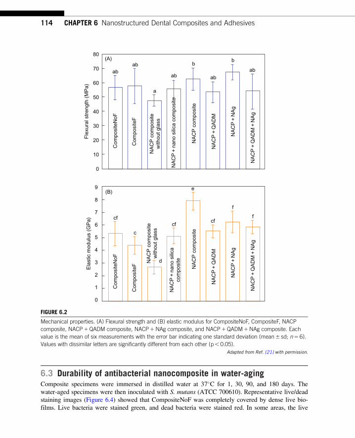
6.3 Durability of antibacterial nanocomposite in water-agingComposite specimens were immersed in distilled water at 37�C for 1, 30, 90, and 180 days. The
water-aged specimens were then inoculated with S. mutans (ATCC 700610). Representative live/dead
staining images (Figure 6.4) showed that CompositeNoF was completely covered by dense live bio-
films. Live bacteria were stained green, and dead bacteria were stained red. In some areas, the live
ab
(A)80
70
60
50
40
Fle
xura
l str
engt
h (M
Pa)
30
20
10
0
(B)9
8
7
6
5
4
3
2
1
0
cfcf
c
d
e
cf
f
f
ab
ab abab
NA
CP
+ Q
AD
M +
NA
gN
AC
P +
QA
DM
+ N
Ag
NA
CP
+ N
Ag
NA
CP
+ N
Ag
NA
CP
+ Q
AD
MN
AC
P +
QA
DM
NA
CP
+ n
ano
silic
a co
mpo
site
NA
CP
+ n
ano
silic
aco
mpo
site
NA
CP
com
posi
tew
ithou
t gla
ss
NA
CP
com
posi
tew
ithou
t gla
ss
Com
posi
teN
oFC
ompo
site
NoF
Ela
stic
mod
ulus
(G
Pa)
Com
posi
teF
Com
posi
teF
NA
CP
com
posi
teN
AC
P c
ompo
site
a
bb
FIGURE 6.2
Mechanical properties. (A) Flexural strength and (B) elastic modulus for CompositeNoF, CompositeF, NACP
composite, NACP1QADM composite, NACP1NAg composite, and NACP1QADM1NAg composite. Each
value is the mean of six measurements with the error bar indicating one standard deviation (mean6 sd; n5 6).
Values with dissimilar letters are significantly different from each other (p, 0.05).
Adapted from Ref. [21] with permission.
114 CHAPTER 6 Nanostructured Dental Composites and Adhesives

(A)
(B)f
f
f
g
h
l
a
ab
b
c
de
1 day
3 day
35
30
25
20C
olon
y fo
rmin
g un
its (
106
per
disk
)
Com
posi
teN
oFC
ompo
site
NoF
Col
ony
form
ing
units
(10
6 pe
r di
sk)
Com
posi
teF
Com
posi
teF
NA
CP
com
posi
teN
AC
P c
ompo
site
NA
CP
+ Q
AD
MN
AC
P +
QA
DM
NA
CP
+ N
Ag
NA
CP
+ N
Ag
NA
CP
+ Q
AD
M +
NA
gN
AC
P +
QA
DM
+ N
Ag
15
10
5
0
160
140
120
100
80
60
40
20
0
FIGURE 6.3
CFU counts of S. mutans biofilms adherent on the composites at (A) 1 day and (B) 3 days, with the y-axis
units being 106 bacteria per composite disk. In each plot, the values (mean6 sd; n5 6) indicated with
dissimilar letters are significantly different from each other (P, 0.05). The NACP1QADM1NAg composite
had the least CFU which was an order of magnitude less than that of CompositeNoF.
Adapted from Ref. [21] with permission.
1156.3 Durability of antibacterial nanocomposite in water-aging

and compromised bacteria were closely associated, and the red color was mingled with green to yield
yellow/orange colors. Examples of these staining colors are indicated by the arrows. Compared to
CompositeNoF and CompositeF, NACP1QADM had much more red/yellow/orange staining.
Representative scanning electron microscopy (SEM) images of the biofilm structures on compo-
sites surfaces are shown in Figure 6.5. In (A) and (B), the CompositeNoF and CompositeF had
(A)
(B)
(C)
(D)
(E)
(F)
(G)
(H)
(I)
FIGURE 6.4
Live/dead staining of 3-days biofilms on composites. Live bacteria were stained green and dead bacteria were stained
red. Live and dead bacteria in close proximity showed yellow/orange colors. The images shown in (A�I) are
representative of each group. CompositeNoF was covered by a dense biofilm with green staining. CompositeF had
some compromised bacteria. NACP1QADM had much more dead bacteria staining than the controls. The area
fraction of live bacteria staining is plotted (mean6 sd; n5 6). There was little difference in biofilm viability versus aging
time, indicating that the antibacterial activity of NACP�QADM nanocomposite was not lost in water immersion. (For
interpretation of the references to color in this figure legend, the reader is referred to the web version of this book.)
Adapted from Ref. [52] with permission.
116 CHAPTER 6 Nanostructured Dental Composites and Adhesives

(A) CompositeNoF, 180 d CompositeNoF, 180 d
CompositeF, 180 dCompositeF, 180 d
(D)
(E)(B)
(C) (F)NACP + QADM, 180 d NACP + QADM, 180 d
FIGURE 6.5
SEM micrographs of biofilms. (A�C) Lower and (D�F) higher magnification. Each type of composite, aged for
1�180 days, had a similar biofilm appearance. The images shown here are for composites aged for 180
days, to demonstrate the long-term antibacterial activity of NACP�QADM nanocomposite. CompositeNoF and
CompositeF had dense biofilms. NACP�QADM had much less biofilm coverage. In (C) and (F), “R” indicates
the resin composite surface not covered by biofilms. Arrows indicate the chain structure of S. mutans
biofilms. The chains are much shorter on NACP�QADM in (F), along with individual cells that did not form a
chain. Each bacterial cell had the shape of a short rod with a length of about 1 µm (arrow in F).
Adapted from Ref. [52] with permission.
1176.3 Durability of antibacterial nanocomposite in water-aging

dense biofilms. In (C), NACP�QADM had less biofilms, where “R” indicates resin composite not
covered by biofilms. Higher magnification (D, E) revealed that the S. mutans grew in chains
(arrows). The chains twisted in three-dimensions and were long or continuous in the biofilm archi-
tecture. The chains were much shorter on NACP�QADM in (F), with each chain containing 3�10
cells.
For MTT assay, disks were placed in 24-well plates, inoculated with 1.5 mL inoculation
medium, and cultured for 3 days. Each disk was transferred to new 24-well plates for the MTT
(3-(4,5-dimethylthiazol-2-yl)-2,5-diphenyltetrazolium bromide) assay [20,21]. MTT is a colorimet-
ric assay that measures the enzymatic reduction of MTT, a yellow tetrazole, to formazan. One mil-
liliter of MTT was added to each well and incubated for 1 h. Disks were transferred to new 24-well
plates, and 1 mL of dimethyl sulfoxide (DMSO) was added to solubilize the formazan crystals. The
DMSO solution from each well was used and the absorbance at 540 nm was measured via a micro-
plate reader (SpectraMax M5, Molecular Devices, Sunnyvale, CA).
For lactic acid measurement, disks with 3-days biofilms were placed in new 24-well plates, and
1.5 mL of buffered-peptone water (BPW) supplemented with 0.2% sucrose was added. They were
incubated for 3 h to allow the biofilms to produce acid. Then the BPW solutions were stored for
lactate analysis. The microplate reader was used to measure the absorbance at 340 nm, and standard
curves were prepared using a lactic acid standard (Supelco, Bellefonte, PA) [21].
Figure 6.6 plots the results for MTT metabolic activity, CFU, and lactic acid production of bio-
films. For each property, the values did not vary significantly over aging time (P. 0.1). In (A),
NACP�QADM yielded much lower MTT than commercial composites (P, 0.05). NACP�QADM
greatly reduced the CFU (B) and lactic acid (C), compared to commercial composites (P, 0.05).
Therefore, the novel NACP�QADM nanocomposite substantially decreased the biofilm growth and
lactic acid production, and its antibacterial properties were maintained during long-term water-
aging [52].
6.4 Antibacterial dentin primerScotchbond multi-purpose (referred as “SBMP”) (3M, St. Paul, MN) was used as the parent bond-
ing agent to test the effect of QDMA and NAg incorporation. The purpose was to develop a model
system, and the novel method of QADM and NAg incorporation can then be applied to other bond-
ing agents. SBMP etchant contained 35% phosphoric acid. SBMP primer contained 35�45%
2-hydroxyethylmethacrylate (HEMA), 10�20% copolymer of acrylic/itaconic acids, and 40�50%
water. SBMP adhesive contained 60�70% BisGMA and 30�40% HEMA. QADM and NAg were
incorporated into SBMP primer. SBMP etchant and adhesive were not modified.
Bis(2-methacryloyloxyethyl) dimethylammonium bromide (QADM) was recently synthesized
[20,21]. Briefly, 10 mmol of 2-(N,N-dimethylamino)ethyl methacrylate (Sigma-Aldrich) and
10 mmol of 2-BEMA (Monomer-Polymer Labs, Trevose, PA) were combined in ethanol and stirred
at 60�C for 24 h. The solvent was then evaporated, yielding the QADM. QADM was mixed with
the SBMP primer at QADM/(primer1QADM)5 10 wt%. This was selected because preliminary
study showed that 10% of QADM in the primer provided antibacterial activity without compromis-
ing the dentin bond strength, while 20% QADM in primer decreased the bond strength.
118 CHAPTER 6 Nanostructured Dental Composites and Adhesives

a aaab
bc
1 da
y
30 d
ays
90 d
ays
180
days
1 da
y
30 d
ays
90 d
ays
180
days
1 da
y
30 d
ays
90 d
ays
180
days
bcbc
d dd
(A)
d
c
1 da
yCF
U c
ount
s (×
107
per
disk
)M
TT
abs
orba
nce
(A54
0/cm
2 )
30 d
ays
90 d
ays
180
days
1 da
y
30 d
ays
90 d
ays
180
days
1 da
y
30 d
ays
90 d
ays
180
days
(B)efef ef
e
fg fgg g
h h
h h
1 da
y
Lact
ic A
cid
Pro
duct
ion
(mm
ol/L
)
30 d
ays
90 d
ays
180
days
1 da
y
30 d
ays
90 d
ays
180
days
1 da
y
30 d
ays
90 d
ays
180
days
(C)18
35
2.5
2.0
1.5
1.0
0.5
0.0
30
25
20
15
10
5
0
16
14
12
10
8
6
4
2
0
iij ij
j j
k kk
k
iiij
FIGURE 6.6
Biofilm viability, growth, and acid production. (A) MTT metabolic activity, (B) CFU counts, and (C) lactic acid
production of 3-days biofilms on the composites water-aged for 1�180 days. Each value is mean6 sd (n5 6).
In each plot, values with dissimilar letters are significantly different (P, 0.05). For the MTT assay, a higher
absorbance indicates a higher formazan concentration, which in turn indicates a higher metabolic activity in
the biofilm. NACP�QADM had biofilm metabolic activity and lactic acid that were about half of those on
commercial composites, and CFU about one-third of those on commercial composites (P, 0.05). Aging for
1�180 days did not reduce the antibacterial potency of the NACP�QADM nanocomposite (P. 0.1).
Adapted from Ref. [52] with permission.

Silver 2-ethylhexanoate (Strem, New Buryport, MA) of 0.08 g was dissolved into 1 g of
TBAEMA (Sigma). TBAEMA could facilitate Ag-salt dissolution in resin [21,51]. This Ag solution
was mixed into SBMP primer at 0.05 wt% of silver 2-ethylhexanoate because preliminary study
indicated that this concentration had no adverse effect on dentin bond strength and color of the
primer. Hence, four primers were fabricated: (1) SBMP primer (control), (2) control primer1 10%
QADM (termed “10QADM”), (3) control primer1 0.05% NAg (termed “0.05NAg”), and (4) con-
trol primer1 10% QADM1 0.05% NAg (termed “10QADM1 0.05NAg”).
A dental plaque microcosm biofilm model was used [53]. Saliva was collected from a healthy
donor having natural dentition without active caries or using antibiotics within 3 months. The donor
did not brush teeth for 24 h and abstained from food/drink intake for 2 h prior to donating saliva
[53]. Uncured primers were tested by agar disk diffusion test (ADT). Saliva was added to growth
medium containing mucin at a concentration of 2.5 g/L, bacteriological peptone at 2.0 g/L, tryptone
at 2.0 g/L, yeast extract at 1.0 g/L, NaCl at 0.35 g/L, KCl at 0.2 g/L, CaCl2 at 0.2 g/L, and cysteine
hydrochloride at 0.1 g/L (at pH 7) [54]. The inoculum was incubated (37�C, 5% CO2) for 24 h.
Three types of culture media were used. First, tryptic soy blood agar plates were used to determine
total microorganisms. Second, mitis salivarius agar (MSA) plates, containing 15% sucrose, were
used to determine total streptococci. Third, MSA plus 0.2 units of bacitracin per milliliter was used
to determine mutans streptococci [53].
For ADT, a 0.4 mL bacterial suspension was poured onto each agar plate. Then, 30 µL of each
primer was impregnated into a sterile paper disk with a diameter of 9 mm and a thickness of
1.5 mm [47]. The primer-impregnated paper disk was placed on a plate with bacteria and incubated
for 48 h. The bacterial inhibition zone size was calculated as: (outer diameter of inhibition
zone2 paper disk diameter)/2.
The uncured QADM�NAg primers had a strong antibacterial activity, as shown in Figure 6.7.
As shown in (A), the control primer had minimal inhibition zones. In (B�D), the primers
10QADM, 0.05NAg, and 10QADM1 0.05NAg had much larger inhibition zones. The inhibition
zone sizes are plotted in (E�G) for total microorganisms, total streptococci, and mutans strepto-
cocci, respectively. The inhibition zone sizes for 10QADM1 0.05NAg were ninefold those of the
control (P, 0.05) [53].
6.5 Antibacterial adhesiveAs shown in Figure 6.8, six groups were used for dentin shear bond strength testing. The purpose
of groups 1�3 was to investigate the effects of QADM or NAg individually. The purpose of 3 and
4 was to examine the effect of NAg mass fraction. The purpose of comparing 2, 3, and 5 was to
examine the effect of combining QADM and NAg together in the same adhesive. The purpose of
comparing 5 with 6 was to investigate the effects of adding QADM and NAg into both the adhesive
and the primer on dentin bond strength and biofilm response.
To measure dentin shear bond strength, extracted caries-free human third molars were cleaned
and stored in 0.01% thymol solution. Flat mid-coronal dentin surfaces were prepared by cutting off
the tips of molar crowns with a diamond saw (Isomet, Buehler, Lake Bluff, IL) [55]. Each tooth
was embedded in a polycarbonate holder (Bosworth, Skokie, IL) and ground perpendicular to the
120 CHAPTER 6 Nanostructured Dental Composites and Adhesives

longitudinal axis on 320-grit silicon carbide paper until the occlusal enamel was completely
removed. As shown in Figure 6.8A, the dentin surface was etched with 37% phosphoric acid gel
for 15 s and rinsed with distilled water for 15 s, following a previous study [56]. The primer was
applied with a brush-tipped applicator and rubbed in for 15 s. The solvent was removed with a
stream of air for 5 s. Then the adhesive was applied and light-cured for 10 s (Optilux VCL 401,
Demetron Kerr, Danbury, CT). A stainless-steel iris, having a central opening with a diameter of
4 mm and a thickness of 1.5 mm, was held against the adhesive-treated dentin surface. The central
opening was filled with a composite (TPH, Caulk/Dentsply, Milford, DE), and light-cured for 60 s.
The bonded specimens were stored in distilled water at 37�C for 24 h. A chisel was connected with
(E)
(A)
(C)
(B)
(D)
a
b
9
8
7
6
5
4
3
2
1
0
c
d
Con
trol
Inhi
bitio
n zo
ne s
ize
for
tota
l mic
roor
gani
sms
(mm
)
10Q
AD
M
10Q
AD
M+0
.05N
Ag
0.05
NA
g
(F)
e
f
9
8
7
6
5
4
3
2
1
0
g
h
Con
trol
Inhi
bitio
n zo
ne s
ize
for
tota
l str
epto
cocc
i (m
m)
10Q
AD
M
10Q
AD
M+0
.05N
Ag
0.05
NA
g
(G)
i
j
9
8
7
6
5
4
3
2
1
0
k
l
Con
trolInhi
bitio
n zo
ne s
ize
for
mut
ans
stre
ptoc
occi
(m
m)
10Q
AD
M
10Q
AD
M+0
.05N
Ag
0.05
NA
gFIGURE 6.7
Antibacterial activity of uncured primers in ADT. (A�D) Control primer, 10QADM, 0.05NAg, and
10QADM1 0.05NAg, respectively. Note a small inhibition zone for control, and much wider inhibition zones
for primers with QADM and NAg. This example is for mutans streptococci. Total microorganisms and total
streptococci had similar results. (E�G) Inhibition zone data for total microorganisms, total streptococci, and
mutans streptococci, respectively. Each value is mean6 sd (n5 6). Bars with dissimilar letters indicate values
that are significantly different (P, 0.05).
Adapted from Ref. [53] with permission.
1216.5 Antibacterial adhesive

(C)
(B)
(A)
Lightcure
Lightcure
Composite
Adhesive
Primer
Etchant
Polycarbonatesupport block
Chiselcrosshead
Assemblypinning device
40
35
30
25
20
15
10
0
5
Con
trol
Den
tin s
hear
bon
d st
reng
th (
mpa
)
A +
10Q
AD
M
A +
0.0
5NA
g
A +
0.1
NA
g
A +
10Q
AD
M +
0.0
5NA
g
A&
P +
10Q
AD
M +
0.0
5NA
g
FIGURE 6.8
Human dentin shear bond testing. (A) Schematic of specimen preparation, (B) schematic of shear bond
strength testing, and (C) shear bond strength data. Ten teeth were used for each group. Each value is
mean6 sd (n5 10). Horizontal line indicates that all six groups had similar shear bond strengths (P. 0.1).
Adapted from Ref. [55] with permission.
122 CHAPTER 6 Nanostructured Dental Composites and Adhesives

a computer-controlled Universal Testing Machine (MTS, Eden Prairie, MN) and held parallel to
the composite�dentin interface. Load was applied at a rate of 0.5 mm/min until the bond failed.
Dentin shear bond strength, SD, was calculated as: SD5 4P/(πd2), where P is the load at failure and
d is the diameter of the composite. Ten teeth were tested for each group (n5 10). The results are
plotted in Figure 6.8C. The six groups had shear bond strengths that were not significantly different
(P. 0.1), indicating that adding QADM and NAg to adhesive and primer did not compromise the
dentin shear bond strength [55].
For biofilm experiments, layered disk specimens for biofilm experiments were fabricated fol-
lowing previous studies [25,46]. Six groups were tested in biofilm experiments (Figure 6.9).
Groups 1�5 had specimens with adhesives covering the top surface of the composite disk, without
primer, in order to test the antibacterial properties of the adhesives, as shown in Figure 6.9A.
Group 6 had the QADM�NAg primer covering the adhesive on the composite disk in order to test
the antibacterial properties of the primer/adhesive combination (Figure 6.9B).
Figure 6.9C plots the MTT metabolic activity of microcosm biofilms. Microcosm bio-
films on the commercial adhesive had a high metabolic activity. Incorporation of QADM and
NAg each markedly reduced the metabolic activity (P, 0.05). Adding QADM and NAg
together in the adhesive resulted in a much lower metabolic activity than using QADM or NAg
alone (P, 0.05). Adding QADM and NAg both in the primer and in the adhesive yielded
the lowest biofilm metabolic activity (P, 0.05). The metabolic activity of biofilms on
A&P1 10QADM1 0.05NAg was nearly an order of magnitude less than that on the commercial
adhesive control [55].
6.6 Antibacterial and remineralizing adhesive containing NACPNACPs were incorporated into the adhesive. The purpose was for the NACP to flow with the adhe-
sive into the hybrid layer as well as the dentinal tubules to form resin tags, with Ca and P ions to
remineralize remnants of lesions in the prepared tooth cavity. The NACP mass fraction in the adhe-
sive varied from 10% to 40% [57]. Typical SEM images of the dentin�adhesive interfaces are
shown in Figure 6.10: (A) SBMP adhesive control, (B) SBMP primer P1NAg, SBMP adhesive
A1NAg1 20NACP (with 20% NACP), and (C) P1NAg, A1NAg1 40NACP (with 40%
NACP). Numerous resin tags “T” from well-filled dentinal tubules were visible in all the samples.
“HL” refers to the hybrid layer between the adhesive and the underlying mineralized dentin. At a
higher magnification, the NACP nanoparticles were visible in (D) with 20% NACP as an example.
Arrows in (D) indicate examples of NACP nanoparticles which successfully infiltrated into the den-
tinal tubules. This feature became more visible at higher magnifications in (E) and (F), where
arrows indicate NACP, which successfully infiltrated into not only the straight and smooth tubules
(E) but also the bent and irregularly-shaped tubules (F) [57].
These results show the high promise of novel bonding agents and composites containing anti-
bacterial agents and remineralizing nanoparticles to combat biofilms and secondary caries. Further
studies are needed to investigate caries inhibition in extracted human teeth at the nanostructured
restoration�tooth margins cultured under biofilms.
1236.6 Antibacterial and remineralizing adhesive containing NACP
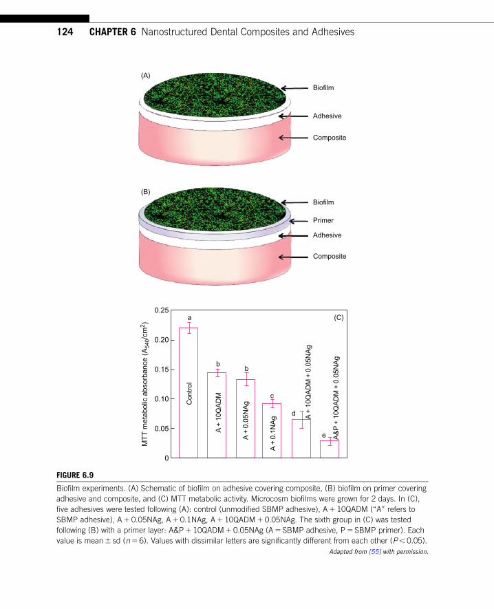
(C)a
bb
c
d
e
(B)
(A)
Biofilm
Composite
Adhesive
Primer
Biofilm
Composite
Adhesive
0.25
0.20
0.15
0.10
0.05
0
Con
trol
MT
T m
etab
olic
abs
orba
nce
(A54
0/cm
2 )
A +
10Q
AD
M
A +
0.0
5NA
g
A +
0.1
NA
g A +
10Q
AD
M +
0.0
5NA
g
A&
P +
10Q
AD
M +
0.0
5NA
g
FIGURE 6.9
Biofilm experiments. (A) Schematic of biofilm on adhesive covering composite, (B) biofilm on primer covering
adhesive and composite, and (C) MTT metabolic activity. Microcosm biofilms were grown for 2 days. In (C),
five adhesives were tested following (A): control (unmodified SBMP adhesive), A1 10QADM (“A” refers to
SBMP adhesive), A1 0.05NAg, A1 0.1NAg, A1 10QADM1 0.05NAg. The sixth group in (C) was tested
following (B) with a primer layer: A&P1 10QADM1 0.05NAg (A5 SBMP adhesive, P5 SBMP primer). Each
value is mean6 sd (n5 6). Values with dissimilar letters are significantly different from each other (P, 0.05).
Adapted from [55] with permission.
124 CHAPTER 6 Nanostructured Dental Composites and Adhesives

6.7 Summary and conclusionsNovel calcium phosphate nanoparticles and antibacterial monomers were synthesized, and a new class
of bioactive nanocomposites and nanostructured adhesives with a combination of antibacterial and
remineralizing capabilities were developed for the first time. The novel NACP�QADM nanocompo-
site had a strong antibacterial activity that was maintained after water-aging for 6 months. Strength
(A) (B) (C)
(D) (E) (F)
FIGURE 6.10
SEM micrographs of dentin�adhesive interfaces. (A) SBMP control (P5 primer, A5 adhesive), (B) P1NAg,
A1NAg1 20NACP, (C) P1NAg, A1NAg1 40NACP. (D) P1NAg, A1NAg1 20NACP at a higher
magnification, and (E, F) at even higher magnifications. Adhesives filled the dentinal tubules and formed
resin tags “T” for all six groups. “HL” indicates the hybrid layer between the adhesive and the underlying
mineralized dentin. (D�F) Numerous NACP nanoparticles were present in the adhesive layer, in the hybrid
zone, and inside the dentinal tubules. Arrows in (D�F) indicate NACP in the dentinal tubules. NACP were
able to infiltrate with the adhesive not only into straight tubules (E) but also into bent and irregularly-shaped
tubules (F).
Adapted from Ref. [57] with permission.
1256.7 Summary and conclusions

and modulus of NACP�QADM nanocomposite after 6-month immersion matched those of commer-
cial control composites without antibacterial properties. Incorporation of QADM into NACP nano-
composite greatly reduced the oral bacterial biofilm viability, metabolic activity, CFU, and lactic acid
production. The antibacterial results were not significantly different after water-aging for 1, 30, 90,
and 180 days. The durable antibacterial properties, plus the previously-reported CaP release and acid
neutralization properties, indicate that the novel NACP�QADM nanocomposite may be useful in
restorations to inhibit secondary caries. In addition, QADM and NAg were incorporated into dental
adhesive and primer, which achieved potent antibacterial effects against dental plaque microcosm bio-
films for the first time. The novel QADM�NAg-containing antibacterial adhesives with NACP for
remineralization are promising to combat residual bacteria in the prepared tooth cavity and invading
bacteria along the tooth�restoration interfaces due to bacterial leakage, thereby inhibiting recurrent
caries. Furthermore, the QADM, NAg, and NACP incorporation methods may have a wide applica-
bility to other dental resin composites and bonding systems.
AcknowledgmentsWe thank Dr. Gary E. Schumacher, Antony A. Giuseppetti, and Kathleen M. Hoffman of the Paffenbarger
Research Center of the American Dental Association Foundation, Dr. Ashraf F. Fouad of the University of
Maryland School of Dentistry, and Dr. Qianming Chen of the West China School of Stomatology for discus-
sions and help. We are grateful to Esstech (Essington, PA) and Ivoclar Vivadent (Amherst, NY) for donating
the materials. This study was supported by NIH R01 grants DE17974 and DE14190 (HX), NIDCR-NIST
Interagency Agreement Y1-DE-7005-01, University of Maryland School of Dentistry, NIST, ADAF, and West
China School of Stomatology.
DisclaimerCertain commercial materials and equipment are identified to specify the experimental procedure.
In no instance does such identification imply recommendation or endorsement by NIST and ADAF
or that the material or equipment identified is the best available for the purpose.
References[1] J.L. Ferracane, Resin composite—state of the art, Dent. Mater. 27 (2011) 29�38.
[2] F.F. Demarco, M.B. Correa, M.S. Cenci, R.R. Moraes, N.J.M. Opdam, Longevity of posterior composite
restorations: not only a matter of materials, Dent. Mater. (2012) 87�101.
[3] S.C. Bayne, J.Y. Thompson, E.J. Swift Jr., P. Stamatiades, M. Wilkerson, A characterization of first-
generation flowable composites, J. Am. Dent. Assoc. 129 (1998) 567�577.
[4] B.S. Lim, J.L. Ferracane, R.L. Sakaguchi, J.R. Condon, Reduction of polymerization contraction stress for
dental composites by two-step light-activation, Dent. Mater. 18 (2002) 436�444.
[5] D.C. Watts, A.S. Marouf, A.M. Al-Hindi, Photo-polymerization shrinkage-stress kinetics in resin-
composites: methods development, Dent. Mater. 19 (2003) 1�11.
126 CHAPTER 6 Nanostructured Dental Composites and Adhesives

[6] X. Xu, L. Ling, R. Wang, J.O. Burgess, Formation and characterization of a novel fluoride-releasing
dental composite, Dent. Mater. 22 (2006) 1014�1023.
[7] J.L. Ferracane, Hygroscopic and hydrolytic effects in dental polymer networks, Dent. Mater. 22 (2006)
211�222.
[8] Q. Wan, J. Sheffield, J. McCool, G.R. Baran, Light curable dental composites designed with colloidal
crystal reinforcement, Dent. Mater. 24 (2008) 1694�1701.
[9] J.L. Drummond, Degradation, fatigue, and failure of resin dental composite materials, J. Dent. Res. 87
(2008) 710�719.
[10] S.P. Samuel, S. Li, I. Mukherjee, Y. Guo, A.C. Patel, G.R. Baran, et al., Mechanical properties of experi-
mental dental composites containing a combination of mesoporous and nonporous spherical silica as
fillers, Dent. Mater. 25 (2009) 296�301.
[11] R.M. Carvalho, A.P. Manso, S. Geraldeli, F.R. Tay, D.H. Pashley, Durability of bonds and clinical suc-
cess of adhesive restorations, Dent. Mater. 28 (2012) 72�86.
[12] M.M. Zalkind, O. Keisar, P. Ever-Hadani, R. Grinberg, M.N. Sela, Accumulation of Streptococcus
mutans on light-cured composites and amalgam: an in vitro study, J. Esthet. Dent. 10 (1998)
187�190.
[13] N. Beyth, A.J. Domb, E.I. Weiss, An in vitro quantitative antibacterial analysis of amalgam and compos-
ite resins, J. Dent. 35 (2007) 201�206.
[14] V. Deligeorgi, I.A. Mjor, N.H. Wilson, An overview of reasons for the placement and replacement of
restorations, Prim. Dent. Care 8 (2001) 5�11.
[15] A. Jokstad, S. Bayne, U. Blunck, M. Tyas, N. Wilson, Quality of dental restorations. FDI Commission
Projects 2�95, Int. Dent. J. 51 (2001) 117�158.
[16] S. Imazato, Review: antibacterial properties of resin composites and dentin bonding systems, Dent.
Mater. 19 (2003) 449�457.
[17] S. Imazato, Bioactive restorative materials with antibacterial effects: new dimension of innovation in
restorative dentistry, Dent. Mater. J. 28 (2009) 11�19.
[18] X. Xu, Y. Wang, S. Liao, Z.T. Wen, Y. Fan, Synthesis and characterization of antibacterial dental mono-
mers and composites, J. Biomed. Mater. Res. B 100 (2012) 1162�1511.
[19] Y. Weng, L. Howard, X. Guo, V.J. Chong, R.L. Gregory, D. Xie, A novel antibacterial resin composite
for improved dental restoratives, J. Mater. Sci. Mater. Med. 23 (2012) 1553�1561.
[20] J.M. Antonucci, D.N. Zeiger, K. Tang, S. Lin-Gibson, B.O. Fowler, N.J. Lin, Synthesis and characteriza-
tion of dimethacrylates containing quaternary ammonium functionalities for dental applications, Dent.
Mater. 28 (2012) 219�228.
[21] L. Cheng, M.D. Weir, H.H.K. Xu, J.M. Antonucci, A.M. Kraigsley, N.J. Lin, et al., Antibacterial amor-
phous calcium phosphate nanocomposite with quaternary ammonium salt and silver nanoparticles, Dent.
Mater. 28 (2012) 561�572.
[22] S. Imazato, M. Torii, Y. Tsuchitani, J.F. McCabe, R.R.B. Russell, Incorporation of bacterial inhibitor
into resin composite, J. Dent. Res. 73 (1994) 1437�1443.
[23] S. Imazato, F.R. Tay, A.V. Kaneshiro, Y. Takahashi, S. Ebisu, An in vivo evaluation of bonding abil-
ity of comprehensive antibacterial adhesive system incorporating MDPB, Dent. Mater. 23 (2007)
170�176.
[24] N. Beyth, I. Yudovin-Farber, R. Bahir, A.J. Domb, E.I. Weiss, Antibacterial activity of dental compo-
sites containing quaternary ammonium polyethylenimine nanoparticles against Streptococcus mutans,
Biomaterials 27 (2006) 3995�4002.
[25] F. Li, Z.G. Chai, M.N. Sun, F. Wang, S. Ma, L. Zhang, et al., Anti-biofilm effect of dental adhesive
with cationic monomer, J. Dent. 88 (2009) 372�376.
[26] N. Namba, Y. Yoshida, N. Nagaoka, S. Takashima, K. Matsuura-Yoshimoto, H. Maeda, et al.,
Antibacterial effect of bactericide immobilized in resin matrix, Dent. Mater. 25 (2009) 424�430.
127References

[27] D. Xie, Y. Weng, X. Guo, J. Zhao, R.L. Gregory, C. Zheng, Preparation and evaluation of a novel glass-
ionomer cement with antibacterial functions, Dent. Mater. 27 (2011) 487�496.
[28] D. Skrtic, J.M. Antonucci, E.D. Eanes, F.C. Eichmiller, G.E. Schumacher, Physiological evaluation of
bioactive polymeric composites based on hybrid amorphous calcium phosphates, J. Biomed. Mater. Res.
B 53 (2000) 381�391.
[29] S.H. Dickens, G.M. Flaim, S. Takagi, Mechanical properties and biochemical activity of remineralizing
resin-based Ca-PO4 cements, Dent. Mater. 19 (2003) 558�566.
[30] S.E. Langhorst, J.N. O’Donnell, D. Skrtic, In vitro remineralization of enamel by polymeric amorphous
calcium phosphate composite: Quantitative microradiographic study, Dent. Mater. 25 (2009) 884�891.
[31] H.H.K. Xu, L. Sun, M.D. Weir, J.M. Antonucci, S. Takagi, L.C. Chow, Nano dicalcium phosphate
anhydrous-whisker composites with high strength and Ca and PO4 release, J. Dent. Res. 85 (2006)
722�727.
[32] H.H.K. Xu, M.D. Weir, L. Sun, J.L. Moreau, S. Takagi, L.C. Chow, et al., Strong nanocomposites with
Ca, PO4 and F release for caries inhibition, J. Dent. Res. 89 (2010) 19�28.
[33] H.H.K. Xu, J.L. Moreau, L. Sun, L.C. Chow, Nanocomposite containing amorphous calcium phosphate
nanoparticles for caries inhibition, Dent. Mater. 27 (2011) 762�769.
[34] J.L. Moreau, L. Sun, L.C. Chow, H.H.K. Xu, Mechanical and acid neutralizing properties and inhibition
of bacterial growth of amorphous calcium phosphate dental nanocomposite, J. Biomed. Mater. Res. B 98
(2011) 80�88.
[35] P. Spencer, Y. Wang, Adhesive phase separation at the dentin interface under wet bonding conditions,
J. Biomed. Mater. Res. 62 (2002) 447�456.
[36] K. Ikemura, F.R. Tay, T. Endo, D.H. Pashley, A review of chemical-approach and ultramorphological
studies on the development of fluoride-releasing dental adhesives comprising new pre-reacted glass iono-
mer (PRG) fillers, Dent. Mater. J. 27 (2008) 315�329.
[37] J. Park, Q. Ye, E. Topp, A. Misra, S.L. Kieweg, P. Spencer, Water sorption and dynamic mechanical
properties of dentin adhesives with a urethane-based multifunctional methacrylate monomer, Dent.
Mater. 25 (2009) 1569�1575.
[38] A.V. Ritter, E.J. Swift Jr., H.O. Heymann, J.R. Sturdevant, A.D. Wilder Jr., An eight-year clinical evalu-
ation of filled and unfilled one-bottle dental adhesives, J. Am. Dent. Assoc. 140 (2009) 28�37.
[39] F. Garcia-Godoy, N. Kramer, A.J. Feilzer, R. Frankenberger, Long-term degradation of enamel and den-
tin bonds: 6-year results in vitro vs. in vivo, Dent. Mater. 26 (2010) 1113�1118.
[40] M.S. Shinohara, M.F. De Goes, L.F. Schneider, J.L. Ferracane, P.N. Pereira, V.D. Hipolito, et al.,
Fluoride-containing adhesive: durability on dentin bonding, Dent. Mater. 25 (2009) 1383�1391.
[41] J. Park, J. Eslick, Q. Ye, A. Misra, P. Spencer, The influence of chemical structure on the properties in
methacrylate-based dentin adhesives, Dent. Mater. 27 (2011) 1086�1093.
[42] D.H. Pashley, F.R. Tay, L. Breschi, L. Tjaderhane, R.M. Carvalho, M. Carrilho, et al., State of the art
etch-and-rinse adhesives, Dent. Mater. 27 (2011) 1�16.
[43] L. Roeder, R.N.R. Pereira, T. Yamamoto, N. Ilie, S. Armstrong, J. Ferracane, Spotlight on bond strength
testing—unraveling the complexities, Dent. Mater. 27 (2011) 1197�1203.
[44] B. Van Meerbeek, K. Yoshihara, Y. Yoshida, A. Mine, J. De Munck, State of the art of self-etch adhe-
sives, Dent. Mater. 27 (2011) 17�28.
[45] S. Imazato, Y. Kinomoto, H. Tarumi, S. Ebisu, F.R. Tay, Antibacterial activity and bonding characteris-
tics of an adhesive resin containing antibacterial monomer MDPB, Dent. Mater. 19 (2003) 313�319.
[46] S. Imazato, A. Ehara, M. Torii, S. Ebisu, Antibacterial activity of dentin primer containing MDPB after
curing, J. Dent. 26 (1998) 267�271.
[47] S. Imazato, A. Kuramoto, Y. Takahashi, S. Ebisu, M.C. Peters, In vitro antibacterial effects of the dentin
primer of clearfil protect bond, Dent. Mater. 22 (2006) 527�532.
128 CHAPTER 6 Nanostructured Dental Composites and Adhesives

[48] N. Hiraishi, C.K. Yiu, N.M. King, F.R. Tay, Effect of chlorhexidine incorporation into a self-etching
primer on dentin bond strength of a luting cement, J. Dent. 38 (2010) 496�502.
[49] L.C. Chow, L. Sun, B. Hockey, Properties of nanostructured hydroxyapatite prepared by a spray drying
technique, J. Res. Natl. Inst. Stand. Technol. 109 (2004) 543�551.
[50] L. Cheng, M.D. Weir, H.H.K. Xu, A.M. Kraigsley, N.J. Lin, S. Lin-Gibson, et al., Antibacterial and
physical properties of calcium-phosphate and calcium-fluoride nanocomposites with chlorhexidine, Dent.
Mater. 28 (2012) 573�583.
[51] Y.J. Cheng, D.N. Zeiger, J.A. Howarter, X. Zhang, N.J. Lin, J.M. Antonucci, et al., In situ formation of
silver nanoparticles in photocrosslinking polymers, J. Biomed. Mater. Res. B 97 (2011) 124�131.
[52] L. Cheng, M.D. Weir, K. Zhang, S.M. Xu, Q. Chen, X.D. Zhou, et al., Antibacterial nanocomposite with
calcium phosphate and quaternary ammonium, J. Dent. Res. 91 (2012) 460�466.
[53] L. Cheng, K. Zhang, M.A.S. Melo, M.D. Weir, X.D. Zhou, H.H.K. Xu, Anti-biofilm dentin primer with
quaternary ammonium and silver nanoparticles, J. Dent. Res. 91 (2012) 598�604.
[54] A.J. McBain, In vitro biofilm models: an overview, Adv. Appl. Microbiol. 69 (2009) 99�132.
[55] K. Zhang, M.A.S. Melo, L. Cheng, M.D. Weir, Y.X. Bai, H.H.K. Xu, Effect of quaternary ammonium
and silver nanoparticle-containing adhesives on dentin bond strength and dental plaque microcosm bio-
films, Dent. Mater. 28 (2012) 842�852.
[56] J.M. Antonucci, J.N. O’Donnell, G.E. Schumacher, D. Skrtic, Amorphous calcium phosphate composites
and their effect on composite-adhesive-dentin bonding, J. Adhes. Sci. Technol. 23 (2009) 1133�1147.
[57] M.A.S. Melo, L. Cheng, K. Zhang, M.D. Weir, L.K.A. Rodrigues, H.H.K. Xu, Antibacterial dental adhesive
containing silver and amorphous calcium phosphate nanoparticles, Dent. Mater. (2012) http://dx.doi.org/
10.1016/j.dental.2012.10.005.
129References
CHAPTER
7Nanotechnology and Nanoparticlesin Contemporary Dental Adhesives
Mohammad Nassifa and Farid El AskarybaBiomaterials Department, Faculty of Dentistry, Ain Shams University, Cairo, Egypt
bOperative Dentistry Department, Faculty of Dentistry, Ain Shams University, Cairo, Egypt
CHAPTER OUTLINE
7.1 Introduction ............................................................................................................................... 132
7.2 Brief history of dental adhesives .................................................................................................132
7.3 Contemporary adhesive systems..................................................................................................133
7.3.1 Etch-and-rinse adhesives ........................................................................................ 135
7.3.2 Self-etching adhesives ............................................................................................ 135
7.3.3 Glass ionomer adhesive........................................................................................... 137
7.4 Chemical compositions of contemporary adhesives ...................................................................... 138
7.4.1 Resin monomers..................................................................................................... 139
7.4.2 Solvents ................................................................................................................ 140
7.4.3 Fillers.................................................................................................................... 140
7.4.3.1 Role of fillers in dental adhesives .................................................................. 140
7.4.3.2 Effect of filler composition and particle size .................................................... 141
7.5 Ketac nanoprimer....................................................................................................................... 145
7.6 Nanoleakage ............................................................................................................................. 147
7.7 Atomic force microscopy in the field of dental adhesion............................................................... 149
7.8 How can nanoscience and nanotechnology improve the outcome of clinical adhesive dentistry? .......151
7.8.1 Use of hydrophobic coating ..................................................................................... 151
7.8.2 Extended polymerization time.................................................................................. 152
7.8.3 Use of MMPs inhibitors........................................................................................... 152
7.8.4 Improved impregnation ........................................................................................... 153
7.8.5 Wet ethanol bonding approach................................................................................. 153
7.8.6 Improving dental collagen network mechanical properties prior to bonding .................. 154
7.8.7 Enhancing biomimetic remineralization .................................................................... 155
7.9 Future prospective of nanotechnology in the field of adhesive dentistry......................................... 155
7.9.1 On-demand antibacterial adhesives.......................................................................... 155
7.9.2 Improving adhesive polymerization through catalytic activity of nanoparticles .............. 155
7.9.3 Antibacterial orthodontic adhesives containing nanosilver .......................................... 156
131Nanobiomaterials in Clinical Dentistry.
© 2013 Elsevier Inc. All rights reserved.

7.9.4 Radiopaque dental adhesives .................................................................................. 156
7.9.5 Self-adhesive composites ........................................................................................ 156
7.9.6 Self-healing adhesives ............................................................................................ 156
7.9.7 High-speed AFM..................................................................................................... 157
7.10 Conclusions and future directions ............................................................................................... 159
References ......................................................................................................................................... 160
7.1 IntroductionThe field of adhesion to dental substrates has gained a lot of interest with massive research work
aiming at improving bond strength and increasing bond durability. Nanoscience and nanotechnol-
ogy are expected to play a major role in understanding and improving the outcome of adhesion and
dental restorative procedures. These improvements are expected to yield adhesives with antibacte-
rial capacities through the incorporation of antimicrobial nanoparticles. Self-healing adhesives will
be available for the dental profession through nanoencapsulation of monomers and incorporation of
these nanocapsules in the adhesives. Adhesives incorporating nanoparticles or nanorods will show
improved physicomechanical properties and catalytic activity with better polymerization and
conversion of adhesive monomers.
7.2 Brief history of dental adhesivesIn the dental research area, Kramer and McLean in 1952 [1] were the first to make an experimental
study in the field of adhesion to tooth substrate, but this was poorly unnoticeable until the
Buonocore’s attempt in 1955 [2]. Nowadays, the concept of “minimum interventions” or “mini-
mally invasive cavity preparations” has been widely accepted [3] which means that the diseased
dental tissues is only removed and replaced with adhesive restorations.
Previously, van Meerbeek et al. [4] classified the dental adhesives according to their actions on
the smear layer. One-step or two-step adhesives that modify the smear layer were the first category
in this classification. Another approach was the total removal of the smear layer, which involved
two-step or three-step adhesives. The last approach was the adhesive that dissolved the smear layer
rather than remove it. These adhesives utilize acidic “self-etching primers” that dissolve the smear
layer making it a part of the hybrid layer (Figure 7.1).
With the introduction of one-step self-etching adhesives, several brands were being available in
the market, which made wide varieties of such adhesives [5]. Dental adhesives were categorized
into (i) etch-and-rinse, either three-step or two-step, (ii) self-etching adhesives, either two-step or
one-step, and (iii) the glass ionomer adhesive which is considered a two-step etch-and-rinse adhe-
sive based on resin-modified glass ionomer (RMGI) formulation. Among all adhesive categories,
three-step etch-and-rinse adhesive is considered the “gold standard” regarding its clinical durability
(Figure 7.2).
132 CHAPTER 7 Nanotechnology and Nanoparticles

The one-step self-etching adhesives are available as mixed/two-bottles, or no-mix/one-bottle
adhesives, 2-hydroxyethyl methacrylate (HEMA)-free or HEMA-containing adhesives, water-free
or water-containing adhesives, and recently solvent-free or solvent-containing adhesives.
7.3 Contemporary adhesive systemsWhen the acid etchant is applied to enamel and dentin, superficial decalcification with the loss of
minerals occurs. Application of adhesive resin will replace the lost minerals of tooth structure and
when cured in situ becomes micromechanically retained within the porosities that are being created
during the acid-etching process [5]. John Gwinnett [6] was the first to evaluate the adhesive/enamel
interface and described an acid-resistance layer. This layer was the first true “hybrid layer” discov-
ered. Unfortunately, the term “hybrid layer” was not applied until the study by Nakabayashi et al.
in 1982 [7]. They pointed to the existence of such intermediate layer between the adhesive layer
and dentin and gave it the name “hybrid layer” (Figure 7.3).
Con
ditio
ner
Con
ditio
ner
Categorization of adhesives according to their clinical application mode
OneNumber ofapplication
steps
Conditioner
Primer
Adhesiveresin
Two
Self-etchingprimer
Combinedprimer
adhesiveresin
Adhesiveresin
Adhesiveresin
Adhesiveresin
Primer Primer
Adhesiveresin
One
-ste
p sm
ear
laye
r m
odify
ing
adhe
sive
s
Tw
o-st
ep s
mea
r la
yer
mod
ifyin
g ad
hesi
ves
Tw
o-st
ep s
mea
r la
yer
diss
olvi
ng a
dhes
ives
Tw
o-st
ep s
mea
r la
yer
rem
ovin
g ad
hesi
ves
Thr
ee-s
tep
smea
r la
yer
rem
ovin
g ad
hesi
ves
Three
FIGURE 7.1
Schematic presentation of categorization of dental adhesives classified according to their clinical application
mode.
1337.3 Contemporary adhesive systems
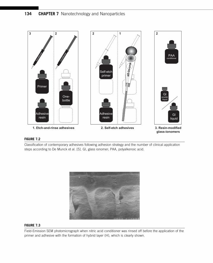
Con
ditio
ner
Con
ditio
ner
Primer
Adhesiveresin
Adhesiveresin
PAAconditioner
Glliquid
Glpowder/liquid
One-bottle
Self-etchprimer
3 2
1. Etch-and-rinse adhesives 2. Self-etch adhesives 3. Resin-modifiedglass-ionomers
2 2
all-
in-o
ne
1
FIGURE 7.2
Classification of contemporary adhesives following adhesion strategy and the number of clinical application
steps according to De Munck et al. [5]. GI, glass ionomer; PAA, polyalkenoic acid.
FIGURE 7.3
Field-Emission SEM photomicrograph when nitric acid conditioner was rinsed off before the application of the
primer and adhesive with the formation of hybrid layer (H), which is clearly shown.
134 CHAPTER 7 Nanotechnology and Nanoparticles

7.3.1 Etch-and-rinse adhesivesThese adhesives utilize the use of acid etching of both enamel and dentin and the application of
both primer and adhesive in separate steps or the application of the primer/adhesive mixture in a
single bonding step. Phosphoric acid with a concentration of 30�40% is the acid of choice for such
adhesive systems. Although enamel etching was established since Buonocore’s study, acid etching
of dentin was not recognized until Fusayama [8] introduced the total-etch concept. One of the
causes for previously reported failures of such concept was the dry bonding technique, until Kanca
[9] described the so-called “wet bonding” (Figure 7.4). In this regard, the complete demineraliza-
tion of dentin with phosphoric acid etching gel is limited to the superficial 5�8 µm (Figure 7.5) of
intertubular dentin [10], which is followed by the application of solvated primer or solvated primer/
adhesive mixture to gain retention through the formation of microresin tags.
Ideally, the resin monomer should completely replace the lost minerals (Figure 7.5). This could
never be ideally performed [9], which is due to the presence of residual solvent or due to the fluid
infiltration through the movement of dentinal fluid from inside the dentinal tubules [11,12]. This
will cause incomplete infiltration of monomer inside the created porosities (Figures 7.6 and 7.7).
Incomplete infiltration of resin monomer is one of the causes that make the etch-and-rinse adhe-
sives technique sensitive.
7.3.2 Self-etching adhesivesSelf-etching adhesives can be classified into four categories according to the pH of the functional
monomer: the strong self-etching adhesives (pH, 1), intermediate adhesives (pH� 1.5), mild adhe-
sives (pH� 2) [13], and extra-mild adhesives (pH. 2.5) [14].
500 nm2 µm
FIGURE 7.4
Scanning electron micrograph of acid-etched dentin showing two dentinal tubules containing remnants of
peritubular dentin matrix. Inset: High magnification of branching collagen fibrils (ca. 75 nm in diameter)
separated by interfibrillar spaces that serve as channels for resin infiltrations during bonding.
1357.3 Contemporary adhesive systems

5–8 µm thickdemineralizedcollagen matrix
Dentinal tubule
Mineralizedintertubular dentin
Peritubular dentin
Highly cross-linked type-I collagen
FIGURE 7.5
Schematic of a hybrid layer (HL) created by an etch-and-rinse adhesive. Note that the depth of the hybrid
layer (green) is about four acid-etched tubule diameters (i.e. ca. 8 µm). The collagen fibrils in the HL are
continuous with the underlying mineralized matrix. A single dentinal tubule is shown devoid of a resin tag to
illustrate its presence. (For interpretation of the references to color in this figure legend, the reader is referred
to the web version of this book.)
Demineralizeddentin matrix
Demineralizeddentin matrix
Mineralized front Mineralized frontPeritubulardentin
Peritubulardentin
Poorly hybridizedresin tag
Perfectly hybridizedresin tag
(A) (B)
FIGURE 7.6
Schematic of micropermeability of (A) poorly hybridized resin tags in etched dentin saturated with water prior
to resin infiltration versus (B) perfectly hybridized resin tags in etched dentin saturated with ethanol prior to
resin infiltration. Yellow fluorescent tracer was forced from the pulp chamber, out the tubules toward the
hybrid layer. In (A) the resin could not displace water-filled lateral branches of tubules in the hybrid layer. This
allowed yellow tracer to diffuse throughout the hybrid layer. In (B) the resin easily dissolved in the ethanol-
filled lateral branches sealing the hybrid layer from dentinal fluid.
136 CHAPTER 7 Nanotechnology and Nanoparticles

The self-etching (etch-and-dry) [14] adhesives utilize simultaneous etch and prime of the dental
substrates. These adhesives do not require a separate acid-etching step. They are less technique sen-
sitive, compared to the etch-and-rinse ones, due to the lack of wet bonding approach. However,
water-free self-etching adhesives require the wet dentin surface, which raise the same question
again: how wet is wet? [14] They are also user friendly, due to the decrease in clinical bonding
steps and shortening application time.
Self-etching adhesives were introduced as two-step adhesives, where the self-etching primer and
adhesive are presented in two separate bottles or as one-step adhesives, where all components are
presented in single solution. The one-step self-etching adhesives can be provided either as mixed/
two-bottles adhesives or no-mix/single-bottle adhesives.
Self-etching adhesives simultaneously etch and infiltrate the tooth substrate, which ensure a com-
plete infiltration of the demineralized zone. On the other hand, they are not free from shortcomings
(Figure 7.8). The decrease in 24-h bond strength as well as a limited durability of such adhesive com-
pared to multistep adhesives was reported. HEMA-containing adhesives suffer from water sorption
and HEMA-free adhesives are prone to phase separation. Decrease in the shelf-life of such adhesives
that combine all components in a single bottle is another shortcoming for such adhesives [14].
7.3.3 Glass ionomer adhesiveGlass ionomer adhesive is considered a two-step etch-and-rinse adhesive, its chemical composition
being based on the glass ionomer cement. It is the diluted version of the RMGI cement, Fuji II LC.
Hybrid layer
Demineralizeddentin zone
Collagen fibrilsMineralizeddentin
Resin
FIGURE 7.7
Schematic illustration of demineralized dentin zone of total-etch adhesives. Inadequate resin infiltration into
collagen fibrils leaves nano- or microspaces within the adhesive interface. This zone is not common for self-
etching adhesive systems. Collagen hydrolysis of the demineralized dentin zone is one of the degradation
patterns for total-etch adhesives. In the absence of a demineralized dentin zone within the bonds, this
collagen hydrolysis does not occur because the encapsulation of cured adhesive resins or protection of the
mineral matrix of dentin prevents chemical degradation of the collagen fibrils even after long-term function.
1377.3 Contemporary adhesive systems

Glass ionomer is the only material that has the self-adhering property. The pretreatment of dentin
surface with 10% or 20% polyacrylic acid (PAA) cleans the surface, removes the smear layer, and
decalcifies the dentin surface to a depth ranging from 0.5 to 1 µm. The glass ionomer adhesive then
infiltrates and mechanically interlocks through the process of hybridization. In addition, a chemical
bond is formed through the ionic exchange between the carboxylic group of the PAA and the
calcium ions, which remain attached to the collagen fibrils [5].
7.4 Chemical compositions of contemporary adhesivesRetention of restoration and providing a perfect seal to cavity wall and margin are the prime aims
for the use of adhesives. Micromechanical bond to enamel and dentin is the primary mechanism of
adhesion to tooth structure, which is achieved by the infiltration of the resin monomer into the
acid-etching-created porosities and become interlocked upon its curing [5]. Another mechanism of
bonding is the ion exchange between the acidic monomers and calcium through adding specific
monomers, which are able to react with the hydroxyapatite (HAP) [15]. The adhesion of adhesives
is not only limited to tooth substrates but also it should provide excellent bond to the overlying
resin composite. The chemical compositions of the adhesive systems should provide components
that are able to achieve such goals (Figure 7.9). Resin monomers, solvents, initiators, inhibitors,
and sometimes fillers are the main constituents of adhesive systems [16].
FIGURE 7.8
The major shortcomings of current one-step adhesives.
138 CHAPTER 7 Nanotechnology and Nanoparticles

7.4.1 Resin monomersMonomers are the most important components of the adhesive, which are considered the key con-
stituents of adhesives. To provide excellent bond with resin composite, monomers in the adhesive
should be similar to those of the resin composite. Two types of monomers are presented, either the
cross-linked monomers or the functional monomers. The latter are presented mainly in the self-
etching adhesives. The most cross-linking monomers, which are used in dental adhesives,
are bisphenol A-glycidyl methacrylate, urethane dimethacrylate, and triethylene glycol dimethacryl-
ate (TEGDMA), while 4-methacryloxy-ethyl trimellitate anhydride, 4-methacryloyloxy-ethyl tri-
mellitic acid, dipentaerythritol penta acrylate monophosphate, 2-(methacryloyloxyethyl) phenyl
hydrogen phosphate, and 10-methacryloyloxydecyl dihydrogen phosphate are the most used
Three-step3 2 2 1
Two-stepA
dhes
ive
resi
n
Adh
esiv
e re
sin
Com
bine
d pr
imer
-adh
esiv
e re
sin
Prim
erC
ondi
tione
r
Con
ditio
ner
Sel
f-et
chin
g pr
imer
Sel
f-et
ch a
dhes
ive
1-co
mpo
nent
2-co
mpo
nent
Two-step One-step
Solvent (=water+ cosolvent)
(~50%)
Solvent (=water +cosolvent)(~50 wt%)
Mono-methacrylates
Mono-methacrylates
Mono-methacrylates
Dimethacrylates
DimethacrylatesDimethacrylates
(HEMA)
HEMAHEMA
Dimethacrylates
HEMA
Solvent
Solvent (50 wt%)
AcidAcid
Initiator +inhibitor
Initiator + inhibitorInitiator + inhibitor
(Initiator +inhibitor)
(Initiator +inhibitor)
(Initiator + inhibitor)
Acidic Mono-methacrylates
(15–30%)
(Filler 1–10 wt%)
(Filler 20–50 wt%)
Etch-and-rinse adhesives Self-etch adhesives
(Filler 20–50 wt%)(Filler 3–10 wt%)
FIGURE 7.9
Classification of contemporary adhesives according to Van Landuyt et al. [16]. Even though most adhesives
contain the same components, they may differ significantly considering the proportional amount of
ingredients. As indicated, most adhesives contain methacrylate-based monomers. The mentioned
percentages of ingredients are approximations; nevertheless a lot of variation considering the proportional
composition of adhesives exists between different products. Two-step etch-and-rinse adhesives often referred
to as “one-bottle” systems. Irrespective of the classification, each component, either primer or bonding or
self-etching adhesive can come in two bottles that need to be mixed prior to application. As such, one-step
self-etch adhesives are often subdivided in one- and two-component systems.
1397.4 Chemical compositions of contemporary adhesives

functional monomers [16]. On the other hand, 2-hydroxyethyl methacrylate (HEMA) is a low
molecular weight monomer, which is characterized by its hydrophilic properties and is an important
constituent of most adhesive systems.
The cross-linking monomers provide strength to the adhesive and have hydrophobic properties
which prevent water sorption of the cured adhesive, while functional monomers are responsible
for the demineralization of tooth substrates and provide chemical bond to calcium in the HAP; nev-
ertheless, their ability to decalcify and adhere to the HAP differs from one functional monomer to
another [16].
7.4.2 SolventsBonding to dentin requires the addition of solvent to the adhesive composition. The presence of sol-
vent is responsible for the improvement of wetting of the hydrophilic monomer and decrease in the
viscosity of the adhesive for enhancement of the diffusivity of primer or primer/adhesive mixture
into the acid-etching-created microporosities. Three main solvents used in the adhesives irrespec-
tive of their adhesion strategy are acetone, ethanol, and water.
Adhesives that contain acetone or ethanol are recommended in the wet bonding technique.
Although acetone has a higher vapor pressure than ethanol, both have better evaporation during the
air-drying step [16], but both the solvents lack the rewetting capacity of collagen fibers, especially
when etch-and-rinse adhesives are used. On the other hand, water is an excellent rewetting agent [9]
but hardly gets evaporated during the air-drying step. A water/ethanol mixture is presented in some
etch-and-rinse adhesive systems to improve the evaporation of water [16]. Acetone is only found as
a mixture with water in the self-etching adhesives, as water is an essential component in the self-
etching adhesives.
7.4.3 FillersAdhesive can be found with fillers (filled adhesive) or without filler contents (unfilled resin).
Unlike resin composite materials, fillers in the adhesive are presented in small amounts compared
to resin composite and the filler size should be small enough to enable the fillers to penetrate into
dentinal tubules or between the collagen fibril spaces.
7.4.3.1 Role of fillers in dental adhesivesFillers are added to resin adhesives to improve the strength of the adhesive layer. Increasing the
strength of adhesive layer will improve the bond strength of the adhesive [17] to either enamel or
dentin, but such improvement was dependent on filler loading [18,19]. Increase in microfiller load-
ing up to 40% had not only negatively affected the shear bond strength but also adversely affected
the polymerization rate (PR) of the adhesive layer [19]. On the other hand, adding 10% silica nano-
fillers did not affect the degree of conversion (DC), but it significantly improved the cohesive
strength of the adhesive [20].
Increasing the viscosity of the adhesive to a level that does not affect its wetting to tooth sub-
strate is another issue for adding fillers to the adhesives. During the air-drying step, excessive air
drying could result in excessive thinning of the adhesive. Very thin adhesive layer suffers from
incomplete PR due to oxygen inhibited layer [16]. Adding filler prevents the excessive thinning of
140 CHAPTER 7 Nanotechnology and Nanoparticles
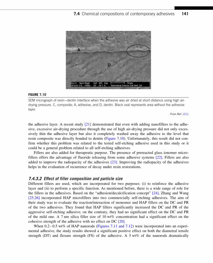
the adhesive layer. A recent study [21] demonstrated that even with adding nanofillers to the adhe-
sive, excessive air-drying procedure through the use of high air-drying pressure did not only exces-
sively thin the adhesive layer but also it completely washed away the adhesive to the level that
resin composite was directly bonded to dentin (Figure 7.10). Unfortunately, this result did not con-
firm whether this problem was related to the tested self-etching adhesive used in this study or it
could be a general problem related to all self-etching adhesives.
Fillers are also added for therapeutic purpose. The presence of prereacted glass ionomer micro-
fillers offers the advantage of fluoride releasing from some adhesive systems [22]. Fillers are also
added to improve the radiopacity of the adhesives [23]. Improving the radiopacity of the adhesives
helps in the evaluation of recurrence of decay under resin restorations.
7.4.3.2 Effect of filler composition and particle sizeDifferent fillers are used, which are incorporated for two purposes: (i) to reinforce the adhesive
layer and (ii) to perform a specific function. As mentioned before, there is a wide range of role for
the fillers in the adhesives. Based on the “adhesion/decalcification concept” [24], Zhang and Wang
[25,26] incorporated HAP microfillers into two commercially self-etching adhesives. The aim of
their study was to evaluate the reaction/interaction of monomer and HAP fillers on the DC and PR
of the two adhesives. They found that HAP fillers significantly increased the DC and PR of the
aggressive self-etching adhesive; on the contrary, they had no significant effect on the DC and PR
of the mild one. A 7 nm silica filler size of 10 wt% concentration had a significant effect on the
cohesive strength of the adhesive with no effect on DC [20].
When 0.2�0.5 wt% of HAP nanorods (Figures 7.11 and 7.12) were incorporated into an experi-
mental adhesive, the study results showed a significant positive effect on both the diametral tensile
strength (DT) and flexure strength (FS) of the adhesive. A 5 wt% of the nanorods dramatically
FIGURE 7.10
SEM micrograph of resin�dentin interface when the adhesive was air dried at short distance using high air-
drying pressure. C, composite; A, adhesive, and D, dentin. Black oval represents area without the adhesive
layer.
From Ref. [21].
1417.4 Chemical compositions of contemporary adhesives

decreased both DT and FS, which was due to the decrease in curing depth of such adhesive. A sig-
nificant higher microshear bond strength was recorded with 0.2 wt% nanorods filler. Since high dis-
persion stability of nanorods in the experimental adhesive was demonstrated, the HAP nanorods
could be used as an alternative to other nanofillers in the adhesives and that the advantage of these
fillers might influence the remineralization at the resin/dentin interface [27].
(A) (B)
FIGURE 7.11
(A) SEM and (B) TEM photomicrographs of synthesized HAP nanorods.
10 µm Ca Ka1
FIGURE 7.12
EDX�calcium mapping image of the adhesive system containing 0.5 wt% HAP nanorods. The bright spots
show the distribution of the HAP nanorods in the adhesive.
142 CHAPTER 7 Nanotechnology and Nanoparticles
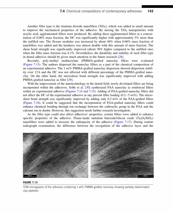
Another filler type is the titanium dioxide nanofillers (TiO2), which was added in small amount
to improve the mechanical properties of the adhesive. By mixing the TiO2 nanoparticles with
acrylic acid, agglomerated fillers were produced. By adding these agglomerated fillers at a concen-
tration of 0.08% mass fraction, the DC was significantly higher with approximately 5% more than
the unfilled one. The flexure modulus was increased by about 48% when 0.06% mass fraction of
nanofillers was added and the hardness was almost double with this amount of mass fraction. The
shear bond strength was significantly improved (about 30% higher compared to the unfilled one)
when the filler mass fraction was 0.1%. Nevertheless, the durability and stability of such filler type
in dental adhesive should be given much attention in the future research [28].
Recently, poly-methyl methacrylate (PMMA)-grafted nanoclay fillers were evaluated
(Figure 7.13). The authors dispersed the nanoclay fillers as a part of the chemical composition of
an experimental adhesive. The 1 wt% PMMA-grafted nanoclay dispersion showed dispersion stabil-
ity over 12 h and the DC was not affected with different percentage of the PMMA-grafted nano-
clay. On the other hand, the microshear bond strength was significantly improved with adding
PMMA-grafted nanoclay as filler [29].
With the improvement of the nanotechnology in the dental field, newly developed fillers are being
incorporated within the adhesives. Solhi et al. [30] synthesized PAA nanoclay as reinforced fillers
within an experimental adhesive (Figures 7.14 and 7.15). Adding of PAA-grafted nanoclay fillers did
not affect the DC of the experimental adhesive at any percent filler loading (0.2�5 wt%). The micro-
shear bond strength was significantly improved by adding only 0.2 wt% of the PAA-grafted fillers
(Figure 7.16). It could be suggested that the incorporation of PAA-grafted nanoclay fillers could
enhance chemical bonding through ion exchange between the carboxylic group in the PAA and the
calcium ion in dentin. However, this suggestion needs further research investigation.
As the filler type could also affect adhesives’ properties, certain fillers were added to enhance
specific properties of the adhesive. Flame-made tantalum butoxide/silicon oxide (Ta2O5/SiO2)
nanofillers were added to increase the radiopacity of the adhesive (Figure 7.17). During routine
radiograph examination, the difference between the recognition of the adhesive layer and the
FIGURE 7.13
TEM micrographs of the adhesive containing 1 wt% PMMA-grafted nanoclay showing partially delaminated
clay platelets.
1437.4 Chemical compositions of contemporary adhesives

recurrence of decay could be mistaken. Radiopacity of the adhesive could be of importance to
avoid such misinterpretation. Adding 20 wt% of Ti2O5/SiO2 nanoparticles (primary filler size of
10 nm) made an adhesive more radiopaque than enamel and dentin. Although the viscosity was
slightly higher than the unfilled one, shear bond strength was not affected [23].
FIGURE 7.14
TEM micrographs of the adhesive containing 1 wt% PAA-grafted nanoclay showing partially delaminated clay
platelets.
Si Ka1
FIGURE 7.15
Silicone map of the cured adhesive containing 0.5% PAA-grafted-nanoclay showing a good dispersion of the
modified fillers.
144 CHAPTER 7 Nanotechnology and Nanoparticles

7.5 Ketac nanoprimerRecently, 3M ESPE introduced a new product, a nanofilled RMGI cement utilizing a Ketac nano-
primer for optimum bonding to enamel and dentin. Ketac nanoprimer is one-component water-
based acidic primer with a pH� 3. Due to its high pH, removal of smear layer was not performed
leading to lower bond strength, and the remnant of the smear layer was observed on scanning
(A1) (A2)
(B)
FIGURE 7.16
SEM micrographs of the fracture area in microshear bond strength test. (A1) The area for adhesive containing
0.2 wt% PAA-grafted nanoclay representing good penetration of adhesive into dentin tubules. (A2) shows the
tubules in higher magnification. (B) The area for adhesive containing 5 wt% PAA grafted nanoclay. Lack of
complete penetration is clear (C, composite; A, adhesive; D, dentin).
1457.5 Ketac nanoprimer

electron microscopy (SEM) photomicrograph (Figure 7.18A) [31]. However, it was reported that
the exact mechanism of smear layer treatment is not clear with the nano-RMGI primer [32]. The
use of Ketac nanoprimer does not require the preconditioning step; nevertheless the use of either
37% phosphoric acid or Ethylene Diamine Tetra Acetic acid (EDTA) solution prior to the applica-
tion of the Ketac nanoprimer to dentin significantly improved the bond strength. In SEM photomi-
crographs (Figure 7.18B), the nanofillers were distributed within the hybrid layer and around the
orifices of the funnel-shaped dentinal tubules [31]. Unfortunately, the studies on the adhesion of
Ketac-nano primer was evaluated after 24 h storage period. However, long-term water storage or
long-term clinical evaluation is required to evaluate its bonding durability.
Centrifugal mixing and sonication Centrifugal mixing
Unt
reat
edF
unct
iona
lized
1.0 µm
FIGURE 7.17
Images of composites containing 20 wt% untreated (top) and functionalized (bottom) fillers. Particles have
been dispersed by ultrasonication and centrifugal mixing (left) and by just centrifugal mixing (right). Two-step
dispersion resulted in smaller and lump-free agglomerates than by sole centrifugal mixing regardless of
particle functionalization. The latter, however, contributed to smaller agglomerates.
146 CHAPTER 7 Nanotechnology and Nanoparticles

7.6 NanoleakageNanometer-sized porosities within the hybrid layer were first described by Sano et al. [33�35]. By
observing the penetration of silver nitrate along gap-free margins with several dentin bonding sys-
tems under SEM or transmission electron microscopy (TEM), they described a leakage pattern
occurring within the nanometer-sized spaces around the collagen fibrils within the hybrid layer.
They termed this phenomenon as “nanoleakage.” This represents permeation laterally through the
hybrid layer and may be the result of incomplete infiltration of adhesive resin into the deminera-
lized dentin. This kind of leakage may allow the penetration of bacterial products and dentinal
or oral fluid along the interface, which may result in hydrolytic breakdown of either the adhesive
resin or collagen within the hybrid layer, thereby compromising the stability of the resin�dentin
bond [36].
Recently, a 50 wt% ammoniacal silver nitrate solution has been used to detect nanometer-sized
defects in bonds analyzed by SEM [37] or TEM [38]. Several recent TEM studies (Figure 7.19) [39]
have revealed several types of nanoleakage (i.e., spotted, reticular patterns, and water tree). Recently
developed resin adhesives contain more acidic hydrophilic monomers and higher amounts of water to
improve monomer impregnation into wet dentin substrate, resulting in lower degrees of polymeriza-
tion of adhesive resin. This results in increased silver uptake into the hybrid and adhesive layers (i.e.,
increased nanoleakage). In addition, it has been reported that there is often a discrepancy between the
depth of acid etching and the degree of resin infiltration and exposed collagen network [40]. This
FIGURE 7.18
(A) SEM photomicrograph of nanofilled RMGI/dentin interface treated with Ketac nanoprimer. No evidence of
hybrid layer or resin tag extensions, with a gap (G) between the restoration and underlying dentin. Smear
layer remnants (arrows) are noticed over the dentin surface. RMGI, nanofilled resin-modified glass ionomer;
D, dentin. (B) SEM photomicrograph of nanofilled RMGI/dentin interface treated with 35% phosphoric acid
before the application of Ketac nanoprimer. Numerous long, funnel-shaped resin tag extensions (RT) with a
thick hybrid layer (H). Fillers distributed at the bottom of and within the hybrid layer as well as around the
orifices of the dentinal tubules (arrows).
From Ref. 31.
1477.6 Nanoleakage

(B)
(C)
(A)
FIGURE 7.19
Different nanoleakage patterns as shown by TEM images. (A) An example of two-step self-etch adhesives
(Clearfil SE Bond) in which a thin basal zone of fine, reticular silver deposits (pointer) was occasionally
observed beneath the hybrid layer (between open arrows). C, composite; A, filled adhesive; arrow, smear
plug; D, mineralized dentin. (B) Adper Prompt and (C) AdheSE. Examples of one-step and two-step self-etch
adhesives in which a basal zone of fine, reticular silver deposits was not observed beneath the hybrid layer,
despite the presence of nanoleakage within the completely demineralized hybrid layer (between open arrows).
In Adper Prompt, silver impregnation (pointers) was also observed to extend within the adhesive layer
beneath the composite (C).
148 CHAPTER 7 Nanotechnology and Nanoparticles

region may be another site for silver uptake. Although the amount of nanoleakage may be very small
(nanometer size) in the bonded assembly, it has the potential to serve as a pathway for water move-
ment within the adhesive�dentin interface over time. The water movement within the adhesive�dentin interface may extract unconverted monomers from resin adhesive or hybrid layer which
contributes to reductions of bond strength [41]. Therefore, the effect of nanoleakage on the bond
strength, and on the integrity of resin�dentin bonds has gained importance not only for short-term,
but especially for long-term adhesion. Evaluation of silver uptake (i.e. nanoleakage evaluation) pro-
vides good spatial resolution of submicron defects in resin infiltration or inadequate polymerization.
Nanometer-sized spaces within resin�dentin interfaces, evidenced by the nanoleakage technique,
might be large enough for enzymes to enter [42]. It is currently believed that exposed collagen fibrils
in resin�dentin interfaces might be digested by host-derived matrix metalloproteinases (MMPs) [43].
MMPs were identified in either nonmineralized or mineralized compartments of human dentin
matrices. MMPs belong to a group of zinc- and calcium-dependent enzymes that have been shown
to be able to cleave native collagenous tissues at neutral pH in the metabolism of all connective tis-
sues. Dentin matrix has shown to contain at least four MMPs: the stromelysin-1 (MMP-3), the true
collagenase (MMP-8), and the gelatinases A and B (MMP-2 and MMP-9, respectively). These
host-derived proteases are thought to play an important role in numerous physiological and patho-
logical processes occurring in dentin, including the degradation of collagen fibrils that are exposed
by suboptimally infiltrated dental adhesive systems after acid etching [44].
7.7 Atomic force microscopy in the field of dental adhesionAtomic force microscopy (AFM) is one of the most important tools in the field of nanoscience and
nanotechnology. AFM could be used for the study of microstructure of dental substrates and can
supply valuable data in this field. It could also be used in the field of characterization of resin tooth
bonds by studying nanoscale mechanical properties of the dental adhesive junctions so that the
quality of hybrid layers could be assessed in terms of its elastic modulus and hardness by nano-
indentation. Many research tools were used to investigate microstructure of hard dental substrates.
These tools included SEM and TEM. Extensive sample preparation and coating for SEM or TEM
technique arises the problem of viewing and imaging samples in their natural conditions. On the
contrary to SEM and TEM, scanning probe microscopes and, in particular, AFM has facilitated the
imaging and analysis of biological surfaces with little or no sample preparation. AFM can operate
in air or in liquid, and the imaging of macromolecules like proteins or DNA has been reported by
several authors [45]. AFM could be used to investigate the effect of conditioner on dentin morphol-
ogy and structure, giving clinicians more knowledge and better understanding of the adhesion pro-
cedure and problems associated with it.
Dentin collagen fibrils were studied in situ by AFM (Figure 7.20). New data on size distribution
and the axial repeat distance of hydrated and dehydrated collagen type-I fibrils are presented [46].
This method provides additional insight into the structure and organization of dentin collagen and
may contribute to a better understanding of alterations in collagen structure induced by chemical or
biochemical treatments, age, or diseases. Modeling of the fibril structure using these data is encour-
aged to better understand the effect of dehydration on the molecular level [46].
1497.7 Atomic force microscopy in the field of dental adhesion

El Feninat et al. [47] used AFM to study the air drying of etched dentin. Air drying resulted in
the collapse of the collagen network but not in the denaturation of collagen fibrils. This study indi-
cated that collapse and denaturation are separate phenomena. It further showed that water loss
occurred rapidly and disrupted the native conformation of the collagen network. This could have
adverse effects on adhesion. It was shown that it is possible to obtain images of demineralized
Peritubulardentin
Etched
Intertubulardentin
NaOCl 200 sNaOCl 100 s
(A) (B)
(C) (D)
10µm
FIGURE 7.20
AFM images showing open dentinal collagen network after acid etching followed by NaOCl treatment.
From Ref. [46]—needs permission.
(A) (B) (C) (D)
(E) (F) (G) (H)
FIGURE 7.21
AFM images of acid-etched dentinal collagen showing dehydration collapse of collagen network by time for
superficial dentin (A�D) and deep dentin (E�H).
150 CHAPTER 7 Nanotechnology and Nanoparticles
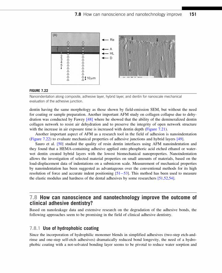
dentin having the same morphology as those shown by field-emission SEM, but without the need
for coating or sample preparation. Another important AFM study on collagen collapse due to dehy-
dration was conducted by Fawzy [48] where he showed that the ability of the demineralized dentin
collagen network to resist air dehydration and to preserve the integrity of open network structure
with the increase in air exposure time is increased with dentin depth (Figure 7.21).
Another important aspect of AFM as a research tool in the field of adhesion is nanoindentation
(Figure 7.22) to evaluate mechanical properties of adhesive junctions and hybrid layers [49].
Sauro et al. [50] studied the quality of resin dentin interfaces using AFM nanoindentation and
they found that a HEMA-containing adhesive applied onto phosphoric acid etched ethanol or water-
wet dentin created hybrid layers with the lowest biomechanical nanoproperties. Nanoindentation
allows the investigation of selected material properties on small amounts of materials, based on the
load-displacement data of indentations on a submicron scale. Measurement of mechanical properties
by nanoindentation has been suggested as advantageous over the conventional methods for its high
resolution of force and accurate indent positioning [51�53]. This method has been used to measure
the elastic modulus and hardness of the dental adhesives by some researchers [51,52,54].
7.8 How can nanoscience and nanotechnology improve the outcome ofclinical adhesive dentistry?Based on nanoleakage data and extensive research on the degradation of the adhesive bonds, the
following approaches seem to be promising in the field of clinical adhesive dentistry.
7.8.1 Use of hydrophobic coatingSince the incorporation of hydrophilic monomer blends in simplified adhesives (two-step etch-and-
rinse and one-step self-etch adhesives) dramatically reduced bond longevity, the need of a hydro-
phobic coating with a not-solvated bonding layer seems to be pivotal to reduce water sorption and
Re(A)
(B)
AHL
10 µm
D
2
2345
111
1
2
3
4
FIGURE 7.22
Nanoindentation along composite, adhesive layer, hybrid layer, and dentin for nanoscale mechanical
evaluation of the adhesive junction.
1517.8 How can nanoscience and nanotechnology improve

stabilize the hybrid layer over time, i.e., etch-and-rinse three steps and self-etch two-step adhesives
should be preferred to simplified ones. Also applying a hydrophobic layer on one-step self-etching
adhesives could improve bond strength and durability [55].
7.8.2 Extended polymerization timeExtending the curing times of simplified adhesives beyond those recommended by the manufac-
turers resulted in improved polymerization and reduced permeability and appears to be a possible
means for improving the performance of these adhesives [56].
7.8.3 Use of MMPs inhibitorsThe use of MMPs inhibitors as additional primer has been claimed to reduce interfacial aging over
time by inhibiting the activation of endogenous dentin enzymes which are responsible for the deg-
radation of collagen fibrils in the absence of bacterial contamination [57]. The recent finding that
chlorhexidine (CHX) also has potent anti-MMP-2, -8, and -9 activity encouraged some researchers
to determine whether CHX could stabilize the organic matrix of resin�dentin bonds. This led to
numerous in vitro [58] and in vivo studies [59] that demonstrated that CHX has beneficial effects
on the preservation of resin�dentin bonds, thereby offering a valuable alternative to clinicians who
seek to delay the degradation process of adhesive restorations. The effectiveness of CHX, as an
antimicrobial or an antiproteolytic agent, has been reported to be related with its substantivity to
oral/dental structures [60]. Substantivity is the prolonged association between a material (e.g.,
Galardin-treated specimens Control specimens(A) (B)
FIGURE 7.23
Much less nanoleakage as demonstrated by silver depositions with galardin-treated specimens (A). Compared
with non treated specimens (B).
152 CHAPTER 7 Nanotechnology and Nanoparticles

CHX) and a substrate (e.g., oral mucosa, oral proteins, dental plaque, or dental surface), an associa-
tion that can be greater and more extended than would be expected from a simple deposition mech-
anism. It is considered that the delivery of an agent to its site of action in a biologically active
form and in effective doses increases the effects for prolonged periods of time. Substantivity of
CHX or its ability to be retained in dentin matrices could be the reason why CHX-treated acid-
etched dentin may form hybrid layers that are more stable over time. Recently Breshi et al. [61]
investigated the pretreatment of dentin with a specific MMP inhibitor galardin (Figure 7.23). The
inhibitory effect of galardin on dentinal MMPs was confirmed by zymographic analysis as com-
plete inhibition of both MMP-2 and -9 was observed. The use of galardin had no effect on immedi-
ate bond strength while it significantly decreased bond degradation after 1 year. Interfacial
nanoleakage expression after aging revealed reduced silver deposits in galardin-treated dentin com-
pared to untreated dentin.
7.8.4 Improved impregnationVarious methods have been recently proposed to enhance dentin impregnation, i.e., prolonged
application time, vigorous brushing technique, and electric impulse assisted adhesive application
[62]. The latter technique recently revealed increased bond strength and reduced nanoleakage
expression if adhesives are applied under the effects of an electric signal. Junior et al. [63]
improved impregnation of dentinal collagen by adhesives via the evaporation of adhesive solvent
by a stream of warm air (Figure 7.24). The use of a warm air-dry stream to evaporate the solvent
of adhesives seems to be a clinical tool to improve the bond strength and the quality of the hybrid
layer (less nanoleakage infiltration).
Another approach to improve impregnation of collagen by the adhesive after acid etching was
the simultaneous acid etching and deproteinization suggested by Nassif and El Korashy [64]. The
simultaneous etching and deproteinization by NaOCl/phosphoric acid for 15 s showed a hybrid
layer with improved bond strength. This was attributed to removal of shredded collagen found in
the smear layer that could not be removed by acid etching only. Removal of this disorganized col-
lagen would give more open structure to the collagen network and improve its impregnation by the
adhesive.
7.8.5 Wet ethanol bonding approachTay et al. [65] proposed the ethanol-wet bonding technique. Ethanol is used to replace water just
prior to bonding, thus avoiding the collapse of the collagen matrix. Ethanol-wet dentin may permit
the infiltration of hydrophobic monomers to disperse into the demineralized dentin, creating a
hydrophobic hybrid layer. Since the concept of ‘‘ethanol-wet bonding’’ was proposed, various
ethanol-wet protocols have been developed to optimize this technique [66,67]. The ethanol-wet
bonding is a time-consuming technique since it needs consecutive application of ascending concen-
trations of ethanol. A simplified protocol for wet ethanol bonding to dentin was suggested by
Sadek et al. [68]. They suggested a protocol of reduced time for each ascending ethanol concentra-
tion application to dentin prior to bonding. This technique achieved high bond strengths, minimal
nanoleakage infiltration, and maintained bond stability after 6 months of artificial aging. A shorter
dehydration period (135 s) may render the bonding of hydrophobic monomers to dentin easily.
1537.8 How can nanoscience and nanotechnology improve

However, biocompatibility issues and bond stability over longer storage periods should be
addressed before such a technique may be recommended for clinical use.
7.8.6 Improving dental collagen network mechanical properties prior to bondingPriming acid-etched dentin with glutaraldehyde before application of the adhesive is one of the sug-
gested methods to improve bond strength and durability [69]. Glutaraldehyde, a substance that has
been used in the adhesive dentistry field, appears to be a potential element to improve deminera-
lized dentin properties. Its capacity to fix proteins irreversibly [70] and to increase the modulus of
elasticity of collagen fibrils [71] is of great interest to maintain the collagen structure in position
during bonding. In addition, its well-reported ability to react chemically with collagen and resin
components such as HEMA [72] may also contribute to facilitating adhesive system penetration
into and wettability of the dentin substrate.
Cold air-dry Warm air-dry
Sin
gle
bond
Prim
e an
d bo
nd
(A) (B)
(C)
25µm
(D)
25 µ
m
25 µm
FIGURE 7.24
SEM photomicrographs showing less nanoleakage (Ag deposition) after warm air drying of the adhesive.
154 CHAPTER 7 Nanotechnology and Nanoparticles

7.8.7 Enhancing biomimetic remineralizationBiomimetic remineralization is a process that allows remineralization of dentinal collagen fibrils
around and within collagen that still have intermolecular cross-links, like collagen fibrils in caries-
affected dentin and phosphoric acid demineralized dentin. Biomimetic remineralization helps the
rebuilding of dentin minerals in the same hierarchical pattern of apatite nanocrystals deposition
both intrafibrillar and interfibrillar.
Tay and Pashly [73] suggested a guided remineralization of partially demineralized human den-
tin where they used set white Portland cement as a source of Ca ions in a phosphate-containing
fluid to precipitate apatite nanocrystals around demineralized collagen. When polyvinyl phosphonic
acid and PAA were included, these nanoprecursors were attracted to the acid-demineralized colla-
gen matrix and transformed into polyelectrolyte-stabilized apatite nanocrystals that assembled along
the microfibrils (intrafibrillar remineralization) and surface of the collagen fibrils (interfibrillar
remineralization). Transition from nanocrystals to larger apatite platelets probably occurred via the
formation of mesocrystal intermediates. Guided tissue remineralization is potentially useful in the
remineralization of acid-etched dentin that is incompletely infiltrated by dentin adhesives as well as
partially demineralized caries-affected dentin. Gandolfi et al. [74] suggested the use of bioactive
“smart” composites containing reactive calcium-silicate Portland-derived mineral powder as
tailored filler. This innovative method for the biomimetic remineralization of apatite-depleted
dentin surfaces and for preventing the demineralization of hypomineralized/carious dentin could be
potentially great advantage in clinical applications.
7.9 Future prospective of nanotechnology in the field ofadhesive dentistry7.9.1 On-demand antibacterial adhesivesWelch et al. [75] incorporated TiO2 nanoparticles in dental adhesives aiming at achieving the com-
bined features of bioactivity and on-demand bactericidal effect. The photocatalytic activity of adhe-
sives containing TiO2 nanoparticles, initiated by UV irradiation, proved to interfere with bacterial
activity. Also TiO2 nanoparticles-containing adhesives were found to have the potential of tooth
remineralization in simulated body fluids. This could open up the potential to create dental adhe-
sives that reduce the incidence of secondary caries and promote closure of gaps forming at the
interface toward the tooth via remineralization of adjacent tooth substance as well as prevention of
bacterial infections via on-demand UV irradiation.
7.9.2 Improving adhesive polymerization throughcatalytic activity of nanoparticlesNanoparticles could improve DC of adhesive polymers yielding an adhesive layer with improved
mechanical properties and resistance to degradation. Yasumoto et al. [76] used colloidal platinum
nanoparticles as a pretreatment to dentin after acid etching and before adhesive application to
improve the resin polymerization resulting in enhanced bond strength. Based on the same principle,
1557.9 Future prospective of nanotechnology in the field of adhesive dentistry

Sun et al. [28] showed that TiO2 nanoparticles in adhesives could improve DC and mechanical
properties expecting better bond strength and durability.
7.9.3 Antibacterial orthodontic adhesives containing nanosilverEnamel demineralization is a commonly recognized complication of orthodontic treatment with
fixed appliance. Preventing these lesions is an important concern for orthodontists because the
lesions are unesthetic, unhealthy, and potentially irreversible. The introduction of antibacterial
adhesives would improve the outcome of orthodontic treatment due to less bacterial adhesion to the
orthodontic appliance. Ahn et al. [77] suggested an orthodontic adhesive containing nanosilver par-
ticles as antimicrobial agent. The adhesive showed less bacterial adhesion and less bacterial growth
without affecting bond strength.
7.9.4 Radiopaque dental adhesivesSecondary caries may be detected visually by the discolorations at the tooth/restoration interface,
though X-ray photographs are often required to safely discriminate such lesions from stained mar-
gins. Radiographs rely on the difference in radiopacity between healthy dental tissue, cariogenic
hard tissue, and restorative material. Modern composite-filling materials commonly contain radi-
opaque components, such as Sr- or Ba-glass fillers, making them easily distinguishable by X-ray
from the tooth. Dental adhesives, however, do not contain such radiopaque materials today making
them hard to distinguish from caries. Schulz et al. [23] focused on the development of radiopaque
adhesives by incorporating flame-made Ta2O5/SiO2 nanoparticles in methacrylic matrices. The
nanofilled adhesive had radiopacity better than enamel and dentin without adverse effect on bond-
ing to hard dental tissues.
7.9.5 Self-adhesive compositesThe introduction of self-adhesive composites will offer clinicians the simple approach toward the
restorative procedure by eliminating the number of steps associated with bonding procedure. The
latest trend has been toward the development of flowable composites containing adhesive mono-
mers such as Vertise Flow (Kerr) and Fusio Liquid Dentin (Pentron Clinical). These formulations
are based on traditional methacrylate systems, but incorporating acidic monomers typically found
in dentin bonding agents such as glycerol phosphate dimethacrylate in Vertise Flow may be capable
of generating adhesion through mechanical and possibly chemical interactions with tooth structure.
These materials are currently recommended for liners and small restorations and are serving as the
entry point for universal self-adhesive composites [78].
7.9.6 Self-healing adhesivesThe concept of self-healing polymers relies on encapsulation of monomers and catalyst and incor-
porating these encapsulated healing precursors into the polymer. When cracks are initiated in the
polymer, the capsules rupture and healing monomers fill the crack and polymerize there allowing
healing of the crack. In the field of dental adhesion, self-healing bonding resins may provide a new
156 CHAPTER 7 Nanotechnology and Nanoparticles

direction for the improvement of the bonding durability. To allow the healing precursors in dental
adhesives to reach the submicron spaces created by acid etching within dentin, the need for nano-
encapsulation is needed. Ouyang et al. [79] prepared, characterized, and incorporated polyurethane
nanocapsules encapsulated with the core material TEGDMA for use as a major component in a
self-healing bonding resin (Figure 7.25). The incorporation of nanoencapsulated monomers in den-
tal adhesives showed enhancement of bond strength and expected to improve durability of resin
tooth bonding (Figure 7.26).
7.9.7 High-speed AFMThe future advancement in research tools will improve our understanding of adhesion and will
further clarify the associated obstacles. One of the most promising tools is the high-speed AFM
(HS-AFM), which is becoming a reference tool for the study of dynamic biological processes.
(A)
1 µm
200 nm
(B)
FIGURE 7.25
Field Emission Scanning Electron Microscope (FESEM) micrographs of nanocapsules (A and B). FESEM
micrographs indicate that the TEGDMA nanocapsules are spherical with smooth and condense surface.
1577.9 Future prospective of nanotechnology in the field of adhesive dentistry

(A) (B)
(C) (D)
1 µm
(E) (F)
1 µm
10 µm 10 µm
1 µm 1 µm
FIGURE 7.26
FESEM observations of the adhesive/dentin interfaces. (A, B) FESEM images of dentin (D) surface show that
TEGDMA nanocapsule (black arrow) surrounded by adhesive are infiltrated into dentinal tubule (T) with resin
tag (RT). (C, D) FESEM micrograph of the adhesive/dentin interface bonded with Prime & Bond NT or
Prime & Bond NT incorporated with TEGDMA nanocapsules. The resin tags (white arrow) were longer and
more regular as compared with the control. (E, F) Local magnification from (D) shows that TEGDMA
nanocapsules (white arrows) could be notified on the surface of resin tag (RT).

The spatial and time resolutions of HS-AFM are on the order of nanometers and milliseconds,
respectively, and allow structural and functional characterization of biological processes at the
single-molecule level [80]. Pyne et al. [81] used HS-AFM to study the demineralizing effect of
citric acid on enamel. They imaged the dissolution of enamel in a real time or movie mode and
concluded that the HS-AFM is able to follow the large changes in height (on the micrometer scale)
that occur during the dissolution process (Figure 7.27). Such real-time imaging can provide dental
profession with valuable data on how surface conditioner interacts with dental substrates and how
we can make use of these data to improve the outcome of the restorative procedures.
7.10 Conclusions and future directionsNanoscience and nanotechnology will have strong impact on adhesion to dental substrates through
the development of nanofilled dental adhesives with improved mechanical properties. Stronger
adhesive junctions with much less defects and more durability will be achieved through improved
penetration of the adhesives, antibacterial potential, and self-healing capacity.
Adhesion to oral mucosa will be an efficient route of nanodrug delivery where oral adhesive
patch, tablets, or stripes could be loaded with nanocarriers with controlled drug release and other
benefits could be obtained through administering the drugs via buccal mucosa. Among the various
0 s
0 s
(A)
(B)5 s 10 s 20 s 50 s
11 s7 s4 s1 s
FIGURE 7.27
Sequences of HS-AFM images of an HAP surface taken (A) in water (3 µm3 3 µm) and (B) in citric acid at
pH 3 (1.5 µm3 1.5 µm). Time is indicated in seconds under each image.
From Ref. [81].
1597.10 Conclusions and future directions

transmucosal routes, buccal mucosa has excellent accessibility, an expanse of smooth muscle, and
relatively immobile mucosa, and hence suitable for administration of retentive dosage forms. Direct
access to the systemic circulation through the internal jugular vein bypasses drugs from the hepatic
first-pass metabolism leading to high bioavailability. Other advantages such as low enzymatic
activity, suitability for drugs or excipients that mildly and reversibly damages or irritates the
mucosa, painless administration, easy drug withdrawal, facility to include permeation enhancer/
enzyme inhibitor or pH modifier in the formulation, and versatility in designing as multidirectional
or unidirectional release systems for local or systemic actions opt buccal adhesive drug delivery
systems as a promising option [82].
References[1] I. Kramer, J. McLean, Alterations in the staining reactions of dentin resulting from a constituent of a
new self-polymerizing resin, Br. Dent. J. 93 (1952) 150�153.
[2] M. Buonocore, A simple method of increasing the adhesion of acrylic filling materials to enamel
surfaces, J. Dent. Res. 34 (1955) 849�853.
[3] M. Peters, M. McLean, Minimally invasive operative care. I. Minimal intervention and concepts for
minimally invasive cavity preparations, J. Adhes. Dent. 3 (2001) 7�16.
[4] B. Meerbeek, J. Perdiggo, P. Lambrecht, G. Vanherle, The clinical performance of adhesives, J. Dent. 26
(1998) l�20.
[5] J. De Munck, K. Van Landuyt, M. Peumans, A. Poitevin, P. Lambrechts, M. Braem, et al., A critical
review of the durability of adhesion to tooth tissue: methods and results, J. Dent. Res. 84 (2005)
118�132.
[6] A. Gwinnett, A.J. Matsui, A study of enamel adhesives. The physical relationship between enamel and
adhesives, Arch. Oral. Biol. 12 (1967) 1615�1620.
[7] N. Nakabayashi, K. Kojima, E. Masuhara, The promotion of adhesion by the infiltration of monomers
into tooth substrates, J. Biomed. Mater. Res. 16 (1982) 1240�1243.
[8] T. Fusayama, New Concepts in Operative Dentistry, Quitessence Publishing Co., Inc, Tokyo, 1980,
pp. 61�156.
[9] J. Kanca, Improved bond strength through acid-etching of dentin and bonding to wet dentin surfaces,
J. Am. Dent. Assoc. 123 (1992) 35�43.
[10] D. Pashley, F. Tay, L. Breschi, L. Tjaderhane, R. Carvalho, M. Carrilho, et al., State of the art etch-and-
rinse adhesives, Dent. Mater. 27 (2011) 1�16.
[11] D. Pashley, J. Horner, P. Brewer, Interactions of conditioners on the dentin surfaces, Oper. Dent. 17
(Suppl. 5) (1992) 127�150.
[12] M. Cadenaro, L. Breschi, F. Rueggeberg, M. Suchko, E. Grodin, K. Agee, et al., Effects of residual etha-
nol on the rate and degree of conversion of five experimental resins, Dent. Mater. 25 (2009) 621�628.
[13] B. Van Meerbeek, J. De Munck, Y. Yoshida, S. Inoue, M. Vargas, P. Vijay, Buonocore memorial
lecture. Adhesion to enamel and dentin: current status and future challenges, Oper. Dent. 28 (2003)
215�235.
[14] B. Van Meerbeek, K. Yoshihara, Y. Yoshida, A. Mine, J. De Munck, K. Van Landuyt, State of the art of
self-etch adhesives, Dent. Mater. 27 (2011) 17�28.
[15] Y. Yoshida, K. Nagakane, R. Fukuda, Y. Nakayama, M. Okazaki, H. Shintani, et al., Comparative study
on adhesive performance of functional monomers, J. Dent. Res. 83 (2004) 454�458.
160 CHAPTER 7 Nanotechnology and Nanoparticles

[16] K. Van Landuyt, J. Snauwaert, J. De Munck, M. Peumans, Y. Yoshida, A. Poitevin, et al., Systematic
review of the chemical composition of contemporary dental adhesives, Biomaterials 28 (2007)
3757�3785.
[17] A. Takahashi, Y. Sato, S. Uno, P. Pereira, H. Sano, Effect of mechanical properties of adhesive resin on
bond strength to dentin, Dent. Mater. 18 (2002) 263�268.
[18] H. Honmoto, Study on light cured composite resins—effect of filler contents in the bonding agent on the
tensile bond strength to enamel, Nihon. Univ. Dent. J. 65 (1991) 246�257.
[19] M. Miyazak, S. Ando, K. Hinoura, H. Onose, B. Moore, Influence of filler addition to bonding agents on
shear bond strength to bovine dentin, Dent. Mater. 11 (1995) 234�238.
[20] M. Conde, C. Zanchi, S. Junior, N. Carreno, F. Ogliari, E. Piva, Nanofiller loading level: influence on
selected properties of an adhesive resin, J. Dent. 37 (2009) 331�335.
[21] F. El-Askary, R. van Noort, The effect of airy-drying pressure and distance on the microtensile bond
strength of a self-etching adhesive, J. Adhes. Dent. 11 (2011) 135�147.
[22] K. Ikemora, F. Tay, Y. Kouro, T. Endo, M. Yoshiyama, K. Miyai, et al., Optimizing filler content of an
adhesive containing pre-reacted glass ionomer fillers, Dent. Mater. 19 (2003) 137�146.
[23] H. Schulz, B. Schimmoeller, S. Pratsinis, U. Salz, T. Bock, Radiopaque dental adhesives: dispersion of
flame-made Ta2O5/SiO2 nanoparticles in methacrylic matrices, J. Dent. 36 (2008) 579�587.
[24] Y. Yoshida, B. Van Meerbeek, Y. Nakayama, J. Snauwaert, L. Hellemans, P. Lambrechts, et al.,
Evidence of chemical bonding at biomaterial-hard tissue interfaces, J. Dent. Res. 79 (2000) 709�714.
[25] Y. Zhang, Y. Wang, Hydroxyapatite effect on photopolymerization of self-etching adhesives with differ-
ent aggressiveness, J. Dent. 40 (2012) 564�570.
[26] Y. Zhang, Y. Wang, The effect of hydroxyapatite presence on the degree of conversion and polymeriza-
tion rate in a model self-etching adhesive, Dent. Mater. 28 (2010) 237�244.
[27] M. Shojai, M. Atai, A. Nodehi, L. Khanlarb, Hydroxyapatite nanorods as novel fillers for improving the
properties of dental adhesives: synthesis and application, Dent. Mater. 26 (2010) 471�482.
[28] J. Sun, A. Forster, P. Johnson, N. Eidelman, G. Quinn, G. Schumacher, et al., Improving performance of
dental resins by adding titaniumdioxide nanoparticles, Dent. Mater. 27 (2011) 972�982.
[29] M. Atai, L. Solhi, A. Nodehi, S. Mirabedini, S. Kasraei, K. Akbari, et al., PMMA-grafted nanoclay as
novel filler for dental adhesives, Dent. Mater. 25 (2009) 339�347.
[30] L. Solhi, M. Atai, A. Nodehi, M. Imani, A. Ghaemi, K. Khosravi, Poly(acrylic acid) grafted montmoril-
lonite as novel fillers for dental adhesives: synthesis, characterization and properties of the adhesive,
Dent. Mater. 28 (2012) 369�377.
[31] F. El-Askary, M. Nassif, Bonding nano-filled resin modified glass ionomer to dentin using different self
etch adhesives, Oper. Dent. 36 (2011) 413�421.
[32] E. Coutinho, M. Cardoso, J. De Munck, A. Neves, K. Van Landuyt, A. Poitevin, et al., Bonding effec-
tiveness and interfacial characterization of a nano-filled resin-modified glass-ionomer, Dent. Mater.
25 (2009) 1347�1357.
[33] H. Sano, T. Shono, T. Takatsu, H. Hosoda, Microporous dentin zone beneath resin-impregnated layer,
Oper. Dent. 19 (1994) 59�64.
[34] H. Sano, T. Takatsu, B. Ciucchi, A. Herner, W. Mattews, D. Pashley, Nanoleakage: leakage within the
hybrid layer, Oper. Dent. 20 (1995) 18�25.
[35] H. Sano, M. Yoshiyama, S. Ebisu, M. Burrow, T. Takatsu, B. Ciucchi, et al., Comparative SEM and
TEM observations of nanoleakage within the hybrid layer, Oper. Dent. 20 (1995) 160�167.
[36] H. Li, M. Burrow, M. Tyas, Nanoleakage patterns of four dentin bonding systems, Dent. Mater. 16
(2000) 48�56.
[37] T. Pioch, H. Staehle, H. Duschner, F. Godoy, Nanoleakage at the composite�dentin interface: a review,
Am. J. Dent. 14 (2001) 252�258.
161References

[38] F. Tay, D. Pashley, M. Yoshiyama, Two modes of nanoleakage expression in single-step adhesives,
J. Dent. Res. 81 (2002) 472�476.
[39] A. Gwinnet, F. Tay, M. Pang, S. Wei, Quantitative contribution of the collagen network in dentin
hybridization, Am. J. Dent. 9 (1996) 140�144.
[40] A. Gwinnet, S. Yu, Effect of long-term water storage on dentin bonding, Am. J. Dent. 7 (1994)
109�111.
[41] M. Hashimoto, J. De Munck, S. Ito, H. Sano, M. Kaga, H. Oguchi, et al., In vitro effect of nanoleakage
expression on resin-dentin bond strengths analyzed by microtensile bond test, SEM/EDX and TEM,
Biomaterials 25 (2004) 5565�5574.
[42] R. Carvalho, S. Chersoni, R. Frankenberger, D. Pashley, C. Prati, F. Tay, A challenge to the conven-
tional wisdom that simultaneous etching and resin infiltration always occurs in self-etch adhesives,
Biomaterials 26 (2005) 1035�1042.
[43] A. Reisa, M. Giannini, P. Pereira, Long-term TEM analysis of the nanoleakage patterns in resin�dentin
interfaces produced by different bonding strategies, Dent. Mater. 2 (3) (2007) 1164�1172.
[44] L. Breschi, P. Martin, A. Mazzoni, F. Nato, M. Carrilhoe, L. Tjaderhane, et al., Use of a specific MMP-
inhibitor (galardin) for preservation of hybrid layer, Dent. Mater. 26 (2010) 571�578.
[45] C. Chen, H. Hansma, Basement membrane macromolecules: insights from atomic force microscopy,
J. Struct. Biol. 131 (2000) 44�55.
[46] S. Habelitz, M. Balooch, S. Marshall, G. Balooch, G. Marshall, In situ atomic force microscopy of par-
tially demineralized human dentin collagen fibrils, J. Struct. Biol. 138 (2002) 227�236.
[47] F. El Feninat, T. Ellis, E. Sacher, I. Stangel, A tapping mode AFM study of collapse and denaturation in
dentinal collagen, Dent. Mater. 17 (2001) 284�288.
[48] A. Fawzy, Variations in collagen fibrils network structure and surface dehydration of acid demineralized
intertubular dentin: effect of dentin depth and air-exposure time, Dent. Mater. 26 (2010) 35�43.
[49] Y. Hosoya, F. Tay, O. Miyazaki, Hardness, elasticity and ultrastructure of primary tooth dentin bonded
with a self-reinforcing one-step self-etch adhesive, J. Dent. 38 (2010) 214�221.
[50] S. Sauro, R. Osorio, T. Watson, M. Toledano, Assessment of the quality of resin�dentin bonded inter-
faces: an AFM nano-indentation, µTBS and confocal ultramorphology study, Dent. Mater. 28 (2012)
622�631.
[51] A. Takahashi, Y. Sato, S. Uno, P. Pereira, H. Sano, Effects of mechanical properties of adhesive resins
on bond strength to dentin, Dent. Mater. 18 (2002) 263�268.
[52] B. Van Meerbeek, G. Willems, J. Celis, J. Roos, M. Braem, P. Lambrechts, et al., Assessment by nano-
indentation of the hardness and elasticity of the resin�dentin bonding area, J. Dent. Res. 72 (1993)
1434�1442.
[53] W. Oliver, G. Pharr, An improved technique for determining hardness and elastic-modulus using load
and displacement sensing indentation experiments, J. Mater. Res. 7 (1992) 1564�1583.
[54] Y. Hosoya, Hardness and elasticity of bonded carious and sound primary tooth dentin, J. Dent. 34 (2006)
164�171.
[55] A. Reis, M. Albuquerque, M. Pegoraro, G. Mattei, J. Bauer, R. Grande, et al., Can the durability of one-
step self-etch adhesives be improved by double application or by an extra layer of hydrophobic resin?
J. Dent. 36 (2008) 309�315.
[56] M. Cadenaro, F. Antoniolli, S. Sauro, F. Tay, R. Di Lenarda, C. Prati, et al., Degree of conversion and
permeability of dental adhesives, Eur. J. Oral. Sci. 113 (2005) 525�530.
[57] M. Carrilhoa, R. Carvalho, E. Sousa, J. Nicolau, L. Breschi, A. Mazzoni, et al., Substantivity of chlor-
hexidine to human dentin, Dent Mater. 26 (2010) 779�785.
[58] R. Stanislawczuk, R. Amaral, C. Grande, D. Gagler, A. Reis, A. Loguercio, Chlorhexidine-containing
acid conditioner preserves the longevity of resin dentin bonds, Oper. Dent. 34 (2009) 481�490.
162 CHAPTER 7 Nanotechnology and Nanoparticles

[59] W. Brackett, F. Tay, M. Brackett, A. Dib, R. Sword, D. Pashley, The effect of chlorhexidine on dentin
hybrid layers in vivo, Oper. Dent. 32 (2007) 107�111.
[60] M. Carrilho, F. Tay, A. Donnelly, K. Agee, L. Tjaderhane, A. Mazzoni, et al., Host-derived loss of den-
tin matrix stiffness associated with solubilization of collagen, J. Biomed. Mater. Res. B Appl. Biomater.
90 (2009) 373�380.
[61] L. Breschi, P. Martin, A. Mazzoni, F. Nato, M. Carrilhoe, L. Tjaderhane, et al., Use of a specific MMP-
inhibitor (galardin) for preservation of hybrid layer, Dent. Mater. 26 (2010) 571�578.
[62] G. Pasquantonio, F. Tay, A. Mazzoni, P. Suppa, A. Ruggeri, M. Falconi, et al., Electric device improves
bonds of simplified etch-and-rinse adhesives, Dent. Mater. 23 (2007) 513�518.
[63] C. Junior, C. Grande, R. Amaral, R. Stanislawczuk, E. Garcia, R. Neto, et al., Evaporating solvents with
a warm air-stream: effects on adhesive layer properties and resin�dentin bond strengths, J. Dent. 36
(2008) 618�625.
[64] M. Nassif, D. El Korashy, Phosphoric acid/sodium hypochlorite mixture as dentin conditioner: a new
approach, J. Adhes. Dent. 11 (2009) 455�460.
[65] F. Tay, D. Pashley, R. Kapur, M. Carrilho, Y. Hur, L. Garrett, et al., Bonding BisGMA to dentin—a
proof of concept for hydrophobic dentin bonding, J. Dent. Res. 86 (2007) 1034�1039.
[66] S. Sauro, M. Toledano, F. Aguilera, F. Mannocci, D. Pashley, F. Tay, et al., Resin�dentin bonds to
EDTA-treated vs. acid-etched dentin using ethanol wet bonding, Dent. Mater. 26 (2010) 368�379.
[67] F. Tay, F. Sadek, D. Pashley, Y. Nishitani, M. Carrilho, A. Donnelly, et al., Application of hydrophobic
resin adhesives to acid-etched dentin with an alternative wet bonding technique, J. Biomed. Mater. Res.
A 84A (2008) 19�29.
[68] F. Sadek, A. Mazzoni, L. Breschi, F. Tay, R. Braga, Six-month evaluation of adhesives interface created
by a hydrophobic adhesive to acid-etched ethanol-wet bonded dentin with simplified dehydration
protocols, J. Dent. 38 (2010) 276�283.
[69] R. Cilli, A. Prakki, P. Jose, J. Pereira, Influence of glutaraldehyde priming on bond strength of an experi-
mental adhesive system applied to wet and dry dentin, J. Dent. 37 (2009) 212�218.
[70] J. Kiernan, Formaldehyde, formalin, paraformaldehyde and glutaraldehyde. What they are and what they
do, Microsc. Today 1 (2000) 8�12.
[71] D. Pashley, Y. Zhang, R. Carvalho, F. Rueggeberg, C. Russel, Induced tension development in deminer-
alized dentin matrix, J. Dent. Res. 79 (2000) 1579�1583.
[72] E. Asmussen, E. Munksgaard, Bonding of restorative resins to dentin promoted by aqueous mixtures of
aldehydes and active monomers, Int. Dent. J. 35 (1985) 160�165.
[73] F. Tay, D. Pashley, Guided tissue remineralisation of partially demineralized human dentin, Biomaterials
29 (2008) 1127�1137.
[74] M. Gandolfi, P. Taddei, F. Siboni, E. Modenab, E. De Stefano, C. Prati, Biomimetic remineralization of
human dentin using promising innovative calcium-silicate hybrid “smart” materials, Dent. Mater. 27
(2011) 1055�1069.
[75] K. Welch, Y. Cai, H. Engqvist, M. Stromme, Dental adhesives with bioactive and on-demand bacteri-
cidal properties, Dent. Mater. 26 (2010) 491�499.
[76] K. Yasumoto, S. Hoshika, F. Nagano, T. Tanaka, D. Selimovic, Y. Miyamoto, et al., Application of wet
bonding for one-step bonding systems, Dent. Mater. 26 (2010) e125�e157.
[77] S. Ahn, S. Lee, J. Kook, B. Lim, Experimental antimicrobial orthodontic adhesives using nanofillers and
silver nanoparticles, Dent. Mater. 25 (2009) 206�213.
[78] J. Ferracane, Resin composite—state of the art, Dent. Mater. 27 (2011) 29�38.
[79] X. Ouyang, X. Huang, Q. Pan, C. Zuo, C. Huang, X. Yang, et al., Synthesis and characterization of
triethylene glycol dimethacrylate nanocapsules used in a self-healing bonding resin, J. Dent. 39 (2011)
825�833.
163References

[80] T. Ando, T. Uchihashi, N. Kodera, D. Yamamoto, M. Taniguchi, A. Miyagi, et al., High-speed atomic
force microscopy for observing dynamic biomolecular processes, J. Mol. Recognit. 20 (2007) 448�458.
[81] A. Pyne, W. Marks, L. Picco, P. Dunton, A. Ulcinas, M. Barbour, et al., High-speed atomic force
microscopy of dental enamel dissolution in citric acid, Arch. Histol. Cytol. 72 (2009) 209�215.
[82] Y. Sudhakar, K. Kuotsu, A. Bandyopadhyay, Buccal bioadhesive drug delivery—a promising option for
orally less efficient drugs, J. Control. Release 114 (2006) 15�40.
164 CHAPTER 7 Nanotechnology and Nanoparticles

CHAPTER
8Nanobiomaterials in PreventiveDentistry
Matthias Hanniga and Christian HannigbaClinic of Operative Dentistry, Periodontology and Preventive Dentistry, University Hospital,
Saarland University Homburg/Saar, GermanybClinic of Operative Dentistry, Faculty of Medicine ‘Carl Gustav Carus’, Technical University of Dresden,
Dresden, Germany
CHAPTER OUTLINE
8.1 Introduction—current challenges in preventive dentistry .............................................................. 167
8.2 The ubiquitous phenomenon of bioadhesion on dental hard tissues............................................... 168
8.3 Main strategies for implementation of nanosized materials in dental prophylaxis........................... 170
8.4 Modulation of bioadhesion and biofilm management .................................................................... 170
8.5 Effects on de- and remineralization............................................................................................. 174
8.6 Erosion...................................................................................................................................... 176
8.7 Nanosized calcium fluoride ........................................................................................................177
8.8 Dentin hypersensitivity ............................................................................................................... 177
8.9 Regeneration of dental hard substances ...................................................................................... 178
8.10 Discussion and clinical recommendations ................................................................................... 179
8.11 Conclusions............................................................................................................................... 180
References ......................................................................................................................................... 181
8.1 Introduction—current challenges in preventive dentistryDental prophylaxis based on mechanical biofilm management and fluoride application has resulted
in considerable improvement of oral health; however, caries is still a great challenge worldwide.
The caries prevalence in newly industrializing countries is still increasing, in industrialized
countries it is stagnating or decreases in some sections of the population [1,2]. Noteworthy, the dis-
tribution of caries in the population has shifted. On the one hand, many seniors of today have their
own teeth but are faced with caries especially at the neck region of the teeth due to exposed dentin
surfaces [3]. On the other hand, many children are nearly free of caries, but a significant percentage
is faced with extensive early childhood caries [2,4]. If thorough mechanical biofilm management
is performed daily on every site of the teeth, there will be no new caries lesion formation or
167Nanobiomaterials in Clinical Dentistry.
© 2013 Elsevier Inc. All rights reserved.

progression, respectively. Despite these considerations and regular use of tooth brushes, most
patients do not clean their teeth as properly as necessary for caries prevention. Accordingly, addi-
tional strategies and new preparations for optimized biofilm management as well as remineraliza-
tion of incipient dental lesions are necessary. In general, a vast number of artificial materials and
related products are available for oral health care and preventive dentistry. However, they are not
as effective as desirable. Furthermore, the conventional oral health-care products represent no bio-
mimetic or at least biological approaches for biofilm management in the oral cavity. Furthermore,
since the physiological remineralization is frequently inadequate to maintain the integrity of enamel
and dentin exposed to modern diet and nutrition behaviors, the natural remineralization process
after an acidic challenge needs to be augmented. This applies not only for carious attacks but also
for dental erosion. The prevalence of erosions is increasing due to the extensive consumption of
acidic beverages in the industrialized countries [5]. In particular, eating disorders combined with
often high resistance to psychological therapy cause extensive erosive defects [6]. Accordingly,
also for dental erosion, additional strategies are required to prevent demineralization and to improve
remineralization. Thereby, the specific mode of demineralization which is different from dental car-
ies has to be considered as well as the interactions of proteolytic enzymes with the demineralized
dentin matrix [7,8]. Due to the interaction of acidic agents with the hierarchically nanostructured
dental hard substances, especially nanomaterials are of interest for treatment of initial erosive
lesions [9].
Loss of periodontal attachment as well as gingival recession leads to the exposure of the root
surface. Wrong or excessive tooth brushing can induce abrasion and accelerated loss of the exposed
dental hard substance. This often results in tooth hypersensitivities, which are characterized by
opened dentinal tubules. Nanoscaled particles might help to obturate the open dentin tubules, which
are responsible for the hydrodynamic and osmotic effects inducing the hypersensitivities [9,10].
Nanomaterials might also be of interest for preventive, therapeutic, and regenerative strategies in
periodontal disease itself, but this is not the topic of the present review.
The relevance of nanotechnology and nanobiomaterials to overcome these challenges in oral
medicine is clearly mirrored by the increasing number of papers that had been published over the
last few years describing nanotechnological approaches and nanosized materials with proposed
applications in preventive dentistry [9,11�14]. In principle, the effects of such nanobiomaterials
are governed by surface interactions and the process of bioadhesion at the tooth surface [15].
This applies for physiological as well as pathophysiological mechanisms and processes taking place
at the tooth�biofilm�saliva interface. Thereby, strategies mimicking nature at the nanolevel seem
to be the most promising approaches as they try to avoid successfully unpredictable adverse effects
potentially caused by artificial agents [16]. For the development and understanding of these strate-
gies, it is necessary to have a closer look on the process of bioadhesion, surface interactions, as
well as de- and remineralization occurring at the tooth to saliva interface.
8.2 The ubiquitous phenomenon of bioadhesion on dental hard tissuesThe teeth are characterized by nonshedding surfaces which are the essential fundament for the inci-
dence of caries, periodontal diseases, and dental erosion [15]. In this context, the bioadhesion of
168 CHAPTER 8 Nanobiomaterials in Preventive Dentistry

acellular and cellular organic structures is a matter of special importance. Thereby, the first step is
represented by the formation of the acquired pellicle which occurs almost instantaneously on all
solid substrates exposed to the oral fluids [17]. The adsorption process is driven by physicochemi-
cal interactions of the tooth surface with proteins and glycoproteins from the oral fluids; the pellicle
formation means a gain in entropy. Fast area-wide pellicle formation is provided by the adsorption
of micelle-like globular structures, as most salivary components are not released as single mole-
cules but as aggregates [17]. These globularly structured aggregates have a diameter of
100�200 nm and are quite similar to milk micelles which could be a starting point for the develop-
ment of new strategies in preventive dentistry based on organic nanostructures [18].
The physiological pellicle layer fulfills several functions besides lubricating the tooth surfaces.
It offers protection against erosive mineral loss and modulates de- and remineralization [19�23].
However, these effects seem to be limited to short exposure duration and fluids with moderate
acidic pH [20]. Longer exposure to beverages or acids at low pH inevitably leads to destruction of
the pellicle and continuous mineral loss as shown in vitro and in situ. Due to the tremendous
increase in the consumption of erosive beverages and the prevalence of eating disorders, there is a
strong demand for preparations improving the antierosive properties of the pellicle.
Furthermore, the pellicle contains a couple of specific and unspecific antibacterial and antimi-
crobial proteins, glycoproteins, and peptides (such as secretory immunoglobulin A or lactoferrin) as
well as, and enzymes (such as lysozyme or peroxidase) [17,24]. Despite these antibacterial proper-
ties, the pioneer bacteria have adapted to the protective properties of the pellicle layer [25]. Many
pellicle components serve as receptors for specific and irreversible bacterial adhesion after the ini-
tial unspecific approach of bacteria to the tooth surface mediated by hydrophobic interactions, van
der Waal’s forces, and other physicochemical interactions [15,17]. Thereby, bacterial glycosyl
transferases are remarkable key molecules synthesizing water insoluble and soluble glucans [25].
These enzymes are already present in the acquired pellicle in an active conformation within min-
utes and can be regarded as pioneer molecules for bacterial biofilm formation [25,26]. In this closer
sense, biofilms have been defined as a structured community of bacterial cells enclosed in a self-
produced polymeric matrix adherent to an inert or living surface [27].
The initial process of bioadhesion is an essential target of recent approaches in dental research
based on nanotechnology in order to prevent bacterial adhesion, to ameliorate remineralization, and
to avoid demineralization [9,14]. Furthermore, the more mature three-dimensionally organized bac-
terial biofilm could be prone to modification by nanomaterials. This means removal of already
established biofilms as well as direct antibacterial effects on adherent bacteria which is still a
drawback of many conventional antibacterial preparations. They are often of high efficacy against
planktonic microorganisms, but fail to substantially affect the adherent bacteria organized in three-
dimensional biofilms and embedded in an extracellular matrix [28,29]. Bacteria living organized in
these biofilms are effectively shielded and protected against external attacks like treatment with
detergents or antibiotics [28]. Nanoscaled particles may yield size-specific interactions with the bio-
film, but they could also serve as drug-release carriers with high affinity to the bacterial surfaces or
the extracellular matrix, respectively.
Besides the biofilm, the substrate of caries, and erosions, the dental hard substances themselves
have moved into the focus of nanoscientists. Both enamel as well as dentin represent unique and
hierarchically organized biological nanostructures [9,30�32]. The inorganic building units
are hydroxyapatite (HA) nanocrystallites; in dental enamel, their length amounts up to several
1698.2 The ubiquitous phenomenon of bioadhesion on dental hard tissues

100 nm and in dentin up to 50 nm. The acid-related destruction of the dental hard substance as a
biocomposite is initiated on the nanolevel. Due to its nonregenerative nature, enamel is unable to
heal and repair itself after surface alterations. Accordingly, modern prophylactic strategies intend
to substitute mineral loss with artificial or biomimetic nanoscopic mineral components or HA parti-
cles fitting to the initial tooth surface nanosized defects. Thereby, it is has to be kept in mind that
from a theoretical point of view, remineralization is the process of transferring anions and cations
to nucleation sites, where the lattices leading to mineralized structures are generated.
8.3 Main strategies for implementation of nanosized materials indental prophylaxisBasically, there are two potential mechanisms for size-dependent effects of nanomaterials in pre-
ventive dentistry: (i) the interaction with bacterial adherence and oral biofilm formation and (ii) the
impact on de- and remineralization. As the process of demineralization starts at the nanolevel
caused by either caries or erosion-induced acidic challenges, it makes sense to supply small nano-
sized building units for optimized remineralization. This applies also for particle-size-related inter-
actions with bacteria and the bacterial membrane, where nanoparticles are much more effective
than microparticles. Many preparations considering these deliberations were developed; most of
them are in the experimental state and few are already available for the consumers [11,33�36].
Pure inorganic nanomaterials applied in preventive dentistry are mostly HA or its derivatives
modified with zinc, fluoride, or carbonate, respectively. Also mineral nanotubes, nanorods, or other
geometric forms are tested, but represent rather a minority [9,14,37�42].
Another group is represented by organic/inorganic compound materials. Nanosized organic
structures are components of several beverages and foodstuffs and contribute considerably to their
characteristic properties. A typical example is milk containing casein micelles and lipid vesicles.
Casein micelles are quite similar to micelle-like salivary structures involved in the formation of the
pellicle layer. These natural structures may be processed to serve as tailored carriers with high
affinity to the pellicle in order to accumulate minerals and protective proteins at the tooth surface.
All the promising options of caseins have not been exploited in full until now, but there is already
a widespread preparation based on casein phosphopeptide (CPP)�stabilized amorphous calcium
phosphate (ACP) nanocomplexes with a diameter of 2.12 nm [11,34,35]. Other unexplored nano-
sized components of foodstuffs or animals such as chitin skeletons of diatoms could serve as
carriers [36,43�45].
Besides these biomimetic or even biological approaches, silver nanoparticles or nanosized syn-
thetic glass structures also have been described for possible application in preventive dentistry
[46�49]. However, though these materials might be efficient, their side effects are not foreseeable.
Thus, the present overview will focus on biological or biomimetic strategies.
8.4 Modulation of bioadhesion and biofilm managementIn principle, there are two general approaches to modulate and to minimize bacterial biofilm forma-
tion on the tooth surface or on the surface of dental materials. It is either possible to establish a
170 CHAPTER 8 Nanobiomaterials in Preventive Dentistry

permanent modification/coating of the substrate’s surface providing antiadhesive or easy-to-clean
characteristics, or to adopt dentifrices and mouthwashes, that will be applied frequently. For both
approaches, there are examples based on nanomaterials [9,14]. If mouthwashes or dentifrices are
adopted, the principal idea is that nanosized particles could interact more effectively with the struc-
tures of the bacterial membrane and the bacterial receptors than microparticles [9,14]. This might
apply for microscaled HA which has been a component of oral care products without appreciable
efficacy. HA does not exhibit cytotoxic effects and shows excellent biocompatibility. However,
previous approaches using microsized HA in toothpastes were not successful. This is different for
nanoscaled particles [9,50]. Various types of bioinspired nanosized apatites have been synthesized
during the last few years in order to develop and create innovative toothpastes, mouth rinsing solu-
tions, and remineralizing pastes (fluids) for use in preventive dentistry [9,33,40,50,51]. On the one
hand, apatite nanoparticles could become integrated in the pellicle layer at the enamel surface under
oral conditions, thereby changing the chemical composition and tenacity of the pellicle, and thus
modifying the subsequent bacterial adherence and the pattern of biofilm formation. On the other
hand, the nanosized apatite particles could also be adsorbed to the surface of planktonic bacteria as
well as to the surface of bacteria adherent at the tooth surface.
Venegas et al. (2006) [52] investigated the interaction of nanosized HA with bacterial cells in
aqueous solution and in human saliva under in vitro conditions. Crystallized HA nanoparticles
(average size 1003 103 5 nm), distinctly smaller than the bacteria (diameter of 1 μm), were used
in these experiments. Electron microscopic investigation of the bacteria cells and HA in saliva
revealed adsorption of nonaggregated and clustered apatite particles at the bacterial surface [52].
The nanosized apatite particles adsorbed to the bacterial surface might interfere with the bacterial
adhesins, which are important for irreversible binding of bacteria to the tooth surface.
Interestingly, at the same time Lu et al. [53,54] reported that needle-like or spheroidal nano-HA
(with dimensions of less than 100 nm) not only has the potential to improve the remineralization of
artificial caries lesions but also could modify bacterial colonization of the tooth surface in a rat ani-
mal model, thus revealing a certain anticaries potential. The capability of nano-HA (spherical HA
with diameters of less than 100 nm) to reduce bacterial adherence and subsequent biofilm accumu-
lation was confirmed by a laboratory experiment using a four-organism bacterial consortium
(Streptococci mutans, S. sanguis, Actinomyces viscosus, Lactobacillus rhamnosus) for biofilm for-
mation over 48 h under in vitro conditions [55]. In addition, treatment of bovine enamel slabs with
an experimental dentifrice containing 15% nano-HA reduced bacterial adherence and colonization
in vitro compared to a test dentifrice without nanoapatite particles [56]. Pretreatment of rough
enamel surfaces with a 10% HA spherulite (particle diameter size of 0.02�1.00 μm) watery suspen-
sion significantly decreases adherence of S. mutans to the enamel surface under in vitro conditions,
probably due to a reduction of enamel surface roughness [57]. Furthermore, in vivo application
of an experimental dentifrice containing 38% nanosized HA spherulites (particle diameter size of
0.02�1.00 μm) using an individual tray one time daily over 7 days decreased the number of
S. mutans in saliva over a 4-week post-treatment evaluation period, whereas application of a denti-
frice containing 38% of dicalcium phosphate dihydrate placebo beads did not decrease S. mutans
levels [57].
An already widespread biological nanomaterial based on derivatives of milk components is a
preparation containing CPP�ACP nanocomplexes with a diameter of 2.12 nm [58]. The effect of
CPP�ACP (GC Tooth-Mousse) nanocomplexes on 24-h S. mutans biofilm formation has been
1718.4 Modulation of bioadhesion and biofilm management

investigated under in vitro conditions [35,59]. This investigation indicates a decrease in the total
number of adherent bacteria and a reduction in the viability of S. mutans as compared to the con-
trols, when HA-disks specimens had been pretreated with CPP�ACP in combination with acidu-
lated phosphate fluoride [59]. Immunolocalization studies showed that CPP�ACP can be
incorporated into supragingival dental plaque by binding to the surfaces of bacterial cells, to
components of the intercellular plaque matrix, and to adsorbed proteins on the tooth surfaces,
thereby influencing the process of biofilm formation [9,60]. In addition, CPP�ACP might become
incorporated into the pellicle layer in exchange for the streptococci-related receptors, thus inhibit-
ing or modifying the bacterial adherence [61]. The effects of CPP�ACP on intraoral biofilm have
been also reported by an in situ study using germanium surfaces as substrate for the investigation
of bacterial biofilm formation [61]. This study demonstrated that treatment with CPP�ACP
resulted in strongly reduced biofilm formation within the 7-days observation period [62].
Since a few years, there are oral health-care products on the market containing clustered zinc
carbonate�HA nanoparticles [33,63]. Actually, we could prove the applicability of a mouth rinsing
solution that contains (besides other components) zinc carbonate�HA nanoparticle clusters
(Biorepair Zahn- und Mundspulung, Dr. Wolff, Bielefeld, Germany) for decreasing bacterial
adherence and reducing initial biofilm formation [64]. Interestingly, pure zinc carbonate�HA
nanoparticle clusters in saline solution also reduced initial bacterial adherence over 12 h considerably
as shown with different fluorescence microscopic techniques such as DAPI (diaminophenolindol)-
staining or BacLight live-dead staining [64].
Furthermore, a new bifunctional system (either as paste or rinsing solution) containing calcium
phosphate nanoparticles (with a diameter of 150�200 nm), functionalized by the antibacterial agent
chlorhexidine and modified by carboxymethyl cellulose to increase the adhesion properties, has
been described for dental maintenance treatment providing both mineralizing and antibacterial
properties [65]. In vitro results indicate that this material sticks to tooth surfaces and closes dentin
tubules and also provides efficient inhibition of bacterial growth under in vitro conditions.
However, much more extensive studies are necessary to prove the activity against bacterial adher-
ence and biofilm formation occurring in the mouth [65].
Besides the application of HA or calcium phosphate nanoparticles for biofilm management, the
nanotechnological modification of microbicide/antimicrobial agents also could provide new routes
in dental prophylaxis. A nanoemulsion composed of 25% soybean oil, 65% water, 10% Triton
X-100, and 1% cetylpyridinium chloride has been tested recently, regarding its effects on in vitro
biofilm formation and related anticariogenic effects. Antimicrobial nanoemulsions are surfactant-
containing oil and water emulsions (droplet size 100�300 nm) which provide antibacterial effects
[66]. Indeed, the data from Lee et al. [66] indicate that the cetylpyridinium-containing nanoemul-
sion causes strong inhibition of bacterial growth under in vitro conditions. Although the mechanism
of antibacterial action of nanoemulsion has not been clarified in detail yet, it has been supposed
that the nanodroplets could fuse with the outer bacterial membrane, thereby destabilizing the bacte-
ria’s lipid envelope and initiating its disruption [66].
Another way for biofilm management could be the use of activated carbon as an adsorbent;
hence it is used in a wide range of oral care products such as toothpastes and mouth rinses. Carbon
nanotubes can adsorb bacteria [67] and exhibit strong antimicrobial activity. Carbon nanotubes
binding oral pathogens might be useful tools at the nanolevel for capturing oral pathogens [67].
Thereby, carbon nanotubes of different diameters provide significantly different effects on the
172 CHAPTER 8 Nanobiomaterials in Preventive Dentistry

precipitation and capturing of S. mutans cells under in vitro conditions [67]. It has been shown that
multiwalled carbon nanotubes with a diameter of approximately 30 nm had the highest precipitation
efficiency which is attributable to both their dispersibility and adequate aggregation activity.
Bundles of flexible single-walled and multiwalled carbon nanotubes (with average diameters of
30 nm) can wind around the curved surface of S. mutans. However, from a toxicological point of
view, such artificial nanotubes could mean a considerable harm for the human organism in contrast
to biomimetic apatites or other biomimetic preparations based on components of foodstuffs.
Various attempts to inhibit biofilm formation have been performed in recent years with newly
developed coating materials characterized by (supposed) self-cleaning properties. Surface coatings
providing self-cleaning properties could be applied to the natural tooth as well as to any artificial
solid surface and restorative material used in dentistry (i.e., fissure sealants, restorations, crown and
bridge work, dentures or implants) in order to topically control biofilm formation. Experimental coat-
ing materials containing fluoroalkylated acrylic acid oligomers (FAAO) have been applied to dental
resin composite substrates [68]. Contact angle measurements have shown that an increase in the con-
centration of FAAO in the coating material enhanced surface hydrophobicity and oil repellence.
However, biofilm assays clearly demonstrated that the amount of in vitro biofilm formation on these
surface coatings decreased only gradually when the concentration of FAAO increased [68]. Thus, the
data indicate that this type of coating material containing incorporated FAAO does not possess suf-
ficient self-cleansing properties and will not inhibit biofilm [68]. In addition, experimental resin
composites with incorporated poly-tetrafluoro-ethylene (PTFE) microparticles have been developed,
which theoretically could improve the surface properties of the materials and thus inhibit bacterial
adherence [69]. Although the hydrophobicity of the resin composites is significantly increased by
incorporation of the PTFE microfillers, the surface resistance against biofilm formation is not
improved. Resin composites with and without microsized PTFE particles harbors the same amount
of bacteria under in vitro conditions [69]. In contrast, a recently developed nanocomposite surface
coating composed of nanoscaled inorganic particles integrated into a fluoropolymer matrix indeed pro-
vides easy-to-clean properties due to a low surface free energy of 20�25 mJ/m2. Coating of enamel or
titanium surfaces by this nanocomposite material leads to the detachment of the outer pellicle layers
and adherent biofilms, as confirmed by an in situ study [70]. Such nanocomposite coatings are conceiv-
able for the coating of implant necks or fissure sealants, just to give some examples. It has been postu-
lated that the bacteria are faced with different physicochemical surface characteristics alternating at the
nanoscale. This could result in a low tenacity of the adherent bacteria or bacterial biofilm [70]. In con-
trast to the above mentioned coatings, this nanomaterial yields the physicochemical properties required
for adoption in the oral cavity.
Further approaches to gain durable easy-to-clean surfaces are in advance in materials science;
thereby, novel manufacturing techniques such as spraying are established. Though some of these pre-
parations might be of interest for dentistry, their efficacy must be proved under the specific conditions
of the oral cavity after ascertainment of toxicological innocuousness [71,72]. It is noteworthy that these
new approaches are also based on the hierarchical arrangement of particles to achieve certain surface
properties [72].
The lotus effect in its classical sense based on an ultrahydrophobic surface is not suitable for
the application in the oral cavity due to the very low mechanical stability [73,74]. Even if this sub-
tle surface texture would be of high tenacity, it will be masked by the ubiquitous pellicle layer
under the conditions prevailing in the oral cavity. Thereby, it has to be pointed out that the surface
1738.4 Modulation of bioadhesion and biofilm management

of the plant’s leaves only sporadically comes in contact with water, whereas the teeth are rather in
a marine environment, which means completely different requirements. However, there are other
unexplored or even undetected strategies in the fauna and the flora which could also mean new
stimuli for dental research. These biological phenomena of self-cleaning surfaces are usually based
on certain nanoscaled surface textures as well as on a specific chemical composition [75]. These
biological surface structures with a hierarchical nanostructure may serve as archetype for the estab-
lishment of new easy-to-clean surfaces in preventive dentistry. However, this requires further
research in the field of bionics. Presumably, a combination of special physical surface structures
and chemical surface coating is the key to biomimetic and therewith biological biofilm manage-
ment on the nanoscale.
8.5 Effects on de- and remineralizationDe- and remineralization are critical to the formation of dental caries and tooth erosion. Both
demineralization and remineralization occur on the tooth surface, and thus can be considered as
highly dynamic processes, characterized by the flow of calcium and phosphate out of and back into
the tooth enamel. Fluoride promotes remineralization and this has been suggested as the main
mechanism by which fluoride protects the teeth. The essence of the remineralization concept of
demineralized tooth surfaces might be achieved by simultaneously supplying calcium, phosphate,
and fluoride ions to the teeth in order to induce formation of various apatites that remineralize and
strengthen the tooth. Therefore, intensive investigations on the remineralizing potential of new
toothpastes and fluid formulations based on nanotechnology are in progress. Nanomaterials might
optimize the process of remineralization. On the one hand, they could fit to the nanoscopic defects
of the enamel which are to be reconstituted; on the other hand, they can serve as carriers for remi-
neralizing ions with high affinity to the pellicle. Thereby, a supersaturation at the tooth surface or
in the pellicle layer would be achieved; this means a slow-release depot.
Biomimetic HA nanocrystals have been designed and synthesized in order to facilitate reminera-
lization of the altered enamel surface. HA nanocrystals provide excellent biological properties such
as biocompatibility, lack of toxicity, as well as lack of inflammatory and immunological responses.
In early in vitro studies, the effects of nano-HA on enamel remineralization were evaluated in static
models [50,76�78]. These pilot studies reveal that nanosized HA possess a certain potential to
remineralize incipient caries lesions. More recently, the potential of an experimental 10 wt% nano-
HA aqueous slurry (HA crystals with a length of 60�80 nm and a diameter of 10�20 nm) or a
toothpaste containing 20 wt% clustered zinc carbonate�nano-HA to remineralize initial caries
lesions under dynamic pH-cycling conditions in vitro has been demonstrated [33,79]. Detailed
investigations indicated that application of nanosized HA under these in vitro conditions promotes
preferential mineral deposition in the outer layer of the initial caries lesion and had a limited capac-
ity to reduce lesion depth or to increase the mineral content in the body of the lesion [33,79].
Interestingly, the remineralization effect strongly depends on the pH during application of the nano-
sized HA: under neutral conditions, full remineralization effect is not achievable, while under
acidic conditions (pH of 4.0) nano-HA can significantly increase the depth of penetration and the
extent of remineralization of artificial incipient caries lesions [79].
174 CHAPTER 8 Nanobiomaterials in Preventive Dentistry

To the best knowledge of the authors, up to now only one randomized, double-blind, cross-
over, in situ study has been published concerning the efficacy of nano-HA dentifrices on caries
remineralization and demineralization [51]. Demineralized enamel specimens were exposed
to dentifrices containing 5% or 10% nano-HA, or 1100 ppm fluoride, respectively, via an
intraoral appliance worn by 30 adults over a 28-day period. Treatment with all three dentifrices
caused significant reduction of the lesion depth and extent of demineralization detected at base-
line. In addition, no demineralization occurred in sound enamel specimens exposed intraorally
over 28 days, while using the 10% nano-HA toothpaste. From these in situ data, the authors con-
cluded that a nano-HA containing dentifrice can be an effective alternative to fluoride tooth-
pastes [51].
Also CPP�ACP nanocomplexes have been tested with respect to remineralizing properties, and
considerable effects were observed. For this preparation, not only in vitro but also clinical data are
available [11,80�82]. In particular, a special chewing gum has been tested in vivo [81,82].
Remarkably a clinical 2-year-study showed that application of a chewing gum containing
CPP�ACP significantly diminished the progression of carious lesions and promoted regression of
initial proximal carious lesions. More than 2500 children were enrolled in this project.
Furthermore, CPP�ACP has been combined with fluoride to enhance the remineralizing properties
[34,83,84].
Most studies focus on the remineralization of enamel. However, the remineralization of demi-
neralized dentin is even more challenging, as it represents a biological compound structure with
inorganic and organic components. The organic matrix is mainly composed of type-I collagen
fibrils forming a three-dimensional matrix that is reinforced by HA nanocrystallites. Though demi-
neralized collagen fibrils may serve as some kind of scaffold for mineral crystallites in the reconsti-
tution of the dentin, they are quite prone to proteolytic degradation if exposed to oral fluids.
However, remineralization of carious dentin has as its ultimate goal the reestablishment of the func-
tionality of the affected tissue [31].
Remineralization of dentin can occur either by precipitation of mineral between collagen fibrils
or functionally bound to its structure [31]. Therefore, simple precipitation of mineral into the loose
demineralized dentinal matrix means an increased mineral content but may not necessarily provide
an optimal interaction with the organic components of the dentin matrix [31]. Partial recovery and
remineralization of human carious dentin were achieved in vitro using colloidal HA and β-trical-cium phosphate over a period of 10 days [85]. Treatment with β-tricalcium phosphate yielded better
reconstitution of the dentin’s micromechanical properties. The β-tricalcium phosphate is assumed
to be partially dissolved in the acidic carious regions recovering intermolecular collagen cross-
linking by combining with corresponding intrafibrillar sites. However, intrafibrillar remineralization
of dentin is difficult due to the denaturation of collagen fibrils by proteolytic enzymes of cariogenic
bacteria [85].
Also nanosized bioactive glass particles for remineralization of the demineralized dentin
were investigated in vitro [49]. After treatment of demineralized dentin for 10�30 days, an
increase in mineral content was observed, but the mechanical properties were below native
dentin. These examples and the required application time illustrate the difficulties of dentin
remineralization. However, also under pure in vitro conditions, toothpastes containing HA nano-
particles revealed better remineralizing effects when compared to a conventional amine fluoride
toothpaste [33].
1758.5 Effects on de- and remineralization

8.6 ErosionFor treatment of eroded tooth surfaces, carbonate�HA nanocrystals were synthesized by precipita-
tion from an aqueous suspension of Ca(OH)2 by slow addition of H3PO4 [86,87]. The nanocrystals
were allowed to form clusters of dimensions ranging from 0.5 to 3.0 μm. They are quite similar as
compared to dentinal apatite crystals. Enamel surfaces etched with phosphoric acid for 1 min were
treated by aqueous slurries containing these experimental carbonate�HA nanocrystals for 10 min
under in vitro conditions [86] and washed. After application of the nanoparticles to the etched
enamel surface, a coating of carbonate�HA is deposited on the enamel surface [86]. This coating
is less crystalline than native enamel apatite and consists of apatite mineral depositions which fill
the micro-rough surface pattern of the etched enamel [86]. Commercially available toothpastes con-
taining comparable HA nanoparticles have been shown to remineralize experimental erosive enamel
defects in vitro [33,63]. Despite these promising in vitro observations, the effects have to be con-
firmed by in situ or in vivo investigations considering the pellicle and the dynamic environment of
the oral cavity.
Another approach for treatment of the demineralized enamel surface is also based on HA nano-
particles, however with a smaller particle size of only 20 nm [40]. It has been shown by in vitro
experiments that adsorption of these nanoparticles onto the tooth surface strongly reduced the pro-
cess of erosive demineralization of natural enamel. This observation indicated that the 20-nm HA
particles themselves are comparatively resistant to dissolution in the acidic milieu which can be
understood by a nanodissolution model [40]. Briefly, this model suggests that active dissolution
pits cannot be produced on nanoparticles. Thus, the nanostructured materials will be kinetically pro-
tected due to their size and remain relatively stable even in case of undersaturated conditions [40].
Therefore, not only repair but also prevention of enamel erosion might be enhanced by the applica-
tion of nanosized HA [40].
However, concerning the protective effect of ACP�CPP-containing agents against dental ero-
sion, the published data are controversial [88]. On the one hand, it had been reported that applica-
tion of CPP�ACP paste is effective in preventing dental erosion produced by a soft drink in vitro
[88]. On the other hand, according to measurements of the surface nanohardness, tooth erosion
could not be prevented or repaired by CPP�ACP pastes in an in vitro cyclic erosion model regard-
less of the paste’s fluoride content [88].
Apatite nanoparticles or calcium phosphate nanocomplexes had been also applied to reduce
the erosive potential of acidic beverages such as sport and soft drinks [88]. Addition of 0.25%
or 0.5% of nano-HA to a low-pH sport drink (pH 2.96) will increase the pH (up to 4.38 or
4.63, respectively), thereby significantly reducing the erosive effect of the acidic drink [88].
A follow-up study indicated that adding 0.25% nano-HA to Powerade sport drink could prevent
erosion in vitro [89]. Furthermore, addition of 0.2% w/v of CPP�ACP to commercially avail-
able soft drinks has been shown to significantly reduce the erosivity of these beverages under
in vitro conditions [90].
In summary, based on the results from in vitro experiments it can be proposed that HA nanopar-
ticles are able to fill microdefects on the enamel surface and might modify the process of erosive
tooth destruction. However, up to now in vivo evidence for these effects is lacking.
176 CHAPTER 8 Nanobiomaterials in Preventive Dentistry

8.7 Nanosized calcium fluorideCalcium fluoride (CaF2) preparations are of significant interest in preventive dentistry due to their role
as labile fluoride reservoir in caries prophylaxis. Low concentrations of fluoride in the oral fluid in the
range of 0.1 ppm F2 derived from dentifrices or mouth rinses have been shown to reveal a profound
effect on the progression of dental caries. However, the low salivary calcium concentration provides a
limited driving force for the formation of CaF2 deposits. Nano-CaF2 powder containing clusters of
10�15 nm sized crystallite particles has been prepared from Ca(OH)2 and NH4F solutions using a
spray drying technique [91]. The advantage of this technique over conventional solution precipitation
methods is that the nanoparticles, once formed, are not subject to further washing, and thus maintain
the high surface reactivity, innate to nanosized particles [91]. Interestingly, the nano-CaF2 powder dis-
played much higher solubility and reactivity than its macrosized counterpart [91]. Thus, nano-CaF2might be used as an effective anticaries agent in increasing the labile fluoride concentration in the oral
fluid, thereby enhancing the process of remineralization. The nanosized calcium fluoride will be more
effectively retained in the mouth due its high affinity to oral substances, thereby serving as a long-
lasting source for ambient fluoride than that produced by currently used NaF products [91]. Results of
a pilot in vivo study indicate that a 1-min application of this nano-CaF2 rinse produces a significantly
greater 1-h postrinse salivary fluoride content (158 μmol/L) than a NaF rinse (36 μmol/L) [91].
8.8 Dentin hypersensitivityDentin hypersensitivity is a widespread and increasing problem especially in the dentate elderly
population. Due to the loss of the root cement, the dentinal tubules are exposed. Liquid movement
in these tubules induced by cold and hot beverages or osmotically active substrates provokes irrita-
tion of the subodontoblastic plexus [10]. According to this hydrodynamic theory, some kind of bio-
mimetic sealing of the dentin surface and especially of the open tubules is demandable.
Carbonate�HA nanocrystals with size, morphology, chemical composition and crystallinity similar
to that of dentin were synthesized and are available as preparation for clinical application [92].
TEM (transmission electron microscopic) analysis indicates that the nanocrystals present a length
ranging from 20 to 100 nm and a thickness ranging from 5 to 10 nm [92]. It has been shown by
in vitro experiments that patent dentinal tubules can be sufficiently closed by application of
carbonate�HA nanocrystals for 10 min [92]. These in vitro effects were confirmed in a double-blind,
randomized clinical trial with 70 patients [93]. A dentifrice based on zinc carbonate�HA nanocrystals
was adopted at regular intervals and repeatedly applied. It reduced dentinal hypersensitivity signifi-
cantly and yielded greater improvement than a conventional fluoride-based preparation, if an airblast
test was conducted [93]. It might be postulated that the preparation also obturated the openings of the
dentinal tubules in vivo. In contrast, singular application of CPP�ACP (Tooth-Mousse) revealed insuf-
ficient effectiveness in treating hypersensitivity in a clinical study, and the therapeutic effect was short
termed [94]. Accordingly, repeated application of the material is necessary. Besides these preparations
which are available on the market, other experimental preparations were tested in vitro. A composite
material based on nanostructured calcium phosphate and collagen yielded successful sealing of dentin
1778.8 Dentin hypersensitivity

tubules in vitro [95]. Another in vitro study showed that tenacious occlusion of the dentinal tubules
could be achieved with nanosized carbonate apatite [96].
Currently, Guentsch et al. (2012) [97] could provide evidence that a biomimetic mineralization
system (BIMIN, Heraeus Kulzer, Wehrheim, Germany) based on the diffusion of calcium ions
from solution into a glycerine-enriched gelatine gel containing phosphate and fluoride ions is as
effective as the clinically established application of a glutaraldehyde containing agent (Gluma) in
treatment of patients suffering from dentin hypersensitivity [97]. SEM (scanning electron micro-
scopic) analysis performed on replica models which had been produced from impressions of the
patients’ teeth demonstrates that a single BIMIN application (over 8 h) caused deposition of a
mineral-like layer on the dentinal surfaces and occlusion of the dentinal tubules and that this layer
(effect) was stable over the observation period of 12 months [97].
8.9 Regeneration of dental hard substancesSeveral recent studies have documented the formation of enamel-like structures from mineral solu-
tions under ambient conditions. Various strategies for self-assembling one-dimensional HA crystal-
lites or nanorods resulting in an enamel-like organized rod array suprastructure have been
described [9,32,98,99]. However, only very thin layers at the micron- or even nanoscale are
obtained; the process of guided mineral formation is quite time consuming. In general, there are
two different approaches to achieve these mineral layers: with and without the application of
organic scaffolds [100]. Especially for the formation of thicker structures, these scaffolds seem to
be necessary in many models [100]. Controlled binding and assembly of proteins onto inorganics is
the core of biological materials science and tissue engineering [101].
A typical scaffold protein used widely is amelogenin. It has been suggested that this predomi-
nant enamel matrix protein has self-assembling properties, thereby facilitating the organization of
organic nanostructures in developing enamel crystallites [102,103]. Higher order HA nanocrystals
were observed, if crystal formation emerged from aggregates of nanospheres with HA cores and
amorphous calcium phosphate shells [104]. Thereby, enamel-like HA architecture was achieved in
the presence of amelogenin or glycine, respectively [104].
In a follow-up study, an oriented amelogenin fluoridated HA layer could be precipitated on
etched enamel indicating a synergistic interaction of fluoride and enamel [105]. This cooperating
role of amelogenin and fluoride ions in formation of oriented apatite-like crystals was also proven
in a cation-selective membrane system as a model for enamel formation [106]. Under in vitro con-
ditions, amelogenin accelerates HA nucleation kinetics, thus decreasing the induction time in a
concentration-dependent manner [32]. Hierarchically organized apatite microstructures are achieved
by self-assembly involving nucleated nanocrystallites and amelogenin oligomers and nanospheres
at low supersaturation and protein concentrations. This in vitro observation provides direct evidence
that amelogenin promotes apatite crystallization and organization [32].
It has been demonstrated recently by in vitro experiments on apatite nucleation in the presence
of amelogenin that hierarchical self-assembly, by a nucleation-growth pathway, gives rise to a
remarkably high degree of cooperativity, mimicking the self-organized microstructure of tooth
enamel [32]. However, also with the aid of organic structures only very thin layers on the
178 CHAPTER 8 Nanobiomaterials in Preventive Dentistry

microscale can be generated, their formation often requires high pressure, or is at least very time
consuming. All these experiments are still on the pure in vitro level, and the entire regeneration of
micronsized defects or even of small cavities is not accomplishable at the moment.
Busch et al. [107,108] have studied fluoroapatite formation in gelatin matrices. It could be
shown that the morphogenesis of hierarchically ordered spherical aggregates of fluoroapatite/gelatin
nanocomposites starts from elongated hexagonal prismatic seed crystals. At later stages of minerali-
zation, fractal branching and the development of growing dumbbell states were observed. Based on
these results, a technique was developed to form dense fluorapatite layers on the human enamel
surface, using the diffusion of calcium ions from solution into a glycerin-enriched gelatin gel con-
taining phosphate and fluoride ions at 37�C [107,108]. To induce mineralization of fluorapatite on
the tooth surface, samples were immersed in a neutral calcium solution [107,108]. The similarity of
the biomimetically grown mineral layer with the natural enamel suggests that the experimental
setup is an attractive model for resembling mineralization of enamel, but the formation rate of
approximately 500 nm/day is very low. Interestingly, one time overnight application of this BIMIN
in patients for at least 8 h caused—according to the authors’ interpretation—deposition/precipita-
tion of a smooth enamel-like layer on the tooth enamel under in vivo conditions [97].
Even if the formation of hierarchically structured and durable minerals would be possible, there
is still the challenge of bonding this biomineral tenaciously to the surrounding dental hard tissue. It
has to be pointed out that the physiological binding between dentin and enamel has not been under-
stood in detail until now [30].
8.10 Discussion and clinical recommendationsThe research on the application of nanobiomaterials in preventive dentistry is just beginning, and
there are numerous open questions, though there are many promising ideas. At the moment, most
of these novel approaches are at the theoretical level or at the experimental stage, and only few pre-
parations are already available on the market [9]. However, it is not evident at the moment whether
the adoption of these nanomaterials means an improvement in preventive dentistry as compared
with conventional dentifrices or mouthwashes. This has to be proved in broad clinical studies. If
this would be the case, biological and biomimetic nanomaterials without adverse effects could
potentially substitute fluorides. This would be of special interest for young children to improve car-
ies prophylaxis without running the risk of dental fluorosis. Dental fluorosis occurs in a dose-
dependent manner, and low-dose fluoridated toothpastes suitable for children are of limited efficacy
for prevention of caries [109�111].
The nanomaterials’ mode of action is based on surface interactions. This applies for the effects
on biomineralization as well as for the modulation of bioadhesion [9]. In this context, it has to be
pointed out that many aspects of these physiological processes are not even approximately under-
stood. Accordingly, extensive basic research is necessary in this field. One example are fluorides—
their clinical efficacy has been proven but the in vivo interactions are not fully understood until
now [112]. For conventional as well as for nanotechnology based preparations, it is necessary to
understand their mode of action besides the evaluation of clinical efficacy. This is also of relevance
for rating the toxicology of the very different materials. It is very difficult to assess potential
1798.10 Discussion and clinical recommendations

adverse effects. The chemistry and composition of the nanomaterials are extremely different—
some might degrade fast, others have a long biological half lifetime, or they are completely inert
with potential effects on cellular and subcellular structures. It is to be expected that biological or
biomimetic nanomaterials mean a minor hazard than completely artificial preparations such as
carbon nanotubes or silver nanoparticles.
Nanosized structures are present in milk and other foods. Due to abrasion and attrition, nano-
sized HA particles are assumed to be present in the oral fluids [16]. Due to these considerations,
future research should focus on biological and biomimetic approaches. The fauna and the flora
offer many unexplored strategies which could be helpful for dental prophylaxis and biofilm man-
agement. The famous lotus effect, though not directly applicable for the adoption in the oral cavity,
is only one prominent example. Experts in dental research have to define the demands not only
from a clinical point of view but also especially with respect to the surface interactions in the oral
cavity; cooperating scientists are to explore possible solutions in the nature. Only interdisciplinary
research will lead to really new strategies in preventive dentistry based on nanotechnology.
Thereby, the patients and the society become more and more interested in biological approaches.
Despite all these promising strategies and research approaches, the application of nanosized par-
ticles or materials in the oral cavity requires a thorough examination of potential risks. Especially,
artificial nanosized particles such as nanotubes mean unpredictable hazards for the human
organism; because their degradation is not provided. Thus, biologic or biomimetic strategies seem
to be advantageous. However, also the fate and behavior of these substances in the organism
require further research since the processing and modification of the basically biological materials
possibly alter the process of degradation. This also applies for slight differences of native and bio-
mimetic HA nanoparticles. To the best knowledge of the authors, there are no publications so far
on this relevant topic investigating the special characteristics of already available preparations
[9,14,16]. Nanomaterials applied as components of mouth rinses or dentifrices could mean an
unphysiological challenge for the organism, and several models are discussed for the oral uptake of
nanoparticles [113�115]. Besides the size of the particles, their physicochemical properties deter-
mine their interaction with the physiological barriers and the cells. Organic nanoparticles not only
might be digested or hydrolyzed by proteolytic enzymes or denatured by gastric juice but could
also be resistant to these mechanisms. Both organic and inorganic nanoparticles can interact with
organic molecules in saliva, mucous layer, or blood respectively. This nanoscaled process of bioad-
hesion of course modifies the possible resorption and properties of the protein-covered particles
with possible impact on the function of underlying cells and structures. The interaction of nanoma-
terials with acellular layers of the orogastrointestinal tract has been investigated [115]. Often, but
not always, smaller particles pass mucous layers much faster than bigger ones. This is modulated
by the surface charge of the particles [115,116].
8.11 ConclusionsSome biomimetic nanomaterials seem to be promising amendments for dental prophylaxis, but
fundamental research is necessary in this field, before any clinical recommendations are possible.
180 CHAPTER 8 Nanobiomaterials in Preventive Dentistry

References[1] P. Cleaton-Jones, P. Fatti, Dental caries in children in South Africa and Swaziland: a systematic review
1919�2007, Int. Dent. J. 59 (2009) 363�368.
[2] R.L. Ettinger, Epidemiology of dental caries. A broad review, Dent. Clin. North. Am. 43 (1999)
679�694.
[3] J.M. Sadowsky, R.D. Bebermeyer, G. Gibson, Root caries�a review of the etiology, diagnosis, restor-
ative and preventive interventions, Tex. Dent. J. 125 (2008) 1070�1082; quiz 1083�1085.
[4] R. Harris, A.D. Nicoll, P.M. Adair, C.M. Pine, Risk factors for dental caries in young children: a system-
atic review of the literature, Community Dent. Health 21 (2004) 71�85.
[5] T. Jaeggi, A. Lussi, Prevalence, incidence and distribution of erosion, Monogr. Oral Sci. 20 (2006)
44�65.
[6] D. Bartlett, Intrinsic causes of erosion, Monogr. Oral Sci. 20 (2006) 119�139.
[7] N. Schlueter, C. Ganss, S. Potschke, J. Klimek, C. Hannig, Enzyme activities in the oral fluids of
patients suffering from bulimia: a controlled clinical trial, Caries Res. 46 (2012) 130�139.
[8] J.D. Featherstone, A. Lussi, Understanding the chemistry of dental erosion, Monogr. Oral Sci. 20 (2006)
66�76.
[9] M. Hannig, C. Hannig, Nanomaterials in preventive dentistry, Nat. Nanotechnol. 5 (2010) 565�569.
[10] M. Brannstrom, Sensitivity of dentin, Oral Surg. Oral Med. Oral Pathol. 21 (1966) 517�526.
[11] E.C. Reynolds, Calcium phosphate-based remineralization systems: scientific evidence? Aust. Dent. J.
53 (2008) 268�273.
[12] E.C. Reynolds, Anticariogenic complexes of amorphous calcium phosphate stabilized by casein phospho-
peptides: a review, Spec. Care Dent. 18 (1998) 8�16.
[13] R.P. Allaker, The use of nanoparticles to control oral biofilm formation, J. Dent. Res. 89 (2010)
1175�1186.
[14] M. Hannig, C. Hannig, Nanotechnology and its role in caries therapy, Adv. Dent. Res. 24 (2012) 53�57.
[15] C. Hannig, M. Hannig, The oral cavity—a key system to understand substratum-dependent bioadhesion
on solid surfaces in man, Clin. Oral Invest. 13 (2009) 123�139.
[16] C. Hannig, M. Hannig, Natural enamel wear—a physiological source of hydroxylapatite nanoparticles
for biofilm management and tooth repair? Med. Hypotheses 74 (2010) 670�672.
[17] M. Hannig, A. Joiner, The structure, function and properties of the acquired pellicle, Monogr. Oral Sci.
19 (2006) 29�64.
[18] L. Vitkov, M. Hannig, Y. Nekrashevych, W.D. Krautgartner, Supramolecular pellicle precursors, Eur. J.
Oral Sci. 112 (2004) 320�325.
[19] C. Hannig, K. Becker, N. Hausler, W. Hoth-Hannig, T. Attin, M. Hannig, Protective effect of the in situ
pellicle on dentin erosion—an ex vivo pilot study, Arch. Oral Biol. 52 (2007) 444�449.
[20] C. Hannig, D. Berndt, W. Hoth-Hannig, M. Hannig, The effect of acidic beverages on the ultrastructure
of the acquired pellicle—an in situ study, Arch. Oral Biol. 54 (2009) 518�526.
[21] M. Hannig, M. Balz, Influence of in vivo formed salivary pellicle on enamel erosion, Caries Res. 33
(1999) 372�379.
[22] M. Hannig, M. Balz, Protective properties of salivary pellicles from two different intraoral sites on
enamel erosion, Caries Res. 35 (2001) 142�148.
[23] M. Hannig, M. Fiebiger, M. Guntzer, A. Dobert, R. Zimehl, Y. Nekrashevych, Protective effect of the in
situ formed short-term salivary pellicle, Arch. Oral Biol. 49 (2004) 903�910.
[24] C. Hannig, M. Hannig, T. Attin, Enzymes in the acquired enamel pellicle, Eur. J. Oral Sci. 113 (2005)
2�13.
181References

[25] W.H. Bowen, H. Koo, Biology of Streptococcus mutans-derived glucosyltransferases: role in extracellular
matrix formation of cariogenic biofilms, Caries Res. 45 (2011) 69�86.
[26] C. Hannig, A. Ruggeri, B. Al-Khayer, P. Schmitz, B. Spitzmuller, D. Deimling, et al., Electron micro-
scopic detection and activity of glucosyltransferase B, C, and D in the in situ formed pellicle, Arch. Oral
Biol. 53 (2008) 1003�1010.
[27] J.W. Costerton, P.S. Stewart, E.P. Greenberg, Bacterial biofilms: a common cause of persistent infec-
tions, Science 284 (1999) 1318�1322.
[28] P.D. Marsh, Controlling the oral biofilm with antimicrobials, J. Dent. 38 (Suppl. 1) (2010) S11�S15.
[29] P.D. Marsh, D.J. Bradshaw, Dental plaque as a biofilm, J. Ind. Microbiol. 15 (1995) 169�175.
[30] V. Imbeni, J.J. Kruzic, G.W. Marshall, S.J. Marshall, R.O. Ritchie, The dentin-enamel junction and the
fracture of human teeth, Nat. Mater. 4 (2005) 229�232.
[31] L.E. Bertassoni, S. Habelitz, J.H. Kinney, S.J. Marshall, G.W. Marshall Jr., Biomechanical perspective
on the remineralization of dentin, Caries Res. 43 (2009) 70�77.
[32] L.J. Wang, X.Y. Guan, H.Y. Yin, J. Moradian-Oldak, G.H. Nancollas, Mimicking the self-organized
microstructure of tooth enamel, J. Phys. Chem. C 112 (2008) 5892�5899.
[33] P. Tschoppe, D.L. Zandim, P. Martus, A.M. Kielbassa, Enamel and dentin remineralization by nano-
hydroxyapatite toothpastes, J. Dent. 39 (2011) 430�437.
[34] Y.J. Tang, Y.F. Tang, C.T. Lv, Z.H. Zhou, Preparation of uniform porous hydroxyapatite biomaterials
by a new method, Appl. Surf. Sci. 254 (2008) 5359�5362.
[35] L.R. Mo, Y.B. Li, G.Y. Lv, J.D. Li, L. Zhang, Comparison of hydroxyapatite synthesized under different
conditions, Mater. Sci. Forum 510�511 (2006) 814�817.
[36] M.Y. Ma, Y.J. Zhu, L. Li, S.W. Cao, Nanostructured porous hollow ellipsoidal capsules of hydroxyapa-
tite and calcium silicate: preparation and application in drug delivery, J. Mater. Chem. 18 (2008)
2722�2727.
[37] L. Li, H.H. Pan, J.H. Tao, X.R. Xu, C.Y. Mao, X.H. Gu, et al., Repair of enamel by using hydroxyapa-
tite nanoparticles as the building blocks, J. Mater. Chem. 18 (2008) 4079�4084.
[38] Y.M. Chen, T.F. Xi, Y.P. Lv, Y.D. Zheng, In vitro biological performance of nano-particles on the
surface of hydroxyapatite coatings, Appl. Surf. Sci. 255 (2008) 375�378.
[39] S.H. Jung, E. Oh, K.H. Lee, W. Park, S.H. Jeong, A sonochemical method for fabricating aligned ZnO
nanorods, Adv. Mater. 19 (2007) 749.
[40] E.C. Reynolds, F. Cai, N.J. Cochrane, P. Shen, G.D. Walker, M.V. Morgan, et al., Fluoride and casein
phosphopeptide-amorphous calcium phosphate, J. Dent. Res. 87 (2008) 344�348.
[41] K.J. Cross, N.L. Huq, E.C. Reynolds, Casein phosphopeptides in oral health—chemistry and clinical
applications, Curr. Pharm. Des. 13 (2007) 793�800.
[42] P. Richthammer, M. Bormel, E. Brunner, K.H. van Pee, Biomineralization in diatoms: the role of silaci-
dins, Chembiochem 12 (2011) 1362�1366.
[43] K. Spinde, M. Kammer, K. Freyer, H. Ehrlich, J.N. Vournakis, E. Brunner, Biomimetic silicification of
fibrous chitin from diatoms, Chem. Mater. 23 (2011) 2973�2978.
[44] M. Sumper, E. Brunner, Learning from diatoms: nature’s tools for the production of nanostructured
silica, Adv. Funct. Mater. 16 (2006) 17�26.
[45] M. Sumper, E. Brunner, G. Lehmann, Biomineralization in diatoms: characterization of novel polya-
mines associated with silica, FEBS Lett. 579 (2005) 3765�3769.
[46] T. Waltimo, T.J. Brunner, M. Vollenweider, W.J. Stark, M. Zehnder, Antimicrobial effect of nanometric
bioactive glass 45S5, J. Dent. Res. 86 (2007) 754�757.
[47] R. Garcia-Contreras, L. Argueta-Figueroa, C. Mejia-Rubalcava, R. Jimenez-Martinez, S. Cuevas-
Guajardo, P.A. Sanchez-Reyna, et al., Perspectives for the use of silver nanoparticles in dental practice,
Int. Dent. J. 61 (2011) 297�301.
182 CHAPTER 8 Nanobiomaterials in Preventive Dentistry

[48] G.Y. Lv, Y.B. Li, A.P. Yang, X. Zhang, W.H. Yang, J.D. Li, Preparation and antibacterial activity of
silver ions-substituted hydroxyapatite/titania, Mater. Sci. Forum 510�511 (2006) 78�81.
[49] M. Vollenweider, T.J. Brunner, S. Knecht, R.N. Grass, M. Zehnder, T. Imfeld, et al., Remineralization
of human dentin using ultrafine bioactive glass particles, Acta Biomater. 3 (2007) 936�943.
[50] S.H. Jeong, S.O. Jang, K.N. Kim, H.K. Kwon, Y.D. Park, B.I. Kim, Remineralization potential of
new toothpaste containing nano-hydroxyapatite, Key Eng. Mater. 309�311 (Pts 1 and 2) (2006)
537�540.
[51] K. Najibfard, K. Ramalingam, I. Chedjieu, B.T. Amaechi, Remineralization of early caries by a nano-
hydroxyapatite dentifrice, J. Clin. Dent. 22 (2011) 139�143.
[52] C. Venegas, J.M. Palacios, M.C. Apella, P.J. Morando, M.A. Blesa, Calcium modulates interactions
between bacteria and hydroxyapatite, J. Dent. Res. 85 (2006) 1124�1128.
[53] K.-L. Lu, J.-X. Zhang, X.-C. Meng, G.-Z. Wie, M.-L. Zhou, Effects of nano-hydroxyapatite on the artifi-
cial caries, Mater. Sci. Technol. 14 (6) (2006) 633�636.
[54] K. Lu, X. Meng, J. Zhang, X. Li, M. Zhou, Inhibitory effect of synthetic nano-hydroxyapatite on dental
caries, Key Eng. Mater. 336�338 (2007) 1538�1541.
[55] X. Meng, K. Lv, J. Zhang, D. Qu, Caries inhibitory activity of the nano-HA in vitro, Key Eng. Mater.
330�332 (2007) 251�254.
[56] Y. Park, J. Kim, K. Hwang, Research about tooth whitening and bacteria sticking capability with using
dentifrice including nano-hydroxyapatite, sodium metaphosphate, Key Eng. Mater. 330�332 (2007)
283�286.
[57] T. Arakawa, T. Fujimaru, T. Ishizak, H. Takeuchi, M. Kageyama, T. Ikemi, et al., Unique functions of
hydroxyapatite with mutans streptococci adherence, Quintessence Int. 41 (2010) e11�e19.
[58] E.C. Reynolds, Remineralization of enamel subsurface lesions by casein phosphopeptide-stabilized
calcium phosphate solutions, J. Dent. Res. 76 (1997) 1587�1595.
[59] A.P. Erdem, E. Sepet, T. Avshalom, V. Gutkin, D. Steinberg, Effect of CPP-ACP and APF on
Streptococcus mutans biofilm: a laboratory study, Am. J. Dent. 24 (2011) 119�123.
[60] E.C. Reynolds, F. Cai, P. Shen, G.D. Walker, Retention in plaque and remineralization of enamel lesions
by various forms of calcium in a mouthrinse or sugar-free chewing gum, J. Dent. Res. 82 (2003)
206�211.
[61] C. Rahiotis, G. Vougiouklakis, G. Eliades, Characterization of oral films formed in the presence of a
CPP-ACP agent: an in situ study, J. Dent. 36 (2008) 272�280.
[62] C. Rahiotis, G. Vougiouklakis, Effect of a CPP�ACP agent on the demineralization and remineralization
of dentin in vitro, J. Dent. 35 (2007) 695�698.
[63] C. Poggio, M. Lombardini, M. Colombo, S. Bianchi, Impact of two toothpastes on repairing enamel
erosion produced by a soft drink: an AFM in vitro study, J. Dent. 38 (2010) 868�874.
[64] C. Hannig, S. Basche, T. Burghardt, A. Al-Ahmad, M. Hannig, Influence of a mouthwash containing
hydroxyapatite microclusters on bacterial adherence in situ, Clin. Oral Invest. (2012) In press.
[65] A. Kovtun, D. Kozlova, K. Ganesan, C. Biewald, N. Seipold, P. Gaengler, et al., Chlorhexidine-loaded
calcium phosphate nanoparticles for dental maintenance treatment: combination of mineralising and anti-
bacterial effects, RSC Adv. 2 (2012) 870�875.
[66] V.A. Lee, R. Karthikeyan, H.R. Rawls, B.T. Amaechi, Anti-cariogenic effect of a cetylpyridinium
chloride-containing nanoemulsion, J. Dent. 38 (2010) 742�749.
[67] T. Akasaka, F. Watari, Capture of bacteria by flexible carbon nanotubes, Acta Biomater. 5 (2009)
607�612.
[68] A. Okada, T. Nikaido, M. Ikeda, K. Okada, J. Yamauchi, R.M. Foxton, et al., Inhibition of biofilm
formation using newly developed coating materials with self-cleaning properties, Dent. Mater. J. 27
(2008) 565�572.
183References

[69] M. Gyo, T. Nikaido, K. Okada, J. Yamauchi, J. Tagami, K. Matin, Surface response of fluorine
polymer-incorporated resin composites to cariogenic biofilm adherence, Appl. Environ. Microbiol. 74
(2008) 1428�1435.
[70] M. Hannig, L. Kriener, W. Hoth-Hannig, C. Becker-Willinger, H. Schmidt, Influence of nanocomposite
surface coating on biofilm formation in situ, J. Nanosci. Nanotechnol. 7 (2007) 4642�4648.
[71] C.H. Su, A simple and cost-effective method for fabricating lotus-effect composite coatings, J. Coating
Technol. Res. 9 (2012) 135�141.
[72] D. Ebert, B. Bhushan, Durable lotus-effect surfaces with hierarchical structure using micro- and nano-
sized hydrophobic silica particles, J. Colloid Interface Sci. 368 (2012) 584�591.
[73] W. Barthlott, C. Neinhuis, The lotus effect: a self-cleaning surface based on a model taken from nature,
Tekstil 50 (2001) 461�465.
[74] R. Furstner, C. Neinhuis, W. Barthlott, The lotus effect: self-purification of microstructured surfaces,
Nachr. Aus. Der. Chem. 48 (2000) 24�28.
[75] R. Helbig, J. Nickerl, C. Neinhuis, C. Werner, Smart skin patterns protect springtails, PLoS One 6
(2011).
[76] M.Y. Kim, H.K. Kwon, C.H. Choi, B.I. Kim, Combined effects of nano-hydroxyapatite and NaF on
remineralisation of early caries lesions, Key Eng. Mater. 330�332 (2007) 1347�1350.
[77] L. Kuilong, Z. Jiuxing, M. Xiangcai, L. Xingyi, Remineralization effect of the nano-HA toothpaste on
artificial caries, Key Eng. Mater. 330�332 (2007) 267�270.
[78] B. Li, J. Wang, Z. Zhao, Y. Sui, Y. Zhang, Mineralizing of nano-hydroxyapatite powders on artificial
caries, Rare Metal Mater. Eng. 36 (Suppl. 2) (2007) 128�130.
[79] S. Huang, S. Gao, L. Cheng, H. Yu, Remineralization potential of nanohydroxyapatite on initial enamel
lesions: an in vitro study, Caries Res. 45 (2011) 460�468.
[80] D.J. Manton, G.D. Walker, F. Cai, N.J. Cochrane, P.Y. Shen, E.C. Reynolds, Remineralization of enamel
subsurface lesions in situ by the use of three commercially available sugar-free gums, Int. J. Paediatr.
Dent. 18 (2008) 284�290.
[81] M.V. Morgan, G.G. Adams, D.L. Bailey, C.E. Tsao, S.L. Fischman, E.C. Reynolds, The anticariogenic
effect of sugar-free gum containing CPP�ACP nanocomplexes on approximal caries determined using
digital bitewing radiography, Caries Res. 42 (2008) 171�184.
[82] F. Cai, D.J. Manton, P. Shen, G.D. Walker, K.J. Cross, Y. Yuan, et al., Effect of addition of citric acid
and casein phosphopeptide�amorphous calcium phosphate to a sugar-free chewing gum on enamel remi-
neralization in situ, Caries Res. 41 (2007) 377�383.
[83] F. Cai, P. Shen, G.D. Walker, C. Reynolds, Y. Yuan, E.C. Reynolds, Remineralization of enamel subsur-
face lesions by chewing gum with added calcium, J. Dent. 37 (2009) 763�768.
[84] P. Shen, D.J. Manton, N.J. Cochrane, G.D. Walker, Y. Yuan, C. Reynolds, et al., Effect of added cal-
cium phosphate on enamel remineralization by fluoride in a randomized controlled in situ trial, J. Dent.
39 (2011) 518�525.
[85] Y. Shibata, L.H. He, Y. Kataoka, T. Miyazaki, M.V. Swain, Micromechanical property recovery of
human carious dentin achieved with colloidal nano-beta-tricalcium phosphate, J. Dent. Res. 87 (2008)
233�237.
[86] N. Roveri, B. Palazzo, M. Iafisco, The role of biomimetism in developing nanostructured inorganic
matrices for drug delivery, Expert Opin. Drug Deliv. 5 (2008) 861�877.
[87] N. Roveri, E. Battistello, C.L. Bianchi, I. Foltran, E. Foresti, M. Iafisco, et al., Surface enamel
remineralisation: biomimetic apatite nanocrystals and fluoride ions different effects, J. Nanomater.
(2009).
[88] H.J. Lee, J.H. Min, C.H. Choi, H.G. Kwon, B.I. Kim, Remineralization potential of sports drink contain-
ing nano-sized hydroxyapatite, Key Eng. Mater. 330�332 (2007) 275�278.
184 CHAPTER 8 Nanobiomaterials in Preventive Dentistry

[89] J.H. Min, H.K. Kwon, B.I. Kim, The addition of nano-sized hydroxyapatite to a sports drink to inhibit
dental erosion—in vitro study using bovine enamel, J. Dent. 39 (2011) 629�635.
[90] D.J. Manton, F. Cai, Y. Yuan, G.D. Walker, N.J. Cochrane, C. Reynolds, et al., Effect of casein
phosphopeptide�amorphous calcium phosphate added to acidic beverages on enamel erosion in vitro,
Aust. Dent. J. 55 (2010) 275�279.
[91] L. Sun, L.C. Chow, Preparation and properties of nano-sized calcium fluoride for dental applications,
Dent. Mater. 24 (2008) 111�116.
[92] L. Rimondini, B. Palazzo, M. Iafisco, L. Canegallo, F. Denarosi, M. Merlo, et al., The remineralizing
effect of carbonate�hydroxyapatite nanocrystals on dentin, Mater. Sci. Forum (2007) 602�605.
[93] G. Orsini, M. Procaccini, L. Manzoli, F. Giuliodori, A. Lorenzini, A. Putignano, A double-blind
randomized-controlled trial comparing the desensitizing efficacy of a new dentifrice containing carbon-
ate/hydroxyapatite nanocrystals and a sodium fluoride/potassium nitrate dentifrice, J. Clin. Periodontol.
37 (2010) 510�517.
[94] A. Kowalczyk, B. Botulinski, M. Jaworska, A. Kierklo, M. Pawinska, E. Dabrowska, Evaluation of the
product based on recaldent technology in the treatment of dentin hypersensitivity, Adv. Med. Sci. 51
(Suppl. 1) (2006) 40�42.
[95] C. Braunbarth, H. Franke, R. Kniep, C. Kropf, T. Poth, G. Schechner, et al., The mineralising effect of
a nanoscaled calcium phosphate protein composite (Nanosactive) on dentin, VDI Berichte 1803
(2003) 283�286.
[96] S.Y. Lee, H.K. Kwon, B.I. Kim, Effect of dentinal tubule occlusion by dentifrice containing nano-
carbonate apatite, J. Oral Rehabil. 35 (2008) 847�853.
[97] A. Guentsch, K. Seidler, S. Nietzsche, A.F. Hefti, P.M. Preshaw, D.C. Watts, et al., Biomimetic miner-
alization: long-term observations in patients with dentin sensitivity, Dent. Mater. 28 (2012) 457�464.
[98] K. Yamagishi, K. Onuma, T. Suzuki, F. Okada, J. Tagami, M. Otsuki, et al., A synthetic enamel for
rapid tooth repair, Nature 433 (2005) 819.
[99] H.F. Chen, Z.Y. Tang, J. Liu, K. Sun, S.R. Chang, M.C. Peters, et al., Acellular synthesis of a human
enamel-like microstructure, Adv. Mater. 18 (2006) 1846�1851.
[100] F.C. Meldrum, H. Colfen, Controlling mineral morphologies and structures in biological and synthetic
systems, Chem. Rev. 108 (2008) 4332�4432.
[101] M. Sarikaya, C. Tamerler, A.K. Jen, K. Schulten, F. Baneyx, Molecular biomimetics: nanotechnology
through biology, Nat. Mater. 2 (2003) 577�585.
[102] A.G. Fincham, J. Moradian-Oldak, J.P. Simmer, The structural biology of the developing dental enamel
matrix, J. Struct. Biol. 126 (1999) 270�299.
[103] J. Moradian-Oldak, Amelogenins: assembly, processing and control of crystal morphology, Matrix
Biol. 20 (2001) 293�305.
[104] J. Tao, H. Pan, Y. Zeng, X. Xu, R. Tang, Roles of amorphous calcium phosphate and biological addi-
tives in the assembly of hydroxyapatite nanoparticles, J. Phys. Chem. B 111 (2007) 13410�13418.
[105] Y. Fan, Z. Sun, J. Moradian-Oldak, Controlled remineralization of enamel in the presence of amelo-
genin and fluoride, Biomaterials 30 (2009) 478�483.
[106] Y. Iijima, J. Moradian-Oldak, Control of apatite crystal growth in a fluoride containing amelogenin-
rich matrix, Biomaterials 26 (2005) 1595�1603.
[107] S. Busch, U. Schwarz, R. Kniep, Chemical and structural investigations of biomimetically grown
fluorapatite-gelatin composite aggregates, Adv. Funct. Mater. 13 (2003) 189�198.
[108] S. Busch, Regeneration of human tooth enamel, Angew. Chem. 43 (2004) 1428�1431.
[109] J.A. Tavener, G.M. Davies, R.M. Davies, R.P. Ellwood, The prevalence and severity of fluorosis in
children who received toothpaste containing either 440 or 1,450 ppm F from the age of 12 months in
deprived and less deprived communities, Caries Res. 40 (2006) 66�72.
185References

[110] T. Walsh, H.V. Worthington, A.M. Glenny, P. Appelbe, V.C. Marinho, X. Shi, Fluoride toothpastes of
different concentrations for preventing dental caries in children and adolescents, Cochrane Database
Syst. Rev. (2010) CD007868.
[111] M.C. Wong, A.M. Glenny, B.W. Tsang, E.C. Lo, H.V. Worthington, V.C. Marinho, Topical fluoride as
a cause of dental fluorosis in children, Cochrane Database Syst. Rev. (2010) CD007693.
[112] C. Van Loveren, Antimicrobial activity of fluoride and its in vivo importance: identification of research
questions, Caries Res. 35 (Suppl. 1) (2001) 65�70.
[113] Z. Yang, Z.W. Liu, R.P. Allaker, P. Reip, J. Oxford, Z. Ahmad, et al., A review of nanoparticle func-
tionality and toxicity on the central nervous system, J. R. Soc. Interface 7 (Suppl. 4) (2010)
S411�S422.
[114] S. Sonkaria, S.H. Ahn, V. Khare, Nanotechnology and its impact on food and nutrition: a review,
Recent Pat. Food Nutr. Agric. 4 (2012) 8�18.
[115] E. Frohlich, E. Roblegg, Models for oral uptake of nanoparticles in consumer products, Toxicology 291
(2012) 10�17.
[116] P.D. Dwivedi, A. Tripathi, K.M. Ansari, R. Shanker, M. Das, Impact of nanoparticles on the immune
system, J. Biomed. Nanotechnol. 7 (2011) 193�194.
186 CHAPTER 8 Nanobiomaterials in Preventive Dentistry
CHAPTER
9Silver and Phosphate Nanoparticles:Antimicrobial Approach and CariesPrevention Application
D.B. Barbosaa, D.R. Monteiroa, A.S. Takamyiaa, E.R. Camargob,A.M. Agostinhoc, A.C.B. Delbema and J.P. Pessana
aSao Paulo State University, Aracatuba Dental School, BrazilbLIEC-Department of Chemistry, Federal University of Sao Carlos, Brazil
cCenter for Engineering Biofilm, Montanta State University, USA
CHAPTER OUTLINE
9.1 Welcome to nanoworld................................................................................................................. 187
9.2 Nanoparticles X “BUGS”............................................................................................................... 190
9.2.1 Microbial biofilms and tolerance ................................................................................ 190
9.2.2 Silver nanoparticles and biofilms ............................................................................... 192
9.2.2.1 Mechanisms of action and literature data ......................................................... 192
9.2.2.2 Processing silver nanoparticles ....................................................................... 193
9.3 Phosphates micro- and nanoparticles and caries prevention........................................................... 195
9.3.1 De- and remineralization process ............................................................................... 195
9.3.2 Nanophosphates and microbial adhesion .................................................................... 195
9.4 Pros and cons of nanoparticles toward biological-dental application .............................................. 196
9.4.1 Toxicity.................................................................................................................... 196
9.4.2 Processing costs....................................................................................................... 197
9.4.3 Potential use in dental field and future directions ....................................................... 198
9.5 Conclusions................................................................................................................................. 198
References ......................................................................................................................................... 199
9.1 Welcome to nanoworldFor centuries, chemists have been able to organize matter, atom by atom, to make molecules.
Particularly, since the nineteenth century, organic chemistry has shown that nearly infinite number
of different molecules could be synthesized, isolated, and purified. Therefore, what is new in this
branch of science of the twenty-first century named nanoscience? Or, what is the difference
187Nanobiomaterials in Clinical Dentistry.
© 2013 Elsevier Inc. All rights reserved.

between an ordinary molecule and a typical nanoparticle? It is a simple question that cannot be
easily answered. First, nature is the same and we are talking about galaxies, trees, and atoms.
Indeed, the same laws are applied for biological systems or artificial devices invented by the human
mind. It means that a computer or a sunflower obey exactly the same restrictions regarding thermo-
dynamics, kinetics, or quantum mechanics. Second, the classification in nanometric or kilometric
scale is just a human way to organize our universe. There are not intrinsic compartments in nature
to define a system as “nanometric” or “macroscopic.” It is just human interpretation.
From the words of Ozin and Arsenault [1], nanoscience is defined being the discipline con-
cerned with making, manipulating, and imaging materials having at least one special dimension in
the size range 1�1000 nm and nanotechnology being a device or machine, product or process,
based upon individual or multiple integrated nanoscale components. However, this definition did
not show the most important question: Why was it so important to us classifying matter in this
way? The answer is properties, not length.
We are accustomed to observe nature as a continuum. All the shapes are possible and all the
sizes are allowed. Although shape and size are the easiest method to classify a system, the most
important property of a system is related to energy. Differently to our common and classical
assumption of nature, energy is a discrete variable and any amount of energy (E) should be propor-
tional to an integral multiple of a specific frequency (ν), represented by Eq. (9.1), where n is an
integer and h is the proportionality constant (the Planck constant) that has the value of
6.6293 10224 J/s, which is an extremely small value of power.
E5 nhν (9.1)
For sake of comparison, a human being has an average power of 100 J/s, which is millions of
billions of billions higher than the Planck constant. While a molecular event involves quanta of
approximately 10218 J, any energetic process occurring in our scale of time and length are infinitely
higher, which makes virtually impossible to us to distinguish the existence of discrete levels [2].
A single atom is fully characterized by very precise energetic transitions that can be easily mea-
sured using spectroscopy techniques. The existence of these energetic levels results from the con-
finement of electrons in movement under the influence of the atomic nucleus and has been well
explained by quantum mechanics [3]. When a finite number of atoms, for example one hundred of
them, are put together, there is complex interaction between their electronic densities, which results
in chemical bonds and new energetic transitions that were forbidden in an isolated atom. If more
and more atoms are added to the system, their energetic properties reach macroscopic values that
characterize a substance.
“Silver is silver,” a silver miner can say. However, it is true only when the number of atoms is large
enough to achieve the limit properties of a macroscopic sample. “One gram of silver has the same prop-
erties of 1 ton.” But 1 g of silver has 1.63 1022 atoms. It is a huge number. For samples with a few
atoms, the properties that depend on the energetic levels will be evidently different. Since each silver
atom has a diameter of 0.165 nm, just seven atoms aligned results in a nanometric system of 1 nm.
That is the point. Nanoscience is the manipulation of a sufficiently small number of atoms to
form particles with energetic characteristics that are intermediate between a single atom and a macro-
scopic sample. It is the reason why chemists and materials scientists are always developing new tech-
niques to control the number of atoms in their nanoparticles. If a molecule is a fixed number of
atoms organized in a precise structure, a nanoparticle, on the other hand, can be prepared with
188 CHAPTER 9 Silver and Phosphate Nanoparticles

different size and shape to tune some specific property of interest. For example, some nanoparticles
can present different colors in function of their sizes because of the modification in the electronic
band gap of semiconductors (Figure 9.1) or in the surface plasmon band of metals (Figure 9.2).
Moreover, the shape and symmetry of a nanoparticle are as important as the size on the energetic
properties.
One fundamental aspect of all nanoparticles is the high surface area to volume ratio [6]. When
an atom is on the surface of a nanoparticle, its coordination number presents imperfections, which
causes a spontaneous contraction of the remaining bond. This bond contraction can be associated to
a potential energy depression that increases the interaction between electrons and the nucleus of
this atom, which enhance the local density of charge, modifying significantly the chemical proper-
ties of the surface. It means that the surface of a nanoparticle is much more reactive than any flat
surface of similar composition and any reaction that occurs on the surface of the nanoparticles will
be immediately accelerated.
It is not difficult to imagine the impact of the nanoscience in the field of human health, particu-
larly in dentistry with treatment opportunities that may include the formulation of functional denti-
frices, tooth repair, dentition renaturalization, drug delivery for local anesthesia, teeth prosthesis
with superior performance, antimicrobial action and oral cancer treatment and diagnostics. The
advent of the nanotechnology resulted in a new paradigm in several fields, and because of their
interdisciplinary character, the potential of nanodentistry to improve the welfare of our society will
be achieved only through an intensive and collaborative work of researchers and experts from
different fields.
450
0
0.2
0.4
0.6
0.8
1.0
1.2
1.4
Absorption/emission
Relative narrowemission(30–35 nm FWHM)
No red tail
Reducedphotobleaching
500 550 600 650Wavelength (nm)
All colors can be excited at one single wavelength
700 750
FIGURE 9.1
Influence of size on the optical properties of colloidal CdSe [4].
1899.1 Welcome to nanoworld
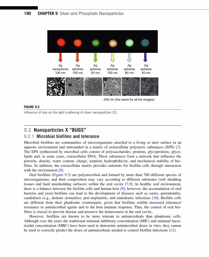
9.2 Nanoparticles X “BUGS”9.2.1 Microbial biofilms and toleranceMicrobial biofilms are communities of microorganisms attached to a living or inert surface in an
aqueous environment and surrounded in a matrix of extracellular polymeric substances (EPS) [7].
The EPS synthesized by microbial cells consist of polysaccharides, proteins, glycoproteins, glyco-
lipids and, in some cases, extracellular DNA. These substances form a network that influence the
porosity, density, water content, charge, sorption, hydrophobicity, and mechanical stability of bio-
films. In addition, the extracellular matrix provides nutrients for biofilm cells through interaction
with the environment [8].
Oral biofilms (Figure 9.3) are polymicrobial and formed by more than 700 different species of
microorganisms, and their composition may vary according to different substrates (soft shedding
tissues and hard nonshedding surfaces) within the oral cavity [7,9]. In healthy oral environment,
there is a balance between the biofilm cells and human host [9]; however, the accumulation of oral
bacteria and yeast biofilms can lead to the development of diseases such as caries, periodontitis,
candidiasis (e.g., denture stomatitis), peri-implantitis, and endodontic infections [10]. Biofilm cells
are different from their planktonic counterparts, given that biofilms exhibit increased tolerance/
resistance to antimicrobial agents and to the host immune response. Thus, the control of oral bio-
films is crucial to prevent disease and preserve the homeostasis in the oral cavity.
However, biofilms are known to be more tolerant to antimicrobials than planktonic cells.
Although over the years the traditional minimal inhibitory concentration (MIC) and minimal bacte-
ricidal concentration (MBC) have been used to determine antimicrobial doses in vitro, they cannot
be used to correctly predict the doses of antimicrobials needed to control biofilm infections [11].
Agnanoprisms
100 nm
Agspheres100 nm
Agspheres100 nm
Agspheres50 nm
Agspheres80 nm
Agspheres40 nm
−200 nm (the same for all the images)
FIGURE 9.2
Influence of size on the light scattering of silver nanoparticles [5].
190 CHAPTER 9 Silver and Phosphate Nanoparticles

Several factors influence biofilm tolerance to antimicrobials including nanoparticles. The first
factor that sets biofilms apart is the presence of EPS. The slime layer that involves the microbial
cells may not only limit the penetration of the agents but also react with it, reducing its effective-
ness [12]. In addition, biofilms are very heterogeneous; so in a community there are not only highly
active cells but also dormant cells. In fact, the presence of dormant cells has been reported as the
major factor in antimicrobial tolerance [13,14].
As many antimicrobial and antibiotics target macromolecule synthesis, when the cells are not
growing or dividing they are less susceptible. Other factors involved in tolerance are the phenotypic
variations, the existence of a highly developed communications system between the cells (quorum
sensing), and the bacterial multidrug efflux pumps [13,15]. A possible strategy to overcome biofilm
tolerance may be the use of several antimicrobials combined or even the combination of physical
and chemical treatments.
FIGURE 9.3
Dental biofilms grown on hydroxyapatite chips in situ. The biofilms were stained with LIVE/DEADs BacLightt
Bacterial Viability Kit and imaged with a confocal microscope. Green cells represent live microorganisms while
red are dead. (For interpretation of the references to color in this figure legend, the reader is referred to the
web version of this book.)
Image courtesy of The Center for Biofilm Engineering at Montana State University.
1919.2 Nanoparticles X “BUGS”

9.2.2 Silver nanoparticles and biofilms9.2.2.1 Mechanisms of action and literature dataInorganic nanoparticles represent a new era in the nanoparticle field. They exhibit unique physical
and chemical properties and can be used in many physical, biological, biomedical, and pharmaceu-
tical applications [16].
Silver nanoparticles are known for their broad-spectrum antimicrobial activity against bacteria,
fungi, and viruses [17], and multifactorial mechanism of action. Nevertheless, data have shown that
biofilm-forming cells are less susceptible to nanosilver (nano-Ag) than planktonic counterparts
[18,19]. An in vitro study found that the MIC of nano-Ag for Escherichia coli biofilms was four-
fold higher than the MIC for planktonic cells [18]. For Candida albicans and Candida glabrata bio-
films, the MIC was 17- to 135-fold and 16.5- to 34-fold higher, respectively, than for planktonic
cells [19].
However, nano-Ag may have an important role in the prevention of biofilm formation.
Kalishwaralal et al. [20] verified that treating Pseudomonas aeruginosa and Staphylococcus epider-
midis with nano-Ag caused 95% inhibition in biofilm formation. A similar result was observed by
Monteiro et al. [21] with respect to C. albicans and C. glabrata biofilms. These authors found that
nano-Ag at 3.3 µg/mL were more effective in inhibiting biofilm formation than in controlling
preformed biofilms.
Although biofilms have water channels through which nanoparticles may diffuse [20], many
nanoparticles may be retained in the extracellular matrix, contributing to biofilm resistance.
Furthermore, several mechanisms have been proposed to explain why nanoparticles have a better
effect when applied to planktonic cells than on mature biofilms.
Another important aspect that may hinder the action of nano-Ag against biofilm cultures is their
predisposition to form aggregates of particles (Figure 9.4). This aggregation can occur due to
changes in ionic strength and interactions of nanoparticles with several substances present in
(A) (B)
FIGURE 9.4
Scanning electron microscopy images showing clusters of silver nanoparticles in contact with mature Candida
albicans biofilm. (B) represents an enlarged view of part of biofilm displayed in (A).
192 CHAPTER 9 Silver and Phosphate Nanoparticles

extracellular matrix produced by microorganisms [18]. Microscopic observations have indicated
that the interactions of E. coli biofilm cells and nano-Ag resulted in an increase in final average
aggregate size by a factor of 40 [18]. The increase in the particle size may reduce the silver toxicity
and retard the particle diffusion within the biofilms, resulting in resistance to nano-Ag [18].
The size of nanoparticle and the type of stabilizing agent are not crucial to their efficacy against
biofilms probably due to particle aggregation. Nano-Ag with different diameters (5, 10, and 60 nm)
formed through the reduction of silver nitrate with sodium citrate [22] and stabilized with ammonia
or polyvinyl pyrrolidone (PVP) (to control particle growth and prevent aggregation) showed no sig-
nificant differences in the effect against C. albicans and C. glabrata mature biofilms [19]. Thus,
the size of nano-Ag originally synthesized is not a good indication of the true nanoparticle size
when in contact with biofilms.
The uptake of nano-Ag by biofilms has also been studied. Interestingly, in a study by Fabrega
et al. [23], only approximately 10% of the total mass of nano-Ag to which Pseudomonas putida
biofilms were exposed (during 24 h) remained trapped through the biofilms.
A study on the diffusion of nanoparticles in Pseudomonas fluorescens biofilms found that self-
diffusion coefficients decreased with the square of the radius of the nanoparticles [24]. In addition,
nano-Ag showed a greater tendency to accumulate in dense biofilms when compared to loose
flakes. These aspects may influence the susceptibility of biofilms to nano-Ag.
In conclusion, the interaction between biofilms and nano-Ag is complex and, possibly, several
factors may interfere with the efficacy of these particles against biofilms. Thus, these issues need to
be more explored in order to better understand the behavior of nanoparticles on biofilms.
9.2.2.2 Processing silver nanoparticlesTo obtain small metallic colloidal nanoparticles, a high density of nuclei at the beginning of the
process is necessary. For this reason, the addition of a reducing agent, such as sodium citrate, into
a solution of silver nitrate should be carried out as fast as possible to form a large number of nano-
particles simultaneously. However, unlike other noble metals, only a fraction of silver ions are
reduced to metal, even when using an excess of sodium citrate or any different reducing reagent.
Due to this inevitable presence of silver ions, new nuclei will still be formed while the nanoparti-
cles initially formed will continue to grow, fed by Ag1 that was not consumed during the formation
of the first nuclei. This process results in a system with a broad size distribution, consisting of large
particles formed initially and small particles that were formed subsequently. To overcome this
problem and obtain stable aqueous colloidal suspensions of spherical silver nanoparticles with sharp
size distribution, there is a way to prevent particle growth and the generation of new nuclei using
ammonia to trap all free silver ions. It is well known that when an excess of ammonia is added in
the presence of Ag1, soluble diamine silver (I) complexes are formed immediately (Eq. (9.2)),
removing the silver ions that have not yet been reduced. It prevents the formation of new
nuclei and the growth of already formed nanoparticles, resulting in virtually monodisperse silver
nanoparticles [22].
Ag1 1 2NH3-½AgðNH3Þ2�1 (9.2)
It is not easy to propose a kinetic model for the growth of silver nanoparticles since there are
many variables to consider in this case. However, the interdependence of density of nuclei and reac-
tion time may serve to shed light on the formation of a monodisperse system of nanoparticles, and
1939.2 Nanoparticles X “BUGS”

how ammonia acts as a moderator of nanoparticle growth in the colloidal medium (Figure 9.5) [22].
This apparently simple idea effectively results in stable colloidal aqueous suspensions of spheri-
cal silver nanoparticles with narrow size distributions. The addition of ammonia, or any agent to
trap the excess of silver ions, to the system immediately after the nucleation stage is an efficient
way to stabilize colloidal suspensions of monodisperse silver nanoparticles with controlled size,
since free silver ions, which are responsible for particle growth and the formation of new nuclei,
are trapped by the formation of soluble complexes, preserving the silver nanoparticles for long
storage periods without coalescing or precipitating. This procedure, which involves the use of
inexpensive and nontoxic reagents, also allows the particle size to be determined by choosing the
right moment at which ammonia. The earlier the ammonia is added the smaller the average parti-
cle size will be.
When silver nanoparticles are synthesized based on Turkevich [25] methods, it is fundamental
to use deionized water to prepare the solution, although the chemicals used are usually of analytical
grade, with no further purification. Silver nanoparticles can be prepared by the reduction of silver
nitrate with sodium citrate or any reducing agent. In a typical procedure, a volume of 100 mL of an
aqueous solution of silver nitrate (1.0 mmol/L) is heated and stirred gently. The silver nitrate solu-
tion should be heated to temperatures between 60�C and 90�C before the addition of 1.0 mL of a
solution of sodium citrate (0.3 mol/L) to accelerate the reaction. At room temperature, this reaction
is too slow that is not possible to follow it. This reaction can be monitored through the typical
yellow color the characteristic plasmon absorbance band at approximately 425 nm. The intensity is
proportional to the nanoparticle concentration; however concentrated colloidal dispersion is not
stable for long time and in this case, an extra stabilizing agent such as PVP is necessary.
Addition of ammonia
Sol
ute
conc
entr
atio
n "A
g°"
Nucleation stage
Without saturation
Saturation
Nucleation and growth
Time
C∞C0
FIGURE 9.5
Reduction in the metallic silver concentration as a function of reaction time, showing the initial nucleation
stage after the addition of citrate ion and the addition of ammonia immediately after the system turned yellow,
reducing the amount of metallic silver to a quantity below the saturation limit [22].
194 CHAPTER 9 Silver and Phosphate Nanoparticles

9.3 Phosphates micro- and nanoparticles and caries prevention9.3.1 De- and remineralization processDental caries is a dietobacterial disease resulting from the slow but progressive dissolution of the
tooth surfaces by acids produced by bacteria in the dental biofilm [26]. It has a multifactorial
nature, in which the concomitant interaction of dietary sugars, dental biofilm, and the host is
needed [27]. In order to understand the dynamics of dental enamel demineralization (DE) and remi-
neralization (RE) processes, a few biochemical aspects of the oral environment must be
highlighted.
In general terms, tooth enamel is mostly composed by hydroxyapatite (HAP) and fluoro
hydroxyapatite (FAP). The supersaturation of saliva with respect to these minerals has important
clinical implications, as HAP and FAP will not dissolve unless significant changes in the oral pH
occur. When the dental biofilm is exposed to fermentable sugars, the pH in the biofilm fluid rapidly
decreases to values below the critical pH for HAP (5.5) and/or FAP (4.5), so that saliva becomes
undersaturated with respect to these minerals. This leads to enamel DE. With time, the pH in the
biofilm fluid begins to rise, due to the action of salivary buffers. When the pH in the biofilm fluid
reaches values above the critical pH for HAP and FAP, enamel RE occurs [28]. The equilibrium
between DE and RE processes will determine that the enamel structure will not dissolve. However,
when DE episodes occur at a higher frequency and intensity, RE cycles will not be able to reverse
enamel dissolution, causing dental caries lesion.
Among the strategies to shift the balance from DE to RE, fluoride therapy is the most widely
used, as it reduces the enamel acid solubility (by converting HAP in FAP after acid challenges) and
increases the RE process (by promoting the reprecipitation of calcium and phosphates into the den-
tal mineral) [26,29]. However, due to the increase in dental fluorosis prevalence observed over the
last few decades, the search for nonfluoride therapeutic agents have been studied, which include
calcium and phosphate salts. These have been studied in association or not with fluoride in denti-
frices, gels, varnishes, and rinses, and include CaCl2, calcium lactate, calcium glycerophosphate,
sodium trimetaphosphate (TMP), nanocomplexes of casein phosphopeptide�amorphous calcium
phosphate, among others.
9.3.2 Nanophosphates and microbial adhesionPhosphates have great affinity with HAP, preventing the release of calcium and phosphate ions and
reducing the surface area available for dissolution [30,31]. In vitro studies showed that it is possible
to reduce the concentration of fluoride in dentifrices and mouth rinses, yet maintaining their protec-
tive action against both dental caries and erosion by adding inorganic phosphate (sodium TMP) to
the products [32�36]. Given the high adsorption of phosphates to the enamel surface, it would be
interesting to investigate whether the use of phosphate would influence bacterial adhesion to tooth
enamel, as well as on mineral loss. The effects of TMP on dental biofilm and its efficacy when
combined to different antimicrobials effect on biofilm were analyzed by colony forming unit
(CFU) counts and lactic acid production. The data presented in that study demonstrated that the
biofilm model used was suitable to grow biofilm on different substrata, both in microcosm and
single species biofilms. The model allows the evaluation of antimicrobial agents and TMP on
1959.3 Phosphates micro- and nanoparticles and caries prevention

biofilm survival and metabolism [35]. It was concluded that TMP, alone or combined to antimicro-
bials, had no direct action on biofilm. In addition, a pilot study compared the effect of treatment
(during 5 min) of bovine enamel blocks with commercial TMP and TMP with smaller particles in
different concentrations (3% and 5%) on the adhesion of C. albicans and C. glabrata. It was possi-
ble to observe that, regardless of the concentration, the treatment with TMP with different particle
sizes did not reduce the number of CFUs on the enamel blocks after a period of 2 h (initial adhe-
sion) for Candida species tested.
9.4 Pros and cons of nanoparticles toward biological-dental application9.4.1 ToxicityProfile toxicity of nanomaterials can be considered different from larger particles mainly because
of their small size and high reactivity [37�39]. Toxicology associated to nanostructures, in general,
is affected by different physical and chemical properties of each nanomaterial [37,38]. Precisely,
because of the number of variables interfering on the toxicity of nanomaterials, it is difficult to gen-
eralize their effects on the biological systems [40].
Composition is clearly a key factor in determining the toxicity of nanomaterials. Actually, tak-
ing into consideration the chemical composition, there are an enormous number of different nano-
materials [40,41]. One of the most widely studied categories of nanomaterials are metal
nanoparticles; among them silver nanoparticles are those that generate more interest in nanotoxico-
logical research, precisely because of their use as antimicrobial agent in the medical area
[17,21,42].
In general, shape and size of the nanomaterials directly influence on the toxicity [39,43]. It has
been stated that cubic nanoparticles induce lower level of toxicity compared to those that are cylin-
drical, spherical, and rod shaped [44]. Usually, smaller particles are considered more toxic since
they occupy less volume in a greater surface area per unit of mass increasing the availability for
the biological interactions [37,38]. Furthermore, it has been amply exposed that the internalization
of nanomaterials is a size-dependent process; smaller particles are much more easily internalized
into the cells and therefore more available for interaction with cellular components [43,45].
The main objectives of surface treatment or functionalization of nanoparticles are ensuring
stability and promote favorable characteristics to a specific proposal [46,47]. Nanoparticles can be
superficially modified with polymers, drugs, peptides, proteins, oligonucleotides, and biological
molecules [46,47] and show different cytotoxic characteristics depending on the coating material
[47,48]. Thus, surface treatment or functionalization can be considered one of the most relevant
factors of cytotoxicity [43,49] since the process modifies chemical properties inherent to the nano-
materials and determines their toxicity profile [43,49].
Considering the agglomeration state of the nanomaterials and their related cytotoxicity, it has
been suggested that the process of agglomeration makes the particles become larger or even exceed
the nanoscale [50]. As discussed above, size is considered to be a relevant factor in determining the
toxicity profile. Recently, it was confirmed that large agglomerates are not effectively cell internal-
ized; therefore, agglomerated particles can be considered lower cytotoxic, once that the agglomera-
tion state of the particles significantly reduces surface area availability and access into the cells [50].
196 CHAPTER 9 Silver and Phosphate Nanoparticles

Bearing in mind the role of silver nanoparticles on current scenario, questions can arise about
the action of silver nanoparticles on human cells. Important preliminary data regarding the toxicity
of nanoparticles at cellular level have been reported. In this sense, it has been accepted that the
mechanisms of cytotoxicity of nanoparticles are the same for microorganisms and human cells
[17,51].
Human contact with silver nanoparticles for therapeutic applications can occur mainly through
inhalation and dermal absorption [52]. From the site of the contact, the particles can be biodistribu-
ted in the organism by the circulatory and lymphatic systems [53]. Thus, it has been accepted that
nanoparticles can achieve different parts of the human body promoting adverse effects that are not
completely understood [37,43,46,53�55].
It has been stated that silver nanoparticles penetrate the cells mainly by the process of endocyto-
sis, and once into the cell they interact with different structures in the cytoplasm and nucleus.
Silver nanoparticles are considered highly mobile and they can be sequestered in various intracellu-
lar compartments or organelles but preferentially are located in the mitochondria [51,53,56]. The
deposition of the silver nanoparticles in this cellular organelle can cause suspension of electron
transport chain, resulting in low ATP production and higher production of reactive oxygen species
(ROS) [51,53,56]. Increased concentrations of ROS can initiate the apoptotic cascade [41,53,56].
Furthermore, it has been known that cellular oxidation�reduction homeostasis is maintained in part
by the glutathione (GSH) intracellular levels [41,56]. Recently, it was found that silver nanoparti-
cles can deplete GSH inhibiting antioxidant defense mechanisms [41].
Silver nanoparticles can also induce DNA damage, as they can be deposited in the nucleus,
affect the DNA synthesis, and cause chromosomal abnormalities; however, the exact mechanisms
that promote these events are not completely understood [39,53,56].
Although the biological effects of nanomaterials constitute research area of growing interest, the
use of biomaterials in nanoscale range in the biomedical area requires more information. It is nec-
essary to assess the toxicological profiles of different nanomaterials taking in account their several
specific properties. Without accurate toxicity data, the use of nanomaterials can become a risk for
human health.
9.4.2 Processing costsThe science and technology of nanomaterials has created great excitement and expectations in the
last few years. The burgeoning of nanoscience creates new opportunities for advanced medical pro-
tocols and disease treatments. However, it is widely claimed that research effort to discover and
develop new pharmaceuticals entails high costs and high risks, mainly when it involves nanotech-
nology. The principal justification for the high prices on patented drugs has been the high cost of
research and development (R&D). Recently, the average cost of a new patented molecule was esti-
mated at $802 million [57,58], although some authors disagree with the methodology used, propos-
ing a median of $43.3 million per new drug [59]. These extremely expensive values imposed by
multinational corporations are related to therapeutic drugs for complex diseases, such as cancer,
HIV, and depression. For the particular use in dentistry, the costs are evidently smaller, which rep-
resent an opportunity for innovative university spin-off companies based on nanotechnology and
advanced materials. Nanoparticles are expensive since they are difficult to isolate and, eventually,
tedious to purify. On the other hand, a commercial nanocomposite shows functional nanofiller loads
1979.4 Pros and cons of nanoparticles toward biological-dental application

typically less than 10 wt%. Obviously, taxes and marketing margins are a few of the components
that impact the final price for the consumer, and the packaging represents more than one-third of
the final price without any gain of economy of scale. The average cost of fabrication of functional
nanoparticles is usually in the range from $100 to $1000 per gram, but its impact in the price
composition of dentistry products is proportional to the parcel of others components, resulting in a
product with commercial viability considering their advantages regarding the performance and
patient comfort.
9.4.3 Potential use in dental field and future directionsIn general, the great interest of nanoparticles is related to its highly small size. By reducing the size
of a material, the surface to volume ratio increases. As a consequence, the majority of the atoms
are at the margin of the particle making them much more reactive [60]. This reactivity is specially
noted in inorganic nanoparticles and might conduce to instabilities and then to degradation and
corrosion processes [60]. Regarding silver nanoparticles, several methods of synthesis have been
developed in the last decade, although most of them are still under development [61]. They are
basically focused on increasing the stability and reducing the aggregation of nanoparticles since the
small size would ensure that an expressive surface area would be in contact with the microbial
effluent [62,63]. However, we agree with Pal et al. [62] when they said that “smallness in itself ”
should not be the only purpose in the development of functional inorganic nanoparticles. The
majority of our studies related to the action of silver nanoparticles against Candida species testified it,
especially the study carried out by Monteiro et al. [19] where the size of the particles did not interfere
with its efficacy. Surface interactions of nanoparticles with the surrounding medium (bioactivity)
before they reach the aimed target are a significant aspect to be considered [64]. Bastus et al. [60]
clearly explained that in functional colloidal inorganic nanoparticles, these interactions are deter-
mined not only by the size, shape, or structure of the center (core) of the nanoparticle but also by
the organic or inorganic molecules that cover the core (shell) [60]. This might be the most relevant
insight that those authors specially pointed out about nanostructured materials, and their applicability
in biomedical/dental field relies on more understanding and comprehension about not only con-
trolled synthesis but also particles interfacial interactions and their toxicity [65,66].
Besides the predominant role of the shell on the interactions of the nanoparticle, it may also
protect the particle and, therefore, defines its selectivity against the microorganism or the molecular
target [60]. We could take advantage of this particular bioactivity of functional nanoparticles and
intentionally engineer them for both core and shell to provide therapeutic effects with low toxicity
on the mammalian cells.
9.5 ConclusionsConcluding, the field of dentistry should be encouraged by the multiple studies in nanotechnology
that have been widely published in different fields like medical, chemical, biological engineering,
and at least environmental field to create “intelligent bionanomaterials” which could act multifunc-
tionally: (i) by preventing/controlling the infection in or on oral tissues as well as (ii) by
stimulating/contributing for the RE of dental and bone tissues. Very recent literature has been
198 CHAPTER 9 Silver and Phosphate Nanoparticles

driving us to this pathway [63,67,68]. Possibly bionanomaterials of silver phosphates are a promis-
ing candidate for different dental materials purposes, like in titanium implants coatings, bone
fillings, dental varnish or sealants, toothpastes and even in dental floss covering, since they would
combine the antimicrobial action of silver and the bioactivity of phosphates [63].
From now, the door for future perspectives is open to welcome the readers into the “nanoworld”
to give their appropriate judgments in regard to the disadvantages and the real benefits of the use
of nanostructured biomaterials in the dental field.
References[1] G.A. Ozin, A.C. Arsenault, Nanochemistry. A chemical approach to nanomaterials, R. Soc. Chem.
(2005).
[2] D.A. McQuarrie, J.D. Simon, Physical Chemistry. A Molecular Approach, University Science Books,
1997.
[3] I. Levine, Quantum Chemistry, sixth ed., Prentice Hall, 2008.
[4] T. Pellegrino, S. Kudera, T. Liedl, A.M. Javier, L. Manna, W.J. Parak, On the development of colloidal
nanoparticles towards multifunctional structures and their possible use for biological applications, Small
1 (1) (2005) 48.
[5] C.A. Mirkin, The beginning of a small revolution, Small 1 (1) (2005) 14.
[6] A.Z. Moshfegh, Nanoparticle catalysts, J. Phys. D. Appl. Phys. 42 (23) (2009).
[7] V. Zijnge, M.B. van Leeuwen, J.E. Degener, F. Abbas, T. Thurnheer, R. Gmur, et al., Oral biofilm archi-
tecture on natural teeth, PLoS One 5 (2) (2010) e9321.
[8] H.C. Flemming, T.R. Neu, D.J. Wozniak, The EPS matrix: the “house of biofilm cells”, J. Bacteriol. 189
(22) (2007) 7945.
[9] S. Filoche, L. Wong, C.H. Sissons, Oral biofilms: emerging concepts in microbial ecology, J. Dent. Res.
89 (1) (2010) 8.
[10] R.P. Allaker, The use of nanoparticles to control oral biofilm formation, J. Dent. Res. 89 (11) (2010)
1175.
[11] C.A. Fux, J.W. Costerton, P.S. Stewart, P. Stoodley, Survival strategies of infectious biofilms, Trends
Microbiol. 13 (1) (2005) 34.
[12] T.R. Zuroff, H. Bernstein, J. Lloyd-Randolfi, L. Jimenez-Taracido, P.S. Stewart, R.P. Carlson,
Robustness analysis of culturing perturbations on Escherichia coli colony biofilm beta-lactam and
aminoglycoside antibiotic tolerance, BMC Microbiol. 10 (2010) 185.
[13] S.A. Rani, B. Pitts, H. Beyenal, R.A. Veluchamy, Z. Lewandowski, W.M. Davison, et al., Spatial pat-
terns of DNA replication, protein synthesis, and oxygen concentration within bacterial biofilms reveal
diverse physiological states, J. Bacteriol. 189 (11) (2007) 4223.
[14] J.P. Folsom, L. Richards, B. Pitts, F. Roe, G.D. Ehrlich, A. Parker, et al., Physiology of Pseudomonas
aeruginosa in biofilms as revealed by transcriptome analysis, BMC Microbiol. 10 (2010) 294.
[15] J. Kim, J.S. Hahn, M.J. Franklin, P.S. Stewart, J. Yoon, Tolerance of dormant and active cells in Pseudomonas
aeruginosa PA01 biofilm to antimicrobial agents, J. Antimicrob. Chemother. 63 (1) (2009) 129.
[16] K. Chaloupka, Y. Malam, A.M. Seifalian, Nanosilver as a new generation of nanoproduct in biomedical
applications, Trends Biotechnol. 28 (11) (2010) 580.
[17] D.R. Monteiro, L.F. Gorup, A.S. Takamiya, A.C. Ruvollo-Filho, E.R. de Camargo, D.B. Barbosa, The
growing importance of materials that prevent microbial adhesion: antimicrobial effect of medical devices
containing silver, Int. J. Antimicrob. Agents 34 (2) (2009) 103.
199References

[18] O. Choi, C.P. Yu, G. Esteban Fernandez, Z. Hu, Interactions of nanosilver with Escherichia coli cells in
planktonic and biofilm cultures, Water Res. 44 (20) (2010) 6095.
[19] D.R. Monteiro, S. Silva, M. Negri, L.F. Gorup, E.R. de Camargo, R. Oliveira, et al., Silver nanoparticles:
influence of stabilizing agent and diameter on antifungal activity against Candida albicans and Candida
glabrata biofilms, Lett. Appl. Microbiol. 54 (5) (2012) 383.
[20] K. Kalishwaralal, S. BarathManiKanth, S.R. Pandian, V. Deepak, S. Gurunathan, Silver nanoparticles
impede the biofilm formation by Pseudomonas aeruginosa and Staphylococcus epidermidis, colloids
surf, Colloids Surf. B Biointerfaces 79 (2) (2010) 340.
[21] D.R. Monteiro, L.F. Gorup, S. Silva, M. Negri, E.R. de Camargo, R. Oliveira, et al., Silver colloidal
nanoparticles: antifungal effect against adhered cells and biofilms of Candida albicans and Candida
glabrata, Biofouling 27 (7) (2011) 711.
[22] L.F. Gorup, E. Longo, E.R. Leite, E.R. Camargo, Moderating effect of ammonia on particle growth and
stability of quasi-monodisperse silver nanoparticles synthesized by the Turkevich method, J. Colloid
Interface Sci. 360 (2) (2011) 355.
[23] J. Fabrega, J.C. Renshaw, J.R. Lead, Interactions of silver nanoparticles with Pseudomonas putida
biofilms, Environ. Sci. Technol. 43 (23) (2009) 9004.
[24] T.O. Peulen, K.J. Wilkinson, Diffusion of nanoparticles in a biofilm, Environ. Sci. Technol. 45 (8)
(2011) 3367.
[25] J. Turkevich, J. Hillier, A study of the nucleation and growth processes in the synthesis of colloidal
gold, Discuss. Faraday Soc. 11 (1951) 55.
[26] G.M. Whitford, J.L. Wasdin, T.E. Schafer, S.M. Adair, Plaque fluoride concentrations are dependent on
plaque calcium concentrations, Caries Res. 36 (4) (2002) 256.
[27] M.A. Buzalaf, J.P. Pessan, H.M. Honorio, J.M. ten Cate, Mechanisms of action of fluoride for caries
control, Monogr. Oral Sci. 22 (2011) 97.
[28] J.M. ten Cate, J.M. Larsen, E.I.F. Pearce, O. Fejerskov, Chemical interactions between the tooth and the
oral fluids, in: O. Fejerskov, E.A.M. Kidd (Eds.), Dental Caries: The Disease and Its Clinical
Management, Blackwell Munksgaard, Oxford, 2008, pp. 209�231.
[29] R.P. Shellis, R.M. Duckworth, Studies on the cariostatic mechanisms of fluoride, Int. Dent. J. 44
(3 Suppl. 1) (1994) 263.
[30] M.E. Barbour, R.P. Shellis, D.M. Parker, G.C. Allen, M. Addy, Inhibition of hydroxyapatite dissolution
by whole casein: the effects of pH, protein concentration, calcium, and ionic strength, Eur. J. Oral Sci.
116 (5) (2008) 473.
[31] M.E. Barbour, R.P. Shellis, D.M. Parker, G.C. Allen, M. Addy, An investigation of some food-approved
polymers as agents to inhibit hydroxyapatite dissolution, Eur. J. Oral Sci. 113 (6) (2005) 457.
[32] M.M. Manarelli, A.E. Vieira, A.A. Matheus, K.T. Sassaki, A.C. Delbem, Effect of mouth rinses with
fluoride and trimetaphosphate on enamel erosion: an in vitro study, Caries Res. 45 (6) (2011) 506.
[33] M.J. Moretto, A.C. Magalhaes, K.T. Sassaki, A.C. Delbem, C.C. Martinhon, Effect of different fluoride
concentrations of experimental dentifrices on enamel erosion and abrasion, Caries Res. 44 (2) (2010) 135.
[34] E.M. Takeshita, L.P. Castro, K.T. Sassaki, A.C. Delbem, In vitro evaluation of dentifrice with low fluo-
ride content supplemented with trimetaphosphate, Caries Res. 43 (1) (2009) 50.
[35] E.M. Takeshita, Evaluation of sodium trimetaphosphate (TMP) effect on biolfim and enamel de- and
remineralization: in vitro and in situ study, Thesis, Sao Paulo State University, Aracatuba, 2010.
[36] E.M. Takeshita, R.A. Exterkate, A.C. Delbem, J.M. ten Cate, Evaluation of different fluoride concentra-
tions supplemented with trimetaphosphate on enamel de- and remineralization in vitro, Caries Res. 45
(5) (2011) 494.
[37] X. Deng, Q. Luan, W. Chen, Y. Wang, M. Wu, H. Zhang, et al., Nanosized zinc oxide particles induce
neural stem cell apoptosis, Nanotechnology 20 (11) (2009) 115101.
200 CHAPTER 9 Silver and Phosphate Nanoparticles

[38] M. Horie, K. Nishio, K. Fujita, S. Endoh, A. Miyauchi, Y. Saito, et al., Protein adsorption of
ultrafine metal oxide and its influence on cytotoxicity toward cultured cells, Chem. Res. Toxicol. 22 (3)
(2009) 543.
[39] M. Mahmood, D.A. Casciano, T. Mocan, C. Iancu, Y. Xu, L. Mocan, et al., Cytotoxicity and biological
effects of functional nanomaterials delivered to various cell lines, J. Appl. Toxicol. 30 (1) (2010) 74.
[40] M. Mahmoudi, K. Azadmanesh, M.A. Shokrgozar, W.S. Journeay, S. Laurent, Effect of nanoparticles on
the cell life cycle, Chem. Rev. 111 (5) (2011) 3407.
[41] S.M. Hussain, K.L. Hess, J.M. Gearhart, K.T. Geiss, J.J. Schlager, In vitro toxicity of nanoparticles in
BRL 3A rat liver cells, Toxicol. In Vitro 19 (7) (2005) 975.
[42] K.J. Kim, W.S. Sung, B.K. Suh, S.K. Moon, J.S. Choi, J.G. Kim, et al., Antifungal activity and mode of
action of silver nano-particles on Candida albicans, Biometals 22 (2) (2009) 235.
[43] Y. Teow, P.V. Asharani, M.P. Hande, S. Valiyaveettil, Health impact and safety of engineered nano-
materials, Chem. Commun. (Camb) 47 (25) (2011) 7025.
[44] A. Albanese, P.S. Tang, W.C. Chan, The effect of nanoparticle size, shape, and surface chemistry on
biological systems, Annu. Rev. Biomed. Eng. (2012).
[45] C. Carlson, S.M. Hussain, A.M. Schrand, L.K. Braydich-Stolle, K.L. Hess, R.L. Jones, et al., Unique
cellular interaction of silver nanoparticles: size-dependent generation of reactive oxygen species, J. Phys.
Chem. B. 112 (43) (2008) 13608.
[46] R. Foldbjerg, D.A. Dang, H. Autrup, Cytotoxicity and genotoxicity of silver nanoparticles in the human
lung cancer cell line, A549, Arch. Toxicol. 85 (7) (2011) 743.
[47] I. Sur, D. Cam, M. Kahraman, A. Baysal, M. Culha, Interaction of multi-functional silver nanoparticles
with living cells, Nanotechnology 21 (17) (2010) 175104.
[48] L. Bohmert, B. Niemann, A.F. Thunemann, A. Lampen, Cytotoxicity of peptide-coated silver nanoparti-
cles on the human intestinal cell line Caco-2, Arch. Toxicol. (2012).
[49] A.M. El Badawy, R.G. Silva, B. Morris, K.G. Scheckel, M.T. Suidan, T.M. Tolaymat, Surface charge-
dependent toxicity of silver nanoparticles, Environ. Sci. Technol. 45 (1) (2011) 283.
[50] M. Ahamed, M. Karns, M. Goodson, J. Rowe, S.M. Hussain, J.J. Schlager, et al., DNA damage
response to different surface chemistry of silver nanoparticles in mammalian cells, Toxicol. Appl.
Pharmacol. 233 (3) (2008) 404.
[51] P.V. AshaRani, G. Low Kah Mun, M.P. Hande, S. Valiyaveettil, Cytotoxicity and genotoxicity of silver
nanoparticles in human cells, ACS Nano 3 (2) (2009) 279.
[52] L. Braydich-Stolle, S. Hussain, J.J. Schlager, M.C. Hofmann, In vitro cytotoxicity of nanoparticles in
mammalian germline stem cells, Toxicol. Sci. 88 (2) (2005) 412.
[53] P.V. Asharani, M.P. Hande, S. Valiyaveettil, Anti-proliferative activity of silver nanoparticles, BMC
Cell. Biol. 10 (2009) 65.
[54] L. Wei, J. Tang, Z. Zhang, Y. Chen, G. Zhou, T. Xi, Investigation of the cytotoxicity mechanism of
silver nanoparticles in vitro, Biomed. Mater. 5 (4) (2010) 044103.
[55] M.V. Park, A.M. Neigh, J.P. Vermeulen, L.J. de la Fonteyne, H.W. Verharen, J.J. Briede, et al.,
The effect of particle size on the cytotoxicity, inflammation, developmental toxicity and genotoxicity of
silver nanoparticles, Biomaterials 32 (36) (2011) 9810.
[56] S. Arora, J. Jain, J.M. Rajwade, K.M. Paknikar, Cellular responses induced by silver nanoparticles:
in vitro studies, Toxicol. Lett. 179 (2) (2008) 93.
[57] J.A. DiMasi, R.W. Hansen, H.G. Grabowski, The price of innovation: new estimates of drug develop-
ment costs, J. Health Econ. 22 (2) (2003) 151.
[58] C.P. Adams, V.V. Brantner, Estimating the cost of new drug development: is it really 802 million
dollars? Health Aff. (Millwood) 25 (2) (2006) 420.
201References

[59] D.W. Light, R. Warburton, Demythologizing the high costs of pharmaceutical research, Biosocieties
6 (1) (2011) 34.
[60] N.G. Bastus, E. Casals, I. Ojea, M. Varon, V. Puntes, The reactivity of colloidal inorganic nanoparticles,
The Delivery of Nanoparticles, InTech, 2012, pp. 377�400, (Chapter 18), ISBN: 978-953-51-0615-9.
[61] H. Korbekandi, S. Iravani, Silver nanoparticles, The Delivery of Nanoparticles, InTech, 2012, pp. 3�36,
(Chapter 1), ISBN: 978-953-51-0615-9.
[62] S. Pal, Y.K. Tak, J.M. Song, Does the antibacterial activity of silver nanoparticles depend on the shape
of the nanoparticle? A study of the Gram-negative bacterium Escherichia coli, Appl. Environ.
Microbiol. 73 (6) (2007) 1712.
[63] M. Miranda, A. Fernandez, S. Lopez-Esteban, F. Malpartida, J.S. Moya, R. Torrecillas, Ceramic/metal
biocidal nanocomposites for bone-related applications, J. Mater. Sci. Mater. Med. (2012).
[64] G. Garnweitneir, In situ versus post-synthetic stabilization of metal oxide nanoparticles, The Delivery of
Nanoparticles, InTech, 2012, pp. 71�92, (Chapter 4), ISBN: 978-953-51-0615-9.
[65] P. Dallas, V.K. Sharma, R. Zboril, Silver polymeric nanocomposites as advanced antimicrobial agents:
classification, synthetic paths, applications, and perspectives, Adv. Colloid Interface Sci. 166 (1�2)
(2011) 119.
[66] H.-M. Liu, J.-K. Hsiao, Magnetic nanoparticles: its effect on cellular behavior and potential applications,
Smart Nanoparticles Technology, InTech, 2012, pp. 358�72, (Chapter 16), ISBN: 978-953-51-0500-8.
[67] L. Cheng, M.D. Weir, H.H. Xu, J.M. Antonucci, N.J. Lin, S. Lin-Gibson, et al., Effect of amorphous
calcium phosphate and silver nanocomposites on dental plaque microcosm biofilms, J. Biomed. Mater.
Res. B Appl. Biomater. 100 (5) (2012) 1378.
[68] L. Cheng, M.D. Weir, H.H. Xu, J.M. Antonucci, A.M. Kraigsley, N.J. Lin, et al., Antibacterial amor-
phous calcium phosphate nanocomposites with a quaternary ammonium dimethacrylate and silver nano-
particles, Dent. Mater. 28 (5) (2012) 561.
202 CHAPTER 9 Silver and Phosphate Nanoparticles

CHAPTER
10Nanoparticles and the Controlof Oral Biofilms
Robert Patrick AllakerQueen Mary University of London, Barts & The London School of Medicine and Dentistry,
Institute of Dentistry, London, UK
CHAPTER OUTLINE
10.1 Introduction ............................................................................................................................... 204
10.2 Biofilms and oral infections ........................................................................................................205
10.2.1 Formation and properties of oral biofilms................................................................ 205
10.2.2 Oral biofilms and disease ...................................................................................... 206
10.2.2.1 Dental caries and periodontal disease ........................................................ 206
10.2.2.2 Peri-implantitis ........................................................................................ 206
10.2.2.3 Candidiasis............................................................................................. 206
10.2.3 Control of oral biofilms.......................................................................................... 207
10.3 Antimicrobial nanoparticles and oral biofilm control .................................................................... 207
10.3.1 Nanoparticulate metals as antimicrobial agents....................................................... 207
10.3.1.1 Silver (Ag) .............................................................................................. 210
10.3.1.2 Copper (Cu)............................................................................................ 210
10.3.1.3 Gold (Au) ............................................................................................... 211
10.3.2 Nanoparticulate metal oxides as antimicrobial agents .............................................. 211
10.3.2.1 Copper oxide (CuO and Cu2O) .................................................................. 211
10.3.2.2 Zinc oxide (ZnO) ..................................................................................... 212
10.3.2.3 Titanium dioxide (TiO2)............................................................................. 213
10.3.3 Oral applications of nanoparticulate metals and metal oxides ................................... 213
10.3.4 Quaternary ammonium compounds ........................................................................ 215
10.4 Antiadhesive nanoparticles and oral biofilm control ..................................................................... 216
10.4.1 Chitosan nano- and microparticles ......................................................................... 216
10.4.2 Silica and silicon nanoparticles ............................................................................. 216
10.4.3 Hydroxyapatite and other calcium phosphate-based systems .................................... 217
10.5 Photodynamic therapy and the use of nanoparticles to control oral biofilms .................................. 218
203Nanobiomaterials in Clinical Dentistry.
© 2013 Elsevier Inc. All rights reserved.

10.6 Biocompatibility of nanoparticles within the oral cavity................................................................ 219
10.7 Conclusions............................................................................................................................... 221
Acknowledgments ............................................................................................................................... 223
References ......................................................................................................................................... 223
10.1 IntroductionNanoparticles can be classified as particles of a size no greater than 100 nm, and their unique attri-
butes to combat infections have received considerable attention within a range of diverse fields,
including medicine and dentistry. Nanomaterials are increasingly finding uses in products such as
antimicrobial surface coatings and semiconductors. These include spherical, cubic, and needle-like
nanoscaled particles (approximately 5�100 nm) and near-nanoscaled devices (up to micrometers)
[1]. Properties of nanoparticles, for example, their active surface area, chemical reactivity, and bio-
logical activity, can be dramatically different from those of micrometer-sized particles [2], and
indeed the biocidal effectiveness of metallic nanoparticles has been suggested to be due to both
their size and their high surface-to-volume ratio. These characteristics should allow them to closely
interact with microbial membranes, and thus elicit an antimicrobial effect that is not solely due to
the release of metal ions [3]. Metallic and other nanoparticles are now being combined with poly-
mers and other base materials and coated onto surfaces which may have a variety of potential anti-
microbial applications within the oral cavity [4,5].
The oral cavity supports the growth of a wide diversity of microorganisms including bacteria,
yeasts, and viruses—members of all groups being associated with oral infections. Bacteria are the
predominant components of this resident microflora, and the diversity of species found in the oral
cavity reflects the wide range of endogenously derived nutrients, the varied types of habitat for col-
onization including surfaces on the teeth, mucosa, and tongue, and the opportunity to survive as a
biofilm. An oral biofilm can be classed as an aggregate of microorganisms in which cells adhere to
each other and to a surface [6]. However, the relationship between this microflora and the host can
be disrupted in a number of ways, resulting in the development of disease of the oral structures.
Potential habitats suitable for attachment within the oral cavity include the nonshedding hard
tooth surfaces or soft, constantly replaced epithelial surfaces, and conditions vary with respect to
oxygen levels and anaerobiosis, availability of nutrients, exposure to salivary secretions or gingival
crevicular fluid (GCF), masticatory forces, and other variables such as oral hygiene procedures.
The composition of the microbial flora of the mouth thus varies considerably from site to site and
at different time points. Up to 1000 different species of bacteria at 108�109 bacteria per milliliter
saliva or per milligram dental plaque are known to be associated with the oral cavity, and it has
been suggested that only 50% of the bacteria found at these sites can be cultured [6].
Most bacterial infections within the oral cavity are polymicrobial in nature, and it is quite
unusual to find any that are clearly due to a single species. The relative contribution of different
bacterial components in such infections is thus difficult to determine. Oral infections may arise
either from an endogenous source, i.e., one yielding microorganisms normally found in the mouth,
such as plaque-related dental caries and periodontal disease, or an exogenous source yielding
microorganisms not normally found as part of the oral microflora. Dental caries and periodontal
disease involve the adherence of bacteria and development of biofilms on both the natural and the
204 CHAPTER 10 Nanoparticles and the Control of Oral Biofilms

restored tooth surface. The use of nanotechnology offers the possibility to control the formation of
these and other oral biofilms through the use of nanoparticles with biocidal, antiadhesive, and
delivery capabilities.
10.2 Biofilms and oral infectionsBiofilms of oral bacteria and yeasts can cause a number of localized diseases in the oral cavity,
including dental caries, gingivitis, periodontitis, candidiasis, endodontic infections, orthodontic
infections, and peri-implantitis [6].
10.2.1 Formation and properties of oral biofilmsWithin the oral cavity, the survival of microorganisms is dependent on their ability to adhere to sur-
faces and subsequently develop into a biofilm, a process influenced by the physical and chemical
properties of the underlying surface [7]. On the tooth surface, the initial colonizers adhere to the
acquired pellicle, a salivary/dietary-derived proteinaceous layer, which can then influence the sub-
sequent sequence of colonization by microorganisms [8]. The acquired pellicle also contains several
salivary components such as secretory immunoglobulin A (sIgA) and lysozyme, and these provide
both barrier and buffering functions [9]. Both de- and remineralization processes of the teeth are
also mediated by the pellicle. In terms of bacterial colonization, many of the proteins that make up
the pellicle act as receptors for the specific interaction with adhesins on the surface of pioneer
bacterial species [9]. The pellicle layer is therefore of particular relevance for the interactions of
both bacteria and nanoparticles with the tooth surface.
The strength of the forces involved in the initial attachment of bacteria is critical to their sur-
vival and the subsequent growth of the biofilm. The major growth of dental plaque mass then
occurs by bacterial cell division within the biofilm rather than by coaggregation at the surface of
the developing biofilm [10]. The initial communities of bacteria found within the supragingival
plaque biofilm are of a relatively low diversity in comparison to those present in the mature com-
munities of both supra- and subgingival plaque. Initial colonizers include Streptococcus oralis,
Streptococcus sanguinis, and Streptococcus mitis. The coaggregating partners with these bacteria
would then include predominantly gram-negative species, e.g., Veillonella atypica, Eikenella corro-
dens, and Prevotella loescheii. Coaggregation bridges between these early colonizers and
Fusobacterium nucleatum are common and the latter then coaggregates with numerous late coloni-
zers. Late colonizers include Aggregatibacter actinomycetemcomitans, Prevotella intermedia,
Treponema denticola, and Porphyromonas gingivalis [10]. The interactions between oral bacteria
are integral to biofilm development and maturation and include physical contact, metabolic
exchange, molecular communication, and genetic material exchange.
Biofilms will accumulate on both the hard and soft oral tissues, and this community of micro-
bial species is embedded in a matrix of bacterial components, salivary proteins/peptides, and food
debris [8]. Extracellular polymeric substances, produced by bacteria in a mature biofilm, contain
large amounts of polysaccharides, proteins, nucleic acids, and lipids. These maintain the structural
integrity of the biofilm and provide an ideal matrix for bacterial cell growth and survival [11]. The
biofilm mode of growth is clearly distinguished from planktonic growth by a number of features,
20510.2 Biofilms and oral infections

which includes the resistance to antimicrobial agents at concentrations that approach 1000 times
greater than that required to kill planktonic microorganisms [12,13]. This is of particular signifi-
cance in the development of nanoantimicrobials and the extrapolation of in vitro findings.
10.2.2 Oral biofilms and disease10.2.2.1 Dental caries and periodontal diseaseDental caries is a destructive condition of the dental hard tissues that can progress to inflammation
and death of vital pulp tissue, and if untreated it may lead to the eventual spread of infection to the
periapical area of the tooth and beyond. The disease process involves acidogenic plaque bacteria,
including Streptococcus mutans, Streptococcus sobrinus, and Lactobacillus spp. [14], whereas the
periodontal diseases can involve both the soft and hard tissues and are initiated by components of the
plaque biofilm that develop on the hard root surface adjacent to the soft tissues of the supporting peri-
odontium. Periodontal disease may be confined to the gingiva (gingivitis) or extend to the deeper sup-
porting structures with destruction of the periodontal ligament and the alveolar bone that supports the
teeth (periodontitis). This loss of attachment, with associated periodontal pocket formation, may ulti-
mately lead to loosening and loss of the affected teeth. P. gingivalis, Tannerella forsythia, and
T. denticola are now regarded as the major pathogens in advancing periodontitis [15].
Prevention of dental caries and periodontal diseases is traditionally targeted at mechanical or
nonspecific control of the plaque biofilm because this is the precipitating factor. The use of antimi-
crobial agents represents a valuable complement to mechanical plaque control [16]. Such strategies
should ideally control plaque biofilm formation without significantly affecting the biological equi-
librium within the oral cavity. However, actual periods of exposure to antimicrobial agents during
tooth brushing and mouth rinsing can be very short and may amount to about 30 s, rather than the
recommended 2 min [17].
10.2.2.2 Peri-implantitisImplant systems are increasingly being used to replace missing teeth and most integrate with bone
without complications. Small amounts of plaque consisting mainly of Streptococcus and
Actinomyces spp. will accumulate on successful implants. However, in peri-implantitis, anaerobic
gram-negative organisms predominate [18]. This infection is a key cause of dental implant failure
whereby the induced inflammatory changes in the soft tissues surrounding oral implants lead to a
progressive destruction of the supporting bone (classified as peri-implantitis and seen in up to 43%
of implant-treated subjects) or soft tissues (classified as peri-implant mucositis and seen in up to
50% of implant-treated subjects) [19]. Current forms of treatment are often inadequate and may
result in chronic infection requiring implant removal and costly resective and regenerative proce-
dures in an attempt to restore and reshape the implant supporting tissue [19]. The incorporation of
nanoparticles into implant coatings may well offer useful osteoconductive and antimicrobial func-
tionalities to prevent dental implant failure.
10.2.2.3 CandidiasisThe development of candidiasis, including denture stomatitis (chronic atrophic candidiasis), which
can affect up to 65% of edentulous individuals [20] involves the formation of a biofilm. Despite
the use of antifungal drugs to treat denture stomatitis, infection can often recur. Chandra et al. [20],
206 CHAPTER 10 Nanoparticles and the Control of Oral Biofilms

using a poly(methyl methacrylate) (PMMA) biofilm model, demonstrated that Candida albicans
biofilms are potentially highly resistant to the currently used antifungal agents, with resistance
developing with time and showing a correlation with biofilm maturation.
10.2.3 Control of oral biofilmsAgents classified as antiplaque generally function by removing or disrupting biofilms or by
preventing the formation of a new biofilm. However, they do not necessarily kill the microorgan-
isms within the biofilm. Whereas, agents classified as antimicrobial act by inhibiting the growth
(bacteriostatic) or killing (bactericidal) microorganisms, as defined by minimum inhibitory concen-
tration (MIC) and minimum bactericidal concentration (MBC), respectively. The uptake and pene-
tration of antimicrobial agents into biofilms are key considerations in the administration of
therapeutics [21]. This is of particular importance within the oral cavity when these agents have to
reach less accessible stagnation sites or through plaque to the enamel. The development of plaque
control measures that require a minimum of patient compliance and professional health-care inter-
vention are therefore of particular interest [22]. Within this context, antimicrobial nanoparticles
may be of particular value if retained at approximal teeth surfaces and below the gum margin. The
anticaries potential of fluoride and other conventional antimicrobial/antiplaque agents, which are
mostly deployed in mouthwashes and toothpastes, have been well characterized [16]. The potential
of nanoparticles as constituents of topical agents to control oral biofilms through either their bio-
cidal or antiadhesive capabilities has now emerged as an area that should be given serious consider-
ation. The studies by Robinson et al. using the “Leeds in situ model,” a device that allows dental
plaque to develop in situ on a removable human enamel surface, have helped in the assessment of
novel antimicrobial agents and take into account the extremely complex microbial composition and
architecture of plaque biofilms [23]. The use of such intact biofilms on natural tooth surfaces would
be of particular value to a study of the penetration of nanoparticles and released ions. This model
has indicated that plaque contains voids and channels, sometimes extending completely through the
biomass to the underlying enamel [24] and may have considerable influence on the transfer of
nanoparticles through biofilms. The main considerations are the physical and chemical characteris-
tics of the particular nanoparticles used, including the surface charge and degree of hydrophobicity,
the surface area-to-mass ratio of the plaque biofilm and the ability of the particles to adsorb to/be
taken up at the biofilm surface. Within this context, nanoparticles are potentially useful because it
is possible to alter their surface charge, hydrophobicity, and other physical and chemical character-
istics [25].
10.3 Antimicrobial nanoparticles and oral biofilm control10.3.1 Nanoparticulate metals as antimicrobial agentsMetals have been used for centuries as antimicrobial agents. Silver, copper, gold, titanium, and
zinc have attracted particular attention, each having different properties and spectra of activity.
Many oral products, including toothpastes, now incorporate powdered (micron-sized) zinc citrate or
acetate to control the formation of dental plaque [26]. Powdered titanium dioxide is also commonly
used as a whitener in toothpastes.
20710.3 Antimicrobial nanoparticles and oral biofilm control

With respect to nanoparticulate metals, the antimicrobial properties of silver [27] and copper [28]
have received the most attention. Both of these have been coated onto or incorporated into various
base materials [29], including PMMA [30] and hydrogels [31]. An inverse relationship between the
size of nanoparticles and antimicrobial activity has been clearly demonstrated, where particles in the
size range of 1�10 nm have been shown to possess the greatest biocidal activity against bacteria
[3,32]. Indeed, it has been shown that smaller silver nanoparticles are more toxic than larger particles,
more so when oxidized [33]. At the nanoscale, Ag1 ions are known to be released (leached) from the
surface [34]. Sotiriou et al. [35] proposed that the antimicrobial activity of small (, 10 nm) nanosil-
ver particles is dominated by Ag1 ions, while for larger particles (. 15 nm) the contributions of Ag1
ions and particles to the antibacterial activity are comparable, the Ag1 ion release being proportional
to the exposed nanosilver surface area.
Particular nanoparticles, as a result of their small size, may be able to offer other advantages to
the biomedical field through improved biocompatibility [36]. Also, it appears that bacteria are far
less likely to acquire resistance to metal nanoparticles than they are to other conventional and
narrow-spectrum antibiotics [37]. This is thought to occur because metals may act on a broad range
of microbial targets, and many mutations would have to occur in order for the microorganisms to
resist their antimicrobial activity. Shape may also affect the activity of nanoparticles. It has been
demonstrated that the shape of silver nanoparticles can influence antimicrobial activity, as has been
shown in the case of Escherichia coli [37]. Truncated triangular silver nanoplates with a {111}
lattice plane as the basal plane showed the greatest biocidal activity compared with spherical and
rod-shaped nanoparticles. The differences appear to be explained by the proportion of active facets
present in nanoparticles of different shapes.
Exploitation of the toxic properties of nanoparticulate metals and metal oxides, such as titanium
dioxide (TiO2; Figure 10.1B) and zinc oxide (ZnO; Figure 10.1C), in particular those that produce
reactive oxygen species (ROS) under UV light, are finding increased use in antimicrobial formulations,
with silver metal nanoparticles (5�40 nm) having been reported to inactivate most microorganisms,
including HIV-1 [38]. The high reactivity of nano-titanium dioxide and nano-silicon dioxide (SiO2) is
exploited extensively for their bactericidal properties in filters and coatings on substrates such as poly-
mers, ceramics, glasses, and alumina [39]. Significant activity using metal and metal oxide nanoparti-
cles and their compound clusters against fungal and bacterial pathogens such as methicillin-resistant
Staphylococcus aureus (MRSA) and E. coli has recently been demonstrated. These have also shown
the capability to inactivate viruses, including SARS (severe acute respiratory syndrome), H1N1 swine
flu, and H5N1 bird flu. For example, new broad-spectrum materials (5�60 nm) can reduce virus levels
by 80�100% through direct or indirect contact. Nanoparticle preparations, including those based upon
nickel (Ni, NiO), zirconium (ZrO2), copper (Cu, CuO, and Cu2O), titanium (TiO2), zinc (ZnO), alumi-
num (Al2O3), silicon (IV) nitride (Si3N4), silver (Ag), and tungsten carbide (WC) have been compared
in regards to their antimicrobial potential. Significant activity with Ag, ZnO, TiO2 (in the presence of
UV light), SiO2, Cu, Cu2O, and CuO against bacterial pathogens, including MRSA and Pseudomonas
aeruginosa, has been demonstrated [40]. MBCs were found to be in the range of 0.1�5 mg/mL. In
comparison, traditional antibiotics are effective at concentrations 1000-fold lower. NiO, Ni, Al2O3,
TiO2 (in the absence of UV light), Si3N4, WC (tungsten carbide), and ZrO2 were found to lack antimi-
crobial activity at the concentrations tested. The oral pathogens P. gingivalis, F. nucleatum, Prev.
intermedia, and A. actinomycetemcomitans were also found to be susceptible to Ag and CuO nano-
particles under anaerobic conditions with MBC values in the range 0.025�2.5 mg/mL [41].
208 CHAPTER 10 Nanoparticles and the Control of Oral Biofilms

(A) (B)
(C) (D)
FIGURE 10.1
TEM images of agglomerated silver (A), titanium dioxide (B), zinc oxide (C), and copper oxide (D)
nanoparticles.
20910.3 Antimicrobial nanoparticles and oral biofilm control

10.3.1.1 Silver (Ag)The antimicrobial actions of elemental silver, Ag1 ions, and silver compounds have been exten-
sively investigated [4]. In comparison to other metals, silver is relatively less toxic to human cells,
albeit at very low concentrations. Ag1 ions have been considered for a range of biomedical applica-
tions, including their use within the dental field as an antibacterial component in dental resin com-
posites [42]. Silver also exhibits a strong affinity for zeolite, a porous crystalline material of
hydrated aluminosilicate which can bind up to 40% Ag1 ions within its structure. Silver zeolite has
been incorporated in tissue conditioners, acrylic resins, and mouth rinses within the dental field
[43�46]. Silver nanoparticles (Figure 10.1A), either alone or together with other antimicrobial
agents, have shown particularly encouraging results [27,47,48]. The use of silver salt nanoparticles
instead of elemental silver or complex silver compounds to prevent biofilm formation on surfaces
for both biomedical and more general use has been investigated. Using silver bromide precipitation
to synthesize polymer-nanocomposites, surfaces that comprised this material were shown to resist
biofilm formation. It was also shown to be possible, through controlling the size of the embedded
AgBr, to modify the release of biocidal Ag1 ions [49].
Surprisingly, little is known about how nanoparticles behave in relation to microorganisms, par-
ticularly at the cellular level. The mechanism of the antimicrobial activity of silver is not
completely understood but is likely to involve multiple targets in comparison to the more defined
targets of antibiotics. Studies have shown that the positive charge on the Ag1 ion is critical for
antimicrobial activity, allowing the electrostatic attraction between the negative charge of the bacte-
rial cell membrane and positively charged nanoparticles [36]. In regards to molecular mechanisms
of the inhibitory action of Ag1 ions on microorganisms, it has been shown that DNA loses its abil-
ity to replicate [50], and the expression of ribosomal subunit proteins and other cellular proteins
and enzymes necessary for ATP production become inactive [51]. It has also been hypothesized that
Ag1 ions affect membrane-bound respiratory enzymes [52]. However, the precise mechanism(s)
of biocidal activity of silver nanoparticles against bacteria remains to be fully elucidated. The work
of Sondi and Salopek-Sondi [27] demonstrated structural changes and damage to bacterial mem-
branes resulting in cell death. These particular studies suggest that sulfur-containing proteins in the
membrane or inside the cells and phosphorus-containing elements, such as DNA, are likely to be
the preferential binding sites for silver nanoparticles. The contribution of Ag1 ion release from
nanoparticles to the overall antimicrobial activity remains unclear. It is suggested that a bacterial
cell in contact with silver nanoparticles will take up Ag1 ions, which possibly in turn will inhibit
respiratory enzymes and so help to generate free radicals and subsequent free-radical-induced dam-
age to the cell membrane. In order to determine the relationship between free-radical formation and
antimicrobial activity, the use of antioxidants does suggest that free radicals may be derived from
the surface of silver nanoparticles [36].
10.3.1.2 Copper (Cu)In comparison to silver, relatively few studies have reported the antimicrobial properties of copper.
It is suggested that copper may well have a similar mode of action to that of silver. However, it
remains unclear as to the precise mechanism by which copper nanoparticles exert activity against
microorganisms. As with silver, it is thought that copper acts by combining with the �SH groups
of key microbial enzymes. Yoon et al. [53] demonstrated superior antimicrobial activity with
copper nanoparticles against E. coli and spore forming Bacillus subtilis when compared to silver
210 CHAPTER 10 Nanoparticles and the Control of Oral Biofilms

nanoparticles. However, other studies demonstrate silver to have superior activity to copper against
a wide range of different species and strains [40].
The antimicrobial properties of both silver and copper nanoparticles were also investigated by
Ruparelia et al. [54] using strains of E. coli, B. subtilis, and S. aureus. The bactericidal effect of the
nanoparticles was compared using disc diffusion tests and MIC and MBC determinations. Bacterial
sensitivity was found to differ according to the species tested and the test system employed. For all
strains of S. aureus and E. coli, the action of silver nanoparticles was found to be superior. Strain-
specific variation for S. aureus was negligible, while some strain-specific variation was observed
for E. coli. A higher sensitivity, as shown with B. subtilis, may be attributed to more amine and car-
boxyl groups (in comparison to other species) on the cell surface; these groups having a greater affin-
ity for copper [55]. Released copper ions within the cell may then disrupt nucleic acid and key
enzymes [56]. In theory, a combination of silver and copper nanoparticles may give rise to a more
complete bactericidal effect, especially against a mixed population of bacteria. Indeed, the studies of
Ren et al. [40] demonstrated that populations of gram-positive and gram-negative bacteria could be
reduced by 68% and 65%, respectively, in the presence of 1.0 mg/mL nanocopper oxide within 2 h.
This was significantly increased to 88% and 100%, respectively, with the addition of a relatively
low concentration (0.05 mg/mL) of nanosilver.
10.3.1.3 Gold (Au)Gold shows a weak antimicrobial effect in comparison to silver and copper. However, gold nano-
particles are employed in multiple applications involving biological systems. The binding properties
of gold are exceptional, and this makes it particularly suitable for attaching ligands to enhance bio-
molecular interactions. Gold nanoparticles also exhibit an intense color in the visible range and
contrast strongly for imaging by electron microscopy [57]. Despite all the current and potential
applications for gold nanoparticles, there remains little information as to how these particles affect
microorganisms. Growth inhibition studies, to measure the effect of gold nanoparticles (polyethyl-
ene glycol (PEG) coated to allow dispersion) on E. coli at various concentrations, demonstrated no
significant activity [58]. Studies with PEG-coated gold nanoparticles also showed no activity
against E. coli. However, the growth of the gram-negative Proteus species and P. aeruginosa was
inhibited at a concentration of 1.0 mg/mL (R.P. Allaker, unpublished observations).
10.3.2 Nanoparticulate metal oxides as antimicrobial agentsNanoparticulate metal oxides have been of particular interest as antimicrobial agents as they can be
prepared with extremely high surface areas and unusual crystal morphologies that have a high num-
ber of edges, corners, and other potentially reactive sites [59]. However, certain metal oxides are
now coming under close scrutiny because of their potential toxic effects [60]. Oxides under consid-
eration as antimicrobial agents include those of copper, zinc oxide, titanium dioxide (titania), and
tungsten trioxide (WO3).
10.3.2.1 Copper oxide (CuO and Cu2O)Copper oxide (CuO) is a semi-conducting compound with a monoclinic structure. CuO has attracted
particular attention because it is the simplest member of the family of copper compounds and exhi-
bits a range of potentially useful physical properties, such as high temperature superconductivity,
21110.3 Antimicrobial nanoparticles and oral biofilm control

electron correlation effects, and spin dynamics [61,62]. Copper oxide is relatively cheap, easily
mixed with polarized liquids (i.e., water) and polymers, and relatively stable in terms of both chem-
ical and physical properties. Highly ionic nanoparticulate metal oxides, such as CuO, may be par-
ticularly valuable antimicrobial agents as they can be prepared with extremely high surface areas
and unusual crystal morphologies [59].
Copper oxide (CuO) nanoparticles have been characterized, both physically and chemically,
and investigated with respect to potential antimicrobial applications [40]. It was found that
nanoscaled CuO, as generated by thermal plasma technology, demonstrated particle sizes in the
range 20�95 nm with a mean surface area of 15.7 m2/g (Figure 10.1D). CuO nanoparticles in
suspension showed activity against a range of bacterial pathogens, including MRSA and E. coli,
with MBCs ranging from 0.1 to 5.0 mg/mL. As with silver, studies of CuO nanoparticles incor-
porated into polymers suggest that the release of ions may be required for optimum killing [40].
Incorporation of nano-CuO into porous elastomeric polyurethane films has demonstrated poten-
tial for a number of applications. Studies have shown this approach to be effective against
MRSA within 4 h of contact [63].
Cu2O (copper (I) oxide; cuprous oxide) is a red powder and can also be produced as nanoparticles.
Similar activity to CuO (copper(II) oxide; cupric oxide) has been shown against a range of species and
strains [40].
10.3.2.2 Zinc oxide (ZnO)As in the case of other nanoparticulate metals and metal oxides, the antimicrobial mechanisms of
zinc are not completely understood. Nano-zinc oxide has received increasing attention, not only
because it is stable under harsh processing conditions but also because it is generally regarded as
safe and biocompatible [59]. Studies have shown that some nanoparticulate metal oxides, such as
ZnO, have a degree of selective toxicity to bacteria with a minimal effect on human cells
[64,65,66]. The proposed mechanisms of antibacterial activity include induction of ROS [67,68]
and damage to the cell membrane with subsequent interaction of the nanoparticle with the intracel-
lular contents [64].
Liu et al. [69] investigated the antimicrobial properties of ZnO nanoparticles against E. coli
strain O157:H7 (verocytotoxin-producing). This strain was significantly inhibited as shown using
scanning electron microscopy (SEM) and transmission electron microscopy (TEM) analyses to
assess the morphological changes of bacterial cells. Leakage of intracellular contents and a
degree of membrane disorganization were observed. Using Raman spectroscopy, the intensities
of lipid and protein bands were shown to increase after exposure to ZnO nanoparticles, whereas
no significant change to nucleic acid was indicated. In comparison to silver nanoparticles
(0.1 mg/mL), a higher concentration of zinc oxide (particle size: approximately 15�20 nm; sur-
face area: 47 m2/g) is required to have growth inhibitory (0.5�2.5 mg/mL) and killing effects
(. 2.5 mg/mL) against a range of pathogens including E. coli and MRSA (K. Memarzadeh and
R.P. Allaker, unpublished observations). While with those organisms implicated in oral infec-
tions, including A. actinomycetemcomitans, P. gingivalis, Prev. intermedia and F. nucleatum,
greater sensitivity was demonstrated under anaerobic conditions, with growth inhibitory and kill-
ing concentrations of 0.25�2.5 and 0.25�2.5 mg/mL, respectively [41].
212 CHAPTER 10 Nanoparticles and the Control of Oral Biofilms

10.3.2.3 Titanium dioxide (TiO2)Titanium dioxide (TiO2) is the commonest titanium compound, and its ability to act as a
photocatalytic antimicrobial compound is well established [70]. TiO2 is widely used in a num-
ber of applications, as a powder and increasingly in a nanoparticulate form, and is generally
considered to be nontoxic at the concentrations normally employed. However, there are recent
concerns that nano-titanium oxide may present a hazard to health through inflammation as
generated by release of interleukin 1α [71]. The anatase form of nano-TiO2 and UV light exci-
tation are required to ensure maximum antimicrobial activity. TiO2 photocatalysis is able to
promote the peroxidation of the polyunsaturated phospholipid component of the microbial lipid
membrane, induce loss of respiratory activity, and elicit cell death [72]. The study of Tsuang
et al. [73] demonstrated TiO2-mediated photocatalytic and bactericidal activities against obli-
gate aerobes (P. aeruginosa), facultative anaerobes (S. aureus, E. coli and Enterococcus hirae),
and obligate anaerobes (Bacteroides fragilis). Concentrations of titanium oxide (predominantly
anatase phase; in the absence of UV light; particle size: approximately 18 nm; surface area:
87 m2/g) required to have a growth inhibitory and killing effect against a range of pathogens
including E. coli and MRSA have been shown to be 1.0�2.5 and .2.5 mg/mL, respectively
(K. Memarzadeh and R.P. Allaker, unpublished observations). While with those organisms
implicated in oral infections, including A. actinomycetemcomitans, P. gingivalis, Prev. interme-
dia, and F. nucleatum, growth inhibitory and killing concentrations under anaerobic conditions
are in the same order at 0.25�2.5 and .2.5 mg/mL, respectively [41].
10.3.3 Oral applications of nanoparticulate metals and metal oxidesSilver nanoparticles are being investigated to reduce bacterial and fungal adhesion to oral biomater-
ials and devices, e.g., incorporation into denture materials (Figure 10.2) [4] and orthodontic adhe-
sives [74]. The optimum amount of silver nanoparticles used within such polymer materials will be
of critical importance to avoid an adverse effect upon their physical properties. The study of Ahn
et al. [74] clearly demonstrated that experimental composite adhesives (ECAs) had rougher surfaces
than conventional adhesives due to the addition of silver nanoparticles, although bacterial adhesion
to ECAs was shown to be less than that to conventional adhesives and was not influenced by saliva
coating. No significant difference between ECAs and conventional adhesives was shown as regards
bond shear strength.
Biofilm growth is known to contribute to secondary caries and the failure of resin-based dental
composites. Within this context, zinc oxide nanoparticles have undergone in vitro testing using bio-
film culture test systems [75]. ZnO nanoparticles blended into a variety of composites were shown
to significantly inhibit S. sobrinus biofilm growth at concentrations not less than 10% w/w over a
3-day test period. The structural characteristics of composites would need to be carefully assessed
with a 10% ZnO loading.
With reference to dental implants, numerous companies market novel synthetic hydroxyapatite
(HA) materials as the “optimal” osteoconductive implant coating available, and some companies
have developed nanoscaled varieties. Some have employed coatings and application methods differ-
ent from the conventional coating techniques, including a HA material available in nanophase and
a nanocrystalline silver-based antimicrobial coating that should reduce the potential for bacterial
21310.3 Antimicrobial nanoparticles and oral biofilm control

colonization. The antibacterial properties of an amorphous carbon film [76] incorporating silver
nanoparticles in a 40�60 nm size range and deposited onto a standard titanium material have been
evaluated. A significant reduction in mixed biofilm counts compared to the standard titanium mate-
rial was observed after 7 days using the coating with silver nanoparticles.
(A)
(B)
FIGURE 10.2
Scanning electron micrograph of a fractured PMMA/Ag nanocomposite containing approximately 0.04% w/w
silver. Distribution of silver particles in the PMMA acrylic resin is shown. (A) White areas are agglomerated
silver nanoparticles distributed in the PMMA (8283 magnification). (B) Silver nanoparticles (white dots) with
approximate mean size 88 nm distributed in the PMMA matrix. (50,0003 magnification).
With permission from Ref. [4].
214 CHAPTER 10 Nanoparticles and the Control of Oral Biofilms

10.3.4 Quaternary ammonium compoundsQuaternary ammonium poly(ethylene imine) (QA-PEI) nanoparticles as an antimicrobial to incor-
porate into restorative composite resins have been developed [77] (Figure 10.3). This may have dis-
tinct advantages over the currently used composite resins employed to restore hard tissues, which
(A) (B)
(C) (D)
FIGURE 10.3
Scanning electron micrograph (40003 magnification) of S. mutans in contact with composite resin (Z250,
3M ESPE Dental) with and without 1% w/w quaternary ammonium polyethylenimine (PEI) nanoparticles.
(A) After 1 h incubation without nanoparticles. (B) After 24 h incubation without nanoparticles showing
bacterial growth and typical biofilm formation. (C) After 1 h of incubation with nanoparticles. (D) After 24 h of
incubation with nanoparticles. There is a decrease in the amount of S. mutans present illustrating the
bactericidal properties of PEI nanoparticles.
With permission from Ref. [77].
21510.3 Antimicrobial nanoparticles and oral biofilm control

are known to possess several disadvantages including development of biofilms on both teeth
and the restorative material [4]. The traditional methods for preparing antibacterial composite
materials have been to impregnate them with low-molecular-weight agents, such as Ag1 ions or
iodine that are then released slowly. Apart from the possible adverse effects on the mechanical
properties of the composite, difficulties in controlling the release of such agents may be a potential
drawback.
QA-PEI nanoparticles at a concentration of 1% w/w enabled complete in vitro growth inhibition
of S. mutans to be achieved for at least 3 months [78]. The proposed mechanism of action of
QA-PEI is suggested to be as a result of transfusion across, and damage to, the bacterial cell wall.
The hydrophobic nature and positive charge of these particles are also thought to further enhance
the antimicrobial activity. Surface chemical analysis of the restorative composite embedded with
QA-PEI demonstrated a surface modification of higher hydrophobicity and the presence of quater-
nary amines when compared to the unmodified material. Further studies to optimize the release
characteristics of QA-PEI and other potentially useful nanoparticulates from dental materials will
be required.
10.4 Antiadhesive nanoparticles and oral biofilm control10.4.1 Chitosan nano- and microparticlesChitosan is a biopolymer derived by the deacetylation of chitin, a natural polymer occurring in the
exoskeleton of crustaceans. Chitosan is positively charged and soluble in acidic to neutral solution,
enabling it to bind to mucosal surfaces. Both chitosan nano- and microparticles have been investi-
gated as a potential platform for local delivery of drugs [79]. Although the antimicrobial irrigants
(without chitosan) are used to disinfect root canals in the treatment of endodontic infections are
capable of killing Enterococcus faecalis, the bacterium frequently associated with this condition,
endodontic restorations often fail [80]. The in vitro study of Kishen et al. [81] demonstrated that
root canal surfaces treated with cationic antibacterial nanoparticulates such as zinc oxide alone
and a combination of zinc oxide and chitosan nanoparticulates are able to significantly reduce
E. faecalis adherence to dentin. In theory, such surface treatment could prevent bacterial recoloni-
zation and biofilm formation in vivo.
10.4.2 Silica and silicon nanoparticlesParticles of a nano and micro size based upon the element silicon, designed to rapidly deliver anti-
microbial and antiadhesive capabilities to the desired site within the oral cavity, have received
attention [82]. Companies have used silica (silicon dioxide “SiO2” and often classed as “microfine,”
but with a particle size within the definition of nanoparticles) in toothpastes for many years, and some
have actively sought new directions in this area through the use of porous silicon and nanocrystalline
silicon technology to carry and deliver antimicrobials, e.g., triclosan. These may well offer advantages
to some of the slower and more prolonged delivery systems under investigation.
The use of silica nanoparticles to polish the tooth surface may help protect against damage by
cariogenic bacteria, presumably because the bacteria can more easily be removed. This has been
216 CHAPTER 10 Nanoparticles and the Control of Oral Biofilms

investigated on human teeth ex vivo [83]. Atomic force microscopy demonstrated lower
nanometer-scale roughness obtained when silica nanoparticles were used to polish the surface of
teeth as compared with conventional polishing pastes. It was also shown that adherent S. mutans
could be more easily removed. However, concerns remain as to the longevity of the effect, and
whether the polished surface will inhibit mineralization and plaque formation in vivo. Spherical
silica nanoparticles (up to 21 nm) deposited onto polystyrene surfaces by polycationic binding have
been investigated with respect to the development of C. albicans biofilms and invasive filament
formation [84]. Modified surfaces were shown to reduce attachment and growth of C. albicans,
with the greatest effect observed with 7 and 14 nm particles. These effects could possibly be attrib-
uted to the surface topography or slow dissolution of the bound silica. Such treatment has the
advantages of being nontoxic, simple to apply and adaptable to three-dimensional surfaces.
Other novel systems based upon silica have been investigated with respect to the control of oral
biofilms. The use of nitric oxide (NO)-releasing silica nanoparticles to kill biofilm-based microbial
cells has been described [85]. The rapid diffusion of NO may well result in enhanced penetration
into the biofilm matrix and therefore improved efficacy against biofilm-embedded bacteria. In vitro
grown biofilms of P. aeruginosa, E. coli, S. aureus, Staphylococcus epidermidis, and C. albicans
were exposed to NO-releasing silica nanoparticles. Over 99% of cells from each type of biofilm
were killed via NO release. In comparison to small-molecule NO donors, the physicochemical
properties, for example, hydrophobicity, charge, and size of nanoparticles, can be altered to
increase antibiofilm efficacy [25].
Bioactive glasses of the SiO2aNa2OaCaOaP2O5 system have been shown to possess antimi-
crobial activity through the release of ionic alkaline species over time and are under consideration
as dentin disinfectants to offer an alternative to calcium hydroxide [86]. Those in the form of amor-
phous nanoparticles with a size of 20�60 nm may show an advantage over micron-sized material
as the decrease in glass particle size should increase, by more than 10-fold, the active exchange sur-
face of glass and surrounding liquid. In turn, this would substantially increase ionic release into sus-
pension and enhance antimicrobial efficacy. Waltimo et al. [86] monitored ionic dissolution
profiles in simulated body fluid. Antimicrobial activity was assessed against E. faecalis as a patho-
gen often isolated from root canal infections. They found that a shift from a micron- to a nanosize
increased the release of silica by a factor of 10 and elicited a pH elevation of at least 3 units. The
killing efficacy was also significantly higher.
10.4.3 Hydroxyapatite and other calcium phosphate-based systemsThe application of nanoscaled HA particles has been shown to impact on oral biofilm formation
and provides a remineralization capability [87,88]. Biomimetic approaches, based upon HA nano-
crystals which resemble the structure at the nanoscale of abraded dental enamel crystallites, should
allow adsorbed particles to interact with bacterial adhesins, reduce bacterial adherence, and hence
impact on biofilm formation [89].
A number of oral health-care products, including dentifrices and mouth rinses, have been devel-
oped containing nanosized apatite particles with and without protein-based additives [90,91]. It is
suggested that the efficacy of these compounds can be attributed to the size-specific effects of the
apatite nanoparticulates. Casein phosphopeptide (CPP)�amorphous calcium phosphate (ACP) nano-
complex (Recaldentt/MI Pastet) is a particular technology based upon ACP and stabilized by
21710.4 Antiadhesive nanoparticles and oral biofilm control

CPP [92]. Use of this technology has demonstrated anticariogenic activity under both in vitro and
in vivo conditions. The levels of calcium and phosphate ions in supragingival plaque have been
shown to increase upon delivery of CPP�ACP in a mouth rinse form and promote remineralization
of enamel subsurface lesions [91]. Analysis of plaque samples demonstrated CPP�ACP nanocom-
plexes to be localized in plaque on the surface of bacterial cells and essentially confirm the studies
by Rose [93,94] who demonstrated tight binding to S. mutans and the intercellular plaque matrix to
provide a calcium ion reservoir. As a result of interaction with calcium binding sites and the mask-
ing of bacterial receptors on salivary molecules, CPP�ACP is thought to reduce bacterial coloniza-
tion as shown with CPP�ACP germanium treated surfaces [90].
10.5 Photodynamic therapy and the use of nanoparticlesto control oral biofilmsPhotodynamic therapy (PDT) is very well suited for the control of bacteria in oral plaque biofilms
where there is relatively easy access for the application of the photosensitizing agent and light
sources to areas requiring treatment [95]. This approach is now being utilized within the clinical set-
ting in some countries. The killing of microorganisms with light depends upon cytotoxic singlet oxy-
gen and free-radical generation by the excitation of a photoactivatable agent or sensitizer. The result
of excitation is that the sensitizer moves from an electronic ground state to a triplet state which then
interacts with microbial components to generate cytotoxic species [96]. One of the advantages of
light-activated killing is that the resistance to action of singlet oxygen is unlikely to become wide-
spread in comparison to that experienced with more traditional chemical antimicrobial agents.
A sensitizer ideally should absorb light at red to near-infrared wavelengths because these wavelengths
are able to penetrate more. The most commonly tested sensitizers on bacteria are tricyclic dyes (e.g.,
methylene blue and erythrosine), tetrapyrroles (e.g., porphyrins), and furocoumarins (e.g., psoralen).
The use of nanoparticles within this area is now under investigation. For example, a complex of bio-
degradable and biocompatible poly(lactic-co-glycolic acid) and colloidal gold nanoparticles, loaded
with methylene blue and exposed to red light at 665 nm, have been tested against planktonic
E. faecalis and in experimentally infected root canals [97]. In theory, gold nanoparticle conjugates
should have improved binding and cell wall penetration properties, and so should deliver a higher
concentration of photoactive molecules. It remains to be fully established whether such conjugates
will show an increased antibacterial activity when compared to more conventional treatments.
Most work on light-activated killing has been performed using suspensions of planktonic bacte-
ria, with relatively few studies observing biofilm-grown microorganisms. In vitro biofilm-grown
S. mutans cells demonstrated a 3-log reduction when treated with erythrosine and white light
(500�650 nm) [98], while an approach using antibody- and erythrosine-labeled nanoparticles has
shown the potential for targeting specific bacterial species in oral plaque biofilms (S. Wood et al.,
unpublished observations). These in vitro studies, employing constant-depth film fermenters with
gold nanoparticles conjugated to erythrosine and antibody to either S. mutans or Lactobacillus
casei, have shown specific killing of target organisms in mixed biofilm cultures.
Considerations in relation to the therapeutic use of light-activated killing of biofilms on host
surfaces include (i) direct toxicity of the sensitizer, (ii) indirect toxicity of the sensitizer in terms of
218 CHAPTER 10 Nanoparticles and the Control of Oral Biofilms

“by-stander” damage to adjacent host cells, (iii) penetration into the biofilm, (iv) light exposure
time required to kill bacteria within in vivo biofilms, and (v) widespread relatively nonspecific bac-
terial killing [95]. The photosensitizer erythrosine has an advantage over other dyes because it is
currently used in dentistry to visualize dental plaque in vivo, and so its lack of toxicity in the host
is well established. For use in periodontitis, the dye needs to be applied subgingivally prior to
fiber-optic laser light activation. However, when disease is present, the periodontal site has a
marked flow of GCF into the pocket, and most photosensitizers lose some activity in the presence
of extraneous protein. Also, some have virtually no effect in the presence of saliva and other body
fluids. This is because the agents complex with proteins and host cells in the GCF and effectively
compete for binding to bacteria. The use of nanoparticles as applied to PDT may help to overcome
some of the issues associated with serum constituents.
10.6 Biocompatibility of nanoparticles within the oral cavityAlthough the development and application of nanotechnology are of major importance in both
industrial and consumer areas, knowledge regarding the possible toxicity of nanotechnology pro-
ducts to humans is limited. Whereas it is well known that copper in a non-nanoparticulate form is
actively excreted from the body, non-nanoparticulate silver can accumulate within the body.
However, the threat posed by these metals in a nanoparticulate form is far from clear [99]. In order
to understand the mechanism of toxicity, a thorough knowledge of the toxicokinetic properties of
nanoparticles is required. This includes information on the absorption, distribution, metabolism,
and excretion of nanoparticles [100]. In theory, certain nanoparticles may be retained within the
body for longer than the desirable time, and thus the safety profile becomes a matter of overriding
significance. Nanomaterials are able to cross biological membranes and access cells, tissues, and
organs that larger-sized particles normally cannot. Nanomaterials can enter the blood stream
following inhalation or ingestion, and some can even penetrate the skin. In vitro studies with lung
epithelial cells, enterocytes, and skin keratinocytes indicate marked differences in susceptibility to
metallic nanoparticles according to cell type tested (R.P. Allaker and M.A. Vargas-Reus, unpub-
lished observations). However, a particle’s surface chemistry, which in some cases can be modified,
can govern whether it should be considered further for biomedical applications [25].
Toxicology and biodynamic studies suggest that silica, silicon, and chitosan nanoparticles are rela-
tively safe if introduced via the oral route [99]. Testing of NO-releasing silica nanoparticles (at the
highest concentration tested of 8 mg/mL) with fibroblasts demonstrated that cell proliferation was
inhibited to a lesser degree than with chlorhexidine [85]. Likewise, QA-PEI nanoparticles incorpo-
rated into composite resins to restore teeth at 1% w/w demonstrate no additional toxic effects on cul-
tured cells or experimental animal tissue in comparison to unmodified composites [78]. In
comparison to other metals, silver is less toxic to human cells and is only ever used at very low con-
centrations in vivo [27]. For example, silver nanoparticles have been shown to inhibit Candida spp.
at a concentration of 0.2 μg/mL, which is markedly less than the concentration (30 μg/mL) required
to demonstrate a toxic effect against human fibroblasts [101].
The safe use of nanotechnology and the design of nanomaterials for biological applications,
including the control of oral biofilms, involve a thorough understanding of the interface between
21910.6 Biocompatibility of nanoparticles within the oral cavity

these materials and biological systems [25]. The interface comprises three interacting components:
(i) the surface of the nanoparticle, (ii) the solid�liquid interface and the effects of the surrounding
medium, and (iii) the contact zone with biological substrates. The nanoparticle characteristics of
most importance as regards interaction with biological systems, whether mammalian or microbial,
are chemical composition, surface function, shape and number of sides, porosity and surface crys-
tallinity, heterogeneity, roughness, and hydrophobicity or hydrophilicity [102]. For example, it has
been shown that titanium dioxide nanoparticles [103] act to resist the formation of surface biofilms
through increased hydrophilicity in comparison to an unmodified surface.
The characteristics of the surface layer, such as zeta charge, nanoparticle aggregation, dispersion
state, stability, and hydration as influenced by the characteristics of the surrounding medium
(including ionic strength, pH, temperature, and presence of organic molecules or detergents) are
critically important. The contribution of surface charge to both mammalian and microbial interac-
tions has been illustrated using surfactant-coated nanoparticles [104]. Antiadherent and antifungal
effects were shown using buccal epithelial cells treated with nondrug-loaded poly(ethylcyanoacry-
late) nanoparticles. Nanoparticles were prepared using emulsion polymerization and stabilized with
cationic, anionic, or nonionic surfactants. Cationic surfactants, for example, cetrimide, which are
known antimicrobial agents, were the most effective in reducing C. albicans blastospore adhesion,
and showed a growth inhibitory and biocidal effect against the yeast. Production of nanoparticles
with an anionic surfactant gave lower yields and wide particle-size distributions. No evidence of
killing against C. albicans was shown. Nonionic surfactant-coated nanoparticles produced interme-
diate kill rates. These studies clearly demonstrate the importance of surface charge on the
nanoparticle surface. It is suggested that the buccal epithelium could possibly be treated using
polymeric-type nanoparticles in a mouthwash-type formulation; in theory, this would prime the
potential target cells against adhesion and infection.
The in vivo screening of around 130 nanoparticles intended for therapeutic use has allowed
detailed assessments as regards biocompatibility [25]. It was shown that the main independent par-
ticle variables which determine compatibility are size, surface charge, and dispersibility (particu-
larly the effect of hydrophobicity). Cationic particles or particles with a high surface reactivity are
more likely to be toxic (to both eukaryotes and prokaryotes). Larger, more hydrophobic or poorly
dispersed particles, which would be rapidly removed by the reticuloendothelial system, were shown
to be less toxic. Karlsson et al. [60] have shown that metal oxide nanoparticles are more toxic than
at first envisaged at concentrations down to 40 μg/mL and show a high variation as regards differ-
ent nanoparticle species to cause cytotoxicity, DNA damage, and oxidative DNA lesions. Toxic
effects on cultured cells were assessed using trypan blue staining, the comet assay to measure DNA
damage and an oxidation-sensitive fluoroprobe to quantify the production of ROS [60]. Copper
oxide was found to be the most toxic and therefore may pose the greatest health risk.
Nanoparticulate ZnO and TiO2, both ingredients in sunscreens and cosmetics, also showed signifi-
cant cytotoxic and DNA-damaging effects. The potential mechanisms of toxicity for these and other
selected nanoparticles are listed in Table 10.1.
In order to help prevent aggregation of nanoparticles, stabilizing (capping) agents that bind to
the entire nanoparticle surface can be used; these include water-soluble polymers, oligo- and poly-
saccharides, sodium dodecyl sulfate, polyethylene glycol, and glycolipids. The specific impact of
surface capping, size scale, and aspect ratio of ZnO particles upon antimicrobial activity and cyto-
toxicity have been investigated [105]. Polyethylene glycol-capped ZnO nanoparticles demonstrated
220 CHAPTER 10 Nanoparticles and the Control of Oral Biofilms

an increase in antimicrobial efficacy with a reduction in particle size. Again, gram-negative bacteria
were more affected than gram positive, which suggests that a membrane damage mechanism of
action rather than one involving the production of ROS is of overriding significance. Polyethylene
glycol-capped nanoparticles were found to be highly toxic to human cells with a very low
concentration (at 100 μM) threshold for cytotoxic action, whereas the concentration for antibacterial
activity was 50 times greater (at 5 mM). It is hypothesized that the toxicity to eukaryotic cells
is related to nanoparticle-enhanced apoptosis by upregulation of the Fas ligand on the cell
membrane [105].
An understanding of the interface between biological systems and nanomaterials should enable
design features to be used to control the exposure, bioavailability, and biocatalytic activities. A
number of possible approaches are now being identified [25] including changing the ability to
aggregate, application of surface coatings, and altering charge density and oxidative state.
However, this may well compromise the intended selective toxicity of antimicrobial nanoparticles.
It remains to be determined how potential mammalian toxicity issues will fully impact on the use
of nanotechnology in the control of oral biofilms.
10.7 ConclusionsThe application of nanoscaled antimicrobials to control oral infections, as a function of their
biocidal, antiadhesive, and delivering capabilities, is of increasing interest. Their use as constituents
of prosthetic device coatings, topically applied agents, and within dental materials is currently
being explored. Future developments are likely to concentrate on those nanoparticles with maximal
antimicrobial activity and minimal host toxicity. Antimicrobial nanoparticulate metals have
Table 10.1 Nanoparticle Cytotoxicity to Mammalian Cells
Nanoparticles Cytotoxicity Mechanism
TiO2 ROS production
Glutathione depletion and toxic oxidative stress
Cell membrane disruption
ZnO ROS production
Dissolution and release of toxic cations
Lysosomal damage
Inflammation
Ag Dissolution and Ag1 ion release inhibits respiratory enzymes and ATP production
ROS production Disruption of membrane integrity and transport processes
Gold Disruption of protein conformation
SiO2 ROS production
Protein unfolding
Membrane disruption
Cu/CuO DNA damage and oxidative stress
Adapted from Ref. [25].
22110.7 Conclusions

received particular attention as a result of their durability. Although certain nanoparticles may be
toxic to oral and other tissues, the surface characteristics of a given particle will help to determine
whether or not it will have potential for oral applications. Approaches to alter biocompatibility and
desired function are now being identified and these include changing the ability to aggregate, appli-
cation of surface coatings, and altering oxidative state and charge density.
Table 10.2 Studies Presenting Data on Effects of Nanoparticles Against Oral Microorganisms
StudyStudyDesign
Nanoparticles/Materials Used
ParametersStudied Results
Microbial FloraStudied
[41] In vitro Metals/metal oxides Antimicrobialactivity
Bactericidalin the range0.025�2.5 mg/mL
P. gingivalis,F. nucleatum,Prev. intermedia, A.actinomycetemcomitans
[74] In vitro Composite adhesiveswith silvernanoparticles
Physicalproperties andantimicrobialactivities
Antiadhesiveproperties andgrowth retardation
S. mutans, S. sobrinus
[75] In vitro Zinc oxidenanoparticles blendedwith resin-baseddental composite
Antibiofilm activity Inhibition of biofilmgrowth withconcentration.10% w/w
S. sobrinus
[77] In vitro Composite resin withquaternary ammoniumpolyethyleniminenanoparticles
Antibiofilm activity Inhibition of biofilmformation at 1 and24 h
S. mutans
[81] Ex vivo Zinc oxide/chitosannanoparticles
Antiadherence ontreated root canalsurfaces
Antiadherent E. faecalis
[83] Ex vivo Silica nanoparticles Antiadherence onpolished teethsurfaces
Antiadherent S. mutans
[84] In vitro Silica nanoparticlesdeposited ontopolystyrene surfaces
Development ofbiofilm andinvasive filamentformation
Decreasedattachment andgrowth
C. albicans
[85] In vitro Nitric oxide-releasingnanoparticles
Antibiofilm activity .99% killingwithin biofilm
C. albicans
[86] In vitro Nanometric bioactiveglass
Antimicrobialactivity insimulated bodyfluid
Significant killingeffects
E. faecalis
[92] In vitroandin vivo
Caseinphosphopeptide�amorphous calciumphosphatenanocomplex
Anticariogenic Reduction ofcolonization
S. mutans
222 CHAPTER 10 Nanoparticles and the Control of Oral Biofilms

AcknowledgmentsThe author is grateful to International Journal of Antimicrobial Agents and Biomaterials for the permission to
use material from 34 (2009) 103�110 and 27 (2006) 3995�4002 (2006), respectively.
References[1] B.L. Cushing, V.L. Kolesnichenko, C.J. O’Connor, Recent advances in the liquid-phase syntheses of
inorganic nanoparticles, Chem. Rev. 104 (2004) 3893�3946.
[2] R.P. Allaker, G.G. Ren, Potential impact of nanotechnology on the control of infectious diseases, Trans.
R. Soc. Trop. Med. Hyg. 102 (2008) 1�2.
[3] J.R. Morones, J.L. Elechiguerra, A. Camacho, K. Holt, J.B. Kouri, J.T. Ramirez, The bactericidal effect
of silver nanoparticles, Nanotechnology 16 (2005) 2346�2353.
[4] D.R. Monteiro, L.F. Gorup, A.S. Takamiya, A.C. Ruvollo-Filho, E.R. de Camargo, D.B. Barbosa, The
growing importance of materials that prevent microbial adhesion: antimicrobial effect of medical devices
containing silver, Int. J. Antimicrob. Agents 34 (2009) 103�110.
[5] M. Hannig, L. Kriener, W. Hoth-hannig, C. Becker-Willinger, H. Schmidt, Influence of nanocomposite
surface coating on biofilm formation in situ, J. Nanosci. Nanotechnol. 7 (2007) 4642�4648.
[6] P.D. Marsh, M.V. Martin, Oral Microbiology, fifth ed., Churchill Livingstone, 2009.
[7] C. Hannig, M. Hannig, The oral cavity—a key system to understand substratum-dependent bioadhesion
on solid surfaces in man, Clin. Oral Invest. 13 (2009) 123�139.
[8] P.D. Marsh, D.J. Bradshaw, Dental plaque as a biofilm, J. Ind. Microbiol. 15 (1995) 169�175.
[9] M. Hannig, A. Joiner, The structure, function and properties of the acquired pellicle, Monogr. Oral Sci.
19 (2006) 29�64.
[10] P.E. Kolenbrander, R.J. Palmer, A.H. Rickard, N.S. Jakubovics, N.I. Chalmers, P.I. Diaz, Bacterial inter-
actions and successions during plaque development, Periodontology 2000 42 (2006) 47�79.
[11] I.W. Sutherland, Biofilm exopolysaccharides: a strong and sticky framework, Microbiology 147 (2001)
3�9.
[12] H.F. Jenkinson, R.J. Lamont, Oral microbial communities in sickness and in health, Trends Microbiol.
13 (2005) 589�595.
[13] K. Lewis, Riddle of biofilm resistance, Antimicrob. Agents Chemother. 45 (2001) 999�1007.
[14] J.M. Hardie, Oral microbiology: current concepts in the microbiology of dental caries and periodontal
disease, Brit. Dent. J. 172 (1992) 271�278.
[15] L.A. Ximenez-Fyvie, A.D. Haffajee, S.S. Socransky, Comparison of the microbiota of supra- and subgin-
gival plaque in health and periodontitis, J. Clin. Periodontol. 27 (2000) 648�657.
[16] P.C. Baehni, Y. Takeuchi, Anti-plaque agents in the prevention of biofilm-associated oral diseases, Oral
Dis. 9 (Suppl. 1) (2003) 23�29.
[17] F.J. van der Ouderaa, Anti-plaque agents. Rationale and prospects for prevention of gingivitis and peri-
odontal disease, J. Clin. Periodontol. 18 (1991) 447�454.
[18] R.P. Allaker, J.M. Hardie, Oral infections, ninth ed., Topley and Wilson’s Microbiology and Microbial
Infections, vol. 3, Arnold, London, 1998, pp. 373�390
[19] N.U. Zitzmann, T. Berglundh, Definition and prevalence of peri-implant diseases, J. Clin. Periodontol.
35 (2008) 286�291.
[20] J. Chandra, D.M. Kuhn, P.K. Mukherjee, L.L. Hoyer, T. McCormick, M.A. Ghannoum, Biofilm forma-
tion by the fungal pathogen Candida albicans: development, architecture, and drug resistance,
J. Bacteriol. 183 (2001) 5385�5394.
223References

[21] P.S. Stewart, Diffusion in biofilms, J. Bacteriol. 185 (2003) 1485�1491.
[22] M. Wilson, Susceptibility of oral bacterial biofilms to antimicrobial agents, J. Med. Microbiol. 44 (1996)
79�87.
[23] P.S. Watson, H.A. Pontefract, D.A. Devine, R.C. Shore, B.R. Nattress, J. Kirkham, et al., Penetration of
fluoride into natural plaque biofilms, J. Dent. Res. 84 (2005) 451�455.
[24] S.R. Wood, J. Kirham, P.D. Marsh, R.C. Shore, B. Nattress, C. Robinson, Architecture of intact natural
human plaque biofilms studied by confocal laser scanning microscopy, J. Dent. Res. 79 (2000) 21�27.
[25] A.E. Nel, L. Madler, D. Velegol, T. Xia, E.M.V. Hoek, P. Somasundaran, et al., Understanding biophysi-
cochemical interactions at the nano-bio interface, Nat. Mater. 8 (2009) 543�557.
[26] E. Giersten, Effects of mouth rinses with triclosan, zinc ions, copolymer, and sodium lauryl sulphate
combined with fluoride on acid formation by dental plaque in vivo, Caries Res. 38 (2004) 430�435.
[27] I. Sondi, B. Salopek-Sondi, Silver nanoparticles as an antimicrobial agent: a case study on E. coli as a
model for Gram-negative bacteria, J. Colloid Interface Sci. 275 (2004) 177�182.
[28] N. Cioffi, L. Torsi, N. Ditaranto, L. Sabbatini, P.G. Zambonin, G. Tantillo, et al., Copper nanoparticle/
polymer composites with antifungal and bacteriostatic properties, Chem. Mater. 17 (2005) 5255�5262.
[29] Z. Li, D. Lee, X. Sheng, R.E. Cohen, M.F. Rubner, Two-level antibacterial coating with both release-
killing and contact-killing capabilities, Langmuir 22 (2006) 9820�9823.
[30] H. Boldyryeva, N. Umeda, O.A. Plaskin, Y. Takeda, N. Kishimoto, High-fluence implantation of nega-
tive metal ions into polymers for surface modification and nanoparticle formation, Surf. Coat Tech. 196
(2005) 373�377.
[31] W.F. Lee, K.T. Tsao, Preparation and properties of nanocomposite hydrogels containing silver nanoparti-
cles by ex situ polymerization, J. Appl. Poly. Sci. 100 (2006) 3653�3661.
[32] J. Verran, G. Sandoval, N.S. Allen, M. Edge, J. Stratton, Variables affecting the antibacterial properties
of nano and pigmentary titania particles in suspension, Dyes Pigments 73 (2007) 298�304.
[33] C.N. Lok, C.M. Ho, R. Chen, Q.Y. He, W.Y. Yu, H. Sun, et al., Silver nanoparticles: partial oxidation
and antibacterial activities, J. Biol. Inorg. Chem. 12 (2007) 527�534.
[34] T.M. Benn, P. Westerhoff, Nanoparticle silver released into water from commercially available sock
fabrics, Environ. Sci. Technol. 42 (2008) 4133�4139.
[35] G.A. Sotiriou, S.E. Pratsinis, Antibacterial activity of nanosilver ions and particles, Environ. Sci.
Technol. 44 (2010) 5649�5654.
[36] J.S. Kim, E. Kuk, K.N. Yu, J.H. Kim, S.J. Park, H.J. Lee, et al., Antimicrobial effects of silver nanopar-
ticles, Nanomedicine 3 (2007) 95�101.
[37] S. Pal, Y.K. Tak, J.M. Song, Does the antibacterial activity of silver nanoparticles depend on the shape
of the nanoparticle? A study of the Gram-negative bacterium Escherichia coli, Appl. Environ.
Microbiol. 73 (2007) 1712�1720.
[38] J.L. Elechiguerra, J.L. Burt, J.R. Morones, A. Camacho-Bragado, X. Gao, H.H. Lara, et al., Interaction
of silver nanoparticles with HIV-1, J. Nanobiotechnol. 3 (2005) 6.
[39] J. Han, L. Chen, S. Duan, Q.X. Yang, M. Yang, C. Gao, et al., Efficient and quick inactivation of SARS
coronavirus and other microbes exposed to the surfaces of some metal catalysts, Biomed. Environ. Sci.
18 (2005) 176�180.
[40] G. Ren, D. Hu, E.W.C. Cheng, M.A. Vargas-Reus, P. Reip, R.P. Allaker, Characterisation of copper
oxide nanoparticles for antimicrobial applications, Int. J. Antimicrob. Agents 33 (2009) 587�590.
[41] M.A. Vargas-Reus, K. Memarzadeh, J. Huang, G.G. Ren, R.P. Allaker, Antimicrobial activity of nanoparti-
culate metal oxides against peri-implantitis pathogens, Int. J. Antimicrob. Agents 40 (2012) 135�139.
[42] M. Herrera, P. Carrion, P. Baca, J. Liebana, A. Castillo, In vitro antibacterial activity of glass-ionomer
cements, Microbios 104 (2001) 141�148.
[43] L.A. Casemiro, C.H. Gomes-Martins, C. Pires-de-Souza Fde, H. Panzeri, Antimicrobial and mechanical prop-
erties of acrylic resins with incorporated silver-zinc zeolite—Part 1, Gerodontology 25 (2008) 187�194.
224 CHAPTER 10 Nanoparticles and the Control of Oral Biofilms

[44] K. Kawahara, K. Tsuruda, M. Morishita, M. Uchida, Antibacterial effect of silver-zeolite on oral bacteria
under anaerobic conditions, Dent. Mater. 16 (2000) 452�455.
[45] T. Matsuura, Y. Abe, Y. Sato, K. Okamoto, M. Ueshige, Y. Akagawa, Prolonged antimicrobial effect of
tissue conditioners containing silver-zeolite, J. Dent. 25 (1997) 373�377.
[46] M. Morishita, M. Miyagi, Y. Yamasaki, K. Tsuruda, K. Kawahara, Y. Iwamoto, Pilot study on the effect
of a mouthrinse containing silver zeolite on plaque formation, J. Clin. Dent. 9 (1998) 94�96.
[47] P. Li, J. Li, C. Wu, Q. Wu, J. Li, Synergistic antibacterial effects of β-lactam antibiotic combined with
silver nanoparticles, Nanotechnology 16 (2005) 1912�1917.
[48] M. Rai, A. Yadav, A. Gade, Silver nanoparticles as a new generation of antimicrobials, Biotechnol. Adv.
27 (2009) 76�83.
[49] V. Sambhy, M.M. MacBride, B.R. Peterson, A. Sen, Silver bromide nanoparticle/polymer composites:
dual action tuneable antimicrobial materials, J. Am. Chem. Soc. 128 (2006) 9798�9808.
[50] Q.L. Feng, J. Wu, G.Q. Chen, F.Z. Cui, T.M. Kim, J.O. Kim, A mechanistic study of the antibacterial
effect of Ag1 ions on Escherichia coli and Staphylococcus aureus, J. Biomed. Mater. Res. 52 (2000)
662�668.
[51] M. Yamanaka, K. Hara, J. Kudo, Bactericidal actions of a silver ion solution on Escherichia coli, studied
by energy-filtering transmission electron microscopy and proteomic analysis, Appl. Environ. Microbiol.
71 (2005) 7589�7593.
[52] P.D. Bragg, D.J. Rainnie, The effect of Ag1 ions on the respiratory chain of E. coli, Can. J. Microbiol.
20 (1974) 883�889.
[53] K.Y. Yoon, J.H. Byeon, J.H. Park, J. Hwang, et al., Susceptibility constants of Escherichia coli and
Bacillus subtilis to silver and copper nanoparticles, Sci. Tot. Environ. 373 (2007) 572�575.
[54] J.P. Ruparelia, A.K. Chatterje, S.P. Duttagupta, S. Mukherji, Strain specificity in antimicrobial activity
of silver and copper nanoparticles, Acta Biomater. 4 (2008) 707�716.
[55] T.J. Beveridge, R.G.E. Murray, Sites of metal deposition in the cell wall of Bacillus subtilis,
J. Bacteriol. 141 (1980) 876�878.
[56] S.J. Stohs, D. Bagchi, Oxidative mechanisms in the toxicity of metal ions, Free Rad. Biol. Med. 18
(1995) 321�336.
[57] C. Lin, Y. Yeh, C. Yang, C. Chen, G. Chen, C.C. Chen, et al., Selective binding of mannose-
encapsulated gold nanoparticles to type I pili in Escherichia coli, J. Am. Chem. Soc. 13 (2002)
155�168.
[58] D.N. Williams, S.H. Ehrman, T.R. Pulliman Holoman, Evaluation of the microbial growth response to
inorganic nanoparticles, J. Nanobiotech. 4 (2006) 3.
[59] P.K. Stoimenov, R.L. Klinger, G.L. Marchin, K.J. Klabunde, Metal oxide nanoparticles as bactericidal
agents, Langmuir 18 (2002) 6679�6686.
[60] H.L. Karlsson, P. Cronholm, J. Gustafsson, L. Moller, Copper oxide nanoparticles are highly toxic: a
comparison between metal oxide nanoparticles and carbon nanotubes, Chem. Res. Toxicol. 21 (2008)
1726�1732.
[61] R.J. Cava, Structural chemistry and the local charge picture of copper oxide superconductors, Science
247 (1990) 656�662.
[62] J.M. Tranquada, B.J. Sternlieb, J.D. Axe, Y. Nakamura, S. Uchida, Evidence for stripe correlations of
spins and holes in copper oxide superconductors, Nature 375 (1995) 561.
[63] Z. Ahmad, M.A. Vargas-Reus, R. Bakhshi, F. Ryan, G.G. Ren, F. Oktar, et al., Antimicrobial properties
of electrically formed elastomeric polyurethane-copper oxide nanocomposites for medical and dental
applications, Methods Enzymol. 509 (2012) 87�99.
[64] R. Brayner, R. Ferrari-Iliou, N. Brivois, S. Djediat, M.F. Benedetti, F. Fievet, Toxicological impact studies
based on Escherichia coli bacteria in ultrafine ZnO nanoparticles colloidal medium, Nano Lett. 6 (2006)
866�870.
225References

[65] K.M. Reddy, K. Feris, J. Bell, D.G. Wingett, C. Hanley, A. Punnoose, Selective toxicity of zinc oxide
nanoparticles to prokaryotic and eukaryotic systems, Appl. Phys. Lett. 90 (2007) 213902.
[66] L.L. Zhang, Y.H. Jiang, Y.L. Ding, M. Povey, D. York, Investigation into the antibacterial behaviour of
suspensions of ZnO nanoparticles (ZnO nanofluids), J. Nanopart. Res. 9 (2007) 479�489.
[67] J. Sawai, Quantitative evaluation of antibacterial activities of metallic oxide powders (ZnO, MgO and
CaO) by conductometric assay, J. Microbiol. Methods 54 (2003) 177�182.
[68] N. Jones, B. Ray, K.T. Ranjit, A.C. Manna, Antibacterial activity of ZnO nanoparticle suspensions on a
broad spectrum of microorganisms, FEMS Microbiol. Lett. 279 (2008) 71�76.
[69] Y. Liu, L. He, A. Mustapha, H. Li, Z.Q. Hu, M. Lin, Antibacterial activities of zinc oxide nanoparticles
against Escherichia coli O157:H7, J. Appl. Microbiol. 107 (2009) 1193�1201.
[70] D.M. Blake, P.-C. Maness, Z. Huang, E.J. Wolfrum, W.A. Jacoby, J. Huang, Application of the photoca-
talytic chemistry of titanium dioxide to disinfection and the killing of cancer cells, Sep. Purif. Methods
28 (1999) 1�50.
[71] A.S. Yazdi, G. Guarda, N. Riteau, S.K. Drexler, A. Tardivel, I. Couillin, et al., Nanoparticles activate
the NLR pyrin domain containing 3 (NIrp3) inflammasome and cause pulmonary inflammation through
release of IL-1α and IL-1β, Proc. Natl. Acad. Sci. U. S. A. 107 (2010) 19449�19454.
[72] P.C. Maness, S. Smolinski, D.M. Blake, Z. Huang, E.J. Wolfrum, W.A. Jacoby, Bactericidal activity of
photocatalytic TiO2 reaction: toward an understanding of its killing mechanism, Appl. Envirol.
Microbiol. 65 (1999) 4094�4098.
[73] Y.H. Tsuang, J.S. Sun, Y.C. Huang, C.H. Lu, W.H.S. Chang, C.C. Wang, Studies of photokilling of
bacteria using titanium dioxide nanoparticles, Artif. Organs 32 (2008) 167�174.
[74] S.J. Ahn, S.J. Lee, J.K. Kook, B.S. Lim, Experimental antimicrobial orthodontic adhesives using nanofil-
lers and silver nanoparticles, Dent. Mater. 25 (2009) 206�213.
[75] B. Aydin Sevnic, L. Hanley, Antibacterial activity of dental composites containing zinc oxide nanoparticles,
J. Biomed. Mater. Res. B Appl. Biomater. 94 (2010) 22�31.
[76] A. Almaguer-Flores, L.A. Ximenez-Fyvie, S.E. Rodil, Oral bacterial adhesion on amorphous carbon and
titanium films: effect of surface roughness and culture media, J. Biomed. Mater. Res. B Appl. Biomater.
92 (2010) 196�204.
[77] N. Beyth, I. Yudovin-Farber, R. Bahir, A.J. Domb, E.I. Weiss, Antibacterial activity of dental compo-
sites containing quaternary ammonium polyethylenimine nanoparticles against Streptococcus mutans,
Biomaterials 27 (2006) 3995�4002.
[78] I. Yudovin-Farber, N. Beyth, A. Nyska, E.I. Weiss, J. Golenser, A.J. Domb, Surface characterization and
biocompatibility of restorative resin containing nanoparticles, Biomacromolecules 9 (2008) 3044�3050.
[79] Y. Wu, W. Yang, C. Wang, J. Hu, S. Fu, Chitosan nanoparticles as a novel delivery system for ammo-
nium glycyrrhizinate, Int. J. Pharm. 295 (2005) 235�245.
[80] L.M. Lin, J.E. Skribner, P. Gaengler, Factors associated with endodontic failures, J. Endod. 18 (1992)
625�627.
[81] A. Kishen, Z. Shi, A. Shrestha, K.G. Neoh, An investigation on the antibacterial and antibiofilm efficacy
of cationic nanoparticulates for root canal infection, J. Endod. 34 (2008) 1515�1520.
[82] K.W. Stephen, Dentrifices: recent clinical findings and implications for use, Int. Dent. J. 43 (1993)
549�553.
[83] R.M. Gaikwaad, I. Sokolov, Silica nanoparticles to polish tooth surfaces for caries prevention, J. Dent.
Res. 87 (2008) 980�983.
[84] B.G. Cousins, H.E. Allison, P.J. Doherty, C. Edwards, M.J. Garvey, D.S. Martin, et al., Effects of a
nanoparticulate silica substrate on cell attachment of Candida albicans, J. Appl. Microbiol. 102 (2007)
757�765.
[85] E.M. Hetrick, J.H. Shin, H.S. Paul, M.H. Schoenfisch, Anti-biofilm efficacy of nitric oxide-releasing
silica nanoparticles, Biomaterials 30 (2009) 2782�2789.
226 CHAPTER 10 Nanoparticles and the Control of Oral Biofilms

[86] T. Waltimo, T.J. Brunner, M. Vollenweider, W.J. Stark, M. Zehnder, Antimicrobial effect of nano-
metric bioactive glass 45S5, J. Dent. Res. 86 (2007) 754�757.
[87] N. Roveri, E. Battistello, I. Foltran, E. Foresti, M. Iafisco, M. Lelli, et al., Synthetic biomimetic
carbonate-hydroxyapatite nanocrystals for enamel remineralization, Adv. Mater. Res. 47�50 (2008)
821�824.
[88] K.J. Cross, N.L. Huq, E.C. Reynolds, Casein phosphopeptides in oral health chemistry and clinical
applications, Curr. Pharm. Des. 13 (2007) 793�800.
[89] S.C. Venegas, J.M. Palacios, M.C. Apella, P.J. Morando, M.A. Blesa, Calcium modulates interactions
between bacteria and hydroxyapatite, J. Dent. Res. 85 (2006) 1124�1128.
[90] C. Rahiotis, G. Vougiouklakis, G. Eliades, Characterization of oral films formed in the presence of a
CPP-ACP agent: an in situ study, J. Dent. 36 (2008) 272�280.
[91] E.C. Reynolds, F. Cai, P. Shen, G.D. Walker, Retention in plaque and remineralization of enamel
lesions by various forms of calcium in a mouthrinse or sugar-free chewing gum, J. Dent. Res. 82
(2003) 206�211.
[92] E.C. Reynolds, Calcium phosphate-based remineralization systems: scientific evidence?, Aus. Dent. J.
53 (2008) 268�273.
[93] R.K. Rose, Binding characteristics of Streptococcus mutans for calcium and casein phosphopeptide,
Caries Res. 34 (2000) 427�431.
[94] R.K. Rose, Effects of an anticariogenic casein phosphopeptide on calcium diffusion in streptococcal
model dental plaques, Arch. Oral Biol. 45 (2000) 569�575.
[95] R.P. Allaker, C.W.I. Douglas, Novel anti-microbial therapies for dental plaque-related diseases, Int.
J. Antimicrob. Agents 33 (2009) 8�13.
[96] A.J. MacRobert, S.G. Bown, D. Phillips, What are the ideal photoproperties for a sensitizer? Ciba
Found. Symp. 146 (1989) 4�12.
[97] T.C. Pagonis, J. Chen, C.R. Fontana, H. Devalapally, K. Ruggiero, X. Song, et al., Nanoparticle-based
endodontic antimicrobial photodynamic therapy, J. Endod. 36 (2010) 322�328.
[98] S. Wood, D. Metcalf, D. Devine, C. Robinson, Erythrosine is a potential photosensitizer for the photo-
dynamic therapy of oral plaque biofilms, J. Antimicrob. Chemother. 57 (2006) 680�684.
[99] R.N. Seetharam, K.R. Sridhar, Nanotoxicity: threat posed by nanoparticles, Curr. Sci. 93 (2006)
769�770.
[100] W.I. Hagens, A.G. Oomen, W.H. de Jong, F.R. Cassee, A.J. Sips, What do we (need to) know about
the kinetic properties of nanoparticles in the body? Reg. Toxicol. Pharmacol. 49 (2007) 217�229.
[101] A. Panacek, M. Kolar, R. Vecerova, R. Prucek, J. Soukupova, V. Krystof, et al., Antifungal activity of
silver nanoparticles against Candida spp, Biomaterials 30 (2009) 6333�6340.
[102] A. Nel, T. Xia, I. Madler, N. Li, Toxic potential of materials at the nanolevel, Science 311 (2006)
622�627.
[103] M.L. Luo, J.Q. Zhao, W. Tang, S. Pu, Hydrophilic modification of poly (ether sulfone) ultrafiltration
membrane surface by self-assembly of TiO2 nanoparticles, Appl. Surf. Sci. 49 (2005) 76�84.
[104] P.A. McCarron, R.F. Donnelly, W. Marouf, D.E. Calvert, Anti-adherent and antifungal activities of
surfactant-coated poly (ethylcyanoacrylate) nanoparticles, Int. J. Pharm. 340 (2007) 182�190.
[105] S. Nair, A. Sasidharan, V.V.D. Rani, D. Menon, S. Nair, K. Manzoor, et al., Role of size scale of ZnO
nanoparticles and microparticles on toxicity toward bacteria and osteoblast cancer cells, J. Mater. Sci.
Mater. Med. 20 (2009) S235�S241.
227References
CHAPTER
11Nanotechnology inOrthodontics�1: The Past,Present, and a Perspectiveof the Future
Karthikeyan Subramani, Sarandeep Huja, G. Thomas Kluemper,Lorri Morford and James K. Hartsfield, Jr.
Division of Orthodontics, College of Dentistry, University of Kentucky, Lexington, KY, USA
CHAPTER OUTLINE
11.1 Introduction ............................................................................................................................... 231
11.2 Nanoindentation and atomic force microscopy studies
on orthodontic brackets and archwires........................................................................................ 233
11.3 Friction reducing nanocoatings on orthodontic archwires ............................................................. 234
11.4 Nanoparticles in orthodontic adhesives ....................................................................................... 236
11.5 Nanoparticle delivery from orthodontic elastomeric ligatures........................................................ 241
11.6 Developing and future applications of nanotechnology in dentistry and orthodontics ...................... 242
11.6.1 The use of shape-memory polymer in orthodontics................................................... 242
11.6.2 BioMEMS/NEMS for orthodontic tooth movement and maxillary expansion ................ 242
11.6.3 Nanorobot delivery for oral anesthesia and improved oral hygeine ............................. 244
11.7 Temporary anchorage devices ....................................................................................................244
11.8 Conclusions............................................................................................................................... 245
References ......................................................................................................................................... 245
11.1 IntroductionThe concept and origin of nanotechnology has been attributed to the American Physicist and
Nobel Laureate Richard Phillips Feynman [1] who presented a paper titled There is Plenty of
Room at the Bottom on December 29, 1959 at the annual meeting of the American Physical
Society at California Institute of Technology. Feynman proposed to employ machines to make
231Nanobiomaterials in Clinical Dentistry.
© 2013 Elsevier Inc. All rights reserved.

smaller machine tools, which in turn could be utilized to make even smaller machine tools and so
on, all the way down to the molecular and nanoscale level (1 nm5 1029 m or one-billionth of a
meter). He suggested that such nanomachines, nanorobots, and nanodevices could be ultimately
used to develop a wide range of atomically precise micro/nanoscopic instrumentation and
manufacturing tools. Feynman also discussed the storage of information on a very small scale,
writing and reading in atoms, miniaturization of computers, and building tiny machines and elec-
tronic circuits with atoms. He stated that “In the year 2000, when they look back at this age, they
will wonder why it was not until the year 1960 that anybody began to seriously move in this
direction.”
However, Feynman did not specifically use the term “nanotechnology” then. The first use of the
word “nanotechnology” has been attributed to Norio Taniguchi in a paper titled On the Basic
Concept of NanoTechnology published in 1974 [2]. Eric Drexler, an MIT graduate took Feynman’s
concept of a billion tiny factories and added the idea that they could replicate more copies of them-
selves via computer control instead of a human operator in his 1986 book Engines of Creation: The
Coming Era of Nanotechnology, to popularize the potential of nanotechnology. Nanotechnology is
described as the multidisciplinary science of the creation of materials, devices, and systems at the
nanoscale level. It refers to the manipulation, precise placement, measurement, modeling, or manu-
facture of sub-100 nm scale matter. In other words, it has been described as the ability to work at
atomic, molecular, and supramolecular levels (on a scale of B1�100 nm) to understand, create,
and use material structures, devices, and systems with fundamentally new properties and functions
resulting from their small structure.
Nanotechnology has been approached in two ways: from the “top-down” or the “bottom-up”
approach [3]. The “top-down” approach is the utilization of miniaturization techniques to construct
micro/nanoscale structures from a macroscopic material or a group of materials by utilizing
machining and etching techniques. The best example of a “top-down” approach is the photolithog-
raphy technique used in the semiconductor industry to fabricate components of an integrated circuit
by etching micro/nanoscale patterns on a silicon wafer. The “bottom-up” approach refers to the
construction of macromolecular structures from atoms or molecules that have the ability to self-
organize or self-assemble to form a macroscopic structure [4,5].
Nanotechnology has much more to offer than just simple miniaturization and building the
molecular structures from the atomic scale. Over the past decade, numerous discoveries and appli-
cations of nanotechnology have revolutionized multiple disciplines of science, technology, medi-
cine, and space exploration. In the field of medicine, nanotechnology has been applied in diagnosis,
prevention, and treatment of diseases. Nanomaterials are being applied in the field of pharma-
cotherapeutics toward new drug synthesis, targeted drug delivery for cancer treatment, regenerative
medicine, imaging, and gene therapy [6,7].
Nanotechnology offers promising scope in dentistry to improve dental treatment, care, and pre-
vention of oral diseases. Currently, numerous nanoscale dental materials, nanocharacterization
methods, and nanofabrication techniques are being employed in dentistry to improve the biomate-
rial properties. During the last decade, the use of nanotechnology and nanoparticles has become
popular in the design and development of dental biomaterials with improved material characteris-
tics. The purpose of this chapter is to give the reader an overview of these recent developments in
nanotechnology, nanomaterials, nanoscale imaging tools, and their applications in dentistry, and
more specifically in orthodontics.
232 CHAPTER 11 Nanotechnology in Orthodontics�1

11.2 Nanoindentation and atomic force microscopy studies onorthodontic brackets and archwiresOrthodontic brackets bonded to teeth provide the means to transfer force from the activated archwire to
the teeth to facilitate tooth movement. Orthodontic brackets can be metallic (stainless steel, titanium, or
gold) or tooth colored (plastic or ceramic). The surface characteristics (roughness and surface free
energy (SFE)) of the brackets play a significant role in reducing friction and plaque (biofilm) forma-
tion. Micro- and nanoscale roughness of these brackets can facilitate early bacterial adhesion. Even
though the surfaces of newly placed brackets are smoother, there can be changes in the surface rough-
ness and SFE during the course of orthodontic treatment. A nanoindenter coupled with atomic force
microscope (AFM) has been traditionally used to evaluate nanoscale surface characteristics of bio-
materials. They have also been used to evaluate mechanical properties like hardness, elastic modulus,
yield strength, fracture toughness, scratch hardness, and wear properties by nanoindentation studies.
AFM, also called the scanning force microscopy, was developed in 1986, subsequently to the
invention of the scanning tunneling microscope (STM) [8]. Similar in operation to the STM, the
AFM involves scanning a sharp cantilever tip across a sample surface while monitoring the tip-
sample interaction to allow the reconstruction of the three-dimensional surface topography. A typi-
cal AFM can provide resolutions of the order of 1 nm laterally and 0.07 nm (sub-angstrom) verti-
cally. AFM has been utilized to look at the nanoscale dimension of the orthodontic armamentarium
and the changes taking place during the course of treatment. Orthodontic brackets and archwires
can undergo changes in surface characteristics during treatment due to the effects of food and oral
hygiene habits and/or calcification (Figure 11.1) [9].
FIGURE 11.1
AFM image of retrieved Ni�Ti archwire exposed in oral cavity for 1 month, depicting rough surface produced
by calcification on wire surface [9].
23311.2 Nanoindentation and atomic force microscopy studies

AFM has been utilized as a tool to evaluate the surface roughness of stainless steel, beta-tita-
nium, and nickel�titanium (Ni�Ti) wires [10]. In this study, AFM measurement of surface rough-
ness reiterated the fact that the roughness influences the effectiveness of sliding mechanics,
corrosion behavior, and esthetics of orthodontic archwires. The effects of decontamination and clin-
ical exposure on elastic modulus, hardness, and surface roughness of stainless steel and Ni�Ti
archwires were evaluated using AFM coupled with a nanoindenter [11]. The results of AFM evalu-
ation showed that the decontamination regimen and clinical exposure had no statistically significant
effect on Ni�Ti wires but did have a statistically significant effect on stainless steel wires. The
study concluded that it is difficult to predict the clinical significance of these statistically significant
changes in archwire properties on orthodontic tooth movement.
Surface roughness of various orthodontic bracket slots before and after the sliding movement of
an archwire in vitro and in vivo was observed quantitatively using AFM in a recent study [12].
Conventional stainless steel, ceramic, self-ligating stainless steel, and ceramic brackets were evalu-
ated. In vitro sliding test results with beta-titanium wire in the conventional stainless steel and
ceramic brackets showed that there was significant increase in surface roughness only in stainless
steel brackets. The results of surface roughness with AFM measurements on the brackets after a
2-year orthodontic treatment regime showed that self-ligating ceramic brackets had undergone less
significant changes in roughness parameters than self-ligating stainless steel brackets. The study
concluded that the self-ligating ceramic bracket has great possibility to exhibit low friction and bet-
ter biocompatibility than the other tested brackets including the conventional brackets. Such studies
using AFM have shown that it can be utilized as an effective imaging tool to visualize and analyze
the surface properties and understand the changes taking place at the nanoscale dimension of ortho-
dontic wires or brackets during treatment.
11.3 Friction reducing nanocoatings on orthodontic archwiresOrthodontic archwires are used to generate mechanical forces that are transmitted through brackets
to move teeth and correct malocclusion, spacing, and/or crowding. They are also used for retentive
purposes, i.e., to maintain teeth in their current position. Currently orthodontic archwires are fabri-
cated from base metal alloys. The types of wires most commonly used are stainless steel, Ni�Ti,
and beta-titanium alloy wires (composed of titanium, molybdenum, zirconium, and tin). When
employing sliding mechanics, friction between the wire and the bracket is one of the primary fac-
tors influencing tooth movement. When one moving object contacts another, friction is introduced
at the interface, which results in resistance to tooth movement. This frictional force is proportional
to the force with which the contacting surfaces are pressed together and is governed by the surface
characteristics at the interface (smooth/rough, chemically reactive/passive, or modified by lubri-
cants). Minimizing the frictional forces between the orthodontic wire and brackets has the potential
to increase the velocity of desired tooth movement and therefore result in less treatment time.
In recent years, nanoparticles have been used as a component of dry lubricants. Dry lubricants
are solid phase materials that are able to reduce friction between two surfaces sliding against each
other without the need for a liquid media. Biocompatible nanoparticles have been coated on ortho-
dontic stainless steel wires to reduce friction. Inorganic fullerene-like nanoparticles of tungsten
234 CHAPTER 11 Nanotechnology in Orthodontics�1

disulfide (IF-WS2) (Figure 11.2), which are potent dry lubricants have been evaluated as self-
lubricating coatings for orthodontic stainless steel wires [14]. In a recent study, orthodontic stain-
less steel wire was coated with a nickel�phosphorus (Ni�P) film impregnated with IF-WS2(Figure 11.3) [13]. Coating was done by inserting stainless steel wires into solutions of Ni�P and
FIGURE 11.2
TEM lattice image of a typical IF-WS2 nanoparticle. Each dark line represents an atomic layer of the basal
planes with a distance of 0.62 nm between the layers [13].
1 µm100 µm
FIGURE 11.3
SEM image of a Ni�P1 IF-WS2 coated stainless steel wire (right) and the same coating shown in large
magnification (left). The impregnated IF nanoparticles are clearly visible in the Ni�P film [13].
23511.3 Friction reducing nanocoatings on orthodontic archwires
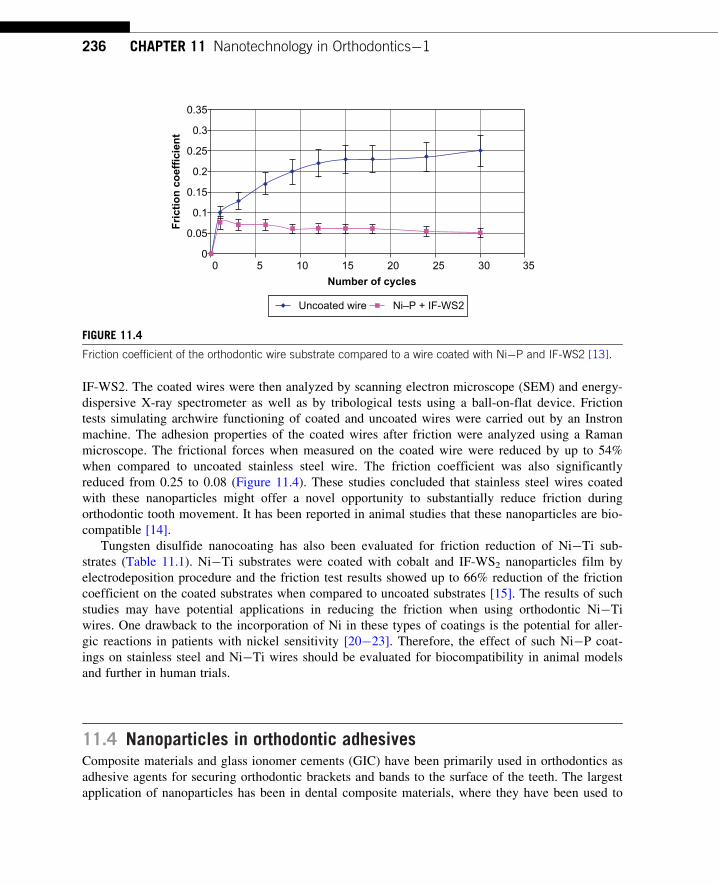
IF-WS2. The coated wires were then analyzed by scanning electron microscope (SEM) and energy-
dispersive X-ray spectrometer as well as by tribological tests using a ball-on-flat device. Friction
tests simulating archwire functioning of coated and uncoated wires were carried out by an Instron
machine. The adhesion properties of the coated wires after friction were analyzed using a Raman
microscope. The frictional forces when measured on the coated wire were reduced by up to 54%
when compared to uncoated stainless steel wire. The friction coefficient was also significantly
reduced from 0.25 to 0.08 (Figure 11.4). These studies concluded that stainless steel wires coated
with these nanoparticles might offer a novel opportunity to substantially reduce friction during
orthodontic tooth movement. It has been reported in animal studies that these nanoparticles are bio-
compatible [14].
Tungsten disulfide nanocoating has also been evaluated for friction reduction of Ni�Ti sub-
strates (Table 11.1). Ni�Ti substrates were coated with cobalt and IF-WS2 nanoparticles film by
electrodeposition procedure and the friction test results showed up to 66% reduction of the friction
coefficient on the coated substrates when compared to uncoated substrates [15]. The results of such
studies may have potential applications in reducing the friction when using orthodontic Ni�Ti
wires. One drawback to the incorporation of Ni in these types of coatings is the potential for aller-
gic reactions in patients with nickel sensitivity [20�23]. Therefore, the effect of such Ni�P coat-
ings on stainless steel and Ni�Ti wires should be evaluated for biocompatibility in animal models
and further in human trials.
11.4 Nanoparticles in orthodontic adhesivesComposite materials and glass ionomer cements (GIC) have been primarily used in orthodontics as
adhesive agents for securing orthodontic brackets and bands to the surface of the teeth. The largest
application of nanoparticles has been in dental composite materials, where they have been used to
00
0.05
Fri
ctio
n c
oef
fici
ent
0.1
0.15
0.2
0.25
0.3
0.35
5 10 15 20 25 30 35
Number of cycles
Uncoated wire Ni–P + IF-WS2
FIGURE 11.4
Friction coefficient of the orthodontic wire substrate compared to a wire coated with Ni�P and IF-WS2 [13].
236 CHAPTER 11 Nanotechnology in Orthodontics�1

Table 11.1 Summary of Studies on Nanoparticle Applications in Materials Used in Orthodontics
Material Studied
Nanoparticle/Nanoscale ImagingTechnique Used
ParametersEvaluated Results
Orthodontic stainlesssteel wire [13,14]
Ni�P film impregnatedwith IF-WS2
nanoparticles
(i) Frictional forcesmeasured on coatedand uncoated wires
(i) Reduced to 54% oncoated wires
(ii) Friction coefficient (ii) Friction coefficientreduced one-third from0.25 to 0.08
Ni�Ti substrates [15] Cobalt and IF-WS2
nanoparticlesFriction coefficient 66% reduction in coated
substrates
Stainless steel, beta-titanium and Ni�Tiarchwires [10]
AFM Surface roughness Surface roughnessinfluenced the effectivenessof sliding mechanics,corrosion behavior, andesthetics
Stainless steel andNi�Ti archwires [11]
AFM coupled withnanoindenter
Effects ofdecontamination andclinical exposure onelastic modulus,hardness, and surfaceroughness
Decontamination regimenand clinical exposure hadno effect on Ni�Ti wiresbut did have a statisticallysignificant effect onstainless steel wires.Decontamination ofstainless steel wiressignificantly increasedsurface hardness (P5 0.01)and reduced the surfaceroughness (P5 0.02)
Conventional stainlesssteel, ceramic, self-ligating stainless steeland ceramicbrackets [12]
AFM Surface roughness 2-year orthodontictreatment regime showedthat self-ligating ceramicbrackets had undergoneless change in roughnessparameters than self-ligating stainless steelbrackets. Self-ligatingceramic brackets exhibitedlow friction and betterbiocompatibility than otherbrackets
Fuji II GIC [16] Nanohydroxy andfluoroapatite, N-vinylpyrrolidone (NVP)-containing polyacids
CS, DTS, BFS Highest values for CS,DTS, and BFS were foundfor NVP-nanoceramicpowder modified cements(184 MPa for CS, 22 MPafor DTS and 33 MPa forBFS)
(Continued )
23711.4 Nanoparticles in orthodontic adhesives

enhance the long-term optical properties by virtue of their small size, and at the same time provide
superior mechanical strength and wear resistance. Ever since the first formulation of composite
resins in the 1960s, the basic compositional triad has been used: monomer, silane-treated filler,
and initiators.
Filler particles improve the mechanical properties of the composite material. The filler used by
Bowen in 1963 [24], consisted of milled quartz particles with average size ranging from 8 to 12 µm(8000�12,000 nm). Due to the esthetic limitations of macrofilled composites (lack of surface
gloss), the minifilled composite was introduced in the 1970s. Improvement in properties such as
tensile and compressive strength (CS), modulus of elasticity, abrasion resistance, radiopacity,
esthetics, and handling was noted with higher filler load. The filler material used was silica parti-
cles of average diameter of 400 nm allowing a maximum filler loading of 55 wt%, with better
polishability, but with a significantly lower mechanical strength [25]. It was not until the 1980s and
1990s that mixtures of filler materials were tested. These hybrid fillers (600�2000 nm) were
Table 11.1 (Continued)
Material Studied
Nanoparticle/Nanoscale ImagingTechnique Used
ParametersEvaluated Results
Nanocomposite material(Filtek Supreme PlusUniversal) and thenanoionomer restorativematerial (Ketact N100Light Curing Nano-Ionomer) and aconventional orthodonticcomposite material(Transbond XT) [17]
Nanofilled compositematerial (Filtek SupremePlus Universal) and GICmodified with nanofillers,and nanofiller “clusters”(Ketact N100 LightCuring Nano-Ionomer)
Shear bond strength Conventional orthodonticcomposite had highershear bond strength thannanocomposite andnanoionomer groups
ECA material and twoconventional adhesives(composite andresin-modified glassionomer) [18]
ECA was modified by theaddition of silicananofillers and silvernanoparticles
Effect of surfacecharacteristics, physicalproperties, andantibacterial activities ofECA against cariogenicStreptococci
ECA had rougher surfacesthan conventionaladhesives due to additionof silver nanoparticles andbacterial adhesion to ECAwas less than toconventional adhesives.No significant difference inshear bond strength andbond failure interfacebetween ECA andconventional adhesiveswere noted
BisGMA/HEMA adhesivemodified with nanogel [19]
Nanogel copolymers at a70:30 molar ratio ofIBMA and either UDMAor BisEMA
Adhesive viscosity, wetmechanical properties,short-term microtensilebond strength
Parameters evaluated wereenhanced by nanogelinclusion in the adhesiveresin
238 CHAPTER 11 Nanotechnology in Orthodontics�1

commercialized as hybrid, microhybrid, and condensable composites [25]. There was an improve-
ment in mechanical strength; however, the polishability was still a limitation. A maximum filler
load from 70 to 77 wt% was recorded then. Microfilled composites (10�100 nm) were not
suitable for high stress bearing areas (e.g., Class I, II, and IV restorations) of the dentition. The par-
ticle sizes of these hybrid composites were not similar to the size of the hydroxyapatite crystal,
dentinal tubule, and enamel rod. There was also a potential for compromise in adhesion between
the macroscopic restorative material and the nanoscopic (1�10 nm in size) tooth structure. So
nanofilled composite materials were introduced.
There are two distinct types of dental nanocomposites currently available: nanofills and nanohy-
brids [26,27]. Nanofills contain nanometer-sized particles (1�100 nm) throughout the resin matrix,
with no other large primary particles included. Nanohybrids consist of larger particles (400�5000 nm)
with added nanometer-sized particles. The use of nanoparticles addresses the aforementioned diffi-
culty by combining high mechanical strength with long-term polish retention in one material.
Another adhesive material used in orthodontics is GIC. GIC is also known as polyalkenoate
cement and contains components of silicate glass and polyacrylic acid [28]. GIC is translucent and
adhesive to tooth structure and has unique properties such as biocompatibility, anticariogenic action
(due to fluoride release), and adhesion to moist tooth structure. In addition, the coefficient of ther-
mal expansion for GIC is low and close to the values of tooth structure. Besides its advantages,
GIC has some disadvantages such as brittleness and inferior mechanical strength. The use of GIC
in orthodontics became popular during the late 1980s due to its fluoride-releasing potential which
made it highly attractive for orthodontic band cementation. It was shown that modifying conven-
tional GIC with nanohydroxy and fluoroapatite (Figure 11.5) and N-vinylpyrrolidone containing
polyacids improved its mechanical properties. When tested, it proved to have greater CS and higher
diametral tensile strength (DTS) and biaxial flexural strength (BFS) than the control group consist-
ing of conventional GIC [16].
Recently, “nanoionomer,” which is resin-modified GIC (Ketact N100 Light Curing Nano-
Ionomer), has been introduced to operative dentistry [29,30]. This light curing nanoionomer is com-
posed of fluoroaluminosilicate glass, nanofillers, and nanofiller “clusters” combined to improve
mechanical properties and high fluoride release. A recent study tested the commercially available
nanocomposite material (Filtek Supreme Plus Universal) and the nanoionomer restorative material
(Ketact N100 Light Curing Nano-Ionomer) for orthodontic bracket bonding [17]. The study evalu-
ated their shear bond strength and failure site locations in comparison with a conventional light-cure
orthodontic bonding adhesive (Transbond XT). Orthodontic brackets should withstand high shearing
force in the oral cavity generated during the orthodontic teeth movement. The results of the study
demonstrated that conventional orthodontic composite had higher shear bond strength than that of
nanocomposite and the nanoionomer groups. The study concluded that nanocomposites and nano-
ionomers may be suitable for bonding but are inferior to conventional orthodontic composites.
In another study, an experimental composite adhesive material (ECA) containing silica nanofil-
lers and silver nanoparticles was compared with two conventional adhesives (composite and resin-
modified glass ionomer) to evaluate the surface characteristics, physical properties, and antibacte-
rial activity against cariogenic Streptococci [18]. Bacterial adhesion was measured by incubating
the adhesive discs for 6 h in saliva sample. The discs were washed with sterile phosphate buffered
saline and the number of adherent cells was determined using a Beckman LS-5000TA liquid scintil-
lation counter. The results of this study showed that ECA had rougher surfaces than conventional
23911.4 Nanoparticles in orthodontic adhesives

adhesives due to the addition of silver nanoparticles. However, the bacterial adhesion to ECA was
less than the conventional adhesives. Bacterial suspension containing ECAs showed slower bacte-
rial growth than those containing conventional adhesives. There was no significant difference
in shear bond strength and bond failure interface between ECA and conventional adhesives evalu-
ated in this study. It can be interpreted from the study that antibacterial nanoparticles can be
(A)
(B)
FIGURE 11.5
SEM of hydroxyapatite (A) and fluoroapatite (B) nanopowders (340,000) [16].
240 CHAPTER 11 Nanotechnology in Orthodontics�1

incorporated into orthodontic adhesive materials to prevent bacterial adhesion and caries during
orthodontic treatment.
One of the current challenges in adhesive dentistry using hybrid materials is over-hydrophilic
bonding formulations, which facilitate water percolation through the hybrid layer resulting in unre-
liable bonded interfaces. A recent study evaluated a nanogel-modified dentin adhesive composed of
BisGMA/HEMA (2,2-bis [4(2-hydroxy-3-methacryloyloxy-propyloxy)-phenyl] propane/2-hydroxy-
ethyl methacrylate) [19]. The nanogel additives of 10- to 100-nm-sized particles with varied hydro-
phobicity were synthesized in solution and added to BisGMA/HEMA. Nanogel copolymers at a
70:30 molar ratio of isobornyl methacrylate (IBMA) and either urethane dimethacrylate (UDMA—
less hydrophobic) or ethoxylated bis-phenol-adimethacrylate (BisEMA—more hydrophobic) was
used. Adhesive viscosity, wet mechanical properties, short-term microtensile bond strength to acid-
etched and primed dentin were studied. All these parameters evaluated were significantly enhanced
in the nanogel-adhesive group over the control group containing just BisGMA/HEMA. These recent
studies reinforce the importance of nanoscale-modified adhesive materials with improved properties
for their use in orthodontics.
11.5 Nanoparticle delivery from orthodontic elastomeric ligaturesFixed orthodontic appliance treatment significantly increases the risk of enamel decalcification and
white spot lesions. These are caused due to prolonged accumulation and retention of bacterial plaque
on the enamel surface adjacent to the attachments (orthodontic brackets and bands). Demineralization
of enamel has been reported to occur around orthodontic brackets within 1 month of bracket
placement in the absence of fluoride supplementation [31,32]. Elastomeric ligature ties have been
conventionally used to hold orthodontic wires securely in the bracket during the treatment pro-
cess. These elastomeric ligatures can serve as a carrier scaffold for delivery of nanoparticles that
can be anticariogenic, antiinflammatory, and/or antibiotic drug molecules embedded in the elasto-
meric matrix. The release of anticariogenic fluoride from elastomeric ligatures has been reported
in the literature previously [33�35]. The studies concluded that the fluoride release was character-
ized by an initial burst of fluoride during the first day and second day, followed by a logarithmic
decrease. For optimum clinical benefit, the fluoride ties should be replaced monthly [33]. These
ties gained weight intraorally with residual, leachable fluoride present in fluoride-impregnated and
non-fluoride elastomeric ligature ties after 1 month of intraoral use, due to imbibition [34].
An in vivo study evaluating the efficacy of fluoride-releasing elastomers in the control of
colony forming units (CFUs) of Streptococcus mutans in the oral cavity concluded that fluoride-
releasing elastomeric ligature ties are not indicated for reducing the incidence of enamel decalcifi-
cation in orthodontic patients. The study found no significant reduction in CFUs in saliva or plaque
around the fluoride-releasing ties when compared with the conventional elastomeric ligature
ties [35]. There are very few published studies evaluating the release and efficacy of nanoparticle-
based anticariogenic, antiinflammatory, analgesic, or antibiotic drug delivery from orthodontic elas-
tomeric ligatures to prevent enamel decalcification, decrease the biofilm accumulation during the
orthodontic treatment, or reduce infections. Medicated wax applied to orthodontic brackets that
slowly and continuously released benzocaine was shown to be significantly more effective in
24111.5 Nanoparticle delivery from orthodontic elastomeric ligatures

reducing pain associated with mucosal irritation caused by brackets [36]. It could be highly advan-
tageous to explore the local delivery of such therapeutic nanoparticles at the site of enamel decalci-
fication, biofilm formation, and gingivitis. As with all new technological applications, however, it
will also be important to carefully evaluate the rate of release of such nanoparticles, ingestion,
biocompatibility, and systemic toxicity level for these new nanotechnology-based applications.
11.6 Developing and future applications of nanotechnology indentistry and orthodonticsThe future of nanotechnology in orthodontics has potential to develop in a number of additional
applications as well including (a) the use of tooth-colored, shape-memory polymers to esthetically
move teeth, (b) tooth movement using orthodontic nanobots that could directly manipulate peri-
odontal tissues allowing rapid and perhaps painless movement, dentifrobots (nanobots in denti-
frices) delivered through mouthwash or toothpaste to patrol supragingival and subgingival surfaces
performing continuous plaque/calculus removal and metabolizing trapped organic matter, and (c)
nanochanges on the surfaces of temporary anchorage devices (TADs) to increase their retention but
still allow them to be removed when no longer needed.
11.6.1 The use of shape-memory polymer in orthodonticsOver the past decade, there has been an increased interest in producing esthetic orthodontic wires
to complement tooth colored brackets. Shape-memory esthetic polymer is an area of potential
research. These are a class of stimuli-responsive materials, which have the capacity to remember a
preprogrammed shape imprinted during the synthesis; can be reformed at a higher temperature to
impart a desired temporary shape; and recover their original shape when influenced by a stimulus,
such as heat, light, or magnetic field [37,38]. Applications of nanoparticles in shape-memory nano-
composite polymers can increase thermal conductivity of the polymers [39,40]. These wires can
also be made with clinically relevant levels of elastic stiffness. Once placed in the mouth, these
polymers can be activated by the body temperature or photoactive nanoparticles activated by light
and thus influence tooth movement. Future research directions in shape-memory nanocomposite
polymers to produce esthetic orthodontic wires can be of interesting potential in orthodontic
biomaterial research.
11.6.2 BioMEMS/NEMS for orthodontic tooth movement and maxillary expansionMicroelectromechanical systems (MEMS) devices are manufactured using similar microfabrication
techniques as those used to create integrated circuits. They often have moving components that
allow a physical or analytical function to be performed by the device in addition to their electrical
functions. The biological MEMS (bioMEMS) are made up of micromachined elements usually on
silicon substrates, including gears, motors, and actuators with linear and rotary motion for applica-
tions to biological systems. Implantable bioMEMS have been used as biosensors for in vivo diag-
nostics of diseases and as drug delivery microchips [41�43]. Nanoelectromechanical systems
(NEMS) are devices integrating electrical and mechanical functionality on the nanoscale level.
242 CHAPTER 11 Nanotechnology in Orthodontics�1

Evidence suggests that orthodontic tooth movement can be enhanced by supplementing the
mechanical forces with electricity [44,45]. Animal experiments indicated that when 15�20 micro-
amperes of low direct current (dc) was applied to the alveolar bone by modifying the bioelectric
potential, osteoblasts and periodontal ligament cells demonstrated increased concentrations of the
second messengers cAMP (adenosine-30,50-cyclic monophosphate) and cGMP (cyclic guanosine
monophosphate). These findings suggest that electric stimulation enhanced cellular enzymatic phos-
phorylation activities, leading to synthetic and secretory processes associated with accelerated bone
remodeling. However, the intraoral source of electricity is a major problem that has to be
addressed.
It has been recently proposed that microfabricated biocatalytic fuel cells (enzyme batteries) can
be used to generate electricity to aid orthodontic tooth movement [46]. An enzymatic microbattery,
when placed on the gingiva near the alveolar bone, might be a possible electrical power source for
accelerating orthodontic tooth movement. It is proposed that this device uses organic compound
(glucose) as the fuel and is noninvasive, and non-osseointegrated. The enzyme battery can be fabri-
cated with the combination of two enzyme electrodes and biocatalysts such as glucose oxidase or
formate dehydrogenase to generate electricity (Figure 11.6) [46]. However, there are several issues
like soft tissue biocompatibility, effect of food with different temperature and pH range on the out-
put of such microfabricated enzyme battery that need to be addressed. The use of microenzyme bat-
teries has issues like enzyme stability, electron transfer rate, and enzyme loading which result in
shorter lifetime and poor power density.
Glucose
Gluconolactone
O2
H2O
FIGURE 11.6
A schematic diagram of an oral biocatalytic fuel cell. In this system, the following reaction generating electricity
for enhancing orthodontic tooth movement occurs: Glucose1O2-gluconolactone1H2O/H2O2 [46].
24311.6 Developing and future applications of nanotechnology

Nanotechnology has offered nanostructured materials like mesoporous media, nanoparticles,
nanofibers, and nanotubes, which have been demonstrated as efficient hosts of enzyme immobiliza-
tion. When nanostructured conductive materials are used, the large surface area of these nanomater-
ials can increase the enzyme loading and facilitate reaction kinetics, and thus improve the power
density of the biofuel cells. It is expected that the MEMS/NEMS based system will be applied over
the next few years to develop biocompatible powerful biofuel cells, which can be safely implanted
in the alveolus of the maxilla or mandible or in the palate to enhance orthodontic tooth movement
or rapid maxillary expansion.
11.6.3 Nanorobot delivery for oral anesthesia and improved oral hygeineIn 2000, Robert A Freitas Jr. [47] suggested that dental nanorobots can be utilized to induce oral
anesthesia. These nanorobots can be controlled by an onboard nanocomputer that executes prepro-
grammed instructions and can be delivered as colloidal suspension containing millions of active
analgesic micrometer-sized nanorobot particles on the patient’s gingiva. The nanorobots might use
specific motility mechanisms to travel through human tissues with navigational precision, acquire
energy, and sense and manipulate their surroundings. They might also achieve safe cytopenetration
and use any of a multitude of techniques to monitor, interrupt, or alter nerve-impulse traffic in indi-
vidual nerve cells [48]. Freitas also proposed that these nanorobots can be guided painlessly
through the gingival sulcus, lamina propria, cementodentinal junction, dentinal tubules, and finally
reach the pulp. Moreover, this journey can be directed by a combination of chemical gradients,
temperature differentials, and even positional navigation, all under the control of the onboard
nanocomputer.
In turn, the nanocomputer would be directed by the dentist to induce anesthesia, for example, in
a specific tooth that requires treatment. More futuristic applications have been proposed by Freitas
on utilizing nanorobots to treat carious lesions, dentin hypersensitivity, and dentifrobots (nanoro-
bots in dentifrices). These could be delivered through mouthwash or toothpaste and could patrol
supra- and subgingival surfaces of teeth, performing continuous plaque/calculus removal and
metabolize trapped organic matter into harmless and odorless vapor [47]. He has also suggested
that orthodontic nanorobots could directly manipulate periodontal tissues allowing rapid, painless
tooth movement and repositioning within minutes to hours. However, it should be noted that
inflammation and bone modeling govern the rate of orthodontic tooth movement. The idea of
achieving orthodontic tooth movement within minutes to hours looks futuristic considering the time
required for the biological adaptation and remodeling processes of the periodontium that accompa-
nies orthodontic treatment.
11.7 Temporary anchorage devicesWhile TADs have become increasingly utilized by orthodontic professionals to assist in the process
of moving teeth, commercially available TADs exhibit a success rate of 60�75% [49�51].
Currently, TADs are manufactured with smooth titanium surfaces (pure titanium or titanium alloy
(Ti�6Al�4V)) because complete osseointegration is a disadvantage that complicates their removal.
On the other hand, lack of osseointegration is also one of the factors for the failure of TADs.
244 CHAPTER 11 Nanotechnology in Orthodontics�1

The success of TADs also depends on other factors like proper initial mechanical stability and
loading quality and quantity. Clinically there are difficulties encountered in the removal of TADs
due to increased osseointegration even on the smoother surface TADs. Therefore, it is postulated
that the balance lies in the fabrication of an ideal surface that could stimulate initial osseointegra-
tion and facilitate its removal once the TAD is no longer needed. Biocompatible coatings like tita-
nium nanotubes should be studied to evaluate if the nanotubular layer can enhance initial
osseointegration and can serve as an interfacial layer between the newly formed bone and the TAD.
11.8 ConclusionsThe applications of nanotechnology and nanoparticles have offered the biomaterial scientists the
opportunity to fabricate materials with improved physical and mechanical properties. The field of
dental biomaterials is constantly evolving. The addition of such nanoparticles to currently available
materials enhances their properties and clinical use. The applications of nanotechnology in ortho-
dontic materials have been reported in the literature over the past few years. However, the number
of studies specifically addressing orthodontics and nanotechnology are small. The authors expect
that the next decade will bring an increase in the amount and quality of research that will help
unveil the next generation of nanomaterials for orthodontic treatment. There are many avenues yet
to be explored that would help move MEMS/NEMS based systems in orthodontics from the draw-
ing board to the bench top and clinical study arena. The utilization of nanoparticles for improving
the TADs should be one such avenue.
References[1] R.P. Feynman, There is plenty of room at the bottom, Eng. Sci. 23 (1960) 22�36.
[2] N. Taniguchi, On the basic concept of ‘NanoTechnology’, Proc. ICPE Tokyo 2 (1974) 18�23.
[3] D. Mijatovic, J.C. Eijkel, A. van den Berg, Technologies for nanofluidic systems: top-down vs. bottom-
up—a review, Lab Chip 5 (5) (2005) 492�500.
[4] M. Lazzari, et al., Self-assembly: a minimalist route to the fabrication of nanomaterials, J. Nanosci.
Nanotechnol. 6 (4) (2006) 892�905.
[5] S.I. Stupp, et al., Supramolecular materials: self-organized nanostructures, Science 276 (5311) (1997)
384�389.
[6] M.C. Roco, Nanotechnology: convergence with modern biology and medicine, Curr. Opin. Biotechnol.
14 (3) (2003) 337�346.
[7] S.K. Sahoo, V. Labhasetwar, Nanotech approaches to drug delivery and imaging, Drug Discov. Today
8 (24) (2003) 1112�1120.
[8] G. Binning, C. Quate, C. Gerber, Atomic force microscope, Phys. Rev. Lett. 12 (1986) 930.
[9] R.P. Kusy, J.Q. Whitley, Effects of surface roughness on the coefficients of friction in model orthodontic
systems, J. Biomech. 23 (9) (1990) 913�925.
[10] C. Bourauel, et al., Surface roughness of orthodontic wires via atomic force microscopy, laser specular
reflectance, and profilometry, Eur. J. Orthod. 20 (1) (1998) 79�92.
[11] J.P. Alcock, et al., Nanoindentation of orthodontic archwires: the effect of decontamination and clinical
use on hardness, elastic modulus and surface roughness, Dent. Mater. 25 (8) (2009) 1039�1043.
245References

[12] G.J. Lee, et al., A quantitative AFM analysis of nano-scale surface roughness in various orthodontic
brackets, Micron 41 (7) (2010) 775�782.
[13] M. Redlich, et al., Improved orthodontic stainless steel wires coated with inorganic fullerene-like nano-
particles of WS(2) impregnated in electroless nickel-phosphorus film, Dent. Mater. 24 (12) (2008)
1640�1646.
[14] A. Katz, M. Redlich, L. Rapoport, H.D. Wagner, R. Tenne, Self-lubricating coatings containing
fullerene-like WS2 nanoparticles for orthodontic wires and other possible medical applications, Triboil.
Lett. 21 (2) (2006) 135�139.
[15] G.R. Samorodnitzky-Naveh, et al., Inorganic fullerene-like tungsten disulfide nanocoating for friction
reduction of nickel-titanium alloys, Nanomedicine (Lond) 4 (8) (2009) 943�950.
[16] A. Moshaverinia, et al., Modification of conventional glass-ionomer cements with N-vinylpyrrolidone
containing polyacids, nano-hydroxy and fluoroapatite to improve mechanical properties, Dent. Mater.
24 (10) (2008) 1381�1390.
[17] T. Uysal, et al., Are nano-composites and nano-ionomers suitable for orthodontic bracket bonding? Eur.
J. Orthod. 32 (1) (2010) 78�82.
[18] S.J. Ahn, et al., Experimental antimicrobial orthodontic adhesives using nanofillers and silver
nanoparticles, Dent. Mater. 25 (2) (2009) 206�213.
[19] R.R. Moraes, et al., Improved dental adhesive formulations based on reactive nanogel additives, J. Dent.
Res. (2011).
[20] G. Rahilly, N. Price, Nickel allergy and orthodontics, J. Orthod. 30 (2) (2003) 171�174.
[21] A.L. Greppi, D.C. Smith, D.G. Woodside, Nickel hypersensitivity reactions in orthodontic patients.
A literature review, Univ. Tor. Dent. J. 3 (1) (1989) 11�14.
[22] R. Lindsten, J. Kurol, Orthodontic appliances in relation to nickel hypersensitivity. A review, J. Orofac.
Orthop. 58 (2) (1997) 100�108.
[23] C.A. Pazzini, et al., Allergy to nickel in orthodontic patients: clinical and histopathologic evaluation,
Gen. Dent. 58 (1) (2010) 58�61.
[24] R.L. Bowen, Properties of a silica-reinforced polymer for dental restorations, J. Am. Dent. Assoc. 66
(1963) 57�64.
[25] J.L. Ferracane, Current trends in dental composites, Crit. Rev. Oral. Biol. Med. 6 (4) (1995) 302�318.
[26] S.B. Mitra, D. Wu, B.N. Holmes, An application of nanotechnology in advanced dental materials, J. Am.
Dent. Assoc. 134 (10) (2003) 1382�1390.
[27] D.A. Terry, Direct applications of a nanocomposite resin system: Part 1—the evolution of contemporary
composite materials, Pract. Proc. Aesthet. Dent. 16 (6) (2004) 417�422.
[28] A.D. Wilson, B.E. Kent, A new translucent cement for dentistry. The glass ionomer cement, Br. Dent. J.
132 (4) (1972) 133�135.
[29] C.M. Killian, T.P. Croll, Nano-ionomer tooth repair in pediatric dentistry, Pediatr. Dent. 32 (7) (2010)
530�535.
[30] Y. Korkmaz, et al., Influence of different conditioning methods on the shear bond strength of novel
light-curing nano-ionomer restorative to enamel and dentin, Lasers Med. Sci. 25 (6) (2010) 861�866.
[31] M.M. O’Reilly, J.D.B. Featherstone, Demineralization and remineralization around orthodontic appli-
ances: an in vivo study, Am. J. Orthod. Dentofacial Orthoped. 92 (1) (1987) 33�40.
[32] B. Øgaard, G. Rølla, J. Arends, Orthodontic appliances and enamel demineralization: Part 1. Lesion
development, Am. J. Orthod. Dentofacial Orthop. 94 (1) (1988) 68�73.
[33] W.A. Wiltshire, Determination of fluoride from fluoride-releasing elastomeric ligature ties, Am. J.
Orthod. Dentofacial Orthop. 110 (4) (1996) 383�387.
[34] W.A. Wiltshire, In vitro and in vivo fluoride release from orthodontic elastomeric ligature ties, Am. J.
Orthod. Dentofacial Orthop. 115 (3) (1999) 288�292.
246 CHAPTER 11 Nanotechnology in Orthodontics�1

[35] K.K. Miura, et al., Anticariogenic effect of fluoride-releasing elastomers in orthodontic patients, Braz.
Oral. Res. 21 (3) (2007) 228�233.
[36] G.T. Kluemper, et al., Efficacy of a wax containing benzocaine in the relief of oral mucosal pain caused
by orthodontic appliances, Am. J. Orthod. Dentofacial Orthop. 122 (4) (2002) 359�365.
[37] S.I. Gunes, S.C. Jana, Shape memory polymers and their nanocomposites: a review of science and tech-
nology of new multifunctional materials, J. Nanosci. Nanotechnol. 8 (4) (2008) 1616�1637.
[38] M.A. Stuart, W.T. Huck, J. Genzer, M. Muller, C. Ober, M. Stamm, et al., Emerging applications of
stimuli-responsive polymer materials, Nat. Mater. 9 (2010) 101�113.
[39] Q. Meng, J. Hu, A review of shape memory polymer composites and blends, Compos. A Appl. Sci.
Manuf. 40 (11) (2009) 1661�1672.
[40] J. Leng, X. Lan, Y. Liu, S. Du, et al., Shape-memory polymers and their composites: stimulus methods
and applications, Prog. Mater. Sci. 56 (7) (2011) 1077�1135.
[41] E.E. Nuxoll, R.A. Siegel, BioMEMS devices for drug delivery, IEEE Eng. Med. Biol. Mag. 28 (1)
(2009) 31�39.
[42] B. Xu, BioMEMS enabled drug delivery, Nanomedicine 1 (2) (2005) 176�177.
[43] P.L. Gourley, Brief overview of BioMicroNano technologies, Biotechnol. Prog. 21 (1) (2005) 2�10.
[44] Z. Davidovitch, M.D. Finkelson, S. Steigman, J.L. Shanfeld, P.C. Montgomery, E. Korostoff, et al.,
Electric currents, bone remodeling, and orthodontic tooth movement. II. Increase in rate of tooth move-
ment and periodontal cyclic nucleotide levels by combined force and electric current, Am. J. Orthod. 77
(1) (1980) 33�47.
[45] Z. Davidovitch, M.D. Finkelson, S. Steigman, J.L. Shanfeld, P.C. Montgomery, E. Korostoff, et al.,
Electric currents, bone remodeling, and orthodontic tooth movement. I. The effect of electric currents on
periodontal cyclic nucleotides, Am. J. Orthod. 77 (1) (1980) 14�32.
[46] J. Kolahi, M. Abrishami, Z. Davidovitch, Microfabricated biocatalytic fuel cells: a new approach to
accelerating the orthodontic tooth movement, Med. Hypotheses 73 (3) (2009) 340�341.
[47] R.A. Freitas Jr., Nanodentistry, J. Am. Dent. Assoc. 131 (11) (2000) 1559�1565.
[48] R.A.J. Freitas, Nanomedicine. Vol. 1. Basic Capabilities, vol. 2011, Landes Bioscience, Georgetown,
TX, 1999.
[49] M. Schatzle, R. Mannchen, M. Zwahlen, N.P. Lang, et al., Survival and failure rates of orthodontic tem-
porary anchorage devices: a systematic review, Clin. Oral. Implants Res. 20 (12) (2009) 1351�1359.
[50] S. Miyawaki, I. Koyama, M. Inoue, K. Mishima, T. Sugahara, T. Takano-Yamamoto, et al., Factors asso-
ciated with the stability of titanium screws placed in the posterior region for orthodontic anchorage, Am.
J. Orthod. Dentofacial Orthop. 124 (4) (2003) 373�378.
[51] S. Kuroda, Y. Sugawara, T. Deguchi, H.M. Kyung, T. Takano-Yamamoto, et al., Clinical use of minis-
crew implants as orthodontic anchorage: success rates and postoperative discomfort, Am. J. Orthod.
Dentofacial Orthop. 131 (1) (2007) 9�15.
247References
CHAPTER
12Nanotechnology inOrthodontics�2: Facts andPossible Future Applications
Tarek El-BialyDepartments of Dentistry and Biomedical Engineering, Division of Orthodontics,
The University of Alberta, Edmonton, AB, Canada
CHAPTER OUTLINE
12.1 Introduction ............................................................................................................................... 249
12.2 Nanoscale in orthodontics ..........................................................................................................250
12.3 Nanotechnology and gene therapy in orthodontics ....................................................................... 251
12.4 Nanofabricated ultrasound device for orthodontics....................................................................... 252
12.5 Nanomechanical sensors for orthodontic forces and moments measurement.................................. 253
12.6 Future applications of nanotechnology in orthodontics ................................................................. 255
12.7 Conclusions............................................................................................................................... 256
Acknowledgment................................................................................................................................. 256
References ......................................................................................................................................... 256
12.1 IntroductionNanotechnology involves the creation, manipulation, and use of materials and devices at the size
scale of , 100 nm. It describes the technique of creating and using devices and components compa-
rable in size to molecules and intracellular architecture [1]. Nanotechnology is well progressing in
many biological sciences including medicine, pharmacy, and dentistry. In general dentistry, nano-
technology has potential to be applied in management of teeth hypersensitivity, anesthesia, produc-
tion of more enhanced dental products, and in orthodontic treatment [2]. This chapter presents the
current status of the use of nanotechnology and nanoanalysis and three possible future applications
in orthodontics. These applications involve nanoscale study of the topography of different ortho-
dontic materials to evaluate their nanocharacteristics or nanomechanical properties. The possibility
of nanogene therapy to enhance generation or to modify growth of different craniofacial structures
is discussed. In addition, a nanofabricated ultrasound device for orthodontics is a future direction
that would change the profile of dentofacial regeneration including possible prevention and treat-
ment of orthodontically induced root resorption or dentoalveolar fracture.
249Nanobiomaterials in Clinical Dentistry.
© 2013 Elsevier Inc. All rights reserved.

12.2 Nanoscale in orthodonticsOne of the known challenges in orthodontics is bond failure of orthodontic attachments includ-
ing orthodontic brackets and tubes. In order to minimize bond failure, many attempts have been
introduced to enhance the strength of orthodontic bonding composites. One of these attempts
was to introduce nanocomposite and nanoionomer [3]. The introduction of nanofiller compo-
nents originally was introduced to enhance some physical properties of the hardened restorative
composites. Because of the decreased dimension of the particles in the nanofillers, a wide size
distribution and increased filler load is achieved, which decreases polymerization shrinkage [4],
and increases mechanical strength and resistance to fracture. In addition, it has been reported
that nanocomposites had a good marginal seal to enamel and dentin compared with total-etch
adhesives [5]. Since these nanofiller-containing resin-modified glass ionomer cements (Ketac
N100) were reported to have improved physical properties, as well as increased fluoride release
than other restorative materials, it has been suggested to be used as a bonding material for ortho-
dontic attachments. The increased fluoride release by nanofiller-bonding materials compared to
other restorative materials make them more attractive in orthodontics since demineralization of
the labial surfaces of teeth during orthodontic therapy is one of the major challenges facing
orthodontists and orthodontic patients, especially in patients with compromised oral hygiene.
However, although the results of using such nanocomposite and nanoionomer bonding system
may be suitable for bonding since they fulfill suggested ranges for clinical acceptability, they
are inferior to a conventional orthodontic composite [3]. There may be ongoing attempts to
enhance bonding strength of these nanocomposite and nanoionomer bonding systems to utilize
their high fluoride release property in order to make them at least comparable in bond strength
to conventional orthodontic bonding systems.
Because of the increased awareness of enamel demineralization around orthodontic attach-
ments, different materials have been used to minimize enamel demineralization. The effective-
ness of these materials has been evaluated using atomic force microscopy (AFM) measurements
that quantitatively evaluate nanoscale enamel surface roughness after using these materials [6]
(Figure 12.1).
Orthodontic treatment may involve removal of some teeth to alleviate dental crowding or to
treat some types of malocclusions. Closing extraction spaces usually requires moving bracketed
teeth along arch wires made from different types of materials using sliding (also known as arch-
guided) tooth movement. The friction between orthodontic brackets and wires, especially with a
combination of metals, sometimes affects the efficiency of tooth movement. Also, friction in ortho-
dontics has contributed to loss of anchorage due to application of increased forces to overcome fric-
tion between the brackets and wires. Increased friction between orthodontic wires and bracket
surfaces has been attributed to micro/nanoasperities or mechanical interlocking at the micro- or
nanolevels [7] (Figure 12.2).
For this purpose, nanotechnology has been introduced to study different orthodontic wires and
bracket slots to evaluate nanomechanical properties and topographic pattern of these materials in
order to understand factors affecting friction between orthodontic wires and brackets. However, the
exact measurements of these micro/nanoasperities have only been evaluated using AFM. AFM
allows for quantitative evaluation of the nanoscale surface roughness of various orthodontic bracket
slots before and after sliding movement of archwire in vitro and in vivo [8].
250 CHAPTER 12 Nanotechnology in Orthodontics�2

12.3 Nanotechnology and gene therapy in orthodonticsMandibular underdevelopment has been attributed to a variable interaction of genetic and environ-
mental factors, which is believed to be difficult to manipulate or stimulate. Bite jumping appli-
ances, also known as functional appliances (FAs), have long been claimed and used to enhance
mandibular growth in cases with deficient mandibles (mandibular retrognathism). A recent study
systematically reviewed reports on the effectiveness of FAs and concluded that the analysis of the
effect of treatment with FAs versus an untreated control group showed skeletal changes that were
statistically significant in the short term but unlikely to be clinically significant [9].
FIGURE 12.2
Because all surfaces have irregularities that are large on a molecular level, real contact occurs only at peaks
of irregularities, called asperities. When interlocking occurs between the peaks and bottoms of the asperities,
resistance to movement occurs [7].
(A) (B) (C) (D)
FIGURE 12.1
AFM of human enamel treated by acid etching only showing narrow grooves (black arrows) and flattened
perikymata ridges (white arrow) with cracks and many destructed areas of nontreated enamel surface (A); enamel
treated with acid etching and fluoride varnish showing moderately wide perikymata groove (black arrow) and
localized areas of destruction (white arrow) (B); enamel treated with acid etching and unfilled sealant group
showing wide perikymata grooves and flattened perikymata ridges (C); and enamel treated with acid etching
followed by proseal showing perikymata ridge and groove with obvious focal holes (frames are 503 50 µm)
From Ref. [6].
25112.3 Nanotechnology and gene therapy in orthodontics

It has been reported that rat condylar growth can be significantly increased by local gene ther-
apy with recombinant adenovirus associated virus (rAAV)-mediated vascular endothelial growth
factor (VEGF), which is an important angiogenic mediator in vascularization and endochondral
ossification [10]. Although rAAV vector for gene delivery is proven to be a strong and effective
vector, its use in human patients is facing controversial and ethical issues. There is a growing
emphasis on nanotechnology in cancer detection and treatment (http://nano.cancer.gov). For exam-
ple, nanovector liposomes have been used successfully in breast cancer therapy [11]. Regardless of
its successful use, nanobiotechnology is still at its early stage of development and its use in treat-
ment of diseases other than cancer could be especially challenging. The challenges facing these
nanovectors might not have viable applications, especially in mandibular growth stimulation and
could end up on the “technology shelf” in the future [12].
12.4 Nanofabricated ultrasound device for orthodonticsIn translational research, proof of concept is usually the first step in testing the viability of new
technology for potential treatment of any disease. We and other researchers have shown in proof of
principle the efficacy of utilizing Low Intensity Pulsed Ultrasound (LIPUS) in stimulating mandib-
ular growth in growing animals and in human patients [13�16]. One of the main challenges we
faced when a pilot clinical trial was conducted to stimulate mandibular growth in humans with
hemifacial microsomia was that the patients (young adults) needed to hold the LIPUS transducers
(applicators) to their mandibular condyles for 20 min every day for at least 1 year in order to
achieve clinical improvement of the deficient side of the mandible. This created a great challenge
and burden on the parents to do this for that extended period of time. In order to minimize errors in
LIPUS application and maximize consistency in treatment, a noncompliant LIPUS application is in
high demand. In addition, we have shown that LIPUS application to orthodontically moving teeth
can minimize root resorption [17]. External apical root resorption (EARR) concurrent with ortho-
dontic treatment is widely accepted as a risk in all types of orthodontic treatment appliances.
Challenges in using LIPUS intraorally in treatment of EARR concurrent with orthodontic treatment
is that the size of commercially available LIPUS transducers are quite large (3.5 cm3), difficult to
adjust, and larger than any human tooth. In addition, the patients have to hold the LIPUS transdu-
cers tightly against the gingiva of the corresponding tooth/teeth for 20 min/day for at least 4 weeks
in order to achieve clinically noticeable decrease in EARR concurrent with orthodontic treatment.
Because of these challenges there are needs for noncompliant LIPUS devices that can be inserted
into the patient’s mouth and deliver the predesigned LIPUS treatment to the tooth/teeth in question.
In order to build an intraoral LIPUS device that is independent of power supply or patient compli-
ance, a nanocircuit design has been incorporated in order to nanofabricate the main operation
circuit as well as LIPUS transducer controller. In addition, a nanofabricated battery is required in a
nanoscale intraoral LIPUS device. The first step in this nanodesigned LIPUS device was the nano-
fabrication of the operation circuit that delivers the required signal to the LIPUS transducer. This
step has been developed by our group and tested for its validity to be potentially used for future
nanofabricated LIPUS transducers and devices [18]. A future nanofabricated LIPUS device is com-
pared to the original large-scaled device in the following figure (Figure 12.3).
252 CHAPTER 12 Nanotechnology in Orthodontics�2

12.5 Nanomechanical sensors for orthodontic forcesand moments measurementOrthodontic forces and moments are an undetermined force system due to many factors that are
involved, including the orthodontic wire materials (that affect its modulus of elasticity) and geome-
try (that affects its stiffness). Both modulus of elasticity and geometry are important in determining
wire stiffness according to the following equation:
K ðstiffnessÞ5E3 I ðE is the modulus of elasticity and I is the area moment of inertiaÞFor round wire (Figure 12.4). Where d is the diameter of the round wire
I5πðd4Þ=64For rectangular wire (Figure 12.5). Dimensions of the rectangular wire where (b) is the wire
base in the in-out direction; and (h) is the wire height in the up-down direction
I5 bðh3Þ=12In addition, wire stiffness is also dependent on the wire length, which is determined intraorally
by the interbracket distance. The smaller the bracket width, the greater the interbracket distance
and lower the wire stiffness according to the following equation:
K ðstiffnessÞα1=L3
where L is the interbracket distance or wire length (Figure 12.6).
All the above factors affect the stiffness of the wire and consequently the force applied to the
teeth. Since any small changes in wire length or diameter/cross section can change the wire stiff-
ness and consequently the applied force by this wire, it is almost impossible to predict the
exact amount of force applied by the same wire to two different patients due to the difference in
(A) (B)
FIGURE 12.3
(A) Large-scaled LIPUS device and (B) future nanofabricated intraoral LIPUS device.
25312.5 Nanomechanical sensors for orthodontic forces

teeth crown widths, resulting in variation in interbracket wire lengths, and consequently applying
different forces using the same type, shape, and size of wire. What complicates this process further
is that the moments applied to the teeth are the resultant of multiplication of the magnitude of force
times the perpendicular distance between the centers of resistance of the tooth to the line of action
of the applied force (Figure 12.7).
FIGURE 12.6
Interbracket distance between the maxillary left canine and maxillary left lateral incisor (yellow line) is less
than that between maxillary left central incisor and maxillary left lateral incisor (blue line). The longer wire
segment is more flexible than the shorter one. (For interpretation of the references to color in this
figure legend, the reader is referred to the web version of this book.)
h
b
FIGURE 12.5
Rectangular wires’ dimension base (b) and height (h).
d
FIGURE 12.4
Diameter of round wires (d).
254 CHAPTER 12 Nanotechnology in Orthodontics�2

Since the teeth root lengths are different among individuals, this consequently changes the
length of the moment arms and the applied moments using similar forces. In order to apply biologi-
cally tolerable forces and moments to the teeth to efficiently move teeth with minimal adverse
effects such as EARR depending on the individual’s intrinsic susceptibility, researchers have been
working to develop brackets that can carry three-dimensional mechanical sensors in the bracket
bases to measure in three dimensions the real-time forces and moments applied to the teeth. This
would facilitate the orthodontist adjusting these forces should they exceed biologically
acceptable limits.
In order to achieve this, microsensors have been proposed in the recent literature. Lapatki et al.
in 2007 reported on the introduction of a “smart” bracket for multidimensional force and moment
measurement [19,20]. They reported on a large-scale prototype bracket that utilized microsystem
chip encapsulation. Development of a nanosystem chip that can be encapsulated into small low-
profile contemporary bracket systems with reduced mesiodistal and occlusogingival dimensions
will allow clinical testing of the utilization of this technology.
12.6 Future applications of nanotechnology in orthodonticsAlthough nanotechnology application in orthodontics is considered to be in its infancy, there is a
huge potential application of nanotechnology in orthodontics including nanodesigned orthodontic
bonding materials, possible nanovector for gene delivery for mandibular growth stimulation, and
nano-LIPUS devices. Also, nanomechanical sensors can be fabricated and be incorporated into the
base of orthodontic brackets and tubes in order to provide real-time feedback about the applied
orthodontic forces. This real-time feedback allows the orthodontist to adjust the applied force to be
within a biological range to efficiently move teeth with minimal side effects. In a fast growing
world of nanotechnology, the hope would be to get these technologies into clinical application
100g�10mm=1000gmm
F=100gX=10mm
FIGURE 12.7
The moment produced by the force (F) equals the magnitude of the force F (100g) multiplied by the
perpendicular distance between the center of resistance and the line of action of the force (X5 10 mm).
25512.6 Future applications of nanotechnology in orthodontics

sooner than later. However, financial burden in developing and applying such technologies is a
road block that requires special funding programs from major funding agencies and organizations.
12.7 ConclusionsIn conclusion, the future in orthodontic treatment will rely mainly on nanotechnology should all the
current attempts succeed to its clinical application at a reasonable cost to the orthodontist and
patients. It is recommended that appropriate research funds be allocated to orthodontic and dentistry
nanotechnology research and development to help take these technologies to the clinical trial
phase.
AcknowledgmentThe author would like to thank all those who provided permission to use figures from their publications in this
chapter.
References[1] M.A. Zarbin, C. Montemagno, J.F. Leary, R. Ritch, Nanomedicine in ophthalmology: the new frontier,
Am. J. Ophthalmol. 150 (2010) 144�162.e2.
[2] F.J. Leary, Nanotechnology: what is it and why is small so big? Can. J. Ophthalmol. 45 (5) (2010)
449�456.
[3] T. Uysal, A. Yagci, B. Uysal, G. Akdogan, Are nano-composites and nano-ionomers suitable for
orthodontic bracket bonding? Eur. J. Orthod. 32 (2010) 78�82.
[4] N. Moszner, U. Salz, New developments of polymeric dental composites, Program. Polym. Sci.
26 (2001) 535�576.
[5] S. Geraldeli, J. Perdigao, Microleakage of a new restorative system in posterior teeth, J. Dent. Res.
(2003) 126 (Abstract).
[6] S.F. Shinaishin, S.A. Ghobashy, T.H. El-Bialy, Efficacy of light-activated sealant on enamel deminerali-
zation in orthodontic patients: an atomic force microscope evaluation, Open. Dent. J. 5 (2011) 179�186.
[7] W. Proffit, Contemporary Orthodontics, fifth ed., 2012, p. 329.
[8] G.J. Leea, K.H. Parkc, Y.G. Parkc, H.K. Park, A quantitative AFM analysis of nanoscale surface rough-
ness in various orthodontic brackets, Micron 41 (2010) 775�782.
[9] E. Marsico, E. Gatto, M. Burrascano, G. Matarese, G. Cordasco, Effectiveness of orthodontic treatment
with functional appliances on mandibular growth in the short term, Am. J. Orthod. Dentofacial Orthop.
139 (1) (2011) 24�36 (Review).
[10] A.B. Rabie, J. Dai, R. Xu, Recombinant AAV-mediated VEGF gene therapy induces mandibular condy-
lar growth, Gene Ther. (2007).
[11] J.W. Park, Liposome-based drug delivery in breast cancer treatment, Breast Cancer Res. 4 (3) (2002)
95�99.
[12] P. Fortina, L.J. Kricka, S. Surrey, P. Grodzinski, Nanobiotechnology: the promise and reality of new
approaches to molecular recognition, Trends Biotechnol. 23 (4) (2005) 168�173.
256 CHAPTER 12 Nanotechnology in Orthodontics�2

[13] R. Oyonarte, M. Zarate, F. Rodriguez, Low-intensity pulsed ultrasound stimulation of condylar growth
in rats, Angle Orthod. 79 (5) (2009) 964�970.
[14] T. El-Bialy, I. El-Shamy, T.M. Graber, Growth modification of the rabbit mandible using therapeutic
ultrasound: is it possible to enhance functional appliance results? Angle Orthod. 73 (6) (2003) 631�639.
[15] T.H. El-Bialy, A. Hassan, T. Albaghdadi, H.A. Fouad, A.R. Maimani, Growth modification of the man-
dible using ultrasound in baboons: a preliminary report, Am. J. Orthod. Dentofacial Orthop. 130 (10)
(2006) 435e7�435e14.
[16] T. El-Bialy, A.H. Hassan, A. Alyamani, T. Albaghdadi, Treatment of hemifacial microsomia by thera-
peutic ultrasound and hybrid functional appliance. A non-surgical approach, Open. Access J. Clin. Trials
2 (2010) 29�36.
[17] T. El-Bialy, I. El-Shamy, T.M. Graber, Repair of orthodontically induced root resorption by ultrasound
in humans, Am. J. Orthod. Dentofacial Orthop. 126 (2) (2004) 186�193.
[18] W.T. Ang, C. Scurtescu, W. Hoy, T. El-Bialy, Y.Y. Tsui, J. Chen, Design and implementation of thera-
peutic ultrasound generating circuit for dental tissue formation and tooth-root healing, IEEE Trans.
Biomed. Circuits Syst. 2 (2010) 49�61.
[19] B.G. Lapatki, O. Paul, Smart brackets for 3D-force-moment measurements in orthodontic research and
therapy—developmental status and prospects, J. Orofac. Orthop. 68 (5) (2007) 377�396.
[20] B.G. Lapatki, J. Bartholomeyczik, P. Ruther, I.E. Jonas, O. Paul, Smart bracket for multi-dimensional
force and moment measurement, J. Dent. Res. 86 (1) (2007) 73�78.
257References

CHAPTER
13Nanoparticle Coating ofOrthodontic Appliances forFriction Reduction
Meir Redlicha and Reshef TennebaNanoMediCOTt (Nanotechnologies for medical and healthcare applications) Israel
bDepartment of Materials Research, Weizmann Institute, Rehovot 76100
CHAPTER OUTLINE
13.1 Introduction ............................................................................................................................... 260
13.2 Friction in orthodontics ..............................................................................................................260
13.3 Materials considerations: fullerene-like nanoparticles ................................................................. 263
13.3.1 Prelude: inorganic fullerene-like nanoparticles of WS2 and MoS2.............................. 263
13.3.2 Synthesis............................................................................................................. 264
13.3.3 Self-lubricating surfaces ....................................................................................... 265
13.4 Orthodontic appliances coated with nanoparticles ....................................................................... 266
13.4.1 Challenges in designing the experimental setup ...................................................... 266
13.4.2 Coating process and tribological measurements....................................................... 267
13.4.2.1 SS wires with IF-WS2 impregnated in electroless nickel�phosphorous film ..... 267
13.4.2.2 SS wires with IF-WS2 impregnated in electrocodeposition nickel film .............. 267
13.4.2.3 Ni�Ti wires with cobalt and IF-WS2 film by electroplating procedure: ............. 267
13.4.3 Coating adhesion and wear .................................................................................... 268
13.4.4 In vitro friction force tests ..................................................................................... 269
13.4.4.1 SS wires with IF-WS2 NP impregnated in electroless Ni�P film ..................... 270
13.4.4.2 SS wires with IF-WS2 impregnated in electrocodeposition of nickel film .......... 272
13.4.4.3 Ni�Ti wires with cobalt and IF-WS2 film by electroplating procedure .............. 272
13.5 Safety: toxicity and biocompatibility............................................................................................ 274
13.6 Conclusions: from the lab to the clinic ........................................................................................ 276
Acknowledgments ............................................................................................................................... 277
References ......................................................................................................................................... 277
259Nanobiomaterials in Clinical Dentistry.
© 2013 Elsevier Inc. All rights reserved.

13.1 IntroductionOrthodontics deals with prevention and correction of malaligned teeth in the jaws as well as the
proper positioning of jaws in the face (dentofacial orthopedics). Esthetics and function are the main
reasons to seek orthodontic treatment. Since the beginning of the twentieth century, orthodontic
tooth movement has been performed by the metal appliances available. These consisted of attach-
ments bonded onto the tooth (brackets) and wires engaged within a specific slot incorporated within
the brackets. This clinical setup is also referred to as fixed orthodontic appliances. Until the
emergence of etching-bonding procedures on the tooth enamel [1,2], metal bands were cemented
onto the teeth. This setup allows the orthodontist to control tooth movement during the entire
course of treatment. In the early 1900s, orthodontists used gold, platinum, silver, steel, gum rubber,
vulcanite, and occasionally zinc, copper, and brass. During the 1950s, stainless steel (SS) was
introduced in orthodontics and became the popular material for making brackets and archwires. In
the early 1970s, nickel�titanium (Ni�Ti) archwire was used for the first time in orthodontics [3]
which originated from the Ni�Ti alloys developed at the Naval Ordnance Laboratory by Buehler [4].
Currently, orthodontists are mostly using SS brackets and archwires made of SS and Ni�Ti metal
alloys with different size and shape (round and rectangular) for archwires. There are also wires made
of cobalt�chromium�nickel and β-titanium, available for use in orthodontics.
In order to initiate orthodontic tooth movement, force has to be applied on the tooth. This force is
exerted either by the archwires engaged into the bracket’s slots or by springs and elastics which are
attached to the brackets or by extra-oral devices such as headgear. Apart from the metallurgical
aspect of orthodontics, teeth movement is entirely dependent on the biological changes occurring
within the tissues encircling the teeth, mainly the periodontal ligament and the alveolar bone.
Consequently, biomechanics is considered as the basis for orthodontic treatment. There are additional
principles which will assure successful orthodontic therapy such as proper diagnosis and treatment
plan, successful bonding, prevention of caries formation and root resorption (shortening the length of
the tooth), patient’s compliance, and retention protocol. However, the level of the orthodontic force
is the core of the treatment. Force magnitude of 40�60 g exerted directly on the tooth for an adequate
continuous period, without the metal fixed appliance setup, is sufficient to move the tooth in the jaw.
However, application of orthodontic force using fixed appliances requires a significant increase in
the force level in order to overcome the friction created at the bracket-wire interface [5].
13.2 Friction in orthodonticsFriction is defined as the force resisting the relative motion of solid surfaces, fluid layers, and
material elements sliding against each other. There are two types of friction: static friction which
occurs between two objects that are not moving relative to each other and it prevents an object
from sliding down, and kinetic friction which occurs when two objects are moving relative to each
other and its value is usually less than the static friction for the same material. In orthodontics, the
kinetic friction is irrelevant because tooth movement is not a continuous motion of the tooth along
the wire. It is a series of tipping and root uprighting movements resulting from the biological
response of the bone, a pseudostatic condition. The orthodontist encounters friction during two
260 CHAPTER 13 Nanoparticle Coating of Orthodontic Appliances
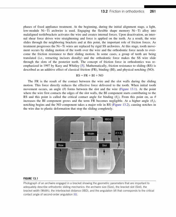
phases of fixed appliance treatment. At the beginning, during the initial alignment stage, a light,
low-module Ni�Ti archwire is used. Engaging the flexible shape memory Ni�Ti alloy into
malaligned teeth/brackets activates the wire and creates internal forces. Upon deactivation, an inter-
nal shear force drives wire straightening and force is applied on the teeth. As a result, the wire
slides through the neighboring brackets and at this point, the important role of friction forces. As
treatment progresses the Ni�Ti wires are replaced by rigid SS archwires. At this stage, tooth move-
ment occurs by sliding motion of the tooth over the wire and the orthodontic force needs to over-
come the friction resistance to their sliding motion. In some cases, a group of teeth are being
translated (i.e., retracting incisors distally) and the orthodontic force makes the SS wire slide
through the slots of the posterior teeth. The concept of friction force in orthodontics was re-
emphasized in 1997 by Kusy and Whitley [5]. Mathematically, friction resistance to sliding (RS) is
described as an additive effect of classical friction (FR), binding (BI), and physical notching (NO).
RS5 FR1BI1NO
The FR is the result of the contact between the wire and the slot walls during the sliding
motion. This force directly reduces the effective force delivered to the tooth. When initial tooth
movement occurs, an angle (θ) forms between the slot and the wire (Figure 13.1). At the point
where the wire first contacts the edges of the slot walls, the BI component starts contributing to the
RS and this point is called the critical contact angle for binding (θc). From this point on, as θincreases the BI component grows and the term FR becomes negligible. At a higher angle (θz),notching begins and the NO component takes a major role in RS (Figure 13.2), causing notches in
the wire due to plastic deformation that stop the sliding completely.
Size
Width
IBD/2
Slot
θ = θc
FIGURE 13.1
Photograph of an archwire engaged in a bracket showing the geometric parameters that are important to
adequately describe orthodontic sliding mechanics: the archwire size (Size), the bracket slot (Slot), the
bracket width (Width), the interbracket distance (IBD), and the angulation (θ) that corresponds to the critical
contact angle of second-order angulation [6].
26113.2 Friction in orthodontics

Following are the number of factors that may influence the RS components directly and
indirectly:
1. The archwire: size, shape, material, surface roughness.
2. The bracket: material, size, and shape of the slot and its edges, surface of slot and the angle
formed between the wire and the slot.
3. Ligation of the wire in the slot: elastic module, metal wire ligature, and self-ligating brackets.
4. Intraoral factors: saliva, plaque, and corrosion.
5. Other factors: distance between teeth and direction of the applied force.
Over the years, attempts have been made by researchers and manufacturers to reduce the
friction. The problem was approached from different aspects:
1. A method for ligating the wire to the slot can theoretically reduce the friction. This was shown
to be true with the self-ligating brackets at a 0 angle (clinically not relevant), but higher friction
was recorded once the wire contacted the slot walls [7�9].
The massive advertisement campaign by most orthodontic manufacturers during the last
decade in favor of the self-ligating brackets to reduce friction and reduce treatment time was
not scientifically and clinically proven [9,10].
2. Surface modification like ion implantation at the wire�bracket interface can minimize friction
during tooth movement. However, limited studies to date have evaluated the frictional
characteristics of wires and brackets which have received this surface treatment [11].
3. Recently, diamond-like carbon (DLC) coatings showed a significant reduction in friction
forces [12]. Such DLC coatings have been also applied on orthodontic wires and brackets,
Angulation, θ (degree)
ActiveConfiguration
PassiveConfiguration
FR FR FR
BIBI
NO
Res
ista
nce
to S
lidin
g, R
S (
cN)
θc θz
FIGURE 13.2
Schematic diagram showing the partition of the resistance to sliding (RS) into classical friction (FR), elastic
binding (BI), and physical notching (NO) within the passive and active configurations with important
boundary conditions (θc and θz) delineated [6].
262 CHAPTER 13 Nanoparticle Coating of Orthodontic Appliances

showing great beneficial effect in terms of reducing friction [13]. However, these experiments
were conducted under dry conditions and the behavior of the DLC coatings in similar wet
environment should be evaluated.
These attempts and many others were successful to some extent but did not fully overcome the
problem. The improvements were mainly directed at the beginning of tooth movement where lower
friction was recorded, but the main impediment of the movement is encountered at the angles
where BI and NO are the components mainly responsible for the RS. Hence significant reduction
of friction at BI might reduce the excessive orthodontic force. Overcoming this inevitable frictional
force means using excessive orthodontic force than what is actually needed to move a tooth. Some
investigators found that 40�60% of the orthodontic force is aimed at overcoming the frictional
resistance.
The use of excessive orthodontic forces has several disadvantages especially on the anchor unit
usually desired to remain stable during treatment and it might increase the risk of root resorption.
It is assumed that the reduction in frictional resistance could enhance the alignment and the space
closure and therefore could lead to reduced treatment time. Thus, reducing frictional forces during
orthodontic tooth movement will significantly contribute to successful treatment outcome.
13.3 Materials considerations: fullerene-like nanoparticlesThe search for material technologies may lead to significant reduction of friction coefficient in
orthodontic devices, self-lubricating coatings have been contemplated and a new technology for
applying these coatings on the wires was thus developed. To explain this technology, the concept
of inorganic fullerene-like (IF) nanoparticles (NP) and inorganic nanotubes (INT) is described
below.
13.3.1 Prelude: inorganic fullerene-like nanoparticles of WS2 and MoS2Hollow closed-cage carbon structures, the fullerenes (C60) and carbon nanotubes are known for
some-time. Research into similar structures from other (inorganic) layered compounds started soon
after. Thus, IF NP and INT of tungsten disulfide (WS2) first and subsequently of molybdenum
disulfide (MoS2) were discovered in 1992 [14�16], and elicited considerable interest ever since in
this emerging field [17�21]. This observation is surprising in view of the fact that the chemical
bond is not stable beyond a few angstroms and hence structures with hollow spaces of a few nano-
meter and above were initially thought to be unfavorable. The formation of such hollow closed
cages can be attributed to the inherent instability of the planar nanostructures of layered
compounds. In graphite, the carbon atoms are bonded in flat sp2 bonds forming a hexagonal
network (Figure 13.3A). The graphene sheets are stacked together via weak van der Waals forces.
In the case of MS2 (Figure 13.3B), where M stands for a metal atom like molybdenum or tungsten,
the molecular sheet is made up of a layer of M atoms sandwiched between two outer sulfur layers.
Each M atom binds to six sulfur atoms forming a lattice with trigonal biprism (octahedral) coordi-
nation. In analogy to graphite, weak van der Waals forces are responsible for the stacking of the
S�M�S layers together. Therefore, these compounds are highly anisotropic with respect to many
26313.3 Materials considerations: fullerene-like nanoparticles

of their physical and chemical properties. The basal (van der Waals) surfaces of the crystal,
which are perpendicular to the c-axis, consist of sulfur atoms that form bonds to three underlying
W/Mo atoms. These sulfur atoms are chemically inert. However, rim W (or Mo) and S atoms
(Figure 13.3), that is, atoms on edge of the layer, which are abundant in the nanostructure, are only
four- and twofold bonded, respectively, making the planar form unstable and forcing it to fold and
close on itself. Therefore, by folding the molecular sheet and stitching the rim atoms together,
seamless and stable nanotubular (one dimensional) and spherical (zero-dimensional fullerene-like)
structures with all W/Mo and S atoms being six- and threefold bonded, respectively, are produced
[14�17]. Initially, only the transition metal chalcogenides of WS2 and MoS2 were known in the
form of closed-cage structures and nanotubes. However, over the years this family has been
expanded considerably and it now encompasses a large number of other compounds like oxides,
hydroxides, nitrides, chlorides, sulfides, selenides, and even pure elements like bismuth, arsine, and
phosphorus.
13.3.2 SynthesisThe most useful method for the synthesis of IF-WS2 NP and WS2 nanotubes (INT-WS2) is the
sulfidization of WO3 NP at elevated temperatures (850�C) [22�25]. Thereafter the initial route for
large-scale synthesis of IF NP and INT of WS2 involved the use of oxides as the starting materials.
After a clear understanding of the growth mechanism was established, large-scale synthesis of
IF-WS2 NP and INT-WS2 was realized by using the fluidized-bed reactor (FBR) [23�25], which
has significant advantages over the previous synthetic approaches. It resulted in IF NP with a more
perfect crystalline structure compared with the product of the previous synthetic systems (which
used horizontal reactors). This observation can be attributed to the fact that much of the reaction
takes place in the gas phase, where an isotropic environment for the reaction prevails. The vertical
posture of the oven allows continuous addition of oxide powder into the chamber during the
reaction, thereby increasing the output of the reactor. Moreover, the fluidized-bed concept lends
itself to further scale-up and to produce larger amounts of a pure IF/INT phases with little or no
compromise on the quality of the synthesized NP.
(B)
Rim atoms
(A)
FIGURE 13.3
Schematic presentation of (A) graphite (the C-atoms in purple) and (B) MoS2 with the Mo atoms in purple
and sulfur atoms in yellow.
264 CHAPTER 13 Nanoparticle Coating of Orthodontic Appliances

The synthesis of IF-MoS2 NP from MoO3 powder was found to be more complex. The reason
for these difficulties was the high volatility of the oxide powder above 700�C. Following a con-
certed effort to elucidate the growth mechanism of IF-MoS2 NP [26], a new vertical (FBR-like)
reactor was erected and used [26], which allowed synthesis of about 0.5 g/day of high-quality IF-
MoS2 NP. Figure 13.4A and B shows typical scanning electron microscope (SEM) and transmission
electron microscope (TEM) images of such NP, respectively. The NP assume the form of oblate
structure having many (.20) closed walls and small hollow core. The diameter of such NP spans
in the range of 60�150 nm with mean value of about 80 nm. When added to lubricating fluids,
these NP were found to exhibit excellent tribological behavior [27,28].
A more recent accomplishment was the synthesis of rhenium (Re) doped IF/INT NP [29].
Rhenium, being one-column to the right of W (Mo) on the periodic table, has five valence electrons
in its outer shell compared to four for molybdenum or tungsten. Thus, substituting about 100 Re
atoms in the IF-MoS2 nanoparticle (B106 atoms) induces negative surface charge on the nanoparti-
cle surface. Such NP behave quite differently from the undoped NP. For example, the doped NP
disperse well and form stable suspensions in various fluids. They also exhibit enhanced conductiv-
ity and may find electronic applications in the future. Most importantly, adding small amounts of
such NP to lubricating fluids (poly-alpha olephin: type 4, i.e., PAO-4) leads to a precipitous reduc-
tion in the friction and wear [29,30] as shown in Figure 13.5. These findings make the IF NP and
particularly the doped ones, very suitable for some medical applications, as explained below.
13.3.3 Self-lubricating surfacesThe idea of making self-lubricating surfaces, i.e., hard surfaces which do not require any fluid to
lubricate the contact area, is not new and has been pursued by many groups before; see for example
(A)(B)
FIGURE 13.4
(A) SEM micrograph of assortment of IF-MoS2 nanoparticles (scale bar is 200 nm); (B) TEM micrograph of
one such nanoparticle (scale bar 5 nm). Note the (nested) closed MoS2 layers.
26513.3 Materials considerations: fullerene-like nanoparticles

Refs. [31�34]. Therefore, it was almost natural, once the IF NP were known to have superior solid
lubrication behavior, to try to incorporate them in metallic coatings. This would be expected to
endow such films the genuine behavior of a very hard coating which is nonetheless also self-
lubricating. A series of articles, where electroless or electroplating baths containing IF NP are used
to prepare self-lubricating surfaces, were thus reported quite recently [35�38]. Different kinds of
films and substrates were used. While their potential applications go beyond dentistry or medical
devices, the focus of much of the present authors was in this direction.
13.4 Orthodontic appliances coated with nanoparticles13.4.1 Challenges in designing the experimental setupThe study of medical devices in a materials laboratory, like the present one requires special consid-
erations. Once a medical issue addressable with the present technology has been identified
and evaluated, the next most intricate issue is to design a working model to be used for the experi-
ments. This working model should reflect the salient features of the medical problem being studied.
00.01
0.02
0.03
0.04
0.05
0.06
0.06
0.07
0.08
Co
eff
icie
nt
of
fric
tio
n
25 50 75 100 125 150 175 200 225
Pure PAO-4oil
8.40E-07
8.15E-08
7.80E-08 0.04–0.05
0.04
Wear rate
(mm3/Nm)
Coeff.of
friction
0.0154.70E-08
0.07–0.08
2H-MoS2plateletsUndopedIF-MoS2 NPRe (0.12 at%):IF-MoS2 NP
250
Time (offset) (min)
2H-MoS2 platelets
Undoped IF-MoS2 NP
Re (0.12 at%):IF-MoS2 NP
Pure PAO-4 oil
FIGURE 13.5
Friction coefficient of different lubricating fluids based on PAO-4: pure purple; formulated with 2H-MoS2(dark brown); IF-MoS2 (undoped—light brown) and Re-doped IF-MoS2 (green). Wear rate of the different
lubricants are shown in the inset table. (For interpretation of the references to color in this figure legend,
the reader is referred to the web version of this book.)
266 CHAPTER 13 Nanoparticle Coating of Orthodontic Appliances

At the same time, the studied model should conform to the measurement techniques used in
materials science laboratories like electron microscopes, X-ray diffraction, tensile tests, and pin on
disk friction testing device. This dilemma was encountered in all the measurements reported here
and beyond. In fact, for most of the experiments done in the present laboratory, the time spent on
designing, constructing, and readapting the experimental models was by far the longest compared
to the data collection and analysis.
13.4.2 Coating process and tribological measurements13.4.2.1 SS wires with IF-WS2 impregnated in electroless nickel�phosphorous filmOrthodontic wires (Ormco, California 0.0193 0.025 in.2 rectangular SS) were coated by electroless
process with a uniform and smooth nickel�phosphorous (Ni�P) film using 100 mL solution
(ENPLATE Ni-425, Enthone Inc.) [35]. The orthodontic wires (or SS plate substrate) were inserted
into the electroless Ni�P bath (88�C, pH5 4.8, magnetic stirring) for 30 min. The plating
resulted in a shiny smooth Ni�P layer. To another electroless solution, 200 mg of agglomerated
IF-WS2 powder with average particle size of 120 nm was added together with a cationic surfactant
(cetyl-trimethylammoniumbromide: CTAB). A short (1 min) sonication (Sonifaier 150, Branson-30
Watts) was used to disperse the agglomerates and ensure the stability of the suspension. A special
procedure was developed to deposit a uniform and relatively smooth composite Ni�P1 IF film
and secure its adequate adherence to the underlying archwire. Following hydorfluoric acid etching,
a pure Ni film was electron-beam deposited onto the substrate; then Ni�P film was deposited from
the electroless solution. Finally, a composite Ni�P1 IF film was deposited. In several cases, the
coated samples were annealed in nitrogen gas atmosphere at 400�C.
13.4.2.2 SS wires with IF-WS2 impregnated in electrocodeposition nickel filmThe IF-WS2 NP were dispersed in an aqueous solution of nickel salt (NiCl23 6H2O) and plated on
a wire with a cross section of 0.0193 0.025 in2 and made of SS 110 mm in length (provided
by Ormco Corporation, Orange, CA) [38]. The coating was produced by the electrochemical
codeposition process with a direct current of 0.02 A and exposure of 20 min for every wire piece at
25�C. To minimize the agglomeration of the IF NP in the bath, a combination of ultrasonic treat-
ment and the CTAB surfactant was used.
13.4.2.3 Ni�Ti wires with cobalt and IF-WS2 film by electroplating procedureNi�Ti (Nitinol) wires were supplied by the following companies: Orthonol (CO, USA); Ormco
(CA, USA), Nitinol Classic (CA, USA); Dentsply (NY, USA). Codeposition of cobalt and IF-WS2NP film was carried out from a cobalt chloride1 IF solution buffered by boric acid and CTAB as
surfactant [39]. The film was deposited at 55�C by galvanostatic plating with a current density
of 15 mA/cm2. In order to assure proper adhesion of the film to the substrate, Ti (10 nm) and
Ni (20 nm) films were deposited onto the cleaned Ni�Ti wires by electron-beam evaporation prior
to the electrodeposition process. Uniformity of the coating was achieved by rotating the sample.
The effects of cobalt coatings of endodontic files with impregnated fullerene-like WS2 NP on
file fatigue and failure were also examined. Dynamic X-ray diffraction (XRD), nanoindentation,
and torque measurements all indicate a significant improvement in the fatigue resistance and
consequently delay in the time to breakage of the coated files. This observation was attributed to
26713.4 Orthodontic appliances coated with nanoparticles

the reduced friction between the coated file and the surrounding. These methods are possibly also
applicable to a variety of other Ni�Ti-based medical devices where fatigue and consequent failure
are of relevance [38]. Figure 13.6 shows the SEM/EDS (energy dispersive X-ray analysis mounted
on scanning electron microscope) analysis of such coatings. A uniform coating a few microns thick
was thus obtained. The atomic ratio of W/Co B12.5 indicates that substantial amount of IF-WS2NP was incorporated into these coatings. The surface is rather rough which suggests that the parti-
tion of the IF NP along the Co-film coating is possibly not uniform and the film surface is enriched
with respect to the NP. XRD of the film (Figure 13.7) shows again a distinct (0002) peak of the
IF-WS2 at 14.2�.
13.4.3 Coating adhesion and wearThe adhesion of the cobalt coating to the Ni�Ti orthodontic wires was determined by inspecting
the folded wire, which showed excellent integrity of the film (see Figure 13.8).
Figure 13.9 shows a scratch test of the Co1 IF coating with nanoindenter. The test is done by
profiling the surface of the Ni�Ti file surface coated with Co1 IF film. This is followed
by scratching it with increasing loads up to a maximal value of 30 mN. As a test of the quality of the
coated surface, a final surface profiling shows little variation with respect to the original file surface.
(A)
Element Atomic%
Cl
Cl
WO
Ti
Ti
S
W W W
0 2 4 6 8 10
Co
Co
Co
O
S
Cl
Ti
Co
W
28.41
7.00
(B) (C)
1.78
2.91
55.38
4.52
FIGURE 13.6
SEM/EDS analysis of the electrodeposited cobalt film onto Ni�Ti surface.
268 CHAPTER 13 Nanoparticle Coating of Orthodontic Appliances

13.4.4 In vitro friction force testsThe validity and the significance of the results obtained in an experiment of friction force rely
on the resemblance of the in vitro setup of the bracket and wire to the relevant clinical
situation. However, the numerous in situ factors affecting orthodontic tooth movement cannot be
2000
1500
1000
500
010 20 30 40 50 60 70 80 90 100 110 120
2-Theta (degree)
NiTi plate
NiTi plate + Co + IF
130
FIGURE 13.7
Diffraction patterns of Ni�Ti plate and a plate coated with Co1 IF-WS2 film.
FIGURE 13.8
Electron micrograph of a Ni�Ti wire after adhesion test of the coating. The straight wire was coated with
Co1 IF film and then folded, exhibiting excellent adhesion as demonstrated in the figure. The high-
magnification micrograph of the film not seen shows a uniform film Co1 IF.
26913.4 Orthodontic appliances coated with nanoparticles

fully duplicated in the laboratory. Nevertheless, these experiments were conducted in various best-
fit models.
The following sections describe various experimental friction models used to evaluate the effect
of IF coatings of orthodontic wires on the friction force.
13.4.4.1 SS wires with IF-WS2 NP impregnated in electroless Ni�P filmIn the first set of experiments, a system which simulated the sliding of a tooth along an archwire,
described previously by Redlich et al. [40] was utilized. Upper incisor SS brackets were bonded to
aluminum plates by a bracket-mounting apparatus. This apparatus ensured the accurate and similar
positioning of the brackets on the plates. The plates were then connected to the base of a universal
mechanical testing apparatus (Instron 4502) through a device with three different notches angulated
at 0�, 5�, and 10� to the long axis of the device (Figure 13.10). Angulations represent the contact
angle between the wire and bracket during the movement of the tooth.
In this setup, the tensile tester was set to move the bracket down along the wire at a constant
speed of 10 mm/min to a distance of 5 mm. The test begins with a steady increase in the force and
reaches a maximum when movement begins on the wire. This maximum force represents the static
friction and it is the force that is of interest in this case. A run-in period was needed before testing
the coated wires with Instron. The run-in was carried out by repeated back and forth movements of
the wire in a bracket slot before connecting the wire to the Instron. A new bracket was used for
each testing. The highest angle was tested in the dry and wet mode. Deionized water was used to
simulate the wet conditions in the mouth. Table 13.1 summarizes the results of the mechanical
1000
−1000
−2000
−300020 60 100
Pen
etra
tion
curv
e w
ith r
ough
ness
(nm
)
Scratch distance (µm)
140
2
3e
d
c
1
180 220 260
0
FIGURE 13.9
Scratch test performed using the nanoindenter showing an almost complete recovery of the Co1 IF-coated
file after release of the load. (1) Surface profile of the pristine surface, (2) scratch test with increasing force,
up to 30 mN, and (3) surface profiling after the pressure release.
270 CHAPTER 13 Nanoparticle Coating of Orthodontic Appliances

measurements. Each data point is an average of five different measurements. The results of these
series of experiments are summarized in Table 13.1.
The mechanism by which the reduced friction of the coated wire is achieved can be explained
by the models suggested in Refs. [36] and [42]. At the first stage, when there is no angle between
the slot and wire, the IF NP act as spacers and reduce the number of metal asperities that come into
contact, resulting in a lower coefficient of friction. As the angle grows, the load at the edges of the
Bracketat 10° slot
FIGURE 13.10
A photograph showing the device used to mount the brackets and orthodontic wires onto the Instron setup
for the simulation of the archwire functioning in the mouth. About 12-cm segments of the orthodontic wires
(coated and uncoated) were attached, on their upper part, to a 10-Newton load cell and the lower end was
connected to a 150 g weight. The wires were then inserted into the slots in the brackets and ligated with an
elastomeric module to all four wings of the brackets with 10� angulation. The 150 g weight was used to
restraighten the wire following its insertion into the bracket similar to the clinical situation [41].
Table 13.1 Summary of the Results of the Tribological Tests of SS Orthodontic Wires (N6SD)
Angle/Coating 0�� 5�� 10��
Noncoated wire 1.326 0.12 2.9560.09 4.006 0.19 dry
3.356 0.21 wet
Ni�P1 IF-coated wire 1.106 0.06 1.5860.25 1.856 0.21 dry
1.576 0.23 wet
27113.4 Orthodontic appliances coated with nanoparticles

slot increases causing a higher friction at the surface of uncoated wire. It is probably at this point
on the coated wire that the release of a few IF NP from the film into the tribological interface and
their rolling and exfoliation occurs, resulting in the formation of a solid lubricant film on the slid-
ing wire. The higher load at this point brings the asperities of the mating surfaces in straight contact
causing the fluid (saliva in the mouth) to be squeezed out of the gap between the wire and slot,
relying on the excellent tribological behavior of the solid lubricant film to allow the sliding of the
archwire. When the two materials are made of SS, as is the case with the uncoated wire a high fric-
tion coefficient is obtained. The presence of WS2 nanosheets at the interface under high loads leads
to a very facile sliding between these sheets thereby reducing the coefficient of friction.
13.4.4.2 SS wires with IF-WS2 impregnated in electrocodeposition of nickel filmCoated and uncoated SS rectangular archwires were engaged into brackets, which were attached
to a special device set at 10� angulations to the wire (Figure 13.10). The wire is connected on
the top to the load gauge and on the bottom to the 150 g (1.5 N) weight. The device is connected
to the base of a testing machine and pulled down at a constant speed of 10 mm/min to a distance
of 5 mm. The tests results showed (Figure 13.11) that friction forces between the rectangular
archwire and the self-ligating bracket were reduced on the coated wire by up to 60% compared
to the uncoated at the 10� angulation. Therefore, coated SS archwires with a composite Ni1 IF-
WS2 offers an opportunity to substantially reduce the friction force during orthodontic tooth
movement [41].
13.4.4.3 Ni�Ti wires with cobalt and IF-WS2 film by electroplating procedureFriction measurements of the coated Ni�Ti wires were made using a device designed to simulate
sliding movement within a bracket system assembled to a Twin Column Testing Machine LR10K
with 10 kN load cell Instron system [43,44] (Figure 13.12). The simulation device consisted of a
horizontal mobile plate bearing SS brackets. The tested Ni�Ti rectangular wire (7 cm long),
0
Fri
cti
on
fo
rce (
g)
150
200
250
300
400
350
1 2 3 4 5Test cycle number
6 7 8 9 10
Ni + IF-WS2 coating Without coating Ni coating
FIGURE 13.11
Dependence of the friction force between bracket and SS archwire on the test cycle number (10� angulation).
272 CHAPTER 13 Nanoparticle Coating of Orthodontic Appliances

vertically positioned was slotted through the moveable bracket and connected to a load cell on one
end and to a counterweight mass of 1.5 N on the other. The friction force was measured as the
tested wire was drawn up with a constant speed of 5 mm/min while slotted in the bracket for a dis-
tance of 5 mm. The bracket was set throughout the experiment at the same position using utility
fork designed for this manner and bonded to the mobile plate using dental resin cement. In order to
simulate different levels of frictional force, the mobile plate was moved horizontally creating angu-
lations of 2� and 3.8� between the wire and the bracket. Tests were repeated 10 times for each
angle both with coated and uncoated wires. Data were obtained and analyzed using NEXYGENt
MT Materials Test and Data Analysis Software.
Results of the friction measurements using the Instron system are given in Table 13.2. Friction
measurements in 2� angle showed a reduction of 20% in the static coefficient and about 30% of the
Double interbracket distance(A) (B)
Wire
HookLoad cell
Bracket
5.3
mm
ele
vatio
n
Pas
sive
rel
ease
FIGURE 13.12
(A) Three-point bending test scheme; (B) a photograph of the assembling device with two brackets mounted
on its upper surface; the wire is engaged into the brackets and elevated to a given height.
Table 13.2 Summary of the Friction Measurements of Cobalt1 IF-Coated Ni�Ti Wires
WireInstantaneousForce (N)
StaticCoefficient
KineticForce (N)
KineticCoefficient
Ni�Ti uncoated (2�) 1.5416 0.3 0.1036 0.02 1.5556 0.3 0.1036 0.02
Ni�Ti coated (2�) 1.2536 0.2 0.0836 0.01 1.1546 0.2 0.0776 0.01
Percentage of friction forcereduction (2�)
19 20 26 30
Ni�Ti uncoated (3.8�) 1.6406 0.2 0.1096 0.02 1.4876 0.2 0.0996 0.01
Ni�Ti coated (3.8�) 1.2906 0.3 0.0866 0.02 0.9906 0.2 0.0666 0.01
Percentage of friction forcereduction (3.8�)
21 22 34 34
Ni�Ti coated (5�) 1.2076 0.4 0.0806 0.02 0.9276 0.2 0.0616 0.01
27313.4 Orthodontic appliances coated with nanoparticles

kinetic coefficient of friction. When enlarging the distance between the bracket and the long axis of
the Instron system, thus creating a contact angle of 3.8� and simulating superior load applied onto
the wire, a decrease in both static and kinetic coefficients was noted at the coated wire. This
decline in static and kinetic coefficients was of 22% and 34%, respectively, with respect to the
uncoated wire. In an additional series of measurements made only on the Co1 IF coated wire in a
contact angle of 5�, a static coefficient of 0.08 and kinetic friction coefficient of 0.061 were
observed. The trend of the frictional force reduction as the load rises sustained throughout the entire
series of experiments. The unique coating of IF-WS2 NP embedded in cobalt matrix demonstrated
a significant friction reduction of the Ni�Ti alloy.
The reduction in the friction as a result of the Co/IF-WS2 coatings can be attributed to the release
of minute amounts of IF-WS2 NP which are impregnated in the cobalt coating. WS2 and MoS2 are
known to provide efficient lubrication even in bulk (platelets) form. These kind of compounds are
typified by the strong covalent bonds between sulfur and metal (tungsten) atoms within each plane
while only weak van der Waals forces exist between planes; thus, low interplanar shear strength
exists and permits sliding of two adjacent planes [31�34]. The WS2 NP prevent asperity contact
between the bracket and wire surfaces. Their round shape suggests that a rolling friction scenario is
also possible in this case. Furthermore, elastic deformations of the NP augment their resilience and
diminish the energy dissipation associated with friction and wear under the load [16]. In addition,
the IF-WS2 NP act as a protection against oxidation of the metal surface [37]; hence, it could be
suggested that the coating also reduces or even obstructs nickel (cobalt) release, which is a known
allergen, from the Ni�Ti wire.
13.5 Safety: toxicity and biocompatibilityThe ongoing emergence and spread of nanoscale man-made materials for medical applications
aroused the awareness of the scientists, the regulatory agencies, and the public to their safety.
Safety, in terms of health-care use is determined by both nontoxic response and biocompatibility.
Briefly, toxicity is the degree to which the material can harm humans and animals. The local and
systemic responses of the host to the mode and the period of material exposure are evaluated [45].
Biocompatibility has recently been defined as the ability of a biomaterial to perform its desired
function with respect to a medical therapy, without eliciting any undesirable local or systemic
effects in the recipient or beneficiary of that therapy, but generating the most appropriate benefi-
cial cellular or tissue response in that specific situation, and optimizing its clinically relevant
performance [46].
The toxicity of the IF-WS2 NP, manufactured by “NanoMaterials,” for industrial applications,
was tested at various modes of exposure by three authorized laboratories. The results showed no
apparent toxic effect of IF-WS2 after oral administration, dermal application, and inhalation tests in
rats [47�49]. Toxicity tests, initially performed on animals, conducted in compliance with the
OECD (Organisation for Economic Co-operation and Development) Principles of Good Laboratory
Practice, are a prerequisite for further biocompatibility evaluations toward medical use. These tests
are done on the as-prepared NP while biocompatibility of the medical device will be evaluated
afterward in conjunction with all the other components comprising the NP coating of the device.
274 CHAPTER 13 Nanoparticle Coating of Orthodontic Appliances

Recently, two acute toxicity tests (oral and dermal) using the Re-doped IF-MoS2 NP were con-
ducted [50,51]. The objective of the first study was to assess the acute oral toxicity of Re-doped
IF-MoS2 NP (Re:IF-MoS2) following single administration of 2000 mg/kg, by oral gavage to mice,
using prespecified fix doses, based on the identification of the dose(s) mortality and/or moribund
status of the animals. The results showed no mortality in any of the animals throughout the entire
14-day study period. No noticeable clinical signs in reaction to dosing were evident in any of the
animals. Mean group body weight gain at the end of the 14-day study period was noted in all
groups. No gross pathological findings were evident in any of the animals at the time of their
scheduled necropsy. Histopathologic findings revealed no treatment related changes in all the
organs examined. Based on the lack of observed adverse reactions Re:IF-MoS2 may be regarded as
not causing acute toxicity risk through this route of administration and is classified as hazard
Category 5 according to the Globally Harmonized Classification System [50] (Figures 13.13 and
13.14).
A second acute dermal toxicity of Re-doped MoS2 was assessed on the basis of the testing proce-
dure recommended by the OECD Guideline for the Testing of Chemicals. A single limit dose level
corresponding to 2000 mg/kg of Re:IF-MoS2 was topically applied for 24-h exposure duration to
mice, for the purpose of evaluating health hazards likely to arise from short-term exposure in consid-
eration of its projected use as a coating on medical devices. The results showed no mortality prior to
the scheduled sacrifice. No clinical signs were observed in any of the animals at the immediate time
postdosing. The body weight gain of all animals at the end of the 14-day observation period was
within the normal limits. No abnormalities were detected in any of the animals at the time of their
scheduled necropsy, at 14-days postdosing. Based on the results obtained following a single dermal
application of Re:IF-MoS2 dose level corresponding to 2000 mg/kg, it may be concluded that
Re-doped MoS2 NP do not represent an acute toxic risk by this route of administration [51].
FIGURE 13.13
Liver centrilobular region. No abnormality detected 14 days after single administration of 2000 mg/kg of
Re-doped IF-MoS2 nanoparticles (Hematoxylin and eosin (H&E),320).
27513.5 Safety: toxicity and biocompatibility

In addition to these animal tests, recent reports demonstrated no cytotoxic effect of IF NP on the
vitality and proliferation rate of human culture cells. Wu [52] et al. tested the effect of MoS2 NP dis-
persed within cell medium, on two types of human cells: A549 cells (lung adenocarcinoma cells) and
K562 cells (leukemic cells). The results showed that the MoS2 NP were reasonably nontoxic and bio-
compatible up to the given concentrations. Furthermore, there was no obvious change of the
morphologies of A549 cells treated with MoS2 NP for 2 days even at the highest concentration.
Another study [53] examined the effect of WS2 nanotubes of (INT-WS2) on human salivary
gland cell line. The cells were cultured and subjected to three different concentrations of INT-WS2with and without INT-WS2. Cell viability, growth, and morphologic features were examined until
reaching cell confluence. The results showed no significant differences between all the various cell
concentrations groups. The authors concluded that INT-WS2 do not affect salivary gland cells’
viability, growth rate, or morphology.
13.6 Conclusions: from the lab to the clinicThe route of the archwires and the brackets, as well as other similar dental and medical devices,
coated with IF-NP from the laboratory bench to the orthodontic patients is a multi-stage process.
This route starts with extensive laboratory work to choose the appropriate IF NP for the coating
and subsequently determining the coating process. Thereafter, comprehensive in vitro tests are to
be conducted, using the proper stimulatory models, to validate the efficacy of the IF-coated ortho-
dontic materials.
The next stage is pursuing biocompatibility approval from the relevant regulatory agencies.
At this stage, the product will be available for initial clinical evaluation and finally commercial
manufacturing and sales in the orthodontic market.
FIGURE 13.14
Lung section. No abnormality detected 14 days after single administration of 2000 of Re-doped IF-MoS2nanoparticles mg/kg (H&E, 320).
276 CHAPTER 13 Nanoparticle Coating of Orthodontic Appliances

AcknowledgmentsWe are grateful to “NanoMaterials” Ltd. for supplying the nanopowders. Collaboration with the following peo-
ple is gratefully acknowledged: Dr. Alon Katz, Dr. Gili Naveh, Mrs. Adi Ram Adini, Prof. Abraham Nyska,
Dr. Doron Aframian, Dr. Rita Rosentsveig, and Mrs. Lena Yadgarov. R.T. gratefully acknowledges the support
of ERC (project INTIF 226639), the Israel Science Foundation, the Harold Perlman Foundation, The GMJ
Schmidt Minerva Center for supramolecular chemistry, and the Irving and Cherna Moskowitz Center for Nano
and Bio-Nano Imaging. R.T. is the Drake Family Chair in Nanotechnology and director of the Helen and
Martin Kimmel Center for Nanoscale Science.
References[1] M.G. Buonocore, A simple method of increasing the adhesion of acrylic filling materials to enamel
surface, J. Dent. Res. 34 (1955) 849�853.
[2] G.V. Newman, Bonding plastic orthodontic attachments to tooth enamel, N. J. Dent. Soc. 35 (1964)
346�358.
[3] G.F. Andreasen, H. Bigelow, J.G. Andrews, 55 Nitinol wire: force developed as a function of “elastic
memory”, Aust. Dent. J. 24 (1979) 146�149.
[4] W.J. Buehler, J.V. Gilfrich, R.C. Wiley, Effects of temperature phase changes on the mechanical proper-
ties of alloys near composition NiTi, J. Appl. Phys. 34 (1963) 1475�1484.
[5] R.P. Kusy, J.Q. Whitley, Friction between different wire-bracket configurations and materials, Semin.
Orthod. 3 (1997) 166�177.
[6] R.P. Kusy, Ongoing innovations in biomechanics and materials for the new millennium, Angle Orthod.
70 (2000) 366�376.
[7] M. Hain, A. Dhopatkar, P. Rock, The effect of ligation method on friction in sliding mechanics, Am. J.
Orthod. Dentofacial Orthop. 123 (2003) 416�422.
[8] G.A. Thorstenson, R.P. Kusy, Resistance to sliding of self-ligating brackets versus conventional stainless
steel twin brackets with second-order angulation in the dry and wet (saliva) states, Am. J. Orthod.
Dentofacial Orthop. 120 (2001) 361�370.
[9] P. Scott, A.T. DiBiase, M. Sherriff, M.T. Cobourne, Alignment efficiency of Damon3 self-ligating and
conventional orthodontic bracket systems: a randomized clinical trial, Am. J. Orthod. Dentofacial
Orthop. 134 (2008) 470e1�470e8.
[10] S.J. Burrow, Friction resistance to sliding in orthodontics: a critical review, Am. J. Orthod. Dentofacial
Orthop. 135 (2009) 442�447.
[11] A. Wichelhaus, M. Geserick, R. Hibst, F.G. Sander, The effect of surface treatment and clinical use on
friction in NiTi orthodontic wires, Dent. Mater. 21 (2005) 938�945.
[12] M. Zolgharni, B.J. Jones, R. Bulpett, A.W. Anson, J. Franks, Energy efficiency improvements in dry
drilling with optimized diamond-like carbon coatings, Diamond Relat. Mater. 17 (2008) 1733�1737.
[13] T. Muguruma, M. Iijima, W.A. Brantley, I. Mizoguchi, Effects of a diamond-like carbon coating on the
frictional properties of orthodontic wires, Angle Orthod. 81 (2011) 143�150.
[14] R. Tenne, L. Margulis, M. Genut, G. Hodes, Polyhedral and cylindrical structures of tungsten disulphide,
Nature 360 (1992) 444�446.
[15] L. Margulis, G. Salitra, R. Tenne, M. Talianker, Nested fullerene-like structures, Nature 365 (1993)
113�114.
[16] Y. Feldman, E. Wasserman, D.J. Srolovitz, R. Tenne, High-rate, gas-phase growth of MoS2 nested
inorganic fullerenes and nanotubes, Science 267 (1995) 222�226.
277References

[17] R. Tenne, Inorganic nanotubes and fullerene-like materials, Nat. Nanotechnol. 1 (2006) 103�111.
[18] H.J. Fan, U. Goesele, M. Zacharias, Formation of nanotubes and hollow nanoparticles based on
Kirkendall and diffusion processes: a review, Small 3 (2007) 1660�1671.
[19] A. Enyashin, S. Gemming, G. Seifert, Nanosized allotropes of molybdenum disulfide, Eur. Phys. J. 149
(2007) 103�125.
[20] C.N.R. Rao, A. Govindaraj, Synthesis of inorganic nanotubes, Adv. Mater. 21 (2009) 4208�4233.
[21] M.N. Tahir, A. Yella, J.K. Sahoo, H. Annal-Therese, N. Zink, W. Tremel, Synthesis and functionaliza-
tion of chalcogenide nanotubes, Phys. Status Solidi B 247 (2010) 2338�2363.
[22] Y. Feldman, G.L. Frey, M. Homyonfer, V. Lyakhovitskaya, L. Margulis, H. Cohen, et al., Bulk synthesis
of inorganic fullerene-like MS2 (M5Mo, W) from the respective trioxides and the reaction mechanism,
J. Am. Chem. Soc. 118 (1996) 5362�5367.
[23] Y. Feldman, A. Zak, R. Popovitz-Biro, R. Tenne, New reactor for production of tungsten disulfide
onion-like (inorganic fullerene-like) nanoparticles, Solid State Sci. 2 (2000) 663�672.
[24] R. Rosentsveig, A. Margolin, Y. Feldman, R. Popovitz-Biro, R. Tenne, WS2 nanotube bundles and foils,
Chem. Mater. 14 (2002) 471�473.
[25] A. Zak, L. Sallacan-Ecker, A. Margolin, M. Genut, R. Tenne, Insight into the growth mechanism of
WS2 nanotubes in the scaled-up fluidized bed reactor, Nano 4 (2009) 91�98.
[26] A. Zak, Y. Feldman, V. Alperovich, R. Rosentsveig, R. Tenne, Growth mechanism of MoS2 fullerene-
like nanoparticles by the gas phase synthesis, J. Am. Chem. Soc. 122 (2000) 11108�11116.
[27] R. Rosentsveig, A. Margolin, A. Gorodnev, R. Popovitz-Biro, Y. Feldman, L. Rapoport, et al., Synthesis of
fullerene-like MoS2 nanoparticles and their tribological behavior, J. Mater. Chem. 19 (2009) 4368�4374.
[28] R. Rosentsveig, R. Tenne, A. Gorodnev, N. Feuerstein, H. Friedman, N. Fleischer, et al., Fullerene-like
MoS2 nanoparticles and their tribological behavior, Tribol. Lett. 36 (2009) 175�182.
[29] L. Yadgarov, R. Rosentsveig, G. Leitus, A. Albu-Yaron, A. Moshkovith, V. Perfilyev, et al., Controlled
doping of MS2 (M5W, Mo) nanotubes and fullerene-like nanoparticles, Angew. Chem. 51 (2012)
1148�1151.
[30] L. Rapoport, A. Moshkovith, V. Perfiliev, A. Laikhtman, I. Lapsker, L. Yadgarov, et al., High lubricity
of Re-doped fullerene-like MoS2 nanoparticles, Tribol. Lett. 45 (2012) 257�264.
[31] P.D. Fleischauer, J.R. Lince, A comparison of oxidation and oxygen substitution in MoS2 solid film
lubricants, Tribol. Int. 32 (1999) 627�636.
[32] M. Chhowalla, G.A.J. Amaratunga, Thin films of fullerene-like MoS2 nanoparticles with ultra-low
friction and wear, Nature 407 (2000) 164�167.
[33] A.A. Voevodin, J.S. Zabinski, Supertough wear-resistant coatings with “chameleon” surface adaptation,
Thin Solid Films 370 (2000) 223�231.
[34] I.L. Singer, S. Fayeulle, P.D. Ehni, Wear behavior of triode-sputtered MoS2 coatings in dry sliding
contact with steel and ceramics, Wear 195 (1996) 7�20.
[35] W.X. Chen, Z.D. Xu, R. Tenne, R. Rosenstveig, W.L. Chen, H.Y. Gan, et al., Wear and friction of
Ni�P electroless composite coating including inorganic fullerene-like WS2 nanoparticles, Adv. Eng.
Mater. 4 (2002) 686�690.
[36] A. Katz, M. Redlich, L. Rapoport, H.D. Wagner, R. Tenne, Self-lubricating coatings containing
fullerene-like WS2 nanoparticles for orthodontic wires and other possible medical applications, Tribol.
Lett. 21 (2006) 135�139.
[37] H. Friedman, O. Eidelman, Y. Feldman, A. Moshkovith, V. Perfiliev, L. Rapoport, et al., Fabrication of
self-lubricating cobalt coatings on metal surfaces, Nanotechnology 18 (2007) 1�8.
[38] A.R. Adini, Y. Feldman, S.R. Cohen, L. Rapoport, A. Moshkovith, M. Redlich, et al., Alleviating
incidental and fatigue-related failure of NiTi root canal files by self-lubricating coatings, J. Mater. Res.
26 (2011) 1234�1242.
278 CHAPTER 13 Nanoparticle Coating of Orthodontic Appliances

[39] G.R. Samorodnitzky-Naveh, M. Redlich, L. Rapoport, Y. Feldman, R. Tenne, Inorganic fullerene-like
tungsten disulphide nanocoating for friction reduction of nickel�titanium alloys, Nanomedicine 4 (2009)
943�950.
[40] M. Redlich, A. Gorodnev, Y. Feldmean, I. Kaplan-Ashiri, R. Tenne, N. Fleischer, et al., Friction reduc-
tion and wear resistance of electro-co-deposited inorganic fullerene-like WS2 coating for improved
orthodontic stainless steel wires, J. Mater. Res. 23 (2008) 2909�2915.
[41] M. Redlich, Y. Mayer, D. Harari, I. Lewinstein, In vitro study of frictional forces during sliding
mechanics of “reduced-friction” brackets, Am. J. Orthod. Dentofacial Orthop. 124 (2003) 69�73.
[42] (a) L. Cizaire, B. Vacher, T. Le Mogne, J.M. Martin, L. Rapoport, A. Margolin, et al., Mechanisms of
ultra-low friction by hollow inorganic fullerene-like MoS2 nanoparticles, Surf. Coat. Technol. 160
(2002) 282�287. (b) L. Joly-Pottuz, F. Dassenoy, M. Belin, B. Vacher, J.M. Martin, N. Fleischer,
Ultralow-friction and wear properties of IF-WS2 under boundary lubrication, Tribol. Lett. 18 (2005)
477�485.
[43] N. Reznikov, G. Har-Zion, Y. Abed, I. Barkana, M. Redlich, Measurement of friction forces between
stainless steel wires and “reduced friction” self-ligating brackets, Am. J. Orthod. Dentofacial Orthop.
138 (2010) 330�338.
[44] N. Reznikov, G. Har-Zion, Y. Abed, I. Barkana, M. Redlich, Influence of friction resistance on
superelastic properties of initial NiTi wires in “reduced friction” and conventional bracket systems,
J. Dent. Biomech. (2010) 613142.
[45] S. Singh, H.S. Nalwa, Nanotechnology and health safety—toxicity and risk assessment of nanostructure
materials on human health, J. Nanosci. Nanotech. 7 (2007) 3048�3070.
[46] D.F. Williams, On the mechanism of biocompatibility, Biomaterial 29 (2008) 2941�2953.
[47] H. Tsabari, Inorganic fullerene-like nano-spheres (IF-WS2): acute oral toxicity, acute toxic class method
in the rat, Final report, Batch no. HP6, Harlan Biotech, Israel, 2005.
[48] I. Haist, Test for sensitization (local lymph node assay—LLNA) with inorganic fullerene-like WS2nanospheres, Project no. 052052, BSL Bioservice, Germany, 2005.
[49] G.E. Moore, Acute inhalation toxicity study in rats�limit test, Product safety laboratories, Study
no. 18503, Dayton, NJ, 2006.
[50] B. Nachshon, Single dose response acute oral toxicity in the mouse, IF-MO (Re) S2, Batch
no. 200910�2, Harlan Biotech, Israel, 2011.
[51] B. Nachshon, Acute dermal toxicity in mice, IF-MO(Re)S2, Batch No. 241111�1, Harlan Biotech,
Israel, 2012.
[52] H. Wu, R. Yang, B. Song, Q. Han, J. Li, Y. Zhang, et al., Biocompatible inorganic fullerene-like
molybdenum disulfide nanoparticles produced by pulsed laser ablation in water, ACS Nano 5 (2011)
1276�1281.
[53] E. Goldman, A. Zak, R. Tenne, Y. Neumann, A. Palmon, A.H. Hovav, et al., Biocompatibility examina-
tion of inorganic nanotubes (INT-WS2) on A5 salivary gland cell line, NanoIsrael, Third International
Nanotechnology Conference, Poster presentation no. 135, 2012.
279References

CHAPTER
14Characterization of SilverNanoparticles IncorporatedAcrylic-Based Tissue Conditionerwith Antimicrobial Effectand Cytocompatibility
Ki Young Nama and Chul Jae LeebaDepartment of Dentistry, College of Medicine, Keimyung University, Daegu, Korea
bSchool of Chemical Industry, Yeungnam College of Science & Technology, Daegu, Korea
CHAPTER OUTLINE
14.1 Introduction ............................................................................................................................... 283
14.2 Preparation and identification of silver nanoparticles................................................................... 285
14.3 Acrylic tissue conditioner combined with silver nanoparticles ...................................................... 287
14.3.1 Fabrication of Ag�-tissue conditioner composites..................................................... 287
14.3.2 In vitro antimicrobial effects on S. aureus, Streptococcus mutans, and C. albicans .... 287
14.3.3 Cytotoxic test on human gingival cell line ............................................................... 289
14.4 Characterization of Ag�-tissue conditioner composites ................................................................. 289
14.4.1 Determination of eluted Ag1 from the specimens .................................................... 289
14.4.2 Microstructure of Ag�-tissue conditioner composites................................................ 291
14.5 Conclusions............................................................................................................................... 292
References ......................................................................................................................................... 293
14.1 IntroductionTissue conditioners have been commonly used to enhance the recovery of denture-bearing tissues
from trauma, damage, or residual ridge resorption usually caused by ill-fitting dentures (Figures 14.1
and 14.2). However, these materials are degenerated and are easily degradable with time. The sur-
faces of acrylic gel polymer are susceptible to colonization by microorganisms [1], and microbial
growth from the adherence of microbial cells is promoted by its rough surface and adhesive interac-
tions between Candida species and oral bacteria, mostly Candida albicans (C. albicans) and oral
283Nanobiomaterials in Clinical Dentistry.
© 2013 Elsevier Inc. All rights reserved.

FIGURE 14.1
Clinical application of acrylic-based tissue conditioner on maxillary full denture.
FIGURE 14.2
Denture-induced stomatitis related with fungal infection on palate and vestibule of maxilla.
284 CHAPTER 14 Characterization of Silver Nanoparticles

streptococci [2,3]. Moreover, Staphylococcus aureus (S. aureus), giving rise to pharyngeal and respi-
ratory infections, has been isolated from dentures and the oral cavity in elderly patients with
decreased immunological activity [4,5]. Therefore, the maintenance of tissue conditioner and the
prevention of the accumulation of microorganisms on such materials are of great importance. Tissue
conditioners could be kept clean by mechanical and chemical methods. However, it is also known
that these methods can cause considerable damage to tissue conditioner [6,7] and to some geriatric
or hospitalized patients, even denture cleansing might be compromised owing to cognitive
impairment, reduced motor dexterity, and memory loss. Though systemic or local antibiotic agents
have been prescribed for eliminating the microbial population, considering the microbial resistance
and the increase in health-care cost, the research on latent antimicrobial material may be needed
[8,9]. Several in vitro and in vivo studies have shown the beneficial effects of antimicrobial agents
combined in tissue conditioners [10�12]. However, bacteria may induce stomatitis [13,14] and no
potentially effective and persistent antimicrobial agent that can be incorporated in tissue conditioners
has yet been developed. Silver (Ag) has been well known for its antimicrobial properties and has a
long history of application in medicine with well-tolerated tissue response and low toxicity profile
[15]. Ag is more toxic than many other metals against a broad spectrum of sessile bacteria and fungi
which colonize on plastic surfaces [16,17]. Such antibacterial characteristics of Ag has drawn atten-
tion recently due to the emergence of antibiotic resistant bacteria as a result of overuse of antibiotics
and far lower propensity to induce microbial resistance than antibiotics. Ag-contained materials have
been already used in various medical fields, such as in vascular graft, central venous catheter, and
wound dressing [18,19]. Particularly, silver nanoparticles (Ag�), the nanosized (nm) inorganic parti-
cle form of Ag, with its rapid and broad-spectrum efficacy and sustained release of silver cation
(Ag1) [18,19] appear to be more effective means of prophylaxis than microsized (µm) Ag powder
which shows lower antimicrobial activity owing to its limited surface [19,20].
Evaluating the biocompatibility of an antimicrobial material is an essential step toward the clini-
cal application of the material in addition to testing its physical properties. Through minute elution
of Ag1, the cations may accumulate in oral epithelial cells or when large areas of oral mucosa are
exposed to Ag� compounds over an extended period, it can possibly cause argyria or disruption of
normal microflora [21,22].
The following sections of this chapter discuss the in vitro (i) synthesis of Ag� dental acrylic gel
polymer and (ii) the antimicrobial effect and cytocompatibility of the modified acrylic tissue condi-
tioner containing Ag�.
14.2 Preparation and identification of silver nanoparticlesAg� was prepared using the following procedure. Aqueous silver sol was prepared with 10.0 mM of
analytical grade AgNO3 in distilled water and 2.0% polyvinyl pyrrolidon was used as stabilizer. All
solutions were deaerated by bubbling with argon gas for 1 h and then were irradiated in the field of
20KGy 60Co Gamma-ray sources. The transmission electron microscope (TEM) image of prepared
Ag� nanoparticles shows that the average size of the Ag� is about 50�80 nm (Figure 14.3). The
UV-vis spectrum of the Ag� is shown in Figure 14.4, the peak at 409.7 nm is the surface plasmon
band of the Ag� synthesized by the gamma irradiation reduction.
28514.2 Preparation and identification of silver nanoparticles

FIGURE 14.3
TEM view of a prepared Ag� used in this study. The average size of silver nanoparticles is approximately
50�80 nm.
2000.0
0.5
1.0
1.5409.7 nm
2.0
Ab
so
rban
ce
2.5
3.0
300 400 500
Wavenumber (nm)
600 700 800
FIGURE 14.4
UV-vis spectrum of Ag� prepared by the gamma irradiation reduction method.
286 CHAPTER 14 Characterization of Silver Nanoparticles

14.3 Acrylic tissue conditioner combined with silver nanoparticles14.3.1 Fabrication of Ag�-tissue conditioner compositesThe acrylic tissue conditioner selected for this study was Soft-Liners supplied as powder and
liquid. Colloidal Ag� was preliminary combined and homogenized with the conditioner liquid in a
sterile glass beaker at concentrations ranging from 0 (control), 0.1, 0.5, 1.0, and 2.0% (vol/vol%:
colloidal Ag�/conditioner liquid), respectively. The conditioner powder was immediately added and
mixed for 30 s at designated powder/liquid ratio by manufacturer’s instruction. In order to fabricate
samples into uniform shape with regular surface, the mixed paste of conditioner was poured onto a
custom-made brass mold with dimensions of 20 mm diameter 33.0 mm depth. The mixed paste
was sandwiched between glass slides until it was solidified under humid condition. Before micro-
bial assay, all samples were sterilized with ethylene oxide gas for 24 h to ensure their initial
sterility.
14.3.2 In vitro antimicrobial effects on S. aureus, Streptococcus mutans,and C. albicansThree standard strains of microorganisms were used: S. aureus (ATCC 6538), S. mutans (ATCC
10449), and C. albicans (ATCC 14053). These microbial species tested are currently recommended
to test the efficacy of antiseptic drugs [23]. S. aureus, a pathogen causing respiratory infections,
has often been isolated from dentures and the oral cavity [4,24], and dentures have recently been
reported to be a carriage of this pathogen [25]. S. mutans has been associated closely with the
pathogenesis of dental caries, which is of limited clinical significance for denture wearers [26].
However, extensive plaque formation on denture might also contribute to the decay of residual
natural teeth and can cause inflammation of gingival tissue adjacent to the denture [26]. C. albicans
has been regularly isolated, suggesting a pathogenic association between bacteria and fungi related
with denture-induced stomatitis.
Microbial suspensions were obtained from single colony isolated on agar plates and inoculated
in appropriate broth for overnight cultures. Bacterial strains were grown in brain�heart infusion
(BHI) broth and plated on agar plates at 37�C while C. albicans strain was grown in Schaedler
broth and plated on agar plates at 30�C. After incubating microbial cells at 37�C overnight, optical
density (OD) of the microbial suspension at 600 nm was adjusted to 1.0 using a spectrophoto-
meter. The suspension was diluted with phosphate-buffered saline (pH 7.4) to 1:100 and suspended
to final concentration of 1.03 107 cells/mL [14,27]. Each disc sample of Ag�-tissue conditioner
and control were placed on multiwell culture plate with 22.1 mm diameter. Initial microbial
suspensions (100 µL) in 1.0 mL of Sabouraud broth were inoculated to each well and incubated
at 37�C. A small volume of microbial suspension (100 µL) was used and the microbes were
adjusted to the stationary phase to be suspended in broth. The oral microbe would appear to be in
a stationary phase rather than in growing phase, when the nutrition is limited in the presence of
antibodies and antimicrobial enzymes in the oral cavity [28]. The assays with samples immersed
in large suspension volume could not reproduce in vivo tissue conditioner which is closely in con-
tact with the gingival mucosa [11]. Microbes in suspension (planktonic phase) are sensitive to
lower antiseptic concentrations than microbes colonizing surfaces protected by a biofilm [29].
28714.3 Acrylic tissue conditioner combined with silver nanoparticles

After an incubation period of 24 h and extended 72 h, suspension (100 µL) was withdrawn, viable
cells colony forming unit (CFU) in the suspension was determined by using the spread plate
method at a level of detection within 500 CFU per plate through serial dilution. As no standard of
the antimicrobial effect on dental material has been established so far, the concept of minimum
bactericidal concentration of antibiotics was adopted as the antimicrobial concentration at more
than 99.9% elimination of the microbes [30,31].
The antimicrobial effects of Ag�-tissue conditioner against S. aureus, S. mutans, and C. albicans
were demonstrated as the mean viable cells after 24 and 72 h of incubation (Table 14.1). When
compared to initial CFU at 0 h, control group (0% Ag�) did not exhibit any microbial inhibitory
effect against all of the strains, though the tissue conditioner itself possesses antimicrobial effects
due to ingredients such as plasticizers and ethanol [32]. Some studies have shown that the antimi-
crobial effect of these ingredients was at variance and has little antimicrobial effect [1,33]. For two
bacterial strains, S. aureus and S. mutans, Ag� incorporated samples showed the minimal bacteri-
cidal effects at the dose above 0.1% and no viable cells were detected from the conditions of 1.0%
above. For fungal strain C. albicans, the minimal fungicidal concentration was shown at the dose
of above 0.5% and no CFU was detected in 2.0%. It is reported that Ag and Ag-based compounds
are highly toxic to prokaryotic cells showing strong biocidal effects on bacterial species [17], while
Ag showed less effect on eukaryotic cells such as mold and yeasts [34]. The mechanism of antimi-
crobial effect of silver supported compound has not been fully explained yet. It was suggested that
as a result of catalytic action of Ag, oxygen is converted into active oxygen (including hydroxyl
Table 14.1 Antimicrobial Effect of Ag�-Tissue Conditioner Samples (0�2.0%) Against S. aureus,
S. mutans, and C. albicans at 24 and 72 h of Incubation Periods
StrainaIncubationPeriod (h)
Ag� Dose (Vol%)
0 (Control) 0.1 0.5 1.0 2.0
24 5.43 108 7.13 103 486 (66) 0 0
S. aureus 6 (63 107) 6 (53 102) 126 (4) 0 0
(107) 72 8.73 107 9.73 102
6 (23 106) 6 (53 102)
24 1.23 107 3.63 103 306 (75) 0 0
S. mutans 6 (33 106) 6 (63 102) 76 (11) 0 0
(107) 72 3.53 106 5.43 102
6 (43 105) 6 (43 102)
24 4.33 107 2.63 105 2.23 102 106 (25) 0
C. albicans 6 (93 106) 6 (93104) 6 (33 102) 206 (34) 0
(107) 72 5.23 107 5.53 104 1.23 102
6 (93 106) 6 (93103) 6 (75)
Results are expressed in mean CFU with standard deviation (sd). Data in bold represent values statistically different fromcontrol (P, 0.01).aStarting inoculums: 107 CFU.
288 CHAPTER 14 Characterization of Silver Nanoparticles

radicals) by the action of light energy and/or H2O in the air or water only at polar surfaces [19].
These active oxygen radicals caused the structural damage in bacteria and lead to the damage or
even the death of the microorganisms, the so-called “oligodynamic action of silver” [19,35].
14.3.3 Cytotoxic test on human gingival cell lineHuman gingival fibroblasts (HGF; ATCC 2014) were used to evaluate the cytotoxicity of Ag�-tissueconditioner by MTT (tetrazolium-based 3-(4,5-dimethylthiazol-2-yl)-2,5 diphenyltetrazolium
bromide) assay. The cells were grown and subcultured for 24 h at 37�C in an atmosphere of 5%
CO2 in air and 100% relative humidity. HGF cell suspension was prepared at a concentration of
43 104 cells/mL and dispensed (180 µL/well) onto 96-well cell-culture plates. The multiwell plates
were incubated at 37�C and 5% CO2 in air for 24 h. The culture medium was removed from the
wells and 20 µL of the extracts from experimental Ag�-tissue conditioner (0%, 0.1%, 0.5%, 1.0%,
and 2.0%) were added into each well and incubated for 2, 8, 24, 48, and 72 h at 37�C and 5% CO2 in
air. Then, 10% MTT (100 µL) was added to each well and kept in a dark environment for 4 h at
37�C and after MTT was aspirated, 100 µL of dimethyl sulfoxide was added to each well. OD for
each group was measured using ELISA reader under the absorbance at 570 nm. Cell viability (CV
(%)) was calculated using the following equation:
Cvð%Þ5 Sample optical density
Control optical density3 100
and cytotoxicity was evaluated by the cell viability as highly cytotoxic when less than 25% and
less than 50�25% were qualified as moderately cytotoxic [36].
It is generally accepted that Ag� express their toxicity through the generation of reactive oxygen
species (ROS) [37]. Increased cellular ROS levels are associated with the induction of genetically pro-
grammed cell death (apoptosis), as well as necrotic cell death in several cell lines [38]. Kim et al. [39]
reported the subchronic oral toxicity and tissue accumulation of Ag� in rats for 90 days and highlighted
that Ag� have significant toxic effects on cells that primarily occur in a dose-dependent manner.
The results indicated that 2.0% Ag� group was expressed as highly cytotoxic effect to HGF at 24
and 72 h incubation periods while control (0%) and 0.1% and 0.5% groups were graded as noncyto-
toxic at all incubation periods even though the 0.5% group showed significant decrease in cell viability
at 24 and 72 h over the control group (Figure 14.5). Phthalates and other esters of aromatic carboxylic
acids are used as plasticizers in Soft-Liners and they have been reported to elute from the conditioners
during masticatory force and to possibly be toxic [40,41]. However, control group, an acrylic tissue
conditioner “Soft-Liners” without Ag� revealed no cytotoxic effect to HGF at all incubation periods.
This result could be related to in vitro, static experimental condition for tested samples unlike normal
functioning of tissue conditioner in oral cavity [41].
14.4 Characterization of Ag�-tissue conditioner composites14.4.1 Determination of eluted Ag1 from the specimensFor Ag1 determination, atomic absorption spectrophotometer and shaking incubator were utilized.
Ag�-tissue conditioner (0.5%, 1.0%, and 2.0%) specimens were put into 100 mL of sterile distilled
water. After storage at 37�C under agitation, the concentration of released Ag1 determined at every
28914.4 Characterization of Ag�-tissue conditioner composites

*
* *
*
*
*
*
*25%
Control0.1%0.5%1.0%2.0%
00
20
40
60
80
100C
ell v
iab
ilit
y o
f H
GF
(%
)
120
2 8 24 72Incubation period (h)
FIGURE 14.5
Cell viabilities (%) for tissue conditioner with varying concentrations in the range of 0�2.0% of Ag� at 2�72 h
of incubation. * represents values statistically different from control (P, 0.01).
240.0
0.1
0.2
0.3
Co
ncen
trati
on
of
Ag
(m
g/L
)
0.4
0.5
0.6
0.7
0.8
0.9
1.0
36 48 60 72
(C)
(B)
(A)
Time (h)
FIGURE 14.6
The quantities of Ag1 releasing from Ag�-tissue conditioner composites according to concentrations of Ag�
incorporated: (A) 0.5%, (B) 1.0%, and (C) 2.0%.
290 CHAPTER 14 Characterization of Silver Nanoparticles

6 h with replacement of new distilled water from 24 to 72 h of storage. The quantity (mg/L) of Ag1
was expressed as the amounts of Ag1 in the solution per unit of surface area of the disc (cm2). The
quantities of released Ag1 were dose dependent and decreased in all tested specimens as time elapsed
(Figure 14.6). The amount of eluted Ag1 followed a steady state of approximately 0.08 mg/L in
0.5% of Ag� and 0.21 mg/L in 1.0% of Ag� after 48-h storage. In 2.0% Ag�-tissue conditioner, elutedAg1 was detected as approximately 0.49 mg/L at 72 h with a steeper decrease as compared to others.
Considering in vivo tissue conditioners closely fitting the oral mucosa, further investigation would be
needed to clarify the main action of Ag�-embedded gel polymer whether the antimicrobial effects
resulted from Ag1 elution or due to direct contact [42�44] or mixed effects.
14.4.2 Microstructure of Ag�-tissue conditioner compositesThe microstructure of obtained Ag�-tissue conditioner was studied by scanning electron microscope
(SEM) equipped with energy dispersion spectroscopy. The Ag� loaded tissue conditioner was gold
(A)
(B)
FIGURE 14.7
SEM image of 2.0% Ag�-tissue conditioner (A) as compared to control and (B) similar surface texture with
large porosity is seen (3250).
29114.4 Characterization of Ag�-tissue conditioner composites
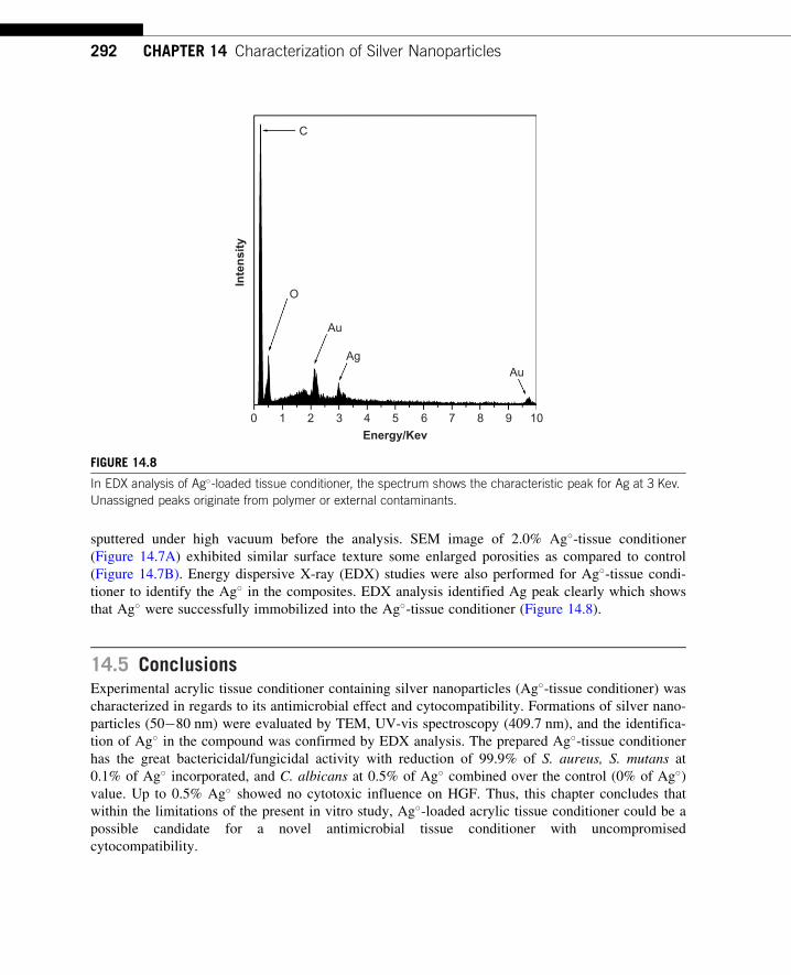
sputtered under high vacuum before the analysis. SEM image of 2.0% Ag�-tissue conditioner
(Figure 14.7A) exhibited similar surface texture some enlarged porosities as compared to control
(Figure 14.7B). Energy dispersive X-ray (EDX) studies were also performed for Ag�-tissue condi-
tioner to identify the Ag� in the composites. EDX analysis identified Ag peak clearly which shows
that Ag� were successfully immobilized into the Ag�-tissue conditioner (Figure 14.8).
14.5 ConclusionsExperimental acrylic tissue conditioner containing silver nanoparticles (Ag�-tissue conditioner) was
characterized in regards to its antimicrobial effect and cytocompatibility. Formations of silver nano-
particles (50�80 nm) were evaluated by TEM, UV-vis spectroscopy (409.7 nm), and the identifica-
tion of Ag� in the compound was confirmed by EDX analysis. The prepared Ag�-tissue conditioner
has the great bactericidal/fungicidal activity with reduction of 99.9% of S. aureus, S. mutans at
0.1% of Ag� incorporated, and C. albicans at 0.5% of Ag� combined over the control (0% of Ag�)value. Up to 0.5% Ag� showed no cytotoxic influence on HGF. Thus, this chapter concludes that
within the limitations of the present in vitro study, Ag�-loaded acrylic tissue conditioner could be a
possible candidate for a novel antimicrobial tissue conditioner with uncompromised
cytocompatibility.
C
O
Au
Ag
0 1 2 3 4 5
Au
Energy/Kev
Inte
nsit
y
6 7 8 9 10
FIGURE 14.8
In EDX analysis of Ag�-loaded tissue conditioner, the spectrum shows the characteristic peak for Ag at 3 Kev.
Unassigned peaks originate from polymer or external contaminants.
292 CHAPTER 14 Characterization of Silver Nanoparticles

References[1] N. Okita, D. Ørstavik, J. Ørstavik, K. Østby, In vivo and in vitro studies on soft denture materials:
microbial adhesion and tests for antibacterial activity, Dent. Mater. 7 (1991) 155�160.
[2] D.R. Radford, S.J. Challacombe, J.D. Walter, Denture plaque and adherence of Candida albicans to
denture-base materials in vivo and in vitro, Crit. Rev. Oral. Biol. Med. 10 (1999) 99�116.
[3] R.G. Nair, L.P. Samaranayake, The effect of oral commensal bacteria on candidal adhesion to denture
acrylic surfaces. An in vitro study, APMIS 104 (1996) 339�349.
[4] C. Wilkieson, L.P. Samaranayake, T.W. MacFarlane, P.-J. Lamey, D. MacKenzie, Oral candidosis in the
elderly in long term hospital care, J. Oral. Pathol. Med. 20 (1991) 13�16.
[5] T. Rossi, J. Laine, E. Eerola, P. Kotilainen, R. Peltonen, Denture carriage of methicillin-resistant
Staphylococcus aureus, Lancet 345 (1995) 1557.
[6] A. Harrison, R.M. Basker, I.S. Smith, The compatibility of temporary soft materials with immersion
denture cleansers, Int. J. Prosthod. 2 (1989) 254�258.
[7] H. Nikawa, H. Iwanaga, T. Hamada, S. Yuhta, Effects of denture cleansers on direct soft denture lining
materials, J. Prosthet. Dent. 72 (1994) 657�662.
[8] L.M. De Visschere, L. Grooten, G. Theuniers, J.N. Vanobbergen, Oral hygiene of elderly people in long-
term care institutions—a cross sectional study, Gerodontology 23 (2006) 195�204.
[9] L.A. Casemiro, C.H. Gomes Martins, C. Pires-de-Souza Fde, H. Panzeri, Antimicrobial and mechanical
properties of acrylic resins with incorporated silver�zinc zeolite—Part I, Gerodontology 25 (2008)
187�194.
[10] D.M. Quinn, The effectiveness, in vitro, of miconazole and ketoconazole combined with tissue condi-
tioners in inhibiting the growth of Candida albicans, J. Oral. Rehabil. 12 (1985) 177�182.
[11] M.R. Truhlar, K. Shay, P. Sohnle, Use of a new assay technique for quantification of antifungal activity
of nystatin incorporated in denture liners, J. Prosthet. Dent. 71 (1994) 517�524.
[12] C.K.W. Chow, D.W. Matear, H.P. Lawrence, Efficacy of antifungal agents in tissue conditioners in treating
candidiasis, Gerodontology 16 (1999) 110�118.
[13] A.S.F. Koopmans, N. Kippuw, J. de Graaff, Bacterial involvement in denture-induced stomatitis, J. Dent.
Res. 67 (1988) 1246�1250.
[14] E. Budtz-Jørgensen, E. Theilade, J. Theilade, H.A. Zander, Method for studying the development, struc-
ture and microflora of denture plaque, Scand. J. Dent. Res. 89 (1981) 149�156.
[15] J. Fu, J. Ji, D. Fan, J. Shen, Construction of antibacterial multilayer films containing nanosilver via
layer-by-layer assembly of heparin and chitosan-silver ions complex, J. Biomed. Mater. Res. A 79
(2006) 665�674.
[16] R.M. Slawson, H. Lee, J.T. Trevors, Bacterial interactions with silver, Biometals 3 (1990) 151�154.
[17] G. Zhao, S.E. Stevens Jr., Multiple parameters for the comprehensive evaluation of the susceptibility of
Escherichia coli to the silver ion, Biometals 11 (1) (1998) 27�32.
[18] V. Alt, T. Bechert, P. Steinrucke, M. Wagener, P. Seidel, E. Dingeldein, et al., An in vitro assessment of
the antibacterial properties and cytotoxicity of the nanoparticles silver bone cement, Biomaterials 25
(2004) 4383�4391.
[19] U. Samuel, J.P. Guggenbichler, Prevention of catheter-related infections: the potential of a new nano-
silver impregnated catheter, Int. J. Antimicrob. Agents 23 (2004) 75�78.
[20] J.B. Wright, K. Lam, D. Hansen, R.E. Burrell, Efficacy of topical silver against fungal burn wound
pathogens, Am. J. Infect. Control. 27 (1999) 344�350.
[21] H.G. Petering, Pharmacology and toxicology of heavy metals: silver, Pharmacol. Therapeut. 1 (1976)
127�130.
293References

[22] G. Ramage, K. Vande Walle, B.L. Wickes, J.L. Lopez-Ribot, Standardized method for in vitro antifungal
susceptibility testing of Candida albicans biofilms, Antimicrob. Agents Chemother. 45 (2001)
2475�2479.
[23] M.E. Reverdy, A. Martra, J. Fleurette, Application of a micromethod to the study of the bactericidal
activity of 2 antiseptics based on chlorhexidine gluconate, Pathol. Biol. 34 (1986) 688�693.
[24] D. Steinberg, S. Eyal, Early formation of Streptococcus sobrinus biofilm on various dental restorative
materials, J. Dent. 30 (2002) 47�51.
[25] J. Satou, A. Fukunaga, A. Morikawa, I. Matsumae, N. Satou, H. Shintani, Streptococcal adherence to
uncoated and saliva-coated restoratives, J. Oral. Rehabil. 18 (1991) 421�429.
[26] S. Hahnel, M. Rosentritt, R. Burgers, G. Handel, Adhesion of Streptococcus mutans NCTC 10449 to
artificial teeth: an in vitro study, J. Prosthet. Dent. 100 (4) (2008) 309�315.
[27] J. Chandra, P.K. Mukherjee, S.D. Leidich, F.F. Faddoul, L.L. Hoyer, L.J. Douglas, et al., Antifungal
resistance of candidal biofilms formed on denture acrylic in vitro, J. Dent. Res. 80 (3) (2001)
903�908.
[28] Y. Abe, M. Ishii, M. Takeuchi, M. Ueshige, S. Tanaka, Y. Akagawa, Effect of saliva on an antimicrobial
tissue conditioner containing silver�zeolite, J. Oral. Rehabil. 31 (2004) 568�573.
[29] P.C. Baehni, Y. Takeuchi, Anti-plaque agents in the prevention of biofilm-associated oral diseases, Oral.
Dis. 9 (2003) 23�29.
[30] W. Bruns, H. Keppeler, R. Bancks, Suppression of intrinsic resistance to penicillins in Staphylococcus
aureus, by polidocanol, a dodecyl polyethyleneoxid ether, Antimicrob. Agents Chemother. 27 (1985)
632�639.
[31] A.G. Gristina, R.A. Jennings, P.T. Naylor, Q.N. Myrvik, L.X. Webb, Comparative in vitro antibiotic
resistance of surface-colonizing coagulase-negative staphylococci, Antimicrob. Agents Chemother. 33
(1989) 813�816.
[32] T. Matsuura, Y. Abe, Y. Sato, M. Okamoto, M. Ueshige, Y. Akagawa, Prolonged antimicrobial effect of
tissue conditioners containing silver-zeolite, J. Dent. 25 (1997) 373�377.
[33] E. Makila, V.K. Hopsu-Havu, Mycotic growth and soft denture lining materials, Acta Odontol. Scand.
35 (1977) 197�205.
[34] M. Christensen, J. Rungby, S.C. Mogensen, Effects of selenium on toxicity and ultrastructural localiza-
tion of mercury in cultured murine macrophages, Toxicol. Lett. 47 (3) (1989) 259�270.
[35] Q.L. Feng, J. Wu, G.Q. Chen, F.Z. Cui, T.N. Kim, J.O. Kim, A mechanistic study of the antibacterial
effect of silver ions on Escherichia coli and Staphylococcus aureus, J. Biomed. Mater. Res. 52 (2000)
662�668.
[36] International Organization for Standardization (ISO 10993-5), Biological evaluation of medical
devices—Part 5, tests for cytotoxicity: in vitro methods, 1992.
[37] R. Foldbjerg, P. Olesen, M. Hougaard, D.A. Dang, H.J. Hoffmann, H. Autrup, PVP-coated silver nano-
particles and silver ions induce reactive oxygen species, apoptosis and necrosis in THP-1 monocytes,
Toxicol. Lett. 190 (2009) 156�162.
[38] M. Ott, V. Gogvadze, S. Orrenius, B. Zhivotovsky, Mitochondria, oxidative stress and cell death,
Apoptosis 12 (2007) 913�922.
[39] Y.S. Kim, M.Y. Song, J.D. Park, K.S. Song, H.R. Ryu, Y.H. Chung, Subchronic oral toxicity of silver
nanoparticles, Part Fibre Toxicol. 7 (2010) 20�31.
[40] D.W. Jones, E.J. Sutow, G.C. Hall, W.M. Tobin, B.S. Graham, Dental soft polymers: plasticizer compo-
sition and leachability, Dent. Mater. 4 (1988) 1�7.
[41] B.S. Graham, D.W. Jones, E.J. Sutow, An in vivo and in vitro study of the loss of plasticizer from soft
polymer-gel materials, J. Dent. Res. 70 (5) (1991) 870�873.
294 CHAPTER 14 Characterization of Silver Nanoparticles

[42] C. Pesci-Bardon, T. Fosse, D. Serre, L. Madinier, In vitro antiseptic properties of an ammonium com-
pound combined with denture base acrylic resin, Gerodontology 23 (2006) 111�116.
[43] K. Yoshida, M. Tanagawa, M. Atsuta, Characterization and inhibitory effect of antibacterial
dental resin composites incorporating silver-supported materials, J. Biomed. Mater. Res. 47 (1999)
516�522.
[44] S. Imazato, N. Ebi, U. Takahashi, T. Kaneko, S. Ebisu, R.R.B. Russel, Antibacterial activity of
bactericide-immobilized filler for resin-based restoratives, Biomaterials 24 (2003) 3605�3609.
295References

CHAPTER
15Bioactive Glass Nanoparticles forPeriodontal Regeneration andApplications in Dentistry
Sandhra M. Carvalho, Agda A.R. Oliveira, Elke M.F. Lemos and Marivalda M. PereiraMetallurgical and Materials Engineering Department, Federal University of Minas Gerais, Belo Horizonte,
Minas Gerais, Brasil
CHAPTER OUTLINE
15.1 Introduction ............................................................................................................................... 299
15.2 Composition and synthesis of bioactive glass nanoparticles ......................................................... 300
15.3 Bioactivity of glass nanoparticles ............................................................................................... 303
15.4 Bioactive glass in dentistry.........................................................................................................305
15.5 Bioactive glass nanoparticles in periodontal regeneration............................................................ 309
15.6 Bioactive glass nanocomposites .................................................................................................312
15.7 Bioactive glass nanocomposite applications in bone regeneration and dental implants .................. 314
15.8 The future of bioactive glass nanoparticles in dentistry................................................................ 315
15.9 Conclusions............................................................................................................................... 317
Acknowledgments ............................................................................................................................... 317
References ......................................................................................................................................... 318
15.1 IntroductionBioactive glasses were first developed by Hench et al. in 1969, and represent a group of reactive
materials that are able to bond to mineralized bone tissue in physiological environment. Bioactive
glasses are widely used in the biomedical area. Early applications of bioactive glasses were in the
form of solid pieces for small bone replacement in middle ear surgery [1,2]. Later, several applica-
tions of bioactive glasses have been proposed, including the dental field. Recently, bioactive glasses
have been widely studied for potential application in tissue engineering and regenerative medicine.
A range of bioactive glasses with attractive properties, like biocompatibility and bioactivity, and
synthesized by newer methods have been developed. Various investigations have been undertaken
to obtain bioactive glasses in different forms, such as bulk, powder, composites, and porous
scaffolds.
299Nanobiomaterials in Clinical Dentistry.
© 2013 Elsevier Inc. All rights reserved.

Nanotechnology offers a new strategy to develop bioactive glasses with a higher surface area.
Nanotechnology is defined as a science that involves the development of materials and devices at
the nanoscale. Specific properties of bioactive glasses can be improved and controlled when synthe-
sized in nanometer scale. Therefore, nanosized biomaterials have greater biocompatibility and bio-
activity. This new approach is important both for bioactive glasses used in particulate form as well
as for coatings on biomedical devices or as fillers in composite materials.
Over time, bioactive glasses have been applied in various specialties of dentistry such as
periodontics, endodontics, and implantology. This chapter describes the importance and applica-
tions of bioactive glass in dentistry and periodontics.
15.2 Composition and synthesis of bioactive glass nanoparticlesBioactive glass when in contact with body fluids induces a specific biological response at the inter-
face of the material, resulting in the formation of a carbonated hydroxyapatite (HA) layer on its
surface and, through this layer, binding to the mineralized tissue. The bioactivity of bioactive glass
is not an absolute concept determined only by the composition but is also affected by the size and
shape of the material. The formation of this HA layer is essential for synthetic materials to exhibit
bioactivity [3,4].
There is an advance in research on bioactive glasses for newer applications in tissue engineer-
ing, and therefore new routes of synthesis of bioactive glass have been proposed. A high surface
area silica-rich is crucial for the formation of HA layer. Sol�gel glasses exhibited increasing spe-
cific surface area and pore volume compared with the glasses produced by conventional melting
process, which enhanced their biocompatibility and bioactivity by accelerating the deposition pro-
cess of HA [3,4]. One approach to improve the properties of bioactive glass material is their devel-
opment at the nanoscale. Reducing the size of bioactive glass granules can accelerate the formation
of HA layer and also provide more active sites for cell attachment. Compared with microparticles,
nanoparticles have significantly higher surface area, and this drastically changes the material char-
acteristics such as surface energy, wettability, surface topography, and surface chemistry.
Furthermore, studies have shown that nanoscale materials have higher biocompatibility [5,6].
Ostomel et al. (2006) also showed that the reduction of bioactive glass particles to nanoscale could
stimulate their bioactivity [7]. Bioactive glasses are capable of forming a HA layer, and the
osseointegration to bone tissue is performed through this layer. The rate of osseointegration to the
mineralized tissue depends on the rate of formation of HA layer, which depends on the composition
of the bioactive glass [4,8].
The basic components of most bioactive glasses are SiO2, Na2O, CaO, and P2O5. However,
among the sol-gel-derived bioactive glasses, the composition of 60% SiO2, 36% CaO, and 4%
P2O5 (by weight) has a high level of bioactivity, showing great potential for engineering applica-
tions of mineralized tissues [9]. In the past decade, several studies have been directed at the devel-
opment of bioactive glass nanoparticles. Recently, Brunner et al. (2006) reported on the preparation
of bioactive glass nanopowders using flame synthesis, a process that requires a high-temperature
environment [10]. By contrast, sol-gel technology is a low-temperature preparation method, and
the glasses prepared by the sol-gel method contain a porous structure with a higher surface area.
300 CHAPTER 15 Bioactive Glass Nanoparticles for Applications in Dentistry

The sol-gel route is based on controlled hydrolysis and condensation of alkoxides to form a suspen-
sion of colloidal particles (sol), which upon polycondensation forms an interconnected network
structure (gel). However, the particle size of the traditional sol-gel-derived bioactive glasses was
larger than 1 μm.
The synthesis of bioactive glass uses typical precursors like tetraethyl orthosilicate (TEOS), calcium
nitrate (CN), and triethylphosphate (TEP). Particle diameter can be controlled by fine-tuning variable
parameters, such as reagent concentration and reaction temperature. According to Bogush and Zukoski
(1991), five parameters play an important role in the size and distribution of silica nanoparticles:
(i) concentration of TEOS, (ii) concentration of ammonia, (iii) concentration of water, (iv) alcohol
effect, and (v) reaction temperature [11,12]. Hong et al. (2009) developed bioactive glass nanoparticles
by combining the sol�gel method and co-precipitation. In that study, the mixture of precursor was
hydrolyzed in acidic medium and condensed under alkaline conditions separately [13,14].
Stober et al. (1968) reported on a pioneering method for the synthesis of spherical and monodisperse
silica nanoparticles from aqueous alcohol solution of silicon alkoxides in the presence of ammonia as a
catalyst from which different sizes of silica particles were obtained [15]. The Stober method can be
modified to produce particles with composition different from those of pure silica. Oliveira (2011)
synthesized bioactive glass nanoparticles based on the Stober method [16] (Figure 15.1).
Chen et al. (2009) investigated the effects of different morphologies on the in vitro bioactivity
of nanosized bioactive glass particles in the system CaO�P2O5�SiO2 by using lactic acid in the
sol�gel method and concluded that not only the surface area but also the surface morphology play
an important role in bioactivity of the material [17].
Nanosized particles contain a large surface area that results in high interfacial energy and
thermodynamic instability. In general, these particles tend to agglomerate during the synthesis of the
particles in an attempt to minimize the free energy of the system. To maintain the stability of the parti-
cles, some studies focused on obtaining spherical bioactive glass particles to create a bioactive material
with dispersion capability under dilute aqueous and alcoholic conditions [18]. Spherical bioactive glass
nanoparticles developed and characterized by Oliveira (2011) are shown in Figure 15.2 [16].
Several techniques were developed in which SiO2�CaO�P2O5 bioactive glass nanoparticles
were prepared. The best bioactivity results were from ternary systems with spherical particles
below 50 nm produced with a pH of 11.5. El-Kady et al. (2010) prepared particles, comprised of
60 wt% SiO2, 36 wt% CaO, and 4 wt% P2O5, with sizes less than 100 nm using alkali-mediation
with the application of moderate ultrasound dispersion combined with mechanical agitation as well
as the addition of ethanol as a dispersant [19]. Previous studies have shown that low concentrations
of TEOS and water provide monodispersed uniform-sized nanoparticles [11,12,15,20�25]. The size
of the nanoparticles was found to increase with TEOS and with increasing concentrations of water
and ammonia of up to 7 M and 2 M, respectively [20]. Particles prepared in methanol solutions are
the smallest, and the particle sizes increase with the increasing chain length of alcohol. The particle
size distribution also becomes broader when longer chain alcohols are used as solvents. Particles
prepared in methanol and ethanol-glycerol solutions resulted in a stable sol. However, when butanol
and ethanol are used, precipitation could easily be observed [11,12,20,24,26].
The ammonia-catalyzed reactions of TEOS in ethanol with water (Stober method) can be used
for the preparation of monodispersed spherical nanoparticles bioactive glass. Alcohol is added to
prevent the liquid�liquid phase separation during the initial phase of the hydrolysis reaction [27].
Methanol is chosen as the solvent medium to act as a surfactant and promote the production of
30115.2 Composition and synthesis of bioactive glass nanoparticles

(A)
50 nm
(B)
50 nm
FIGURE 15.2
TEM images of bioactive glass nanoparticles. Images (A) and (B) show the difference between the particle sizes.
TEOSTEP
(2)(3)
0.22 µm
(4)
(6)
(7)(9)
(8)
40 min
200°C
(5)
0.22 µm
0.10 µm
75
75
25
25
24 h
100
(1)
CH3OHH2ONH3
NH3
50°C
3 h5 min 48 hml
100ml
FIGURE 15.1
Schematic representation of bioactive glass nanoparticles synthesis based on the Stober method: (1) Reaction
medium; (2) Alkoxide hydrolysis and condensation; (3) The formed sol; (4) BGNP filtration; (5) Addition of
calcium nitrate; (6) BGNP filtrations; (7) BGNP freeze drying; (8) Thermal treatment and (9) BGNP powder.
302 CHAPTER 15 Bioactive Glass Nanoparticles for Applications in Dentistry

dispersed particles with nanometric diameter [26]. In general, the hydrolysis reaction of TEOS can
be expressed by the following simple equation [27]:
SiðORÞ4 1 2H2O-SiO2 1 4ROH (15.1)
In fact, the controlled hydrolysis process produces the singly hydrolyzed monomer [(OR)3Si(OH)]:
SiðORÞ4 1 xH2O1NH3-ð20ÞxSiðORÞ42x 1 xROH1NH14 (15.2)
These intermediate reaction products participate in the condensation reactions, Eqs (15.3) and
(15.4), in turn forming silica nanoparticles.
SiðORÞ4 1 ðOHÞSiðORÞ3-ðORÞ3Si2O2 SiðORÞ3 1ROH (15.3)
ðORÞ3SiðOHÞ1 ðOHÞSiðORÞ3-ðORÞ3Si2O2 SiðORÞ3 1H2O (15.4)
15.3 Bioactivity of glass nanoparticlesNumerous responses can be observed when a biomaterial comes in contact with living cells and tis-
sues, and these responses depend on the type of material used. If this material is biologically active
and induces a specific response, leading to formation of a continuous interface between the material
and living tissue, it is called bioactive. Bioactive glasses have this unique feature and when they
come in contact with the body, form a layer of carbonated HA on the surface of the material,
which permits the interaction of biomaterials especially with mineralized tissue (Figure 15.3).
The complex process of HA formation has been extensively studied in the literature [27].
A sequence of interfacial reactions, which begin immediately after bioactive glass is soaked in
simulated body fluid, is interpreted in terms of the electrostatic interaction of the functional groups
with the ions in the fluid. The reaction is divided into four steps [9,27,28]:
1. Initially, the surface becomes negative, attributed to the silanols (Si�OH) formed on bioactive
glass particles upon being soaked in simulated body fluid, by two mechanisms: (i) The�OH
formation occurs due to release of calcium ions (Ca21) from their surfaces by means of exchange
with H3O1, Na1, and K1 ions in simulated body fluid and (ii) a loss of soluble silica to solution, in
Si(OH)4 form, causing the breakdown of Si�O�Si bonds and silanols formation. As a result, many
Si�OH groups are formed on their surfaces and a local supersaturation of Ca21 ions is established.
2. As time passes, there occurs a selective combination of OH� charged surface with the Ca21
ions from the simulated body fluid solution. As the calcium ions accumulate on the surface, the
surface gradually gains an overall positive charge.
3. The pH of simulated body fluid solution, where the concentration of HPO422 is much larger
than that of PO432, favors the incorporation of HPO4
22 ions in the bioactive glass surface and
the migration of PO432 ions from the bulk to the surface of the glass. The result leads to
calcium deficiency, and the surface once again becomes negatively charged.
4. The calcium ions combine with the phosphate ions (PO432 and HPO4
22) and form amorphous
calcium phosphates (Ca3(PO4)2 and CaHPO4). These calcium phosphates spontaneously
transform into the apatite through the incorporation of OH� and CO322 anions from the solution
30315.3 Bioactivity of glass nanoparticles

to form hydroxyl carbonate apatite (HCA) Ca5(PO4CO3)3(OH) layers. Once apatite nuclei forms,
crystals grow by consuming calcium and phosphate ions from the simulated body fluid.
Several in vitro and in vivo studies show that a series of reactions occur that lead to the forma-
tion of a carbonated HA layer on the bioactive glass surface (Figure 15.4). Parameters such as sur-
face charge, composition, structure, and morphology will be important in the formation of the Ca/P
layer as well as in the interaction between the material surface and the surrounding medium, pro-
teins, and cells [9,28�33].
Several theories have been proposed to explain the formation of this layer. The HA layer is
spontaneously formed in the surface of bioactive glass of the CaO�SiO2�P2O5 system after
exposure to body fluids [27�30,34,35]. Studies have shown that apatite is preferentially formed on
a surface material composed mainly of CaO and SiO2 because the Ca21 ions released from the
glass increase the degree of saturation of the apatite in relation to surrounding fluid. In addition,
the Si�OH groups of the hydrated silica gel formed on the surface induce a heterogeneous
nucleation of apatite. These crystals grow by consuming calcium and phosphate ions from the body
fluid and those that migrate from the bulk to the surface of the glass.
Compared with microparticles, bioactive glass nanoparticles have advantages in bone repair and
regeneration, with the decrease in grain size promoting an increase in cellular adhesion, enhanced
osteoblast proliferation and differentiation, and an increase in the biomineralization process
O
Apatita
Ca2+
Na+
OH
Simulatedbody fluid
Hydroxyapatite
Bioactive glassnanoparticles
K+
Mg2+
Cl–
HCO3–
H3O+Ca2+ H3O+Ca2+
HPO 2– HPO 2– HPO 2–
O
O
O
Si
Si
Si
OHHO
OO
OO
O
Si
O
OO
O
O O
Si
Si
OHHO
O O
Si
O
OO
O
O O
Si
Si
OHHO
O
4
So 2–4
Apatita
Ca2+ OH
4
Apatita
Ca2+ OH
4
FIGURE 15.3
Schematic drawing of the formation process of hydroxyapatite layer. It shows the formation of hydroxyapatite
layer after interaction of the bioactive glass with simulated body fluid.
304 CHAPTER 15 Bioactive Glass Nanoparticles for Applications in Dentistry
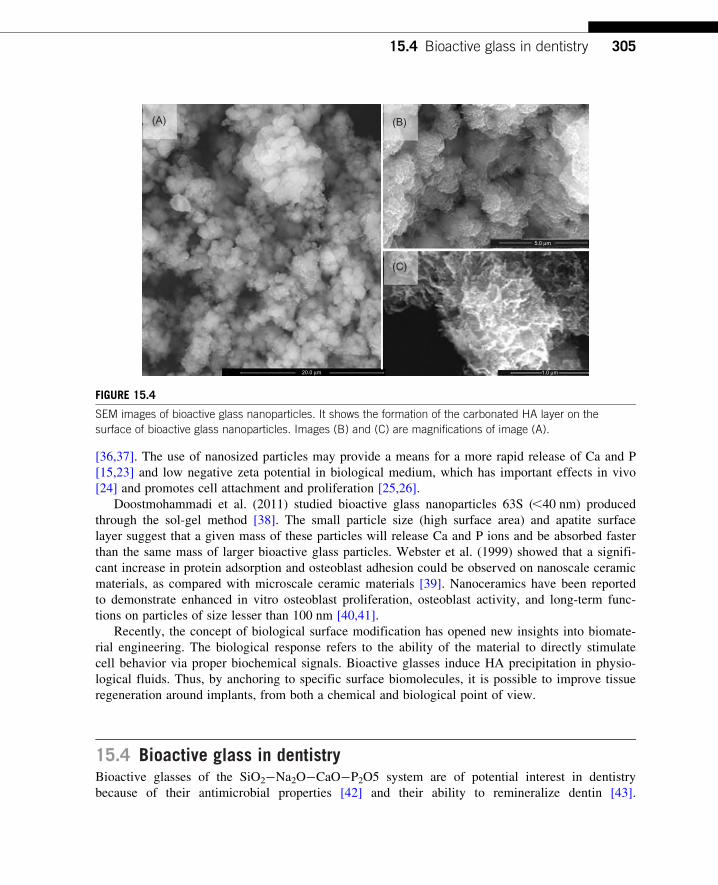
[36,37]. The use of nanosized particles may provide a means for a more rapid release of Ca and P
[15,23] and low negative zeta potential in biological medium, which has important effects in vivo
[24] and promotes cell attachment and proliferation [25,26].
Doostmohammadi et al. (2011) studied bioactive glass nanoparticles 63S (,40 nm) produced
through the sol-gel method [38]. The small particle size (high surface area) and apatite surface
layer suggest that a given mass of these particles will release Ca and P ions and be absorbed faster
than the same mass of larger bioactive glass particles. Webster et al. (1999) showed that a signifi-
cant increase in protein adsorption and osteoblast adhesion could be observed on nanoscale ceramic
materials, as compared with microscale ceramic materials [39]. Nanoceramics have been reported
to demonstrate enhanced in vitro osteoblast proliferation, osteoblast activity, and long-term func-
tions on particles of size lesser than 100 nm [40,41].
Recently, the concept of biological surface modification has opened new insights into biomate-
rial engineering. The biological response refers to the ability of the material to directly stimulate
cell behavior via proper biochemical signals. Bioactive glasses induce HA precipitation in physio-
logical fluids. Thus, by anchoring to specific surface biomolecules, it is possible to improve tissue
regeneration around implants, from both a chemical and biological point of view.
15.4 Bioactive glass in dentistryBioactive glasses of the SiO2�Na2O�CaO�P2O5 system are of potential interest in dentistry
because of their antimicrobial properties [42] and their ability to remineralize dentin [43].
(A) (B)
(C)
20.0 µm 1.0 µm
5.0 µm
FIGURE 15.4
SEM images of bioactive glass nanoparticles. It shows the formation of the carbonated HA layer on the
surface of bioactive glass nanoparticles. Images (B) and (C) are magnifications of image (A).
30515.4 Bioactive glass in dentistry

Bioactive glasses are interesting materials for use as bone grafts, implant coatings, bone cements,
toothpaste, and various other applications in dentistry.
The application of bioactive glass in dentistry began in the mid-1980s when Dr. Clark,
Dr. Stanley, and Dr. Hall tested the bioactive glass and have been successful in preserving the
alveolar ridge in edentulous patients. These results have led to regulatory approval for commercial
use of Bioglasss. In this decade, not only the pioneer brand Bioglasss but also several new bioac-
tive glass types have been increasingly studied and applied in various areas of dentistry.
Perioglasss, for example, is widely used in implant dentistry and periodontics [4].
In the following sections, the impacts and applications in dentistry of this new strategy of devel-
opment of nanoscale bioactive glasses will be discussed. Further research will be presented and the
future of bioactive glass nanoparticles in dentistry will be discussed.
Nanoscale bioactive glasses have been gaining attention due to their reported superior bio-
activity when compared with conventional micron-sized bioactive glass materials. There is evi-
dence in the literature of a more rapid mineralization in bones and teeth when in contact with
bioactive glass nanoparticles [44].
Evidence shows that surface properties of the bioactive glass may affect cellular response.
Bioactive glass on the nanoscale shows excellent properties, such as surface energy, surface wett-
ability, surface topography, and surface chemistry. This improvement is due to the increased
surface area of the material at the nanoscale.
The events occurring at the interface of bioactive glass with cells attempt to mimic the natural
interaction of cells with extracellular matrix. Biomaterials do not directly interact with living tissue
but with a layer of adsorbed proteins such as fibronectin, vitronectin, fibrinogen, collagen, and
laminins called soluble proteins of the matrix. Cells recognize proteins of the matrix through a
family of cell surface receptors called integrins [45,46]. When these receptors are sensed, they
group together and form adhesion complexes that provide anchoring cells to the bioactive glass
surface and unleash subsequent cellular responses. Therefore, the initial interaction of the cell
material is a complex process that begins with the adsorption of proteins followed by adhesion,
spreading, cell differentiation, and ends with the full operation of the cell [46]. So the improvement
of surface properties of bioactive glass has a direct influence on the adsorption of specific proteins,
thus causing an increase in bioactivity and biocompatibility of the material.
There are several applications for bioactive glass nanoparticles in dentistry; for example, various
products with bioactive glass nanoparticles have been developed to control the formation of oral bio-
film, such as toothpaste and mouth rinses. These products are designed to decrease bacterial adhesion
thereby reducing the formation of biofilms. Although bioactive glasses have been used in bone re-
generation for many years, only recently studies on their antimicrobial properties have gained focus.
A further application of the antibacterial properties of bioactive glass nanoparticles is in the
endodontic area. Disinfection of a complex root canal system is essential for long-term success in
endodontics. However, even after the chemical and mechanical treatment of the channels is some-
times impossible to achieve complete disinfection. Microbiological studies have demonstrated the
presence of resistant microorganisms such as Enterococcus faecalis and Candida albicans in persis-
tent infections of the channel. Microorganisms remaining in the root canals are the major cause of
treatment failure in the root canal. An ideal drug for endodontic treatment should achieve a wide-
spectrum antimicrobial, be biocompatible, be able to reduce inflammation, and induce repair of
mineralized tissue [47�50].
306 CHAPTER 15 Bioactive Glass Nanoparticles for Applications in Dentistry

Calcium hydroxide, originally designed as a channel disinfectant and filler material by Hermann
in 1920, remains a topical antiseptic widely used. The suspension of calcium hydroxide releases Ca
ions that results in alkalinization of the environment. Similarly, the antibacterial properties of bio-
active glasses are based on the potential of increasing the pH in aqueous suspensions [51] resulting
from the exchange of sodium ions and protons of the glass matrix in an aqueous environment [52].
The broad-spectrum antimicrobial effect of bioactive glass in different oral microorganisms has
been reported, which justifies its use as an intracanal medication endodontic therapy [42]. The
increase in specific surface area of bioactive glass nanoparticles improves the area of active release
of ions, resulting in the improvement of antibacterial properties of the material. E. faecalis is one
of the more resistant microorganisms in endodontic therapy and is often associated with treatment
failure. However, research has shown that particles of 20�50 nm bioactive glass increased antibac-
terial properties against E. faecalis in direct contact model [43].
Bioactive glasses are known for osteoconductivity and bonding to bone through the release of
ions and formation of a layer of apatite [53]. Because of these characteristics, bioactive glasses are
widely used for bone reconstruction and tissue engineering, but are also interesting candidates for
the mineralization in dentistry. In the past decades, bioactive glass nanoparticles have been used on
studies about the specific effects of remineralization of dentin, due to their excellent regenerative
properties in mineralized tissues.
Dentin is a permeable tubular structure representing most of the dental tissue composed of the
organic matrix embedded in a crystalline matrix of apatite, having about 70% of apatite, 20% of
collagen, and 10% of water. Although there is a physiological balance between demineralization
and remineralization of dental hard tissues in the oral cavity, some factors such as an acidic diet or
the presence of plaque can cause disequilibrium, which results in tooth demineralization.
Several studies suggest that bioactive glass has potential as an agent of dentin mineralization and
restorative filling [54,55]. As explained above, bioactive glass when in contact with body fluids inter-
acts with the environment, releasing ions and subsequently forming a layer of apatite. Saliva is also a
body fluid that will stimulate the interaction of bioactive glass with the environment and release of
ions and the formation of apatite. However, the mineralization process is long, which impedes the
use of bioactive glass in dental practices. A possible acceleration of this process is the use of bio-
active glass nanoparticles, since the high specific surface area of the nanoparticles may facilitate the
dissolution of ions from the glass and thus accelerate the dentin mineralization [10]. Vollenweider
et al. (2007) showed that after treatment with bioactive glass nanoparticles for 30 days, there was a
pronounced increase in the mineral content of the dentin samples [53]. In addition, bioactive glass
nanoparticles can be used in minimally invasive cavity preparation teeth. However, it is still neces-
sary to create alternatives that allow effective use of bioactive glass nanoparticles in this procedure.
Another application of the ability of remineralization of bioactive glass is related to the dentin
sensitivity. Dentin hypersensitivity is characterized by sharp, localized, non-spontaneous pain
occurring in response to some stimulus. This pain is caused by loss of the tooth structure, resulting
in exposure of dentinal tubules to the oral environment. The currently accepted theory for hypersen-
sitive dentin is the hydrodynamic theory, which proposes that external stimuli such as cold, hot,
pressure, or tactile when applied to exposed dentin causes movement within the dentinal tubules
[56]. This movement of fluid stimulates mechanoreceptors near the dentinal tubule and can trigger
a pain response. This theory is based on the understanding that open tubules allow fluid flow
through the tubules, which results in pressure changes that excite the nerve endings in the tooth
30715.4 Bioactive glass in dentistry

pulp. Therefore, it is coherent with the observation that occluded tubules can treat and reduce den-
tinal sensitivity [55,57,58]. Occlusion of dentinal tubules is an approach currently used in the treat-
ment of dentin hypersensitivity, and numerous products using active ingredients may occlude the
dentinal tubule. Although there are advantages on the use of products in terms of availability and
cost to the consumer, a great disadvantage is the time. They may take up to 2�4 weeks for efficacy
of the relief symptoms [58].
The majority of the bioactive glasses used today are based on the original composition developed
by Hench et al. [4]. Silica is the key component of bioactive glass, and acts as a nucleation site for the
precipitation of calcium and phosphate ions in the formation of HA. Research indicates that bioactive
glass nanoparticles induce formation of apatite in dentin, indicating that it is a potential material to
be used for the treatment of dentin hypersensitivity. The occlusion of dentin tubules may reduce or
eliminate hypersensitivity by restricting dentinal fluid movement. This process of remineralization of
dentin tubes is quicker when using products with bioactive glass nanoparticles, since the larger surface
area associated with nanoscale particles leads to a more efficient ion release (Figure 15.5).
As previously mentioned, bioactive glasses are widely used for bone restoration; therefore, there
are several studies about its application in oral and craniofacial surgery. Every year, millions of
people worldwide suffer bone loss in the region of the face resulting from trauma, tumor, or bone
diseases. Bone grafting procedures are required frequently in oral and maxillofacial surgery. One of
the traditional bone-filling treatments is the autograft using autogenous materials. The autogenous
bone appears as an excellent biological alternative. However, the need for a donor site and a
surgical procedure represent additional factors that can limit their use.
Enamel
Dentin
Dental pulp
Remineralization ofdentinal tubules
Bgnp
Gum
Cementum
Periodontalligament
Aveolarbone
Vasculo-nervous
FIGURE 15.5
Schematic drawing of treatment of dentinal hypersensitivity. It shows the mineralization of dentinal tubules
after treatment with bioactive glass nanoparticles.
308 CHAPTER 15 Bioactive Glass Nanoparticles for Applications in Dentistry

Another procedure is allograft, which is made between different individuals of the same species.
However, this procedure also has limitations such as need for donors and the possible transmission
of antigenic proteins among others. Another alternative that has been widely used is the use of allo-
plastic material, and among them bioactive glass is one of the most researched.
Bone tissue is a complex of organic and inorganic materials organized in a global architecture
of several length scales, including the nanoscale. Therefore, the applications of glass nanoparticles
in bone grafts have the dual aim of improving the mechanical properties as well as incorporating
nanotopographic features that mimic properties of bone.
The purpose of tooth replacement is the restoration of function and esthetics without affecting
the mineralized structures and soft tissues of the oral cavity. One of the challenges in implantology
is to achieve and maintain the osseointegration as well as the epithelial junction of the gingival
tissue with implants [59]. Bone regeneration is required for many clinical issues in the dental area.
The autogenous graft is always a choice, but sometimes the host tissue is thin and cannot be mod-
eled with the desired shape.
Metals and alloys have wide application in the area of implant dentistry, and the most
commonly used are titanium alloys. Titanium implants have excellent mechanical properties,
but do not exhibit bioactivity; that is, they are not capable of binding to the living tissue. Unlike
them, bioactive glasses are able to cause specific biological responses. Therefore, coating of
implants with bioactive glasses is a way to combine good titanium mechanical properties and bio-
activity of the glasses. Various techniques for surface treatment are applied to modify the surface
of titanium.
Many studies have shown that implants coated with bioactive glasses have a higher rate of inte-
gration and increased bone fixation, faster when compared with uncoated implants [60,61].
Development of new technologies is also very important to improve the properties and applications
of biomaterials. Application of bioactive glass coatings using nanoparticles is a promising alterna-
tive for increasing the interaction of glass with the implant surface.
The bioactive glass material is one of the most studied and widely used to treat bone periodontal
defects due to its bioactive properties, so in the next section this topic will be discussed more
extensively.
15.5 Bioactive glass nanoparticles in periodontal regenerationPeriodontium is a complex structure, consisting of epithelial tissue (gum) and connective (periodon-
tal ligament) and mineralized tissue (cementum and alveolar bone) (Figure 15.6). To achieve suc-
cess in periodontal therapy, the sealing of the junctional epithelium, the insertion of new
connective tissue fibers, the formation of new cementum, and the alveolar bone restoration are
needed. The main aims of periodontal therapy are to eliminate the disease by infection control and
correct anatomical defects through regeneration of the tissues supporting the teeth.
Although Hench started to develop bioactive glass in 1969, only from the 1990s that material
was first applied in studies ‘in vivo’ in periodontal lesions. These studies evaluated histological
efficacy of bioactive glass, and the results were positive compared with conventional treatments
[62]. At this time, bioactive glass was clinically approved for use both in dense form—for
30915.5 Bioactive glass nanoparticles in periodontal regeneration

application as middle ear prostheses and as bone implants for alveolar ridge maintenance after
extraction—and in particulate form, for the treatment of periodontal bone defects [63,64].
Based on these studies, bioactive glasses have been widely applied in dentistry for the treatment
of periodontal defects, probably due to its properties of bone formation by osteoconduction and
osteoinduction. The clinical use of bioactive glass in the particulate form of the brand Perioglasss
has shown, over the years, positive results, with a good adaptation to the bone defects and maintain-
ing a good clot. Several studies and clinical reports indicate the glass as a material of easy handling
with excellent bioactivity and biocompatibility. Histological studies show that the use of bioactive
glasses can induce formation of new cementum and the formation of a new insertion [63].
Schepers et al. (1991) also showed that bioactive glass particles have the property of stimulating
the formation of bone tissue in the treatment of bone lesions created surgically in the jaws of
dogs [65]. In a histological study conducted in the tibia of rabbits, MacNeill et al. (1999) confirmed
the study of Schepers et al. (1991) [66].
Studies in humans evaluated the use of bioactive glass compared with the scrape surgery in the
treatment of intrabony periodontal defects. Results demonstrated significant clinical improvements,
such as an increase in radiographic density and a decrease in probing depth [62].
Some clinical studies have shown beneficial effects of using platelet-rich plasma (PRP) associ-
ated with bioactive glass in periodontal therapy. Periodontal regeneration involves a complex series
of events, and the recruitment of progenitor cells to the injured site and growth factors are vital
during this process. These factors are responsible for the migration, proliferation, and differentia-
tion of periodontal progenitor cells. PRP is derived from a platelet concentrate that contains high
concentrations of growth factors. Some studies have encouraged the association between the use of
PRP with bioactive glass for periodontal regeneration [67].
Enamel
Dentin
Dental pulp
Gum
Cementum
Periodontalligament
Aveolarbone
Vasculo-nervous
FIGURE 15.6
Structures forming part of the organ dental and periodontal tissue.
310 CHAPTER 15 Bioactive Glass Nanoparticles for Applications in Dentistry

In recent decades, there is an advance in the study of new therapies for complete periodontal
regeneration. Regenerative procedures are forms of treatment aimed to reconstruct the lost structure
or damaged periodontal tissue. The periodontal regeneration involves a sequence of biological
events including adhesion, migration, proliferation, and differentiation [68,69]. Numerous methods
of therapies have been tested to achieve periodontal regeneration.
The complete regeneration of periodontal tissues has not yet been achieved, but significant
progress has been achieved through the use of autogenous bone grafts, bone allografts, guided
tissue regeneration (GTR), the implantation of alloplastic materials, and use of cellular factors.
Autogenous bone grafts are transplanted from one place to another in the same individual.
Because of their osteogenic potential, autogenous bone grafts have been widely used in periodontal
therapy. They can be from intra- or extra-oral sources. However, the necessity of a second surgical
site, which can complicate post-surgery, is a disadvantage that has been associated with their use.
Allografts are transplanted between genetically different individuals of the same species. Iliac
bone marrow, lyophilized bone graft, freeze-dried bone allograft (FDBA), and decalcified FDBA
are the types of bone allografts widely available in commercial tissue banks [70]. However, despite
the approval of bone tissue banks, the real osteogenic potential of these transplants is questionable.
In dentistry, GTR is often used in the reconstruction of periodontal defects. GTR technique is
based on the use of biocompatible membranes in order to prevent accelerated migration of the
epithelium to the injured site, and allows conditions for the regeneration of all periodontal attach-
ment apparatus. The use of GTR has shown favorable results on the periodontal regeneration
when applied in some specific kinds of bony defects. It is used in different types of barriers, such
as membranes, expanded polytetrafluoroethylene, collagen [71], cellulose, and polylactic acid
[72�74].
Although polymeric products show positive results, there are still major challenges to overcome
in their use in periodontal regeneration. Research attempting to overcome these challenges include
the use of ceramic nanoparticles, and among them, bioactive glass nanoparticles incorporated into
GTR membranes are of particular interest because they induce a significant increase in cellular
uptake and cell adhesion compared with bioactive glass microscale [75,76].
Alloplastic materials are synthetic, bioactive, and biocompatible, and may act as a substitute for
living tissue. Recently, bioactive materials have shown to be an excellent alternative for periodontal
structure regeneration, stimulating the infiltration of responsive cells, promoting cell differentiation,
and formation of bone tissue. Research shows that the bioactive materials are capable of stimulating
periodontal healing. Among these materials, bioactive glass has been widely researched in peri-
odontal regeneration.
As mentioned previously, bioactive glass nanoparticles have been widely studied due to its
superior bioactivity compared with conventional bioactive glasses. So this justifies the development
of new systems with bioactive glass nanoparticles that can be used in the periodontal regeneration.
Cell therapy for periodontal regeneration is a new option; studies suggest that periodontal liga-
ment consists of different cells in various stages and that these cells when necessary may differenti-
ate into cementoblasts, osteoblasts, or fibroblasts of the periodontal ligament [77]. The discovery of
these called progenitor cells suggests the possibility of repair of damaged periodontal tissue.
Recent studies have shown that periodontal ligament cells in direct contact with bioactive glass
nanoparticles exhibited increased proliferation and cell viability and also an increase in alkaline
phosphatase activity [78] (Figure 15.7).
31115.5 Bioactive glass nanoparticles in periodontal regeneration

Another study examined the behavior of cementoblasts in contact with the bioactive glass nano-
particles, and it also demonstrated an increase in cell viability and proliferation [79]. Together,
these results show that the bioactive glass nanoparticles are capable of inducing cell proliferation of
periodontal ligament, especially cementoblast, indicating that it is a potential material for use in
periodontal tissue regeneration by tissue engineering (Figure 15.8).
15.6 Bioactive glass nanocompositesThe combination of biodegradable polymers and bioactive ceramic creates a new type of material
for tissue engineering applications. The association of ceramic and polymeric materials has been
used to produce composites in order to create materials with special properties that do not exist in
the isolated materials. The aim of these composite materials is to improve strength and bioactivity
given by the inorganic component while maintaining the polymer properties such as flexibility.
However, to maximize interaction between the components of the composite, increasing the number
of surfaces and interfaces is required. From this observation arose the concept of development of
nanocomposites.
The particle size of the inorganic phase is an important parameter that affects the mechanical
properties of composite materials. The introduction of filler materials at the nanoscale usually
increases the strength and stiffness of composites, as compared with the properties of the pure poly-
mer or composites.
Nanocomposites can be developed by several ways: (i) a combination of polymers and inorganic
phase, (ii) a combination of functionalized copolymers and an inorganic phase, (iii) a precipitation
of nanoparticles in the polymer phase, among others.
150
P < 0.05(A) (B)
****** ***
(C)P < 0.05 P < 0.05
After 75 h
Contro
l
Contro
l +
BGnano
p
After 75 h After 1 week
Contro
l
Contro
l +
BGnano
p
Contro
l
BGnano
p
100
50
Cel
l via
bilit
y (%
)(n
umbe
r of
cel
ls x
104 )
Brd
U u
ptak
e(%
of c
ontr
ol)
ALP
pel
let a
ctiv
ity(%
of c
ontr
ol)
0
150
100
50
0
300
200
100
0
Contro
l–
Contro
l–
FIGURE 15.7
Cell viability, cell proliferation, and alkaline phosphatase activity. A significant increase in cell viability, cell
proliferation, and ALP activity was observed in the presence of bioactive glass nanoparticles when compared
with the control group.
312 CHAPTER 15 Bioactive Glass Nanoparticles for Applications in Dentistry

Bioactive glasses, especially when processed as porous scaffolds, have inferior mechanical
properties. In particular, the low toughness values limit their use in situations where there is a load
application. Different concepts for the structural design of composites have been proposed to over-
come the inherent fragility of bioactive glasses, and advances in nanotechnology have stimulated
the creation of composite biomaterials with improved properties and functions.
One approach to improve the mechanical properties of the bioactive glass is the production of
organic/inorganic hybrid in which an inorganic phase, with nanometric dimensions, is inserted into
a polymeric matrix. The sol-gel process is potentially useful in enabling the combination of poly-
mers with ceramic materials at the nanometer scale. It allows for the preparation of ceramic materi-
als at temperatures compatible with the polymer processing. Bioactive glass composites have been
developed based on this strategy [8,16,19,80�85].
The use of bioactive glass nanoparticles in a polymer matrix mimics the structure of natural
bone, which contains nano-scaled-size HA cristallites. Webster et al. (2001) reported a significant
increase in protein adsorption and osteoblast adhesion on ceramic nanocomposites in comparison
with ceramic microcomposites [76].
Several synthetic or natural polymers, such as polyvinyl alcohol (PVA), chitosan, polyethylene
glycol (PEG), gelatin, collagen, poly(caprolactone) (PCL), and polyurethanes, are used to construct
nanocomposites for tissue engineering applications. Biopolymers have the advantage of being bio-
degradable and present similar structure to the extracellular matrix components. The polymer phase
plays a fundamental role in the final properties of the composite.
Misra et al. (2008) reported the successful preparation of poly(3hydroxybutyrate) (P(3HB))/
bioactive glass nanoparticle composite. The addition of nanoparticles has shown a significant
effect on the mechanical properties of the material. Moreover, they demonstrated in vitro
degradation (30 days) in simulated body fluid and an increase in bioactivity [86].
250P < 0.001
(A) (B)
***200150100500
Con
trol
w/o
FB
S
nano
BG
MT
T(%
of c
ontr
ol)
nano
BG
w/o
FB
S
P < 0.001
***200
150
100
50
0
Con
trol
w/o
FB
S
nano
BG
Brd
U u
ptak
e(%
of c
ontr
ol)
nano
BG
w/o
FB
S
FIGURE 15.8
Cell viability and cell proliferation. The MTT assay and cell proliferation BrdU assay were used to evaluate the
behavior of cementoblasts in direct contact with particles. A significant increase in cell viability and cell
proliferation in the presence of bioactive glass nanoparticles was observed when compared with the control group.
31315.6 Bioactive glass nanocomposites

Oliveira et al. (2011) synthesized and characterized bioactive glass nanoparticles/polyurethane
nanocomposites for tissue engineering applications. The materials presented good cell viability and
HA layer formation upon immersion in simulated body fluid [16] (Figure 15.9). Currently, there is
great interest in materials with biodegradable polyurethanes for use in medical and dental areas
because of its versatility and biocompatibility. Pereira et al. (2009) developed polyurethane/
montmorillonite nanocomposite to function as membrane for GTR to treat periodontal disease. The
composition, morphology, and mechanical properties of the biomaterials were evaluated. The cellular
viability, proliferation, and morphology changes of rat culture cementoblasts were also investigated.
El-Kady et al. (2010) developed scaffolds of nanocomposite bioactive glass/poly(L-lactide) by
sol-gel method. The addition of bioactive glass nanoparticles improved the bioactivity of the mate-
rial in vitro. Marelli et al. (2011) developed composites scaffolds of nanofibrillar collagen with bio-
active glass nanoparticles for use in bone tissue engineering [87]. Therefore, those nanocomposites
of bioactive glass have the potential to be applied in tissue engineering.
15.7 Bioactive glass nanocomposite applications in bone regenerationand dental implantsSeveral natural and synthetic materials or a combination of them are being developed for tissue engi-
neering in dentistry. Among the natural polymers, chitosan has been widely studied for applications in
dentistry. Chitosan is a biopolymer (polysaccharide) derived from partial deacetylation of chitin. Chitin
is a polysaccharide found abundantly in nature and which constitutes the exoskeletons of insects and
crustaceans. Chitosan is considered as an appropriate functional material for biomedical applications
because of its high biocompatibility, biodegradability, and antibacterial properties. Due to its antibacte-
rial properties [9], chitosan becomes a material suitable for the design of scaffolds for the regeneration
of the alveolar bone since these areas are highly susceptible to bacterial infection [88,89].
(A) (B)
FIGURE 15.9
SEM image of composite scaffold PU/PVA/bioactive glass nanoparticles: (A) the structure of the
nanocomposite with 2% of bioactive glass nanoparticles and (B) magnification of image.
314 CHAPTER 15 Bioactive Glass Nanoparticles for Applications in Dentistry
Peter et al. (2010) developed chitosan-gelatin/bioactive glass nanoparticles composite scaffolds
for alveolar bone tissue engineering. Studies of mineralization had higher amounts of mineral
deposits in the nanocomposite scaffold. Cell viability assays with osteoblast lineage showed the
biocompatibility of the material, indicating that the material has the potential for alveolar bone
regeneration applications [80].
Sowmya et al. (2011) synthesized and characterized β-chitin hydrogel/bioactive glass nanoparti-
cles nanocomposite scaffolds for periodontal regeneration. The porosity, swelling, degradation
‘in vitro’, biomineralization, toxicity, cell attachment, and cell proliferation were evaluated. The
nanocomposite scaffolds were found to be satisfactory in all aspects. Therefore, these nanocompo-
sites are promising candidates for the treatment of periodontal lesions [90].
Another application of nanocomposites is the coating of dental implants. Current research
focuses on improving the mechanical performance and biocompatibility of Ti-based systems
through variations in alloy composition, microstructure, and surface treatment. A method that
allows changing the biological properties of Ti alloy is a modification of the chemical composition.
Surface modification methods such as chemical etching and coating by plasma spraying are often
used to improve the ability of osseointegration of titanium dental implants [91,92].
Another method that may allow a change in biological properties of Ti alloy is the development
of a nanocomposite Ti/bioactive glass, which will combine the favorable mechanical properties of
titanium and the excellent biocompatibility and bioactivity of the glass [64]. Jurczyk et al. (2011)
synthesized and characterized nanocomposite Ti/45S5 Bioglass for use in dental implants [93].
In summary, there are several points that favor the use of nanocomposites of bioactive glass in
dentistry, including best cellular response, biocompatibility, and bioactivity.
15.8 The future of bioactive glass nanoparticles in dentistryNanotechnology has as a principle the ambitious challenge to precisely control individual particles
in the nanometer range. Some of these results, relevant and of great impact on human life,
are already in use in modern dentistry, helping in the recovery of the smile of people in need of
oral rehabilitation.
The dental regenerative medicine results from the integration of several appropriate areas such
as cell biology, molecular genetics, and materials engineering. Recent advances in dental tissue
engineering, materials science, and cell culture suggest that in the near future the total regeneration
of the teeth will not be a utopian concept but can become a reality [94,95].
The understanding of mechanisms involved in wound healing and tissue formation, together
with recent advances in materials science and stem cell research, are helping to find the ways that
lead to tissue regeneration. More and more evidence has shown that regeneration of affected dental
tissues is becoming possible, and this breakthrough can be developed for use in future clinical treat-
ments. Progress in research on regeneration in the dental field coincides with the advancement of
tissue engineering. This multidisciplinary field aims to regenerate tissues and organs that have been
injured or lost due to trauma, cancer resection, congenital deformities, or degenerative diseases.
The concept of tissue regeneration depends on the development of biomimetic materials, growth
factors, and sources of specific cells that can regenerate lost tissues. Therefore, it is an ideal that, in
31515.8 The future of bioactive glass nanoparticles in dentistry

the future, tissue engineering can provide a variety of products such as stem cells, smart scaffolds,
and growth factors for clinical therapies in dentistry.
In this context, new and different conceptions of nanostructured materials have been developed
for use in the dental field. An interesting alternative for use in tissue engineering is the develop-
ment of injectable systems containing bioactive glass nanoparticles. Smart materials that respond to
specific stimuli have been extensively described in literature. In particular, the hydrogel polymer
systems have many advantages: (i) They have adequate permeability for the transport of cell nutri-
ents and metabolites, (ii) they exhibit biocompatibility and biodegradability, and (iii) some formula-
tions are thermoresponsive. These bioactive gelling systems can be used as injectable materials for
small bone defects, and may also be combined with three-dimensional scaffolds to enhance
mechanical properties (Figure 15.10).
Couto et al. (2009) developed and characterized an injectable system containing bioactive glass
nanoparticles [96]. This system could be used, for example, in periodontal bone lesions where cells
and cellular factors could be injected directly into the periodontal tissue.
Another approach for the bioactive glass nanoparticles is their use in systems for drug delivery.
Bioactive glasses have ideal characteristics for these systems; they can carry an active dose of
drug molecules to target sites without any leak and premature negative effect on other areas.
Generally, high specific surface area and high porosity are prerequisites for drug delivery systems.
Cell expansion
Injectable systemcontains bioactive
glass nanoparticles
Cell isolation
Fibroblast
Cementoblast
Osteoblast
FIGURE 15.10
Schematic representation of injectable system. The figure shows cells removed from the periodontal tissue,
expanded and injected into the injured tissue.
316 CHAPTER 15 Bioactive Glass Nanoparticles for Applications in Dentistry

The functionalization of the system can also be performed using metal nanoparticles such as gold,
copper, and silver. These systems can act as therapeutic agents directly or carry biologically active
molecules. An indication of use of these systems in dentistry is the treatment of head and neck
tumors that needs controlled release of drugs at specific sites [97�99]. Bonici et al. (2012)
synthesized and characterized bioactive glasses functionalized with Cu nanoparticles and organic
molecules for use in drug delivery systems [99].
Recent decades have seen the development of regenerative therapies based on stem cells and
molecular factors. Tissue regeneration is oriented by stimulus and regulatory factors such as differ-
ent growth factors and extracellular matrix of the molecular factors that promote specific responses
to the target cells.
As mentioned above, the bioactive glass because of its bioactivity has been responsible for
increasing the proliferation and differentiation of osteoprogenitor cells. Understanding the role of
growth factors, their mechanisms of action, and molecular signaling pathways suggest the way
stem cells could be used as regenerative therapeutics in dentistry.
Bioactive glasses at the nanoscale are emerging as a powerful approach of last generation of
bioactive materials for applications in dentistry. There are substantial advantages of such systems
compared with the conventional scale, allowing the use of these materials in more sophisticated
applications.
15.9 ConclusionsThe use of nanotechnology in tissue engineering expands continuously, but pragmatic challenges
have hampered the clinical use of emerging nanobiomaterials. Many research groups are active in
this area to increase the versatility of this technology. On the other hand, the increase in the devel-
opment of new devices in the dental field and its regenerative applications require a thorough
analysis of how they can be assessed before clinical use in humans. The progress in dental therapy
using nanobiomaterials strongly depends on the selection of a preclinical model suitable to evaluate
the safety and efficacy of therapies in humans.
The scientific challenge of tissue engineering involves the understanding of cellular mechanisms
as well as the development of appropriate biomaterials that act as supports for adhesion, migration,
and cell proliferation.
Bioactive glass nanoparticles have shown advantages over (micron-sized) conventional glass
due to their large surface area and increased bioactivity. These nanomaterials inspired researchers
to investigate new routes of synthesis and applications in tissue engineering. However, many tests
are still needed in vivo to complete clinical validation of these materials.
AcknowledgmentsThe authors are grateful to Rodolfo Cunha Santos for his contribution to the preparation of figures and authors
are also grateful to CNPq for the financial support.
317Acknowledgments

References[1] L.L. Hench, Bioceramics and the origin of life, J. Biomed. Mater. Res. 23 (1989) 685�703.
[2] V. Mourino, A.R. Boccaccini, Bone tissue engineering therapeutics: controlled drug delivery in three-
dimensional scaffolds, J. R. Soc. Interface 7 (2010) 209�227.
[3] F. Balas, M. Kawashita, T. Nakamura, T. Kokubo, Formation of bone-like apatite on organic polymers
treated with a silane-coupling agent and a titania solution, Biomaterials 27 (2006) 1704�1710.
[4] L.L. Hench, The story of Bioglass, J. Mater. Sci. Mater. Med. 17 (2006) 967�978.
[5] T.J. Webster, C. Ergun, R.H. Doremus, R.W. Siegel, R. Bizios, Enhanced functions of osteoblasts on
nanophase ceramics, Biomaterials 21 (2000) 1803�1810.
[6] T.J. Webster, C. Ergun, R.H. Doremus, R.W. Siegel, R. Bizios, Specific proteins mediate enhanced
osteoblast adhesion on nanophase ceramics, J. Biomed. Mater. Res. 51 (2000) 475�483.
[7] T.A. Ostomel, Q. Shi, C.K. Tsung, H. Liang, G.D. Stucky, Spherical bioactive glass with enhanced rates
of hydroxyapatite deposition and hemostatic activity, Small 2 (2006) 1261�1265.
[8] M.M. Pereira, J.R. Jones, R.L. Orefice, L.L. Hench, Preparation of bioactive glass-polyvinyl alcohol
hybrid foams by the sol-gel method, J. Mater. Sci. Mater. Med. 16 (2005) 1045�1050.
[9] T. Kokubo, H.M. Kim, M. Kawashita, Novel bioactive materials with different mechanical properties,
Biomaterials 24 (2003) 2161�2175.
[10] T.J. Brunner, R.N. Grass, W.J. Stark, Glass and bioglass nanopowders by flame synthesis, Chem.
Commun. (Camb.) (2006) 1384�1386.
[11] G.H. Bogush, C.F. Zukoski, The kinetics of the precipitation of uniform silica particles through the
hydrolysis and condensation of silicon alkoxides, J. Colloid Interface Sci. 142 (1991) 1�18.
[12] D.L. Green, J.S. Lin, Y.F. Lam, M.Z. Hu, D.W. Schaefer, M.T. Harris, Size, volume fraction, and
nucleation of Stober silica nanoparticles, J. Colloid Interface Sci. 266 (2003) 346�358.
[13] W. Xia, J. Chang, Preparation and characterization of nano-bioactive-glasses (NBG) by a quick alkali-
mediated sol-gel method, Mater. Lett. 61 (2007) 3251�3253.
[14] Z. Hong, R.L. Reis, J.F. Mano, Preparation and in vitro characterization of novel bioactive glass ceramic
nanoparticles, J. Biomed. Mater. Res. A 88 (2009) 304�313.
[15] W. Stober, A. Fink, E. Bohn, Controlled growth of monodisperse silica spheres in the micron size range,
J. Colloid Interface Sci. 26 (1968) 62�69.
[16] A.A.R. Oliveira. New method to obtain bioactive glass nanoparticles, biodegradable polyurethanes and
their composites for biomedical applications. Tese de doutorado. Universidade Federal de Minas Gerais,
Belo Horizonte, Minas Gerais, Brasil (2011). English version avaliable online at http://www.ppgem.eng.
ufmg.br/tese_detalhes.php?aluno=1016.
[17] X. Chen, B. Lei, Y. Wang, N. Zhao, Morphological control and in vitro bioactivity of nanoscale
bioactive glasses, J. Non-Cryst. Solids 355 (2009) 791�796.
[18] S. Labbaf, O. Tsigkou, K.H. Muller, M.M. Stevens, A.E. Porter, J.R. Jones, Spherical bioactive glass
particles and their interaction with human mesenchymal stem cells in vitro, Biomaterials 32 (2011)
1010�1018.
[19] A.M. El-Kady, A.F. Ali, M.M. Farag, Development, characterization, and in vitro bioactivity studies of
sol-gel bioactive glass/poly(L-lactide) nanocomposite scaffolds, Mater. Sci. Eng. C 30 (2010) 120�131.
[20] C.J. Brinker, G.W. Scherer, The Sol-Gel Science the Physics and Chemistry of Sol-Gel Processing,
Academic Press, ISBN 0-12-134970-5, 1990.
[21] Z. Hong, R.L. Reis, J.F. Mano, Preparation and in vitro characterization of scaffolds of poly(L-lactic
acid) containing bioactive glass ceramic nanoparticles, Acta Biomater. 4 (2008) 1297�1306.
[22] Z. Hong, E.G. Merino, R.L. Reis, J.F. Mano, Novel rice-shaped bioactive ceramic nanoparticles,
Adv. Eng. Mater. 11 (2009) 25�29.
318 CHAPTER 15 Bioactive Glass Nanoparticles for Applications in Dentistry

[23] M. Hosokawa, K. Nogi, M. Naito, T. Yokoyama, Nanoparticle Technology Handbook, Elsevier, ISBN
9780444531223, 2007.
[24] K.S. Rao, K. El-Hami, T. Kodaji, K. Matsushige, A novel method for synthesis of silica nanoparticles,
J. Colloid Interface Sci. 289 (2005) 125�131.
[25] X.-D. Wang, Z.-X. Shen, T. Sang, X.-B. Cheng, M.-F. Li, L.-Y. Chen, et al., Preparation of spherical
silica particles by Stober process with high concentration of tetra-ethyl-orthosilicate, J. Colloid Interface
Sci. 341 (2010) 23�29.
[26] J.S. Park, H.J. Hah, S.M. Koo, Y.S. Leea, Effect of alcohol chain length on particle growth in a mixed
solvent system, J. Ceram. Proc. Res. 7 (1) (2006) 83�89.
[27] M.M. Pereira, L.L. Hench, Mechanisms of hydroxyapatite formation on porous gel-silica substrates,
J. Sol-Gel Sci. Technol. 7 (1996) 59�68.
[28] T. Kokubo, H. Kushitani, C. Ohtsuki, S. Sakka, Chemical reaction of bioactive glass and glass-ceramics
with a simulated body fluid, J. Mater. Sci.: Mater. Med. 3 (1992) 79�83.
[29] P. Ducheyne, P. Bianco, S. Radin, E. Schepers, Bioactive materials: mechanisms and bioengineering
considerations, in: Bone Bonding, Reed Healthcare Communications, 1992, pp. 1�12.
[30] P. Li, C. Ohtsuki, T. Kokubo, K. Nakanishi, N. Soga, K. De Groot, The role of hydrated silica, titania,
and alumina in inducing apatite on implants, J. Biomed. Mater. Res. 28 (1994) 7�15.
[31] H. Lu, S.R. Pollack, P. Ducheyne, Particle electrophoresis of 45S5 bioactive glass particles in simulated
physiological electrolyte solutions, Proc. Surf. Biomater. 24 (1995).
[32] K. Yamashita, N. Oikawa, T. Umegaki, Acceleration and deceleration of bone-like crystal growth on
ceramic hydroxyapatite by electric poling, Chem. Mater. 8 (1996) 2697�2700.
[33] M. Ueshima, S. Nakamura, K. Yamashita, Huge, millicoulomb charge storage in ceramic hydroxyapatite
by bimodal electric polarization, Adv. Mater. 14 (2002) 591�594.
[34] M.M. Pereira, A.E. Clark, L.L. Hench, Homogeneity of bioactive sol-gel derived glasses in the system
SiO2-CaO-P2O5, J. Mater. Synth. Proc. 2 (3) (1994) 189�195.
[35] M.B. Coelho, M.M. Pereira, Sol-gel synthesis of bioactive glass scaffolds for tissue engineering: effect
of surfactant type and concentration, J. Biomed. Mater. Res. Part B: Appl. Biomater. 75B (2005)
451�456.
[36] T. Pauloin, M. Dutot, J.M. Warnet, P. Rat, In vitro modulation of preservative toxicity: high molecular
weight hyaluronan decreases apoptosis and oxidative stress induced by benzalkonium chloride, Eur. J.
Pharm. Sci. 34 (2008) 263�273.
[37] M.S. Tung, F.C. Eichmiller, Dental applications of amorphous calcium phosphates, J. Clin. Dent.
10 (1999) 1�6.
[38] A. Doostmohammadi, A. Monshi, R. Salehi, M.H. Fathi, Z. Golnyia, A.U. Daniels, Bioactive glass
nanoparticles with negative zeta potential, Ceram. Int. 37 (2011) 2311�2316.
[39] T.J. Webster, R.W. Siegel, R. Bizios, Osteoblast adhesion on nanophase ceramics, Biomaterials
20 (1999) 1221�1227.
[40] E. Palin, H. Liu, T.J. Webster, Mimicking the nanofeatures of bone increases bone-forming cell adhesion
and proliferation, Nanotechnology 16 (2005) 1828�1835.
[41] O.D. Schneider, S. Loher, T.J. Brunner, L. Uebersax, M. Simonet, R.N. Grass, et al., Cotton wool-like
nanocomposite biomaterials prepared by electrospinning: in vitro bioactivity and osteogenic differentia-
tion of human mesenchymal stem cells, J. Biomed. Mater. Res. Part B Appl. Biomater. 84 (2008)
350�362.
[42] P. Stoor, E. Soderling, J.I. Salonen, Antibacterial effects of a bioactive glass paste on oral micro-
organisms, Acta Odontol. Scand. 56 (1998) 161�165.
[43] T. Waltimo, T.J. Brunner, M. Vollenweider, W.J. Stark, M. Zehnder, Antimicrobial effect of nanometric
bioactive glass 45S5, J. Dent. Res. 86 (2007) 754�757.
319References

[44] Z. Hong, G.M. Luz, P.J. Hampel, M. Jin, A. Liu, X. Chen, et al., Mono-dispersed bioactive glass
nanospheres: preparation and effects on biomechanics of mammalian cells, J. Biomed. Mater. Res. A
95 (2010) 747�754.
[45] A.G. Arroyo, D. Taverna, C.A. Whittaker, U.G. Strauch, B.L. Bader, H. Rayburn, et al., In vivo roles of
integrins during leukocyte development and traffic: insights from the analysis of mice chimeric for alpha
5, alpha v, and alpha 4 integrins, J. Immunol. 165 (2000) 4667�4675.
[46] R.O. Hynes, Q. Zhao, The evolution of cell adhesion, J. Cell Biol. 150 (2000) F89�F96.
[47] B.S. Chong, T.R. Pitt Ford, The role of intracanal medication in root canal treatment, Int. Endod. J.
25 (1992) 97�106.
[48] B.R. Oguntebi, Dentin tubule infection and endodontic therapy implications, Int. Endod. J. 27 (1994)
218�222.
[49] V. Ballal, M. Kundabala, S. Acharya, M. Ballal, Antimicrobial action of calcium hydroxide,
chlorhexidine and their combination on endodontic pathogens, Aust. Dent. J. 52 (2007) 118�121.
[50] P. Mehrvarzfar, H. Akhavan, H. Rastgarian, N.M. Akhlagi, R. Soleymanpour, A. Ahmadi, An in vitro
comparative study on the antimicrobial effects of bioglass 45S5 vs. calcium hydroxide on Enterococcus
faecalis, Iran. Endod. J. 6 (2010) 29�33.
[51] I. Allan, H. Newman, M. Wilson, Antibacterial activity of particulate Bioglasss against supra- and
subgingival bacteria, Biomaterials 22 (2001) 1683�1687.
[52] L.L. Hench, J. Wilson, Surface-active biomaterials, Science 226 (1984) 630�636.
[53] M. Vollenweider, T.J. Brunner, S. Knecht, R.N. Grass, M. Zehnder, T. Imfeld, et al., Remineralization
of human dentin using ultrafine bioactive glass particles, Acta Biomater. 3 (2007) 936�943.
[54] A.P. Forsback, S. Areva, J.I. Salonen, Mineralization of dentin induced by treatment with bioactive glass
S53P4 in vitro, Acta Odontol. Scand. 62 (2004) 14�20.
[55] D.G. Gillam, J.Y. Tang, N.J. Mordan, H.N. Newman, The effects of a novel Bioglasss dentifrice
on dentin sensitivity: a scanning electron microscopy investigation, J. Oral Rehabil. 29 (2002)
305�313.
[56] M. Brannstrom, A. Astrom, The hydrodynamics of the dentin; its possible relationship to dentinal pain,
Int. Dent. J. 22 (1972) 219�227.
[57] D.G. Gillam, H.S. Seo, J.S. Bulman, H.N. Newman, Perceptions of dentin hypersensitivity in a general
practice population, J. Oral Rehabil. 26 (1999) 710�714.
[58] D.G. Gillam, A. Aris, J.S. Bulman, H.N. Newman, F. Ley, Dentin hypersensitivity in subjects recruited
for clinical trials: clinical evaluation, prevalence and intra-oral distribution, J. Oral Rehabil. 29 (2002)
226�231.
[59] S. Lavenus, G. Louarn, P. Layrolle, Nanotechnology and dental implants, Int. J. Biomater. (2010)
915327
[60] L.L. Hench, I.D. Xynos, J.M. Polak, Bioactive glasses for in situ tissue regeneration, J. Biomater. Sci.
Polym. Ed. 15 (2004) 543�562.
[61] L.L. Hench, Biomaterials, Science 208 (1980) 826�831.
[62] J.S. Zamet, U.R. Darbar, G.S. Griffiths, J.S. Bulman, U. Bragger, W. Burgin, et al., Particulate
Bioglasss as a grafting material in the treatment of periodontal intrabony defects, J. Clin. Periodontol.
24 (1997) 410�418.
[63] N. Neto, T. Deliberador, C. Storrer, A. Sousa, A. Campos, T. Lopes, The use of the bioactive glass in
the regenerative periodontal therapy—a literature review, RSBO 5 (2008) 81�89.
[64] L.L. Hench, Bioactive ceramics, Ann. N.Y. Acad. Sci. 523 (1988) 54�71.
[65] E. Schepers, M. De Clercq, P. Ducheyne, R. Kempeneers, Bioactive glass particulate material as a filler
for bone lesions, J. Oral Rehabil. 18 (1991) 439�452.
320 CHAPTER 15 Bioactive Glass Nanoparticles for Applications in Dentistry

[66] S.R. Macneill, C.M. Cobb, J.W. Rapley, A.G. Glaros, P. Spencer, In vivo comparison of synthetic
osseous graft materials. A preliminary study, J. Clin. Periodontol. 26 (1999) 239�245.
[67] B.C. Vasconcelos Gurgel, P.F. Goncalves, S.P. Pimentel, G.M. Ambrosano, F.H. Nociti Jr.,
E.A. Sallum, et al., Platelet-rich plasma may not provide any additional effect when associated with
guided bone regeneration around dental implants in dogs, Clin. Oral Implants. Res. 18 (2007)
649�654.
[68] W.V. Giannobile, R.A. Hernandez, R.D. Finkelman, S. Ryan, C.P. Kiritsy, M. D’andrea, et al., Comparative
effects of platelet-derived growth factor-BB and insulin-like growth factor-I, individually and in combina-
tion, on periodontal regeneration inMacaca fascicularis, J. Periodontal Res. 31 (1996) 301�312.
[69] W.V. Giannobile, Periodontal tissue engineering by growth factors, Bone 19 (1996) S23�S37.
[70] H.L. Wang, J. Cooke, Periodontal regeneration techniques for treatment of periodontal diseases,
Dent. Clin. North Am. 49 (2005) 637�659, vii.
[71] R.L. Van Swol, R. Ellinger, J. Pfeifer, N.E. Barton, N. Blumenthal, Collagen membrane barrier therapy
to guide regeneration in Class II furcations in humans, J. Periodontol. 64 (1993) 622�629.
[72] A.M. Polson, G.L. Southard, R.L. Dunn, A.P. Polson, G.L. Yewey, D.D. Swanbom, et al., Periodontal
healing after guided tissue regeneration with Atrisorb barriers in beagle dogs, Int. J. Periodontics
Restorative Dent. 15 (1995) 574�589.
[73] A.M. Polson, S. Garrett, N.H. Stoller, G. Greenstein, A.P. Polson, C.Q. Harrold, et al., Guided tissue
regeneration in human furcation defects after using a biodegradable barrier: a multi-center feasibility
study, J. Periodontol. 66 (1995) 377�385.
[74] A.M. Polson, G.L. Southard, R.L. Dunn, A.P. Polson, J.R. Billen, L.L. Laster, Initial study of guided
tissue regeneration in Class II furcation defects after use of a biodegradable barrier, Int. J. Periodontics
Restorative Dent. 15 (1995) 42�55.
[75] G. Balasundaram, M. Sato, T.J. Webster, Using hydroxyapatite nanoparticles and decreased crystallinity
to promote osteoblast adhesion similar to functionalizing with RGD, Biomaterials 27 (2006) 2798�2805.
[76] T.J. Webster, C. Ergun, R.H. Doremus, R.W. Siegel, R. Bizios, Enhanced osteoclast-like cell functions
on nanophase ceramics, Biomaterials 22 (2001) 1327�1333.
[77] C.A. Mcculloch, E. Nemeth, B. Lowenberg, A.H. Melcher, Paravascular cells in endosteal spaces of
alveolar bone contribute to periodontal ligament cell populations, Anat. Rec. 219 (1987) 233�242.
[78] S.M. Carvalho, A.A.R. Oliveira, L.M. Andrade, M.F. Leite, M.M. Pereira., The effect of bioactive glass
nanoparticles on the behavior of human periodontal ligament cells, Dent. Mater. 27 (2011) 42�43.
[79] S.M. Carvalho, A.A.R. Oliveira, C.A. Jardim, C.B.S. Melo, D.A. Gomes, M. De Fatima Leite, et al.,
Characterization and induction of cementoblast cell proliferation by bioactive glass nanoparticles,
J. Tissue Eng. Regen. Med. (2011).
[80] M. Peter, N.S. Binulal, S.V. Nair, N. Selvamurugan, H. Tamura, R. Jayakumar, Novel biodegradable
chitosan—gelatin/nano-bioactive glass ceramic composite scaffolds for alveolar bone tissue engineering,
Chem. Eng. J. 158 (2010) 353�361.
[81] Oliveira A.A.R., V. Ciminelli, Dantas M.S.S., H.S. Mansur, M.M. Pereira, Acid character control
of bioactive glass/polyvinyl alcohol hybrid foams produced by sol-gel, J. Sol-Gel Sci. Technol. (2008)
doi: 10.1007/s10971-008-1777-1.
[82] H.S. Costa, E.F. Stancioli, M.M. Pereira, R.L. Orefice, H.S. Mansur, Synthesis, neutralization and
blocking procedures of organic/inorganic hybrid scaffolds for bone tissue engineering applications,
J. Mater. Sci.: Mater. Med. 20 (2009) 529�535.
[83] T. Niemela, H. Niiranen, M. Kellomaki, Self-reinforced composites of bioabsorbable polymer and bio-
active glass with different bioactive glass contents. Part II: in vitro degradation, Acta Biomater. 4 (2008)
156�164.
321References

[84] K. Rezwan, J.J. Blaker, A.R. Boccaccini, Biodegradable and bioactive porous polymer/inorganic
composite scaffolds for bone tissue engineering, Biomaterials 27 (2006) 3413�3431.
[85] R. Ravarian, F. Moztarzadeh, M. Solati Hashjin, S.M. Rabiee, P. Khoshakhlagh, M. Tahriri, Synthesis,
characterization and bioactivity investigation of bioglass/hydroxyapatite composite, Ceram. Int.
36 (2010) 291�297.
[86] S.K. Misra, D. Mohn, T.J. Brunner, W.J. Stark, S.E. Philip, I. Roy, et al., Comparison of nanoscale and
microscale bioactive glass on the properties of P(3HB)/Bioglasss composites, Biomaterials 29 (2008)
1750�1761.
[87] B. Marelli, C.E. Ghezzi, D. Mohn, W.J. Stark, J.E. Barralet, A.R. Boccaccini, et al., Accelerated
mineralization of dense collagen-nano bioactive glass hybrid gels increases scaffold stiffness and
regulates osteoblastic function, Biomaterials 32 (2011) 8915�8926.
[88] R. Jayakumar, R. Ramachandran, V.V. Divyarani, K.P. Chennazhi, H. Tamura, S.V. Nair, Fabrication of
chitin—chitosan/nano TiO2-composite scaffolds for tissue engineering applications, Int. J. Biol.
Macromol. 48 (2011) 336�344.
[89] R. Muzzarelli, R. Tarsi, O. Filippini, E. Giovanetti, G. Biagini, P.E. Varaldo, Antimicrobial properties of
N-carboxybutyl chitosan, Antimicrob. Agents Chemother. 34 (1990) 2019�2023.
[90] S. Sowmya, P.T. Sudheesh Kumar, K.P. Chennazhi, S.V. Nair, H. Tamura, R. Jayakumar, Biocompatible
β-chitin hydrogel/nanobioactive glass ceramic nanocomposite scaffolds for periodontal bone regenera-
tion, Trends Biomater. Art. Organs 25 (2011) 1�11.
[91] W. Xue, X. Liu, X. Zheng, C. Ding, In vivo evaluation of plasma-sprayed titanium coating after alkali
modification, Biomaterials 26 (2005) 3029�3037.
[92] L. Le Guehennec, P. Layrolle, G. Daculsi, A review of bioceramics and fibrin sealant, Eur. Cell Mater.
8 (2004) 1�10.
[93] K. Jurczyk, K. Niespodziana, M.U. Jurczyk, M. Jurczyk, Synthesis and characterization of titanium-45S5
Bioglass nanocomposites, Mater. Des. 32 (2011) 2554�2560.
[94] M.P. Young, P. Sloan, A.A. Quayle, D.H. Carter, A survey of clinical members of the association of
dental implantology in the United Kingdom. Part II. The use of augmentation materials in dental implant
surgery, Implant. Dent. 10 (2001) 149�155.
[95] M.P. Young, D.H. Carter, P. Sloan, A.A. Quayle, Survey of clinical members of the association of dental
implantology in the United Kingdom: Part I. Levels of activity and experience in oral implantology,
Implant. Dent. 10 (2001) 68�74.
[96] D.S. Couto, Z. Hong, J.F. Mano, Development of bioactive and biodegradable chitosan-based
injectable systems containing bioactive glass nanoparticles, Acta Biomater. 5 (2009) 115�123.
[97] A.J. Salinas, S. Shruti, G. Malavasi, L. Menabue, M. Vallet-Regi, Substitutions of cerium, gallium and
zinc in ordered mesoporous bioactive glasses, Acta Biomater. 7 (2011) 3452�3458.
[98] V. Aina, T. Marchis, E. Laurenti, E. Diana, G. Lusvardi, G. Malavasi, et al., Functionalization of sol gel
bioactive glasses carrying Au nanoparticles: selective Au affinity for amino and thiol ligand groups,
Langmuir 26 (2010) 18600�18605.
[99] A. Bonici, G. Lusvardi, G. Malavasi, L. Menabue, A. Piva, Synthesis and characterization of bioactive
glasses functionalized with Cu nanoparticles and organic molecules, J. Eur. Ceram. Soc. 30 (2012).
322 CHAPTER 15 Bioactive Glass Nanoparticles for Applications in Dentistry

CHAPTER
16Impact of Nanotechnology onDental Implants
Sandrine Lavenusa,b,c, Julie Rozea, Guy Louarnb and Pierre LayrollecaINSERM, U791, Laboratory for Osteoarticular and Dental Tissue Engineering Faculty of Dental Surgery,
University of Nantes, Nantes, FrancebCNRS, UMR6502, Institut des Materiaux Jean Rouxel (IMN), University of Nantes, Nantes, France
cInserm U957, Bone Resorption Physiopathology and Primary Bone Tumors Therapy,
Faculty of Medicine, University of Nantes, Nantes, France
CHAPTER OUTLINE
16.1 Introduction ............................................................................................................................... 323
16.2 Nanoscale surface modifications ................................................................................................ 326
16.3 Interactions of surface dental implants with blood ....................................................................... 327
16.4 Interactions between surfaces and mesenchymal stem cells......................................................... 328
16.4.1 Origin of mesenchymal stem cells.......................................................................... 329
16.4.2 Migration, adhesion, and proliferation .................................................................... 329
16.4.3 Differentiation...................................................................................................... 329
16.5 Tissue integration ...................................................................................................................... 330
16.6 Conclusions............................................................................................................................... 332
Acknowledgments ............................................................................................................................... 332
References ......................................................................................................................................... 332
16.1 IntroductionImplants are commonly used in dental surgery for restoring teeth. One of the challenges in implan-
tology is to achieve and maintain the osseointegration as well as the epithelial junction of the gingi-
val with implants. An intimate junction of the gingival tissue with the neck of dental implants may
prevent bacterial colonizations leading to periimplantitis while direct bone bonding may ensure a
biomechanical anchoring of the artificial dental root (Figure 16.1).
The first step of the osseointegration of implants is called primary stability and is related to the
mechanical anchorage, design of implants, and bone structure [1]. This primary interlock decreases
with time at the benefit of the secondary anchorage, which is characterized by a biological bonding
at the interface between bone tissues and implant surface. Between the primary mechanical and
323Nanobiomaterials in Clinical Dentistry.
© 2013 Elsevier Inc. All rights reserved.

secondary biological anchorage, a decreased implant stability could be observed. Many studies
have attempted to enhance the osseointegration of implants by various surface modifications. The
aim is to provide metal implants with surface biological properties for the adsorption of proteins,
the adhesion and differentiation of cells, and tissue integration. These biological properties are
related to chemical composition, wettability, and roughness of metal implant surfaces. However,
the control of these surface properties at the protein and cell levels, thus in the nanometer range,
remains a challenge for researchers and dental implant manufacturers.
Nanotechnologies may produce surfaces with controlled topography and chemistry that would
help understanding biological interactions and developing novel implant surfaces with
predictable tissue-integrative properties [2�4]. Various processing methods derived from the elec-
tronic industry such as lithography, ionic implantation, anodization, and radio frequency plasma
treatments may be applied to the surfaces of dental implants to produce controlled features at the
nanometer scale. These surfaces may then be screened by using high throughput biological assays
in vitro. For instance, specific protein adsorption, cell adhesion, and differentiation of stem cells
should be studied in relation to the surface properties. This approach may define the ideal surface
Intimate contact withgingival tissue
Distance osteogenesis
or
Contact osteogenesis
FIGURE 16.1
Tissue integration of dental implant. Note the intimate contact with gingival tissue in the upper part and the
desired contact osteogenesis in the tapered lower part rather than distance osteogenesis.
324 CHAPTER 16 Impact of Nanotechnology on Dental Implants

for a specific biological response. Following in vitro screening, nanostructured surfaces may then
be tested in animal models to validate hypothesis in a complex in vitro environment.
New coating technologies have also been developed for applying hydroxyapatite (HA) and
related calcium phosphates (CaP), the mineral of bone, onto the surface of implants (Figure 16.2).
Many studies have demonstrated that these CaP coatings provided titanium implants with an osteo-
conductive surface [5,6]. Following implantation, the dissolution of CaP coatings in the periimplant
region increased ionic strength and saturation of blood leading to the precipitation of biological
apatite nanocrystals onto the surface of implants. This biological apatite layer incorporates proteins
and promotes the adhesion of osteoprogenitor cells that would produce the extracellular matrix of
bone tissue. Furthermore, it has been also shown that osteoclasts, the bone resorbing cells, are able
to degrade the CaP coatings through enzymatic ways and can create resorption pits on the coated
surface [6]. Finally, the presence of CaP coatings on metals promotes an early osseointegration of
implants with a direct bone bonding as compared to noncoated surfaces. The challenge is to pro-
duce CaP coatings that would dissolve at a similar rate than bone apposition in order to get a direct
bone contact on implant surfaces.
(A) Nanostructured titanium (B) Nanosized CaP crystals
FIGURE 16.2
Scanning electron micrographs and energy dispersive analysis for X-ray of (A) nanostructured titanium
surface obtained by anodization and (B) nanosized thin CaP coating on titanium produced by electrochemical
deposition. Note the regular array of TiO2 nanopores of approximately 100 nm in diameter and the nanosized
CaP crystals on titanium surfaces.
32516.1 Introduction
This chapter reviews the different steps of the interactions between biological fluids, cells, tis-
sues, and surfaces of implants. Recent nanoscale surface modifications and CaP coating technolo-
gies of dental implants are discussed. The sequence of biological events in relation to surface
properties is related. Mechanisms of interaction with blood, platelets, and hematopoietic and mes-
enchymal stem cells (MSCs) on the surface of implants are described. These early events have
shown to condition the adhesion, proliferation, and differentiation of cells as well as the osseointe-
gration of implants. Future implant surfaces may improve the tissue-integrative properties and
long-term clinical success for the benefits of patients.
16.2 Nanoscale surface modificationsSurface properties play a determinant role in biological interactions. In particular, the nanometer-
sized roughness and the chemistry have a key role in the interactions of surfaces with proteins and
cells. These early interactions will in turn condition the late tissue integration. In this prospect, dif-
ferent methods have been reported for enhancing bone healing around metal implants [2,7].
Modifying surface roughness has been shown to enhance the bone to implant contact and improve
their clinical performance [8,2]. Grit blasting, anodization, acid etching, chemical grafting, and
ionic implantation were the most commonly used methods for modifying surface roughness of
metal implants. Combinations of these techniques could be used such as acid etching after grit
blasting in order to eliminate the contamination by blasting residues on implant surfaces. This grit-
blasting residue may interfere with the osseointegration of the titanium dental implants [9�11]. It
has been shown that grit blasting with biphasic calcium phosphate (BCP) ceramic particles gave a
high average surface roughness and particle-free surfaces after acid etching of titanium implants.
Studies conducted both in vitro and in vivo have shown that BCP grit-blasted surfaces promoted an
early osteoblast differentiation and bone apposition as compared to mirror-polished or alumina grit-
blasted titanium [12,13]. Anodization is a method commonly used to obtain nanoscale oxides on
metals including titanium [14,15]. By adjusting the anodization condition such as voltage, time,
and shaking, nanoscale properties could be controlled. Shankar et al. [16] have reported that the
diameters of the nanotubes could be modified to a range from 20 to 150 nm in modifying voltage
conditions. On the other hand, Kang et al. [17] found that TiO2 nanotube arrays were more uniform
on electro-polished than machined titanium. Moreover, TiO2 nanotubes on Ti improved the produc-
tion of alkaline phosphatase (ALP) activity by osteoblastic cells. In particular, nanotubes with a
diameter of 100 nm upregulated the level of ALP activity as compared to nanotube surfaces with a
diameter of 30�70 nm [18]. Since ALP is a marker of osteogenic differentiation, these surfaces
may demonstrate enhanced bone tissue-integrative properties.
Another approach for improving osseointegration of dental implants is to apply a CaP coating
having osteoconductive properties [19�21]. Different methods such as plasma spraying, biomimetic,
and electrophoretic deposition have been developed to coat metal implants with CaP layers.
Nevertheless, plasma-sprayed HA-coated dental implants have been related to clinical failures due to
coating delimitation and heterogeneous dissolution rate of deposited phases. An electrochemical
process which consists of depositing CaP crystals from supersaturated solutions has been proposed
for coating titanium implants with CaP layers [22,23]. Upon implantation, these CaP coatings
dissolve and release Ca21 and HPO224 increasing saturation of blood in the periimplant region. This
dissolution led to the precipitation of biological apatite nanocrystals with the incorporation of various
326 CHAPTER 16 Impact of Nanotechnology on Dental Implants

proteins. This biological apatite layer will promote cell adhesion, differentiation into osteoblast, and
the synthesis of mineralized collagen, the extracellular matrix of bone tissue. In addition to dissolu-
tion, osteoclast cells are also able to resorb the CaP coatings and activate osteoblast cells to produce
bone tissue. As a result, these CaP coatings promote a direct bone-implant contact without an inter-
vening connective tissue layer leading to a proper biomechanical fixation of dental implants.
16.3 Interactions of surface dental implants with bloodDuring surgery, blood vessels are injured and thus, dental implant surfaces interact with blood com-
ponents (Figure 16.3). Various plasma proteins get adsorbed on the material surface within a min-
ute. Platelets from blood also interact with the implant surface. Plasma proteins modify the surface
while activated platelets are responsible for thrombus formation and blood clotting. Subsequently,
the various cell types that are migrated to the injured site interact with the surface through
membrane integrin receptors. These early events occur prior to periimplant tissue healing.
Plasma contains dissolved substances such as glucose, amino acids, cholesterols, hormones,
urea, and various ions (Figure 16.4). Most of these components are needed for the viability of cells
and tissues. All of these blood substances could interact with implant surface thus modifying their
chemical properties like charge or hydrophobicity.
Blood interactions with implants lead to protein adsorption, which is dependent on the surface
properties of the material and occurs through a complex series of adsorption and displacement steps
Red bloodcells
Platelets
Fibrin
Titaniumsurface withnanopores
FIGURE 16.3
Interactions of surface of dental implants with blood. Note the numerous proteins, red blood cells, and
activated platelets that lead to blood clotting on implants.
32716.3 Interactions of surface dental implants with blood

known as the Vroman effect [24]. A hydrophilic surface is better for blood coagulation than a
hydrophobic surface. Consequently, dental implant manufacturers have developed high hydrophilic
and rough implant surfaces which in turn exhibited better osseointegration than conventional ones
[25]. Adsorption of proteins such as fibronectin, vitronectin on surface of dental implants could
promote cell adhesion by cell-binding RGD (Arg�GlyAsp) domain. This RGD sequence interacts
with integrin present on the cell membrane [26]. Interactions between cell membrane integrins and
proteins coated onto implant surface play a key role in adhesion of many cell types. After proteins
absorption, the osseointegration is characterized by platelets adhesion and fibrin clots formation at
the injured blood vessels site. It has been shown that implants in contact with platelet-rich plasma
(PRP) with a platelet concentration of approximately 1,000,000 protein/μL have a positive effect on
osseointegration. At lower concentrations of PRP, the effect was not optimal, while higher concen-
trations resulted in a paradoxically inhibitory effect of bone regeneration. Other studies were not in
agreement with this PRP’s beneficial effect on the osseointegration of dental implants [27]. The
assessment of bioactivity of surface-treated dental implants should be tested in vitro using biologi-
cal fluids containing blood components [2].
16.4 Interactions between surfaces and mesenchymal stem cellsFollowing blood clotting around dental implants, several cells interact with surfaces for tissue
healing. MSCs attracted to the injured site by chemotactic factors have a determinant role in peri-
implant tissue healing.
Plasma (55%) Water (92%)
Solute (8%)
Blood (8%)
Blood cells (45%) Erythrocyte (99%)
White blood cells (0.2%) such as neutrophil,monocyte, lymphocyte, eosinophil
Proteins (7%) of which fibrinogen,albumin, protein hormone, serumenzyme and so on
Others (1%) of which nutrients,lipid hormone, Gas, electrolyte,nitrogen substance, vitamin,metabolic waste and so on
Thrombocyte (0.2–1%)
FIGURE 16.4
Scheme showing blood composition and components that primarily interact with surface of dental implants.
328 CHAPTER 16 Impact of Nanotechnology on Dental Implants

16.4.1 Origin of mesenchymal stem cellsMSCs are stem cells derived from somatic tissues which can be differentiated into mesenchymal
lineages such as bone, cartilage, fat, and skin. In addition, MSCs are present in many connective
tissues and blood at low concentrations serving as a sort of internal repair system. MSCs are distin-
guished from other cell types by two important characteristics. First, they are unspecialized cells
able to renew themselves through cell division, sometimes after long periods of inactivity. Second,
under certain physiologic or experimental conditions, they can be induced to become tissue- or
organ-specific cells with special functions. MSCs have high proliferative and multipotent capacity
leading to differentiated cells under the guidance of various cues or niches. MSCs are convention-
ally defined as adherent, non-hematopoietic cells expressing markers such as CD13, CD29, CD44,
CD54, CD73, CD90, CD105, and CD166, and being negative for CD14, CD34, and CD45 [28,29].
While originally identified in the bone marrow [30] MSCs have been extracted from numerous
tissues including adipose [31,32], heart [33], dental pulp [34], peripheral blood [35], and cord
blood [36]. One of the major properties of MSCs is their ability to differentiate into various cells
like adipocytes [37], chondrocytes [31], osteoblasts [38], neurons [39,40], muscles [40,41], and
hepatocytes [42] in vitro after treatment with induction agents.
16.4.2 Migration, adhesion, and proliferationThe integration of implant with neighboring bone and gingival tissue depends on successful cross
talk between old tissue and implant surface. The challenge in dental implant research is the capabil-
ity of the surface to guide cells’ colonization and differentiation. Cell migration, adhesion, and pro-
liferation on implant surfaces are a prerequisite to initiate the tissue regeneration (Figure 16.5).
Authors have shown that some factors present in tissues and secreted during the inflammatory
phase are able to attract MSCs to the injured site [43,44]. MSCs migration and proliferation were
stimulated in vitro by many growth factors including Platelet-derived growth factor (PDGF)
[45,46], Epidermal growth factor (EGF) [46,47], Vascular endothelial growth factor (VEGF) [48],
Transforming growth factor (TGF-β) [45,49], Bone morphogenetic protein-2 (BMP-2) and BMP-4
[45,48]. These factors are certainly released in the injured sites by cells involved in tissue healing.
Furthermore, plasma clot serves as storage to fibrin molecules and release system for a variety of
bioactive factors including growth factors that attract and differentiate MSCs into specific lineages
[50�52]. The platelet factors are well known to stimulate the proliferation of MSCs [53]. The for-
mation of a clot matrix with a potent chemo-attractive factor like PDGF, EGF, or fibrin may further
enhance MSCs numbers and periimplant tissue healing surface. Moreover, the plasma clot in con-
tact with implant surface represents a three-dimensional microporous structure that allows diffusion
of regulatory factors [54,55] and is involved in the migration, proliferation, and differentiation of
MSCs. After MSCs recruitment in the injured site, cells adhere on the local extracellular matrix as
well as on the implant surface beginning an extensive proliferation in order to build up new tissue.
Again, surface modifications of implants in the nanometer range condition the biological responses.
16.4.3 DifferentiationIn the microenvironment, MSCs are stimulated by some specific factors to differentiate into the
adequate cell line. Under the influence of these factors, MSCs switch to osteoblastic cells in contact
32916.4 Interactions between surfaces and mesenchymal stem cells

to bone tissue while they differentiate into fibroblastic lineage in the gingival tissue region. These
two differentiation pathways are in concurrence around dental implants. In some cases, implants
are encapsulated by fibrous tissue due to the proliferation and differentiation of MSCs into fibro-
blastic cells. In response to cytokine, fibroblasts migrate and generate a capsule of collagen, which
is the first step in the generation of gingival tissue or rejection on contact to bone. This fibrous cap-
sule prevents bonding between implant surface and juxtaposed bone and caused implant failure
[56]. On the other hand, both the differentiation of MSCs into fibroblastic lineage and the fibroblas-
tic adhesion are desired in the gingival upper part of dental implants. Fibroblasts adhesion has been
shown to be lower on nanoscale surface compared to conventional surfaces [57]. Moreover,
nanometer-size features have been shown to decrease fibroblast adhesion and proliferation [58,59].
The micro- and nanoscale surface properties of metal implant including chemistry, roughness, and
wettability, could affect bone formation [60]. Numerous treatment such as machining, grit blasting,
Ti/HA plasma spray, chemical etching, and anodization are available to modify the implant surface.
Research has specifically demonstrated that nanorough Ti [61] and nanostructured Ti can enhance
osteoblast adhesion and differentiation compared to their nanosmooth control [62]. Furthermore,
surface with micro- and nanopores has been shown to greatly enhance osseointegration [63,64].
Surface properties may control the steps of adhesion, proliferation, and differentiation of MSCs,
and thus condition tissue integration.
16.5 Tissue integrationBranemark et al. [65] described the osseointegration as a direct structural and functional bone to
implant contact under load. As previously discussed, the biological events occurring at the tissue-
Adhesion Proliferation Differentiation
Adhesion and stem cellmarkers: VCAM, ITG, THY1
Osteoblastic markers:Runx2, ALP, OCN, OPN
FIGURE 16.5
The adhesion, proliferation, and differentiation of mesenchymal stem cells on nanostructured surfaces.
The adhesion of stem cells is characterized by the expression of cell surface markers (VCAM, ITG, THY1)
while phenotypic markers (Runx2, ALP, OCN, OPN) are specific to their osteoblastic differentiation.
OCN, osteocalcin; OPN, osteopontin.
330 CHAPTER 16 Impact of Nanotechnology on Dental Implants

implant interface are influenced by the chemistry, topography, and wettability of dental implant
surfaces. The challenge in developing new implant surface is increasing the clinical success rate as
well as decreasing the tissue healing time for immediate loading of implants, particularly in esthetic
situations [66�68]. One of the objectives is to develop implant surfaces having predictable, con-
trolled, and guided tissue healing. For instance, surfaces that promote contact osteogenesis rather
than distance osteogenesis would be desired in bone site while intimate fibrous tissue healing would
be in the gingival tissue (Figure 16.1). In order to enhance this intimate contact between tissues
and implant, surface treatments at the nanometer scale have been performed on metal implants and
tested in various animal models. Implant surface with various roughness has been used to increase
the total area available for osteoapposition. Kubo et al. [66] observed a substantial increase by 3.1
times in bone�titanium interfacial strength by Ti nanotube (300 nm) at 2 weeks of implantation in
femur of rats. These results suggest the establishment of nanostructured surfaces for improved
osteoconductivity. Moreover, Ogawa et al. [69] have prepared Ti nanostructure by physical vapor
deposition and tested their osseointegration in femur of rats. They found an increased surface area
by up to 40% and a greater strength of osseointegration for the nanostructured compared to an
Ti CaP
His
tolo
gy
BS
EM
FIGURE 16.6
Micrographs showing the osseointegration of bare titanium (Ti) and CaP-coated implants after implantation in
femoral condyles of rabbits for 4 weeks. Note the direct bone apposition on CaP-coated implants (arrows) on
both histology (basic fuchsine, toluidine blue staining) and back-scattered electron microscopy (BSEM)
images.
33116.5 Tissue integration

acid-etched surface. Some authors have correlated the initial events in bone formation adjacent to
surface with the long-term tissue response to these materials in humans [70,71].
By mimicking the chemical composition of natural bone, HA and CaP coatings on Ti greatly
enhance osseointegration. As shown in Figure 16.6, a greater direct bone apposition was observed
on CaP coated than on bare Ti implants. During the bone healing process, calcium and phosphate
ions are released from the CaP coating in the periimplant region and saturate body fluids to precipi-
tate a biological apatite, which serves as a substrate for osteoblastic cells producing bone tissue.
Several authors have shown the benefit of using CaP-coated titanium implants for improving the
osseointegration [72,73]. In particular, Le Guehennec et al. [21] have studied the osseointegration
of four implant surfaces in the femoral epiphyses of rabbits after 2 and 8 weeks of healing. In this
study, the bone-implant contact and bone growth inside the chambers were compared for four dif-
ferent implant surfaces and it was shown that biomimetic coating method may enhance the bone
apposition onto titanium. In order to prevent coating delamination and implant loosening, the CaP
coating should dissolve or degrade under osteoclastic activity at a similar rate than bone apposition.
The final result should be a direct bone-implant coating without the presence of fibrous tissue.
Another advantage of these CaP coatings is related to their preparation by biomimetic methods at
physiological temperature and pH from simulated body fluids. CaP crystals have characteristics
that resemble bone mineral in terms of size and composition. Furthermore, it is possible to incorpo-
rate biologically active drugs such as antibiotics or growth factors during the precipitation of CaP
coatings on Ti implants [74]. These molecules could be locally and gradually released in the peri-
implant region for either preventing bacterial infections or stimulating bone growth.
16.6 ConclusionsMany reports have shown that nanometer-controlled surfaces have a great effect on early events
such as the adsorption of proteins, blood clot formation, and cell behaviors occurring upon implan-
tation of dental implants. These early events have an effective impact on the migration, adhesion,
and differentiation of MSCs. Nanostructured surfaces may control the differentiation pathways into
specific lineages and ultimately direct the nature of periimplant tissues. Despite an active research
in dental implants, the ideal surface for predictive tissue integration remains a challenge.
AcknowledgmentsThe authors are grateful to Jean-Charles Ricquier for contribution in figures preparation. We acknowledge the
pharmaceutical company SERVIER for allowing us to use some drawings taken from their Web site.
References[1] J. Roze, S. Babu, A. Saffarzadeh, M. Gayet-Delacroix, A. Hoornaert, P. Layrolle, Correlating implant
stability to bone structure, Clin. Oral Implants Res. 20 (2009) 1140�1145.
332 CHAPTER 16 Impact of Nanotechnology on Dental Implants

[2] L. Le Guehennec, A. Soueidan, P. Layrolle, Y. Amouriq, Surface treatments of titanium dental implants
for rapid osseointegration, Dent. Mater. 23 (2007) 844�854.
[3] S. Lavenus, J.-C. Ricquier, G. Louarn, P. Layrolle, Cell interaction with nanopatterned surface of
implants, Nanomedicine 5 (2010) 937�947.
[4] S. Lavenus, G. Louarn, P. Layrolle, Nanotechnology and dental implants, Int. J. Biomater. 2010 (2010)
915327.
[5] R.G.T. Geesink, Osteoconductive coatings for total joint arthroplasty, Clin. Orthop. Relat. Res. (2002)
53�65.
[6] S. Leeuwenburgh, et al., Osteoclastic resorption of biomimetic calcium phosphate coatings in vitro,
J. Biomed. Mater. Res. 56 (2001) 208�215.
[7] R.G. Geesink, K. de Groot, C.P. Klein, Chemical implant fixation using hydroxyl-apatite coatings. The
development of a human total hip prosthesis for chemical fixation to bone using hydroxyl-apatite coat-
ings on titanium substrates, Clin. Orthop. Relat. Res. (1987) 147�170.
[8] M.M. Shalabi, J.G.C. Wolke, J.A. Jansen, The effects of implant surface roughness and surgical tech-
nique on implant fixation in an in vitro model, Clin. Oral Implants Res. 17 (2006) 172�178.
[9] M. Esposito, J.M. Hirsch, U. Lekholm, P. Thomsen, Biological factors contributing to failures of
osseointegrated oral implants. (I). Success criteria and epidemiology, Eur. J. Oral Sci. 106 (1998)
527�551.
[10] M. Esposito, J.M. Hirsch, U. Lekholm, P. Thomsen, Biological factors contributing to failures of
osseointegrated oral implants. (II). Etiopathogenesis, Eur. J. Oral Sci. 106 (1998) 721�764.
[11] W.-D. Mueller, et al., Evaluation of the interface between bone and titanium surfaces being blasted by
aluminium oxide or bioceramic particles, Clin. Oral Implants Res. 14 (2003) 349�356.
[12] L. Le Guehennec, et al., Osteoblastic cell behaviour on different titanium implant surfaces, Acta
Biomater. 4 (2008) 535�543.
[13] A. Citeau, et al., In vitro biological effects of titanium rough surface obtained by calcium phosphate grid
blasting, Biomaterials 26 (2005) 157�165.
[14] S. Oh, et al., Stem cell fate dictated solely by altered nanotube dimension, Proc. Natl. Acad. Sci.
U. S. A. 106 (2009) 2130�2135.
[15] L. Zhang, Y. Han, Effect of nanostructured titanium on anodization growth of self-organized TiO2 nano-
tubes, Nanotechnology 21 (2010) 55602.
[16] K. Shankar, et al., Highly-ordered TiO2 nanotube arrays up to 220 μm in length: use in water photoelec-
trolysis and dye-sensitized solar cells, Nanotechnology (2007).
[17] S.H. Kang, H.S. Kim, J.-Y. Kim, Y.-E. Sung, An investigation on electron behavior employing
vertically-aligned TiO2 nanotube electrodes for dye-sensitized solar cells, Nanotechnology 20 (2009)
355307.
[18] K.S. Brammer, et al., Improved bone-forming functionality on diameter-controlled TiO(2) nanotube
surface, Acta Biomater. 5 (2009) 3215�3223.
[19] N.C. Geurs, R.L. Jeffcoat, E.A. McGlumphy, M.S. Reddy, M.K. Jeffcoat, Influence of implant geometry
and surface characteristics on progressive osseointegration, Int. J. Oral Maxillofac. Implants 17 (2002)
811�815.
[20] J.E. Davies, Understanding peri-implant endosseous healing, J. Dent. Educ. 67 (2003) 932�949.
[21] L. Le Guehennec, et al., Histomorphometric analysis of the osseointegration of four different implant
surfaces in the femoral epiphyses of rabbits, Clin. Oral Implants Res. 19 (2008) 1103�1110.
[22] M.A. Lopez-Heredia, P. Weiss, P. Layrolle, An electrodeposition method of calcium phosphate coatings
on titanium alloy, J. Mater. Sci. Mater. Med. 18 (2007) 381�390.
[23] R.Z. LeGeros, Properties of osteoconductive biomaterials: calcium phosphates, Clin. Orthop. Relat. Res.
(2002) 81�98.
333References

[24] R. Miller, Z. Guo, E.A. Vogler, C.A. Siedlecki, Plasma coagulation response to surfaces with nanoscale
chemical heterogeneity, Biomaterials 27 (2006) 208�215.
[25] T. Sawase, et al., Photo-induced hydrophilicity enhances initial cell behavior and early bone apposition,
Clin. Oral Implants Res. 19 (2008) 491�496.
[26] G. Balasundaram, M. Sato, T.J. Webster, Using hydroxyapatite nanoparticles and decreased crystallinity
to promote osteoblast adhesion similar to functionalizing with RGD, Biomaterials 27 (2006)
2798�2805.
[27] G. Weibrich, T. Hansen, W. Kleis, R. Buch, W.E. Hitzler, Effect of platelet concentration in platelet-rich
plasma on peri-implant bone regeneration, Bone 34 (2004) 665�671.
[28] W. Richter, Mesenchymal stem cells and cartilage in situ regeneration, J. Intern. Med. 266 (2009)
390�405.
[29] T.E. Ichim, et al., Mesenchymal stem cells as anti-inflammatories: implications for treatment of
Duchenne muscular dystrophy, Cell. Immunol. 260 (2010) 75�82.
[30] A.J. Friedenstein, K.V. Petrakova, A.I. Kurolesova, G.P. Frolova, Heterotopic of bone marrow. Analysis
of precursor cells for osteogenic and hematopoietic tissues, Transplantation 6 (1968) 230�247.
[31] A.C.W. Zannettino, et al., Multipotential human adipose-derived stromal stem cells exhibit a perivascu-
lar phenotype in vitro and in vivo, J. Cell. Physiol. 214 (2008) 413�421.
[32] M.Q. Wickham, G.R. Erickson, J.M. Gimble, T.P. Vail, F. Guilak, Multipotent stromal cells derived
from the infrapatellar fat pad of the knee, Clin. Orthop. Relat. Res. (2003) 196�212, doi:10.1097/01.
blo.0000072467.53786.ca.
[33] M.J. Hoogduijn, et al., Human heart, spleen, and perirenal fat-derived mesenchymal stem cells have
immunomodulatory capacities, Stem Cells Dev. 16 (2007) 597�604.
[34] Y.-Y. Jo, et al., Isolation and characterization of postnatal stem cells from human dental tissues, Tissue
Eng. 13 (2007) 767�773.
[35] Q. He, C. Wan, G. Li, Concise review: multipotent mesenchymal stromal cells in blood, Stem Cells 25
(2007) 69�77.
[36] W. Oh, D.-S. Kim, Y.S. Yang, J.K. Lee, Immunological properties of umbilical cord blood-derived
mesenchymal stromal cells, Cell. Immunol. 251 (2008) 116�123.
[37] D.L. Morganstein, et al., Human fetal mesenchymal stem cells differentiate into brown and white adipo-
cytes: a role for ERRalpha in human UCP1 expression, Cell Res. 20 (2010) 434�444.
[38] L. Marinucci, et al., Effects of hydroxyapatite and Biostites on osteogenic induction of hMSC, Ann.
Biomed. Eng. 38 (2010) 640�648.
[39] G. Lepski, et al., Limited Ca21 and PKA-pathway dependent neurogenic differentiation of human
adult mesenchymal stem cells as compared to fetal neuronal stem cells, Exp. Cell Res. 316 (2010)
216�231.
[40] A.J. Engler, S. Sen, H.L. Sweeney, D.E. Discher, Matrix elasticity directs stem cell lineage specification,
Cell 126 (2006) 677�689.
[41] Y. Liu, et al., Flk-11 adipose-derived mesenchymal stem cells differentiate into skeletal muscle satellite
cells and ameliorate muscular dystrophy in mdx mice, Stem Cells Dev. 16 (2007) 695�706.
[42] M. Chivu, et al., In vitro hepatic differentiation of human bone marrow mesenchymal stem cells under
differential exposure to liver-specific factors, Transl. Res. 154 (2009) 122�132.
[43] H. Agis, B. Kandler, M.B. Fischer, G. Watzek, R. Gruber, Activated platelets increase fibrinolysis of
mesenchymal progenitor cells, J. Orthop. Res. 27 (2009) 972�980.
[44] J.P. Vogel, et al., Platelet-rich plasma improves expansion of human mesenchymal stem cells and retains
differentiation capacity and in vivo bone formation in calcium phosphate ceramics, Platelets 17 (2006)
462�469.
[45] Y. Mishima, M. Lotz, Chemotaxis of human articular chondrocytes and mesenchymal stem cells,
J. Orthop. Res. 26 (2008) 1407�1412.
334 CHAPTER 16 Impact of Nanotechnology on Dental Implants

[46] Y. Ozaki, et al., Comprehensive analysis of chemotactic factors for bone marrow mesenchymal stem
cells, Stem Cells Dev. 16 (2007) 119�129.
[47] S.A. Kuznetsov, A.J. Friedenstein, P.G. Robey, Factors required for bone marrow stromal fibroblast
colony formation in vitro, Br. J. Haematol. 97 (1997) 561�570.
[48] J. Fiedler, F. Leucht, J. Waltenberger, C. Dehio, R.E. Brenner, VEGF-A and PIGF-1 stimulate chemotac-
tic migration of human mesenchymal progenitor cells, Biochem. Biophys. Res. Commun. 334 (2005)
561�568.
[49] H. Jian, et al., Smad3-dependent nuclear translocation of beta-catenin is required for TGF-beta1-induced
proliferation of bone marrow-derived adult human mesenchymal stem cells, Genes Dev. 20 (2006)
666�674.
[50] I. Catelas, J.F. Dwyer, S. Helgerson, Controlled release of bioactive transforming growth factor beta-1
from fibrin gels in vitro, Tissue Eng. Part C Methods 14 (2008) 119�128.
[51] C. Wong, E. Inman, R. Spaethe, S. Helgerson, Fibrin-based biomaterials to deliver human growth
factors, Thromb. Haemost. 89 (2003) 573�582.
[52] M.W. Mosesson, Fibrinogen and fibrin structure and functions, J. Thromb. Haemost. 3 (2005)
1894�1904.
[53] G. Rock, D. Neurath, M. Lu, A. Alharbi, M. Freedman, The contribution of platelets in the production
of cryoprecipitates for use in a fibrin glue, Vox Sang. 91 (2006) 252�255.
[54] I. Catelas, et al., Human mesenchymal stem cell proliferation and osteogenic differentiation in fibrin
gels in vitro, Tissue Eng. 12 (2006) 2385�2396.
[55] T. Schildhauer, D. Seybold, J. Gebmann, G. Muhr, M. Koller, Fixation of porous calcium phosphate
with expanded bone marrow cells using an autologous plasma clot, Mater. Sci. Eng. Technol. (2007)
1012�1014.
[56] J.A. Hobkirk, Progress in implant research, Int. Dent. J. 33 (1983) 341�349.
[57] E. Eisenbarth, J. Meyle, W. Nachtigall, J. Breme, Influence of the surface structure of titanium materials
on the adhesion of fibroblasts, Biomaterials 17 (1996) 1399�1403.
[58] A. Cohen, P. Liu-Synder, D. Storey, T. Webster, Decreased fibroblast and increased osteoblast functions
on ionic plasma deposited nanostructured Ti coatings, (2007) 385�390.
[59] D. Miller, R. Vance, A. Thapa, T. Webster, K. Haberstroch, Comparaison of fibroblast and vascular cell
adhesion to nanostructured poly(lactic co glycolic acid) films, Appl. Bion. Biochem. (2005) 1�7.
[60] R.M. Streicher, M. Schmidt, S. Fiorito, Nanosurfaces and nanostructures for artificial orthopedic
implants, Nanomedicine 2 (2007) 861�874.
[61] S. Puckett, R. Pareta, T.J. Webster, Nano rough micron patterned titanium for directing osteoblast mor-
phology and adhesion, Int. J. Nanomed. 3 (2008) 229�241.
[62] C. Yao, E.B. Slamovich, T.J. Webster, Enhanced osteoblast functions on anodized titanium with
nanotube-like structures, J. Biomed. Mater. Res. A 85 (2008) 157�166.
[63] K.-H. Frosch, et al., Growth behavior, matrix production, and gene expression of human osteoblasts in
defined cylindrical titanium channels, J. Biomed. Mater. Res. A 68 (2004) 325�334.
[64] S.H. Oh, R.R. Finones, C. Daraio, L.-H. Chen, S. Jin, Growth of nano-scale hydroxyapatite using chemi-
cally treated titanium oxide nanotubes, Biomaterials 26 (2005) 4938�4943.
[65] P.I. Branemark, et al., Osseointegrated titanium fixtures in the treatment of edentulousness, Biomaterials
4 (1983) 25�28.
[66] K. Kubo, et al., Cellular behavior on TiO2 nanonodular structures in a micro-to-nanoscale hierarchy
model, Biomaterials 30 (2009) 5319�5329.
[67] R.Z. LeGeros, R.G. Craig, Strategies to affect bone remodeling: osteointegration, J. Bone Miner. Res. 8
(Suppl. 2) (1993) S583�S596.
[68] R.M. Pilliar, Cementless implant fixation—toward improved reliability, Orthop. Clin. N. Am. 36 (2005)
113�119.
335References

[69] T. Ogawa, L. Saruwatari, K. Takeuchi, H. Aita, N. Ohno, Ti nano-nodular structuring for bone integra-
tion and regeneration, J. Dent. Res. (2008) 751�756.
[70] B.D. Boyan, Z. Schwartz, J.C. Hambleton, Response of bone and cartilage cells to biomaterials in vivo
and in vitro, J. Oral Implantol. 19 (1993) 116�122; (Discussion 136�137).
[71] D. Kohavi, et al., Effect of titanium implants on primary mineralization following 6 and 14 days of rat
tibial healing, Biomaterials 13 (1992) 255�260.
[72] M.K. Jeffcoat, E.A. McGlumphy, M.S. Reddy, N.C. Geurs, H.M. Proskin, A comparison of hydroxyapa-
tite (HA)-coated threaded, HA-coated cylindric, and titanium threaded endosseous dental implants, Int. J.
Oral Maxillofac. Implants 18 (2003) 406�410.
[73] E.A. McGlumphy, L.J. Peterson, P.E. Larsen, M.K. Jeffcoat, Prospective study of 429 hydroxyapatite-
coated cylindric omniloc implants placed in 121 patients, Int. J. Oral Maxillofac. Implants 18 (2003)
82�92.
[74] Y. Liu, K. de Groot, E.B. Hunziker, BMP-2 liberated from biomimetic implant coatings induces and
sustains direct ossification in an ectopic rat model, Bone 36 (2005) 745�757.
336 CHAPTER 16 Impact of Nanotechnology on Dental Implants

CHAPTER
17Titania Nanotube Coatings onDental Implants with EnhancedOsteogenic Activity andAnti-Infection Properties
Lingzhou Zhaoc, Kaifu Huoa,b and Paul K. ChubaSchool of Materials and Metallurgy, Wuhan University of Science and Technology, Wuhan, China
bDepartment of Physics and Materials Science, City University of Hong Kong, Tat Chee Avenue, Kowloon,
Hong Kong, ChinacDepartment of Periodontology and Oral Medicine, School of Stomatology,
The Fourth Military Medical University, West Changle Road, Xi’an, China
CHAPTER OUTLINE
17.1 Introduction ............................................................................................................................... 337
17.2 Fabrication of NTs on Ti ............................................................................................................. 339
17.3 Factors influencing the bioactivity of the NTs .............................................................................. 340
17.3.1 Influence of sterilization on the bioactivity of the NTs.............................................. 340
17.3.2 Influence of cell phenotype on the bioactivity of the NTs.......................................... 342
17.3.3 Influence of protein concentration in culture medium on the bioactivity of the NTs .... 342
17.3.4 Influence of protein distribution pattern on the bioactivity of the NTs........................ 344
17.4 In vitro bioactivity of the NTs and in vivo osseointegration ........................................................... 346
17.4.1 In vitro bioactivity of the NTs................................................................................. 346
17.4.2 In vivo osseointegration of the NTs ......................................................................... 348
17.5 Drug-loading NTs for better bioactivity and antibacterial properties .............................................. 350
17.6 Conclusions............................................................................................................................... 355
Acknowledgments ............................................................................................................................... 355
References ......................................................................................................................................... 355
17.1 IntroductionDental implants are an ideal option for people in good general oral health who have lost a tooth or
teeth due to periodontal disease, an injury, or some other reasons. Titanium (Ti) and its alloys are
337Nanobiomaterials in Clinical Dentistry.
© 2013 Elsevier Inc. All rights reserved.

widely used as dental implant materials due to their good mechanical properties, excellent corrosion
resistance, and biocompatibility. The long-term normal functions of dental implants are related to
their early and rigid osseointegration. Although the spontaneously formed thin TiO2 passive layer
provides some corrosion resistance and biocompatibility for the Ti implant, it is not able to induce
bone formation effectively. Providing that the natural extracellular matrix (ECM) has a hierarchical
nanostructure [1], the formation of a nanostructured surface on an implant is a good strategy to
achieve enhanced osseointegration. Grit blasting, anodization, acid etching, chemical grafting,
and ion implantation are some common techniques used to modify Ti implants to enhance bone�implant contact (BIC) and improve their clinical performance. Anodization is a newly developed
method to form an oxide nanotube (NT) coating on metals including Ti [2�5]. By adjusting the
anodization conditions such as the voltage and time, nanoscale properties can be controlled [2,3].
The nanotubular topography mimics the dimensions of collagen fibril in bones to some extent [6]
and has elasticity similar to that of bones [7]. The NT coatings on Ti have been found to foster the
growth of nanostructured hydroxyapatite in simulated body fluids (SBF) [8,9], enhance ECM secre-
tion, mineralization, and other functionalities of osteoblasts, and even induce the commitment of
mesenchymal stem cells (MSCs) toward bone lineage in the absence of extra osteogenic supple-
ments (OS) [2,3,10�12]. Emerging in vivo evidence also suggests the ability of the NT coatings
to enhance osseointegration [13�16].
Implant-associated infection which is another issue impairing the normal function of dental
implants is usually difficult to treat and sometimes requires implant removal and repeated revi-
sion of surgeries [17]. Various means such as thorough sterilization and stringent aseptic surgical
protocols have been proposed to mitigate bacterial contamination. However, bacterial invasion
usually occurs after surgery, and complications can arise from infection of nearby tissues or a
hematogenous source at a later time [18]. Infections associated with dental implants are character-
ized by bacterial colonization and biofilm formation on the implanted device and infection of the
adjacent tissues (periimplantitis). Bacteria in the biofilm are far more resistant to antibiotics
resulting in persistent infection despite aggressive antibiotic therapy [19]. As emerging antibiotic
resistance becomes more challenging, developing novel implants or surface modification methods
with dual functions of excellent bone-bonding ability and long-lasting antibacterial ability
through a procedure ready for industrial production and clinical application is the need for the
hour in implant dentistry.
NTs on dental Ti implants not only provide a nanotopographical surface to induce bone forma-
tion effectively but also serve as a good drug loading and delivering platform for various targeted
agents to attain extra functions. Many kinds of agents including antibacterial agents, bone growth
favoring agents, and anti-inflammatory agents have been incorporated into NTs to enhance the
implants in clinical applications. It is our belief that NTs are more suitable for loading and delivery
of the inorganic agents that are stable and have very low effective doses. Hence, we have loaded
silver [5], strontium [20], zinc and so on into the NTs to achieve long-term functions concerning
antibacterial ability and osteogenesis induction.
In this chapter, we summarize the latest progress on TiO2 NT coatings and review the factors
influencing the NT bioactivity, the effects of the NTs on bone cell functionalities in vitro, osseoin-
tegration in vivo, and drug loading and delivery. This chapter will not describe in detail the prepa-
ration and mechanism of TiO2 NT by anodization because there are already some reviews in the
literature regarding the fabrication of anodized NT arrays on Ti implants.
338 CHAPTER 17 Titania Nanotube Coatings on Dental Implants

17.2 Fabrication of NTs on TiUniform and highly ordered NT arrays can be readily fabricated by anodization of a Ti foil in
F-containing electrolytes. The as-anodized NTs, which grow in situ on the Ti substrate, are highly
oriented perpendicular to the Ti surface [21]. The NTs are generally fabricated in the aqueous
hydrofluoric acid (HF) electrolyte in a two-electrode electrochemical cell at a constant potential
between 5 and 35 V [4,8,10]. At a low anodization voltage of 5 V, the morphology of the anodized
film is sponge-like with a typical pore size of 25 nm. At 20 V, hollow and cylindrical tube-like fea-
tures with an inner diameter of 80 nm form (Figure 17.1). Besides the HF/H2O electrolyte, many
other aqueous electrolytes have also been developed to fabricate NTs, for example, H2SO4/NaF/
(A) (B)
(C) (D)
FIGURE 17.1
(A and B) NTs formed at 5 and 20 V in 0.5 wt% aqueous HF solution with tube diameters of 25 and 80 nm,
respectively. (C and D) NTs formed at 10 and 40 V in an EG solution with 0.5 wt% NH4F,
5 vol% CH3OH, and 5 vol% H2O with tube diameters of 30 and 80 nm, respectively.
Reprinted with permission from Ref. [4].
33917.2 Fabrication of NTs on Ti

H2O [7], NaH2PO4/HF [9], and Na2SO4/HF [9]. The diameter of the NTs can be regulated to vary
from 15 to 140 nm and the length can range from 200 to 1000 nm by adjusting the electrolyte con-
tent and anodization voltage [21]. When using polar organic electrolytes, much longer NTs of hun-
dreds of micrometers can be fabricated [22]. Ethylene glycol (EG) is the commonly used organic
solvent to fabricate NTs. The typical NTs formed by anodization of a Ti foil in an EG solution
with 0.5 wt% NH4F, 5 vol% CH3OH, and 5 vol% H2O at 10 V for 1 h and 40 V for 40 min are
shown in Figure 17.1C and D, respectively.
17.3 Factors influencing the bioactivity of the NTsVarious factors can influence the bioactivity of the NTs during their preparation and cell culture
process. A better understanding of these factors helps to optimize the NTs for better biological
performance. Here, we summarize the factors such as the sterilization process, phenotype of cells,
protein concentration in the culture medium, and smoothness of the top ends of the tube walls that
affect the bioactivity of the NTs.
17.3.1 Influence of sterilization on the bioactivity of the NTsSterilization process is essential for the in vitro bioactivity assay and finally in vivo applications
of dental implants. However, many researchers choose sterilization methods arbitrarily when study-
ing the biocompatibility of NTs. In some experiments, autoclaving is used [10�13,23], whereas in
some others, ultraviolet (UV) irradiation or ethanol immersion is used [24�26]. Sterilization meth-
ods can be considered as a posttreatment of the samples, and the various sterilization methods can
change the surface properties of the samples and corresponding bioactivity. We have compared the
effects of three commonly used sterilization methods, namely autoclaving, UV irradiation, and eth-
anol immersion on the bioactivity of NTs.
We have found that the sterilization process can modify the surface of the biomaterials. Strong
discoloration of samples is occasionally observed after autoclaving, but it is usually not found after
UV or ethanol sterilization. The most significantly modified characteristics after sterilization are
surface chemistry and wettability. Autoclaving decreases the surface free energy of biomedical
implants, but contrarily UV treatment dramatically enhances that of Ti (Figure 17.2A). The
decrease in hydrophilicity by autoclaving is related to the deposition of hydrophobic contaminants
on the implant surfaces [27]. The enhancement in the surface free energy of Ti by UV treatment is
associated with the molecular structure alteration of surface titania with abundant Ti�OH group
formation and removal of surface hydrophobic contaminants especially hydrocarbons [28,29].
The changes in the surface properties subsequently lead to differential cell responses.
UV and ethanol sterilizations induce higher initial adherent cell numbers (Figure 17.2B) and cell
proliferation than autoclaving, and UV irradiation leads to the best cell functionalities including
adhesion, proliferation, as well as differentiation represented by related gene expressions. UV steril-
ization appears to be the optimal sterilization method from the viewpoint of eliminating surface
contamination.
Oh et al. [30] have investigated the influence of two sterilization methods of wet autoclaving
versus dry autoclaving on the functionalities of osteoblasts cultured on the NTs. Their results
340 CHAPTER 17 Titania Nanotube Coatings on Dental Implants

5 V 20 V
Total
0
20
40
Sur
face
free
ene
rgy
(mJ/
m2 )
60
80
(A)
Polar Dispersive
P 5 V 20 V P 5 V 20 V P
Autoclave
UV
Ethanol
5 V 20 V30 min
0
Cel
l num
ber/
field
450
400
350
*
*
**#
#
#
****** **
**
** ****
****
**
****300
250
200
150
100
50
(B)
P 5 V 20 V60 min
P 5 V 20 V120 min
P
Autoclave
UV
Ethanol
FIGURE 17.2
(A) Values of surface free energy (mJ/m2) and (B) initial osteoblast adhesion of 5 V NTs, 20 V NTs, and
polished Ti after sterilized by autoclaving, UV irradiation, and ethanol immersion. The samples sterilized by
UV and ethanol generate higher surface free energy and thus initial adherent cell numbers. *p, 0.05 and
**p, 0.01 compared with the autoclave-sterilized of each surface, #p, 0.05 compared with the
UV-sterilized of each surface.
Reprinted with permission from Ref. [4].
34117.3 Factors influencing the bioactivity of the NTs

indicate that the adhesion, proliferation, and alkaline phosphatase (ALP) activity of osteoblasts
cultured on the larger 70 and 100 nm NTs are dramatically changed by the different sterilization
conditions at a low cell seeding density of 10,000 cells/well in 12-cell culture well. The different
autoclaving methods create huge differences in cell adherence on 70 and 100 nm NTs compared to
30 and 50 nm NTs. These results reveal that the nanofeatures of proteins adhered on the NTs can
be altered by different sterilization methods.
17.3.2 Influence of cell phenotype on the bioactivity of the NTsNTs have been assayed for various biological purposes such as bone implants [2,3,10�12,31],
transcutaneous part of the implants [32], and vascular prostheses [24]. There is much evidence
from the various primary cell phenotypes including primary osteoblasts, osteoblast cell lines,
MSCs, endothelial cells (ECs), vascular smooth muscle cells (VSMCs), dermal fibroblasts, and
epidermal keratinocytes suggesting that different cell phenotypes respond differently to the NTs.
We have observed differential responses of primary rat calvarial osteoblasts to the NTs compared
to those of the osteoblast cell lines [10,31]. Our results to some degree corroborate the report by
Brammer et al. [33]. They have compared the effects of TiO2 and C-coated NT surface chemistries
on osteoblast and osteoprogenitor cell behaviors. The TiO2 NT surface induces an increase in osteo-
blast functionalities in terms of ALP activity. In contrast, it is the carbon chemistry that results in
increased bone mineral deposition and bone matrix protein expression of osteoprogenitor cells.
More significant evidence unambiguously demonstrating the phenotypic dependence of cell
responses to the NTs is obtained from ECs/VSMCs and dermal fibroblasts/epidermal keratinocytes.
Peng et al. [24] have found that the NTs significantly enhance EC proliferation but decrease
VSMC proliferation (Figure 17.3). Smith et al. [32] have reported increased dermal fibroblast and
decreased epidermal keratinocyte adhesion, proliferation, and differentiation on the NTs.
The evidence reminds us that when comparing the reports on the bioactivity of the NTs from
different sources, it should be borne in mind that the responses of cells to the NTs are phenotypic
dependent. In addition, the differential response of the different cell phenotypes to the NTs provides
a good approach for tissue specific implants that selectively benefit from the desired tissue integra-
tion while simultaneously inhibiting the unwanted response.
17.3.3 Influence of protein concentration in culture mediumon the bioactivity of the NTsThe proteins adsorbed to the implant surface play a key role in cell/implant interactions. We have
compared the influence of the serum concentration in the culture medium on the change in the pro-
tein adsorption amount and the consequent initial cell spreading on the NTs and flat Ti [2].
Different serum concentrations do not influence cell adhesion on flat Ti control and 25 nm NTs
but seriously affect that on 80 nm NTs (Figure 17.4). The cells attach and spread well on the
80 nm NTs when cultured with 5% or 10% serum while 2% serum leads to poor cell adhesion.
This phenomenon can be explained by the cell adhesion mechanism. A requirement for normal cell
functionalities on biomaterials is stable adhesion or else cell apoptosis will occur [34]. Therefore,
the amount of adsorbed proteins is very important for the biological performance of biomaterials.
The amounts of proteins on the nanostructured surface increase with serum concentrations from
342 CHAPTER 17 Titania Nanotube Coatings on Dental Implants

2% to 10% (Figure 17.4B, F, G, J, and K). With regard to the flat surface and 25 nm NTs, because
large microscale focal adhesion can form, the cells can attach and spread well in 10%, 5%, or 2%
serum. As for the large NTs with size of 50�100 nm, because they constrain cell focal adhesion to
the top of the tube walls, high quality is needed for the small focal adhesion to support stable cell
adhesion. As shown in Figure 17.4G, H, K, and L), when cultured in 10% or 5% serum, the
adsorbed proteins not only widen the intertubular areas but also provide adequate integrin adhesion
sites, thus giving rise to enough high-quality focal adhesion and good cell adhesion. In 2% serum,
the smaller amount of adsorbed proteins results in narrow tube walls, a low density of integrin
adhesion sites, low-quality small focal adhesion, and poor cell adhesion (Figure 17.4C and D).
2
1.4
1.2
1
0.8
0.6
0.4
0.2
0
NT Flat(A)
(B)
1.8
1.6
1.4
1.2
Nor
mal
ized
rat
io o
f EdU
+ E
Cs
1
0.8
0.6
0.4
0.2
0
Nor
mal
ized
rat
io o
f EdU
+ V
SM
C
Day 1
*
Day 3
Day 1
**
**
Day 3
NT Flat
FIGURE 17.3
Ratio of 5-ethynyl-20-deoxyuridine (EdU) positive (A) ECs and (B) VSMCs on flat or NT substrate normalized
by the average proportion of positive cells on flat surfaces on day 1 and 3. Data are presented as
average6standard deviation. *P, 0.05, **P, 0.01 versus same day flat control.
Reprinted with permission from Ref. [24].
34317.3 Factors influencing the bioactivity of the NTs

Long and thin cell fillopodia are observed on 80 nm NTs cultured in 2% serum indicating that cells
cannot form stable adhesion (Figure 17.4D), while strong and thick lamellipodia are observed from
cells cultured in 5% or 10% serum demonstrating stable cell adhesion (Figures 17.4H and L). We
have also observed many cell fragments on 80 nm NTs in 2% serum on the cell retraction path
(Figure 17.4D). This may partly account for cell apoptosis on the surface. In Park et al.’s [25,35]
experiments, a low-medium serum concentration of 2% is used in cell cultures and should account
for the unfavorable effect of larger NTs on MSC functions observed by them. Since there are abun-
dant proteins in vivo, the results obtained from 10% serum should reflect the in vivo performance
of the NTs more accurately. Our results indicate the influence of protein concentration in the cul-
ture medium on the evaluated bioactivity of the NTs, which should be of concern when comparing
different reports on the NTs bioactivity.
17.3.4 Influence of protein distribution pattern on the bioactivity of the NTsAs aforementioned, the proteins adsorbed onto the biomaterials mediate cell adhesion and follow
functions on the biomaterials and play a crucial role in conveying the biological effects of the topo-
graphical cue. Besides the amount, other aspects of the adsorbed proteins such as species, confor-
mation, and orientation have also been reported to influence the cell/biomaterials interaction. We
notice that the NTs formed in an inorganic electrolyte have relatively flat top ends of NT walls
Flat Ti2
%5
%10
%5 V NT 20 V NT 20 V NT
(A) (B) (C) (D)
(E) (F) (G) (H)
(I) (J) (K) (L)
FIGURE 17.4
Cell shape on flat Ti, 25 and 80 nm NTs cultured with 2%, 5% or 10% serum for 12 h. The insets in (B), (C),
(F), (G), (J), and (K) show the ECM deposition along the nanotopographies.
Reprinted with permission from Ref. [2].
344 CHAPTER 17 Titania Nanotube Coatings on Dental Implants

(Figure 17.5A), and consequently induce an even distribution of proteins along the tube walls and
intimate cell attachment (Figure 17.5C). On contrary, for the NTs formed in an EG solution with
0.5 wt% NH4F, 5 vol% CH3OH, and 5 vol% H2O, the top ends of the NT walls are not completely
smooth and have flakes (Figure 17.5B). Although the NTs formed in the EG solution induce more
protein deposition forming thicker protein layers, the adsorbed proteins do not distribute evenly but
form pillars (Figure 17.5D). Tall protein pillars with relatively small top dimensions mostly
,50 nm are distributed at a certain distance from each other. The uneven protein distribution can
be ascribed to the unsmooth top ends of the NT walls with flakes, which provide local nucleation
(A) (B)
(C) (D)
FIGURE 17.5
(A) NTs formed at 20 V in 0.5 wt% HF aqueous solution; (B) NTs formed at 40 V in an EG solution with 0.5 wt
% NH4F, 5 vol% CH3OH, and 5 vol% H2O. Red arrows indicate the unsmooth top ends of nanotube walls
with flakes; (C) protein adsorption on the NTs shown in (A) and the detail of cell interaction with them;
(D) protein adsorption on the NTs shown in (B) and details of cell interaction with them. Yellow arrows show
the protein pillars formed on them. (For interpretation of the references to color in this figure legend, the
reader is referred to the web version of this book.)
Parts of the figure are reprinted with permission from Ref. [2].
34517.3 Factors influencing the bioactivity of the NTs

sites for protein aggregation leading to pillar formation. Collectively, the evidence demonstrates that
even subtle changes in the nanotopography can lead to dramatic alteration in the protein deposition
pattern. The immediate substrate that cells interact with is the adsorbed proteins rather than the primi-
tive nanotopographies. The notably uneven protein deposition will change the nanotopography and
make the ultimate topographical cues exposed to cells to be quite different from the primitive nano-
topographies, thereby altering the biological performance. The uneven protein distribution con-
sequently leads to compromised cell focal adhesion and following functions including proliferation
and differentiation. It is thus strongly suggested that the protein adsorption pattern should also be
carefully inspected when studying the bioactivity of nanoscale biomaterials.
17.4 In vitro bioactivity of the NTs and in vivo osseointegration17.4.1 In vitro bioactivity of the NTsNTs can foster the growth of nanostructured hydroxyapatite. Oh et al. [8] have treated the NTs with
NaOH solution to investigate their bioactivity. NTs induce the growth of extremely fine-scale (B8 nm
feature) nanofibers of bioactive sodium titanate on the top edge of the B15 nm thick NT wall. After
immersion in a SBF, the nanoscale sodium titanate can induce the nucleation and growth of nanodi-
mensioned HA phase. The kinetics of HA formation can be significantly accelerated by the presence
of the NTs. Pittrof et al. [9] have developed micropatterned NT layers surrounded by compact oxide
via an optimized process. By immersing such patterns in SBF, selective and dense apatite deposition
occurs only on the NT surfaces (Figure 17.6). These results verify the strong ability of the NTs to
induce apatite deposition. Although there is still debate whether SBF can predict the bioactivity of
biomaterials [36], the strong ability of the NTs to foster nanostructured hydroxyapatite deposition to
a certain degree demonstrates their bone formation favoring properties.
(A) (B)
Apatite
Apatite
Apatite
FIGURE 17.6
Patterned samples after immersion in 1.53 SBF. (A and B) Field emission scanning electron microscopy
(FE-SEM) micrographs showing selective apatite nucleation exclusively on nanotube regions.
Reprinted with permission from Ref. [9].
346 CHAPTER 17 Titania Nanotube Coatings on Dental Implants

Osteoblasts are responsible for bone formation, and the effects of NTs on osteoblast functions
have been widely observed on primary rat calvarial osteoblasts [3,4,31] and MC3T3-E1 mouse
osteoblasts [10,26]. There is some controversy on the effects of the NTs on some of the osteoblast
functions due to the differential experimental conditions in the literature. However, it seems that
the NTs are effective in promoting ECM secretion and mineralization. The NTs with diameters of
25 and 80 nm formed in 0.5 wt% aqueous HF solution can induce more collagen secretion and min-
eral deposition in primary rat calvarial osteoblast cultures [4]. Hierarchical hybrid micro/nanotex-
tured titanium surface topographies with NTs produced by our group by simple acid etching
followed by anodization mimic the hierarchical structures of bone tissues, thereby inducing more
collagen secretion compared to the microrough and flat Ti surfaces (Figure 17.7) [3].
(A) (B)
(C) (D)
FIGURE 17.7
Collagen secretion by osteoblasts on samples after 7 days of incubation. (A) Hierarchical hybrid micro/
nanotextured surface with 15 nm NTs, (B) hierarchical hybrid micro/nanotextured surface with 80 nm NTs,
(C) acid-etched microstructured surface, and (D) flat Ti surface.
Reprinted with permission from Ref. [3].
34717.4 In vitro bioactivity of the NTs and in vivo osseointegration

MSCs play a crucial role in bone regeneration and bony fixation of implanted biomaterials. Most
of the osteoblastic cells that colonize the implant surface to induce bone growth originate from MSCs
and hence, in order to accomplish good osseointegration, it is critical to induce the differentiation of
MSCs preferentially toward osteogenitor cells and then into osteoblasts in lieu of other cell lineages.
We find that the NTs significantly promote MSC attachment and spreading (Figure 17.8), collagen
secretion and ECM mineralization (Figure 17.9), as well as osteogenesis-related gene expression in
the absence of extra OS [2]. The osteogenesis-inducing ability of the 80 nm NTs is higher than that
of the 25 nm ones. Oh et al. [37] have also observed that small 30 nm NTs promote MSC adhesion
without noticeable differentiation whereas larger ones of 70�100 nm elicit selective MSC differentia-
tion to osteoblasts. Moon et al. [38] have recently assessed the size effect of NTs on the behavior and
osteogenic functionality of human MSCs. After incubation for 2 weeks, expression of ALP, osteopon-
tin, integrin-β, and protein kinase R-like endoplasmic reticulum kinase genes are significantly higher
in cells cultured on 70 nm NTs than those cultured on 30, 50, and 100 nm NTs and Ti. The evidence
demonstrates that NTs with a suitable tube size have osteogenesis-inducing ability.
The osteogenesis-inducing ability of the NTs arises from their modulating effect on cell shape and
focal adhesion. This will lead to changes in the mechanotransduction including the indirect one, that
is integrin-dependent signal pathways, and the direct one that is gene expression originating from the
cell nucleus distortion by force transferred via the cytoskeleton [39,40]. The shape of stem cells
on biomaterials is closely related to the high cytoskeletal tension such as the well-spread stem cell
and that with the proper aspect ratio undergoing osteogenesis with the poorly spread stem cell becom-
ing adipocytes [41]. Therefore, the effects of the NTs on promoting MSC spreading constitute an
important mechanism for the osteogenesis-inducing ability. The higher osteogenesis-inducing ability
rendered by the 80 nm NTs than the 25 nm ones can be explained by the influence of the nanotopo-
graphy on the cell focal adhesion size, distribution, and related mechanotransduction. The presenta-
tion of integrin ligation sites at a distance larger than a certain value (about 50�70 nm) perturbs
integrin clustering, focal adhesion assembly, and organization of the actin stress fiber anchored to the
focal adhesion [42]. Accordingly, the 25 nm NTs do not, or slightly, influence the integrin clustering
and focal adhesion formation. Instead, the 80 nm NTs constrain the cell focal adhesion to the inter-
tubular area. In this way, the 80 nm NTs modulate the size, shape, and distribution of focal adhesion
to a nanoscale periodic occurrence. On one hand, it triggers more integrin-related signals, and on the
other hand, it induces a nanoscale periodic distribution of the cytoskeletal actin and stress leading to
extensive nucleus distortion and related direct mechanotransduction signals.
17.4.2 In vivo osseointegration of the NTsThe good bone-favoring properties of the NTs with suitable size have also been verified by
various in vivo studies. Bjursten et al. [13] have investigated the in vivo bone bonding between
80 nm NTs and grit-blasted TiO2. Four weeks after implantation into rabbit tibias, the NTs improve
the bone bonding strength by as much as ninefolds compared to the grit-blasted TiO2 surface. The
histological analysis confirms greater BIC areas, new bone formation, and calcium and phosphorus
levels on the NTs. Von Wilmowsky et al. [14] have reported that a NT structured implant surface
with a diameter of 30 nm can influence bone formation and bone development by enhancing the
osteoblast functionalities and the NT coatings resist shearing forces evoked by implant insertion.
They have recently reported a significantly higher value of the BIC for the 50, 70, and 100 nm NTs
348 CHAPTER 17 Titania Nanotube Coatings on Dental Implants

(A) (B)
(C) (D)
(E) (F)
FIGURE 17.8
SEM pictures showing the morphology of cells after 2 days of culture on samples. (A and B) flat Ti control;
(C and D) 25 nm NTs; (E and F) 80 nm NTs.
Reprinted with permission from Ref. [2].
34917.4 In vitro bioactivity of the NTs and in vivo osseointegration

compared to the pristine Ti controls [15]. The bone morphogenetic protein 2 (BMP-2) expression
within the 50, 70, and 100 nm groups is statistically different compared to the control group. In
addition, a significant difference is found from the osteocalcin expression in the 70 nm group.
Wang et al. [16] have investigated the effects of the NTs with different diameters of 30, 70, and
100 nm on the biological attachment mechanism of implants to bone in vivo. When comparing to
machined Ti implants, a significant increase in BIC (Figure 17.10) and gene expression level is
found in the bone attached to implants with the NTs, especially with the 70 nm diameter ones. The
evidence demonstrates the strong ability of the NTs to induce better osseointegration, and the NTs
with a size of about 70 nm may be the optimal ones for osseointegration.
17.5 Drug-loading NTs for better bioactivity and antibacterial propertiesThe nanotubular structure of the NTs provide space for drug loading, thereby opening the possibil-
ity of endowing the implant surface with extra properties by loading targeted agents. Desai’s group
(A) (B) (C)
FIGURE 17.9
ECM mineralization on different samples after 2 weeks culturing of MSCs. (A) Flat Ti control, (B) 25 nm NTs,
and (C) 80 nm NTs.
Reprinted with permission from Ref. [2].
100
80
60
BIC
%
40
20
0Week 3 Week 5 Week 8
Time after operation
BIC
****
**
**
****
**
**
******
**
Machined30 nm70 nm100 nm
FIGURE 17.10
Values of BIC for all implant surfaces at 3, 5 and 8 weeks after implantation. Asterisk (*) shows significant
difference in comparison with machined implant (P, 0.05). Double asterisks (**) show a significant
difference in comparison with all other groups in experiment (P, 0.05).
Reprinted with permission from Ref. [16].
350 CHAPTER 17 Titania Nanotube Coatings on Dental Implants

first tested the suitability of the NTs to serve as a potential drug loading and delivering platform
[43,44]. They used bovine serum albumin and lysozyme as model proteins to investigate the load-
ing and release efficiencies from the NT platforms. They demonstrated the efficacy of using NTs
as drug eluting coatings for implantable devices. Various amounts of drugs can be incorporated
into the NTs and their release can be adjusted by varying the tube length, diameter, and wall
thickness [43]. Another report demonstrated that the NTs can control small molecule delivery
within weeks and larger molecules in months [44]. Various agents have been experimentally loaded
into the NTs to attain better bioactivity and extra properties such as antibacterial ability. There are
many reports on the incorporation of growth factors or antibiotics to the NTs and certain bioactivity
and antibacterial ability. However, we believe that the NTs are ideal for loading and delivering
targeted inorganic agents such as silver (Ag), strontium (Sr), and zinc (Zn). First of all, these are
much smaller molecules than growth factors and antibiotics and function at very low doses. Long-
lasting activity can be achieved by increasing the loaded amounts and controlling the release rate
appropriately. Secondly, these agents are stable due to their inorganic nature, thereby facilitating
the use of loading processes and loading methods that tend to have harsh conditions. Thirdly, the
stable properties of the agents may also permit relatively long storage after fabrication of the
implants and it is important to commercial adoption.
As mentioned in the introduction section, postoperation infection remains one of the most com-
mon and serious complications for a dental implant, and so a surface boasting long-term antibacte-
rial ability is highly desirable in order to prevent implant-associated infection. We have fabricated
Ag nanoparticles incorporated NTs (NT�Ag) on Ti implants to achieve this purpose [5]. The
Ag nanoparticles adhere tightly to the wall of the NTs prepared by immersion in a AgNO3 solution
followed by UV light irradiation (Figure 17.11). The amount of Ag introduced to the NTs can be
controlled by changing the processing parameters such as the AgNO3 concentration and immersion
time. The NT�Ag can kill all the planktonic bacteria in the suspension during the first several
days, and the ability of the NT�Ag to prevent bacterial adhesion is maintained without obvious
decline for 30 days, which are normally long enough to prevent postoperation infection in the early
and intermediate stages and perhaps even late infection around the implant. The ability of the
NT�Ag to prevent viable bacterial colonization is vividly displayed by fluorescence staining
(Figure 17.12). After 7 days of repeated bacterial invasion every 24 h, there are large amounts of
viable bacteria on the flat Ti and smaller amounts on the TiO2�NTs. In comparison, the amounts
of viable bacteria are obviously smaller on the NT�Ag samples due to the Ag loading amount.
Although the NT�Ag structure shows some cytotoxicity, it can be reduced by properly controlling
the Ag release rate. This NT�Ag structure with relatively long-term antibacterial ability has prom-
ising applications in bone implants after eradicating the cytotoxicity by properly controlling the
Ag release.
Sr shows the effect to modulate bone turnover toward osteogenesis by enhancing osteoblast func-
tions and inhibiting osteoclast functions, and so Sr-loaded nanotubular structures (NT�Sr) that allow
controlled and long-term Sr release are expected to yield favorable osteogenic effects. Well-ordered
SrTiO3 NT arrays capable of Sr release at a small rate and for a long time have been successfully fab-
ricated on titanium by simple hydrothermal treatment of anodized titania NTs (Figure 17.13) [20].
This surface architecture combines the functions of nanoscale topography and Sr release to enhance
osseointegration while at the same time leaving space for loading of other functional substances. In
vitro experiments reveal that the SrTiO3 NT arrays possess good biocompatibility (Figure 17.13) and
35117.5 Drug-loading NTs for better bioactivity and antibacterial properties

(A) (B)
(C) (D)
(E)
(F)
FIGURE 17.11
SEM images of the samples. (A) NTs, (B�E) Ag incorporating NTs formed in AgNO3 solutions of different
concentrations (0.5, 1, 1.5, and 2 M). The inset in (A) is the side-view SEM image revealing that the length of
the nanotubes is about 7 μm. The red arrows in (B�E) indicate the Ag nanoparticles. (F) TEM image
acquired from the Ag incorporating NTs formed in 1 M AgNO3 shows that the Ag nanoparticles attached to
the inner wall of the NTs have a diameter of about 10�20 nm. (For interpretation of the references to color in
this figure legend, the reader is referred to the web version of this book.)
Reprinted with permission from Ref. [5].
352 CHAPTER 17 Titania Nanotube Coatings on Dental Implants

(A) (B)
(C) (D)
(E) (F)
FIGURE 17.12
Representative images showing viability of the bacteria after 7 days of incubation on samples: (A) Ti, (B) NTs,
(C�F) Ag incorporating NTs formed in AgNO3 solutions of different concentrations (0.5, 1, 1.5, and 2 M).
Live bacteria appear green while dead ones appear orange. (For interpretation of the references to color in
this figure legend, the reader is referred to the web version of this book.)
Reprinted with permission from Ref. [5].
35317.5 Drug-loading NTs for better bioactivity and antibacterial properties

can induce precipitation of hydroxyapatite from SBF. Recently, we have found that the NT�Sr coat-
ing can dramatically improve MSC spreading as well as proliferation and enhance MSC differentia-
tion toward osteogenitor cells and subsequently osteoblasts. The inorganic NT�Sr gives rise to good
osteogenic activity without the need to apply foreign complex biomolecules. The materials are easy
to fabricate and have good stability that would facilitate large-scale industrial production, storage,
transport, sterilization, and clinical use. They are thus very attractive to bone implants, especially
osteoporotic bone implants for clinical applications.
In addition to Sr, there are many other agents showing attractive properties in bone�implant
applications, e.g., Zn. Besides the effects of positively regulating bone turnover, Zn has good
antibacterial activity and anti-inflammation ability. Hence, Zn is a good candidate for implant
surface loading and long-term release to attain better clinical performance. We have also developed
Zn loaded NTs (NT�Zn) using a method similar to that of NT�Sr. The NT�Zn can also promote
(A) (B)
(C) (D)
FIGURE 17.13
SEM micrographs of (A) titania nanotube arrays and (B) SrTiO3 nanotube arrays. Cell morphology on SrTiO3
nanotube arrays after 5 days of culturing: (C) low magnification and (D) high magnification.
Reprinted with permission from Ref. [20].
354 CHAPTER 17 Titania Nanotube Coatings on Dental Implants

MSC attachment and spread and induce MSC osteogenic differentiation. In addition, the NT�Zn
samples also exhibit excellent antibacterial effects to prevent bacterial colonization.
17.6 ConclusionsMany reports have revealed the effectiveness of NTs in promoting the functions of osteoblasts and
MSCs and MSC osteogenic differentiation in vitro and enhancing implant osseointegration in vivo.
Our studies demonstrate the suitability of the NTs to load and deliver some inorganic bioactive
agents such as Ag, Sr, and Zn to achieve antibacterial- and/or osteogenesis-inducing abilities. The
NT surfaces, especially those loaded with suitable inorganic bioactive agents, have huge promise in
fabricating dental implants with better clinical performance.
AcknowledgmentsThis work was jointly supported by National Natural Science Foundation of China Nos. 50902104 and
31200716, City University of Hong Kong Applied Research Grant (ARG) No. 9667066, and the Opening
Project of State Key Laboratory of High Performance Ceramics and Superfine Microstructure (SKL201103SIC).
References[1] J.Y. Rho, L. Kuhn-Spearing, P. Zioupos, Mechanical properties and the hierarchical structure of bone,
Med. Eng. Phys. 20 (1998) 92�102.
[2] L. Zhao, L. Liu, Z. Wu, Y. Zhang, P.K. Chu, Effects of micropitted/nanotubular titania topographies on
bone mesenchymal stem cell osteogenic differentiation, Biomaterials 33 (2012) 2629�2641.
[3] L. Zhao, S. Mei, P.K. Chu, Y. Zhang, Z. Wu, The influence of hierarchical hybrid micro/nano-textured
titanium surface with titania nanotubes on osteoblast functions, Biomaterials 31 (2010) 5072�5082.
[4] L. Zhao, S. Mei, W. Wang, P.K. Chu, Z. Wu, Y. Zhang, The role of sterilization in the cytocompatibility
of titania nanotubes, Biomaterials 31 (2010) 2055�2063.
[5] L. Zhao, H. Wang, K. Huo, L. Cui, W. Zhang, H. Ni, et al., Antibacterial nano-structured titania coating
incorporated with silver nanoparticles, Biomaterials 32 (2011) 5706�5716.
[6] M. Tzaphlidou, The role of collagen in bone structure: an image processing approach, Micron 36 (2005)
593�601.
[7] G.A. Crawford, N. Chawla, K. Das, S. Bose, A. Bandyopadhyay, Microstructure and deformation behav-
ior of biocompatible TiO2 nanotubes on titanium substrate, Acta Biomater. 3 (2007) 359�367.
[8] S.H. Oh, R.R. Finones, C. Daraio, L.H. Chen, S.H. Jin, Growth of nano-scale hydroxyapatite using
chemically treated titanium oxide nanotubes, Biomaterials 26 (2005) 4938�4943.
[9] A. Pittrof, S. Bauer, P. Schmuki, Micropatterned TiO2 nanotube surfaces for site-selective nucleation of
hydroxyapatite from simulated body fluid, Acta Biomater. 7 (2011) 424�431.
[10] K.S. Brammer, S. Oh, C.J. Cobb, L.M. Bjursten, H.V. Heyde, S. Jin, Improved bone-forming functional-
ity on diameter-controlled TiO2 nanotube surface, Acta Biomater. 5 (2009) 3215�3223.
[11] K. Das, S. Bose, A. Bandyopadhyay, TiO2 nanotubes on Ti: influence of nanoscale morphology on bone
cell-materials interaction, J. Biomed. Mater. Res. A 90 (2009) 225�237.
355References
[12] S. Oh, C. Daraio, L.H. Chen, T.R. Pisanic, R.R. Finones, S. Jin, Significantly accelerated osteoblast cell
growth on aligned TiO2 nanotubes, J. Biomed. Mater. Res. A 78A (2006) 97�103.
[13] L.M. Bjursten, L. Rasmusson, S. Oh, G.C. Smith, K.S. Brammer, S. Jin, Titanium dioxide nanotubes
enhance bone bonding in vivo, J. Biomed. Mater. Res. A 92 (2010) 1218�1224.
[14] C. von Wilmowsky, S. Bauer, R. Lutz, M. Meisel, F.W. Neukam, T. Toyoshima, et al., In vivo evalua-
tion of anodic TiO2 nanotubes: an experimental study in the pig, J. Biomed. Mater. Res. B Appl.
Biomater. 89 (2009) 165�171.
[15] C. von Wilmowsky, S. Bauer, S. Roedl, F.W. Neukam, P. Schmuki, K.A. Schlegel, The diameter of
anodic TiO2 nanotubes affects bone formation and correlates with the bone morphogenetic protein-2
expression in vivo, Clin. Oral Implants Res. 23 (2012) 359�366.
[16] N. Wang, H. Li, W. Lu, J. Li, J. Wang, Z. Zhang, et al., Effects of TiO2 nanotubes with different
diameters on gene expression and osseointegration of implants in minipigs, Biomaterials 32 (2011)
6900�6911.
[17] L. Zhao, P.K. Chu, Y. Zhang, Z. Wu, Antibacterial coatings on titanium implants, J. Biomed. Mater.
Res. B Appl. Biomater. 91 (2009) 470�480.
[18] T.P. Schmalzried, H.C. Amstutz, M.K. Au, F.J. Dorey, Etiology of deep sepsis in total hip arthroplasty.
The significance of hematogenous and recurrent infections, Clin. Orthop. Rel. Res. 280 (1992) 200�207.
[19] T.F. Mah, G.A. O’Toole, Mechanisms of biofilm resistance to antimicrobial agents, Trends Microbiol. 9
(2001) 34�39.
[20] Y. Xin, J. Jiang, K. Huo, T. Hu, P.K. Chu, Bioactive SrTiO3 nanotube arrays: strontium delivery plat-
form on Ti-based osteoporotic bone implants, ACS Nano 3 (2009) 3228�3234.
[21] L. Yang, S. Luo, Q. Cai, S. Yao, A review on TiO2 nanotube arrays: fabrication, properties, and sensing
applications, Chin. Sci. Bull. 55 (2010) 331�338.
[22] K. Shankar, G.K. Mor, H.E. Prakasam, S. Yoriya, M. Paulose, O.K. Varghese, et al., Highly-ordered
TiO2 nanotube arrays up to 220 μm in length: use in water photoelectrolysis and dye-sensitized solar
cells, Nanotechnology 18 (2007) 065707.
[23] C. Yao, E.B. Slamovich, T.J. Webster, Enhanced osteoblast functions on anodized titanium with
nanotube-like structures, J. Biomed. Mater. Res. A 85 (2008) 157�166.
[24] L. Peng, M.L. Eltgroth, T.J. LaTempa, C.A. Grimes, T.A. Desai, The effect of TiO2 nanotubes on endo-
thelial function and smooth muscle proliferation, Biomaterials 30 (2009) 1268�1272.
[25] J. Park, S. Bauer, K. von der Mark, P. Schmuki, Nanosize and vitality: TiO2 nanotube diameter directs
cell fate, Nano Lett. 7 (2007) 1686�1691.
[26] K.C. Popat, M. Eltgroth, T.J. Latempa, C.A. Grimes, T.A. Desai, Decreased Staphylococcus epidermis
adhesion and increased osteoblast functionality on antibiotic-loaded titania nanotubes, Biomaterials 28
(2007) 4880�4888.
[27] R.E. Baier, A.E. Meyer, C.K. Akers, J.R. Natiella, M. Meenaghan, J.M. Carter, Degradative effects of
conventional steam sterilization on biomaterial surfaces, Biomaterials 3 (1982) 241�245.
[28] Y. Han, D. Chen, J. Sun, Y. Zhang, K. Xu, UV-enhanced bioactivity and cell response of micro-arc oxi-
dized titania coatings, Acta Biomater. 4 (2008) 1518�1529.
[29] R. Wang, K. Hashimoto, A. Fujishima, M. Chikuni, E. Kojima, A. Kitamura, et al., Photogeneration of
highly amphiphilic TiO2 surfaces, Adv. Mater. 10 (1998) 135�138.
[30] S. Oh, K.S. Brammer, K.-S. Moon, J.-M. Bae, S. Jin, Influence of sterilization methods on cell behavior
and functionality of osteoblasts cultured on TiO2 nanotubes, Mater. Sci. Eng. C 31 (2011) 873�879.
[31] L. Zhao, S. Mei, W. Wang, P.K. Chu, Y. Zhang, Z. Wu, Suppressed primary osteoblast functions on
nanoporous titania surface, J, Biomed. Mater. Res. A 96 (2011) 100�107.
[32] B.S. Smith, S. Yoriya, T. Johnson, K.C. Popat, Dermal fibroblast and epidermal keratinocyte functional-
ity on titania nanotube arrays, Acta Biomater. 7 (2011) 2686�2696.
356 CHAPTER 17 Titania Nanotube Coatings on Dental Implants

[33] K.S. Brammer, C. Choi, C.J. Frandsen, S. Oh, G. Johnston, S. Jin, Comparative cell behavior on carbon-
coated TiO2 nanotube surfaces for osteoblasts vs. osteo-progenitor cells, Acta Biomater. 7 (2011)
2697�2703.
[34] L. Zhao, L. Hu, K. Huo, Y. Zhang, Z. Wu, P.K. Chu, Mechanism of cell repellence on quasi-aligned
nanowire arrays on Ti alloy, Biomaterials 31 (2010) 8341�8349.
[35] J. Park, S. Bauer, P. Schmuki, K. von der Mark, Narrow window in nanoscale dependent activation of
endothelial cell growth and differentiation on TiO2 nanotube surfaces, Nano Lett. 9 (2009) 3157�3164.
[36] T. Kokubo, H. Takadama, How useful is SBF in predicting in vivo bone bioactivity? Biomaterials 27
(2006) 2907�2915.
[37] S. Oh, K.S. Brammer, Y.S. Li, D. Teng, A.J. Engler, S. Chien, et al., Stem cell fate dictated solely by
altered nanotube dimension, Proc. Natl. Acad. Sci. U. S. A. 106 (2009) 2130�2135.
[38] K.-S. Moon, S.-H. Yu, J.-M. Bae, S. Oh, Biphasic osteogenic characteristics of human mesenchymal
stem cells cultured on TiO2 nanotubes of different diameters, J. Nanomater. (2012) doi: 10.1155/2012/
252481.
[39] A.S. Curtis, M. Dalby, N. Gadegaard, Cell signaling arising from nanotopography: implications for nano-
medical devices, Nanomedicine 1 (2006) 67�72.
[40] L.E. McNamara, R.J. McMurray, M.J. Biggs, F. Kantawong, R.O. Oreffo, M.J. Dalby,
Nanotopographical control of stem cell differentiation, J. Tissue Eng. (2010) 120623.
[41] R. McBeath, D.M. Pirone, C.M. Nelson, K. Bhadriraju, C.S. Chen, Cell shape, cytoskeletal tension, and
RhoA regulate stem cell lineage commitment, Dev. Cell 6 (2004) 483�495.
[42] E.A. Cavalcanti-Adam, T. Volberg, A. Micoulet, H. Kessler, B. Geiger, J.P. Spatz, Cell spreading and
focal adhesion dynamics are regulated by spacing of integrin ligands, Biophys. J. 92 (2007) 2964�2974.
[43] K.C. Popat, M. Eltgroth, T.J. LaTempa, C.A. Grimes, T.A. Desai, Titania nanotubes: a novel platform
for drug-eluting coatings for medical implants? Small 3 (2007) 1878�1881.
[44] L. Peng, A.D. Mendelsohn, T.J. LaTempa, S. Yoriya, C.A. Grimes, T.A. Desai, Long-term small mole-
cule and protein elution from TiO2 nanotubes, Nano Lett. 9 (2009) 1932�1936.
357References

CHAPTER
18Carbon Nanomaterials forImplant Dentistry and BoneTissue Engineering
Qing Caia, Karthikeyan Subramanib, Reji Mathewc and Xiaoping YangaaState Key Laboratory of Organic-Inorganic Composites, College of Materials Science and Technology,
Beijing University of Chemical Technology, Beijing, People’s Republic of ChinabDepartment of Orthodontics, University of Kentucky, Lexington, KY, USA
cDepartment of Oral and Maxillofacial Radiology, Midwestern University, Downers Grove, IL, USA
CHAPTER OUTLINE
18.1 Introduction ............................................................................................................................... 359
18.2 Enhanced functions of osteoblasts on carbon nanomaterials......................................................... 361
18.3 CNT/CNF applications in dentistry ............................................................................................... 367
18.4 Fabrication of carbon nanomaterials ........................................................................................... 370
18.4.1 Carbon nanotubes (CNTs) ...................................................................................... 371
18.4.2 Carbon nanofibers (CNFs) ..................................................................................... 371
18.5 Cytotoxicity of carbon nanomaterials........................................................................................... 372
18.6 Fabrication of carbon nanomaterials with improved osteogenic bioactivity .................................... 374
18.6.1 Biomineralization ................................................................................................. 374
18.6.2 Sol�gel/electrospinning ........................................................................................ 376
18.7 Unique properties of CaP nanoparticles�embedded CNFs for bone tissue engineering ................... 381
18.8 Conclusions............................................................................................................................... 383
References ......................................................................................................................................... 383
18.1 IntroductionIn natural tissues or organs, cells directly interact with nanostructured extracellular matrices
(ECM), which are mainly composed of nanofibrous collagen fibrils. Nanomaterials, resembling the
natural ECM in some features and possessing unique physiochemical properties, play a key role in
stimulating cell growth as well as guide tissue regeneration [1]. Carbon nanotubes (CNTs) are
essentially cylindrical molecules made of carbon atoms. CNTs can be considered as made from
graphene sheets rolled into a seamless cylinder that can be open ended or capped, and have a high
359Nanobiomaterials in Clinical Dentistry.
© 2013 Elsevier Inc. All rights reserved.

aspect ratio with diameters as small as 1 nm and length of several micrometers. CNTs made from a
single graphene sheet result in single-walled carbon nanotubes (SWCNT), while several graphene
sheets make up multiwalled carbon nanotubes (MWCNTs). Ever since their discovery in 1991 by
Iijima [2], there has been intense interest in these allotropes of carbon due to their unique physical
and chemical properties and potential applications in a wide range of fields, from electronic devices
and sensors to nanocomposite materials of high strength and low weight. On the other hand, CNFs
are long, thin strands of material with diameters about 10�1000 nm, which are also mostly com-
posed of carbon atoms but bonded together in microscopic crystals and aligned parallel to the long
axis of the fiber. The crystal alignment makes the CNFs exceptionally strong for their size. These
two kinds of three-dimensional carbon nanomaterials are also viewed as a class of biomaterials
with high potential for biomedical applications [3,4] (Figure 18.1).
CNTs are well-ordered, hollow nanostructures consisting of carbon atoms bonded to each other
via sp2 (hybrid orbital comes from 1 S and 2 P orbital) bonds, which are stronger than sp (hybrid
orbital comes from 1 S and 1 P orbital) and sp3 (hybrid orbital comes from 1 S and 3 P orbital)
bonds. These features are exactly the key factors rendering CNTs excellent mechanical strength
and high electrical and thermal conductivity.
In a biomimetic viewpoint, the three-dimensional CNTs/CNFs resemble the nanofibrous net-
work of natural ECM. SWCNTs have an average diameter of 1.5 nm, and their length varies from
several hundred nanometers to several micrometers. In contrast, the diameter of MWCNTs typically
ranges between 10 and 100 nm [8]. These were envisioned to be good candidates for bone tissue
engineering. Studies have demonstrated that CNTs/CNFs were superior for bone regeneration due
to their osteoconductivity [9]. One of the underlying mechanisms might be the electrochemical
interactions between CNTs/CNFs and cells. A study had reported that electroconductive MWCNTs
were less cytotoxic as compared to MWCNTs [10]. Moreover, conductive MWCNTs affected signifi-
cantly the mitochondrial membrane polarity, the intracellular pH, and the reorganization of cytoskele-
ton actin filaments, and cell functions were strictly dependent on electrochemical interactions [10].
Another study stated that CNTs/CNFs with cell favorable surface properties might promote
500 nm10 nm
5 nm
(A) (B) (C)
FIGURE 18.1
(A) High-resolution TEMmicrograph of a SWNT bundle, (B) TEM image of MWCNT, and (C) SEM image of CNFs.
Part (A) reproduced with permission from Ref. [5], Part (B) reproduced with permission
from Ref. [6], and Part (C) reproduced with permission from Ref. [7]
360 CHAPTER 18 Carbon Nanomaterials for Dentistry

adsorption and specific protein interactions when they were compared with conventional materials
[11]. Obviously, this would efficiently stimulate new bone growth, and this was thought to be the
underlying mechanism behind why carbon nanomaterials were excellent for bone regeneration [11].
Moreover, the unique mechanical, physical, and chemical properties of CNTs/CNFs categorize
them as outstanding reinforcement additives in polymeric nanocomposites. With the extraordinary
stiffness and strength, CNTs/CNFs are deemed as ideal materials to provide structural reinforce-
ment for bone tissue scaffold, especially for those load-bearing defect reparations [12]. Thus,
CNTs/CNFs have been used in two main areas of bone tissue engineering: for structural and electri-
cal enhancement of polymer and ceramic composites, and for nanostructured coatings to improve
the bioactivity of titanium implant surfaces [13].
However, pristine CNTs/CNFs tend to bundle up and are insoluble in most types of solvents,
making them difficult to be used in biological systems [14]. It was only after the development of
strategies to functionalize them with organic groups and render them soluble that had opened the
way to bio-applications of CNTs/CNFs. Moreover, there are conflicting data concerning the safety
and biocompatibility of CNTs/CNFs. Although in some cases, like gene delivery, CNTs/CNFs were
used without significant toxicity, other cytotoxic effects have been observed, including induction of
intracellular reactive oxygen species (ROS), DNA damage, and apoptosis (cell death) [15]. Usually,
the cytotoxicity of pristine CNTs/CNFs is due to the residual metal catalysts resulting from produc-
tion methods and also the insolubility of pristine CNTs [16]. Therefore, to integrate CNTs/CNFs
into biological systems, CNTs/CNFs need to be functionalized and purified by the removal of resid-
ual metal catalysts to improve their solubility and biocompatibility properties.
Having a high aspect ratio (i.e., the length to diameter ratio) and high surface area with many
dangling bonds on the side walls, CNTs/CNFs are capable of adsorbing or conjugating a wide vari-
ety of therapeutic molecules [17,18]. Thus, CNTs/CNFs can be surface engineered (i.e., functiona-
lized) and utilized as carriers of biomolecular motifs. In this chapter, attempts on CNTs/CNFs for
implant dentistry and bone regeneration application are reviewed.
18.2 Enhanced functions of osteoblasts on carbon nanomaterialsEfforts on bone regeneration by using cells/scaffold construction have been tried for decades, aim-
ing at replacing the use of autographs and allographs in bone transplantation [19]. Various materi-
als, including synthetic polymers, biopolymers, and ceramics, have been investigated as substrates
to grow bone-related cells (osteoblasts, bone-marrow-derived stromal cells (BMSCs), fibroblasts)
and induce bone formation both in vitro and in vivo.
When designing a material to be used as a bone scaffold, one criterion concerning the mechani-
cal properties of the materials is usually the first consideration because bone is a hard tissue provid-
ing mechanical support to the body and protecting internal organs. With the excellent mechanical
strength (Young’s modulus, 0.2�1 TPa; tensile strength, 11�63 GPa) [20] and diameters close to
the size of the triple helix of collagen fibrils (which are 1.5 nm in width and 300 nm in length and
have a periodicity of 67 nm) [21], CNTs/CNFs are naturally ideal candidates as reinforcing agents
in bone scaffolds. Lahiri et al. [22] proposed the use of CNTs as reinforcements to increase the
mechanical properties of a polylactide-caprolactone copolymer (PLC) matrix. Addition of 2 wt %
36118.2 Enhanced functions of osteoblasts on carbon nanomaterials

CNTs showed a uniform dispersion in the copolymer matrix, whereas severe agglomeration
occurred at 5 wt % CNTs due to CNT entanglement and resulted in high porosity. The mechanical
properties of PLC composite with 2 wt % CNTs increased remarkably, and an increase in elastic
modulus by 100% and tensile strength by 160% were detected without any adverse effect on the
ductility of PLC up to 240% elongation. An in vitro biocompatibility study on the PLC�CNT com-
posites showed an increase in the viability of human osteoblast cells compared with the PLC
matrix, which was attributed to the combined effect of CNT content and surface roughness of the
composite films. Mattioli-Belmonte et al. [23] reported the mechanical, thermal, and biological
characterization of a solid free form microfabricated polycaprolactone (PCL)�CNT composite. By
changing the ratio of CNTs to PCL, the elastic modulus of the nanocomposites varied between
10 MPa and 75 MPa (Figure 18.2). When the CNT concentration in the composite reached 12.5 mg/ml,
the elastic modulus achieved the maximum point. If CNT concentrations were high, their inhomo-
geneous dispersion in the composite materials would lead to an abrupt decrease of elastic modulus
as well as an increase in fragility. Pan et al. [24] fabricated MWCNT/PCL composite scaffolds by
the solution evaporation technique. Their results also showed that mechanical properties of the
composite scaffolds were improved with the addition of MWCNTs (0.25�2 wt %).
Sitharaman et al. [25] examined the suitability of various nanocomposite materials made of
poly(propylene fumarate) (PPF) and SWCNT for potential use as bone tissue engineering scaffolds.
They demonstrated that SWCNTs, especially ultra-short SWCNTs (US-tube), could significantly
90
80
70
60
Yo
un
g’s
mo
du
lus (
MP
a)
50
B
BC D
D
C
A
A
E
E
40
30
20
10
00 50 100 150 200 250 300
CNT concentration (mg/mL)
PCL2PCL1
FIGURE 18.2
Elastic modulus of composite spin coated films as function of CNT concentration. The errors are of the order
of 4%. The PCL (MW65,000) polymer was dissolved in chloroform to give 0.1 g/ml and 0.2 g/ml solutions
(w/v), referred to as solutions PCL1 and PCL2, respectively.
Reproduced with permission from Ref. [23].
362 CHAPTER 18 Carbon Nanomaterials for Dentistry

reinforce PPF to cover the limitations of inferior mechanical properties of PPF for use in load-
bearing applications. On the other hand, many reports have revealed that the incorporation of CNTs
into polymeric matrix could render nanocomposite scaffolds with some osteogenic and bioactive
properties. Pan et al. [24] found that the scaffolds with low concentration (0.5 wt %) of MWCNTs
were able to enhance the proliferation and differentiation of rat BMSCs. Sitharaman et al. [26]
evaluated the in vivo biocompatibility of US-tube-reinforced PPF scaffolds in a rabbit model. US-
tube nanocomposite scaffolds and control polymer scaffolds were implanted in rabbit femoral con-
dyles and in subcutaneous pockets. At 4 and 12 weeks after implantation, examinations showed
that the porous US-tube nanocomposite scaffolds exhibited favorable hard and soft tissue responses
at both time points. At 12 weeks, US-tube nanocomposite scaffolds had promoted a three-fold
greater bone tissue ingrowth than control polymer scaffolds. As shown in Figure 18.3, both PPF
(A) (B)
(C) (D)
100 μm 100 μm
100 μm 100 μm
FIGURE 18.3
Representative histological sections of scaffolds implanted subcutaneously: (A) a PPF scaffold 4 weeks after
implantation, (B) a US-tube/PPF scaffold after 4 weeks, (C) a PPF implant after 12 weeks, and (D)
a US-tube/PPF implant after 12 weeks. The images are presented at 103 magnification. P: PPF scaffold,
UST: US-tubes, CT: connective tissue, AC: adipose cells, BV: blood vessels, IC: inflammatory cells.
Reproduced with permission from Ref. [26].
36318.2 Enhanced functions of osteoblasts on carbon nanomaterials

and US-tube/PPF scaffolds exhibit a diffuse cell density at the earlier (4 week) time point and a
denser cell population at the later (12 week) time point. Lin et al. [27] prepared biodegradable poly
(lactic-co-glycolic acid) (PLGA)/carboxyl-functionalized MWCNT (c-MWCNT) nanocomposites
via solvent casting technique and assessed the biocompatibility of the nanocomposites in vitro by
using rat BMSCs. The presence of c-MWCNTs not only increased the mechanical properties of the
nanocomposites but also promoted cell adhesion, viability, and production levels of alkaline phos-
phatase (ALP). These results demonstrated that CNT-modified polymer composites were beneficial
for promoting cell growth and inducing BMSCs to differentiate into osteoblasts.
In addition to use of CNTs as reinforcement, some studies intended to culture cells directly on
CNT films or scaffolds. An earlier study on metabolic activity and adhesion of human osteoblasts
on SWCNT films demonstrated that the SWCNT films were nontoxic for osteoblast activity and
adhesion, which were in the same range as Ti6Al4V alloy control group used in the study [28].
When the maturation of human osteoblast-like SaoS-2 cells on MWCNT compact substrate was
evaluated using assays for osteonectin, osteopontin, and osteocalcin gene expression, total protein
(TP) amount, and ALP activity, the results indicated that the CNTs induced osteogenic maturation
of the osteoblasts [29]. Zanello et al. [9] prepared CNT-coated glass coverslips by spraying differ-
ent CNT dispersions onto preheated (ca. 80oC) glass coverslips. These coverslips were used for
cell culture after dry sterilization directly. The CNTs in the study included as-prepared CNTs
(AP-CNTs), as well as nitric acid-treated SWNTs (SWNT-COOH), poly(m-aminobenzene sulfonic
acid) functionalized SWNTs (SWNT-PABS), and poly(ethylene glycol) (PEG) functionalized
SWNTs (SWNT-PEG), on the basis of their net negative, zwitterionic, and neutral electric charge,
respectively, at the pH of the experiment. The authors studied osteoblastic ROS 17/2.8 cells prolif-
eration on these CNTs in 5-day-old cultures and found that the CNTs supported ROS 17/2.8 cell
growth in the order of electrically neutral AP-CNTs and SWNT-PEG, and then the negative
SWNT-COOH and zwitterionic SWNT-PABS. The results suggested the surface charge of CNTs
was a vital property necessary for adequate secretion of bone matrix, although one could not say
that it alone was responsible for osteoblast growth. Furthermore, the cells cultured on SWNTs pro-
duced plate-shaped crystals (100�1000 nm in length and approximately 20 nm in thickness) similar
in shape to hydroxyapatite (HA) crystals found in woven bone, which aggregated in clusters. These
results indicated that SWNTs could facilitate the deposition of mineralized matrix (Figure 18.4).
To make CNTs, which are of relatively short aspect ratio, into three-dimensional scaffolds,
Zhang et al. [30] wrapped natural polysaccharides such as amylose (AMY), alginate (ALG), and
chitosan (CHI) onto SWCNTs to give a series of SWCNT scaffolds. The polysaccharide-wrapped
SWCNTs well mimicked the natural nanofibrous ECM and significantly enhanced cell adhesion
and proliferation. The surface properties of the SWCNT scaffolds, such as functional groups, sur-
face charge, and hydrophilicity, directly influenced the protein adsorption, which led to significant
changes in the expression of cellular focal adhesion kinase (FAK) and thus affected the mammalian
cell morphology and proliferation. Hirata et al. [31] coated 3-D collagen scaffold surface with
MWCNTs to obtain porous structures (MWCNT-coated sponge) for bone tissue engineering. They
cultured rat primary osteoblasts on MWCNT-coated sponge in a 3-D dynamic flow cell culture
system and measured differentiation markers. The measurements showed that ALP activity,
calcium and osteopontin contents of cells on the MWCNT-coated sponges at 7 days were signifi-
cantly higher than those on uncoated ones. This confirmed the earlier differentiation of osteoblasts
on the MWCNT-coated sponges. By using 12-week-old Wistar rats as the animal model, the bone
364 CHAPTER 18 Carbon Nanomaterials for Dentistry

defects (1.53 1.5 mm) in the left femur regenerated more effectively by implanting MWCNT-
coated sponges in comparison with pure collagen sponges. At 28 and 56 days after implantation,
new bone attachment to MWCNTs was observed (Figure 18.5).
As for using CNFs in scaffolds for tissue regeneration, many literatures have referred to Elias
et al.’s [32] study as the first report. In 2002, Elias et al. studied the in vitro culture of osteoblasts on
CNF compacts with different fiber dimensions (i.e., 100 nm or less or conventional CNFs with dimen-
sions larger than 100 nm). They analyzed functions of osteoblasts, like proliferation, synthesis of intra-
cellular proteins, ALP activity, and deposition of calcium-containing mineral, to evaluate the effect of
CNFs dimension on cell behaviors. After 3�21 days of culture, it was interesting to find that prolifera-
tion and differentiation of osteoblasts were enhanced on CNFs with smaller diameters. This study pro-
vided the first evidence of the size of the CNFs playing an important role in increasing osteoblast
functions because those CNFs used in their study were not functionalized with bioactive molecules
and were in their raw state. In another study, Price et al. [33] tested the effect of some select properties
of carbon fibers (specifically, dimension, surface energy, and chemistry) on osteoblasts and osteoblast
competitive cell line (e.g., fibroblasts, chondrocytes, and smooth muscle cells) adhesion. In the study,
researchers dispersed CNFs in polycarbonate urethane (PCU) to create PCU/CNF composites. They
found that the composites with smaller scale (i.e., nanometer dimension) carbon fibers promoted osteo-
blast adhesion but decreased the adhesion of other cells. Surface energy of CNFs was considered as
another material property to have influenced the initial adhesion of competitive cells and that these
cells’ adhesion decreased when CNF surface energy was increased. Similarly, CNT-incorporated poly-
mer composites also showed such cell selectivity. Mei et al. [34] electrospun MWCNTs into PLLA
Glass
(A)
(B)
(C)
Ti6Al4V LA-purified LA-not purified AD HiPco
FIGURE 18.4
Fluorescent staining of osteoblasts adherent to glass, titanium alloy, and differently prepared SWCNTs (SWCNT
films prepared by laser ablation (LA) (purified and not purified), by arc discharge (AD), and by HiPco
processes) after 48 h. (A) Visualization of vinculin (red) and nucleus (blue), (B) visualization of actin (green)
and nucleus (blue), (C) visualization of vinculin (red), actin (green), and nucleus (blue). (For interpretation of
the references to color in this figure legend, the reader is referred to the web version of this book.)
Reproduced with permission from Ref. [28].
36518.2 Enhanced functions of osteoblasts on carbon nanomaterials

nanofibers. Histologic examinations showed that periodontal ligament cells (PDLCs) attached on the
membranes functioned well in vivo, while the growth of gingival epithelial cells (GECs) was prohib-
ited on electrospun PLLA/MWCNT/HA membranes. In orthopedic applications, early cellular events
(such as cell adhesion) are critical since subsequent cell functions are influenced by such early events.
Therefore, this selectivity in the initial stages of cellular interaction with high surface energy CNTs/
CNFs may give such materials an advantage over current orthopedic/dental materials.
CNFs/CNTs can influence cell behaviors in many ways, and this effect is governed by surface
area, energy, chemistry, and roughness. Although the exact mechanism of why and how cell functions
are affected by CNTs/CNFs remains unclear, protein adsorption and conformation on CNF/CNT
materials clearly play an important role in increasing their bioactivity. Myoblastic mouse cells
(C2C12) are a multipotent cell line that is capable of differentiating toward different phenotypes
under the action of specific proteins, chemical, or biological factors. Li et al. [35] cultured them on
MWCNT and graphite compacts, respectively. They found that the ALP activity by C2C12 cells on
MWCNTs was significantly higher than on graphite, suggesting that MWCNTs were able to induce
C1C12 cells to differentiate into osteogenic cells more than graphite. The authors had observed
(A) (B)(C)
(E)(D)
5.0 mm 5.0 mm
FIGURE 18.5
(A) The whole shape of the uncoated collagen sponge honeycomb and (B, C) MWCNT-coated sponge.
Histology at 28 days after implantation of each sponge with osteoblasts cultured for 1 day in the
subcutaneous tissue. (A) After implantation of the uncoated sponge, flattened bone-like tissue is observed.
(B) The bone formed in the transplanted sponge coated with MWCNTs maintains the original shape of the
sponge even after the sponge walls have been absorbed. Some of the MWCNTs (white arrowhead) are
attached to the bone tissue (white asterisk) directly.
Reproduced with permission from Ref. [31].
366 CHAPTER 18 Carbon Nanomaterials for Dentistry

increased protein adsorption on MWCNTs and proposed it be the underlying mechanism for the
enhanced functions of C2C12 cells. Li et al. [36] also evaluated attachment, proliferation, osteogenic
gene expression, ALP/DNA, protein/DNA, and mineralization of human adipose-derived stem cells
cultured in vitro on MWCNT and graphite compacts with the same dimension. They placed the com-
pacts in culture medium with 50% fetal bovine serum (FBS) before cell culture. With the adsorption
of the protein in advance, the increments of the ALP/DNA and protein/DNA for the MWCNT com-
pacts were found to be significantly more than the increments of those for the graphite compacts.
It has been generally believed that matrixes interacted with proteins existing in culture medium
first and then attracted cells to attach and spread [37], suggesting that the larger amount of protein
adsorbed on matrixes is crucial. Using this feature, CNTs/CNFs might stimulate inducible cells in
soft tissues to form bone by concentrating more proteins, including bone-inducing proteins. Some
reports showed that nano-roughness alone was sufficient to modulate cellular behavior and early stage
of stem cell lineage recruitment without the aid of an induction medium [38]. This phenomenon was
also found on CNT compacts. Tay et al. [39] cultured human MSCs on a thin mesh-like layer of
carboxylic-functionalized SWCNTs with a vertical height of less than 100 nm. It was observed that
the cells spread better on a SWCNT film as compared to cover slip, resulting in larger cell area and
having higher occurrence of filopodia (microspikes) at the cell boundaries. Cytoskeleton arrangement
was observed to be less orientated in the cells cultured on a SWCNT film as compared to control.
According to Khang et al. [40], it should be the contribution of nanoscale surface roughness on the
adsorption of one key cell adhesive protein, fibronectin, that promoted cell differentiation. Khang
et al. prepared various surface energies by creating different nanosurface roughness features via
mixing MWCNTs and PCU. Specifically, independent contributions of surface chemistry (70%) and
surface nano-roughness (30%) were found to mediate fibronectin adsorption. The results clarified one
of the important reasons why MWCNTs/PCU composites enhance cellular functions and tissue
growth, which was their physical nano-roughness on promoting the adsorption of fibronectin, a pro-
tein well known to be critical for mediating the adhesion of anchorage-dependent cells.
All these studies indicate that the CNTs/CNFs and their composites can serve as osteogenic
scaffolds with good cytocompatibility properties, reinforced mechanical properties, and improved
electrical conductivity to effectively enhance bone tissue growth. In addition to those controversial
issues on cytotoxicity, however, another point needs to be stated, which is CNTs/CNFs are nonbio-
degradable. CNT/CNF scaffolds are quite unlike the conventional biodegradable polymeric scaf-
folds, which are able to disappear as the new bone grows. The nonbiodegradable CNTs/CNFs
would behave as inert matrixes, on which cells proliferate and deposit new live matrix, and finally
integrate into functional, normal bone. Therefore, more studies are needed to address how the body
will interact with nonbiodegradable CNTs/CNFs, more specifically the reaction of the immune
system, before we can fully take advantage of their promising applications in bone regeneration.
18.3 CNT/CNF applications in dentistryOne potential area in dentistry for carbon nanomaterial applications might be the carbon-fiber-
reinforced epoxy resin posts, for their close elastic modules to dentin, no erosion, less time-consuming,
and less expensive clinical procedures than conventional procedures for cast metal posts [41]. Another
application is carbon/graphite fiber-reinforced poly(methyl methacrylate) (PMMA) denture resin to
36718.3 CNT/CNF applications in dentistry

fulfill the mechanical requirements [42,43]. However, an important clinical problem related to carbon
material�incorporated products in oral application is the poor esthetics because of their black color.
As a phenomenon, with the fast development of nanocomposites application in dentistry [44], sparse
report can be found on using CNTs/CNFs in oral cavity.
Zhang et al. [45] have developed a process to improve the application of SWCNTs in dental
resin-based composites. To achieve uniform dispersion of SWCNTs in matrix, they deposited a thin
shell of nano-SiO2 onto the oxidized surface of SWCNTs, and then surface modified this
SWCNTs/SiO2 with organosilane (allyl triethoxysilane, ATES). Finally, the authors blended this
SWCNTs/SiO2/ATES into a resin monomer. Although the composite resin specimens with the
modified SWCNTs exhibited improved flexural strength, the nanocomposite resin specimens pre-
pared as described above were still grayish black, which was disharmonious with the color of natu-
ral teeth and was therefore not suitable for direct oral use. The authors suggested the application of
other inorganic additives such as sol-gel-based opalescent fillers or chromophoric xerogel pigment
particles to achieve the desired esthetic requirements. In one of our previous studies, a composite
fibrous network consisting of poly(L-lactic acid) (PLLA), MWCNTs, and HA (PLLA/MWCNT/HA)
was fabricated to develop novel guided tissue regeneration (GTR) membranes for periodontal
defects [34]. Its unique feature is the selective promotion of attachment and proliferation of
PDLCs, while inhibition of GECs making it a potential candidate for GTR application, which is
suggested as the inner layer of functional-graded membranes to face the periodontal defects directly
(Figure 18.6).
For application in implant dentistry, CNTs also can be used as a coating on titanium implants.
An earlier study showed that osteoblasts synthesized more ALP and calcium on the surfaces of non-
functionalized MWCNTs grown from anodized nanotubular titanium surface than on anodized
nanotubular titanium without MWCNTs and currently used unanodized commercial titanium sur-
face for implant manufacturing [46]. This study concluded that bone growth could possibly be
enhanced on currently used titanium implants coated with MWCNTs. In a recent study, titanium
plates were aminated and coated with collagen. Carboxylated MWCNTs were coated onto this
collagen surface and mouse osteoblasts were cultured on the nanotubes. The results of this study
showed increased cell proliferation and adhesion on the MWCNTs [47]. The reason for these
results was thought due to the similarity of the dimensions of SWCNTs/MWCNTs to that of the tri-
ple helix collagen fibers in bone, which makes these nanotubes ideal candidates as substrates for
bone growth and as a coating on titanium implants.
Another possible application of CNTs/CNFs in dentistry might be the use of CNT/CNF-rein-
forced ceramic to improve fracture toughness. Over the past three decades, considerable research
has been devoted to the development of HA acting as a coating material for titanium or other
metals used as implants. The metal substrates are able to provide mechanical properties, while the
biocompatibility is usually rendered by HA coating. Although plasma-sprayed HA coatings have
successfully improved the aspects of bone attachment and integration of the implants, the long-term
stability of these coatings is still a very challenging issue since these coatings tend to have uncon-
trollable dissolution and sometimes exhibit insufficient fracture toughness and bond strength to the
metal substrate [48,49]. Due to their outstanding mechanical properties and excellent chemical sta-
bility, introducing small amounts of CNTs to ceramic are envisioned to produce tougher ceramic
materials [50�52]. Kobayashi et al. [51] used CNFs as reinforcement for HA composites. The frac-
ture toughness values for CNF/HA composites were around 1.6 times higher than those obtained
368 CHAPTER 18 Carbon Nanomaterials for Dentistry

for HA. Equal bioactivity was obtained for CNF/HA composites and HA by being evaluated by
immersion tests in simulated body fluid (SBF). Chen et al. [52] introduced MWCNTs into HA coat-
ings using laser surface alloying. Scratching test results indicated that the as-alloyed HA composite
coatings exhibited improved wear resistance and lower friction coefficient by increasing the amount
of CNTs in the precursor material powders (Figure 18.7). These composites have
1
0.9
0.8
0.7
0.6
0.5
0.4
0.3
0.2
0.1
01 3 5 7
Day
Mean
ab
so
rban
ce (
490 n
m)
PLLA PLLA/HA PLLA/MWNTs/HA TCPs
0.7
0.6
0.5
0.4
0.3
0.2
0.1
01 3 5 7
Day
Mean
ab
so
rban
ce (
490 n
m)
PLLA PLLA/HA PLLA/MWNTs/HA TCPs
FIGURE 18.6
Effect of nanofibrous membranes with different compositions on the adhesion and proliferation of periodontal
ligament cells (PDLCs, top) and gingival epithelial cells (GECs, bottom) determined by MTT assay. Standard
deviations are shown as bars.
Reproduced with permission from Ref. [34].
36918.3 CNT/CNF applications in dentistry

potential applications in the field of coating materials for metal implants under high load-bearing
conditions. In addition, titanium substrates with such a coating can increase the surface roughness
at the nanoscale level and enhance osteoblast response. Such surfaces can be of immense use in
dental implants and orthodontic mini-implants (TADs).
18.4 Fabrication of carbon nanomaterialsThe fabrication of CNTs has been extensively discussed in the previous chapter. Conventionally,
CNTs are fabricated by several methods, such as electric arc vaporization, laser vaporization,
gas phase catalytic growth from carbon monoxide, and chemical vapor deposition (CVD) from
(A) (B)
(C) (D)
FIGURE 18.7
Field-emission SEM images of morphologies of the scratch tracks (scratch performed from left to right) of as-
alloyed HA coatings containing different amount of CNTs at the bottom stage during scratching: (A) CNT-free
coating, (B) HA-5% CNT coating, (C) HA-10% CNT coating, and (D) HA-20% CNT coating.
Reproduced with permission from Ref. [52].
370 CHAPTER 18 Carbon Nanomaterials for Dentistry

hydrocarbons [53]. With a similar growth mechanism, CNFs also can be synthesized by the above
methods [54]. But with the fast development of electrospinning, CNFs fabricated from polymer
solution electrospinning and carbonization has been preferred [55]. To date, therefore, CVD and
electrospinning are the most promising ones among many techniques used for the synthesis of
CNTs and CNFs, respectively.
18.4.1 Carbon nanotubes (CNTs)CNTs were discovered as a microscopic miracle in the cathode deposits obtained in the arc evapora-
tion of graphite [56]. Since then, arc discharge, laser ablation, and CVD have been well developed
as the three main and modified methods to obtain good yields of both MWCNTs and SWCNTs. The
arc discharge and laser ablation employ solid-state carbon precursors to provide carbon sources
needed for nanotube growth and require high temperatures (thousands of degrees Celsius) to vapor-
ize carbon. And in the procedure, large amounts of by-products are associated [5]. CVD technique
utilizes hydrocarbon gases as sources for carbon atoms and metal catalyst particles as “seeds” for
nanotube growth, which can take place at relatively lower temperatures (500�1000oC) [53]. The
synthesis of CNTs by CVD generally involves heating a catalyst material in a furnace and passing a
hydrocarbon gas through the tube reactor for a period of time. The catalytic species are transition-
metal nanoparticles that serve as seeds to nucleate the growth of nanotubes. Briefly, supersaturation
occurs when carbon is dissolved in a transition metal that melts to form a carbon�iron solid-state
solution, and carbon atoms will precipitate out from the nanoparticle, leading to the growth of a
nanotube to a maximum length (typically 50 μm) [2]. Over patterned catalyst arrays, organized
nanotube structures can be synthesized for nanotubes growing from specific sites on surfaces. The
most effective metals have been shown to be iron, nickel, and cobalt. Typically, the as-prepared
CNTs contain metal particles, metal clusters coated with carbon, amorphous carbon, and in some
cases fullerenes, with a 30 wt % abundance of CNT ropes. Thus, the pristine CNTs cannot be used
directly in biomedical applications without purification and functionalization.
18.4.2 Carbon nanofibers (CNFs)With a similar growth mechanism to CNTs, CNFs also can be synthesized by methods like vapor
growth, arc discharge, laser ablation, and CVD [53]. Among them, CVD is commonly used for its
lower process temperature. For example, Agiral et al. [57] developed nickel thin-film catalyst
coated inside a closed channel fused silica microreactor in order to grow CNFs on Ni/alumina. By
directly flowing reactant gases over a catalytic coating inside the microchannels, a mechanically
stable and porous CNF�alumina composite was formed with high surface area (160 m2/g). Mori
et al. [58] reported a catalyst-free low-temperature growth of CNFs by microwave plasma-
enhanced CVD, whose diameter was about 50�100 nm and growth rate about 5 nm/s. The maxi-
mum length of CVD produced CNFs was around tens of microns. However, these are very expen-
sive processes due to low product yield and the expensive equipment required. To produce
relatively long and continuous CNFs at low cost, the rapidly developing technology of electrospin-
ning has provided a unique opportunity [55]. In this technique, a polymer precursor for producing
CNFs is dissolved in organic solvent and electrospun into fibers of several hundreds of nanometers
under the application of an electrostatic force. The applied electric field and solution conductivity
37118.4 Fabrication of carbon nanomaterials

are important parameters that influence the fiber diameter during the spinning, in addition to para-
meters such as the jet length, solution viscosity, surrounding gas, flow rate, and the geometry of
the collector assembly [59,60]. Then the polymeric nanofibers are subjected to thermal treatment
(oxidative stabilization and carbonization) to obtain CNFs [61].
Among the various precursors for producing CNFs, polyacrylonitrile (PAN) is the most com-
monly used and important polymer, mainly due to its high carbon yield (up to 56%), flexibility for
tailoring the structure of the final CNF products, and the ease of obtaining stabilized products due
to the formation of a ladder structure via nitrile polymerization [55]. In the oxidative stabilization
treatment (200oC�300oC), cyclization of nitrile groups and cross-linking of the chain molecules
occurs upon heating to prevent PAN fibers melting during subsequent carbonization. In the follow-
ing carbonization procedure, usually 1000oC�1500oC, noncarbonized components would be
removed in the form of H2O, NH3, CO, HCN, CO2, and N2 gases. It is noteworthy that applying
tension during stabilization and carbonization is crucial for preparation of CNFs with high mechani-
cal strength [62]. Unlike CNTs or conventional CNFs, which are produced by bottom-up methods,
the CNFs produced by electrospinning are through a top-down nanomanufacturing process, which
results in low-cost, continuous nanofibers that do not require further expensive purification and that
are also easy to align, assemble, and process for many applications.
18.5 Cytotoxicity of carbon nanomaterialsHowever promising a new technology or material might be for biomedical applications, it must be
safe. It is crucial to understand its response to a foreign substance when it is introduced into the
body at any time. There are debates in numerous literatures regarding the cytotoxicity of CNTs and
CNFs [63]. There are two types of in vitro studies done on evaluating the cell response to CNTs/
CNFs: one with CNTs/CNFs dispersed in the cell culture and the second with CNTs/CNFs held
in a substrate in contact with the cell culture. The studies with CNTs/CNFs dispersed in the cell
culture showed low biocompatibility [6,64], whereas the other studies showed an obvious prefer-
ence of osteoblasts or neurons cell growth on a CNT/CNF surface [9,65,66].
With such high specific surface areas, CNTs/CNFs have high interfacial chemical and physical
reactivity that translate to biological reactivity. Can a CNT/CNF pierce through a phospholipid
bilayers of living cell? Do the catalytic metal impurities in CNTs/CNFs and poor CNT dispersion
in aqueous media cause toxicity? Great efforts have been made to clarify their cytotoxicity; how-
ever, it is still a controversial topic in the literature. The results should be considered carefully
because the CNTs/CNFs applied for cytotoxicity study were in different size, shape, surface area,
surface chemistry, etc. The first source of toxicity in CNTs/CNFs comes from the catalyst metal
residuals, such as Co, Ni [16]. These metals are known to be toxic to biological systems if the pris-
tine products were used. It has been shown that SWCNTs decreased keratinocyte [67] and HEK293
cell (human embryonic kidney cells) survival significantly [64], thus raising important concerns
about their biocompatibility. This disadvantage has been eliminated by now for most CNTs/CNFs
used for biomedical application have been chemically modified to improve solubility and biological
properties, a process during which metal ions have been removed with oxidative treatment [14,18].
The chemically functionalized CNTs have been shown to be biocompatible on different types of
372 CHAPTER 18 Carbon Nanomaterials for Dentistry

cells like neurons and osteoblasts [9,68]. MWCNTs that were chemically functionalized with car-
boxylic acid, ethylene diamine, or poly-m-aminobenzene sulfonic acid were shown to be biocom-
patible and were observed to provide a substrate for neurite extension [68]. A study on mouse
fibroblast cell attachment on MWCNTs chemically functionalized with carboxylic acid group
(aCOOH) reported no cytotoxicity with elongated cytoplasmic projections onto the MWCNTs [9].
The second source of toxicity in CNTs/CNFs might come from their needle-like, long fibrous
structures because their clearance by macrophage engulfment is hindered and therefore resulting in
cell and tissue injuries [69,70]. According to such a viewpoint, some researchers believe that mak-
ing CNTs/CNFs smaller and shorter will make them less toxic. Those chemically modified CNTs/
CNFs with surfaceaCOOH oraOH groups, or functionalized ones with bioactive molecules, dis-
persed well in aqueous solution, and were believed to have better clearance from systemic blood
circulation through the renal excretion routes and reduced accumulation in tissues [71�73].
Fraczek et al. [74] studied the in vivo behavior of two types of CNTs (SWCNTs and MWCNTs)
by implanting them into the skeletal rat muscle. It was observed that MWCNTs were found to form
large aggregates within the living tissue, while distinctly smaller particles consisting of SWCNTs
were easily phagocytosed by macrophages and transported to local lymph nodes. Mutlu et al. [75]
treated lung sections from mice with dispersed SWCNTs, and the results revealed uptake of
SWCNTs by macrophages and gradual clearance over time. They concluded that the toxicity of
SWCNTs in vivo was attributable to the aggregation of the nanomaterial rather than the large
aspect ratio of the individual nanotubes. However, some others thought that, with the increase in
dispersity, the modified CNTs/CNFs would show higher cytotoxicity compared to untreated ones.
Cells would be exposed to higher concentration of free CNTs/CNFs if they dispersed well in a solu-
tion, compared to those pristine CNTs aggregation [76,77]. Thus, the particle dimension or surface
area of carbon nanomaterials might also be a potential cytotoxicity. Jia et al. [78] reported that the
degree of cytotoxicity caused by carbon nanomaterials followed a mass basis with
SWNTs.MWCNTs. quartz.C60. Tian et al. [77] also tested the cytotoxicity of five forms of
carbon materials on human fibroblasts. They found that all five types of carbon materials decreased
cell survival, and the steepest decrease was in cells treated with SWCNT. Pogodin et al. [79]
recently calculated the energy cost associated with the insertion of a CNT into a model phospho-
lipid bilayer using the single-chain mean field theory. The results of their calculation of different
diameters of nanotubes suggested that the thinner a nanotube, the less an energy barrier it became.
Thus, it might be concluded that larger objects, such as MWCNTs having diameters more than
4 nm, will have an even larger energy barrier, which requires the application of an external force to
pierce through the bilayer. In this case, other energy-dependent translocation mechanisms like
endocytosis might be responsible for the CNTs penetrating the living cell.
Some other researchers were concerned if CNT electrical properties affected the so-called
“charge-sensitive” cell parameters, interacting with cellular electrical activity. Fiorito et al. [10]
compared the cytotoxicity of two kinds of MWCNTs to neuronal and bone cells. One was as-
prepared and the other was purified by annealing at 2400oC to get better electroconductive proper-
ties (a-MWCNT). Their findings showed that a-MWCNTs were less cytotoxic as compared to
as-prepared MWCNTs. Moreover, only annealed and better conductive a-MWCNTs could signi-
ficantly affect the mitochondrial membrane polarity, the intracellular pH, and the reorganization
of cytoskeleton actin filaments, demonstrating cell functions were strictly dependent on electro-
chemical mechanisms. To investigate the in vitro effects of SWCNTs in cells of the oral
37318.5 Cytotoxicity of carbon nanomaterials

cavity, Cicchetti et al. [80] exposed human gingival fibroblasts to 50, 75, 100, 125, 150 μg/ml
SWCNTs for 24 h and investigated genotoxicity, cytotoxicity, oxidative stress, and stress response.
They found that SWCNTs produced genotoxic effects at all doses, but the two highest doses
induced a strong decrease of the cell proliferation and cell survival, causing apoptosis.
18.6 Fabrication of carbon nanomaterials with improvedosteogenic bioactivityAlthough the toxicity of carbon nanomaterials remains a controversial topic, researchers are still very
much interested in using them for bone regeneration due to their unique mechanical properties and
special enhancement on bone-related cells’ response. How further to reduce their toxicity and increase
their osteogenic activity has long been an important issue for bone tissue engineering [81]. The
as-produced CNTs tend to entangle and bundle up. They are insoluble in most types of solvents; par-
ticularly, their poor dispersion in aqueous environment makes them difficult to be used in biological
systems [82]. As stated above, the CNTs aggregation might cause cytotoxicity. Besides, the residual
metal catalysts in pristine CNTs are another source of cytotoxicity. For applications as scaffolds for
bone regeneration, CNTs/CNFs should further support new bone growth and initiate apatite formation,
while pristine CNTs/CNFs have little functional groups and are hard to meet these demands. To inte-
grate CNTs/CNFs into biological systems, therefore, functionalization of CNTs/CNFs is necessary.
The most common functionalization strategy of CNTs is through oxidation. In this method,
CNTs are refluxed in strong acids (usually sulfuric acid and nitric acid) to bring some carboxylic
groups to the caps and sidewalls of CNTs through an oxidation process. The introduction of hydro-
philic carboxylic groups not only helps the stable dispersion of CNTs in aqueous solutions but also
provides bonding sites for other suitable chemicals [14,17,18,83]. Meanwhile, it allows better
capacity to attract calcium cations to enhance nucleation and formation of apatite in vitro and
in vivo, making CNTs bioactive and osteoconductive. CNFs can be chemically modified similarly
to CNTs, except that the reactions can only take place on fiber surface [84]. Calcium phosphate
(CaP) compounds are key structural materials of natural bones and teeth [85]. Among different
forms of CaP compound, HA and β-tricalcium phosphate (β-TCP) are the two most important and
well-known biomaterials used for bone substitution and reconstruction. They demonstrate high bio-
affinity owing to their chemical composition, crystal structure, and Ca/P ratio similar to apatite
found in the human skeletal system. Integrating CaP compounds into or onto CNT/CNF scaffolds,
possessing good mechanical properties, and excellent osteogenicity can be valuable for bone regen-
erative applications [86].
18.6.1 BiomineralizationBy different ways, HA/CNT composites have been prepared, such as electrophoretic deposition
[87], in situ CVD [88], inclusion of CNTs in a CaP sol-gel matrix [89], and biomineralization [90].
Among them, biomineralization by immersing CNTs/CNFs in SBFs or applying an alternative
soaking process are simple, quick, and efficient, and by which apatite layers form on CNT/CNF
surface. The surface chemistry of CNTs/CNFs plays a vital role in these biomineralization
374 CHAPTER 18 Carbon Nanomaterials for Dentistry

processes. Various SBFs have been developed for biomineralization study. The conventional SBF
mimics the human blood plasma in the ion concentrations of Ca21, HPO422, Na1, Cl2, K1, Mg21,
and SO422, except with a significant deficiency in its HCO2
3 concentration (4.2 mM). From this
conventional SBF, the SBF-induced biomimetic process is rather slow and takes normally up to a
few weeks to induce the nucleation of CaP mineralites. It has been known that the biomineraliza-
tion induced by SBF includes two steps of nucleation and particle growth. One way to accelerate
the nucleation is to use supersaturated SBFs (e.g., 1.5, 2.5, 5, and even 10 times SBF). Another
way is to use the surface functional groups, usually negative carbonyl groups, to attract calcium
cations and induce nucleation [91].
Using a revised supersaturated SBF (r-SBF), Akasaka et al. [92] found that needle-like apatite
crystallites directly grew starting from the surface of HCl-purified MWCNTs after immersion for 2
weeks, considering the MWCNTs might be acting as core for initial crystallization of the apatites.
However, in this condition, the reproducibility of sizes and shapes of apatites formed on MWCNTs
were poor because the r-SBF was highly supersaturated and was difficult to handle. Instead of using
supersaturated SBF, Liao et al. [93] applied solutions of 0.5 M CaCl2 and 0.5 M H3PO4 (Ca/
P5 1.66) to coat HCl-treated MWCNTs. They gradually added the solutions to MWCNT disper-
sions through separate tube pumps. In these procedures, fibril-like nHA polycrystals were formed
and oriented at a certain angle to the long axis of the CNTs. The defects were analogous to edge
dislocations along the surface of CNTs. Having served as the nucleation sites for nHA, these
defects had been functionalized principally into carboxylic groups. Aryal et al. [94] immersed car-
boxylated CNTs in conventional SBF for 7�21 days and found that MWCNT-COOH was capable
of nucleating HA crystals from SBF within 7 days, which resulted in the formation of hierarchy
assemblies. The large surface area of nanotubes enabled the interaction of SBF and carboxyl group
and the nucleation of HA initiated through the carboxyl group.
But some researchers thought CNTs-COOH itself are not efficient for inducing the formation of
apatite. They used the carbonyls on CNTs to do some additional modification. Xiao et al. [95] pre-
pared two types of prefunctionalized MWNTs, which were acid-oxidized MWCNTs and covalently
modified MWCNTs with PEG. The influences of the acid-oxidization duration, prephosphorylation,
and PEGylation of MWCNTs on in situ growth of HA were further investigated in SBF with ionic
concentrations of 2, 5, and 10 times, respectively, at 37oC for 24 h. The results exhibited that all these
factors had positive effects on the HA crystals growth; especially the PEGylation of MWCNTs
played a key role during the deposition (Figure 18.8). The binding affinity between the HA crystals
and the PEG-MWCNT surface was strong enough by the ionic and hydrogen bond interactions,
which was very helpful to improve the dispersion ability and biocompatibility of nanotubes. Yan
et al. [96] coated CNTs with polydopamine by a simple and feasible route, and then its in vitro bioac-
tivity and cytocompatibility was assessed by immersion study in SBF. As a result, it had been demon-
strated that the introduction of polydopamine coating could greatly enhance the bioactivity, which
was attributed to the good combination of catecholamine structure of polydopamine. Tan et al. [97]
functionalized MWCNTs with surfactant sodium dodecyl sulfate (SDS) and conducted mineralization
by alternately dispersing the SDS-MWNTs into aqueous solutions of CaCl2 and Na2HPO4 each for
30 min. SDS could provide some negative charges as nucleation sites for HA on the tube surface.
Mineralization study on CNFs is relatively sparse. Wan et al. [7] prepared 3-D CNFs by carbon-
ization under inert conditions with 3-D bacterial cellulose nanofibers as starting carbon sources.
The resulting CNFs showed 3-D fibrous structural features with diameter ranging from 10 nm to
37518.6 Fabrication of carbon nanomaterials with improved osteogenic bioactivity

20 nm. In vitro biomineralization process was performed on the surface-treated 3-D CNFs. The
results showed that surface treatment of CNFs in nitric acid promoted the mineralization on CNFs.
18.6.2 Sol�gel/electrospinningAnother method to fabricate CaP/CNF hybrid is the preparation of CaP nanoparticles embedded
CNFs, produced in our lab, by combining sol�gel and electrospinning techniques [98,99]. Among
the different techniques available for the production of CaP compounds, including co-precipitation,
solid-state reactions, hydrothermal reactions, and electrochemical deposition, the sol�gel technique is
the simplest and most versatile to produce ceramic materials with high homogeneity [85,100]. The
sol-gel technique is generally based on hydrolysis reaction followed by the condensation of
the precursors to achieve the formation of colloidal particles (sol) and subsequent formation of
a three-dimensional network (gel). Calcination, sintering, or chemical conversion of the precursor
into the desired ceramic at an elevated temperature is conducted with concomitant removal of all
organic components from the precursor fibers. To produce CaP compounds, like HA and β-TCP, cal-cium nitrate and triethyl phosphate (TEP) are usually used as the precursors. A feasible way to pro-
duce ceramic nanofibers is to electrospin the above sol�gel solution and subsequently calcinize
HC1 2 h 25°C
HNO3 4 h 140°C
O COH
Electrostatic
interaction
OH
PO4
OH
OC
OH
O O
OOH
n
DMAP/DCC
12 h 25°C
(1)
(B)
(A)
(C)
OC OH
OHO
O
O
O
OO
n
C
C
C
OH
OH
OHOO O
C
On
OH
C
3– OH–
PEG
O Hydroxyapatite
Ca2+
Ca2+
FIGURE 18.8
(A) Scheme of synthetic route for the preparation of PEG-MWCNT, (B) scheme of biomineralization mechanism
of the preparation of HA-PEG-MWCNT, and (C) SEM and inserted TEM images of HA-PEG-MWCNT.
Reproduced with permission from Ref. [95].
376 CHAPTER 18 Carbon Nanomaterials for Dentistry

[101,102]. However, the inappropriate rheological properties and rapid hydrolysis rates of precursors
make it very hard to control the electrospinning process because an inorganic sol is a thermostatically
unstable system. To make the solution spinnable, one method is to introduce a polymer into the solu-
tion to regulate the rheological properties. This is very easy and involves simply co-dissolving the
precursor and polymer in a cosolvent. Poly(vinyl pyrolidone) (PVP) is one of the most popular poly-
mers employed as a matrix. Other polymers, such as poly(vinyl alcohol) (PVA), poly(vinyl acetate)
(PVAc), PAN, PMMA, and poly(acrylic acid) (PAA) have also been widely used. Inorganic/polymer
composite nanofibers are obtained via electrospinning from the above solutions. In the following
steps of calcinations, the organic phase is burnt out while the nanofibers evolve into ceramic nanofi-
bers [103,104]. As we have learnt, a popular and simple way to produce CNFs is from electrospin-
ning the PAN/DMF solution, followed by oxidation and carbonization. This has provided us an
excellent opportunity to modify CNFs. If we add PAN as the matrix to CaP precursor sol�gel solu-
tion to modify rheological properties, the electrospun CaP precursor/PAN composite nanofibers can
be envisioned into CaP ceramic/CNF hybrids under proper calcination condition. In the calcination
process, the precursors transform into CaP ceramic like HA or β-TCP depending on the feeding ratio
of Ca/P, and meanwhile PAN transforms into CNFs. This is such an easy way to embed bioactive
CaP nanoparticles into CNFs, to improve their biological properties.
Our lab has done some work on preparing β-TCP or bioglass-embedded CNFs via sol�gel/
electrospinning. As shown in Figure 18.9, to produce β-TCP@CNFs, TEP is dissolved in distilled
water and hydrolyzed first, followed by dissolving calcium nitrate tetrahydrate into the hydrolyzed
Triethyl phosphate
75°C, hydrolysis 48 h
Ca(NO3)2
R.T., aging, 120 h
PAN, DMF
Sol–gel
Electrospinning
Hot-stretched
Preoxidationcarbonization
FIGURE 18.9
Schematic illustration for the preparation of β-TCP@/CNF membranes.
37718.6 Fabrication of carbon nanomaterials with improved osteogenic bioactivity

TEP solution. The solution is stirred at room temperature for a certain time to generate a CaP com-
plex. Then a certain amount of the prepared sol�gel solution is added to the pre-prepared PAN/
DMF solution and stirred to obtain a homogeneous solution. This solution is then electrospun and
the as-spun nanofibrous membranes are collected in an aluminum roller. The as-spun nanofibrous
membranes are stabilized at 533 K for 30 min in air and then carbonized at 1373 K for 2 h
in N2 surrounding to obtain the β-TCP@CNF hybrid nanofibers.
In Figure 18.10, the macroscopic and microscopic changes of β-TCP@CNFs in different stages
of preparation process are shown. It can be seen clearly that the average fiber diameters continue to
decrease as the as-spun composite fibers are treated with hot-stretching, pre-oxidation, and finally
carbonization. The pre-oxidation is performed at the temperature range of 250�300oC, cyclization
takes place in the procedure, and some hydrogen atoms are released. Therefore, the white PAN
mats change into brown and the average fiber diameter further decreases. As the temperature is fur-
ther increased above 600oC, denitrogenation will happen and CNFs are formed. At the same time,
the CaP crystals will result from the precursors with the removal of organic component. After car-
bonization, nanoparticles can be observed on the fiber surface in the SEM photos (Figure 18.11).
Via TEM observation, numerous nanoparticles can be seen inside the fibers.
To further reveal the crystallographic microstructures of CNFs and β-TCP nanoparticles in
hybrid CNFs, HR-TEM characterizations are performed. The HR-TEM image of a typical CNF dis-
tinctly shows the disordered lattice structure with low degree of crystallization. The presence of
two well-resolved concentric diffuse rings (Figure 18.12A), corresponding to the (0 0 2) and the
Diameter (nm)
(A) (B)
(C) (D)15
0–17
9
300–
329
270–
299
240–
269
210–
239
180–
209
Diameter (nm)
145–
169
170–
194
195–
219
220–
244
245–
269
Diameter (nm)
150–
189
190–
229
230–
289
290–
309
Diameter (nm)
150–
189
240–
269
230–
269
270–
309
FIGURE 18.10
Change of fiber morphology and diameter at different stages of preparation of β-TCP@CNFs: (A) as-spun
nanofiber, (B) after 100% hot-stretched, (C) after preoxidation, and (D) after carbonization.
378 CHAPTER 18 Carbon Nanomaterials for Dentistry

(1 0 1) planes of graphite (Figure 18.13A), as well as the absence of specific discrete diffraction spots
are observed in the selected area electron diffraction (SAED) pattern of the CNFs [105]. With the incor-
poration of β-TCP, it displays well-defined lattice fringes with a d-spacing of 5.23 A, which can be
assigned to the (1 1 0) lattice plane of rhombohedral (R3c) β-TCP (Figure 18.12B and Figure 18.13B).
The homologous interplanar spacing indicates the high crystallization of β-TCP nanoparticles [106].
FIGURE 18.11
SEM and TEM images of the β-TCP nanoparticles on CNFs carbonized at 1373 K.
Reproduced with permission from Ref. [98].
(A) (B)
10 nm 10 nm
FIGURE 18.12
(A) HR-TEM images and SAED patterns (inset) of CNFs and (B) β-TCP nanoparticles in the β-TCP@CNFs.
Reproduced with permission from Ref. [99].
37918.6 Fabrication of carbon nanomaterials with improved osteogenic bioactivity

With a similar method, bioglass@CNF hybrids can also be prepared by adding a ratio of tetraethyl
orthosilicate into CaP precursor solution. By varying their molar ratios, different bioglass-like 45S, 58S,
68S, etc. can be incorporated. Also, ion-doped CaP compounds embedded CNFs can be fabricated.
Considering the important role of Mg21 and Zn21 in bone growth [107], we have also incorporated dif-
ferent amounts of Mg21 and Zn21 into β-TCP to achieve better osteoinductivity (Figure 18.14). All
these alterations have provided a powerful way to produce CNFs with significantly improved biological
properties.
(A)
10 20 30 40 50 602-Theta (°)
70 100 20 30 40 50 602-Theta (°)
70
(B)
(0 0
2)
Inte
nsity
(a.
u.)
Inte
nsity
(a.
u.)
(1 0
1)
(012
)
(104
) (110
)
(202
) (024
)
(101
0)(2
14)
(021
0)
(220
)
(122
)
(300
) (128
)
(211
0)(1
211)
(101
6)(4
04)
(301
2)(0
48)
(401
0)(2
38)
(202
0)(0
54)
(517
)
FIGURE 18.13
XRD patterns of (A) CNFs and (B) β-TCP@CNFs carbonized at 1373 K in N2 for 2 h.
Reproduced with permission from Ref. [99].
(A) (B) (C) (D)
(E) (F) (G) (H)
FIGURE 18.14
Element mapping analysis of ion-doped β-TCP@CNFs: (A�D) Ca2.97Mg0.03 (PO4)2@CNFs and (E�H)
Ca2.99Zn0.01(PO4)2@CNFs.
380 CHAPTER 18 Carbon Nanomaterials for Dentistry

18.7 Unique properties of CaP nanoparticles�embedded CNFs forbone tissue engineeringAccording to Kokubo’s definition [91], a “bioactive” compound is a material that accelerates
heterogeneous apatite crystallization in a solution supersaturated toward HA. In recent years, there
are some contradictory reviews pointing out that the use of SBF for bioactivity testing leads to false
positive and false negative results [108,109]. Despite these criticisms, the use of an in vitro protocol
for testing the bone bonding potential of a material remains a very attractive concept, although it
must be contemplated very carefully. In a parallel test, by using the same SBF and soaking for the
same period, we believe the apatite-forming ability of pure CNFs and β-TCP@CNFs still can give
us some proof to show their difference in osteogenic activity. By immersing them in 1.5 times SBF
at 37oC for just 2 days, the apatite formation on pure CNFs and β-TCP@CNFs were observed to be
significantly different. As shown in Figure 18.15, the β-TCP-embedded CNFs have enhanced the
apatite nucleation and growth on fiber surface, compared to pure CNFs. The driving force was
thought to be coming from the initial dissolving of β-TCP, which served as the site for nucleation.
PDLCs were cultured on CNFs and β-TCP@CNF membranes, and their proliferation was evalu-
ated (Figure 18.16). The PDLCs adhered on both CNFs and β-TCP/CNF membranes with prolifer-
ating preference along the aligned longitudinal direction of nanofibers. After 7 days of culture, the
cells proliferated on the membrane surfaces with increasing coverage areas. It was of interest to
note that the PDLCs were more actively extended on the β-TCP/CNF membrane than those on the
CNF membrane. This behavior was attributed to more adhesion sites for PDLCs provided by the
β-TCP nanoparticles on the nanofiber external surfaces.
In addition to improved biological properties of CNFs, more importantly, the incorporation of
β-TCP can give CNFs some “biodegradability.” That is to decrease the aspect ratio of continuous
CNFs via the dissolution of inorganic component and to favor its clearance from body. In an accel-
erating degradation test, we immersed the pure and the β-TCP-decorated CNFs in diluted hydrogen
chloride acid solutions for 24 h (Figure 18.17). Observed with SEM and TEM, it could be seen that
(A) (B)
2 μm 2 μm
FIGURE 18.15
Mineral deposition on pure CNFs (A) and β-TCP@CNFs (B) by immersing in 1.5 times SBF at 37oC for
2 days.
Unpublished data from our lab.
38118.7 Unique properties of CaP nanoparticles embedded CNFs

CNF
1 Day
7 Day
β -TCP @ CNF(A) (B)
FIGURE 18.16
SEM images of PDLCs cultured on (A) CNFs and (B) β-TCP@CNF membranes for 1 day and 7 days.
Reproduced with permission from Ref. [99].
Calcination Degradation
CNFs
(A) (B)
200 nm 200 nm
HC1
2 μm 2 μm
β-TCP
PAN&(NO3) -Ca-O-PO(OEt)3-x-
FIGURE 18.17
Schematic degradation of β-TCP@CNF procedure and observations of (A) HCl-treated pure CNFs and
(B) β-TCP@CNFs.
Reproduced with permission from Ref. [99].
382 CHAPTER 18 Carbon Nanomaterials for Dentistry

the pure CNFs remained in their initial morphology after acid treatment. This is similar to the
results reported by R.H. Hurt, who found no degradation occurring to normal carbon materials
under the acid treatment [110]. The β-TCP-decorated CNF have broken into short segments due to
the dissolution of β-TCP nanoparticles. The shortened CNFs and the release of calcium cations and
phosphate anions as nutrient mineral salts may be advantageous to improve the physiochemical
compatibility of CNF-based scaffold. As compared to conventional large aspect ratio CNFs, these
short CNFs maybe more easily eliminated from systemic blood circulation through the renal excre-
tion routes, which can be potentially used as the degradable scaffold [72,73].
18.8 ConclusionsThis chapter discussed some of the researches concerning the modification of CNTs and CNFs to
improve their biocompatibility and bioactivity properties and highlighted their applications in bone
regeneration and implant dentistry. These nanomaterials are becoming increasingly attractive as
they can be modified to be integrated into human body for promoting tissue regeneration. Despite
the tremendous potential CNTs and CNFs can bring, the presence of unreacted catalysts in CNTs/
CNFs is a key factor promoting their toxicity, so care should be taken when synthesizing CNTs/
CNTs. Inorganic nanoparticles embedded CNFs, which was developed in our lab, provided a prom-
ising method to produce CNFs with bioactivity and biocompatibility, while without using any cata-
lyst. However, the toxicity of these materials is one of the issues that remain further investigation,
to see if they can really be safe to be used for applications discussed in this chapter.
References[1] L.J. Zhang, T.J. Webster, Nanotechnology and nanomaterials: promises for improved tissue regeneration,
Nano Today 4 (2009) 66�80.
[2] S. Iijima, Helical microtubules of graphitic carbon, Nature 354 (1991) 56�58.
[3] P.A. Tran, L.J. Zhang, T.J. Webster, Carbon nanofibers and carbon nanotubes in regenerative medicine,
Adv. Drug Deliv. Rev. 61 (2009) 1097�1114.
[4] B.S. Harrison, A. Atala, Carbon nanotube applications for tissue engineering, Biomaterials 28 (2007)
344�353.
[5] C.T. Kingston, Z.J. Jakubek, S. Denommee, B. Simard, Efficient laser synthesis of single-walled carbon
nanotubes through laser heating of the condensing vaporization plume, Carbon 42 (2004) 1657�1664.
[6] J. Chłopek, B. Czajkowska, B. Szaraniec, E. Frackowiak, K. Szostak, F. Beguin, In vitro studies of carbon
nanotubes biocompatibility, Carbon 44 (2006) 1106�1111.
[7] Y. Wan, G. Zuo, F. Yu, Y. Huang, K. Ren, H. Luo, Preparation and mineralization of three-dimensional
carbon nanofibers from bacterial cellulose as potential scaffolds for bone tissue engineering, Surf. Coat.
Technol. 205 (2011) 2938�2946.
[8] Y. Lin, S. Taylor, H. Li, K.A.S. Fernando, L. Qu, W. Wang, et al., Advances toward bioapplications of
carbon nanotubes, J. Mater. Chem. 14 (2004) 527�541.
[9] L.P. Zanello, B. Zhao, H. Hu, R.C. Haddon, Bone cell proliferation on carbon nanotubes, Nano Lett.
6 (2006) 562�567.
383References

[10] S. Fiorito, M. Monthioux, R. Psaila, P. Pierimarchi, M. Zonfrillo, E. D’Emilia, et al., Evidence for
electro-chemical interactions between multi-walled carbon nanotubes and human macrophages, Carbon
47 (2009) 2789�2804.
[11] J. Du, C. Ge, Y. Liu, R. Bai, D. Li, Y. Yang, et al., The interaction of serum proteins with carbon nano-
tubes depend on the physicochemical properties of nanotubes, J. Nanosci. Nanotechnol. 11 (2011)
10102�10110.
[12] K. Sahithi, M. Swetha, K. Ramasamy, N. Srinivasan, N. Selvamurugan, Polymeric composites containing
carbon nanotubes for bone tissue engineering, Int. J. Biolog. Macromole. 46 (2010) 281�283.
[13] T.J. Webster, M.C. Waid, J.L. McKenzie, R.L. Price, J.U. Ejiofor, Nano-biotechnology: carbon nano-
fibres as improved neural and orthopaedic implants, Nanotechnology 15 (2004) 48�54.
[14] K.A. Worsley, I. Kalinina, E. Bekyarova, R.C. Haddon, Functionalization and dissolution of nitric acid
treated single-walled carbon nanotubes, J. Am. Chem. Soc. 131 (2009) 18153�18158.
[15] W.H. Suh, K.S. Suslick, G.D. Stucky, Y.H. Suh, Nanotechnology nanotoxicology and neuroscience,
Prog. Neurobiol. 87 (2009) 133�170.
[16] O. Vittorio, V. Raffa, A. Cuschieri, Influence of purity and surface oxidation on cytotoxicity of multiwalled
carbon nanotubes with human neuroblastoma cells, Nanomed. Nanotechnol. Med. 5 (2009) 424�431.
[17] B.I. Kharisov, O.V. Kharissova, H.L. Gutierrez, U.O. Mendez, Recent advances on the soluble carbon
nanotubes, Ind. Eng. Chem. Res. 48 (2009) 572�590.
[18] K.A. Wepasnick, B.A. Smith, K.E. Schrote, H.K. Wilson, S.R. Diegelmann, D.H. Fairbrother, Surface
and structural characterization of multi-walled carbon nanotubes following different oxidative treatments,
Carbon 49 (2011) 24�36.
[19] R. Cancedda, P. Giannoni, M. Mastrogiacomo, A tissue engineering approach to bone repair in large
animal models and in clinical practice, Biomaterials 28 (2007) 4240�4250.
[20] J.N. Coleman, U. Khan, W.J. Blau, Y.K. Gun’ko, Small but strong: a review of the mechanical proper-
ties of carbon nanotube-polymer composites, Carbon 44 (2006) 1624�1652.
[21] B. Zhao, H. Hu, S.K. Mandal, R.C. Haddon, A. Bone Mimic, Based on the self-assembly of hydroxyapa-
tite on chemically functionalized single-walled carbon nanotubes, Chem. Mater. 17 (2005) 3235�3241.
[22] D. Lahiri, F. Rouzaud, S. Namin, A.K. Keshri, J.J. Valdes, L. Kos, et al., Carbon nanotube reinforced
polylactide-caprolactone copolymer: mechanical strengthening and interaction with human osteoblasts
in vitro, Appl. Mater. Interfaces 1 (2009) 2470�2476.
[23] M. Mattioli-Belmonte, G. Vozzi, Y. Whulanza, M. Seggiani, V. Fantauzzi, G. Orsini, et al., Composites
for bone tissue engineering scaffolds, Mater. Sci. Eng. 32 (2012) 152�159.
[24] L. Pan, X. Pei, R. He, Q. Wan, J. Wang, Multiwall carbon nanotubes/polycaprolactone composites for
bone tissue engineering application, Colloids Surf., B 93 (2012) 226�234.
[25] X. Shi, B. Sitharaman, Q.P. Pham, F. Liang, K. Wu, W.E. Billups, et al., Fabrication of porous ultra-
short single-walled carbon nanotube nanocomposite scaffolds for bone tissue engineering, Biomaterials
28 (2007) 4078�4090.
[26] B. Sitharaman, X. Shi, X.F. Walboomers, H. Liao, V. Cuijpers, L.J. Wilson, et al., In vivo biocompatibil-
ity of ultra-short single-walled carbon nanotube/biodegradable polymer nanocomposites for bone tissue
engineering, Bone 43 (2008) 362�370.
[27] C. Lin, Y. Wang, Y. Lai, W. Yang, F. Jiao, H. Zhang, et al., Incorporation of carboxylation multiwalled
carbon nanotubes into biodegradable poly(lactic-co-glycolic acid) for bone tissue engineering, Colloids
Surf. B 83 (2011) 367�375.
[28] M. Kalbacova, M. Kalbac, L. Dunsch, U. Hempel, Influence of single-walled carbon nanotube films on
metabolic activity and adherence of human osteoblasts, Carbon 45 (2007) 2266�2272.
[29] X. Li, H. Gao, M. Uo, Y. Sato, T. Akasaka, S. Abe, et al., Maturation of osteoblast-like SaoS2 induced
by carbon nanotubes, Biomed. Mater. 4 (2009) 015005.
384 CHAPTER 18 Carbon Nanomaterials for Dentistry

[30] X. Zhang, L. Meng, Q. Lu, Cell behaviors on polysaccharide-wrapped single-wall carbon nanotubes: a
quantitative study of the surface properties of biomimetic nanofibrous scaffolds, ACS Nano 3 (2009)
3200�3206.
[31] E. Hirata, M. Uo, H. Takita, T. Akasaka, F. Watari, A. Yokoyama, Multiwalled carbon nanotube-coating
of 3D collagen scaffolds for bone tissue engineering, Carbon 49 (2011) 3284�3291.
[32] K.L. Elias, R.L. Price, T.J. Webster, Enhanced functions of osteoblasts on nanometer diameter carbon
fibers, Biomaterials 23 (2002) 3279�3287.
[33] R.L. Price, M.C. Waid, K.M. Haberstroh, T.J. Webster, Selective bone cell adhesion on formulations
containing carbon nanofibers, Biomaterials 24 (2003) 1877�1887.
[34] F. Mei, J. Zhong, X. Yang, X. Ouyang, S. Zhang, X. Hu, et al., Improved biological characteristics of
poly(L-lactic acid) electrospun membrane by incorporation of multiwalled carbon nanotubes/hydroxyapa-
tite nanoparticles, Biomacromolecules 8 (2007) 3729�3735.
[35] X. Li, H. Gao, M. Uo, Y. Sato, T. Akasaka, Q. Feng, et al., Effect of carbon nanotubes on cellular func-
tions in vitro, J. Biomed. Mater. Res. A 91 (2009) 132�139.
[36] X. Li, H. Liu, X. Niu, B. Yu, Y. Fan, Q. Feng, et al., The use of carbon nanotubes to induce osteogenic
differentiation of human adipose-derived MSCs in vitro and ectopic bone formation in vivo,
Biomaterials 33 (2012) 1�10.
[37] M.A. Cole, N.H. Voelcker, H. Thissen, H.J. Griesser, Stimuli-responsive interfaces and systems for the
control of protein-surface and cell-surface interactions, Biomaterials 30 (2009) 1827�1850.
[38] M.J. Dalby, N. Gadegaard, R. Tare, A. Andar, M.O. Riehle, P. Herzyk, et al., The control of human mes-
enchymal cell differentiation using nanoscale symmetry and disorder, Nat. Mater. 6 (2007) 997�1003.
[39] C.Y. Tay, H. Gu, W.S. Leong, H. Yu, H. Li, B. Heng, et al., Cellular behavior of human mesenchymal
stem cells cultured on single-walled carbon nanotube film, Carbon 48 (2010) 1095�1104.
[40] D. Khang, S.Y. Kim, P. Liu-Snyder, G.T.R. Palmore, S.M. Durbin, T.J. Webster, Enhanced fibronectin
adsorption on carbon nanotube/poly(carbonate)urethane: independent role of surface nano-roughness and
associated surface energy, Biomaterials 28 (2007) 4756�4768.
[41] M. Fredriksson, J. Astback, M. Pamenius, K. Arvidson, A retrospective study of 236 patients with teeth
restored by carbon fiber-reinforced epoxy resin posts, J. Prosthet. Dent. 80 (1998) 151�157.
[42] K. Ekstrand, I.E. Ruyter, H. Wellendorf, Carbon/graphite fiber reinforced poly(methylmethyacrylate):
properties under dry and wet conditions, J. Biomed. Mater. Res. 21 (1987) 1065�1080.
[43] W.R. Larson, D.L. Dixon, S.A. Aquilino, J.M. Clancy, The effect of carbon graphite fiber reinforcement on
the strength of provisional crown and fixed partial denture resins, J. Prosthet. Dent. 66 (1991) 816�820.
[44] A. Mihranyan, N. Ferraz, M. Strøme, Current status and future prospects of nanotechnology in
cosmetics, Prog. Mater. Sci. 57 (2012) 875�910.
[45] F. Zhang, Y. Xia, L. Xu, N. Gu, Surface modification and microstructure of single-walled carbon nano-
tubes for dental resin-based composites, J. Biomed. Mater. Res. B Appl. Biomater. 86B (2008) 90�97.
[46] S. Sirivisoot, C. Yao, X. Xiao, B.W. Sheldon, T.J. Webster, Greater osteoblast functions on multiwalled
carbon nanotubes grown from anodized nanotubular titanium for orthopedic applications,
Nanotechnology 18 (2007) 365102�365107.
[47] M. Terada, S. Abe, T. Akasaka, M. Uo, Y. Kitagawa, F. Watari, Multiwalled carbon nanotube coating
on titanium, Biomed. Mater. Eng. 19 (2009) 45�52.
[48] J.F. Kay, Calcium phosphate coatings for dental implants, Dent. Clin. North Am. 36 (1992) 1�18.
[49] H.T. Zeng, W.F. Lacefield, XPS, EDX and FTIR analysis of pulsed laser deposited calcium phosphate
bioceramic coatings: the effects of various process parameters, Biomaterials 21 (2000) 23�30.
[50] F. Lupo, R. Kamalakaran, C. Scheu, N. Grobert, M. Ruhle, Microstructural investigations on zirconium
oxide-carbon nanotube composites synthesized by hydrothermal crystallization, Carbon 42 (2004)
1995�1999.
385References

[51] S. Kobayashi, W. Kawai, Development of carbon nanofiber reinforced hydroxyapatite with enhanced
mechanical properties, Compos. Part A 38 (2007) 114�123.
[52] Y. Chen, T.H. Zhang, C.H. Gan, G. Yu, Wear studies of hydroxyapatite composite coating reinforced by
carbon nanotubes, Carbon 45 (2007) 998�1004.
[53] H. Dai, Carbon nanotubes: synthesis integration and properties, Acc. Chem. Res. 35 (2002) 1035�1044.
[54] N.M. Rodriguez, A review of catalytically grown carbon nanofibers, J. Mater. Res. 8 (1993)
3233�3250.
[55] S.K. Nataraj, K.S. Yang, T.M. Aminabhavi, Polyacrylonitrile-based nanofibers—a state-of-the-art
review, Prog. Polym. Sci. 37 (2012) 487�513.
[56] R. Andrews, D. Jacques, D. Qian, T. Rantell, Multiwall carbon nanotubes: synthesis and application,
Acc. Chem. Res. 35 (2002) 1008�1017.
[57] A. Agiral, L. Lefferts, J.G.E. (Han) Gardeniers, In situ CVD of carbon nanofibers in a microreactor,
Catal. Today 150 (2010) 128�132.
[58] S. Mori, M. Suzuki, Catalyst-free low-temperature growth of carbon nanofibers by microwave plasma-
enhanced CVD, Thin Solid Films 517 (2009) 4264�4267.
[59] D.H. Reneker, A.L. Yarin, Electrospinning jets and polymer nanofibers, Polymer 49 (2008) 2387�2425.
[60] N. Bhardwaj, S.C. Kundu, Electrospinning: a fascinating fiber fabrication technique, Biotechnol. Adv.
28 (2010) 325�347.
[61] Z. Zhou, C. Lai, L. Zhang, Y. Qian, H. Hou, D.H. Reneker, et al., Development of carbon nanofibers
from aligned electrospun polyacrylonitrile nanofiber bundles and characterization of their microstructural
electrical and mechanical properties, Polymer 50 (2009) 2999�3006.
[62] J. Liu, Z.R. Yue, H. Fong, Continuous nanoscale carbon fibers with superior mechanical strength, Small
5 (2009) 536�542.
[63] W.H. Suh, K.S. Suslick, G.D. Stucky, Y.H. Suh, Nanotechnology, nanotoxicology and neuroscience,
Prog. Neurobiol. 87 (2008) 133�170.
[64] D. Cui, F. Tian, C.S. Ozkan, M. Wang, H. Gao, Effect of single wall carbon nanotubes on human
HEK293 cells, Toxicol. Lett. 155 (2005) 73�85.
[65] M.A. Correa-Duarte, N. Wagner, J. Rojas-Chapana, C. Morsczeck, M. Thie, M. Giersig, Fabrication and
biocompatibility of carbon nanotube-based 3D networks as scaffolds for cell seeding and growth, Nano
Lett. 4 (2004) 2233�2236.
[66] T. Gabay, E. Jakobs, E. Ben-Jacob, Y. Hanein, Engineered self-organization of neural networks using
carbon nanotube clusters, Phys. A: Stat. Mech. Appl. 350 (2005) 611�621.
[67] A.A. Shvedova, V. Castranova, E.R. Kisin, D. Schwegler-Berry, A.R. Murray, V.Z. Gandelsman, et al.,
Exposure to carbon nanotube material: assessment of nanotube cytotoxicity using human keratinocyte
cells, J. Toxicol. Environ. Health Part A 66 (2003) 1909�1926.
[68] H. Hu, Y. Ni, V. Montana, R.C. Haddon, V. Parpura, Chemically functionalized carbon nanotubes as
substrates for neuronal growth, Nano Lett. 4 (2004) 507�511.
[69] S. Hirano, S. Kanno, A. Furuyama, Multi-walled carbon nanotubes injure the plasma membrane of
macrophages, Toxicol. Appl. Pharmacol. 232 (2008) 244�251.
[70] Y.B. Zhang, Y. Xu, Z.G. Li, T. Chen, S.M. Lantz, P.C. Howard, et al., Mechanistic toxicity evaluation
of uncoated and PEGylated single-walled carbon nanotubes in neuronal PC12 cells, ACS Nano 5 (2011)
7020�7033.
[71] K. Kostarelos, The long and short of carbon nanotube toxicity, Nat. Biotechnol. 26 (2008) 774�776.
[72] R. Singh, D. Pantarotto, L. Lacerda, G. Pastorin, C. Klumpp, M. Prato, et al., Tissue biodistribution and
blood clearance rates of intravenously administered carbon nanotube radiotracers, PNAS 103 (2006)
3357�3362.
386 CHAPTER 18 Carbon Nanomaterials for Dentistry

[73] A. Abarrategi, M.C. Gutie�rrez, C. Moreno-Vicente, M.J. Hortiguela, V. Ramos, J.L. Lopez-Lacomba,
et al., Multiwall carbon nanotube scaffolds for tissue engineering purposes, Biomaterials 29 (2008)
94�102.
[74] A. Fraczek, E. Menaszek, C. Paluszkiewicz, M. Blazewicz, Comparative in vivo biocompatibility study
of single and multi-wall carbon nanotubes, Acta Biomater. 4 (2008) 1593�1602.
[75] G.M. Mutlu, G.R.S. Budinger, A.A. Green, D. Urich, S. Soberanes, S.E. Chiarella, et al., Biocompatible
nanoscale dispersion of single-walled carbon nanotubes minimizes in vivo pulmonary toxicity, Nano
Lett. 10 (2009) 1664�1670.
[76] A. Magrez, S. Kasas, V. Salicio, N. Pasquier, J.W. Seo, M. Celio, et al., Cellular toxicity of carbon-
based nanomaterials, Nano Lett. 6 (2006) 1121�1125.
[77] F. Tian, D. Cui, H. Schwarz, G.G. Estrada, H. Kobayashi, Cytotoxicity of single-wall carbon nanotubes
on human fibroblasts, Toxicol. In Vitro 20 (2006) 1202�1212.
[78] G. Jia, H. Wang, L. Yan, X. Wang, R. Pei, T. Yan, et al., Cytotoxicity of carbon nanomaterials: single-
wall nanotube, multi-wall nanotube, and fullerene, Environ. Sci. Technol. 39 (2005) 1378�1383.
[79] S. Pogodin, V.A. Baulin, Can a carbon nanotube pierce through a phospholipid bilayer? ACS
Nano 4 (2010) 5293�5300.
[80] R. Cicchetti, M. Divizia, F. Valentini, G. Argentin, Effects of single-wall carbon nanotubes in human
cells of the oral cavity: geno-cytotoxic risk, Toxicol. In Vitro 25 (2011) 1811�1819.
[81] R.H. Hurt, M. Monthioux, A. Kane, Toxicology of carbon nanomaterials: status, trends, and perspectives
on the special issue, Carbon 44 (2006) 1028�1033.
[82] Y. Sun, K. Fu, Y. Lin, W. Huang, Functionalized carbon nanotubes: properties and applications, Acc.
Chem. Res. 35 (2002) 1096�1104.
[83] E.L. Bakota, L. Aulisa, D.A. Tsyboulski, R.B. Weisman, J.D. Hartgerink, Multidomain peptides as
single-walled carbon nanotube surfactants in cell culture, Biomacromolecules 10 (2009) 2201�2206.
[84] K.L. Klein, A.V. Melechko, T.E. McKnight, S.T. Retterer, P.D. Rack, J.D. Fowlkes, et al., Surface char-
acterization and functionalization of carbon nanofibers, J. Appl. Phys. 103 (2008) 061301�061326.
[85] H. Zhou, J. Lee, Nanoscale hydroxyapatite particles for bone tissue engineering, Acta Biomater.
7 (2011) 2769�2781.
[86] S. Jalota, S.B. Bhaduri, A.C. Tas, Using a synthetic body fluid (SBF) solution of to make bone substi-
tutes more osteointegrative, Mater. Sci. Eng. C 28 (2008) 129�140.
[87] K. Grandfield, F. Sun, M. FitzPatrick, M. Cheong, I. Zhitomirsky, Electrophoretic deposition of
polymer-carbon nanotube�hydroxyapatite composites, Surf. Coat. Technol. 203 (2009) 1481�1487.
[88] H. Najafi, Z.A. Nemati, Z. Sadeghian, Inclusion of carbon nanotubes in a hydroxyapatite Sol-Gel matrix,
Ceram. Int. 35 (2009) 2987�2991.
[89] H. Li, L. Wang, C. Liang, Z. Wang, W. Zhao, Dispersion of carbon nanotubes in hydroxyapatite powder
by in situ chemical vapor deposition, Mater. Sci. Eng. B 166 (2010) 19�23.
[90] L. Niu, H. Kua, D.H.C. Chua, Bonelike apatite formation utilizing carbon nanotubes as template,
Langmuir 26 (2010) 4037�4069.
[91] T. Kokubo, H. Takadama, How useful Is SBF in predicting in vivo bone bioactivity? Biomaterials 27
(2006) 2907�2915.
[92] T. Akasaka, F. Watari, Y. Sato, K. Tohji, Apatite formation on carbon nanotubes, Mater. Sci. Eng. C 26
(2006) 675�678.
[93] S. Liao, G. Xu, W. Wang, F. Watari, F. Cui, S. Ramakrishna, et al., Self-assembly of nano-
hydroxyapatite on multi-walled carbon nanotubes, Acta Biomater. 3 (2007) 669�675.
[94] S. Aryal, S.R. Bhattarai, R. Bahadur, M.S. Khil, D.R. Lee, H.Y. Kim, Carbon nanotubes assisted biomi-
metic synthesis of hydroxyapatite from simulated body fluid, Mater. Sci. Eng. A 426 (2006) 202�207.
387References

[95] Y. Xiao, T. Gong, S. Zhou, The functionalization of multi-walled carbon nanotubes by in situ deposi-
tion of hydroxyapatite, Biomaterials 31 (2010) 5182�5190.
[96] P. Yan, J. Wang, L. Wang, B. Liu, Z. Lei, S. Yang, The in vitro biomineralization and cytocompatibility
of polydopamine coated carbon nanotubes, Appl. Surf. Sci. 257 (2011) 4849�4855.
[97] Q. Tan, K. Zhang, S. Gu, J. Ren, Mineralization of surfactant functionalized multi-walled carbon nano-
tubes (MWNTs) to prepare hydroxyapatite/MWNTs nanohybrid, Appl. Surf. Sci. 255 (2009) 7036�7039.
[98] H.Y. Liu, Q. Cai, P.F. Lian, Z. Fang, S. Duan, X.P. Yang, et al., β-tricalcium phosphate nanoparticles
adhered carbon nanofibrous membrane for human osteoblasts cell culture, Mater. Lett. 64 (2010)
725�728.
[99] H.Y. Liu, Q. Cai, P.F. Lian, Z. Fang, S. Duan, S. Ryu, et al., The biological properties of carbon
nanofibers decorated with β-tricalcium phosphate nanoparticles, Carbon 48 (2010) 2266�2272.
[100] F. Wang, M.S. Li, Y.P. Lu, Y.X. Qi, A simple sol-gel technique for preparing hydroxyapatite nanopow-
ders, Mater. Lett. 59 (2005) 916�919.
[101] Y.Q. Dai, W.Y. Liu, E. Formo, Y.M. Sun, Y.N. Xia, Ceramic nanofibers fabricated by electrospinning
and their applications in catalysis, environmental science, and energy technology, Polym. Adv.
Technol. 22 (2010) 326�338.
[102] D. Li, J.T. McCann, Y.N. Xia, Electrospinning: a simple and versatile technique for producing ceramic
nanofibers and nanotubes, J. Am. Ceram. Soc. 89 (2006) 1861�1869.
[103] Y. Wu, L. Hench, J. Du, K. Choy, J. Guo, Preparation of hydroxyapatite fibers by electrospinning
technique, J. Am. Ceram. Soc. 87 (2004) 1988�1991.
[104] X. Dai, S. Shivkumar, Electrospinning of PVA-calcium phosphate sol precursors for the production of
fibrous hydroxyapatite, J. Am. Ceram. Soc. 90 (2007) 1412�1419.
[105] E. Zussman, X. Chen, W. Ding, L. Calabri, D.A. Dikin, J.P. Quintana, et al., Mechanical and structural
characterization of electrospun PAN-derived carbon nanofibers, Carbon 43 (2005) 2175�2185.
[106] J. Tao, W. Jiang, H. Zhai, H. Pan, X. Xu, R. Tang, Structural components and anisotropic dissolution
behaviors in one hexagonal single crystal of β-tricalcium phosphate, Cryst. Growth Des. 8 (2008)
2227�2234.
[107] V. Zaichick, S. Zaichick, V. Karandashev, S. Nosenko, The effect of age and gender on Al, B, Ba, Ca,
Cu, Fe, K, Li, Mg, Mn, Na, P, S, Sr, V, and Zn contents in rib bone of healthy humans, Biol. Trace.
Elem. Res. 129 (2009) 107�115.
[108] M. Bohner, J. Lemaitre, Can bioactivity be tested in vitro with SBF solution? Biomaterials 30 (2009)
2175�2179.
[109] H.B. Pan, X.L. Zhao, B.W. Darvell, W.W. Lu, Apatite-formation ability—predictor of “bioactivity”?
Acta Biomater. 6 (2010) 4181�4188.
[110] X. Liu, L. Guo, D. Morris, A.B. Kane, R.H. Hurt, Targeted removal of bioavailable metal as a detoxifi-
cation strategy for carbon nanotubes, Carbon 46 (2008) 489�500.
388 CHAPTER 18 Carbon Nanomaterials for Dentistry

CHAPTER
19Nanoceramics for BoneRegeneration in the Oral andCraniomaxillofacial Complex
R. Dziaka, K. Mohana, B. Almaghrabia and Y. ParkbaDepartment of Oral Biology, State University of New York at Buffalo, NY, USA
bDepartment of Prosthodontics Yonsei University College of Dentistry, Seoul, Korea
CHAPTER OUTLINE
19.1 Introduction ............................................................................................................................... 389
19.2 Nanoceramics and bone repair ...................................................................................................391
19.3 Hydroxyapatite...........................................................................................................................392
19.4 Nano-HA�collagen composites...................................................................................................397
19.5 Hydrogels and nano-HA ..............................................................................................................397
19.6 Chitosan and nano-bioactive glass composites ............................................................................ 398
19.7 Nanocalcium sulfate ..................................................................................................................398
19.8 Conclusions............................................................................................................................... 405
Acknowledgment................................................................................................................................. 406
References ......................................................................................................................................... 406
19.1 IntroductionThe goal of tissue engineering is to develop biological substitutes that maintain, improve, or restore
tissue and organ functionality damaged through disease, trauma, or congenital abnormalities [1].
Bone tissue engineering in particular aims to replace critical bone loss due to trauma or disease. In
the oral cavity and craniofacial region, bone tissue engineering approaches are used in clinical
procedures such as restoration of alveolar bone after periodontal disease, sinus augmentation, peri-
implantitis, as well as reconstructive surgery after trauma or conditions such as cancer.
Although there have been tremendous advances in the development of tissue engineering
approaches for bone that involve specific aspects of the bone regenerative process, basically the
goal of all strategies is to provide at the site of defect, an environment in which the appropriate
cells such as stem cells or more immediate osteoblastic precursors can migrate and attach to
389Nanobiomaterials in Clinical Dentistry.
© 2013 Elsevier Inc. All rights reserved.

scaffolds that can provide mechanical support as well as signaling factors to optimize the cells’
osteogenic functions (Figure 19.1).
Recent reviews have focused on cell sources for bone regeneration [2] and delivery of growth
factors [3] in tissue engineering. The requirements of scaffolds are complex and specific to the
structure and function of the site of interest. Generally accepted characteristics of scaffolds have
the properties listed in Table 19.1.
As noted by Hollister and Murphy the technical requirements of scaffolds can also be qualita-
tively described as the 4 Fs: Form, Function, Fixation, and Formation [5]. Form is the requirement
that scaffolds fill complex three-dimensional (3D) osseous defects. Function is the requirement
that scaffolds provide temporary mechanical load bearing within the defects. Fixation is the
requirement that scaffolds are securely attached to bone at the defect margins, with formation
requiring that scaffolds enhance bone formation by providing the appropriate growth environ-
ments, allowing perfusion of needed nutrients and delivering osteoinductive factors, including
cells, proteins, and/or genes [5].
Nanotechnology has been very pivotal in the fabrication of materials with properties that
support the cellular processes involved in bone regeneration and repair and provide a scaffold for
optimal 3D tissue formation. This chapter will discuss some of the recent advances in the
Cellsosteoblastic, stem
cells
Tissueengineering
ofbone
Scaffoldsnatural, synthetic
Signaling factorsosteogenic,angiogenic
FIGURE 19.1
Basic components for tissue engineering of bone. The design of optimal bone tissue engineering for bone
regeneration in specific sites such as in the craniofacial/oral cavity regions requires the attraction of the
appropriate endogenous osteogenic cells or the addition of exogenous cells to scaffolds that will support the
bone regenerative processes with the addition of growth factors that will enhance osteogenesis as well as
support angiogenesis.
390 CHAPTER 19 Nanoceramics for Bone Regeneration

development of nanoceramics that fulfill many of the desired qualities for a bone scaffold and have
great clinical potential in bone regenerative procedures in dentistry.
19.2 Nanoceramics and bone repairMany investigations of nanophase materials to date have illustrated their general characteristics for
bone repair. For example, increased bone forming osteoblast adhesion on nanograined materials in
comparison to conventional (micron-grained) materials has been reported [6,7]. Osteoblast prolifer-
ation in vitro and long-term functions were also enhanced on ceramics with grain or fiber sizes less
than 100 nm [7,8]. In addition to osteoblast responses, reports of modified behavior of bone resorp-
tive osteoclastic cells have also been documented on nanophase ceramics [6] and in vivo studies
have demonstrated increased new bone formation on metals coated with nanohydroxyapatite com-
pared to conventional apatite [9].
The significance of nanotechnology is that it creates materials that mimic the natural nanostruc-
ture of living human tissues. With specific reference to bone, it is important to note that this tissue
is a natural nanostructured composite material composed of intimately connected inorganic (bone
apatite) and organic compounds (mainly collagen but also noncollagenous proteins). Due to the
Table 19.1 Design Criteria for Bone Tissue Engineering Scaffolds
1. Ability to deliver and/or mediateappropriate cellular interactions
The material should not only be biocompatible, but also fosterattachment, differentiation, and proliferation of osseous andvascular cells as needed.
2. Osteoconductivity with host bone An ideal scaffold should not only eliminate the formation of fibroustissue encapsulation but also result in a strong bond between thescaffold and host bone.
3. Biodegradability The composition of the material, combined with the porousstructure of the scaffold, should result in biodegradation in vivo atrates appropriate to tissue regeneration.
4. Mechanical properties The mechanical strength of the scaffold should be sufficient toprovide mechanical stability in load bearing sites before thesynthesis of new extracellular matrix by cells is completed.
5. Porous structure The scaffold should have an interconnected porous structure withporosity .90% and diameters between 300�500 μm for cellpenetration, tissue ingrowth and vascularization, and nutrientdelivery.
6. Fabrication The material should possess desired fabrication capability, e.g.,being readily produced into irregular shapes of scaffolds that matchthe defects in bone of individual patients.
7. Translational potential The fabrication of the scaffold should be suitable forcommercialization and approval for use in specified clinicalprocedures.
(Adapted from Ref. [4])
39119.2 Nanoceramics and bone repair

hierarchical nature of bone with the lowest level of osseous materials in the nanoscale range,
materials with nanometer structures are logically natural choices for fabrication of optimal bone
implants and graft materials for osseous regeneration [10].
When considering bioceramics for bone tissue engineering it is essential to consider the degree
of biodegradability (resorption) as well as the mechanical strength of the materials. For example,
porous hydroxyapatite (HA) and tricalcium phosphate (TCP) have been shown to have excellent
osteoconductive properties, but their biodegradation is poor [10]. Nanotechnology has been shown
to play an important role in the development of porous bioceramics with high mechanical strength
and enhanced bioactivity and resorbability [11,12].
19.3 HydroxyapatiteNanosized HA is the main component of mineral bone in the form of nanometer-sized needlelike
crystals of approximately 5�20 nm width by 60 nm length [10]. Since synthetic HA has been
shown to possess exceptional biocompatibility and bioactivity properties, it has been widely used
clinically in the form of powders, granules, dense and porous blocks, and various composites.
Most recently, for tissue engineering applications the trend is to develop nanosized HA with
properties closer to those of living bone. Nanophase HA properties such as surface grain size,
pore size, and wettability can be modulated for optimal osteoblast adhesion and long-term osteo-
conductivity [11].
nanOsss bone void filler originally from Angstrom Medica is considered to be the first nanotech-
nology medical device to receive clearance by the US Food and Drug Administration in 2005.
According to that original developing company and Pioneer Surgical Technology Inc. that now mar-
kets the material, nanOsss is an innovative structural biomaterial that is highly osteoconductive and
remodels over time into human bone with applications in the sports medicine, trauma, spine, and
general orthopedics markets. It is formulated by precipitating nanoparticles of calcium phosphate in
aqueous phase and the resulting white powder is compressed and heated to form a dense, transparent,
and nanocrystalline material. It is claimed to be the first material that duplicates the microstructure,
composition, and performance of human bone.1 Pioneer Surgical Technology Inc. presently has
commercially available nanOss Bioactive Loadeds, a prefilled mixing syringe with nanOss mixed
with a collagen-based biopolymer, designed for use in minimally invasive orthopedic surgical proce-
dures as well as nanOss Bioactive 3D bone graft that utilizes the nanocrystalline HA suspended in a
porous gelatin-based foam to promote bone growth in the posterolateral spine.1
Another commercially available product for use in oral maxillofacial applications, Ostims, also
is comprised of nanocrystalline particles of HA. This material which is available in a syringe as a
ready-to-use paste (Heraeus Kutzer, Hanau, Germany) and contains approximately 65% water and
35% nanoscopic HA particles has been used for augmentation procedures in osseous defects [13].
It should be noted that Ostims does not harden in situ upon mixing with blood or the spongiosa
material at the osseous defect site [14]. In a case series to evaluate the healing potential of
intrabony peri-implantitis the application of the nano-HA material resulted in clinically significant
1http://www.pioneersurgical.com/#
392 CHAPTER 19 Nanoceramics for Bone Regeneration

reductions in periodontal depth and gains in clinical attachment 6 months after therapy [15]. These
improvements in periodontal healing were comparable to those obtained in patients treated in the
same series with a bovine-derived xenograft (Bio-Osss) in combination with a collagen membrane
(Bio-Gides) (Figure 19.2).
Although the clinical improvement noted in the nanomaterial group appeared to be within the
range of other regenerative treatment procedures in this as well as other studies, there were some
reported difficulties with the material. The material seemed to compromise initial adhesion of the
mucoperiosteal flaps in all of the patients treated with it and the low consistency seemed to lead to
collapse of these flaps into the intrabony defect and to migration of inflammatory cells to the area
of the wound [15]. Although no histological data were available from this study, another study with
the nanosized material did provide some information regarding the tissue composition of aug-
mented intraoral sites 6�7 months after the application of the material in patients needing lateral
ridge augmentation [16] {Figure 19.3}.
Histological analyses showed the presence of small pieces of the nanocrystalline HA in
biopsy cores after 6 months without any signs of inflammatory reactions. Moreover, upon
(A) (B)
(C) (D)
FIGURE 19.2
Case study comparing nano-HA Ostims to calcium phosphate xenograft and collagen membrane for
treatment of intrabony peri-implantitis. (A) Radiograph immediately before application of bovine-derived
xenograft in combination with a collagen membrane. (B) Postoperative radiograph at 6 months. (C)
Radiograph immediately before application of nanocrystalline HA. (D) Postoperative radiograph at 6 months.
Figure from Ref. [15] with permission from publisher.
39319.3 Hydroxyapatite

clinical and radiographic analysis, it was determined that significant alveolar ridge width gain
occurred with the material providing an appropriate site for placement of a primary
stable implant. The histological assessment of the action of nano-HA material in this study was
fairly consistent with studies of the material in various animal models such as the critical size
calvarial defect in the adult domestic pig and metaphyseal defects in the Gottinger Minipig [14].
Although in the pig calvarial defect model it was reported that complete resorption of the nano-
sized material occurred after 12 weeks [13], in the Gottinger Minipig metaphyseal model, there
was still incomplete resorption after 1 year despite a period of robust resorption during the first
(A) (B)
(C) (D)
FIGURE 19.3
Overview of treatment course with nano-HA for lateral ridge augmentation. (A) A full-thickness flap was
elevated and the augmentation area of future implant sites for replacement of the left mandibular second
premolar and the first and second molars was prepared with additional cortical perforations. (B) The titanium
mesh was fixed and the nano-HA bone substitution material is in place. (C) After elevating a full-thickness
flap at reentry 6 months post augmentation, a dense hard tissue gain was found with a thin layer of soft
tissue directly underneath the titanium mesh. (D) After removal of the titanium mesh, a gain in alveolar ridge
width due to defect regeneration by hard tissue formation was noted.
Figure from Ref. [15] with permission from publisher.
394 CHAPTER 19 Nanoceramics for Bone Regeneration

6 weeks after implantation. In this minipig model there was also indication of extensive bioac-
tivity 6 weeks after the placement of the nanomaterial with osseous deposition and extensive
contact of osteoblasts with the material that appeared to act as a scaffold. There was also indica-
tion of resorption with polynucleated cells in close contact to the material’s surface.
Vascularization at the implant site was also histologically identified from the 6 week time period
onward [14]. Based on the handling characteristics as well as the chemical properties of this
nanosized HA material, it was concluded in this study that despite the fact that full resorption
did not occur even after 12 months postoperatively, Ostims is suited for minimally invasive
applications into defect sites that are stable or that need to be surgically augmented [14].
Although there are several reports of successful osseointegration with the use of nano-HA pro-
ducts such as Ostims and nanOsss, it is still difficult to ascertain if these nanoproducts yield
significantly more optimal bone remodeling properties than the conventional materials without
nanosized properties. For example, a study by Huber et al. [17] designed to evaluate the Ostims
paste and a solid HA ceramic, Cerabones, for treatment of critical size bone defects in rabbits
found only slightly more significant increases in bone ingrowth with Ostims in comparison to
Cerabones and reported similarly excellent results for Ostims and a group in which a
Cerabones core was surrounded by the Ostims paste. No doubt, however, that different osseous
defect models might produce relatively different results depending upon such parameters as the
size, shape, and location of the defect and so care must be taken in the interpretation of results
from various animal models that might not truly represent the clinical conditions in which the
grafts are intended to be used.
There have been recent published reports of two randomized controlled clinical studies
designed to assess the efficacy of Ostims after open flap debridement (OFD) on healing of intra-
bony periodontal defects [18,19]. The results of these studies have indicated statistically signifi-
cantly higher clinical improvements following OFD and subsequent defect fill with the nano paste
compared to OFD alone [18,19]. However, since these studies did not provide histological data, it
has been difficult to definitively assess the effect of the nanomaterial on bone regeneration in the
treated individuals. In a very recent study [20], the Ostims material was further evaluated, both
clinically and histologically, in six patients, each of them displaying very advanced intrabony
defects around teeth scheduled for extraction due to advanced chronic periodontitis and further
prosthodontics considerations. Seven months following surgery, there was a significant reduction
in probing pocket depth and clinical attachment level gain similar to other studies with the
material. However, histological analysis of the surrounding hard and soft tissue around the teeth
extracted after end of the regeneration period revealed a healing predominantly characterized by
epithelial down growth with only limited formation of new cementum (NC) and bone regeneration
in three of the six biopsies (Figure 19.4).
There was resorption of the nano-HA in four out of the six biopsies with a few remnants of the
graft particles (either surrounded by newly formed mineralized tissue or encapsulated in connective
tissue) in two biopsies. On the basis of these results and with the recognition that the teeth selected
for this study displayed very advanced destruction of the periodontal supporting apparatus and pos-
sible limited regenerative potential, the investigators in this study concluded that this nanomaterial
has only limited potential for periodontal healing of intrabony defects [20]. Therefore, clinical out-
comes obtained following surgery with OFD and the nano-HA may not always be indicative of
actual periodontal regeneration.
39519.3 Hydroxyapatite
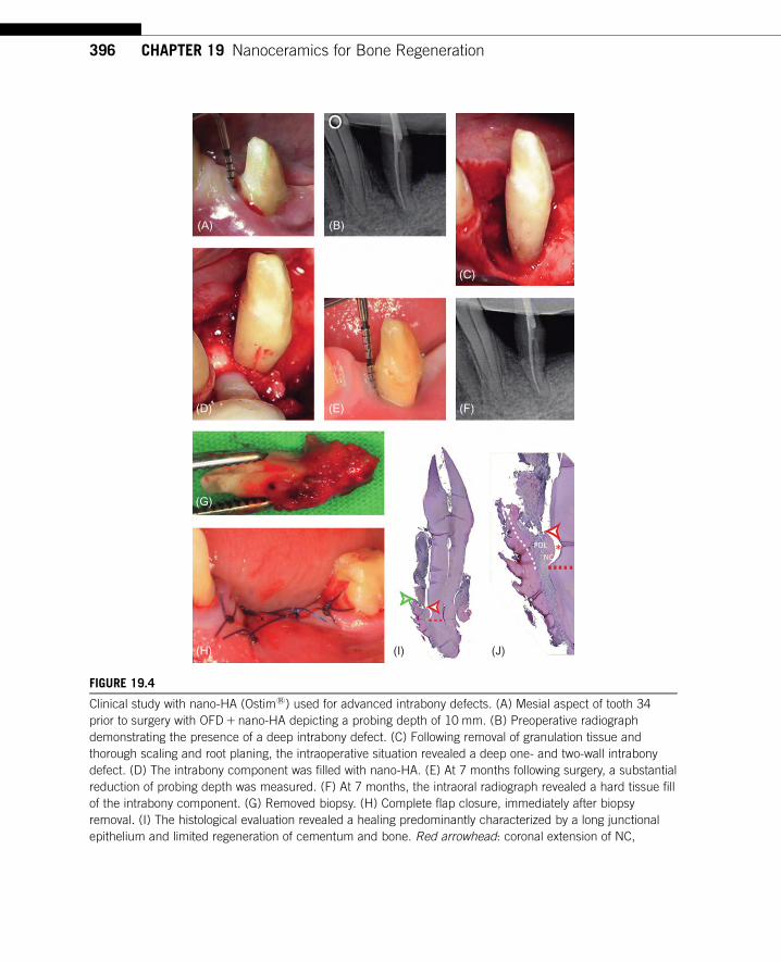
(A) (B)
(C)
(D) (E)
(G)
(H) (I) (J)
(F)
FIGURE 19.4
Clinical study with nano-HA (Ostims) used for advanced intrabony defects. (A) Mesial aspect of tooth 34
prior to surgery with OFD1 nano-HA depicting a probing depth of 10 mm. (B) Preoperative radiograph
demonstrating the presence of a deep intrabony defect. (C) Following removal of granulation tissue and
thorough scaling and root planing, the intraoperative situation revealed a deep one- and two-wall intrabony
defect. (D) The intrabony component was filled with nano-HA. (E) At 7 months following surgery, a substantial
reduction of probing depth was measured. (F) At 7 months, the intraoral radiograph revealed a hard tissue fill
of the intrabony component. (G) Removed biopsy. (H) Complete flap closure, immediately after biopsy
removal. (I) The histological evaluation revealed a healing predominantly characterized by a long junctional
epithelium and limited regeneration of cementum and bone. Red arrowhead: coronal extension of NC,
396 CHAPTER 19 Nanoceramics for Bone Regeneration

19.4 Nano-HA�collagen compositesIn addition to nano-HA preparations, in an attempt to mimic natural bone even more closely,
several researchers have fabricated nano-HA�collagen composites (nano-HA/collagen) [21�23]. In
particular, one laboratory has prepared a hierarchical structure resembling natural bone by allowing
self-assembly of collagen triple helices and the formation of nano-HA crystals on the surface of
these fibrils [23]. In this technique, nano-HA crystals grow on the surface of collagen fibrils such
that their c-axes orient along the longitudinal axes of the collagen fibrils and the mineralized fibrils
become aligned parallel to each other forming mineralized collagen fibers as in situ in bone. In
vitro studies have shown that this composite supports osteoblastic cell growth and new bone forma-
tion [24,25]. The nano-HA/collagen-based scaffolds have now been successfully used in many clin-
ical cases requiring various types of bone repair [26] with wound healing and no abnormalities
found in local and systematic examinations during long-term follow-up [27].
19.5 Hydrogels and nano-HAOther studies, although recognizing the advantages of nano-HA for bone regenerative purposes,
have made efforts to improve some of the properties of this material with respect to controlling its
biodegradability and porosity for better bioactivity. Because of the inherent ability of hydrogels to
swell in aqueous media and to permit the transport of enzymes and nutrients to and through various
supporting ceramic scaffolds, there has been increasing interest in the use of hydrogels with cera-
mics in tissue engineering [28,29].
Chitosan is a promising hydrogel material for bone regeneration because it is biocompatible
and biodegradable with a degradation rate that is dependent on factors such as degree of deacetyla-
tion and crystallinity [30]. It can also be easily formed into beads, fibers, or more complex
structures [31,32]. In another recent study [33], β chitin hydrogel/nano-HA composite scaffolds
were synthesized and shown to have improved porosity, swelling ability, protein adsorption, and
retention as well as biomineralization properties for use as a potential candidate for bone tissue
engineering applications. Although it appears to date that only in vitro studies and a preliminary
in vivo study in a rat calvarial defect model [30] have been conducted with this type of nanocom-
posite scaffolds, there have been a series of studies that have tested the combination of polymers
such as chitin with traditional-sized HA. These earlier studies which do include in vivo experi-
ments suggest that the composites support bony ingrowths into the implant as the matrix gradually
resorbs [34�36].
� green arrowhead: coronal extension of new bone, red dotted line: apical extension of the notch. Original
magnification 3 5. (J) Higher magnification of the defect shown in (I). Formation of NC and new periodontal
ligament (PDL) was confined to the area of the notch. Red arrowhead: coronal extension of NC, red dotted
line: apical extension of the notch, white dotted line: margin between the newly formed bone and old bone,
red asterisk: artifact. Original magnification 3 25. (For interpretation of the references to color in this
figure legend, the reader is referred to the web version of this book.)
Figure from Ref. [20] with permission from publisher.
39719.5 Hydrogels and nano-HA

The composites may have optimal porosity to support the attachment and growth of osteoblastic
cells with sufficient retention of plasticity for ease of filling defects while maintaining sufficient
mechanical strength for support.
Metallic nanoparticles such as copper and zinc have high antibacterial activity, low toxicity, and
chemical stability. Moreover, zinc has been shown to be an important trace element in bone [37],
required for cell proliferation [38] and it has been suggested to play an important role in collagen
production and biomineralization [39,40]. Because of these properties, these metals have been uti-
lized in the fabrication of materials for bone tissue engineering. The addition of nanocopper and
zinc has been recently shown to significantly increase swelling, decrease degradation, increase
protein adsorption, and increase antibacterial activity in chitosan/nano-HA scaffolds [41]. These
composites have been shown to have no toxicity toward osteoprogenitor cells and have therefore
been postulated to have advantages over the chitosan nano-HA scaffolds without added metals for
use in osseous regeneration in many critical sites such as in the oral cavity where antimicrobial
effects might be particularly useful [41]. Although to date it does not appear that clinical studies
with chitosan/nano-HA composites of this type have been reported, the experimental in vitro stud-
ies do target them as promising scaffolds for use in oral bone tissue engineering. Their development
should be further pursued in order to optimize the bioresorbability and mechanical strength proper-
ties of the scaffold material for various craniofacial bone and periodontal intrabony defect sites.
19.6 Chitosan and nano-bioactive glass compositesOther variations of chitosan and nanoceramics have recently been developed and tested for bone
tissue engineering. In particular, a chitosan�gelatin/nano-bioactive glass ceramic composite has
been shown to have many excellent properties for use in alveolar bone tissue regeneration [42]. This
composite extends the valuable characteristics of chitosan discussed above with the blending of gela-
tin, a unique sequence of amino acids such as glycine, proline, and hydroxyproline that enhances cell
attachment and nanoparticles of glass ceramics that are osteoconductive and biodegradable.
Bioactive glass particles, particularly those synthesized by a sol�gel process, have been shown to
bond to hard tissues because of their ability to form a surface layer of hydroxycarbonate apatite and
produce no local or systemic toxicity or inflammatory or foreign-body response [6,43,44]. A compos-
ite of chitosan�gelatin and nanoglass ceramic can be fabricated to have pore sizes in the range of
150�300 μm which should be optimal for migration of cells into the interior of the scaffold and osse-
ous ingrowths and vascularization [45]. The degradation and swelling behavior of the composite
scaffold also appeared optimized for cell attachment and spreading with biomineralization occurring
with the formation of an apatite layer on the surface of the composite [42]. These properties point to
the usefulness of these composites in tissue engineering applications in bone regeneration.
19.7 Nanocalcium sulfateCalcium sulfate (CS) is another highly biocompatible material with a long clinical history as a syn-
thetic ceramic material. The hemihydrate form of CS, also known as plaster of Paris, is one of the
398 CHAPTER 19 Nanoceramics for Bone Regeneration

simplest synthetic bone-like grafts with a report in 1892 by Dreesman, as described by Peltier,
showing its use to fill bone cavities which later were found to be filled with solid bone [46].
CS has been further demonstrated in many animal and clinical studies to be an effective osteoconduc-
tive scaffold that enhances bone regeneration [47]. In our laboratory, CS has been shown to adsorb
platelet-derived growth factor (PDGF-BB) and to support enhanced human osteoblastic cell prolifera-
tion in vitro when treated with the growth factor [48]. Although CS is a desirable osteoconductive
material and potential carrier of osteoinductive factors in its presently available state, it has some
deficiencies, including slow and variable degradation rate and weak mechanical properties [49]. In
an attempt to use nanotechnology to improve some of the properties of CS, our laboratory has been
working to fabricate a nanocalcium sulfate (nCS) scaffold material. We recently presented our stud-
ies on the use of a cryo-vacuum technique to process dihydrate CS into dihydrate nCS, which was
then subjected to oven drying to produce a hemihydrate nCS. The cryo-vacuum process for prepar-
ing CS dihydrate nanocrystals was based on that previously demonstrated by Salvadori et al. [50].
The advantages of this cryo-vacuum technique are that it is simple and avoids the introduction of
any surfactants or other components beyond CS and water.
The nCS synthesized in this manner was sterilized by glow discharge treatment, a practical
method for most clinical situations. Electron microscopy showed that the nCS powder consists of
aggregates of closely arranged acicular crystals, approximately 30�80 nm in width, 400�600 nm
in length and approximately 80�100 nm in diameter, providing a surface area as determined by
Brunauer, Emmett, and Teller surface area analysis using a Micromeretics Model 2000 ASAP nitro-
gen physisorption apparatus, to be about 10 times that of conventional CS. Physicochemical charac-
terization confirmed the composition and phase of the material. Surface microhardness testing
showed that the nCS was stronger than conventional CS and may provide an additional advantage
to the scaffolding properties of the material. Cell viability/metabolic activity assays with human
osteoblastic and PDL cells verified the safety and biocompatibility of nCS and alkaline phosphatase
assays showed that the material supports the differentiation of osteoblastic cells [49].
Studies from our lab as well as many others point to the potential use of scaffold materials
as carriers that can release and maintain levels of growth factors to aid in their ability to facili-
tate bone repair [51]. Release kinetics for adsorbed PDGF and bone morphogenetic protein-2
(BMP-2) [49] suggested that nCS may serve as an appropriate vehicle for slow release delivery
of these agents that have been approved for clinical use in a number of bone regenerative
procedures [52,53].
Our laboratory has found that when human recombinant BMP-2 (rhBMP-2) was mixed with CS
(conventional size, medical-grade calcium sulfate) or nCS and allowed to dry into disks of equal
weight and proportion, there was a significantly greater amount of the BMP-2 released from the
nCS disks at 37�C over a 7-day period as measured with a specific immunoassay for the growth
factor. However, when a mixture of 10% alginate, a natural polysaccharide, and nCS was mixed
with the BMP-2, there was a much faster and significantly greater release of the BMP-2 compared
to nCS alone with a significant amount released after 2 h that did not change for up to 7 days. This
study suggests that the use of alginate can significantly improve the properties of an nCS scaffold
at least with respect to the release of growth factors (Figure 19.5).
However, although these data suggest that nCS has release characteristics for growth factors
different than conventional-sized CS and that they can be modified with the use of alginate, they
do not provide sufficient information to predict how this nCS material will function as a vehicle
39919.7 Nanocalcium sulfate

delivery agent in various in vivo situations where bone formation is the desired end function.
Although our in vitro data suggest that nCS releases only a small percentage of absorbed BMP-2,
an effective dose cannot be predicted since the in vivo conditions in a particular site may signifi-
cantly alter the kinetics of release and availability of the growth factor at that site. The complexities
involved in the delivery of growth factors to in vivo sites in tissue engineering strategies have been
reviewed [54,55] and it must be recognized that extrapolation from in vitro studies to a particular
in vivo model is extremely limited.
Therefore, as an extension of these in vitro studies, most recently, we also tested nCS in the rat
calvarial defect model, which is a well-studied model that allows in vivo testing of the bone regen-
erative ability of a material [56,57]. All protocols used here were approved by the Institutional
Animal Care and Use Committee of State University of New York at Buffalo, New York, USA.
Basically the technique involved creating an 8 mm critical size defect using a low-speed hand piece
and trephine drill in the calvaria of adult male Sprague-Dawley rats. The cranial defect created in
each rat was then filled with a constant amount (100 mg) of either nCS alone, or nCS mixed with
10 or 50% alginate with and without PDGF-BB or rhBMP-2. In some other rats, the defect was
filled with Capsets which was a commercially available conventional-sized CS, or GEM21s, a
commercially available (Osteohealth) mixture of beta TCP and PDGF, or DFDBA (demineralized
freeze-dried bone allograft, commercially available, Dentsply). In the negative control group, the
defects were left untreated. There were a minimum of four animals in each treatment and control
group. In all animals, the overlying tissues were closed in layers with resorbable 4�0 Vicryls
sutures. As shown in Figure 19.6, radiographic analysis using Image J software to measure bone
filling in the calvaria from the margin of defect after 2 weeks of treatment, revealed that were no
1400
1200
1000
800
600
400
200
−200
00 h 2 h 24 h 7 days
Time
pg/m
L
pg/mL CS
pg/mL nCS
pg/mL nCS+alg
FIGURE 19.5
Release of rhBMP-2 from nCS alone, nCS1 10% alginate in comparison to CS. Disks were fabricated from
the scaffold materials with rhBMP-2 (Prospec, Rehovot, Israel), added at a final concentration of 1 μg/disk.BMP-2 released from the disks at 37�C was measured with a specific immunoassay (Quantikine Kit, R&D
Systems). Values are the mean6 standard deviation of four samples in each group.
400 CHAPTER 19 Nanoceramics for Bone Regeneration

significant differences between control and Capsets, GEM21s and nCS. However, there were
significant increases compared to control with DFDBA, BMP-2 alone and all nCS samples that
included BMP-2. It is also interesting to note that the combination of nCS with alginate resulted in
greater bone fill than nCS alone and comparison of the samples treated with nCS to those with
Capsets did show a greater fill with nCS although at this time period, neither resulted in signifi-
cantly greater effects compared to untreated controls. It can also be observed that under these
conditions, PDGF-BB either added as a part of the GEM21s treatment or with the nCS did not pro-
duce increases in bone fill. This experiment suggested that a combination of nCS with alginate
might enhance the bone-filling properties of the material at least with respect to some critical bone
defects sites and lead to further studies on the manner in which alginate might be modifying the
properties of the nanomaterial.
Alginate, a natural polysaccharide extracted from brown sea algae, has been extensively used in
various aspects of tissue engineering because it is biocompatible, hydrophilic, and biodegradable
under normal physiological conditions [58]. It forms stable hydrogels in the presence of certain
divalent cations (e.g., Ca21) in low concentrations [59]. When scaffolds containing alginate are
placed in a liquid milieu, there is uncontrolled degradation of ionically cross-linked alginate due to
loss of divalent cations. This leads to formation of pores inside the scaffold. In the in vivo experi-
ment described above, alginate was added to nCS powder to induce in situ pore formation and
60.000%Bone fill 2 weeks
50.000%
40.000%
30.000%
20.000%
10.000%
0.000%
Contro
l
GEM21
DFDBABM
P
Capse
tnC
S
nCS +
A10
nA10
+ B
...
nA10
+ P
...
nA50
+ B
...
nA50
+ P
...
FIGURE 19.6
Bone fill in rat calvarial defects. Rat calvarial defects were filled with either GEM21s (300 μg/mL PDGF in
sterile water mixed 1 to 1 with solid TCP), DFDBA, Capsets (medical-grade CS), BMP (1500 μg BMP-2/mL
deposited on a collagenous sponge matrix), nCS alone, nCS1A10 (nCS with 10% alginate), nA101B
(nCS1 10% alginate1BMP-2 [900 μg/g nCS]) and nA101P (nCS1 10% alginate1PDGF-BB[180 μg/gnCS]), nA501B (nCS1 50% alginate1BMP-2 [900 μg/g nCS]), nA501P (nCS1 50% alginate1PDGF-BB
[180 μg/g nCS]). The controls were nonfilled defect. There were four animals/treatment group. The mixtures
of alginate and nCS increased the bone fill compared to nCS alone. The bone fill was significantly greater with
the combination of nCS1 alginate and BMP-2 compared to GEM21s, DFDBA, but was not significantly
different than with BMP-2 applied on a collagenous sponge.
40119.7 Nanocalcium sulfate

optimize the properties of the implant scaffold to lead to enhanced bone fill at the defect site.
A previous study with an alginate/HA composite had shown that scaffolds of this type have a
porous structure that supports osteoblastic cell attachment and growth [60]. It appears the addition
of alginate to nCS optimizes the conditions to enable more osteoblastic cell growth and thus more
bone fill in the rat calvarial model, however, more studies are needed to understand the nature of
alginate’s effects in this system.
In order to better understand the nature of the scaffold created by combining nCS and alginate
the microscopic structure and cell-material interaction of different proportions of nCS and alginate
were studied in our laboratory. Samples of different proportions of alginate (5%, 10%, 15%, 20%,
and 50% by weight) and nCS were incubated in water for 24 h. It was noted that once the scaffolds
were placed in water, they started to absorb water and swell, with the loss of divalent cations in the
surrounding liquid causing dissolution of the ionically cross-linked alginate. The integrity of the
50%, 20%, and 15% alginate scaffolds was lost completely after 24 h. Although the 10% and 5%
alginate scaffolds still kept their shape, their mechanical properties were weakened (Figure 19.7).
Scanning electron microscope (SEM) analysis showed increased roughness on the surface which
may be due to the sponge-like consistency of the scaffold (Figure 19.8). The alginate absorbed
water and became gelatinous with the gel layer isolating nanoparticles of CS and covering the
FIGURE 19.7
Dissolution behavior of nCS and alginate composite scaffolds after 24 h incubation in water. From left to right:
nCS:alginate5 50:50, 80:20, 85:15, 90:10, and 95:5.
200 µm 150X 200 µm 150X 200 µm 150X
(A) (B) (C)
FIGURE 19.8
(A)�(C) SEM images of nCS and alginate samples after 24 h of incubation in water at 1503 magnification.
(A) nCS:alginate5 95:5, (B) nCS:alginate5 90:10, and (C) nCS:alginate5 50:50.
402 CHAPTER 19 Nanoceramics for Bone Regeneration

space in between the clustered crystals of nCS (Figure 19.9) and creating macropores within the
scaffold.
The integrity of the 50%, 20% and 15% alginate, nCS scaffolds was lost completely after 24 h.
Although the 10% and 5% alginate, nCS scaffolds still kept their shape, their mechanical properties
were weakened.
Cell attachment studies were conducted by seeding human PDL cells on the nCS1 alginate samples
and incubating for 24 h under standard conditions with MEM media. SEM studies revealed that the
cells attached to 95 nCS:5 alginate and the 90 nCS:10 alginate scaffolds but not to the 50:50 scaffolds
with the greatest attachment to the 95:5 scaffolds. Also it was observed that the cells attached to the
nCS:alginate scaffolds were spherical rather than elongated like those attached to the nCS alone scaf-
folds (Figure 19.10). Cell viability assays, however, revealed no significant differences among cells
cultured on the nCS:alginate scaffolds although the 95 nCS:5 alginate group did possess the highest
values of viability compared to the other two composite groups and nCS alone (Figure 19.11).
nCS:alginate (90:10)
(A) (B)
nCS:alginate (50:50)
FIGURE 19.9
SEM images of nCS and alginate samples after setting without incubation: (A) Alginate layer wraps around the
cluster of crystals of nCS. (B) More alginate layers than separate clusters of nCS particles.
60 µm 500X 60 µm 500X 60 µm 500X
(A) (B) (C)
FIGURE 19.10
SEM images of periodontal cell attachment on nCS scaffolds after 24 h of incubation (5003 ). (A) 95 nCS:5
alginate; (B) 50 nCS:50 alginate; (C) nCS alone.
40319.7 Nanocalcium sulfate

Although the nCS1 alginate scaffold developed in our laboratory has not been yet tested in
clinical studies, on the basis of the in vitro work and the in vivo animal studies, there are no appar-
ent reasons why the material should not be biocompatible and effective in supporting bone regener-
ation in the craniofacial as well as other bony sites. We are presently in the process of optimizing
conditions of the nCS1 alginate mixture so that it can be used as an injectable product with or
without growth factors in a variety of clinical conditions.
Presently, there is a different nCS product that is available for clinical use. This material is
marketed and sold as NanoGens (Orthogen) and has been approved for clinical use in the United
States. Since it is a proprietary product, the exact conditions of its fabrication are not known.
However, according to Orthogen’s website,2 it has unique microscopic structure and properties.
Although the material is described as a “nanocrystalline calcium sulfate bone graft,” it is also stated
on the company’s website that in the fabrication of the material microcrystalline CS is converted to
grains of CS in the range of 200�900 nm that are then compressed to form granulates in the sizes
ranged from 400�1000 μm, clearly not in the range of nanosized particles as they are usually
defined. Although to date there does not appear to be studies yet published documenting the claims,
the website,2 also states that NanoGens undergoes controlled degradation over a period of 12
weeks in contrast to the 4�6 weeks for traditional CS. Moreover, there are claims that the material
is completely replaced with regenerated vital bone, critical to its success as a bone graft. The
material is purposed to be used as a stand-alone bone graft material, in combination with other
bone graft materials or to serve as a resorbable barrier over other bone grafts in postextraction sites,
periodontal infra-osseous defects, apicoectomy, root perforations, dehiscence and fenestrations,
mini and great sinus lifting. There is a published case report in which the NanoGens material has
been shown to be effective in regeneration of bone in an extraction socket of a 55-year-old female
patient with histomorphometric analysis of the bone core extracted from the regenerated socket
6 months after grafting showing 47% vital bone volume with osteoclasts and osteoblastic remodel-
ing involvement [61]. Although this one case does suggest that the properties of the CS product
0.400
0.300
0.200
0.100
0.000Control nCS 95:5 90:10
MTT assay
50:50 nCS:alg
FIGURE 19.11
Periodontal cell viability tests with nCS and nCS alginate scaffolds. The MTT (3-(4,5-dimethyl-thiazol-2-yl)-
2,5-diphenyl tetrazolium bromide) cell assay was conducted according to the method described previously to
measure the metabolic cell activity as an assessment of viability in the presence of the nCS-alginate scaffolds
[49]. Control represents cells grown in tissue culture wells without added scaffolds. Viability data are
expressed as average OD6 standard deviation with n5 4 samples per group.
2http://www.orthogencorp.com/
404 CHAPTER 19 Nanoceramics for Bone Regeneration
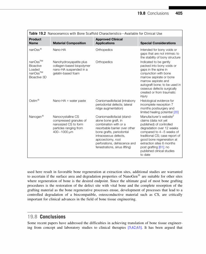
used here result in favorable bone regeneration at extraction sites, additional studies are warranted
to ascertain if the surface area and degradation properties of NanoGens are suitable for other sites
where regeneration of bone is the desired endpoint. Since the ultimate goal of most bone grafting
procedures is the restoration of the defect site with vital bone and the complete resorption of the
grafting material as the bone regenerative processes ensue, development of processes that lead to a
controlled degradation of a biocompatible, osteoconductive material such as CS, are critically
important for clinical advances in the field of bone tissue engineering.
19.8 ConclusionsSome recent papers have addressed the difficulties in achieving translation of bone tissue engineer-
ing from concept and laboratory studies to clinical therapies [5,62,63]. It has been argued that
Table 19.2 Nanoceramics with Bone Scaffold Characteristics—Available for Clinical Use
ProductName Material Composition
Approved ClinicalApplications Special Considerations
nanOsss Nano-HA Orthopedics Intended for bony voids orgaps that are not intrinsic tothe stability of bony structure
nanOssTM
BioactiveLoadednanOssTM
Bioactive 3D
Nanohydroxyapatite pluscollagen-based biopolymernano-HA suspended in agelatin-based foam
Orthopedics Indicated to be gentlypacked into bony voids orgaps in the spine inconjunction with bonemarrow aspirate or bonemarrow aspirate andautograft bone; to be used inosseous defects surgicallycreated or from traumaticinjury
Ostims Nano-HA1water paste Craniomaxillofacial (intrabonyperiodontal defects; lateralridge augmentation)
Histological evidence forincomplete resorption 7months postsurgery andlimited healing potential [20]
Nanogens Nanocrystalline CScompressed granules ofnanosized CS to formparticles ranging from400�1000 μm
Craniomaxillofacial (stand-alone bone graft, incombination with or aresorbable barrier over otherbone grafts, periodontalintraosseous defects,apicoectomy, rootperforations, dehiscence andfenestrations, sinus lifting)
Manufacturer’s website2
claims (data not yetpublished) of controlleddegradation over 12 weekscompared to 4�5 weeks oftraditional CS; case report ofgood bone regeneration atextraction sites 6 monthspost grafting [61]; nopublished clinical studiesto date
40519.8 Conclusions

despite over 25 years of research with extensive funding and over 12,000 papers on bone tissue
engineering and over 2000 papers on bone scaffolds alone in the past 10 years, there are very lim-
ited numbers of bone tissue engineering clinical products and auto or allo bone grafts remain the
gold standard and treatment of choice, for large defects, especially in craniomaxillofacial recon-
struction [5]. For any therapy to be successfully adapted for clinical use, there are significant tech-
nical, business, and philosophical barriers that must be overcome. It has been argued that the
technical challenges for scaffold translation alone are significant in that the materials must fill the
“Form, Fixation, Function and Formation needs” [5] of repair of bony defects. However, it has also
been noted that this can be best accomplished by targeting specific clinical applications and then
developing a material as a modular system with increasing levels of complexity as needed [5]. This
is the approach that appears to be emerging with the development of nanoceramics bone tissue
engineering therapies in the craniomaxillofacial and oral cavity (Table 19.2).
As pointed out in this chapter, nanohydroxyapatite is presently commercially available for clini-
cal use as Ostims and nanOsss with complexity to the material recently introduced with nanOss
Bioactives bone graft that utilizes a collagen- or gelatin-based biopolymer in addition to the nano-
HA material.3 As further discussed here, nanomaterials such as the HA as well as bio active glass
and CS can be further fabricated with materials such as chitosan and alginate to modify the porosity
and growth factor delivery properties of the nanoscaffold for a more optimal functional form
designed for a particular clinical procedure. Although there are many components such as cell
sources, growth factors, and signaling pathways, in addition to the scaffold material, that have to be
considered in the fabrication of an ideal bone tissue engineered product, there is an ever increasing
body of basic and preclinical studies that suggest that nanoceramics will continue to play an
important role in the development of sound clinical therapeutics for repair of critical defects in the
craniofacial region as well as throughout the skeleton.
AcknowledgmentThe authors are grateful for the clinical insight of Dr. Sebastiano Andreana throughout our work with CS and
especially during the preparation of this paper.
References[1] R. Langer, J.P. Vacanti, Tissue engineering, Science 260 (1993) 920�926.
[2] P.G. Robey, Cell sources for bone regeneration: the good, the bad, and the ugly (but promising), Tissue
Eng. 17 (2011) 423�430.
[3] F.-M. Chen, M. Zhang, Z.-F. Wu, Toward delivery of multiple growth factors in tissue engineering,
Biomaterials 31 (2010) 6279�6308.
[4] Q. Chen, J.A. Roether, A.R. Boccaccini, Bioactive glass and composite materials, in: W. Ashammakhi,
R. Reis, F. Chiellini (Eds.), Topics in Tissue Engineering, vol. 4, 2008, pp. 1�27. Chapter 6
3http://www.prweb.com/releases/2012/5/prweb9555762.htm
406 CHAPTER 19 Nanoceramics for Bone Regeneration

[5] S.J. Hollister, W.L. Murphy, Scaffold translation: barriers between concept and clinic, Tissue Eng. Part
B 17 (2011) 459�474.
[6] T.J. Webster, C. Ergun, R.H. Doremus, R.W. Siegel, R. Bizios, Enhanced functions of osteoblasts on
nanophase ceramics, Biomaterials 21 (2000) 1803�1810.
[7] Z. Shi, X. Huang, Y. Cai, R. Tang, D. Yang, Size effect of hydroxyapatite nanoparticles on proliferation
and apoptosis of osteoblast-like cells, Acta Biomater. 5 (2009) 338�345.
[8] H. Liu, T.J. Webster, Nanomedicine for implants: a review of studies and necessary experimental tools,
Biomaterials 28 (2007) 354�369.
[9] P. Li, Biomimetic nano-apatite coating capable of promoting bone ingrowth, J. Biomed. Mater. Res. A
66 (2003) 79�85.
[10] N. Tran, T.J. Webster, Nanotechnology for bone materials, WIREs nanomed, Nanobiotechnol 1 (2008)
336�351.
[11] G.M. Cunniffe, F.J. O’Brien, S. Partap, T.J. Levingstone, K.T. Staton, G.R. Dickson, The synthesis and
characterization of nanophase hydroxyapatite using a novel dispersant-aided precipitation method,
J. Biomed. Mater. Res. 95A (2010) 1142�1149.
[12] P.N. Kumta, C. Sfeir, D.H. Lee, D. Olton, D. Choi, Nanostructured calcium phosphates for biomedical
applications: novel synthesis and characterization, Acta Biomater. 1 (2005) 65�83.
[13] M. Thorwarth, S. Schultze-Mosgau, P. Kessler, J. Wiltfang, K.A. Schlegel, Bone regeneration in osseous
defects using a resorbable nanoparticular hydroxyapatite, J. Oral Maxillofac. Surg. 63 (2005)
1626�1633.
[14] C. Spies, S. Shnurer, T. Gotterbarm, S. Breusch, Animal study of the bone substitute material Ostim
within osseous defects in Gottinger minipigs, Z. Orthop. Unfall. 145 (2008) 64�69.
[15] F. Schwarz, K. Bieling, T. Latz, E. Nuesry, J. Becker, Healing of intrabony peri-implantitis defects
following application of a nanocyrstalline hydroxyapatite (Ostim) or a bovine-derived xenograft
(Bio-Oss) in combination with a collagen membrane (Bio-Gide). A case series, J. Clin. Periodontol. 33
(2006) 491�499.
[16] F.P. Strietzel, P.A. Reichart, H.L. Graf, Lateral alveolar ridge augmentation using a synthetic nano-
crystalline hydroxyapatite bone substitution material (Ostims). Preliminary clinical and histological
results, Clin. Oral Implants Res. 18 (2007) 743�751.
[17] F.X. Huber, N. McArthur, J. Hillmeier, H.J. Kock, M. Baier, M. Diwo, et al., Void filling of tibia com-
pression fracture zones using a novel resorbable nanocrystalline hydroxyapatite paste in combination
with a hydroxyapatite ceramic core: first clinical results, Arch. Orthop. Trauma. Surg. 126 (2006)
533�540.
[18] A. Kasaj, B. Rhrig, G.G. Zafiropoulos, B. Willershausen, Clinical evaluation of nanocrystalline hydroxy-
apatite paste in the treatment of human periodontal bony defects�a randomized controlled clinical trial:
6-month results, J. Periodontol. 79 (2008) 394�400.
[19] B. Heinz, A. Kasaj, M. Teich, S. Jepsen, Clinical effects of nanocrystalline hydroxyapatite paste in the
treatment of intrabony periodontal defects: a randomized controlled clinical study, Clin. Oral Invest. 14
(2010) 525�531.
[20] A. Horvath, A. Stavropoulos, P. Windisch, L. Lukacs., I. Gera, A. Sculean, Histological evaluation of
human intrabony periodontal defects treated with an unsintered nanocrystalline hydroxyapatite paste,
Clin. Oral Invest. (2012) (published online, May 12).
[21] J. Aizenberg, J.C. Weaver, M.S. Thanawala, V.C. Sundar, D.E. Morse, P. Fratzl, Skeleton of Euplectella
sp.: structural hierarchy from the nanoscale to the macroscale, Science 309 (2005) 275�278.
[22] M.C. Chang, T. Ikoma, M. Kikuchi, J. Tanaka, Preparation of a porous hydroxyapatite/collagen
nanocomposite using glutaraldehyde as a crosslinkage agent, J. Mater. Sci. Lett. 20 (2001)
1199�1201.
407References

[23] W. Zhang, S.S. Liao, F.Z. Cui, Hierarchical self-assembly of nanofibrils in mineralized collagen, Chem.
Mater. 15 (2003) 3221�3226.
[24] C. Du, F.Z. Cui, W. Zhang, Q.L. Feng, X.D. Zhu, K. de Groot, Formation of calcium phosphate/collagen
composites through mineralization of collagen matrix, J. Biomed. Mater. Res. 50 (2000) 518�527.
[25] S.S. Liao, F.Z. Cui, W. Zhang, Q.L. Feng, Hierarchically biomimetic bone scaffold materials: nano-HA/
collagen/PLA composite, J. Biomed. Mater. Res. B Appl. Biomater. 69 (2004) 158�165.
[26] Z. Chen, H. Liu, X. Liu, F.Z. Cui, Injectable calcium sulfate/mineralized collagen-based bone repair
materials with regulable self-setting properties, J. Biomed. Mater. Res. Part A 99A (2011) 554�563.
[27] X. Yu, L. Xu, L.Y. Bi, Y. Qu, D.B. Zheng, X. Cao, Posterolateral fusion using nano-crystal/collagen
bone materials in lumbar spine, Orthop. J. China 13 (2005) 586�588.
[28] K. Gkioni, S.C.G. Leeuwenburgh, T.E.L. Douglas, A.G. Mikos, J.A. Jansen, Mineralization of hydrogels
for bone regeneration, Tissue Eng.: Part B 16 (2010) 577�585.
[29] H. Tamura, T. Furuike, S.V. Nair, R. Jayakumar, Biomedical applications of chitin hydrogel membranes
and scaffolds, Carbohydr. Polym. 84 (2011) 821�825.
[30] B.M. Chesnutt, Y. Yuan, K. Buddington, W.O. Haggard, J.D. Bumgardner, Composite chitosan/nano-
hydroxyapatite scaffolds induce osteocalcin production by osteoblasts in vitro and support bone forma-
tion in vivo, Tissue Eng. Part A 15 (2009) 2571�2579.
[31] A. Di Martino, M. Sittinger, M.V. Risbud, Chitosan: a versatile biopolymer for orthopaedic tissue-
engineering, Biomaterials 26 (2005) 5983�5990.
[32] E. Khor, Chitosan: Fulfilling a Biomaterial’s Promise, Elsevier, Oxford, UK, 2001.
[33] R. Jayakumar, K.P. Chennazhi, S. Srinivasan, S.V. Nair, T. Furuike, H. Tamura, Chitin scaffolds in tis-
sue engineering, Int. J. Mol. Sci. 12 (2011) 1876�1987.
[34] S. Higashi, T. Yamamuro, T. Nakamura, Y. Ikada, S.H. Hyon, K. Jamshidi, Polymer-hydroxyapatite
composites for biodegradable bone filler, Biomaterials 7 (1986) 183�187.
[35] M. Ito, In vitro properties of a chitosan-bonded hydroxyapatite bone-filling paste, Biomaterials 12
(1991) 41�45.
[36] A.C.A. Wan, E. Khor, G.W. Hastings, Hydroxyapatite modified chitin as potential hard tissue substitute
material, J. Biomed. Mater. Res. 38 (1997) 235�241.
[37] H.W. Kim, H.E. Kim, V. Salihi, Stimulation of osteoblast responses to biomimetic nanocomposites of
gelatin�hydroxyapatite for tissue engineering scaffolds, Biomaterials 26 (2005) 5221�5230.
[38] G. Wei, P. Ma, Structure and properties of nano-hydroxyapatite/polymer composite scaffolds for bone
tissue engineering, Biomaterials 25 (2004) 4749�4757.
[39] M. Yamaguchi, H. Oishi, Y. Suketa, Stimulatory effect of zinc on bone formation in tissue culture,
Biochem. Pharmacol. 22 (1987) 4007�4012.
[40] D. Chen, L. Waite, W.M. Pierce Jr, In vitro effects of zinc on markers of bone formation, Biol. Trace
Elem. Res. 68 (1999) 225�234.
[41] A. Tripathi, S. Saravanan, S. Pattnaik, A. Moorthi, N.C. Partridge, N. Selvamurugan, Bio-composite
scaffolds containing chitosan/nano-hydroxyapatite/nano-copper�zinc for bone tissue engineering, Int. J.
Biol. Macromol. 50 (2012) 294�299.
[42] M. Peter, N. Ganesh, N. Selvamurugan, S.V. Nair, T. Furuike, H. Tamura, et al., Preparation and charac-
terization of chitosan-gelatin/nanohydroxyapatite composite scaffolds for tissue engineering applications,
Carbohydr. Polym 80 (2010) 687�694.
[43] M. Vogel, C. Voigt, U.M. Gross, C.M. Mai, In vivo comparison of bioactive glass particles in rabbits,
Biomaterials 22 (2001) 357�362.
[44] W. Xia, J. Chang, Preparation and characterization of nano-bioactive-glasses (NBG) by a quick alkali-
mediated sol�gel method, Mater. Lett. 61 (2007) 3251�3253.
408 CHAPTER 19 Nanoceramics for Bone Regeneration

[45] V. Karageorgiou, D. Kaplan, Porosity of 3D biomaterial scaffolds and osteogenesis, Biomaterials 26
(2005) 5474�5491.
[46] L.F. Peltier, The use of plaster of Paris to fill defects in bone, Clin. Orthopedics 21 (1961) 1�29.
[47] M.P. Sidqui, P. Collin, C. Vitte, N. Forest, Osteoblast adherence and resorption activity of isolated osteo-
clasts on calcium sulphate hemihydrate, Biomaterials 16 (1995) 1327�1332.
[48] J. Bateman, G. Intini, J. Margarone III, S. Goodloe III, P. Bush, S.E. Lynch, et al., Platelet-derived
growth factor enhancement of two alloplastic bone matrices, J. Periodontol. 76 (2005) 1833�1841.
[49] Y.B. Park, K. Mohan, A. Al-Sanousi, B. Almaghrabi, R.J. Genco, M.T. Swihart, et al., Synthesis and
characterization of nanocrystalline calcium sulfate for use in osseous regeneration, Biomed. Mater. 6
(2011) 055007.
[50] B. Salvadori, G. Capitani, M. Mellini, L. Dei, A novel method to prepare inorganic water-soluble nano-
crystals, J. Colloid Interface Sci. 298 (2006) 487�490.
[51] Q. Zhang, Q.F. He, T.H. Zhang, X.L. Yu, Q. Liu, F.L. Deng, Improvement in the delivery system of
bone morphogenetic protein-2: a new approach to promote bone formation, Biomed. Mater. 7 (2012)
045002.
[52] J.O. Hollinger, C.E. Hart, S.N. Hirsch, S. Lynch, G.E. Friedlaender, Recombinant human platelet derived
growth factor: biology and clinical applications, J. Bone Joint Surg. Am. 80 (2008) 48�54.
[53] D.H.R. Kempen, L. Lu, T.E. Hefferan, L.B. Creemers, A. Maran, K.L. Classic, et al., Retention of
in vitro and in vivo BMP-2 bioactivities in sustained delivery vehicles for bone tissue engineering,
Biomaterials 29 (2008) 3245�3252.
[54] H. Uludag, T. Gao, T.J. Porter, W. Friess, J.M. Wozney, Delivery systems for BMPs: factors
contributing to protein retention at an application site, J. Bone Joint Surg. Am. 83A (2001) S128�S135.
[55] M. Nakashima, A. Reddi, The application of bone morphogenetic proteins to dental tissue engineering,
Nat. Biotechnol. 21 (2003) 1025�1032.
[56] H. Perinpanayagam, T. Martin, V. Mithal, M. Dahman, N. Marzec, J. Lampasso, et al., Alveolar bone
osteoblast differentiation and Runx2/Cbfa1 expression, Arch. Oral Biol. 51 (2006) 406�415.
[57] M.A. Rauschmann, T.A. Wichelhaus, V. Stirnal, E. Dingeldein, L. Zichner, R. Schnettler,
Nanocrystalline hydroxyapatite and calcium sulphate as biodegradable composite carrier material for
local delivery of antibiotics in bone infections, Biomaterials 26 (2005) 2677�2684.
[58] A.D. Augst, H.J. Kong, D.I. Mooney, Alginate hydrogels as biomaterials, Macromol. Biosci. 6 (2006)
623�633.
[59] X. Qi, J. Ye, Y. Wang, Alginate/poly (lactic-co-glycolic acid)/calcium phosphate cement scaffold with
oriented pore structure for bone tissue engineering, J. Biomed. Mater. Res. 89A (2009) 980�987.
[60] H.-R. Lin, Y.-J. Yeh, Porous alginate/hydroxyapatite composite scaffolds for bone tissue engineering:
preparation, characterization, and in vitro studies, J. Biomed. Mater. Res. 71B (2004) 52�65.
[61] Z. Mazor, R. Horowitz, J. Ricci, H. Alexander, I. Chesnoiu-Matei, S. Mamidwar, The use of novel nano-
crystalline calcium sulfate for bone regeneration in extraction socket, J. Implant Adv. Clin. Dent. 3
(2011) 39�49.
[62] R. O’Keefe, J. Mao, Bone tissue engineering and regeneration: from discovery to the clinic—an
overview, Tissue Eng. Part B 17 (2011) 389�392.
[63] C.H. Evans, Barriers to the clinical translation of orthopedic tissue engineering, Tissue Eng. Part B 17
(2011) 437�441.
409References
CHAPTER
20Biomimetics UsingNanotechnology/Nanoparticlesin Dental Tissue Regeneration
Shengbin Huanga,b, Tingting Wua,b and Haiyang Yua,baState Key Laboratory of Oral Diseases, Chengdu, People’s Republic of China
bWest China Hospital of Stomatology, Sichuan University, Chengdu, People’s Republic of China
CHAPTER OUTLINE
20.1 Introduction ............................................................................................................................... 411
20.2 Nanotechnology for craniofacial bone and cartilage tissue engineering ........................................ 412
20.3 Nanotechnology for periodontal regeneration............................................................................... 414
20.4 Nanotechnology for tooth regeneration ........................................................................................ 417
20.4.1 Nanomaterials in biomimetic enamel regeneration .................................................. 417
20.4.2 Nanomaterial in enamel and dentine remineralization.............................................. 419
20.4.3 Nanomaterial in dentin�pulp complex regeneration................................................. 421
20.5 Conclusions............................................................................................................................... 423
References ......................................................................................................................................... 423
20.1 IntroductionTissue engineering is a multidisciplinary field by nature bringing together biology, engineering, and
clinical sciences with the goal of generating new tissues and organs [1]. This field builds on the
interface between material science and biocompatibility, and integrates cells, natural or synthetic
scaffolds, and specific signals to create new tissues. Nowadays, regenerative dentistry is viewed
synonymous to tissue engineering in dentistry. Continuous research is going on in this field at both
preclinical and clinical levels; remarkable and promising results are also being obtained. However,
the high demand for esthetics of dental tissue structures and the complex atmosphere pose special
challenges in this area [2]. Nanotechnology is described as science and techniques which control
and manipulate matter at a nanometric level. It has progressed tremendously in the last few dec-
ades. Nanomaterials are materials with basic structural units, grains, particles, fibers or other con-
stituent components smaller than 100 nm in at least one dimension [3] and have great potential in
disease prevention, diagnosis, and treatment. To date, advances in this field have led to significant
411Nanobiomaterials in Clinical Dentistry.
© 2013 Elsevier Inc. All rights reserved.

progress in tissue repair and regeneration. With the help of nanotechnology it is possible to interact
with cell components, to manipulate cell proliferation and differentiation, and in the production and
organization of extracellular matrices. New nanomaterials are leading to a range of emerging dental
treatments that utilize more biomimetic materials that closely duplicate natural tooth structure. The
uses of nanostructures that will work in harmony with the body’s own regenerative processes are
moving into dental clinical practice. In this chapter, we will focus on the recent progress of the
applications of nanotechnology in dental tissue regeneration, the contributions of these new tech-
nologies in the development of innovative biomimetic materials and their potential clinical
applications.
20.2 Nanotechnology for craniofacial bone andcartilage tissue engineeringCraniofacial bone defects secondary to trauma, infection, cancer, and congenital disorders represent
a major health problem. Current strategies aimed at replacing bony defects include the utilization
of autografts, allografts, and synthetic biomaterials. Despite the fact that these substitutes restore
stability and function to a reasonable degree, however, they still have limitations. Tissue engineer-
ing is considered as an optimal approach for various tissue repairs including craniofacial defect
repair [4]. Biomaterials, acting as scaffolds for tissue engineering, play an essential role in the pro-
cess of tissue regeneration. Moreover, incorporation of nanotechnology into scaffold design and
manufacture will further enhance the quality and function of regenerated tissues. Due to the biomi-
metic features and excellent physicochemical properties, nanomaterials have been shown to
improve adhesion, proliferation, and differentiation of cells, which would finally guide tissue regen-
eration (Figure 20.1) [5].
Within the craniofacial tissue engineering field, the major types of materials used are natural
and synthetic polymers, ceramics, composite materials, and electrospun nanofibers. Synthetic and
natural polymers are excellent candidates for bone/cartilage tissue engineering application due to
their biodegradability and ease of fabrication. Numerous studies have shown successful bone
formation with nanofibrous synthetic and natural polymer scaffolds such as electrospun poly-
caprolactone [6], poly(lactic-co-glycolic acid) (PLGA) [7], polyvinyl alcohol/type I collagen blend
[8], and many others [9]. Nanofibrous scaffolds would be an advantageous microenvironment for
bone tissue formation by mimicking the type I collagen fibers that are a major component of bone
and provide a cellular platform for bone formation [10].
Nanophase ceramics are popular as bone substitutes, coatings, and filler materials due to their
dimensional similarity to bone/cartilage tissue and unique surface properties including surface
topography, surface chemistry, surface wettability, and surface energy. Numerous in vitro studies
have revealed that nano-hydroxyapatite (HA) significantly enhances osteoblast adhesion and func-
tion [11�13]. In vivo studies have also demonstrated that nanostructured HA can improve cell
attachment and mineralization suggesting that nanosized HA may be a better candidate for clinical
use in terms of bioactivity [14�19]. In general, nanostructured ceramics offer much improved per-
formances compared to their larger particle-sized counterparts due to their huge surface-to-volume
412 CHAPTER 20 Biomimetics Using Nanotechnology/Nanoparticles

ratio and unusual chemical synergistic effects. Nanosized HA is expected to have a better bio-
activity than coarser crystals [20�22]. Similar tendencies have been reported for other nano-
ceramics including alumina, zinc oxide, and titania. Osteoblast adhesion increased by 146% and
200% on nanophase zinc oxide (23 nm) and titania (32 nm) compared to microphase zinc oxide
(4.9 μm) and titania (4.1 μm), respectively [23,24].
Commercial formulations (nano-bone) have also been developed and extensively used in clinic.
nanOsss bone void filler from Angstrom Medica Inc. is considered as the first nanotechnological
medical device receiving clearance by the US Food and Drug Administration (FDA) in 2005.
(A)
Collagen fibers
Large fibersOsteons
Layers in the osteon
Microfibril withhydroxyapatite
Compact bone Nanocrystalline hydroxyapatite hydrogel scaffolds
88
66
44
2 2µm
1 µm
µm
(B)Nanostructured bone Nanomaterials
Unique nanotopographyand surface chemistrymay increase proteinadsorption, osteoblastfunctions and rapidly
induce osseointegration
Protein adsorptions onsubstrates immediately
Osteoblast attachment andproliferation (0–3 days)
Conventional materialConventional materialConventional material
Nanophase material Nanophase material Nanophase material
(C)
Biomimeticnanomaterials have
improved cytocompatible,mechanical, or electrical
properties.
Osteoblast differentiation andbone remodeling (>21 days)
FIGURE 20.1
The biomimetic advantages of nanomaterials. (A) The nanostructured hierarchal self-assembly of bone.
(B) Nanophase titanium (top, the atomic force microscopy image) and nanocrystalline HA/HRN hydrogel
scaffold (bottom, the SEM image). (C) Schematic illustration of the mechanism by which nanomaterials may
be superior to conventional materials for bone regeneration. The bioactive surfaces of nanomaterials mimic
those of natural bones to promote greater amounts of protein adsorption and efficiently stimulate more new
bone formation than conventional materials.
Adapted from Ref. [5]. Reprinted with permission from Elsevier.
41320.2 Nanotechnology for craniofacial bone and cartilage tissue engineering

Its major composition is calcium orthophosphate nanoparticles, which mimics the nanostructure,
composition, and performance of human bone. nanOsss is remodeled over time into human bone
with applications in sports medicine, trauma, spine, and general orthopedics [25]. Ostims (Osartis
GmbH & Co. KG, Obernburg, Germany) is another popular commercial formulation. It is a ready-
to-use injectable paste that received CE (Conformite Europeans) approval in 2002. Ostims is a
suspension of synthetic nano-HA in water, prepared by a wet chemical reaction [26]. Ostims can
be used to treat metaphyseal fracture and cysts, alveolar ridge augmentation, osteotomies, etc.
[25,27�37].
Although inorganic and organic substances show potential to promote bone regeneration, they
have inferior mechanical properties. Bone is composed of both collagen (mainly type I) and miner-
alized substance (mainly HA), therefore, a biomimetic scaffold should contain both inorganic and
organic components. Kim et al. [38] demonstrated that a rapid screening tool for potential biomi-
metic analogs of collagen mineralization and the nanoscopic protocol could accelerate the applica-
tion of Collagen-HA in bone regeneration. Recently, another study showed that chondroitin sulfate
(CS) combined with nano-HA exhibited the potential to mimick native bone extracellular matrix
(ECM) to promote bone regeneration [39]. These findings show that tissue engineering based on
the nanotechnology can become a breakthrough approach to reconstructing bone deformities in a
more effective and less traumatic way.
As it relates to craniofacial reconstruction, the design of polymer scaffolds with defined
mechanical and degradative properties has opened a new avenue to cartilage reconstruction.
Cartilage destruction is associated with trauma and with degenerative articular cartilage destruction
at the temporomandibular joint. The limited capacity of cartilaginous tissue to regenerate and the
lack of inductive molecules have focused interest among researchers and manufacturers in develop-
ing engineered cartilage. Cartilage itself is avascular and has relatively limited ability for intrinsic
repair. A pilot clinical study showed that a newly developed biomimetic osteochondral scaffold
with nucleating collagen fibrils along with HA nanoparticles could be used to repair femoral con-
dyle defects of knee joints. Magnetic resonance imaging (MRI) demonstrated good short-term
stability of the scaffold. Histologic analysis showed the formation of subchondral bone without the
presence of biomaterials. This result is encouraging and should be a cue for TMJ defects repair
[40]. Gene therapy approaches based on nanotechnology are promising for growth factor signaling
mediated cartilage regeneration. As shown by Erisken et al. [41], osteochondral tissue regeneration
could be induced with nanofibrous scaffolds fabricated with two different layers that were respec-
tively conjugated with insulin (for chondrogenic differentiation) or with A-glycerophosphate
(for osteogenic differentiation). After being seeded on this mimetic scaffold, adipose-derived stem
cells could be induced to chondrogenic cells at insulin-rich location and to osteogenic cells at a
A-glycerophosphate-released region. This approach may also be applied for regenerating complex
craniofacial tissues such as TMJ [41].
20.3 Nanotechnology for periodontal regenerationPeriodontal disease leads to destruction of the periodontium: alveolar bone, cementum, the
periodontal ligament, and gingiva. Effective treatment for periodontal tissue regeneration
414 CHAPTER 20 Biomimetics Using Nanotechnology/Nanoparticles

plays an important role in the normal function of craniofacial and systemic system.
However, a logical various conventional therapies (open flap debridement (OFD), guided tis-
sue regeneration (GTR), and bone replacement grafts, provided either alone or in a combina-
tion) for periodontal tissue regeneration have shown limited and variable clinical outcomes
(Figure 20.2) [42].
To accelerate clinical translation, there is an ongoing need to develop therapeutics based
on endogenous regenerative technology (ERT), which can stimulate latent self-repair mechan-
isms in patients and harness the host’s innate capacity for regeneration. ERT in periodontics
applies the patient’s own regenerative “tool,” i.e. patient-derived growth factors and fibrin
scaffolds, sometimes in association with commercialized products (e.g., Emdogain and Bio-
OSS), to create a materials niche in an injured site where the progenitor/stem cells from
neighboring tissues can be recruited for in situ periodontal regeneration. The selection and
design of materials influence therapeutic potential and the number and invasiveness of the
associated clinical procedures [42]. This has shifted the focus from the attempt to recreate
tissue replacement/constructs ex vivo to the development of biofunctionalized biomaterials
that incorporate and release regulatory signal in a precise and near-physiological fashion to
achieve in situ regeneration. Therefore, certain artificially designed scaffold features such as
porosity, pore size, and interpore connectivity are necessary for optimal tissue engineering
applications (accelerated/expedited tissue regeneration) no matter which biomaterial scaffold
is proposed [43].
In this regard, a biomimetic scaffold mimicking certain features such as nanoscale topogra-
phy and biological cues of natural ECM is advantageous for facilitating cell recruitment,
seeding, adhesion, proliferation, differentiation, and neo tissue genesis [42]. Thus, as mentioned
above, biomimetic features and excellent physicochemical properties of nanomaterials play a key
role in stimulating cell growth and guiding tissue regeneration. Nanotechnology is expected to
play an important role in the design and application of biofunctionalized biomaterials in the
periodontal tissue repair process. For example, alginate/nano bioactive glass ceramic (nBGC)
(synthesis of nBGC particles) composite scaffolds were successfully fabricated using lyophiliza-
tion technique and characterized. The scaffolds were found to have characteristic materialistic
and biological properties essential to facilitate periodontal regeneration [44]. The composite
scaffolds had a pore size of about 100�300 μm, controlled porosity and swelling ability, lim-
ited degradation and enhanced biomineralization, due to the presence of nBGC in the alginate
scaffold. Incorporation of nBGC did not alter the viability of MG-63 and hPDLF cells and
also helped to attain good protein adsorption, cell attachment, and cell proliferation onto the
scaffolds. The hPDLF cells also showed distinct osteoblast-like behavior with enhanced alka-
line phosphatase activity. All these results suggested that alginate/nBGC composite scaffold
serves as an appropriate bioactive matrix for periodontal tissue regeneration, thus indicating
signs of another successive outbreak in the field of periodontal tissue engineering. In another
study, Yang et al. [45] developed an electrospun nano-apatite/PCL composite membrane for
GTR/GBR application, the results showed that the electrospun membrane incorporating nano-
apatite is strong, enhances bioactivity and supports osteoblast-like cell proliferation and differ-
entiation. The membrane system can be used as a prototype for the further development of an
optimal membrane for clinical use.
41520.3 Nanotechnology for periodontal regeneration

Gum
Bone
Blood clot
(A)
(B)
(C)
Membrane
Bone grafts
FIGURE 20.2
Schematic diagrams of several techniques commonly used in periodontal surgery. (A) OFD procedure
involves the periodontal surgeon lifting the gum away from the tooth and surrounding bone, providing
increased access for scaling and root planning. However, periodontal defects, if left empty after OFD, fill with
the first cells to reach the area, i.e., epithelial cells (1) and fibroblasts (2), after cell proliferation, which
generates core of fibro-epithelial tissues that attach to the root surface, hence bone (3) and periodontal
416 CHAPTER 20 Biomimetics Using Nanotechnology/Nanoparticles

20.4 Nanotechnology for tooth regenerationTooth regeneration has long been the dental profession’s aspiration; however, the combination of
tissue bioengineering along with the development of genetically designed trigger nanoparticles,
which are biomimetic with mineralized tissues, have begun to bear fruit in the manufacturing of
in vitro teeth.
Mao and coworkers, the pioneer researchers in the dental regeneration, suggested that the regen-
eration of teeth can be divided into several specific areas as follows [46]:
1. Regeneration or de novo formation of an entire, anatomically correct tooth;
2. Regeneration of the root;
3. Regeneration of dental pulp;
4. Regeneration of dentin that may either act as reparative dentin to seal off an exposed pulp
chamber or as a replacement of current synthetic materials;
5. Regeneration of cementum as a part of periodontium regeneration or for loss of cementum
and/or dentin resulting from orthodontic tooth movement;
6. Regeneration of periodontium including cementum, periodontal ligament, and alveolar bone;
7. Regeneration or synthesis of enamel-like structures that may be used as biological substitute for
enamel;
8. Remineralization of enamel and dentin.
For tooth regeneration, biomaterials have served primarily as a scaffold for (1) transplanted
stem cells and/or (2) recruitment of endogenous stem cells. It is indispensable for the regeneration
of tooth root, tooth crown, dental pulp, or an entire tooth. Nanomaterials, which can mimic surface
properties of natural tissues, have been highlighted as promising candidates for improving tradi-
tional dental tissue engineering materials. The various forms of tooth tissue engineering related to
nanotechnology and nanomaterials are described in the following sections.
20.4.1 Nanomaterials in biomimetic enamel regenerationEnamel is the hardest material formed by vertebrates and is the most highly mineralized skeletal
tissue present in the body. Mature enamel is composed of 95�97% carbonated HA by weight with
less than 1% organic material. Mature dental enamel has a complex form, providing a striking
example of a highly mineralized structure exquisitely adapted to absorb essential mechanical and
abrasive stresses throughout the lifetime of the organism (Figure 20.3) [47].
� ligament (3) regeneration are cumbered. (B) GTR is a surgical procedure that utilizes a barrier membrane
which is placed under the gum and over the remaining bone to prevent epithelial down-growth (1) and
fibroblast trans-growth (2) into the wound space, thereby maintaining a space for true periodontal tissue
regeneration (3 and 4). (C) The use of bone grafts is a surgical procedure that replaces missing bone with
materials from the patient’s own body (autogenous bone) or an artificial, synthetic, or natural substitute. Bone
growth may be stimulated by the grafts and new bone fills the defect which may provide support for the tooth.
Adapted from Ref. [42]. Reprinted with permission from Elsevier.
41720.4 Nanotechnology for tooth regeneration

However, enamel cannot heal itself by a cellular repair as enamel is both acellular and avascu-
lar. It loses mineral substances due to caries, trauma, and erosion. Restorations of damaged tooth
tissues with artificial materials represent the traditional therapeutic solutions. Although many
sophisticated materials are now available for restoration, their use is not yet completely satisfactory.
A combination of tissue bioengineering with the development of genetically designed trigger nano-
particles which are biomimetic with mineralized tissues, have begun to bear fruit in the manufactur-
ing of in vitro teeth tissue even the whole teeth. For example the amelogenin gene has been
manipulated to adhere to HA nanoparticles. When these are directly shot to pluripotential cells
encapsulated in nanohydrogels they begin to work on the formation of the enamel tissue [48]. The
previous attempts to engineer enamel focused mainly on chemical synthesis. Chen et al. [49] syn-
thesized and modified the HA nanorod surface with monolayers of surfactants to create specific
surface characteristics that allowed the nanorods to self-assemble into an enamel prism-like struc-
ture at a water/air interface. The size of the synthetic HA nanorods can be controlled, and synthe-
sized nanorods were similar in size to both human and rat enamel crystals. In other studies, prism-
like structures, consisting of fluoroapatite crystals similar to the dimensions of those seen in human
enamel have been synthesized using hydrothermal method [50]. This method is a widely adopted
nanotechnology to create nanorods, nanowires, and whiskers and has already been shown to be an
effective way to create different kinds of nanomaterials [51�53]. However, the majority of these
synthesis methods were developed using high temperature, high pressure, and extremely acidic pH
or in the presence of a concentrated solution of surfactants. It is generally accepted that the biomi-
metic synthesis of enamel-like apatite structures under physiological condition is an alternative
restorative pathway. Recently, Li et al. [54] reported that a bioinspired cooperative effect of
an amino acid (glutamic acid, Glu) and nano-apatite particles can result in the regeneration of
FIGURE 20.3
The organization of dental enamel. Scanning electron micrograph of the surface of an acid-etched ground
section of mature mouse incisal dental enamel. Ordered arrays of enamel prisms are each constructed of
parallel bundles of carbonated HA enamel crystallites.
Adapted from Ref. [47]. Reprinted with permission from Elsevier.
418 CHAPTER 20 Biomimetics Using Nanotechnology/Nanoparticles

enamel-like structure under physiological conditions. Importantly, the mechanical characteristics of
the repaired enamel are well maintained by using this feasible enamel remodel. These successful
approaches of enamel regeneration implies a potential of material-inspired strategy of nano
assembling in biomedical application and opens the possibility that in the future dental practice
might drastically change, allowing the manufacturing of teeth in the dental practice office.
20.4.2 Nanomaterial in enamel and dentine remineralizationThe prevention of tooth decay and the treatment of lesions and cavities are ongoing challenges in
dentistry. In recent years, biomimetic approaches have been used to develop nanomaterials for the
remineralization of early enamel lesions [55]. Nowadays, nano-HA is widely studied as a biomi-
metic material for the reconstruction of tooth enamel suffering from mineral loss and as an effec-
tive anticaries agent because of its unique potential for remineralization [56�63]. Our previous
studies demonstrated that nano-HA has the potential to remineralize initial enamel caries lesions
under dynamic pH cycling conditions. In addition, a concentration of 10% nano-HA may be opti-
mal for remineralization of early enamel caries in vitro [64]. In further research, however we found
that nano-HA helped mineral deposition predominantly in the outer layer of the lesion and only had
a limited capacity to reduce lesion depth. Nevertheless, the remineralization effect of nano-HA
increased significantly when the pH was less than 7.0 [65]. Further, our research showed that there
was a significant synergistic effect of combined GCE and nano-HA treatment on promoting the
remineralization of initial enamel lesion [66]. When GCE was added with nano-HA, significant
higher volume percent mineral was present in the body of lesion, it would not completely inhibit
the deposition of nano-HA on the out layer of lesion in the remineralization process, so full remi-
neralization on the initial enamel lesion was obtained. The SEM images showed that the crystals of
surface layer in the GCE1 nano-HA group were arranged regularly and densely uniform structure
was formed (Figure 20.4E), whereas, irregularly arranged crystals were present in the nano-HA
group (Figure 20.4C).
Accumulated evidence has demonstrated that the average size of the calcium phosphate crystals
play an essential role in the formation of hard tissues and has a significant influence on its intrinsic
properties, including solubility and biocompatibility [67,68]. An in vitro study demonstrated that
evenly sized nano-apatite particles (20-nm-sized HA and building blocks of biological apatite of
dental enamel) could simultaneously repair and prevent initial erosive lesions in enamel compared
with conventional HA crystals that are hundreds of nanometers in length [62]. Our in vitro study
also demonstrated that nano-HA provides better remineralization than micro-HA. Generally, these
studies suggest that analogs of nanobuilding blocks of biominerals should be highlighted in the
entire subject of biomineralization.
In summary, the remineralization effect of nano-HA on caries lesions is clear, but the mecha-
nism of action is still open to debate. A number of researchers have proposed that nano-HA pro-
motes remineralization through excellent deposition onto etched enamel [62] or by depositing
apatite nanoparticles in the defects on demineralized enamel. Other researchers, however, have sug-
gested that nano-HA acts to deliver a calcium source to the mouth, which can increase oral calcium
levels, and has the potential to limit acid challenges by reducing enamel demineralization while
promoting enamel remineralization [56�58]. Based on these theories combined with our current
results, we propose that the mechanism of remineralization is that HA acts as a calcium phosphate
41920.4 Nanotechnology for tooth regeneration

reservoir, helping to maintain a state of supersaturation with respect to enamel minerals, thereby
depressing enamel demineralization and enhancing remineralization; this is in accordance with the
classic paradigm of “top-down” ion-mediated crystalline growth to account for the intricate biomin-
eralization strategies identified in nature [69]. Nano-HA, however, shows promising remineraliza-
tion efficacy on enamel lesions in view of its unique characteristics, including excellent deposition
properties, which are in good agreement with the “bottom-up” concept of particle-mediated nano-
precursor assembly and mesocrystalline transformation in the biomineralization process [70].
Other biomimetic approaches for remineralization of initial submicrometer enamel erosions and
lesions are based on nanosized casein phosphopeptide-amorphous calcium phosphate (CPP-ACP).
The CPP-ACP prevents demineralization and promotes remineralization of initial enamel lesions
in laboratory, animal, human experiments and in randomized, controlled clinical trials [71�79].
(A) (B) (C)
(D) (E)
FIGURE 20.4
SEM images of the enamel surfaces in different groups (60,0003 ). Many micropores and honeycomb
structures were apparently on enamel surface in DDW group (B), however, after application of nano-HA,
acicular crystals had sedimented on the lesion surface and the cavities and microspores significantly
decreased, meanwhile, the surface of the demineralized enamel appeared to be covered by crystal, arranged
in a thick and homogenous apatite layer (C). Some fingerlike crystals disorderly distributed on the surface of
enamel after being treated with GCE, a honeycomb structure still remained in some regions on the surface of
lesion (D). In GCE1 nano-HA group, the surface morphology was similarly to that in the nano-HA group,
however, the crystals were arranged regularly and dense layer was also obtained after addition of GCE (E).
Different sized globules were formed on the lesion surface in the NaF group (A).
420 CHAPTER 20 Biomimetics Using Nanotechnology/Nanoparticles

The CPP-ACP literature has been reviewed by several authors [80�82] with the most recent being
a systematic meta-analysis concluding that there is sufficient clinical evidence demonstrating
enamel remineralization and caries prevention by regular use of products containing CPP-ACP
(82). The CPPs stabilize calcium and phosphate ions through the formation of amorphous nanocom-
plexes, which would be expected to enter the porosities of an enamel subsurface lesion and diffuse
down concentration gradients into the body of the subsurface lesion. Once present in the enamel
subsurface lesion, the CPP-ACP would release the weakly bound calcium and phosphate ions which
then deposits into crystal voids [83]. Further, the CPP-ACP nanocomplexes have also been demon-
strated to bind onto the tooth surface and into supragingival plaque to significantly increase the
level of bioavailable calcium and phosphate ion [84]. In all of the remineralization technologies
currently available commercially, the CPP-ACP and CPP-ACFP technology has the most evidence
to support its use.
Except for the nano-HA and CPP-ACP, other nanosized calcium phosphates have also been
considered as remineralization agents due to their unique properties. For nanodimensional DCPA,
decreasing of DCPA particle dimensions were found to increase the Ca21 and PO432 ions release
from DCPA-based biocomposites. Nano-DCPA-based biocomposites, possessing both a high
strength and good release of Ca21 and PO432 ions, may therefore provide the needed and unique
combination of stress-bearing and caries-inhibiting capabilities suitable for dental applications [85].
A positive influence of adding nanodimensional β-TCP against acid demineralization and promoted
remineralization of enamel surface was also detected [86]. In another in vitro study, nanosized
amorphous calcium carbonate particles applied twice a day for 20 days promoted remineralization
of artificial white-spot enamel lesions [87].
Dentine remineralization is clinically significant for the prevention and treatment of dentine car-
ies, root caries, and dentine hypersensitivity. Dentine remineralization is, however, more difficult
than enamel remineralization due to the abundant presence of organic matrix in dentine. An
accepted notion is that dentine remineralization occurs neither by the spontaneous precipitation nor
by the nucleation of mineral on the organic matrix (mainly type I collagen) but by the growth of
residual inorganic crystals in the lesions [88]. Reconstitution and remineralization of dentine using
nanosized bioactive glass particles and betatricalcium phosphate was also tested in vitro, however,
the mechanical properties of original dentine could not be reproduced [89,90]. Fortunately, the bio-
mimetic remineralization scheme provides a proof of concept for the adoption of nanotechnology
as an alternative strategy to remineralization of dentine. Metastable ACP nanoprecursors were gen-
erated when polyacrylic acid was included in the phosphate-containing fluid. The nanoprecursors
were attracted to the acid-demineralized collagen matrix and transformed into polyelectrolyte-
stabilized apatite nanocrystals that assembled along the microfibrils (intrabrillar remineralization)
and surface of the collagen fibrils (interfibrillar remineralization) to achieve dentine remineraliza-
tion [91]. The results revealed that guided tissue remineralization based on nanotechnology is
potentially useful in the remineralization of acid-etched dentine that is incompletely infiltrated by
dentine adhesives, and partially demineralized caries-affected dentine.
20.4.3 Nanomaterial in dentin�pulp complex regenerationRestorative dentistry is looking for techniques and materials to regenerate the dentin�pulp complex
in a biological manner. This showed the great potential in the treatment of our most common oral
42120.4 Nanotechnology for tooth regeneration
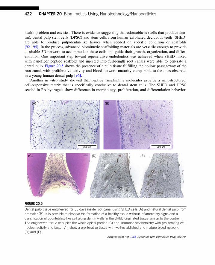
health problem and cavities. There is evidence suggesting that odontoblasts (cells that produce den-
tin), dental pulp stem cells (DPSC) and stem cells from human exfoliated deciduous teeth (SHED)
are able to produce pulp/dentin-like tissues when seeded on specific condition or scaffolds
[92�95]. In the process, advanced biomimetic scaffolding materials are versatile enough to provide
a suitable 3D network to accommodate these cells and guide their growth, organization, and differ-
entiation. One important step toward regenerative endodontics was achieved when SHED mixed
with nanofiber peptide scaffold and injected into full-length root canals were able to generate a
dental pulp. Figure 20.5 shows the presence of a pulp tissue fulfilling the hollow passageway of the
root canal, with proliferative activity and blood network maturity comparable to the ones observed
in a young human dental pulp [96].
Another in vitro study showed that peptide�amphiphile molecules provide a nanostructured,
cell-responsive matrix that is specifically conducive to dental stem cells. The SHED and DPSC
seeded in PA hydrogels show difference in morphology, proliferation, and differentiation behavior.
(A) (B)
(C) (D) (E)
200 µm200 µm
FIGURE 20.5
Dental pulp tissue engineered for 35 days inside root canal using SHED cells (A) and natural dental pulp from
premolar (B). It is possible to observe the formation of a healthy tissue without inflammatory signs and a
densification of odontoblast-like cell along dentin walls in the SHED originated tissue similar to the control.
The engineered tissue occupies the whole apical portion (C) and immunohistochemistry with proliferating cell
nuclear activity and factor VIII show a proliferative tissue with well-established and mature blood network
(D) and (E).
Adapted from Ref. [96]. Reprinted with permission from Elsevier.
422 CHAPTER 20 Biomimetics Using Nanotechnology/Nanoparticles

SHED seem to be a suitable tool for soft tissue regeneration, such as dental pulp, whereas DPSC
might be useful for engineering mineralized tissues like dentin [97]. Further development and
successful application of these strategies to regenerate dentin and dental pulp could one day revolu-
tionize the treatment of our most common oral health problem and cavities.
20.5 ConclusionsDespite the challenges in dental tissue regeneration that lie ahead, significant evidence exists to
support the premise that recent advances in nanotechnology, acting as biomimetic tools, show great
potential to overcome the challenges and promise for improved the dental tissue regeneration.
Nanomaterials tailored for engineering dental tissues are continually being introduced and yield
numerous clinical dental benefits. These include improved treatments for periodontal defects,
enhanced maxillary and mandibular bone regeneration, perhaps more biological methods to repair
teeth after carious damage and possibly even regrowing lost teeth. In the near future, advances in
bioengineering research will lead to the wide application of the regenerative dentistry into general
dental practice to produce wonderful treatments and dramatically improve patients’ quality of life.
References[1] R. Langer, J.P. Vacanti, Tissue engineering, Science 260 (1993) 920�926.
[2] S. Yildirim, S.Y. Fu, K. Kim, H. Zhou, C.H. Lee, A. Li, et al., Tooth regeneration: a revolution in
stomatology and evolution in regenerative medicine, Int. J. Oral Sci. 3 (2011) 107�116.
[3] R.W. Siegel, G.E. Fougere, Mechanical properties of nanophase metals, Nanostruct. Mater. 6 (1995)
205.
[4] S.M. Warren, K.D. Fong, C.M. Chen, E.G. Loboa, C.M. Cowan, H.P. Lorenz, et al., Tools and techniques
for craniofacial tissue engineering, Tissue Eng. 9 (2003) 187�200.
[5] L.J. Zhang, T.J. Webster, Nanotechnology and nanomaterials: promises for improved tissue regeneration,
Nano Today 4 (2009) 66�80.
[6] H. Yoshimoto, Y. Shin, H. Terai, J. Vacanti., A biodegradable nanofiber scaffold by electrospinning and
its potential for bone tissue engineering, Biomaterials 24 (2003) 2077�2082.
[7] M. Jose, V. Thomas, Y. Xu, S. Bellis, E. Nyairo, D. Dean, Aligned bioactive multi-component nano-
fibrous nanocomposite scaffolds for bone tissue engineering, Macromol. Biosci. 10 (2010) 433�444.
[8] A. Asran, S. Henning, G. Michler, Polyvinyl alcohol-collagen-hydroxyapatite biocomposite nanofibrous
scaffold: mimicking the key features of natural bone at the nanoscale level, Polymer 51 (2010)
868�876.
[9] J. Venugopal, M. Prabhakaran, Y. Zhang, S. Low, A.S. Choon, S. Ramakrishna, Biomimetic
hydroxyapatite-containing composite nanofibrous substrates for bone tissue engineering, Philos Transact.
A Math Phys. Eng. Sci. 368 (2010) 2065�2081.
[10] M.J. Gupte, P.X. Ma, Nanofibrous scaffolds for dental and craniofacial applications, J. Dent. Res. 91
(2012) 227�234.
[11] T.J. Webster, C. Ergun, R.H. Doremus, R.W. Siegel, R. Bizios, Specific proteins mediate enhanced
osteoblast adhesion on nanophase ceramics, J. Biomed. Mater. Res. 51 (2000) 475�483.
[12] T.J. Webster, C. Ergun, R.H. Doremus, R.W. Seigel, R. Bizios, Enhanced osteoclast-like cell functions
on nanophase ceramics, Biomaterials 22 (2001) 1327�1333.
423References

[13] S.P. Nukavarapu, S.G. Kumbar, J.L. Brown, N.R. Krogman, A.L. Weikel, M.D. Hindenlang, et al.,
Polyphosphazene/nano-hydroxyapatite composite microsphere scaffolds for bone tissue engineering,
Biomacromolecules 9 (2008) 1818�1825.
[14] M. Sato, M.A. Sambito, A. Aslani, N.M. Kalkhoran, E.B. Slamovich, T.J. Webster, Increased osteoblast
functions on undoped and yttrium-doped nanocrystalline hydroxyapatite coatings on titanium,
Biomaterials 27 (2006) 2358�2369.
[15] E.S. Thian, J. Huang, S.M. Best, Z.H. Barber, R.A. Brooks, N. Rushton, et al., The response of osteoblasts
to nanocrystalline silicon-substituted hydroxyapatite thin films, Biomaterials 27 (2006) 2692�2698.
[16] K.U. Lewandrowski, S.P. Bondre, D.L. Wise, D.J. Trantolo, Enhanced bioactivity of a poly(propylene
fumarate) bone graft substitute by augmentation with nano-hydroxyapatite, Biomed. Mater. Eng.
13 (2003) 115�124.
[17] E.S. Thian, Z. Ahmad, J. Huang, M.J. Edirisinghe, S.N. Jayasinghe, D.C. Ireland, et al., Bioactivity of
nanoapatite produced by electrohydrodynamic atomization, J. Bionanosci. 1 (2007) 60�63.
[18] S. Pezzatini, R. Solito, L. Morbidelli, S. Lamponi, E. Boanini, A. Bigi, et al., The effect of hydroxyapatite
nanocrystals on microvascular endothelial cell viability and functions, J. Biomed. Mater. Res. A 76A
(2006) 656�663.
[19] S. Pezzatini, L. Morbidelli, R. Solito, E. Paccagnini, E. Boanini, A. Bigi, et al., Nanostructured HA
crystals up-regulate FGF-2 expression and activity in microvascular endothelium promoting angio-
genesis, Bone 41 (2007) 523�534.
[20] S.I. Stupp, G.W. Ciegler, Organoapatites: materials for artificial bone. I. Synthesis and microstructure,
J. Biomed. Mater. Res. 26 (1992) 169�183.
[21] T.J. Webster, C. Ergun, R.H. Doremus, R.W. Siegel, R. Bizios, Enhanced osteoclast-like cell functions
on nanophase ceramics, Biomaterials 22 (2001) 1327�1333.
[22] J. Huang, S.M. Best, W. Bonfield, R.A. Brooks, N. Rushton, S.N. Jayasinghe, et al., In vitro assessment
of the biological response to nanosized hydroxyapatite, J. Mater. Sci. Mater. Med. 15 (2004) 441�445.
[23] G. Colon, B.C. Ward, T.J. Webster, Increased osteoblast and decreased Staphylococcus epidermidis
functions on nanophase ZnO and TiO2, J. Biomed. Mater. Res. A 78 (2006) 595�604.
[24] T.J. Webster, E.L. Hellenmeyer, R.L. Price, Increased osteoblast functions on theta1 delta nanofiber alu-
mina, Biomaterials 26 (2005) 953�960.
[25] W. Paul, C.P. Sharma, Nanoceramic matrices: biomedical applications, Am. J. Biochem. Biotechnol.
2 (2006) 41�48.
[26] F.X. Huber, N. McArthur, J. Hillmeier, H.J. Kock, M. Baier, M. Diwo, et al., Void filling of tibia compres-
sion fracture zones using a novel resorbable nanocrystalline hydroxyapatite paste in combination with a
hydroxyapatite ceramic core: first clinical results, Arch. Orthop. Trauma Surg. 126 (2006) 533�540.
[27] R. Smeets, G. Jelitte, M. Heiland, A. Kasaj, M. Grosjean, D. Riediger, et al., Hydroxylapatit-
Knochenersatzmaterial (Ostims) bei der Sinusbodenelevation, Schweiz Monatsschr. Zahnmed. 118
(2008) 203�208.
[28] K.L. Gerlach, D. Niehues, Die Behandlung der Kieferzysten mit einem neuartigen nanopartikularen
Hydroxylapatit, Mund Kiefer GesichtsChir 11 (2007) 131�137.
[29] F. Schwarz, K. Bieling, T. Latz, E. Nuesry, J. Becker, Healing of intrabony periimplantitis defects following
application of a nanocrystalline hydroxyapatite (Ostimt) or a bovine-derived xenograft (Bio-Osst) in com-
bination with a collagen membrane (Bio-Gidet). A case series, J. Clin. Periodontol. 33 (2006) 491�499.
Materials 2 (2009) 2037
[30] F.P. Strietzel, P.A. Reichart, H.L. Graf, Lateral alveolar ridge augmentation using a synthetic nano-
crystalline hydroxyapatite bone substitution material (Ostims). Preliminary clinical and histological
results, Clin. Oral Implants Res. 18 (2007) 743�751.
424 CHAPTER 20 Biomimetics Using Nanotechnology/Nanoparticles

[31] C. Spies, S. Schnurer, T. Gotterbarm, S. Breusch, Tierexperimentelle Untersuchung des Knochenersatzstoffs
OstimTM im knochernen Lager des Gottinger Miniaturschweins, Z. Orthop. Unfall. 146 (2008) 64�69.
[32] M. Thorwarth, S. Schultze-Mosgau, P. Kessler, J. Wiltfang, K.A. Schlegel, Bone regeneration in osseous
defects using a resorbable nanoparticular hydroxyapatite, J. Oral Maxillofac. Surg. 63 (2005)
1626�1633.
[33] J. Brandt, S. Henning, G. Michler, M. Schulz, A. Bernstein, Nanocrystalline hydroxyapatite for bone
repair, Key Eng. Mater. 361�363 (2008) 35�38.
[34] F.X. Huber, J. Hillmeier, L. Herzog, N. McArthur, H.J. Kock, P.J. Meeder, Open reduction and palmar
plate-osteosynthesis in combination with a nanocrystalline hydroxyapatite spacer in the treatment of
comminuted fractures of the distal radius, J. Hand Surg. Br. 31B (2006) 298�303.
[35] F.X. Huber, J. Hillmeier, N. McArthur, H.J. Kock, P.J. Meeder, The use of nanocrystalline hydroxyapatite
for the reconstruction of calcaneal fractures: preliminary results, J. Foot Ankle Surg. 45 (2006) 322�328.
[36] M.W. Laschke, K. Witt, T. Pohlemann, M.D. Menger, Injectable nanocrystalline hydroxyapatite paste
for bone substitution: in vivo analysis of biocompatibility and vascularization, J. Biomed. Mater. Res. B
Appl. Biomater. 82B (2007) 494�505.
[37] C.K.G. Spies, S. Schnurer, T. Gotterbarm, S. Breusch, The efficacy of Biobont and Ostimt within
metaphyseal defects using the Gottinger Minipig, Arch. Orthop. Trauma Surg. 129 (2009) 979�988.
[38] Y.K. Kim, L.S. Gu, T.E. Bryan, J.R. Kim, L. Chen, Y. Liu, et al., Mineralisation of reconstituted colla-
gen using polyvinylphosphonic acid/polyacrylic acid templating matrix protein analogues in the presence
of calcium, phosphate and hydroxyl ions, Biomaterials 31 (2010) 6618�6627.
[39] Y. Zhang, V.J. Reddy, S.Y. Wong, X. Li, B. Su, S. Ramakrishna, et al., Enhanced biomineralization
in osteoblasts on a novel electrospun biocomposite nanofibrous substrate of hydroxyapatite/collagen/
chitosan, Tissue Eng. Part A 16 (2010) 1949�1960.
[40] E. Kon, M. Delcogliano, G. Filardo, D. Pressato, M. Busacca, B. Grigolo, et al., A novel nanocomposite
multilayered biomaterial for treatment of osteochondral lesions: technique note and an early stability
pilot clinical trial, Injury 41 (2010) 693�701.
[41] C. Erisken, D.M. Kalyon, H. Wang, C. Ornek-Ballanco, J. Xu, Osteochondral tissue formation through
adipose-derived stromal cell differentiation on biomimetic polycaprolactone nanofibrous scaffolds with
graded insulin and Beta-glycerophosphate concentrations, Tissue Eng. Part A 17 (2011) 1239�1252.
[42] F.M. Chen, J. Zhang, M. Zhang, Y. An, F. Chen, Z.F. Wu, A review on endogenous regenerative
technology in periodontal regenerative medicine, Biomaterials 31 (2010) 7892�7927.
[43] P.X. Ma, Biomimetic materials for tissue engineering, Adv. Drug. Deliv. Rev. 60 (2008) 184�198.
[44] S. Srinivasan, R. Jayasree, K.P. Chennazhi, S.V. Nair, R. Jayakumar, Biocompatible alginate/nano bio-
active glass ceramic composite scaffolds for periodontal tissue regeneration, Carbohydr. Polym. 87
(2012) 274�283.
[45] F. Yang, S.K. Both, X. Yang, X.F. Walboomers, J.A. Jansen, Development of an electrospun nano-apatite/
PCL composite membrane for GTR/GBR application, Acta Biomater. 5 (9) (2009) 3295�3304.
[46] Z. Yuan, H. Nie, S. Wang, C.H. Lee, A. Li, S.Y. Fu, et al., Biomaterial selection for tooth regeneration,
Tissue Eng. Part B Rev. 17 (2011) 373�388.
[47] A.G. Fincham, J. Moradian-Oldak, J.P. Simmer, The structural biology of the developing dental enamel
matrix, J. Struct. Biol. 126 (1999) 270�299.
[48] Z. Huang, T.D. Sargeant, J.F. Hulvat, A. Mata, P. Bringas, S.I. Ch Koh, et al., Bioactive nanofibers
instruct cells to proliferate and differentiate during enamel regeneration, J. Bone Mineral Res. 23 (2008)
1995�2006.
[49] H.F. Chen, B.H. Clarkson, K. Sun, J.F. Mansfield, Self-assembly of synthetic hydroxyaptite nanorods
into enamel prism like structure, J. Colloid Interface Sci. 188 (2005) 97�103.
425References

[50] H.F. Chen, Z.Y. Tang, J. Liu, K. Sun, S.R. Chang, M.C. Peters, et al., Acellular synthesis of a human
enamel-like microstructure, Adv. Mater. 18 (2006) 1846�1851.
[51] Y. Fujishiro, A. Fujimoto, T. Sato, A. Okuwaki, Coating of hydroxyapatite on titanium plates using
thermal-dissociation of calcium-EDTA chelate complex in phosphate solutions under hydrothermal
conditions, J. Colloid Interface Sci. 173 (1995) 119�127.
[52] W. Suchannek, M. Yoshimura, Processing and properties of hydroxyapatite-based biomaterials for use as
hard tissue replacement implants, J. Mater. Res. 13 (1998) 94�117.
[53] M. Cao, Y. Wang, C. Guo, Y. Qi, C. Hu, Preparation of ultrahigh-aspect-ratio hydroxyapatite nanofibers
in reverse micelles under hydrothermal conditions, Langmuir 20 (2004) 4784.
[54] L. Li, C. Mao, J. Wang, X. Xu, H. Pan, Y. Deng, et al., Bio-inspired enamel repair via Glu-directed
assembly of apatite nanoparticle: an approach to biomaterials with optimal characteristic, Adv. Mater. 23
(2011) 4695�4701.
[55] M. Hannig, C. Hannig, Nanomaterials in preventive dentistry, Nat. Nanotechnol. 5 (2010) 565�569.
[56] K. Onuma, K. Yamagishi, A. Oyane, Nucleation and growth of hydroxyapatite nanocrystals for non-
destructive repair of early caries lesions, J. Cryst. Growth 282 (2005) 199�207.
[57] Y. Yamagishi, K. Onuma, T. Suzuki, F. Okada, J. Tagami, M. Otsuki, et al., A synthetic enamel for
rapid tooth repair, Nature 433 (2005) 819.
[58] K.L. Lv, J.X. Zhang, X.C. Meng, X.Y. Li, Remineralization effect of the nano-HA toothpaste on artificial
caries, Key Eng. Mater. 330�332 (2007) 267�270.
[59] M.Y. Kim, H.K. Kwon, C.H. Choi, B.I. Kim, Combined effects of nano-hydroxyapatite and NaF on
remineralization of early caries lesion, Key Eng. Mater. 330�332 (2007) 1347�1350.
[60] N. Roveri, E. Battistella, I. Foltran, E. Foresti, M. Iafisco, M. Lelli, et al., Synthetic biomimetic carbonate-
hydroxyapatite nanocrystals for enamel remineralization, Adv. Mater. Res. 47�50 (2008) 821�824.
[61] N. Roveri, E. Battistella, C.L. Bianchi, I. Foltran, E. Foresti, M. Lafisco, et al., Surface enamel reminera-
lization: biomimetic apatite nanocrystals and fluoride ions different effects, J. Nanomater. (2009)
10.1155/2009/746383.
[62] L. Li, H.H. Pan, J.H. Tao, X.R. Xu, C.Y. Mao, X.H. Gu, et al., Repair of enamel by using hydroxyapatite
nanoparticles as the building blocks, J. Mater. Chem. 18 (2008) 4079�4084.
[63] P. Tschoppe, D.L. Zandim, P. Martus, A.M. Kielbassa, Enamel and dentine remineralization by nano-
hydroxyapatite toothpastes, J. Dent. 3 (2011) 430�437.
[64] S.B. Huang, S.S. Gao, H.Y. Yu, Effect of nano-hydroxyapatite concentration on remineralization of
initial enamel lesion in vitro, Biomed. Mater. 4 (2009) 034104.
[65] S. Huang, S. Gao, L. Cheng, H. Yu, Remineralization potential of nano-hydroxyapatite on initial enamel
lesions: an in vitro study, Caries Res. 45 (2011) 460�468.
[66] S. Huang, S. Gao, L. Cheng, H. Yu, Combined effects of nano-hydroxyapatite and Galla chinensis on
remineralization of initial enamel lesion in vitro, J. Dent. 38 (2010) 811�819.
[67] G. Balasundaram, M. Sato, T.J. Webster, Using hydroxyapatite nanoparticles and decreased crystallinity
to promote osteoblast adhesion similar to functionalizing with RGD, Biomaterials 27 (2006) 2798�2805.
[68] Q. Hu, Z. Tan, Y. Liu, J. Tao, Y. Cai, M. Zhang, et al., Effect of crystallinity of calcium phosphate
nanoparticles on adhesion, proliferation, and differentiation of bone marrow mesenchymal stem cells,
J. Mater. Chem. 17 (2007) 4690�4698.
[69] S. Mai, Y.K. Kim, M. Toledano, L. Breschi, J.Q. Ling, D.H. Pashley, et al., Phosphoric acid esters
cannot replace polyvinylphosphonic acid as phosphoprotein analogs in biomimetic remineralization of
resin-bonded dentin, Dent. Mater. 25 (2009) 1230�1239.
[70] A.W. Xu, Y.R. Ma, H. Colfen, Biomimetic mineralization, J. Mater. Chem. 17 (2007) 415�449.
426 CHAPTER 20 Biomimetics Using Nanotechnology/Nanoparticles

[71] E.C. Reynolds, Remineralization of enamel subsurface lesions by casein phosphopeptide-stabilized
calcium phosphate solutions, J. Dent. Res. 76 (1997) 1587�1595.
[72] M.V. Morgan, G.G. Adams, D.L. Bailey, C.E. Tsao, S.L. Fischman, E.C. Reynolds, The anticariogenic
effect of sugar-free gum containing CPP-ACP nanocomplexes on approximal caries determined using
digital bitewing radiography, Caries Res. 42 (2008) 171�184.
[73] F. Cai, D.J. Manton, P. Shen, G.D. Walker, K.J. Cross, Y. Yuan, et al., Effect of addition of citric acid
and casein phosphopeptide amorphous calcium phosphate to a sugar-free chewing gum on enamel remi-
neralization in situ, Caries Res. 41 (2007) 377�383.
[74] Y. Iijima, F. Cai, P. Shen, G. Walker, C. Reynolds, E.C. Reynolds, Acid resistance of enamel subsurface
lesions remineralized by a sugar-free chewing gum containing casein phosphopeptide-amorphous
calcium phosphate, Caries Res. 38 (2004) 551�556.
[75] A.M. Al-Mullahi, K.J. Toumba, Effect of slow-release fluoride devices and casein phosphopeptide/amor-
phous calcium phosphate nanocomplexes on enamel remineralization in vitro, Caries Res. 44 (2010)
364�371.
[76] J.D. Bader, Casein phosphopeptide-amorphous calcium phosphate shows promise for preventing caries,
Evid. Based Dent. 11 (2010) 11�12.
[77] G.D. Walker, F. Cai, P. Shen, G.G. Adams, C. Reynolds, E.C. Reynolds, Casein phosphopeptide-
amorphous calcium phosphate incorporated into sugar confections inhibits the progression of enamel
subsurface lesions in situ, Caries Res. 44 (2010) 33�40.
[78] N. Srinivasan, M. Kavitha, S.C. Loganathan, Comparison of the remineralization potential of CPP-ACP
and CPP-ACP with 900 ppm fluoride on eroded human enamel: an in situ study, Arch. Oral Biol. 55
(2010) 541�544.
[79] H. Hamba, T. Nikaido, G. Inoue, A. Sadr, J. Tagami, Effects of CPP-ACP with sodium fluoride on inhi-
bition of bovine enamel demineralization: a quantitative assessment using micro-computed tomography,
J. Dent. 39 (2011) 405�413.
[80] E.C. Reynolds, Anticariogenic complexes of amorphous calcium phosphate stabilized by casein
phosphopeptides: a review, Spec. Care Dent. 18 (1998) 8�16.
[81] C. Llena, L. Forner, P. Baca, Anticariogenicity of casein phosphopeptide-amorphous calcium phosphate:
a review of the literature, J. Contemp. Dent. Prac. 10 (2009) 1�9.
[82] V. Yengopal, S. Mickenautsch, Caries preventive effect of casein phosphopeptide-amorphous calcium
phosphate (CPP-ACP): a metaanalysis, Acta Odontol. Scand. 21 (2009) 1�12.
[83] N.J. Cochrane, F. Cai, N.L. Huq, M.F. Burrow, E.C. Reynolds, New approaches to enhanced reminerali-
zation of tooth enamel, J. Dent. Res. 89 (2010) 1187�1197.
[84] E.C. Reynolds, F. Cai, P. Shen, G.D. Walker, Retention in plaque and remineralization of enamel lesion
by various forms of calcium in a mouthrinse or sugar-free chewing gum, J. Dent. Res. 82 (2003)
206�211.
[85] H.H.K. Xu, M.D. Weir, L. Sun, Nanocomposites with Ca and PO4 release: effects of reinforcement,
dicalcium phosphate particle size and silanization, Dent. Mater. 23 (2007) 1482�1491.
[86] Y.W. Hong, J.H. Kim, B.H. Lee, Y.K. Lee, B.J. Choi, J.H. Lee, et al., The effect of nano-sized β-tricalciumphosphate on remineralization in glass ionomer dental luting cement, Key Eng. Mater. 361�363 (2008)
861�864.
[87] S. Nakashima, M. Yoshie, H. Sano, A. Bahar, Effect of a test dentifrice containing nano-sized calcium
carbonate on remineralization of enamel lesions in vitro, J. Oral Sci. 51 (2009) 69�77.
[88] K. Kawasaki, J. Ruben, I. Stokroos, O. Takagi, J. Arends., The remineralization of EDTA-treated human
dentine, Caries Res. 33 (1999) 275�280.
427References

[89] Y. Shibata, L.H. He, Y. Kataoka, T. Miyazaki, M.V. Swain, Micromechanical property recovery of
human carious dentin achieved with colloidal nano-betatricalcium phosphate, J. Dent. Res. 87 (2008)
233�237.
[90] M. Vollenweider, T.J. Brunner, S. Knecht, R.N. Grass, M. Zehnder, T. Imfeld, et al., Remineralization
of human dentin using ultrafine bioactive glass particles, Acta Biomater. 3 (2007) 936�943.
[91] F.R. Tay, D.H. Pashley, Biomimetic remineralization of resin-bonded acid-etched dentin, J. Dent. Res.
88 (2009) 719�724.
[92] V.T. Sakai, Z. Zhang, Z. Dong, K.G. Neiva, M. Machado, S. Shi, et al., SHED differentiate into
functional odontoblast and endothelium, J. Dent. Res. 89 (2010) 791�796.
[93] M.M. Cordeiro, Z. Dong, T. Kaneko, Z. Zhang, M. Miyazawa, S. Shi, et al., Dental pulp tissue engineer-
ing with stem cells from exfoliated deciduous teeth, J. Endod. 34 (2008) 962�969.
[94] V. Rosa, T.M. Botero, J.E. Nor, Regenerative endodontics in light of the stem cell paradigm, Int. Dent.
J. 61 (2011) 23�28.
[95] G. Huang, T. Yamaza, L.D. Shea, F. Djouad, N.Z. Kuhn, R.S. Tuan, et al., Stem/progenitor
cell-mediated de novo regeneration of dental pulp with newly deposited continuous layer of dentin in an
in vivo model, Tissue Eng. Part A 16 (2009) 605�615.
[96] V. Rosa, A. Della Bona, B.N. Cavalcanti, J.E. Nor, Tissue engineering: from research to dental clinics,
Dent. Mater. 28 (2012) 341�348.
[97] K.M. Galler, A. Cavender, V. Yuwono, H. Dong, S. Shi, G. Schmalz, et al., Self-assembling peptide
amphiphile nanofibers as a scaffold for dental stem cells, Tissue Eng. Part A 14 (2008) 2051�2058.
428 CHAPTER 20 Biomimetics Using Nanotechnology/Nanoparticles

CHAPTER
21Scope of Nanotechnology inEndodontics
Sami M.A. Choglea,b, Bassam M. Kinaiac,d and Harold E. GoodiseaAssociate Professor and Program Director, Graduate Endodontics, Henry M. Goldman School of Dental Medicine,
Boston University, Boston, MA, USA,bAdjunct Associate Professor, Department of Endodontics, School of Dental Medicine,
Case Western Reserve University, Cleveland, OH, USA,cAssistant Professor in Periodontology, Director of Continuing Education, The European University,
Dubai Health Care City, Dubai, UAE,dAdjunct Assistant Professor, Department of Periodontology and Dental Hygiene, School of Dentistry,
University of Detroit Mercy, Detroit, MI, USA,eProfessor Emeritus, Department of Endodontics, University of California School of Dentistry,
San Francisco, CA, USA
CHAPTER OUTLINE
21.1 Introduction ............................................................................................................................... 432
21.2 Clinical applications ..................................................................................................................433
21.2.1 Instrument modifications ...................................................................................... 433
21.2.2 Enhancement of canal disinfection ........................................................................ 434
21.2.2.1 Irrigants ................................................................................................. 435
21.2.2.2 Medicaments.......................................................................................... 436
21.2.3 Material modifications .......................................................................................... 436
21.2.3.1 Obturating materials ................................................................................ 436
21.2.3.2 Sealers................................................................................................... 437
21.2.3.3 Retro-filling and root-repair materials ......................................................... 438
21.3 Applications for repair and pulp regeneration .............................................................................. 440
21.4 Repair and regeneration ............................................................................................................. 441
21.5 Nanotechnology applications for repair and pulp regeneration...................................................... 442
21.6 Conclusion ................................................................................................................................ 444
Acknowledgments ............................................................................................................................... 445
References ......................................................................................................................................... 445
431Nanobiomaterials in Clinical Dentistry.
© 2013 Elsevier Inc. All rights reserved.

21.1 IntroductionRoot canal treatment is a highly predictable procedure with success rates of up to 96% [1,2]. The
success of the treatment primarily depends on proper cleaning and shaping to disrupt the microbial
ecology, disinfecting the root canal system, and finally sealing it to prevent microleakage.
Although the treatment has a high success rate, failure still occurs due to inadequate cleaning and
shaping in anatomically complex root canal systems and/or continued microbial leakage due to lack
of adequate sealing material characteristics [3�5]. Current materials possess certain limitations
such as shrinkage, solubility in oral environment, and moisture intolerance. Therefore, development
of proper material for cleaning and shaping as well as sealing the root canal system are essential
for long-term root canal treatment success.
Newer advances, including developments in armamentarium such as the use of the dental oper-
ating microscope and improved materials, have influenced the outcome of periradicular surgery,
according to some studies [6�10]. Although success rates have increased incrementally, the option
to extract teeth and replace them with dental implants has grown in popularity. Outcomes of pri-
mary endodontic treatment, which have the highest success rate of endodontics procedures, have
been compared to implants [11�13]. Because of the “predictable” option to replace a tooth with a
dental implant when endodontic treatment has failed, the perceived benefit of endodontic treatment
depends on the continued improvement and refinement in the physical characteristics of materials,
techniques, and armamentarium.
One such focus in both the medical and dental fields is the clinical applications of nanotechnol-
ogy. Nanomaterial research has initiated a new era in material development for improved clinical
outcomes. Nanotechnology is defined as the creation of functional materials with structures sized
100 nm or smaller [14]. In the field of endodontics, a fair amount of research is underway in an
attempt to enhance every step in clinical procedures from files to filling materials. The smaller
sized nanomaterials, more resistant to wear and fatigue, are being suggested for surface modifica-
tions of currently used rotary nickel�titanium files for root canal treatment to help reduce the inci-
dence of instrument failure. The antimicrobial properties of some nanoparticles may be able to
enhance the efficacy of irrigants and intracanal medicaments due to their size and possible disper-
sion in complex root canal anatomies.
Apart from these, a more concentrated effort has been ongoing to develop “nanomodified” mate-
rials. Dispersion of these particles into current and novel materials could fortify the sealing ability of
obturating and sealer materials, as well as root-repair/root-end filling materials. For example, nano-
composites constitute a relatively new class of materials with the dispersed phase having at least one
ultrafine dimension, typically a few nanometers as demonstrated in Figure 21.1. They include poly-
meric materials composed of nanoparticles like carbon nanotubes (CNTs), organically-modified clays
(organoclays), or other nanoscale materials. Because of the nanoscale structure and huge interfacial
area between polymer and organoclay, polymer�clay nanocomposites exhibit enhanced mechanical
and thermal properties. Polymer�clay nanocomposites are particularly attractive for potential appli-
cations where enhanced barrier properties as well as physical properties are desired.
CNTs are predicted to have unique mechanical properties including high stiffness and axial
strength as a result of their cylindrical graphitic structure. Experimental studies have shown that
isolated CNTs possess exceptionally high Young’s moduli in the terapascal range which are
much higher than those typically found in stainless steel and carbon fibers [15]. Carbon fibers
432 CHAPTER 21 Scope of Nanotechnology in Endodontics

have already found applications in reinforced composites where there is a need for high-strength
and light-weight materials [16]. The smaller diameters and larger aspect ratios found for CNTs
is expected to lead to even higher strength composites. Recently, CNTs have been incorporated
into methyl methacrylate-based resins and found to substantially enhance their load-bearing
mechanical characteristics [17]. These studies suggest that the addition of CNTs in dental
implant materials will prevent fatigue and failure caused by forces in the oral cavity and extend
the life of dental restorations.
Another application of nanotechnology gaining significant interest is the use of nanoscaffolds for
pulp regeneration. At the nanolevel, these scaffolds may eventually prove to be applicable to therapies
ranging from pulp capping to complete pulp-dentin complex regeneration. This chapter aims to review
these uses of nanotechnology for current and future clinical applications in endodontics.
21.2 Clinical applications21.2.1 Instrument modificationsRotary instruments, especially those made of nickel�titanium (Ni�Ti) alloys, are widely used by
clinicians in everyday dental practice. Ni�Ti alloys possess many favorable characteristics such as
resistance against corrosion and more importantly super-elasticity and excellent shape memory that
allow it to navigate complex root canal systems for proper cleaning and shaping [18]. Despite these
favorable characteristics of rotary Ni�Ti instruments, fatigue and failure of Ni�Ti alloys occur
leading to possible intracanal instrument fracture. Although a disarticulated instrument in the canal
may not directly result in failure of the endodontic procedure, the inability to clean beyond the frac-
tured instrument (Figure 21.2) may result in persistent endodontic disease requiring surgical
FIGURE 21.1
TEM demonstrating exfoliation of organoclay nanoparticles (dark aggregates) in a monomer matrix
(100,0003 magnification (50 nm scale)).
43321.2 Clinical applications

intervention [19]. Therefore, to reduce instrument fatigue and failure, reports have suggested
changes in instrument design, instrumentation protocol, and more recently the use of nanomaterials
for surface modifications of the currently used rotary Ni�Ti instruments.
Recent studies have attempted to modify the instrument surface by coating the Ni�Ti surface
with different nanomaterials to overcome these characteristic shortcomings. Adini et al. [20] exam-
ined the effects of cobalt coatings with impregnated fullerene-like WS2 nanoparticles on file fatigue
and failure of Ni�Ti files. The addition of these nanoparticles significantly improved the fatigue
resistance and time to breakage of the coated files as observed under dynamic X-ray diffraction.
The authors attributed the improvement to the reduced friction between the file and the surrounding
tissue with the nanoparticles coating [20]. In another study, the distal end of the file was modified
by selective coating of endodontic files. The selective coating of the files distributed the nanoparti-
cles nonuniformly along the surface of the file leaving only a part of the file coated and thereby
having a higher torque making it more resistant to fatigue and failure [21]. Thus the use of nano-
particles for surface coatings of the instruments may significantly enhance their performance with
less potential for fatigue and failure.
21.2.2 Enhancement of canal disinfectionA number of solutions and medications are used for disinfecting the root canal system and may
be broadly classified under irrigants and medicaments. While irrigants usually signify high vol-
ume with low contact time within the canal system, medicaments are relatively more passive but
rely on a longer contact time to exert their disinfecting properties. Both of these categories are
being investigated for nanoparticle enhancement to help improve disinfection and sealing of the
root canal system.
FIGURE 21.2
Periapical radiograph demonstrating a disarticulated file in the mesial canal of the first molar preventing
further apical cleaning and shaping and obturation.
434 CHAPTER 21 Scope of Nanotechnology in Endodontics

21.2.2.1 IrrigantsThe main idea for cleaning and shaping of the root canal system is to disrupt and eradicate the
microbial biofilms in the root canal system. Even though a lot of emphasis is placed on instru-
mentation of canals, it is well documented that irrigants play the central role in disinfection of
the root canal system [22,23]. Irrigants can augment mechanical debridement by flushing out
debris, dissolving tissue, and disinfecting the root canal system. This is especially needed for
teeth with complex internal anatomy such as fins or other irregularities that might be missed by
instrumentation [24]. According to Torabinejad et al. [25], the desired functions of an irrigating
solution include (i) complete removal of the smear layer, (ii) disinfection of dentin and its
tubules, (iii) sustained antibacterial effect after use, (iv) penetration of antimicrobial agents
present in the solution into the dentinal tubules, (v) nonantigenic, nontoxic, and noncarcino-
genic, (vi) no adverse effects on the physical properties of exposed dentin, (vii) no adverse
effect on the sealing ability of filling material, (viii) not discoloring the tooth, (ix) convenient
application, and (x) relatively inexpensive.
The main concern with irrigating techniques and irrigating fluids is the ability to reach all areas
of the root canal system and removal of debris (i.e., smear layer) and biofilms without damage to
host tissues. Different irrigants and techniques both manual and machine assisted are being used
to disinfect the root canal system. Recent reports have investigated the types of nanoparticles to
enhance the root canal disinfection properties as well as the tissue reaction to their use.
Over the last decade, silver nanoparticles have been used in various applications from electron-
ics to antibacterial/antifungal agents in biotechnology and bioengineering including dental applica-
tions. In an animal study, Gomes-Filho et al. [26] observed the tissue response to implemented
polyethylene tubes filled with fibrin sponge embedded with either 47 or 23 ppm silver nanoparticles
dispersion material and compared them to plain fibrin sponges and those imbedded with 2.5%
sodium hydrochloride used as controls over a 90-day period. The results of the study concluded
that silver nanoparticles dispersion material at 23 ppm concentration appeared biocompatible com-
pared to the rest of the groups. However, health concerns have been raised and toxicological
reviews have reported that exposure to silver nanoparticles may be associated with “inflammatory,
oxidative, genotoxic, and cytotoxic consequences” [27]. Further investigations are warranted to dic-
tate the safe use of these nanoparticles in medical and dental applications.
Another way of root canal disinfection that is being investigated is the nanoparticle-based anti-
microbial photodynamic therapy. Preliminary studies seem promising. In one study, Pagonis et al.
[28] studied the in vitro effects of poly(lactic-co-glycolic acid) (PLGA) nanoparticles loaded with
the photosensitizer methylene blue (MB) and light against Enterococcus faecalis by transmission
electron microscopy (TEM). The nanoparticles were found to be concentrated mainly on cell walls
of the microorganisms. The synergism of light and MB-loaded nanoparticles led to approximately
2 and 1 log 10 reduction of colony-forming units (CFUs) in planktonic phase and root canals,
respectively. In both cases, mean log 10 CFU levels were significantly lower than controls and
MB-loaded nanoparticles without light. The authors concluded that the utilization of PLGA nano-
particles encapsulated with photoactive drugs may be a promising adjunct in antimicrobial end-
odontic treatment. In another similar study, the cationic photosensitizer was able to inactivate the
microbial biofilm bacteria (E. faecalis) and disrupt the biofilm structure [29,30]. Thus these thera-
pies may provide a new alternative to conventional irrigants used in endodontic treatment.
43521.2 Clinical applications

21.2.2.2 MedicamentsFor endodontic procedures that require more than one visit to complete, the remaining bacteria
within the system can grow and reinfect the root canal space between appointments [31].
Historically, placement of intracanal medicaments became a popular method of preventing bacterial
regrowth. Some form of calcium hydroxide (CaOH) is an intracanal medicament often used
between visits. Although E. faecalis is an insignificant organism in infected but untreated root
canals [32], it is extremely resistant to most of the intracanal medicaments used, particularly to the
calcium hydroxide-containing dressings [33]. It can also survive in root canals as monoinfection,
without any synergistic support from other bacteria [32,34]. Thus E. faecalis is a recalcitrant candi-
date among the causative agents of failed endodontic treatments.
Nanoparticulates such as chitosan (CS-np) and zinc oxide (ZnO-np) have been shown to possess
significant antibacterial properties. Shrestha et al. in their study [35] tested the efficacy of fresh and
aged CS-np and ZnO-np in disinfecting and disrupting E. faecalis biofilms. Using confocal laser
scanning microscopy, total elimination of planktonic bacteria was observed in contrast to the bio-
film bacteria, which survived even after 72 h. There was a significant reduction in the thickness of
biofilm after nanoparticulate treatment and the authors concluded that the rate of bacterial killing
by the nanoparticulates depended on the concentration and time of interaction and that aging for
90 days did not affect their antibacterial properties. Although these findings need to be further
investigated and confirmed in animal and in vivo studies, the preliminary reports seem promising.
21.2.3 Material modifications21.2.3.1 Obturating materialsThe American Association of Endodontists published “Appropriateness of Care and Quality
Assurance Guidelines” [36] regarding contemporary endodontic treatment. In that publication, root
canal obturation is defined and characterized as:
The three-dimensional filling of the entire root canal system as close to the cementodentinal
junction as possible. Minimal amounts of root canal sealers, which have been demonstrated to
be biologically compatible, are used in conjunction with the core-filling material to establish
an adequate seal.
Although a number of materials have been advocated over the last 150 years for root canal
obturation, gutta-percha has remained as the material of choice. Gutta-percha has satisfied a num-
ber of the tenets for an ideal root filling material highlighted by Grossman [37]. However, its main
disadvantages cited include lack of rigidity and adhesiveness, ease of displacement under pressure,
minimal antimicrobial property, and shrinkage if thermo-plasticized. Proper adaptation of obturat-
ing materials to the cleaned and shaped root canal walls and in increased antimicrobial activity
would be important to reduce gaps and microleakage.
The incorporation of nanoparticles may increase the surface area between the dentin and the
obturating material leading to enhanced adaptation. Bioactive glass 45S5 is one of the recent nano-
particles used in endodontic therapy. It has amorphous nanoparticles of 20�60 nm in size. The
smaller particle size increases the contact surface area and thereby possesses a higher antimicrobial
effect than the macrosized material. The clinical antimicrobial efficacy of bioactive glass 45S5 was
436 CHAPTER 21 Scope of Nanotechnology in Endodontics

evaluated in persisting root canal infections containing isolates of Enterococci. The killing efficacy
against the bacteria was significantly better with the nanosized treatment material [38].
Bioactive glass 45S5, similarly to CaOH, has also been used for treatment of traumatized front
teeth with open apices. The conventional treatment with CaOH may affect mechanical dentin prop-
erties by decreasing its flexural strength over time. Recently, Marending et al. [39] used suspen-
sions of nanoparticulate bioactive glass 45S5 as dressing material and compared it to CaOH in
traumatized front teeth with open apices. The results showed a 35% drop in dentin mean flexural
strength values with CaOH and a 20% drop with bioactive glass 45S5 indicating superiority of
the latter. However, these results should be interpreted with caution since the affected dentin was
mainly in the superficial layers. Furthermore, Mohn et al. [40] prepared nanosized particles of bio-
active glass and modified it with bismuth oxide to obtain radiopaque properties. They evaluated the
performance of it as a filling material. Based on scanning electron microscopy (SEM), the authors
concluded that bioactive glass modified with bismuth oxide is a radiopaque bioactive root canal
filling material.
21.2.3.2 SealersSealer materials used during obturation are grouped based on their prime constituent, such as zinc
oxide-eugenol, calcium hydroxide, resins, glass ionomers, or silicones. The use of a sealer during
root canal obturation is essential and enhances the possible attainment of an impervious seal and
serves as filler for canal irregularities and minor discrepancies between the root canal wall and gutta-
percha. Sealers are often expressed through lateral or accessory canals, and they can assist in micro-
bial control should there be microorganisms left on the root canal walls or in the tubules [41�43].
Sealers can also serve as lubricants to assist in the thorough seating of the core-filling material during
compaction. In canals where the smear layer has been removed, many sealers demonstrate increased
adhesive properties to dentin (in addition to flowing into the patent tubules) [44�46].
As stated by Gutmann and Witherspoon [47], the future directions for the ideal sealer should
focus on materials that (i) penetrate the patent dentinal tubules, (ii) bind intimately to both the
organic and inorganic phases of dentin, (iii) neutralize or destroy microorganisms and their products,
(iv) predictably induce a cemental regenerative response over the apical foramen, and (v) strengthen
the root system. Since the size of nanoparticles can penetrate the dentinal tubules to ensure that all
the spaces have been sealed effectively, the development of a sealer based on nanotechnology
may be an important step to achieve a better sealer material in endodontics. Chen et al. [48] in their
study used a new root canal filling sealer primarily composed of nanohydroxyapatite crystals in
279 nm after setting in an extracted tooth model. The sealer demonstrated superior antimicrobial
activity (Actinomyces naeslundii, Peptostreptococcus anaerobius, Porphyromonas gingivalis,
Porphyromonas endodontalis, and Fusobacterium nucleatum) as well as minimal microleakage com-
pared to two other materials.
Another preliminary report [49] demonstrated that nanocrystalline tetracalcium phosphate had
significantly higher antimicrobial potency in an agar-diffusion test. The formation of amorphous
Ca(OH)2 during setting was thought to increase the pH value in the agar gel around the specimens
yielding a zone of inhibition. However, a similar study reported contradictory results. Masudi et al.
[50] evaluated the apical sealing ability of an experimental NHA (40�60 nm) resin-based endodon-
tic sealer and compared it to a commonly used resin-based sealant material. Teeth in the first group
were obturated using gutta-percha with AH26. The second group was obturated with the
43721.2 Clinical applications

experimental sealer. The results showed that there was no statistically significant difference in api-
cal sealing ability when measuring penetration of the dye using stereo microscope. Taken together,
the above three reports may indicate promising initial reports that with further research may yield
superior materials.
21.2.3.3 Retro-filling and root-repair materialsNumerous studies have demonstrated the importance of root-end filling placement during periapical
surgery. According to Harty et al. [51], the most important factor in determining the success of an
apicoectomy is the efficiency of the apical seal. Wu et al. [52] suggested that a tight and
long-lasting seal of root-end fillings are of primary clinical importance. Several studies have indi-
cated that the lack of a good root canal filling could compromise the surgical outcome [53,54].
In addition, a number of clinical studies on healing after periradicular surgery have confirmed the
benefit of placing a high-quality root canal filling prior to surgery [55,56].
The properties of an ideal root-end filling material have been well documented in the scientific
literature and are summarized as this material would adhere or bond to tooth tissue and ‘‘seal’’ the
root end three dimensionally; not promote, and preferably inhibit, the growth of pathogenic micro-
organisms; be dimensionally stable and unaffected by moisture in either the set or unset state; be
well tolerated by periradicular tissues with no inflammatory reactions; stimulate the regeneration of
normal periodontium; be nontoxic both locally and systemically; not corrode or be electrochemi-
cally active; not stain the tooth or the periradicular tissues; be easily distinguishable on radiographs;
have a long shelf life; and be easily handled. Although almost every available dental restorative
material or cement has at one time or another been suggested for root-end filling, these properties
have yet to be found in any one material. It may therefore be concluded that the ideal retrofill
material does not yet exist [57].
Mineral Trioxide Aggregate (MTA) has become the material of choice of retrograde filling in
spite of its handling and long setting time. To overcome these disadvantages, a very recent study
by Saghiri et al. [58] evaluated a nanomodified MTA for enhanced physiochemical properties.
They concluded that the increased surface area of powder by nanodispersion can reduce setting
time and increase microhardness. This may help the MTA to set faster without losing its required
hardness once set. Other studies are investigating new materials rather than modifications to current
materials. A polymer nanocomposite (PNC) is a generalized term for polymeric materials that
is loaded with minimal amount of nanoparticles such as clays and CNTs [59]. As opposed to con-
ventional composites, the dispersed phase has a very high surface-to-volume ratio. PNCs have
therefore shown greatly improved mechanical and thermal properties of the material even at very
low filler content (typically between 0% and 5%). Previous studies have indicated substantial
improvements in heat resistance, [59,60] dimensional stability [61], stiffness, [62,63], reduced elec-
trical conductivity [64,65], and most uniquely, drug elution capabilities [66,67]. Recently, two such
novel nanocomposites were investigated for initial apical seal along with a commonly used
polymer-based compomer in an in vitro model [68]. Although one of the PNCs did not significantly
reduce leakage, the results revealed that leakage of commercial compomer was more than 12 times
more likely than the second PNC. SEM of these PNCs placed as root-end filling materials revealed
a tight interface with the PNC entering into the dentinal tubules (Figure 21.3) [69]. This was cor-
roborated with equal or greater values (Figure 21.4) of push-out force required when compared to
MTA and Geristores [70].
438 CHAPTER 21 Scope of Nanotechnology in Endodontics

FIGURE 21.3
SEM image revealing the dentin�nanocomposite interface. The dentinal tubules (right) are seen to be
penetrated by the nanocomposite fibers.
FIGURE 21.4
Bar chart comparing push-out force for nanocomposites Poly(methyl methacrylate) (PMM) containing varying
quantities of organoclay nanoparticles (1�2%) with MTA and Geristore. The 2% nanocomposite groups
performed significantly better than MTA and Geristore.
43921.2 Clinical applications
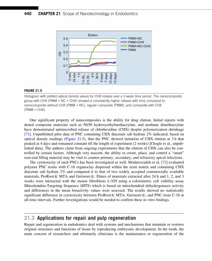
One significant property of nanocomposites is the ability for drug elution. Initial reports with
dental composite materials such as 50/50 hydroxyethylmethacrylate, and urethane dimethacrylate
have demonstrated antimicrobial release of chlorhexidine (CHX) despite polymerization shrinkage
[71]. Unpublished pilot data of PNC containing CHX diacetate salt hydrate 2% indicated, based on
optical density readings (Figure 21.5), that the PNC showed initiation of CHX elution at 3 h that
peaked at 4 days and remained constant till the length of experiment (2 weeks) [Chogle et al., unpub-
lished data]. The authors claim from ongoing experiments that the elution of CHX can also be con-
trolled by certain factors. Although very nascent, the ability to create, place, and control a “smart”
root-end filling material may be vital to counter primary, secondary, and refractory apical infections.
The cytotoxicity of such PNCs has been investigated as well. Modareszadeh et al. [72] evaluated
polymer PNC resins with C-18 organoclay dispersed within the resin matrix and containing CHX
diacetate salt hydrate 2% and compared it to that of two widely accepted commercially available
materials, ProRoots MTA and Geristores. Elutes of materials extracted after 24 h and 1, 2, and 3
weeks were interacted with the mouse fibroblasts L-929 using a colorimetric cell viability assay
Mitochondria-Targeting Sequence (MTS) which is based on mitochondrial dehydrogenases activity
and differences in the mean bioactivity values were assessed. The results showed no statistically
significant difference in cytotoxicity between ProRoots MTA, Geristores, and PNC resin C-18 at
all-time intervals. Further investigations would be needed to confirm these in vitro findings.
21.3 Applications for repair and pulp regenerationRepair and regeneration in endodontics deal with systems and mechanisms that maintain or restores
original structures and functions of tissue by reproducing embryonic development. In the tooth, the
main concern of researchers and ultimately clinicians is the maintenance or regeneration of the
Elution0.5
0.4
0.3
0.2O
D
0.1
00
min
15 m
in1
h1
h 45
...2
h 30
... 6 h
36 h
4 da
ys7
days
10 d
ays
13 d
ays
PMM+NCPMM+CHXPMM+NC+CHXPMM
FIGURE 21.5
Histogram with plotted optical density values for CHX release over a 2-week time period. The nanocomposite
group with CHX (PMM1NC1CHX) showed a consistently higher release with time compared to
nanocomposite without CHX (PMM1NC), regular composite (PMM), and composite with CHX
(PMM1CHX).
440 CHAPTER 21 Scope of Nanotechnology in Endodontics

dental pulp. The dental pulp is considered a low-compliance system that does not suffer disease or
injury in a manner to retain those cells, connective tissue, nerve fibers, and blood vessels as they
were originally formed embryonically and postbirth [73].
The dental pulp is a difficult tissue to access in humans without causing an inflammatory
response. It is a result of its environment and the hard tissues (enamel, dentin) that surround it.
From the standpoint of the possibility of repair and/or regeneration when the dental pulp is injured,
infected, or necrosed, its nature must be understood before any treatment modalities are attempted.
Another unusual aspect of the dental pulp is that it is a completely sensory tissue. That sensory
design implies that the adult pulp is protective of that tissue throughout the life of the tooth.
However, that same protective nature can be disturbed due to its location and its functions.
The following is a brief review of the nature of the dental pulp’s location that mitigates against
its ability to survive, repair itself, and regenerate a pulp-like tissue. First, the pulp has a terminal
microvascular supply with few, if any, anastomoses (with the exception of multirooted teeth).
Second, the dental pulp has a relatively large volume of tissue with a relatively small vascular sup-
ply. The largest vessels to enter and exit the tissue are arterioles and venules. Last, and probably
the major deterrent to repair and regeneration in response to injury or infection occurs due to it
being surrounded by a hard, unyielding tissue (dentin) and itself surrounded on the crown of the
tooth by enamel and the root by cementum [74]. However, in spite of its environment, the pulp has
in infinite capacity to repair itself.
21.4 Repair and regenerationThe main culprit in relation to untoward events occurring in the dental pulp is due to the onset of
dental caries. The hard tissue surrounding the pulp is susceptible to the microorganisms that cause
enamel and dentin to become infected, with extension into the pulp tissue. When caries begins on
the outer surface of enamel, the effect pulpally is an inflammatory response. As caries develops
deeper into the enamel and dentin, microorganisms and their toxins travel directly into the pulp tis-
sue. If caries is removed early in the pulpal inflammation process, any damage occurring within the
pulp will be repaired by specialized cells (odontoblasts). Another form of dentin matrix will be
formed and mineralized. This repair process narrows the pulp space but is still considered protec-
tive. The tooth is eventually restored and, if there are no further insults, will remain in the mouth
over the lifetime of the patient. However, if caries is not removed early enough, the pulp continues
to create dentin matrix. Sensory nerves, connective tissue, and blood vessels are compromised and
narrowing becomes profound. Reduction of the space occurs and function is lost. The repair process
occurs due to materials placed near to or into the pulp which cause the replacement of the original
cells with cells that function in a manner not unlike the original cells. Today, ongoing research
indicates that repair and regeneration can occur in a low-compliance environment. By necessity,
the greater numbers of studies utilize animals whose teeth function in a manner similar to human
teeth. Furthermore, bench-top research also is used to test theories before turning to animals [75].
The ultimate aim of these studies is to generate a natural tooth, including its hard and soft tissues
to replace a missing tooth. Unfortunately, there is still very much to learn about these tissues and
the therapy needed to return them to normal form and function.
44121.4 Repair and regeneration

21.5 Nanotechnology applications for repair and pulp regenerationIn today’s studies, concerns have been raised as to the ability to transfer results from the laborato-
ries and animals in order to use what has already been developed in a confident manner. The
mechanisms of stem and progenitor cell propagation [76], differentiation [77] and growth, types of
scaffolds [78], neural and vascular regeneration [79], and signaling mechanisms and the proteins
involved in signaling [80], all without changing the genetic makeup of the tissue and without tissue
toxicity, appear to be overwhelming. In fact, the only thing that changes is the scope of the research
to deliver an environment that is able to regenerate tissue.
While human cells are larger than many of the new materials being produced at the nanoscale,
the incorporation of these materials may lead to the development and production of restorative
materials and new techniques that will close the interface completely between the margin of a tooth
preparation and the restorative material used to fill the preparation. If that occurs, microleakage of
microorganisms and other toxic substances (reinfection) will be halted and the dental pulp will be
protected with no need for treatment.
Five bonding systems were tested for microleakage using nanoparticles of silver ammoniacal
nitrate and were observed in a field emission SEM in a Yttrium aluminum garnet (YAG) backscat-
tered electron mode. Electron dispersive system analysis was carried out in parallel to identify the
existence of silver particles. Three of the restorative systems showed clear silver uptake in the
adhesive and hybrid layers [81]. A recent study examined nanostructured assemblies that would
not be toxic to pulp tissue. Melanocortin peptides (alpha-Melanocyte-stimulating hormone (MSH))
possess anti-inflammatory properties. Pulpal fibroblasts proliferation was observed in this sub-
stance which was covalently coupled with a Poly-Glutamic Acid (PGA-alpha-MSH) in the absence
of lipopolysaccharide. While the mechanisms for this effect have not been elucidated, PGA-alpha-
MSH may have important regulatory functions to modulate pulp inflammation [82]. Another study
demonstrated the use of caffeic acid phenyl ester inhibited endogenous matrix metalloproteinases
that cause hybrid layer degradation. The in vitro experiment inhibited microleakage [83]. Mine
et al. [84] found that the nanointeraction between a silorane composite (a slow shrinking, two-step
adhesive) bonded to enamel and dentin in an adhesive thickness of 10�20 µm. A later study [85]
used a self-adhesive composite material to examine the ultrastructure between the adhesive and the
enamel/dentin. The resultant hybrid layer of a maximum of 100 nm was found. These types of stud-
ies are necessary to limit the amount of closure of root canal system space inherent to pulpal injury.
The aspect of using nanotechnological methods of measuring nano- and micromechanical prop-
erties of a biologic and mechanical tissue, such as dentin, has recently been reported. The study
was undertaken to test the zone of dentin immediately beneath the enamel�dentin junction. The
area appears to demonstrate a softer dentin than in other areas of a tooth and is thought to play an
important role in tooth function, strain distribution, and fracture resistance. Results showed well-
known gradual increases in mechanical properties with increasing distance from the dento-enamel
junction. Control dentin showed a higher elastic modulus and hardness on the lingual side of teeth
for all measurements, while root dentin was harder on the buccal side. This suggests that nano- and
micro-mechanical properties vary with tooth side, agreeing with literature using macroscopic meth-
ods of analysis. The buccal�lingual ratios of hardness for both nano- and micromeasurements of
hardness in opposite directions in crown and root dentin suggest compensatory functions [86].
442 CHAPTER 21 Scope of Nanotechnology in Endodontics

Dentin as a biologic and mechanical tissue was thought to be more prone to brittleness if the dentin
surrounding the pulp canal space had been exposed to the stress of cleaning and shaping, and to the
use of irrigants, sealers, and core materials [87]. In a recent study Cheron et al. [88] found that
while not knowing what irrigants were used in the endodontic techniques, patient (tooth) age may
be correlated to higher fracture susceptibility. A nanoscope atomic force microscope (AFM) with
a triboscope head used with a Bercovich diamond tip was used in the experiment [89,90].
Interestingly, results of the Cheron et al. study [88] demonstrated that there was no difference in
the modulus of elasticity or hardness of radicular intertubular dentin when comparing root-treated
dentin (root canal procedure) with normal root dentin. The use of the AFM was very helpful in the
concluding that treated dentin appeared to be the same.
As previously stated, research into the use of nanotechnology in various phases of the repair
and regeneration of the dental pulp has not frequently been seen. However, several papers have
theorized as to where the technology must move. While theoretical, there has been movement to
begin such studies. Kanaparthy and Kanaparthy [91] and Freitas [92] have speculated that nanoro-
bots could be constructed from parts with dimensions in the range of 1�100 nm. This may allow
the co-use of substances as nanosensors for the delivery of precise amounts of a therapeutic
agents used with pulp capping materials and drugs, such as antibiotics, as previously proposed.
There was great hope that new systems of dental restorative materials using nanoparticles would
also be developed.
The hard tissue directly below enamel is dentin. Dentin has a tubular structure formed by pulp
cells, the odontoblasts. The dentin tubules contain an arm-like soft tissue extension, the odontoblast
process, and fluid composed mainly of various proteins. Pulp sensory neurons are found and may
extend into the tubules some distance around the odontoblast process [93]. The odontoblast and its
process is the principal cell that is responsible for formation of the dentin matrix, which itself
mineralizes into two types of dentin. Primary dentin matrix is initiated once the dental pulp cells
and other cells involved in enamel formation are completed embryonically. Mineralization occurs
throughout the crown and roots to complete tooth formation. Each odontoblast is an end-stage cell
that remains functional over the entire lifetime of the pulp, unless replaced by an odontoblast-like
cell during infection or injury. Secondary dentin forms throughout the life of the tooth through the
same mechanisms and same cells that produced primary dentin and is a normal physiologic process.
Both dentins are tubular with the tubules of secondary dentin being roughly a continuation of the
tubules of primary dentin [94]. The formation of secondary dentin is at the expense of the root
canal space and its contents. Therefore, since it is physiologic, the eventual result, if one lives long
enough, is to see a marked narrowing and shortening of the canal space. A third type of dentin also
is formed, which is the tertiary dentin. It is formed in response to trauma, injury, or infection and
is atubular. While also protective in the first instance, it narrows the pulp space and is sometimes
spoken of as a pathologic process. In an unchecked carious lesion, when microorganisms or toxins
enter the pulp space and tissue, odontoblasts in the area of inflammation and infection are killed.
Repair and regeneration may be possible through several therapies that are available and studies to
date indicate formation of an odontoblast-like cell in place of the original odontoblast. The result-
ing tertiary dentin is formed with an atubular structure [95]. This explanation is necessary to under-
stand the role of nanotechnology in repair and regeneration since these processes are not the same
as the embryologic processes that formed the tooth originally.
44321.5 Nanotechnology applications for repair and pulp regeneration

Presently, there are only a few studies of nanotechnology as to what has been demonstrated at a
larger scale. Smith et al. [96] examined the development of nanostructured polymer scaffolds for
regeneration and bioengineering. The study focused on nanofibrous (NF) scaffolds with the incor-
poration of other components. Since extracellular matrices (ECM) are composed of collagen fibers
between 50 and 500 nm, a biodegradable polymer was cast into a porous scaffold resulting in a NF
pore-wall structure with nanofibers of the same diameter as found in ECM. In both NF and com-
posite control scaffolds, cell adhesion, proliferation, and differentiation improved. The creation of a
synthetic replica of the naturally occurring ECM has the potential to promote new tissue formation
and is a huge step in understanding the enhanced biological regulation of cell behavior for tissue
repair and regeneration [96].
The behavior of dental pulp stem cells on NF/gelatin/nano-hydroxyapatite NHA scaffolds was
investigated. Dental pulp stem cells (DPSCs) were seeded on electrospun poly(epsilon caprolac-
tone)/gelatin scaffolds with or without nanohydroxyapatite (NHA). Various tests (in vitro DNA
content, ALP activity, and osteocalcin measurements) showed that the scaffolds supported DPSC
adhesion, proliferation, and odontoblast differentiation. The presence of NHA upregulated ALP
activity and promoted OC expression. Both scaffolds seeded with DPSCs were subcutaneously
implanted into immunocompromised nude mice. Controls consisted of scaffolds with NHA but not
seeded with DPSCs. Results showed that the combination of NHA on scaffolds upregulated expres-
sion of specific odontogenic genes and NHAs on nanofibers enhanced DPSC differentiation toward
and odontoblast-like phenotype (-like cell) both in vitro and in vivo [97]. Wang et al. [98] exam-
ined the odontogenic differentiation of human DPSCs on NF poly(L-lactic acid) PLLA scaffolds.
Highly porous NF-PLLA scaffolds mimicking collagen, type-I fibers were fabricated and seeded
with DPSCs with and without Bone Morphogenic Protein-7 (BMP-7) growth factors and DXM
-Dexamethasone(DXM) medium containing an assortment of other molecules. The combination of
BMP-7 and DXM induced odontogenic differentiation more effectively than DXM alone. The
nanoscaffolds provide an excellent environment for DPSCs to regenerate dental pulp and dentin. In
a recent review, Gupta and Ma [99] used a multiscale scaffold incorporating nanofibrous features
to mimic ECM with a porous network for regeneration of tissues. Results showed that creation of a
microenvironment using nanofibrous scaffolds led to the formation of cartilage, enamel, dentin, and
periodontal ligament regeneration. The authors state, however, that more studies are needed to
understand the mechanisms of the nanofiber effects. There remains a significant technical challenge
for the synthetic integration of structural mechanisms with biologic mechanisms to achieve func-
tional tissue regeneration.
21.6 ConclusionAs may be seen by the above text, the full impact of nanotechnology in endodontics is still not real-
ized. There seems to be nanoapplication for all aspects of routine root canal procedures. Whether it
is the instruments and irrigants that are used to clean and shape root canals, or materials used to
seal the cleaned root canal system, nanomaterials show potential to further improve their physical
and chemical characteristics. In addition to these physical and chemical improvements, perhaps a
major outcome of nanoenhancement would be the development of “smart” materials. “Smart” by
444 CHAPTER 21 Scope of Nanotechnology in Endodontics

virtue of the reactive abilities of the nanoparticle dispersed in the material. If preliminary studies
are confirmed and successful in animal and human models, these “smart” materials would be able
to react to the local environment and/or insults. For example, a “smart” root-end filling material
may be loaded with an antimicrobial agent along with a pH-sensitive nanoparticle acting as a
“release gate.” Here in case of apical infection, the ensuing decrease in pH in the inflamed/infected
apical region could activate the release of drug from the root-end restoration to counter the infec-
tion. However, this would have local and systemic implications that would need to be thoroughly
investigated for clinical viability.
Another major impact of nanotechnology would be in the area of pulp regeneration. Original
and new studies have explored the use of scaffolds at the nanolevel that may eventually prove
applicable to therapies that will limit their effect to small areas of a tooth rather than to the whole
tooth. Results of the use on nanofibrous constructs as scaffolds is forward looking but they have to
be tested in animals more frequently if they are to be used in humans. The use of nanoparticles to
move growth factors into areas that require regeneration or in understanding signaling processes
need to be examined further. Production of a synthetic EMC is a powerful advance for cell-based
therapy development. Nanotechnology is at the door step of the research areas that have to be
explored. It is time that steps are taken to walk through the doorway.
AcknowledgmentsThe authors would like to thank Drs Sohel Shaikh, Syed Qutubuddin, Andre K. Mickel, Mohan Sankaran, and
Saeed Al Hassan for their continuous help and guidance in the nanotechnology research. The authors would
also like to thank all the students and residents who have worked on this project particularly Drs Sumesh
Potluri, Craig Duhaime, Mahmoud Modareszadeh, Ryan Reese, Clara Rhieu, Jeff Beacham, Jason Graves,
Andrew Langston, and Logan Hazard. Some of the research in the chapter is supported by a Presidential
Research Initiative (PRI) grant from the Case Western Reserve University, Cleveland, OH.
References[1] J. Rud, J.O. Andreasson, J.E. Jensen, A follow-up study of 1,000 cases treated by endodontic surgery, Int.
J. Oral Surg. 1 (1972) 215�228.
[2] L.Z. Strindberg, The dependence of the results of pulp therapy on certain factors, Acta Odontol. Scand.
14 (1956) 1�175.
[3] L.M. Lin, J.E. Skribner, P. Gaengler, Factors associated with endodontic treatment failures, J. Endod. 18
(1992) 625�627.
[4] H.A. Ray, M. Trope, Periapical status of endodontically treated teeth in relation to the technical quality of
the root filling and the coronal restoration, Int. Endod. J. 28 (1995) 12�18.
[5] D.E. Vire, Failure of endodontically treated teeth: classification and evaluation, J. Endod. 17 (1991)
338�342.
[6] R.A. Rubinstein, S. Kim, Short-term observation of the results of endodontic surgery with the use of a
surgical operation microscope and super-EBA as a root-end filling material, J. Endod. 25 (1999) 43�48.
[7] T. Testori, M. Capelli, S. Milani, R.L. Weinstein, Success and failure in periradicular surgery: a longitudi-
nal retrospective analysis, Oral Surg. Oral Med. Oral Pathol. Oral Radiol. Endod. 87 (1999) 493�498.
445References

[8] M.L. Zuolo, M.O. Ferreira, J.L. Gutmann, Prognosis in periradicular surgery: a clinical prospective
study, Int. Endod. J. 33 (2000) 91�98.
[9] M. Maddalone, M. Gagliani, Periapical endodontic surgery: a 3-year follow-up study, Int. Endod. J. 36
(2003) 193�198.
[10] B.S. Chong, T.R. Pitt Ford, M.B. Hudson, A prospective clinical study of mineral trioxide aggregate and
IRM when used as root-end filling materials in endodontic surgery, Int. Endod. J. 36 (2003) 520�526.
[11] S.L. Doyle, J.S. Hodges, I.J. Pesun, A.S. Law, W.R. Bowles, Retrospective cross sectional comparison
of initial nonsurgical endodontic treatment and single-tooth implants, J. Endod. 32 (2006) 822�827.
[12] S.L. Doyle, J.S. Hodges, I.J. Pesun, M.K. Baisden, W.R. Bowles, Factors affecting outcomes for single-
tooth implants and endodontic restorations, J. Endod. 33 (2007) 399�403.
[13] M. Iqbal, S. Kim, Single-tooth implant versus root canal treatment and restoration for compromised
teeth: a meta-analysis, Oral Maxillofac. Implants 21 (2007) 96�116.
[14] The American Heritage Science Dictionary, Houghton Mifflin Company, 2005.
[15] M.M.J. Treacy, T.W. Ebbesen, J.M. Gibson, Exceptionally high Young’s modulus observed for individ-
ual carbon nanotubes, Nature 381 (1996) 678�680.
[16] P. Calvert, Strength in disunity, Nature 357 (1992) 365�366.
[17] D.A. Pienkowski, R.J. Andrews, US Patent No. 6, 872, 403, (2005).
[18] O. Peters, F. Paque, Current developments in rotary root canal instrument technology and clinical use: a
review, Quintessence Int. 41 (2010) 479�488.
[19] P. Parashos, H. Messer, Rotary NiTi instrument fracture and its consequences, J. Endod. 32 (2006)
1031�1043.
[20] A.R. Adini, Y. Feldman, S. Cohen, L. Rapoport, A. Moshkovich, M. Redlich, et al., Alleviating fatigue
and failure of NiTi endodontic files by a coating containing inorganic fullerene-like WS2 nanoparticles,
J. Mater. Res. 26 (10) (2011) 1234�1242.
[21] R. Tenne, M. Redlich, A. Ram-Adini, Y. Feldman, G. Samorodnitzky, J. Moshonov, et al., Endodontic
files with coatings of metals and inorganic fullerene-like structures, PCT Int. Appl. (2011)WO
2011161676 A1 20111229.
[22] M. Haapasalo, Y. Shen, W. Qian, Y. Gao, Irrigation in endodontics, Dent. Clin. North Am. 54 (2010)
291�312.
[23] M. Zehnder, Root canal irrigants, J. Endod. 32 (2006) 389�398.
[24] N.A. Baker, P.D. Eleazer, R.E. Averbach, S. Seltzer, Scanning electron microscopic study of the efficacy
of various irrigation solutions, J. Endod. 1 (1975) 127�135.
[25] M. Torabinejad, R. Handysides, A.A. Khademi, L.K. Bakland, Clinical implications of the smear layer
in endodontics: a review, Oral Surg. Oral Med. Oral Pathol. Oral Radiol. Endod. 94 (2002) 658�666.
[26] J.E. Gomes-Filho, F.O. Silva, S. Watanabe, L.T. Cintra, K.V. Tendoro, L.G. Dalto, et al., Tissue reaction
to silver nanoparticles dispersion as an alternative irrigating solution, J. Endod. 36 (2010) 1698�1702.
[27] H.J. Johnston, G. Hutchison, F.M. Christensen, S. Peters, S. Hankin, V. Stone, A review of the in vivo
and in vitro toxicity of silver and gold particulates: particle attributes and biological mechanisms respon-
sible for the observed toxicity, Crit. Rev. Toxicol. 40 (2010) 328�346.
[28] T.C. Pagonis, J. Chen, C.R. Fontana, H. Devalapally, K. Ruggiero, X. Song, et al., Nanoparticle-based
endodontic antimicrobial photodynamic therapy, J. Endod. 36 (2010) 322�328.
[29] A. Kishen, M. Upadya, G.P. Tegos, M.R. Hamblin, Efflux pump inhibitor potentiates antimicrobial pho-
todynamic inactivation of Enterococcus faecalis biofilm, Photochem. Photobiol. 86 (6) (2010)
1343�1349.
[30] M. Upadya, A. Shrestha, A. Kishen, Role of efflux pump inhibitors on the antibiofilm efficacy of cal-
cium hydroxide, chitosan nanoparticles, and light-activated disinfection, J. Endod. 37 (2011)
1422�1426.
446 CHAPTER 21 Scope of Nanotechnology in Endodontics

[31] B.S. Chong, T.R. Pitt Ford, The role of intracanal medication in root canal treatment, Int. Endod. J. 25
(1992) 97.
[32] G. Sundqvist, D. Figdor, S. Persson, U. Sjogren, Microbiologic analysis of teeth with failed endodon-
tic treatment and the outcome of conservative re-treatment, Oral Surg. Oral Med. Oral Pathol. 85
(1998) 86.
[33] A. Bystrom, R. Claeson, G. Sundqvist, The antibacterial effect of camphorated paramonochlorophenol,
camphorated phenol, and calcium hydroxide in the treatment of infected root canals phenol, Endod.
Dent. Traumatol. 1 (1985) 170�175.
[34] L. Fabricius, G. Dahlen, S.C. Holm, A.J.R. Moller, Influence of combinations of oral bacteria on periapi-
cal tissues of monkeys, Scand. J. Dent. Res. 90 (1982) 200.
[35] A. Shrestha, Z. Shi, K.G. Neoh, A. Kishen, Nanoparticulates for antibiofilm treatment and effect of
aging on its antibacterial activity, J. Endod. 36 (2010) 1030�1035.
[36] Appropriateness of Care and Quality Assurance Guidelines, fouth ed., American Association of
Endodontics, 2004.
[37] L.I. Grossman, Endodontic Practice, tenth ed., Lea and Febiger, Philadelphia, PA, 1981.
[38] T. Waltimo, T.J. Brunner, M. Vollenweider, W.J. Stark, M. Zehnder, Antimicrobial effect of nanometric
bioactive glass 45S5, J. Dent. Res. 86 (2007) 754�757.
[39] M. Marending, W.J. Stark, T.J. Brunner, J. Fischer, M. Zehnder, Comparative assessment of time-related
bioactive glass and calcium hydroxide effects on mechanical properties of human root dentin, Dent.
Traumatol. 25 (2009) 126�129.
[40] D. Mohn, M. Zehnder, T. Imfeld, W.J. Stark, Radio-opaque nanosized bioactive glass for potential root
canal application: evaluation of radiopacity, bioactivity and alkaline capacity, Int. Endod. J. 43 (2010)
210�217.
[41] Z.Z. Al-Khatib, R.H. Baum, D.R. Morse, C. Yesilsoy, S. Bhambhani, M.L. Furst, The antimicrobial
affect of various endodontic sealers, Oral Surg. Oral Med. Oral Pathol. Oral Radiol. Endod. 70 (1990)
784�790.
[42] I. Heling, N.P. Chandler, The antimicrobial effect within dentinal tubules of four root canal sealers,
J. Endod. 22 (1996) 257.
[43] L.B. Peters, P.R. Wesselink, W.R. Moorer, The fate and role of bacteria left in root dentinal tubules, Int.
Endod. J. 28 (1995) 95.
[44] B.M. Briseno, B. Willerhausen, Root canal sealer cytotoxicity on human gingival fibroblasts. 1. Zinc
oxide-eugenol based sealers, J. Endod. 16 (1990) 383.
[45] B.H. Sen, B. Piskin, N. Baran, The effect of tubular penetration of root canal sealers on dye microleak-
age, Int. Endod. J. 29 (1996) 23.
[46] A. Wennberg, D. Orstavik, Adhesion of root canal sealers to bovine dentin and gutta-percha, Int. Endod.
J. 23 (1990) 13.
[47] J. Gutmann, D. Witherspoon, Obturation of the Cleaned and Shaped Root Canal System: Pathways of
the Pulp, eighth ed., Mosby Elsevier, St. Louis, MS, 2002.
[48] Z. Chen, W. Wei, Z. Feng, X. Liu, X. Chen, W. Huang, K.Y. Shanghai, Development and in-vitro exper-
iment study of bio-type root canal filling sealer using calcium phosphate cement, 16 (2007) 530�533.
[49] U. Gbureck, O. Knappe, N. Hofmann, J.E. Barralet, Antimicrobial properties of nanocrystalline tetracal-
cium phosphate cements, J. Biomed. Mat. Res. Part B Appl. Biomat. 83B (2007) 132�137.
[50] S.M. Masudi, J.J. Alshakhshir, A. Hassan, Apical Sealing Ability of Nano Hybrid Resin Based
Endodontic Sealer: In Vitro Evaluation of New Experimental Nano Hydrodroxyapatite-Filled Epoxy
Resin, first ed., Lap Lambert Academic Publishing, 2010.
[51] F.J. Harty, B.J. Parkins, A.M. Wengraf, The success rate of apicectomy. A retrospective study of 1,016
cases, Br. Dent. J. 129 (1970) 407�413.
447References

[52] M.K. Wu, E.G. Kontakiotis, P.R. Wesselink, Long-term seal provided by some root-end filling materials,
J. Endod. 24 (1998) 557�560.
[53] H. Tassery, M. Remusat, G. Koubi, W.J. Pertot, Comparison of the intraosseous biocompatibility of
Vitremer and Super EBA by implantation into the mandible of rabbits, Oral Surg. Oral Med. Oral
Pathol. Oral Radiol. Endod. 83 (1997) 602�608.
[54] J. Rud, V. Rud, E.C. Munksgaard, Periapical healing of mandibular molars after root-end sealing with
dentin-bonded composite, Int. Endod. J. 34 (2001) 285�292.
[55] J. Rud, J.O. Andreasen, A study of failures after endodontic surgery by radiographic, histologic and ste-
reomicroscopic methods, Int. J. Oral Surg. 1 (1972) 311�328.
[56] O. Molven, A. Halse, B. Grung, Surgical management of endodontic failures: indications and treatment
results, Int. Dent. J. 41 (1991) 33�42.
[57] B.S. Chong, T.R. Pitt Ford, Root-end filling materials: rationale and tissue response, Endod. Top. 11
(2005) 114�130.
[58] M.A. Saghiri, K. Asgar, M. Lotfi, F. Garcia-Godoy, Nanomodification of mineral trioxide aggregate for
enhanced physiochemical properties, Int. Endod. J. 24 (2012) (e-pub).
[59] P.S. Krishnan, M. Joshi, P. Bhargava, S. Valiyaveettil, C. He, Effect of heterocyclic based organoclays
on the properties of polyimide-clay nanocomposites, J. Nanosci. Nanotechnol. 5 (2005) 1148�1157.
[60] K. Tanaka, J. Tamura, K. Kawanabe, M. Nawa, M. Uchida, T. Kokuboe, T. Nakamura, Phase stability
after aging and its influence on pin-on-disk wear properties of Ce-TZP/Al2O3 nanocomposite and
conventional Y-TZP, J. Biomed. Mater. Res. 67 (2003) 200�207.
[61] V. Petkov, V. Parvanov, P. Trikalitis, C. Malliakas, T. Vogt, M.G. Kanatzidis, Three-dimensional struc-
ture of nanocomposites from atomic pair distribution function analysis: study of polyaniline and (polya-
niline)(0.5)V(2)O(5)3 1.0 H(2)O, J. Am. Chem. Soc. 127 (2005) 8805�8812.
[62] A. Gaboune, S.S. Ray, A. Ait-Kadi, B. Riedl, M. Bousmina, Polyethylene/clay nanocomposites prepared
by polymerization compounding method, J. Nanosci. Nanotechnol. 6 (2006) 530�535.
[63] J.K. Pandey, A.P. Kumar, M. Misra, A.K. Mohanty, L.T. Drzal, R.P. Singh, Recent advances in biode-
gradable nanocomposites, J. Nanosci. Nanotechnol. 5 (2005) 497�526.
[64] M.S. Cho, H.J. Choi, W.S. Ahn, Enhanced electrorheology of conducting polyaniline confined in MCM-
41 channels, Langmuir 20 (2004) 202�207.
[65] X.G. Li, R.R. Zhang, M.R. Huang, Synthesis of electroconducting narrowly distributed nanoparticles and
nanocomposite films of orthanilic acid/aniline copolymers, J. Com. Chem. 8 (2006) 174�183.
[66] S.H. Cypes, W.M. Saltzman, E.P. Giannelis, Organosilicate-polymer drug delivery systems: controlled
release and enhanced mechanical properties, J. Contr. Release 24 (2003) 163�185.
[67] S. Shaikh, A. Birdi, S. Qutubuddin, E. Lakatosh, H. Baskaran, Ann. Biomed. Eng. 35 (2007) 2130�2137.
[68] S.M. Chogle, C.F. Duhaime, A.K. Mickel, S. Shaikh, R. Reese, J.H. Bogle, et al., Preliminary evaluation
of a novel polymer nanocomposite as a root-end filling material, Int. Endod. J. 44 (2011) 1055�1060.
[69] J. Beacham, S.M. Chogle, S. Shaikh, J. Phark, A.K. Mickel, J. Graves, et al., Qualitative assessment of a
nanocomposite-dentin interface using scanning electron microscopy, Poster Presentation at the American
Association of Endodontists Annual Session, Orlando, FL, 2009.
[70] J. Graves, S.M. Chogle, S. Shaikh, J. Phark, J. Beacham, A.K. Mickel, et al., Evaluation of bond strength
of a new nano-clay enhanced retrofill polymer material in endodontics, Poster presentation at the
American Association of Endodontists Annual session, Orlando, FL, 2009.
[71] D. Leung, D.A. Spratt, J. Pratten, K. Gulabivala, N.J. Mordan, A.M. Young, Chlorhexidine-releasing
methacrylate dental composite materials, Biomaterials 26 (2005) 7145�7153.
[72] M.R. Modareszadeh, S.A. Chogle, A.K. Mickel, G. Jin, H. Kowsar, N. Salamat, et al., Cytotoxicity of
set polymer nanocomposite resin root-end filling materials, Int. Endod. J. 44 (2011) 154�161.
[73] S. Kim, Microcirculation of the dental pulp in health and disease, J. Endod. 11 (1985) 465�471.
448 CHAPTER 21 Scope of Nanotechnology in Endodontics

[74] S. Kim, Regulation of pulpal blood flow, J. Dent. Res. (1985) 590�596.
[75] Regenerative endodontics, Dent. Clin. N. Am., 2012 (in press).
[76] K.M. Galler, R.N. D’Souza, Tissue engineering approaches for regenerative dentistry, Regen. Med. 6
(2011) 111�240.
[77] D. Tziafas, K. Kondonas, Differentiation potential of dental papilla, dental pulp and apical papilla pro-
genitor cells, J. Endod. 36 (2010) 781�789.
[78] K.M. Galler, R.N. D’Souza, J.D. Hartgerink, et al., Scaffolds for dental pulp tissue engineering, Adv.
Dent. Res. 23 (2011) 333�339.
[79] E.M. Mullane, Z. Dong, C. Sedgley, et al., Effects of VEGF and FGF2 on the revasculature of severed
human dental pulps, J. Dent. Res. 87 (2008) 1144�1148.
[80] H. Lovschall, M. Tummers, I. Thesleff, et al., Activation of the NOTCH signaling pathways in response
to pulp capping of rat molars, Eur. J. Oral Sci. 113 (2005) 312�317.
[81] Y. Yuan, Y. Shimada, S. Ichinose, et al., Qualitative analysis of adhesive interface nanoleakage using
FE-SEM/EDS, Dent. Mater. 23 (2007) 561�569.
[82] F. Fioretti, C. Mendoza-Palomares, M.C. Avoaka-Boni, et al., Nanostructured assemblies for endodontic
regeneration, J. Biomed. Nanotechnol. 7 (2011) 471�475.
[83] M. Dundar, M. Ozcan, M.E. Comlekoglu, et al., Nanoleakage inhibition within hybrid layer using new
protective chemicals and their effect on adhesion, J. Dent. Res. 90 (2011) 93�98.
[84] A. Mine, J. De Munck, A. Van Ende, et al., TEM characterization if a silorane composite bonded to
enamel/dentin, Dent. Mater. 26 (2010) 524�532.
[85] M. Hanabusa, A. Mine, T. Kuboki, et al., TEM interfacial characterization of an experimental self-
adhesive material bonded to enamel/dentin, Dent. Mater. 27 (2011) 818�824.
[86] D.S. Brauer, J.F. Gilton, G.W. Marshall, et al., Nano- and micromechanical properties of dentin: investi-
gation if differences with tooth side, J. Biomech. 44 (2011) 1626�1629.
[87] C. Sedgely, H.H. Messer, Are endodontically treated teeth more brittle? J. Endod. 18 (1992) 332�335.
[88] R.A. Cheron, G.W. Marshall, H.E. Goodis, Nanomechannical properties of endodontically treated teeth,
J. Endod. 37 (2011) 1562�1565.
[89] J.H. Kinney, S.J. Marshall, G.W. Marshall, The nanomechanical properties of human dentin: a critical
review and re-evalutation of the literature, Cri. Rev. Oral Bio. Med. (2003) 13�29.
[90] A. Nazari, B. Bajaj, D. Zhang, et al., Aging and reduction in fracture toughness of human dentin,
J. Mech. Behav. Biomed. Mater. 2 (2009) 550�559.
[91] R. Kanaparthy, A. Kanaparthy, The changing face of dentistry: nanotechnology, Int. J. Neuromed. 6
(2011) 2799�2804.
[92] R.A. Freitas Jr., Nanodentistry, J. Am. Dent. Assoc. 131 (2000) 1559�1565.
[93] P.E. Murray, I. About, P. Lumley, et al., Odontoblast morphology and dentin repair, J. Dent. 31 (2003)
75�82.
[94] A.J. Smith, H. Lescof, Introduction and regulation of crown dentino-genesis: embryonic events as a tem-
plate for dental tissue repair? Crit. Rev. Oral Bio. Med. 12 (2001) 425�437.
[95] A.J. Smith, N. Cassidy, H. Perry, Reactionary dentinogenesis, Int. J. Dev. Biol. 39 (1995) 273�280.
[96] I.O. Smith, X.H. Liu, L.A. Smith, et al., Nanostructured polymer scaffolds for tissue engineering and
regenerative medicine, Wiley Interdisc. Rev. Nanomed. Nanobiotechnol. 1 (2009) 226�236.
[97] X. Yang, F. Yang, X.F. Walboomers, et al., The performance of dental pulp stem cells on nanofibrous
PCL/gelatin/nHA scaffolds, J. Biomed. Mater. Res. A 93 (2010) 247�257.
[98] J. Wang, X. Liu, J. Xiaobing, et al., The odontogenic differentiation of human dental pulp cells on nano-
fibrous poly(L-lacticacid) scaffolds in vitro and in vivo, Acta Biomater. 6 (2010) 3856�3863.
[99] M.J. Gupta, P.X. Ma, Nanofibrous scaffolds for dental and craniofacial applications, J. Dent. Res. 91
(2012) 227�234.
449References

CHAPTER
22Saliva as an Emerging Biofluidfor Clinical Diagnosis andApplications of MEMS/NEMSin Salivary Diagnostics
Chamindie Punyadeeraa,b and Paul D. SloweycaSaliva Translational Research Group, The Australian Institute for Bioengineering and Nanotechnology,
bSchool of Chemical Engineering, The University of Queensland, St. Lucia, Queensland, AustraliacOasis Diagnosticss Corporation, Vancouver, WA, USA
CHAPTER OUTLINE
22.1 Introduction ............................................................................................................................... 454
22.1.1 Saliva—A miracle biofluid? ................................................................................... 454
22.1.2 Saliva production and bimolecular transport ........................................................... 455
22.2 Saliva as a biofluid for disease detection .................................................................................... 456
22.2.1 Saliva diagnostic assays in the market to date......................................................... 458
22.2.1.1 HIV........................................................................................................ 458
22.2.1.2 Drugs of abuse ....................................................................................... 458
22.2.1.3 Steroid hormones for general wellness ....................................................... 460
22.2.1.4 Cotinine ................................................................................................. 460
22.2.1.5 Applications of saliva in molecular diagnostics ............................................ 461
22.2.1.6 Applications of saliva in proteomics ........................................................... 462
22.2.2 Saliva research update.......................................................................................... 462
22.3 Applications of saliva for early detection of ischemic heart disease and in head and neck cancers ..........463
22.3.1 Salivary C-reactive protein levels as a proxy to diagnose ischemic heart disease ......... 463
22.3.2 Salivary DNA methylation as a proxy to diagnose head and neck cancer .................... 463
22.3.2.1 Current clinical work flow for head and neck cancer .................................... 463
22.3.2.2 Current unmet clinical need in head and neck cancer patient management.... 463
22.3.3 Applications of Micro Electromechanical Systems (MEMS)/Nano Electromechanical
Systems (NEMS) in salivary diagnostics ................................................................. 465
22.4 Future outlook and conclusions ..................................................................................................466
Acknowledgments ............................................................................................................................... 468
References ......................................................................................................................................... 468
453Nanobiomaterials in Clinical Dentistry.
© 2013 Elsevier Inc. All rights reserved.

22.1 Introduction22.1.1 Saliva—A miracle biofluid?
Diagnostic tests based on biological fluids in general utilize blood, cerebrospinal fluid, perito-
neal fluid, drainage fluid, urine, feces, and seldomly use esoteric fluids such as saliva, sweat, and
tear. One may even say that saliva’s popularity has suffered because it lacks “the drama of blood”,
the “sincerity of sweat,” and the “emotional appeal of tears” [1]. With regard to obtaining sufficient
sample volumes for clinical biochemical analysis, sweat and tears pose sample volume issues and
urine lacks the wider acceptance by patients due to privacy issues. Therefore, saliva by default
becomes the biological fluid of interest.
Human saliva offers several advantages over traditional blood-based biochemical assays for
clinical diagnostics due to its noninvasiveness and stress-free sample collection, ease and multiple
sampling opportunities, reduced need for sample preprocessing, minimal risk of contracting infec-
tious organisms such as human immunodeficiency virus (HIV) and hepatitis-B virus (HEP-B), and
it is an ideal biofluid for developing countries in the world due to cost-effective sample collection
and processing [2�4]. In addition, saliva is an ideal biological fluid for performing clinical assays
in neonates and in the elderly due to its noninvasive properties and ease of collection. The question
that comes to mind then is why are there no saliva-based tests at the doctor’s office or in use at the
clinical pathology laboratories to date? The answer is that the translation and advancement of saliva
diagnostics is hindered by two major obstacles: the analyte concentration in saliva is typically 100th
to 1000th fold less than in blood, therefore requiring sensitive detection technologies to discern the
diagnostic wealth of knowledge trapped within a saliva sample, and up until now the dearth of
available technologies for sample collection and processing.
Human saliva mirrors the body’s health and well-being, and most of the biomolecules that are
present in blood or urine can also be found in salivary secretions [5]. A recent study by Yan et al.
[6] compared the human salivary proteome to the plasma proteome by using a peptide fractionation
method coupled to a cation exchange and mass spectrometry (MS) technique and revealed a total
of 3020 proteins in plasma, 597 (B20%) of which were also found in human saliva. This highlights
the clinical usefulness of saliva for disease detection. When using a hexapeptide library to compress
the dynamic range of proteins present in saliva (i.e., to enrich low abundant proteins), Bandhakavi
et al. [7] identified 2340 salivary proteins using a single analysis platform. In contrast to the plasma
proteome, in which 99% of the total protein content is made up of only 22 abundant proteins [8],
the 20 most abundant proteins in human whole saliva (WS) constitute only 40% of the protein con-
tent [8] This implies that it should be feasible to detect biomolecules of clinical sensitivity and
specificity in saliva with ease as compared to blood.
Saliva is a clinically informative biofluid that may be useful for early disease detection, disease
prognosis, and risk stratification as well as monitoring treatment response in patients facilitating
easy clinical management of diseases. However, most of the current attempts to discern biomolecules
in saliva that are suitable for clinical applications (i.e., technologies with high sensitivity and high
specificity) are in their infancy, and have not yet been translated from a research laboratory to the
clinic. As an example, researchers have developed rapid immunoassays to measure salivary
C-reactive protein (CRP) levels (an acute inflammation marker that is also associated with the devel-
opment of ischemic heart disease (IHD) [9,10]), to detect coronary events at an early stage [11�13].
454 CHAPTER 22 Saliva as an Emerging Biofluid for Clinical Diagnosis

Saliva has been used as a biological fluid for the diagnosis and prognosis of periodontal disease [14],
oral cancer [15�17], diabetes [3], and autoimmune disorders [18]. In addition, researchers have
identified biomarkers in saliva for the detection of early stage pancreatic cancer [19]. Streckfus et al.
[20,21] measured soluble c-erbB-2 (also known as Her2/neu and is a prognostic breast cancer marker
assayed in tissue biopsies from women diagnosed with malignant tumors) levels in saliva collected
from breast cancer patients and concluded that it may have potential use in the initial detection
and/or follow-up screening to determine the recurrence of breast cancer, thus paving the way towards
personalized medicine.
The barriers to widespread implementation of salivary diagnostics are primarily (a) the lack of
understanding of saliva physiology, most importantly diurnal and circadian variation of molecules
present in saliva; (b) age (age-related variations have emerged, with a particular focus on the pedi-
atric age group), gender, and genetic differences; (c) lack of understanding of the modes of mole-
cule transportation from blood capillaries to saliva; (d) limited functional characterization of
specific salivary peptides and proteins; (e) the fact that many proteins in saliva (i.e., histatins,
statherins, and proline-rich peptides) are highly polymorphic and undergo post-translational modifi-
cations (PTMs) leading to large inter-individual and intra-individual variations [22]; (f) the lack of
standardization of appropriate saliva sampling collection methods and proper sampling procedures
with minimal influence on downstream applications [23,24]; and (g) the lack of universally
accepted normalization/reference calibrators. Further adding complexity to the above-mentioned
challenges is the reality that the composition of saliva can change based on diet and fluid intake
[25]. It is important to minimize these variables in a clinical setting by asking participants to refrain
from eating or drinking 1 h prior to donating a saliva sample to obtain similar baseline values
between individuals and to report the salivary analyte/protein concentrations as a function of sali-
vary flow rate.
22.1.2 Saliva production and bimolecular transportHuman saliva is a plasma ultra-filtrate and contains proteins that are either synthesized in situ in
the salivary glands or are derived from blood. Saliva is primarily produced by three major glands
(parotid, submandibular, and sublingual) and about 400 minor glands that are located within the oral
cavity. A healthy adult produces 500�1500 mL of saliva in general per day, at a rate of approxi-
mately 0.5 mL/min [24], but several physiological and pathological conditions can modify saliva
production quantitatively and qualitatively. Smell and taste stimulate saliva production and secretion,
as do chewing, psychological and hormonal status, drugs, age, hereditary influences, oral hygiene,
and physical exercise [26]. Also, the composition of saliva may be affected by many physiological
variables [27], of which the most important factors are the salivary flow rate [28], the type of saliva
(e.g., stimulated versus unstimulated), genetic polymorphisms [29], nature and duration of the stimu-
lus, and circadian and circannular rhythms [30,31]. As an example, salivary cortisol levels are high-
est in the morning, soon after awakening and lowest in the evening and at night, and one should take
this factor into consideration when interpreting salivary cortisol measurements [32,33].
Salivary glands are made up of two types of epithelial cells, and these are acinar and ductal
cells. Saliva is produced in the acinar cells and stored in the salivary granules until an appropriate
stimulation occurs. Upon stimulation, the salivary fluid passes from the lumen of the acinar cells to
a branching network of ducts, where it is collected and enters into the oral cavity. Upon release
45522.1 Introduction

into the oral cavity, the fluid is mixed with a number of exocrine, non-exocrine, cellular, and exog-
enous components, ultimately constituting WS. Human WS represents a mixture of secretions from
salivary glands, gingival crevicular fluid (GCF), expectorated bronchial secretions, serum and blood
cells from oral wounds, microorganisms, proteins from food debris, and desquamated epithelial
cells. Therefore, the composition of WS is highly variable depending on the time and the nature of
collection [34] and therefore represents a complex balance between local and systemic sources that
can be of diagnostic use [35].
There are a number of mechanisms whereby molecules are transported from blood to saliva.
Lipophilic molecules including steroid hormones such as testosterone, estrogens, and progesterone
are transported into saliva by passive diffusion [36,37], while water and electrolytes filter from
blood circulation through the pores of acinar cells. Various peptides from blood are transported
through protein channels, while large proteins are transported into saliva via pinocytosis [4]. As an
example, a molecule such as CRP (115 kDa) is too large to pass from the circulation to the salivary
glands by diffusion or ultrafiltration [38], and it is hypothesized to enter into saliva, like many other
serum proteins, as a component of GCF [39]. For a detailed description of molecular transportation
mechanisms, refer to our review article [4] (see Figure 22.1 and Table 22.1).
22.2 Saliva as a biofluid for disease detectionIn modern times, the early pioneers in oral diagnostics were two companies located in the Pacific
Northwest region of the United States—Epitope, Inc. (Beaverton, Oregon) and Saliva Diagnostic
Bloodcapillary
Interstitialspace
Salivary acinarcell
Saliva
Gingivalfluid
Tooth
Gum
Activetransport
Ultrafiltration
Diffusion
FIGURE 22.1
The transportation of biomolecules from blood capillaries (endothelium) to salivary acinar (epithelium) cells.
Steroid hormones diffuse into saliva and other small molecules are filtered through the gap junctions. Large
proteins are transported across the receptors present on the salivary acinar epithelial cells or through the
gingival crevicular fluid.
456 CHAPTER 22 Saliva as an Emerging Biofluid for Clinical Diagnosis
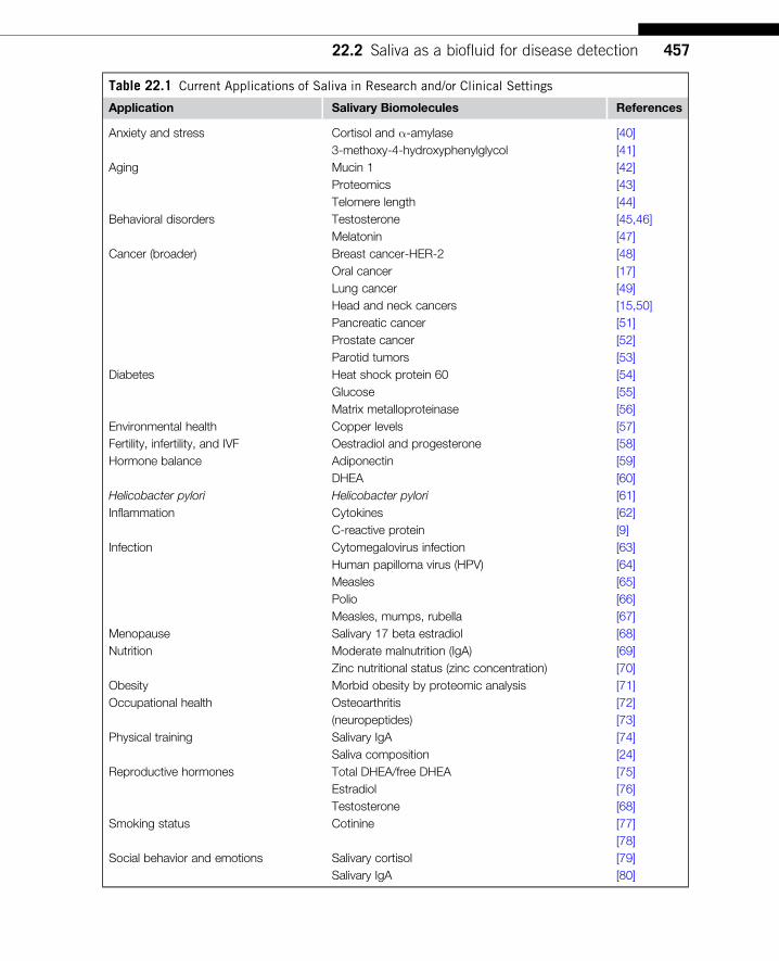
Table 22.1 Current Applications of Saliva in Research and/or Clinical Settings
Application Salivary Biomolecules References
Anxiety and stress Cortisol and α-amylase [40]
3-methoxy-4-hydroxyphenylglycol [41]
Aging Mucin 1 [42]
Proteomics [43]
Telomere length [44]
Behavioral disorders Testosterone [45,46]
Melatonin [47]
Cancer (broader) Breast cancer-HER-2 [48]
Oral cancer [17]
Lung cancer [49]
Head and neck cancers [15,50]
Pancreatic cancer [51]
Prostate cancer [52]
Parotid tumors [53]
Diabetes Heat shock protein 60 [54]
Glucose [55]
Matrix metalloproteinase [56]
Environmental health Copper levels [57]
Fertility, infertility, and IVF Oestradiol and progesterone [58]
Hormone balance Adiponectin [59]
DHEA [60]
Helicobacter pylori Helicobacter pylori [61]
Inflammation Cytokines [62]
C-reactive protein [9]
Infection Cytomegalovirus infection [63]
Human papilloma virus (HPV) [64]
Measles [65]
Polio [66]
Measles, mumps, rubella [67]
Menopause Salivary 17 beta estradiol [68]
Nutrition Moderate malnutrition (IgA) [69]
Zinc nutritional status (zinc concentration) [70]
Obesity Morbid obesity by proteomic analysis [71]
Occupational health Osteoarthritis [72]
(neuropeptides) [73]
Physical training Salivary IgA [74]
Saliva composition [24]
Reproductive hormones Total DHEA/free DHEA [75]
Estradiol [76]
Testosterone [68]
Smoking status Cotinine [77]
[78]
Social behavior and emotions Salivary cortisol [79]
Salivary IgA [80]
45722.2 Saliva as a biofluid for disease detection

Systems, Inc. (SDS, Vancouver Washington). These two companies commercialized devices for
saliva collection in the early 1990s, and these devices continue to be in widespread use for specific
applications today. In addition, the products developed by these companies led to a much broader
interest in saliva as a diagnostic fluid, and since then a plethora of new tools have become available
that has greatly expanded the applications and opportunities for salivary diagnostics. This chapter
attempts to cover the potential uses of saliva that have been explored so far and provides an indica-
tion of what can be expected in the future as the role of salivary diagnostics grows in an exponen-
tial fashion.
22.2.1 Saliva diagnostic assays in the market to date22.2.1.1 HIVCurrently the OraSures HIV-1 Oral Fluid Collection Device is available in conjunction with a
newly FDA-approved HIV 1/2 ELISA kit from Avioq Diagnostics for laboratory HIV testing.
A second oral-based test from Bio-Rad (the GS HIV 1/2 plus O ELISA) is also available for HIV
diagnosis using saliva. In each case, results are confirmed by a definitive laboratory-based oral fluid
(Western blot) test also manufactured by OraSure. Current markets for the OraSure HIV-1 test
device include public health screening, surveillance, and a very large market in insurance risk
assessment. In 2000, OraSure Technologies also launched the very first rapid diagnostic test for
HIV diagnosis using oral fluid specimens. The OraQuicks HIV 1/2 device is an immunochromato-
graphic test that delivers results in 20 min or less at the point of care. OraQuicks HIV 1/2 collects
saliva around the gum line under the lip area, using a paddle-shaped device, which incorporates a
proprietary test strip in the handle of the device (. 99.5% sensitivity). This test may soon receive
FDA approval for over-the-counter use (reference: Washington Post, May 14, 2012).
22.2.1.2 Drugs of abuseOraSure Technologies is also a major player in the drugs of abuse area. The Intercepts
Collection Device is used to collect saliva, which is immediately reflexed to a laboratory and
tested for a range of drug entities using ELISA tests originally developed by the company.
Currently, the predominant tests are the NIDA-5 series of drugs (cannabinoids (THC), opiates,
amphetamines, cocaine, and phencyclidine (PCP)); however, there are now also applications for a
number of drugs tested for by a variety of companies. One of the other most successful companies
is Immunalysis, who provides a range of microplate ELISA assays that are optimized for oral
fluid samples. Examples of other drugs of abuse in the market are tests for buprenorphine, metha-
done, and benzodiazepines, among others. In some cases it is parent drug that is detected, whereas
in others it is a metabolite of the parent compound that is quantified. The current major applica-
tions include the workplace testing environment (including the Federal workplace), drug courts,
methadone clinics, and military applications.
Newer devices are now entering the market, and these include the Versi•SALs Saliva Collection
Device (Oasis Diagnostics, www.4saliva.com, Vancouver, WA), which has been validated for use in
the forensics area with ELISA test kits from Neogen Corporation (www.neogen.com, Lansing, MI)
and its subsidiary company, International Diagnostic Systems (IDS, St. Joseph, MI) and the Greiner
458 CHAPTER 22 Saliva as an Emerging Biofluid for Clinical Diagnosis

Bio-One Saliva Collection System (www.grienerbioone.com, Vienna, Austria]. Neogen also has its
own large method of collecting saliva known as UltraSal-2t, which has been validated to oral fluid
specimens for multiple Neogen drug assays for the forensic market place. As well as traditional labo-
ratory ELISA procedures, there are other technologies for drug screening that have moved to oral
fluid testing. One such technology for rapid, high throughput testing is homogeneous immunoassay
performed on large instrument platforms. Examples of companies providing such technologies
includes Thermo Scientific (www.thermoscientific.com), who provide multiplex testing for the
NIDA-5 drugs using the Oral-Eze Saliva Collection Device from Quest Diagnostics (www.questdiag-
nostics.com) and its own CEDIA reagents optimized for oral specimens. The Thermo Scientific
reagents are optimized for a series of automated analyzers. A similar technology is available from
Roche Diagnostics (www.roche-diagnostics.us), through collaboration with OraSure Technologies. In
this instance, collection of specimens using the OraSure Intercepts Collection Device is followed
by homogeneous immunoassay using Roche’s KIMS (Kinetic Interaction of Micro-particles in
Solution) technology. Four drug assays are now FDA cleared and on sale in the United States for
multiple automated systems.
It is beyond the scope of this chapter to list all of the many available rapid oral drug screens,
but some of the manual point-of-care tests that are available are shown in Table 22.2 for reference
purposes.
There is a need and a market for roadside testing for drugs of abuse; however, currently avail-
able tools (mostly qualitative lateral flow-based systems) require additional improvements in order
to provide value in law-enforcement decision making and these are as follows:
1. Improvement in the sensitivity for key drugs, for instance, marijuana (tetrahydrocannabinol,
THC), where a cutoff close to the SAMHSA (Substance Abuse Mental Health Services
Administration) cutoff of 4 ng/mL is needed
2. Linkage to a hand-held reading device to eliminate any subjectivity in reading test results
Table 22.2 List of Representative Rapid Oral Drugs of Abuse Tests/Manufacturers
Manufacturer Web Site Product Name
American Biomedica Corporation www.abmc.com OralStat
JAJ Scientific www.jajinternational.com QikTech
Innovacon (Alere) www.innovaconinc.com OrALert
Mavand www.mavand.com RapidSTAT
Envitec www.envitec.com SmartClip
Sun Biomedical www.sunbiomed.com OraLine
Branan Medical www.brananmedical.com Oratect XP
Ulti-med www.ultimed.org SalivaScreen
Varian www.varian.com OraLab 6
Securetec www.securetec.net DrugWipe 6
45922.2 Saliva as a biofluid for disease detection

3. Provision of a hard copy of test results for evidentiary purposes
4. Faster acquisition of test results
5. Provision of a secondary (“B”) sample for confirmation and anticorruption practices
Several companies have tried to solve the above problems and have met with limited commercial
success. The most notable companies are Cozart Biosciences (United Kingdom, www.concateno.
com, now Alere, Inc.), Securetec (Germany, www.securetec.net), and Mavand (Germany, www.
mavand.com). While these companies have met with partial commercial success, additional improve-
ments will allow these manufacturers to meet the requirements of an EU organization known as
ROSITA (ROadSIde Testing Assessment) for use at the roadside (www.ROSITA.org). ROSITA is an
independent body that evaluates all drug testing devices with potential application in law enforce-
ment. ROSITA was set up to address the $164 billion annual cost in the European Union of “drivers
who are under the influence of drugs.” While all devices come under the scope of ROSITA, the
strong preference is for oral-based tests that are user friendly and will be adopted by all police forces
in the world.
22.2.1.3 Steroid hormones for general wellnessSaliva is an ideal medium for hormone assessment, and this has resulted in quite an “explosion”
in the number of laboratories testing for specific hormones. Driven by naturopaths, herbalists,
and nontraditional practicing physicians, a large and thriving market has developed where multi-
ple laboratories provide “saliva collection kits” direct to consumers. Responding to advertise-
ments in magazines focused on general health, nutrition, and fitness, customers are sent a kit in
the regular mail that allows them to collect their own saliva (usually into a saliva cup) and send
the sample back to a centralized laboratory where the results are evaluated for cortisol, testoster-
one, progesterone, estradiol, dehydroepiandrosterone (DHEA), and others and the results reported
back to the individual as “normal” or “abnormal” together with recommendations on any follow-
up actions. Perhaps this would include joining a fitness program or consulting a doctor because
the level of a particular hormone is outside of the accepted “normal range,” but since no diagno-
sis is given (just tips and recommendations related to general health status), these tests fall out-
side of the realm of “diagnostic tests.” Among hormones tested clinically, cortisol is by far the
largest due to the correlation of cortisol levels to stress and the growing hypothesis that stress is
implicated in many chronic diseases, such as cardiovascular diseases, infectious diseases, and
others.
Salimetrics Corporation (United States, www.salimetrics.com) has an FDA-cleared salivary cor-
tisol (ELISA) assay kit and also sells a complete range of salivary hormone assays optimized to
saliva specimens (Table 22.3).
22.2.1.4 CotinineCotinine, the active metabolite of nicotine, is evaluated in smoking cessation programs and is used
as a key indicator of risk in life insurance testing. Urine and salivary cotinine can be evaluated
using a series of ELISA test kits. Available systems include the OraSure Collection device and
460 CHAPTER 22 Saliva as an Emerging Biofluid for Clinical Diagnosis

ELISA test kit from OraSure, the Immunalysis Quantisalt Device and ELISA test kit, the Oasis
Diagnosticss Versi•SALs Device and Neogen/IDS ELISA microplate kits, as well as the Neogen
UltraSal-2t Collection Device and associated ELISA kit from Neogen.
22.2.1.5 Applications of saliva in molecular diagnostics22.2.1.5.1 DNADNA Genotek (Ottawa, Canada, www.dnagenoetk.com) was the first company to commercialize a
broad-based tool for the collection of saliva with subsequent application in genotyping, microar-
rays, and sequencing. Collection of (whole) mouth saliva into DNA Genotek’s OraGenes device
takes 10 min after which pure DNA is isolated from the stabilized sample and used in one of the
above-mentioned downstream applications. Until recently, this device was used specifically for
research applications; however, the device was recently cleared by the US FDA for clinical use in
conjunction with the GenMark Diagnostics eSensor Assay for Warfarin sensitivity. The OraGene
device has also found application in the high-profile “direct to consumer” area where companies
such as 23 and Me, Navigenics, Complete Genomics, Knome, and Pathway Genomics offer “per-
sonal genome” testing to members of the public. Newer tools in this area include the DNA isola-
tion and stabilization kits from Isohelix (www.isohelix.com) and Norgen Biotek Ontario Canada
(www.norgenbiotek.com) and the DNA•SALt Salivary DNA Collection Device from Oasis
Diagnosticss (Vancouver, United States, www.4saliva.com).
OralDNA Labs (www.oraldna.com), a subsidiary of Quest Diagnostics, offers a testing service
in the United States for two tests in its Clinical Laboratory Implementation Act (CLIA)-approved
testing facility in Brentwood, Tennessee. My PerioPaths is promoted as a “Salivary DNA Test
that determines the risk of periodontal infections” and is based upon the detection of a series of
bacterial pathogens in saliva. OraRisk HPVs is a “Salivary DNA Test that determines who is at
increased risk for HPV-related oral cancers” and identifies various HPV subtypes as low,
medium, or high risk as an indicator of overall risk for HPV-related oral carcinoma. Each patient
gargles a solution, which harvests DNA, that is subsequently transferred by a funnel device into a
transportation tube that is sent to the laboratory for downstream testing.
Table 22.3 Manufacturers of Salivary Hormone (ELISA) Test Kits
Company Country Web Site
DRG United States www.drg-international.com
IBL Hamburg Germany www.ibl-international.com
Hoelzel Diagnostika Germany www.hoelzel-biotech.com
Diametra Italy www.diametra.com
Alpco United States www.alpco.com
IBL America United States www.ibl-america.com
46122.2 Saliva as a biofluid for disease detection

22.2.1.5.2 RNARNA can be isolated directly from saliva using a number of available “Salivary RNA Isolation”
kits sold by Qiagen, GE Healthcare, Life Technologies, and others. Although the procedure to iso-
late RNA by this method is time consuming and costly, saliva has become a “trusted” medium for
RNA research and development. There are no current tools available for direct RNA isolation;
however, there are tools in development that will be useful for this purpose. Novel tools should be
available within 1�2 years.
22.2.1.6 Applications of saliva in proteomicsHuman saliva consists of a large number of proteins and peptides (the salivary proteome and pepti-
dome) [81,82] that aids in maintaining oral homeostasis. Unlike the plasma proteome, the saliva
proteome is highly susceptible to a variety of physiological and biochemical processes, and this
presents a challenge for clinical salivary proteomics [29,83,84]. The dynamic range of proteins in
saliva is another challenge. For instance, the abundant α-amylase in saliva is present at mg/mL con-
centrations, while the IL-6 and IL-8 cytokines of potential clinical relevance are present only at
concentrations of pg/mL [85]. The saliva proteome also changes as a function of age. A loss of sali-
vary acinar cell function was documented in healthy adults as a consequence of aging [86,87],
while salivary production remained age stable in healthy adults. Such effects must be carefully con-
sidered in the development of salivary diagnostic assays, primarily by inclusion of appropriate
control groups in assay development and validation.
22.2.2 Saliva research updateSaliva research expands from infectious disease detection, to dental research to assess gum dis-
eases, to psychology and forensic sciences. As of today, a number of researchers are focusing on
developing techniques and tools to discern the biomolecular composition of saliva with the aim of
facilitating clinical translation. Saliva collection is a crucial step in the utilization of saliva for clini-
cal purposes, so it is very important that saliva collection technique should not influence down-
stream applications. There are commercially available saliva collection devices suited for both the
life science research as well as for diagnostic purposes, such as DNA Genotek (www.dnagenotek.
com); Salimetrics oral swabs (http://www.salimetrics.com); Oasis Diagnosticss VerOFys,
Versi � SALs, and DNA � SALt (http://www.4saliva.com); OraSure Technologies OraSure Oral
Fluid Collection Device (http://www.orasure.com); Cozarts drugs of abuse collection devices
(http://www.concateno.com), Immunalysis Quantisalt Saliva Collection Device; and the Greiner
Bio-One Saliva Collection System (http://www.gbo.com) [4]. These saliva sample collection tech-
nologies assist in obtaining either unstimulated or stimulated saliva.
Saliva collection procedures differ based on the type of saliva that one is interested in collecting.
As an example, for ductal secretion collections, one can use Carlson�Crittenden cup [88,89] over the
orifice of the Stenson’s duct [90]. However, these methods are invasive and forfeit the noninvasive
advantage of saliva for clinical use. It is important to determine experimentally which collection
device is suited for a particular application before commencing any clinical trials. Standardization of
saliva collection methods is also vital in translating saliva research from the lab to the clinic [91].
462 CHAPTER 22 Saliva as an Emerging Biofluid for Clinical Diagnosis

22.3 Applications of saliva for early detection of ischemic heartdisease and in head and neck cancersIn this section, we will highlight a case study where saliva as a biological medium has been applied
to diagnose IHD and head and neck squamous cell carcinoma (HNSCC) at an early stage.
22.3.1 Salivary C-reactive protein levels as a proxy to diagnose ischemic heartdiseaseC-reactive protein (CRP) is a marker of inflammation. CRP is a member of the class of acute-phase
reactants that mediates innate and adaptive immunity [92]. It is produced by the hepatocytes in
response to a variety of inflammatory cytokines [93] and may rise rapidly by as much as 1000-fold
or more after an acute inflammatory stimulus [94]. CRP has been shown to be an independent
predictor of cardiovascular events, and this biomolecule has also been proven to add prognostic
value to cardiovascular risk [11,12].
We found that salivary CRP concentrations in 55 healthy volunteers ranged from 50.6 to
872.4 pg/mL. Using ranked statistical methods the derived reference interval in a healthy popula-
tion was ,824 pg/mL. The mean CRP level in the saliva of healthy human volunteers was
285 pg/mL and in cardiac patients was 1680 pg/mL (P,0.01). Analysis of CRP concentrations in
paired serum and saliva samples from cardiac patients gave a positive correlation (r25 0.84,
P,0.001) (see Figure 22.2).
22.3.2 Salivary DNA methylation as a proxy to diagnose head and neck cancerHNSCC is the fifth most common cancer in men with an incidence of about 780,000 new cases per
year worldwide [95]. Despite advances in therapy, its prognosis has not markedly improved in the
past 20 years [96]. This is mainly caused by the late diagnosis of HNSCC, when cancer cells may
have metastasized to other parts of the body. HNSCC can affect the nasal passages, sinuses, mouth,
throat, larynx (voice box), swallowing passages, salivary glands, and thyroid gland and arise from
the surface epithelium. Tobacco use is a major risk factor for this type of cancer, and smoking kills
over 1,000,000 people a year, causing 30% of all cancer-related deaths in western societies. Yet,
one in three people worldwide is addicted to nicotine. In addition, 30% of HNSCC are a direct result
of human papillomavirus (HPV) infections [97]. Of all HPV types, the high-risk strains HPV16 and,
to a lesser extent, HPV18 are most commonly identified in oral squamous cell cancer biopsies [98].
22.3.2.1 Current clinical work flow for head and neck cancerHNSCC detection is currently based on an expert clinical examination of the upper aerodigestive
tract and histologic analysis of suspicious areas, but it may be undetectable in hidden sites, such as
crypts of the tongue base or tonsils.
22.3.2.2 Current unmet clinical need in head and neck cancer patient managementAt present, there are no early detection/screening tests for head and neck cancers. At the time of
diagnosis of HNSCC, in 80% of the patients cancer cells may have already metastasized into other
parts of the body, resulting in a low 5-year survival rate.
46322.3 Applications of saliva for early detection of IHD

Early diagnosis of HNSCC holds the promise of improved prognosis but is currently impeded in
many patients who delay seeking medical attention due to a number of factors associated with
tobacco and alcohol intake. Moreover, if the tumors are tiny (unable to detect by modern cameras)
and/or located in areas in the oral cavity that are not easily accessible, saliva offers the opportunity
as a diagnostic medium for early detection since these tiny tumors secrete biomarkers that are
indicative of a pathological condition. More so, the direct impact of smoking can clearly be seen in
2.5x10−09
2E−08
y = 0.0324x − 2E−10
1.5E−08
1E−08
5E−09
0
Serum CRP (g/mL)
0
0.00
0000
1
Sal
iva
CR
P (
g/m
L)
0.00
0000
2
0.00
0000
3
0.00
0000
4
0.00
0000
5
2.0x10−09
1.5x10−09
1.0x10−09
5.0x10⁻¹⁰
0
(A)
(B)
Healthy volunteers Cardiac patients
Sal
ivar
y C
RP
(pg
/mL)
FIGURE 22.2
(A) Human salivary CRP levels in healthy volunteers (n5 55) and in cardiac patients (n5 28). (B) Correlation
of salivary CRP levels to plasma CRP levels [9].
464 CHAPTER 22 Saliva as an Emerging Biofluid for Clinical Diagnosis

the oral cavity due to its proximity; thus, human saliva is an ideal diagnostic medium for investigat-
ing smoking-related cancers.
The absence of definite early warning signs for most HNSCC suggests that sensitive and spe-
cific biomarkers are likely to be important for screening in high-risk patients [99]. DNA methyla-
tion in cells (the addition of methyl groups to cytosine residues on the DNA sequence) is an early
event that occurs during tumor initiation [100]. In fact, promoter DNA hypermethylation is a more
frequent mechanism of gene silencing than genetic mutation [101]. Unlike DNA mutations, DNA
methylation abnormalities are reversible by drugs in a laboratory setting, and this reversal allows
cancer cells to reactivate the silenced (da Silva, 2009 #3394) genes and produce tumor-suppressor
proteins. Because DNA methylation normally leads to gene silencing (a negative biological event),
a tumor-suppressor protein is not produced and thus protein detection methods cannot be used. For
a diagnostic test to be implemented clinically, the test will ideally measure a positive event occur-
ring in tumor cells de novo; therefore, by detecting DNA methylation in cells, one can turn a nega-
tive biological event into a positive clinical test. Understanding how abnormal DNA methylation
arises in cancer cells, and how this change leads to silencing of genes, is extremely important in
the development of treatments that could reverse this process as a strategy to prevent and/or treat
cancer (Figures 22.3 and 22.4).
With the development of noninvasive early screening tools and strategies (such as the ones that
are currently being developed in our laboratory) would enable the diagnosis of HNSCC at an earlier
stage and render treatment strategies.
22.3.3 Applications of Micro Electromechanical Systems (MEMS)/NanoElectromechanical Systems (NEMS) in salivary diagnosticsNanotechnology platforms are foreseen to change health care in a fundamental way by providing
novel methods for disease diagnosis and prevention, therapeutics selection and administration, tai-
lored to the patients’ profile, drug delivery, and gene therapy. Nanotechnology is about manipulating
matter atom by atom. Nanodentistry is defined as the science and technology of maintaining near-
perfect oral health through the use of nanomaterials such as nano oral anesthesia inductions [102],
nanodental techniques for major tooth repair, nano in-tooth repositioning, and nanorobotics [103].
Nanotechnology-based NEMS biosensors result in high sensitivity and specificity for analyte
detection in complex matrices such as saliva, sensitivity of the detection system reaching down to
single molecule levels. These convert (bio)chemical to electrical signal [104]. As an example, the
Oral Fluid NanoSensor Test (OFNASET) technology is used for multiplex detection of salivary bio-
markers for oral cancer. A previous study has demonstrated that the combination of two salivary
proteomic biomarkers (thioredoxin and IL-8) [105] and four salivary mRNA biomarkers (SAT,
ODZ, IL-8, and IL-1b) can be used to detect oral cancer with high specificity and sensitivity [106].
In addition, the optical nanobiosensor is a unique fiberoptics-based technology platform that allows
minimally invasive analysis of intracellular components such as cytochrome c (which regulates
apoptosis or programmed cell death and cellular energy production) [104]. Nanotechnology is not
only providing information on diagnosing a disease but also provides treatment opportunities. As
an example, BrachySilTM (Sivida, Australia) delivers 32P clinical trial for brachytherapy.
In summary, nanodentistry faces significant challenges in realizing its tremendous potential in revo-
lutionizing the current dental care practice. Some of the obstacles in the advancement of nanodentistry
46522.3 Applications of saliva for early detection of IHD

include basic engineering problem from precise positioning and assembly of molecular-scale parts to
biocompatibility issues, public acceptance, ethics, regulation, and human safety. When the issues raised
above have been adequately addressed, nanodentistry will soon become a reality.
22.4 Future outlook and conclusionsThere are many other areas where saliva may be used either as a replacement for traditional blood
testing or as an adjunct to current testing methods. This section highlights some of the many possibil-
ities that saliva may play a role in future clinical application. In each case, scholarly articles are read-
ily available relating to the utility of saliva and serve as a solid basis for the development of future
testing products. A series of biomarkers including CRP [9,23], α-amylase, and cortisol [65,66] have
been used in the assessment of cardiovascular health, and rapid point-of-care test devices using saliva
will in the near future be available to assess risk of CVD. In a related area LabNow, a company
spearheaded by Dr. John McDevitt, a professor of biochemistry from the University of Texas at
Austin, has developed a nano-biochip method that uses saliva to diagnose early heart attack. The
80
70
60
50
p16INK4a MSP
3-gene MSP panel
Smokers + − + −
Patients Controls
40
Pos
itive
s %
30
20
10
0
FIGURE 22.3
The DNA promoter hypermethylation of three tumor suppressor genes (DAPK1, RASSF1a, and p16) in saliva
collected from a healthy control group (n5 41) and HNSCC patients (n5 121, both smokers and
nonsmokers). On the Y-axis, if one of the genes is methylated that particular saliva sample is included in the
data set [15].
466 CHAPTER 22 Saliva as an Emerging Biofluid for Clinical Diagnosis

method is reported to be more accurate than the standard EKG, which can miss up to 25% of poten-
tial heart attacks. In the oncology area, the use of saliva to isolate, characterize, and identify specific
roles for various messenger RNAs and microRNAs has already been done, and the value of saliva as
a tool to provide pure mRNA and pure miRNA for use in targeted therapies and general research
will become increasingly important over the next several years. An early application for the use of
miRNAs will be in the diagnosis of oral cancers, pancreatic cancer, and other malignancies, but the
impact will not stop there. mRNAs and miRNAs have been reported in many disease processes, so it
is expected that the role of RNA and salivary RNA in particular will expand dramatically.
Studies have also been performed confirming the detection of specific proteins such as Her-2/
neu and tumor markers such as CA-125, CA 15�3 are possible, but to date no diagnostic tests have
been developed using saliva specimens. Viral diseases represent another target area for salivary
diagnostics with a number of major disease antibodies and antigens (hepatitis A, hepatitis B, hepati-
tis C, HHV-1 to HHV-8, EBV, CMV herpes, and influenza viruses) all detectable in saliva. Oral
fluid samples have already proved useful in the evaluation of immunization efficacy, particularly in
the developing world, where immune response to measles, mumps, polio, tetanus, and rubella vac-
cines have been routinely carried out. More recently, a company from the United Kingdom,
MicroImmune, has developed a saliva-based point-of-care device for the detection of measles-
specific IgM antibodies. The group made up of scientists from the Public Health Laboratory in
Colindale (London) intend developing additional vaccine-specific rapid tests in the future.
The success of OralDNA Labs in the United States has spurred a “fever” of activity in the detec-
tion of bacterial infections using saliva specimens, and a number of companies are looking to target
the dental office as the first line of attack in our general health. These companies will provide dental
Tumor
FIGURE 22.4
The patient is a 66-year-old gentleman—retired boatbuilder. He has been aware of a foreign-body sensation
in the right side of his throat for up to 2 years. He had no pain, swallowing or voice problems. Dr. S. Coman
staged this case as T2N0 SCC. Histology reports “moderately differentiated” squamous cell carcinoma. Stains
for p16 are strongly evident.
(Photograph provided by Professor William B. Coman and Dr. S. Coman.)
46722.4 Future outlook and conclusions

tests or collection kits direct to practicing dentists, who in many instances see patients on a more rou-
tine basis than a general practice physician. In such circumstances, the dentist is well placed to iden-
tify the disease risk early on. Tests targeted for the dentist office that are in the development or
available already include tests for dental caries, HPV, periodontal disease, and gingivitis.
The area of drug abuse is rapidly growing, but linked with this is an increase in the abuse of
prescription drugs, particularly painkillers and antidepressants. With this comes a need to detect
drug concentrations accurately and in real time. Saliva offers the best matrix in most cases to do
this, and a number of companies with detection kits (ELISA tests) are evaluating options to use
saliva as a specimen of choice to expand their product portfolios. As another example, the
“designer” drug known as Spice (or K2) has led to several deaths in the United States and has been
banned in many states. This and other such drugs will be logical targets for saliva test developers.
Lateral flow immunochromatography is a technique used to provide rapid diagnostic test results
for multiple diseases using bodily fluids. Progress in manufacturing and development technology in
the 1990s has been rapid, and this has resulted in the development of a whole series of rapid, point-
of-care devices that initially were based upon the use of urine (the currently accepted mainstay for
drug testing today) or blood specimens. In the area of drug testing, there has been a lot of work
done to validate a number of multi-drug screening panels based on oral sampling. Up until now,
most tests are qualitative in nature, i.e. provide a yes or no indication of drug presence. The oppor-
tunity to provide immediate results at the point-of-care using noninvasive samples is an attractive
proposition; however, oral-based rapid tests for drugs of abuse have certain drawbacks that have
limited the broader utilization of these devices to date. Potential problems include poor recovery of
analytes (particularly marijuana (THC) from collection media), insufficient saliva delivery to the
test strips, strip failure, and lack of sensitivity. Despite this, the “convenience factor” of oral testing
has led to a proliferation of companies developing such tests and subsequent adoption of these tests
in drug screening projects, criminal justice, employee screening, random testing, and other
instances where immediate results can be beneficial. Further inroads into the market will be made
once the above issues have been resolved.
AcknowledgmentsThe authors would like to acknowledge the financial support from the Queensland Government Smart Futures
Fellowship Programme (QGSFF), the University of Queensland New Staff Research Funds (UQNSRSF
601252), and the University of Queensland Foundation Research Excellence Award Scheme. In addition, we
would like to express our sincere gratitude to Professor William B. Coman and Dr. Scott Coman for providing
us with the illustrations. In addition, we thank Mr. Jared Foo and Ms Ling Li Long for their technical
assistance.
References[1] D. Marmud, Saliva as a diagnostic fluid second now to blood? BMJ 305 (1992) 25.
[2] S. Hu, D.T. Wong, M. Arellano, P. Boontheing, J. Wang, H. Zhou, et al., Salivary Protein Biomarkers for
Human Oral Cancer, Clin Cancer Research 14 (19) (2008) 6246�6252.
468 CHAPTER 22 Saliva as an Emerging Biofluid for Clinical Diagnosis

[3] P.V. Rao, A.P. Reddy, X. Lu, S. Dasari, A. Krishnaprasad, E Biggs, et al., Proteomic identification of
salivary biomarkers of type-2 diabetes, J. Proteome Res. 8 (1) (2009) 239�245.
[4] T. Pfaffe, J. Cooper-White, P. Beyerlein, K. Kostner, C. Punyadeera, et al., Diagnostic potential of
saliva: current state and future applications, Clin. Chem. 57 (5) (2011) 675�687.
[5] F. Bouwman, et al., 2D-electrophoresis and multiplex immunoassay proteomic analysis of different body
fluids and cellular components reveal known and novel markers for extended fasting, BMC Med.
Genomics 4 (1) (2011) 24.
[6] W. Yan, et al., Systematic comparison of the human saliva and plasma proteomes, Proteomics Clin.
Appl. 3 (1) (2009) 116�134.
[7] S. Bandhakavi, et al., A dynamic range compression and three-dimensional peptide fractionation analysis
platform expands proteome coverage and the diagnostic potential of whole saliva, J. Proteome Res. 8
(12) (2009) 5590�5600.
[8] J.A. Loo, et al., Comparative human salivary and plasma proteomes, J. Dent. Res. 89 (10) (2010)
1016�1023.
[9] C. Punyadeera, G. Dimeski, K. Kostner, P. Beyerlein, J. Cooper-White, et al., One-step homogeneous
C-reactive protein assay for saliva, J. Immunol. Methods (2011).
[10] N. Christodoulides, M. Sanghamitra, M.S. Craig, L.M. Chris, N. Floriano, P.D. Priya, et al., Application
of microchip assay system for the measurement of C-reactive protein in human saliva, Lab Chip 5 (3)
(2005) 261�269.
[11] P.M. Ridker, C-reactive protein, inflammation, and cardiovascular disease: clinical update, Tex. Heart
Inst. J. 32 (3) (2005) 384�386.
[12] P.M. Ridker, et al., Rosuvastatin to prevent vascular events in men and women with elevated C-reactive
protein, N. Engl. J. Med. 359 (21) (2008) 2195�2207.
[13] H. Arima, et al., High-sensitivity C-reactive protein and coronary heart disease in a general population
of Japanese: the Hisayama study, Arterioscler Thromb Vasc. Biol. 28 (7) (2008) 1385�1391.
[14] C.S. Miller, et al., Salivary biomarkers of existing periodontal disease: a cross-sectional study, J. Am.
Dent. Assoc. 137 (3) (2006) 322�329.
[15] D.A. Ovchinnikov, M.A. Cooper, P. Pandit, W.B. Coman, J. Cooper-White, P. Keith, et al., Tumour-sup-
pressor gene promoter hypermethylation in saliva of head and neck cancer patients. J. Trans. Oncol.,
September 2012.
[16] T. Shpitzer, et al., A comprehensive salivary analysis for oral cancer diagnosis, J. Cancer Res. Clin.
Oncol. 133 (9) (2007) 613�617.
[17] D.T. Wong, Oral cancer and saliva diagnostics, in: National Oral Health Conference, 2007,
Denver, CO.
[18] S. Hu, et al., Preclinical validation of salivary biomarkers for primary Sjogren’s syndrome, Arthritis
Care Res. 62 (11) (2010) 1633�1638.
[19] L. Zhang, J.J. Farrell, H. Zhou, D. Elashoff, D. Akin, N.-H. Park, et al., Researchers find biomarkers in saliva
for detection of early-stage pancreatic cancer, Gastroenterology, (138) (2010) 949�957.
[20] C.F. Streckfus, et al., The presence of soluble c-erbB-2 in saliva and serum among women with breast
carcinoma: a preliminary study, Clin. Cancer Res. 6 (6) (2000) 2363�2370.
[21] C.F. Streckfus, et al., Breast cancer related proteins are present in saliva and are modulated secondary to
ductal carcinoma in situ of the breast, Cancer Invest. 26 (2) (2008) 159�167.
[22] M. Castagnola, P.M. Picciotti, I. Messana, C. Fanali, A. Fiorita, T. Cabras, et al., Potential applications
of human saliva as diagnostic fluid, Acta Otorhinolaryngol Ital. 31 (6) (2011) 347�357.
[23] E. Topkas, et al., Evaluation of saliva collection devices for the analysis of proteins, Clin. Chim. Acta
413 (13�14) (2012) 1066�1070.
[24] J.L. Chicharro, et al., Saliva composition and exercise, Sports Med. 26 (1) (1998) 17�27.
469References

[25] M.A. Anzano, A.J. Lamb, J.A. Olson, Impaired salivary gland secretory function following the induction
of rapid, synchronous vitamin A deficiency in rats, J. Nutr. 111 (3) (1981) 496�504.
[26] N.P. Walsh, et al., Saliva flow rate, total protein concentration and osmolality as potential markers of
whole body hydration status during progressive acute dehydration in humans, Arch. Oral Biol. 49 (2)
(2004) 149�154.
[27] D.B. Ferguson, Current diagnostic uses of saliva, J. Dent. Res. 66 (2) (1987) 420�424.
[28] C. Dawes, Salivary flow patterns and the health of hard and soft oral tissues, J. Am. Dent. Assoc. 139
(Suppl. 2) (2008) 18S�24S.
[29] F.G. Oppenheim, et al., Salivary proteome and its genetic polymorphisms, Ann. N.Y. Acad. Sci. 1098
(2007) 22�50.
[30] I.D. Mandel, et al., Salivary studies in cystic fibrosis, Am. J. Dis. Child. 113 (4) (1967) 431�438.
[31] I.D. Mandel, Relation of saliva and plaque to caries, J. Dent. Res. 53 (2) (1974) 246�266.
[32] A.M. Hansen, A.H. Garde, R. Persson, Measurement of salivary cortisol—effects of replacing polyester
with cotton and switching antibody, Scand. J. Clin. Lab. Invest. 68 (8) (2008) 826�829.
[33] A.M. Hansen, A.H. Garde, R. Persson, Sources of biological and methodological variation in salivary
cortisol and their impact on measurement among healthy adults: a review, Scand. J. Clin. Lab. Invest. 68
(6) (2008) 448�458.
[34] E.J. Helmerhorst, F.G. Oppenheim, Saliva: a dynamic proteome, J. Dent. Res. 86 (8) (2007) 680�693.
[35] L. Caporossi, A. Santoro, B. Papaleo, Saliva as an analytical matrix: state of the art and application for
biomonitoring, Biomarkers 15 (6) (2010) 475�487.
[36] M. Groschl, Current status of salivary hormone analysis, Clin. Chem. 54 (11) (2008) 1759�1769.
[37] M. Groschl, The physiological role of hormones in saliva, Bioessays 31 (8) (2009) 843�852.
[38] D.A. Granger, et al., Integration of salivary biomarkers into developmental and behaviorally-
oriented research: problems and solutions for collecting specimens, Phys. Behav. 92 (4) (2007)
583�590.
[39] E. Megson, et al., C-reactive protein in gingival crevicular fluid may be indicative of systemic inflamma-
tion, J. Clin. Periodontol. 37 (9) (2010) 797�804.
[40] Y. Tanaka, et al., Salivary alpha-amylase and cortisol responsiveness following electrical stimulation
stress in major depressive disorder patients, Prog. Neuropsychopharmacol. Biol. Psychiatry 36 (2) (2012)
220�224.
[41] S. Yamada, et al., Saliva level of free 3-methoxy-4-hydroxyphenylglycol (MHPG) as a biological index
of anxiety disorders, Psychiatry Res. 93 (3) (2000) 217�223.
[42] W.-I. Chang, et al., MUC1 expression in the oral mucosal epithelial cells of the elderly, Arch. Oral Biol.
56 (9) (2011) 885�890.
[43] K.S. Ambatipudi, et al., Quantitative analysis of age specific variation in the abundance of human female
parotid salivary proteins, J. Proteome Res. 8 (11) (2009) 5093�5102.
[44] E.J. Giltay, et al., Salivary testosterone: associations with depression, anxiety disorders, and antidepres-
sant use in a large cohort study, J. Psychosomatic. Res. 72 (3) (2012) 205�213.
[45] I. Paclt, et al., Circadian rhythms of saliva melatonin in ADHD, anxious and normal children, Neuro
Endocrinol. Lett. 32 (6) (2011) 790�798.
[46] M. Novakova, et al., Salivary melatonin rhythm as a marker of the circadian system in healthy children
and those with attention-deficit/hyperactivity disorder, Chronobiol. Int. 28 (7) (2011) 630�637.
[47] C.F. Streckfus, D. Arreola, C. Edwards, L. Bigler, Salivary protein profiles among HER2/neu-receptor-
positive and -negative breast cancer patients: support for using salivary protein profiles for modeling
breast cancer progression, J. Oncol. 2012 (2012) 9.
[48] S. Punyani, R. Sathawane, Salivary level of interleukin-8 in oral precancer and oral squamous cell carci-
noma, Clin. Oral Invest. (2012) 1�8.
470 CHAPTER 22 Saliva as an Emerging Biofluid for Clinical Diagnosis

[49] X. Li, T. Yang, J. Lin, Spectral analysis of human saliva for detection of lung cancer using surface-
enhanced Raman spectroscopy, J. Biomed. Opt. 17 (2012) 3.
[50] T. Jarai, et al., Mass spectrometry-based salivary proteomics for the discovery of head and neck squa-
mous cell carcinoma, Pathol. Oncol. Res. 18 (3) (2012) 623�628.
[51] L. Zhang, J.J. Farrell, H. Zhou, D. Elashoff, D. Akin, N.-H. Park, et al., Salivary transcriptomic biomar-
kers for detection of resectable pancreatic cancer, Gastroenterology 38 (2010) 949�957.
[52] N. Shiiki, et al., Association between saliva PSA and serum PSA in conditions with prostate adenocarci-
noma, Biomarkers 16 (6) (2011) 498�503.
[53] G. Dyckhoff, et al., Carbohydrate antigen 19�9 in saliva, Otolaryngol. Head Neck Surgery 145 (5)
(2011) 772�777.
[54] J. Yuan, P. Dunn, R.D. Martinus, Detection of Hsp60 in saliva and serum from type 2 diabetic and non-
diabetic control subjects, Cell Stress Chaperones 16 (6) (2011) 689�693.
[55] A.C.U. Vasconcelos, et al., Comparative study of the concentration of salivary and blood glucose in type
2 diabetic patients, J. Oral Sci. 52 (2) (2010) 293�298.
[56] P.P. Costa, et al., Salivary interleukin-6, matrix metalloproteinase-8, and osteoprotegerin in patients with
periodontitis and diabetes, J. Periodontol. 81 (3) (2009) 384�391.
[57] T. Thompson, et al., Copper levels in buccal cells of vineyard workers engaged in various activities,
Ann. Occup. Hyg. 56 (3) (2012) 305�314.
[58] Y.F. Wong, L.E. Mao, K.R. Tam, P.P. Panesar, N.S. Neale, E. Chang, et al., Salivary oestradiol and pro-
gesterone after in vitro fertilization and embryo transfer using different luteal support regimens, Reprod.
Fertil Dev. 2 (4) (1990) 351�358.
[59] E.-N. Akuailou, P. Vijayagopal, V. Imrhan, C. Prasad, Measurement and validation of the nature of sali-
vary adiponectin, Acta Diabetol. 15 (1) (2012) 110�114.
[60] A. Oskis, A. Clow, L. Thorn, C. Loveday, F. Hucklebridge, Diurnal patterns of salivary cortisol and
DHEA in adolescent anorexia nervosa, Stress 15 (6) (2012) 601�607.
[61] H. Momtaz, et al., Study of Helicobacter pylori genotype status in saliva, dental plaques, stool and gas-
tric biopsy samples, World J. Gastroenterol. 18 (17) (2012) 2105�2111.
[62] K. Szczeklik, D. Owczarek, J. Pytko-Polonczyk, B. Kesek, T.H. Mach, Proinflammatory cytokines in the
saliva of patients with active and non-active Crohn’s disease, Pol. Arch. Med. Wewn. April 24 (2012)
(pii: AOP79).
[63] E.J. Plosa, et al., Cytomegalovirus infection, Pediatr. Rev. 33 (4) (2012) 156�163 (quiz 163).
[64] D.O. Turner, et al., High-risk human papillomavirus (HPV) screening and detection in healthy patient
saliva samples: a pilot study, BMC Oral Health 11 (2011) 28.
[65] R. Dantzer, N. Kalin, Psychoneuroendocrinology 34 (1) (2009).
[66] E.A. Shirtcliff, D.A. Granger, E. Schwartz, M.J. Curran, Use of salivary biomarkers in biobehavioral
research: cotton-based sample collection methods can interfere with salivary immunoassay results.
Psychoneuroendocrinology 26 (2) (2001) 165�173.
[67] D.J. Nokes, et al., Has oral fluid the potential to replace serum for the evaluation of population immunity
levels? A study of measles, rubella and hepatitis B in rural Ethiopia, Bull. World Health Org. 79 (7)
(2001) 588�595.
[68] F. Agha-Hosseini, et al., Relationship of stimulated saliva 17beta-estradiol and oral dryness feeling in
menopause, Maturitas 62 (2) (2009) 197�199.
[69] D.N. McMurray, et al., Effect of moderate malnutrition on concentrations of immunoglobulins and
enzymes in tears and saliva of young Colombian children, Am. J. Clin. Nutr. 30 (12) (1977) 1944�1948.
[70] T. Hovi, M. Stenvik, Selective isolation of poliovirus in recombinant murine cell line expressing the
human poliovirus receptor gene, J. Clin. Microbiol. 32 (5) (1994) 1366�1368.
[71] R.S. Hobson, Challenges to future dental education, Br. Dent. J. 206 (3) (2009) 125�126.
471References

[72] V.J. Howard, et al., Does sex matter? Thirty-day stroke and death rates after carotid artery stenting in
women versus men: results from the Carotid Revascularization Endarterectomy versus Stenting Trial
(CREST) lead-in phase, Stroke 40 (4) (2009) 1140�1147.
[73] H.P. Fischer, W. Eich, I.J. Russell, A Possible Role for Saliva as a Diagnostic Fluid in Patients with
Chronic Pain, Elsevier, Seminars in Arthritis and Rheumatism 26 (6) 1998 348�359.
[74] V. Neville, M. Gleeson, J.P. Folland, Salivary IgA as a risk factor for upper respiratory infections in elite
professional athletes, Med. Sci. Sports Exercise 40 (7) (2008) 1228.
[75] L.M. Swinkels, et al., Concentrations of total and free dehydroepiandrosterone in plasma and dehydro-
epiandrosterone in saliva of normal and hirsute women under basal conditions and during administration
of dexamethasone/synthetic corticotropin, Clin. Chem. 36 (12) (1990) 2042�2046.
[76] C.M. Worthman, J.F. Stallings, L.F. Hofman, Sensitive salivary estradiol assay for monitoring ovarian
function, Clin. Chem. 36 (10) (1990) 1769�1773.
[77] R.E. Latchaw, et al., Recommendations for imaging of acute ischemic stroke: a scientific statement from
the American Heart Association, Stroke 40 (11) (2009) 3646�3678.
[78] G.P. Harris, et al., Three distinct amine receptors operating at different levels within the locomotory cir-
cuit are each essential for the serotonergic modulation of chemosensation in Caenorhabditis elegans,
J. Neurosci. 29 (5) (2009) 1446�1456.
[79] R.P. Hobson, et al., How mothers with borderline personality disorder relate to their year-old infants,
Br. J. Psychiatry 195 (4) (2009) 325�330.
[80] E.F. Nsutebu, R. Hobson, Is combination systemic therapy superior to monotherapy for enterococcal
prosthetic joint infection? Clin. Infect. Dis. 48 (4) (2009) 495 (author reply 495�6).
[81] B.L. Schulz, J. Cooper-White, C.K. Punyadeera, Saliva proteome research: current status and future out-
look, Crit. Rev. Biotechnol. (2012).
[82] C.-M. Huang, Comparative proteomic analysis of human whole saliva, Arch. Oral Biol. 49 (12) (2004)
951�962.
[83] C. Dawes, Considerations in the development of diagnostic-tests on saliva, Ann. N.Y. Acad. Sci. 694
(1993) 265�269.
[84] J.R. Whiteaker, et al., A targeted proteomics-based pipeline for verification of biomarkers in plasma,
Nat. Biotechnol. 29 (7) (2011) 625�634.
[85] M.A. St John, et al., Interleukin 6 and interleukin 8 as potential biomarkers for oral cavity and oropha-
ryngeal squamous cell carcinoma, Arch. Otolaryngol. Head Neck Surg. 130 (8) (2004) 929�935.
[86] E.M. Ghezzi, J.A. Ship, Aging and secretory reserve capacity of major salivary glands, J. Dent. Res. 82
(10) (2003) 844�848.
[87] A. Westermark, et al., Basic fibroblast growth factor in human saliva decreases with aging,
Laryngoscope 112 (5) (2002) 887�889.
[88] M. Navazesh, S.K.S. Kumar, Measuring salivary flow: challenges and opportunities, J. Am. Dent. Assoc.
139 (Suppl. 2) (2008) 35S�40S.
[89] A.J. Carlson, A.L. Crittenden, The relation of ptyalin concentration to the diet and to the rate of secre-
tion of the saliva, Am. J. Physiol. 26 (1) (1910) 169�177 (Legacy Content).
[90] M.W. Heft, B.J. Baum, Basic biological sciences unstimulated and stimulated parotid salivary flow rate
in individuals of different ages, J. Dent. Res. 63 (10) (1984) 1182�1185.
[91] Mohamed, J.-L. Campbell, J. Cooper-White, G. Dimeski, C. Punyadeera, The impact of saliva collection
and processing methods on CRP, IgE, and myoglobin immunoassays, J. Clin. Trans. Med. September, 5,
(2012).
[92] S. Chomdej, et al., Detection of SNPs and linkage and radiation hybrid mapping of the porcine
C-reactive protein (CRP) gene, Anim. Genet. 35 (6) (2004) 469�470.
[93] T.W. Du Clos, Function of C-reactive protein, Ann. Med. 32 (4) (2000) 274�278.
472 CHAPTER 22 Saliva as an Emerging Biofluid for Clinical Diagnosis

[94] S. Black, I. Kushner, D. Samols, C-reactive protein, J. Biol. Chem. 279 (47) (2004) 48487�48490.
[95] R. Sankaranarayanan, et al., An overview of cancer survival in developing countries, IARC Sci. Publ.
(145) (1998) 135�173.
[96] A. Forastiere, et al., Head and neck cancer, N. Engl. J. Med. 345 (26) (2001) 1890�1900.
[97] D. Turner, et al., High-risk human papillomavirus (HPV) screening and detection in healthy patient
saliva samples: a pilot study, BMC Oral Health 11 (1) (2011) 28.
[98] P. Goon, et al., HPV & head and neck cancer: a descriptive update, Head Neck Oncol. 1 (1) (2009) 36.
[99] Y. Li, et al., Salivary transcriptome diagnostics for oral cancer detection, Clin. Cancer Res. 10 (24)
(2004) 8442�8450.
[100] R. Guerrero-Preson, et al., Global DNA methylation: a common early event in oral cancer cases with
exposure to environmental carcinogens or viral agents, P. R. Health Sci. J. 28 (1) (2009) 24�29.
[101] P.M. Das, R. Singal, DNA methylation and cancer, J. Clin. Oncol. 22 (22) (2004) 4632�4642.
[102] P.E. Chandra Mouli, S. Manoj Kumar, S. Parthiban, Nanotechnology in dentistry—a review,
Int. J. Biol. Med. Res. 3 (2) (2012) 1550�1553.
[103] R. Freitas Jr, Nanodentistry, J. Am. Dent. Assoc. 131 (11) (2000) 1559�1563.
[104] Y. Li, P. Denny, C.M. Ho, The oral fluid MEMS/NEMS Chip (OFMNC): diagnostic and translational
applications, Adv. Dent. Res. 18 (2005) 3�5.
[105] M. Sundaram, Nanodentistry: a step ahead, JIDAT 4 (2012) 23�26.
[106] V. Gau, D. Wong, Oral fluid nanosensor test (OFNASET) with advanced electrochemical-based molec-
ular analysis platform, Ann. N.Y. Acad. Sci. 1098 (2007) 401�410.
473References

CHAPTER
23Nanoparticles as DentalDrug-Delivery Systems
E. Pinon-Segundoa, N. Mendoza-Munozb and D. Quintanar-GuerrerobaLaboratorio de Sistemas Farmaceuticos de Liberacion Modificada (L-13),
Unidad de Investigacion Multidisciplinaria, Facultad de Estudios Superiores Cuautitlan,
Universidad Nacional Autonoma de Mexico, Estado de Mexico, Mexico.bLaboratorio de Investigacion y Posgrado en Tecnologıa Farmaceutica, Facultad de Estudios Superiores Cuautitlan,
Universidad Nacional Autonoma de Mexico, Estado de Mexico, Mexico.
CHAPTER OUTLINE
23.1 Introduction ............................................................................................................................... 475
23.2 Definitions ................................................................................................................................. 479
23.3 Dental applications of nanoparticles ........................................................................................... 482
23.3.1 Polymeric nanoparticles ........................................................................................ 484
23.3.2 Nonpolymeric nanoparticles .................................................................................. 489
23.4 Future trends ............................................................................................................................. 491
Acknowledgments ............................................................................................................................... 491
References ......................................................................................................................................... 491
23.1 IntroductionUntil a few decades ago, it was uncommon to find words with the prefix nano (e.g., nanotechnology,
nanomaterials, nanoparticles, nanoemulsions, and nanotubes). It is highly noticeable how the word
nano has been dynamically incorporated into our scientific language and even into our day-to-day
life. This can be explained by the evident advantages of working at nanoscale level compared with
the traditional micro/macroscale level. The term nanotechnology was used for the first time in 1974
by Norio Taniguchi to describe the intimate engineering (atoms or molecules) of the matter.
However, the basic and inspirational rules of nanotechnology probably come from the historical
lecture There is plenty of room at the bottom given by Richard Feynman in the meeting of the
American Physical Society in 1959. In this conference, Feynman established that there is no physical
restriction that prevents tiny particles assembling, like several processes in nature. He pointed out
that miniaturization is a challenge to be solved in the near future, and that it will be possible by
assembling systems atom by atom. Due to these nearly prophetic predictions, Feynman is considered
475Nanobiomaterials in Clinical Dentistry.
© 2013 Elsevier Inc. All rights reserved.

by several scientific groups as the father of nanotechnology. It is important to point out that many
current researchers are focused on bringing into reality the Feynman’s “futuristic” ideas [1�3].
Nano comes from the Greek term nanos, which means “dwarf,” and refers to things whose
dimensions are a billion-fold smaller than the precedent unit, e.g., 1 nm represents a thousandth of
a thousandth of a thousandth (0.000,000,001 or 1029) part of 1 m. It is important to point out that
nature has designed several biological systems, including structures of the oral cavity, within the
nanometer range. Figure 23.1 shows the different scales (macro-, micro- and nanoscale) and
the dimensional sizes of some representative dental materials [4,5]. A nanometer is so small that it
is very difficult to conceptualize it. Light microscopy cannot observe a nanosystem in its real size
and the use of high-resolution microscopies is necessary (e.g., scanning or transmission electron
microscopy, atomic force microscopy, scanning probe microscopy, etc.).
A material at the nanoscale is expected to have different properties and behavior than larger par-
ticles due to the fact that nanosystems have a much greater surface area. Less material can be used
for specific important technological, economic, and environmental applications. The large surface
area causes nanosystems to be more reactive than larger particles and some of the fundamental
0.1 nm 1 nm
Meters
Nanometers
Atoms
Molecules
Organelles
Cells
Atomic force microscopy(AFM)
Minimum resolution withscanning electron microscope
Minimum resolution withoptical microscope
Minimum resolution of thehuman eyes
10−1
10−10 10−9 10−8 10−7 10−6 10−5 10−4 10−3 10−2
102 103 104 105 106 10710 1
10 nm 100 nm 100 µm 1 mm 1 cm10 µm1 µm
FIGURE 23.1
Representation of macro-, micro-, and nanoscales and the dimensional sizes of some representative dental
systems.
476 CHAPTER 23 Nanoparticles as Dental Drug-Delivery Systems

chemical and physical properties change (e.g., transparency, color, and conductivity). In dentistry,
these properties are beginning to be used to prepare more efficient materials and devices.
Furthermore, the development of dental products tends toward more microscopic aspects; therefore,
nanotechnology is positioning itself as a tool for important dental applications [6,7].
Several authors use the expression “top-down” to describe the preparation of nanosystems by
the rupture (e.g. milling) of a material block. The expression “bottom-up” however relates to the
fabrication of nanosystems by the assembly of basic components such as atoms or molecules [8,9].
The idea of encapsulating substances into nanosystems has different purposes; it can serve as a pro-
tection capable of preserving the functionality and bioavailability of encapsulated substances.
Furthermore, encapsulation can be used for controlled release. Encapsulation can also modify the
physical characteristics of the original material thus extending the therapeutic effect next to, or
directly in contact with, the target site [10�12]. Representative nanomaterials used in dentistry are
shown in Figure 23.2.
Nanocrystals are formed by the “top-down” approach using energy-intensive processes, where
the active ingredient is directly fragmented (e.g., ball milling, high-shear homogenization, and
ultrasonication) into submicron size from the bulk material [44]. Nanocrystals of potassium nitrate,
poorly water-soluble calcium salts, calcium fluoride, and carbonate-substituted hydroxyapatite have
been proposed as active substances for the treatment of dentine sensitivity, remineralization of tooth
surfaces, and caries inhibition. These nanocrystals can be formulated as compounds for oral or den-
tal hygiene such as solutions, suspensions, oils, resins, or other solid products. The enhanced effect
of nanocrystals compared with their powders can be explained by their tiny size, which enables the
nanocrystals to infiltrate and permeate the micronsized dentinal tubules or porous surfaces of
the teeth forming a therapeutic depot [13,14,16,17].
Metal nanoparticles are submicron scale entities made of pure metals (e.g., gold, platinum,
silver, titanium, zinc, cerium, iron, and thallium) or their compounds (e.g., oxides, hydroxides,
sulfides, phosphates, fluorides, and chlorides) [44,45]. One of the most documented nanosystems in
dentistry is silver nanoparticles. Due to their small size, they have a large area available for oxida-
tion [38]. Silver nanoparticles, either as dispersion or incorporated into different materials, have
shown different properties and applications in dental practice as antimicrobials, caries inhibitors,
dental restorative materials, endodontic retrofilling cement, dental implants, and intraoral devices to
prevent microbial accumulation (e.g., mouth guards) [33,38]. Metal oxide nanoparticles also have
important dental applications. For example, Sevinc et al. [34] showed the antibacterial activity of
zinc oxide nanoparticles by reducing the biofilm growth or plaque accumulation when they were
included at 10% w/w in a resin-based formulation. Elsaka et al. [35] evaluated the addition of tita-
nium oxide nanoparticles to a conventional glass ionomer and confirmed their potent antibacterial
effect. Gold nanoparticles have also showed high bactericidal activity by a synergistic action with
gallic acid. This promising antibacterial effect has attracted considerable interest from researchers
and pharmaceutical companies due to their high microbial resistance to antibiotics and the develop-
ment of resistant strains [31]. An interesting property of metal oxides is their photocatalytic capac-
ity, which can be improved by increasing the surface area [10]. A recent patent [37] described the
use of zinc oxide and titanium dioxide nanoparticles as bleaching agents. The dispersion is applied
on the teeth and activated by a light source inducing a photocatalytic reaction promoting the
bleaching. Similar results have been reported for selenium nanoparticles, which also removed
smoking-induced dental stains and calculus [46]. An interesting approach with metal nanoparticles
47723.1 Introduction

was proposed by Nagano et al. [41], who used platinum nanoparticles to prolong and increase the
adhesive properties between tooth structure and adhesive resin. The administration of platinum
nanoparticles before the application of composite resin prolonged bond durability by creating a
higher conversion at the interface compared with conventional bonding procedures. Other effects
attributed to metallic nanoparticles (e.g., zinc oxide nanoparticles) include antiplaque and antiodor
effects, enhanced strength, and lower polymerization shrinkage, in addition to providing a good
appearance and an esthetic surface [36,45].
Dentalapplication ofnanosystems
[40–43]
[39]
[33–38] [21–23]
[24–29][30]
[13–17]
[18–20]
[12, 31–32]
Nanoclays
Nanotubes
Nanocrystals
FIGURE 23.2
Examples of nanosystems with dental applications [13�43].
478 CHAPTER 23 Nanoparticles as Dental Drug-Delivery Systems

Although nanotubes are some of the most thoroughly studied nanomaterials with important appli-
cations in several fields, their use in dentistry is still limited. Considering their extraordinary mechani-
cal properties and ultra-high-tensile strength, carbon nanotubes have been proposed as composites in
different formulations (e.g., dental resins). Zhang et al. [39] included single-walled carbon nanotubes
coated with a thin shell of nano-SiO2 in dental resin-based composites. It was concluded that the com-
posite with modified carbon nanotubes exhibited an improved flexural strength. A similar use has
been reported for TiO2 nanotubes when introduced into alloys for dental materials. Recently, Ma et al.
[47] proposed a sophisticated system with Ag nanoparticles/fibroblast growth factor 2 (FGF-2) depos-
ited on a TiO2 nanotubular surface, which has a large potential for use in dental implant abutment.
Nanoclays (natural silica) and their modified varieties (organoclays) are commonly used in den-
tal products (e.g., toothpaste abrasives). Essentially they are nontoxic and nonirritant when used
orally. In the last decade, silica nanoparticles (e.g., available under the trade names Aerosil
(Degussa), HDK (Wacker), Cab-O-Sil (Cabot Corp.)) have been proposed as nanoscale fillers to
enhance dental resins [40]. They can improve the rheological behavior, scratch/abrasion resistance,
and surface hardness of the final products [41]. Gaikwad et al. [48] studied the roughness of the
polished surfaces using atomic force microscopy after the use of different polishing materials,
including silica nanoparticles. Significantly lower nanometer-scale roughness was obtained when
silica nanoparticles were used to polish tooth surfaces, compared with conventional polishing
pastes. Furthermore, silica nanoparticles are efficient at removing bacteria (e.g., Streptococcus
mutans) from the polished areas. Silica nanoparticles demonstrate improved fixation of dental pros-
theses by increasing the strength, the adhesive ability and the resistance to oral fluid impact, and by
reducing film thickness and heat emission in formulations based on zinc�phosphate cement [49].
Recently, an invention proposed by Muller and Wiens [43] combined hydroxyapatite and nano- or
microspheres (e.g., silica) bound by an oligopeptide which can be used for sealing dental pits, fis-
sures, and dentinal tubules to prevent tooth decay formation and to reduce dentin hypersensivity.
23.2 DefinitionsThe term nanoparticles will be used as the collective name to describe both nanospheres and nano-
capsules [50] which differ in their morphology and architecture. Nanospheres are formed by a dense
polymeric matrix, whereas nanocapsules are composed of an oil core surrounded by a polymeric
membrane (see Figure 23.3). Nanoparticles show several advantages in relation to other materials
including colloids used in the dental field, such as (i) better stability in biological fluids and during
storage, (ii) easy preparation and diversity in preparation techniques, (iii) easy large-scale
manufacturing, (iv) batch-to-batch reproducibility, and (v) controlled release. Nanoparticles also sat-
isfy the purpose of encapsulating and delivering active substances to a target site (e.g., the dentogin-
gival sac) known as carriers or vectors. Solid lipid nanoparticles and nanostructured lipid carriers are
attractive modalities of nanoparticles. Both systems are solid at both room and body temperatures
and are highly suitable for carrying lipophilic substances. Solid lipid nanoparticles are constituted
only of solid lipids and nanostructured lipid carriers contain solid lipids and oils which increase their
capacity to load active substances. The reasons for using lipids are their low toxicity, high biodegrad-
ability, and the possibility of modifying the bioavailability of some active substances [51].
47923.2 Definitions

The “top-down” approach to prepare nanoparticles requires more process steps and, conse-
quently, more control of the preparative variables. The materials are generally dissolved in a sol-
vent to achieve a molecular solution that can be precipitated by solvent changes, e.g., pH,
temperature, and addition of a nonsolvent, thus reducing its solubility. It is possible to prepare
nanoparticles by polymerization of dispersed monomers, but their byproducts may not be
completely biocompatible and toxic residues such as monomers, oligomers, and catalysts may per-
sist. Consequently, their use in dental formulations is restricted. In general, it is preferable to use
preformed materials, especially when polymers are involved. In this sense, another “top-down”
technique is to disperse the molecular solution in an aqueous solution containing stabilizer in order
to obtain a nanoemulsion, which by solvent removal forms nanoparticles. The nanoparticle prepara-
tion methods with high potential in dentistry from preformed polymers can be classified into five
categories: (i) emulsification evaporation, (ii) salting out, (iii) solvent displacement, (iv) emulsifi-
cation diffusion, and (v) spray drying [50]. They are summarized in Figure 23.4. These techniques
have been broadly discussed elsewhere [50,52]. One of the main problems with these techniques is
the poor encapsulation of water-soluble materials, which separate from the organic phase into the
continuous aqueous phase. A double emulsion technique can be used to overcome this drawback.
In the case of solid lipid nanoparticles, the preferred method is high-pressure homogenization
(HPH), in which high-efficiency devices are used to disperse the lipid in a stabilizer solution at
high shear forces, breaking the particles into submicron sizes. This technique has two modalities:
hot and cold homogenization (H-HPH and C-HPH); in both cases, if an active substance is
included, it is necessary to melt the lipid to incorporate the drug (Figure 23.5) [51].
(A) (B)
FIGURE 23.3
Schematic representation and microphotographies of nanoparticles: (A) nanocapsules and (B) nanospheres.
480 CHAPTER 23 Nanoparticles as Dental Drug-Delivery Systems

Solvent emulsification-evaporation technique
Solvent displacement technique
Salting-out technique
Emulsification-difussion technique
Double emulsion-evaporation technique
Spray-drying technique
Starting solution
Matrix former and drugin water or organicsolvent solution
Aqueous phase(w2)
Aqueous phase(w1)
Organic phase
Stabilizer
Emolsifier and drug
Polymer a drug inwater immisciblesolvent, (emulsifieroptional)
Aqueous phase
Organic phase Water addition
Water addition
Stabilizer
Polymer and drug inpartir water misciblesolvent
Organic phase
Organic phase
Organic phase
Aqueous phase
Aqueous phase
Aqueous phase
Polymer and drug inwater miscible solvent
Polymer and drug inwater miscible solvent
Polymer and drug inimmiscible solvent
Stabilizer
Stabilizer
Stabilizer and salting-out agent
(W1/O) (W
1/O/W
2)
FIGURE 23.4
Methods used to prepare nanoparticles from preformed polymers.
48123.2 Definitions

23.3 Dental applications of nanoparticlesNanoparticles have been proposed as drug-delivery systems for caries control and restoration, tooth
remineralization, management of dentinal hypersensivity, dental caries vaccine, oral biofilm man-
agement, root canal disinfection, local anesthesia, and periodontal infection. For example, nanoparti-
cles can be used to improve treatments for diseases of dental and oral structures using the classical
drugs. It is proposed that nanoparticles can be selectively delivered to target sites or cells. One of the
most important applications of nanoparticles in dentistry is the treatment of periodontal disease.
Periodontal disease is a collective term that includes several pathological conditions character-
ized by degeneration and inflammation of the tissues surrounding and supporting the teeth: gum tis-
sue (gingiva), periodontal ligament, alveolar bone, and dental cementum [7,8,53�57]. The
relationship between the subgingival plaque and the development of periodontal disease is well
established. This infectious process shows different grades of severity: (i) gingivitis, the early phase
High-pressure homogenization - hot technique
High-pressure homogenization - cold technique
Meltinglipids and drug
Meltinglipids and drug
Pre-emulsion in a hotaqueous surfactant
mixture
Solidification in liquidnitrogen or dry ice
Grinding in apowder mill
Dispersing thepowder in an
aqueous surfactantdispersion medium
High-pressure homogenization atroom temperatue or below
High-pressure homogenizationat a temperatue above the
lipids melting point
Hot o/wnanoemulsion
Solidification bycooling down to
room temperature
SLNs
SLNs
FIGURE 23.5
Modalities used to prepare solid lipid nanoparticles (SLNs) by HPH.
482 CHAPTER 23 Nanoparticles as Dental Drug-Delivery Systems

of the disease that is confined to the gingiva, (ii) mild periodontitis, (iii) moderate periodontitis,
and (iv) advanced periodontitis [53]. Periodontitis denotes inflammation of the gingival and adja-
cent deeper periodontal tissues, leading to gingival swelling, bleeding, and bad breath. In the last
phase of the disease, the supporting structures of the periodontium are degenerated, alveolar bone
begins to resorb, and the gingival epithelium migrates along the tooth surface, forming a periodon-
tal pocket [7,56,58]. The periodontal pocket provides an excellent environment for the growth of
pathogenic microorganisms, such as Actinobacillus actinomycetemcomitans, Bacteroides spp.
(B. gingivalis and B. intermedius), Wolinella recta, Eikenella spp. Porphyromonas gingivalis, and
Provetella intermedia [7,57,59]. Progressive pocket formation leads to the destruction of the sup-
porting periodontal tissues and to loosening or exfoliation of the teeth [58]. Figure 23.6 shows the
evolution patterns of periodontal disease from healthy gingiva to pathological periodontitis.
As soon as gingivitis with pocket formation occurs, the therapeutic approaches should be aimed
at reducing the etiologic factors in order to decrease or eliminate inflammation and control the
interaction between the plaque bacteria and the host response. The aim of periodontal therapy is to
eliminate bacterial deposits or dental plaque (biofilm) from the tooth surface by mechanical treat-
ment in combination with an adequate oral hygiene to prevent reinfection of the subgingival area
by periodontopathic microorganisms and consequently to preserve the tooth [7,53,59]. Furthermore,
various regenerative treatment options are available.
Local and/or systemic delivery of several antimicrobial and antibacterial agents has been effec-
tively used to manage periodontal infections [54,56,57]. Systemic doses of antibiotics reach the
periodontal tissues by transudation from the serum and then cross the crevicular and junctional epi-
thelia to enter the gingival sulcus [54]. There are however, some disadvantages such as a rapid
decline of the therapeutic plasma antibiotic concentration, the development of microbial resistance,
and high peak plasma antibiotic concentrations, which may be associated with side effects such as
gastrointestinal complaints, depression, and tachycardia [7,59]. The systemic administration of
Gingiva
Plaque
Periodontalpocket
Reducedbone level
(A) Healthy gingivae (B) Gingivitis (C) Periodontal pocket (D) Periodontitis
Bone
Cementum andperiodontal
ligament
FIGURE 23.6
Schematic representation of the stages of periodontal diseases. (A) Healthy gum tissue, tooth anchored by
periodontal structures. (B) Plaque formation (oral biofilm) causing gingivitis. (C) Formation of periodontal
pocket (lesions between teeth and the junctional epithelium) and tooth connective tissue attachment
gradually destroyed. (D) Periodontitis, destruction of gingiva, and bone that support the tooth and the
cementum that protects the root.
48323.3 Dental applications of nanoparticles

drugs leads to therapeutic concentrations at the site of infection for short periods of time; therefore,
forced repeated doses over longer periods are required [59]. In contrast, the use of local delivery of
antibiotics specifically administered in the site of infection (periodontal pocket) could be very use-
ful in eliminating pathogens, and thus enhancing the effect of conventional surgical therapy without
the side effects of systemically administered antibiotics [53,60].
In addition to the antiinfective therapy to prevent the progression of periodontal disease, it is
necessary to initiate a regenerative therapy to restore the structures destroyed by the disease [61].
In 2006, Kong et al. [2] published a review focusing on the development of nanomaterials and their
potential use in the treatment of periodontal diseases, including diagnosis and treatment. Several
regenerative options have since then been developed to treat diverse causes of periodontal dis-
eases [62]. These include bone grafting, guided tissue regeneration, enamel matrix protein deriva-
tive, basic fibroblast growth factor, stem cell therapy, and photodynamic therapy (PDT) [55,62].
Due to the advances in biotechnology, progress in recombinant protein technology, protein- and/or
gene based therapy, and tissue engineering has made it possible to use growth factors (GFs) and
polynucleotides as effective drugs for facilitating wound healing and tissue regeneration [55].
Unquestionably, the localized delivery of GFs to the periodontium is an emerging and versatile
therapeutic approach with the potential to regenerate the periodontium and the bone [63]. The half-
lives of soluble GFs and other polynucleotides in the body are short because they are rapidly
degraded and are typically deactivated by enzymes. They are also susceptible to other chemical and
physical degradation reactions that occur in the body [55,63]. In order to preserve the GF bioactiv-
ity and control the GF release, several controlled release technologies are being explored, including
the delivery of GFs by means of micro- or nanoscale particles, prefabricated scaffolds,
injectable gels, composites, and so on. According to Chen et al. [55], carriers and delivery systems
for GFs must be able to increase their retention at treatment sites for enough time to allow tissue
regenerating cells to migrate to the area of injury and to proliferate and differentiate and eliminate
loss of bioactivity. Furthermore, properties such as easy administration, targeted delivery, con-
trolled release kinetics, and cell/tissue permeation enhancement are desirable.
23.3.1 Polymeric nanoparticlesPolymeric materials have been widely investigated for drug-delivery devices and tissue engineering
[55]. Nonbiodegradable as well as biodegradable polymers have been used for the preparation of
micro- and nanoparticles administered by the nasal, pulmonary, oral, or parenteral routes. These
materials include synthetic or natural polymers and modified natural substances [7,53,59].
Biodegradable polymers, of natural or synthetic origin, have been widely used as drug-delivery sys-
tems for many bioactive compounds and are extensively employed in periodontal drug-delivery
devices because of their biocompatibility, since they can be degraded into acceptable biocompatible
products by chemical or enzymatic processes [53,55]. The devices manufactured with biodegradable
materials do not require removal at the end of the treatment.
A number of drug-delivery systems for the treatment of periodontal diseases are being designed
for targeted controlled drug release. Research has involved the use of local drug-delivery systems
based on micro- and nanoparticles made from biocompatible or biodegradable polymers.
A comprehensive review [53] has been recently published, where the use of drug-loaded micro-
particles in the management of endodontic and periodontal diseases was analyzed. Several
484 CHAPTER 23 Nanoparticles as Dental Drug-Delivery Systems

microparticulate systems were described, including microspheres or microcapsules as delivery sys-
tems for naproxen, succinyl sulfathiazole, histatins, alendronate, minocycline, chlorhexidine, doxy-
cycline, or tetracycline. Polymers as alginate, chitosan, polyhydroxybutirate-co-hydroxyvalerate and
polyester polymers such as poly(L-lactide), poly(D,L-lactide), poly(glycolide) and copolymers, poly-
caprolactones, and polyphosphazenes were used to obtain the particulate systems. Some of these car-
riers have the advantage that they can be incorporated into typical oral formulations as suspensions
or toothpastes, or in hydrogels or novel bioadhesive drug-delivery systems, or they can even be
directly injected into the periodontal pocket. Local polymeric-based drug-delivery systems, such as
fibers, films, strips, gels, vesicular systems, microparticles, or nanoparticles have been used in den-
tistry for local drug delivery to provide adequate drug concentrations directly at the site of action.
These systems are usually inserted into the periodontal pocket or injected in periodontal tissues to
enhance the therapeutic effect of drugs and reduce the side effects of drugs associated with their sys-
temic use [7,28,53,59,64]. Several specialized local delivery systems have been designed for the con-
trolled release of drugs in periodontal tissues; however, the complexity of accessing periodontal
tissues makes all of these systems only partially successful [25,28,59,60,65].
Compared to microparticles, nanoparticles offer several advantages, such as the ability to pene-
trate extracellular and intracellular areas that may be inaccessible to other delivery systems due to
their small size, including the periodontal pocket areas below the gum line [7,28,61,66], as shown
in Figure 23.7. The confocal laser scanning microscopy studies carried out by Ganem-Quintanar
[67] established that biodegradable nanoparticles, when gently applied to the porcine gingival sul-
cular space, are able to penetrate into the junctional epithelium. Likewise, nanoparticles in the peri-
odontal pocket could be a drug-delivery system that reduces the frequency of administration, in
addition to providing an efficient active agent accumulation in the target sites over an extended
period of time, maintaining an effective drug release rate [2,7,59]. Furthermore, nanoparticles have
better stability in biological fluids. Unfortunately, there are very few studies on the preparation of
antibacterial nanoparticles for periodontal therapy [28].
Poly(D,L-lactide) acid (PLA), poly(glycolic) acid, and poly(D,L-lactide-co-glycolide) acid (PLGA),
have been the central focus in the development of nano/microparticles encapsulating therapeutic
drugs in controlled release applications [68]. These materials offer several advantages, such as good
biocompatibility and biodegradability, mechanical strength, and ease of administration via injection;
in addition, the use of biodegradable materials allows sustained drug release within the target site
over a period of days or even weeks [55,63,68].
In 2005, our research group [25] produced and characterized triclosan-loaded nanoparticles of
less than 500 nm in diameter to obtain a novel intrapocket delivery system adequate for the treatment
of periodontal disease. Triclosan (2,4,40-trichloro-hydroxydiphenylether) (TCS) is a noncationic anti-microbial agent with a recognized efficacy against several plaque-forming bacteria. The nanoparti-
cles are prepared using the previously patented emulsification�diffusion technique [69]. PLGA,
PLA, and cellulose acetate phthalate were used as polymer and nanoparticles were stabilized with
poly(vinyl alcohol). Different TCS/polymer ratios were used in order to analyze the effect of TCS on
nanoparticle properties. Scanning electron microscopy and light scattering analysis indicated that
high concentrations of TCS appear to cause an increase in nanoparticles mean size. Differential scan-
ning calorimetry showed that solid TCS nanoparticles behaved as a homogeneous polymer matrix-
type delivery system where the drug (TCS) is molecularly dispersed, suggesting that TCS could
behave as a plasticizer. Additionally, a preliminary in vivo study was performed on dogs in which
48523.3 Dental applications of nanoparticles

TCS-loaded PLGA nanoparticles (9.09% of TCS) were injected in the bottom of the experimental
pockets; sterilized water was applied to the control periodontal pockets. After 15 days, a clear differ-
ence between control and experimental sites was detected. It was concluded that TCS nanoparticles
diminished the inflammation at the experimental sites. Figure 23.8 shows photographs of the experi-
mental sites 8 and 15 days after the administration of the nanoparticles.
Natural extracts have also been incorporated into polymeric nanoparticles for the treatment of
dental caries and gingivitis infections [26]. The leaf extract of Harungana madagascariensis
(a popular drug native to Africa and Madagascar) is known for its biological properties with mainly
antibacterial, antifungal, and antiviral effects. The in vitro bactericidal activity of the ethyl acetate
H. madagascariensis leaf extracts (HLE) on the main oral bacterial strains largely implicated in
dental caries and gingivitis infections, and the possibility of potentialization of HLE antibacterial
effects using the PLGA nanoparticles was analyzed. HLE/PLGA nanoparticles smaller than 300 nm
were obtained by the solvent displacement technique. Encapsulation efficiencies were higher than
75%. The in vitro bactericidal activity results revealed that the incorporation of HLE into the biode-
gradable colloidal carrier increased the antimicrobial effects. When HLE was incorporated into
PLGA nanoparticles, a reduction in the bactericidal concentration compared to HLE was observed.
Nanoparticles
FIGURE 23.7
Schematic diagram of nanoparticles administered in periodontal pockets.
486 CHAPTER 23 Nanoparticles as Dental Drug-Delivery Systems

This enhanced bactericidal activity of HLE/PLGA nanoparticles may be due to the bioadhesive
property of the PLGA biopolymer, which remains on the bacterial cells for a prolonged period,
thus extending the drug action.
A drug-delivery system for dental applications was proposed by Bako et al. [27]. Biocompatible
nanoparticles were obtained by free radical initiated copolymerization of the monomers
2-hydroxyethyl methacrylate and polyethylene glycol dimethacrylate in aqueous solution. This
polymerization yielded a well-dispersible white powder material composed of nanoparticles with a
size between 50 and 180 nm suitable for incorporation into a hydrogel matrix and to design new
drug-delivery media for dental applications.
It is well recognized that minocycline is one of the broad-spectrum antibiotics frequently used
for the treatment of periodontitis and related infections in periodontal diseases [53,54,64]. Recently,
different methods, such as single emulsion (oil/water, modified oil/water and oil/oil) and double
emulsion�solvent evaporation (water/oil/water), ion pairing, and nanoprecipitation were used to
prepare both PLGA nanoparticles and PLGA with polyethylene glycol (PEG) nanoparticles
(PEGylated PLGA nanoparticles) containing minocycline [28]. Almost all of the nanoparticles pre-
pared from PLGA and PEG�PLGA under different conditions were less than 500 nm with a spheri-
cal shape and a smooth surface. The nanoparticles obtained by solid/oil/water ion pairing showed
(A)
(B)
FIGURE 23.8
Experimental sites after the administration of TCS-loaded PLGA nanoparticles in dogs: (A) 8 and (B) 15 days.
48723.3 Dental applications of nanoparticles

higher entrapment efficiency (29.9%). Drug release studies using a dialysis technique were per-
formed in phosphate buffer at pH 7.4, indicating a slow release of minocycline ranging from 3 days
to several weeks. The antibacterial analysis against Aggregatibacter actinomycetemcomitans indi-
cated that the minimum inhibitory concentration and minimum bactericidal concentration of nano-
particles were at least two times lower than that of the free drug. The results obtained by this group
clearly showed that the antibacterial activity of minocycline-loaded nanoparticles was greater than
that of the free drug, possibly due to a better penetration of nanoparticles into bacterial cells and to
a better delivery of minocycline to the site of action.
Microbial biofilms in the oral cavity films are not only involved in causing caries, gingivitis,
and periodontitis but are also involved in the etiology of various oral conditions, including oral
malodor, denture stomatitis, candidiasis, and dental implant failure [70]. Chavez de Paz et al. [71]
prepared nanoparticle complexes using chitosan of various molecular weights and degrees of dea-
cetylation. These nanosystems were obtained by ion gelation with polyanionic sodium triphos-
phate. The penetrative antimicrobial effect on biofilms of Streptococcus mutans was assessed.
Nanocomplexes prepared from low molecular weight chitosan showed the highest antimicrobial
effect (. 95% of cells damaged). The authors concluded that the effect of low molecular weight
formulations affected the cell membrane integrity of S. mutans in a homogenous manner across
the entire biofilm.
Previously, Liu et al. [72] had proposed the use of chitosan nanoparticles as the delivery vehicle
through toothpaste. The chitosan nanoparticles were obtained by an emulsion dispersion technique,
followed by glutaraldehyde cross linking, with NaF or cetylpyridinium chloride (CPC) as drugs.
The nanoparticles between 100 and 500 nm in size showed good stability at neutral pH, while they
precipitated quickly at alkaline conditions, increasing their sizes. The loaded drugs could be sus-
tained released for at least 10 h, with a release percentage of 33% for CPC and 88% for NaF,
respectively. Floccules were formed when the nanoparticles containing CPC were mixed with
toothpaste lixivium. In contrast, nanoparticles with NaF showed very good stability in toothpaste
lixivium after incubation at 60�C for 30 days. The authors concluded that the chitosan nanoparticles
have a great potential to be used for the delivery of toothpaste actives and for in situ release of the
actives in a sustained manner.
Additionally, chitosan was used to produce antisensitive oligonucleotide-loaded chitosan-
tripolyphosphate nanoparticles [73]. Oligonucleotides form complexes with chitosan and the
release of the former from nanoparticles is dependent on the loading methods and pH conditions.
The percentage of oligonucleotides released from nanoparticles at pH 10.0 was higher than that
under acidic conditions (pH 5.0). The results achieved suggested that the sustained release of oli-
gonucleotides from chitosan nanoparticles may be suitable for the local therapeutic application in
periodontal diseases.
Recently, Son et al. [74] have described the development of novel porous calcium phosphate
(CaP) granules with an excellent drug-delivery system using drug-loaded biodegradable nanoparti-
cles for bone regeneration. Dexamethasone (DEX)-loaded PLGA nanoparticles were prepared by
the single oil in water emulsion�solvent evaporation technique. DEX was used as a model bioac-
tive molecule because it induces osteoblastic differentiation in vitro and increases alkaline phospha-
tase activity. The DEX/PLGA nanoparticles produced were precoated with positively charged poly
(ethyleneimmine) molecules and were then successfully incorporated and well dispersed in the
microchannels of the CaP granules, which have a negative charge. In vitro release studies showed
488 CHAPTER 23 Nanoparticles as Dental Drug-Delivery Systems

that the nanoparticles were not released from the CaP granules, allowing a sustained release of
DEX from the nanoparticle-based CaP granules over the course of 1 month. This work opens up
new avenues of research to deliver bioactive drugs for bone regeneration using biodegradable nano-
particles incorporated into CaP granules.
Recently Dixon et al. [75] designed a nanoparticle-based targeted drug-delivery system for the
treatment of bone loss containing an enantiomeric phenothiazine. Some of the proposed formula-
tions describe the fabrication of PLGA nanoparticles and PLGA�PEG nanoparticles using the dou-
ble emulsion�solvent evaporation method.
Another interesting alternative to antimicrobial treatments and mechanical removal of dental
plaque is the PDT. PDT for human infections is based on the concept that an agent (a photosensi-
tizer) which absorbs light can be preferentially taken up by bacteria and subsequently activated by
light of the appropriate wavelength in the presence of oxygen to generate singlet oxygen and free
radicals that are cytotoxic to microorganisms or cells of the target tissue [70,76].
There are some patent applications [77,78] related to the use of photosensitizing compounds for
treating oral diseases, including inflammatory periodontal disease, by utilizing photosensitizing
compounds in long-term effect or timed-release formulations and activating the photosensitizers
with radiation to selectively destroy bacteria and other microbial bodies. These applications include
the use of photosensitizers loaded in nanoparticles. The proposed formulations could be applied to
the oral cavity, in periodontal pockets, or coated at the desired sites.
Pagonis et al. [79] proposed the incorporation of methylene blue (MB) into PLGA nanoparticles
for antimicrobial endodontic treatment. MB is a well-established photosensitizer that has been used
in PDT for targeting various gram-positive and gram-negative oral bacteria. MB/PLGA nanoparti-
cles (150�200 nm in diameter) were obtained by the solvent displacement technique. The suscepti-
bility of Enterococcus faecalis to PDT mediated by MB/PLGA nanoparticles was evaluated in
experimentally infected root canals of extracted teeth. More recently, the in vitro effect of PDT on
human dental plaque bacteria using MB-loaded PLGA nanoparticles with a positive or negative
charge and red light at 665 nm was analyzed [76]. The surface properties of nanoparticles were
modified with a cationic or anionic charge using cetyl trimethyl ammonium bromide or Pluronics
F-108, respectively. The results indicated that cationic MB/PLGA nanoparticles have the potential
to be used as carriers of MB for the photodestruction of oral biofilms. The greater PDT bacterial
killing by cationic MB-loaded nanoparticles showed the ability of nanocarriers to diffuse in bio-
films and release the encapsulated drug in the active form. It is uncertain, however, whether the
sufficient concentrations of MB were released in order to have the greatest possible effect in the
eradication of the biofilm organisms. Even though additional studies are required, it is important to
note that these nanoparticles are a promising area of research.
23.3.2 Nonpolymeric nanoparticlesA development of nanoparticle application for dental drug delivery was proposed in a recent patent
by Shefer and Shefer [21]. These researchers suggest a biodegradable bioadhesive controlled
release system of nanoparticles for oral-care products, which is useful for site specific delivery of
biologically active ingredients or sensory markers over an extended period of time, targeting bio-
logical surfaces comprising the oral cavity and the mucous membranes of various tissues.
Specifically, these nanoparticles can be used in hygiene products, such as toothpaste or mouthwash,
48923.3 Dental applications of nanoparticles

and for the treatment and prevention of periodontal diseases, considering their capacity to remain
in the periodontal pocket. The system was prepared by dispersing and homogenizing candelilla wax
in a cetylpirydinium chloride (bioadhesive) solution at 90�C. The uniform milk-like formulation
was immediately cooled at room temperature by immersing it into an ice/water bath under continu-
ous mixing. The solid lipid nanoparticles obtained showed high bioadhesiveness when evaluated by
in vitro measurements (HeLa cells). The nanoparticle bioadhesiveness is attributed to the cationic
surface, which can attach to tooth surfaces via complex interaction between the cationic portion of
the material and the proteinaceous portion of the tooth in order to predispose the surface of the
tooth to allow nanoparticles to adhere to the surface of the tooth. Different biologically active
ingredients such as anticalculus ingredients, antimicrobials, anti-inflammatory agents, antibiotics,
and local anesthetics can be entrapped in the solid lipid nanoparticles during the fusion of the wax.
The same authors [22] have proposed a multicomponent controlled release system with bioadhe-
sive properties for oral care. In this invention, solid lipid nanoparticles are encapsulated into
moisture-sensitive microspheres by spray drying. The dry system in contact with water or biological
fluids disintegrates releasing the nanoparticles. The system can encapsulate different flavors, sen-
sory markers, and active ingredients, or combinations. Holpuch et al. [23] have confirmed that solid
lipid nanoparticles are internalized by monolayer-cultured human oral mucosal cell line explants
and normal human oral explants, supporting the premise that solid lipid nanoparticles-based deliv-
ery results in higher final intracellular levels relative to bolus administration. Furthermore, the pen-
etration and subsequent internalization of nanoparticles within the proliferating basal layer cells
demonstrate the feasibility of nanoparticle formulations for local delivery and stabilization of oral
chemopreventive compounds.
An interesting patent [80] proposes a complex controlled release system based on polymerizable
resinous dental cement with porous nanoparticles of silica, chlorhexidine (antibacterial agent) and
its salts, or inclusion compounds (cyclodextrines). The extended release can be defined by desorp-
tion of chlorhexidine from silica and the film formed.
The encapsulation of inorganic particles with polymers is a promising system for dental applica-
tions. These systems are known as core-shell nanoparticles and combine various properties in one
entity consisting of different chemical components. Dong et al. [18] synthesized N-halamine func-
tionalized silica-polymer core-shell nanoparticles via copolymerization with styrene, acrylate acid,
methyl methacrylate, and vinyl acetate. These nanoparticles displayed a powerful antibacterial
activity against gram-negative bacteria and gram-positive bacteria, and their antibacterial activities
have been greatly improved compared to their bulk counterparts. This antibacterial effect can be
applied in dental devices and dental office equipment.
Recombinant human platelet-derived GF is a potent and extensively investigated GF in the field
of periodontal regeneration. This factor, however, has a high degree of variability, mainly due to
the lack of a continual supply for a required period of time. Elangovan et al. [24] have suggested
the use of CaP nanoparticles as vectors for platelet-derived GF to target fibroblasts. The results
demonstrated that the nanoparticles synthesized have higher levels of biocompatibility and effi-
ciently transfected platelet-derived GF plasmids into murine fibroblasts, indicating that CaP nano-
particles can be a potential candidate to deliver the genes of interest into fibroblasts, the major cell
in the periodontium.
Recently, Kovtun et al. [81] synthesized chlorhexidine-loaded CaP nanoparticles for dental
maintenance. Two effects are combined, the remineralization effect and the antibacterial effect,
490 CHAPTER 23 Nanoparticles as Dental Drug-Delivery Systems

the former being attributed to CaP and its improved adhesion by carboxymethyl cellulose and the
latter to the controlled release of chlorhexidine. Nanoparticles can be applied either as dispersion
(mouth rinse) or as a paste. The functionalized nanoparticles showed a higher adsorption on tooth
surfaces (enamel and dentin) and the ability to close open dentin tubules. The authors concluded
that these nanoparticles represent a very promising tool to improve oral hygiene and dental treat-
ment in cases of common enamel and/or dentin erosion, dentin hypersensivity, gingivitis, and
marginal periodontitis.
A novel patent [82] proposes the use of an organic dye-encapsulated silica shell nanosphere for
minimizing color fading by oral-care compositions containing peroxide and fluoride ions. The
methods to prepare the nanospheres include the water-in-oil microemulsion method, the condensa-
tion method using silica and dye precursors and the silicate crystal growth technique. The results
clearly indicated that color loss due to a redox reaction between the peroxide species and the dye
was minimized by using dye-encapsulated silica.
23.4 Future trendsSeveral nanomaterials have been proposed for dental applications showing clear advantages com-
pared to their conventional formulations. A number of these have potential applications but further
research is required in order to consolidate their real therapeutic effect. However, to date, there is
insufficient information available to generalize the use of nanoparticles in dentistry and to allow
the development of a plan for a wide range of other applications. The toxicological aspect about
the safety of nanoparticles is a controversial issue in dentistry considering tissue properties and
potential internalization. It is clear that these aspects should be critically examined. However, the
research activity on nanotechnology in the dental field is gaining pace and several of these chal-
lenges will be solved in a short time, consolidating the transition of these products to the market.
AcknowledgmentsThis work was funded by PAPIIT/UNAM (Ref. IN222411-3, and IN224111-3), and CONACYT (Ref.
128799). N. Mendoza-Munoz acknowledges a grant from CONACYT, Mexico (Ref. 177414).
The authors are grateful to Mr. Rodolfo Robles for his technical assistance in obtaining the microphotogra-
phies (scanning electron microscopy) included in Figures 23.3 and 23.8.
References[1] R.W. Kesall, I.W. Hamley, M. Geoghegan, Nanoscale Science and Technology, John Wiley & Sons,
New Jersey, USA. 2005.
[2] L.X. Kong, Z. Peng, S.-D. Li, P.M. Bartold, Nanotechnology and its role in the management of periodon-
tal diseases, Periodontology 40 (2006) 184�196.
[3] A. Nugent, Application of hebbian and anti-hebbian learning to nanotechnology-based physical neural net-
works, US20040162796, 2009
491References

[4] K. Henry, Z. Bao, Methods of fabricating nanoscale-to-microscale structures, US20080038170, 2008.
[5] A. Renugalakshmi, T.S. Vinothkumar, D. Kandaswamy, Nanodrug delivery systems in dentistry: a
review on current status and future perspectives, Curr. Drug Deliv. 8 (2011) 586�594.
[6] R. Shorr, R. Rodriguez, Pharmaceutical and diagnostic compositions containing nanoparticles useful for
treating targeted tissues and cells, WO02102311, 2002.
[7] N. Jain, G.K. Jain, S. Javed, Z. Iqbal, S. Talegaonkar, F.J. Ahmad, et al., Recent approaches for the treat-
ment of periodontitis, Drug Discov. Today 13 (2008) 932�943.
[8] S. Sharma, S.E. Cross, C. Hsueh, R.P. Wali, A.Z. Stieg, J.K. Gimzewski, Nanocharacterization in den-
tistry, Int. J. Mol. Sci. 11 (2010) 2523�2545.
[9] P.S. Kumar, S. Kumar, R.C. Savadi, J. John, Nanodentistry: a paradigm shift�from fiction to reality,
J. Indian Prosthodont. Soc. 11 (2011) 1.6.
[10] R.P. Allaker, The use of nanoparticles to control oral biofilm formation, J. Dent. Res. 89 (2010)
1175�1186.
[11] S. Nugen, M. Hiorth, M. Rykke, G. Smistad, The potential of liposomes as dental drug delivery systems,
Eur. J. Pharm. Biopharm. 77 (2011) 75�83.
[12] S.A. Saunders, Current practicality of nanotechnology in dentistry. Part I: Focus on nanocomposite
restoratives and biomimetics, Clin. Cosm. Invest. Dent. 1 (2009) 47�61.
[13] G. Gazzaniga, N. Roveri, L. Rimondini, B. Palazzo, M. Lafisco, Biologically active nanoparticles of a
carbonate-substituted hydroxyapatitte process for their preparation and compositions incorporating the
same, WO137606A1, 2007.
[14] A.A.K. Henkel, Suthech GMBH Co. KG, A. Barth, C. Kropf, T. Poth, Oral and dental care product,
WO020878A2, 2005.
[15] H.W.H. Lee, S.M. Colby, Process to form nano-sized materials, the compositions and uses thereof,
WO140476A2, 2008.
[16] L. Cheng, M.D. Weir, K. Zhang, S.M. Xu, Q. Chen, X. Zhou, et al., Antibacterial nanocomposite with
calcium phosphate and quaternary ammonium, J. Dent. Res. 91 (2012) 460�466.
[17] H.H.K. Xu, M.D. Weir, L. Sun, J.L. Moreau, S. Takagi, L.C. Chow, et al., Strong nanocomposites with
Ca, PO4, and F release for caries inhibition, J. Dent. Res. 89 (2010) 19�28.
[18] A. Dong, J. Huang, S. Lan, T. Wang, L. Xiao, W. Wang, et al., Synthesis of N-halamine-functionalized
silica-polymer core-shell nanoparticles and their enhanced antibacterial activity, Nanotechnology 22
(2011) 1�9.
[19] M.L. Zambrano-Zaragoza, E. Mercado-Silva, E. Gutierrez Cortez, E. Castano-Tostado, D. Quintanar-
Guerrero, Optimization of nanocapsules preparation by the emulsification-diffusion method for food
applications, LWT-Food Sci. Technol. 44 (2011) 1362�1368.
[20] J.P. Anais, N. Razzouq, M. Carvalho, C. Fernandez, A. Astier, M. Paul, et al., Development of α-tocopherolacetate nanoparticles: influence of preparative processes, Drug Dev. Ind. Pharm. 35 (2009) 216�223.
[21] A. Shefer, S. Shefer, Biodegradable bioadhesive controlled release system of nano-particles for oral care
products, WO41765A2, 2002.
[22] A. Shefer, S.D. Shefer, Multi component controlled release system for oral care, food products, nutraceu-
tical, and beverages, US0152629A1, 2003.
[23] A.S. Holpuch, G.J. Hummel, G.A. Seghi, P. Pei, P. Ma, R.J. Mumpr, et al., Nanoparticles for local drug
delivery to the oral mucosa: proof of principle studies, Pharm. Res. 27 (2010) 1224�1236.
[24] S. Elangovan, S. Jain, P.C. Tasi, H.C. Margolis, M. Amiji, Nano-sized calcium phosphate particles for
periodontal gene therapy, J. Periodont. (2012) DOI: 10.1902/jop.2012.120012 Posted online on March
13, 2012.
[25] E. Pinon-Segundo, A. Ganem-Quintanar, V. Alonso-Perez, D. Quintanar-Guerrero, Preparation and char-
acterization of triclosan nanoparticles for periodontal treatment, Int. J. Pharm. 294 (2005) 217�232.
492 CHAPTER 23 Nanoparticles as Dental Drug-Delivery Systems

[26] B. Moulari, H. Lboutounne, J.P. Chaumont, Y. Guillaume, J. Millet, Y. Pellequer, Potentiation of the
bactericidal activity of Harungana madagascariensis Lam. ex Poir. (Hypericaceae) leaf extract against
oral bacteria using poly(D,L-lactide-co-glycolide) nanoparticles: in vitro study, Acta Odontol. Scan. 64
(2006) 153�158.
[27] J. Bako, M. Szepesi, I. Marton, J. Borbely, C. Hegedus, Synthesis of nanoparticles for dental drug deliv-
ery systems, Forgorv. Sz. 100 (2007) 109�113.
[28] T.S.J. Kashi, S. Eskandarion, M. Esfandyari-Manesh, S.M.A. Marashi, N. Samadi, S.M. Fatemi, et al.,
Improved drug loading and antibacterial activity of minocycline-loaded PLGA nanoparticles prepared by
solid/oil/water ion pairing method, Int. J. Nanomed. 7 (2012) 221�234.
[29] V. Klepac-Ceraj, N. Patel, X. Song, C. Holewa, C. Patel, R. Kent, et al., Photodynamic effects of methy-
lene blue-loaded polymeric nanoparticles on dental plaque bacteria, Lasers Surg. Med. 43 (2011)
600�606.
[30] C. Xu, C. Lei, L. Meng, C. Wang, Y. Song, Chitosan as a barrier membrane material in periodontal tis-
sue, J. Biomed. Mater. Res. B Appl. Biomater. 100 (2012) 1435�1443.
[31] I.M. Hamouda, Current perspectives of nanoparticles in medical and dental biomaterials, J. Biomed. Res.
26 (2012) 143�151.
[32] R. Kanaparthy, A. Kanaparthy, The changing face of dentistry: nanotechnology, Int. J. Nanomed. 6
(2011) 2799�2804.
[33] R. Garcıa-Contreras, L. Argueta-Figueroa, C. Mejıa-Ruvalcava, R. Jimenez-Martınez, S. Cuevas-
Guajardo, P.A. Sanchez-Reyna, et al., Perspectives for the use of silver nanoparticles in dental practice,
Int. Dent. J. 61 (2011) 297�301.
[34] B.A. Sevinc, L. Hanley, Antibacterial activity of dental composites containing zinc oxide nanoparticles,
J. Biomed. Mater. Res. Appl. Biomat. 948B (2010) 22�31.
[35] S.E. Elsaka, I.M. Hamouda, M.V. Swain, Titanium dioxide nanoparticles addition to a conventional
glass-ionomer restorative: influence on physical and antibacterial properties, J. Dent. 39 (2011)
589�598.
[36] T.J. Boyd, G. Xu, D. Viscio, A. Gaffar, E.S. Arvanitidou, L. Fruge, Oral composition containing non-
aggregated zinc nanoparticles, US0020201A1, 2007.
[37] A. Konzett, J. Pingitzer, B. Pregenzer, J. Wernisch, J. Wernisch, G.S. Lorenz, Bleaching agent and
method and device for bleaching teeth using such a bleaching agent, WO036290A1, 2007.
[38] D.R. Monteiro, L.F. Gorup, A.S. Takamiya, E. Rodrıguez de Camargo, A.C.R. Filho, D.B. Barbosa,
Silver distribution and release from an antimicrobial denture base resin containing silver colloidal nano-
particles, J. Prosthodont. 21 (2012) 7�15.
[39] F. Zhang, Y. Cia, L. Xu, N. Gu, Surface modification and microstructure of single-walled carbon nano-
tubes for dental resin-based composites, J. Biomed. Mater. Res. Appl. Biomater. 86B (2008) 90�97.
[40] S. Klapdohr, N. Moszner, New inorganic components for dental filling composites, Monatsh. Chem. 136
(2005) 21�45.
[41] F. Nagano, D. Selimovic, M. Noda, T. Ikeda, T. Tanaka, Y. Miyamoto, et al., Improved bond perfor-
mance of a dental adhesive system using nano-technology, Bio-Med. Mater. Eng. 19 (2009) 249�257.
[42] M. Du, Y. Zheng, Modification of silica nanoparticles and their application in UDMA dental polymeric
composites, Polym. Compos. 28 (2007) 198�207.
[43] E.G.W. Muller, M. Wiens, Hydroxyapatite-binding nano- and microparticles for caries prophylaxis and
reduction of dental hypersensitivity, WO010520A1, 2012
[44] A. Mihranyan, N. Ferraz, M. Strømme, Current status and future prospects of nanotechnology in cos-
metics, Prog. Mater. Sci. 57 (2012) 875�910.
[45] W.H. Wang, Physicochemical characteristics of resin restorative material and its clinical application in
repairing dental caries, J. Clin. Rehabil. Tissue Eng. Res. 14 (2010) 2983�2986.
493References

[46] J. Cheng, G. Cheng, Nanosize selenium-containing teeth-whitening agent for sterilizing, removing
smoking-induced dental stain and calculus, and whitening teeth, CN102090990A, 2011.
[47] Q. Ma, M. Shenglin, J. Kun, Y. Zhang, P.K. Chu, Immobilization of Ag nanoparticles/FGF-2 on a modi-
fied titanium implant surface and improved human gingival fibroblasts behavior, J. Biomed. Mater. Res.
98A (2011) 274�286.
[48] R.M. Gaikwad, I. Sokolov, Silica nanoparticles to polish tooth surfaces for caries prevention,
J. Dent. Res. 87 (2008) 980�983.
[49] E.S. Kalivradzhiyan, V.M. Kashkarov, M.A. Kryuchkow, N.V. Chirkova, P.I. Manelyak, Z.V.
Gavrillova, et al., Zinc-phosphate cement for fixation of undetachable dental prostheses with addition of
silicon nanoparticles, RU2428165C1, 2011.
[50] D. Quintanar-Guerrero, E. Allemann, H. Fessi, E. Doelker, Preparation techniques and mechanisms of
formation of biodegradable nanoparticles from preformed polymers, Drug. Dev. Ind. Pharm. 24 (1998)
1113�1128.
[51] E.K. Noriega-Pelaez, N. Mendoza-Munoz, A. Ganem-Quintanar, D. Quintanar-Guerrero, Optimization of
the emulsification and solvent displacement method for the preparation of solid lipid nanoparticles, Drug
Dev. Ind. Pharm. 37 (2011) 160�166.
[52] C.E. Mora-Huertas, H. Fessi, A. Elaissari, Influence of process and formulation parameters on the forma-
tion of submicron particles by solvent displacement and emulsification-diffusion methods, critical com-
parison, Adv. Colloid. Interfac. 163 (2011) 90�122.
[53] A. Luzardo Alvarez, F. Otero Espinar, J. Blanco Mendez, The application of microencapsulation techni-
ques in the treatment of endodontic and periodontal diseases, Pharmaceutics 3 (2009) 538�571.
[54] R.S. Leite, K.L. Kirkwood, Present and future non-surgical therapeutic strategies for the management of
periodontal diseases, in: J. Manakil (Ed.), Periodontal Diseases—A clinician’s Guide, InTech, 2012.
Available from: ,http://www.intechopen.com/books/periodontal-diseases-a-clinician-s-guide/present-and-
future-non-surgical-therapeutic-strategies-for-the-management-of-periodontal-diseases/
[55] F.M. Chen, R.M. Shelton, Y. Jin, I.L.C. Chapple, Localized delivery of growth factors for periodontal
tissue regeneration: role, strategies, and perspectives, Med. Res. Rev. 29 (2009) 472�513.
[56] M.L. Bruschi, H. Panzeri, O. de Freitas, E.H.G. Lara, M.P.D. Gremiao, Sistemas de liberacao de farmaco
intrabolsa periodontal, Braz. J. Pharm. Sci. 42 (2006) 29�47.
[57] A. Leszczynska, P. Buczko, W. Buczko, M. Pietruska, Periodontal pharmacotherapy—an updated
review, Adv. Med. Sci. 56 (2011) 123�131.
[58] S.P. Vyas, V. Sihorkar, V. Mishra, Controlled and targeted drug delivery strategies towards intraperio-
dontal pocket diseases, J. Clin. Pharm. Ther. 25 (2000) 21�42.
[59] S. Pragati, S. Ashok, S. Kuldeep, Recent advances in periodontal drug delivery systems, Int. J. Drug
Deliv. 1 (2009) 1�14.
[60] R.L. Dunn, A.J. Tipton, R.J. Harkrader, J.A. Rogers, Intragingival delivery systems for the treatment of
periodontal disease, US005324520A, 1994.
[61] A. Bhardwaj, S.V. Bhardwaj, R. Pandey, Advances in periodontal drug delivery systems,
Int. J. Novel Drug Deliv. Tech. 2 (2012) 271�275.
[62] Y. Izumi, A. Aoki, Y. Yamada, H. Kobayashi, T. Iwata, T. Akizuki, et al., Current and future periodon-
tal tissue engineering, Periodontol 2000 (56) (2011) 166�187.
[63] F.M. Chen, Y. An, M. Zhang, New insights into and novel applications of release technology for peri-
odontal reconstructive therapies, J. Control. Release 149 (2011) 92�110.
[64] L.M. Shaddox, C.B. Walker, Treating chronic periodontitis: current status, challenges, and future direc-
tions, Clin. Cosm. Invest. Dent. 2 (2010) 79�91.
[65] X.-M. Liu, R.A. Reinhardt, D. Wang, Drug delivery strategies for common orofacial diseases,
J. Drug Target. 14 (2006) 583�597.
494 CHAPTER 23 Nanoparticles as Dental Drug-Delivery Systems

[66] S.K. Sahoo, S. Parveen, J.J. Panda, The present and future of nanotechnology in human health care,
Nanomedicine 3 (2007) 20�31.
[67] A. Ganem-Quintanar, Etude de la permeabilite de la muqueuse orale: evaluation de differentes
approaches pour augmenter le passage des principes actifs, Ph.D. thesis, Universite de Geneve/
Universite Claude Bernard, Lyon, Geneve, 1997.
[68] R.C. Mundargi, V.R. Babu, V. Rangaswamy, P. Patel, T.M. Aminabhavi, Nano/micro technologies for
delivering macromolecular therapeutics using poly(D,L-lactide-co-glycolide) and its derivatives,
J. Control. Release 125 (2008) 193�209.
[69] D. Quintanar-Guerrero, E, Allemann, R. Gurny, H. Fessi, E. Doelker, Method for producing aqueous col-
loidal dispersions of nanoparticles, WO0102087, 2001.
[70] N.S. Soukos, J.M. Goodson, Photodynamic therapy in the control of oral biofilms, Periodontol. 2000 55
(2011) 143�166.
[71] L.E. Chavez de Paz, A. Resin, K.A. Howard, D.S. Sutherland, P.L. Wejse, Appl. Environ. Microbiol. 77
(2011) 3892�3895.
[72] H. Liu, B. Chen, Z. Mao, C. Gao, Chitosan nanoparticles for loading of toothpaste actives and adhesion
on tooth analogs, J. Appl. Polym. Sci. 106 (2007) 4248�4256.
[73] T.H. Dung, S.R. Lee, S.D. Han, S.J. Kim, Y.M. Ju, M.S. Kim, et al., Chitosan-TPP nanoparticle as a
release system of antisense oligonucleotide in the oral environment, J. Nanosci. Nanotechnol. 11 (2007)
3695�3699.
[74] J.S. Son, K.-B. Lee, S.-G. Kim, T.-Y. Kwon, K.-H. Kim, Porous calcium phosphate granules containing
drug-loaded polymeric nanoparticles for bone regeneration, Mater. Lett. 76 (2012) 243�246.
[75] H. Dixon, Q. Ni, J.A. McDonough, Nanoparticle-based targeted drug delivery for in vivo bone loss miti-
gation, US0070503A1, 2012.
[76] V. Klepac-Ceraj, N. Patel, X. Song, C. Holewa, C. Patel, R. Kent, et al., Photodynamic effects of methy-
lene blue-loaded polymeric nanoparticles on dental plaque bacteria, Laser. Surg. Med. 43 (2011)
600�606.
[77] W. Neuberger, Microbe reduction in the oral cavity with photosensitizers, US0052798A1, 2004.
[78] W. Neuberger, Treatment of periodontal disease with photosensitizers, US0069781A1, 2008.
[79] T.C. Pagonis, J. Chen, C.R. Fontana, H. Devalapally, K. Ruggiero, X. Song, et al., Nanoparticle-based
endodontic antimicrobial photodynamic therapy, J. Endod. 36 (2010) 322�328.
[80] R.D.S. Millan, M.E. Cortez, E.M.G. Raso, Controlled release system for chlorhexidine based on a resin-
ous cement, BR004752, 2009.
[81] A. Kovtun, D. Kozlova, K. Ganesan, C. Biewald, N. Seipold, P. Gaengler, et al., Chlorhexidine-loaded
calcium phosphate nanoparticles for dental maintenance treatment: combination of mineralizing and anti-
bacterial effects, RSC Adv. 2 (2012) 870�875.
[82] H.S. Tiba, Antibacterial thermoplastic compound containing silver nanoparticles and the application of
the same, BR2009004937A2, 2012.
495References

CHAPTER
24Orally Delivered NanoparticleDrug Delivery Systems forDental Applications and TheirToxicity on Systemic Organs
Yashwant Pathaka and Charles PreussbaCollege of Pharmacy, University of South Florida, Tampa, Department of Pharmaceutical Sciences, FL, USA
bDepartment of Molecular Pharmacology and Physiology, Morsani College of Medicine,
University of South Florida, Tampa, FL, USA
CHAPTER OUTLINE
24.1 Introduction ............................................................................................................................... 497
24.2 Dental applications of nano drug delivery .................................................................................... 499
24.2.1 Nanoparticulate drug delivery systems local anesthesia............................................ 499
24.2.2 Curing the hypersensitivity in oral treatments.......................................................... 500
24.2.3 Nanorobotic dentifrices......................................................................................... 500
24.2.4 Dental durability and cosmetics applications in dentistry ......................................... 501
24.2.5 Nanophase alumina for dental applications............................................................. 501
24.2.6 Nanoparticulate drug delivery in dental applications................................................ 501
24.2.7 Treatment of oral cancer using nanoparticulate drug delivery system......................... 502
24.3 Toxicity of nanoparticles ............................................................................................................502
24.3.1 Toxicokinetics ...................................................................................................... 503
24.3.2 Acute and chronic toxicity ..................................................................................... 504
24.3.3 Genotoxicity and carcinogenicity............................................................................ 505
24.3.4 Reproductive and developmental toxicity ................................................................ 505
24.4 Conclusions............................................................................................................................... 506
References ......................................................................................................................................... 506
24.1 IntroductionThe unprecedented growth of nanosciences [1�4] in every walk of life has created lots of optimism
in nano applications for medicine and the field of dentistry is not lagging behind in the race.
497Nanobiomaterials in Clinical Dentistry.
© 2013 Elsevier Inc. All rights reserved.

Several areas of dentistry are testing the fruits of nanotechnology with specific applications in oral
diagnostics and dental biomaterials. Some of the areas in dentistry with reference to nanoparticulate
drug delivery systems where nanotechnology is being explored include [5,6]
1. Nanoparticulate drug delivery systems for local anesthesia
2. Curing the hypersensitivity in oral treatments
3. Nanorobotic dentifrices
4. Dental durability and cosmetics applications in dentistry
Drug delivery and tissue engineering research areas have witnessed tremendous progress in
recent years exploring and using the unlimited potential of these areas of research to improve
human health. Recent development of nanotechnology provides opportunities to characterize,
manipulate and organize matter systematically at the nanometer scale. The analytical techniques
like SEM, TEM, and many developments in characterizing the materials and biomaterials with
nanoscale architecture have been used as controlled release reservoirs for drug delivery and artifi-
cial matrices for tissue engineering. All these new developments have applications in dental
research. Drug delivery systems can be synthesized and the drug release can be controlled with dif-
ferent composition, shape, size, and morphology. Their surface modifications using appropriate pre-
cursors and adjuvants can be manipulated to increase solubility, immune-compatibility, and cellular
uptake. The limitations of current drug delivery systems include suboptimal bioavailability, limited
effective targeting, and potential cytotoxicity. Promising and versatile nanoscale drug delivery sys-
tems include nanoparticles, nanocapsules, nanotubes, nanogels, and dendrimers. They can be used
to deliver both small-molecule drugs and various classes of biomacromolecules, such as peptides,
proteins, plasmid DNA, and synthetic oligodeoxynucleotides. Whereas traditional tissue engineer-
ing scaffolds were based on hydrolytically degradable macroporous materials, current approaches
emphasize the control over cell behaviors and tissue formation by nanoscale topography that
closely mimics the natural extracellular matrix (ECM). The understanding that the natural ECM is
a multifunctional nanocomposite has motivated researchers to develop nanofibrous scaffolds
through electro spinning or self-assembly. Nanocomposites containing nanocrystals have been
shown to elicit active bone growth. Drug delivery and tissue engineering are closely related fields.
In fact, tissue engineering can be viewed as a special case of drug delivery where the goal is to
accomplish controlled delivery of mammalian cells. Controlled release of therapeutic factors in turn
will enhance the efficacy of tissue engineering. From a materials point of view, both the drug deliv-
ery vehicles and tissue engineering scaffolds need to be biocompatible and biodegradable. The bio-
logical functions of encapsulated drugs and cells can be dramatically enhanced by designing
biomaterials with controlled organizations at the nanometer scale.
Nanoscale drug delivery systems with nanoscale materials (1029 to 1027 m) can exhibit distinc-
tive physical, electrical, mechanical, quantum, and optical properties [1]. Through rational design,
nanoscale drug delivery systems can be manufactured to combine desirable modules, using both
biological and synthetic polymeric materials, for various therapeutic applications, including
implantable, inhalable, injectable, oral, and topical and transdermal drug delivery. Many properties
of nanoscale drug delivery systems can be tailored for specific applications such as either improv-
ing the solubility or delaying the drug release by adjusting the hydrophobicity/hydrophilicity,
attaching moieties to target the drug or its biodistribution, biocompatibility, biodegradability, modi-
fied drug release with appropriate drug encapsulation [1,2]
498 CHAPTER 24 Nanoparticles: Dental Applications and Toxicity

The review [5] provides an insight into various potential areas of dentistry that are being
invaded by nanotechnology-based drugs and drug delivery systems. Current treatments for oral/
dental diseases rely on the use of classical therapeutic agents which are applied or delivered, but in
many cases their application and efficacy is limited by low level of absorption locally and lack of
selectivity to target cells. The different treatments in dental areas of importance include caries
control, restorations, tooth remineralization, management of dentinal hypersensitivity, dental caries
vaccine, management of oral biofilm, root canal disinfection, local anesthesia, and periodontal
infection [6�8]. Some areas are identified in dental applications demanding extensive research to
emerge as a promising therapeutic strategy. The authors of the review have concluded by claiming
that dentistry should follow the trend of probing matter at nanoscale to achieve a
predictable treatment outcome.
24.2 Dental applications of nano drug delivery24.2.1 Nanoparticulate drug delivery systems local anesthesiaPain control is one of the top priorities in therapeutics in dental treatments and in spite of the recent
advances in clinical investigation of new therapeutic agents, pain relief is still a significant chal-
lenge for dental physicians. The reasons include difficulties of correctly evaluating pain, underesti-
mation of patient’s pain, misconceptions about analgesic use and side effects, gaps in pain
management process, and lack of acute pain service. Local anesthetics (LAs) are among the most
widely used classes of pharmacological compounds, used to eliminate pain.
LAs are small molecules that could be easily eluted from the site of administration, thus lim-
iting the analgesic property. Tan et al. [9] reported the nanogel systems for LAs such as bupiva-
caine�poly(DL-lactide-co-glycolide) nanospheres, procaine hydrochloride (PrHy) hydrogel
delivery systems and showed that the drug release can be delayed over 7�15 h depending on
the type of hydrogel combination used and also reported various factors affecting the drug
release of LAs, which can be used in the dental scenario. The use of LA could be limited by the
relatively short therapeutic action and systemic toxicity related to high drug plasma concentra-
tion as a result of fast systemic uptake. Improvement of regional administration of LAs could
be achieved by incorporating them into drug delivery systems. Nanogels are probably one of
the best candidates due to the lesser pain during injection and longer blood circulation time.
However, designing a perfect candidate would require one to have a thorough knowledge of the
interaction between the drug and the carrier and the effect of size and drug loading on drug
release [9].
There are several reports relating the applications of nano drug delivery for LAs, Tan et al. [10]
reported about PrHy from pH-responsive nanogels. Date and Nagarsenker [11] have reported the use
of micro emulsions for LA delivery. Some of the drugs with LAs properties used in nanodelivery
are clonixic acid [12], benzocaine-β-cyclodextrin inclusion complexes [13], bupivacaine-loaded poly
(ε-caprolactone) microspheres [14], spray-dried bupivacaine-loaded microspheres [15], carrageenan
microspheres containing allopurinol and LA agents [16], bupivacaine included in poly(acrylamide-
co-monomethyl itaconate) hydrogels [17] and LA bupivacaine in biodegradable poly(DL-lactide-co-
glycolide) nanospheres [18].
49924.2 Dental applications of nano drug delivery
24.2.2 Curing the hypersensitivity in oral treatmentsDental hypersensitivity (DHS) is a major problem. The pain and discomfort from this problem
negatively affect the patient’s ability to practice proper oral hygiene. As a result, plaque builds
up and the teeth and periodontal health become compromised [19]. One of the most important
factors is diet. Giving patients a clear understanding of how diet affects DHS and how changing
it can improve DHS is very important. Microscopic studies reported earlier show that when den-
tin is exposed for 5 min to fluids like red and white wine, citrus fruit juices, apple juice, and
yogurt, they remove the smear layer and open up dentinal tubules [20]. The loss of the smear
layer is known to enhance DHS [21]. One good tip is to tell patients not to brush right after
ingesting acidic food or drink. A small sip of water after ingesting acidic food or drink will go a
long way in helping DHS. Another important factor is brushing. Educating patients how to brush
properly and recommending that they change their toothbrush every 3 months will also be helpful
in reducing DHS [22]. Natural hypersensitive teeth have eight times higher surface density of
dentinal tubules and diameter with twice as larger than nonsensitive teeth. Futuristic applications
proposed involve the construction of reconstructive dental nanorobots, using native biological
materials, could selectively and precisely occlude specific tubules within minutes, offering
patients a quick and permanent cure [23].
24.2.3 Nanorobotic dentifricesFrietas in his review article [23] described effective prevention which reduced caries in children
and he suggested that a caries vaccine may be available in near future. A sub occlusal-dwelling
nanorobotic dentifrice delivered by mouthwash or toothpaste could patrol all supragingival and
subgingival surfaces at least once a day, metabolizing trapped organic matter into harmless and
odorless vapors and performing continuous calculus debridement. These invisibly small (1�10 μm)
dentifrice robots, perhaps numbering 1032 105 nanodevices per oral cavity and crawling at
1�10 μm/s, might have the mobility of tooth amebas but would be purely inexpensive mechanical
devices that would safely deactivate themselves if swallowed and would be programmed with strict
occlusal avoidance protocols. Properly configured dentifrice robots could identify and destroy path-
ogenic bacteria residing in the plaque and elsewhere, while allowing the B500 species of harmless
oral microflora to flourish in a healthy ecosystem. Dentifrice robots would also provide a continu-
ous barrier to halitosis, since bacterial putrefaction is the central metabolic process involved in oral
malodor. With the tremendous developments in microelectromechanical systems and nanoelectro-
mechanical systems and if these futuristic ideas become a reality and if this kind of daily dental
care becomes available to everyone from an early age, conventional tooth decay, and gum disease
will disappear into the annals of medical history. However the toxicity of such systems needs to be
evaluated first before their clinical use.
Artificial phagocytes called microbivores could patrol the bloodstream, seeking out and
digesting unwanted pathogens including bacteria, viruses, or fungi [24]. Microbivores would
achieve complete clearance of even the most severe septicemic infections in hours or less. The
nanorobots do not increase the risk of sepsis or septic shock because the pathogens are
completely digested into harmless sugars, amino acids, and the like, which are the only effluents
from the nanorobot [24].
500 CHAPTER 24 Nanoparticles: Dental Applications and Toxicity

24.2.4 Dental durability and cosmetics applications in dentistryIt has been proposed that tooth durability and appearance can be improved by replacing outer
enamel layers with pure nanoscale sapphire and diamonds. These are more fracture resistant with
high strength, possibly by embedding carbon nanotubes [25]. Durability and appearance of the
tooth may be improved by replacing upper enamel layers with covalently bonded materials such
as sapphire and diamonds. Nanotechnology has improved the properties of various kinds of fibers
[26] which can be used in improving cosmetic appearance and also provide a possible alternative
for delayed drug delivery at the site of action in the oral cavity [27]. Polymer nanofiber materials
have been explored as drug delivery systems, scaffolds, and filters. Such scaffolds can also be
used as drug delivery scaffolds to deliver osteoinductive and anti-inflammatory drugs. Carbon
nanofibers with nanometer dimensions showed selective increase in osteoblast adhesion necessary
for successful orthopedic/dental implant applications due to a high degree of surface roughness
[27,28].
24.2.5 Nanophase alumina for dental applicationsAlumina samples (with a nanophase grain size of 23 nm and a conventional grain size of 177 nm)
were synthesized and evaluated for mechanical and cyto compatibility properties. Compared to the
177 nm grain size, the modulus of elasticity of the 23 nm alumina grain size decreased by 70%;
ductility of alumina can, therefore, be controlled and improved through the use of nanophase for-
mulations. Moreover compared to the 177 nm grain-size alumina, osteoblast (the bone-forming
cells) adhesion on the 23 nm nanomaterial increased by 46%. The improved mechanical properties
of nanomaterials, in addition to the biocompatibility of nanophase ceramics, constitute characteris-
tics that promise improved orthopedic/dental implant efficacy. These nanophase alumina can also
be explored for drug delivery locally in oral applications [8].
24.2.6 Nanoparticulate drug delivery in dental applicationsDrug delivery scaffolds made of nanomaterials could aid in developing craniofacial tissue as well
as for the delivery of therapeutic drugs following implantation, such as for periodontal disease
treatment with antibiotics [29]. Controlled release of drugs or growth factors in vivo is highly
desired to sustain their bioactivity [30] Hydrogels such as polyethylene glycol are often used as
drug carriers because drugs can be easily incorporated into the hydrogel solution [31]. However,
biodegradable polyesters such as poly(lactic-co-glycolic acid) (PLGA) can be made into nano-
spheres by a double-emulsion technique to achieve significantly longer controlled release compared
with that of hydrogels.
PLGA nanospheres were used to deliver Bone Morphogenetic Protein-7 (BMP-7) to induce
ectopic bone formation [32]. Nanospheres were immobilized on the nanofibers of a phase-
separated nanofibrous scaffold without blocking interpore connections. Scaffolds with BMP-7
nanospheres without cells were implanted into rats and evaluated after 3 weeks. Scaffolds soaked
with BMP-7 or with blank nanospheres contained only fibrous tissue, but scaffolds with BMP-7
nanospheres revealed initial bone formation [32]. A longer implantation time resulted in more sig-
nificant bone formation in the nanofibrous scaffolds incorporated with BMP-7 nanospheres.
50124.2 Dental applications of nano drug delivery

Similarly, platelet-derived growth factor releasing nanospheres immobilized on a phase-separated
nanofibrous scaffold have been shown to promote angiogenesis [33]. The temporally and spatially
controlled drug-delivering PLGA nanospheres on the nanofibrous scaffolds can be beneficially
applied to dental and craniofacial tissue regeneration.
24.2.7 Treatment of oral cancer using nanoparticulate drug delivery systemDendrimer nanoparticles will facilitate drug delivery in the treatment of oral cancer. A single
dendrimer can carry an anticancer drug molecule that recognizes cancer cells, a therapeutic agent
to kill those cells. Dendrimer nanoparticles have shown promise as drug delivery vehicles capable
of targeting tumors with large doses of anticancer drugs. Nanoshells have a core of silica and a
metallic outer layer. By manipulating the thickness of the layer, scientists can design beads to
absorb near infrared light, creating an intense heat that is lethal to cancer cells. The physical
selectivity to cancer lesion site occurs through a phenomenon called enhanced permeation reten-
tion [25]. Christoph et al. [34] have reported magnetic nanoparticles carrying mitoxantrone for
the treatment of loco-regional cancer treatment in the oral cavity. Bhirde et al. [35] have shown
the killing of the cancer cells in vivo and in vitro using epidermal growth factor (EGF) directed
carbon nanotube-based drug delivery; they used anticancer agent cisplatin and EGF which were
attached to single-walled carbon nanotubes (SWCNTs) to specifically target squamous cancer,
and the nontargeted control was SWCNT�cisplatin without EGF. They have proposed the appli-
cation of this approach in oral cancer.
24.3 Toxicity of nanoparticlesIn general the toxicity of the nanoparticle drug delivery system can be caused by the drug itself,
the nanoparticles or both. Other terms used for the toxicity of the drug itself are side effects or
adverse drug reactions. The focus of this section is on the toxicity of nanoparticle drug delivery
systems, i.e., the nanoparticles themselves. Four broad mechanisms for the toxicity of nanoparticles
are [36]:
1. Cytotoxicity of one or more of the nanoparticle constituents which is an inherent property of
the chemical compound
2. Cytotoxicity of one or more of the degradation products of the nanoparticle constituents
3. Endocytosed nanoparticle-mediated apoptosis (cell death)
4. Nanoparticle-mediated cell membrane lysis
The nanoparticles are toxic to the cells by mediating cell lysis and/or apoptosis. One important
mechanism for cytotoxicity is oxidative stress mediated by the nanoparticles.
The largest literature for the toxicity of nanoparticles comes from the inhalation toxicity of par-
ticles with a mass of a mean diameter of 10 μm [37]. Therefore, there is a lot of literature on the
nanoparticle-mediated pulmonary inflammation and tumors in animal models such as rats. Even
though the focus of this chapter section is on the toxicity of nanoparticles from oral drug delivery
502 CHAPTER 24 Nanoparticles: Dental Applications and Toxicity

systems, inhalation nanoparticle toxicity can provide insights into the mechanism or mechanisms of
nanoparticle drug delivery systems when used clinically for dentistry.
Three important characteristics that can affect the toxicity of nanoparticles are [36]:
1. Size
2. Shape
3. Charge
The size is the primary characteristic of a nanoparticle which is commonly defined as 100 nm,
although drug delivery nanoparticles can be larger [36]. A murine macrophage cell line was
exposed to 0.45 and 3.53 μm polystyrene nanobeads and it was found that cytotoxicity was mainly
mediated by apoptosis and necrosis, respectively [38]. The size of the nanoparticle affects the phy-
siochemical properties such as surface chemistry and charge [36].
The shape of the nanoparticle can affect the interaction of that particle with important macromo-
lecules like potassium channels and blood cells like platelets. Carbon-based nanotubes (single-
walled nanotubes) are rod shaped and carbon-based fullerenes are spherical shaped. The single-
walled nanotubes were two- to threefold more potent in blocking different potassium channels than
fullerenes [39]. The clinical relevance of this observation is that blocking potassium channels in the
heart could cause cardiac arrhythmias. There was increased platelet aggregation in rats in vivo with
single-walled nanotubes as compared with fullerenes [40].
The charge of the nanoparticle can affect its bioavailability and this is dependent on the nano-
particle’s zeta potential which is a measure of the electrostatic potential at the surface of the parti-
cle [36]. Cationic nanoparticles such as gold and polystyrene can cause hemolysis and blood
clotting, whereas anionic nanoparticles were found to be less toxic [37].
24.3.1 ToxicokineticsToxicokinetics is the study of the time course of absorption, distribution, metabolism, and elimina-
tion of a toxicant [41]. By understanding the toxicokinetics of nanoparticles, researchers, and clini-
cians can better assess the health risks of nanoparticles applied to dentistry. Absorption is the
initial step in toxicokinetics and it can occur through many different routes including ingestion,
inhalation, and dermal contact [42]. Parentally administered nanoparticles, e.g., intravenous and
subcutaneous, can be important clinical routes of exposure for nanoparticles used in dental proce-
dures. Once the nanoparticles are absorbed, they enter the systemic circulation where they are dis-
tributed throughout the body. There is evidence that nanoparticles whose diameter is smaller than
200 nm are able to distribute into the Central Nervous System (CNS) by crossing the blood�brain
barrier [42]. Nanoparticles such as gold, silver, fullerenes, and carbon nanotubes are thought to be
inert and do undergo biotransformation or metabolism [42]. However, quantum dots with attached
protein structures can undergo proteolysis [42]. SWCNTs can undergo biotransformation by human
neutrophils in vitro via the enzyme myeloperoxidase [43]. Two important routes of excretion for
nanoparticles are urinary and biliary routes which are similarly observed with many clinically used
drugs. For example, mice were intravenously given fluorescent dye-labeled silica nanoparticles which
ranged in size from 50, 100, or 200 nm and the silica nanoparticles were found to be renally and
biliary excreted [44] (Figure 24.1).
50324.3 Toxicity of nanoparticles

24.3.2 Acute and chronic toxicityNanoparticles because of their small size and increased surface area give them a different toxic-
ity profile than their bulk chemical or larger particles of the same chemical composition [45].
An example of acute toxicity with nanoparticles is with nickel. A 38-year-old healthy man was
exposed to nanoparticle-sized nickel while spraying them onto bushes for turbine bearings using
a metal welding technique [46]. The patient died 13 days later. The inhaled nickel nanoparticle
caused adult respiratory distress syndrome as well as acute tubular necrosis in the patient.
Transmission electron microscope observation showed that nickel nanoparticles of ,25 nm
were present in lung macrophages of the patient. Nickel is a well-known toxicant and therefore,
it is unlikely to be used as a dental nanoparticle drug delivery system. However, this occupa-
tional exposure provides clinically important insights into the acute toxicity of nanoparticles in
humans. A small number of nude mice were intravenously given functionalized SWCNTs over
a 4-month-period and the mice did not show signs of acute or chronic toxicity [47]. However,
the functionalized SWCNTs persisted in liver and spleen macrophages of the mice over the
4 months which could be a concerning observation to be followed up in future larger animal
studies.
Toxiceffectscan be
observed
Absorption in oral cavity
Accumulationin fat
Main factors affecting absorption inoral cavity
1. Formulation related2. Surface area available for absorption/particle size3. Oral cavity physical/pathological condition
Blood: extensive binding to albuminand lipoproteins
Distribution
Biotransformation
Excretion
Bile Urine < 2%
Feces > 90%
Fat
Liver
Wide andextensive
distribution
Toxiceffects can
beobserved
Toxiceffects can
beobserved
FIGURE 24.1
Schematic description of toxicokinetics for oral nanoparticulate drug delivery systems.
504 CHAPTER 24 Nanoparticles: Dental Applications and Toxicity
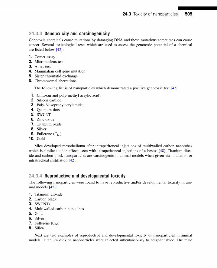
24.3.3 Genotoxicity and carcinogenicityGenotoxic chemicals cause mutations by damaging DNA and these mutations sometimes can cause
cancer. Several toxicological tests which are used to assess the genotoxic potential of a chemical
are listed below [42]:
1. Comet assay
2. Micronucleus test
3. Ames test
4. Mammalian cell gene mutation
5. Sister chromatid exchange
6. Chromosomal aberrations
The following list is of nanoparticles which demonstrated a positive genotoxic test [42]:
1. Chitosan and poly(methyl acrylic acid)
2. Silicon carbide
3. Poly-N-isopropylacrylamide
4. Quantum dots
5. SWCNT
6. Zinc oxide
7. Titanium oxide
8. Silver
9. Fullerene (C60)
10. Gold
Mice developed mesothelioma after intraperitoneal injections of multiwalled carbon nanotubes
which is similar to side effects seen with intraperitoneal injections of asbestos [48]. Titanium diox-
ide and carbon black nanoparticles are carcinogenic in animal models when given via inhalation or
intratracheal instillation [42].
24.3.4 Reproductive and developmental toxicityThe following nanoparticles were found to have reproductive and/or developmental toxicity in ani-
mal models [42]:
1. Titanium dioxide
2. Carbon black
3. SWCNTs
4. Multiwalled carbon nanotubes
5. Gold
6. Silver
7. Fullerene (C60)
8. Silica
Next are two examples of reproductive and developmental toxicity of nanoparticles in animal
models. Titanium dioxide nanoparticles were injected subcutaneously to pregnant mice. The male
50524.3 Toxicity of nanoparticles

offspring were found to have changes to their genital and cranial nervous system as well as accu-
mulation of titanium dioxide in their testes and brain in 6-week-old offspring [49]. This data sug-
gests that titanium dioxide may harm the developing mouse embryo. Pregnant mice were injected
intraperitoneally with fullerene (C60) [50]. At a dose of 137 mg/kg of body weight, all the embryos
died. At a dose of 50 mg/kg of body weight, 50% of the embryos were abnormal in shape espe-
cially in the head and tail areas. A clinical concern with these two examples is that nanoparticle
drug delivery systems might have teratogenic effects in pregnant women who are exposed to them.
24.4 ConclusionsEven though there are wide arrays of promising applications, a concern with the use of orally deliv-
ered nanoparticle drug delivery systems in dentistry is that the nanoparticles might be inherently
toxic. Many toxicology studies on nanoparticles are with the inhalation of particles with a size of
10 μm or less. Three important factors that can contribute to the toxicity of a nanoparticle are size,
shape, and charge. Two common major mechanisms of toxicity are cell lysis and apoptosis. The
absorption, distribution metabolism, and excretion, i.e., toxicokinetics can affect the toxicity of
nanoparticles. A broad classification of toxic effects, i.e., toxicodynamics caused by nanoparticles
are acute and chronic, genotoxic and carcinogenic, and reproductive and developmental. The major-
ity of the toxicology literature is in animal models with few examples in humans. More rigorous
studies need to be conducted on the safety of nanoparticle drug delivery systems when used in den-
tistry by clinicians and scientists of these important drug delivery systems in order to safeguard the
patient’s health.
References[1] Y.V. Pathak, D. Thassu, Drug Delivery Nanoparticles: Formulation and Characterization, Informa
Healthcare, New York, NY, 2009. p. 1�16.
[2] Y.V. Pathak, H. Tran, Advances in Nanotechnology and Applications, vol. 1, Centera, Louisville, KY,
2009. p. 18, 185.
[3] Y.V. Pathak, H. Tran, Advances in Nanotechnology and Applications, vol. 2, Centera, Louisville, KY,
2010. p., 4�5, 121�136.
[4] D. Thassu, M. Deleers, Y.V. Pathak, Nanoparticulate Drug Delivery Systems, 1�32, Informa Healthcare,
New York, NY, 2009 185�212.
[5] A. Renugalakshmi, T.S. Vinothkumar, D. Kandaswamy, Nanodrug delivery systems in dentistry: a review
on current status and future perspectives, Curr. Drug Deliv. 8 (5) (2011;) 586�594. Review. PubMed
PMID: 21696348.
[6] S.R. Kumar, R. Vijaylakshmi, Nanotechnology in dentistry, Indian J. Dent. Res. 17 (2006) 62�69.
[7] M. Goldberg, R. Langer, X. Jia, Nanostructured materials for applications in drug delivery and tissue
engineering, J. Bio. Mat. Sci Polym. Ed. 18 (2007) 241�268.
[8] T.J. Webster, R.W. Siegel, R. Bizios, Design and evaluation of nanophase alumina for orthopaedic/dental
applications, Nanostruct. Mater. 12 (1999) 983�986.
[9] J.P.K. Tan, M.B.H. Tan, M.K.C. Tam, Application of nanogel systems in the administration of local anes-
thetics, Local Reg. Anesth. vol. 3 (August) (2010) 1�8.
506 CHAPTER 24 Nanoparticles: Dental Applications and Toxicity
[10] J.P.K. Tan, A.Q.F. Zeng, C.C. Chang, K.C. Tam, Release kinetics of procaine hydrochloride from pH-
responsive nanogels: theory and experiments, Int. J. Pharm. 357 (2008) 305�313.
[11] A.A. Date, M.S. Nagarsenker, Parenteral microemulsions: an overview, Int. J. Pharm. 355 (2008)
19�30.
[12] J.M. Lee, K.M. Park, S.J. Lim, M.K. Lee, C.K. Kim, Microemulsion formulation of clonixic acid: solu-
bility enhancement and pain reduction, J. Pharm. Pharmacol. 54 (2002) 43�49.
[13] L.M.A. Pinto, L.F. Fraceto, M.H.A. Santana, T.A. Pertinhez, S.O. Junior, E. de Paula, Physico-chemical
characterization of benzocaine-β-cyclodextrin inclusion complexes, J. Pharm. Biomed. Anal. 39 (2005)
956�963.
[14] M.D. Blanco, M.V. Bernardo, R.L. Sastre, R. Olmo, E. Muniz, J.M. Teijon, Preparation of bupivacaine-
loaded poly(ε-caprolactone) microspheres by spray drying: drug release studies and biocompatibility,
Eur. J. Pharm. Biopharm. 55 (2003) 229�236.
[15] P.L. Corre, J.P. Estebe, R. Clement, et al., Spray-dried bupivacaine-loaded microspheres: in vitro evalua-
tion and biopharmaceutics of bupivacaine following brachial plexus administration in sheep, Int.
J. Pharm. 238 (2002) 191�203.
[16] K. Tomoda, M. Asahiyama, E. Ohtsuki, T. Nakajima, H. Terada, M. Kanebako, et al., Preparation and
properties of carrageenan microspheres containing allopurinol and local anesthetic agents for the treat-
ment of oral mucositis, Colloids Surf. B 71 (2009) 27�35.
[17] M.V. Bernardo, M.D. Blanco, R. Olmo, J.M. Teijon, Delivery of bupivacaine included in poly(acrylam-
ide-co-monomethyl itaconate) hydrogels as a function of the pH swelling medium, J. Appl. Polym. Sci.
86 (2002) 327�334.
[18] C.M. Moraes, R. de Lima, A.H. Rosa, E. de Paula, L.F. Fraceto, Encapsulation of local anesthetic bupi-
vacaine in biodegradable poly(DL-lactide-co-glycolide) nanospheres: factorial design, characterization
and cytotoxicity studies, Macromol. Symp. 281 (2009) 106�112.
[19] P.S. Madhu, S. Setty, S. Ravindra, Dentinal hypersensitivity?—Can this agent be the solution? Indian
J. Dent. Res. 17 (2006) 178�184.
[20] M. Addy, E.G. Absi, D. Adams, Dentin hypersensitivity. The effects in vitro of acids and dietary sub-
stances on root-planed and burred dentin, J. Clin. Periodontol. 14 (1987) 274�279.
[21] M. Brannstrom, G. Johnson, Effects of various conditioners and cleaning agents on prepared dentin sur-
faces: a scanning electron microscopic investigation, J. Prosthet. Dent. 31 (1974) 422�430.
[22] R.H. Dababneh, A.T. Khouri, M. Addy, Dentin hypersensitivity—an enigma? A review of terminology,
mechanisms, aetiology and management, Br. Dent. J. 187 (1999) 606�611.
[23] R.A. Freitas Jr, Nano-dentistry, J. Am. Dent. Assoc. 131 (2000) 1559�1566.
[24] R.A. Freitas Jr, Micro biovores artificial mechanical phagocytes using digest and discharge protocol,
J. Evol. Technol. 14 (2005) 1�52.
[25] T.S.V. Satyanarayana, R. Rai, Nanotechnology: the future, J. Interdiscip. Dent. 1 (2) (2011) 93�100.
[26] A.V. Rybachuk, I.S. Chekman, T.Y. Nebesna, Nanotechnology and nanoparticles in dentistry,
Pharmacol. Pharm. 1 (2009) 18�21.
[27] K. Jayaraman, M. Kotaki, Y. Zhang, et al., Recent advances in polymer fibers, J. Nanosci. Nanotechnol.
4 (2004) 65�67.
[28] R.L. Price, K. Ellison, K.M. haberstroh, et al., Nanometer surface roughness increase select osteoblasts
adhesion on carbon nanofiber compacts, J. Biomed. Mater. 70 (2004) 38�40.
[29] M. Zamani, M. Morshed, J. Varshosaz, M. Jannesari, Controlled release of metronidazole benzoate from
poly epsilon-caprolactone electrospun nanofibers for periodontal diseases, Eur. J. Pharm. Biopharm. 75
(2010) 179�185.
[30] R. Krishnamurthy, M. Manning, The stability factor: importance in formulation development, Curr.
Pharm. Biotechnol. 3 (2002) 361�371.
507References

[31] J. Elisseeff, W. McIntosh, K. Fu, T. Blunk, R. Langer, Controlled-release of IGF-I and TGF-β1 in a
photopolymerizing hydrogel for cartilage tissue engineering, J. Orthop. Res. 19 (2001) 1098�1104.
[32] G. Wei, P.X. Ma, Macroporous and nanofibrous polymer scaffolds and polymer/bone-like apatite com-
posite scaffolds generated by sugar spheres, J. Biomed. Mater. Res. A 78 (2006) 306�315.
[33] Q. Jin, G. Wei, Z. Lin, J. Sugai, S. Lynch, P.X. Ma, Nanofibrous scaffolds incorporating PDGF-BB
microspheres induce chemokine expression and tissue neogenesis in vivo, PloS One 3 (2008) e1729.
[34] A. Christoph, J. Roland, J.S. Roswitha, B. Christian, H. Julia, E. Wolf, et al., Magnetic drug targeting—
biodistribution of the magnetic carrier and the chemotherapeutic agent mitoxantrone after loco-regional
cancer treatment, J. Drug Target. 11 (3) (2003) 139�149.
[35] A.A. Bhirde, V. Patel, J. Gavard, G. Zhang, A.A. Sousa, et al., Targeted killing of cancer cells in vivo
and in vitro with EGF-directed carbon nanotube-based drug delivery, ACS Nano 3 (2009) 307�316.
[36] S. Costigan, The toxicology of nanoparticles used in healthcare products. ,http://www.mhra.gov.uk/
Safetyinformation/Generalsafetyinformationandadvice/Technicalinformation/Nanotechnology/index.htm/.,
2006 (accessed 05.09.12).
[37] W. DeJong, P. Borm, Drug delivery and nanoparticles: applications and hazards, Int. J. Nanomed. 3
(2008) 133�149.
[38] V. Olivier, J.L. Duval, M. Hindie, P. Pouletaut, M.D. Nagel, Comparative particle-induced cytotoxicity
toward macrophages and fibroblasts, Cell Biol. Toxicol. 19 (3) (2003) 145�159.
[39] K.H. Park, M. Chhowalla, Z. Iqbal, F. Sesti, Single-walled carbon nanotubes are a new class of ion chan-
nel blockers, J. Biol. Chem. 278 (50) (2003) 50212�50216.
[40] A. Radomski, P. Jurasz, D. Alonso-Escolano, M. Drews, M. Morandi, T. Malinski, et al., Nanoparticle-
induced platelet aggregation and vascular thrombosis, Br. J. Pharm. 146 (6) (2005) 882�893.
[41] D. Shen, Toxicokinetics, in: C. Klassen, J. Watkins, III (Eds.), Casarett and Doull’s Essentials of
Toxicology, second ed., McGraw-Hill, NewYork, NY, 2010 (online access).
[42] J. Zhou, V. Castranova, Toxicology of nanomaterials used in nanomedicine, J. Toxicol. Environ. Health
Part B 14 (2011) 593�632.
[43] V.E. Kagan, N.V. Konduru, W. Feng, B.L. Allen, J. Conroy, Y. Volkov, et al., Carbon nanotubes
degraded by neutrophil myeloperoxidase induce less pulmonary inflammation, Nat. Nanotechnol. 5
(2010) 354�359.
[44] M. Cho, W.S. Cho, M. Choi, S.J. Kim, B.S. Han, S.H. Kim, et al., The impact of size on tissue distribu-
tion and elimination by single intravenous injection of silica nanoparticles, Toxicol. Lett. 189 (2009)
177�183.
[45] G. Oberdorster, E. Oberdorster, J. Oberdorster, Nanotoxicology: an emerging discipline evolving from
studies of ultrafine particles, Environ. Health Perspect. 113 (2005) 823�839.
[46] J.I. Phillips, F.Y. Green, J.C. Davies, J. Murray, Pulmonary and systemic toxicity following exposure to
nickel nanoparticles, Am. J. Ind. Med. 53 (2010) 763�767.
[47] M.L. Schipper, N. Nakayama-Ratchford, C.R. Davis, N.W. Kam, P. Chu, Z. Liu, et al., A pilot toxicol-
ogy study of single-walled carbon nanotubes in a small sample of mice, Nat. Nanotechnol. 3 (2008)
216�221.
[48] C. Poland, R. Duffin, I. Kinloch, A. Wallace, A. Seaton, V. Stone, et al., Carbon nanotubes introduced
into the abdominal cavity of mice show asbestos-like pathogenicity in a pilot study, Nat. Nanotechnol. 3
(2008) 423�428.
[49] T. Ken, S. Ken-ichiro, I. Aki, K.-I. Miyoko, F. Rie, T. Masako, et al., Nanoparticles transferred from
pregnant mice to their offspring can damage the genital and cranial nerve systems, J. Health Sci. 55
(2009) 95�102.
[50] T. Tsuchiya, I. Oguri, Y.N. Yamakoshi, N. Miyata, Novel harmful effects of fullerene on mouse embryos
in vitro and in vivo, FEBS Lett. 393 (1996) 139�145.
508 CHAPTER 24 Nanoparticles: Dental Applications and Toxicity

Index
Note: Page number followed by “b”, “f” and “t” refer to boxes, figures and tables, respectively.
AA549 cells (lung adenocarcinoma cells), 276
Acid etching, 326
Acid-related destruction of dental substance, 169�170
Acrylic-based tissue conditioner, 284f
Aero OT, dioctyl sulfosuccinate sodium salt (AOT), 74
Agar disk diffusion test (ADT), 120
Aggregatibacter actinomycetemcomitans, 205, 487�488
Ag-loaded nanotubes, 351
Ag-tissue conditioner composites, characterization of,
289�292
Ag1 determination from specimens, 289�291, 290f, 291f
energy dispersive X-ray (EDX) studies, 291�292
microstructure of Ag-tissue conditioner, 291�292
AIBN (2,20-azobisisobutyronitrile), 42�43
Alginate, 401�402
Alignments techniques in CNT�polymer composite, 39�40
Alkaline phosphatase (ALP) activity by osteoblastic cells, 326
Al/Si ratio, 96
Alumina/zirconia nanocomposites, 29�30
Amine-functionalized MWNT, 40
Ammonium citrate, 19
Amorphous calcium phosphate (ACP), 25, 27
Anodic aluminum oxide (AAO), 76�77
Anodization, 326
Antiadhesive nanoparticles
calcium phosphate-based systems, 217�218
chitosan nano- and microparticles, 215�216
hydroxyapatite, 217�218
silica and silicon nanoparticles, 216�217
Antiadhesive surface coatings, 24�25
Antibacterial adhesive, 120�123
adhesive containing NACP, 123�124
dentin shear bond strength, 120�123
MTT metabolic activity of microcosm biofilms, 123, 124f
QADM�NAg primer, 120�123
of uncured primers, 121f
Antiinfective therapy, 484
Antimicrobial agents, beneficial effects of, 283�285
Apatite nanoparticles, 176
As-alloyed HA composite coatings, 368�370
ASPA. see Glass ionomer cement (GIC)
As-prepared CNTs (AP-CNTs), 364
Atomic force microscopy (ATM)
of dental adhesives, 150f
of orthodontic brackets and archwires, 233�234, 233f
Autogenous bone grafts for periodontal regeneration, 311
Avidin�biotin couple, 77�78
BBacillus subtilis, 210�211
Bacterial multidrug efflux pumps, 191
Beckman LS-5000TA liquid scintillation counter, 239�241
Bioactive glass nanoparticles
in alveolar bone tissue engineering, 315
antimicrobial effect of, 307
bioactive gelling systems, 316
bioactivity of, 303�305
in bone grafting procedures, 308
in bone regeneration and dental implants, 314�315
chitosan-gelatin/bioactive glass nanoparticles composite
scaffolds, 315
for coating of dental implants, 315
composition and synthesis of, 300�303
in dentistry, 305�309
effects of platelet-rich plasma (PRP) associated with,
310
future of, 315�317
hypersensitive dentin, 307�308
injectable systems, 316, 316f
nanocomposites, 312�314
for osteoconductivity and bonding, 307
in periodontal regeneration, 309�312
Ti/bioactive glass nanocomposites, 315
in tooth replacement procedures, 309
Bioactive silver ions, 29
Biodegradable polymers, 484
Bio-Gides, 392�393
Biomechanics, 260
BioMEMS/NEMS for orthodontic tooth movement and
maxillary expansion, 242�244
Biomimetic HA nanocrystals, 174
Biomimetic mineralization system (BIMIN), 178
Biomimetic remineralization, 155
Biomimetics, 15
Biomineralization of CNTs/CNFs, 374�376
Biphasic calcium phosphate (BCP) ceramic particles,
326�327
BisGMA/HEMA (2,2-bis [4(2-hydroxy-3-methacryloyloxy-
propyloxy)-phenyl] propane/2-hydroxyethyl
methacrylate), 241
Bisphenol A ethoxylated dimethacrylate (BisEMA), 25�27
509
Bisphenol-α-glycidyl methacrylate (bis-GMA), 80
Bite jumping appliances, 251
Blood interactions with implants, 327�328, 327f
Bone�implant contact (BIC), 337�338
Bone metabolism and silica nanoparticles, 84�85
Bone replacement grafts, 414�415
Bony defect replacement therapy, 52
thermal-cross-linking particulate-leaching technique for,
52
Bovine-derived xenograft (Bio-Osss), 392�393
Buffered-peptone water (BPW), 118
CC. albicans adhesion, 82�83, 192�193, 195�196, 216�217
blastospore, 220
Calcination, 76�77
Calcium fluoride (CaF2) preparations, 177
Calcium fluoroaluminosilicate (FAS) glass, 96
Calcium glycerophosphate, 195
Calcium hydroxide (CaOH), 435�436
Calcium lactate, 195
Calcium phosphate-based systems, 217�218
Calcium phosphate (CaP) nanoparticles, 27, 325, 488�489
-coated titanium implants, 332
preparation of, 376�377
properties of, 381�383
Calcium sulfate (CS), nano, 398�405
cell attachment studies using, 403, 403f
human recombinant BMP-2 (rhBMP-2) with, 399
nanocalcium sulfate (nCS) scaffold material, 398�399
for slow release delivery, 399
Candida albicans, 82�83
Candida species and oral bacteria
adhesive interactions between, 283�285
antimicrobial effects of silver nanoparticles, 287�289
Candidiasis, 206�207
Capsets, 400�401
Carbon/graphite fiber-reinforced poly(methyl methacrylate)
(PMMA) denture, 367�368
Carbon nanofibers (CNFs), fabrication of, 371�372
bioglass-embedded, 377�378
biomineralization of, 374�376
decorated, for bone tissue engineering, 381�383
PDLCs culturing of, 381
β-TCP-decorated, 381�383, 381f, 382f
XRD patterns of, 380f
Carbon nanotubes (CNTs), 9, 37�38, 359�360, 433
applications in dentistry, 50�55
biomineralization of, 374�376
cell behaviors and CNFs/CNTs, 366�367
in cell culturing, 364
cytotoxicity of, 372�374
cytotoxic or genotoxic effects of, 48�50
dentistry, CNT/CNF applications in, 367�370
enhanced functions of osteoblasts on carbon
nanomaterials, 361�367
fabrication of, 371
with improved osteogenic bioactivity, 374�380
MWNT/PCU composites and cellular functions, 367
PCU/CNF composites, 365�366
preparation of composites, 38�47, 41t
pristine CNTs/CNFs, 361
sol�gel and electrospinning techniques for, 376�380
techniques of manufacturing, 15�16
three-dimensional CNTs/CNFs, 360�361
three-dimensional scaffolds, 364�365
for tissue regeneration, 365�366
Cartilage tissue engineering, nanotechnology for, 412�414
Casein phosphopeptide-amorphous calcium phosphate
(CPP�ACP) nanocomplexes
treated germanium surfaces, 25
Casein phosphopeptide�amorphous calcium phosphate
(CPP-ACP) nanocomplexes, 170�172, 175, 195,
217�218, 420�421
Casein phosphopeptides (CPP), 25
Cationic liposomes, 13�14
Cell therapy for periodontal regeneration, 311
Ceramic implants, 29�30
Cetylpyridinium chloride (CPC), 109�110, 488
Cetyl-trimethylammoniumbromide (CTAB), 74, 267
Chemical composition of dental adhesives, 138�144
Chemical grafting, 326
Chemical properties, advantages, 4
Chemical vapor deposition (CVD), 5, 370�372, 374�375,
466�467
Chitosan-gelatin/bioactive glass nanoparticles composite
scaffolds, 315
Chitosan nano- and microparticles, 216, 488
Chlorhexidine (CHX), 152�153
Clinical dentistry, nanobiomaterials in, 15�16
CNT�aliphatic polyester composites, 42�43
CNT�PCL composite, 52�53
CNT�polymer composite, 42�43
alignments techniques, 39�40
carboxylic acid groups and, 39
cytotoxic or genotoxic effects of, 48�50
electrical conductivity, 47�48
by electrospun technique, 43�45
by LbL technique, 46�47
mechanical properties of, 47
by melt processing, 40�42
methods of synthesis and dispersibility, 40�42
polymers used, 40
preparation, 38�47
510 Index

properties of, 41t
reinforcement of materials using fillers, 39
by in situ polymerization, 38�39
by solution casting method, 42
use of functionalized nanotubes, 39�40
using in situ polymerization technique, 42�43
van der Waals forces and, 39
CNT�thermosetting polyimide composites, 42�43
Coaggregation, 22�24
Cobalt ferrite (CoFe2O4), 76f
Composite resin technology, 27�28
Conductive properties of CNT composites, 47�48
Copper nanoparticles, 210�211
Copper oxide (CuO) nanoparticles, 211�212
Craniofacial bone defects, nanotechnology for, 412�414
Cytosol, 14
Cytotoxicity of carbon nanomaterials, 372�374
DDCPA-based biocomposites, 421
Deionized water, 270�271
Demineralization effects, 174�175, 195
Dental adhesives
antimicrobial orthodontic appliance, 156
atomic force microscopy of, 149�151, 150f
biomimetic remineralization, 155
brief history, 132�133
chemical composition, 138�144
contemporary, 133�138
dental collagen network improving methods, 154
etch-and-rinse, 135
ethanol-wet bonding technique, 153�154
extended polymerization time, 152
fillers, 140�144
future prospective of nanotechnology, 155�159
glass ionomer, 137�138
high-speed AFM (HS-AFM) of, 157�159, 159f
improved dentin impregnation methods, 153
incorporation of hydrophilic monomer blends, 151�152
Ketac nanoprimer, 145�146
monomers, types of, 139�140
nanophosphates, 195�196
on-demand antibacterial, 155
radioplaque, 156
resin polymerization of, 155�156
self-adhesive composites, 156
self-etching, 135�137
self-healing, 156�157
solvents, 140
strategies to improve, 151�155
TEM micrographs of, 143f, 144f
use of MMPs inhibitors, 152�153
Dental caries, 204�206
formation of, 22�24
and phosphates, 195�196
Dental collagen network, improved, 154
Dental fluorosis, 179
Dental hard substances, regeneration of, 178�179
Dental implants, 337�338
bacterial colonizations and, 323
CaP-coated titanium implants, 332
implant-associated infection, 338
mesenchymal stem cells and, 328�330
osseointegration of, 323�324
surface interactions with blood, 327�328, 327f
tissue integration of, 324f, 330�332
titanium (Ti) alloys, use of, 337�338
Dental nanocomposites, 25�28
antibacterial adhesive, 120�123
antibacterial dentin primer, 118�120
antibacterial nanocomposite with CaP nanoparticles,
111�113
chemical structures of monomers used in, 26f
popularity, 109�110
water-aged specimens, durability of, 114�118
Dental plaque, 22�24
microcosm biofilm model, 120
Dental pulp stem cells (DPSCs), 444
Dental treatment methodologies, modern,
dental nanocomposites, 25�28
easy-to-clean coatings, 24�25
historic progress, 21
nanobiomaterials in clinical dentistry, 15�16, 21�22
nanocomposite surface coatings, 24�25
nano-enabled approaches for biofilm management, 25
preventive dentistry, 22�25
Dentin-adhesive interfaces, 125f
Dentine remineralization, 421
Dentin hypersensitivity, treatment of, 177�178, 307�308,
308f
Dentin impregnation methods, 153
Dentin matrix protein (DMP1), 83
Denture-induced stomatitis, 284f
Denture stomatitis, 206�207
Dexamethasone (DEX)-loaded PLGA nanoparticles, 488�489
Diamond-like carbon (DLC) coatings, 262�263
Diamonds, in dental applications, 501
Dicalcium phosphate anhydrous (DCPA), 27
1,2-didecanoyl-sn-glycero-3-phosphocholine (DOPE), 13�14
Dip-pen nanolithography (DPN), 10
applications of, 11f
functions, 10
Disinfecting root canal
chitosan (CS-np), 436
511Index

Disinfecting root canal (Continued)
gutta-percha, 436
irrigants, 434�435
medicaments, 435�436
nanocrystalline tetracalcium phosphate, 437
obturation materials, 436�437
poly(lactic-co-glycolic acid) (PLGA) nanoparticles, 435
retro-filling and root-repair materials, 437�440
sealer materials, 437
zinc oxide (ZnO-np), 436
DNA Genotek’s OraGenes Collection Device, 461
Doxils, 14
Drexler, Dr. K. Eric, 3�4, 17�18
Drug delivery systems, nanoparticle-based, 12, 499,
501�502
advantages, 485
dental applications of, 482�491
electrospun scaffolds, 44
liposomes, biological functionality of, 13
poly(D,L-lactide) acid (PLA), 485
poly(D,L-lactide-co-glycolide) acid (PLGA), 485
poly(glycolic) acid, 485
polymeric-based, 484�485
proteins and DNA, 54�55
treatment of oral cancer, 502
for the treatment of periodontal diseases, 484�485
use of polymer nanofiber materials, 21
via diffusion for nondegradable polymers, 44�45
Drug-loading NTs, 350�355
Dry lubricants, 234�236
EEasy-to-clean coatings, 24�25
Eikenella corrodens, 205
Elastomeric ligatures, 241�242
Electrospinning, 38�39, 43�45
alignment of nanotubes, 45
of core-shell fibers, 44
incorporation of biological molecules, 44
influencing parameters, 44
orientation of the fiber, 45
PS/CNT electrospun fibers, 44�45
schematic representation of, 43f
surface area�volume ratio of meshes, 44�45
syringe�capillary setup, 43
Taylor cone, 43�44
Enamel demineralization around orthodontic attachments,
management of, 250
Endocytosis, 77
Endodontics, 440�441
Endogenous regenerative technology (ERT), 415
Engineered nanomaterials, 70
Engines of Creation: The Coming Era of Nanotechnology,
3�4
ENPLATE Ni-425, 267
Enterococcus faecalis, 217
Epidermal growth factor (EGF), 55
Escherichia coli, 208, 210�212, 217
Etching-bonding procedures on the tooth enamel, 260
Ethanol-wet bonding technique, 153�154
Ethoxylated bis-phenol-adimethacrylate (BisEMA—more
hydrophobic), 241
Ethylene glycol (EG), 339�340
FFeynman, Richard Phillips, 3�4, 11, 18, 231�232
Fibronectin, 327�328
Fillers in dental adhesives, 140�144
Filtek Supreme Plus Universal, 239
Fluidic properties, advantages, 4
Fluoroalkylated acrylic acid oligomers (FAAO), 173
Fluoroapatite formation, 179
Fluoro hydroxyapatite (FAP), 195
Freeze-dried bone allograft (FDBA), 311
Friction in orthodontics, 260�263
DLC coatings, impact of, 262�263
improvements to reduce, 262�263
material technologies to reduce, 263�266
self-ligating brackets, impact of, 262
surface modification, impact of, 262
Friction resistance to sliding (RS), 260�261
binding (BI) component, 260�261
classical friction (FR), 260�261
factors influencing RS components, 262
notching (NO) component, 260�261
Functional appliances (FAs), 251
Functionalized CNT, 55
Functionalized SWCNT, 50, 54
GGEM21s, 400�401
“Generally regarded as safe” (GRAS) agent, 70
Gene therapy, 13�14, 414
in orthodontics, 251�252
Geristores, 438
Gingival crevicular fluid (GCF), 204
Gingivitis, 482�483
Glass ionomer cement (GIC), 95�97, 236�238
Al/Si ratio in, 96
amount of CaF2, 96
components, 96
factors influencing setting and mechanical properties, 97
Filtek Supreme XT, 99�100
Fuji IX GP, 100
512 Index

hydrolytic stability of, 96
limitations, 95�96
3M ESPE�Ketactt N100 (KN), 98
metal-reinforced, 97
modified, 97�98
nanofiller-containing resin-modified, 250
with nanohydroxy and fluoroapatite, 239
nanoparticles-based, 103�105
network-dwelling ions, 96
for orthodontic band cementation, 239
polyelectrolytes used in, 97
resin-modified nano, 98�103, 239
structure of glass particle, 96
Transbond XT, 99�100
Glass ionomer cements (GICs), 15�16
Globally Harmonized Classification System, 275
Glycerin-enriched gelatin gel, 179
Gold nanoparticles, 211
Gottinger Minipig metaphyseal model, 393�395
Graded a-SixCy:H interfacial layers, 5
Greiner Bio-One Saliva Collection System, 458�459
Grit blasting, 326
Guided tissue regeneration (GTR), 311, 414�415
HHarungana madagascariensis, 486�487
HEMA-containing adhesive, 151
Heterogeneous microfills, 27
Homogeneous microfills, 27
H2SO4/NaF/H2O, 339�340
Human dentin shear bond testing, 122f
Human gingival fibroblasts (HGF), preparation of, 289
Hybrid organic/inorganic composites, 29�30
Hydrodynamic theory, 307�308
Hydrogel polymer systems, 316
Hydrogenated phosphatidylinositol (HPI), 14
Hydrophilic monomer blends, 151�152
Hydroxyapatite, 217�218, 392�396
Hydroxyapatite (HA), 325
nanocrystallites, 169�171
Hydroxyapatite (HAP), 195
nanocrystallite particles, 25
Hypersensitivity, management of, 177�178, 307�308, 500
bioactive glass nanoparticles, 307�308
IIF-WS2 nanoparticle, 234�236, 235f
Iliac bone marrow, 311
Imidazoline, 19
Immunalysis Quantisalt Collection Device, 460�461
In situ polymerization of CNT composites, 38�39, 42�43
Instron 4502, 270
Intercepts Collection Device, 458
Ionic implantation, 326
KK562 cells (leukemic cells), 276
Ketac N100, 250
Ketac nanoprimer, 145�146
Kinetic friction, 260�261
LLactobacillus spp., 206
Layer-by-layer (LbL) assembly technique, 46�47
composites based on hydrogen bonding, 46
3D scaffold, 52�53
impact of covalent bonds, 46�47
Liposomes, biological functionality of, 13
cationic, 13�14
long-circulating (sterically stabilized), 13
pH-sensitive, 13�14
thermosensitive, 13�14
ultradeformable, 13�14
LIPUS treatment, 252, 253f
Lithography, 5, 10
Lotus effect, 173�174, 180
Lyophilized bone graft, 311
Lysozyme, 205
MMacrofilled composites, 238�239
Malocclusions, 250
Material science, classification of, 93�94
Material technologies to reduce friction, 263�266
appliances coated with nanoparticles, 266�274
inorganic fullerene-like (IF) nanoparticles, 263�265
self-lubricating surfaces, 265�266
Matrix metalloproteinases (MMPs), 147�149
inhibitors, 152�153
Mechanical properties, advantages, 4
Melanocortin peptides (alpha-MSH), 442
Melt processing of CNT, 40�42
Mesenchymal stem cells and dental implants, 328�330
differentiation into fibroblastic lineage and fibroblastic
adhesion, 329�330
migration, adhesion, and proliferation of, 329
origin of MSCs, 329
Mesoporous silica nanoparticles (MSNs), 72
Metastable ACP nanoprecursors, 421
Methacryloxylethyl cetyl dimethylammonium chloride
(DMAE-CB), 109�110
γ-methacryloxypropyltrimethoxysilane (γ-MPS), 78
12-methacryloyloxydodecylpyridinium bromide (MDPB),
109�110
513Index

Methicillin-resistant Staphylococcus aureus (MRSA), 208
Methylene blue (MB), 489
Microbial biofilms, 190�191
antimicrobial tolerance of, 191
C. albicans and C. glabrata biofilms, 192�193, 192f
effects of TMP, 195�196
grown on hydroxyapatite chips in situ, 191f
nano-Ag for Escherichia coli biofilms, 192�193
Pseudomonas fluorescens biofilms, 193
silver nanoparticles and, 192�194
Microelectro mechanical systems/nanoelectro mechanical
systems (MEMS/NEMS), 16
Microfilled composites, 27�28
Micro-HA added GIC group, 102�103, 102f
Micrometer-sized HAP, 25
Mineral trioxide aggregate (MTA), 438
Minifilled composites, 238�239
Minimal bactericidal concentration (MBC), 190, 207�208
Minimal inhibitory concentration (MIC), 190, 192, 207
Monodisperse nanoparticles, 19
Monosialoganglioside (GM1), 14
MTT (3-(4,5-dimethylthiazol-2-yl)-2,5-diphenyltetrazolium
bromide) assay, 118
MTT metabolic activity of microcosm biofilms, 123
Multilamellar vesicles (MLVs), 13
Multiwalled carbon nanotubes (MWCNTs), 9, 40, 43, 47,
51�52, 359�361
chitosan scaffold, 54
coated sponges, 364�365
PEGylation of, 375
in PMMA-based bone cement, 51
sodium dodecyl sulfate (SDS), 375
NNaH2PO4/HF, 339�340
Nanoantimicrobials
antimicrobial nanoemulsions, 172
biocompatibility of, 219�221
orthodontic appliance, 156
Nanobiomaterials in clinical dentistry, 21�22
agglomeration state of, 196
in biomimetic enamel regeneration, 417�419
in bone regeneration, 29�30
cytotoxicity of carbon nanomaterials, 372�374
in dentin�pulp complex regeneration, 421�423, 422f
in enamel and dentine remineralization, 419�421
future directions, 198
in preventive dentistry, 22�25
pros and cons, 196�198
in restorative dentistry, 25�29
Nanocalcium sulfate (nCS) scaffold material, 398�399
Nanoceramics and bone repair, 391�392
available for clinical use, 405t
calcium sulfate (CS), 398�405
chitosan�gelatin/nano-bioactive glass ceramic composite,
398
hydroxyapatite, 392�396
nano-HA�collagen composites, 397
Nanoclusters, 28
Nanocomposite surface coatings, 24�25
Nano-DCPA-based biocomposites, 421
Nanoengineering, 18
Nano-FA/ionomers, 100�101
Nanofibers, 21
Nanofiller-containing resin-modified glass ionomer cements,
250
Nanofills, 239
NanoGens, 404�405
Nano-HA-added GIC group, 102�103, 102f
Nano-HA�collagen composites, 397
Nano-HA/ZrO2, 103
Nanohybrids, 239
Nanoleakage, 147�149, 148f, 152f
Nanomanufacturing technology, 5, 12f
Nanomechanical sensors, 253�255
modulus of elasticity and geometry, 253
wire stiffness, 253, 254f
Nanoparticle-reinforced hybrid system, 95
Nanoparticles, 20
of amorphous calcium phosphate (NACP), 110
carcinogenicity of, 505
characterization, 20
definitions, 479�481
dental applications of, 482�491
genotoxicity of, 505
polymeric, 484�489
from preformed polymers, 481f
reproductive and/or developmental toxicity of, 505�506
toxicity of, 502�506
toxicokinetics of, 503
Nanoparticulate metal oxides as antimicrobial agents
copper oxide (CuO and Cu2O), 211�212
oral applications, 213�214
quaternary ammonium poly(ethylene imine) (QA-PEI),
215�216
titanium dioxide (TiO2), 213
zinc oxide (ZnO), 212
Nanoparticulate metals as antimicrobial agents, 207�211
copper (Cu), 210�211
gold (Au), 211
oral applications, 213�214
silver (Ag), 210
Nanophase alumina for dental applications, 501
Nanophase ceramics, 412�413
514 Index

Nano-RMGI primer, 145�146
Nanorobotic dentifrices, 500
Nanorobots, 244
Nanoscale, defined, 18�19
Nanoscale materials, 18�21
characterization, 20
nanofibers, 21
nanoparticles, 20
orthodontics, 250
Nanoscale surface modifications, 326�327
Nanoscience
defined, 188�189
impacts, 189
Nanosized calcium fluoride, 177
Nanosized YSZ�GIC composites, 103�105, 104f
NanOss Bioactive Loadeds, 392
NanOsss bone void filler, 392, 413�414
Nanotechnology
approaches to, 4
biomimetic scaffold mimicking features, 415
“bottom-up” approach to, 4, 6�10, 22, 232
clinical applications, 433�440
for craniofacial bone and cartilage tissue engineering,
412�414
defined, 432
definitions, 4
in dentistry, 11�12, 94�95, 478f
in durability and cosmetics applications, 501
nanobiomaterials in clinical dentistry, 15�16
nanoparticle-based drug delivery systems, 12
nanoscale sapphire and diamonds, 501
for periodontal regeneration, 414�416, 416f
properties, 4
for repair and pulp regeneration, 441�444
for tooth regeneration, 417�423
“top-down” approach to, 4�5, 22, 232
Nanotubes
Ag-loaded, 351
drug loading, bioactivity and antibacterial properties of,
350�355
MSC attachment and spreading, 348
osteoblast functions, effect on, 347, 347f
osteogenesis-inducing ability of, 348
Sr-loaded, 351�354
in vitro bioactivity of, 346�348
in vivo osseointegration of, 348�350
Zn loaded, 354�355
Nanotubes, factors influencing bioactivity of
cell/implant interactions, 342�344
cell phenotype, 342
dermal fibroblasts, 342
dry autoclaving, 340�342
endothelial cells (ECs), 342
epidermal keratinocytes, 342
MSCs, 342
osteoblast cell lines, 342
protein concentration, 342�344
protein distribution pattern, 344�346
sterilization, 340�342, 341f
ultraviolet (UV) irradiation, 340
vascular smooth muscle cells (VSMCs), 342
wet autoclaving, 340�342
NaOCl/phosphoric acid, 153
Na2SO4/HF, 339�340
N-[1-(2,3-dioleoyloxy)propyl] N,N,N-trimethylammonium
chloride (DOTAP), 13�14
N-halamine-functionalized silica core-shell nanoparticles,
82�83
Nickel�titanium (Ni�Ti) archwire, 260�261
Nickel�titanium (Ni�Ti) wires, 234
Niobium FAS glass powder, 105
Ni�Ti-based medical devices, 267, 268f, 269f
adhesion and wearing, 268�269
Ni�Ti wires with cobalt and IF-WS2 film, 267�268,
272�274
Nitric acid�treated SWNTs (SWNT-COOH), 364
Nitric oxide, 75
Nitric oxide (NO), 82�83
Nonfluoride therapeutic agents, 195
Nonmesoporous silica-based nanomaterials, 77
Nonpolymeric nanoparticles, 489�491
OOleic acid, 14
Open flap debridement (OFD), 414�415
Optical properties, advantages, 4
Oral biocatalytic fuel cell, 243f
Oral biofilms, 190
as antimicrobial agents, 207�211
and candidiasis, 206�207
control of, 207
and dental caries, 206
formation and properties of, 205�206
and infections, 205�207
and peri-implantitis, 206
and periodontal disease, 206
photodynamic therapy (PDT) and, 218�219
therapeutic use of light-activated killing of, 218�219
Oral cavity, bioadhesion and biofilm management, 23f
Oral Fluid NanoSensor Test (OFNASET) technology,
465
Oral health-care products. see Dental treatment
methodologies, modern
OraSures HIV-1 Oral Fluid Collection Device, 458
515Index

Organic polymer matrix (2,2-bis[p-(20-hydroxy-30methacryloxypropoxy) phenylene] propane
(BisGMA), 25�27
Orthodontic appliances coated with nanoparticles
adhesion and wearing issues, 268�269
challemges in designing, 266�267
coating process and tribological measurements, 267�268
due to Co/IF-WS2 coatings, 274
Ni�Ti wires with cobalt and IF-WS2 film, 267�268, 268f
safety of, 274�276
SS wires with IF-WS2, impregnated, 267, 270�272, 271t
summary of friction measurements, 273�274, 273t
toxicity and biocompatibility, 274�276
tribological Tests, 271t
in vitro friction force tests, 270�274, 271f
Orthodontic brackets and archwires
atomic force microscopy (ATM) studies, 233�234
beta-titanium alloy wires, 234
and dry lubricants, 234�236
friction reducing nanocoatings on, 234�236
Ni�Ti archwire, 233f, 234
self-ligating ceramic bracket, 234
surface characteristics, 233�234
Orthodontics
adhesives, 236�241
application of orthodontic force, 260
BioMEMS/NEMS for orthodontic tooth movement and
maxillary expansion, 242�244
BisGMA/HEMA (2,2-bis [4(2-hydroxy-3-
methacryloyloxy-propyloxy)-phenyl] propane/2-
hydroxyethyl methacrylate), 241
brackets, 233�234
elastomeric ligatures, 241�242
ethoxylated bis-phenol-adimethacrylate (BisEMA—more
hydrophobic), 241
external apical root resorption (EARR) and, 252
friction in, 260�263
future applications of nanotechnology, 255
gene therapy in, 251�252
LIPUS treatment, 252, 253f
nanofabricated ultrasound device for, 252
nanomaterials used in, 237t
nanomechanical sensors, 253�255
nanorobots, 244
nanoscale in, 250
real-time feedback and, 255
stainless steel (SS) in, 260
temporary anchorage devices (TADs), 244�245
ultrasound device for, 249
urethane dimethacrylate (UDMA—less hydrophobic), 241
use of glass ionomer cement (GIC), 239
use of shape-memory polymer in, 242
Orthodontic sliding mechanics, 261f
Orthodontic tooth movement, initiating, 260
Orthopedic/dental implant applications, 21
Osseointegration, 85
of dental implants, 326�327
Ostims, 392�393, 393f, 395, 413�414
for advanced intrabony defects, 396f
PPalmitoyl homocysteine, 14
PEGylated silica nanoparticles, 84�85
Pellicle, 22�24
formation, 168�169
Peri-implantitis, 206
Peri-implant mucositis, 206
Periodontal disease, 204�206
Periodontal regeneration, nanotechnology for, 414�416, 416f
Periodontitis, 482�483
Perioglasss, 310
Periradicular surgery, 432
Phosphatidylethanolamine, 14
Photodynamic therapy (PDT), 218�219
PH-sensitive liposomes, 13�14
Physical vapor deposition (PVD), 5
Planck constant, 188
Plaque-related dental caries, 204�205
Plasma-sprayed HA-coated dental implants, 326�327
Plaster of Paris, 398�399
Platelet-rich plasma (PRP), 327�328
Poly(acrylic acid) (PAA), 376�377
Polyalkenoate cement. see Glass ionomer cement (GIC)
Poly (allyamine hydrochloride), 46
Polyamide, 40
Polycaprolactone (PCL), 42�43
Polycarbonate (PC), 40
Polycrystalline colloidal structures, 19
Polyesters, 40
Polyethylene, 40, 42�43
Polyethylene glycol (PEG), 14, 78
Poly(ethylene glycol) (PEG) functionalized SWNTs, 364
Poly(3hydroxybutyrate) (P(3HB))/bioactive glass nanoparticle
composite, 313
Poly (lactic-co-glycolic acid) (PLGA)/carboxyl-functionalized
MWCNT (c-MWCNT) nanocomposites, 362�364
Polylactide, 42�43
Poly(L-lactate-co-ε-caprolactone) copolymer, 45
Poly (L-lactic acid) (PLLA), 52
Poly(m-aminobenzene sulfonic acid) functionalized SWNTs
(SWNT-PABS), 364
Polymeric nanoparticles, 484�489
Polymer nanocomposite (PNC), 438
Polymer nanofiber materials, 21
516 Index

Poly(methyl methacrylate) (PMMA) biofilm model, 206�208
Poly-methyl methacrylate (PMMA)-grafted nanoclay fillers,
143
Poly (methyl methylacrylate) (PMMA), 40, 42�43
PMMA-based bone cement, 51
Poly(propylene fumarate) (PPF), 362�364
Poly(propylene fumarate) scaffold, 52
Polypropylene (PP), 40, 42�43
Polystyrene, 40
with different MWCNT concentration, 45f
Poly (styrene sulfonate), 46
Poly-tetrafluoro-ethylene (PTFE) microparticles, 173
Polyurethane, 42�43
Polyurethane/montmorillonite nanocomposites, 314
Poly(vinyl acetate) (PVAc), 376�377
Polyvinyl alcohol (PVA), 313, 376�377
Poly(vinyl pyrolidone) (PVP), 376�377
Porphyromonas gingivalis, 205
Preventive dentistry, 22�25
challenges in, 167�168
clinical recommendations, 179�180
de- and remineralization, management of, 174�175
dentin hypersensitivity, management of, 177�178
erosion, management of, 176
implementation of nanosized materials in dental
prophylaxis, 170
nanosized calcium fluoride, 177
phenomenon of bioadhesion on dental hard tissues,
168�170
regeneration of dental hard substances, 178�179
size-dependent effects of nanomaterials, 170
Prevotella intermedia, 205
Prevotella loescheii, 205
Processing cost of nanomaterials, 197�198
Programmed cell death (apoptosis), 289
Prophylactic toothpaste, 82
ProRoots, 440
PS/CNT electrospun fibers, 44
Pseudomonas fluorescens biofilms, 193
Pseudomonas putida biofilms, 193
Pulp regeneration, 440�441
dental caries and, 441
nanotechnology for, 441�444
QQuantum corrals, 8f
Quantum size effect, 19
Quantum states, 8�9
Quaternary ammonium poly(ethylene imine) (QA-PEI)
nanoparticles, 215�216
Quaternary ammonium salts (QAS), 109�110
Quorum sensing, 22�24
RRadioplaque dental adhesives, 156
Recombinant adenovirus associated virus (rAAV)-mediated
vascular endothelial growth factor (VEGF), 252
Recombinant human bone morphogenetic protein-2 (rhBMP-
2), 54
surgery implantation of, 55f
Re-doped IF-MoS2 NP, dermal toxicity of, 275, 275f
Re-doped MoS2, dermal toxicity of, 275
Remineralizing effects, 174�175, 195
Remineralizing nanoparticles, 123�124
Resin-based composite (RBC) materials, 25�27
Resin-modified nano-glass ionomer composites, 98�103
biaxial flexural strength (BFS), 100�101
BisGMA, 98
bond strength, 99�100
compressive strength (CS), 100�101
diametral tensile strength (DTS), 100�101
G-Coat coating resin, 100
HEMA, 98
particle size distribution, 100�101
resin monomers, 98
TEGDMA, 98
Restorative dentistry, nanobiomaterials in, 25�29, 50�51,
93�94
ACP fillers, 27
dentin, 50�51
dentinal tubule, 94�95
denture base, 51
enamel rod, 94�95
glass ionomer cement (GIC), 95�97
hydroxyapatite (HA) crystal, 94�95
microfilled composites, 27�28
nanocomposites, 25�28
nanofillers, 94�95
periodontal ligament cell (PDLC) adhesion and
proliferation, 52
resin-based composite (RBC) materials, 25�27
silver nanoparticles, 29
smart dental materials, 94
tooth slices coated with CNTs, 51f
Reticuloendothelial system (RES) cells, 13
Revised supersaturated SBF (r-SBF), 375
Root canal obturation materials, 436�437
bioactive glass 45S5, 436�437
gutta-percha, 436
Root canal treatment, 431
disinfecting root canal, 434�436
repair and pulp regeneration, 440�441
retro-filling and root-repair materials, 437�440
root canal obturation materials, 436�437
sealer materials, 437
517Index

Root-end filling materials, 437�440
Root-repair materials, 437�440
Rotary instruments, 433
Ni�Ti instruments, 433�434
Runt-related transcription factor-2 (Runx2), 83�84
SSaliva
for abuse assessment, 458�460
advantages, 454
applications in clinical settings, 457t
barriers to implementation of diagnostics, 455
as a biofluid for disease detection, 456�462
body’s health and well-being, effect on, 454
cotinine assessment, 460�461
C-reactive protein (CRP) levels, 454�455, 463, 464f
diagnostic assays for, 458�462
DNA methylation as a proxy to diagnose HNSCC,
463�465
DNA test, 461
for early detection of IHD and in head and neck cancers,
463�466
ELISA test kits, 460�461, 461t
for HIV assessment, 458
for hormone assessment, 460
and ischemic heart disease (IHD), 454�455
of MEMS/Nano Electromechanical Systems (NEMS) in
diagnostics, 465�466
in molecular diagnostics, 461�462
oral drugs of abuse tests/manufacturers, 459t
production and bimolecular transport, 455�456
in proteomics, 462
research, 462
RNA test, 462
Salivary/dietary-derived proteinaceous layer, 205
Sapphire, in dental applications, 501
Scanning tunneling microscope (STM), 6�8
application, 8�9
Secretory immunoglobulin A (sIgA), 205
Self-adhesive composites, 156
Self-healing adhesives, 156�157
Shape-memory polymers, 242
Silane infiltration, 28
Silane-treated filler, 236�238
Silanol group, 77
Silica nanoparticles, 70�71, 216�217
biocompatibility/toxicology, 85
and bone metabolism, 84�85
composites and functionalization, 74�75
in dental applications, 70
dental applications of, 78�83
dietary silica, 70
dispersibility and purification, 73�74
fluorescent, 80f
as a food additive, 70
fumed silica, 72
mesoporous silica nanoparticles (MSNs), 72
in osseointegration, 85
physicochemical properties of, 75�78
in prophylactic toothpaste, 82
shape, 76�77
size, 75�76
skeletal applications of, 83�85
by sol�gel process, 72�73, 75, 77, 80
by Stober method, 72
surface properties and modifications, 77�78, 79f
synthesis of, 71�75, 72f
Silica nanoparticles, dental applications of, 78�83, 81t
as an antimicrobial agent, 82�83
composite resins, 78�82
as a polishing agent, 82
prophylactic toothpaste, 82
use in bacteria and mammalian cell attachment, 82
Silver (Ag) nanoparticles, 210
Silver 2-ethylhexanoate (Strem), 120
Silver nanoparticles, 29
acrylic tissue conditioner combined with, 287�289
antimicrobial effects on S. aureus, Streptococcus mutans,
and C. albicans, 287�289, 288t
antimicrobial properties of, 283�285
characterization of Ag-tissue conditioner composites,
289�292
colloidal, 287
cytotoxic test on human gingival cell line, 289
for disinfecting root canal, 435
fabrication of conditioner composites, 287
preparation and identification of, 285�286
TEM view of a prepared, 286f
UV-vis spectrum of, 286f
Silver nanoparticles and microbial biofilms, 192�194
mechanism of action, 192�193
processing of, 193�194
Turkevich methods of processing, 194
Silver zeolite, 210
Single-walled carbon nanotubes (SWCNT), 9, 39, 359�360,
362�364
in dentistry, 368
functionalized, 50
SWCNTs/SiO2/ATES, 368
SiO2�Na2O�CaO�P2O5 system, 217
Skeletal applications of silica-based nanomaterials, 83�85
in bone formation, 83�84
in bone metabolism, 84�85
in healing/repair of the jaw, 85
518 Index
modeling and remodeling, 83�84
in osseointegration, 85
osteoblasts and osteoclasts, 83�84
Smart dental materials, 94
Sodium cholate, 14
Sodium trimetaphosphate (TMP), 195
Soft chemistry, 96
Soft-Liners, 287, 289
Sol�gel process, 72�73, 75, 77, 80, 105
Solution processing of CNT composites, 42
Solvents in dental adhesives, 140
Sr-loaded nanotubes, 351�354
Staphylococcus aureus, 217, 283�285
antimicrobial effects of silver nanoparticles, 287�289
Staphylococcus epidermidis, 217
Static friction, 260�261
Stealtht technology, 14
Stents, 7f
Streptococcus mitis, 205
Streptococcus mutans, 29, 110, 206
and durability of antibacterial nanocomposite in water-
aging, 114�118, 115f, 116f, 117f
Streptococcus oralis, 205
Streptococcus sanguinis, 205
Streptococcus sobrinus, 206
TTaniguchi, Norio, 232
Tannerella denticola, 206
Tannerella forsythia, 206
Tantalum butoxide/silicon oxide (Ta2O5/SiO2) nanofillers,
143�144
Tartrate-resistant acid phosphatase (TRAP), 83�84
β-TCP@CNF hybrid nanofibers, 377�378, 379f
Tetracalcium phosphate (TTCP), 27
Tetraethoxysilane, 27
Tetraethyl orthosilicate, 75
Tetramethyl orthosilicate (TMOS), 75
Thermal-cross-linking particulate-leaching technique, 52
Thermal properties, advantages, 4
Thermosensitive liposomes, 13�14
Three-dimensional nanostructure, 6f
TiO2 nanotubes, 326
Tissue engineering, 21�22, 389
bioceramics in, 392
components of, 390f
design criteria for bone scaffolds, 391t
hydrogels, 397�398
hydroxyapatite, 392�396
nano-HA�collagen composites, 397
Tissue integration of dental implants, 324f, 330�332
Tissue regeneration, 315�317
Titania nanotube coatings on dental implants, 337�338
fabrication of, 339�340, 339f
factors influencing bioactivity of, 340�346
Titanium dioxide (TiO2), 213
Titanium implants, 309
Titanium (Ti), 29�30
Tooth decay, early stages of, 24f
Tooth hypersensitivities, 168
Toxicity of nanomaterials, 196�197
Transbond XT, 239
Treponema denticola, 205
β-tricalcium phosphate, 175
Triclosan (2,4,40-trichloro-hydroxydiphenylether) (TCS),485�486
Triethylene glycol dimethacrylate (TEGDMA), 25�27, 80
Tungsten disulfide nanocoating, 236
UUltradeformable liposomes, 13�14
Ultrasound attenuation spectroscopy, 20
Urethane dimethacrylate (UDMA), 25�27
less hydrophobic, 241
VVeillonella atypical, 205
Versi•SALs Collection Device, 458�459
N-Vinylpyrrolidone, 239
Vitronectin, 327�328
Vroman effect, 327�328
WWet and plasma etching, 5
XXerostomia, 24�25
X-ray lithography, 5
YYbF3/BaSO4 nanoparticles, 105
Yttriastabilized ZrO2 (YSZ) powders, 103�105
ZZinc carbonate�HA nanoparticles, 172
Zinc oxide (ZnO) nanoparticles, 212
Zn loaded nanotubes, 354�355
ZrOCl2 solution, 27
519Index